Comprehensive Analysis of MicroRNA (miRNA) Targets in Breast Cancer Cells
15
Comprehensive Analysis of MicroRNA (miRNA) Targets in Breast Cancer Cells * □ S Received for publication, June 6, 2013, and in revised form, August 6, 2013 Published, JBC Papers in Press, August 6, 2013, DOI 10.1074/jbc.M113.491803 Meiyun Fan 1 , Raisa Krutilina, Jing Sun, Aarti Sethuraman, Chuan He Yang, Zhao-hui Wu, Junming Yue, and Lawrence M. Pfeffer From the Departments of Pathology and Laboratory Medicine, Center for Adult Cancer Research, University of Tennessee Health Science Center, Memphis, Tennessee 38163 Background: miRNA deregulation contributes to tumor progression. Results: Endogenous miRNA targets were identified in two breast cancer cell lines by integrated analysis of miRNA/mRNA expression and miRNA-mRNA interaction. Conclusion: miRNAs collectively function to promote survival but suppress cell migration/invasion. Significance: The defined endogenous miRNA targets will facilitate future studies to link miRNA deregulation with breast cancer cell properties. MicroRNAs (miRNAs) regulate mRNA stability and transla- tion through the action of the RNAi-induced silencing complex. In this study, we systematically identified endogenous miRNA target genes by using AGO2 immunoprecipitation (AGO2-IP) and microarray analyses in two breast cancer cell lines, MCF7 and MDA-MB-231, representing luminal and basal-like breast cancer, respectively. The expression levels of 70% of the AGO2-IP mRNAs were increased by DROSHA or DICER1 knockdown. In addition, integrated analysis of miRNA expres- sion profiles, mRNA-AGO2 interaction, and the 3-UTR of mRNAs revealed that >60% of the AGO2-IP mRNAs were puta- tive targets of the 50 most abundantly expressed miRNAs. Together, these results suggested that the majority of the AGO2-associated mRNAs were bona fide miRNA targets. Functional enrichment analysis uncovered that the AGO2-IP mRNAs were involved in regulation of cell cycle, apoptosis, adhesion/migration/invasion, stress responses (e.g. DNA dam- age and endoplasmic reticulum stress and hypoxia), and cell-cell communication (e.g. Notch and Ephrin signaling pathways). A role of miRNAs in regulating cell migration/invasion and stress response was further defined by examining the impact of DROSHA knockdown on cell behaviors. We demonstrated that DROSHA knockdown enhanced cell migration and invasion, whereas it sen- sitized cells to cell death induced by suspension culture, glucose depletion, and unfolding protein stress. Data from an orthotopic xenograft model showed that DROSHA knockdown resulted in reduced growth of primary tumors but enhanced lung metastasis. Taken together, these results suggest that miRNAs collectively function to promote survival of tumor cells under stress but sup- press cell migration/invasion in breast cancer cells. MicroRNAs (miRNAs) 2 are emerging as key modulators of gene expression at the post-transcriptional level by repressing translation and/or inducing mRNA degradation (1, 2). Most miRNAs are initially transcribed as long primary transcripts (pri-miRNAs) that are processed within the nucleus into short stem-loops (pre-miRNAs) by DROSHA, a member of the ribo- nuclease III superfamily of double-stranded RNA-specific endoribonucleases (3). The pre-miRNAs are transported to the cytoplasm and further processed by DICER1, another doubled- stranded RNA-specific ribonuclease, to generate mature miRNAs, which are loaded into the RNA-induced silencing complexes (RISCs) (4). miRNAs recruit mRNA targets to RISCs through Watson-Crick base pairing (2). Computational sequence analysis and experimental evidence suggest that bases 2– 8 at the 5-end of mature miRNAs (termed seed sequences) and their complementary sequences located in the 3-untrans- lated region (3-UTR) of mRNA are the major determinants of miRNA-mRNA interaction (5– 8). A single miRNA can target hundreds of mRNAs, and a single mRNA can be coordinately regulated by multiple miRNAs (7, 9). Approximately 60% of mammalian mRNAs have one or more evolutionarily con- served miRNA target sequences (7). However, it is unclear whether a miRNA exerts its effects via regulating its entire rep- ertoire of targets or a subset of specific effectors in a given cell context. The complexity of miRNA function can hardly be depicted by traditional studies that focus on a single miRNA and its predicted targets one at a time. A prerequisite for under- standing the collective function of endogenous miRNAs is to determine what mRNAs and signaling pathways are targeted by miRNAs under physiologically relevant conditions. Argonaute proteins are the catalytic components of the RISCs for mRNA silencing or destruction. All four human argo- naute proteins (AGO1, AGO2, AGO3, and AGO4) are able to interact with miRNAs as components of RISCs to inhibit trans- lation, but only AGO2 possesses the endoribonuclease activity * This work was supported, in whole or in part, by National Institutes of Health Grants CA140346 (to M. F.) and CA133322 (to L. M. P.). □ S This article contains supplemental Table S1. All expression microarray data were deposited in the Gene Expression Omnibus with accession number GSE48162. 1 To whom correspondence should be addressed: 19 South Manassas St., Memphis, TN 38163. Tel.: 901-448-4192; Fax: 901-448-3910; E-mail: [email protected]. 2 The abbreviations used are: miRNA, microRNA; pri-miRNA, primary miRNA; IP, immunoprecipitation; AGO2-IP, AGO2 immunoprecipitation; RISC, RNAi-induced silencing complex; KD, knockdown; Ab, antibody; qPCR, quantitative PCR; APA, alternative polyadenylation. THE JOURNAL OF BIOLOGICAL CHEMISTRY VOL. 288, NO. 38, pp. 27480 –27493, September 20, 2013 © 2013 by The American Society for Biochemistry and Molecular Biology, Inc. Published in the U.S.A. 27480 JOURNAL OF BIOLOGICAL CHEMISTRY VOLUME 288 • NUMBER 38 • SEPTEMBER 20, 2013 at UTHSC Library on March 20, 2015 http://www.jbc.org/ Downloaded from
Transcript of Comprehensive Analysis of MicroRNA (miRNA) Targets in Breast Cancer Cells

Comprehensive Analysis of MicroRNA (miRNA) Targets inBreast Cancer Cells*□S
Received for publication, June 6, 2013, and in revised form, August 6, 2013 Published, JBC Papers in Press, August 6, 2013, DOI 10.1074/jbc.M113.491803
Meiyun Fan1, Raisa Krutilina, Jing Sun, Aarti Sethuraman, Chuan He Yang, Zhao-hui Wu, Junming Yue,and Lawrence M. PfefferFrom the Departments of Pathology and Laboratory Medicine, Center for Adult Cancer Research, University of Tennessee HealthScience Center, Memphis, Tennessee 38163
Background:miRNA deregulation contributes to tumor progression.Results: Endogenous miRNA targets were identified in two breast cancer cell lines by integrated analysis of miRNA/mRNAexpression and miRNA-mRNA interaction.Conclusion:miRNAs collectively function to promote survival but suppress cell migration/invasion.Significance: The defined endogenous miRNA targets will facilitate future studies to link miRNA deregulation with breastcancer cell properties.
MicroRNAs (miRNAs) regulate mRNA stability and transla-tion through the action of the RNAi-induced silencing complex.In this study, we systematically identified endogenous miRNAtarget genes by using AGO2 immunoprecipitation (AGO2-IP)and microarray analyses in two breast cancer cell lines, MCF7and MDA-MB-231, representing luminal and basal-like breastcancer, respectively. The expression levels of �70% of theAGO2-IP mRNAs were increased by DROSHA or DICER1knockdown. In addition, integrated analysis of miRNA expres-sion profiles, mRNA-AGO2 interaction, and the 3�-UTR ofmRNAs revealed that>60%of theAGO2-IPmRNAswere puta-tive targets of the 50 most abundantly expressed miRNAs.Together, these results suggested that the majority of theAGO2-associated mRNAs were bona fide miRNA targets.Functional enrichment analysis uncovered that the AGO2-IPmRNAs were involved in regulation of cell cycle, apoptosis,adhesion/migration/invasion, stress responses (e.g. DNA dam-age and endoplasmic reticulumstress andhypoxia), and cell-cellcommunication (e.g. Notch and Ephrin signaling pathways). Arole of miRNAs in regulating cell migration/invasion and stressresponsewas furtherdefinedbyexaminingthe impactofDROSHAknockdown on cell behaviors. We demonstrated that DROSHAknockdown enhanced cell migration and invasion, whereas it sen-sitized cells to cell death induced by suspension culture, glucosedepletion, and unfolding protein stress. Data from an orthotopicxenograft model showed that DROSHA knockdown resulted inreduced growth of primary tumors but enhanced lung metastasis.Taken together, these results suggest that miRNAs collectivelyfunction to promote survival of tumor cells under stress but sup-press cell migration/invasion in breast cancer cells.
MicroRNAs (miRNAs)2 are emerging as key modulators ofgene expression at the post-transcriptional level by repressingtranslation and/or inducing mRNA degradation (1, 2). MostmiRNAs are initially transcribed as long primary transcripts(pri-miRNAs) that are processed within the nucleus into shortstem-loops (pre-miRNAs) by DROSHA, a member of the ribo-nuclease III superfamily of double-stranded RNA-specificendoribonucleases (3). The pre-miRNAs are transported to thecytoplasm and further processed by DICER1, another doubled-stranded RNA-specific ribonuclease, to generate maturemiRNAs, which are loaded into the RNA-induced silencingcomplexes (RISCs) (4). miRNAs recruit mRNA targets toRISCs through Watson-Crick base pairing (2). Computationalsequence analysis and experimental evidence suggest that bases2–8 at the 5�-end of mature miRNAs (termed seed sequences)and their complementary sequences located in the 3�-untrans-lated region (3�-UTR) of mRNA are the major determinants ofmiRNA-mRNA interaction (5–8). A single miRNA can targethundreds of mRNAs, and a single mRNA can be coordinatelyregulated by multiple miRNAs (7, 9). Approximately 60% ofmammalian mRNAs have one or more evolutionarily con-served miRNA target sequences (7). However, it is unclearwhether a miRNA exerts its effects via regulating its entire rep-ertoire of targets or a subset of specific effectors in a given cellcontext. The complexity of miRNA function can hardly bedepicted by traditional studies that focus on a single miRNAand its predicted targets one at a time. A prerequisite for under-standing the collective function of endogenous miRNAs is todeterminewhatmRNAs and signaling pathways are targeted bymiRNAs under physiologically relevant conditions.Argonaute proteins are the catalytic components of the
RISCs formRNAsilencing or destruction.All four human argo-naute proteins (AGO1, AGO2, AGO3, and AGO4) are able tointeract withmiRNAs as components of RISCs to inhibit trans-lation, but only AGO2 possesses the endoribonuclease activity
* This work was supported, in whole or in part, by National Institutes of HealthGrants CA140346 (to M. F.) and CA133322 (to L. M. P.).
□S This article contains supplemental Table S1.All expression microarray data were deposited in the Gene Expression Omnibus
with accession number GSE48162.1 To whom correspondence should be addressed: 19 South Manassas St.,
Memphis, TN 38163. Tel.: 901-448-4192; Fax: 901-448-3910; E-mail:[email protected].
2 The abbreviations used are: miRNA, microRNA; pri-miRNA, primary miRNA;IP, immunoprecipitation; AGO2-IP, AGO2 immunoprecipitation; RISC,RNAi-induced silencing complex; KD, knockdown; Ab, antibody; qPCR,quantitative PCR; APA, alternative polyadenylation.
THE JOURNAL OF BIOLOGICAL CHEMISTRY VOL. 288, NO. 38, pp. 27480 –27493, September 20, 2013© 2013 by The American Society for Biochemistry and Molecular Biology, Inc. Published in the U.S.A.
27480 JOURNAL OF BIOLOGICAL CHEMISTRY VOLUME 288 • NUMBER 38 • SEPTEMBER 20, 2013
at UT
HSC
Library on M
arch 20, 2015http://w
ww
.jbc.org/D
ownloaded from

to catalyze small RNA-directed, site-specific mRNA cleavage(10, 11). In addition, AGO2 is the most abundant argonauteprotein in the majority of mammalian tissues, including mam-mary gland (12–14). Therefore, AGO2probably plays a key rolein RNA-induced silencing in mammary gland epithelial cells.Several studies have demonstrated that miRNA targets can beidentified from immunopurified AGO2 complexes (6, 15–21).Among the various approaches developed to identify miRNAtargets, AGO2 immunoprecipitation (AGO2-IP), combinedwithmRNA expressionmicroarray analysis, represents a directand feasible approach to systematically identify miRNA targetsin a physiologically relevant manner, which was employed inour study to investigate miRNA targets in breast cancer cells.Such systematic studies will advance our understanding of thecomplex features of miRNA function.Deregulation of miRNAs is associated with breast cancer
development and progression (22–33). Although several keytargets of breast cancer-associated miRNAs have been identi-fied and linked to tumor phenotypes, the gene networks orches-trated bymiRNAs in breast cancer cells are largely unknown. Inthis study, we performed AGO2-IP, followed by expressionmicroarray analysis, to systematically identifymiRNA targets inMCF7 and MDA-MB-231, the two widely used cell lines thatrepresent luminal estrogen-dependent and basal-like triplenegative breast tumors, respectively. The numbers of mRNAsdetected inAGO2-IP fromMCF7 andMDA-MB-231 cellswere877 and 703, respectively (false discovery rate � 0.1). In silicoanalysis of the 3�-UTRs of these AGO2-IP mRNAs as well astheir expression in cells with impaired miRNA synthesis sug-gested that themajority of theAGO2-IPmRNAswere bona fidemiRNA targets. Functional enrichment analysis revealed thatthe endogenous miRNAs predominantly target genes that reg-ulate cell cycle, apoptosis, adhesion/migration/invasion, stressresponses (e.g. DNA damage, hypoxia, and endoplasmic retic-ulum stress), and cell-cell communication (e.g.Notch and Eph-rin signaling pathways). Accordingly, inhibiting miRNA pro-cessing by DROSHA or DICER1 knockdown enhanced cellability formigration and invasion but sensitized cells to apopto-sis induced by various types of stress.
EXPERIMENTAL PROCEDURES
Cell Culture—MCF7 and MDA-MB-231 were purchasedfrom ATCC (Manassas, VA) and cultured in minimal essentialmedium supplemented with 10% fetal bovine serum (FBS) and100 units/ml penicillin-streptomycin. To generate cells thatstably express shRNA against DROSHA or DICER1, cells weretransduced with lentivirus containing pSicoR-Drosha1 orpSicoR-Dicer1 (Addgene 14766 or 14763) (34) and selected inmedium supplemented with 2 �g/ml puromycin.AGO2 Immunoprecipitation—Cells (3 � 107) were sus-
pended in 3 ml of ice-cold hypotonic buffer (10 mM Tris (pH7.5), 10 mM KCl, 2 mM MgCl2, 1 mM DTT, 100 units/ml RNaseOUT, and protease inhibitormixture) for 15min. The cytoplas-mic fraction was isolated by homogenization with a Douncehomogenizer and centrifugation at 14,000� g at 4 °C for 10minand incubated with control IgG� (5 �g of Ab/mg of lysate) andanti-mouse IgG-coated magnetic beads for 1 h to eliminatenonspecific binding. The precleaned lysates were then mixed
with mouse anti-human Ago2 (5 �g of Ab/mg of lysate; clone2E12-1C9, Abnova (Taipei City, Taiwan)) and anti-mouse IgG-coated magnetic beads. After incubation overnight at 4 °C on arocking platform, AGO2-IP beads were washed twice with ice-cold wash buffer (hypotonic buffer supplemented with 150 mM
NaCl and 0.5% Nonidet P-40) and once with high salt buffer(hypotonic buffer supplemented with 400 mM NaCl and 0.5%Nonidet P-40). RNA and protein were extracted from theAGO2-IP complexes using TRIzol (Invitrogen) and Laemmlibuffer, respectively.Quantitation of mRNA, miRNA, and pri-miRNA Expression
Using qPCR—Total RNA was converted to cDNA by usingiScript cDNA synthesis kits (Bio-Rad) or the NCodeTMmiRNAFirst-Strand cDNA Synthesis Kit (Life Technologies) formRNAormiRNAdetection, respectively. qPCRwas performedon the CFX96TM Real-Time PCR Detection System using SYBRGreensupermix (Bio-Rad). ExpressiondataofmRNAandmiRNAwere normalized to GAPDH and U6 snRNA, respectively, usingthe 2���CT method, and presented as mean � S.E. (n � 3). qPCRprimers were obtained from PrimerBank or designed usingPrimer3Plus (35, 36). The expression levels of Pri-miRNA wereexamined by using TaqMan Pri-miRNA assays according to themanufacturer’s instructions (Invitrogen).Immunoblotting—Protein extracts were resolved in SDS-
PAGE, transferred to PVDF membrane, and immunoblottedwith the indicated antibodies. Antibodies for DROSHA,MAP1LC3, and GAPDH were from Cell Signaling Technolo-gies (Boston, MA), and AGO2 was from Abnova.Microarray Analysis—The purified RNA samples from
whole cells (input RNA) and AGO2-IP were submitted to theUniversity of Tennessee Health Science Center Center ofGenomics and Bioinformatics (Memphis, TN) for labeling andhybridization to HT-12 expression BeadChips (Illumina Inc.).Three independent AGO2-IP experiments were performed.Hybridization signals were processed (annotation, backgroundsubtraction, quantile normalization, and presence call filtering)using Illumina Genome Studio software (Illumina). AGO2-IP-enrichedmRNAs were identified using Genespring GX version9.0 (Agilent Technologies Inc., Santa Clara, CA) with the fol-lowing cut-offs: false discovery rate � 0.1 (AGO2-IP versusinput,n� 3), -fold enrichment (AGO2-IP versus input)� 1.5 inmore than 2 of 3 experiments. Functional annotation and path-waymapping of theAGO2-IPmRNAswere performed by Inge-nuity pathway analysis (Ingenuity Systems, Inc., Redwood City,CA). The microarray data can be found in the Gene ExpressionOmnibus database with accession number of GSE48162.Apoptosis Assays—To induce anoikis, cells (5 � 104/well)
were seeded in 6-well dishes coated with polyHEMA (Sigma) toprevent cell attachment. To induce endoplasmic reticulumstress, cells were treated with thapsigargin (50 nM). For glucosedepletion, cells were seeded in growth medium overnight,washed with PBS twice, and cultured in glucose-free mediumfor 16 h. The glucose-free medium consists of DMEM (withoutglucose; Life Technologies), 5% dialyzed FBS (Life Technolo-gies), and 100 units/ml penicillin-streptomycin. Apoptotic cellswith compromised membrane integrity were detected withYO-PRO-1 dye according to the manufacturer’s instructions(Life Technologies), followed by flow cytometer analysis.
miRNA Targets in Breast Cancer Cells
SEPTEMBER 20, 2013 • VOLUME 288 • NUMBER 38 JOURNAL OF BIOLOGICAL CHEMISTRY 27481
at UT
HSC
Library on M
arch 20, 2015http://w
ww
.jbc.org/D
ownloaded from
Transient Transfection and Luciferase Reporter Assay—CMV-d2eGFP-21 (miR-21 sponge), CMV-d2eGFP-empty(vector control for miR-21 sponge) and pCMV-luc-miR21(luciferase reporter with miR-21 target sites have been charac-terized previously (Addgene 21927, 26164, and 20876) (37). Toexamine the efficiency of the miR-21 sponge to inhibit miR-21function, MDA-MB-231 cells were transfected with pCMV-luc-miR21, along with CMV-�-galactosidase and various dosesof CMV-d2eGFP-21 or vector control, using Lipofectamine2000 (Life Technologies). Luciferase and �-galactosidase activ-ities were measured 48 h after transfection using the luciferaseand �-galactosidase assay system, respectively (Promega, Mad-ison,WI). Luciferase activitywas normalized to�-galactosidaseactivity and expressed as mean � S.E. (n � 6). To examine theeffect of miR-21 inhibition on interaction between AGO2 andmiR-21 targets, MDA-MB-231 cells (1 � 107) were transfectedwith 15 �g of CMV-d2eGFP-21 or empty vector using Lipo-fectamine 2000, followed by AGO2-IP 48 h after transfection.RNA samples prepared fromwhole cells and AGO2-IP were sub-jected to qPCR analysis. To examine the effect of miRNA inhibi-tiononmRNAexpression, cells (4�105)were transfectedwith 50nMmiRCURY LNAmiRNA inhibitor (Exiqon) or a control oligo-nucleotide using Lipofectamine RNAiMAX (Invitrogen). TotalRNAwas prepared 48 h after transfection and subjected to qPCRanalysis. The sequences of themiRNA inhibitors formiR-221 andmiR-200a are AACCCAGCAGACAATGTAGC and CATCGT-TACCAGACAGTGTT, respectively.Migration and Invasion Assays—Cells (20,000 cells/0.5
ml/well) were plated onto controlmembrane inserts with 8-�mpores or Matrigel-coated membrane inserts (BD Biosciences),which are placed in 24-well chambers filled with 0.6 ml ofgrowth medium. Twenty-four hours after plating, cells thatremained on the upper surface of themembrane were removedby cotton-tipped swabs, and cells that migrated/invaded to thelower surface of the membrane were fixed with methanol,stained with 0.5% crystal violet, and counted under a micro-scope. The percent invasion was expressed as follows: % inva-sion � (mean number of cells invading throughMatrigel insertmembrane � 100)/mean number of cells migrating throughcontrol insert membrane.Orthotopic Xenograft Model and Lung Metastasis—All ani-
mal studies adhered to protocols approved by the InstitutionalAnimal Care and Use Committee of the University of Tennes-seeHealth Science Center. Cells (7.5� 105 cells in 10�l of PBS)were surgically inoculated into the right inguinal mammarygland fat pads of 4-week-old female NSG mice (NOD.Cg Prk-dcscid Il2rgtm1Wjl/SzJ, The Jackson Laboratory). Mice wereinspected weekly for tumor appearance by visual observationand palpation. Primary tumor outgrowth was monitored twicea week using digital calipers. Tumor volume was calculated asfollows: volume� (width2 � length)/2. Tumor and lung tissueswere extracted 7 weeks after inoculation. The left lung lobeswere fixed with 4% paraformaldehyde and subjected to tissuesection (10 �M) and H&E staining. Genomic DNA from lungtissues (�20mg from the right lung lobes) was prepared using theWizard Genomic DNA Purification Kit (Promega) and subjectedto qPCR analysis using primers specific for the human Alu
sequences (forward, 5�-ACG CCTGTAATCCCAGCACTT-3�;reverse, 5�-TCGCCCAGGCTGGAGTGCA-3�) (38).Statistical Analysis—Data from two or three independent
experiments with replicates are presented as means � S.D.Analysis of variance and post hoc least significant differenceanalysis or t tests were performed usingGraphPad Prism 5 soft-ware. p values of �0.05 (*) were considered statisticallysignificant.
RESULTS
Identification of Endogenous miRNA Targets in Luminal andBasal-like Breast Cancer Cells—MCF7 andMDA-MB-231 cellswere chosen for study because they are the most frequentlyused cell lines that represent luminal and basal-like breast can-cer, respectively. A better understanding of the regulatory net-works of gene expression in these two cell lines is critical tounderstand changes in breast cancer cell behavior elicited byvarious types of stress or genetic manipulations. AGO2 is themost abundantly expressed argonaute protein in mammarygland (see the Tissue-specific Gene Expression and Regulation(TiGER)Web site). Therefore, AGO2 probably plays a key rolein RNA-induced silencing in mammary gland epithelial cells,and mRNAs coimmunoprecipitated with AGO2 may well rep-resent the majority of endogenous miRNA targets.Firstwe examined the specificity andAGO2-IP efficiency of a
mousemonoclonal AGO2 antibody (clone 2E12-1C9, Abnova).When whole cell lysates were used for immunoblotting, theantibody recognized a single band at �95 kDa in both MCF7and MDA-MB-231 cells (Fig. 1A, top). The efficiency of theAGO2 antibody for IP was confirmed by enrichment of AGO2protein in the IP complexes (anti-AGO2 versus control IgG�)and depletion of AGO2 protein in the IP flow-through (Fig. 1A,bottom). Next, we examined the enrichment of miRNA targetsin the AGO2-IP complexes. As shown in Fig. 1B, severalmRNAs that have been established as miRNA targets inMCF7 cells (i.e. BTG2, CCNE1, CDC25A, DICER1, EZH2, andRUNX1) were significantly enriched by AGO2-IP (p � 0.05,AGO2-IP versus IgG-IP).To systematically identify mRNA targets, total RNAwas iso-
lated from AGO2-IP complexes and subjected to microarrayanalysis using human HT-12 expression BeadChips (IlluminaInc.). Three independent IP and array analysis were conducted.Using a cut-off set that combined false discovery rate and -foldenrichment (false discovery rate � 0.1 (AGO2-IP versus input,n� 3), -fold enrichment (AGO2-IP versus input)� 1.5 inmorethan 2 of 3 independent biological repeats), 877 and 703mRNAs were detected in AGO2-IP from MCF7 in MDA-MB-231, respectively (Fig. 1C and supplemental Table S1 andGSE48162). The AGO2-IP mRNAs from the two cell linesshared a marked overlap and also exhibited cell type-specificmRNA-AGO2 interaction, as summarized in Fig. 1C. The dif-ferences between these two cell linesmay reflect the differentialexpression of mRNAs and miRNAs as well as the presence ofdifferent isoforms of mRNAs due to alternative splicing and/orpolyadenylation.Identification of Signaling Pathways Targeted by miRNAs—
To understand the physiological role of these miRNA targets,we performed functional enrichment analysis of Ago2-IP
miRNA Targets in Breast Cancer Cells
27482 JOURNAL OF BIOLOGICAL CHEMISTRY VOLUME 288 • NUMBER 38 • SEPTEMBER 20, 2013
at UT
HSC
Library on M
arch 20, 2015http://w
ww
.jbc.org/D
ownloaded from

mRNAs by using the Ingenuity pathway analysis system (Inge-nuity Systems, Inc.). The signaling pathways and cellular func-tions commonly regulated by miRNAs in both MCF7 andMDA-MB-231 cells included cell cycle, apoptosis, adhesion/migration/invasion, lipid metabolism, stress response (e.g.ATM, autophagy, endoplasmic reticulum stress, hypoxia, andmitochondrial dysfunction), and transmembrane receptor sig-naling (e.g. notch, ephrin, and tumor necrosis factor) (Fig. 1C,right). Several signaling pathways critical for luminal phenotypeof breast cancerwere found to be targeted bymiRNAs inMCF7,including nuclear receptor (e.g. signaling pathwaysmediated byestrogen, androgen, and retinoic acid receptor receptors),HER-2, and p53 signaling pathways. In contrast, the Wnt/�-catenin signaling pathway that confers phenotypic plasticity tobasal-like breast cancer cells was targeted bymiRNAs inMDA-MB-231 cells (39). These results provide an overview of signal-ing pathways targeted by miRNA in luminal and basal-likebreast cancer cells, suggesting that miRNAs play an importantrole in regulating cell response to extracellular stimuli andtransmembrane receptor-mediated cell-cell communications.Validation of AGO2-IPmRNAs as Bona FidemiRNATargets—
To validate that the identified AGO2-mRNA interactions were
indeedmediated bymiRNAs, we examined the effect ofmiR-21inhibition onmRNA-AGO2 interaction inMDA-MB-231 cells.A construct (CMV-d2eGFP-21) that expresses a sponge RNAwith multiple target sites complementary to miR-21 was usedto inhibit miR-21 function (37). The efficiency of miR-21sponge to inhibit miR-21 activity was monitored by expressionof a luciferase indicator (pCMV-luc-miR21) that harbors fourcopies of miR-21 target sites in the 3�-UTR (40). In transientlytransfected MDA-MB-231 cells, miR-21 sponge increased theexpression of the luciferase indicator in a dose-dependentman-ner (Fig. 2A) but showed no significant effect on the expressionof a control luciferase reporter (data not shown). Next, weexamined the effect of miR-21 sponge on AGO2 interaction ofa panel of established miR-21 targets. As shown in Fig. 2B,miR-21 sponge significantly decreased the amount of miR-21targets detected in AGO2-IP fromMDA-MB-231 cells, includ-ing BTG2, COL4A1, DCUN1D3, EIF4EBP2, EPHA4, JAG1,SPRY4, and ZCCHC3.In addition, we examined the effect of miRNA inhibition on
the expression of cell type-specific AGO2-IP mRNAs by usingLNA-modified antisense oligonucleotides for miR-221 andmiR-200a, which represent cell line-specific miRNAs that are
FIGURE 1. Identification of mRNAs associated with AGO2 in MCF7 and MDA-MB-231 cells. A, specificity and efficiency of anti-AGO2 antibody. AGO2protein from whole cell lysate, AGO2-IP, and IP flow-through was detected using immunoblotting. GAPDH was used as loading control. B, enrichment of miRNAtargeted mRNAs by AGO2-IP. Total RNA was prepared from cell lysate or AGO2-IP and subjected to qPCR analysis. The mRNA levels were normalized to GAPDH.The -fold enrichment was calculated with the equation, -fold enrichment � mRNA level detected in AGO2-IP/mRNA level in cell lysate, and is presented asmean � S.E. (error bars) (n � 3). C, Venn diagrams and enriched cell signaling pathways of AGO2-IP mRNAs in MCF7 and MDA-MB-231 cells.
miRNA Targets in Breast Cancer Cells
SEPTEMBER 20, 2013 • VOLUME 288 • NUMBER 38 JOURNAL OF BIOLOGICAL CHEMISTRY 27483
at UT
HSC
Library on M
arch 20, 2015http://w
ww
.jbc.org/D
ownloaded from

highly expressed in MDA-MB-231 and MCF7, respectively. Asshown in Fig. 2C, miR-221 inhibition in MDA-MB-231 cellsincreased the expression of a panel ofmiR-221 targets that werespecifically detected in AGO2-IP from MDA-MB-231 cells.The expression of these mRNAs was not significantly affectedby anti-miR-221 in MCF7 cells (data not shown). Conversely,miR-200a inhibition in MCF7 cells increased the expression ofa panel of miR-200a targets that were specifically found inAGO2-IP fromMCF7 (Fig. 2D). The expression of these geneswas not significantly affected by miR-200a inhibition in MDA-MB-231 (data not shown). Collectively, these results suggestthat the AGO2-IP mRNAs are likely targets of endogenousmiRNAs.DROSHA Knockdown Increases Expression of AGO2-IP
mRNAs—mRNA destabilization is closely correlated withtranslation suppression by miRNAs (1). Therefore, we specu-lated that blocking DROSHA-mediated miRNA synthesiswould result in the accumulation of AGO2-IP mRNAs ifthey are bona fide miRNA targets. To knockdown DROSHA,cells were stably transduced with a lentiviral construct(pSicoR-Drosha1) that expresses DROSHA-specific shRNA(34). Immunoblotting and qPCR showed that DROSHA expres-sionwas reduced by�80% at both themRNAand protein level inMDA-MB-231 cells expressing the shRNA (designated asDROSHA-KD) compared with control cells that were transducedwith a lentiviral construct expressing scramble RNA (designed asMDA-MB-231/C) (Fig. 3A). Because MCF7 showed modestDROSHA knockdown efficiency (�50%), the following studieswere conducted in MDA-MB-231 only.
To examine the effect of DROSHA knockdown on miRNAprocession, the expression levels of 13 pri-miRNAs were exam-ined using the TaqMan Pri-miRNA assays (Invitrogen), includ-ing pri-MIRLET7D, MIR7–3HG, MIR17HG, MIR25, MIR21,MIR22HG, MIR30B, MIR30C2, MIR100HG, MIR106A,MIR125B2, MIR130A, and MIR221). DROSHA knockdownsignificantly increased the abundance of seven pri-miRNAs,concomitant with a decreased expression of the correspondingmature miRNAs (Fig. 3, B and C). These results demonstratedthat DROSHA knockdown abolished processing of some, butnot all, of the pri-miRNAs in MDA-MB-231 cells. The variouseffects of DROSHA knockdown on different pri-miRNAs areprobably due to the presence of multiple miRNA processingpathways (41). This finding suggests thatDROSHAknockdowncould have cell context-dependent effects, dependent on theexpression profiles of pri-miRNAs and activities of variousmiRNA processing pathways.Having demonstrated that DROSHA knockdown reduced
the expression of a subset of miRNAs, we examined its impacton the expression of AGO2-IP mRNAs by microarray analysisusing the human HT-12 expression BeadChips. As shown inFig. 4, the expression levels of the vast majority of AGO2-IPmRNAs (70%)were increased byDROSHAknockdown. Sim-ilarly, the specific increase in expression levels of AGO2-IPmRNAs was also observed inMDA-MB-231 cells with DICER1knockdown (data not shown and GSE48162). Taken together,these results support the conclusion that the AGO2-IPmRNAsare bona fidemiRNA targets.
FIGURE 2. miR-21 sponge inhibits AGO2 association of miR-21 targets in MDA-MB-231 cells. A, a luciferase reporter that harbors miR-21 target sites in the3�-UTR was used to monitor the efficiency of miR-21 inhibition by sponge mRNA. B, enrichment of mRNAs by AGO2-IP in the absence (black bars) and presence(gray bars) of miR-21 sponge. The results are presented as mean � S.E. (error bars) (n � 3). *, p � 0.05 (Student’s t test). C, increased expression of miR-221 targetsin MDA-MB-231 cells transfected with miRCURY LNA miR-221 inhibitor. D, increased expression of miR-200a targets in MCF7 cells transfected with miRCURYLNA miR-200a inhibitor. The results were presented as mean -fold change (miRNA inhibitor versus control oligonucleotide) � S.E. (n � 3). EV, empty vector.
miRNA Targets in Breast Cancer Cells
27484 JOURNAL OF BIOLOGICAL CHEMISTRY VOLUME 288 • NUMBER 38 • SEPTEMBER 20, 2013
at UT
HSC
Library on M
arch 20, 2015http://w
ww
.jbc.org/D
ownloaded from

The Majority of AGO2-IP mRNAs Are Putative Targets ofAbundantly Expressed miRNAs—Next, we examined the rela-tionship between AGO2-IP mRNAs and putative targets of
miRNAs that are abundantly expressed in MCF7 and MDA-MB-231 cells. miRNA expression in MCF7 and MDA-MB-231cells has been extensively studied, and several global miRNA
FIGURE 3. DROSHA knockdown in MDA-MB-231 cells leads to pri-miRNA accumulation and mature mRNA reduction. A, DROSHA expression levels in cellsstably expressing shRNA (MDA-MB-231/DROSHA-KD) or scramble RNA (MDA-MB-231/C). DROSHA mRNA and protein levels were examined by qPCR and immuno-blotting, respectively. B, -fold changes of pri-miRNAs in response to DROSHA knockdown. Pri-miRNA levels were examined by using TaqMan pri-miRNA assays. Theresults are presented as mean -fold change (DROSHA-KD versus control) � S.E. (error bars) (n � 3). *, p � 0.05 (Student’s t test). C, expression levels of mature miRNA inDROSHA knockdown and control (C) cells. The results are presented as mean expression levels of miRNAs (normalized to U6) � S.E. (n � 3).
FIGURE 4. DROSHA knockdown in MDA-MB-231 cells increases expression levels of putative miRNA targets identified by AGO2-IP. A, -fold change of individ-ual mRNAs in response to DROSHA knockdown. Gene expression was examined by array analysis using Illumina HT-12 expression BeadChips. B, box plots of expres-sion levels of putative miRNA targets and non-miRNA targets. The box shows the 25th to 75th percentile with a line at the median. C, control. Error bars, S.D.
miRNA Targets in Breast Cancer Cells
SEPTEMBER 20, 2013 • VOLUME 288 • NUMBER 38 JOURNAL OF BIOLOGICAL CHEMISTRY 27485
at UT
HSC
Library on M
arch 20, 2015http://w
ww
.jbc.org/D
ownloaded from

expression data sets are publicly available (see the Array-Express, Gene Expression Omnibus, and the Cell Catalogue ofSomatic Mutations in Cancer web sites). The reported expres-sion levels of individual miRNAs appear to vary greatly amongthese data sets, presumably due differences in RNA sampleprocessing and the platforms used for miRNA profiling. Tocompile a reliable list of abundantly expressedmiRNAs in thesetwo cell lines, we performed a meta-analysis of a total of sevendata sets, as indicated in Fig. 5A. The expression levels of indi-vidual miRNAs in each data set were ranked according to Zscores (42), and the average Z scores of seven data sets wereused to identify the 50 most abundantly expressed miRNAs ineach cell line. The relative expression levels of these miRNAswere presented in Fig. 5A, among which 35 miRNAs exhibitedcomparable expression levels in both cell lines (Group C),whereas 28 miRNAs were differentially expressed (Groups Aand B).
Among the abundantly expressed miRNAs grouped in Fig.5A, �50% of the miRNAs in each group were randomly chosento examine their association with AGO2 in MCF7 and MDA-MB-231 cells. As shown in Fig. 5B, 27miRNAswere detected inAGO2-IP. The relative abundance of the 27 miRNAs detectedin AGO2-IP from MCF7 and MDA-MB-231 cells was consis-tent with the result from meta-analysis of their overall expres-sion levels. For example, higher levels ofGroupAmiRNAsweredetected in AGO2-IP fromMCF7 than in that fromMDA-MB-231, whereas higher levels ofGroupBmiRNAswere detected inAGO2-IP from MDA-MB-231 than in that from MCF7. Thisresult suggests that AGO2 binding is well correlated with theexpression levels of most miRNAs.These abundantly expressed miRNAs in MCF7 (Groups A
and C in Fig. 5A) and MDA-MB-231 (Groups B and C in Fig.5A) harbor a total of 35 different seed sequences (Table 1). Theputative targets of these miRNAs were identified by using the
FIGURE 5. Identification of the 50 most abundantly expressed miRNAs in MCF7 and MDA-MB-231 cells. A, heat map of miRNAs abundantly expressed inMCF7 and MDA-MB-231 cells. The relative expression levels miRNA were calculated according to Z scores from seven publicly available data sets. B, relativemiRNA levels detected in AGO2-IP from MCF7 and MDA-MB-231 cells. The results are presented as mean � S.E. (error bars) (n � 3).
miRNA Targets in Breast Cancer Cells
27486 JOURNAL OF BIOLOGICAL CHEMISTRY VOLUME 288 • NUMBER 38 • SEPTEMBER 20, 2013
at UT
HSC
Library on M
arch 20, 2015http://w
ww
.jbc.org/D
ownloaded from

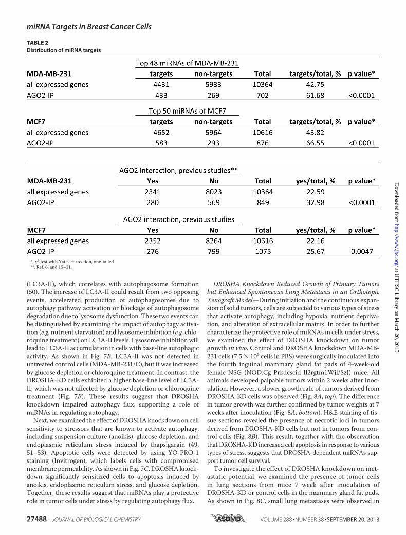
miRNA Target Filter of the Ingenuity pathway analysis systembased on 3�-UTR sequences of mRNAs. As summarized inTable 2, over 60% of AGO2-IP mRNAs were predicted targetsof the abundantly expressed miRNAs in corresponding cells.Compared with all expressed genes detected in the input RNAsamples, the predicted miRNA targets were significantly over-represented in AGO2-IP mRNAs (p � 0.05, �2 test with Yatescorrection). These results suggest that the majority of theAGO2-IPmRNAs are targets ofmiRNAs abundantly expressedin MCF7 and MDA-MB-231 cells. The absence of miRNAtarget sites in the 3�-UTRs of �40% AGO2-IP mRNAs couldbe attributed to the presence of miRNA target sites outsidethe 3�-UTRs, AGO2-mRNA interaction mediated bymiRNAs expressed at low levels, and undefined miRNAbinding sequences.Alternative Polyadenylation Contributes to Cell Type-specific
AGO2 Interaction of mRNAs—Among the cell type-specificAGO2-IP mRNAs are putative targets of miRNAs commonlyexpressed in bothMCF7 andMDA-MB-231 cells. One possiblecause of this cell type-specific AGO2 binding is differentialexpression of mRNA isoforms with 3�-UTRs of varying lengths(43). A previous study reported that about one-third ofmRNAsin various human tumor cells use alternative polyadenylation(APA) to generate multiple mRNA isoforms that differ in their3�-UTRs (44). The usage of APA appears to be cell context-de-pendent, resulting in expression of cell type-specificmRNA iso-forms (44–47).
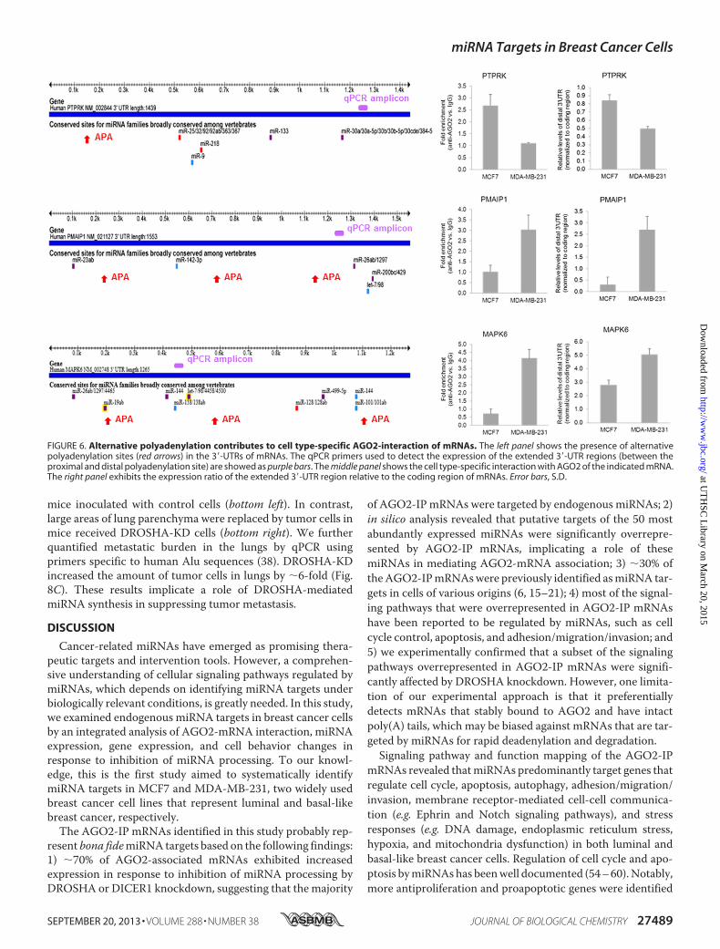
To investigate whether APA plays a role in cell type-specificmRNA-miRNA interactions in breast cancer cells, we exam-ined the expression ratio of the extended 3�-UTR regions(between the proximal and distal polyadenylation site) relativeto the coding region of a panel ofmRNAs. The cell type-specificAGO2-interacting mRNAs selected for this study are putativetargets of miRNAs commonly expressed in both MCF7 andMDA-MB-231 cells and harbor APA sites according to theAREsite and xPAD Expression & Poly(A) Database (44, 48). Asshown in Fig. 6, PTPRK, a MCF7-specific AGO2-IP mRNA,showed a higher ratio of the extended 3�-UTR relative to codingregion in MCF7 than in MDA-MB-231 cells. This result sug-gests that an RTPRK isoform with a long 3�-UTR is prefer-entially produced in MCF7 cells. Conversely, PMAIP1 andMAPK6, two mRNAs that were detected in AGO2-IP specifi-cally in MDA-MB-231 cells, exhibited a higher ratio of theirextended 3�-UTRs relative to coding regions in MDA-MB-231than in MCF7 cells. In addition, we also detected a preferentialexpression of the extended 3�-UTRs of SIAH1, SLC35A1,SPRY4, UBE2N, and APITD1 in MBA-MB-231 cells, wherethey were found to be associated with AGO2. However, 13mRNAs (from a total of 21 examined) showed no differencein the expression of their extended 3�-UTRs, despite theirdifferential interaction with AGO2, which included CCDC25,CLASP1, DGCR8, FXR1, MMD, PRKRA, RUNX1, TARBP2,TFDP1, TRAM2, UBE2N, XPO5, and ZCCHC11. These resultssuggest that APA accounts for the cell type-specific miRNAinteraction of some, but not all, mRNAs.DROSHA Knockdown Increases Cell Migration and Invasion—
Because genes encoding proteins involved in adhesion, migra-tion, and invasion (Fig. 1C) were overrepresented in AGO2-IPmRNAs, we speculated that blocking miRNA processing mayalter cell migration and invasion. Boyden chamber migrationand invasion assays showed that DROSHA knockdown signifi-cantly enhancedmigration and invasion ofMDA-MB-231 cells(Fig. 7A). These observations suggest that the endogenousmiRNAs of MDA-MB-231 function collectively to suppressmigration and invasion.DROSHA Knockdown Promotes Cell Death in Response to
Various Types of Stress—Genes involved in various stresssignaling pathways were significantly enriched in AGO2-IPmRNAs in both MCF7 and MDA-MB-231 cells, implicating arole of miRNAs in cell damage control and adaptation. Intrigu-ingly, among the AGO2-IP mRNAs are several criticalcomponents of the autophagy pathway (e.g. CTSL1, DDIT4,ERN1, HSPA5, IDUA, LAMTOR1, RAC3, ULK3, and VTI1B).Autophagy is a catabolic process that delivers cellular compo-nents through double-membrane vesicles (autophagosomes) tolysosomes for degradation. Autophagy plays an important rolein eliminating damaged cellular components and recycling cel-lular materials for macromolecular and organelle biosynthesisandnutrient and energy homeostasis (49).Given the prominentcytoprotective roles of autophagy, we hypothesized that block-ing miRNA processing may alter autophagy activity and conse-quently cell sensitivity to stress.First, we examined the effect of DROSHA knockdown on
autophagy activity bymeasuring the conversion ofMAP1LC3Afrom cytosolic (LC3A-I) to membrane-bound lipidated form
TABLE 1Seed sequences of abundantly expressed miRNAs
Seed sequence MiRBase IDCell line
specificicity
AAAGCUG has-miR-320a CommonAAAGUGC hsa-miR-17-5p/20b-5p/93-5p/106a-5p/
106b-5pCommon
AACAGUC hsa-miR-212-5p CommonACAUUCA hsa-miR-181a-5p/181b-5p CommonACCCGUA hsa-miR-99b-5p/100-5p CommonAGCAGCA hsa-miR-15b-5p/15b-5p/16-5p/195-5p CommonAGCUUAU hsa-miR-21-5p CommonAGUGCAA hsa-miR-130a-3p/301a-3p CommonAUUGCAC hsa-miR-25-3p/92a-3p CommonCCCUGAG hsa-miR-125a-5p/125b-5p CommonCGUACCG hsa-miR-126-3p CommonGAGGGGC hsa-miR-423-5p CommonGAGGUAG hsa-let-7a-5p/7b-5p/7c/7d-5p/
7e-5p/7i-5pCommon
GCAGCAU hsa-miR-103a-3p/107 CommonGGCUCAG hsa-miR-24-3p CommonGUAAACA hsa-miR-30a-5p/30b-5p/30c-5p/
30d-5pCommon
UCAAGUA hsa-miR-26a-5p CommonUCACAGU hsa-miR-27a-5p CommonUCACAUU hsa-miR-23a-3p/23b-3p CommonACCCUGU hsa-miR-10a-5p MDA-MB-231AGCACCA Hsa-miR-29a-3p/29b-3p/29c-3p MDA-MB-231AGCUGCC hsa-miR-22-3p MDA-MB-231GAGAACU hsa-miR-146a-5p/146b-5p MDA-MB-231GCUACAU hsa-miR-221-3p/222-3p MDA-MB-231GCUGGUG hsa-miR-138-5p MDA-MB-231AACACUG hsa-miR-141-3p/200a-3p MCF7AACGGAA hsa-miR-191-5p MCF7AAUACUG hsa-miR-200b-3p/200c-3p/429 MCF7AGGUAGU hsa-miR-196a-5p MCF7GGAAGAC hsa-miR-7-5p MCF7GGGGUGC hsa-miR-342-5p MCF7UCAUAGA hsa-miR-376a-3p MCF7UGAAAUG hsa-mir-203a MCF7UUGGCAA Hsa-miR-182 MCF7UUGGCAC hsa-miR-96-5p MCF7
miRNA Targets in Breast Cancer Cells
SEPTEMBER 20, 2013 • VOLUME 288 • NUMBER 38 JOURNAL OF BIOLOGICAL CHEMISTRY 27487
at UT
HSC
Library on M
arch 20, 2015http://w
ww
.jbc.org/D
ownloaded from
(LC3A-II), which correlates with autophagosome formation(50). The increase of LC3A-II could result from two opposingevents, accelerated production of autophagosomes due toautophagy pathway activation or blockage of autophagosomedegradation due to lysosomedysfunction. These two events canbe distinguished by examining the impact of autophagy activa-tion (e.g. nutrient starvation) and lysosome inhibition (e.g. chlo-roquine treatment) on LC3A-II levels. Lysosome inhibition willlead to LC3A-II accumulation in cells with base-line autophagicactivity. As shown in Fig. 7B, LC3A-II was not detected inuntreated control cells (MDA-MB-231/C), but it was increasedby glucose depletion or chloroquine treatment. In contrast, theDROSHA-KD cells exhibited a higher base-line level of LC3A-II, which was not affected by glucose depletion or chloroquinetreatment (Fig. 7B). These results suggest that DROSHAknockdown impaired autophagy flux, supporting a role ofmiRNAs in regulating autophagy.Next, we examined the effect ofDROSHAknockdownon cell
sensitivity to stressors that are known to activate autophagy,including suspension culture (anoikis), glucose depletion, andendoplasmic reticulum stress induced by thapsigargin (49,51–53). Apoptotic cells were detected by using YO-PRO-1staining (Invitrogen), which labels cells with compromisedmembrane permeability. As shown in Fig. 7C, DROSHAknock-down significantly sensitized cells to apoptosis induced byanoikis, endoplasmic reticulum stress, and glucose depletion.Together, these results suggest that miRNAs play a protectiverole in tumor cells under stress by regulating autophagy flux.
DROSHA Knockdown Reduced Growth of Primary Tumorsbut Enhanced Spontaneous Lung Metastasis in an OrthotopicXenograftModel—During initiation and the continuous expan-sion of solid tumors, cells are subjected to various types of stressthat activate autophagy, including hypoxia, nutrient depriva-tion, and alteration of extracellular matrix. In order to furthercharacterize the protective role ofmiRNAs in cells under stress,we examined the effect of DROSHA knockdown on tumorgrowth in vivo. Control and DROSHA knockdown MDA-MB-231 cells (7.5� 105 cells in PBS) were surgically inoculated intothe fourth inguinal mammary gland fat pads of 4-week-oldfemale NSG (NOD.Cg Prkdcscid Il2rgtm1Wjl/SzJ) mice. Allanimals developed palpable tumors within 2 weeks after inoc-ulation. However, a slower growth rate of tumors derived fromDROSHA-KD cells was observed (Fig. 8A, top). The differencein tumor growth was further confirmed by tumor weights at 7weeks after inoculation (Fig. 8A, bottom). H&E staining of tis-sue sections revealed the presence of necrotic loci in tumorsderived from DROSHA-KD cells but not in tumors from con-trol cells (Fig. 8B). This result, together with the observationthat DROSHA-KD increased cell apoptosis in response to varioustypes of stress, suggests that DROSHA-dependent miRNAs sup-port tumor cell survival.To investigate the effect of DROSHA knockdown on met-
astatic potential, we examined the presence of tumor cellsin lung sections from mice 7 week after inoculation ofDROSHA-KD or control cells in the mammary gland fat pads.As shown in Fig. 8C, small lung metastases were observed in
TABLE 2Distribution of miRNA targets
*, �2 test with Yates correction, one-tailed.**, Ref. 6, and 15–21.
miRNA Targets in Breast Cancer Cells
27488 JOURNAL OF BIOLOGICAL CHEMISTRY VOLUME 288 • NUMBER 38 • SEPTEMBER 20, 2013
at UT
HSC
Library on M
arch 20, 2015http://w
ww
.jbc.org/D
ownloaded from
mice inoculated with control cells (bottom left). In contrast,large areas of lung parenchyma were replaced by tumor cells inmice received DROSHA-KD cells (bottom right). We furtherquantified metastatic burden in the lungs by qPCR usingprimers specific to human Alu sequences (38). DROSHA-KDincreased the amount of tumor cells in lungs by �6-fold (Fig.8C). These results implicate a role of DROSHA-mediatedmiRNA synthesis in suppressing tumor metastasis.
DISCUSSION
Cancer-related miRNAs have emerged as promising thera-peutic targets and intervention tools. However, a comprehen-sive understanding of cellular signaling pathways regulated bymiRNAs, which depends on identifying miRNA targets underbiologically relevant conditions, is greatly needed. In this study,we examined endogenous miRNA targets in breast cancer cellsby an integrated analysis of AGO2-mRNA interaction, miRNAexpression, gene expression, and cell behavior changes inresponse to inhibition of miRNA processing. To our knowl-edge, this is the first study aimed to systematically identifymiRNA targets in MCF7 and MDA-MB-231, two widely usedbreast cancer cell lines that represent luminal and basal-likebreast cancer, respectively.The AGO2-IP mRNAs identified in this study probably rep-
resent bona fidemiRNA targets based on the following findings:1) �70% of AGO2-associated mRNAs exhibited increasedexpression in response to inhibition of miRNA processing byDROSHA or DICER1 knockdown, suggesting that the majority
of AGO2-IP mRNAs were targeted by endogenous miRNAs; 2)in silico analysis revealed that putative targets of the 50 mostabundantly expressed miRNAs were significantly overrepre-sented by AGO2-IP mRNAs, implicating a role of thesemiRNAs in mediating AGO2-mRNA association; 3) �30% oftheAGO2-IPmRNAswere previously identified asmiRNA tar-gets in cells of various origins (6, 15–21); 4) most of the signal-ing pathways that were overrepresented in AGO2-IP mRNAshave been reported to be regulated by miRNAs, such as cellcycle control, apoptosis, and adhesion/migration/invasion; and5) we experimentally confirmed that a subset of the signalingpathways overrepresented in AGO2-IP mRNAs were signifi-cantly affected by DROSHA knockdown. However, one limita-tion of our experimental approach is that it preferentiallydetects mRNAs that stably bound to AGO2 and have intactpoly(A) tails, which may be biased against mRNAs that are tar-geted by miRNAs for rapid deadenylation and degradation.Signaling pathway and function mapping of the AGO2-IP
mRNAs revealed thatmiRNAs predominantly target genes thatregulate cell cycle, apoptosis, autophagy, adhesion/migration/invasion, membrane receptor-mediated cell-cell communica-tion (e.g. Ephrin and Notch signaling pathways), and stressresponses (e.g. DNA damage, endoplasmic reticulum stress,hypoxia, and mitochondria dysfunction) in both luminal andbasal-like breast cancer cells. Regulation of cell cycle and apo-ptosis bymiRNAshas beenwell documented (54–60).Notably,more antiproliferation and proapoptotic genes were identified
FIGURE 6. Alternative polyadenylation contributes to cell type-specific AGO2-interaction of mRNAs. The left panel shows the presence of alternativepolyadenylation sites (red arrows) in the 3�-UTRs of mRNAs. The qPCR primers used to detect the expression of the extended 3�-UTR regions (between theproximal and distal polyadenylation site) are showed as purple bars. The middle panel shows the cell type-specific interaction with AGO2 of the indicated mRNA.The right panel exhibits the expression ratio of the extended 3�-UTR region relative to the coding region of mRNAs. Error bars, S.D.
miRNA Targets in Breast Cancer Cells
SEPTEMBER 20, 2013 • VOLUME 288 • NUMBER 38 JOURNAL OF BIOLOGICAL CHEMISTRY 27489
at UT
HSC
Library on M
arch 20, 2015http://w
ww
.jbc.org/D
ownloaded from

as miRNA targets than proproliferation and antiapoptoticgenes, indicating that miRNAs, in general, support cell prolif-eration and protect cells against apoptosis. Consistent with thishypothesis, global miRNA elevation due to increased activity ofXPO5 was found to be critical for cell G1/S entry, whereasglobal miRNA inhibition by DROSHA knockdown in humancolon adenocarcinoma HT29 cells has been shown to enhanceapoptosis induced by 5-fluorouracil treatment (61, 62).miRNA deregulation has been frequently described in meta-
static tumors, implicating a role of miRNAs in regulating cellproperties associatedwithmetastasis (63).We found that genesinvolved in cell adhesion/migration/invasion were overrepre-sented in AGO2-IP mRNAs, and DROSHA knockdown signif-icantly enhanced cell migration and invasion in vitro andenhanced spontaneous lung metastasis in an orthotopic xeno-graft model. These results suggest that miRNAs collectivelyfunction to inhibit cell migration and invasion, which is con-sistent with the observation that miRNA down-regulationrather than up-regulation occurs frequently in metastatictumor cells (63). In support, a recent high throughput studyshowed that over 20% of the 904 human miRNAs have regula-
tory activity on migration and invasion of cancer cells fromdiverse origins, and most of these miRNAs exhibited suppres-sive impact (60). In addition, DICER1 down-regulation hasbeen shown to enhance tumor metastasis (64).One intriguing finding of our study is that a large number of
genes critically involved in cell stress response were miRNAtargets. In solid tumors, cells must adapt continuously tofluctuations in their microenvironment, including hypoxia,nutrient deprivation, therapeutic insults, and alteration ofextracellular signals (e.g. extracellular matrix, cytokines, andhormones). Cell response to environment changes involvesconcerted action of diverse signaling pathways to eliminatedamage and facilitate adaptation. Recent studies suggest thatautophagy is a common downstream event of various types ofcellular stress and plays an important role in promoting tumorcell survival and adaptation (49, 51–53, 65, 66). Autophagy is acatabolic process that delivers cellular components throughdouble-membrane vesicles (autophagosomes) to lysosomes fordegradation, allowing cells to eliminate damaged componentsand recycle cellular materials for macromolecular and organ-elle biosynthesis and nutrient and energy homeostasis (49).We
FIGURE 7. Global miRNA inhibition by DROSHA knockdown in MDA-MB-231 cells enhances cell migration and invasion, but promotes cell death inresponse to various types of stress. A, DROSHA knockdown increases cell potential for migration and invasion, which were detected by Boyden chamberassays with uncoated or Matrigel-coated membrane, respectively. The results are presented as mean number of cells/field � S.E. (error bars) (n � 3). C, control.B, DROSHA knockdown impairs autophagy flux, indicated by the lack of response of MAP1LC3A to glucose depletion (GD) or chloroquine (CQ) treatment. Incontrol cells with normal autophagy activity, glucose depletion induces conversion of MAP1LC3A from cytosolic (LC3A-I) to membrane-bound lipidated form(LC3A-II) due to increased autophagosome assembly, whereas chloroquine causes accumulation of LC3A-II by inhibiting autophagosome degradation bylysosome. C, DROSHA knockdown sensitizes cells to apoptosis induced by various types of stress. Apoptotic cells with compromised membrane integrity weredetected with YO-PRO-1 dye, followed by flow cytometer analysis.
miRNA Targets in Breast Cancer Cells
27490 JOURNAL OF BIOLOGICAL CHEMISTRY VOLUME 288 • NUMBER 38 • SEPTEMBER 20, 2013
at UT
HSC
Library on M
arch 20, 2015http://w
ww
.jbc.org/D
ownloaded from

found that blocking DROSHA-mediated miRNA synthesis ledto impaired autophagy flux and sensitized cells to apoptosisinduced by various stressors that activate autophagy. Our datafrom in vivo studies provided further evidence supporting aprotective role of miRNAs against cell death. Our results sug-gest that miRNAs collectively function to maintain properautophagy flux and protect cells against stress-induced celldeath. Given the critical roles of miRNA and autophagy in cellhomeostasis, the interaction between these two pathways war-rants further investigation.Cell type-specific effects of miRNAs have been recognized,
but the underlying mechanism is not clear. One potentialmechanism is the presence of mRNA isoforms with variouslengths of 3�-UTRs due to the usage of alternative polyadenyl-ation sites. By comparing mRNA-AGO2 interaction, miRNAexpression, and mRNA expression in MCF7 and MDA-MB-231 cells, we identified a panel of mRNAs that were targeted bymiRNAs in a cell type-specific manner. We provided experi-mental evidence suggesting that cell type-specific usage ofalternative polyadenylation may be responsible for differential
regulation by miRNAs of some mRNAs, such as RTPRK,PMAIP1, and MAPK6.In conclusion, we conducted a genome-wide analysis of
miRNA targets in luminal and basal-like breast cancer cells,followed by experimental validation in cells with impairedmiRNA function at the level of single miRNA or global miRNAprocessing. Our results suggest that miRNAs play an importantrole inprotectingcells against cell deathandrepressingmetastasis.We also provided experimental evidence supporting the possibil-ity that alternative polyadenylation contributes to cell type-spe-cific regulation of certain mRNAs bymiRNA. These data providean overview of the function of endogenousmiRNAs in twomajorsubtypes of breast cancer and a base of future studies to link breastcancer cell properties with individual miRNAs.
REFERENCES1. Guo, H., Ingolia, N. T., Weissman, J. S., and Bartel, D. P. (2010) Mamma-
lian microRNAs predominantly act to decrease target mRNA levels. Na-ture 466, 835–840
2. Bartel, D. P. (2009) MicroRNAs. Target recognition and regulatory func-tions. Cell 136, 215–233
FIGURE 8. DROSHA knockdown in MDA-MB-231 cells reduces growth of orthotopic xenografts but increases lung metastasis. A, growth rates ofxenograft tumors derived from DROSHA knockdown or control cells inoculated in mammary gland fat pads. The results are presented as average tumorvolume � S.D. (n � 12) (top). For wet weight of tumors at 7 weeks after inoculation, the results are presented as average tumor weight � S.D. (n � 8) (bottom).B, H&E staining of tumor sections. Necrosis loci were observed in tumors derived from DROSHA knockdown cells but not in tumors from control cells. C,metastatic burden in lungs. H&E staining showed the presence of clusters of human tumor cells in mouse lung sections. Human tumor cells in mouse lungtissues were quantified by qPCR using primers specific for human Alu sequences. The cell number was calculated using standard curve of genomic DNApurified from cultured MDA-MB-231 cells and presented as mean � S.D. (error bars) (n � 8).
miRNA Targets in Breast Cancer Cells
SEPTEMBER 20, 2013 • VOLUME 288 • NUMBER 38 JOURNAL OF BIOLOGICAL CHEMISTRY 27491
at UT
HSC
Library on M
arch 20, 2015http://w
ww
.jbc.org/D
ownloaded from

3. Lee, Y., Ahn, C., Han, J., Choi, H., Kim, J., Yim, J., Lee, J., Provost, P.,Rådmark, O., Kim, S., and Kim, V. N. (2003) The nuclear RNase III Droshainitiates microRNA processing. Nature 425, 415–419
4. Djuranovic, S., Nahvi, A., and Green, R. (2011) A parsimonious model forgene regulation by miRNAs. Science 331, 550–553
5. Hausser, J., Syed, A. P., Bilen, B., and Zavolan, M. (2013) Analysis of CDS-locatedmiRNA target sites suggests that they can effectively inhibit trans-lation. Genome Res. 23, 604–615
6. Hafner, M., Landthaler, M., Burger, L., Khorshid, M., Hausser, J.,Berninger, P., Rothballer, A., Ascano, M., Jr., Jungkamp, A. C., Mun-schauer, M., Ulrich, A.,Wardle, G. S., Dewell, S., Zavolan, M., and Tuschl,T. (2010) Transcriptome-wide identification of RNA-binding protein andmicroRNA target sites by PAR-CLIP. Cell 141, 129–141
7. Friedman, R. C., Farh, K. K., Burge, C. B., and Bartel, D. P. (2009) Mostmammalian mRNAs are conserved targets of microRNAs. Genome Res.19, 92–105
8. Frank, F., Sonenberg, N., and Nagar, B. (2010) Structural basis for 5�-nucleotide base-specific recognition of guide RNA by human AGO2.Na-ture 465, 818–822
9. Selbach, M., Schwanhausser, B., Thierfelder, N., Fang, Z., Khanin, R., andRajewsky, N. (2008) Widespread changes in protein synthesis induced bymicroRNAs. Nature 455, 58–63
10. Czech, B., and Hannon, G. J. (2011) Small RNA sorting. Matchmaking forArgonautes. Nature Rev. Genet. 12, 19–31
11. Azuma-Mukai, A.,Oguri, H.,Mituyama, T.,Qian, Z. R., Asai, K., Siomi,H.,and Siomi, M. C. (2008) Characterization of endogenous human Argo-nautes and their miRNA partners in RNA silencing. Proc. Natl. Acad. Sci.U.S.A. 105, 7964–7969
12. Valdmanis, P. N., Gu, S., Schuermann, N., Sethupathy, P., Grimm, D., andKay,M. A. (2012) Expression determinants of mammalian argonaute pro-teins in mediating gene silencing. Nucleic Acids Res. 40, 3704–3713
13. Wang, D., Zhang, Z., O’Loughlin, E., Lee, T., Houel, S., O’Carroll, D.,Tarakhovsky, A., Ahn, N. G., and Yi, R. (2012) Quantitative functions ofArgonaute proteins in mammalian development.Genes Dev. 26, 693–704
14. Liu, X., Yu, X., Zack, D. J., Zhu, H., and Qian, J. (2008) TiGER. A databasefor tissue-specific gene expression and regulation. BMC Bioinformatics 9,271
15. Thomson, D. W., Bracken, C. P., and Goodall, G. J. (2011) Experimentalstrategies for microRNA target identification. Nucleic Acids Res. 39,6845–6853
16. Tan, L. P., Seinen, E., Duns, G., de Jong, D., Sibon, O. C., Poppema, S.,Kroesen, B. J., Kok, K., and van den Berg, A. (2009) A high throughputexperimental approach to identify miRNA targets in human cells.NucleicAcids Res. 37, e137
17. Easow,G., Teleman,A.A., andCohen, S.M. (2007) Isolation ofmicroRNAtargets by miRNP immunopurification. RNA 13, 1198–1204
18. Burroughs, A. M., Ando, Y., de Hoon, M. J., Tomaru, Y., Suzuki, H.,Hayashizaki, Y., and Daub, C. O. (2011) Deep-sequencing of human Ar-gonaute-associated small RNAs provides insight into miRNA sorting andreveals Argonaute association with RNA fragments of diverse origin. RNABiol. 8, 158–177
19. Hendrickson, D. G., Hogan, D. J., Herschlag, D., Ferrell, J. E., and Brown,P. O. (2008) Systematic identification of mRNAs recruited to argonaute 2by specific microRNAs and corresponding changes in transcript abun-dance. PloS One 3, e2126
20. Hassan, M. Q., Gordon, J. A., Lian, J. B., van Wijnen, A. J., Stein, J. L., andStein, G. S. (2010) Ribonucleoprotein immunoprecipitation (RNP-IP). Adirect in vivo analysis ofmicroRNA-targets. J. Cell Biochem. 110, 817–822
21. Leung, A. K., Young, A. G., Bhutkar, A., Zheng, G. X., Bosson, A. D.,Nielsen, C. B., and Sharp, P. A. (2011) Genome-wide identification ofAgo2 binding sites from mouse embryonic stem cells with and withoutmature microRNAs. Nat. Struct. Mol. Biol. 18, 237–244
22. Wang, L., andWang, J. (2012) MicroRNA-mediated breast cancer metas-tasis. From primary site to distant organs. Oncogene 31, 2499–2511
23. Png, K. J., Halberg, N., Yoshida, M., and Tavazoie, S. F. (2012) A mi-croRNA regulon that mediates endothelial recruitment and metastasis bycancer cells. Nature 481, 190–194
24. Wee, E. J., Peters, K., Nair, S. S., Hulf, T., Stein, S., Wagner, S., Bailey, P.,
Lee, S. Y., Qu,W. J., Brewster, B., French, J. D., Dobrovic, A., Francis, G. D.,Clark, S. J., and Brown, M. A. (2012) Mapping the regulatory sequencescontrolling 93 breast cancer-associated miRNA genes leads to the identi-fication of two functional promoters of the Hsa-mir-200b cluster, methyl-ation of which is associated withmetastasis or hormone receptor status inadvanced breast cancer. Oncogene 31, 4182–4195
25. Farazi, T. A., Horlings,H.M., TenHoeve, J. J.,Mihailovic, A., Halfwerk,H.,Morozov, P., Brown, M., Hafner, M., Reyal, F., van Kouwenhove, M.,Kreike, B., Sie, D., Hovestadt, V., Wessels, L. F., van de Vijver, M. J., andTuschl, T. (2011) MicroRNA sequence and expression analysis in breasttumors by deep sequencing. Cancer Res. 71, 4443–4453
26. Ventura, A., and Jacks, T. (2009) MicroRNAs and cancer. Short RNAs goa long way. Cell 136, 586–591
27. Hurst, D. R., Edmonds, M. D., and Welch, D. R. (2009) Metastamir. Thefield of metastasis-regulatory microRNA is spreading. Cancer Res. 69,7495–7498
28. Ma, L., andWeinberg, R. A. (2008)Micromanagers of malignancy. Role ofmicroRNAs in regulating metastasis. Trends Genet. 24, 448–456
29. Wang, C., Bian, Z., Wei, D., and Zhang, J. G. (2011) miR-29b regulatesmigration of human breast cancer cells.Mol. Cell Biochem. 352, 197–207
30. Persson, H., Kvist, A., Rego, N., Staaf, J., Vallon-Christersson, J., Luts, L.,Loman, N., Jonsson, G., Naya, H., Hoglund, M., Borg, A., and Rovira, C.(2011) Identification of new microRNAs in paired normal and tumorbreast tissue suggests a dual role for the ERBB2/Her2 gene.Cancer Res. 71,78–86
31. Xin, F., Li, M., Balch, C., Thomson, M., Fan, M., Liu, Y., Hammond, S. M.,Kim, S., and Nephew, K. P. (2009) Computational analysis of microRNAprofiles and their target genes suggests significant involvement in breastcancer antiestrogen resistance. Bioinformatics 25, 430–434
32. Maillot, G., Lacroix-Triki, M., Pierredon, S., Gratadou, L., Schmidt, S.,Benes, V., Roche, H., Dalenc, F., Auboeuf, D., Millevoi, S., and Vagner, S.(2009) Widespread estrogen-dependent repression of microRNAs in-volved in breast tumor cell growth. Cancer Res. 69, 8332–8340
33. Iorio, M. V., Ferracin, M., Liu, C. G., Veronese, A., Spizzo, R., Sabbioni, S.,Magri, E., Pedriali,M., Fabbri,M., Campiglio,M.,Menard, S., Palazzo, J. P.,Rosenberg, A., Musiani, P., Volinia, S., Nenci, I., Calin, G. A., Querzoli, P.,Negrini, M., and Croce, C. M. (2005) MicroRNA gene expression dereg-ulation in human breast cancer. Cancer Res. 65, 7065–7070
34. Kumar, M. S., Lu, J., Mercer, K. L., Golub, T. R., and Jacks, T. (2007)Impaired microRNA processing enhances cellular transformation andtumorigenesis. Nat. Genet. 39, 673–677
35. Spandidos, A., Wang, X., Wang, H., and Seed, B. (2010) PrimerBank. Aresource of human and mouse PCR primer pairs for gene expression de-tection and quantification. Nucleic Acids Res. 38, D792–D799
36. Untergasser, A., Nijveen, H., Rao, X., Bisseling, T., Geurts, R., and Leunis-sen, J. A. (2007) Primer3Plus, an enhanced web interface to Primer3. Nu-cleic Acids Res. 35,W71–W74
37. Ebert, M. S., Neilson, J. R., and Sharp, P. A. (2007) MicroRNA sponges.Competitive inhibitors of small RNAs in mammalian cells. Nat. Methods4, 721–726
38. Nicklas, J. A., and Buel, E. (2003) Development of an Alu-based, real-timePCR method for quantitation of human DNA in forensic samples. J. Fo-rensic Sci. 48, 936–944
39. Lehmann, B. D., Bauer, J. A., Chen, X., Sanders,M. E., Chakravarthy, A. B.,Shyr, Y., and Pietenpol, J. A. (2011) Identification of human triple-negativebreast cancer subtypes and preclinical models for selection of targetedtherapies. J. Clin. Invest. 121, 2750–2767
40. Zeng, Y., and Cullen, B. R. (2003) Sequence requirements for microRNAprocessing and function in human cells. Rna 9, 112–123
41. Winter, J., Jung, S., Keller, S., Gregory, R. I., andDiederichs, S. (2009)Manyroads to maturity. MicroRNA biogenesis pathways and their regulation.Nat. Cell Biol. 11, 228–234
42. Cheadle, C., Vawter, M. P., Freed, W. J., and Becker, K. G. (2003) Analysisof microarray data using Z score transformation. J. Mol. Diagn. 5, 73–81
43. Mayr, C., and Bartel, D. P. (2009) Widespread shortening of 3�UTRs byalternative cleavage and polyadenylation activates oncogenes in cancercells. Cell 138, 673–684
44. Lin, Y., Li, Z., Ozsolak, F., Kim, S. W., Arango-Argoty, G., Liu, T. T.,
miRNA Targets in Breast Cancer Cells
27492 JOURNAL OF BIOLOGICAL CHEMISTRY VOLUME 288 • NUMBER 38 • SEPTEMBER 20, 2013
at UT
HSC
Library on M
arch 20, 2015http://w
ww
.jbc.org/D
ownloaded from
Tenenbaum, S. A., Bailey, T., Monaghan, A. P., Milos, P. M., and John, B.(2012) An in-depth map of polyadenylation sites in cancer. Nucleic AcidsRes. 40, 8460–8471
45. Sandberg, R., Neilson, J. R., Sarma, A., Sharp, P. A., and Burge, C. B. (2008)Proliferating cells express mRNAs with shortened 3� untranslated regionsand fewer microRNA target sites. Science 320, 1643–1647
46. Di Giammartino, D. C., Nishida, K., and Manley, J. L. (2011) Mechanismsand consequences of alternative polyadenylation.Mol. Cell 43, 853–866
47. Fu, Y., Sun, Y., Li, Y., Li, J., Rao, X., Chen, C., and Xu, A. (2011) Differentialgenome-wide profiling of tandem 3� UTRs among human breast cancerand normal cells by high-throughput sequencing. Genome Res. 21,741–747
48. Gruber, A. R., Fallmann, J., Kratochvill, F., Kovarik, P., and Hofacker, I. L.(2011) AREsite. A database for the comprehensive investigation ofAU-rich elements. Nucleic Acids Res. 39, D66–69
49. Kroemer, G., Marino, G., and Levine, B. (2010) Autophagy and the inte-grated stress response.Mol. Cell 40, 280–293
50. Mizushima, N., Yoshimori, T., and Levine, B. (2010)Methods inmamma-lian autophagy research. Cell 140, 313–326
51. Clarke, R., Cook, K. L., Hu, R., Facey, C. O., Tavassoly, I., Schwartz, J. L.,Baumann, W. T., Tyson, J. J., Xuan, J., Wang, Y., Warri, A., and Shajahan,A. N. (2012) Endoplasmic reticulum stress, the unfolded protein response,autophagy, and the integrated regulation of breast cancer cell fate.CancerRes. 72, 1321–1331
52. Altman, B. J., and Rathmell, J. C. (2012)Metabolic stress in autophagy andcell death pathways. Cold Spring Harb. Perspect. Biol. 4, a008763
53. Lock, R., and Debnath, J. (2008) Extracellular matrix regulation of au-tophagy. Curr. Opin. Cell Biol. 20, 583–588
54. Cheng, A. M., Byrom, M. W., Shelton, J., and Ford, L. P. (2005) Antisenseinhibition of human miRNAs and indications for an involvement ofmiRNA in cell growth and apoptosis. Nucleic Acids Res. 33, 1290–1297
55. Jovanovic, M., and Hengartner, M. O. (2006) miRNAs and apoptosis.RNAs to die for. Oncogene 25, 6176–6187
56. Wang, Y., and Blelloch, R. (2009) Cell cycle regulation by MicroRNAs in
embryonic stem cells. Cancer Res. 69, 4093–409657. Georges, S. A., Biery,M.C., Kim, S. Y., Schelter, J.M., Guo, J., Chang, A.N.,
Jackson, A. L., Carleton,M.O., Linsley, P. S., Cleary,M.A., andChau, B.N.(2008) Coordinated regulation of cell cycle transcripts by p53-induciblemicroRNAs, miR-192 and miR-215. Cancer Res. 68, 10105–10112
58. Carleton, M., Cleary, M. A., and Linsley, P. S. (2007) MicroRNAs and cellcycle regulation. Cell Cycle 6, 2127–2132
59. Lima, R. T., Busacca, S., Almeida, G. M., Gaudino, G., Fennell, D. A., andVasconcelos, M. H. (2011) MicroRNA regulation of core apoptosis path-ways in cancer. Eur. J. Cancer 47, 163–174
60. Zhang, H., Hao, Y., Yang, J., Zhou, Y., Li, J., Yin, S., Sun, C.,Ma,M., Huang,Y., and Xi, J. J. (2011) Genome-wide functional screening of miR-23b as apleiotropic modulator suppressing cancer metastasis. Nat. Commun. 2,554
61. Iwasaki, Y. W., Kiga, K., Kayo, H., Fukuda-Yuzawa, Y., Weise, J., Inada, T.,Tomita, M., Ishihama, Y., and Fukao, T. (2013) Global microRNA eleva-tion by inducible Exportin 5 regulates cell cycle entry. Rna 19, 490–497
62. Wang, B. D., Kline, C. L., Pastor, D. M., Olson, T. L., Frank, B., Luu, T.,Sharma,A.K., Robertson,G.,Weirauch,M.T., Patierno, S. R., Stuart, J.M.,Irby, R. B., and Lee, N. H. (2010) Prostate apoptosis response protein 4sensitizes human colon cancer cells to chemotherapeutic 5-FU throughmediation of an NF�B and microRNA network.Mol. Cancer 9, 98
63. Zhang, H., Li, Y., and Lai, M. (2010) The microRNA network and tumormetastasis. Oncogene 29, 937–948
64. Su, X., Chakravarti, D., Cho,M. S., Liu, L., Gi, Y. J., Lin, Y. L., Leung, M. L.,El-Naggar, A., Creighton, C. J., Suraokar, M. B., Wistuba, I., and Flores,E. R. (2010) TAp63 suppresses metastasis through coordinate regulationof Dicer and miRNAs. Nature 467, 986–990
65. Azad,M. B., Chen, Y., andGibson, S. B. (2009) Regulation of autophagy byreactive oxygen species (ROS). Implications for cancer progression andtreatment. Antioxid. Redox Signal. 11, 777–790
66. Dalby, K. N., Tekedereli, I., Lopez-Berestein, G., and Ozpolat, B. (2010)Targeting the prodeath and prosurvival functions of autophagy as noveltherapeutic strategies in cancer. Autophagy 6, 322–329
miRNA Targets in Breast Cancer Cells
SEPTEMBER 20, 2013 • VOLUME 288 • NUMBER 38 JOURNAL OF BIOLOGICAL CHEMISTRY 27493
at UT
HSC
Library on M
arch 20, 2015http://w
ww
.jbc.org/D
ownloaded from

Junming Yue and Lawrence M. PfefferSethuraman, Chuan He Yang, Zhao-hui Wu, Meiyun Fan, Raisa Krutilina, Jing Sun, Aarti (miRNA) Targets in Breast Cancer CellsComprehensive Analysis of MicroRNACell Biology:
doi: 10.1074/jbc.M113.491803 originally published online August 6, 20132013, 288:27480-27493.J. Biol. Chem.
10.1074/jbc.M113.491803Access the most updated version of this article at doi:
.JBC Affinity SitesFind articles, minireviews, Reflections and Classics on similar topics on the
Alerts:
When a correction for this article is posted•
When this article is cited•
to choose from all of JBC's e-mail alertsClick here
Supplemental material:
http://www.jbc.org/content/suppl/2013/08/06/M113.491803.DC1.html
http://www.jbc.org/content/288/38/27480.full.html#ref-list-1
This article cites 66 references, 28 of which can be accessed free at
at UT
HSC
Library on M
arch 20, 2015http://w
ww
.jbc.org/D
ownloaded from