Complex intubation, cricothyrotomy and tracheotomy - B-ENT
16
B-ENT, 2016, 12, Suppl. 26/2, 103-118 Part I: Complex intubation 1. Introduction The word intubation is derived from Latin, and means the process of inserting or putting a tube or a cannula into a hollow organ. This definition may encompass many types of procedures; the most commonly used is the introduction and the placement of an endotracheal tube (ETT) in the trachea for ventilatory purposes. The most widely used route is orotracheal, in which an ETT is passed through the mouth and vocal apparatus into the trachea. In a nasotracheal procedure, an ETT is passed throaugh the nose and vocal apparatus into the trachea. Other methods of intubation involve surgery such as cricothyrotomy (used almost exclusively in emergency circumstances) and the tracheotomy (where a prolonged need for airway support is anticipated). The gold standard in intubation is the successful insertion of the ETT through the mouth under direct vision, requiring a laryngoscope with either a curved (Macintosh) or a straight (Miller) blade. The size of the blade will be selected in relation to the size of the patient, in order to effectively push the tongue out of the way and to lift the supraglottic structures, allowing for a direct view of the glottic structures, that is, the vocal cords. The occasional use of a stylet placed as a guide inside the ETT’s lumen or of a McGill forceps remains within the realm of simple intubation. The introduction of an ETT via the rima glottidis into the trachea is not without risk for the patient, and this procedure should therefore only be attempted after appropriate consideration and evaluation. 1 This should be done so that the first attempt is executed under the best possible conditions, so that the chances of success are stacked in one’s favour. However, in some cases simple intubation may not be accomplished or may not even be attempted due to some of the reasons mentioned below, or due to the fact that the case was immediately identified as being difficult. Complex intubation (CI) requires specific equipment that is normally not needed with simple cases of intubation. Some of this equipment Complex intubation, cricothyrotomy and tracheotomy R. Leach 1 , S. Hachimi Idrissi 2 , J. Meulemans 3 , K. Monsieurs 4 , O. Vanderveken 5 , V. Vander Poorten 3 1 Department of Emergency Medicine, Cliniques St. Jean – St. Jansziekenhuis, Bd du Jardin Botanique 32 Kruidtuinlaan, 1000 Brussels, Belgium; 2 Department of Emergency Medicine, Ghent University Hospital, De Pintelaan 185, 9000 Ghent, Belgium; 3 Otorhinolaryngology, Head and Neck Surgery, University Hospitals Leuven and KU Leuven Department of Oncology Section Head and Neck Oncology, Kapucijnenvoer 33, 3000 Leuven, Belgium; 4 Department of Emergency Medicine, Antwerp University Hospital, Wilrijkstraat 10, 2650 Edegem, Belgium; 5 Department of Otorhinolaryngology, Head and Neck Surgery, Antwerp University Hospital, Wilrijkstraat 10, 2650 Edegem, Belgium Key-words. Emergency; airway management; cricothyrotomy; surgical airway; cannot intubate cannot ventilate; tracheostomy; percutaneous Abstract. Complex intubation, cricothyrotomy and tracheotomy. Successful management of a difficult airway begins with recognizing the potential problem. When the patient cannot breathe spontaneously, oxygenation and ventilation should start first with bag-valve ventilation, with or without an airway adjunct such as a Mayo cannula, followed by an orotracheal intubation attempt, performed by an experienced emergency doctor. If orotracheal intubation fails, a quick decision must be made regarding surgical options. In a “cannot intubate, cannot ventilate” situation, a surgical cricothyrotomy should be considered. When orotracheal intubation is impossible, but bag-valve or laryngeal mask ventilation is possible, an urgent surgical tracheostomy should be performed. In the long run, patients in need of long- term artificial ventilation will need a percutaneous or open tracheostomy. This review provides an update of all aspects of immediate and long-term airway management. 09 - leach-.indd 103 8/11/16 12:19
-
Upload
khangminh22 -
Category
Documents
-
view
2 -
download
0
Transcript of Complex intubation, cricothyrotomy and tracheotomy - B-ENT

B-ENT, 2016, 12, Suppl. 26/2, 103-118
Part I: Complex intubation
1. Introduction
The word intubation is derived from Latin, and means the process of inserting or putting a tube or a cannula into a hollow organ. This definition may encompass many types of procedures; the most commonly used is the introduction and the placement of an endotracheal tube (ETT) in the trachea for ventilatory purposes.
The most widely used route is orotracheal, in which an ETT is passed through the mouth and vocal apparatus into the trachea. In a nasotracheal procedure, an ETT is passed throaugh the nose and vocal apparatus into the trachea. Other methods of intubation involve surgery such as cricothyrotomy (used almost exclusively in emergency circumstances) and the tracheotomy (where a prolonged need for airway support is anticipated).
The gold standard in intubation is the successful insertion of the ETT through the mouth under direct vision, requiring a laryngoscope with either
a curved (Macintosh) or a straight (Miller) blade. The size of the blade will be selected in relation to the size of the patient, in order to effectively push the tongue out of the way and to lift the supraglottic structures, allowing for a direct view of the glottic structures, that is, the vocal cords. The occasional use of a stylet placed as a guide inside the ETT’s lumen or of a McGill forceps remains within the realm of simple intubation. The introduction of an ETT via the rima glottidis into the trachea is not without risk for the patient, and this procedure should therefore only be attempted after appropriate consideration and evaluation.1 This should be done so that the first attempt is executed under the best possible conditions, so that the chances of success are stacked in one’s favour. However, in some cases simple intubation may not be accomplished or may not even be attempted due to some of the reasons mentioned below, or due to the fact that the case was immediately identified as being difficult. Complex intubation (CI) requires specific equipment that is normally not needed with simple cases of intubation. Some of this equipment
Complex intubation, cricothyrotomy and tracheotomy
R. Leach1, S. Hachimi Idrissi2, J. Meulemans3, K. Monsieurs4, O. Vanderveken5, V. Vander Poorten3 1Department of Emergency Medicine, Cliniques St. Jean – St. Jansziekenhuis, Bd du Jardin Botanique 32 Kruidtuinlaan, 1000 Brussels, Belgium; 2Department of Emergency Medicine, Ghent University Hospital, De Pintelaan 185, 9000 Ghent, Belgium; 3Otorhinolaryngology, Head and Neck Surgery, University Hospitals Leuven and KU Leuven Department of Oncology Section Head and Neck Oncology, Kapucijnenvoer 33, 3000 Leuven, Belgium; 4Department of Emergency Medicine, Antwerp University Hospital, Wilrijkstraat 10, 2650 Edegem, Belgium; 5Department of Otorhinolaryngology, Head and Neck Surgery, Antwerp University Hospital, Wilrijkstraat 10, 2650 Edegem, Belgium
Key-words. Emergency; airway management; cricothyrotomy; surgical airway; cannot intubate cannot ventilate; tracheostomy; percutaneous
Abstract. Complex intubation, cricothyrotomy and tracheotomy. Successful management of a difficult airway begins with recognizing the potential problem. When the patient cannot breathe spontaneously, oxygenation and ventilation should start first with bag-valve ventilation, with or without an airway adjunct such as a Mayo cannula, followed by an orotracheal intubation attempt, performed by an experienced emergency doctor. If orotracheal intubation fails, a quick decision must be made regarding surgical options. In a “cannot intubate, cannot ventilate” situation, a surgical cricothyrotomy should be considered. When orotracheal intubation is impossible, but bag-valve or laryngeal mask ventilation is possible, an urgent surgical tracheostomy should be performed. In the long run, patients in need of long-term artificial ventilation will need a percutaneous or open tracheostomy. This review provides an update of all aspects of immediate and long-term airway management.
09 - leach-.indd 103 8/11/16 12:19

104 R. Leach
patients, and although the correct use of anaesthesia is a crucial factor in successful intubation, the different pharmacological agents that are available and their usage is outside the scope of this chapter. The discussion will address the various signs that may alert the laryngoscopist that the patient may be a CI patient, and will identify the available devices that can assist the laryngoscopist in intubating a CI patient.
2. Determining intubation difficulty
As mentioned above, intubation-related patient complications are a significant factor contributing to patient morbidity and mortality. ETT placement must therefore be thought through before any attempt is made. However, the question of how to identify the CI patient before having tried several attempts remains; the ability to identify CI patients ahead of time may have life-or-death consequences. The prior identification of this type of case would allow the laryngoscopist to prepare equipment which is normally not required, and that will assist in the secure placement of the ETT. This would of course save time and money; however, more importantly, this would be a much safer way to proceed. The vast majority of CI cases can be predicted by considering the following four aspects.
2.1. Operator experience
Operator experience is of course paramount. Numerous studies show that the laryngoscopist’s experience is a significant factor for success.5 Although this may seem to be straightforward, the amount of training required can vary widely, depending on the individual and their qualifications.6 Ideally, therefore, when considering intubation, one should seek an experienced laryngoscopist or at least attempt the procedure in the presence of an experienced laryngoscopist. This may not always be possible, and inexperienced laryngoscopists may therefore sometimes be confronted with direct laryngoscopic intubation failure, requiring the use of alternative intubation methods in order to successfully intubate. Paradoxically, even the inexperienced laryngoscopist must thus be aware and preferably be able to use these alternative methods, which require techniques that are often even more difficult to acquire.7 The use of an alternative method of intubation is therefore not always synonymous with a CI patient.
can also provide a significant advantage to the laryngoscopist even in cases of simple intubation.
Often CI is due to anatomical individuality or to variations in the landmarks used by the laryngoscopists when placing the ETT; however CI may also be due to as many factors as there are proposed definitions.2,3 Those who have experienced a difficult intubation are able recognize this when they are faced with it, although there is as yet no agreement on a universally accepted definition. This point should not be too quickly passed over or considered too theoretically. Numerous methods for predicting CI exist, but questions remain as to what is being addressed: for example, whether CI is patient-dependent or operator-dependent; whether CI refers to the patient with whom one doctor may have difficulty, but whose colleague intubates with ease; and whether CI refers to a patient taking, for instance, 11 minutes to intubate when an intubation of 9 minutes would have been considered simple. It may be generally agreed that a patient who is intubated quickly is certainly a simple intubation patient, and one who takes a long time is most probably a CI patient, but the categories here have not been defined, and neither has the term ‘a long time’. The use of time as an indicator is not always reliable, since few doctors, at least those working in a pre-hospital setting, start the stopwatch before commencing intubation; in addition, even though it can be generally agreed that the inflation of the cuff is one of the final steps for the laryngoscopist, this step does not require any technical expertise, and it could therefore be argued that intubation ends with the deflated cuff passing through the rima glottidis. Thus the sole use of time as a definition for CI can be rather subjective. Does a CI involve four attempts at ETT introduction, and simple intubation only three? What is considered an attempt to intubate? Does the introduction of a laryngoscope mean this is an intubation attempt? Does the introduction of an ETT no further than the buccal cavity represent an attempt? What exactly is complex or difficult intubation? The American Society of Anaesthesiologists (ASA) has achieved a consensus defining complex intubation as “when proper insertion of the endotracheal tube with conventional laryngoscopy requires more than three attempts, or more than ten minutes.”4 This consensus definition, despite its shortfalls, will be the definition adopted in further discussion of this subject. The topic of CI will be limited to adult
09 - leach-.indd 104 8/11/16 12:19

Complex intubation, cricothyrotomy and tracheotomy 105
of the jaw is measured, with a distance of less than 7 cm being an indicator of intubation difficulty. The thyroid notch corresponds to the superior ridge of the thyroid cartilage.
2.3.2. Cormack-Lehane Classification The Cormack-Lehane classification (modified in 1998) was first developed for anaesthetists in training in 1984, and describes the extent to which the different laryngeal structures are visible during direct laryngoscopy. This classification system predicts the likelihood of intubation difficulty. However, even amongst professionals, this classification scoring is not easy to use and the same patient may be graded differently depending on the evaluator.8,9
2.3.3. Mallampati ScoreThe Mallampati score is another means of predicting difficult intubation. Here the patient, in a sitting position, opens the mouth and extends the tongue as far as possible. The distance between the base of the tongue and the roof of the mouth is evaluated. This score is based on the fact that intubation difficulty is considered relative to the size of the base of the tongue. The modified Mallampati score is carried out with the patient’s cervical spine in a neutral position, and is standard in the pre-operative assessment of patients. Cervical spine extension will not improve the predictive power of the Mallampati score and thus should not be used.10 The Cormark-Lehane appears to be more precise; however, these are often used together. A recent study combined the Mallampati with ultrasound measurements to determine CI, again confirming that no single score alone is sufficient.11
2.3.4. Simplified Airway Risk Index The Simplified Airway Risk Index (SARI) uses seven points, one of them being the Mallampati score, and is also commonly used to predict difficult intubation.12 The remaining six points are mouth opening, thyromental distance, neck movement, underbite, body weight and previous intubation history.
2.3.5. Intubation Difficulty Scale The Intubation Difficulty Scale (IDS) is another score which also uses seven parameters, and was developed by a group of emergency physicians and anaesthesiologists from Paris, in an attempt to address the shortcomings of the other scales.
2.2. Medical history
Medical history, whether current issues, past treatments, birth deformations or past difficult intubations, can provide very important information when available. All too often, this aspect is poorly explored in the pre-hospital setting. There may be anatomical deformations or distortions due to inflammation, hematomas, tumours of the underlying structures or medical interventions (surgery, radiotherapy) on neoplastic lesions. The patient’s current medical state must also be considered, and particularly the reasons for to the decision to intubate. For example, patients with a suspected cervical spine injury will be automatically considered in the CI category, since head mobilization will be limited.
2.3. Clinical examination
The patient must be examined before any intubation attempt, in order to evaluate possible intubation difficulty. A quick look at the patient’s neck for a thyroid surgery scar may identify a more challenging intubation procedure; the muscles governing the vocal cords are innervated by the recurrent laryngeal nerve, which is a branch of the Vagus nerve and is sometimes damaged after thyroid surgery. There are also individual anatomical situations where direct vision of the vocal cords is difficult or even impossible. The physical examination should be as complete as possible, and include evaluation of cervical and temporo-mandibular mobility and the shape of the jaw, along with an endobuccal exploration evaluating the tongue, the palate and the teeth. Developmental abnormalities of the oral cavity, especially the jaw (such as hypoplasia or micrognathia) as well as the dental state (prostheses) must be taken into consideration before the final decision is made concerning the method of intubation. Cervical spine limitations due to age, surgery, trauma or birth defects will also compromise intubation by limiting the positioning of the patient, as discussed below. Numerous scales have been developed to assist the laryngoscopist in the physical assessment of the prediction of intubation difficulty.8
2.3.1. Thyromental DistanceThe thyromental distance is a commonly-used method for easy prediction of intubation difficulty. The distance between the thyroid notch and the tip
09 - leach-.indd 105 8/11/16 12:19

106 R. Leach
should use the device that he or she is most familiar with. It therefore goes without saying that the laryngoscopist should be familiar with more than one of these devices, so that it is easy to change to another device if the first choice is not successful; the first choice of device may not be able to place the ETT.
3.1. Temporary airway devices
These devices establish an airway without the introduction of an ETT. Airway protection is satisfactory on a temporary basis, and most of these devices will be replaced later by an ETT. There are some concerns over the degree of airway protection, however, especially during CPR.18 These are clearly very useful in the pre-hospital field, since health care providers here include not only doctors, but also nurses and paramedics, who do not always have sufficient intubation training and experience. Further developments of these devices are also enabling them to become interesting options in the hospital setting.
3.1.1. I-Gel/Air-QI-gel (Intersurgical Ltd, Berkshire, UK) is a second-generation supraglottic airway device. It is anatomically shaped to fit the laryngeal structures without cuff inflation. It must be introduced through the oral cavity and, once positioned, simply needs to be secured in place from the outside. Its use as a conduit for intubation was accepted in 2012. Air-Q (Mercury Medical, Clearwater, FL, USA) is a very similar single-usage device, which also allows for the introduction and placement of an ETT through its airway lumen.
3.1.2. Laryngeal Mask Airway (LMA)This device is inserted into the mouth and creates a low-pressure seal with the trachea without actually having to be guided through the larynx, thus remaining supraglottic. This provides a relatively easy solution for the protection of the upper airway by providers who are unable to intubate a patient or for those without sufficient training or experience in ETT intubation. Although this is not a definitive airway solution, it is recommended in the ALS guidelines for its simplicity of use, especially since its placement does not require interruption of chest compression.19 However, there are concerns about the efficiency of this device in preventing the regurgitation of stomach contents.18,20
This scale was devised after comparing CI criteria when intubating in the operating room and in a pre-hospital context.13 The seven parameters take into consideration the number of attempts, the number of laryngoscopists, the number of alternative devices, the lifting force, the amount of external pressure laryngeal pressure, vocal cord mobility and the Cormack score minus one.
2.3.6. UltrasoundUltrasound may be used to more precisely assess and predict CI.11,14
2.4. Positioning
Proper positioning of the patient is also important. For those working in the pre-hospital field, the repositioning of the patient before attempting the introduction of the ETT can be the deciding factor between success and failure. A recent study of 500 patients also indicated that placing the patient in a sitting upright, head-elevated position may reduce intubation-related complications in hospitalised patients.15 Since the tongue and the laryngeal structures are connected, they can be moved together when displaced, and the correct positioning of the head can therefore move these structures, opening up a view of the vocal cords.
3. Alternative equipment to ETT for CI
When the intubation is evaluated as a CI based on one or more of the above-mentioned factors, the laryngoscopist has numerous options. These can be divided into two general categories.
The first category includes specialized devices that establish a temporary airway solution without necessitating the introduction of an ETT. Later, under more optimal conditions, these may be replaced by a definitive airway solution.
The second category involves the numerous fibre-optic devices that are available to assist in ETT intubation. Although these devices provide numerous advantages, they are only useful as long as their distal lens remains clear; in patients with excessive secretion, bleeding or vomiting their indication may therefore be limited.16,17
It is not reasonable to attempt to give precise indications as to which device is more adapted to a specific type of patient, nor it is valuable to rank these devices as superior to one another. As is often the case in medicine, the laryngoscopist
09 - leach-.indd 106 8/11/16 12:19

Complex intubation, cricothyrotomy and tracheotomy 107
new generation of rigid videolaryngoscopes has eliminated the blade and the working channel, and has combined the light source and the optic lens into one thin stylet.
3.2.1.1. Airtraq This fibre-optic device (Prodol Meditec SA, Guecho, Spain) is specifically shaped to assist in the placement of an ETT with minimal head manipulation. It is equipped with an eyepiece that transmits the view from its distal end. There is also a model that is connected to a video monitor. The ETT is inserted via a lateral channel along the device.
3.2.1.2. GlidescopeThis product (Verathon Inc., Bothell, WA, USA) transmits an image from the distal end of its blade, which is slightly more angled than the more common laryngoscopic blades, to a video monitor. Numerous studies have indicated that even for inexperienced laryngoscopists, glottic visualisation is superior to direct laryngoscopy. This would appear to indicate that ETT intubation is facilitated; surprisingly, however, this seemingly logical conclusion has not always been confirmed in studies.28,30,31
3.2.1.3. Bullard ScopeThe Bullard scope is a rigid fibre-optic laryngoscope, with a light source, a fibre-optic lens and a working channel. It is connected to a video screen and a light source. This device is only used within the hospital setting. It is also the most studied of the rigid laryngoscopes.
3.2.1.4. Upsher ScopeThis is a rigid fibre-optic laryngoscope that is particularly useful in cases where there is minimal mouth opening and where minimal head manipulation is allowed. This laryngoscope, despite its advantages, takes some time to master and actually is quite difficult to use.32,33 The Upsher scope has a C-shaped lumen through which the ETT is advanced, unlike the Bullard scope which uses a stylet.
3.2.1.5. WuScopeThis intubating device, which combines a rigid laryngoscope with fibre-optics, was developed in the early 1990s by Dr. Wu. Due to the 110° angle between its handle and blade, there is no need for head extension, tongue lifting or forceful
3.1.3. Oesophageal-Tracheal Combitube (OTC-Combitube)OTC is also known as the oesophageal tracheal double-lumen airway (two tubes); this is a blind insertion airway device used in pre-hospital and emergency settings. It is designed to provide an airway to facilitate the mechanical ventilation of a patient in respiratory distress. It consists of a cuffed, double-lumen tube that is inserted through the patient’s mouth to secure an airway and enable ventilation. Generally, the distal tube (the second tube) enters the oesophagus, where the cuff is inflated, and ventilation is provided through the proximal tube (the first tube) which opens at the level of the larynx. In the rare instance where the distal tube intubates the trachea, ventilation is provided through the distal tube. Inflation of the cuff in the oesophagus allows a level of protection against the aspiration of gastric contents, similar to that found in the LMA. Its placement is determined by auscultation, and the cuffs are then inflated. This seemingly simple method is an interesting alternative to ETT intubation, especially in the pre-hospital setting,21 and is relatively popular amongst paramedics in North America; however, it is not without severe complications.22
3.2. Definitive airway placement devices
The following devices assist the laryngoscopist with the positioning of an ETT in cases of CI or after failed ETT intubation.
3.2.1. VideolaryngoscopeThese techniques assist the laryngoscopist via fibre-optics, providing an indirect view of the different laryngeal structures on a small portable video screen or video monitor. The laryngeal blades are adapted to the anatomy of the pharyngeal-hypopharyngeal region. The blades are equipped with a fibre-optic lens, a light source and a working channel, thus providing a rigid guide for the ETT. These methods can be used both in simple initial intubation and also as a rescue method when intubation becomes complicated. Although this would appear to be a fail-safe method, videolaryngoscopy does require a certain period of training in order to master the technique.7,16,17,23-29 Common problems with the use of rigid videolaryngoscopes include difficulty in orientating and advancing the ETT, and the fogging or blocking of the fibre-optic lens. The
09 - leach-.indd 107 8/11/16 12:19

108 R. Leach
Part II: Cricothyrotomy
1. Introduction
Cricothyrotomy (CTT), also known as cricothyroidotomy or coniotomy, refers to the technique of creating an opening in the membrana cricothyroidea or cricothyroid membrane (CTM) (Figure 1), followed by the placement of a stenting tube. In the absence of a needle or surgical knife, sharp instruments should be used. The CTM is bordered superiorly by the thyroid cartilage and inferiorly by the cricoid cartilage. The lateral aspects of the CTM are partially covered by the cricothyroid muscles, while the central triangular portion is subcutaneous, the latter being the ideal location for accessing the airway.37
CCT is an important and potentially life-saving emergency procedure for obtaining a surgical airway when a patent airway cannot be controlled by other techniques such as bag valve ventilation, endotracheal intubation or laryngeal mask airway due to ineffectiveness or contraindication.38As such, an emergency CCT is the last resort in most airway management protocols, and is performed when it is not possible to intubate or ventilate a patient.37,38
jaw opening. The WuScope is introduced into the middle of the mouth and then the handle is rotated toward the laryngoscopist. This device is unique in that a flexible fibre-optic endoscope is fitted inside this three-part device. Once the ETT is in place, the two-piece blade is disassembled and removed from the mouth.34,35
3.2.2. Lighted StyletsThis technique uses transillumination for intubating. Here, the laryngoscopist does not have a direct view of the glottic structures when placing the ETT. The light from the stylet passes through the skin when placed in the trachea and is visible from the outside. This is not the case when the stylet is placed in the oesophagus.
3.2.3. Airway BougiesThese are pre-angled stylets that are blindly placed in the trachea; the ETT is threaded over them.
3.2.4. Bi-Manual LaryngoscopyThis is a technique rather than a device; one hand is used to immobilise the head, so that the jaw can be solely manipulated without moving the head, and the other will intubate.
3.2.5. Cricothyroidotomy and TracheostomyThese techniques will be addressed in further chapters.
4. Conclusion
This chapter has provided the reader with a valid working definition of CI, keeping in mind that any definition will be subject to controversy, and illustrates the various steps that can assist in prediction of a potential case of CI, thereby allowing the laryngoscopist to prepare for this, with the ultimate goal of decreasing complications for the patient.36 A brief description of the different devices that are available has also been given, from as objective and factual a point of view as possible.Videolaryngoscopy is developing rather rapidly, and future solutions for ventilating a CI patient are likely to be dominated either by the easily-placed supraglottic ventilatory devices or by ETT ventilation via videolaryngoscopy. It should be borne in mind that the effectiveness of these innovative devices is determined by the laryngoscopist’s familiarity with each specific device.
Figure 1From reference Heffner JE. J Crit Illn. 1995. See also reference Burkey et al.39 2009
09 - leach-.indd 108 8/11/16 12:19

Complex intubation, cricothyrotomy and tracheotomy 109
kits which include all the necessary equipment for performing a CCT are commercially available and can be divided into two categories. The first category relies on the Seldinger technique, which refers to a method using a punctured CTM with a needle attached to a syringe. Once the correct position of the needle in the trachea is confirmed by the aspiration of air, a guidewire is inserted through the needle. The kit also contains a specially designed endotracheal tube to be inserted into the trachea over the guidewire if necessary, following the use of a dilator and after removal of the needle. The kits in the second category include a cutting device, instead of a guidewire, and are used to create a lumen in the CTM wide enough to accommodate the endotracheal tube provided in the kit.38-40 Needle CCT may be used for approximately 40 minutes, after which time carbon dioxide accumulates, requiring a rapid reestablishment of a proper airway.37,40
Smith et al. do not recommend needle CCT as a rescue technique, and suggest that a correctly performed transtracheal jet insufflation is the only small catheter option.40 However, in a 2005 study comparing the Seldinger technique with the standard surgical technique, the time to tracheal puncture and ventilation was significantly longer for the latter. In addition, injury to the thyroid
2. Indications and contraindications for CCT
In general, CCT is indicated by the impossibility of obtaining or failure to obtain a patent airway with traditional, less invasive methods.37,39 In Table 1, two additional categories of indications for CCT are provided; that is, relative and non-emergent indications.39,40 According to the literature, an absolute contraindication to CCT is the age of the patient; however, there is a great deal of disagreement on the exact age at which a CCT can be safely performed. In addition, CCT should not be performed when the anatomical landmarks cannot be palpated. In the presence of acute laryngeal pathology, tracheal transection, fracture, or obstruction below the cricothyroid membrane, CCT is also contraindicated.37,39
3. Techniques, technical considerations and outcomes
Various approaches to CCT can be distinguished in the literature, such as needle CCT, percutaneous CCT and surgical CCT.38 The standard surgical technique consists of making a vertical incision through the skin, followed by horizontally incising the CTM into the tracheal lumen through which an endotracheal tube is inserted.37,38,40A number of
Table 1Indications for cricothyrotomy according to the literature37,39,40
IndicationsEmergency cricothyrotomy in case of:Airway obstruction due to:(supra)glottic foreign body
- Oedema (e.G., Due to anaphylaxis) with inability to visualize larynxBleeding such as:
- Severe oro- and/or nasopharyngeal or tracheobronchial haemorrhage (e.G., Caused by trauma)- Uncontrollable emesis
Facial muscle spasms or laryngospasmUpper airway stenosis or congenital deformitiesClenched teethTumour, cancer, or other disease process or trauma causing a mass effectRelative indications:Severe maxillofacial traumaCervical spine immobilization / manipulation of the neck is contraindicated Non-emergent indications:Prolonged intubationMaxillofacial, laryngeal, or oral surgeryBronchoscopy or laryngoscopy without endotracheal tube
09 - leach-.indd 109 8/11/16 12:19

110 R. Leach
unclear whether appropriate training shortens the time required to perform ultrasound-guided CCT.42
5. Conclusion
CCT is an emergency intervention of last resort that is performed infrequently and under inherently stressful conditions, including severe time pressure and suboptimal conditions. As such, appropriate training and a proper knowledge of indications, contraindications and foreseeable complications regarding CCT are mandatory. On the other hand, many complications from CCT are relatively minor compared with those of the prolonged hypoxia associated with the “cannot intubate, cannot ventilate” situation.
Part III: Tracheostomy
Tracheostomy was first described by the ancient Greeks around 2000 BC and was subsequently documented in the Roman and Arabic literature as a surgical procedure to relieve airway obstruction. Following the standardization of open surgical tracheostomy (OT) in the early 20th century by Chevalier Jackson, OT was the standard practice until 1985, when the New York surgeon Ciaglia reported on the first percutaneous dilatational tracheostomy (PDT) for which he used serial tracheal dilatations over a guidewire. The underlying idea of PDT was to provide a bedside, minimally invasive, safe and efficient way to establish a secure and semi-permanent airway in patients with respiratory failure.44
1. Percutaneous Tracheostomy
1.1. Techniques44,45
The standard practice is to perform PDT on paralyzed patients who are under general anaesthesia and have been placed on a 100% fraction of inspired oxygen (100% FiO2). In order to minimize the risk of injury to the posterior tracheal wall, bronchoscopic guidance is advocated during the procedure. As a consequence, PDT requires two experienced operators, one to operate the bronchoscope and another to perform the procedure. The patient is placed in a supine position with the neck in hyperextension (such as a shoulder roll placement) in order to maximize the length of the cervical trachea. After the creation
vasculature was more common with the standard surgical technique.41 In their systematic review, Langvad et al. concluded that, in terms of success rates and time required, neither technique was shown to be superior when comparing the results using commercial kits to perform emergency CCT with surgical and needle techniques. These authors also suggest that success rates may be operator-dependent. It appears that experience and training with CCT are the limiting factors, rather than the technique used for CCT.38 In addition, it should be noted that no direct comparisons between the different techniques in real-life use have been published to date.40
4. Potential complications of CCT
Acute complications after emergency CCT occur in up to 15% of cases,40 with the most frequently observed complication of CCT being glottic or subglottic stenosis.39 The data regarding the occurrence of subglottic stenosis as a long-term complication of CCT are highly variable, and the factors tending to increase the likelihood of this include concurrent laryngotracheal pathology, prolonged time to decannulation, older age and diabetes.37
Other complications include haemorrhage, tube displacement, tube obstruction with blood or secretions, infection, laryngotracheal injury, subcutaneous emphysema, obstruction, bronchial intubation, aspiration, or tension pneumothorax.37,39 Apart from subglottic or glottic stenosis, other potential late complications following CCT include subjective voice changes, difficulties with swallowing, persistent shortness of breath and persistent tracheostoma.37 In addition, it has been reported that emergency percutaneous CCT may frequently injure the posterior tracheal wall, particularly when it is difficult to locate the puncture site by palpation.42 Emergency percutaneous CCT may also frequently produce another life-threatening complication, that of tearing to the posterior tracheal wall; it has been illustrated recently that the use of ultrasonography might be able to significantly reduce this complication.43 However, in comparison with conventional CCT, ultrasound-guided CCT may require a longer insertion time, resulting in impracticability in dire situations such a severe hypoxaemia after failed intubation and unsuccessful oxygenation. It is
09 - leach-.indd 110 8/11/16 12:19

Complex intubation, cricothyrotomy and tracheotomy 111
cords. Reintubation will be necessary if the scope and ETT are accidentally withdrawn to a level above the glottis. Next, the insertion is performed using the Ciaglia or Griggs technique. Figure 2 illustrates the procedural set-up.
The original Ciaglia technique entailed a series of dilatations using a guidewire tract. However, current commercially available kits all use a single gradual dilator, such as the Smiths-Portex PDT UltraPerc® (Smiths Medical, Dublin, Ohio, USA) or the Ciaglia Blue Rhino® (Cook Medical, Bloomington, Indiana, USA) systems. The Ciaglia Blue Dolphin® (Cook Medical, Bloomington, Indiana, USA) uses an inflatable dilation balloon instead of a gradual dilator. This technique starts with a 2 to 2.5 cm incision at the level of the previously determined insertion site. Under bronchoscopic vision, a 14-gauge needle-cannula is inserted into the midline of the anterior tracheal wall, and care has to be taken not to puncture the posterior tracheal wall. During this manoeuvre, the surgeon stabilizes the trachea using the non-dominant hand. Following puncture of the trachea, the Seldinger guidewire is inserted into the trachea under direct bronchoscopic vision, in order to confirm its intratracheal position. After removal of the needle-cannula, the guidewire tract is first dilated with a short and firm tracheal dilator seated on the guidewire, and next by a well-lubricated single gradual dilator, which is advanced into the trachea to the skin marking position on the dilator. The dilator is then removed, leaving the guidewire in place, and the tracheostomy tube and loading dilator are inserted into the trachea, following the curvature of the tracheostomy tube. Figure 3 shows the insertion of the loading dilator and tracheostomy cannula into the trachea. After tube placement, all dilators and wires are removed, the bronchoscope is switched from the ETT into the tracheostomy tube to confirm intratracheal tube positioning, and the ventilator circuit is finally moved from the ETT to the tracheostomy tube.
As an alternative for the serial dilatations initially used during the Ciaglia technique, Griggs introduced the guidewire dilating forceps (GWDF) technique in 1994. The first steps are identical to the Ciaglia technique, up to the placement of the guidewire in the tracheal lumen. After introducing the guidewire in the trachea, and after removal of the needle-cannula, specially designed forceps, the so-called ‘Griggs forceps’, are passed over the wire and
of a sterile field, the cricoid and thyroid cartilages are palpated and a needle-insertion site is selected between the second and third tracheal ring. The skin and subcutaneous tissue of the insertion site are infiltrated with 5 to 10 ml of lidocaine (20 mg/ml) with adrenalin (5 µg/ml) solution, with the aim of achieving pre- and postoperative haemostasis and immediate postprocedural local analgesia. The flexible bronchoscope is positioned at the distal end of the endotracheal tube (ETT), and the scope and ETT are withdrawn to a level just below the vocal
Figure 2 Illustration of procedural set-up for percutaneous tracheostomy. The patient is prepped and draped and the flexible bronchoscope is positioned at the distal end of the endotracheal tube (ETT). The scope and the ETT are withdrawn to a level just below the vocal cords
Figure 3The insertion of the loading dilator and tracheostomy cannula into the trachea is achieved seated on the guidewire. In the bottom right corner of the image, the gradual dilator can be seen
09 - leach-.indd 111 8/11/16 12:19

112 R. Leach
was observed between OT and PDT in terms of minor postprocedural bleeding, major and minor intraprocedural bleeding, false route, subcutaneous emphysema, pneumothorax and desaturation. An additional eight RCTs compared different PT techniques in 700 patients. When compared to the GWDF technique, multiple (MDT) and single step (SSDT) dilatator techniques were associated with the lowest risk for difficult dilatation or tracheostomy tube insertion (OR 0.30, 95% CI 0.12−0.80) and major intraprocedural bleeding (OR 0.29, 95% CI 0.10−0.85), making the MDT and SSDT techniques preferable to the GWDF technique.
A recent systematic review exploring long-term outcomes following PDT identified 29 studies (9 RCTs, 6 non-randomized comparative studies and 14 single-arm cohort studies) which involved 5473 patients.48 No significant differences were detected in the prevalence of tracheal stenosis for a range of percutaneous techniques and surgical tracheostomy. When considering the pooled proportion meta-analysis across all studies, the tracheal stenosis rate varied from 2.8% to 0.6% for ST and SSDT respectively, with all percutaneous techniques being broadly comparable. While these findings may suggest a tendency towards a higher rate of tracheal stenosis after OT, selection bias in the non-randomized and single arm cohort studies maybe responsible for this tendency. In terms of cost, PDT is on average 50% more expensive than bedside surgical tracheostomy, due to the associated bronchoscopy and the cost of disposable devices. However, when compared to surgical tracheostomy performed in the operating theatre, PDT is significantly cheaper.49
1.4. Bronchoscopic guidance versus ultrasound guidance
Bronchoscopic guidance has traditionally been considered safer than non-bronchoscope-assisted PDT. With the bronchoscope, the correct tracheostomy site can be ascertained (midline placement of the needle-cannula, 2nd−3rd tracheal ring), intratracheal guidewire placement, intratracheal dilator placement without tracheal damage and correct position of the tracheal tube after insertion. However, no RCTs are available which compare PDT with versus without bronchoscopic guidance.46 In a systematic review analysing cases
used to dilate the pretracheal and tracheal tissues to the appropriate diameter. The tracheostomy tube is loaded on a dilator and inserted over the guidewire.
1.2. Indications and contraindications 46
The typical candidate for PDT is the critically ill adult patient who is nursed in the intensive care unit and is unable to be weaned from mechanical ventilation. PDT is not considered as an initial airway in patients who cannot be intubated. In these patients, an urgent CTT or OT is still the undisputed golden standard. Contraindications for PDT include unstable fractures of the cervical spine, severe local infection of the anterior neck and uncontrollable coagulopathy. Relative contraindications include controlled local infection, coagulopathy, high PEEP or FiO2 requirements, difficult anatomy (e.g., inability to extend the neck, obesity and subsequent troublesome location of the laryngeal and tracheal structures, aberrant neck vasculature, distortion of the anterior tracheal anatomy, voluminous goitre), elevated intracranial pressure, haemodynamic instability, previous radiotherapy to the neck and proximity of the puncture site to extensive burns or surgical wounds.
1.3. Complications and long-term outcomes
Complications of PDT are generally the same as for OT, and include pneumothorax, intraprocedural and postprocedural bleeding, tube misplacement/false route, posterior tracheal perforation with eventual tracheal-oesophageal fistula, thyroid injury, recurrent laryngeal nerve damage, subcutaneous emphysema and cardiopulmonary arrest/death.
Recently, a high-quality meta-analysis investigated whether PDT should be considered superior to OT and whether any one PDT technique was superior to the others.47 These authors included 14 randomized controlled trials (RCT), which tested PT techniques versus OT, using 973 patients in total. When compared to OT, PT techniques were associated with significantly shorter procedural times (mean difference of −13.06 minutes and 95% CI of −19.37 to −6.76), reduced risk for postprocedural major bleeding (OR 0.39, 95% CI 0.15–0.97), stoma inflammation (OR 0.38, 95% CI 0.19–0.76) and infection (OR 0.22, 95% CI 0.11–0.41), but increased risk for procedural technical difficulties (difficult dilation and/or insertion) (OR 4.58, 95% CI 2.21−9.47). No difference
09 - leach-.indd 112 8/11/16 12:19
Complex intubation, cricothyrotomy and tracheotomy 113
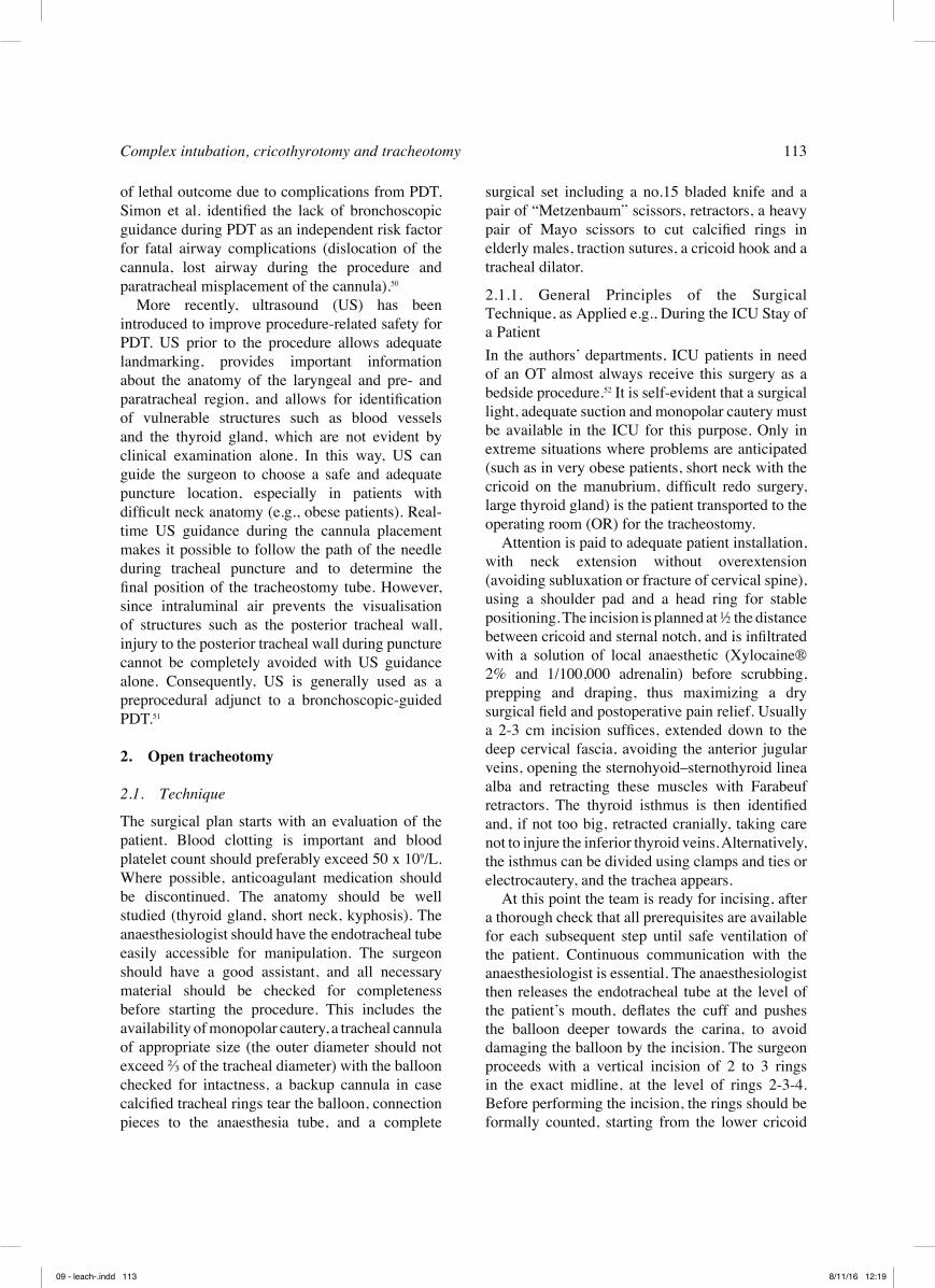
surgical set including a no.15 bladed knife and a pair of “Metzenbaum” scissors, retractors, a heavy pair of Mayo scissors to cut calcified rings in elderly males, traction sutures, a cricoid hook and a tracheal dilator.
2.1.1. General Principles of the Surgical Technique, as Applied e.g., During the ICU Stay of a PatientIn the authors’ departments, ICU patients in need of an OT almost always receive this surgery as a bedside procedure.52 It is self-evident that a surgical light, adequate suction and monopolar cautery must be available in the ICU for this purpose. Only in extreme situations where problems are anticipated (such as in very obese patients, short neck with the cricoid on the manubrium, difficult redo surgery, large thyroid gland) is the patient transported to the operating room (OR) for the tracheostomy.
Attention is paid to adequate patient installation, with neck extension without overextension (avoiding subluxation or fracture of cervical spine), using a shoulder pad and a head ring for stable positioning. The incision is planned at ½ the distance between cricoid and sternal notch, and is infiltrated with a solution of local anaesthetic (Xylocaine® 2% and 1/100,000 adrenalin) before scrubbing, prepping and draping, thus maximizing a dry surgical field and postoperative pain relief. Usually a 2-3 cm incision suffices, extended down to the deep cervical fascia, avoiding the anterior jugular veins, opening the sternohyoid–sternothyroid linea alba and retracting these muscles with Farabeuf retractors. The thyroid isthmus is then identified and, if not too big, retracted cranially, taking care not to injure the inferior thyroid veins. Alternatively, the isthmus can be divided using clamps and ties or electrocautery, and the trachea appears.
At this point the team is ready for incising, after a thorough check that all prerequisites are available for each subsequent step until safe ventilation of the patient. Continuous communication with the anaesthesiologist is essential. The anaesthesiologist then releases the endotracheal tube at the level of the patient’s mouth, deflates the cuff and pushes the balloon deeper towards the carina, to avoid damaging the balloon by the incision. The surgeon proceeds with a vertical incision of 2 to 3 rings in the exact midline, at the level of rings 2-3-4. Before performing the incision, the rings should be formally counted, starting from the lower cricoid
of lethal outcome due to complications from PDT, Simon et al. identified the lack of bronchoscopic guidance during PDT as an independent risk factor for fatal airway complications (dislocation of the cannula, lost airway during the procedure and paratracheal misplacement of the cannula).50
More recently, ultrasound (US) has been introduced to improve procedure-related safety for PDT. US prior to the procedure allows adequate landmarking, provides important information about the anatomy of the laryngeal and pre- and paratracheal region, and allows for identification of vulnerable structures such as blood vessels and the thyroid gland, which are not evident by clinical examination alone. In this way, US can guide the surgeon to choose a safe and adequate puncture location, especially in patients with difficult neck anatomy (e.g., obese patients). Real-time US guidance during the cannula placement makes it possible to follow the path of the needle during tracheal puncture and to determine the final position of the tracheostomy tube. However, since intraluminal air prevents the visualisation of structures such as the posterior tracheal wall, injury to the posterior tracheal wall during puncture cannot be completely avoided with US guidance alone. Consequently, US is generally used as a preprocedural adjunct to a bronchoscopic-guided PDT.51
2. Open tracheotomy
2.1. Technique
The surgical plan starts with an evaluation of the patient. Blood clotting is important and blood platelet count should preferably exceed 50 x 109/L. Where possible, anticoagulant medication should be discontinued. The anatomy should be well studied (thyroid gland, short neck, kyphosis). The anaesthesiologist should have the endotracheal tube easily accessible for manipulation. The surgeon should have a good assistant, and all necessary material should be checked for completeness before starting the procedure. This includes the availability of monopolar cautery, a tracheal cannula of appropriate size (the outer diameter should not exceed ⅔ of the tracheal diameter) with the balloon checked for intactness, a backup cannula in case calcified tracheal rings tear the balloon, connection pieces to the anaesthesia tube, and a complete
09 - leach-.indd 113 8/11/16 12:19

114 R. Leach
and consequently damage is more easily caused to these structures following paratracheal false dissection. The recurrent laryngeal nerves are also immediately next to the trachea and should be carefully avoided.56
Technical modifications in the paediatric OT are the removal of subcutaneous fat and eventual monopolar cauterization of the thyroid isthmus. The incision is made at the level of rings 3-4-5 (which again must be carefully counted to avoid entering the trachea too close to the cricoid, causing subglottic stenosis), using nylon 4/0 traction sutures that are placed vertically on either side of the intended vertical tracheotomy. These traction sutures are preserved at the end of the procedure to facilitate cannula replacement in case of accidental dislocation, which occurs much more frequently in children. They are marked “left” and “right” and fixed to the chest.56
Cannulas used in a child can be neonatal (length 30–36 mm) or paediatric (length 39–56 mm) in order for the tip of the cannula to be positioned about 1 cm cranially above the carina. The diameter should be well adapted to the age-related tracheal diameter (Table 2).56 These cannulas have no inner cannula, due to their small size, and will have to be changed at least once weekly, or more frequently in the case that secretions accumulate more rapidly. Tweedie et al. have provided an excellent overview of the factors to consider when choosing the appropriate cannula.57
2.2. Indications and contraindications for OT.
The indications for open tracheotomy (OT) parallel those for PDT, as listed above, when the latter technique is unavailable. Indications include (1) mechanical upper airway obstruction, (2) protection of the tracheobronchial tree in the patient at risk for aspiration, (3) respiratory failure and (4) retention of bronchial secretions. In these patients, OT results in prevention of fluid inhalation, access for suction of bronchial secretions, reduction of dead space by 50%, and increased alveolar ventilation. Indications for elective OT exist (5) in patients undergoing major head and neck surgery and (6) ICU patients with prolonged intubation that are at risk for developing laryngotracheal complications; here, the consensus was previously to wait for no longer than two weeks before switching the endotracheal tube to a tracheostomy. Several recent meta-analyses conclude that waiting for more than
border. When using monopolar cautery, it should be double-checked that the oxygen concentration is not higher than 21%, in order to avoid a fatal airway fire.53 Although some groups are proponents of making an inferiorly or superiorly based Björk flap,54,55 most authors do not recommend this modification; OT nowadays is mostly reserved for patients with a short or difficult neck, where the skin approximation of the Björk flap is all the more difficult, while many of the technically easier tracheostomies are done by PDT. In case of a deep trachea, a cricoid hook can help, although this can damage the cricoid cartilage, and the authors therefore prefer nylon 3/0 or 4/0 traction sutures that are placed on either side of the intended vertical cut through the rings. These help to lift the trachea cranially and keep it there while the tracheotomy is being performed; following this, traction on the sutures provides for eversion of the tracheotomy edges, facilitating the introduction of the cannula. Alternatively, a tracheal dilator can be used. Once the tracheal edges are retracted, the anaesthesiologist pulls up the endotracheal tube under direct vision, until the tip of the tube just disappears cranially. The tube is held there during the rest of the procedure, to be advanced again for ventilation should cannula insertion problems arise. The cannula is introduced gently in order to avoid tearing of the balloon. After that, the wound should not be packed in order to avoid surgical site infection and emphysema, and should be closed with interrupted sutures that are not too tight, for the same reason. The tube then is secured using both sutures to the skin (that leave space for a lyofoam dressing) and tracheostomy ties. The latter are secured once the head is out of extension, leaving space for one fingertip in between the skin and the ties.
Eventual tube changes should be done only after 48–72 hrs, when a more or less stable tract has formed, and most clinicians wait for as long as 5 to 7 days. If the tube has to be changed prior to this, it is best to use a guide wire to lead the tip of the new cannula into the right trajectory, thus avoiding a false route.
2.1.2. Technical Details in the Paediatric Patient
In paediatric OT, the anatomy is different in that the landmarks such as the cricoid are higher in the neck, mediastinal structures such as the brachiocephalic artery, the pleural domes and the oesophagus are more readily encountered,
09 - leach-.indd 114 8/11/16 12:19

Complex intubation, cricothyrotomy and tracheotomy 115
of tracheal stenosis between PDT and OT.48 The tracheal stenosis rate of 2.8% for OT may seem higher than the 0.6% for PDT, but is not significant, and selection bias in the non-randomized and single arm cohort studies is likely to be responsible for this discrepancy.
In absolute terms, complications in published series range from 5% to 40%, with bleeding, tube obstruction and tube displacement being the most frequent. Most importantly, a meticulous surgical technique is the key to avoiding most of these complications. Tube displacement is very serious and is responsible for the 0.5%–1.6% mortality rate of tracheostomy. Compared to an elective OT, emergency OT is associated with a two- to fivefold increase in complications. Table 3 lists the most frequent complications of OT.
2.4. Decannulation
Once the indication for tracheotomy placement is resolved, decannulation should be considered. Clinical practice should be based on a systematic assessment including the endoscopic evaluation of airway and swallowing functions, and progressive capping tests during inpatient observation (capping should be supported for longer than 24 hrs; in children, capped polysomnography should be considered, and further observation for a period of 24 hrs is recommended).65 Recently a clinical score was proposed in a meta-analysis including all quantitative and semi-quantitative parameters. This score mathematically combines all relevant decision elements in a weighted fashion, in order to make a scientific decision with the best chance for successful tracheostomy tube removal; however, this method awaits prospective validation.66
ten days is associated with negative outcomes; some have found increased morbidity,58 prolonged sedation59 and length of stay at the ICU,60,61 or increased mortality.62,63 All of these data suggest a ten-day time limit for switching to tracheostomy.
In patients with these indications where contraindications for PDT exist, OT is often the preferred approach. PDT contraindications still manageable by OT include (1) the initial airway in patients who cannot be intubated (an urgent CTT or OT is still the uncontested gold standard in these cases), (2) unstable cervical spine fractures, (3) severe local infection, burns or surgical wounds of the anterior neck, (4) uncontrollable coagulopathy, (5) high PEEP or FiO2 requirements, (6) difficult anatomy and previous radiotherapy, (7) elevated intracranial pressure and (8) haemodynamic instability.64
2.3. Complications and long-term outcomeS
The complications of OT have already been highlighted in the previous discussion of PDT, and are well detailed by a recent meta-analysis of 14 RCTs,47 in which it was shown that OT took a longer mean procedure time of 13 minutes as compared to PDT and had an increased risk of 2.6 times for postoperative major bleeding and stoma inflammation. In addition, rates of infection were shown to be 4.5 times higher. The risk of peroperative technical difficulties, however, was decreased by 79% for OT as compared to PDT. No difference was observed in per- and postoperative bleeding, false route, subcutaneous emphysema, pneumothorax and desaturation. In regard to the complication of late tracheal stenosis, another recent meta-analysis found no difference in the prevalence
Table 2Paediatric tracheostomy: age-related tracheal diameters and corresponding tube sizes (modified from56)
Age Tracheal diameter in mm Inner diameter of cannula<1 mth 5 2.5 / 3.01–6 mth 5–6 3.56–18 mth 6–7 4.018 mth–3 yrs 7–8 4.53–6 yrs 8–9 5.06–9 yrs 9–10 5.59–12 yrs 10–13 6.012–14 yrs 13 7.0
09 - leach-.indd 115 8/11/16 12:19

116 R. Leach
8. The lack of bronchoscopic guidance during percutaneous dilatational tracheostomy is an independent risk factor for fatal airway complications.9. All necessary material should be checked for completeness before starting an elective tracheostomy procedure.10. Tracheostomy tube dislocation, the most serious complication with associated mortality, occurs more easily in children. For this reason, traction sutures are left to facilitate cannula replacement when needed.
References
1. Hasegawa K, Hiraide A, Chang Y, Brown DF. Association of prehospital advanced airway management with neurologic outcome and survival in patients with out-of-hospital cardiac arrest. JAMA.2013;309(3):257-266.
2. Boisson-Bertrand D, Bourgain JL, Camboulives J, Crinquette V, Cros AM, Dubreuil M, Eurin B, Haberer JP, Pottecher T, Thorin D, Ravussin P, Riou B. [Difficult intubation. French Society of Anesthesia and Intensive Care. A collective expertise]. Ann Fr Anesth Reanim. 1996;15(2):207-214.
3. Cormack RS, Lehane J. Difficult tracheal intubation in obstetrics. Anaesthesia. 1984;39(11):1105-1111.
4. Practice guidelines for management of the difficult airway. A report by the American Society of Anesthesiologists Task Force on Management of the Difficult Airway. Anesthesiology. 1993;78(3):597-602.
5. von Goedecke A., Herff H, Paal P, Dorges V, Wenzel V. Field airway management disasters. Anesth Analg. 2007;104(3):481-483.
6. Stephens CT, Kahntroff S, Dutton RP. The success of emergency endotracheal intubation in trauma patients: a 10-year experience at a major adult trauma referral center. Anesth Analg. 2009;109(3):866-872.
7. Russo SG, Nickel EA, Leissner KB, Schwerdtfeger K, Bauer M, Roessler MS. Use of the GlideScope(R)-
Take-Home Messages
1. Even simple intubation is not without risk for the patient, and should therefore only be attempted after proper thought and evaluation, so that the first attempt is executed under the best possible conditions.2. Complex intubation is defined as “when proper insertion of the endotracheal tube with conventional laryngoscopy requires more than three attempts, or more than ten minutes”.3. A thorough medical history and clinical examination are essential in anticipating a complex intubation and can be well summarized using indices such as the Simplified Airway Risk Index and the Intubation Difficulty Scale.4. One option for addressing an anticipated complex intubation is to consider specialized devices that establish a temporary airway solution without the introduction of an ETT, such as I-gel/Air-Q, a laryngeal mask airway, or an oesophageal-tracheal Combitube.5. Another option for dealing with an anticipated complex intubation is the variety of available fibre-optic devices; however, these are only as effective as the laryngoscopist’s familiarity with the specific device.6. Cricothyrotomy is the last resort in most airway management protocols, and should be performed in the “cannot intubate, cannot ventilate” situation.7. Cricothyrotomy is performed infrequently compared to other airway procedures (such as endotracheal intubation, laryngeal mask airway and tracheotomy). Appropriate training should therefore form an inherent part of the curriculum of medical specialists.
Table 3Peroperative, early or late postoperative complications of OT
Peroperative Early postoperative Late postoperativeTracheal/laryngeal injury including airway fire
Wound infection, tracheal necrosis Granuloma formationLaryngotracheal stenosisPersistent opening / ugly scar
Injury to recurrent nerve/ oesophagus/ bra-chiocephalic artery
Swallowing problems Tracheo-oesophageal fistula
Haemorrhage/Air embolism Secondary haemorrhage Secondary haemorrhageApnoea/cardiac arrest Tube blocking/displacement with subcuta-
neous emphysema – pneumothorax/pneu-momediastinum
Difficult decannulation
09 - leach-.indd 116 8/11/16 12:19
Complex intubation, cricothyrotomy and tracheotomy 117
Combitube in the pre-hospital setting. Can J Anaesth. 2007;54(2):124-128.
23. Cortellazzi P, Caldiroli D, Byrne A, Sommariva A, Orena EF, Tramacere I. Defining and developing expertise in tracheal intubation using a GlideScope((R)) for anaesthetists with expertise in Macintosh direct laryngoscopy: an in-vivo longitudinal study. Anaesthesia. 2015;70(3):290-295.
24. Hung OR, Stewart RD. Intubating stylets. In: Hagberg CA, ed. Benumof’s Airway Management. 2 ed. Philadelphia: Mosby; 2007;463-475.
25. Delaney KA, Hessler R. Emergency flexible fiberoptic nasotracheal intubation: a report of 60 cases. Ann Emerg Med. 1988;17(9):919-926.
26. Mlinek EJ, Jr., Clinton JE, Plummer D, Ruiz E. Fiberoptic intubation in the emergency department. Ann Emerg Med. 1990;19(4):359-362.
27. Morris IR. Fibreoptic intubation. Can J Anaesth. 1994;41(10):996-1007.
28. Gu Y, Robert J, Kovacs G, Milne AD, Morris I, Hung O, MacQuarrie K, Mackinnon S, Adam LJ. A deliberately restricted laryngeal view with the GlideScope(R) video laryngoscope is associated with faster and easier tracheal intubation when compared with a full glottic view: a randomized clinical trial. Can J Anaesth. 2016;63(8):928-937.
29. Pieters BM, Wilbers NE, Huijzer M, Winkens B, van Zundert AA. Comparison of seven videolaryngoscopes with the Macintosh laryngoscope in manikins by experienced and novice personnel. Anaesthesia. 2016;71(5):556-564.
30. Trimmel H, Kreutziger J, Fitzka R, Szuts S, Derdak C, Koch E, Erwied B, Voelckel WG. Use of the GlideScope Ranger Video Laryngoscope for Emergency Intubation in the Prehospital Setting: A Randomized Control Trial. Crit Care Med. 2016;44(7):e470-476
31. Driver BE, Prekker ME, Moore JC, Schick AL, Reardon RF, Miner JR. Direct Versus Video Laryngoscopy Using the C-MAC for Tracheal Intubation in the Emergency Department, a Randomized Controlled Trial. Acad Emerg Med. 2016;23(4):433-439.
32. Fridrich P, Frass M, Krenn CG, Weinstabl C, Benumof JL, Krafft P. The UpsherScope in routine and difficult airway management: a randomized, controlled clinical trial. Anesth Analg. 1997;85(6):1377-1381.
33. Pearce AC, Shaw S, Macklin S. Evaluation of the Upsherscope. A new rigid fibrescope. Anaesthesia. 1996;51(6):561-564.
34. Wu TL, Chou HC. A new laryngoscope: the combination intubating device. Anesthesiology. 1994;81(4):1085-1087.
35. Wu TL, Chou HC. WuScope versus conventional laryngoscope in cervical spine immobilization. Anesthesiology. 2000;93(2):588-589.
36. Georgi R, Krier C. Complications of managing the airway. In: Hagberg CA, ed. Benumof’s Airway Management. 2 ed. Philadelphia: Mosby; 2007;1181-1218.
37. Hebert RB, Bose S, Mace SE. Cricothyrotomy and Percutaneous Translaryngeal Ventilation. In: Roberts JR, Custalow CB, Thomsen TW, Hedges JR, eds. Roberts and Hedges’ Clinical Procedures in Emergency Medicine. 6 ed. Philadelphia: Saunders Elsevier; 2014;120-133.
38. Langvad S, Hyldmo PK, Nakstad AR, Vist GE, Sandberg M. Emergency cricothyrotomy--a systematic review. Scand J Trauma Resusc Emerg Med. 2013;21:43.
Ranger for pre-hospital intubations by anaesthesia trained emergency physicians - an observational study. BMC Emerg Med. 2016;16:8.
8. Cook TM. A new practical classification of laryngeal view. Anaesthesia. 2000;55(3):274-279.
9. Krage R, van RC, van GD, Loer SA, Schwarte LA, Schober P. Cormack-Lehane classification revisited. Br J Anaesth. 2010;105(2):220-227.
10. Healy DW, LaHart EJ, Peoples EE, Jewell ES, Bettendorf RJ, Jr., Ramachandran SK. A Comparison of the Mallampati evaluation in neutral or extended cervical spine positions: a retrospective observational study of >80 000 patients. Br J Anaesth. 2016;116(5):690-698.
11. Pinto J, Cordeiro L, Pereira C, Gama R, Fernandes HL, Assuncao J. Predicting difficult laryngoscopy using ultrasound measurement of distance from skin to epiglottis. J Crit Care. 2016;33:26-31.
12. Norskov AK. Preoperative airway assessment - experience gained from a multicentre cluster randomised trial and the Danish Anaesthesia Database. Dan Med J.63(5):B5241.
13. Adnet F, Borron SW, Racine SX, Clemessy JL, Fournier JL, Plaisance P, Lapandry C. The intubation difficulty scale (IDS): proposal and evaluation of a new score characterizing the complexity of endotracheal intubation. Anesthesiology. 1997;87(6):1290-1297.
14. Fulkerson JS, Moore HM, Anderson TS, Lowe RF, Jr. Ultrasonography in the preoperative difficult airway assessment. J Clin Monit Comput. 2016 May 7. [Epub ahead of print]
15. Khandelwal N, Khorsand S, Mitchell SH, Joffe AM. Head-Elevated Patient Positioning Decreases Complications of Emergent Tracheal Intubation in the Ward and Intensive Care Unit. Anesth Analg. 2016;122(4):1101-1107.
16. Practice guidelines for management of the difficult airway: an updated report by the American Society of Anesthesiologists Task Force on Management of the Difficult Airway. Anesthesiology. 2003;98(5):1269-1277.
17. Hagberg CA, Benumof JL. The American Society of Anesthesiologists’ management of the difficult airway algorithm and explanation-analysis of the algorithm. In: Hagberg CA, ed. Benumof’s Airway Management. 2 ed. Philadelphia: Mosby; 2007;236-254.
18. Piegeler T, Roessler B, Goliasch G, Fischer H, Schlaepfer M, Lang S, Ruetzler K.. Evaluation of six different airway devices regarding regurgitation and pulmonary aspiration during cardio-pulmonary resuscitation (CPR) - A human cadaver pilot study. Resuscitation. 2016;102:70-74.
19. Resuscitation Council UK. Adult Advanced Life Support. Available at: https://www.resus.org.uk/resuscitation-guidelines/adult-advanced-life-support/. Accessed September 12, 2016.
20. Timmermann A. Supraglottic airways in difficult airway management: successes, failures, use and misuse. Anaesthesia. 2011;66 Suppl 2:45-56.
21. Rabitsch W, Schellongowski P, Staudinger T, Hofbauer R, Dufek V, Eder B, Raab H, Thell R, Schuster E, Frass M. Comparison of a conventional tracheal airway with the Combitube in an urban emergency medical services system run by physicians. Resuscitation.2003;57(1):27-32.
22. Vezina MC, Trepanier CA, Nicole PC, Lessard MR. Complications associated with the Esophageal-Tracheal
09 - leach-.indd 117 8/11/16 12:19

118 R. Leach
Ann Otorhinolaryngol Head Neck Dis. 2014;131(3):197-199.
54. Malata CM, Foo IT, Simpson KH, Batchelor AG. An audit of Bjork flap tracheostomies in head and neck plastic surgery. Br J Oral Maxillofac Surg. 1996;34(1):42-46.
55. Hammarfjord O, Ekanayake K, Norton J, Stassen LF. Limited dissection and early primary closure of the tracheostomy stoma in head and neck oncology operations: a retrospective study of 158 cases. Int J Oral Maxillofac Surg. 2015;44(3):297-300.
56. Cochrane LA, Bailey CM. Surgical aspects of tracheostomy in children. Paediatr Respir Rev. 2006;7(3):169-174.
57. Tweedie DJ, Skilbeck CJ, Cochrane LA, Cooke J, Wyatt ME. Choosing a paediatric tracheostomy tube: an update on current practice. J Laryngol Otol. 2008;122(2):161-169.
58. Siempos II, Ntaidou TK, Filippidis FT, Choi AM. Effect of early versus late or no tracheostomy on mortality and pneumonia of critically ill patients receiving mechanical ventilation: a systematic review and meta-analysis. Lancet Respir Med. 2015;3(2):150-158.
59. Meng L, Wang C, Li J, Zhang J. Early vs late tracheostomy in critically ill patients: a systematic review and meta-analysis. Clin Respir J. 2015 Mar 12. doi: 10.1111/crj.12286. [Epub ahead of print] 60. Liu CC, Livingstone D, Dixon E, Dort JC. Early versus late tracheostomy: a systematic review and meta-analysis. Otolaryngol Head Neck Surg. 2015;152(2):219-227.
61. Griffiths J, Barber VS, Morgan L, Young JD. Systematic review and meta-analysis of studies of the timing of tracheostomy in adult patients undergoing artificial ventilation. BMJ. 2005;330(7502):1243.
62. Andriolo BN, Andriolo RB, Saconato H, Atallah AN, Valente O. Early versus late tracheostomy for critically ill patients. Cochrane Database Syst Rev. 2015;1:CD007271.
63. Keeping A. Early versus late tracheostomy for critically ill patients: A clinical evidence synopsis of a recent Cochrane Review. Can J Respir Ther. 2016;52(1):27-28.
64. Price T. Surgical tracheostomy. In: Russell C, Matta B, eds. Tracheostomy, a multiprofessional handbook. 1 ed. London: Greenwich Medical; 2004;35-57.
65. Knollman PD, Baroody FM. Pediatric tracheotomy decannulation: a protocol for success. Curr Opin Otolaryngol Head Neck Surg. 2015;23(6):485-490.
66. Santus P, Gramegna A, Radovanovic D, Raccanelli R, Valenti V, Rabbiosi D, Vitacca M, Nava S. A systematic review on tracheostomy decannulation: a proposal of a quantitative semiquantitative clinical score. BMC Pulm Med. 2014;14:201.
Vincent Vander Poorten, M.D., Ph.D., M.Sc.Otorhinolaryngology, Head and Neck SurgeryUniversity Hospitals LeuvenKapucijnenvoer 333000 LeuvenTel.: 003216332342Fax: 003216332335E-mail: [email protected]
39. Burkey BB, Goudy SL, Rohde SL. Airway control and Laryngotracheal stenosis in adults. In: John Jacob Ballenger, James Byron Snow, eds. Ballenger’s Otorhinolaryngology: Head and Neck Surgery. Shelton: BC Decker; 2009;903-912.
40. Smith MD, Yealy DM. Surgical airways. In: Tintinalli JE, ed. Tintinalli’s Emergency Medicine. 8 ed. Columbus: McGraw-Hill; Ohio, 2016;192-198.
41. Schaumann N, Lorenz V, Schellongowski P, Staudinger T, Locker GJ, Burgmann H, Pikula B, Hofbauer R, Schuster E, Frass M. Evaluation of Seldinger technique emergency cricothyroidotomy versus standard surgical cricothyroidotomy in 200 cadavers. Anesthesiology. 2005;102(1):7-11.
42. Asai T. Emergency Cricothyrotomy: Toward a Safer and More Reliable Rescue Method in “Cannot Intubate, Cannot Oxygenate” Situation. Anesthesiology. 2015;123(5):995-996.
43. Siddiqui N, Arzola C, Friedman Z, Guerina L, You-Ten KE. Ultrasound Improves Cricothyrotomy Success in Cadavers with Poorly Defined Neck Anatomy: A Randomized Control Trial. Anesthesiology. 2015;123(5):1033-1041.
44. Liao LF, Myers JG. Percutaneous Dilatational Tracheostomy. Atlas Oral Maxillofac Surg Clin North Am. 2015;23(2):125-129.
45. Akulian JA, Yarmus L, Feller-Kopman D. The role of cricothyrotomy, tracheostomy, and percutaneous tracheostomy in airway management. Anesthesiol Clin. 2015;33(2):357-367.
46. Madsen KR, Guldager H, Rewers M, Weber SO, Kobke-Jacobsen K, White J. Danish Guidelines 2015 for percutaneous dilatational tracheostomy in the intensive care unit. Dan Med J. 2015;62(3).
47. Putensen C, Theuerkauf N, Guenther U, Vargas M, Pelosi P. Percutaneous and surgical tracheostomy in critically ill adult patients: a meta-analysis. Crit Care. 2014;18:544.
48. Dempsey GA, Morton B, Hammell C, Williams LT, Tudur SC, Jones T. Long-Term Outcome Following Tracheostomy in Critical Care: A Systematic Review. Crit Care Med. 2016;44(3):617-628.
49. Oliver ER, Gist A, Gillespie MB. Percutaneous versus surgical tracheotomy: an updated meta-analysis. Laryngoscope. 2007;117(9):1570-1575.
50. Simon M, Metschke M, Braune SA, Puschel K, Kluge S. Death after percutaneous dilatational tracheostomy: a systematic review and analysis of risk factors. Crit Care. 2013;17:R258.
51. Alansari M, Alotair H, Al AZ, Elhoseny MA. Use of ultrasound guidance to improve the safety of percutaneous dilatational tracheostomy: a literature review. Crit Care. 2015;19:229.
52. Terra RM, Fernandez A, Bammann RH, Castro AC, Ishy A, Junqueira JJ. Open bedside tracheostomy: routine procedure for patients under prolonged mechanical ventilation. Clinics (Sao Paulo). 2007;62(4):427-432.
53. Gorphe P, Sarfati B, Janot F, Bourgain JL, Motamed C, Blot F, Temam S. Airway fire during tracheostomy. Eur
09 - leach-.indd 118 8/11/16 12:19