Complement mediated cell death is associated with DNA fragmentation
11
Complement mediated cell death is associated with DNA fragmentation MS Cragg* ,1 , WJ Howatt 2 , L Bloodworth 1 , VA Anderson 1 , BP Morgan 3 and MJ Glennie 1 1 Cancer Sciences Division, Tenovus Laboratory, Southampton University Hospital, Southampton SO16 6YD 2 University Medicine, Southampton University Hospital, Southampton SO16 6YD 3 Department of Medical Biochemistry, University of Wales College of Medicine, Cardiff, CF4 4XN * Corresponding author: MS Cragg, Cancer Sciences Division, Tenovus Laboratory, Southampton University Hospital, Southampton SO16 6YD, Tel: 01703 798566; Fax: 01703 704061; E-mail: [email protected]. Received 8.7.99; revised 27.9.99; accepted 25.10.99 Edited by D Green Abstract In this study, we demonstrate for the first time that complement attack of target cells, in the presence of suitably high levels of serum, can induce the oligonucleosomal DNA fragmentation characteristic of apoptosis. This phenomenon requires membrane permeabilisation induced by formation of the complete membrane attack complex and relies on physiolo- gically relevant levels of serum. TUNEL analysis detected complement mediated DNA fragmentation as early as 30 min after the addition of serum and electron microscopy confirmed that chromatin became condensed after complement attack. Various experiments implicate serum DNase I as the mediator of this DNA fragmentation. Intriguingly, membrane perme- ability induced by melittin gave rise to similar serum dependent DNA fragmentation. The implications of these results for the study of apoptosis in vitro and in vivo are discussed. Cell Death and Differentiation (2000) 7, 48 – 58. Keywords: complement; apoptosis; DNases; DNA fragmentation; cell death Abbreviations: Ab, antibody; ATA, aurintricarboxylic acid; BSA, Bovine Serum Albumin; C, complement; HSA, Human Serum Albumin; mAb, monoclonal antibody; MAC, membrane attack complex; PI, propidium iodide Introduction Since the publication of the seminal paper by Kerr et al, 1 cell death has rigidly been classified as either necrotic or apoptotic. Necrotic death results from severe cell damage and is typified by osmotic dysregulation, cell swelling and, ultimately, cell lysis. Almost invariably inflammation occurs in the surrounding tissue. 1–3 In contrast, apoptosis is typified by a controlled series of morphological and biochemical events which act in concert to prevent inflammation. Inflammation is clearly not a desirable outcome within a tissue and so it is no surprise that apoptosis is the predominant mode of death seen under physiological conditions in multicellular organ- isms. 3–6 The changes which occur in an apoptotic cell include cytoplasmic shrinkage, membrane blebbing and chromatin condensation. Another event commonly observed in apoptotic cells, and one of the most readily detectable, is oligonucleosomal fragmentation of the nuclear DNA. This process is performed by endonuclease enzymes that cleave the DNA at nucleosomal distances, creating a so- called ‘DNA ladder’ which can be detected by agarose gel electrophoresis. DNA may also become degraded at late stages of necrosis, but fragmentation in this instance is predominantly due to the action of hydrolytic enzymes and does not result in internucleosomal cutting or a defined ladder. 1,2,7 For this reason, detection of a DNA ladder is one of the most definitive diagnostic characteristics of apoptotic death. 2,7,8 Recently it has become evident that the rigid character- isation of cell death as apoptotic or necrotic is too simplistic and that a broader range of death mechanisms than those typified by classic necrosis or apoptosis may well exist. Several reports demonstrate death mechanisms which are neither fully necrotic nor apoptotic, but which are a mixture of both. 9 – 11 For this reason, it is now believed that a wide range of cell death mechanisms exists and that necrosis and apoptosis merely reflect the two physiological extremes. This proposal has led to the re-examination of a number of different modes of cell death and has recently led to the suggestion that hypoxia, classically considered to induce necrotic death, may involve typical features of apoptosis such as activation of endonuclease activity and signalling pathways. 12 Like hypoxia, complement (C) induced death, resulting from C5b-9 lesions in target cell plasma membranes, has traditionally been cited as an inducer of necrotic death. 2,3 However, a number of lines of evidence led us to believe that C induced death might also display features of apoptosis. Firstly, recent work by Papadimitriou et al 13 has shown that chromatin condensation, typical of apoptosis, is present early during C attack of Ehrlich cells. Secondly, several authors have reported that C death, at least for nucleated cells, is not merely a function of osmotic swelling and lysis, 14 implying a mechanism more complex than classical necrosis. Thirdly, under conditions where the culture media has a higher (more physiological) osmotic potential, C attacked target cells lose their intracellular contents less rapidly. 15 Finally, and perhaps most intriguingly, in 1971 Shipley et al reported that C Cell Death and Differentiation (2000) 7, 48 – 58 ª 2000 Macmillan Publishers Ltd All rights reserved 1350-9047/00 $15.00 www.nature.com/cdd
Transcript of Complement mediated cell death is associated with DNA fragmentation

Complement mediated cell death is associated with DNAfragmentation
MS Cragg*,1, WJ Howatt2, L Bloodworth1,
VA Anderson1, BP Morgan3 and MJ Glennie1
1 Cancer Sciences Division, Tenovus Laboratory, Southampton UniversityHospital, Southampton SO16 6YD
2 University Medicine, Southampton University Hospital, Southampton SO166YD
3 Department of Medical Biochemistry, University of Wales College of Medicine,Cardiff, CF4 4XN
* Corresponding author: MS Cragg, Cancer Sciences Division, TenovusLaboratory, Southampton University Hospital, Southampton SO16 6YD,Tel: 01703 798566; Fax: 01703 704061; E-mail: [email protected].
Received 8.7.99; revised 27.9.99; accepted 25.10.99Edited by D Green
AbstractIn thisstudy,wedemonstrate for the first timethatcomplementattack of target cells, in the presence of suitably high levels ofserum, can induce the oligonucleosomal DNA fragmentationcharacteristic of apoptosis. This phenomenon requiresmembrane permeabilisation induced by formation of thecomplete membrane attack complex and relies on physiolo-gically relevant levels of serum. TUNEL analysis detectedcomplement mediated DNA fragmentation as early as 30 minafter theadditionof serum andelectron microscopy confirmedthat chromatin became condensed after complement attack.Various experiments implicate serum DNase I as the mediatorof this DNA fragmentation. Intriguingly, membrane perme-ability induced by melittin gave rise to similar serumdependent DNA fragmentation. The implications of theseresults for the study of apoptosis in vitro and in vivo arediscussed. Cell Death and Differentiation (2000) 7, 48 ± 58.
Keywords: complement; apoptosis; DNases; DNA fragmentation;cell death
Abbreviations: Ab, antibody; ATA, aurintricarboxylic acid; BSA,Bovine Serum Albumin; C, complement; HSA, Human SerumAlbumin; mAb, monoclonal antibody; MAC, membrane attackcomplex; PI, propidium iodide
Introduction
Since the publication of the seminal paper by Kerr et al,1 celldeath has rigidly been classified as either necrotic orapoptotic. Necrotic death results from severe cell damageand is typified by osmotic dysregulation, cell swelling and,ultimately, cell lysis. Almost invariably inflammation occurs in
the surrounding tissue.1 ± 3 In contrast, apoptosis is typified bya controlled series of morphological and biochemical eventswhich act in concert to prevent inflammation. Inflammation isclearly not a desirable outcome within a tissue and so it is nosurprise that apoptosis is the predominant mode of deathseen under physiological conditions in multicellular organ-isms.3 ± 6
The changes which occur in an apoptotic cell includecytoplasmic shrinkage, membrane blebbing and chromatincondensation. Another event commonly observed inapoptotic cells, and one of the most readily detectable, isoligonucleosomal fragmentation of the nuclear DNA. Thisprocess is performed by endonuclease enzymes thatcleave the DNA at nucleosomal distances, creating a so-called `DNA ladder' which can be detected by agarose gelelectrophoresis. DNA may also become degraded at latestages of necrosis, but fragmentation in this instance ispredominantly due to the action of hydrolytic enzymes anddoes not result in internucleosomal cutting or a definedladder.1,2,7 For this reason, detection of a DNA ladder isone of the most definitive diagnostic characteristics ofapoptotic death.2,7,8
Recently it has become evident that the rigid character-isation of cell death as apoptotic or necrotic is too simplisticand that a broader range of death mechanisms than thosetypified by classic necrosis or apoptosis may well exist.Several reports demonstrate death mechanisms which areneither fully necrotic nor apoptotic, but which are a mixtureof both.9 ± 11 For this reason, it is now believed that a widerange of cell death mechanisms exists and that necrosisand apoptosis merely reflect the two physiologicalextremes. This proposal has led to the re-examination ofa number of different modes of cell death and has recentlyled to the suggestion that hypoxia, classically considered toinduce necrotic death, may involve typical features ofapoptosis such as activation of endonuclease activity andsignalling pathways.12
Like hypoxia, complement (C) induced death, resultingfrom C5b-9 lesions in target cell plasma membranes, hastraditionally been cited as an inducer of necrotic death.2,3
However, a number of lines of evidence led us to believethat C induced death might also display features ofapoptosis. Firstly, recent work by Papadimitriou et al13
has shown that chromatin condensation, typical ofapoptosis, is present early during C attack of Ehrlichcells. Secondly, several authors have reported that Cdeath, at least for nucleated cells, is not merely a functionof osmotic swelling and lysis,14 implying a mechanism morecomplex than classical necrosis. Thirdly, under conditionswhere the culture media has a higher (more physiological)osmotic potential, C attacked target cells lose theirintracellular contents less rapidly.15 Finally, and perhapsmost intriguingly, in 1971 Shipley et al reported that C
Cell Death and Differentiation (2000) 7, 48 ± 58ã 2000 Macmillan Publishers Ltd All rights reserved 1350-9047/00 $15.00
www.nature.com/cdd
attack induced rapid fragmentation of target cell DNA.16
Unfortunately, the relevance of oligonucleosomal DNAfragmentation was not appreciated at this time and furtherobservations were not reported. With these data in mind,we undertook an in vitro study utilising six differentneoplastic B-cell lines, to assess the mechanism by whichC attacked cells die.
Results
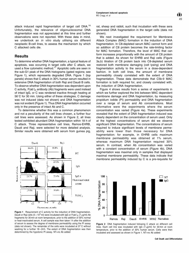
To determine whether DNA fragmentation, a typical feature ofapoptosis, was occurring in target cells after C attack, weused a flow cytometric method.17 Apoptotic cells are seen inthe sub-G1 peak of the DNA histograms (gated regions; seeFigure 1), which represents degraded DNA. Figure 1 (toppanels) shows that C attack in 30% human serum resulted inextensive DNA fragmentation of both Raji and Daudi B cells.To observe whether DNA fragmentation was dependent uponC activity, F(ab')2 antibody (Ab) fragments were used insteadof intact IgG, or C was rendered inactive through heating at568C for 30 min. Using either of these strategies, C damagewas not induced (data not shown) and DNA fragmentationwas not evident (Figure 1). Thus DNA fragmentation occurredonly in the presence of intact Ab and C.
To determine whether this was a common phenomenonand not a peculiarity of the cell lines chosen, a further fourcell lines were assessed. As shown in Figure 2, all linestested exhibited abundant DNA fragmentation within 18 h ofC attack. Three representative cell lines, Ramos-EHRB,Daudi and Raji, were selected for more detailed analysis.Similar results were obtained with serum from guinea pig,
rat, sheep and rabbit, such that incubation with these seragenerated DNA fragmentation in the target cells (data notshown).
We next investigated the requirement for MembraneAttack Complex (MAC) formation in the induction of DNAfragmentation. In C8-depleted serum MAC cannot form andso addition of C8 protein becomes the rate-limiting factorfor MAC formation. Therefore, the level of MAC that canform increases proportionally with the amount of C8 proteinthat is added. As shown for EHRB and Raji cells (Figure3a,b) titration of C8 protein back into C8-depleted serumrestored both membrane damaging (cell lysing) and DNAfragmentation activity to the serum in a dose-dependentfashion. In both cell lines, the amount of membranepermeability closely correlated with the extent of DNAfragmentation. These data demonstrate that C5b-9 MACformation is both required for, and closely correlated with,the induction of DNA fragmentation.
Figure 4 shows results from a series of experiments inwhich we further explored the link between MAC dependentmembrane damage and DNA fragmentation, by measuringpropidium iodide (PI) permeability and DNA fragmentationover a range of serum and Ab concentrations. Mostinformative were the experiments where the serumconcentration was varied (Figure 4a). These experimentsrevealed that the extent of DNA fragmentation induced wasclearly dependent on the concentration of serum used. Onlyat the highest concentrations of serum did we observesignificant DNA fragmentation. The concentrations of serumrequired to induce significant levels of membrane perme-ability were lower than those necessary for DNAfragmentation: for example, in EHRB cells maximummembrane permeability was obtained at 1% serum,whereas maximum DNA fragmentation required 30%serum. In contrast, when Ab concentration was variedwith a constant concentration of serum (Figure 4b), DNAfragmentation was maximal only in samples that displayedmaximal membrane permeability. These data indicate thatmembrane permeability induced by C is a pre-requisite for
Figure 1 Requirement of C activity for the induction of DNA fragmentation.Daudi or Raji cells (56105 ml) were incubated with IgG or F(ab')2 (7 mg/ml) Abfragments for 30 min at room temperature, prior to the addition of 30% normalor heat-inactivated serum. A cell sample was then taken 1 h after the additionof serum to assess the degree of membrane permeability using the PI assay(data not shown). The remainder of the cells were incubated at 378C withoutwashing for a further 18 ± 24 h. The extent of DNA fragmentation was thendetermined by the hypotonic PI assay. NT=no Ab added
Figure 2 DNA fragmentation induced following C attack on different celllines. Each cell line was incubated with IgG (7 mg/ml) for 30 min at roomtemperature, prior to the addition of 30% human serum. Cells were thenincubated and assessed as shown in Figure 1. NT=no Ab added
Complement induced apoptosisMS Cragg et al
49
Cell Death and Differentiation

subsequent DNA fragmentation, but that the membranepermeability alone is not sufficient and that once cells aredamaged, it is the concentration of serum which determinesthe degree of DNA fragmentation. In addition, these datademonstrate that at levels of serum typically used by otherworkers (e.g. 520%) only low levels of DNA fragmentationwere seen with Daudi and Raji cells, whilst at morephysiological concentrations of serum (420%), there wasextensive DNA fragmentation. This perhaps explains whythese observations have not been reported previously.
To assess whether the observed DNA fragmentationwas apoptotic in nature, agarose gel analysis wasperformed. These data, shown in Figure 5 for Daudi cells,revealed that the DNA fragmentation induced wasoligonucleosomal in nature and displayed discrete bandedDNA ladder patterns typical of apoptosis.2,7,8 Bands on thegels were approximately 180 ± 200 base-pairs apart,indicating their nucleosomal origins. In addition, the
analysis supported the idea that fragmentation was afunction of serum concentration, as increasing DNAfragmentation was observed with increasing levels ofserum.
In the experiments detailed above, DNA fragmentationwas detected after an overnight (18 ± 24 h) incubation. Inorder to determine how soon after C attack the target cellDNA was degraded, the TUNEL technique was employed.This technique is a sensitive method for distinguishingapoptotic and necrotic cell death.18 TUNEL analysis(Figure 6) revealed that C-induced DNA fragmentationwas due to the induction of apoptotic-like strand breaks.TUNEL positivity was observed as rapidly as 30 min afteraddition of serum (Figure 6b) and was evident at highlevels after 1 h. The extent of TUNEL positivity was foundto be a function of serum concentration. Presumably,these strand breaks subsequently lead to the formation ofthe apoptotic DNA ladders, as DNA ladders were
Figure 3 Dependence of C lysis and DNA fragmentation on C8 protein. IgGwas added to EHRB (a) or Raji (b) cells for 30 min at room temperature, prior tothe addition of C8-deficient serum (30%) reconstituted with varying levels ofC8 protein. The extent of membrane permeability was determined by the PIassay (squares) after 1 h. DNA fragmentation was then determined 18 h afteraddition of serum by the hypotonic PI assay, shown as circle symbols. Opensymbols represent control samples and filled symbols represent resultsfollowing addition of IgG
Figure 4 Dependence of DNA fragmentation on serum (a) and Ab (b)concentration. In (a), IgG (7 mg/ml) was added to Raji, Daudi, or EHRB cells,allowed to bind for 30 min at room temperature and varying concentrations ofserum added. In (b), varying concentrations of IgG were added to a constantconcentration of serum (20%). The degree of membrane permeability wasdetermined by the PI assay after 30 min, represented as unshaded bars. Theextent of DNA fragmentation was assessed via the hypotonic PI assay 18 ±24 h after the addition of serum, shown as shaded bars
Cell Death and Differentiation
Complement induced apoptosisMS Cragg et al
50

observed as early as 2 h after the addition of serum (datanot shown).
Given that apoptotic-like DNA fragmentation wasapparent after C attack, we were intrigued to see whetherany other features of apoptosis were evident. Electronmicroscopy (EM) revealed that following C attack, cells innear-physiological concentrations of serum retained fargreater plasma and nuclear membrane integrity than cellssubjected to a similar level of attack in the presence of lowlevels of serum, shown for EHRB cells in Figure 7. Thirtyminutes after C attack, cells in high concentrations ofserum (Figure 7 panel c) have generally intact cell andnuclear membranes, whereas those in low levels of serumdemonstrate discontinuity of the plasma membrane, cell-lysis and nuclear leakage (Figure 7 panel b). Furthermore aproportion of the cells in high levels of serum retainednuclear integrity for up to 6 h (data not shown).
Analysis of the EM data also demonstrated thatchromatin was condensed following C attack in thepresence of high levels of serum, in a manner similar tothat reported by Papadimitriou for Ehrlich cells.13 Thisobservation, in conjunction with the DNA fragmentationdata, demonstrates that features of apoptosis wereapparent during C-mediated death of these cells. How-ever, although cells attacked in high levels of serum weremore resistant to complete lysis, signs of osmotic swellingand intracellular leakage were evident and ultimately manyof the cells lysed completely. This was reflected by thegeneral increase in cell size and by organelle swelling; twofeatures associated with necrotic death. Furthermore,membrane blebbing, a common feature of apoptosis, wasnot evident. Clearly then, although some of the hallmarkfeatures of apoptosis are apparent during C mediateddeath, not all were present and many of the membrane andcytoplasmic events more closely resembled a necrotic
phenotype. Similar observations were made in all three celllines examined.
Once the nature of the DNA fragmentation had beenverified in these cells, we wished to identify the mechanismby which it was induced. Ca2+ ions rapidly enter target cellsafter C attack19,20 and have been suggested as one of theeffectors of C-induced cell death.21 Ca2+ is also a knownco-factor for certain apoptotic endonucleases.2,8,22,23,24 Asserum contains high levels of Ca2+ (1.1 ± 1.23 mM)25 it wassuggested that the serum concentration dependence forDNA fragmentation was due to differences in theconcentration of Ca2+ present in the different levels ofserum. To determine if this was the case, low levels ofserum were supplemented with increasing levels ofexogenous Ca2+ ions and assessed for their ability toinduce DNA fragmentation (Figure 8a). Ramos-EHRB cellswere used in these experiments, as in this cell line low
Figure 5 DNA fragmentation induced in Daudi cells after C attack in varyinglevels of serum. IgG (7 mg/ml) was added to 1 ± 26106 Daudi cells for 30 min atroom temperature and then varying levels of serum added and the cellsincubated for 18 h at 378C. Samples were then harvested, treated withProteinase K, RNAse and subjected to agarose gel electrophoresis on a 1%gel for 2 h. Untreated control cells are shown in lane 1. In lanes 2 ± 6 increasinglevels of serum (1, 3, 10, 20, 30%) were added. A 100 bp molecular weightladder is shown in lane 7
Figure 6 Assay of TUNEL positivity after C attack. EHRB cells wereincubated with IgG (7 mg/ml) for 30 min at 208C and then varying levels ofserum (3, 10 or 30%) added and the cells incubated at 378C. The extent ofDNA strand breakage was assessed after 10, 30, 60 or 120 min via the TUNELassay in (a). In (b) the means+S.D. of duplicate samples over the 2 h timecourse are shown. The histogram demonstrates the degree of TUNELpositivity after 1 h
Cell Death and Differentiation
Complement induced apoptosisMS Cragg et al
51

A B
b b
c
a a
c
Figure 7 Light and electron microscopy of C attacked EHRB cells in the presence of high (30%) or low (3%) levels of serum. EHRB cells were incubated with IgG(7 mg/ml) for 30 min at 208C and then either 3 or 30% serum was added. After 30 min at 378C the cells were harvested, fixed by adding glutaraldehyde (1.5% final),washed and embedded in epon resin. For EM, thin (90 nm) sections were cut and stained with lead citrate. The views illustrated were gained at a magnification of75006and are shown in (A). In this figure, (a) represents normal untreated EHRB cells, (b) represents EHRB cells lysed in 3% serum and (c) represents cellsattacked in 30% serum. For light microscopy, thick (0.5 mm) sections were cut and stained with toluene blue as shown in (B). Again, (a) represents normal untreatedEHRB cells, (b) represents EHRB cells lysed in 3% serum and (c) represents cells attacked in 30% serum
Cell Death and Differentiation
Complement induced apoptosisMS Cragg et al
52

levels of serum are sufficient to induce maximal levels ofmembrane permeability, without concurrently high levels ofDNA fragmentation, enabling us to separate the phenom-enon of cell lysis and DNA fragmentation. As shown inFigure 8a, low levels of serum supplemented withexogenous Ca2+ did not increase the DNA fragmentationobserved.
Alternatively, differences between the effects of highand low levels of serum might be explained on a basisof osmotic potential. Within this rationale, it wasproposed that the osmotic protection afforded by highlevels of serum would enable cells to survive longenough for intracellular endonucleases to becomeactivated (possibly by the Ca2+ influx) and instigateDNA fragmentation. In contrast, low levels of serumwould not inhibit osmotic swelling and cell lysis wouldoccur prior to DNA fragmentation. To investigate thispossibility, low levels of serum were supplemented withdextran, Human serum albumin (HSA) or Bovine serumalbumin (BSA), to provide equivalent osmotic protection
to that afforded by high levels of serum. Theseexperiments, represented in Figure 8b, demonstrate thatC attacked cells, in the presence of osmotic protectants,though protected from complete lysis, do not undergosignificant DNA fragmentation in the absence ofsufficiently high levels of serum. Next we investigatedwhether both osmotic protection and high levels of Ca2+
ions were sufficient to initiate DNA fragmentation (Figure8c). These experiments, using dextran as the osmoticprotectant, revealed that osmotically protected cellsundergoing C attack in the presence of elevated levelsof Ca2+ still did not undergo significant DNA fragmenta-tion in the absence of high levels of serum.
To ascertain whether other forms of membranepermeability could induce DNA fragmentation, the beevenom derived pore-forming polypeptide melittin wasused. DNA fragmentation was not observed followingtreatment with melittin in the absence of serum, althoughmembrane permeability was induced (as measured by PIpermeability). When serum was included (without Ab to
Figure 8 Effect of additional Ca2+, osmotic protection or melittin on the extent of DNA fragmentation. In (a ± c), EHRB cells were lysed in the presence of IgG(7 mg/ml) and 3% serum with (a) varying concentrations of exogenous Ca2+, (b) different osmotic protectants (dextran was added to a final concentration of0.66 mM, and HSA and BSA were used at 30%), or (c) varying concentrations of exogenous Ca2+ and 0.33 mM dextran, added to the cultures. In (d), Raji or Daudicells were incubated with melittin (5 ± 20 mg/ml) in various doses of serum without IgG. Membrane permeability was determined 1 h after addition of serum; shownas open bars. DNA fragmentation was assessed 18 h after the addition of serum; shown as shaded bars
Cell Death and Differentiation
Complement induced apoptosisMS Cragg et al
53

activate C), DNA fragmentation was observed and in adose-dependent manner. Furthermore, heat-inactivatedserum was also able to induce DNA fragmentation inthese experiments (data not shown) excluding thepossibility that C activation is necessary. Together thesedata demonstrated that the DNA fragmentation inducedwas dependent upon a property of serum that was notrelated to osmotic potential, Ca2+ concentration or Cactivation.
In view of these results, we decided to fractionate thecomponents of serum and determine if the DNA fragment-ing activity could be restricted to molecules of a given size.Fractionation was carried out using an ACA44 gel matrix.As illustrated in Figure 9, the major DNA fragmentingactivity was found to be restricted to fractions correspond-ing to proteins of approximately 25 ± 50 kDa in size.Fractionation of animal sera (guinea pig, sheep, rabbit)yielded similar results (data not shown), indicating that asimilar factor is involved. Intriguingly, DNase I is 32 ±38 kDa26,27 and furthermore, is the only well known serumendonuclease.23,27,28,41
To further characterise the nature of the DNA fragmenta-tion-factor in serum, we undertook a series of inhibitionstudies. The data from these experiments (Figure 10)demonstrate that EGTA, EDTA, and Zn2+ ions werecapable of inhibiting the DNase activity in serum, but thataurintricarboxylic acid (ATA) was not. This inhibitor profile issimilar to that of serum DNase I.26 To eliminate thepossibility of a cytosolic DNase being involved during thedeath process, we isolated nuclei from the cells andrepeated these studies. Similar results were achieved withisolated nuclei as in whole cells, such that DNAfragmentation was dependent on serum concentration,inhibited by EGTA/EDTA and Zn2+ and not by ATA (datanot shown). Finally, to examine whether caspases wereinvolved in the DNA fragmentation, three tetrapeptide
caspase inhibitors (YVAD, ZVAD and DEVD) were used.All three inhibitors were ineffective even when applied at aconcentration of 250 mM, implying that caspases are notinvolved in this process (data not shown). All of these dataindicate that the DNA fragmentation which follows C attackis due to an extracellular endonuclease which is present inserum and that this endonuclease is likely to be DNase I.
Discussion
We have shown that following C attack, various cell linesunderwent an extensive and rapid apoptotic-like DNAfragmentation, which was dependent on high levels ofserum. The DNA fragmentation was not seen in low levelsof serum. However, low levels of serum (1 ± 10%) were able toinduce substantial membrane permeability. Titration experi-ments with C8-depleted serum and C8 protein confirmed that
Figure 9 DNA fragmentation induced by various fractions of serum followingseparation on ACA44. Human serum was added to a pre-equilibrated ACA44column and fractions collected. The approximate molecular weight of proteinsin the different fractions was then determined by comparison with the elutionprofile of known molecular weight standards. 10% (v/v final) of fractionatedserum was added to 3% native serum (to ensure membrane permeabilisation)and the DNA fragmentation activity of each fraction was determined using thehypotonic PI assay assessed 16 h after addition of serum. Also shown are thelevels of DNA fragmentation induced with serum reconstituted from equivalentportions of the fractionated serum in the absence (NT) or presence of Ab
Figure 10 Inhibitor profile of DNase activity following C attack of EHRB cellsin 30% serum. Following C attack for 15 min at 378C to allow maximalmembrane permeability, various endonuclease inhibitors were added to thesamples. In a series of experiments 1 ± 5 mM final concentrations of ZnCl2,ZnSO4, EGTA, EDTA or ATA were used. Shown in (a) is an example of atypical set of results. In (b) two identical experiments are compared with themeans shown
Cell Death and Differentiation
Complement induced apoptosisMS Cragg et al
54

functional C was necessary for DNA fragmentation to occurand, more importantly, demonstrated that MAC formation wasvital for the induction of DNA fragmentation. Subsequentexperiments revealed that melittin pores could substitute forMAC in inducing membrane permeability, although highdoses of serum were again necessary for the induction ofDNA fragmentation. The requirement for high doses of serumin both of these systems indicated that DNA fragmentationwas dependent upon a titratable factor. Furthermore, asisolated nuclei were similarly susceptible to the effects of highlevels of serum (data not shown) this indicated that cytosolicendonucleases did not play an important role in thefragmentation process. Lack of a requirement for the cytosolalso indicated that caspases were not involved in this processand this was supported by the fact that the tetrapeptidesYVAD, ZVAD and DEVD did not inhibit the observed DNAfragmentation (data not shown).
The effects of high serum levels could not be mimickedby osmotic protectants either alone or in conjunction withelevated levels of Ca2+, indicating that the DNA fragmenta-tion which occurred was not simply due to an influx of Ca2+
and activation of a nuclear-based Ca2+ dependentendonuclease such as that described by Wyllie.24 How-ever, once initiated, it is possible that endogenous,cytosolic and nuclear endonucleases would becomeactivated. In the absence of cytosolic and nuclearendonucleases, it must be supposed that the DNAfragmentation following C attack is due to the entry of anexternal DNase present in serum.
One possible source of external endonucleases wouldbe mycoplasma infected cells, which have been shown tobe capable of secreting endonucleases capable of inducinginternucleosomal DNA fragmentation.29 However, in ourstudy the cell lines used were routinely screened formycoplasma and found to be uninfected. The FCS usedin cell culture is screened for mycoplasma infection and cansimilarly be disregarded as a source of mycoplasma-derived external endonucleases.
DNase activity in serum and plasma has long beenreported for a plethora of species, including rat,30
mouse23,31 and human.27,28,32 Various authors haveinvestigated the nature of this serum DNase27,28,32 andalthough its origin is unclear, it would appear that the majorendonuclease activity is due to isoforms of DNase I.27 Insupport of this suggestion, Polzar and Mannherz33 haveidentified a potential signal peptide in the rat DNase I gene,which would readily facilitate its secretion allowing its entryinto the serum.
In addition to its role as the main endonuclease inserum, several groups have suggested that DNase I is thecentral endonuclease involved in apoptotic cell death2,26
and prior to the discovery of Caspase Activated DNase(CAD) in 1998,34 provided a number of convincing lines ofevidence in support of this. However, one hurdle thatproponents of DNase I face is that it is found predominantlywithin the endoplasmic reticulum (ER) and for DNAfragmentation to occur it must enter the nucleus. How itmight do this is uncertain, although the rise in calcium seenduring apoptosis has been suggested as a potentialmechanism,26 as Ca2+ fluxes have been reported to
induce dissolution of the ER and hence perturbation ofthe perinuclear membrane.35
Intriguingly, C attack also induces a rise in cytoplasmiccalcium.19,20 Could it be that the endonuclease responsiblefor fragmentation of target cell DNA following C attack isalso DNase I? Our study shows that fragmentation of thenuclear DNA occurs at neutral pH and is potently inhibitedby Zn2+ ions, chelating agents EDTA and EGTA, but notATA, an inhibitor profile which closely matches thatreported for serum DNase I.26 In addition, DNase I is theonly well known serum endonuclease23,27,28,41 and ourfractionation data indicate that a protein of its size (32 ±38 kDa) is involved in this process. Furthermore, it hasbeen established by Peitsch et al, that DNase I is capableof oligonucleosomal apoptotic DNA fragmentation in thepresence of serum.23 For these reasons, we hypothesisethat the observed DNA fragmentation of C attacked cells isa direct result of serum DNase and that this DNase is eitherDNase I, or a close homolog. Endogenous, intracellularDNase I may contribute to the DNA degradation in themanner suggested by Peitsch et al 23,26 but our dataindicate a requirement for the external DNase present inserum. Moreover, we have recently carried out experimentsthat show that when increasing concentrations of purifiedDNase I are titrated into low (but lytic) levels of serum, acorresponding increase in DNA fragmentation is seen (datanot shown). Ongoing work will attempt to define the natureof the external DNase more precisely.
Our proposed model then, for C attack of cells undernear-physiological conditions, is as follows. Firstly, C5b-9lesions are punched into the cell membrane allowing influxof small protein molecules, such as the serum DNase.Subsequently, the internalised DNase appears to accessand degrade the target cell DNA. This process appearssimilar to that used by cytotoxic cells to kill their targets.During CTL- and NK-cell killing, cytotoxic granules arereleased which deliver perforin and granzymes to the targetcell. Perforin forms pores in the target cell membrane,which facilitates the entry and activity of the granzymes.36
The granzymes then initiate the caspase cascades presentwithin the target cell, which result in apoptosis andfragmentation of the resident DNA. Given this resem-blance, it is intriguing to discover that perforin hassubstantial homology to the terminal component of C,C9.37 Furthermore, it is possible that defensins mayoperate similarly, given that they also are able to disruptmembrane integrity.38
One of the central predictions from our hypothesis is thatall membrane damaging events should lead to similarmodes of cell death. The preliminary results with themembrane pore forming polypeptide, melittin, seem tosupport this idea. Exposure to melittin in the presence ofserum results in potent DNA fragmentation, in a dose-dependent manner similar to that reported here for Cdamage and in a range of cells including mouse LTKfibroblasts, Chinese Hamster Ovary cells and EBV-transformed B-cells (data not shown), as well as thelymphoma lines (Figure 8d). Membrane permeabilityinduced with melittin in the absence of serum yields noobservable DNA fragmentation, even in the presence of
Cell Death and Differentiation
Complement induced apoptosisMS Cragg et al
55

osmotic protectants and elevated levels of Ca2+, similar toC attack in low levels of serum (data not shown). Ifapoptotic DNA fragmentation is commonly seen followingany form of membrane permeability, this has greatrelevance for the study of apoptosis both in vitro and inparticular in vivo. In in vitro experiments, membranedamaging agents, such as residual C activity andperforin, may result in false detection of apoptotic deathby DNA fragmentation criteria. Indeed, we have observedjust such a phenomenon in the sensitive cell line Ramos-EHRB cultured in 10% non heat-inactivated FCS whichcontained enough residual C to facilitate lysis and DNAfragmentation with certain mAb (data not shown). Thisconcern is even more relevant in vivo, where C and DNaseactivity will be maximal in circulating plasma. For thisreason, criteria other than DNA fragmentation may have tobe utilised to define apoptosis in vivo, or more likely anumber of such criteria used in conjunction, as suggestedby Kerr et al.39
Although we have demonstrated that the cellularintegrity maintained following C attack in high concentra-tions of serum is not an important factor in initiatingsubsequent DNA fragmentation, its role in the deathprocess should not be ignored. Under conditions of highserum, the external environment is at relatively highosmolarity and helps to prevent total cell lysis, allowingthe cell to retain its integrity, particularly nuclear, for alonger period of time. These conditions mimic thoseseen in vivo more closely than those used in previousstudies and the delay in lysis and retention of thenuclear material may be extremely important whenconsidering C mediated death in vivo, since it shouldallow the DNA to be fragmented before its release intothe surrounding environment.
In a multicellular organism, cells are targeted for deathwhen they are either unwanted, infected or neoplastic.Therefore, maintaining nuclear integrity of a dying cell fora longer period of time allowing time for DNA to bedegraded helps to prevent exposure of surrounding cellsto potentially neoplastic or foreign DNA. The protection ofneighbouring cells from so-called dangerous DNA hasbeen suggested as the main role of the endonucleaseduring apoptosis.8 It seems that the serum DNase is welladapted for this role in several ways. Firstly, the single-strand breaks induced by the serum DNase halttranscription, thereby rendering any dangerous DNAtranscriptionally inactive.40 Secondly, the induction ofstrand-breaks into target cell DNA facilitates phagocyticactivity in neighbouring cells. In addition to aiding theclearance of potentially dangerous and immunogenic DNA,this function may also serve to prevent inflammation andsubsequent auto-Ab generation.26 In particular, preventingchromatin, a highly immunogenic protein implicated indevelopment of auto-immune disorders such as SLE,getting into the circulation may be important. It isinteresting to note that SLE patients display diminishedlevels of DNase in their serum,41 perhaps implying thatserum DNase may play a role in degrading DNA in serumand preventing auto-immune reactions from developing.Thirdly, DNA fragmentation prevents surrounding cells
from having to cope with the huge volume of unpackagedgenomic DNA which would be released after death.
All of these facets of DNA fragmentation are gearedto reduce inflammation and remove potential immuno-gens: the primary goals of apoptosis. In recent years ithas become apparent that the components of C linkquite closely with those of apoptosis, certainly more sothan was envisaged as little as 5 years ago. A spate ofrecent papers have demonstrated that C may beinvolved in the removal of apoptotic cells throughtagging for phagocytosis via C1q or C3b fragments.42 ± 44
In conclusion, this work demonstrates that C attack ofmammalian cells can result in apoptotic-like condensationand fragmentation of nuclear DNA, which appears to arisefrom the influx of a serum DNase, which may be DNase I.These data contribute to the idea that C-induced death isnot entirely necrotic in nature but may instead resemble amore rapid form of apoptosis which demonstrates certainnecrotic-like cytosolic and membrane features. Further-more, these data imply that other forms of membranedamage may also induce apoptotic-like cell death in vivo.
Materials and Methods
Materials
Daudi, Ramos, Ramos-EHRB, Raji, and Namalwa cells were allobtained from the European Collection of Cell Cultures (ECACC) andwere maintained in RPMI-1640 medium supplemented with antibioticsand 10% FCS at 378C in a 5% CO2 humidified incubator. Cells weremaintained in log phase of growth for 24 h prior to experiments. WW2,EBV immortalised B-cells were a kind gift from Myf Spellerberg, HITgroup, Southampton University Hospital. The sensitivity of these celllines to C mediated cell lysis was directly related to the levels of Ccontrol proteins (CD55 and CD59) expressed at the cell surface (datanot shown).
Serum from all species was prepared as follows: whole blood wasallowed to clot at room temperature for 30 ± 60 min. Serum was thencollected and centrifuged at 9006g for 20 min. Serum was stored at7808C. For experiments, 100% serum was taken to be the undilutedsoluble fraction remaining after clotting. C8 depleted human serumand human C8 protein were generated and prepared as describedpreviously.45 All human serum used in this study was from AB donors,although analogous results were achieved with serum from otherdonors (data not shown). Heat-inactivation of serum was carried out at568C for 30 min.
The Ab used for activation of the classical C pathway was anti-CD20 mAb [IF5;46], although similar results were achieved with theIgG fraction of rabbit anti-serum raised against human tonsil cells. Thehybridoma line secreting IF5 was expanded in tissue culture andpurified from culture supernatant using a Protein A column as detailedby Glennie et al.47 F(ab')2 fragments of IgG were prepared by limitedproteolysis with pepsin at pH 4.0 ± 4.2 as detailed by Glennie et al.47
BSA (fraction V, 65 000 mw) was obtained from Wilfred Smith Ltd,Middlesex. HSA was obtained from BPL, Elstree, Herts. Dextran (65 ±85 000 mw) and melittin were purchased from Sigma, Poole, Dorset.
Electron microscopy (EM)
EM was performed using a protocol similar to that described byPapadimitriou et al.13 Briefly, after 30 min in serum, cells were fixed inthe presence of their incubation media by the addition of an equal
Cell Death and Differentiation
Complement induced apoptosisMS Cragg et al
56

volume of 3% glutaraldehyde in 0.1 M sodium cacodylate bufferpH 7.4 for 2 h at room temperature. Cells were then washed severaltimes in cacodylate buffer. Post-fixation with osmium was followed byen bloc staining with uranyl acetate before dehydration via gradedalcohols and embedding in epon resin. Thick (0.5 mM) and thin(90 nM) sections were then cut, stained with toluene blue or leadcitrate and viewed via light and electron microscopes respectively. EMwas performed on a Hitachi H-7000.
Flow cytometric analysis of DNA fragmentationusing PI
Samples were assessed rapidly for DNA fragmentation essentially asper the method of Nicoletti et al.17 Briefly, aliquots of cells (5 ±106105) were centrifuged for 5 min at 5006g and then washed oncein PBS. The cells were then resuspended in hypotonic fluorochromesolution (50 mg/ml PI, 0.1% (w/v) sodium citrate, 0.1% (w/v) Triton X-100) and incubated in the dark at 48C overnight or 1 h at roomtemperature. PI staining of DNA from 10 000 cells was detected on aFACScan flow cytometer (Becton Dickinson) and the proportion ofDNA giving fluorescence below the G1/0 peak was taken as ameasure of apoptosis.
Analysis of DNA fragmentation by agarose gelelectrophoresis
Following incubation of cells under appropriate conditions, samplescontaining 1 ± 26106 cells were harvested. Twenty ml TE-SLS (10 mMEDTA, 50 mM Tris HCl pH 8.0, 0.5% (v/v) sodium lauryl sarkosinate(SLS, Sigma)) plus 0.5 mg/ml Proteinase K (Sigma) was then added,and the samples incubated for 1 ± 2 h at 508C. Ten ml 0.16 mg/ml RNaseA (Sigma) was added and samples were incubated for a further 1 ± 2 hat 508C. The temperature of the samples was then increased to 708C foraddition of 10 ml of loading buffer (10 mM EDTA, 1% (w/v) low meltingpoint agarose, 0.25% (w/v) bromophenol blue, 40% (w/v) sucrose).Samples were loaded into dry wells of 1% (w/v) agarose gels containing0.5 mg/ml ethidium bromide and run either overnight at 30 ± 40 V or for2 ± 3 h at 80 V, in TAE (89 mM Tris, 89 mM boric acid, 2 mM EDTA,pH 8.4). DNA was visualised under UV light illumination.
Nuclei extraction
Nuclei were isolated using the method of Nieto et al.48 Briefly, Ramos-EHRB cells were washed twice in PBS and lysed in a hypotonicsolution of 1.5 mM MgCl2 for 30 min on ice, releasing intact nuclei.Nuclei were then harvested by centrifugation for 5 min at 5006g andwashed twice in 1.5 mM MgCl2 before resuspension in a solutioncontaining 10 mM Tris (pH 7.5), 200 mM sucrose and 60 mM NaCl.
Osmotic protection
In osmotic protection experiments, dextran, HSA, or BSA were used.Dextran was used at concentrations between 0.33 and 1.33 mM. HSAand BSA were used at 10 ± 30% (w/v) final concentrations. Thesesolutions allowed permeabilisation of the plasma membrane (asdetermined by PI entry) but delayed full cell lysis (as determinedmicroscopically or via flow cytometry) and mimicked closely theosmotic protection effects of 30% human serum.
Membrane permeability assay
Membrane permeability was detected using a rapid and simple PIexclusion assay. Diminished membrane integrity facilitates entry of PI
into cells where it binds DNA causing a large (1000-fold) increase influorescence in the red area of the spectrum. This assay was carriedout by adding 100 ml of PI solution (10 mg/ml in PBS) to 150 ml of agiven cell sample. The mixture was then assessed immediately byFACS, using a 488 nm argon laser for excitation and a 560 nmdichroic mirror and 600 nm band pass filter (bandwidth 35 nm) fordetection. More traditional 51Cr release assays gave comparableresults (data not shown).
Serum separation
The protein components of human serum were separated on a basis ofsize using an ACA44 gel filtration column. ACA44 gel (IBF, France)was packed by gravity flow into two 640 ml columns, which wereconnected in series and equilibrated with PBS. Human serum wasthen added to the column and run in PBS under pressure from gravity.Flow through was assessed by UV absorbance at 280 nm to detectprotein. Minor fractions were collected, pooled into one of six majorfractions, concentrated and adjusted back to the same volume as wasloaded onto the column and subsequently assessed for DNAfragmentation activity. The approximate molecular weight of proteinsin the different major fractions was then determined by comparisonwith the elution profile of known molecular weight standards.
AcknowledgementsThe authors would like to thank Dr Andrew George and Dr Ruth Frenchfor careful reviewing of the manuscript. This work was funded by grantsfrom Tenovus, Cardiff and The Wellcome Trust.
References
1. Kerr JFR, Wyllie AH and Currie AR (1972) Apoptosis: A basic biological
phenomenon with wide-ranging implications in tissue kinetics. Br. J. Cancer. 26:
239 ± 257
2. Hughes MF and Cidlowski JA (1996) Enzymology of apoptosis. In Apoptosis in
normal development and cancer, Sluyser M, ed (London: Taylor and FrancisPress Ltd) pp. 20 ± 31
3. Wyllie AH, Kerr JFR and Currie AR (1980) Cell Death: the significance of
apoptosis. Intl. Rev. Cytol. 68: 251 ± 306
4. Umansky SR (1996) Apoptosis: molecular and cellular mechanisms: A review.
Mol. Biol. 30: 285 ± 295
5. Kerr JFR, Winterford CM and Harmon BV (1994) Apoptosis: its significance in
cancer and cancer therapy. Cancer. 73: 2013 ± 2026
6. Kerr JFR and Harmon BV (1991) Definition and incidence of apoptosis: an
historical perspective. In Apoptosis: the Molecular Basis of Cell Death, Tomei LD
and Cope OF, eds. (New York: Cold Spring Harbor Laboratory Press) pp. 5 ± 29
7. Darzynkiewicz Z, Juan G, Li X, Gorczyca W, Murakami T and Traganos F (1997)
Cytometry in cell Necrobiology: analysis of apoptosis and accidental cell death
(necrosis). Cytometry 27: 1 ± 20
8. Arends MJ, Morris RG and Wyllie AH (1990) Apoptosis: the role of the
endonuclease. Am. J. Pathol. 136: 593 ± 608
9. Ishigami T, Kim KM, Horiguchi Y, Higaki Y, Hata D, Heike T, Katamura K, Mayumi
M and Mikawa H (1992) Anti-IgM antibody-induced cell death in a human B
lymphoma cell line, B104, represents a novel programmed cell death. J.Immunol. 148: 360 ± 368
10. Matsuoka S, Asano Y, Sano K, Kishimoto I, Yorifuji H, Utsuyama M, Hirokawa K
and Tada T (1995) A novel type of cell-death of lymphocytes induced by a
monoclonal antibody without participation of complement. J. Exp. Med. 181:
2007 ± 2015
11. Bhat NM, Bieber MM, Stevenson FK and Teng NNH (1996) Rapid cytotoxicity of
human B lymphocytes induced by VH4-34 (VH4.21) gene-encoded monoclonal
antibodies. Clin. Exp. Immunol. 105: 1 ± 8
12. Hagar H, Ueda N and Shah SV (1997) Tyrosine phosphorylation in DNA damage
and cell death in hypoxic injury to LLC-PK1 cells. Kidney Intl. 51: 1747 ± 1753
Cell Death and Differentiation
Complement induced apoptosisMS Cragg et al
57

13. Papadimitriou JC, Drachenberg CB, Shin ML and Trump BF (1994)
Ultrastructural studies of complement mediated cell death: a biological reaction
model to plasma membrane injury. Virch. Arch. 424: 677 ± 685
14. Kim SH, Carney DF, Papadimitriou JC and Shin ML (1989) Effect of osmotic
protection on nucleated cell killing by C5b-9: Cell death is not affected by the
prevention of cell swelling. Mol. Immunol. 26: 323 ± 331
15. Green HR, Barrow P and Goldberg B (1959) Effect of antibody and complement
on permeability control in ascites tumour cells and erythrocytes. J. Ex. Med. 110:699 ± 713
16. Shipley WU, Baker AR and Colten HR (1971) DNA degradation in mammalian
cells following complement-mediated cytolysis. J. Immunol. 106: 576 ± 579
17. Nicoletti I, Migliorati G, Pagliacci MC, Grignani F and Riccardi C (1991) A rapid
and simple method for measuring thymocyte apoptosis by propidium iodide
staining and flow cytometry. J. Immunol. Meths. 139: 271 ± 279
18. Gold R, Schmied M, Giegerich G, Breitschopf H, Hartung HP, Toyka KV and
Lassman H (1994) Differentiation between cellular apoptosis and necrosis by the
combined use of in-situ tailing and nick translation techniques. Lab. Invest. 71:
219 ± 225
19. Campbell AK, Daw RA and Luzio JP (1979) Rapid increases in intracellular free
Ca2+ induced by antibody plus complement. FEBS. Lett. 107: 55 ± 60
20. Campbell AK, Daw RA, Hallett MB and Luzio JP (1981) Direct measurement of
the increase in intracellular free calcium ion concentration in response to the
action of complement. Biochem. J. 194: 551 ± 560
21. Kim S, Carney DF and Shin ML (1987) Nucleated cell killing by complement:
Effects of C5b-9 channel size and extracellular Ca2+ on the lytic process. J.
Immunol. 138: 1530 ± 153622. Gaido ML and Cidlowski JA (1991) Identification, purification and characterisa-
tion of a calcium-dependent endonuclease (NUC18) from apoptotic rat
thymocytes. J. Biol. Chem. 266: 18580 ± 18585
23. Peitsch MC, Hesterkamp T, Polzar B, Mannherz HG and Tschopp J (1992)
Functional characterisation of serum DNase I in MRL-lpr/lpr mice. Biochem.
Biophys. Res. Commun. 186: 739 ± 745
24. Wyllie AH (1980) Glucocorticoid-induced thymocyte apoptosis is associated
with endogenous endonuclease activation. Nature 284: 555 ± 556
25. Wyngaarden JB and Smith LH (1988) Cecil Textbook of Medicine (Philadelphia:
W.B. Saunders Company) pp. 2395
26. Peitsch MC, Mannherz HG and Tschopp J (1994) The apoptosis endonucleases:
cleaning up after cell death? Trends Cell Biol. 4: 37 ± 41
27. Kishi K, Yasuda T, Ikehara Y, Sawazaki K, Sato W and Iida R (1990) Human
serum deoxyribonuclease I (DNase I) polymorphism: Pattern similarities among
isozymes from serum, urine, kidney, liver and pancreas. Am. J. Hum. Genet. 47:
121 ± 126
28. Love JD and Hewitt RR (1979) The relationship between human serum and
human pancreatic DNase I. J. Biol. Chem. 254: 12588 ± 1259429. Paddenberg R, Wulf S, Weber A, Heimann P, Beck L and Mannherz HG (1996)
Internucleosomal DNA fragmentation in cultured cells under conditions reported
to induce apoptosis may be caused by mycoplasma endonucleases. Eur. J. Cell
Biol. 71: 105 ± 119
30. Lacks SA (1981) Deoxyribonuclease I in mammalian tissues ± specificity of
inhibition by actin. J. Biol. Chem. 256: 2644 ± 2648
31. Shack J (1957) Deoxyribonucleases of mouse tissues. J. Biol. Chem. 226: 573 ±
581
32. Herriot RM, Connolly JH and Gupta S (1961) Blood nucleases and infectious viral
nucleic acids. Nature 189: 817 ± 820
33. Polzar B and Mannherz HG (1990) Nucleotide sequence of a full length cDNA
clone encoding the deoxyribonuclease I from the rat parotid gland. Nuc. Acids
Res. 18: 7151
34. Sakahira H, Enari M and Nagata S (1998) Cleavage of CAD inhibitor in CADactivation and DNA degradation during apoptosis. Nature 391: 96 ± 99
35. Booth C and Koch GL (1989) Perturbation of cellular calcium induces secretion of
luminal ER proteins. Cell 59: 729 ± 737
36. Henkart PA (1985) Mechanism of lymphocyte-mediated cytotoxicity. Ann. Rev.
Immunol. 3: 31 ± 58
37. Lowrey DM, Aebischer T, Olsen KJ, Lichtenhelf M, Rupp F, Hentgartner H and
Podack ER (1989) Cloning, analysis and expression of murine perforin 1 cDNA, a
component of cytotoxic T-cell granules with homology to complement
component C9. Proc. Natl. Acad. Sci. U.S.A. 86: 247 ± 251
38. Selsted ME and Oullette AJ (1995) Defensins in granules of phagocytic and non-
phagocytic cells. Trends Cell Biol. 5: 114 ± 119
39. Collins RJ, Harmon BV, Gobe GC and Kerr JFR (1992) Internucleosomal DNA
cleavage should not be the sole criterion for identifying apoptosis. Intl. J. Radiat.
Biol. 61: 451 ± 453
40. Peitsch MC, Muller C and Tschopp J (1993) DNA fragmentation during apoptosis
is caused by frequent single-strand cuts. Nucl. Acids Res. 18: 4206 ± 4209
41. Chitrabamrung S, Rubin RL and Tan EM (1981) Serum deoxyribonuclease-I and
clinical activity in systemic lupus erythematosus. Rheumatol. Int. 1: 55 ± 6042. Matsui H, Tsuji S, Nishimura H and Nagasawa S (1994) Activation of the
alternative pathway of complement by apoptotic Jurkat cells. FEBS Lett. 351:
419 ± 422
43. Tsuji S, Kaji K and Nagasawa S (1994) Activation of the alternative pathway of
human complement by apoptotic human umbilical vein endothelial cells. J.
Biochem. 116: 794 ± 800
44. Botto M, DellAgnola C, Bygrave AE, Thompson EM, Cook HT, Petry F, Loos M,
Pandolfi PP and Walport MJ (1998) Homozygous C1q deficiency causes
glomerulonephritis associated with multiple apoptotic bodies. Nature Genet. 19:
56 ± 59
45. Abraha A, Morgan BP and Luzio JP (1988) The preparation and characterisation
of monoclonal antibodies to human complement component C8 and their use in
purification of C8 and C8 subunits. Biochem. J. 251: 285 ± 292
46. Clark EA, Shu G and Ledbetter JA (1985) Role of the Bp35 cell surface
polypeptide in human B-cell activation. Proc. Natl. Acad. Sci. U.S.A. 82: 1766 ±
1770
47. Glennie MJ, Tutt AL and Greenman J (1993) Preparation of multispecific F(ab')2and F(ab')3 antibody derivatives. In Tumour Immunobiology, a PracticalApproach, Rees RC, Reynolds CW, eds. (Oxford: Oxford University Press) pp.
225 ± 244
48. Nieto MA and Lopez-Rivas A (1989) IL-2 protects T lymphocytes from
glucocorticoid-induced DNA fragmentation and cell death. J. Immunol. 143:
4166 ± 4170
Cell Death and Differentiation
Complement induced apoptosisMS Cragg et al
58