Comparison of the radiosensitisation ability of metal oxide ...
184
Alexandra Furtado Guerreiro Master of Science May, 2020 Comparison of the radiosensitisation ability of metal oxide nanoparticles using clinical megavoltage X-rays Thesis submitted in partial fulfilment of the requirements for the degree of Doctor of Philosophy in Radiation Biology and Biophysics Supervisor: Jon Golding, Senior Lecturer, Open University Co-supervisors: Nigel Mason, Professor and Head of School, University of Kent Maria Alice Santos Pereira, Assistant Professor, NOVA School of Sciences and Technology Examination Committee: Chairperson: Professor Paulo Manuel Assis Loureiro Limão-Vieira Raporteurs: Professor Frederick Currell Dr Octávia Gabriela da Silva Viegas Nené Monteiro Gil Members: Professor Malgorzata S " mialek-Telega Professor Pedro António de Brito Tavares Professor Jon Golding
-
Upload
khangminh22 -
Category
Documents
-
view
1 -
download
0
Transcript of Comparison of the radiosensitisation ability of metal oxide ...
Alexandra Furtado Guerreiro
[Nome completo do autor]
[Nome completo do autor]
[Nome completo do autor]
[Nome completo do autor]
[Nome completo do autor]
[Nome completo do autor]
[Nome completo do autor]
Master of Science
[Habilitações Académicas]
[Habilitações Académicas]
[Habilitações Académicas]
[Habilitações Académicas]
[Habilitações Académicas]
[Habilitações Académicas]
[Habilitações Académicas]
May, 2020
Comparison of the radiosensitisation ability of metal oxide nanoparticles using clinical
megavoltage X-rays [Título da Tese]
Thesis submitted in partial fulfilment of the requirements for the degree of Doctor of Philosophy in
Radiation Biology and Biophysics
Dissertação para obtenção do Grau de Mestre em [Engenharia Informática]
Supervisor: Jon Golding, Senior Lecturer, Open University
Co-supervisors: Nigel Mason, Professor and Head of School, University of Kent
Maria Alice Santos Pereira, Assistant Professor, NOVA School of Sciences and Technology
Examination Committee:
Chairperson: Professor Paulo Manuel Assis Loureiro Limão-Vieira
Raporteurs: Professor Frederick Currell
Dr Octávia Gabriela da Silva Viegas Nené Monteiro Gil
Members: Professor Malgorzata S"mialek-Telega
Professor Pedro António de Brito Tavares
Professor Jon Golding

2

NOVA Lisbon University
Faculty of Science and Technology
[Nome completo do autor]
[Nome completo do autor]
[Nome completo do autor]
[Nome completo do autor]
[Nome completo do autor]
[Nome completo do autor]
[Nome completo do autor]
Alexandra Furtado Guerreiro
[Nome completo do autor]
[Nome completo do autor]
[Nome completo do autor]
[Nome completo do autor]
[Nome completo do autor]
[Nome completo do autor]
[Nome completo do autor]
Master of Science
[Habilitações Académicas]
[Habilitações Académicas]
[Habilitações Académicas]
[Habilitações Académicas]
[Habilitações Académicas]
[Habilitações Académicas]
[Habilitações Académicas]
Comparison of the radiosensitisation ability of metal oxide nanoparticles using clinical
megavoltage X-rays
[Título da Tese]
May, 2020
Thesis submitted in partial fulfilment of the requirements for the degree of Doctor of Philosophy in
Radiation Biology and Biophysics
Dissertação para obtenção do Grau de Mestre em [Engenharia Informática]

ii

iii
Comparison of the radiosensitisation ability of metal oxide nanoparticles using clinical megavoltage X-rays
Copyright © Alexandra Furtado Guerreiro, Faculty of Sciences and Technology, NOVA University Lisbon.
The Faculty of Sciences and Technology and the NOVA University Lisbon have the right, perpetual and without geographical boundaries, to file and publish this dissertation through printed copies reproduced on paper or on digital form, or by any other means known or that may be invented, and to disseminate through scientific repositories and admit its copying and distribution for non-commercial, educational or research purposes, as long as credit is given to the author and editor.

iv

v
To my mother, sister and
niece

vi

vii
Acknowledgements
The work presented in this thesis was performed at Open University and at GenesisCare in Milton Keynes and I would like to thank everyone that contributed to make this work possible.
To my supervisor, Dr Jon Golding, I would like to thank his commitment in my work and for always being there when I needed. His guidance and assistance were crucial to make this thesis come true.
To Eleanor Crabb and Nicholas Chatterton I would like to thank for all their input and all the brain storm that helped developed this thesis.
To Genesis Care staff, such as Aquila Sharif and all the radiographers, I would like to thank all their availability and effort to assist me in this thesis and make it possible.
Also, I would like to thank Open University staff such as Dr Igor Kraev, Dr Radka Gromnicova, Brett Keith, George Bryant and Dr Matthew Kershaw for all their technical assistance.
I would like to thank professor Nigel Mason and Sir John Mason Academic Trust for the support provided for the development of this work. To professor Alice S. Pereira for all her support from Portugal.
Many thanks to Fundação para a Ciências e Tecnologia (FCT/MCTES), Radiation Biology and Biophysics Doctoral Training Programme (RaBBiT, PD/00193/2012); UID/Multi/04378/2013 (UCIBIO); UID/FIS/00068/2013 (CEFITEC); and grant number PD/ BD/114450/2016 for granting me this PhD fellowship.
I would like to thank all the friends I made at Open University and that made my journey much easier and happier. A special thanks to Marcelle Silva, Sonia Azeggagh, Edu Frias and Radka Gromnicova for all the shared moments and their amazing support as friends that I will keep for life.
Also, I would like to thank my friends Nidia Almeida and Fausto that I met in the beginning of this journey and that made my experience so much better and fun. I hope we share many other journeys together. To Dr Cristina Timóteo for the support and friendship provided whenever I needed.
I would like to thank my closest friends and family for their support and for being present in my life. Thank you, Alexandros Kostopoulos, for all the moments we shared, all your support, encouragement, and companionship. A special thanks to my mother, sister and father for believing in me, and being so patient and so supportive. Mum and sister there are no words to describe how important you are for me and I dedicate this thesis to you. Also, I want to welcome our new member of the family, my niece Alice, which although still very young I hope in the future she will be able to read this thesis and know that my life became a brighter place with her presence. Thank you so much!

viii
ix
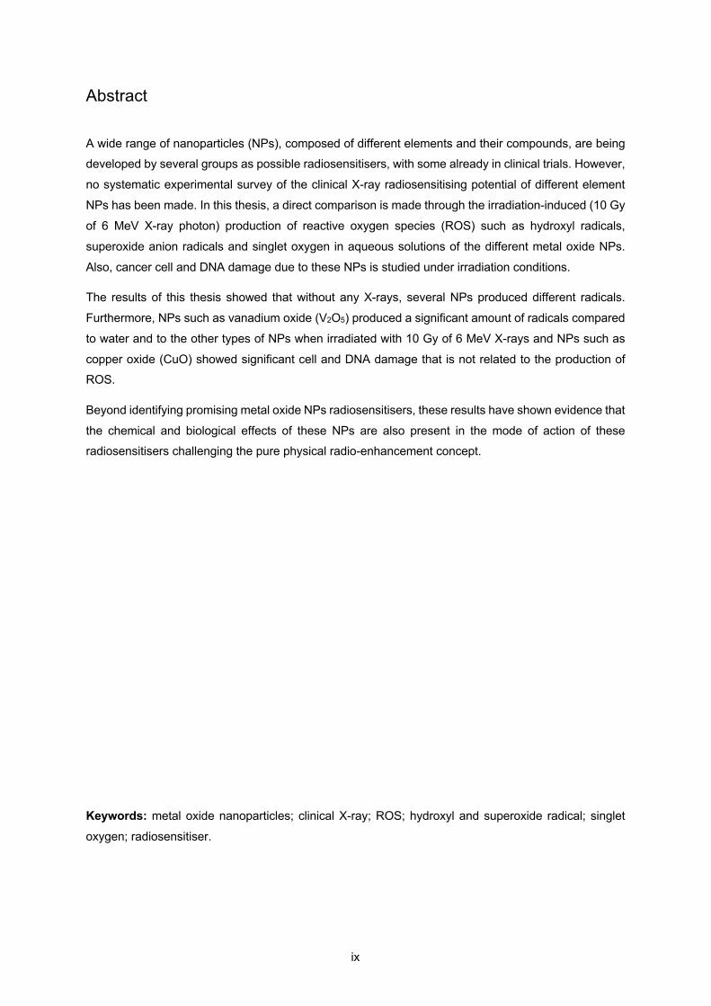
Abstract
A wide range of nanoparticles (NPs), composed of different elements and their compounds, are being developed by several groups as possible radiosensitisers, with some already in clinical trials. However,
no systematic experimental survey of the clinical X-ray radiosensitising potential of different element
NPs has been made. In this thesis, a direct comparison is made through the irradiation-induced (10 Gy
of 6 MeV X-ray photon) production of reactive oxygen species (ROS) such as hydroxyl radicals,
superoxide anion radicals and singlet oxygen in aqueous solutions of the different metal oxide NPs.
Also, cancer cell and DNA damage due to these NPs is studied under irradiation conditions.
The results of this thesis showed that without any X-rays, several NPs produced different radicals.
Furthermore, NPs such as vanadium oxide (V2O5) produced a significant amount of radicals compared
to water and to the other types of NPs when irradiated with 10 Gy of 6 MeV X-rays and NPs such as
copper oxide (CuO) showed significant cell and DNA damage that is not related to the production of ROS.
Beyond identifying promising metal oxide NPs radiosensitisers, these results have shown evidence that
the chemical and biological effects of these NPs are also present in the mode of action of these radiosensitisers challenging the pure physical radio-enhancement concept.
Keywords: metal oxide nanoparticles; clinical X-ray; ROS; hydroxyl and superoxide radical; singlet
oxygen; radiosensitiser.

x

xi
Resumo
Uma gama ampla de nanopartículas (NPs), compostas de diferentes elementos e dos seus compostos,
encontram-se a ser desenvolvidas por diferentes grupos como possíveis radiossensibilizadores, onde
algumas encontram-se em ensaios clínicos. No entanto, nenhum estudo sistemático comparativo do
potencial de radiossensibilização por raios-X usados clinicamente dos diferentes elementos de NPs foi realizado. Nesta tese, a comparação directa é realizada através da indução de espécies reactivas de
oxigénio (ROS), como os radicais hidroxilo e o anião superóxido, e o oxigénio singleto, por irradiação
(10 Gy de 6 MeV de fotões raios-X) em soluções aquosas das NPs dos diferentes óxidos de metais.
Para além disso, o dano causado a células cancerígenas e ao DNA devido à acção destas NPs é
estudado nestas condições de irradiação.
Os resultados desta tese mostram que, sem a acção dos raios-X, diferentes NPs produziram diferentes radicais. Nanopartículas como o óxido de vanádio (V2O5) produziram uma quantidade significativa de
radicais quando comparado à ausência de NPs e também quando comparado aos outros tipos de NPs
quando estas foram irradiadas com 10 Gy de 6 MeV de fotões raios-X e NPs de óxido de cobre (CuO)
mostraram um dano significativo celular e ao DNA que não se encontra relacionado com a produção
de ROS.
Para além de identificarem NPs de óxidos de metais promissores à radiossensibilização, estes
resultados mostraram evidência de que os efeitos químicos e biológicos destas NPs estão também presentes no seu modo de acção enquanto radiossensibilizadores, desafiando assim o conceito da
acção puramente física da radiossensibilização.
Palavras-chave: nanopartículas de óxidos de metais; raios-X clínicos; espécies reactivas de oxigénio; radicais hidroxilo e o anião superóxido; oxigénio singleto; radiossensibilizador.

xii

xiii
Table of Contents
Acknowledgements ........................................................................................................................... vii Abstract .............................................................................................................................................. ix Resumo ............................................................................................................................................... xi List of Figures ................................................................................................................................... xvii List of Tables ...................................................................................................................................... xix List of Abbreviations and Symbols ..................................................................................................... xxi
1 – INTRODUCTION ........................................................................................... 1
1.1 Radiotherapy ......................................................................................................................... 3 X-ray beam treatment .................................................................................................................. 3 Megavoltage X-ray physical properties ......................................................................................... 4 Current methods of improving radiotherapy targeting ................................................................. 7
1.2 Importance of Reactive Oxygen Species (ROS) ......................................................................... 8
1.3 Effects of radiation in cells ..................................................................................................... 10
1.4 Tumour environment ............................................................................................................ 12
1.5 Radiosensitisers ................................................................................................................... 16
1.6 Radioprotectors .................................................................................................................... 22
1.7 Gold NPs as radiosensitisers .................................................................................................. 25
1.8 Metal oxides as radiosensitisers ............................................................................................. 31
1.9 Research aims ....................................................................................................................... 35
2 – MATERIALS AND METHODS .................................................................. 39
2.1 Metal oxide nanoparticle-based methods ............................................................................... 41 Nanoparticle characteristics ....................................................................................................... 41 Nanoparticle coating .................................................................................................................. 42 Nanoparticle physico-chemical characterization ........................................................................ 43
2.1.3.1 Transmission Electron Microscopy (TEM) ............................................................................... 43 2.1.3.2 Dynamic Light Scattering (DLS)/Zeta potential ...................................................................... 43 2.1.3.3 X-ray diffraction ...................................................................................................................... 43
2.2 Cell culture-based methods .................................................................................................. 44 Cell culture ................................................................................................................................. 44 Cytotoxicity of metal oxide nanoparticles ................................................................................... 44
2.2.2.1 Clonogenic assay .................................................................................................................... 44 Cellular uptake of metal oxide nanoparticles .............................................................................. 44
2.2.3.1 Inductively coupled plasma mass spectrometer (ICP-MS) ....................................................... 44
2.3 Reactive oxygen species (ROS) based methods ....................................................................... 45 ROS probes ................................................................................................................................ 45
2.3.1.1 Hydroxyl radical probe ............................................................................................................ 45 2.3.1.2 Hydrogen peroxide probe ....................................................................................................... 45
xiv
2.3.1.3 Singlet oxygen probe .............................................................................................................. 45 2.3.1.4 Superoxide anion probe .......................................................................................................... 46
ROS measurement ..................................................................................................................... 46 ROS calibration measurement .................................................................................................... 46
2.4 Plasmid DNA based methods ................................................................................................. 47 Plasmid DNA extraction ............................................................................................................. 47 Plasmid DNA measurement ....................................................................................................... 47
2.5 Hole quenching-based methods ............................................................................................. 47 Hole scavenger ........................................................................................................................... 47
2.5.1.1 Formic acid ............................................................................................................................. 47 Hole quenching measurement .................................................................................................... 47
2.6 Nanoparticle catalytic ability ................................................................................................ 48
2.7 Hypoxia .............................................................................................................................. 49
2.8 X-ray irradiation ................................................................................................................... 49
2.9 Statistical analysis ............................................................................................................... 49
3 COMPARISON OF THE RADIOSENSITISING ABILITY OF PERIOD 4 TRANSITION METAL OXIDE NANOPARTICLES ....................................... 51
3.1 Introduction ......................................................................................................................... 53
3.2 Experimental design ............................................................................................................ 56 Choice of nanoparticle size, shape and concentration ................................................................. 56 Choice of nanoparticle coating ................................................................................................... 57 Choice of cell line ....................................................................................................................... 58 Choice of irradiation parameters and radical probes ................................................................... 59
3.3 Results ................................................................................................................................ 60 Physico-chemical characterization of NPs .................................................................................. 60
3.3.1.1 Transmission electron microscopy (TEM) and histogram size ................................................. 60 3.3.1.2 Analysis of zeta potentials of NPs ........................................................................................... 64
Catalytic ability ........................................................................................................................... 67 ROS production .......................................................................................................................... 68
3.3.3.1 ROS probe fluorescence quenching ........................................................................................ 68 3.3.3.2 Testing coumarin-3-carboxylic acid (3-CCA) for best dose ........................................................ 73 3.3.3.3 Evaluation of hydroxyl radicals in normoxia and hypoxia conditions ....................................... 74 3.3.3.4 Evaluation of hydrogen peroxide in normoxia conditions ........................................................ 76 3.3.3.5 Quenching results effect on radiosensitisation ......................................................................... 77 3.3.3.6 Spearman correlation results for fluorescence data ................................................................ 79
Nanoparticle cytotoxicity results ................................................................................................ 80 3.3.4.1 Clonogenic assay .................................................................................................................... 80 3.3.4.2 Inductively coupled plasma mass spectrometry (ICP-MS) ....................................................... 82 3.3.4.3 Irradiation of cancer cells in normoxia and hypoxia ................................................................. 84 3.3.4.4 Spearman correlation results for clonogenic data ................................................................... 86
Spearman correlation results between fluorescence and clonogenic data ................................... 86
3.4 Discussion ........................................................................................................................... 88
xv
4 A COMPARISON OF THE RADIOSENSITISATION ABILITY OF 22 DIFFERENT ELEMENT METAL OXIDE NANOPARTICLES USING CLINICAL MEGAVOLTAGE X-RAYS – PUBLISHED PAPER ................... 99
4.1 Introduction ....................................................................................................................... 101
4.2 Paper ................................................................................................................................. 101 Background .............................................................................................................................. 102 Results ..................................................................................................................................... 104
4.2.2.1 NP characterisation. ............................................................................................................. 104 4.2.2.2 Radical formation ................................................................................................................. 105 4.2.2.3 Hole scavenger ...................................................................................................................... 111 4.2.2.4 DNA damage assay ................................................................................................................ 112
Discussion ................................................................................................................................. 115 Conclusions .............................................................................................................................. 119 Methods ................................................................................................................................... 120
4.2.5.1 Nanoparticles ....................................................................................................................... 120 4.2.5.2 TEM ...................................................................................................................................... 120 4.2.5.3 DLS ...................................................................................................................................... 120 4.2.5.4 XRD ....................................................................................................................................... 121 4.2.5.5 Hydroxyl radical probe ........................................................................................................... 121 4.2.5.6 Singlet oxygen probe ............................................................................................................. 121 4.2.5.7 Superoxide anion probe ......................................................................................................... 121 4.2.5.8 Radical measurement ............................................................................................................ 121 4.2.5.9 Radical probe calibration curves ........................................................................................... 122 4.2.5.10 Hole quenching ................................................................................................................. 122 4.2.5.11 Plasmid DNA damage assay .............................................................................................. 122 4.2.5.12 X-ray irradiation ................................................................................................................ 122 4.2.5.13 Statistical analysis ............................................................................................................. 123
4.3 Spearman correlation analysis ............................................................................................. 127 Results ...................................................................................................................................... 127 Discussion ................................................................................................................................. 133
5 CONCLUSIONS AND FUTURE DIRECTIONS ...................................... 135
6 BIBLIOGRAPHY .......................................................................................... 141

xvi
xvii
List of Figures
Figure 1.1: Design configuration for a medical LINAC.. ......................................................................... 4 Figure 1.2: Typical dose distribution on the central axis of a megavoltage beam. ................................ 5 Figure 1.3: Percentage depth dose curves for MV X-ray beams with energies from Co to 25 MV at SSD of 100 cm for a 10×10 cm2 field. ............................................................................................................ 6 Figure 1.4: Energy spectra of 6 MV photons at different depths of water. ............................................. 6 Figure 1.5: Radicals and non-radicals from reactive oxygen species. ................................................... 9 Figure 1.6: Direct and indirect actions of radiation in cell DNA. ........................................................... 10 Figure 1.7: Main reactions occuring during the three stages of water radiolysis ................................. 10 Figure 1.8: The biology of cancer redox. ............................................................................................. 13 Figure 1.9: Schematic representation of tumour vasculature structure. .............................................. 14 Figure 1.10: Photoelectron and Auger electron schematic in an atom. ............................................... 19 Figure 1.11: Relative probabilities of Auger emission and X-ray fluorescence. ................................... 20 Figure 1.12: Schematic representation of Compton scattering. ........................................................... 20 Figure 1.13: Schematic illustration of metal-based NPs radiosensitisation process. ........................... 21 Figure 1.14: Probability of tumour control and normal tissue damage at varying radiation doses. ..... 23 Figure 1.15: Chain reaction. ................................................................................................................ 24 Figure 1.16: Plot of mice survival after various treatments of subcutaneous EMT-6 tumours. ............ 26 Figure 1.17: Diagram of a Au NP with ligands. .................................................................................... 26 Figure 1.18: Ratio of scattering to absorption of NPs with nanosphere diameter. ............................... 27 Figure 1.19: Dissociative chemisorption energies for oxygen on transition metal surfaces with respect to a molecule in vacuum. ..................................................................................................................... 28 Figure 1.20: Variation of relative mass attenuation coefficient of gold to water with photon energy. .. 28 Figure 1.21: Average radial dose deposited following a single ionising event in a 2 nm GNP during a 6 MeV Linac irradiation. .......................................................................................................................... 29 Figure 1.22: General mechanism of the photocatalysis. ...................................................................... 33 Figure 2.1: Periodic table with the period four metal oxides chosen for this project. ........................... 42 Figure 2.2: PEI branched structure. ..................................................................................................... 42 Figure 2.3: Nanoparticle catalytic ability measurement set-up. ........................................................... 48 Figure 2.4: Plate set-up for irradiation at GenesisCare Milton Keynes Elekta LINAC using 6 MeV and 10 Gy. .................................................................................................................................................. 49 Figure 3.1: PEI structures. ................................................................................................................... 58 Figure 3.2: Catalytic activity of non-coated (A) and PEI-coated (B) NPs. ............................................ 68 Figure 3.3: Signal of coumarin-3-carboxylic acid for different doses. .................................................. 73 Figure 3.4: HO• production for each type of NP in both conditions. ..................................................... 74 Figure 3.5: Significance results for HO• using Prism Graphpad. .......................................................... 74 Figure 3.6: HO• production for each type of NP in both conditions after taking the ratio between the irradiated and non-irradiated values. ................................................................................................... 75 Figure 3.7: H2O2 production for each type of NP in normoxia conditions. ........................................... 76 Figure 3.8: Significance results for H2O2 using Prism Graphpad. ........................................................ 76 Figure 3.9: H2O2 production for each type of NP in normoxia conditions after taking the ratio between the irradiated and non-irradiated values. ............................................................................................. 77 Figure 3.10: HO• production for each type of NP in both conditions taking into account quenching results. ............................................................................................................................................................. 78 Figure 3.11: H2O2 production for each type of NP in normoxia conditions taking into account quenching results. ................................................................................................................................................. 79 Figure 3.12: DHR vs CCA results scatterplots between each variable for all the NPs with the regression line. ...................................................................................................................................................... 79 Figure 3.13: ICP-MS uptake results for each type of NP. .................................................................... 83 Figure 3.14: ICP-MS metal loading intracellular ratio results for each type of NP. .............................. 84 Figure 3.15: HSC cell clonogenic assay for each type of NP in both conditions. ................................ 85 Figure 3.16: HSC cell clonogenic assay for each type of NP in both conditions after taking the ratio between the irradiated and non-irradiated values. ............................................................................... 85
xviii
Figure 3.17: Cell irradiation vs ICP-MS results scatterplots between each variable for all the NPs with the regression line. .............................................................................................................................. 86 Figure 3.18: CCA vs cell irradiation results scatterplots between each variable for all the NPs with the regression line. .................................................................................................................................... 87 Figure 3.19: DHR vs cell irradiation results scatterplots between each variable for all the NPs with the regression line. .................................................................................................................................... 87 Figure 3.20: ZnPEI and Zn NPs illustration. ........................................................................................ 94 Figure 4.1: Hydroxyl radical production. ............................................................................................ 106 Figure 4.2: Superoxide anion radical production. .............................................................................. 107 Figure 4.3: Singlet oxygen production. .............................................................................................. 109 Figure 4.4: A) Stacked bar chart summarising the irradiation-induced (‘real’) change in overall radical production for each NP. B) Data from panel A replotted to normalise the proportion of metallic element atoms, based on each NP chemical formula. .................................................................................... 110 Figure 4.5: A) Superoxide formation in the presence of pH 3.5 10 mM nitric acid or pH 3.5 10 mM formic acid hole scavenger. B) Data from panel (A) replotted as ‘real’ values (10 Gy irradiated value minus 0 Gy control) ±SEM. ............................................................................................................................. 112 Figure 4.6: DNA damage assays based on the appearance of the relaxed circular form of plasmid DNA. ........................................................................................................................................................... 114 Supp Figure 4.1: NP TEM images (low and high magnification) and size histograms. ..................... 124 Figure 4.7: Scatterplots between each variable for all the NPs with the regression line. .................. 128 Figure 4.8: Scatterplots between each variable for the NPs of period 4 of the periodic table with the regression line. .................................................................................................................................. 130 Figure 4.9: Scatterplots between each variable for the lanthanide NPs with the regression line. ..... 132

xix
List of Tables Table 1.1: Comparison of the oxygenation in organ and respective tumours. ..................................... 14 Table 1.2: Example of radiosensitiser for each radiosensitisation mechanism. .................................. 17 Table 3.1: Important information of strategies used for designing polymer coated IONPs for optimized stem cell uptake. .................................................................................................................................. 55 Table 3.2: TEM images at 3000x and 150000x magnification, and the size distribution for all NPs, non-coated and PEI-coated ones. .............................................................................................................. 61 Table 3.3: Zeta potential for each NP. ................................................................................................. 65 Table 3.4: Mean diameters from TEM analysis and mean zeta potential data for each type of NP. ... 67 Table 3.5: 7OH-3-CCA quenching results for each NP. Each plot compares the coated and non-coated pair of each NP. ................................................................................................................................................ 68 Table 3.6: 7OH-3-CCA quenching significant results.. ........................................................................ 70 Table 3.7: Rhodamine 123 quenching results for each NP. ........................................................................ 71 Table 3.8: Rhodamine 123 quenching significant results. ................................................................... 73 Table 3.9: Cell toxicity for each type of NP. ......................................................................................... 80 Table 3.10: Loading metal concentration for each type of NP. ............................................................ 83 Table 3.11: Summary of the results for each type of NP, where HO• and H2O2 production data include the quenching data. ............................................................................................................................. 89 Table 3.12: Summary of toxicity results for each type of NP. .............................................................. 91 Table 3.13: Summary of the metal oxide NPs and their respective induced damage. ........................ 97 Table 4.1: NP diameters measured by TEM and DLS. ...................................................................... 104 Table 4.2: Summary table of total irradiation-induced: ROS, DNA damage and e-/h+ pair formation. ........................................................................................................................................................... 117

xx
xxi
List of Abbreviations and Symbols
3D-CRT: Three-Dimensional Conformal Radiation Therapy
7OH-CCA: 7-hydroxycoumarin-3-carboxylic acid
A549: Human Alveolar Basal Epithelial Adenocarcinoma cell line
AgNP: silver nanoparticles
Balb/c 3T3: Mouse Fibroblast cell line
BEAS-2B: Human bronchial epithelium non-cancerous cell line
Caco2: Human Epithelial Colorectal Adenocarcinoma cell line
CAT: Catalase
CCA: Coumarin-3-Carboxylic Acid
CIT: citrate
DEF: Dose Enhancement Factor
DHE: Dihydroethidium
DLS: Dynamic Light Scattering
DMEM: Dulbecco´s Modified Eagle Medium
DNA: Deoxyribonucleic Acid
DSBs: Double Strand Breaks
DU145: Human Prostate Adenocarcinoma cell line
e-/h+ : electron/hole
EPR: Enhanced Permeability Retention
FF: Flattening Filter
H2O2 : Hydrogen peroxide
HaCaT: Human Keratinocyte cell line
hMSCs: Human Mesenchymal Stem cells
HO. : Hydroxyl radical
HSC-3: Human Tongue Squamous carcinoma cell line
IMRT: Intensity-modulated Radiation Therapy

xxii
L132: Lung Epithelial carcinoma cell line
LINAC: Linear Accelerator
LNCaP: Human Prostate Adenocarcinoma cell line
MDA-MB-231: Human Breast carcinoma cell line
mMSCs: Mouse Mesenchymal Stem cells
NP: Nanoparticle
NPs: Nanoparticles
NSs: NanoSheets
1O2 : Singlet oxygen
O2-. : Superoxide
PAA: Polyacrylic Acid
PBS: Phosphate-Buffered Saline
PEG: Polyethylene Glycol
PEI: Polyethylenimine
PVA: Polyvinyl Alcohol
PVP: Polyvinyl-pyrrolidone
RAW 264.7: Mouse Myeloid cell line
rMSCs: Rat Mesenchymal Stem cells
ROS: Reactive Oxygen Species
RT: Radiotherapy
SABR: Stereotactic Ablative Radiotherapy
SEM: Standard Error of the Mean
SNB-19: Human Glioblastoma cell line
SOD: Superoxide Dismutase
SOSG: Singlet Oxygen Sensor Green
SPIONs: SuperParamagnetic Iron Oxide Nanoparticles
SSBs: Single Strand Breaks
TEM: Transmission Electron Microscopy
xxiii
U87MG: Human Glioblastoma cell line
XRD: X-ray Diffraction
Z: Atomic number

xxiv

1
1 – Introduction

2
3
One of the applications of radiation is to treat cancer, a disease that arises approximately in one among
every three individuals. Cancer is a disease that consists of the abnormal growth of cells that do not
follow the normal rules of cell division (Hejmadi, 2010).
The cancer treatment that uses radiation to kill cancer cells is called radiotherapy (RT). People have been using ionising radiation to treat cancer for more than 120 years (Nickoloff, 2015). The main goal
of RT treatment is the irradiation of a target tumour volume while minimizing the amount of radiation
absorbed in the healthy tissue.
In RT there are essentially two modes of treatment: internal radiation therapy and external beam
radiation therapy. Internal RT uses radiation sources that are placed inside the body, such as implants
(brachytherapy) or as injections, capsules or drinks (radioisotope therapy). External RT uses a machine
that produces radiation and directs it into the body, to the tumour.
This project focuses on methods to selectively enhance external RT, which is the most common type
of radiation therapy used for cancer treatment.
1.1 Radiotherapy
X-ray beam treatment
In the past 40 years, the combination of high-voltage linear accelerators able to reach deep tissues with computed tomography to allow 3D treatment planning in RT has gained widespread use and minimised
exposure to normal tissues (Nickoloff, 2015).
X-rays are produced through the bombarding of a target with energetic electrons. These X-rays consist
of bremsstrahlung photons, which result from electron-nucleus Coulomb interactions, and characteristic
photons, which result from electron transitions between atomic shells. Different X-ray energies
(wavelengths) can be produced. X-ray tubes produce X-rays in the range of kilovoltage energies, called
superficial X-rays (50-150 kV) or orthovoltage X-rays (150-300 kV); while linear accelerators (LINACs) produce X-rays with megavoltage energies.
Kilovoltage X-ray beams do not penetrate the body very deeply and deposit most of the X-ray dose
close to the surface of the patient and due to the attenuation and scattering of the beam the dose drops-
off rapidly with depth. Nowadays, the primary therapeutic clinical use for kilovoltage X-rays is the
treatment of skin cancers (Hill et al., 2014). Megavoltage X-rays are more penetrating and have largely
replaced kilovoltage treatments. Due to its extensive use and due to its different applications,
megavoltage X-rays is the beam energy used in this thesis.
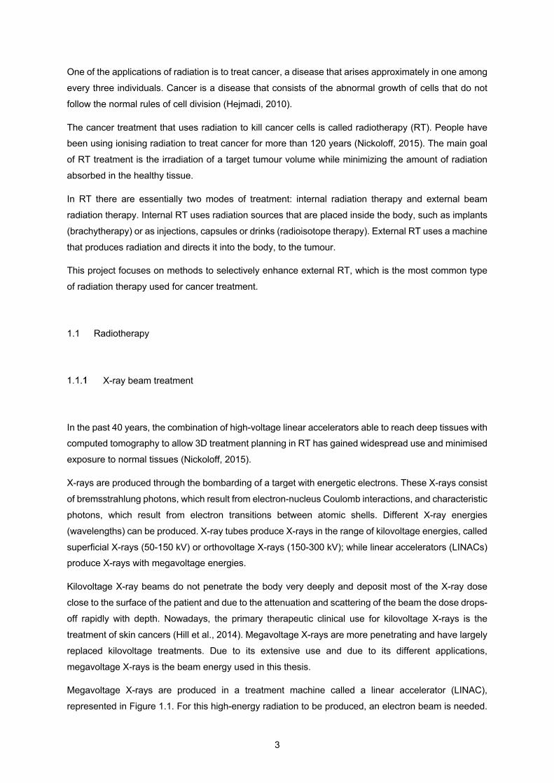
Megavoltage X-rays are produced in a treatment machine called a linear accelerator (LINAC),
represented in Figure 1.1. For this high-energy radiation to be produced, an electron beam is needed.
4
These electrons are produced by an electron gun and then accelerated through a waveguide that
increases their energy from keV to MeV range (Podgorsak, 2005). When the electrons hit an X-ray
target, X-rays are produced. The LINAC and the treatment couch rotate about a point called isocentre.
Figure 1.1: Design configuration for a medical LINAC.
Megavoltage X-ray physical properties
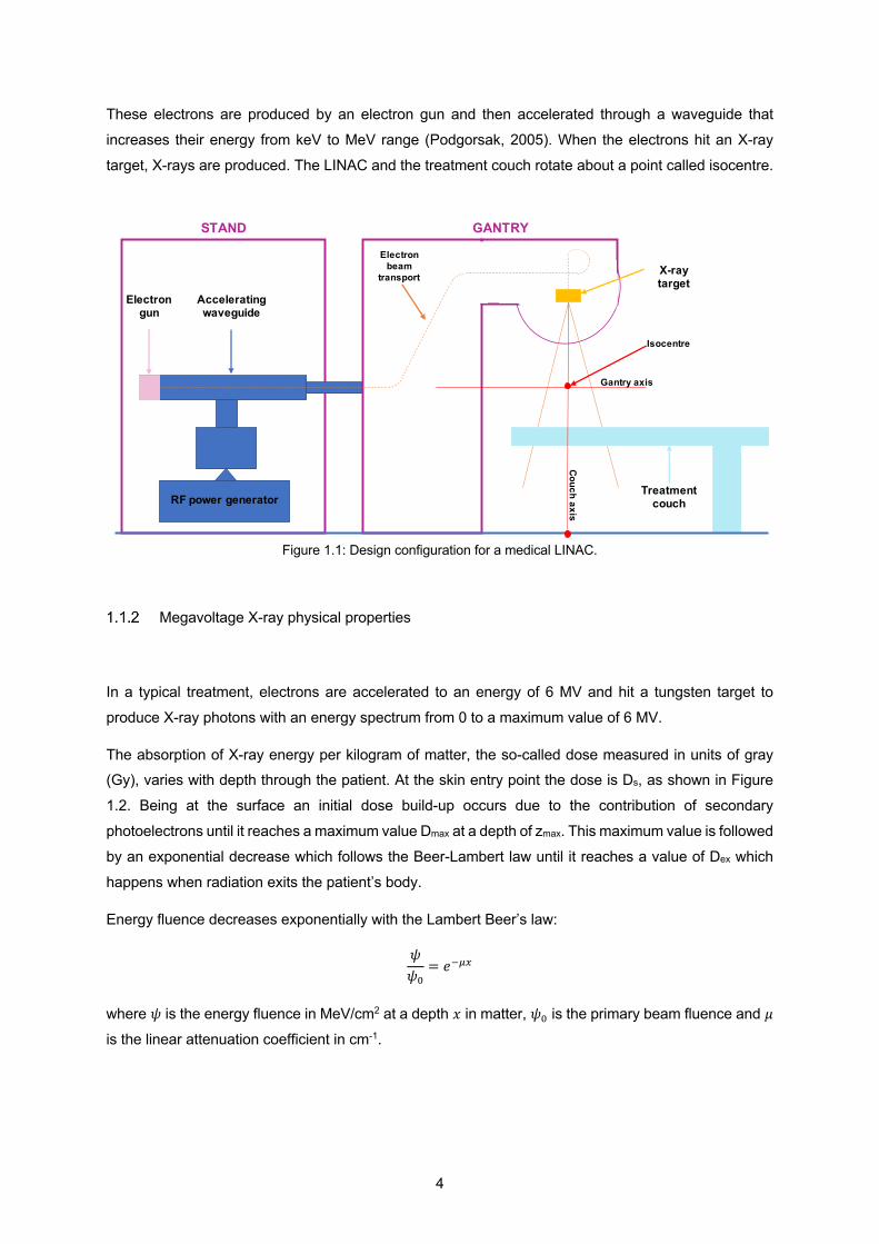
In a typical treatment, electrons are accelerated to an energy of 6 MV and hit a tungsten target to
produce X-ray photons with an energy spectrum from 0 to a maximum value of 6 MV.
The absorption of X-ray energy per kilogram of matter, the so-called dose measured in units of gray (Gy), varies with depth through the patient. At the skin entry point the dose is Ds, as shown in Figure
1.2. Being at the surface an initial dose build-up occurs due to the contribution of secondary
photoelectrons until it reaches a maximum value Dmax at a depth of zmax. This maximum value is followed
by an exponential decrease which follows the Beer-Lambert law until it reaches a value of Dex which
happens when radiation exits the patient’s body.
Energy fluence decreases exponentially with the Lambert Beer’s law:
𝜓𝜓&
= 𝑒)*+
where 𝜓 is the energy fluence in MeV/cm2 at a depth 𝑥 in matter, 𝜓& is the primary beam fluence and 𝜇
is the linear attenuation coefficient in cm-1.
RF power generator
Electron gun
Accelerating waveguide
GANTRY
X-ray target
Treatment couch
STAND
Gantry axis
Couch axis
Electron beam
transport
Isocentre
5
Figure 1.2: Typical dose distribution on the central axis of a megavoltage beam. Ds is a superficial dose, at zmax depth dose reach a maximum value Dmax, Dex is the dose delivered to the patient at the beam exit point zex: we can observe a small curve downwards due to missing scatter contribution at the exit point from point beyond the exit dose point (Podgorsak, 2005).
As shown in Figure 1.3, the build-up region increases with increasing photon energy. Also, surface
dose, Ds, and dose maximum depth, zmax, depend mainly on the beam energy. For a 6 MV X-ray beam,
the first 1.5 mm of tissue (the skin) receives ~15% of the maximum dose while for an 18 MV X-ray skin
receives ~10% (Podgorsak, 2005).
6
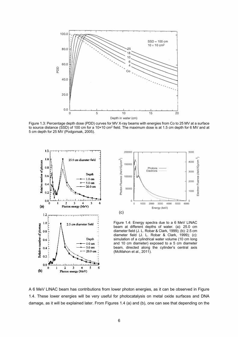
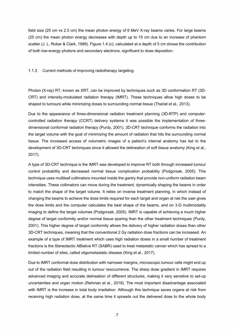
Figure 1.3: Percentage depth dose (PDD) curves for MV X-ray beams with energies from Co to 25 MV at a surface to source distance (SSD) of 100 cm for a 10×10 cm2 field. The maximum dose is at 1.5 cm depth for 6 MV and at 5 cm depth for 25 MV (Podgorsak, 2005).
A 6 MeV LINAC beam has contributions from lower photon energies, as it can be observed in Figure
1.4. These lower energies will be very useful for photocatalysis on metal oxide surfaces and DNA
damage, as it will be explained later. From Figures 1.4 (a) and (b), one can see that depending on the
(c)
(c)
(c)
(c)
(c)
(c)
(c)
(c)
Figure 1.4: Energy spectra due to a 6 MeV LINAC beam at different depths of water. (a): 25.0 cm diameter field (J. L. Robar & Clark, 1999); (b): 2.5 cm diameter field (J. L. Robar & Clark, 1999); (c): simulation of a cylindrical water volume (10 cm long and 10 cm diameter) exposed to a 5 cm diameter beam, directed along the cylinder’s central axis (McMahon et al., 2011).
7
field size (25 cm vs 2.5 cm) the mean photon energy of 6 MeV X-ray beams varies. For large beams
(25 cm) the mean photon energy decreases with depth up to 15 cm due to an increase of phantom
scatter (J. L. Robar & Clark, 1999). Figure 1.4 (c), calculated at a depth of 5 cm shows the contribution
of both low-energy photons and secondary electrons, significant to dose deposition.
Current methods of improving radiotherapy targeting
Photon (X-ray) RT, known as XRT, can be improved by techniques such as 3D conformation RT (3D-CRT) and intensity-modulated radiation therapy (IMRT). These techniques allow high doses to be
shaped to tumours while minimizing doses to surrounding normal tissue (Thariat et al., 2013).
Due to the appearance of three-dimensional radiation treatment planning (3D-RTP) and computer-
controlled radiation therapy (CCRT) delivery systems it was possible the implementation of three-
dimensional conformal radiation therapy (Purdy, 2001). 3D-CRT technique conforms the radiation into
the target volume with the goal of minimizing the amount of radiation that hits the surrounding normal
tissue. The increased access of volumetric images of a patient’s internal anatomy has led to the development of 3D-CRT techniques since it allowed the delineation of soft tissue anatomy (King et al.,
2017).
A type of 3D-CRT technique is the IMRT was developed to improve RT both through increased tumour
control probability and decreased normal tissue complication probability (Podgorsak, 2005). This
technique uses multileaf collimators mounted inside the gantry that provide non-uniform radiation beam
intensities. These collimators can move during the treatment, dynamically shaping the beams in order
to match the shape of the target volume. It relies on inverse treatment planning, in which instead of changing the beams to achieve the dose limits required for each target and organ at risk the user gives
the dose limits and the computer calculates the best shape of the beams, and on 3-D multimodality
imaging to define the target volumes (Podgorsak, 2005). IMRT is capable of achieving a much higher
degree of target conformity and/or normal tissue sparing than the other treatment techniques (Purdy,
2001). This higher degree of target conformity allows the delivery of higher radiation doses than other
3D-CRT techniques, meaning that the conventional 2 Gy radiation dose fractions can be increased. An
example of a type of IMRT treatment which uses high radiation doses in a small number of treatment
fractions is the Stereotactic ABlative RT (SABR) used to treat metastatic cancer which has spread to a limited number of sites, called oligometastatic disease (King et al., 2017).
Due to IMRT conformal dose distribution with narrower margins, microscopic tumour cells might end up
out of the radiation field resulting in tumour reoccurrence. The sharp dose gradient in IMRT requires
advanced imaging and accurate delineation of different structures, making it very sensitive to set-up
uncertainties and organ motion (Rehman et al., 2018). The most important disadvantage associated
with IMRT is the increase in total body irradiation. Although this technique saves organs at risk from
receiving high radiation dose, at the same time it spreads out the delivered dose to the whole body
8
increasing total body exposure (Rehman et al., 2018). To achieve the prescribed dose, IMRT requires
a significantly larger number of monitor units than other techniques which results in an increase of
radiation scattered and radiation leakage that causes secondary malignancies (Purdy, 2008).
Furthermore, this technique is time consuming and requires complex procedures such as its planning, delivery, and quality assurance. Despite all the efforts to improve radiation therapy, healthy tissue
surrounding the tumour may still be exposed to levels of radiation that can cause both short and long-
term side effects. For this reason, my thesis is focused on the use of radiosensitisers to better target
the radiation dose to the tumour and help decrease the side effects of MV X-ray RT treatment.
1.2 Importance of Reactive Oxygen Species (ROS)
One of the main effects of ionising radiation in an aqueous environment, such as a cell, is the formation
of highly reactive species that can damage cells. This section describes those reactive species, while the interaction of radiation with cells will be discussed in Section 1.3.
Reactive oxygen species (ROS) play a very important role in aerobic life. The damage that ROS
produce to biological molecules, such as DNA, constitutes one of the most efficient pathways used in
RT to kill cancer cells (Hosoya & Miyagawa, 2014).
Within the group of reactive chemical species two major types can be distinguished: radicals that have
one or more unpaired electron(s) in their outer molecular orbitals; and non-radicals that do not contain unpaired electrons.
Non-radical species, although they do not contain unpaired electrons, are chemically reactive and can
be easily converted into radical species.
Reactive oxygen species are partially reduced forms of molecular oxygen (O2) and include hydroxyl
radical (HO.), hydrogen peroxide (H2O2), superoxide anion (O2-), and ozone (O3), as shown in Figure
1.5.

9
Figure 1.5: Radicals and non-radicals from reactive oxygen species.
The radicals serve as the reactive intermediates for the inherent biological action of oxygen and can be
found in high amounts in mitochondria, where the respiratory chain from this cell structure liberates
electrons that may interact with molecular oxygen producing superoxide. This superoxide can in turn
produce other ROS starting a chain reaction (Trachootham et al., 2009).
In aqueous solutions, superoxide exists in equilibrium with its conjugate acid, the perhydroxyl radical
(HO2) (Rose, 2012):
HO2 ↔ H+ + O2-
This means that, when talking about superoxide anion in water, both O2- and HO2 are included since one does not exist without the other.
Under normal physiological conditions, ROS production can promote cell proliferation and differentiation
(Boonstra & Post, 2004), but when the levels of these species become too high it starts to damage
biological material, such as: nucleic acids, proteins and free amino acids, lipids and carbohydrates
(Sies, 1985). For this reason, the maintenance of ROS homeostasis is crucial for normal cell function.
Some ways in which cells detoxify ROS include superoxide dismutase (SOD) which converts
superoxide to hydrogen peroxide and oxygen; catalase converts hydrogen peroxide into water and oxygen; glutathione peroxidase converts hydrogen peroxide into water or lipid peroxides into alcohols.
The loss of these defence mechanisms provokes an abnormal ROS level leading to a condition known
as oxidative stress (Toyokuni et al., 1995).
Diseases including cardiovascular disorders, diabetes, chronic inflammation, and cancer can be the
product of perturbations in cellular redox homeostasis, since pathological conditions are a result of
serious increase in ROS levels which can lead to an irreversible oxidative damage (Alimoradi et al.,
2017).
10
1.3 Effects of radiation in cells
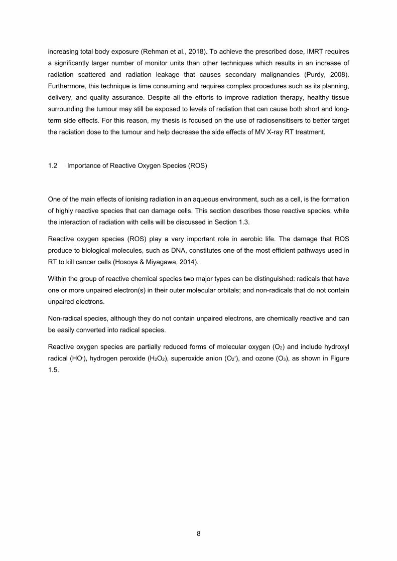
There are two ways of radiation interacting with cells:
directly or indirectly, as shown in Figure 1.6. It is
estimated that around 40% of X-ray damage is direct and
60% indirect (Baskar et al., 2014).
Through the direct way, X-rays are absorbed in biological
material interacting directly with critical targets in the cells
(Hall et al., 2016). Biological damage can occur due to the
ionized or excited atoms of the target. The most effective
constituent is the cell DNA in the nucleus. In this case, the
ability of the cell to reproduce and thus survive may be
affected. The cell may be destroyed if the chromosomes
do not replicate properly or the information carried by the DNA molecule undergoes significant alteration due to the
interaction of the radiation with the atoms within the cell.
In the indirect way, the radiation damages the cell through the formation of free radical species, which
occur due to the excitation and ionization of water molecules inside or in the vicinity of the cell.
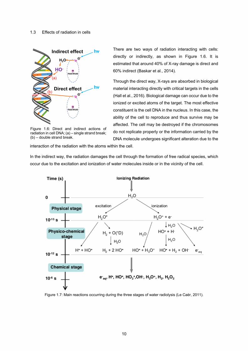
Figure 1.7: Main reactions occurring during the three stages of water radiolysis (Le Caër, 2011).
Figure 1.6: Direct and indirect actions of radiation in cell DNA; (a) – single strand break; (b) – double strand break.
+
-e-
nucleus
hn
+
- e-
nucleus
hn
HO.
H2O
Indirect effect
Direct effect
(a)
(b)
11
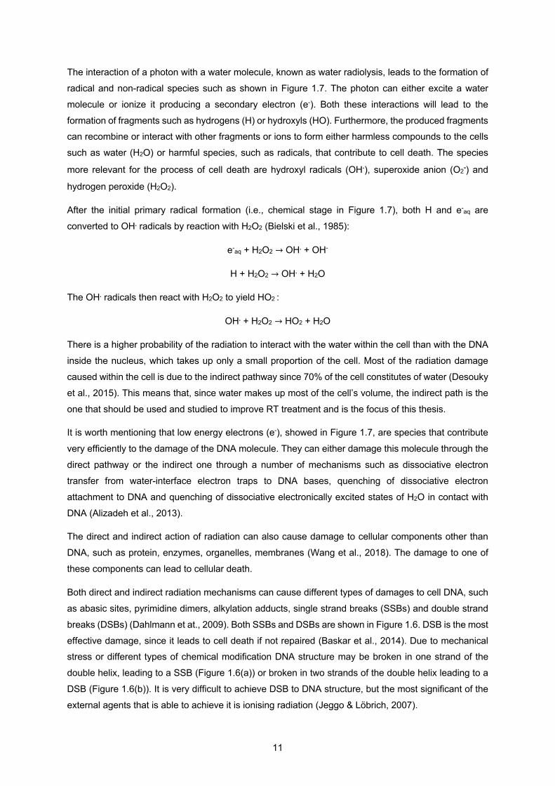
The interaction of a photon with a water molecule, known as water radiolysis, leads to the formation of
radical and non-radical species such as shown in Figure 1.7. The photon can either excite a water
molecule or ionize it producing a secondary electron (e-). Both these interactions will lead to the
formation of fragments such as hydrogens (H) or hydroxyls (HO). Furthermore, the produced fragments can recombine or interact with other fragments or ions to form either harmless compounds to the cells
such as water (H2O) or harmful species, such as radicals, that contribute to cell death. The species
more relevant for the process of cell death are hydroxyl radicals (OH.), superoxide anion (O2-) and
hydrogen peroxide (H2O2).
After the initial primary radical formation (i.e., chemical stage in Figure 1.7), both H and e-aq are
converted to OH. radicals by reaction with H2O2 (Bielski et al., 1985):
e-aq + H2O2 → OH. + OH-
H + H2O2 → OH. + H2O
The OH. radicals then react with H2O2 to yield HO2 :
OH. + H2O2 → HO2 + H2O
There is a higher probability of the radiation to interact with the water within the cell than with the DNA
inside the nucleus, which takes up only a small proportion of the cell. Most of the radiation damage
caused within the cell is due to the indirect pathway since 70% of the cell constitutes of water (Desouky
et al., 2015). This means that, since water makes up most of the cell’s volume, the indirect path is the
one that should be used and studied to improve RT treatment and is the focus of this thesis.
It is worth mentioning that low energy electrons (e-), showed in Figure 1.7, are species that contribute
very efficiently to the damage of the DNA molecule. They can either damage this molecule through the
direct pathway or the indirect one through a number of mechanisms such as dissociative electron
transfer from water-interface electron traps to DNA bases, quenching of dissociative electron
attachment to DNA and quenching of dissociative electronically excited states of H2O in contact with
DNA (Alizadeh et al., 2013).
The direct and indirect action of radiation can also cause damage to cellular components other than
DNA, such as protein, enzymes, organelles, membranes (Wang et al., 2018). The damage to one of
these components can lead to cellular death.
Both direct and indirect radiation mechanisms can cause different types of damages to cell DNA, such
as abasic sites, pyrimidine dimers, alkylation adducts, single strand breaks (SSBs) and double strand
breaks (DSBs) (Dahlmann et at., 2009). Both SSBs and DSBs are shown in Figure 1.6. DSB is the most
effective damage, since it leads to cell death if not repaired (Baskar et al., 2014). Due to mechanical stress or different types of chemical modification DNA structure may be broken in one strand of the
double helix, leading to a SSB (Figure 1.6(a)) or broken in two strands of the double helix leading to a
DSB (Figure 1.6(b)). It is very difficult to achieve DSB to DNA structure, but the most significant of the
external agents that is able to achieve it is ionising radiation (Jeggo & Löbrich, 2007).
12
Reactive oxygen species primarily induce base damage and SSBs. DSBs arise indirectly from further
action of trying to repair some lesions in the strand or can arise from two closely located SSBs (Jeggo
& Löbrich, 2007).
DSBs can also occur during the normal life cycle of the cell, more specifically during DNA replication or in meiosis (Mladenov & Iliakis, 2011). Furthermore, within the cell, DSBs arise due to reactive oxygen
species generated by normal respiratory metabolism (Featherstone & Jackson, 1999).
When oxygen is present a higher number of reactions will occur (Parveen, 2001) such as:
H. + O2 → HO2. → H2O2 + O2 reaction 1
HO2 + e- → HO2- + H+ → H2O reaction 2
X. + O2 → XO2. → X + O2. reaction 3
Reaction 3 refers to the interaction of oxygen with an organic molecule X, such as DNA, RNA, and
proteins.
In an oxygenated environment, oxygen molecules can extract an electron from the radical anions
turning them into radical cations that have a long life to cause DNA damage. On the other hand, in a
hypoxic environment the electron from the radical anion tends to combine with the radical cation, with
no damage to the DNA. In conclusion, the presence of oxygen increases the DNA damage produced
by a given dose of radiation.
Cells sensitivity increase due to ionising radiation in normal oxygen environment compared to cells
sensitivity in hypoxia conditions is called the oxygen effect, and although it has the ability of modifying
the dose it is independent of the radiation dose. The ratio between the dose required to achieve a given
cell survival in the absence of oxygen compared to the dose required for the same effect under normoxia
conditions is called the oxygen enhancement ratio (OER), and its value varies between 2.5-3 for X-ray
radiation (Nair et al., 2001).
For radiosensitisation to happen the lowest concentration of oxygen at which normoxia
radiosensitisation occurs is as low as 0.25%, since at this concentration dose-response curve is shifted
half-way towards the fully aerated condition (Kennedy et al, 1980).
1.4 Tumour environment
It is important to outline the characteristics of the tumour environment to find ways to make these cells weaker and more susceptible to radiation. Increased levels of ROS are a common feature in tumour
environment since cancer cells have a higher metabolism and produce more ROS via mitochondrial
respiration. Cancer cells adapt to this toxic environment and these adaptation mechanisms also
contribute to tumour metastasis (Chen et al., 2007).

13
Although cancer cells have a higher capability of resisting to high levels of oxidative stress compared
to normal cells, their maximum level is still limited. If cancer cells are exposed to agents that overcome
that limit, they will die (Fruehauf & Meyskens, 2007).
Figure 1.8: The biology of cancer redox (Trachootham et al., 2009).
Figure 1.8 presents a schematic of what happens in redox cancer cell biology. Redox homeostasis is
maintained by normal cells through the balance between generation and elimination of ROS.
Exogenous ROS stress can be fought through the reserve antioxidant capacity of normal cells,
preventing the ROS level from reaching the cell-death threshold, showed by the horizontal dotted line
in the figure. In cancer cells, ROS level is higher than in normal cells but still below the threshold, since
these abnormal cells arise from a redox adaptation response that leads to an upregulation of antioxidant capacity and a shift of redox dynamics with high ROS generation and elimination. In this way, cancer
cells are more dependent on the antioxidant system and more sensitive to further oxidative stress due
to the proximity to the cell-death threshold. This redox characteristic of cancer cells constitutes a
possibility for a therapeutic approach to defeat them, since a further increase in ROS production (red
bar) will induce cell death (Trachootham et al., 2009).
Characteristics such as high metabolic rate, poor blood supply and a ‘leaky’ vascular system (Figure
1.9) lead to low oxygen regions, known as hypoxia regions. Different parts of tumour tissues present heterogeneous areas of oxygenation, where peripheral zones are higher in oxygen levels than the
innermost ones (Alimoradi et al., 2017).
14
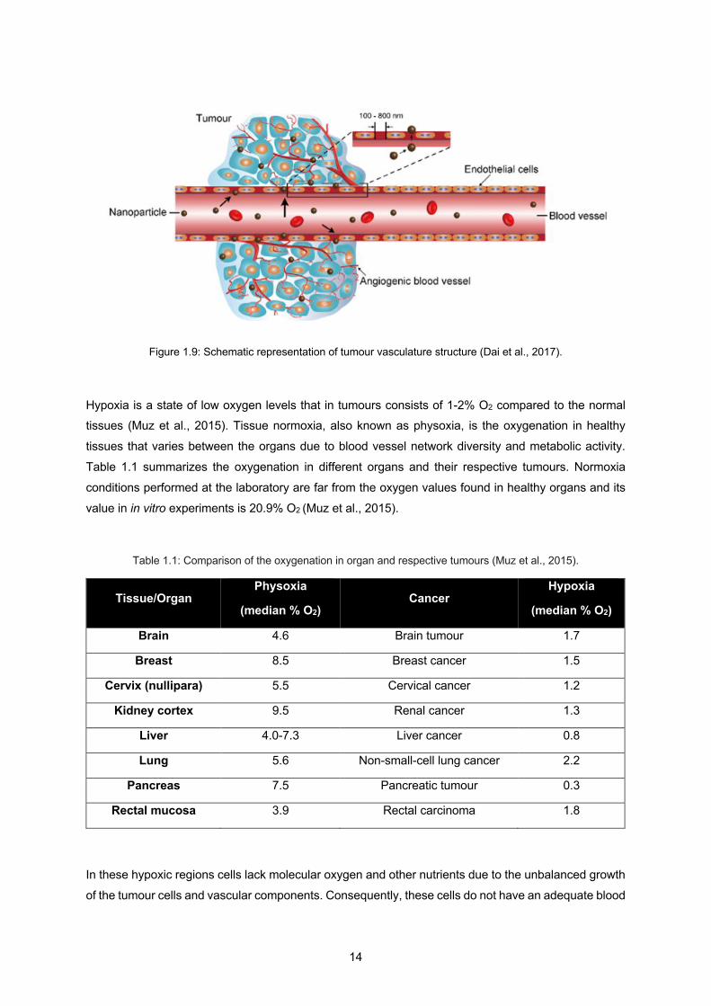
Figure 1.9: Schematic representation of tumour vasculature structure (Dai et al., 2017).
Hypoxia is a state of low oxygen levels that in tumours consists of 1-2% O2 compared to the normal tissues (Muz et al., 2015). Tissue normoxia, also known as physoxia, is the oxygenation in healthy
tissues that varies between the organs due to blood vessel network diversity and metabolic activity.
Table 1.1 summarizes the oxygenation in different organs and their respective tumours. Normoxia
conditions performed at the laboratory are far from the oxygen values found in healthy organs and its
value in in vitro experiments is 20.9% O2 (Muz et al., 2015).
Table 1.1: Comparison of the oxygenation in organ and respective tumours (Muz et al., 2015).
Tissue/Organ Physoxia
(median % O2) Cancer
Hypoxia
(median % O2)
Brain 4.6 Brain tumour 1.7
Breast 8.5 Breast cancer 1.5
Cervix (nullipara) 5.5 Cervical cancer 1.2
Kidney cortex 9.5 Renal cancer 1.3
Liver 4.0-7.3 Liver cancer 0.8
Lung 5.6 Non-small-cell lung cancer 2.2
Pancreas 7.5 Pancreatic tumour 0.3
Rectal mucosa 3.9 Rectal carcinoma 1.8
In these hypoxic regions cells lack molecular oxygen and other nutrients due to the unbalanced growth
of the tumour cells and vascular components. Consequently, these cells do not have an adequate blood
15
supply. At distances more than about 150 µm from the capillary tissue, oxygen tension decreases
rapidly with distance falling to a level where cell division cannot happen. Cells deprived of oxygen die, forming necrotic regions, while in the interface between the well-oxygenated and the necrotic regions
viable hypoxic cells tend to occur (McKeown, 2014).
The lack of oxygen in the environment of cancer cells makes these cells more resistant to radiation
damage than those in a normal oxygen environment. Due to the hypoxic environment of cancer cells,
tumours are up to three times less susceptible to radiation damage than normal cells (Paterson, 1981).
The reason for this is that oxygen is an important ingredient in the pathway of ROS production. This
happened due to oxygen being a potent radiosensitiser in a way that it aids formation of DNA-damaging free radicals increasing the effectiveness of a given dose of radiation.
Cells use glycolysis and oxidative phosphorylation to make ATP for cellular energy. Although glycolysis
is less efficient than oxidative phosphorylation, 2 ATP molecules vs 30 ATP molecules respectively, it
is a faster process since production of lactate through glucose is 10 to 100 times faster than the
complete oxidation of glucose in the mitochondria. Furthermore, using glycolysis to produce energy not
only ensures ATP production but this process requires the pentose-phosphate pathway which
additionally produces ribose sugars essential for nucleotides required for cell division. Also, this cycle produces a high amount of NADPH essential to produce fatty acids, cholesterol and regenerating
gluthione antioxidants. On the whole, the amount of ATP synthesised over any given period of time is
comparable between glycolysis and oxidative phosphorylation (Liberti & Locasale, 2016). Otto Warburg,
in the 1920s, observed very high amounts of glucose being consumed in tumours compared to the
surrounding tissue. This ‘Warburg effect’ describes the metabolism of a cancer cell to produce energy,
from the predominantly oxidative phosphorylation to predominantly aerobic glycolysis. It is thought that
one of the main reasons why cancer cells use this respiratory mechanism is that upregulated glycolysis
constitutes a growth advantage which promotes proliferation and invasion (Gatenby & Gillies, 2004).
To overcome this limitation approaches such as hyperbaric oxygen, hypoxic cell radiosensitisers such
as misonidazole and nitroimidazole, and hypoxic cytotoxins, such as tirapazamine were used without
clinical success (Wardman, 2007). Hyperbaric oxygen is the oxygen released at pressures of 2-4
atmospheres to increase the oxygen in the plasma. This technique was supposed to allow the O2 to
diffuse further through tissue than normobaric oxygen breathing, since diffusion distance of oxygen
through respiring tissue increases with the initial oxygen tension (Rockwell et al., 2009). Hyperbaric
oxygenation demonstrates an overall positive effect on RT treatment but has not been adopted and remains to be validated as a standard treatment (Gérard et al., 2019).
More recently, hypoxia imaging during RT treatment became a technique of study. The level of oxygen
present in tumour cells is cyclic, where cells that are hypoxic before RT treatment become oxygenated
during or after the treatment. In this way, monitoring the oxygen level during the treatment may help if
the RT fraction is given when tumour reoxygenation is expected to be at its maximum so as to optimize
the oxygen enhancement ratio (Gérard et al., 2019).

16
Radio-enhancing NPs and new radiosensitising drugs may be delivered into the tumour to improve
oxygenation. In the class of radiosensitising drugs are fluorochemicals able to dissolve considerable
amounts of oxygen and deliver it through passive diffusion into the hypoxic regions (Gérard et al., 2019).
Although both pre-clinical and clinical studies have shown that hypoxia can be reduced to improve the outcome of radiation therapy, hypoxic modification has still not been established as a standard
treatment with RT (Horsman & Overgaard, 2016).
The need to predict and assess the patient’s response to the RT treatment is of extreme importance
since RT continues to be the most widely used treatment for cancer, even in conjunction with other
types of cancer treatment such as surgery and chemotherapy (Jeggo & Löbrich, 2007). This would lead
to an important step for the optimization of this cancer treatment.
RT can be optimized by either the use of radiosensitisers to kill the tumour cells or by radioprotectors to protect the normal tissues from radiation damage.
1.5 Radiosensitisers
The poor response of many tumours to radiation despite the improvement of techniques using
conventional sources of radiation have led to the development of other approaches. Three main
approaches were considered: hyperbaric oxygen, proton and neutron beams, and radiosensitisers
(Paterson, 1981).
The use of hyperbaric oxygen, to increase oxygen tension during irradiation in tumour cells, has not
shown an increase in tumour control rates (Henk, 1986). Radiosensitisers have become the most
reliable approach since they are inexpensive and widely available and still have a whole potential to be
explored.
Radiosensitisers make cancer cells more vulnerable to radiation therapy by enhancing the effect of
ionising radiation within the cancer cells while leaving the healthy cells out of the target. The advantage of these materials is that since they passively or actively are conformed to the tumour site the effect of
the enhanced radiation leads to an increased dose in the initial treatment within the cancer cells
enabling the reduction of the number of treatments for the patient, while still achieving the initially
prescribed dose.
The role of these materials can be viewed as the same as the oxygen, in the sense that they can act to
restore, at least to some extent, the radiosensitivity of hypoxic tumour cells. In summary,
radiosensitisers are oxygen substitutes that are capable of diffusing into hypoxic tumour cells and sensitise them via a similar mechanism to oxygen.
An important feature of radiosensitisers that relates to oxygen is that these compounds are not easily
metabolised. In this way, during diffusion to tumour cells they can penetrate further into hypoxic regions
(Parveen, 2001).
17
To define a compound as a radiosensitiser it is crucial to understand the interaction between the
compound and the radiation of study. If the combination results in a biological response greater than
what would be expected from the radiation by itself, than one can conclude that there is a synergistic
effect behind it and the compound is classified as a radiosensitiser.
Radiosensitisers can perform through different mechanisms in order to apply their radiation-enhancing
effects. Some of the possible mechanisms of radiosensitisation are (Gill & Vallis, 2019):
1. Inhibition of post-irradiation cellular repair processes: being DNA a critical target for ionising
radiation the inhibition of its damage repair pathways will contribute to an increase in
radiotoxicity;
2. Cell-cycle dysregulation: compounds that are able to arrest the cell-cycle at a specific phase
can be considered radiosensitisers. For example, cells in late G1 or G2/M phases are more sensitive to the effects of radiation while cells in S phase are the most resistant;
3. Radiosensitivity enhancement in hypoxic cells: the radioresistance of hypoxic cells makes it
difficult the generation of ROS by ionising radiation. Oxygen-mimetic sensitisers help the
increase in ROS in these cells, enhancing the efficacy of the treatment;
4. Production of cytotoxic substances: Radiolysis of the sensitiser produces cytotoxic molecular
fragments which in turn contribute to cell-death;
5. Generation of Auger electrons near DNA: Explained in the next section, high atomic number materials after radiation exposure produce Auger electrons due to inner-shell ionization. If the
X-rays energy matches the K-shell energy of its electrons, these materials, when internalised
by the tumour cells, will enhance the tumour-absorbed dose. This phenomenon may enhance
the radiobiological effects of ionising radiation and potentially explains sensitisation by heavy-
metal nanomaterials.
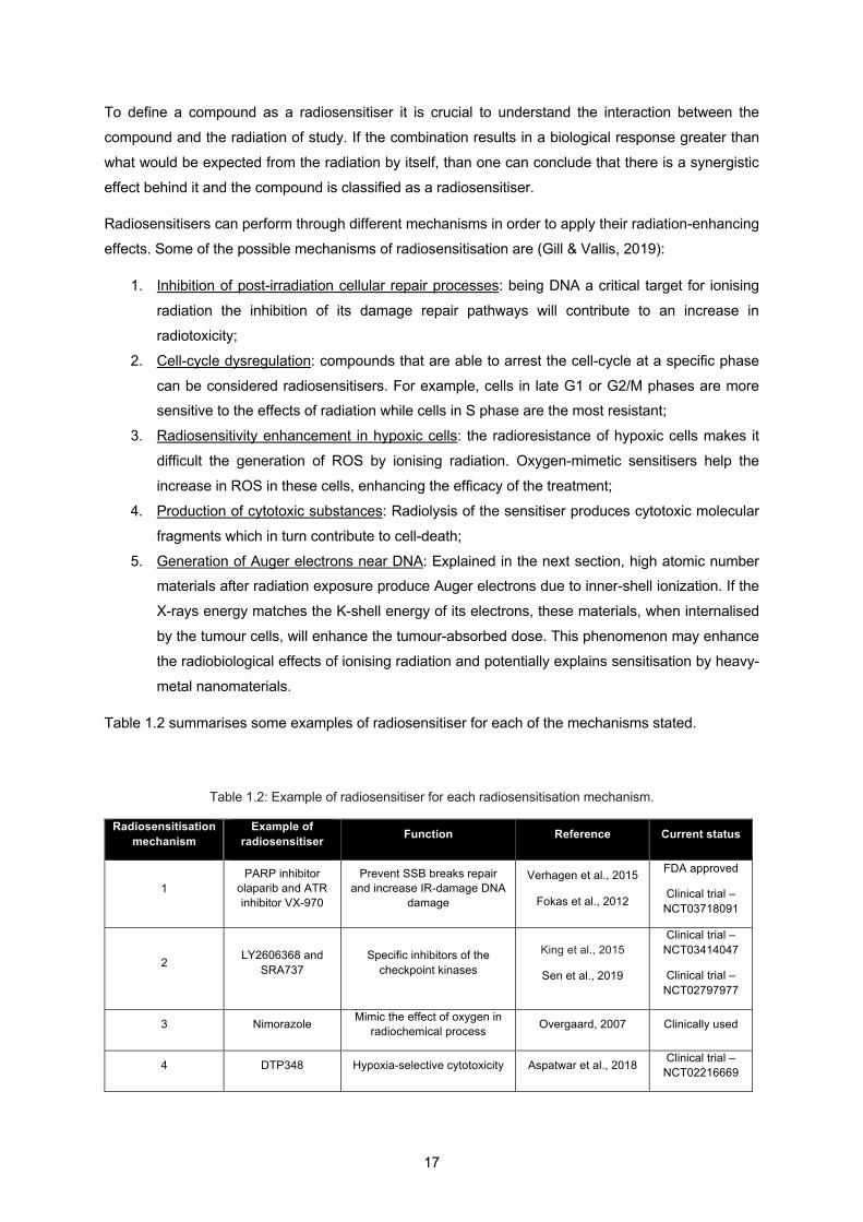
Table 1.2 summarises some examples of radiosensitiser for each of the mechanisms stated.
Table 1.2: Example of radiosensitiser for each radiosensitisation mechanism.
Radiosensitisation mechanism
Example of radiosensitiser Function Reference Current status
1 PARP inhibitor
olaparib and ATR inhibitor VX-970
Prevent SSB breaks repair and increase IR-damage DNA
damage
Verhagen et al., 2015
Fokas et al., 2012
FDA approved
Clinical trial – NCT03718091
2 LY2606368 and SRA737
Specific inhibitors of the checkpoint kinases
King et al., 2015
Sen et al., 2019
Clinical trial – NCT03414047
Clinical trial – NCT02797977
3 Nimorazole Mimic the effect of oxygen in radiochemical process Overgaard, 2007 Clinically used
4 DTP348 Hypoxia-selective cytotoxicity Aspatwar et al., 2018 Clinical trial – NCT02216669
18
Physicochemical properties such as, for example, size, shape, coating, and functionalization, control
their pharmacokinetics, bioavailability, biodistribution, as well as targeting and intracellular delivery.
Tumour vasculature is more leaky than normal vasculature, allowing small particles and larger drugs to
enter the tumour environment. Furthermore, the lymphatic drainage of tumour tissue is less efficient
than in normal tissues, meaning that particles are not easily removed. Together, these two factors constitute the Enhanced Permeability and Retention (EPR) effect. Thus, specific tumour targeting can
be achieved by passive targeting via the EPR effect, active targeting by using high-affinity targeting-
molecules, and stimuli-responsive triggered release to endogenous or exogenous stimuli (Liu et al.,
2018).
Studies have shown a general relationship between the efficiency of sensitisation and the electron
affinity, i.e. oxidizing abilities for chemical radiosensitisers (Adams et al., 1971). Hypoxic cell
radiosensitisation happens when one electron is transferred to the radiation-induced lesion, resulting in the fixation of the chemical damage. A lot of studies regarding chemical radiosensitisers have been
done (Candelaria et al., 2006; Wardman, 2007) and are still being undertaken (Farhood et al., 2019;
Wang et al., 2018).
This thesis is focussed on metal-based radiosensitisers with a diameter within the nanometer range
(nanoparticles). When an incoming high-energy X-ray photon encounters heavy-metal atom, two
important phenomena occur: ejection of photoelectrons and ejection of Auger electrons, as shown in
Figure 1.10.
5 AGuIX and NBTXR3 Enhanced absorbed dose
Lux et al., 2018
Bonvalot et al., 2019
Clinical trial – NCT03818386
Clinical trial – NCT03589339
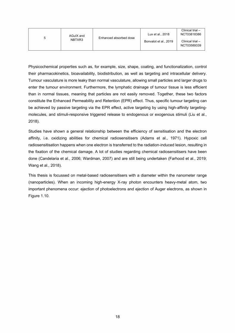
19
Figure 1.10: Photoelectron and Auger electron schematic in an atom. Step 1: emission of a photoelectron with a kinetic energy (DE) equal to the difference in energy between the incoming photon (E) and the binding energy of the electron in the K-shell (E0); Step 2: emission of a Auger electron with a kinetic energy (KE) equal to the difference in energy between the binding energy of the electron in the K-shell (EK) and the binding energies of the electrons in the L-shell (EL1 and EL23).
In Figure 1.10, if a high energy X-ray photon has enough energy and it is absorbed by an atom in the
surface of the metal-based NP, an innermost electron will be ejected. This phenomenon is the
photoelectric effect and it results in the ejection of a photoelectron with a kinetic energy equal to the
difference in energy between the incoming photon and the binding energy of the electron in the K-shell.
An electron from a higher energy state, in this case, the L-shell, will then occupy the empty state in the
K-shell. From this transition an X-ray is emitted and if absorbed by another electron in the L-shell, an
Auger electron is ejected. In this example, this process is termed a KLL Auger transition. The kinetic energy of an Auger electron is equal to the energy difference of the singly ionized initial state and the
doubly ionized final state. The resulting kinetic energy EKLL is given by EKL1L2,3 = EK - EL1 - EL2,3 - F. The
term F comes from the electron work function and from the equation it can be concluded that the kinetic
energy of the Auger electron is independent of the type and energy of the primary beam.
The energy EK - EL1 can be then released as a radiative decay, with emission of an X-ray and in this
case an electron vacancy in the K-shell is filled by an electron from the L-shell so the characteristic
energy of the emitted photon is called the K-alpha (Ka) spectral line, or as a non-radiative decay, with
emission of an Auger electron and the result depends on the relative probabilities of Auger emission
and X-ray fluorescence, as shown in Figure 1.11.
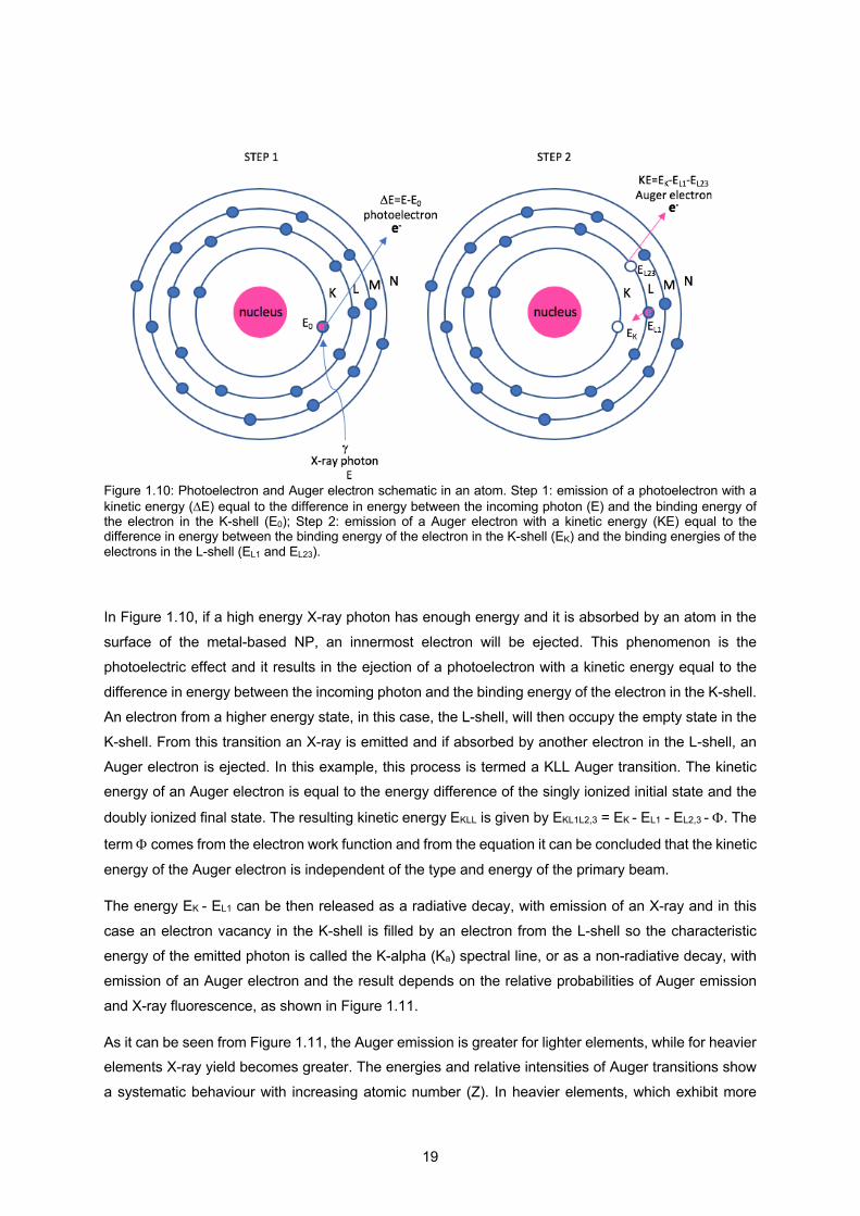
As it can be seen from Figure 1.11, the Auger emission is greater for lighter elements, while for heavier elements X-ray yield becomes greater. The energies and relative intensities of Auger transitions show
a systematic behaviour with increasing atomic number (Z). In heavier elements, which exhibit more
20
energy levels, more Auger transitions are possible.
Figure 1.11: Relative probabilities of Auger emission and X-ray fluorescence. The solid lines show the Auger yield; the dashed lines show the fluorescence yield (results obtained from Reinhardt & Kern, 2008 and modified for the purpose of this thesis where irrelevant curves have been removed).
In terms of importance for the RT treatment, the fluorescent photons from X-ray fluorescence are able
to travel longer ranges, up to centimetres, while Auger electrons travel much shorter distances, typically
10 nm (Hainfeld et al., 2008). This means that the former, depending on the tumour size, may not
provide the desired localized tumour effect, but the latter, due to the fact that the electrons are weakly bound, they can be effective in producing very high local ionization density. For the Auger effect to be
useful, the emitting atom should be close to the target molecule to damage the molecule, due to the
short distance they are able to travel, although some Auger electrons can travel 50 nm in water.
In summary, photoelectrons are the result of the electrons emitted along the primary paths of the
ionising radiation which induce inner shell ionization of the metal atoms, whether the Auger electrons
result from the relaxation of the excited core of the metal atoms.
Emission of Auger electrons and fluorescent photons
can be caused by either photoelectric absorption or
by Compton scattering. Basically, in photoelectric
absorption the photon is absorbed by the
photoelectron, while in Compton scattering the
incident photon greatly exceeds the binding energy
of the electron to the atom, so the photon does not
disappear. Instead it is deflected through a scattering
angle q, as shown in Figure 1.12. Part of its energy
is transferred to the recoil electron, thus the photon
loses energy in the process. The energy transferred
nucleus
Incidentphoton
Scattered Photon
q, scattering angle
Ejected Compton recoil electron
Figure 1.12: Schematic representation of Compton scattering.

21
does not depend on the density, atomic number, or any other property of the absorbed material (Cherry
et al., 2004).
At a photon energy of about 100 keV or more the photon radiation beam attenuation in the target is
mostly caused by Compton scattering and not by photoelectric absorption (Kljuev, 2001). The photoelectric effect varies approximately as (Z/E)3, where Z is the atomic number of the target and E is
the incident photon energy. For high-Z elements this effect dominates the interaction with matter at
energies <0.5 MeV, while for energies >1 MeV pair production dominates (Hainfeld et al., 2008). In pair
production electron-positron pairs are created if the incident photon energy exceeds twice the rest mass
of the electron (2×0.511 = 1.022 MeV) and it depends on Z2. All these physical interactions of radiation
with metals made these materials a possible choice to improve the RT treatment.
Adding to the physical interactions, biocompatibility is a quality that needs to be studied since these
metals will be introduced into the human body, specifically that they will need to be injected into the
blood flow and reach the tumour site. Luckily, if these metals are produced in the nanometer range, it
will allow them to accumulate into the cancer cells due to the leaky nature of the tumour vasculature and its lack of efficient lymphatic drainage as explained before in Figure 1.9. Relying on the EPR effect
for NP delivery is a passive strategy while relying on the ligand-receptor binding is an active strategy.
The later improves selective accumulation to targeted site and thus discriminates between the
cancerous and healthy tissues (Attia et al., 2019).
For a metal-based NP the basic operation of the radiosensitisation process focuses on physical dose
enhancement, chemical contribution and biological phase, as shown in Figure 1.13.
Figure 1.13: Schematic illustration of metal-based NPs radiosensitisation process (modified from Liu et al., 2018).
As illustrated in Figure 1.13, the common procedure for the study of metal-based NPs is to load them into the cells for a specific time and irradiate them (Upper-left). Biological mechanisms will happen
22
within the cells due to the radiation itself, the combined effect of radiation with the NP in study and also
due to the NP itself (Upper-right). The basic operation of the radiosensitisation process will then lead to
lethal cellular damage where the primary targets, depending on the cellular and subcellular distribution
of the NPs, will include cell membrane, cytoplasm, nucleus, mitochondria, endoplasmic reticulum, and other organelles. In this way, the photoelectrons and Auger electrons produced from the combination
of radiation with NPs will contribute to a dose enhancement due to the direct interaction of these species
with the cells or due to the indirect interaction where these species will increase the amount of ROS
damaging even more cells than the amount of ROS produced by radiation alone. Also, HO. production
occurs due to the catalytic-like mechanism/surface-catalyzed reaction present in metal-based NPs
(Lower-left). These interactions produce cell damage that leads to cell death and can be probed by the
number of cells that survived after a certain period of time. The combination of ionising radiation and
metal-based NPs induces physical, chemical and biological mechanisms that contribute for the observed enhanced cell-killing effects (Lower-right) (Liu et al., 2018).
As mentioned before, the increase of ROS in cancer cell can exert the opposite of what it is expected
from the RT treatment, such as the initiation and progression of cancer (Wu, 2006). On the other hand,
increasing ROS levels can also be toxic to cancer cells which will make them more vulnerable to
damage by further ROS attacks induced by external agents such as the ionising radiation used in RT
(Trachootham et al., 2009).
This method is not straightforward due to the complexity of redox alterations in cancer cells. A lot of
factors are involved in the redox regulation and stress response and they still need to be better
understood. In this way, the simple addition of ROS may not lead to cancer cell apoptosis and the
persistency of this approach may actually instead contribute to a community of cancer cells that
developed an antioxidant capacity making them resistant to exogenous stress (Tiligada, 2006).
A lot of investigations have been made using radiosensitisers to cooperate with RT treatment, however,
a diversity of challenges such as poor biocompatibility, lack of targeting specificity, and radiosensitisation with clinical energies, mostly MV energies, increase the need for new and optimal
radiosensitisers.
1.6 Radioprotectors
Hypoxic cells may limit the response of tumours to conventional RT because of their inherent
radioresistance (Kennedy et al, 1980). Although this fact constitutes one of the main limitations for this
type of treatment it is not the only one.
Another main challenge of RT is the fact that tumours are located near normal tissues and organs at
risk making the doses delivered to the target volumes limited. For this reason, agents that present
radioprotective properties have been used to reduce normal tissue complication probability (Liu et al.,
2018). These agents are called radioprotectors and have the opposite role of a radiosensitiser.
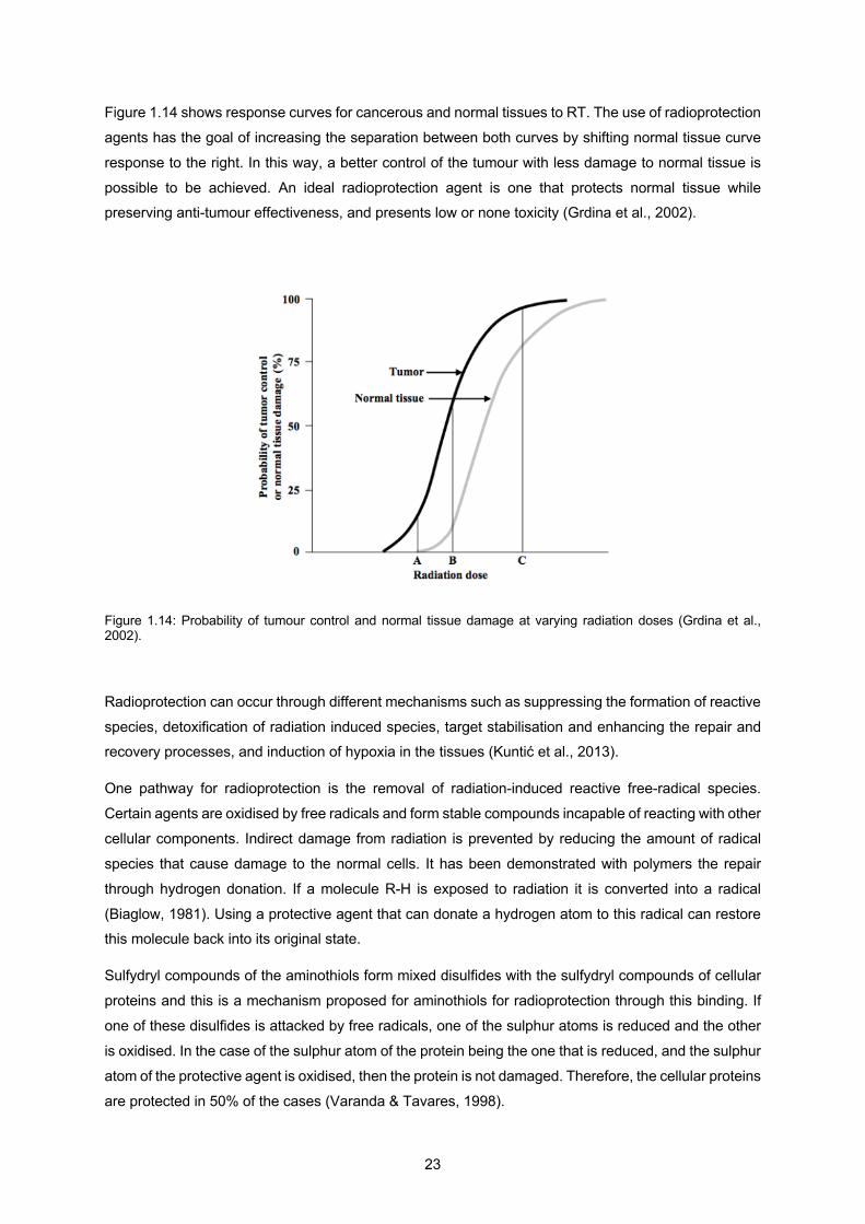
23
Figure 1.14 shows response curves for cancerous and normal tissues to RT. The use of radioprotection
agents has the goal of increasing the separation between both curves by shifting normal tissue curve
response to the right. In this way, a better control of the tumour with less damage to normal tissue is
possible to be achieved. An ideal radioprotection agent is one that protects normal tissue while preserving anti-tumour effectiveness, and presents low or none toxicity (Grdina et al., 2002).
Figure 1.14: Probability of tumour control and normal tissue damage at varying radiation doses (Grdina et al., 2002).
Radioprotection can occur through different mechanisms such as suppressing the formation of reactive
species, detoxification of radiation induced species, target stabilisation and enhancing the repair and recovery processes, and induction of hypoxia in the tissues (Kuntić et al., 2013).
One pathway for radioprotection is the removal of radiation-induced reactive free-radical species.
Certain agents are oxidised by free radicals and form stable compounds incapable of reacting with other
cellular components. Indirect damage from radiation is prevented by reducing the amount of radical
species that cause damage to the normal cells. It has been demonstrated with polymers the repair
through hydrogen donation. If a molecule R-H is exposed to radiation it is converted into a radical
(Biaglow, 1981). Using a protective agent that can donate a hydrogen atom to this radical can restore this molecule back into its original state.
Sulfydryl compounds of the aminothiols form mixed disulfides with the sulfydryl compounds of cellular
proteins and this is a mechanism proposed for aminothiols for radioprotection through this binding. If
one of these disulfides is attacked by free radicals, one of the sulphur atoms is reduced and the other
is oxidised. In the case of the sulphur atom of the protein being the one that is reduced, and the sulphur
atom of the protective agent is oxidised, then the protein is not damaged. Therefore, the cellular proteins
are protected in 50% of the cases (Varanda & Tavares, 1998).
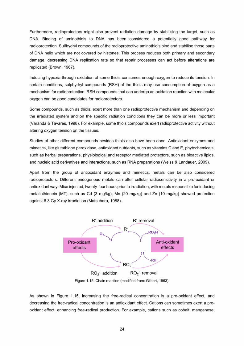
24
Furthermore, radioprotectors might also prevent radiation damage by stabilising the target, such as
DNA. Binding of aminothiols to DNA has been considered a potentially good pathway for
radioprotection. Sulfhydryl compounds of the radioprotective aminothiols bind and stabilise those parts
of DNA helix which are not covered by histones. This process reduces both primary and secondary damage, decreasing DNA replication rate so that repair processes can act before alterations are
replicated (Brown, 1967).
Inducing hypoxia through oxidation of some thiols consumes enough oxygen to reduce its tension. In
certain conditions, sulphydryl compounds (RSH) of the thiols may use consumption of oxygen as a
mechanism for radioprotection. RSH compounds that can undergo an oxidation reaction with molecular
oxygen can be good candidates for radioprotectors.
Some compounds, such as thiols, exert more than one radioprotective mechanism and depending on the irradiated system and on the specific radiation conditions they can be more or less important
(Varanda & Tavares, 1998). For example, some thiols compounds exert radioprotective activity without
altering oxygen tension on the tissues.
Studies of other different compounds besides thiols also have been done. Antioxidant enzymes and
mimetics, like glutathione peroxidase, antioxidant nutrients, such as vitamins C and E, phytochemicals,
such as herbal preparations, physiological and receptor mediated protectors, such as bioactive lipids,
and nucleic acid derivatives and interactions, such as RNA preparations (Weiss & Landauer, 2009).
Apart from the group of antioxidant enzymes and mimetics, metals can be also considered
radioprotectors. Different endogenous metals can alter cellular radiosensitivity in a pro-oxidant or
antioxidant way. Mice injected, twenty-four hours prior to irradiation, with metals responsible for inducing
metallothionein (MT), such as Cd (3 mg/kg), Mn (20 mg/kg) and Zn (10 mg/kg) showed protection
against 6.3 Gy X-ray irradiation (Matsubara, 1988).
Figure 1.15: Chain reaction (modified from: Gilbert, 1963).
As shown in Figure 1.15, increasing the free-radical concentration is a pro-oxidant effect, and
decreasing the free-radical concentration is an antioxidant effect. Cations can sometimes exert a pro-oxidant effect, enhancing free-radical production. For example, cations such as cobalt, manganese,
25
nickel and zinc increase the rate of oxidation of reduced glutathione exposed to high oxygen pressure
(Gilbert et al., 1958). On the other hand, cobalt and manganese cations accelerate the decomposition
of H2O2 and this removal can be considered an antioxidant action. Cobalt was tested in different
biological systems such as peas, mice, bacteria and showed an antioxidant action against oxygen toxicity (Gilbert, 1963).
A more recent study showed a radioprotective effect of date syrup (Abou-Zeid et al., 2018). The authors
pre-treated rats with date syrup by stomach intubation and then irradiated them with 6 Gy gamma-rays.
They concluded that this substance was effective in reducing radiation-induced hepatotoxicity, oxidative
stress, inflammatory response, and DNA damage. Due to these results, date syrup can be a potential
supplement in the RT to protect normal cells from the toxic effects of radiation.
Amifostine was the only compound that has been officially approved for human use, but it still presents significant toxicity (Kuntić et al., 2013). Cerium oxide (CeO2) NPs are a type of metal oxide NP that are
known for its radioprotection effects. These NPs offer protection against radiation induced damage in
different tissues due to its potential as a biological free radical scavenger or antioxidant (Baker, 2013).
Still a lot of investigation needs to be done to find a good radioprotector, but toxicity and inability to
differentiate between normal and cancer cells are the main reasons for their failure in clinical
applications. For this reason, a compound with the ability to protect only normal cells and to differentiate
tumour cells is ideal for a radioprotector and so these compounds need to be modified by taking advantage of the difference in oxygen levels between normal cells and tumour cells as well as their
associated biochemical characteristics (Nair et al., 2001).
1.7 Gold NPs as radiosensitisers
A study showed that 1.9 nm gold (Au) NPs, through intravenous injection, in combination with 250 kVp
X-rays resulted in eradication of most tumours in mice, while the mice with no irradiation or gold only
treatment died within 15 days, as it can be seen from Figure 1.16 (Hainfeld et al., 2004). This study introduced a new perspective to the use of metal NPs, especially gold.

26
Figure 1.16: Plot of mice survival after various treatments of subcutaneous EMT-6 tumours; Circles: no treatment (n=17), and gold only (1.35 g Au/kg, no irradiation), indistinguishable from no treatment (n=4); triangles: irradiation only (26 Gy, 250 kVp), producing 20% long-term (> 1 year) survival (n=15); squares: irradiation after i.v. injection of 1.35 g Au/kg gold NPs, 50% long-term survival (n=4); diamonds: irradiation after 2.7g Au/kg injection producing 86% long-term survival (n=7) (Hainfeld et al., 2004). Au NPs have the following advantages (Hainfeld et al., 2008):
• Biocompatibility;
• Absorbs energy 3 times more than iodine at 20 and 100 keV;
• Can be cleared from the blood slower than small molecules enhancing tumour delivery;
• Provide tumour specificity through EPR effect;
• Can be made over a wide range of sizes (1-1000 nm);
• Have different surface ligands, thiols (S-R) or phosphorous (P-R), allowing multifunctionality for optimal properties.
Surface characteristics and size are two very important features
easy to modify in Au NPs that can have very important outcomes
regarding toxicity and uptake. Using for example a sulphur-
polyethylene glycol (PEG) as coating of Au NP can prevent
reticuloendothelial system uptake and the overall size will also be a strong factor for blood retention time, as well as the surface coating.
Surface atoms have a covalent-strength binding affinity with sulphur
(S-R) or phosphorus (P-R) atoms, so various functional groups
such as PEG, carboxyl or amino groups, thiol derivatized drugs,
DNA, lipids, and carbohydrates are easily attached, as shown in
Figure 1.17. Through covalent bounds with this functional groups,
antibodies or peptides can be included on Au NP surface, as represented in the figure by M2. Another possibility is adsorbing
Figure 1.17: Diagram of a Au NP with ligands (Hainfeld et al., 2008); S-R and P-R represent sulphur (S) or phosphorus (P) atoms, respectively, attached to a functional group (R); M1 and M2 represent molecules attached by ionic and covalent interactions, respectively.
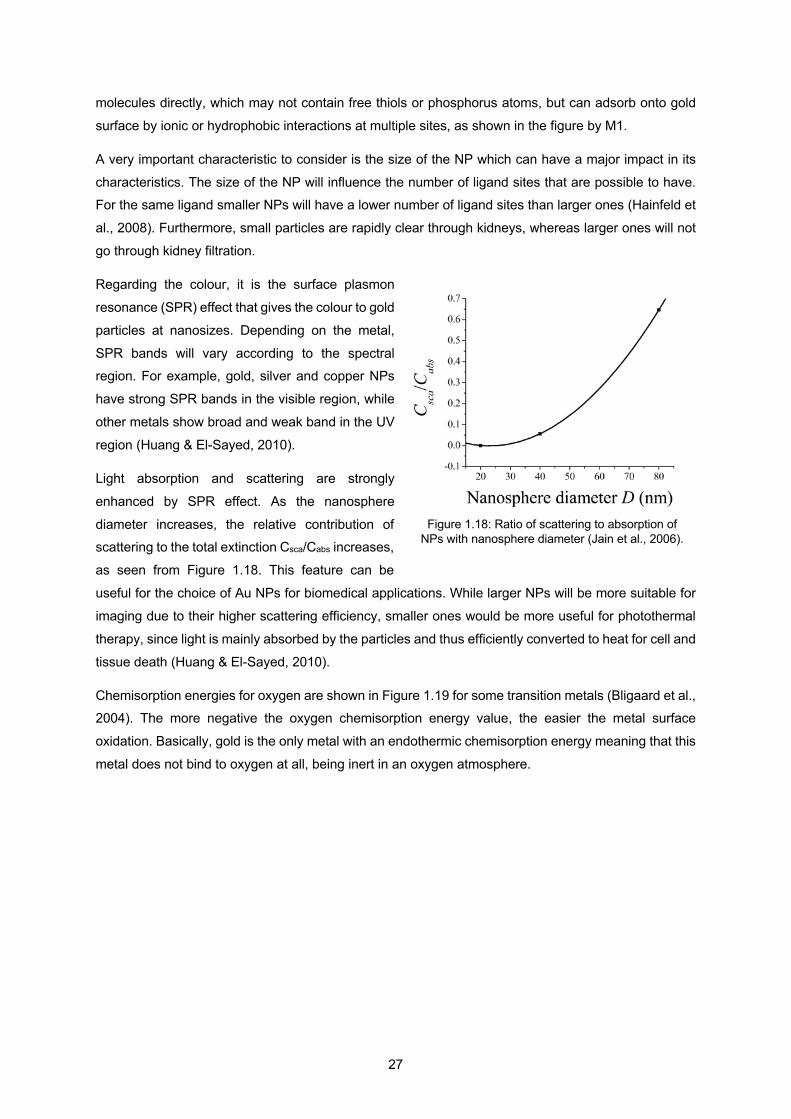
27
molecules directly, which may not contain free thiols or phosphorus atoms, but can adsorb onto gold
surface by ionic or hydrophobic interactions at multiple sites, as shown in the figure by M1.
A very important characteristic to consider is the size of the NP which can have a major impact in its
characteristics. The size of the NP will influence the number of ligand sites that are possible to have. For the same ligand smaller NPs will have a lower number of ligand sites than larger ones (Hainfeld et
al., 2008). Furthermore, small particles are rapidly clear through kidneys, whereas larger ones will not
go through kidney filtration.
Regarding the colour, it is the surface plasmon
resonance (SPR) effect that gives the colour to gold
particles at nanosizes. Depending on the metal,
SPR bands will vary according to the spectral region. For example, gold, silver and copper NPs
have strong SPR bands in the visible region, while
other metals show broad and weak band in the UV
region (Huang & El-Sayed, 2010).
Light absorption and scattering are strongly
enhanced by SPR effect. As the nanosphere
diameter increases, the relative contribution of scattering to the total extinction Csca/Cabs increases,
as seen from Figure 1.18. This feature can be
useful for the choice of Au NPs for biomedical applications. While larger NPs will be more suitable for
imaging due to their higher scattering efficiency, smaller ones would be more useful for photothermal
therapy, since light is mainly absorbed by the particles and thus efficiently converted to heat for cell and
tissue death (Huang & El-Sayed, 2010).
Chemisorption energies for oxygen are shown in Figure 1.19 for some transition metals (Bligaard et al., 2004). The more negative the oxygen chemisorption energy value, the easier the metal surface
oxidation. Basically, gold is the only metal with an endothermic chemisorption energy meaning that this
metal does not bind to oxygen at all, being inert in an oxygen atmosphere.
Figure 1.18: Ratio of scattering to absorption of NPs with nanosphere diameter (Jain et al., 2006).
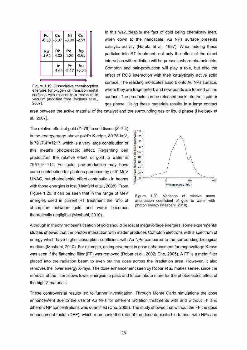
28
In this way, despite the fact of gold being chemically inert,
when down to the nanoscale, Au NPs surface presents
catalytic activity (Haruta et al., 1987). When adding these
particles into RT treatment, not only the effect of the direct interaction with radiation will be present, where photoelectric,
Compton and pair-production will play a role, but also the
effect of ROS interaction with their catalytically active solid
surface. The reacting molecules adsorb onto Au NPs surface,
where they are fragmented, and new bonds are formed on the
surface. The products can be released back into the liquid or
gas phase. Using these materials results in a large contact
area between the active material of the catalyst and the surrounding gas or liquid phase (Hvolbæk et al., 2007).
The relative effect of gold (Z=79) to soft tissue (Z=7.4)
in the energy range above gold’s K-edge, 80.75 keV,
is 793/7.43=1217, which is a very large contribution of
this metal’s photoelectric effect. Regarding pair
production, the relative effect of gold to water is
792/7.42=114. For gold, pair-production may have some contribution for photons produced by a 10 MeV
LINAC, but photoelectric effect contribution in beams
with those energies is lost (Hainfeld et al., 2008). From
Figure 1.20, it can be seen that in the range of MeV
energies used in current RT treatment the ratio of
absorption between gold and water becomes
theoretically negligible (Mesbahi, 2010).
Although in theory radiosensitisation of gold should be lost at megavoltage energies, some experimental
studies showed that the photon interaction with matter produces Compton electrons with a spectrum of
energy which have higher absorption coefficient with Au NPs compared to the surrounding biological
medium (Mesbahi, 2010). For example, an improvement in dose enhancement for megavoltage X-rays
was seen if the flattening filter (FF) was removed (Robar et al., 2002; Cho, 2005). A FF is a metal filter
placed into the radiation beam to even out the dose across the irradiation area. However, it also
removes the lower energy X-rays. The dose enhancement seen by Robar et al. makes sense, since the
removal of the filter allows lower energies to pass and to contribute more for the photoelectric effect of the high-Z materials.
These controversial results led to further investigation. Through Monte Carlo simulations the dose
enhancement due to the use of Au NPs for different radiation treatments with and without FF and
different NP concentrations was quantified (Cho, 2005). The study showed that without the FF the dose
enhancement factor (DEF), which represents the ratio of the dose deposited in tumour with NPs and
Figure 1.19: Dissociative chemisorption energies for oxygen on transition metal surfaces with respect to a molecule in vacuum (modified from Hvolbæk et al., 2007).
Figure 1.20: Variation of relative mass attenuation coefficient of gold to water with photon energy (Mesbahi, 2010).
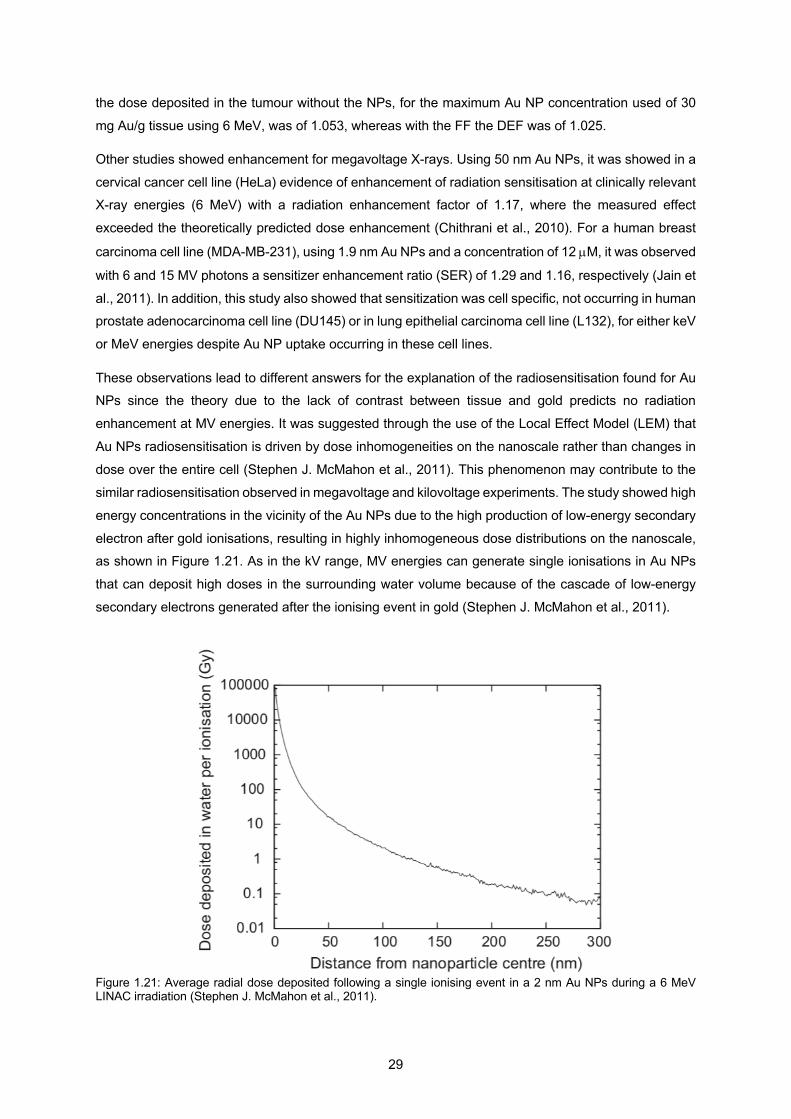
29
the dose deposited in the tumour without the NPs, for the maximum Au NP concentration used of 30
mg Au/g tissue using 6 MeV, was of 1.053, whereas with the FF the DEF was of 1.025.
Other studies showed enhancement for megavoltage X-rays. Using 50 nm Au NPs, it was showed in a
cervical cancer cell line (HeLa) evidence of enhancement of radiation sensitisation at clinically relevant X-ray energies (6 MeV) with a radiation enhancement factor of 1.17, where the measured effect
exceeded the theoretically predicted dose enhancement (Chithrani et al., 2010). For a human breast
carcinoma cell line (MDA-MB-231), using 1.9 nm Au NPs and a concentration of 12 µM, it was observed
with 6 and 15 MV photons a sensitizer enhancement ratio (SER) of 1.29 and 1.16, respectively (Jain et
al., 2011). In addition, this study also showed that sensitization was cell specific, not occurring in human prostate adenocarcinoma cell line (DU145) or in lung epithelial carcinoma cell line (L132), for either keV
or MeV energies despite Au NP uptake occurring in these cell lines.
These observations lead to different answers for the explanation of the radiosensitisation found for Au NPs since the theory due to the lack of contrast between tissue and gold predicts no radiation
enhancement at MV energies. It was suggested through the use of the Local Effect Model (LEM) that
Au NPs radiosensitisation is driven by dose inhomogeneities on the nanoscale rather than changes in
dose over the entire cell (Stephen J. McMahon et al., 2011). This phenomenon may contribute to the
similar radiosensitisation observed in megavoltage and kilovoltage experiments. The study showed high
energy concentrations in the vicinity of the Au NPs due to the high production of low-energy secondary
electron after gold ionisations, resulting in highly inhomogeneous dose distributions on the nanoscale, as shown in Figure 1.21. As in the kV range, MV energies can generate single ionisations in Au NPs
that can deposit high doses in the surrounding water volume because of the cascade of low-energy
secondary electrons generated after the ionising event in gold (Stephen J. McMahon et al., 2011).
Figure 1.21: Average radial dose deposited following a single ionising event in a 2 nm Au NPs during a 6 MeV LINAC irradiation (Stephen J. McMahon et al., 2011).
30
As mentioned before, Au NPs at the nanoscale present catalytic activity. A study demonstrated that Au
NPs showed intrinsic catalytic activity toward H2O2 decomposition and superoxide scavenging (He et
al., 2013). It was observed that the generation of radical species such as hydroxyl radicals and oxygen
was strongly dependent on the pH, where lower pH was needed for HO. formation and higher pH for
O2 (He et al., 2013).
Metal NPs may be good candidates to improve RT treatment and so their study under hypoxic
conditions is crucial since tumour cells live under these conditions. Investigation towards the understanding of Au NPs behaviour under hypoxic conditions showed that for MCF-7 and HeLa cells
the highest uptake following prolonged exposure under 0.2% of oxygen was for NPs of 50 nm size
(Neshatian et al., 2014). A concentration of 0.6 nmol of NPs was used and it was showed that NP
uptake of the cells pre-exposed to hypoxia for 18 h was in average 1.5 times higher than the one of
cells under normoxic conditions. The stability of the NP size was measured under normoxic and hypoxic
conditions for 24 h and it was concluded that NPs are stable under both conditions.
Conflicting results regarding the performance of Au NPs under hypoxic conditions show that more research under cancer cells environmental conditions is needed. A study reported that actually the Au
NP uptake was significantly lower under hypoxic than oxic conditions (Jain et al., 2014). In this case
cell lines such as DU145, MDA-MB-231 and L132 cells were exposed to a concentration of 12 µM of
1.9 nm Au NPs. Both the DU145 and MDA-MB-231 cell lines showed that a higher reduction in cell
growth induced by Au NPs in hypoxia than in normoxia, but for L132 cells the growth inhibitory effects were similar under both conditions. Regarding radiosensitisation using 160 kV X-rays, for MDA-MB-231
cells the SERs were similar at 21% and 1% oxygen concentration, 1.41 and 1.39, respectively, but was
much lower at 1.1 for 0.1% oxygen. It was suggested that the lower radiosensitisation observed under
hypoxia was due to the reduction in Au NPs cellular uptake. No significant radiosensitisation occurred
in DU145 or L132 cells in 0.1% or 21% oxygen conditions.
Another important question is whether the localisation of the NPs should be within the cell to provoke
its apoptosis. It is generally assumed that NPs have to be inside the cell nucleus, where the most important constituent is (the DNA) to destroy cancer cells. However, it was seen through experimental
studies that Au NPs are mostly taken up into the cytoplasm of the cell and still show radiosensitisation
effects (Chithrani et al., 2010). For example, mitochondrion can be a good target since is responsible
for many important functional roles in the cell such as energy metabolism, apoptosis regulation, ROS
production, and cell signalling. Through Monte Carlo simulations, a study showed that although Au NPs
tend to be within the cytosol, physical dose enhancements are not restricted to the vicinity of the NPs
and can extend to the nucleus and to the mitochondria, contributing in this way to NP dose enhancement
effects observed experimentally (McNamara et al, 2016).
The mechanism, through which Au NPs increase cell killing and DNA damage with X-rays, is still
unknown. Using 1.9 nm Au NPs at a concentration of 500 µg/ml, MDA-MB-231 cells were irradiated
with 1.5 Gy X-rays of photon energies between 10 keV to 60 keV (McQuaid et al., 2016). The energy
31
range used in the study was justified by the fact that, although is much lower than the clinical energies
use in RT treatments, a large proportion of X-rays produced by a LINAC are in the low keV energy
range. For instance, 10% of a 6 MeV LINAC beam spectrum is made up of photons with energies less
than 200 keV (please refer back to Fig. 1.4). The results suggest a different interaction between Au NPs and DNA molecules, due to the paradox of increased cell killing with little evidence of the increase in
short term DNA damage. It was concluded that other mechanisms are responsible for cell death at
lower X-ray energies, so photoelectrons can be playing a less effective role than it was thought.
Furthermore, according to the prediction of LEM, Auger electrons are the predominant source of dose
enhancement. Considering their short range, within several hundred nanometres from Au NP surface,
and the localisation of the NPs within the cell but outside of the cell nucleus, nuclear DNA is
consequently outside the range of the majority of these particles, which means that there must be a
different target to provoke the cell damage observed with the use of Au NPs with radiation. Studies have been focusing on finding the target responsible for the radiosensitisation seen with Au NPs and
the results suggest mitochondria as the main target (McMahon et al., 2017; Ghita et al., 2017).
More recently, a study aimed to identify other mechanisms than the physical assumptions alone for the
radiosensitisation measured with Au NPs found a biological mechanism that might be behind this
enhanced radiation-induced cell death (Taggart et al., 2016). Au NPs were found to inhibit the enzyme
protein disulphide isomerase, an important enzyme for correct folding of proteins in the endoplasmic
reticulum and for the formation of glutathione antioxidant. Inhibition of this enzyme can lead to cell stress and make cells more susceptible to radiation-induced ROS damage.
The controversial results concerning Au NP radiosensitisation can be originated from the differences in
performed investigations in terms of key parameters including Au NP size, shape, and concentration,
type of cell lines, and radiation source.
Although a promising candidate for the improvement of RT treatment there are still crucial unknown
phenomena underlying Au NPs radiosensitisation due to the lack of agreement between theoretical and experimental evidence.
1.8 Metal oxides as radiosensitisers
Different types of metals have been considered to explore radiation dose enhancement in tumours.
Theoretical research, using Interactive Data Language (IDL) program to solve the equation for DEF,
was made exploring materials with atomic numbers ranging from 25 to 90 and showed, for 6 MV X-rays
and a NP density of 5 mg/ml, a DEF range from 1.004 to 1.007 (Roeske et al., 2007).
Elements such as manganese, copper, iron and zinc have crucial roles as co-factors in various proteins
and enzymes, such as catalase (CAT), superoxide dismutase (SOD), cerulo-plasmin and
metallothionein (Weiss & Landauer, 2009). Although it is probable that antioxidant enzymes, such as
glutathione peroxidase, manganese SOD (MnSOD), copper-zinc SOD, and CAT, are important in
32
providing protection from radiation exposure, the proper balance of the enzymes in specific cells and in
the whole organism required for maximum radioprotection is not understood yet. For example, a large
increase in Mn-SOD in some model systems may have a radiosensitising effect rather than a protective
effect, probably related to the inability of the cell to cope with overproduction of hydrogen peroxide or hydroxyl radicals (Weiss & Landauer, 2006).
Not all the NPs used in this thesis have been considered for RT, although research has been made
with some of them for the purpose of improving this type of cancer treatment:
Iron-based NPs: radiosensitisation of this metal NPs has been attributed to the production of ROS,
owing to their surface-catalysed Haber-Weiss cycle and Fenton reaction, compensating in this way their
low atomic number of 26 (Klein et al., 2012). Iron NP surfaces catalyse in aqueous solutions H2O2 through the Haber-Weiss cycle which starts via the superoxide-driven reduction of Fe3+ to Fe2+ and
results into the formation of highly reactive HO. (Voinov et al., 2011). A study showed
superparamagnetic iron oxide NPs (SPIONs) radiosensitisation effect through their incubation in MCF-
7 cells for 24 h at a concentration of 0.1 mg Fe/mL after irradiation with 120 kV X-rays at a dose of 3
Gy (Klein et al., 2012). Human Alveolar Basal Epithelial Adenocarcinoma cell line (A549) irradiated with
5 Gy and incubated for 24 h with uncoated Fe3O4 NP systems and a concentration of 500 µg/ml, showed
a significant increase in ROS production when compared with radiation alone (Hauser et al, 2016).
The iron-catalysed Haber-Weiss reaction, which makes use of Fenton chemistry, is considered the main
mechanism by which the highly reactive radical is generated in biological systems (Hauser et al, 2016):
Fenton chemistry: Fe2+ + H2O2 → Fe3+ + HO- + HO.
Haber-Weiss reaction: O2- + H2O2 → O2 + HO- + HO.
Under normal conditions, only traces of iron exist in proteins such as transferrin or ferritin. Proteins take
up Fe ions in a way that the circulating Fe ions bind to transferrin and accumulate in the cells in the
form of ferritin. Iron absorption is controlled to prevent excess storage or oxidative attack. The uptake
of iron NPs can increase the intracellular unbound iron, resulting in cell damage or death, especially
when combined to additional therapies. Cell uptake of iron oxide NPs enhances ROS production through two different pathways: release of Fe ions into the cytosol where chelation by citrate (CIT) or
adenosine phosphate takes place, resulting in the iron ions participating in the Haber-Weiss cycle; or
the surface of the NP may act directly as a catalyst for the Haber-Weiss cycle and the Fenton reaction.
Via these pathways formation of hydroxyl radical occurs (Hauser et al, 2016).
Only a few formulations of SPIONs have been approved for clinical uses due to insufficient tissue
selectivity, low drug loading capacity, and lack of control over their biodistribution (Revia & Zhang,
2016).
Hafnium-based NPs: due to their high atomic number (Z=72) and chemical stability, a type of 50 nm-
sized functionalised hafnium oxide NPs called NBTXR3, with a negative surface charge has been used
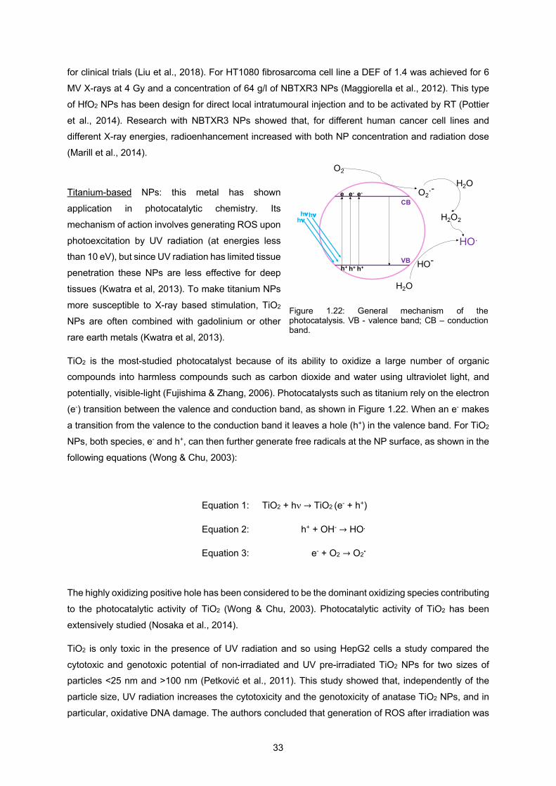
33
for clinical trials (Liu et al., 2018). For HT1080 fibrosarcoma cell line a DEF of 1.4 was achieved for 6
MV X-rays at 4 Gy and a concentration of 64 g/l of NBTXR3 NPs (Maggiorella et al., 2012). This type
of HfO2 NPs has been design for direct local intratumoural injection and to be activated by RT (Pottier
et al., 2014). Research with NBTXR3 NPs showed that, for different human cancer cell lines and different X-ray energies, radioenhancement increased with both NP concentration and radiation dose
(Marill et al., 2014).
Titanium-based NPs: this metal has shown
application in photocatalytic chemistry. Its
mechanism of action involves generating ROS upon
photoexcitation by UV radiation (at energies less than 10 eV), but since UV radiation has limited tissue
penetration these NPs are less effective for deep
tissues (Kwatra et al, 2013). To make titanium NPs
more susceptible to X-ray based stimulation, TiO2
NPs are often combined with gadolinium or other
rare earth metals (Kwatra et al, 2013).
TiO2 is the most-studied photocatalyst because of its ability to oxidize a large number of organic compounds into harmless compounds such as carbon dioxide and water using ultraviolet light, and
potentially, visible-light (Fujishima & Zhang, 2006). Photocatalysts such as titanium rely on the electron
(e-) transition between the valence and conduction band, as shown in Figure 1.22. When an e- makes
a transition from the valence to the conduction band it leaves a hole (h+) in the valence band. For TiO2
NPs, both species, e- and h+, can then further generate free radicals at the NP surface, as shown in the
following equations (Wong & Chu, 2003):
Equation 1: TiO2 + hn → TiO2 (e- + h+)
Equation 2: h+ + OH- → HO.
Equation 3: e- + O2 → O2-
The highly oxidizing positive hole has been considered to be the dominant oxidizing species contributing
to the photocatalytic activity of TiO2 (Wong & Chu, 2003). Photocatalytic activity of TiO2 has been
extensively studied (Nosaka et al., 2014).
TiO2 is only toxic in the presence of UV radiation and so using HepG2 cells a study compared the
cytotoxic and genotoxic potential of non-irradiated and UV pre-irradiated TiO2 NPs for two sizes of particles <25 nm and >100 nm (Petković et al., 2011). This study showed that, independently of the
particle size, UV radiation increases the cytotoxicity and the genotoxicity of anatase TiO2 NPs, and in
particular, oxidative DNA damage. The authors concluded that generation of ROS after irradiation was
CB
VB
hnhnhn
e- e-e-
h+h+ h+
O2
O2.-H2O
HO.
H2O2
H2O
HO-
Figure 1.22: General mechanism of the photocatalysis. VB - valence band; CB – conduction band.

34
the main mechanism responsible for the observed toxic effects, since UV radiation has enough energy
to excite electrons from the valence band to conduction band (see Figure 1.22), resulting in the
formation of highly reactive electron-hole pairs. In an aqueous environment, electrons reduce oxygen
to give superoxide anion radicals, which can be dismutated to H2O2, while holes oxidize water to give hydroxyl radicals, as shown in Figure 1.22. Also, an interesting finding in this study was the fact of TiO2
NPs to remain activated, i.e. in the excited stage, even after irradiation was discontinued, which can
explain their higher toxic and genotoxic potential compared to the non-irradiated NPs.
For TiO2 NPs coated with a dye it was found that visible-light activates the NPs leading to degradation
of plasmid DNA in vitro (Blatnik et al., 2012). An in vivo study demonstrated the efficacy of TiO2 NPs for
RT when these particles are doped with rare earth elements after irradiation with 200 kV X-rays
(Townley et al., 2012). Human glioblastoma cell lines, SNB-19 and U87MG, internalized with titanium oxide nanotubes (TiONts) showed no cytotoxicity and decreased DNA repair efficiency after irradiation
as well as amplified G2/M cell-cycle arrest (Mirjolet et al., 2013). In this study, cells were incubated with
a concentration of 1 µg/ml of TiONts for 24 h and irradiated with 10 Gy by a linear photon accelerator
(clinac600 Varian).
A study on titanium-based NPs to use them for deep-tumour treatments concluded that titanium peroxide NPs (TiOxNPs) could be used to optimize pancreatic cancer therapy. TiOxNPs were tested in
a human pancreatic cancer model both, in vitro and in vivo, and the results showed a high ROS
production upon 150 keV X-rays irradiation (Nakayama et al., 2016).
Zinc-based NPs: a semiconductor material with a broad range of applications with a very low risk to
human health. A great advantage of this type of NPs is their ability to absorb a wide range of the visible
spectrum due to its wide band gap energy. This results in fast recombination of photogenerated charges and thus leads to a low photocatalytic efficiency (Lee et al., 2016). ZnO NPs have the ability to respond
to ionising radiation since they exhibit scintillation upon excitation with X-rays. The induced
radioluminescence results from the excitation of fluorescent centers by energy conversion taking place
in the semiconductor NP. Such as for TiO2 NPs the production of ROS will follow Equations 1, 2 and 3
(Lee et al., 2016). ZnO NPs induce cell death through their ability to form ROS (Bogdan et al., 2017).
ZnO NPs were investigated and to ensure chemical stability of an atmospheric zinc oxide core, the
particles were coated with a silica layer (Generalov et al., 2015). Two human prostate adenocarcinoma cell lines, LNCaP and DU145, were exposed to 200 kV X-ray radiation after incubation of ZnO/SiO2
NPs. The results demonstrated a radiation enhancement due to the NPs with an enhancement ratio of
1.97 for LNCaP cells and one of 1.19 for DU145 cells. It was concluded that the radiosensitising effect
of the NPs was due to the X-ray induced catalysis at the NP surface.
ZnO NPs with a diameter less than 100 nm are relatively biocompatible, can additionally be used for
drug delivery, and show anticancer activity by inducing apoptosis and autophagy (Jiang et al., 2018).
For example, regarding apoptosis, ZnO NPs promote cancer cell death due to the intracellular release of zinc ions. This leads to an increase in ROS induction and subsequent cell death mediated by the
35
ROS triggered mitochondrial pathway. This process happens because the mitochondrial electron
transport chain is associated with intracellular ROS generation. Excessive ROS production damages
the mitochondria resulting in the release of cytochrome C that triggers intrinsic apoptosis (Jiang et al.,
2018).
Different approaches have been used with metal-based nanomaterials to avoid radioresistance induced
by tumour hypoxia. One of the approaches is through the generation of O2 in situ. Due to the
overproduction of hydrogen peroxide within malignant tumours, the approach of O2 generation in situ
via catalysing intracellular H2O2 provides a new way to provide continuous O2 release in tumour cells
(Chenyang et al., 2019). Manganese dioxide (MnO2) is an excellent catalyst for decomposing H2O2 into
O2 within the tumour environment (Zhu et al., 2016). MnO2 in the form of nanosheets (NSs) has high surface-to-volume ratio, which allow easy contact between the reactant molecules and the active sites,
thus providing enhanced catalytic activities (Wu et al., 2019). Fan et al., (2015) explored MnO2 NSs that
can be reduced to Mn2+ by acidic H2O2 in solid tumours, improving radiosensitisation under hypoxia
conditions through ROS generation.
Another approach is the use of nano-radiosensitisers with diminished oxygen dependence for hypoxic-
tumour RT. In this way, the possibility of a radiosensitiser that could generate ROS without the use of
oxygen was studied. Semiconductors have the ability of generating holes that can decompose H2O molecules during RT and generate OH.. Using the semiconducting abilities of ZnO NPs, research
showed that was possible to generate abundant electron-hole pairs and create highly toxic hydroxyl
radicals, enhancing the anti-tumour therapeutic efficacy (Zhang et al., 2015). Furthermore, adding to
the decomposition of water by a semiconductor, another possibility is the decomposition of H2O2 to
generate oxygen-independent ROS. More importantly, using H2O2 instead of H2O molecules for ROS
production provides a more efficient way of enhancing cell killing in tumours and reducing damage to
the surrounding healthy tissues, since H2O2 is overexpressed within tumour cells relative to normal cells
(Chenyang et al., 2019). This approach was introduced by the use of Cu2(OH)PO4 nanocrystals that can simultaneously respond to endogenous stimuli, such as H2O2 and exogenous stimuli, such as X-
ray irradiation (Chenyang et al., 2019). Under X-ray irradiation, these nanocrystals decompose
intracellular H2O2 into OH. via a Fenton-like reaction. This research showed that Fenton-like reaction
was selectively activated in tumour cells due to the high amount of hydrogen peroxide found in cancer
cells, compared with normal cells, resulting in enhanced RT with tumour selectivity. Compared to Fe-
based NPs Fenton reaction, results showed that the Fenton-like reaction of Cu2(OH)PO4 nanocrystals
had a more effective activity in hydroxyl radical generation at pH 6.5 (Chenyang et al., 2019).
In summary, new metal elements are being developed and used for cancer treatment. Each of the
metallic elements offers specific characteristics due to their intrinsic properties that can be used in
combination with different strategies to optimise RT treatment.
1.9 Research aims

36
This thesis has the aim of exploring and comparing different types of metal oxides and analyse their
behaviour with respect to the production of ROS, within water, plasmid-DNA, and cancer cells. This
might bring a deeper knowledge regarding radiosensitisation of metal oxide NPs and also explore other
candidates for application in the RT treatment.
Even more important is the consistency of the results. Different studies have shown results regarding different types of NPs, but they are inconsistent. This has to do with the fact that different authors use
different cell lines, different NP sizes and different radiations, so a fair comparison between the NPs
used in different studies is not possible. For this reason, an effort was made in this thesis to try to
overcome this lack of consistency.
For this thesis, characteristics such as NP size and coating remained constrained as far as possible
(keeping in mind that small variations are always present), the same cell line was used, the same
radiation source and same measurement techniques and analysis were used. Each experiment was done at the same time with all the different types of NPs used in each part of the thesis.
There were seven research questions:
• What is the metal oxide NP with the highest radiosensitisation for clinical 6 MeV X-rays?
• Does this type of NP, although with a higher radiosensitisation, have a good cell uptake?
• How toxic is this type of NP to cancer cells without radiation?
• What types of ROS does each NP produce?
• What is the ability of these metal oxide NPs to cause damage to DNA?
• Is there any correlation between the types of ROS produced and the damage caused to cancer
cells or DNA?
• Does coating the NPs bring any advantages to cell uptake or radiosensitisation?
All these questions were addressed during this thesis and organized in different chapters.
• Chapter 1: Introduction
• Chapter 2: Materials and Methods; all the conditions, techniques and equipment used are
described in this chapter.
• Chapter 3: Comparison of the radiosensitising ability of Period 4 transition metal oxide NPs; this part studies the ability of the different metal oxide NPs to produce the ROS that is more
important for cell damage, hydroxyl radical, and how the NPs interact with cancer cells. A
coating was chosen to understand if its presence would bring any advantage to the uptake and
radiosensitisation of the NPs.
• Chapter 4: Comparison of the radiosensitisation ability of 22 different element metal oxide NPs using 6 MeV clinical X-rays; here focus is given to the interaction of the metal oxide NPs with
plasmid DNA. Also, the main types of ROS, such as hydroxyl radical, superoxide, and single
oxygen, responsible for cancer cell killing in the interaction of NPs with radiation, are studied.

37
• Chapter 5: Conclusions; a general conclusion is given based on the different results, and also
future lines of experiment that can be conducted are described to bring more knowledge into
this field.

38

39
2 – Materials and methods

40
41
2.1 Metal oxide nanoparticle-based methods
Nanoparticle characteristics
Metals from period four of periodic table, as shown in Figure 2.1, such as manganese (Mn), copper
(Cu), nickel (Ni), zinc (Zn), iron (Fe), chromium (Cr), cobalt (Co), vanadium (Vn) and scandium (Sc),
were investigated in the form of NPs for the first part of this thesis and compared to hafnium (Hf). For
the second part other types were added such as aluminium (Al) and silica (Si) from period 3, zirconium
(Zr) and molybdenum (Mo) from period 5, and lanthanides such as neodymium (Nd), samarium (Sm),
europium (Eu), gadolinium (Gd), terbium (Tb), dysprosium (Dy), and erbium (Er).
Nanoparticles purchased from Sigma-Aldrich: Al2O3 (544833, <50 nm, 99.8%), SiO2 (637238, 10-20 nm, 99.5%), TiO2 (718467, 21 nm, 99.5%), Fe3O4 (637106, 50-100 nm, 97%), NiO (637130, <50 nm,
99.8%), CuO (544868, <50 nm, 82.6%) and ZnO (721077, <100 nm).
Nanoparticles purchased from US Research Nanomaterials: Cr2O3 (US3060, 60 nm, >99%), MnO2
(US3319NMP, 50 nm, 98%), CoO (US3051, 50 nm, 99.7%), ZrO2 (US3600, 40 nm, >99%), MoO3
(US3330, 13-80 nm, 99.94%), Nd2O3 (US3350, 30-45 nm, 99.9%), Sm2O3 (US3450, 15-45 nm,
99.95%), Eu2O3 (US3543, 10-100 nm, 99.99%), Gd2O3 (US3240, 10-100 nm, 99.9%), Tb4O7 (US3455,
10-100 nm, 99.99%), Dy2O3 (US3080, 30 nm, >99.9%), Er2O3 (US3140, 10-100 nm, 99.9%) and HfO2 (US3245, 61-80 nm, 99.99%).
Nanoparticles purchased from American elements: Sc2O3 (SC-OX-02-NP, <80 nm, 99%).
Nanoparticles purchased from Nanoshel: V2O5 (NS6130-03-399, <80 nm, 99.9%).
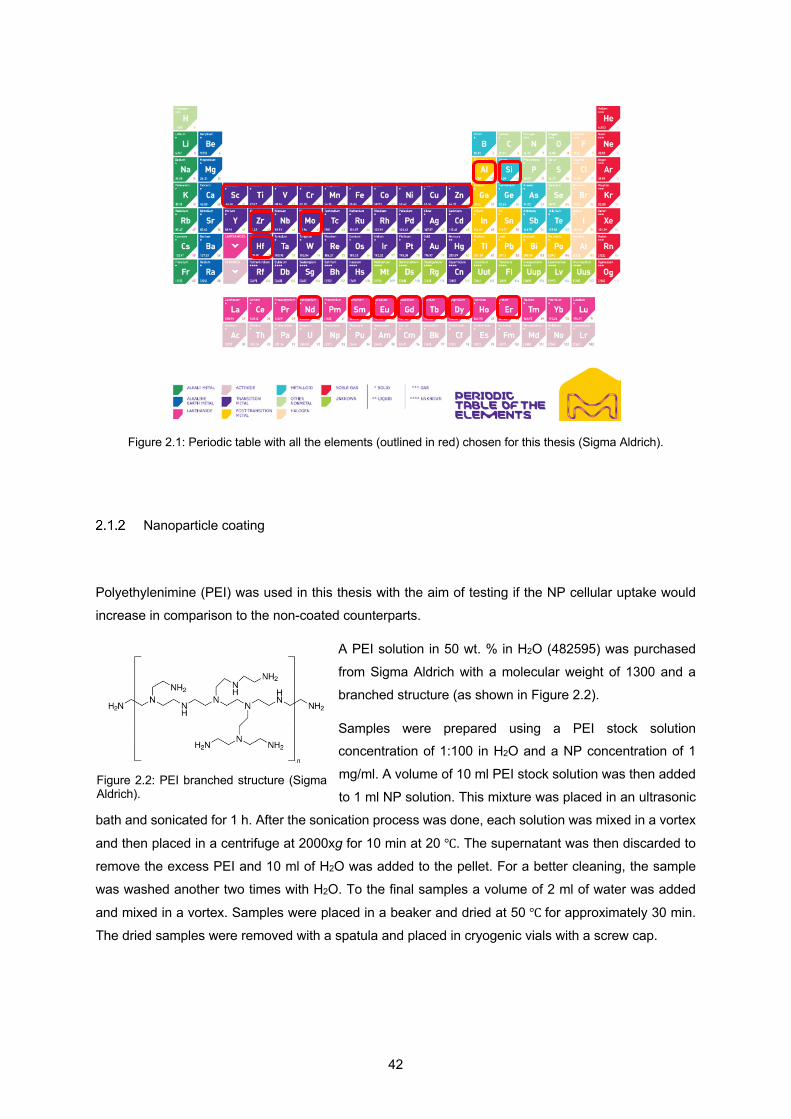
42
Figure 2.1: Periodic table with all the elements (outlined in red) chosen for this thesis (Sigma Aldrich).
Nanoparticle coating
Polyethylenimine (PEI) was used in this thesis with the aim of testing if the NP cellular uptake would
increase in comparison to the non-coated counterparts.
A PEI solution in 50 wt. % in H2O (482595) was purchased
from Sigma Aldrich with a molecular weight of 1300 and a
branched structure (as shown in Figure 2.2).
Samples were prepared using a PEI stock solution concentration of 1:100 in H2O and a NP concentration of 1
mg/ml. A volume of 10 ml PEI stock solution was then added
to 1 ml NP solution. This mixture was placed in an ultrasonic
bath and sonicated for 1 h. After the sonication process was done, each solution was mixed in a vortex
and then placed in a centrifuge at 2000xg for 10 min at 20 ℃. The supernatant was then discarded to
remove the excess PEI and 10 ml of H2O was added to the pellet. For a better cleaning, the sample
was washed another two times with H2O. To the final samples a volume of 2 ml of water was added
and mixed in a vortex. Samples were placed in a beaker and dried at 50 ℃ for approximately 30 min.
The dried samples were removed with a spatula and placed in cryogenic vials with a screw cap.
Figure 2.2: PEI branched structure (Sigma Aldrich).
43
Nanoparticle physico-chemical characterization
2.1.3.1 Transmission Electron Microscopy (TEM)
NP size and shape was determined using TEM images. Solutions were prepared by mixing the NP
powder in water and sonicating it for 1 h. A sample volume of 2 µl was dried on an electrostatically
discharged carbon-coated grid. Each grid was placed in a JEM-1400 model (JEOL-USA) and different images were taken for each sample. Using ImageJ 1.49 software, NP diameters were calculated using
10 images of each TEM sample with 3000x magnification. Threshold approach was used by adjusting
the images brightness to optimize the contrast with the background and then applying a threshold.
ImageJ “Analyse particles” function was used, and the different steps were taken:
1. Image magnification scale was defined;
2. Pixel dimensions were then converted into nanometres;
3. Size dimensions were set between 1 nm and infinity;
A MACROS code was used to run the threshold approach through all the 10 images of each sample.
Values of each sample were transferred into an excel spreadsheet and NP diameter was calculated
using 𝑑 = 2 × (√56) , where 𝐴 is the measured area and 𝑑 is the diameter. The excel “Data analysis”
function was used, and a histogram was created for each type of NP.
2.1.3.2 Dynamic Light Scattering (DLS)/Zeta potential
The hydrodynamic diameter of 2.5 mM NPs in 3% PBS pH 7.4 was calculated with The Nano-flex II
180° DLS system using Flex 11.1.0.5 software (Microtrac). PEI coating in each sample was identified
through zeta potential measurements. For this technique, a 5 mM NP sample of 10 ml in water was
prepared and the potential was measured using Colloid Metrix Stabino II. Non-coated and PEI-coated
samples potential was then compared to look for evidence of the PEI coating through the differences in
the potential.
2.1.3.3 X-ray diffraction
Samples were analysed using a Siemens D5005 XRD with a long fine focus copper X-ray tube
energised at 40 kV and 40 mA running conventional Bragg-Brentano geometry. Most runs were 0.5
s/step, with some at 8 s/step. Crystal phases were determined from XRD spectra using QualX2 software (Altomare et al., 2015) (version 2.24; Institute of Crystallography CNR, Bari, Italy) and PowCod
database 1901.
44
2.2 Cell culture-based methods
Cell culture
The cell line used in this thesis was HSC-3 that is a human tongue squamous carcinoma cell line. This
cell line was purchased from the American Type Culture Collection. HSC-3 cells were cultured in low
glucose (1 g/L) DMEM (Gibco, 21885-108), supplemented with 10% FBS (Sigma-Aldrich, F9885) and
1% penicillin/streptomycin (ThermoFisher Scientific, 15140122) in a humidifier incubator at 37 ℃ with
5% CO2. Cells were grown in T-75 culture flasks to about 80% confluency before passage.
Cytotoxicity of metal oxide nanoparticles
2.2.2.1 Clonogenic assay
The toxicity of metal oxide NPs and their effect on cell survival and proliferation was evaluated with a
clonogenic assay on HSC-3 cell line. HSC-3 cells were seeded at 150 cells/well in a 24-well plate and
allowed to adhere overnight. Next day, cells were exposed to 0, 1, 10, 30, and 100 µg/ml of each type
of metal oxide NP for 24 h. After 24 h have passed the NP medium was removed and the wells were washed three times with DMEM media supplemented with 10% FBS and 1% penicillin/streptomycin.
Cells were left to grow for 6 days to form colonies. After the end of the 6th day, cells were briefly rinsed
with water, stained, and fixed with 2% methylene blue in 50% ethanol solution for 4 h. The
staining/fixation solution was removed, plates were rinsed with distilled water and dried. Colony counts
were done by eye using a Zeiss AxioVert microscope and the IC25 values were estimated from
logarithmic plots of NP concentration vs colony survival, normalized to the control. IC25 is the
concentration at which the metal oxide NPs cause 25% cell colony reduction compared to the untreated controls.
Cellular uptake of metal oxide nanoparticles
2.2.3.1 Inductively coupled plasma mass spectrometer (ICP-MS)
Metal oxide NP cellular uptake was assessed by ICP-MS in the HSC-3 cell line. HSC-3 cells were
seeded at 150 cells/well in 24-well plates and allowed to adhere overnight. Next day, cells were exposed
to the IC25 concentration of each type of metal oxide NP for 24 h. After 24 h have passed the NP
medium was removed and the wells were washed three times with DMEM media supplemented with
10% FBS and 1% penicillin/streptomycin. Cells were left to grow for six days to form colonies. After the
45
end of the 6th day, cells were briefly rinsed with water and dried. In order to measure the concentration
of the NPs uptake by the cells, nitric acid was used to remove all the cells and the concentration of the
remaining NPs were put into solution and measured by Agilent 7500 ICP-MS or Agilent 8800 triple quad
ICP-MS. To convert the uptake in ppm into intracellular concentration in mg/ml it was assumed that 25,000 cells were loaded in each well for a total volume of 4 ml of HNO3. Considering that each cell
volume is equal to 2,000 microns cubed (Mitsui & Schneider, 1976) and that each NP is evenly
dispersed in that volume, the intracellular concentration is equal to:
intracellular c = 4
0.00005 × ppb = 80000 × ppb
, where 0.00005 ml is the volume occupied by the 25,000 cells, since 25,000 cells
= 50 million microns cubed = 0.00005 ml. Since 1 million ppb is equal to 1 mg/ml the intracellular
concentration becomes:
intracellular c = 80,000
1,000,000 × ppb = 0.08 × ppb.
2.3 Reactive oxygen species (ROS) based methods
ROS probes
2.3.1.1 Hydroxyl radical probe
Coumarin-3-carboxylic acid (3-CCA) powder was purchased from Sigma Aldrich. A 5 mM stock was
prepared in 6% PBS pH 7.4.
2.3.1.2 Hydrogen peroxide probe
Dihydrorhodamine 123 (DHR) solution was purchased from ThermoFisher Scientific. A 20 µM was
prepared in 6% PBS pH7.4.
2.3.1.3 Singlet oxygen probe
Singlet oxygen sensor green (SOSG) was purchased from ThermoFisher. From a 5 mM methanolic
46
stock, a working solution of 1 µM was prepared in 6% PBS pH 7.4.
2.3.1.4 Superoxide anion probe
Dihydroethidium (DHE) was purchased from ThermoFisher. From a stock of 20 mM DHE in DMSO a
working solution of 50 µM was prepared in 6% PBS pH 7.4.
ROS measurement
Experiments were run a minimum of three times independently and each time containing technical
triplicates. Fifty microlitres of probe working solution was added to 50 µl of 5 mM NP in deionized water
in black-walled 96-well plates (Cellstar, Greiner), producing a 2.5 mM NP working solution. After
irradiation the fluorescence of each well was measured using a FLUOstar Optima plate reader (BMG
Labtech), according to the type of probe: 3-CCA at Ex 390 nm/ Em 450 nm, DHR at Ex 485 nm/ Em
544 nm, SOSG at Ex 485 nm /Em 520 nm, and DHE at Ex 485 nm/ Em 590 nm (Zhao et al., 2003).
ROS calibration measurement
Each NP could potentially alter the fluorescence of the probe solution. Therefore, for each probe 50 µl
serial dilutions of the fully oxidized fluorescence molecule in 6% PBS were mixed with 50 µl 5 mM NP
solution and the fluorescence measured as already described.
For 3-CCA calibration was measured with 7-hydroxycoumarin-3-carboxylic acid (7OH-CCA, Sigma-
Aldrich). For DHR calibration, rhodamine 123 (ThermoFisher Scientific) was used.
For SOSG calibration, 0.5 µM SOSG was reacted with 6% H2O2 and 2% NaClO. The reaction of H2O2
with NaClO generates singlet oxygen (To et al., 2014).
For DHE calibration, to generate DHE fluorescence products namely 2OH-E, 1.5 µM DHE was reacted
with 0.5 mM xanthine in 0.9% NaCl and 10 mU/ml xanthine oxidase (Sigma-Aldrich). The stock of 2OH-
E was generated by complete DHE oxidation for 6 h as previously described (Nazarewicz et al, 2013).
The molarity of each radical species is reported assuming an equimolar reaction between each probe
and its cognate radical.
47
2.4 Plasmid DNA based methods
Plasmid DNA extraction
Plasmid pBR322 was extracted from bacteria using Qiagen plasmid midi kits and kept as a 50 ng/µl
stock in 10 mM Tris-HCl, pH 8.5. Plasmid concentration was obtained from UV-vis measurements using
NanoDrop One Spectrophotometer from ThermoFisher.
Plasmid DNA measurement
Ten microlitres of plasmid DNA were mixed with 10 µl of 5 mM NP solution in 96 well plates. Solutions
were either left untreated or irradiated with 10 Gy of 6 MeV X-rays, as described below.
After irradiation, to assess the DNA irradiation damage a 50 well agarose gel electrophoresis was used.
Five µl of each sample were mixed with 5 µl loading dye (Purple, New England Biolabs) and loaded
onto 1% agarose electrophoresis gels containing 0.2 µg/ml ethidium bromide with a running buffer of
Tris-Borate-EDTA with 0.2 µg/ml ethidium bromide. A 1 kb DNA ladder was also used (Quick-load
Purple Plus, New England Biolabs).
Gels were run at 110 V for 1 h and then photographed using a G:Box Chemi XX6 gel documentation
system (Syngene). DNA band intensities were quantified using the Gel Analysis feature of ImageJ
software (version 1.52; NIH).
2.5 Hole quenching-based methods
Hole scavenger
2.5.1.1 Formic acid
A working concentration of 10 mM formic acid pH 3.5 was obtained from a 99.5% concentrated formic
acid solution (Puangpetch et al., 2011; Tan et al., 2003).
Hole quenching measurement
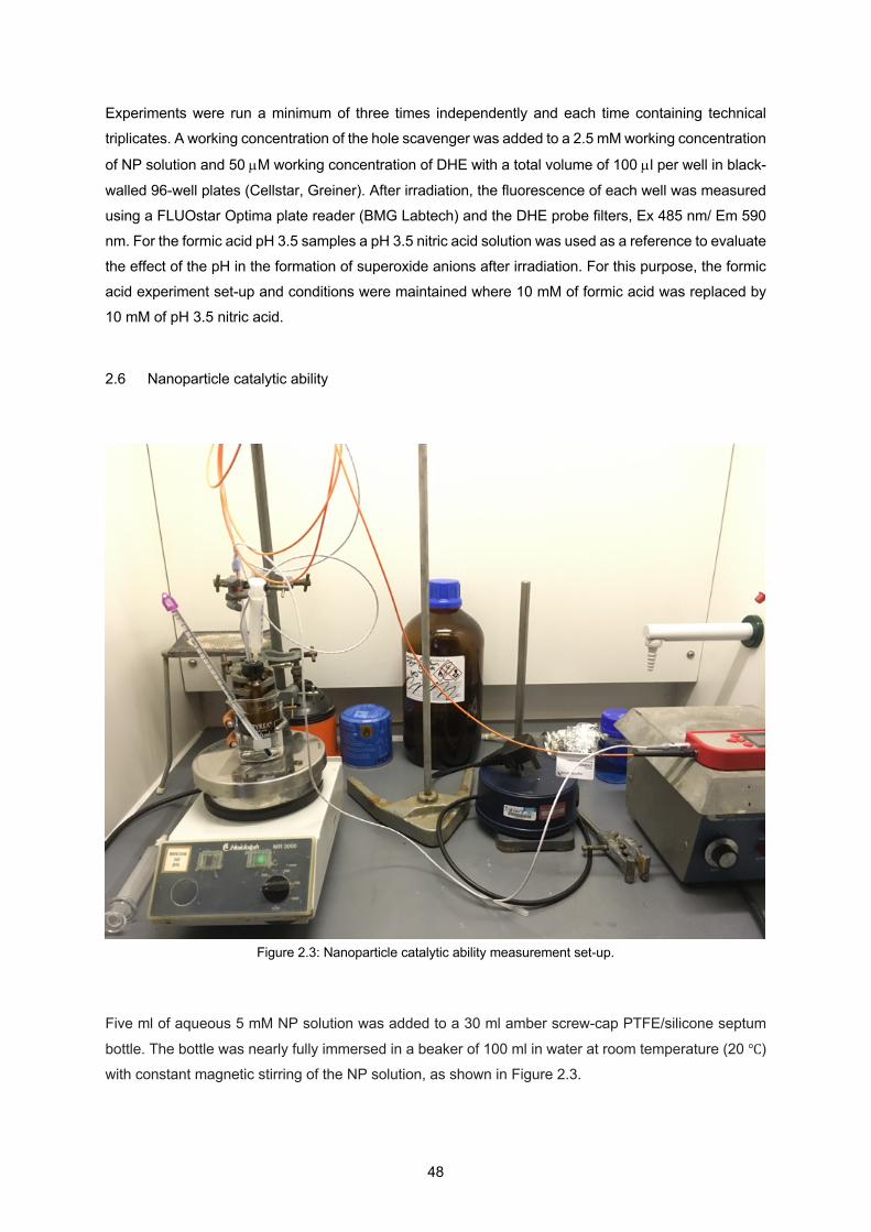
48
Experiments were run a minimum of three times independently and each time containing technical
triplicates. A working concentration of the hole scavenger was added to a 2.5 mM working concentration
of NP solution and 50 µM working concentration of DHE with a total volume of 100 µl per well in black-
walled 96-well plates (Cellstar, Greiner). After irradiation, the fluorescence of each well was measured
using a FLUOstar Optima plate reader (BMG Labtech) and the DHE probe filters, Ex 485 nm/ Em 590
nm. For the formic acid pH 3.5 samples a pH 3.5 nitric acid solution was used as a reference to evaluate
the effect of the pH in the formation of superoxide anions after irradiation. For this purpose, the formic
acid experiment set-up and conditions were maintained where 10 mM of formic acid was replaced by
10 mM of pH 3.5 nitric acid.
2.6 Nanoparticle catalytic ability
Five ml of aqueous 5 mM NP solution was added to a 30 ml amber screw-cap PTFE/silicone septum
bottle. The bottle was nearly fully immersed in a beaker of 100 ml in water at room temperature (20 ℃)
with constant magnetic stirring of the NP solution, as shown in Figure 2.3.
Figure 2.3: Nanoparticle catalytic ability measurement set-up.
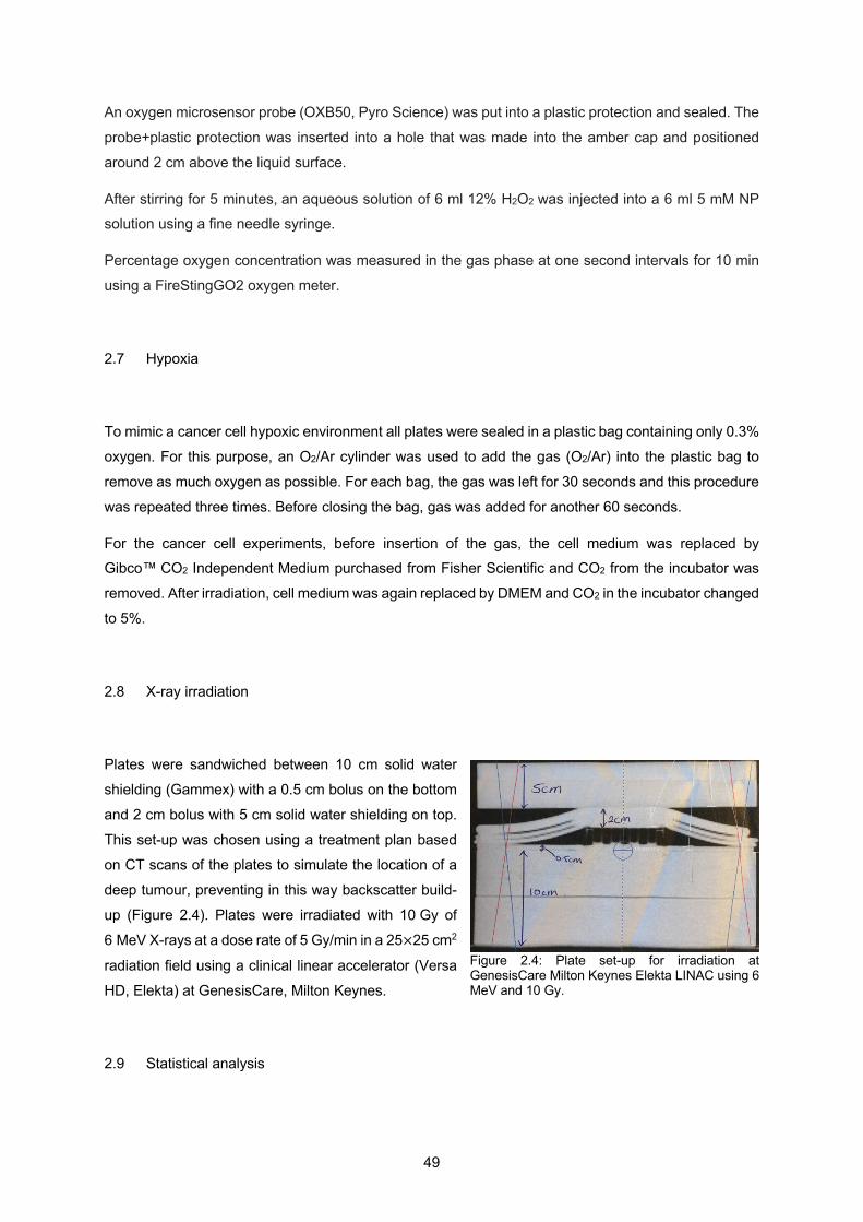
49
An oxygen microsensor probe (OXB50, Pyro Science) was put into a plastic protection and sealed. The
probe+plastic protection was inserted into a hole that was made into the amber cap and positioned
around 2 cm above the liquid surface.
After stirring for 5 minutes, an aqueous solution of 6 ml 12% H2O2 was injected into a 6 ml 5 mM NP solution using a fine needle syringe.
Percentage oxygen concentration was measured in the gas phase at one second intervals for 10 min
using a FireStingGO2 oxygen meter.
2.7 Hypoxia
To mimic a cancer cell hypoxic environment all plates were sealed in a plastic bag containing only 0.3%
oxygen. For this purpose, an O2/Ar cylinder was used to add the gas (O2/Ar) into the plastic bag to
remove as much oxygen as possible. For each bag, the gas was left for 30 seconds and this procedure
was repeated three times. Before closing the bag, gas was added for another 60 seconds.
For the cancer cell experiments, before insertion of the gas, the cell medium was replaced by Gibco™ CO2 Independent Medium purchased from Fisher Scientific and CO2 from the incubator was
removed. After irradiation, cell medium was again replaced by DMEM and CO2 in the incubator changed
to 5%.
2.8 X-ray irradiation
Plates were sandwiched between 10 cm solid water
shielding (Gammex) with a 0.5 cm bolus on the bottom
and 2 cm bolus with 5 cm solid water shielding on top.
This set-up was chosen using a treatment plan based
on CT scans of the plates to simulate the location of a
deep tumour, preventing in this way backscatter build-
up (Figure 2.4). Plates were irradiated with 10 Gy of
6 MeV X-rays at a dose rate of 5 Gy/min in a 25×25 cm2
radiation field using a clinical linear accelerator (Versa
HD, Elekta) at GenesisCare, Milton Keynes.
2.9 Statistical analysis
Figure 2.4: Plate set-up for irradiation at GenesisCare Milton Keynes Elekta LINAC using 6 MeV and 10 Gy.
50
Data were analysed using GraphPad 6.0 Prism software. All the experiments were performed in
triplicate and on three independent times, so all values were normalized to their control, n=3.
The data of ROS and clonogenic assay where both non-irradiated and irradiated results are shown was
analysed using a two-way ANOVA with Tukey’s post-hoc test and a=0.05.
In the data of ROS and clonogenic assay for ratio and quenching results a one-way ANOVA with
Dunnett’s post-hoc test and a=0.05 was used.
The significance is always compared to the respective control. Asterisks indicate significant differences
of *P<0.05, **P<0.01, ***P<0.001, ****P<0.0001. Mean ± SEM was plotted on all graphs.
51
3 Comparison of the radiosensitising ability of Period 4 transition metal oxide
nanoparticles

52

53
3.1 Introduction
Different types of NPs have been a target of study in order to enhance RT treatment. One of the major
issues with this field of research is the lack of comparable results due to the existence of different
experimental variables and the assumption that NP radiosensitisation is primarily due to photoelectric
electrons without much consideration for the role of surface catalysis. For example, within gold NPs,
different results are shown regarding the gold radiosensitising ability due to the differences in size, cell
line, beam type used (Rosa et al., 2017). For this reason, this thesis is focused on a systematic survey
of the radiosensitising potential of different metallic NPs.
In this way, metal oxide NPs from period four of the periodic table, and hafnium oxide NPs for
comparison, were chosen for this research, where size, cell line and beam type were maintained
constant through the experimental procedure. Thus, the following metal oxide NPs were studied: Sc2O3, TiO2, V2O5, CrO2, MnO2, Fe2O3, CoO, NiO, CuO, ZnO and HfO2 as a heavy element positive control.
For clonogenic assays with cancer cells, the coating was one of the variables of concern in this study. Coating metal oxide NPs with organic molecules that are recognised and biocompatible may improve
their cell uptake. Also, when injected into the bloodstream, it is important that the NPs present a high
retention time before being taken up by macrophages. Coating of the NPs can overcome these
limitations.
The coating of NPs with hydrophilic, neutral polymers can increase their stability in the blood circulatory
system. The coating attaches to the surface of the NP either by adsorption, hydrophobic or electrostatic
interactions, or by grafting via covalent bonds (Storm et al., 1995).
The enhanced stability in the blood through these polymer-coated NPs is due to their ability to prevent
the adsorption of various blood components onto their surface. The coating confers “invisibility” to the
NPs, which in turn reduces the uptake by liver and spleen macrophages and, thus, extends blood
circulation times (Storm et al., 1995). Different types of coating can be used to achieve different
properties such as stability, biocompatibility, or active targeting, where the NPs are modified with a
specific active molecule that binds to the desired target cells or tissues.
One of the purposes of the coating in this thesis was to improve cellular uptake. Different polymer
coatings have been used to modify NPs and improve their biocompatibility such as thiol-terminated
methoxypoly, e.g. ethylene glycol (Liao & Hafner, 2005) and PEG that due to its amphiphilic characteristic provides good solubility to the NP, in both polar and non-polar solvents, which in turn is
responsible for the high biocompatibility and good affinity for cell membranes (Choi et al., 2003).
The surface of NPs has also been modified with polyethylenimine (PEI) to increase cellular binding and
uptake and prevent their aggregation under physiological conditions (Pissuwan et al., 2011). A study
comparing the cellular uptake in human-derived retinal pigment epithelial (ARPE-19) cells between 80
nm silver NPs (AgNP) with four different coatings such as branched PEI, CIT, polyvinyl-pyrrolidone
(PVP) and PEG showed that AgNP-PEI were positively charged and entered the cells at a higher rate
54
than the negatively charged or neutral particles. Furthermore, AgNP-PEI were toxic to the cells at lower
doses than the other coatings and both AgNP-PEI and AgNP-CIT had a greater intracellular
accumulation compared to AgNP-PEG and AgNP-PVP. This investigation concluded that positively
charged AgNP-PEI increased cellular uptake, the particles agglomerated in the peri-nuclear region and presented increased mitochondrial toxicity (Zucker et al., 2019).
Although PEI can induce cell death and inhibition of cell differentiation, the use PEI with lower molecular
weight, around 2 kDa, has shown to improve biocompatibility (Barrow et al., 2015). Furthermore, this type of PEI was reacted with a hydrophobic alkyl chain, which allowed for stabilisation of clusters of
pre-formed hydrophobic SPIONs in water. This is represented by the mode of assembly shown in Table
3.1 for PEI. It was shown that for mouse mesenchymal stem cells (mMSCs) the uptake for the alkyl
group on PEI polymer coated SPION was 7 pg[Fe] per cell at highest dose and incubation time, e.g. 7
mg/ml Fe for 24 h (Liu et al., 2011). Other different polymer coatings have been tried in iron oxide NPs,
as shown in Table 3.1.
In in vitro studies, cellular growth media contains serum proteins, essential amino acids, vitamins,
electrolytes, and other chemicals, such as antibiotics, and trace metals, that in contact with NPs can
alter their physico-chemical properties, such as surface charge, size and aggregation state, and surface
chemistry (Alkilany & Murphy, 2010). In cell growth media, NPs adopt the physico-chemical properties of the adsorbed protein shell, called “protein corona”, meaning that the surface chemistry of the NPs it
is not the same as the original (Cedervall et al., 2007). Coatings such as PEG are often used to prevent
the protein corona (Oh et al., 2018). For in vivo studies, the injection of the NPs into the blood stream
may also modify the surface of NP and aggregation state due to their interaction with the proteins and
electrolytes present in the blood (Alkilany & Murphy, 2010). Functionalized gold nanorods with PEG
were used successfully to increase the circulation time of the injected NPs within the body (Eghtedari
et al, 2009). The reticuloendothelial system of the body filters the injected NPs based on their size and
surface characteristics limiting circulation time, which means NPs do not remain long enough in the blood stream to find their target. A recent study showed that an important parameter to be considered
in the clearance of NPs is the density of the coating (Bertrand et al., 2017). In this research a PEG
density threshold was identified below which blood clearance is fast.
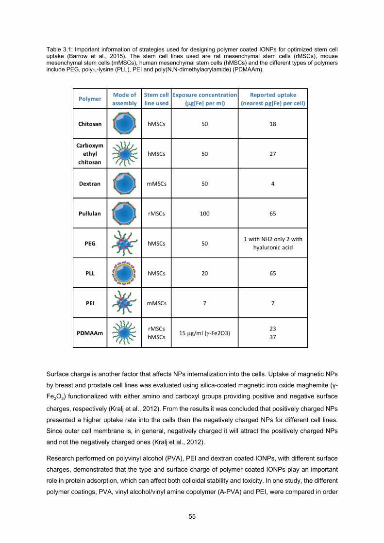
55
Table 3.1: Important information of strategies used for designing polymer coated IONPs for optimized stem cell uptake (Barrow et al., 2015). The stem cell lines used are rat mesenchymal stem cells (rMSCs), mouse mesenchymal stem cells (mMSCs), human mesenchymal stem cells (hMSCs) and the different types of polymers include PEG, poly-L-lysine (PLL), PEI and poly(N,N-dimethylacrylamide) (PDMAAm).
Surface charge is another factor that affects NPs internalization into the cells. Uptake of magnetic NPs
by breast and prostate cell lines was evaluated using silica-coated magnetic iron oxide maghemite (γ-Fe2O3) functionalized with either amino and carboxyl groups providing positive and negative surface
charges, respectively (Kralj et al., 2012). From the results it was concluded that positively charged NPs
presented a higher uptake rate into the cells than the negatively charged NPs for different cell lines. Since outer cell membrane is, in general, negatively charged it will attract the positively charged NPs
and not the negatively charged ones (Kralj et al., 2012).
Research performed on polyvinyl alcohol (PVA), PEI and dextran coated IONPs, with different surface
charges, demonstrated that the type and surface charge of polymer coated IONPs play an important
role in protein adsorption, which can affect both colloidal stability and toxicity. In one study, the different
polymer coatings, PVA, vinyl alcohol/vinyl amine copolymer (A-PVA) and PEI, were compared in order
56
to understand their influence on colloidal stability, cytotoxicity and cellular uptake in different cell media
using SPIONs and HeLa cell line (Petri-Fink et al., 2008). Regarding the colloidal stability, all polymer
coated SPIONs were stable in water and PBS but the stabilities were different in DMEM or Roswell
Park Memorial Institute (RPMI) media. This data showed that the choice of medium influences the uptake of these particles by HeLa cells. Another study compared SPIONs coated with two different
polymers, PVA and dextran (Sakulkhu, Mahmoudi, et al., 2014). Obtaining different surface charges,
positive, neutral, and negative, the effect of the surface charge and surface polymer material on protein
adsorption was investigated. PVA-coated SPIONs with negative and neutral charge adsorbed more
serum proteins than the dextran-coated SPIONs, which resulted in a higher blood circulation time for
PVA-coated NPs compared to the dextran-coated ones. The same group (Sakulkhu, Maurizi, et al.,
2014) performed a study using PVA-coated SPIONs comparing the different surface charges, positive,
neutral and negative, investigating the protein corona in vivo. Rat serum was extracted after interaction of NPs with the rat’s physiological system, leading to more realistic results and better understanding
and prediction of the destiny of NPs in vivo. Particles were left for 15 min in blood circulation in living
rats and their protein corona showed differences due to their surface charge. Neutral particles showed
the highest number of different proteins both in vitro and in vivo, positively charged particles showed
the lowest number of different proteins in vivo and particles negatively charged showed the lowest
number of different proteins in vitro.
Conformation, charge distribution and also the hydrophilicity/hydrophobicity of the adsorbed proteins
could all affect the zeta potential measurement (Barrow et al., 2015). Protein adsorption is one of the important driving forces in the selection of a NP target destination, which will further affect cells, tissues
and finally the body system. Proteins such as albumin, fibrinogen, IgG, complement C3, apolipoprotein
A-I and apolipoprotein E19–22 bind to NPs of polymeric or inorganic nature (Sakulkhu et al., 2015).
Surface charge effects of IONPs in protein adsorption and cell uptake were investigated comparing a
negatively charged coating such as polyacrylic acid (PAA) with a positively charged coating such as
PEI. The hydrodynamic diameter was the same for both particles, 30 nm, and after 24 h of incubation
large NP-protein bioconjugates with hydrodynamic sizes of around 1500 nm for PAA-coated NPs and around 3000 nm for PEI-coated NPs were observed. Although both NPs present a similar negative zeta
potential within cell culture, the results showed that PEI-coated NPs were incorporated in much larger
amounts than the PAA-coated NPs. Quantitative analysis showed that SHSY5Y cells can incorporate
100% of the added PEI-coated NPs up to 100 pg/cell, whereas for PAA-coated NPs the uptake was
less than 50% (Calatayud et al., 2014).
3.2 Experimental design
Choice of nanoparticle size, shape and concentration
57
In this part of the work, the effects, such as toxicity, uptake and radiosensitisation, of different metal
oxide NPs on human cancer cells was studied.
The NP diameter was aimed to be around 50 nm, since this size is optimal for cellular uptake (Chithrani
et al., 2010). The NPs were collected based on their size, with no control of their shape, meaning the
NPs used are mostly amorphous. Ideally, NPs should have a spherical shape. To increase cellular
uptake, it was shown that for 50 nm gold NPs, the spherical shaped ones showed the highest cellular
response when compared to gold nanospikes and gold nanorods, after a 24 h cell incubation (Ma et al.,
2017).
To explore the interaction of the NPs with radiation without cells, the concentration chosen was 2.5 mM.
This was a concentration high enough to give consistently detectable ROS species. In cell experiments,
NPs could be administered at either equimolar or equitoxic doses. Both methods have their respective
merits, but equimolar loading of NPs was chosen at a concentration designed to reduce clonogenic cell survival by 25% of control values (IC25).
Choice of nanoparticle coating
In the Introduction in Chapter 1, various types of NP coating were discussed. Here, the rationale for choosing PEI coating for the experiments in this thesis is explained. A study by Du and collaborators
(Du et al., 2016) showed that iron oxide NPs coated with low-molecular weight PEI, 2000 Da, can be
used as drug delivery carriers and cell tracking probes. Furthermore, a layer by layer technique was
used to coat gold nanorods with PEI and the results demonstrated an increase in cellular uptake
(Takahashi et al., 2008). To try to improve NP uptake and toxicity, in this thesis the chosen coating
was PEI with a low-molecular weight of 1300 Da. Furthermore, the low-molecular weight was used to
have a thin organic coating that would allow water to get to the NP surface. It has been shown that a
thick coating lowers radiosensitisation efficiency (Gilles et al., 2014). This study used gold NPs functionalised with PEG or human serum albumin and the results showed that NP functionalisation
significantly decreases hydroxyl radical production, so the authors suggested the use of a thin coating
to avoid this problem. Another study using PEG-amine coating AuNPs showed that the use of these
coating produced less hydroxyl radicals when irradiated with 10 Gy of 6 MeV X-rays compared to the
CIT-capped AuNPs (Grellet et al., 2017).
PEI is a polymer with repeating unit that consists of an amine group and two carbon aliphatic CH2CH2
spacer with the chemical formula (C2H5N)n. Polyethylenimines can be linear or branched, where the former consists of secondary amine groups and the latter can contain primary, secondary and tertiary
amino groups, as shown in Figure 3.1.
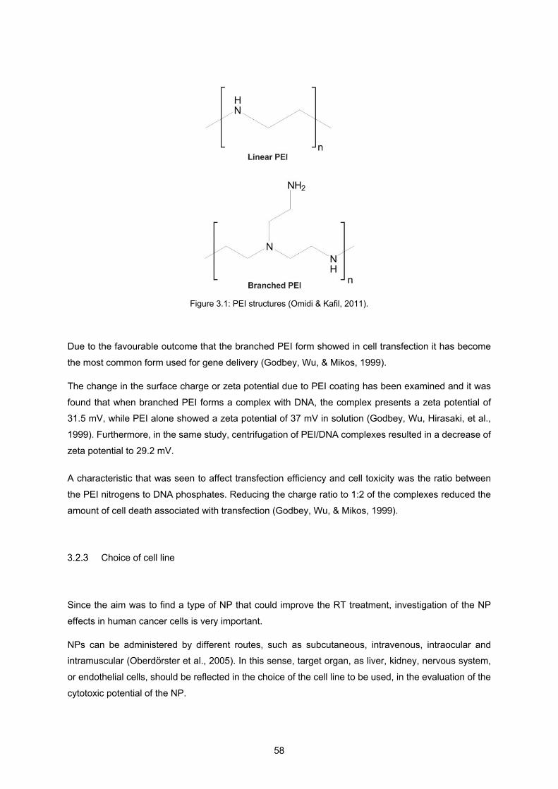
58
Figure 3.1: PEI structures (Omidi & Kafil, 2011).
Due to the favourable outcome that the branched PEI form showed in cell transfection it has become
the most common form used for gene delivery (Godbey, Wu, & Mikos, 1999).
The change in the surface charge or zeta potential due to PEI coating has been examined and it was
found that when branched PEI forms a complex with DNA, the complex presents a zeta potential of 31.5 mV, while PEI alone showed a zeta potential of 37 mV in solution (Godbey, Wu, Hirasaki, et al.,
1999). Furthermore, in the same study, centrifugation of PEI/DNA complexes resulted in a decrease of
zeta potential to 29.2 mV.
A characteristic that was seen to affect transfection efficiency and cell toxicity was the ratio between
the PEI nitrogens to DNA phosphates. Reducing the charge ratio to 1:2 of the complexes reduced the
amount of cell death associated with transfection (Godbey, Wu, & Mikos, 1999).
Choice of cell line
Since the aim was to find a type of NP that could improve the RT treatment, investigation of the NP
effects in human cancer cells is very important.
NPs can be administered by different routes, such as subcutaneous, intravenous, intraocular and intramuscular (Oberdörster et al., 2005). In this sense, target organ, as liver, kidney, nervous system,
or endothelial cells, should be reflected in the choice of the cell line to be used, in the evaluation of the
cytotoxic potential of the NP.
59
Due to the amount of the different types of NPs studied in this thesis only one type of cell line was
tested. In the Methods section, it was explained that HSC-3 oral squamous carcinoma cells were used.
Here, the reasons behind this choice are clarified. HSC-3 cell line was chosen due to its proliferation
ability and its round-shaped colony formation which facilitate the separation between colonies for colony counting. Also, although it is a cell type with oral squamous tissue origin, it presents a high degree of
local invasiveness and a high rate of metastasis to cervical lymph nodes (Kinouchi et al., 2018). Studies
have shown the ability of human oral squamous cell carcinoma cell lines to promote the growth and
metastasis of solid tumours, known as angiogenesis (Michi et al., 2000). This ability makes this cell line
appropriate for studies that aim at the understanding of the effects of NPs with 6 MeV X-ray energies.
Choice of irradiation parameters and radical probes
A 6 MeV X-ray beam, the most clinically-relevant megavoltage radiation (Stephen J. McMahon et al.,
2016), was used, with a dose of 10 Gy, chosen based on the highest signal given by a 3-CCA testing
at different Gy doses.
NP radiosensitising capacity was based on the amount of hydroxyl radicals they would generate when interacting with ionising radiation. The protocol chosen to measure the main radical type responsible
for cancer cells death was the coumarin assay that was adapted to NP suspension (Sicard-Roselli et
al., 2014). This technique was used due to its compatibility with NPs, experiment set-up, and sensitivity,
down to 30 nM HO. (Louit et al., 2009). The major known fluorescent product of hydroxylation of
coumarin is 7-hydroxycoumarin (7-OHC). 3-CCA was chosen instead of coumarin, since the hydrogen
in C3 with the carboxylic group eliminates hydroxylation in this position increasing, in this way, the
fluorescence of 7-hydroxylated coumarin derivatives by twofold (Manevich et al., 1997).
Hydrogen peroxide is an important intermediate species during production of the radicals species
involved in the process of cancer cell death. This assay was included with the aim of measuring the
amount of H2O2 produced in water by megavoltage irradiation. It has been proposed that hydrogen
peroxide produced by UV irradiation may be a precursor to hydroxyl radicals in the presence of metal oxide NPs (Miguel & Patrício, 2013). The fluorescent-responsive dye used to probe the amount of H2O2
was dihydrorhodamine (DHR). This non-fluorescent probe is oxidized in the presence of H2O2 and
converted into a fluorescent product called rhodamine 123 (Henderson & Chappell, 1993).
To exclude any interference caused by the presence of NPs during the measurement a quenching
assay was used for all radical probes. Studies showed that a direct comparison between the signal
intensity attributed to 7-OH-CCA in water and the one in a gold NP suspension is not possible since the
presence of NPs alters the mechanism, and hence regioselectivity, for HO. yield of formation (Sicard-
Roselli et al., 2014). The same happens to different metal oxide NP solutions. This quenching assay
was designed with the aim to exclude the fluorescent background due to NP interference, since the
experimental set-up used in this thesis was not suitable for the application of other techniques. Other
60
techniques included addition of NaCl solution to induce NP aggregation and therefore, its precipitation.
This technique was tested for gold NPs and it was showed that due to the very large reduction of the
surface area of gold, quenching was no longer seen in the aggregated state (Sicard-Roselli et al.,
2014). However, salt precipitation may not work for all types of NPs. Furthermore, Sicard-Roselli et al showed that 1 nM of 32.5 nm diameter gold NPs do not induce any modification in the fluorescence
intensity for incubation times shorter than 30 s. The present study used 22 different types of metal oxide
NPs of around 50 nm diameter, where the NP incubation time exceeded 30 s since the irradiation set-
up was at least 15 min away from the incubation set-up. In this sense, a simpler and adequate assay
was used, assuming that NP fluorescence interference is constant after 30 min of incubation time, and
consequently NP fluorescence interference can be excluded.
3.3 Results
Physico-chemical characterization of NPs
3.3.1.1 Transmission electron microscopy (TEM) and histogram size
In Table 3.2, TEM images and respective size distribution for each NP is presented. Size histograms
showed consistency with TEM images of 3000x magnification. For structural observation reasons, an
image at 150000x magnification is shown for each type of NP, not being representative of the average
size.

61
Table 3.2: TEM images at 3000x and 150000x magnification, and the size distribution for all NPs, non-coated and PEI-coated ones. ImageJ was used to determine the diameter of each NP through the TEM images of 3000x magnification.
NPs TEM image 3000x TEM image 150000x Size distribution
(x axis: NP diameter in nm) (y axis: counts)
Sc
ScPEI
Ti
TiPEI
V
VPEI
10 20 30 40 50 60 70 80 90 100
110
120
130
140
150
160
02004006008001000
200000
400000
600000
10 20 30 40 50 60 70 80 90 100
110
120
130
140
150
160
0
1000
2000
3000
10 20 30 40 50 60 70 80 90 100
110
120
130
140
150
160
0
1000
2000
3000
10 20 30 40 50 60 70 80 90 100
110
120
130
140
150
160
0
2000
4000
6000
8000
10000
10 20 30 40 50 60 70 80 90 100
110
120
130
140
150
160
0
1000
2000
3000
10 20 30 40 50 60 70 80 90 100
110
120
130
140
150
160
0
500
1000
1500
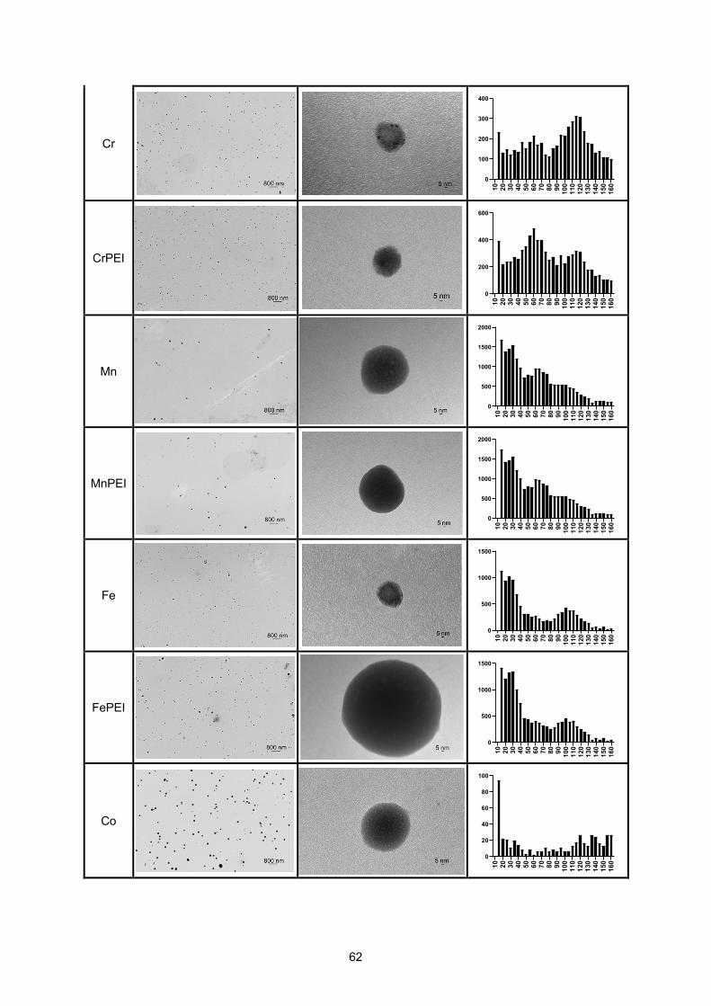
62
Cr
CrPEI
Mn
MnPEI
Fe
FePEI
Co
10 20 30 40 50 60 70 80 90 100
110
120
130
140
150
160
0
100
200
300
400
10 20 30 40 50 60 70 80 90 100
110
120
130
140
150
160
0
200
400
600
10 20 30 40 50 60 70 80 90 100
110
120
130
140
150
160
0
500
1000
1500
2000
10 20 30 40 50 60 70 80 90 100
110
120
130
140
150
160
0
500
1000
1500
2000
10 20 30 40 50 60 70 80 90 100
110
120
130
140
150
160
0
500
1000
1500
10 20 30 40 50 60 70 80 90 100
110
120
130
140
150
160
0
500
1000
1500
10 20 30 40 50 60 70 80 90 100
110
120
130
140
150
160
0
20
40
60
80
100

63
CoPEI
Ni
NiPEI
Cu
CuPEI
Zn
ZnPEI
10 20 30 40 50 60 70 80 90 100
110
120
130
140
150
160
0
50
100
150
200
250
10 20 30 40 50 60 70 80 90 100
110
120
130
140
150
160
0
500
1000
1500
2000
10 20 30 40 50 60 70 80 90 100
110
120
130
140
150
160
0
500
1000
1500
2000
2500
10 20 30 40 50 60 70 80 90 100
110
120
130
140
150
160
0
200
400
600
10 20 30 40 50 60 70 80 90 100
110
120
130
140
150
160
0
200
400
600
800
10 20 30 40 50 60 70 80 90 100
110
120
130
140
150
160
0
500
1000
1500
2000
2500
10 20 30 40 50 60 70 80 90 100
110
120
130
140
150
160
0
50000
100000
150000

64
Hf
HfPEI
The histograms of Sc and ScPEI NPs are very different. TEM analysis demonstrated inconsistent
results. In some cases, such as Sc, Cr, and Zn NPs, PEI incubation decreased the average NP size. In other cases, such as V, Co, and Cu, PEI incubation increased the average NP size. In the cases, such
as Ti, Mn, Fe, Ni, and Hf, PEI incubation gave no obvious change in NP size. Various explanations are
possible: either the coating process did not work (no size change); resulted in a selective loss of smaller
particles during washing (larger particles left); or caused the breakdown of larger particles to smaller
ones, or there was a selective loss of larger particles (smaller particles left).
3.3.1.2 Analysis of zeta potentials of NPs
Change in the zeta potential indicates the existence of PEI coating. As explained above, section 3.2.2,
change in zeta potential indicates the presence of PEI/NP complexes. It is expected that, due to PEI
positive charge, NPs with PEI coating present a more positive zeta potential.
Table 3.3 presents the zeta potential for each NP, where in each plot both PEI-coated and non-coated
ones are compared. Colloidal dispersions with a zeta potential between ±10 to ±30 have incipient
stability such as the case of V, Mn, NiPEI, CuPEI, Zn, ZnPEI, Hf and HfPEI NPs. A zeta potential
between ±30 to ±40 means moderate stability, which is the case of Sc, ScPEI, TiPEI, VPEI, Cr, CrPEI
NP solutions and the remaining NP solutions, such as Ti, MnPEI, Fe, FePEI, Co, CoPEI, Ni and Cu
have a good stability, since their zeta potential has a value between ±40 to ±60. Fe, Sc, Ni, Cu, Zn, Cr,
Co, and Ti NPs showed a more positive zeta potential due to PEI coating. On the other hand, PEI
complexes with V, Mn, and Hf NPs showed a more negative zeta potential.
Table 3.4 summarises the TEM and zeta potential data. In general, a higher diameter corresponds to a
more negative zeta potential, between coated and non-coated NP pairs. There are exceptions to the
correlation found within TEM diameter and the zeta potential data. In the case of Co and CoPEI and Cu
and CuPEI NPs pairs, a higher diameter corresponds to a less negative zeta potential.
10 20 30 40 50 60 70 80 90 100
110
120
130
140
150
160
0
500
1000
1500
2000
10 20 30 40 50 60 70 80 90 100
110
120
130
140
150
160
0
500
1000
1500
2000
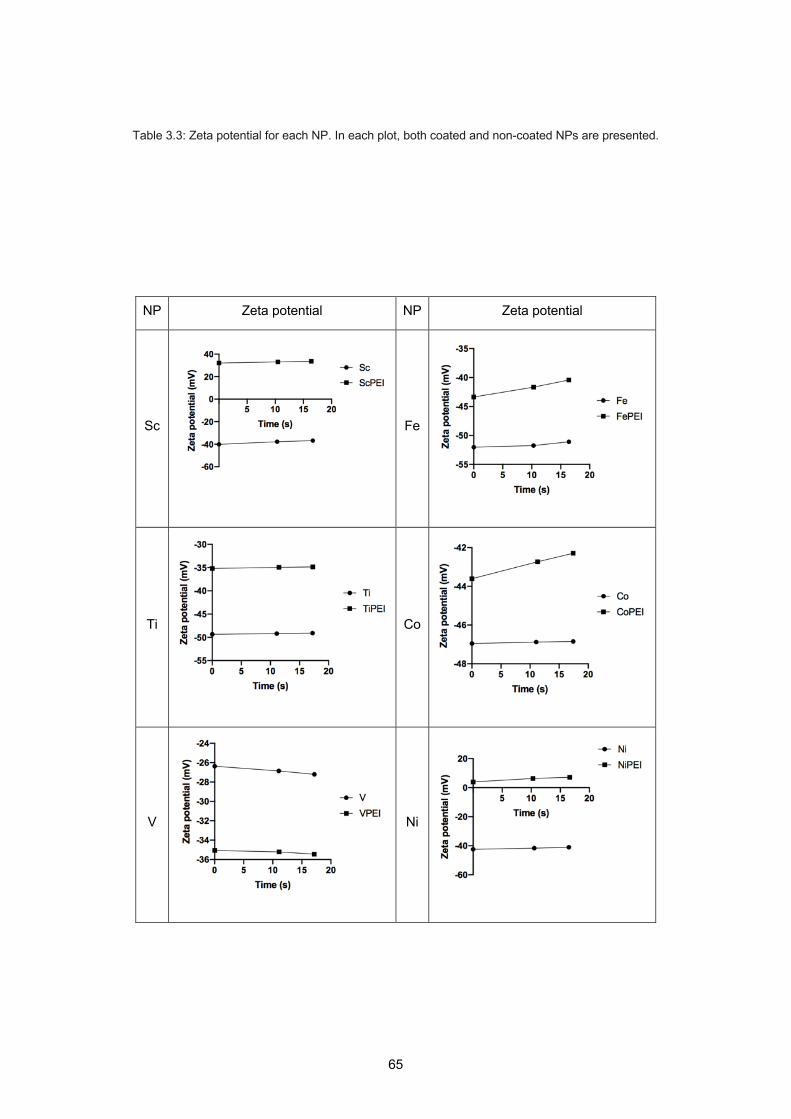
65
Table 3.3: Zeta potential for each NP. In each plot, both coated and non-coated NPs are presented.
NP Zeta potential NP Zeta potential
Sc
Fe
Ti
Co
V
Ni

66
Cr
Cu
Mn
Zn
Hf

67
Table 3.4: Mean diameters from TEM analysis and mean zeta potential data for each type of NP.
Catalytic ability
Metal oxide NPs are known for their catalytic ability (Ray & Pal, 2017). To probe the catalytic ability of
the different types of NP studied in this thesis, the rate at which each NP decomposed H2O2 and
generated O2 was evaluated (Figure 3.2).

68
Figure 3.2: Catalytic activity of non-coated (A) and PEI-coated (B) NPs.
The results of the catalytic activity assays for non-coated NPs showed that manganese NP was the
most efficient metal oxide NP, capable of decomposing H2O2 into O2. PEI-coated NPs showed similar
results to the non-coated NPs.
ROS production
3.3.3.1 ROS probe fluorescence quenching
An important phenomenon that needs to be considered when studying NPs interaction with radiation is
the quenching of the fluorescent chemical compound in solution. When the direct measure used is the
amount of ROS produced in the sample after irradiation, the presence of NP at the time of the
measurement may compromise the real results. This is because NPs surface may interact with the
produced ROS before interaction with the probe.
To quantify the effect that each type of NP has on the different probes used to measure the different
ROS, quenching results were obtained. The interaction of each type of NP with the fluorescent versions
of the probes used, namely 7OH-3-CCA and rhodamine 123, was measured. All results were
normalized to the control. In Table 3.5, the quenching results related to 7OH-3-CCA are presented.
Table 3.5: 7OH-3-CCA quenching results for each NP. Each plot compares the coated and non-coated pair of each NP.
A B

69
NP CCA quenching NP CCA quenching
Sc
Fe
Ti
Co
V
Ni
Cr
Cu
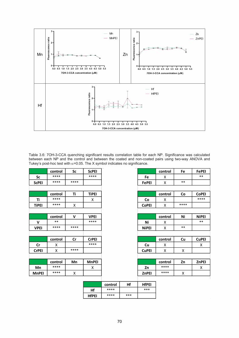
70
Mn
Zn
Hf
Table 3.6: 7OH-3-CCA quenching significant results correlation table for each NP. Significance was calculated between each NP and the control and between the coated and non-coated pairs using two-way ANOVA and Tukey’s post-hoc test with a=0.05. The X symbol indicates no significance.
control Sc ScPEI control Fe FePEISc **** **** Fe X **
ScPEI **** **** FePEI X **
control Ti TiPEI control Co CoPEITi **** X Co X ****
TiPEI **** X CoPEI X ****
control V VPEI control Ni NiPEIV ** **** Ni X **
VPEI **** **** NiPEI X **
control Cr CrPEI control Cu CuPEICr X **** Cu X X
CrPEI X **** CuPEI X X
control Mn MnPEI control Zn ZnPEIMn **** X Zn **** X
MnPEI **** X ZnPEI **** X
control Hf HfPEIHf **** ***
HfPEI **** ***
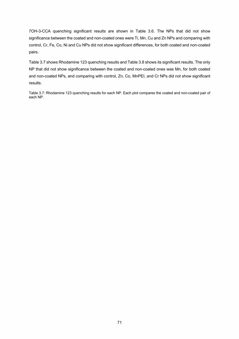
71
7OH-3-CCA quenching significant results are shown in Table 3.6. The NPs that did not show
significance between the coated and non-coated ones were Ti, Mn, Cu and Zn NPs and comparing with
control, Cr, Fe, Co, Ni and Cu NPs did not show significant differences, for both coated and non-coated
pairs.
Table 3.7 shows Rhodamine 123 quenching results and Table 3.8 shows its significant results. The only
NP that did not show significance between the coated and non-coated ones was Mn, for both coated
and non-coated NPs, and comparing with control, Zn, Co, MnPEI, and Cr NPs did not show significant results.
Table 3.7: Rhodamine 123 quenching results for each NP. Each plot compares the coated and non-coated pair of each NP.
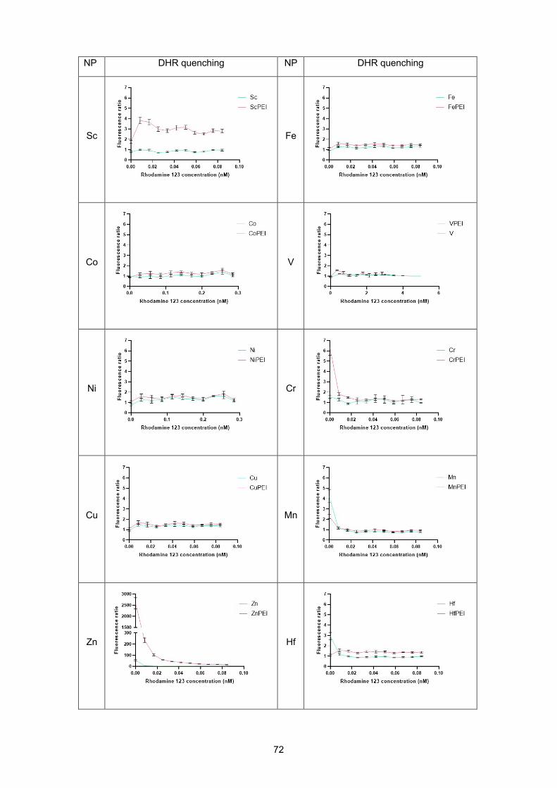
72
NP DHR quenching NP DHR quenching
Sc
Fe
Co
V
Ni
Cr
Cu
Mn
Zn
Hf
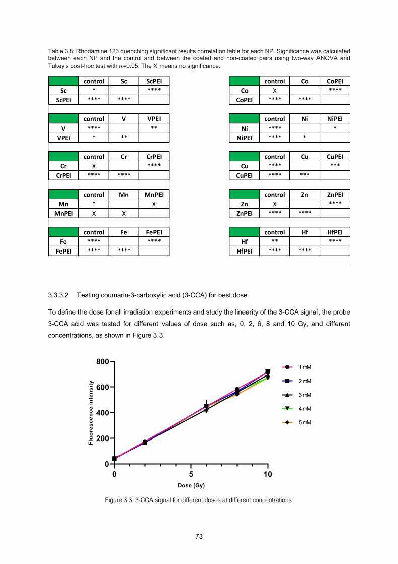
73
Table 3.8: Rhodamine 123 quenching significant results correlation table for each NP. Significance was calculated between each NP and the control and between the coated and non-coated pairs using two-way ANOVA and Tukey’s post-hoc test with a=0.05. The X means no significance.
3.3.3.2 Testing coumarin-3-carboxylic acid (3-CCA) for best dose
To define the dose for all irradiation experiments and study the linearity of the 3-CCA signal, the probe
3-CCA acid was tested for different values of dose such as, 0, 2, 6, 8 and 10 Gy, and different
concentrations, as shown in Figure 3.3.
Figure 3.3: 3-CCA signal for different doses at different concentrations.
control Sc ScPEI control Co CoPEISc * **** Co X ****
ScPEI **** **** CoPEI **** ****
control V VPEI control Ni NiPEIV **** ** Ni **** *
VPEI * ** NiPEI **** *
control Cr CrPEI control Cu CuPEICr X **** Cu **** ***
CrPEI **** **** CuPEI **** ***
control Mn MnPEI control Zn ZnPEIMn * X Zn X ****
MnPEI X X ZnPEI **** ****
control Fe FePEI control Hf HfPEIFe **** **** Hf ** ****
FePEI **** **** HfPEI **** ****
0 5 100
200
400
600
800
Dose (Gy)
Fluo
resc
ence
inte
nsity
1 mM
2 mM
3 mM
4 mM
5 mM
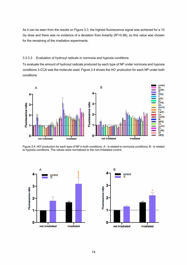
74
As it can be seen from the results on Figure 3.3, the highest fluorescence signal was achieved for a 10
Gy dose and there was no evidence of a deviation from linearity (R2=0.98), so this value was chosen
for the remaining of the irradiation experiments.
3.3.3.3 Evaluation of hydroxyl radicals in normoxia and hypoxia conditions
To evaluate the amount of hydroxyl radicals produced by each type of NP under normoxia and hypoxia
conditions 3-CCA was the molecule used. Figure 3.4 shows the HO. production for each NP under both
conditions.
Figure 3.4: HO• production for each type of NP in both conditions. A - is related to normoxia conditions; B - is related to hypoxia conditions. The values were normalized to the non-irradiated control.
A B
B A

75
Figure 3.5: Significance results for HO• using Prism Graphpad with two-way ANOVA and Tukey’s post-hoc test with a=0.05, for hydroxyl radical production in normoxia (A) and hypoxia (B) conditions. The significance results showed are between the NP and the respective control (irradiated control vs irradiated Ti NP).
Results in Figure 3.5 do not consider the not irradiated data, so they do not show the actual HO.
production. On the other hand, these results are shown to outline the behaviour of certain NPs without
radiation. As it can be seen, Ti NPs produce hydroxyl radicals without the presence of radiation.
Representation of the ratio between the irradiated and non-irradiated 3-CCA data is shown in Figure
3.6.
Figure 3.6: HO• production for each type of NP in both conditions after taking the ratio between the irradiated and non-irradiated values. A - is related to normoxia conditions; B - related to hypoxia conditions. Significance was calculated between each NP and the control using one-way ANOVA and Dunnett’s post-hoc test with a=0.05.
From the ratio results one can see that Zn NPs showed high significance in both conditions, normoxia
and hypoxia. Also, V NPs showed significance in hypoxia conditions although not as high as Zn ones.
B A

76
3.3.3.4 Evaluation of hydrogen peroxide in normoxia conditions
To evaluate the amount of hydrogen peroxide produced by each type of NP under normoxia condition
DHR was the molecule used. Results are presented in Figures 3.7 and 3.8.
Figure 3.7: H2O2 production for each type of NP in normoxia conditions. A and B are from the same experiment (same control), but due to scale effects NPs were divided into two different plots. The values were normalized to the non-irradiated control.
Figure 3.8: Significance results for H2O2 using Prism Graphpad with two-way ANOVA and Tukey’s post-hoc test with a=0.05, for hydrogen peroxide production for each type of NP in normoxia conditions. The significance results showed are between the NP and the respective control (not irradiated control vs not irradiated Mn; irradiated control vs irradiated VPEI).
A B
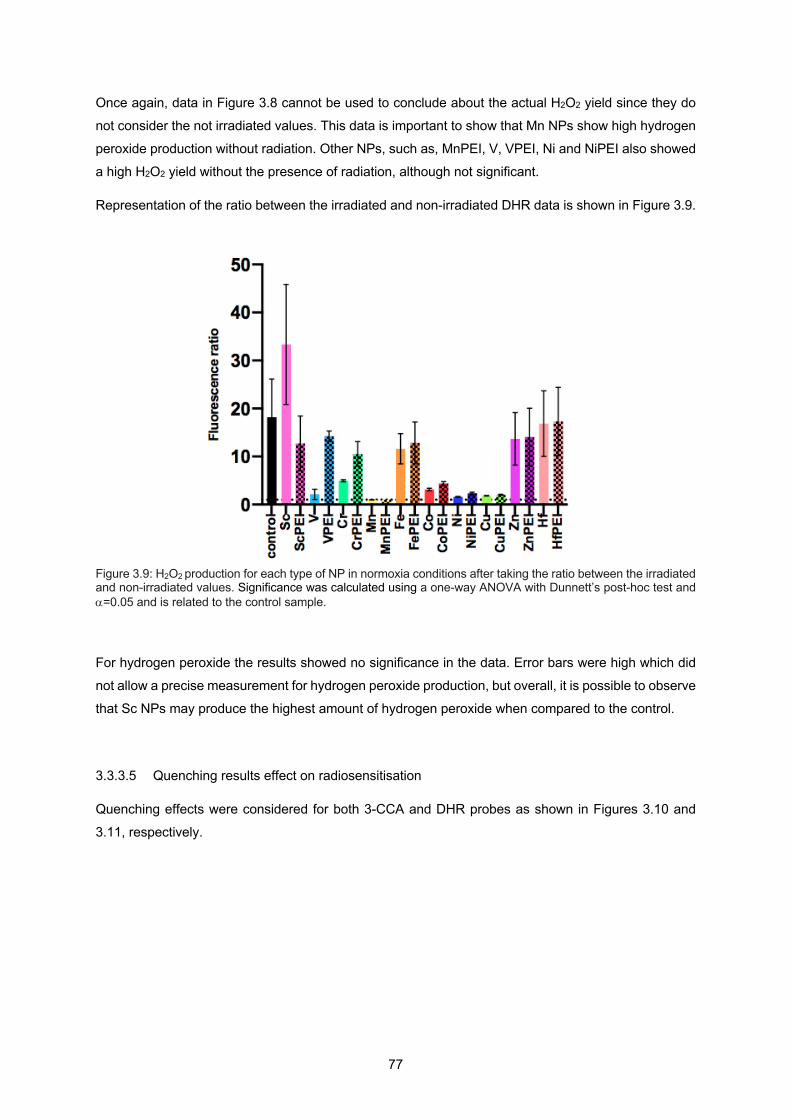
77
Once again, data in Figure 3.8 cannot be used to conclude about the actual H2O2 yield since they do
not consider the not irradiated values. This data is important to show that Mn NPs show high hydrogen
peroxide production without radiation. Other NPs, such as, MnPEI, V, VPEI, Ni and NiPEI also showed
a high H2O2 yield without the presence of radiation, although not significant.
Representation of the ratio between the irradiated and non-irradiated DHR data is shown in Figure 3.9.
Figure 3.9: H2O2 production for each type of NP in normoxia conditions after taking the ratio between the irradiated and non-irradiated values. Significance was calculated using a one-way ANOVA with Dunnett’s post-hoc test and a=0.05 and is related to the control sample.
For hydrogen peroxide the results showed no significance in the data. Error bars were high which did
not allow a precise measurement for hydrogen peroxide production, but overall, it is possible to observe
that Sc NPs may produce the highest amount of hydrogen peroxide when compared to the control.
3.3.3.5 Quenching results effect on radiosensitisation
Quenching effects were considered for both 3-CCA and DHR probes as shown in Figures 3.10 and
3.11, respectively.

78
Figure 3.10: HO• production for each type of NP in both conditions considering quenching results. A – normoxia; B – hypoxia conditions. Non-irradiated data was subtracted to the irradiated data, normalized to the control, and divided by the averaged of the quenching results for each NP. The significant results between each NP and the control were calculated using a one-way ANOVA with Dunnett’s post-hoc test and a=0.05.
For 3-CCA probe, the inclusion of the quenching measurements leads to a different interpretation of the
results. The hydroxyl radical production, as shown in Figure 3.10, the results showed significance for
vanadium NPs, meaning that these NPs under both conditions produce the highest amount of hydroxyl
radical when irradiated with 6 MeV X-rays. For hypoxia conditions, the coated version of vanadium NPs
also showed significant results. Furthermore, although without any significance, it is possible to see that
Cr, Mn and Zn NPs may produce a fluorescence signal that is 1.5 times higher than the amount produced without the presence of these NPs.
For hydrogen peroxide production, under normoxia conditions, after taking into account the quenching results the results remain very similar. Figure 3.11 shows that VPEI NPs lead to a significant increase
on the fluorescence signal.
B A
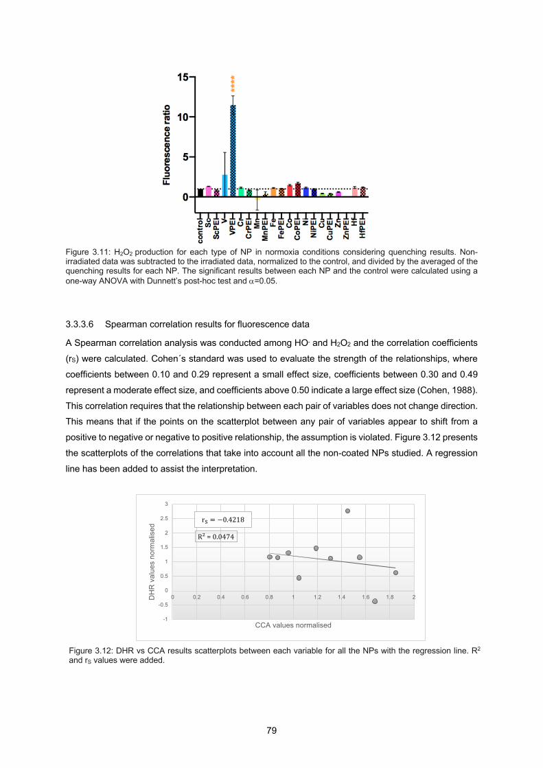
79
Figure 3.11: H2O2 production for each type of NP in normoxia conditions considering quenching results. Non-irradiated data was subtracted to the irradiated data, normalized to the control, and divided by the averaged of the quenching results for each NP. The significant results between each NP and the control were calculated using a one-way ANOVA with Dunnett’s post-hoc test and a=0.05.
3.3.3.6 Spearman correlation results for fluorescence data
A Spearman correlation analysis was conducted among HO. and H2O2 and the correlation coefficients
(rS) were calculated. Cohen´s standard was used to evaluate the strength of the relationships, where
coefficients between 0.10 and 0.29 represent a small effect size, coefficients between 0.30 and 0.49
represent a moderate effect size, and coefficients above 0.50 indicate a large effect size (Cohen, 1988).
This correlation requires that the relationship between each pair of variables does not change direction. This means that if the points on the scatterplot between any pair of variables appear to shift from a
positive to negative or negative to positive relationship, the assumption is violated. Figure 3.12 presents
the scatterplots of the correlations that take into account all the non-coated NPs studied. A regression
line has been added to assist the interpretation.
Figure 3.12: DHR vs CCA results scatterplots between each variable for all the NPs with the regression line. R2 and rS values were added.
R²=0.0474
-1
-0.5
0
0.5
1
1.5
2
2.5
3
0 0.2 0.4 0.6 0.8 1 1.2 1.4 1.6 1.8 2DH
R v
alue
s no
rmal
ised
CCA values normalised
r" = −0.4218
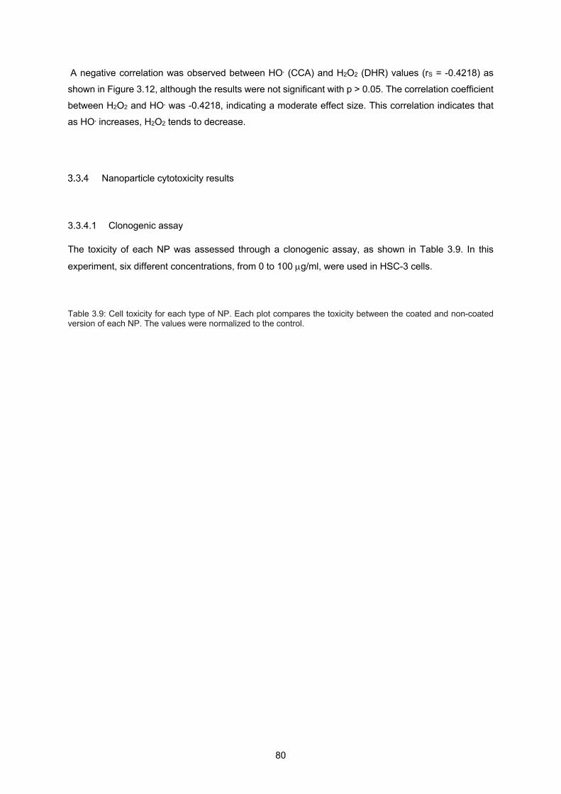
80
A negative correlation was observed between HO. (CCA) and H2O2 (DHR) values (rS = -0.4218) as
shown in Figure 3.12, although the results were not significant with p > 0.05. The correlation coefficient
between H2O2 and HO. was -0.4218, indicating a moderate effect size. This correlation indicates that
as HO. increases, H2O2 tends to decrease.
Nanoparticle cytotoxicity results
3.3.4.1 Clonogenic assay
The toxicity of each NP was assessed through a clonogenic assay, as shown in Table 3.9. In this
experiment, six different concentrations, from 0 to 100 µg/ml, were used in HSC-3 cells.
Table 3.9: Cell toxicity for each type of NP. Each plot compares the toxicity between the coated and non-coated version of each NP. The values were normalized to the control.
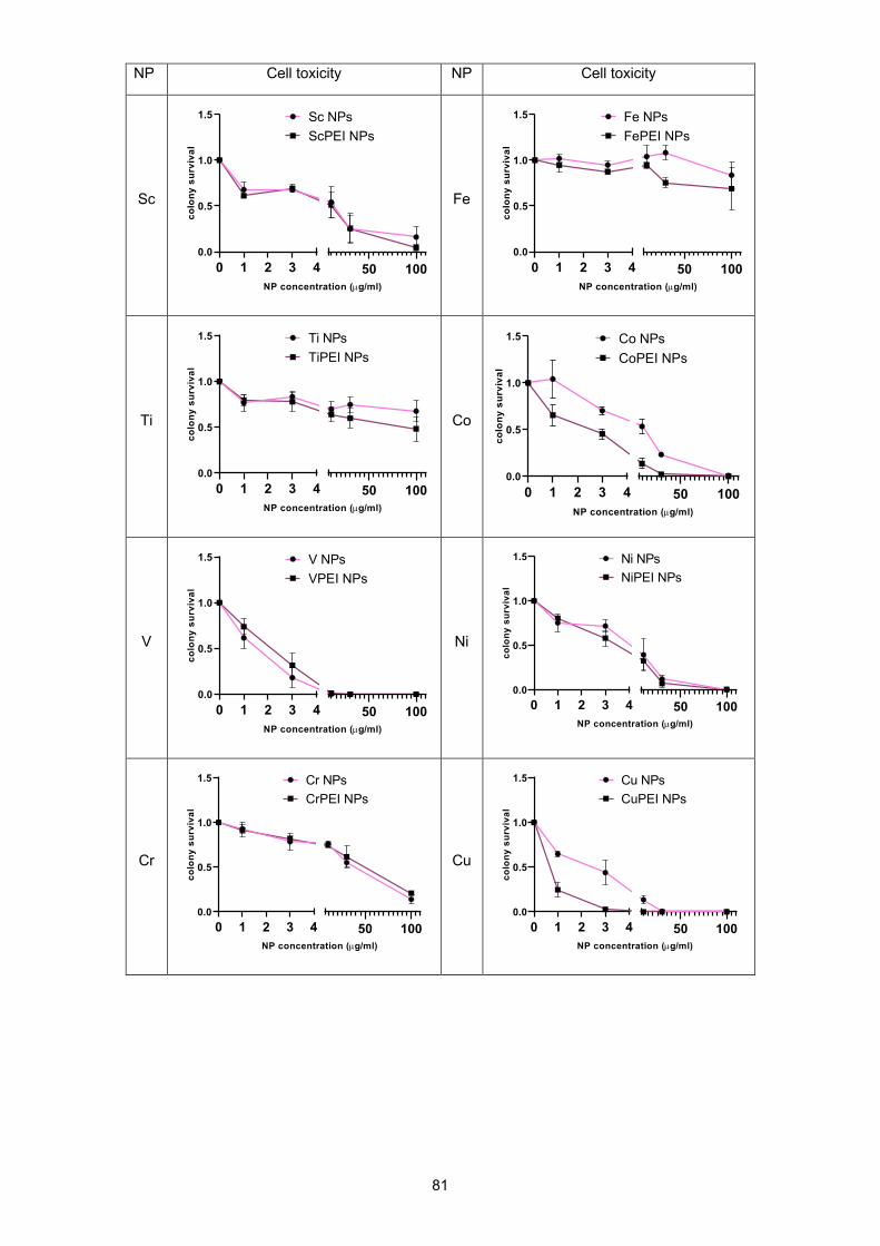
81
NP Cell toxicity NP Cell toxicity
Sc
Fe
Ti
Co
V
Ni
Cr
Cu
0 1 2 3 40.0
0.5
1.0
1.5
50 100NP concentration (µg/ml)
colo
ny s
urvi
val
Sc NPsScPEI NPs
0 1 2 3 40.0
0.5
1.0
1.5
50 100NP concentration (µg/ml)
colo
ny s
urvi
val
Fe NPsFePEI NPs
0 1 2 3 40.0
0.5
1.0
1.5
50 100NP concentration (µg/ml)
colo
ny s
urvi
val
Ti NPsTiPEI NPs
0 1 2 3 40.0
0.5
1.0
1.5
50 100NP concentration (µg/ml)
colo
ny s
urvi
val
V NPsVPEI NPs
0 1 2 3 40.0
0.5
1.0
1.5
50 100NP concentration (µg/ml)
colo
ny s
urvi
val
Ni NPsNiPEI NPs
0 1 2 3 40.0
0.5
1.0
1.5
50 100NP concentration (µg/ml)
colo
ny s
urvi
val
Cr NPsCrPEI NPs
0 1 2 3 40.0
0.5
1.0
1.5
50 100NP concentration (µg/ml)
colo
ny s
urvi
val
Cu NPsCuPEI NPs
0 1 2 3 40.0
0.5
1.0
1.5
50 100NP concentration (µg/ml)
colo
ny s
urvi
val
Co NPsCoPEI NPs
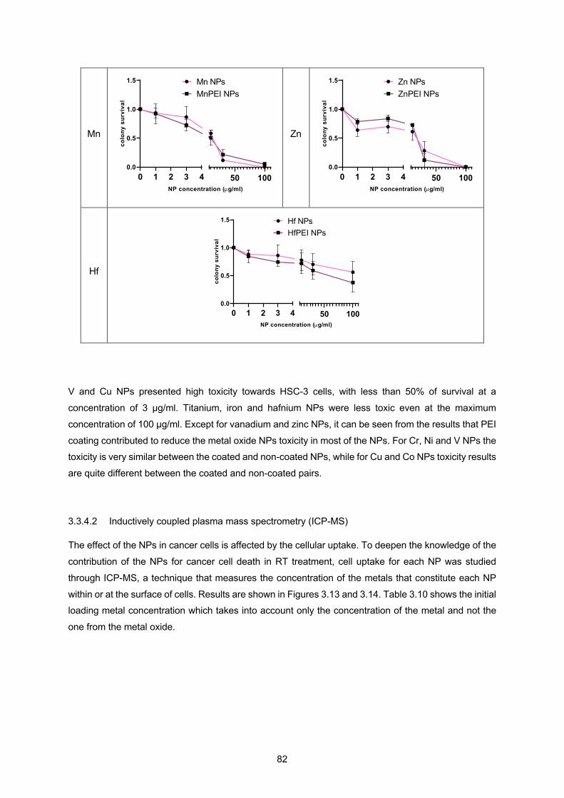
82
V and Cu NPs presented high toxicity towards HSC-3 cells, with less than 50% of survival at a
concentration of 3 µg/ml. Titanium, iron and hafnium NPs were less toxic even at the maximum
concentration of 100 µg/ml. Except for vanadium and zinc NPs, it can be seen from the results that PEI
coating contributed to reduce the metal oxide NPs toxicity in most of the NPs. For Cr, Ni and V NPs the toxicity is very similar between the coated and non-coated NPs, while for Cu and Co NPs toxicity results
are quite different between the coated and non-coated pairs.
3.3.4.2 Inductively coupled plasma mass spectrometry (ICP-MS)
The effect of the NPs in cancer cells is affected by the cellular uptake. To deepen the knowledge of the
contribution of the NPs for cancer cell death in RT treatment, cell uptake for each NP was studied
through ICP-MS, a technique that measures the concentration of the metals that constitute each NP
within or at the surface of cells. Results are shown in Figures 3.13 and 3.14. Table 3.10 shows the initial loading metal concentration which takes into account only the concentration of the metal and not the
one from the metal oxide.
Mn
Zn
Hf
0 1 2 3 40.0
0.5
1.0
1.5
50 100NP concentration (µg/ml)
colo
ny s
urvi
val
Mn NPsMnPEI NPs
0 1 2 3 40.0
0.5
1.0
1.5
50 100NP concentration (µg/ml)
colo
ny s
urvi
val
Zn NPsZnPEI NPs
0 1 2 3 40.0
0.5
1.0
1.5
50 100NP concentration (µg/ml)
colo
ny s
urvi
val
Hf NPsHfPEI NPs

83
Figure 3.13: ICP-MS uptake results for each type of NP.
From the uptake results shown in Figure 3.13, it is possible to conclude that Mn and MnPEI NPs are
the ones with the highest uptake. Fe and FePEI NPs have the highest loading concentration, as shown
in Table 3.10, although their uptake was very similar to NPs such as Co, CoPEI, Ni, NiPEI, Cu, CuPEI,
Table 3.10: Loading metal concentration for each type of NP.

84
Cr, Ti, TiPEI, Hf, HfPEI, Zn and ZnPEI. All these NPs had similar uptake values, independently of
loading concentrations. Cu NPs, both coated and non-coated, are worth mentioning, since they have
one of the lowest loading metal concentration, but a quite high uptake compared to the other types of
NPs.
Figure 3.14: ICP-MS metal loading intracellular ratio results for each type of NP. A and B are from the same experiment (same control), but due to scale effects NPs were divided into two different plots.
The ICP-MS plot in Figure 3.14 represents the metal loading intracellular ratio for each NP. The higher
this ratio, higher the number of NPs left outside the cell compared to the number inside of the cell. As
shown in Figure 3.14, CrPEI NP is the one that exhibits the highest ratio. It can be concluded that this
type of PEI-coated NP does not diffuse across the cell membrane. Other types of NPs that had a low
intracellular intake and can be outlined are Cr, Hf, PEI-coated Hf, Fe, and PEI-coated Fe, although with
a much lower value when compared to CrPEI. On the other hand, NPs such as Mn, MnPEI, Cu and CuPEI showed a low ratio, meaning that these NPs have a high uptake in HSC-3 cells.
3.3.4.3 Irradiation of cancer cells in normoxia and hypoxia
To understand how these metal oxide NPs would improve the RT treatment, HSC-3 cells were used.
The interaction between the NPs, cells and radiation was studied using a clonogenic assay where the
number of viable cell colonies was counted. The cell irradiation results for both normoxia and hypoxia
conditions are shown in Figure 3.15.
A B
ScSc
PEI Ti
TiPE
I VVP
EI Cr
Mn
MnP
EI FeFe
PEI
CoC
oPEI Ni
NiP
EI CuC
uPEI Zn
ZnPE
IH
fH
fPEI
0.0
0.5
1.0
1.5
100200300400
Met
al lo
adin
g in
trac
ellu
lar
ratio
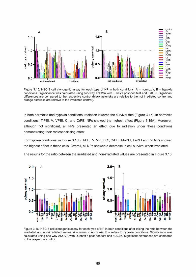
85
Figure 3.15: HSC-3 cell clonogenic assay for each type of NP in both conditions. A – normoxia; B – hypoxia conditions. Significance was calculated using two-way ANOVA with Tukey’s post-hoc test and a=0.05. Significant differences are compared to the respective control (black asterisks are relative to the not irradiated control and orange asterisks are relative to the irradiated control).
In both normoxia and hypoxia conditions, radiation lowered the survival rate (Figure 3.15). In normoxia
conditions, TiPEI, V, VPEI, Cr and CrPEI NPs showed the highest effect (Figure 3.15A). Moreover,
although not significant, all NPs presented an effect due to radiation under these conditions
demonstrating their radiosensitising effect.
For hypoxia conditions, in Figure 3.15B, TiPEI, V, VPEI, Cr, CrPEI, MnPEI, FePEI and Zn NPs showed
the highest effect in these cells. Overall, all NPs showed a decrease in cell survival when irradiated.
The results for the ratio between the irradiated and non-irradiated values are presented in Figure 3.16.
Figure 3.16: HSC-3 cell clonogenic assay for each type of NP in both conditions after taking the ratio between the irradiated and non-irradiated values. A – refers to normoxia; B – refers to hypoxia conditions. Significance was calculated using one-way ANOVA with Dunnett’s post-hoc test and a=0.05. Significant differences are compared to the respective control.
B A
B A
*
**

86
Only normoxia conditions showed significant results for V and VPEI NPs. Also, in these conditions and
without significance, Cr and CrPEI showed a low number of cell survival. In hypoxia conditions, results
did not showed significance although VPEI, Cr and Zn NPs had the lowest value of cell survival.
3.3.4.4 Spearman correlation results for clonogenic data
A Spearman correlation analysis was conducted among cell irradiation and ICP-MS data and the
correlation coefficients (rS) were calculated. Once again, Cohen´s standard was used to evaluate the
strength of the relationships. Figure 3.17 presents the scatterplots of the correlations that take into
account all the non-coated and coated NPs studied for both normoxia and hypoxia conditions. A
regression line has been added to assist the interpretation.
A positive correlation was observed for both conditions between cell irradiation and ICP-MS values, as
shown in Figure 3.17, although the results were not significant with p > 0.05. The correlation coefficients between cell irradiation and ICP-MS were 0.3021 and 0.2575 for normoxia and hypoxia conditions,
respectively. These results show a small effect size in normoxia conditions and a moderate effect size
in the case of hypoxia conditions. This correlation indicates that as the uptake increases, cell survival
tends to increase.
Spearman correlation results between fluorescence and clonogenic data
A Spearman correlation analysis was conducted among fluorescence and clonogenic data and the
correlation coefficients (rS) were calculated. Cohen´s standard was used to evaluate the strength of the
relationships. Clonogenic data was corrected for the uptake values obtained from the ICP-MS results
before the analysis.
Figures 3.18 and 3.19 present the scatterplots of the correlations that take into account all the non-
coated and coated NPs studied. A regression line has been added to assist the interpretation.
R²=0.0032
0
0.2
0.4
0.6
0.8
1
1.2
0 5 10 15 20 25 30 35Cel
l nor
mox
ia n
orm
alis
ed v
alue
s
ICP-MS uptake values (ppb)
r" = 0.3021
Figure 3.17: Cell irradiation vs ICP-MS results scatterplots between each variable for all the NPs with the regression line. R2, and rS values added. A- for normoxia; B – for hypoxia.
R²=0.0541
0
0.2
0.4
0.6
0.8
1
1.2
1.4
1.6
1.8
0 5 10 15 20 25 30 35
Cel
l hyp
oxia
nor
mal
ised
val
ues
ICP-MS uptake values (ppb)
r" = 0.2575
A B
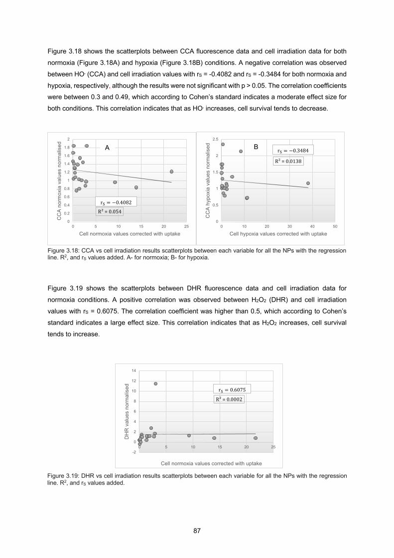
87
Figure 3.18 shows the scatterplots between CCA fluorescence data and cell irradiation data for both
normoxia (Figure 3.18A) and hypoxia (Figure 3.18B) conditions. A negative correlation was observed
between HO. (CCA) and cell irradiation values with rS = -0.4082 and rS = -0.3484 for both normoxia and
hypoxia, respectively, although the results were not significant with p > 0.05. The correlation coefficients were between 0.3 and 0.49, which according to Cohen’s standard indicates a moderate effect size for
both conditions. This correlation indicates that as HO. increases, cell survival tends to decrease.
Figure 3.19 shows the scatterplots between DHR fluorescence data and cell irradiation data for
normoxia conditions. A positive correlation was observed between H2O2 (DHR) and cell irradiation
values with rS = 0.6075. The correlation coefficient was higher than 0.5, which according to Cohen’s
standard indicates a large effect size. This correlation indicates that as H2O2 increases, cell survival
tends to increase.
Figure 3.18: CCA vs cell irradiation results scatterplots between each variable for all the NPs with the regression line. R2, and rS values added. A- for normoxia; B- for hypoxia.
Figure 3.19: DHR vs cell irradiation results scatterplots between each variable for all the NPs with the regression line. R2, and rS values added.
R²=0.054
0
0.2
0.4
0.6
0.8
1
1.2
1.4
1.6
1.8
2
0 5 10 15 20 25
CC
A no
rmox
ia v
alue
s no
rmal
ised
Cell normoxia values corrected with uptake
r" = −0.4082
R²=0.0138
0
0.5
1
1.5
2
2.5
0 10 20 30 40 50
CC
A hy
poxi
a va
lues
nor
mal
ised
Cell hypoxia values corrected with uptake
r" = −0.3484
R²=0.0002
-2
0
2
4
6
8
10
12
14
0 5 10 15 20 25
DH
R v
alue
s no
rmal
ised
Cell normoxia values corrected with uptake
r" = 0.6075
A B
88
3.4 Discussion
In this part of the thesis different types of NPs of metal oxides that are known for their catalytic ability, such as Mn and Cu (Haruta et al., 1987) or Fe known for its surface-catalysed Haber-Weiss cycle and
Fenton reactions (Klein et al., 2012), were studied aiming the understanding of their interaction with
cancer cells.
It is known from previous studies (Alkilany & Murphy, 2010) that coating with an organic molecule may
sometimes help in their uptake by cells. This reason together with the low molecular weight of the PEI
molecule, to decrease the interference with the radiosensitisation ability of the NPs, made this molecule
the coating of choice for the NPs. To assess the presence of the PEI molecule at the NP surface zeta
potential technique was used and the results showed a change in the charge between the coated NPs
and their respective non-coated pair. Furthermore, the effect of PEI coating on the radiosensitising
capacity of NPs was evaluated and the results showed that the coating overall did not had a great impact on this property. These conclusions are based on the fact that PEI molecule has bound to all
the NPs equally, but on the basis of TEM and zeta potential results we are not confident that the coating
happened equally for all NPs. Based on the ICP-MS results from Figures 3.12 and 3.13, one can
conclude that the PEI coating did not improve cellular uptake.
Experiments were performed to assess the intrinsic ROS production and cellular radiosensitising ability
of each NP and NP-PEI. These experiments are important to understand how any improvements in
intrinsic ROS production are translated to the cancer cell environment.
In Table 3.11 a summary of the results regarding each NP.

89
Table 3.11: Summary of the results for each type of NP, where HO• and H2O2 production data include the quenching data. Highlighted in red boxes are the NPs with the most promising results in relation to all the parameters studied.
For the ROS evaluation experiment, both hydroxyl radicals and hydrogen peroxide were studied.
Results showed that the NP with the highest hydroxyl radical production is the vanadium one. Also,
although without significance, results suggested that the metal oxide NPs that might achieve the highest
HO• production under normoxia conditions are VPEI, Cr, Mn and Zn. On the other hand, with significant
results, V and VPEI are the NPs that produce more HO• under hypoxia conditions. NPs such as Cr, Mn
and Zn also showed a high HO• production but not significant. Furthermore, regarding hydrogen
peroxide, the highest H2O2 yield under normoxia conditions was PEI-coated vanadium NP.
To measure NP radiosensitising ability within cancer cells and radiation two experiments were
performed, cytotoxicity and ICP-MS. The former is related to the harm these NPs can bring to the cancer
cells (non-irradiated) and the latest is related to the uptake of each NP, which can both compromise the
results when exposed to radiation. For example, if a particular NP has the capability of increasing HO• production when irradiated, but its uptake is very low, the hydroxyl radicals produced will not have a
high impact in cancer cell death as expected, since they are not being produced inside the cell.
Furthermore, toxicity is an important measurement for understanding if cancer cells are viable due to the combined effect of the NP with the radiation or because of the NP only. Toxicity results showed that
vanadium and copper are the metallic-oxides NPs that present more toxicity towards HSC-3 cells and
ICP-MS results demonstrated a very low uptake towards PEI-coated Cr NPs. NPs such as Cr, Hf, PEI-
90
coated Hf, Fe, and PEI-coated Fe also demonstrated a compromised uptake, although much less
significant than CrPEI.
Table 3.12 summarises the toxicity results found in this thesis and the results from previous toxicity
studies.
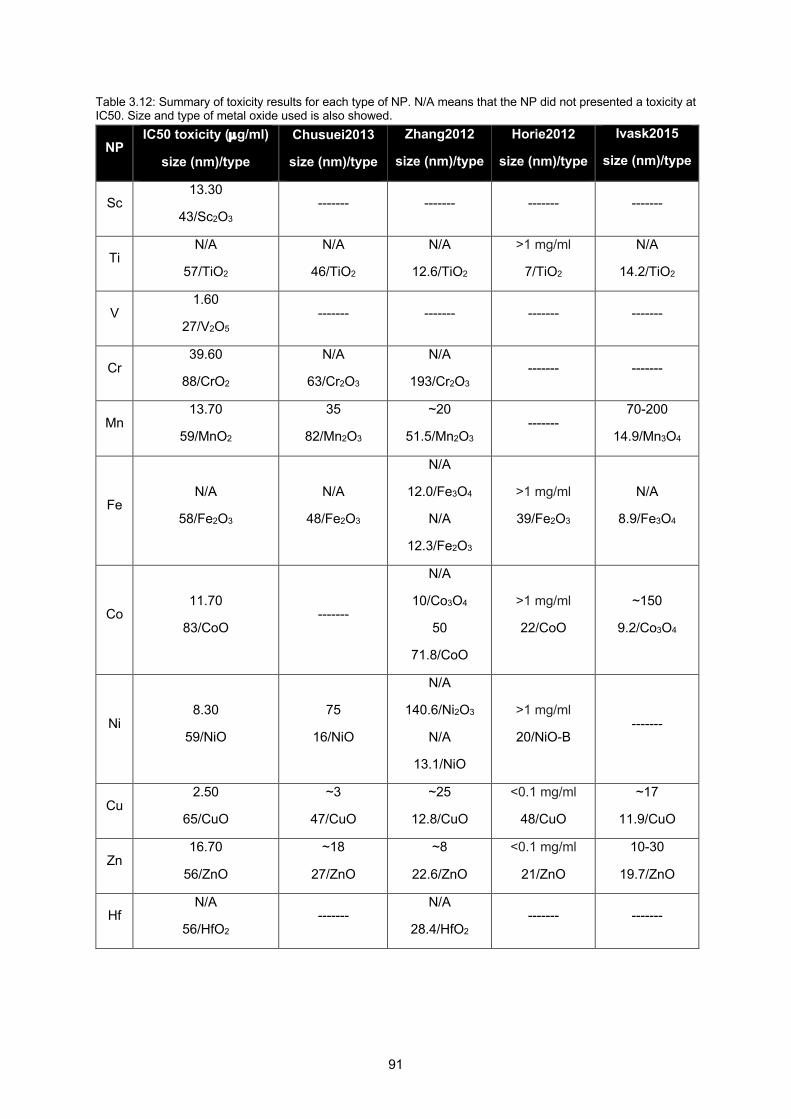
91
Table 3.12: Summary of toxicity results for each type of NP. N/A means that the NP did not presented a toxicity at IC50. Size and type of metal oxide used is also showed.
NP IC50 toxicity (µg/ml)
size (nm)/type
Chusuei2013
size (nm)/type
Zhang2012
size (nm)/type
Horie2012
size (nm)/type
Ivask2015
size (nm)/type
Sc 13.30
43/Sc2O3 ------- ------- ------- -------
Ti N/A
57/TiO2
N/A
46/TiO2
N/A
12.6/TiO2
>1 mg/ml
7/TiO2
N/A
14.2/TiO2
V 1.60
27/V2O5 ------- ------- ------- -------
Cr 39.60
88/CrO2
N/A
63/Cr2O3
N/A
193/Cr2O3 ------- -------
Mn 13.70
59/MnO2
35
82/Mn2O3
~20
51.5/Mn2O3 -------
70-200
14.9/Mn3O4
Fe N/A
58/Fe2O3
N/A
48/Fe2O3
N/A
12.0/Fe3O4
N/A
12.3/Fe2O3
>1 mg/ml
39/Fe2O3
N/A
8.9/Fe3O4
Co 11.70
83/CoO -------
N/A
10/Co3O4
50
71.8/CoO
>1 mg/ml
22/CoO
~150
9.2/Co3O4
Ni 8.30
59/NiO
75
16/NiO
N/A
140.6/Ni2O3
N/A
13.1/NiO
>1 mg/ml
20/NiO-B -------
Cu 2.50
65/CuO
~3
47/CuO
~25
12.8/CuO
<0.1 mg/ml
48/CuO
~17
11.9/CuO
Zn 16.70
56/ZnO
~18
27/ZnO
~8
22.6/ZnO
<0.1 mg/ml
21/ZnO
10-30
19.7/ZnO
Hf N/A
56/HfO2 -------
N/A
28.4/HfO2 ------- -------
92
Chusuei et al focused on the fourth period metal oxide NPs such as TiO2, Cr2O3, Mn2O3, Fe2O3, NiO,
CuO and ZnO and found that the cytotoxicity of fourth period metal oxide NPs increased with the atomic
number of the transition metal oxide and it was not cell-type specific (Chusuei et al., 2013). They
proposed that toxicity falls into three categories: the first, where Ti, Cr and Fe have zero to minimal toxicity; the second, where Mn and Ni have around 40% cell viability; and the third, where Cu and Zn
present less than 20% cell viability. They used both human bronchial epithelium non-cancerous cell line
(BEAS-2B) and A549 and the results were the same for both cell lines. In the results of this thesis, the
trend with the atomic number was not seen although NPs such as Cu, Zn, Ti and Fe had similar toxicity
results. NPs such as Cr and Mn showed differences in toxicity and it might have been due to the type
of metal oxide used, CrO2 vs Cr2O3 and MnO2 vs Mn2O3. For Ni NPs although the same type of metal
oxide was used in both studies the difference in toxicity might have been due to the NP size used, 59
vs 16 nm.
Furthermore, another research on 24 types of metal oxide NPs such as CO3O4, Cr2O3, Ni2O3, CuO,
Mn2O3, CoO, ZnO, Al2O3, CeO3, Fe2O3, Fe3O4, Gd2O3, HfO2, In2O3, NiO, SiO3, SnO2, TiO2, WO3, Y2O3,
Yb2O3, ZrO2, La2O3, Sb2O3, using single-parameter assays in BEAS-2B as well as mouse myeloid (RAW
264.7) cell lines showed that for an exposure period of 24 h and a dose range between 400 ng/ml to
200 µg/ml seven NPs significantly decreased cell viability (Zhang et al, 2012). These seven NPs
included CuO, Mn2O3, CoO, and ZnO with a marked declined in cell viability while CO3O4, Cr2O3, and
Ni2O3 had lesser but still significant effects. The rest of the metals analysed had no significant effects.
NP sizes were in the range 10-100 nm except for Cr2O3 and Ni2O3 that were much larger, 193 nm and
140 nm, respectively. For the cell line used in this thesis our results regarding Ti, Mn, Fe and Hf NPs
were similar to the ones from Zhang et al.. In the case of Cr NPs, the difference on the toxicity results
might be due to the difference in the metal oxide used, while for Ni, Cu and Zn NPs the difference in
toxicity can be explained by the difference in size. The result regarding Co NPs cannot be explained due to differences in size or metal oxide used since they are very similar, so the type of cell line used
also play an important role on the toxicity results obtained.
Horie et al. (2012) concluded that the most important cytotoxic factor of metal oxide NPs was metal ion
release. This group used human keratinocyte cell line (HaCaT) and A549, and 24 kinds of industrial
metal oxide NPs such as ZnO, CuO, NiO, Sb2O3, CoO, MoO3, Y2O3, MgO, Gd2O3, SnO2, WO3, ZrO2,
Fe2O3, TiO2, CeO2, Al2O3, Bi2O3, La2O3, In2O3/SnO2 (ITO), and cobalt blue pigments with different sizes.
For the cell viability assay they used MTT assay with 24 h exposure to the NPs where ZnO, ZnO-SiO2 coated and CuO NPs induced severe cell death at concentrations of 0.1 and 1.0 mg/mL in both HaCaT
and A549 cells. Also, NiO green NPs induced severe cell death at concentration of 0.1 mg/mL. The cell
viabilities of NiO black, Sb2O3, CoO and MoO3 exposed cells were approximately 50% at concentration
of 1.0 mg/mL. The cell viabilities of Y2O3 and MgO exposed cells were approximately 60%. The other
types of metal oxides nanoparticles showed a very small effect on cell viability with a value of 80% or
more. Particularly, there was no significant decrease of cell viability in Fe2O3, TiO2, Al2O3 and cobalt
blue exposed cells. Moreover, ZnO, CuO, NiO, MgO, and WO3 showed a large amount of metal ion
release in the culture medium, and TiO2, SnO2, and CeO2 NPs showed strong protein adsorption ability.
93
In our results, ZnO, CuO and NiO NPs presented 100% cell death at 0.1 mg/mL and, Fe2O3 and TiO2
did not presented any significant decrease in cell viability, similar to the results found by this group. The
results in this thesis showed that CoO NPs decreased by 100% cell viability at 0.1 mg/mL, which does
not match the 50% value found by this group at a much higher concentration of 1.0 mg/mL. This difference might be due to differences in the cell line used, NP size (22 nm vs 83 nm), or type of assay
(MTT vs clonogenic).
For three different cell lines such as, A549, human epithelial colorectal adenocarcinoma cell line
(Caco2) and mouse fibroblast cell line (Balb/c 3T3), exposure for 24 h to suspensions of 3-100 µg/ml
metal oxide NPs was conducted and cellular viability was evaluated using Neutral Red Uptake (NRU) assay (Ivask et al., 2015). Toxicity results were very similar among the three cell lines, where Al2O3,
Fe3O4, MgO, SiO2, TiO2, WO3 were not toxic for concentrations below 100 µg/ml and for the remaining
NPs the IC50 values were 16.4 µg/ml for CuO, 22.4 µg/ml for ZnO, 57.3 µg/ml for Sb2O3, 132.3 µg/ml
for Mn3O4 and 129 µg/ml for Co3O4. In this study, toxicity from CuO, ZnO and Sb2O3 was attributed to
the release of metal ions by these NPs, and toxicity from Mn3O4 and Co3O4 attributed to the ROS-
inducing ability of these NPs (Ivask et al., 2015). From the catalytic ability results found in this thesis
(Figure 3.4), Mn and Co NPs showed the highest value in oxygen levels which is consistent with the
conclusion made by this group regarding the ROS-inducing ability of these NPs. Similarities with our
results include the toxicity found for Fe2O3, TiO2 and ZnO (22.4 µg/ml vs 16 µg/ml). IC50 values for our
NPs were 2.4 µg/ml for CuO, 14 µg/ml for MnO2 and 13 µg/ml for CoO. The latter results differ
significantly from the ones found by this research group and these might be due to differences regarding the cell line used, NP size (11.9 nm CuO vs 65 nm CuO; 14.9 nm Mn3O4 vs 59 nm MnO2; 9.2 nm Co3O4
vs 83 nm CoO) and type of assay (NRU vs clonogenic).
NP radiosensitisation ability within cancer cells is a phenomenon that is affected not only by the physical
characteristics of the NPs but also their interaction with cancer cells. For both normoxia and hypoxia
conditions experiments were conducted in this thesis where the interaction between NPs, cancer cells
and radiation were assessed. For HSC-3 cells, in normoxia conditions, the NPs that had the highest
effect in cell viability were V and VPEI with significant results. Cr and CrPEI NPs also showed an effect in cell death for normoxia conditions although not significant. For hypoxia conditions VPEI, Cr and Zn
NPs showed the highest effect, although these results are not significant due to the high error bars
present in the experiment.
Taking the results of ROS evaluation and comparing them with the HSC-3 cells clonogenic experiment
a possible conclusion for the low cell survival due to V, VPEI, Cr, CrPEI and Zn NPs is the fact that
these metal oxide NPs produce more hydroxyl radicals when exposed to radiation, that will in turn lead
to cancer cell death. The NPs that showed the highest production in ROS, both HO. and H2O2, were V
and VPEI and both present significant effects also in the clonogenic experiments. Interestingly, PEI-
coated vanadium NPs seem to contribute more for either hydroxyl radicals and cell death under hypoxia
conditions, as can be seen from the fact that VPEI NPs appear with a higher significance for hydroxyl
radical production under hypoxia conditions, but not under normoxia conditions. Even for cell death VPEI NP has a higher contribution than its non-coated counterpart, and a possible reason might be due
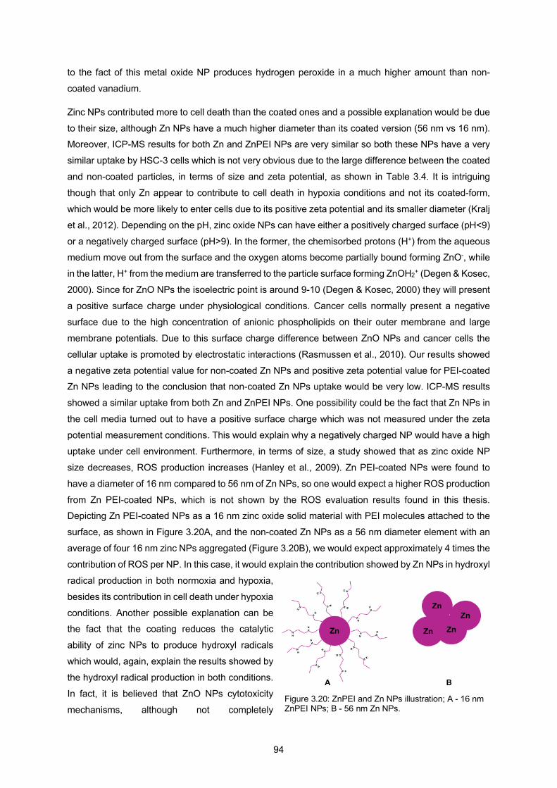
94
to the fact of this metal oxide NP produces hydrogen peroxide in a much higher amount than non-
coated vanadium.
Zinc NPs contributed more to cell death than the coated ones and a possible explanation would be due
to their size, although Zn NPs have a much higher diameter than its coated version (56 nm vs 16 nm). Moreover, ICP-MS results for both Zn and ZnPEI NPs are very similar so both these NPs have a very
similar uptake by HSC-3 cells which is not very obvious due to the large difference between the coated
and non-coated particles, in terms of size and zeta potential, as shown in Table 3.4. It is intriguing
though that only Zn appear to contribute to cell death in hypoxia conditions and not its coated-form,
which would be more likely to enter cells due to its positive zeta potential and its smaller diameter (Kralj
et al., 2012). Depending on the pH, zinc oxide NPs can have either a positively charged surface (pH<9)
or a negatively charged surface (pH>9). In the former, the chemisorbed protons (H+) from the aqueous medium move out from the surface and the oxygen atoms become partially bound forming ZnO-, while
in the latter, H+ from the medium are transferred to the particle surface forming ZnOH2+ (Degen & Kosec,
2000). Since for ZnO NPs the isoelectric point is around 9-10 (Degen & Kosec, 2000) they will present
a positive surface charge under physiological conditions. Cancer cells normally present a negative
surface due to the high concentration of anionic phospholipids on their outer membrane and large
membrane potentials. Due to this surface charge difference between ZnO NPs and cancer cells the
cellular uptake is promoted by electrostatic interactions (Rasmussen et al., 2010). Our results showed
a negative zeta potential value for non-coated Zn NPs and positive zeta potential value for PEI-coated Zn NPs leading to the conclusion that non-coated Zn NPs uptake would be very low. ICP-MS results
showed a similar uptake from both Zn and ZnPEI NPs. One possibility could be the fact that Zn NPs in
the cell media turned out to have a positive surface charge which was not measured under the zeta
potential measurement conditions. This would explain why a negatively charged NP would have a high
uptake under cell environment. Furthermore, in terms of size, a study showed that as zinc oxide NP
size decreases, ROS production increases (Hanley et al., 2009). Zn PEI-coated NPs were found to
have a diameter of 16 nm compared to 56 nm of Zn NPs, so one would expect a higher ROS production
from Zn PEI-coated NPs, which is not shown by the ROS evaluation results found in this thesis. Depicting Zn PEI-coated NPs as a 16 nm zinc oxide solid material with PEI molecules attached to the
surface, as shown in Figure 3.20A, and the non-coated Zn NPs as a 56 nm diameter element with an
average of four 16 nm zinc NPs aggregated (Figure 3.20B), we would expect approximately 4 times the
contribution of ROS per NP. In this case, it would explain the contribution showed by Zn NPs in hydroxyl
radical production in both normoxia and hypoxia,
besides its contribution in cell death under hypoxia
conditions. Another possible explanation can be the fact that the coating reduces the catalytic
ability of zinc NPs to produce hydroxyl radicals
which would, again, explain the results showed by
the hydroxyl radical production in both conditions.
In fact, it is believed that ZnO NPs cytotoxicity
mechanisms, although not completely Figure 3.20: ZnPEI and Zn NPs illustration; A - 16 nm ZnPEI NPs; B - 56 nm Zn NPs.
95
understood, are due to ROS generation (Rasmussen et al., 2010). ROS oxidative stress has been
described as a three-tier model, where Tier 1 is characterized by anti-oxidant defence, where there is
an increase in antioxidant enzymes in order to restore cellular redox homeostasis, Tier 2 is
characterized by an inflammation due to an increase in potent pro-inflammatory cytokines and chemokines and finally Tier 3 characterized by cytotoxicity which involves mitochondrial perturbation
that leads to cell death by apoptosis or necrosis (Nel et al., 2006). These three tiers have been observed
in ZnO NPs in immortalized phagocytic or bronchial epithelial cells leading to damage of lipids, proteins
and DNA, and death by either necrosis or apoptosis (Rasmussen et al., 2010).
In general, it would be expected, due to the important role of oxygen in ROS production, that under
hypoxia conditions ROS levels would be much lower than under normoxia conditions. The results on
this thesis do not follow this behaviour. Instead, several reports indicate that the production of ROS actually increases in response to hypoxia (Hermes-Lima et al., 2015). Furthermore, there might exist a
relation between the presence of the PEI coating and the production of hydrogen peroxide, which in
turn is very useful under hypoxia conditions, since even with VPEI NPs presenting a lower uptake than
V NPs they presented a higher toxicity to cancer cells. One of the major differences is the high hydrogen
peroxide production seen by VPEI NPs. PEI exhibits weak-base buffering properties since it can take
up H+ ions (Curtis et al., 2016). These ions might contribute to the generation of HO. through the reaction
of O22- with e- and 2H+ which will in turn generate H2O2 which then reacting with Cl- will form HOCl and
reacting with NO2- will form NO2. leaving HO. as the remaining product (Mitra et al., 2019). The presence of metal oxides will generate O2 through the Haber-Weiss reaction (O2- + H2O2 à O2 + OH- + OH.) and
also H2O2 will be decomposed through the catalytic ability of these metal oxide NPs contributing even
more for the production of hydroxyl radicals. Maybe the presence of O2 for the formation of ROS is
fundamental due to the reaction with 2H+ but in the presence of PEI the uptake of H+ might increase
radical formation through other mechanisms. Moreover, in general, PEI-coated NPs showed a lower
hydroxyl radical production in both conditions. PEI-coated may not favor the production of this radical
since it blocks the contact of the metal oxide surface with water molecules, not allowing the catalytic
reaction to take place.
Manganese NPs showed some contributions in hydroxyl radical production in both conditions although
not significant. Catalytic ability of manganese is very high as showed by the results in Figure 3.2. Some
studies showed the intracellular oxidative stress and apoptosis in alveolar epithelial cells (AEC) induced
by Mn3O4 NPs (VanWinkle et al., 2009; Frick et al., 2011). Oxidative stress induced by manganese is
due to the following reactions (Aust et al., 1985):
Mn2+ + O2 à Mn3+ + O2-
2 O2- + 2H+ à H2O2 + O2 (superoxide dismutase)
H2O2 products can then take two different routes (Frick et al., 2011):
2GSH + H2O2 à GSSG + 2H2O (catalyzed by glutathione peroxidase)
96
or
Mn2+ + H2O2 à Mn3+ + OH- + OH. (Fenton or Haber-Weiss reactions)
NP cytotoxicity results did not show any contribution from manganese NPs, only on hydroxyl radical
production.
Cr NPs showed contribution in both hydroxyl radicals and cytotoxicity experiments. Studies on cell lines
have shown their ability to produce ROS species inside cells. For example, a study on A549 and HaCaT
cells showed an increase in intracellular reactive oxygen species level decreasing cell viability after
exposure to 60 nm Cr2O3 NPs (Horie et al., 2011). Another study performed in A549 cells using 30 nm
Cr2O3 NPs also showed increase intracellular ROS level indicating that oxidative stress might be the primary mechanism for its toxicity (Senapati et al., 2015). These observations agree with our results
that showed the ROS generation capability of chromium NPs in producing hydroxyl radicals leading to
cell death. Moreover, Cr NPs were more toxic to cells than CrPEI ones, which is in accordance with the
ICP-MS results, that showed a much higher uptake from Cr NPs than for the coated ones.
Another potential toxicity mechanism can be through cell exterior damage even when the uptake is low
(VanWinkle et al., 2009). Transition metal ions can transfer electrons to O2 leading to H2O2 generation
in the extracellular compartment. The H2O2 produced can induce damage on the cell exterior. This might be the reason why although CrPEI NPs presented very low uptake and most of the NPs were outside
the cells it still presented relevant results for cell death under hypoxia conditions.
Overall, all the NPs that had a relevant effect on HSC-3 cells viability had a negative zeta potential,
which is not in accordance with the results showed by some studies, where NPs with positive zeta
potential have a better cell uptake (Kralj et al., 2012). A possible reason for this discrepancy is the fact
that zeta potential measurements were made in PBS, pH 7.4 and NPs under cell medium will present
a different surface charge due to the protein corona, as in the case of Zn NPs (Rasmussen et al., 2010).
In general, the NPs that showed the most significant results in ROS production and cell death are
vanadium, both the coated and non-coated.
Vanadium like other transition metals, e.g. Fe, Cu, Cr and Si, produces ROS through Haber-Weiss and
Fenton-type reactions (Manke et al., 2013). Vanadium NPs toxicity depends on the cell line used as
showed by a study made for 2 different sizes of vanadium NPs, 20 nm and 100 nm, using 5 different
cell lines (Ivanković et al., 2006). For 30 nm VO2 NPs interaction with A549 and BEAS-2B cells, a similar sensitivity to V NPs was found for both cell lines and it was also found that enhanced reactive oxygen
species generation and suppressed reduced glutathione were the major mechanisms of VO2 NPs
cytotoxicity (Xi et al., 2019). A study on thickness-tunable VO2-modified quartz surface using human
cholangiocarcinoma cells suggested that the released of vanadium ions can disrupt the mitochondrial
electron transport chain and therefore elevate the intracellular ROS level of tumour cells, thereby
leading to the dysfunction of the mitochondria through collapse of mitochondria membrane potential,
inducing cancer cells apoptosis (Li et al., 2019).
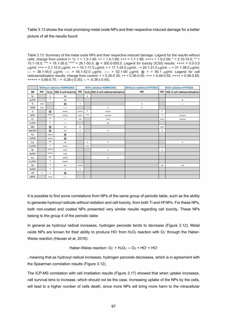
97
Table 3.13 shows the most promising metal oxide NPs and their respective induced damage for a better
picture of all the results found.
Table 3.13: Summary of the metal oxide NPs and their respective induced damage. Legend for the results without cells: change from control (= 1): + = 1.3-1.49; ++ = 1.5-1.69; +++ = 1.7-1.89; ++++ = 1.9-2.09; * = 2.10-10.0; ** = 10.1-18.0; *** = 18.1-26.0; **** = 26.1-50.0; ֍ = 300.0-500.0. Legend for toxicity (IC50) results: ++++ = 0.0-3.0 µg/ml; +++ = 3.1-10.0 µg/ml; ++ = 10.1-17.0 µg/ml; + = 17.1-24.0 µg/ml; - = 24.1-31.0 µg/ml; -- = 31.1-38.0 µg/ml; --- = 38.1-45.0 µg/ml; ---- = 45.1-52.0 µg/ml; ----- = 52.1-90 µg/ml; ֍ = > 90.1 µg/ml. Legend for cell radiosensitisation results: change from control: + = 0.26-0.35; ++ = 0.36-0.45; +++ = 0.46-0.55; ++++ = 0.56-0.65; +++++ = 0.66-0.75; - = -0.26-(-0.35); -- = -0.36-(-0.45).
It is possible to find some correlations from NPs of the same group of periodic table, such as the ability
to generate hydroxyl radicals without radiation and cell toxicity, from both Ti and Hf NPs. For these NPs,
both non-coated and coated NPs presented very similar results regarding cell toxicity. These NPs
belong to the group 4 of the periodic table.
In general as hydroxyl radical increases, hydrogen peroxide tends to decrease (Figure 3.12). Metal
oxide NPs are known for their ability to produce HO. from H2O2 reaction with O2- through the Haber-Weiss reaction (Hauser et al, 2016):
Haber-Weiss reaction: O2- + H2O2 → O2 + HO- + HO.
, meaning that as hydroxyl radical increases, hydrogen peroxide decreases, which is in agreement with
the Spearman correlation results (Figure 3.12).
The ICP-MS correlation with cell irradiation results (Figure 3.17) showed that when uptake increases,
cell survival tens to increase, which should not be the case. Increasing uptake of the NPs by the cells,
will lead to a higher number of cells death, since more NPs will bring more harm to the intracellular
Without radiation HYPOXIANP HO. H2O2 HSC-3 cell toxicity HO. H2O2 HSC-3 cell radiosensitisation HO. HO. HSC-3 cell radiosensitisationSc + ++ +
ScPEI * ++ +Ti +++ ֍ + + --
TiPEI +++ ----- + +V ֍ ++++ ++++ * ++++ *
VPEI **** ++++ +++ ** +++++ * +++++Cr * --- ++ +++ +++ +++++
CrPEI * ---- ++Mn ֍ ++ ++ +++
MnPEI ֍ ++ + + +Fe ++++ ֍
FePEI ++++ ֍Co ** ++ + + +
CoPEI * ++++ +++Ni **** +++ + +
NiPEI **** +++Cu ** ++++ +
CuPEI * ++++Zn * ++ ++++ ++ ++
ZnPEI * + +Hf + + ֍
HfPEI +++ -----
Without radiation NORMOXIA With radiation NORMOXIA With radiation HYPOXIA
98
components, increasing the chances of the cell to lose its ability to reproduce when irradiated. This is a
correlation with a small size effect (rS=0.3), so this result would need more representative data for better
conclusions regarding this relationship. There is always a possibility of some NPs help the cell removing
the radicals formed during irradiation.
The result from the correlation between CCA and cell irradiation data was expected (Figure 3.18), since
a higher increase in the number of HO. is expected to reduce cell survival, as shown by the moderate
size effect found through the Spearman correlation (rS=-0.4). HO. is considered one of the main species responsible for cancer cell death during irradiation, so an increase in the number of these species will
lead to a higher number of cancer cells death, decreasing in this way their survival.
The result from DHR and cell irradiation correlation showed that as H2O2 increases, cell survival tends
to increase (Figure 3.19), with a large size effect from Spearman correlation (rS=0.6). This result might
have two completely opposite conclusions. One could be the fact that, an increase in H2O2 formation
means that HO. is not being formed, since, from the Haber-Weiss reaction, H2O2 should decrease to
form HO.. If a lot of H2O2 is being formed and not reacting with O2- to form HO., then cell survival should
increase, since HO. is the main species responsible for cancer cell death during irradiation. If this is the
case, our results are in conformation with what is expected to be the mechanism responsible for cancer cell death during RT. The other conclusion could be that the high number of H2O2 species present would
lead to a higher number of HO. species, which in turn would lead to a higher number of cancer cells
death, decreasing cell survival. From this point of view, our results would not be in agreement with the
responsible mechanism that occurs during RT. There is the possibility that the probes, used to measure
both H2O2 and HO., interfere with the main mechanism, changing the course of the reaction.

99
4 A comparison of the radiosensitisation ability of 22 different element metal oxide nanoparticles using clinical megavoltage
X-rays – Published Paper

100
101
4.1 Introduction
This chapter is made of a paper I published with my supervisor, Dr Jon Golding. Here I tested a much
wider range of elements for the NPs, including rare-earth metals. In this work, I was more interested in
comparing the different types of NPs and their interaction with DNA, so experimental work with cells
was not performed.
To try to understand more about radiosensitisation and the different types of ROS generated during the
course of RT, here the approach was focused on studying not only hydroxyl radicals, but also
superoxide and singlet oxygen and try to understand if there was any correlation between the different ROS produced and the type of NP.
While in the previous chapter there was a concern regarding the coating due to the fact that interaction
with cells was one of the main studies and also an attempt to try to see a correlation between the formation of hydroxyl radicals and the hydrogen peroxide, here not only the number of elements was
extended but also the type of ROS, and the main study was interaction with DNA. Furthermore, since
some of the metals involved in this work are known for their catalytic ability, there was also an interest
in studying how the formation of electron-hole pairs might be involved in the ROS production.
Following presentation of my paper, I made a discussion where I correlate the different type of ROS
produced to try to understand if there is a major mechanism connecting hydroxyl radicals, superoxide,
and singlet oxygen.
4.2 Paper
A comparison of the radiosensitisation ability of 22 different element metal oxide nanoparticles using clinical megavoltage X-rays.
Alexandra Guerreiro1,2, Nicholas Chatterton2, Eleanor M. Crabb2, Jon P. Golding2*.
1 Faculty of Science and Technology. New University of Lisbon, Portugal.
2 School of Life, Health and Chemical Sciences. The Open University, Milton Keynes, UK.
*Corresponding author: [email protected]. Tel: 01908 653748
Abstract
Background: A wide range of nanoparticles (NPs), composed of different elements and their
compounds, are being developed by several groups as possible radiosensitisers, with some already in
102
clinical trials. However, no systematic experimental survey of the clinical X-ray radiosensitising potential
of different element nanoparticles has been made.
Here we directly compare the irradiation-induced (10 Gy of 6 MV X-rays) production of hydroxyl radicals,
superoxide anion radicals and singlet oxygen in aqueous solutions of the following metal oxide nanoparticles: Al2O3, SiO2, Sc2O3, TiO2, V2O5, Cr2O3, MnO2, Fe3O4, CoO, NiO, CuO, ZnO, ZrO2, MoO3,
Nd2O3, Sm2O3, Eu2O3, Gd2O3, Tb4O7, Dy2O3, Er2O3 and HfO2.
We also examine DNA damage due to these NPs in unirradiated and irradiated conditions.
Results: Without any X-rays, several NPs produced more radicals than water alone. Thus, V2O5 NPs
produced around 5-times more hydroxyl radicals and superoxide radicals. MnO2 NPs produced around
10-times more superoxide anions and Tb4O7 produced around 3-times more singlet oxygen. Lanthanides produces fewer hydroxyl radicals than water.
Following irradiation, V2O5 NPs produced nearly 10-times more hydroxyl radicals than water.
Changes in radical concentrations were determined by subtracting unirradiated values from irradiated
values. These were then compared with irradiation-induced changes in water only.
Irradiation-specific increases in hydroxyl radical were seen with most NPs, but these were only
significantly above the values of water for V2O5, while the Lanthanides showed irradiation-specific decreases in hydroxyl radical, compared to water.
Only TiO2 showed a trend of irradiation-specific increase in superoxides, while V2O5, MnO2, CoO, CuO,
MoO3 and Tb4O7 all demonstrated significant irradiation-specific decreases in superoxide, compared to
water.
No irradiation-specific increases in singlet oxygen were seen, but V2O5, NiO, CuO, MoO3 and the
Lanthanides demonstrated irradiation-specific decreases in singlet oxygen, compared to water.
MoO3 and CuO produced DNA damage in the absence of radiation, while the highest irradiation-specific
DNA damage was observed with CuO. In contrast, MnO2, Fe3O4 and CoO were slightly protective
against irradiation-induced DNA damage.
Conclusions: Beyond identifying promising metal oxide NP radiosensitisers and radioprotectors, our
broad comparisons reveal unexpected differences that suggest the surface chemistry of NP
radiosensitisers is an important criterion for their success.
Keywords: Radiosensitiser, X-ray, megavoltage, metal oxide, nanoparticle, survey, radicals, DNA
damage.
Background
103
Water radiolysis by ionising radiation results in the formation of several species, mainly: e−aq, ˙OH, ˙H,
H3O+, OH−, ˙O2H, H2O2 and H2. These products react with one another to form secondary species such as
O2, ˙O2− and ˙O3, with many additional short-lived intermediates (reviewed in Le Caë, 2011). Water
radiolysis products range from highly oxidizing (e.g. ˙OH, H2O2 and O2) to highly reducing (e.g. ˙H, ˙eaq− and ˙O2−). These reactive species damage biological molecules and contribute around 60% of the biological
damage due to X-ray radiotherapy (Saenko et al., 2013; D. Zhang et al., 2016; Jayakumar et al., 2014
and reviewed in Baskar et al., 2014). Although each clinical X-ray irradiation session only lasts for a few
minutes, the radical-mediated damage can induce prolonged metabolic oxidative stress that continues
to damage cells long after irradiation (Azzam et al., 2012).
Theoretical studies (McMahon et al., 2016; Hwang et al., 2017; Haume et al., 2016; Roeske et al., 2007)
and experimental studies (Retif et al., 2015; Y. Liu et al., 2018) have demonstrated that X-ray-induced radical formation can be greatly increased in the vicinity of nanoparticles of high atomic number (Z)
elements, their oxides or sulphides (e.g. Ag: (P. Liu et al., 2016); Gd: (Luchette et al., 2014; Lux et al.,
2018); Hf: (Marill, Anesary, Zhang, Vivet, Borghi, et al., 2014); Pt: (Muhammad et al., 2018); Au: (Haume
et al., 2016; Rahman et al., 2014); Bi: (Algethami, 2015; Sahu & Cates, 2017), which have large X-ray
absorption cross sections and produce a shower of secondary electrons when irradiated. For this
reason, high atomic number nanoparticles have been the focus of nanoparticle radiosensitiser research,
some of which are being clinically evaluated (Y. Liu et al., 2018), such as the Gd NP, AGuIX (Lux et al.,
2018) and the Hf NP, NBTXR3 (Marill, Anesary, Zhang, Vivet, Barendsen, et al., 2014), both of which are currently in Phase II clinical trials (NCT03818386 and NCT03589339).
However, NPs of many lower atomic number element oxides have been shown to be effective
radiosensitisers, such as: Al (Roth et al., 2011), Si (Gara et al., 2012; Generalov et al., 2015; N. Zhao
et al., 2016), Ca (Chu et al., 2013), Ti (Morrison et al., 2017), Fe (Khoei et al., 2014), Cu (Y. W. Jiang
et al., 2019), Zn (Generalov et al., 2015), Zr (Carrasco-Flores & LaVerne, 2007). This suggests that
other, physico-chemical and chemical processes, likely involving the nanoparticle surface, contribute to
the overall pool of X-ray-induced radicals.
What is missing, however, are systematic experimental comparisons of these different element NPs as
radiosensitisers. Most in vitro studies have been limited to comparing small ranges of elements, or more often to different compounds or surface functionalisation of a single element NP. Experimental
differences, such as X-ray energy and dose, and concentration of nanoparticles further impede
comparisons (Hwang et al., 2017; Rahman et al., 2014; Retif et al., 2015). Recent proposals have been
made to standardise these parameters (Retif et al., 2015; Schuemann et al., 2018), but it will take time
for these to become routinely adopted.
Only two studies have been published using a broad range of NPs, both with gamma radiation. Petrik
et al (2001) compared hydrogen production from adsorbed water on a wide range of 28 different
element metal oxide powders (Petrik et al., 2001). Chelnokov et al (2014) compared solvated electron
production from 6 different element NPs (Chelnokov et al., 2014).
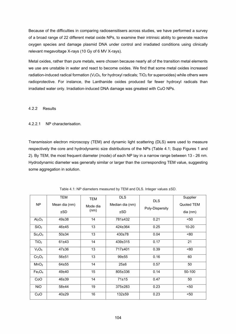
104
Because of the difficulties in comparing radiosensitisers across studies, we have performed a survey
of a broad range of 22 different metal oxide NPs, to examine their intrinsic ability to generate reactive
oxygen species and damage plasmid DNA under control and irradiated conditions using clinically
relevant megavoltage X-rays (10 Gy of 6 MV X-rays).
Metal oxides, rather than pure metals, were chosen because nearly all of the transition metal elements
we use are unstable in water and react to become oxides. We find that some metal oxides increased
radiation-induced radical formation (V2O5, for hydroxyl radicals; TiO2 for superoxides) while others were radioprotective. For instance, the Lanthanide oxides produced far fewer hydroxyl radicals than
irradiated water only. Irradiation-induced DNA damage was greatest with CuO NPs.
Results
4.2.2.1 NP characterisation.
Transmission electron microscopy (TEM) and dynamic light scattering (DLS) were used to measure respectively the core and hydrodynamic size distributions of the NPs (Table 4.1; Supp Figures 1 and
2). By TEM, the most frequent diameter (mode) of each NP lay in a narrow range between 13 - 26 nm.
Hydrodynamic diameter was generally similar or larger than the corresponding TEM value, suggesting
some aggregation in solution.
Table 4.1: NP diameters measured by TEM and DLS. Integer values ±SD.
NP
TEM
Mean dia (nm)
±SD
TEM
Mode dia (nm)
DLS
Median dia (nm)
±SD
DLS
Poly-Dispersity
Supplier
Quoted TEM
dia (nm)
Al2O3 49±38 14 781±432 0.21 <50
SiO2 46±45 13 424±364 0.25 10-20
Sc2O3 50±34 13 430±78 0.04 <80
TiO2 61±43 14 439±315 0.17 21
V2O5 47±36 13 717±401 0.39 <80
Cr2O3 56±51 13 99±55 0.16 60
MnO2 64±55 14 25±6 0.57 50
Fe3O4 49±40 15 805±336 0.14 50-100
CoO 46±39 14 71±15 0.47 50
NiO 58±44 19 375±283 0.23 <50
CuO 40±29 16 132±59 0.23 <50
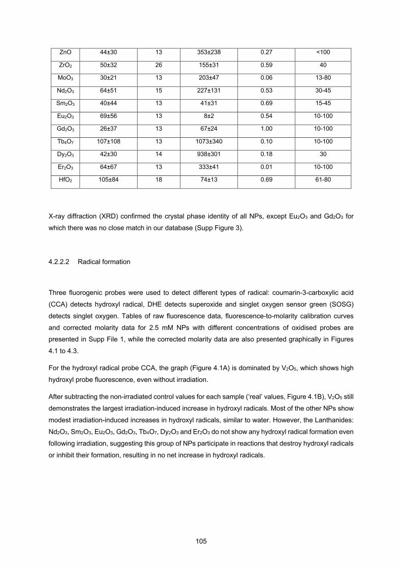
105
ZnO 44±30 13 353±238 0.27 <100
ZrO2 50±32 26 155±31 0.59 40
MoO3 30±21 13 203±47 0.06 13-80
Nd2O3 64±51 15 227±131 0.53 30-45
Sm2O3 40±44 13 41±31 0.69 15-45
Eu2O3 69±56 13 8±2 0.54 10-100
Gd2O3 26±37 13 67±24 1.00 10-100
Tb4O7 107±108 13 1073±340 0.10 10-100
Dy2O3 42±30 14 938±301 0.18 30
Er2O3 64±67 13 333±41 0.01 10-100
HfO2 105±84 18 74±13 0.69 61-80
X-ray diffraction (XRD) confirmed the crystal phase identity of all NPs, except Eu2O3 and Gd2O3 for
which there was no close match in our database (Supp Figure 3).
4.2.2.2 Radical formation
Three fluorogenic probes were used to detect different types of radical: coumarin-3-carboxylic acid
(CCA) detects hydroxyl radical, DHE detects superoxide and singlet oxygen sensor green (SOSG)
detects singlet oxygen. Tables of raw fluorescence data, fluorescence-to-molarity calibration curves
and corrected molarity data for 2.5 mM NPs with different concentrations of oxidised probes are presented in Supp File 1, while the corrected molarity data are also presented graphically in Figures
4.1 to 4.3.
For the hydroxyl radical probe CCA, the graph (Figure 4.1A) is dominated by V2O5, which shows high
hydroxyl probe fluorescence, even without irradiation.
After subtracting the non-irradiated control values for each sample (‘real’ values, Figure 4.1B), V2O5 still
demonstrates the largest irradiation-induced increase in hydroxyl radicals. Most of the other NPs show
modest irradiation-induced increases in hydroxyl radicals, similar to water. However, the Lanthanides:
Nd2O3, Sm2O3, Eu2O3, Gd2O3, Tb4O7, Dy2O3 and Er2O3 do not show any hydroxyl radical formation even
following irradiation, suggesting this group of NPs participate in reactions that destroy hydroxyl radicals
or inhibit their formation, resulting in no net increase in hydroxyl radicals.
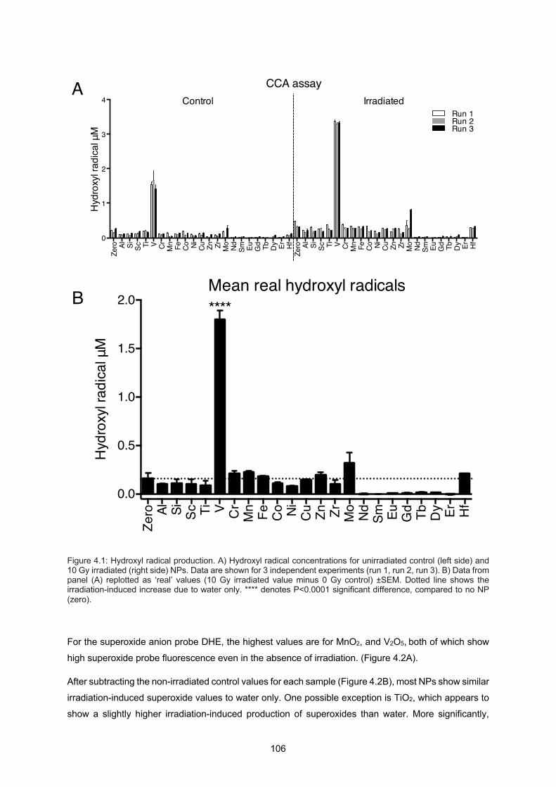
106
Figure 4.1: Hydroxyl radical production. A) Hydroxyl radical concentrations for unirradiated control (left side) and 10 Gy irradiated (right side) NPs. Data are shown for 3 independent experiments (run 1, run 2, run 3). B) Data from panel (A) replotted as ‘real’ values (10 Gy irradiated value minus 0 Gy control) ±SEM. Dotted line shows the irradiation-induced increase due to water only. **** denotes P<0.0001 significant difference, compared to no NP (zero).
For the superoxide anion probe DHE, the highest values are for MnO2, and V2O5, both of which show
high superoxide probe fluorescence even in the absence of irradiation. (Figure 4.2A).
After subtracting the non-irradiated control values for each sample (Figure 4.2B), most NPs show similar
irradiation-induced superoxide values to water only. One possible exception is TiO2, which appears to
show a slightly higher irradiation-induced production of superoxides than water. More significantly,
Zero Al Si Sc Ti V Cr Mn Fe Co Ni Cu Zn Zr Mo Nd Sm Eu Gd Tb Dy Er Hf
Zero Al Si Sc Ti V Cr Mn Fe Co Ni Cu Zn Zr Mo Nd Sm Eu Gd Tb Dy Er Hf0
1
2
3
4H
ydro
xyl r
adica
l μM
CCA assay
Run 1Run 2Run 3
Control Irradiated
Zero Al Si Sc Ti V C
rM
n Fe Co Ni Cu Zn Zr Mo
Nd
Sm Eu Gd Tb Dy Er Hf
0.0
0.5
1.0
1.5
2.0
Hyd
roxy
l rad
ical μ
M
Mean real hydroxyl radicals****
A
B

107
although V2O5, MnO2, CoO, CuO and Tb4O7 show greater superoxide generation overall (Figure 4.2A);
after subtracting the non-irradiated value they show less irradiation-induced superoxide than water
(Figure 4.2B), suggesting that irradiation makes less difference for these NPs as they are already
actively generating these radicals, or that possibly these NPs might participate in reactions that destroy superoxides or inhibit their formation. MoO3 shows considerably lower superoxide radical formation
than water after irradiation, also suggesting that it inhibits superoxide formation and/or destroys already
produced superoxide radicals.
Figure 4.2: Superoxide anion radical production. A) Superoxide radical concentrations for unirradiated control (left side) and 10 Gy irradiated (right side) NPs. Data are shown for 3 independent experiments (run 1, run 2, run 3). B) Data from panel (A) replotted as ‘real’ values (10 Gy irradiated value minus 0 Gy control) ±SEM. Dotted line shows the irradiation-induced increase due to water only. ****, ** and * denote respectively P<0.0001, P<0.01 and P<0.05 significant differences, compared to no NP (zero).
Zero Al Si Sc Ti V Cr Mn Fe Co Ni Cu Zn Zr Mo Nd Sm Eu Gd Tb Dy Er Hf
Zero Al Si Sc Ti V Cr Mn Fe Co Ni Cu Zn Zr Mo Nd Sm Eu Gd Tb Dy Er Hf0
1
2
3
4
Supe
roxid
e an
ion μM
DHE assay
Run 1Run 2Run 3
Control Irradiated
Zero Al Si Sc Ti V C
rM
n Fe Co Ni Cu Zn Zr Mo
Nd
Sm Eu Gd Tb Dy Er Hf0.0
0.5
1.0
1.5
2.0
Supe
roxid
e an
ion μM
Mean real superoxide anions
********
****
****
***
A
B

108
For the singlet oxygen probe SOSG, the graph is dominated by Tb4O7, which shows high probe
fluorescence in the absence and presence of irradiation (Figure 4.3A). There is also suggestion that
MoO3 again shows lower radical production than water with or without irradiation.
After subtracting the non-irradiated control values for each sample (Figure 4.3B), nearly half of the NPs
show similar irradiation-induced singlet oxygen values to water-only: Al2O3, SiO2, Sc2O3, TiO2, Cr2O3,
Fe3O4, CoO, ZrO2 and HfO2. The remaining NPs all show a tendency for less irradiation-induced singlet
oxygen production than water; significantly lower in the cases of MoO3 and V2O5 in particular, as well
as for NiO, CuO, Nd2O3, Eu2O3, Gd2O3, and Dy2O3. Tb4O7 shows less irradiation-induced singlet oxygen
production than water, although its absolute value is higher than water as it is so active on its own.
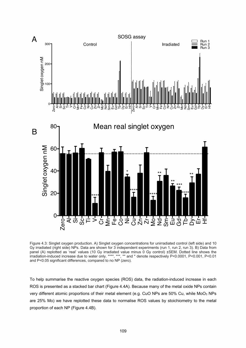
109
Figure 4.3: Singlet oxygen production. A) Singlet oxygen concentrations for unirradiated control (left side) and 10 Gy irradiated (right side) NPs. Data are shown for 3 independent experiments (run 1, run 2, run 3). B) Data from panel (A) replotted as ‘real’ values (10 Gy irradiated value minus 0 Gy control) ±SEM. Dotted line shows the irradiation-induced increase due to water only. ****, ***, ** and * denote respectively P<0.0001, P<0.001, P<0.01 and P<0.05 significant differences, compared to no NP (zero).
To help summarise the reactive oxygen species (ROS) data, the radiation-induced increase in each
ROS is presented as a stacked bar chart (Figure 4.4A). Because many of the metal oxide NPs contain very different atomic proportions of their metal element (e.g. CuO NPs are 50% Cu, while MoO3 NPs
are 25% Mo) we have replotted these data to normalise ROS values by stoichiometry to the metal
proportion of each NP (Figure 4.4B).
Zero Al Si Sc Ti V Cr Mn Fe Co Ni Cu Zn Zr Mo Nd Sm Eu Gd Tb Dy Er Hf
Zero Al Si Sc Ti V Cr Mn Fe Co Ni Cu Zn Zr Mo Nd Sm Eu Gd Tb Dy Er Hf
0
100
200
300Si
ngle
t oxy
gen
nMSOSG assay
Run 1Run 2Run 3
Control Irradiated
Zero Al Si Sc Ti V C
rM
n Fe Co Ni Cu Zn Zr Mo
Nd Sm Eu Gd Tb Dy Er Hf
0
20
40
60
80
Sing
let o
xyge
n nM
Mean real singlet oxygen
********
**** ****
* ****
**
***
A
B
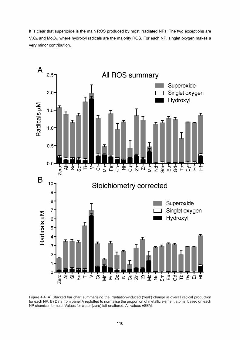
110
It is clear that superoxide is the main ROS produced by most irradiated NPs. The two exceptions are
V2O5 and MoO3, where hydroxyl radicals are the majority ROS. For each NP, singlet oxygen makes a
very minor contribution.
Figure 4.4: A) Stacked bar chart summarising the irradiation-induced (‘real’) change in overall radical production for each NP. B) Data from panel A replotted to normalise the proportion of metallic element atoms, based on each NP chemical formula. Values for water (zero) left unaltered. All values ±SEM.
Zero Al Si Sc Ti V Cr Mn Fe Co Ni Cu Zn Zr Mo Nd Sm Eu Gd Tb Dy Er Hf
0.0
0.5
1.0
1.5
2.0
2.5
Rad
icals µ
M
All ROS summary
HydroxylSinglet oxygenSuperoxide
Zero Al Si Sc Ti V Cr Mn Fe Co Ni Cu Zn Zr Mo Nd Sm Eu Gd Tb Dy Er Hf
0
1
2
3
4
5
6
7
8
9
10
Rad
icals µ
M
Stoichiometry corrected
HydroxylSinglet oxygenSuperoxide
A
B
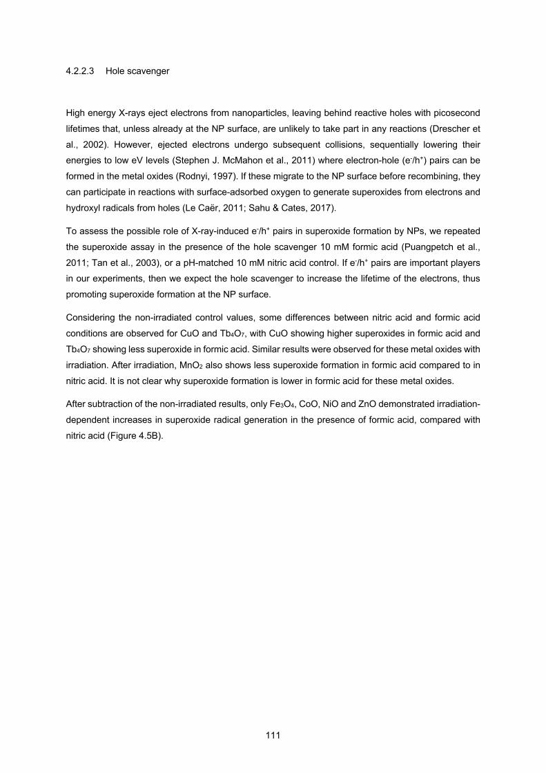
111
4.2.2.3 Hole scavenger
High energy X-rays eject electrons from nanoparticles, leaving behind reactive holes with picosecond
lifetimes that, unless already at the NP surface, are unlikely to take part in any reactions (Drescher et
al., 2002). However, ejected electrons undergo subsequent collisions, sequentially lowering their energies to low eV levels (Stephen J. McMahon et al., 2011) where electron-hole (e-/h+) pairs can be
formed in the metal oxides (Rodnyi, 1997). If these migrate to the NP surface before recombining, they
can participate in reactions with surface-adsorbed oxygen to generate superoxides from electrons and
hydroxyl radicals from holes (Le Caër, 2011; Sahu & Cates, 2017).
To assess the possible role of X-ray-induced e-/h+ pairs in superoxide formation by NPs, we repeated
the superoxide assay in the presence of the hole scavenger 10 mM formic acid (Puangpetch et al.,
2011; Tan et al., 2003), or a pH-matched 10 mM nitric acid control. If e-/h+ pairs are important players in our experiments, then we expect the hole scavenger to increase the lifetime of the electrons, thus
promoting superoxide formation at the NP surface.
Considering the non-irradiated control values, some differences between nitric acid and formic acid
conditions are observed for CuO and Tb4O7, with CuO showing higher superoxides in formic acid and
Tb4O7 showing less superoxide in formic acid. Similar results were observed for these metal oxides with
irradiation. After irradiation, MnO2 also shows less superoxide formation in formic acid compared to in
nitric acid. It is not clear why superoxide formation is lower in formic acid for these metal oxides.
After subtraction of the non-irradiated results, only Fe3O4, CoO, NiO and ZnO demonstrated irradiation-
dependent increases in superoxide radical generation in the presence of formic acid, compared with
nitric acid (Figure 4.5B).
112
Figure 4.5: A) Superoxide formation in the presence of pH 3.5 10 mM nitric acid or pH 3.5 10 mM formic acid hole scavenger. Left side shows unirradiated controls and right side shows 10 Gy irradiated. N=3 and all values ±SEM. B) Data from panel (A) replotted as ‘real’ values (10 Gy irradiated value minus 0 Gy control) ±SEM.
4.2.2.4 DNA damage assay
Plasmid DNA normally exists as a tightly-wound supercoil. Single strand breaks allow the supercoil to
relax to an open circle which demonstrates slower migration on an electrophoretic gel. We therefore
quantified DNA damage in the presence of NPs by measuring the proportion of relaxed circular DNA
(Figure 4.6).
Zero Al Si Sc Ti V Cr Mn Fe Co Ni Cu Zn Zr Mo Nd Sm Eu Gd Tb Dy Er Hf
Zero Al Si Sc Ti V Cr Mn Fe Co Ni Cu Zn Zr Mo Nd Sm Eu Gd Tb Dy Er Hf0
50
100
150Su
pero
xide
anio
n μM
DHE assay
FormicNitric
Control Irradiated
Zero Al Si Sc Ti V Cr Mn Fe Co Ni Cu Zn Zr Mo Nd Sm Eu Gd Tb Dy Er Hf
0
20
40
60
Supe
roxid
e an
ion μM
Mean real superoxide anions
FormicNitric
A
B
113
In the absence of irradiation, MoO3 and CuO caused the greatest damage to DNA. These NPs also
demonstrated the greatest DNA damage following irradiation (Figure 4.6A-C).
When the DNA damage due to non-irradiated controls was subtracted from each irradiated value, it
became clear that most NPs produced only slightly more irradiation-specific DNA damage than water
(Figure 4.6D). Only CuO performed significantly better than water in terms of irradiation-specific DNA
damage (Figure 4.6D).

114

115
Discussion
The ability of nanoparticles to act as X-ray radiosensitisers has generally been viewed as a physical
process, due to the increased absorption of X-rays, followed by ejection of secondary electrons
(photoelectric effect) and fluorescence photons that then interact with water or biomolecules to produce
radiolysis products, which include damaging radicals (Kuncic & Lacombe, 2018). For this reason, the
emphasis of NP radiosensitiser research has been on high atomic number elements, due to their expected greater interaction cross section with X-ray photons.
A theoretical study by McMahon et al, (2016), based on physical processes, computed the energy
deposition within 1 µm around a series of X-ray irradiated pure element 20 nm NPs in water (Z=14, Si
to Z=80, Hg). When all energy outputs are added together (Auger electrons, photoelectrons, Compton
electrons, fluorescence, electron impact) the total energy deposition from 6 MV X-rays was relatively
flat across all the elements studied (Fig. 3C,D in McMahon et al., 2016), implying that the choice of
element makes little difference to the amount of ROS produced at these energies.
However, experimental data from Sicard-Roselli et al (2014) and Gilles et al (2018) demonstrated that
many more hydroxyl radicals (Gilles et al., 2018; Sicard-Roselli et al., 2014) and solvated electrons
(Gilles et al., 2018) are produced from X-ray irradiated Au NPs than can be explained purely by physical processes, such as the photoelectric effect. They proposed that additional radicals are produced via
reactions of secondary electrons with interfacial water molecules, that become stretched due to their
adsorption to the Au NP and therefore require much less energy for H-OH bond radiolysis than bulk
water (255 kJ/mol instead of 460 kJ/mol) (R. Liu, 2013). This water distorting effect is not restricted to
Au NPs: water molecules form similar structured layers up to 2 nm thick around any NP (Zobel, 2016;
Zobel et al., 2015) and this is where most radiolytic reactions are expected to occur.
Moreover, nanoparticles will inevitably come into contact with excited water (H2O*) and ionised water
(H2O+, e-aq), formed during the first 10-15 seconds of irradiation, plus the physico-chemical reaction
products of these species, produced during the subsequent 10-12 seconds, such as ˙OH, ˙H, H3O+ and
OH−; and downstream products such as H2O2 (reviewed in Le Caër, 2011). Because of their high surface area and specific surface chemistries, nanoparticles could catalyse many more reactions from
these intermediates and thereby increase the overall concentration of radicals.
This mismatch between theory and experimental observations for Au NPs prompted us to consider if a broad range of metal oxide NPs, well known for their catalytic properties, but hitherto not considered as
Figure 4.6: DNA damage assays based on the appearance of the relaxed circular form of plasmid DNA. A) Agarose gel from one experiment showing paired non-irradiated (-) and 10 Gy irradiated (+) samples. Bands corresponding to undamaged (supercoiled, SC), single strand break (relaxed circular, RC) and double strand break (linear, L) plasmid DNA are marked. Ld is 1 kb DNA ladder. Note that this gel is not loaded in atomic number order. B) Percentage relaxed circular DNA from each experiment. C) Average values from (B) ±SEM. D) Data from panel (C) replotted as ‘real’ values (10 Gy irradiated value minus 0 Gy control) ±SEM. Dotted line shows the irradiation-induced increase due to water only. ****, *** and ** denote respectively P<0.0001, P<0.001 and P<0.01 significant differences, compared to the respective no NP (zero) control.
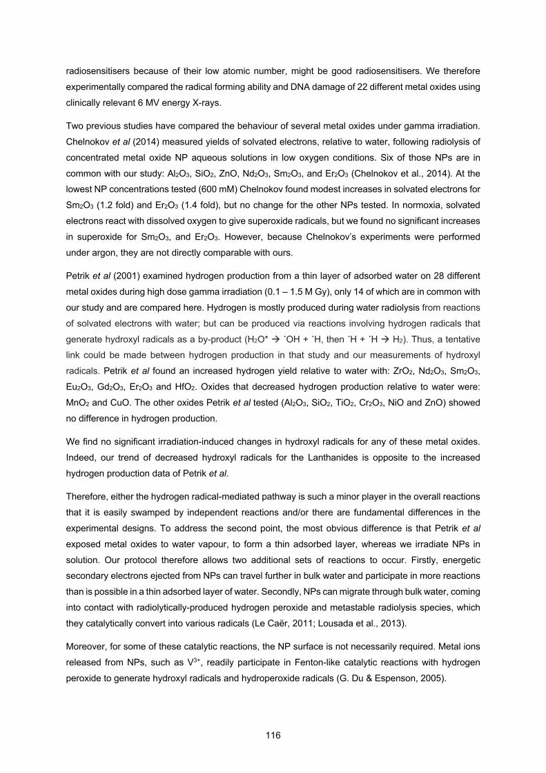
116
radiosensitisers because of their low atomic number, might be good radiosensitisers. We therefore
experimentally compared the radical forming ability and DNA damage of 22 different metal oxides using
clinically relevant 6 MV energy X-rays.
Two previous studies have compared the behaviour of several metal oxides under gamma irradiation.
Chelnokov et al (2014) measured yields of solvated electrons, relative to water, following radiolysis of
concentrated metal oxide NP aqueous solutions in low oxygen conditions. Six of those NPs are in
common with our study: Al2O3, SiO2, ZnO, Nd2O3, Sm2O3, and Er2O3 (Chelnokov et al., 2014). At the lowest NP concentrations tested (600 mM) Chelnokov found modest increases in solvated electrons for
Sm2O3 (1.2 fold) and Er2O3 (1.4 fold), but no change for the other NPs tested. In normoxia, solvated
electrons react with dissolved oxygen to give superoxide radicals, but we found no significant increases
in superoxide for Sm2O3, and Er2O3. However, because Chelnokov’s experiments were performed
under argon, they are not directly comparable with ours.
Petrik et al (2001) examined hydrogen production from a thin layer of adsorbed water on 28 different
metal oxides during high dose gamma irradiation (0.1 – 1.5 M Gy), only 14 of which are in common with
our study and are compared here. Hydrogen is mostly produced during water radiolysis from reactions
of solvated electrons with water; but can be produced via reactions involving hydrogen radicals that
generate hydroxyl radicals as a by-product (H2O* à ˙OH + ˙H, then ˙H + ˙H à H2). Thus, a tentative link could be made between hydrogen production in that study and our measurements of hydroxyl
radicals. Petrik et al found an increased hydrogen yield relative to water with: ZrO2, Nd2O3, Sm2O3,
Eu2O3, Gd2O3, Er2O3 and HfO2. Oxides that decreased hydrogen production relative to water were:
MnO2 and CuO. The other oxides Petrik et al tested (Al2O3, SiO2, TiO2, Cr2O3, NiO and ZnO) showed
no difference in hydrogen production.
We find no significant irradiation-induced changes in hydroxyl radicals for any of these metal oxides.
Indeed, our trend of decreased hydroxyl radicals for the Lanthanides is opposite to the increased
hydrogen production data of Petrik et al.
Therefore, either the hydrogen radical-mediated pathway is such a minor player in the overall reactions
that it is easily swamped by independent reactions and/or there are fundamental differences in the
experimental designs. To address the second point, the most obvious difference is that Petrik et al
exposed metal oxides to water vapour, to form a thin adsorbed layer, whereas we irradiate NPs in
solution. Our protocol therefore allows two additional sets of reactions to occur. Firstly, energetic secondary electrons ejected from NPs can travel further in bulk water and participate in more reactions
than is possible in a thin adsorbed layer of water. Secondly, NPs can migrate through bulk water, coming
into contact with radiolytically-produced hydrogen peroxide and metastable radiolysis species, which
they catalytically convert into various radicals (Le Caër, 2011; Lousada et al., 2013).
Moreover, for some of these catalytic reactions, the NP surface is not necessarily required. Metal ions
released from NPs, such as V3+, readily participate in Fenton-like catalytic reactions with hydrogen
peroxide to generate hydroxyl radicals and hydroperoxide radicals (G. Du & Espenson, 2005).
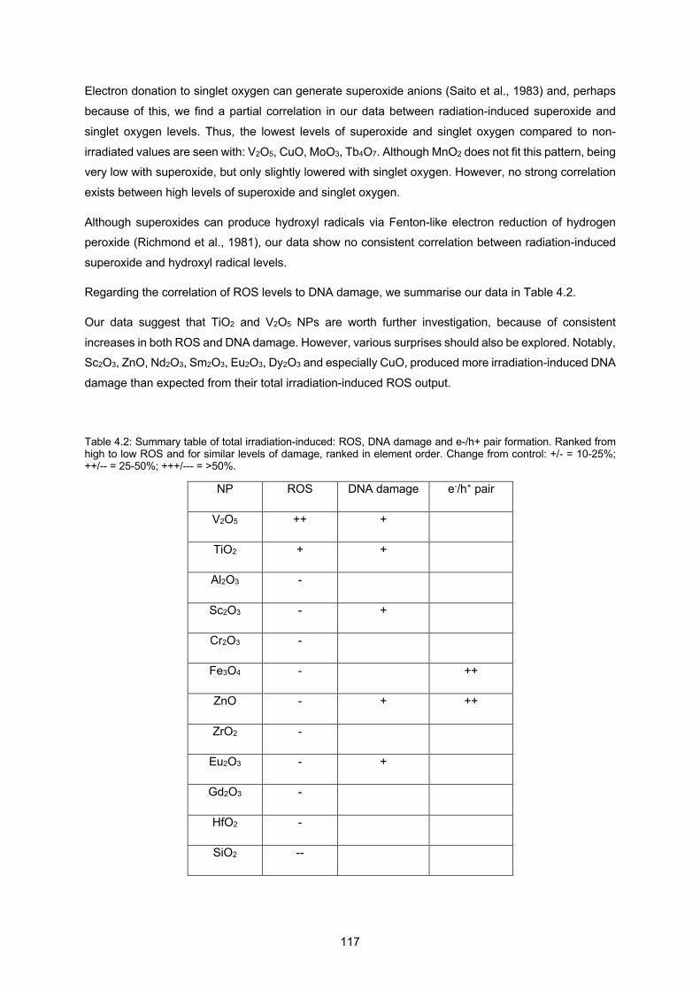
117
Electron donation to singlet oxygen can generate superoxide anions (Saito et al., 1983) and, perhaps
because of this, we find a partial correlation in our data between radiation-induced superoxide and
singlet oxygen levels. Thus, the lowest levels of superoxide and singlet oxygen compared to non-
irradiated values are seen with: V2O5, CuO, MoO3, Tb4O7. Although MnO2 does not fit this pattern, being very low with superoxide, but only slightly lowered with singlet oxygen. However, no strong correlation
exists between high levels of superoxide and singlet oxygen.
Although superoxides can produce hydroxyl radicals via Fenton-like electron reduction of hydrogen peroxide (Richmond et al., 1981), our data show no consistent correlation between radiation-induced
superoxide and hydroxyl radical levels.
Regarding the correlation of ROS levels to DNA damage, we summarise our data in Table 4.2.
Our data suggest that TiO2 and V2O5 NPs are worth further investigation, because of consistent
increases in both ROS and DNA damage. However, various surprises should also be explored. Notably, Sc2O3, ZnO, Nd2O3, Sm2O3, Eu2O3, Dy2O3 and especially CuO, produced more irradiation-induced DNA
damage than expected from their total irradiation-induced ROS output.
Table 4.2: Summary table of total irradiation-induced: ROS, DNA damage and e-/h+ pair formation. Ranked from high to low ROS and for similar levels of damage, ranked in element order. Change from control: +/- = 10-25%; ++/-- = 25-50%; +++/--- = >50%.
NP ROS DNA damage e-/h+ pair
V2O5 ++ +
TiO2 + +
Al2O3 -
Sc2O3 - +
Cr2O3 -
Fe3O4 - ++
ZnO - + ++
ZrO2 -
Eu2O3 - +
Gd2O3 -
HfO2 -
SiO2 --
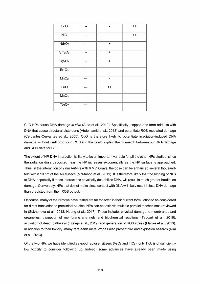
118
CoO -- - ++
NiO -- ++
Nd2O3 -- +
Sm2O3 -- +
Dy2O3 -- +
Er2O3 --
MnO2 --- -
CuO --- ++
MoO3 ---
Tb4O7 ---
CuO NPs cause DNA damage in vivo (Atha et al., 2012). Specifically, copper ions form adducts with
DNA that cause structural distortions (Abdelhamid et al., 2018) and potentiate ROS-mediated damage (Cervantes-Cervantes et al., 2005). CuO is therefore likely to potentiate irradiation-induced DNA
damage, without itself producing ROS and this could explain the mismatch between our DNA damage
and ROS data for CuO.
The extent of NP-DNA interaction is likely to be an important variable for all the other NPs studied, since
the radiation dose deposited near the NP increases exponentially as the NP surface is approached.
Thus, in the interaction of 2 nm AuNPs with 6 MV X-rays, the dose can be enhanced several thousand-
fold within 10 nm of the Au surface (McMahon et al., 2011). It is therefore likely that the binding of NPs to DNA, especially if these interactions physically destabilise DNA, will result in much greater irradiation
damage. Conversely, NPs that do not make close contact with DNA will likely result in less DNA damage
than predicted from their ROS output.
Of course, many of the NPs we have tested are far too toxic in their current formulation to be considered
for direct translation to preclinical studies. NPs can be toxic via multiple parallel mechanisms (reviewed
in (Sukhanova et al., 2018; Huang et al., 2017). These include: physical damage to membranes and
organelles, disruption of membrane channels and biochemical reactions (Taggart et al., 2016), activation of death pathways (Tzelepi et al., 2019) and generation of ROS stress (Manke et al., 2013).
In addition to their toxicity, many rare earth metal oxides also present fire and explosion hazards (Rim
et al., 2013).
Of the two NPs we have identified as good radiosensitisers (V2O5 and TiO2), only TiO2 is of sufficiently
low toxicity to consider following up. Indeed, some advances have already been made using
119
radiosensitisers based on TiO2 NPs (Nakayama et al., 2016; Morrison et al., 2017; Townley, Rapa, et
al., 2012).
We consider that the future of NP radiosensitisers most probably lies in the combination of high Z
elements with catalytic oxides: using the photoelectric effect of high Z elements to feed secondary
electrons into the catalyst. These composite NPs could be made as complex oxides, as has been done
with Bi2WO6 (Sahu & Cates, 2017); aggregate mixtures, or core-shell arrangements. For instance,
improved radiosensitisation has been reported using lanthanide-doped TiO2 (Townley, Rapa, et al., 2012) and ZnO NPs (Ghaemi et al., 2016), ZnO-SiO2 nanocomposites (Generalov et al., 2015) and Au-
Bi2S3 heterostructures (X. Wang et al., 2019).
As with all catalysts, it will be important to consider ways to prevent ‘fouling’ of the catalytic surface by
ligands or serum proteins in vitro, since we and others have previously shown that the presence of a
ligand shell on AuNPs greatly decreases irradiation-induced ROS production (Gilles et al., 2014; Grellet
et al., 2017).
Conclusions
We find large variations in the formation of three types of radical species in solutions of metal oxide
NPs. In general, the largest irradiation-induced increases are seen for superoxides, where TiO2
produces an additional 1.6 µM superoxide upon irradiation. This being some 0.2 µM more than water
only. A more impressive increase is seen for hydroxyl radicals using V2O5, demonstrating an additional
1.8 µM hydroxyl radicals upon irradiation; 1.6 µM more than water only. However, bulk radical formation
per se does not predict the extent of radiation-induced DNA damage in the presence of these NPs. Thus, although TiO2 and V2O5 both produce more irradiation-induced DNA damage than water, CuO
produces nearly 8% more DNA damage even though it shows one of the lowest irradiation-induced
increases in radicals. Therefore, other factors that probably involve intimate DNA-NP interactions, such
as DNA destabilisation and highly-localised increases in radical concentration near the NP surface,
likely predominate.
In summary, when considering NP radiosensitisers, it is perhaps worth remembering the old phrase
‘don’t judge a book by its cover’. Theoretical predictions of energy output and biological damage are
currently based on physical principles and need to be expanded to take account of subsequent chemical
reactions on and near the NP surface.
Also, the purely catalytic nature of many NPs, within a ‘sea’ of radiolytic reactants needs to be
considered in order to obtain a fuller picture of the intrinsic radiosensitising ability of a NP. These
physical/chemical considerations then need to be put into a biological context. How well are they taken
up by cells and is there any selectivity towards cancer cells? Once the NPs are in a biological medium of proteins and sugars, does this alter their ability to produce ROS and damage cancer cells? We know
that our results pose more questions than they answer, but if this leads to a deeper understanding of
120
radiosensitisation theory and allows for rational choices of the best nanoparticle materials, then this is
ultimately a good thing.
Methods
4.2.5.1 Nanoparticles
The following nanoparticles were obtained from US Research Nanomaterials: Cr2O3 (US3060, 60 nm,
>99%), MnO2 (US3319NMP, 50 nm, 98%), CoO (US3051, 50 nm, 99.7%), ZrO2 (US3600, 40 nm,
>99%), MoO3 (US3330, 13-80 nm, 99.94%), Nd2O3 (US3350, 30-45 nm, 99.9%), Sm2O3 (US3450, 15-
45 nm, 99.95%), Eu2O3 (US3543, 10-100 nm, 99.99%), Gd2O3 (US3240, 10-100 nm, 99.9%), Tb4O7
(US3455, 10-100 nm, 99.99%), Dy2O3 (US3080, 30 nm, >99.9%), Er2O3 (US3140, 10-100 nm, 99.9%) and HfO2 (US3245, 61-80 nm, 99.99%).
The following nanoparticles were obtained from Sigma-Aldrich: Al2O3 (544833, <50 nm, 99.8%), SiO2
(637238, 10-20 nm, 99.5%), TiO2 (718467, 21 nm, 99.5%), Fe3O4 (637106, 50-100 nm, 97%), NiO
(637130, <50 nm, 99.8%), CuO (544868, <50 nm, 82.6%) and ZnO (721077, <100 nm).
Sc2O3 was obtained from American Elements (SC-OX-02-NP, <80 nm, 99%).
V2O5 was obtained from Nanoshel (NS6130-03-399, <80 nm, 99.9%).
4.2.5.2 TEM
Samples were prepared by drop-coating films of the NP solutions on electrostatically discharged
carbon-coated copper TEM grids and visualized on a JEM-1400 model EM instrument (JEOL, USA)
operated at an accelerating voltage of 80 kV and x 30,000 magnification. Size histograms were measured from 20 TEM images per nanoparticle type at x3000 magnification using the Analyze
Particles feature of ImageJ software (version 1.52; NIH) and are reported as the mean and median
values.
4.2.5.3 DLS
The hydrodynamic diameter of 2.5 mM NPs in 3% PBS pH 7.4 were measured using a Nanotrac Flex particle size analyser, with Flex 11.1.0.5 software (Microtrac).

121
4.2.5.4 XRD
Samples were analysed using a Siemens D5005 XRD with a long fine focus copper X-ray tube
energised at 40 kV and 40 mA, running conventional Bragg-Brentano geometry. Most runs were 0.5
s/step, with some at 8 s/step. Crystal phases were determined from XRD spectra using QualX2 software (Altomare et al., 2015) (version 2.24; Institute of Crystallography CNR, Bari, Italy) and PowCod
database 1901.
4.2.5.5 Hydroxyl radical probe
5 mM stock of coumarin-3-carboxylic acid (CCA) powder (Sigma Aldrich) was prepared in 6% PBS pH
7.4.
4.2.5.6 Singlet oxygen probe
Singlet oxygen sensor green (SOSG) was purchased from ThermoFisher. From a 5mM methanolic
stock, a working solution of 1 µM was prepared in 6% PBS pH 7.4.
4.2.5.7 Superoxide anion probe
Dihydroethidium (DHE) was purchased from ThermoFisher. From a stock of 20 mM DHE in DMSO a
working solution of 50 µM was prepared in 6% PBS pH 7.4.
4.2.5.8 Radical measurement
Experiments were run a minimum of three times independently and each time containing technical
triplicates. Fifty microlitres of probe working solution was added to 50 µl of 5 mM NP in deionized water
in black-walled 96-well plates (Cellstar, Greiner), producing a 2.5 mM NP working solution. After
irradiation, the fluorescence of each well was measured using a FLUOstar Optima plate reader (BMG
Labtech), according to the type of probe: CCA at Ex 390 nm/ Em 450 nm, SOSG at Ex 485 nm /Em
520 nm, and DHE at Ex 485 nm/ Em 590 nm (H. Zhao et al., 2003).
122
4.2.5.9 Radical probe calibration curves
Each NP could potentially alter the fluorescence of the probe solution. Therefore, for each probe, 50 µl
serial dilutions of the fully oxidized fluorescence molecule in 6% PBS were mixed with 50 µl 5 mM NP
solution and the fluorescence measured as already described.
For CCA calibration, 7-hydroxycoumarin-3-carboxylic acid (7OH-CCA, Sigma-Aldrich) was used. For
SOSG calibration, 0.5 µM SOSG was reacted with 6% H2O2 and 2% NaOCl. For DHE calibration, 1.5
µM DHE was reacted with 0.5 mM xanthine in 0.9% NaCl and 10 mU/ml xanthine oxidase (Sigma-
Aldrich). We report the molarity of each radical species, assuming an equimolar reaction between each
probe and its cognate radical.
4.2.5.10 Hole quenching
Fifty microlitres of 5 mM NP solutions were mixed with 50 µl of 50 µM DHE in either 10 mM formic acid
pH 3.5 as a hole scavenger (Puangpetch et al., 2011; Tan et al., 2003) or 10 mM nitric acid pH 3.5 as a control.
Samples were irradiated in 96-well plates, as described and fluorescence measured at Ex 485 nm/ Em
590 nm, as described.
4.2.5.11 Plasmid DNA damage assay
Plasmid pBR322 was extracted from bacteria using Qiagen plasmid midi kits and kept as a 50 ng/µl
stock in 10 mM Tris-HCl, pH 8.5. Ten microlitres of plasmid DNA were mixed with 10 µl of 5 mM NP
solution in 96-well plates. Solutions were either left untreated or irradiated with 10 Gy of 6 MV X-rays,
as described below. Five microlitres of each sample were mixed with 5 µl loading dye (Purple, New
England Biolabs) and loaded onto 1% agarose electrophoresis gels containing 0.2 µg/ml ethidium
bromide with a running buffer of Tris-Borate-EDTA with 0.2 µg/ml ethidium bromide. A 1 kb DNA ladder
was also used (Quick-load Purple Plus, New England Biolabs). Gels were run at 110 V for 1 h and then
photographed using a G:Box Chemi XX6 gel documentation system (Syngene), ensuring that no image
pixels were saturated. DNA band intensities were quantified using the Gel Analysis feature of ImageJ
software (version 1.52; NIH).
4.2.5.12 X-ray irradiation
123
Plates were sandwiched between two blocks of 10 cm solid water shielding (Gammex) to simulate the
location of a deep tumour and X-rays were deposited within the liquid in each well by beam shaping,
using a treatment plan based on CT scans of the plates. Plates were irradiated with 10 Gy of 6 MV X-
rays at a dose rate of 5 Gy/min using a clinical linear accelerator (Versa HD, Elekta) at GenesisCare, Milton Keynes. The radiation field was 25 x 25 cm and previous calculations with a similar linear
accelerator machine have shown that this field size, with 10 cm depth of water and 6 MV photons will
produce a broad polychromatic energy spectrum from 0 – 6 MV, with the majority of the photon energies
below 1 MeV (Konefał et al., 2015).
4.2.5.13 Statistical analysis
Data from three independent experiments were compared to the no NP control using one-way ANOVA
with Dunnett’s multiple comparisons (GraphPad Prism v6 software).
Declarations
Ethics approval and consent to participate: Not applicable.
Consent for publication: Not applicable.
Availability of data and materials: The datasets for the ROS assays are included in the supplementary
information file. All other datasets are available from the corresponding author on reasonable request.
Competing interests: The authors declare that they have no competing interests.
Funding: This work was funded by grants from the Sir John Mason Academic Trust and Santander
Bank. Alexandra Guerreiro is a PhD candidate in the New University of Lisbon RaBBiT doctoral training
program and her stipend was provided by Fundação para a Ciências e Tecnologia (FCT/MCTES),
Radiation Biology and Biophysics Doctoral Training Programme (RaBBiT,
PD/00193/2012); UID/Multi/04378/2013 (UCIBIO); UID/FIS/00068/2013 (CEFITEC); and grant number
PD/BD/114450/2016 to Alexandra Guerreiro. None of the funding bodies played any role in the study
design, data collection and analysis, or writing of the manuscript. Authors’ contributions: AG performed all of the experiments and contributed to study design and data
analysis. JG was the primary supervisor and designed the study and data analysis. NC and EC were
members of the supervision team and contributed to study design and data analysis. All authors read
and approved the final manuscript.
Acknowledgements: We thank the radiographers at Genesis Care, Milton Keynes for help with
irradiations.
We also thank colleagues at the Open University for technical assistance: Dr Igor Kraev for help with
TEM, George Bryant for help with DLS and Dr Matthew Kershaw for help with XRD. Additional thanks

124
to Open University undergraduate students Heather Walsh and Heather Allderidge for pilot work
developing radical assays with TiO2 nanoparticles.
Supplementary Figures
NPs TEM image low magnification
TEM image high magnification
Size distribution (x axis: NP diameter in nm)
(y axis: counts)
Al
Si
Sc
Ti
V
Cr
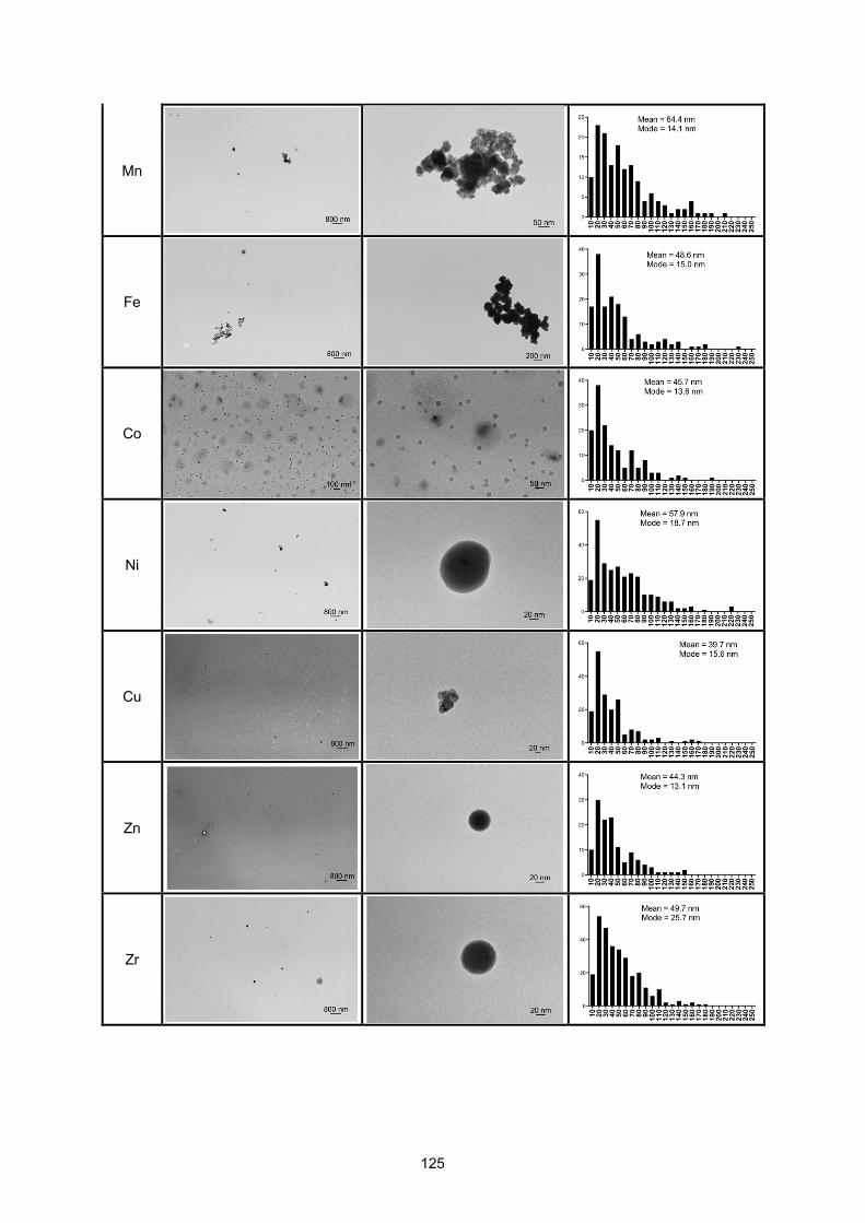
125
Mn
Fe
Co
Ni
Cu
Zn
Zr

126
Mo
Nd
Sm
Eu
Gd
Tb
Dy
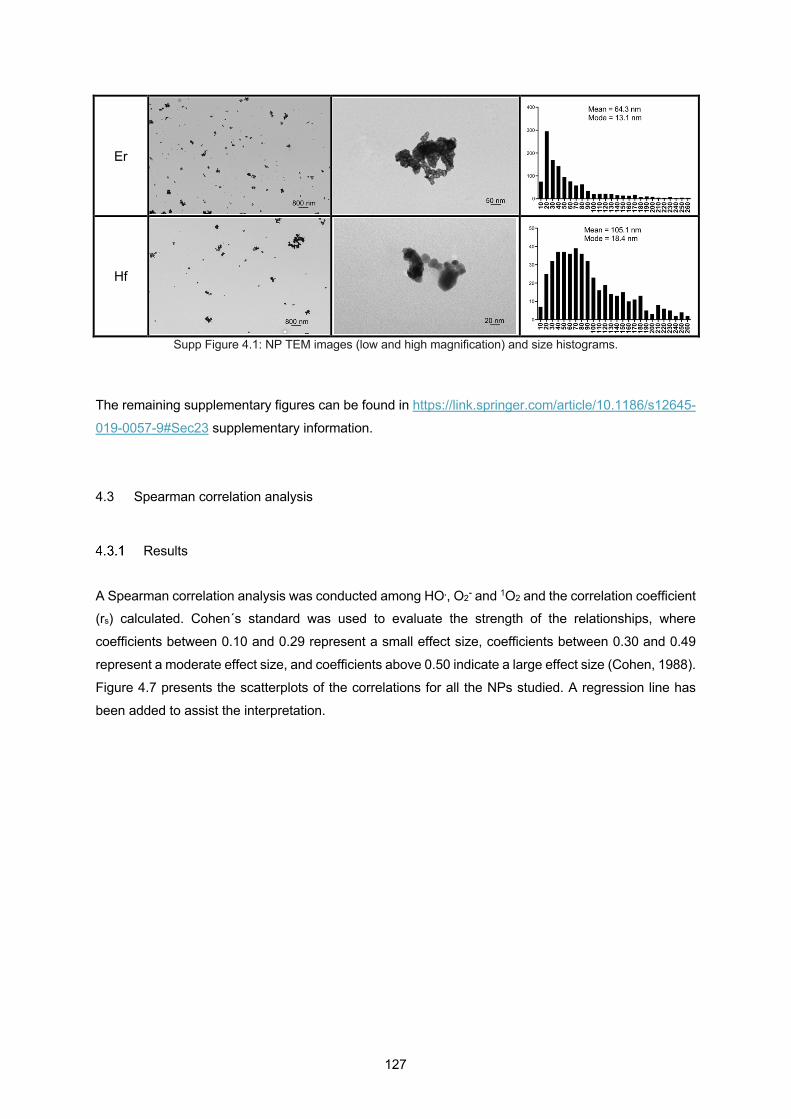
127
Er
Hf
Supp Figure 4.1: NP TEM images (low and high magnification) and size histograms.
The remaining supplementary figures can be found in https://link.springer.com/article/10.1186/s12645-
019-0057-9#Sec23 supplementary information.
4.3 Spearman correlation analysis
Results
A Spearman correlation analysis was conducted among HO., O2- and 1O2 and the correlation coefficient (rs) calculated. Cohen´s standard was used to evaluate the strength of the relationships, where
coefficients between 0.10 and 0.29 represent a small effect size, coefficients between 0.30 and 0.49
represent a moderate effect size, and coefficients above 0.50 indicate a large effect size (Cohen, 1988).
Figure 4.7 presents the scatterplots of the correlations for all the NPs studied. A regression line has
been added to assist the interpretation.
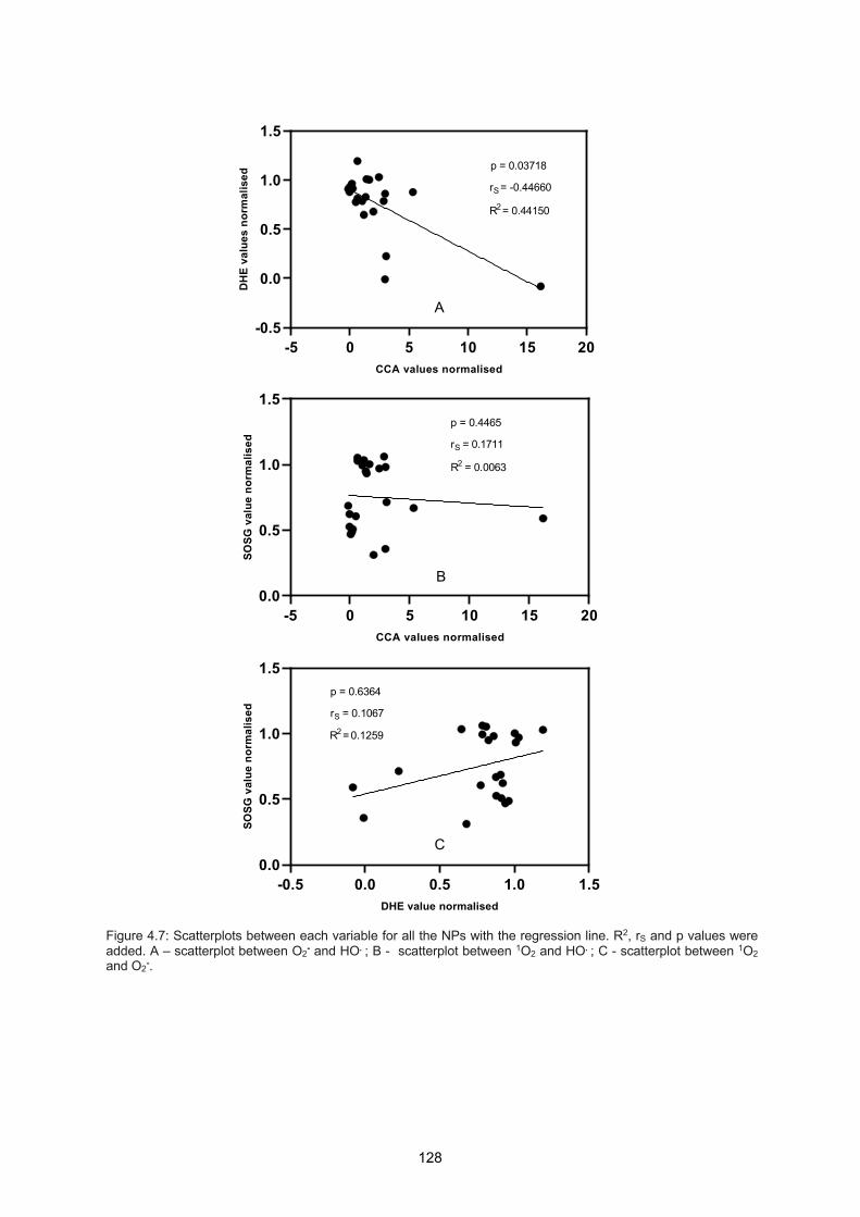
128
Figure 4.7: Scatterplots between each variable for all the NPs with the regression line. R2, rS and p values were added. A – scatterplot between O2- and HO. ; B - scatterplot between 1O2 and HO. ; C - scatterplot between 1O2 and O2-.
-5 0 5 10 15 20-0.5
0.0
0.5
1.0
1.5
CCA values normalised
DH
E va
lues
nor
mal
ised
R2 = 0.44150
rS = -0.44660
p = 0.03718
A
-5 0 5 10 15 200.0
0.5
1.0
1.5
CCA values normalised
SOSG
val
ue n
orm
alis
ed
R2 = 0.0063
rS = 0.1711
p = 0.4465
B
-0.5 0.0 0.5 1.0 1.50.0
0.5
1.0
1.5
DHE value normalised
SOSG
val
ue n
orm
alis
ed
R2 = 0.1259
rS = 0.1067
p = 0.6364
C
129
A significant negative correlation was observed between O2- (DHE) and HO. (CCA) (rS = -0.4466, p =
0.03718), as shown in Figure 4.7A. Positive but non-significant correlations were observed between 1O2 (SOSG) and HO. (CCA) (rS = 0.1711, p = 0.4465), and between 1O2 (SOSG) and O2- (DHE) (rS =
0.1067, p = 0.6364), as shown in Figures 4.7 B and C, respectively.
Considering only the NPs of period 4 of the periodic table: Sc, Ti, V, Cr, Mn, Fe, Co, Ni, Cu, and Zn,
Spearman correlation analysis was again conducted among HO., O2- and 1O2 (Figure 4.8).
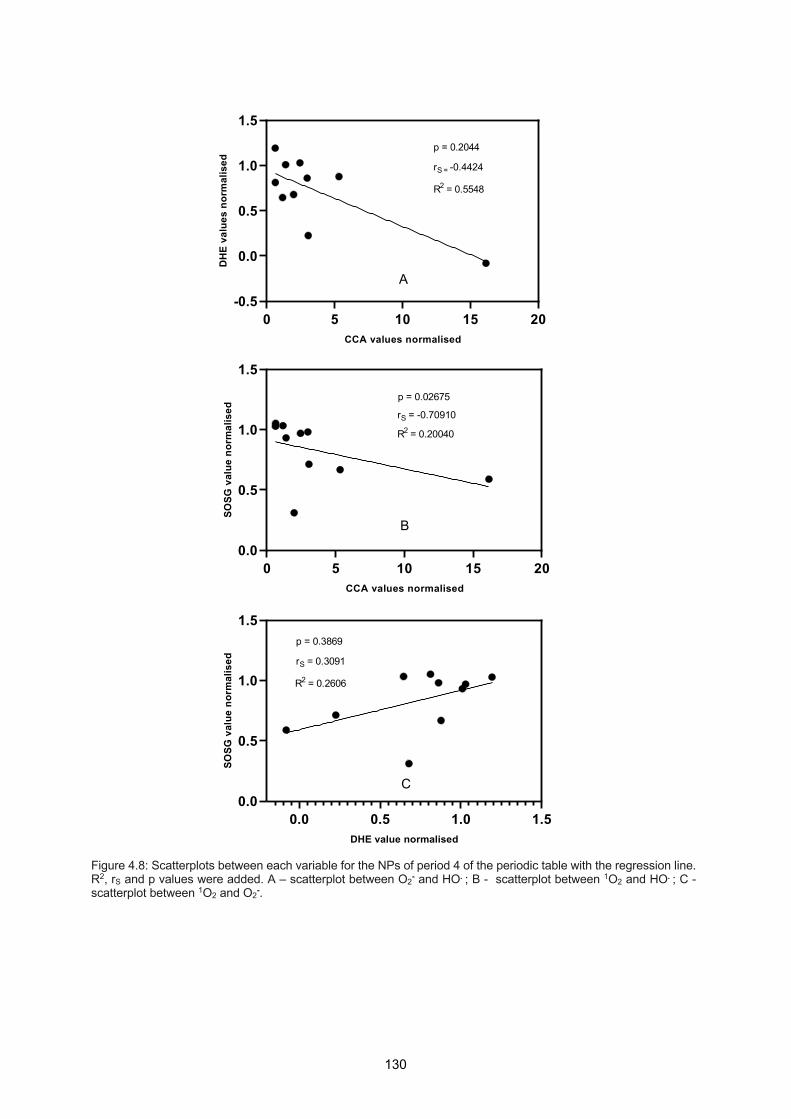
130
Figure 4.8: Scatterplots between each variable for the NPs of period 4 of the periodic table with the regression line. R2, rS and p values were added. A – scatterplot between O2- and HO. ; B - scatterplot between 1O2 and HO. ; C - scatterplot between 1O2 and O2-.
0 5 10 15 20-0.5
0.0
0.5
1.0
1.5
CCA values normalised
DH
E va
lues
nor
mal
ised
R2 = 0.5548
rS = -0.4424
p = 0.2044
A
0 5 10 15 200.0
0.5
1.0
1.5
CCA values normalised
SOSG
val
ue n
orm
alis
ed
R2 = 0.20040
rS = -0.70910
p = 0.02675
B
0.0 0.5 1.0 1.50.0
0.5
1.0
1.5
DHE value normalised
SOSG
val
ue n
orm
alis
ed
R2 = 0.2606
rS = 0.3091
p = 0.3869
C
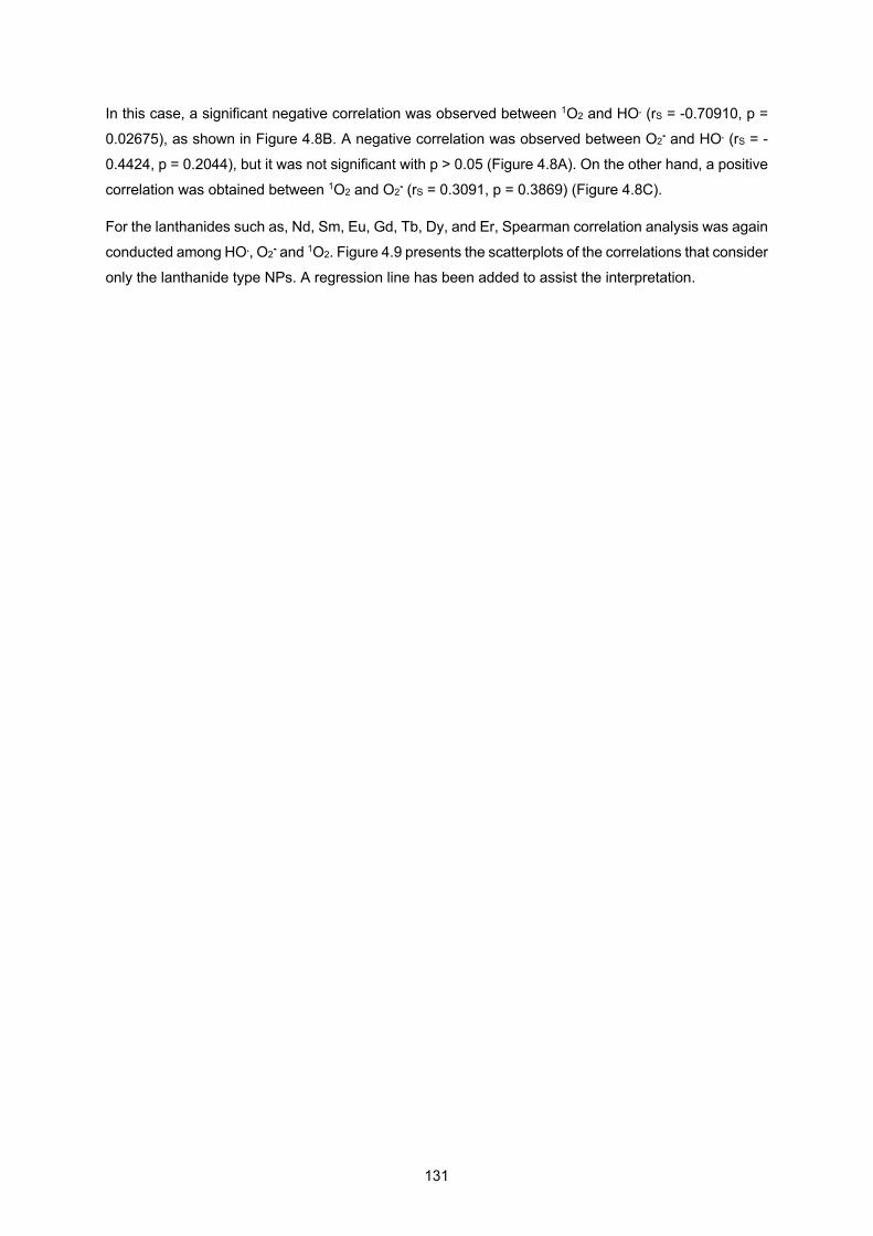
131
In this case, a significant negative correlation was observed between 1O2 and HO. (rS = -0.70910, p =
0.02675), as shown in Figure 4.8B. A negative correlation was observed between O2- and HO. (rS = -
0.4424, p = 0.2044), but it was not significant with p > 0.05 (Figure 4.8A). On the other hand, a positive
correlation was obtained between 1O2 and O2- (rS = 0.3091, p = 0.3869) (Figure 4.8C).
For the lanthanides such as, Nd, Sm, Eu, Gd, Tb, Dy, and Er, Spearman correlation analysis was again
conducted among HO., O2- and 1O2. Figure 4.9 presents the scatterplots of the correlations that consider
only the lanthanide type NPs. A regression line has been added to assist the interpretation.
132
Figure 4.9: Scatterplots between each variable for the lanthanide NPs with the regression line. R2, rS and p values were added. A – scatterplot between O2- and HO. ; B - scatterplot between 1O2 and HO. ; C - scatterplot between 1O2 and O2-.
-0.2 0.0 0.2 0.4 0.60.0
0.5
1.0
CCA values normalised
DH
E va
lues
nor
mal
ised
R2 = 0.3570
rS = 0.0000
p = 1.0000
A
-0.2 0.0 0.2 0.4 0.60.0
0.2
0.4
0.6
0.8
CCA values normalised
SOSG
val
ue n
orm
alis
ed
R2 = 0.05577
rS = -0.42860
p = 0.35360
B
0.0 0.5 1.0 1.50.0
0.2
0.4
0.6
0.8
1.0
DHE value normalised
SOSG
val
ue n
orm
alis
ed
R2 = 0.1508
rS = -0.5714
p = 0.2000
C
133
No significant results were found for lanthanides correlation between each variable, with all p values
above 0.05, as shown in Figure 4.9. There is no correlation between O2- and HO. (rS = 0.0) for
lanthanides (Figure 4.9A). For the remaining pairs, such as 1O2 and HO., and O2- and 1O2 (Figures 4.9
B and C, respectively), Spearman correlation was negative and indicates a moderate effect size for 1O2 and HO. (rS = -0.42860), and a large effect size for O2- and 1O2 (rS = -0.5714).
Discussion
Except for lanthanides (Figure 4.9 A and B), in general as hydroxyl radical production increases, both
superoxide and singlet oxygen tend to decrease, and as superoxide increases, singlet oxygen increases
as well (Figures 4.7C and 4.8C). It is possible to assume that reactions such as, for example:
1O2 + A ↔ HO. + B
O2- + C ↔ HO. + D
O2- + 1O2 + E ↔ HO. + F
, with A, B, C, D, E and F, as unknown species, might be happening in the presence of some of NPs
studied, with the exception of the lanthanide ones.
These results are in agreement with the reported Haber-Weiss reaction, which is considered the main
mechanism by which the highly reactive radical is generated in the presence of metal oxide NPs
(Hauser et al, 2016):
Haber-Weiss reaction: O2- + H2O2 → O2 + HO- + HO.
In the case of singlet oxygen, different reactions have been reported in the presence of catalytic
surfaces, such as (Hirakawa, 2018):
O2- + h+ → 1O2 reaction 4.1
O2- + HO. → 1O2 + OH- reaction 4.2
O2- + HO2 + H+ → 1O2 + H2O2 reaction 4.3
, where the photogenerated h+ in the valence band of the catalyst, such as for example, TiO2, and HO., can act as the oxidants to produce 1O2. Also, perhydroxyl radical generated from O2- and H+, also
produces 1O2.
Reaction 4.1 would mean that as superoxide decreases, singlet oxygen production would increase,
which is not consistent with the Spearman correlation analysis. In addition, the previous hole scavenger
results of some NPs such as, Fe3O4, CoO, NiO and ZnO, showed increases in superoxide radical
134
generation in the presence of formic acid (Figure 4.5B). There are two possible routes for the hole
scavenger results: the hole scavenger is increasing the lifetime of the electrons and promoting
superoxide formation at the NP surface, or the hole scavenger does not allow reaction 4.1 to happen,
and so superoxide is not being consumed through the reaction with holes to generate singlet oxygen. Since Spearman correlation analysis found that as superoxide increases singlet oxygen increases, it is
more likely that the hole scavenger experiment is actually testing the route of superoxide formation at
the NP surface due to the interaction of oxygen with the electrons from the e-/h+ pairs.
Reaction 4.2 although it would mean that as superoxide decreases, singlet oxygen production would
increase, which again is not consistent with the Spearman correlation analysis, it also means that as
hydroxyl radical decreases, singlet oxygen increases which is consistent with the correlation results
(Figures 4.7B and 4.8B). In this case, since through reaction 4.2 hydroxide ion (OH-) is also formed,
this species would have to be somehow present in a reaction that would generate superoxide radicals, due to the findings of the correlation analysis that as superoxide increases, singlet oxygen increases
(Figures 4.7C and 4.8C). For example, or the Haber-Weiss reaction, where both hydroxyl radicals and
hydroxide ions, in the presence of oxygen, form both hydrogen peroxide and superoxide, or the
electrochemical reduction of O2 (Hayyan et al., 2016):
2O2- + H2O → O2 + HO2 + OH-
Due to the fact that O2- and its protonated form, HO2, are in equilibrium in an aqueous solution (Rose,
2012):
O2- + H2O ↔ O2 + HO2 + OH-
, and that O2- dominates the equilibrium in solutions whose pH is above the pKa = 4.8 (Bielski et al.,
1985), in the nanoparticles solutions studied with a pH=7.4, superoxide is the predominant species.
This means that the hydroxide ions produced in reaction 4.2, will produce superoxide through the
electrochemical reduction of O2. If this is the case, as singlet oxygen production increases through
reaction 4.2, superoxide will increase through the electrochemical reduction of O2, which is in
accordance with the Spearman correlation results.
Reaction 4.3 leads to the conclusion that as superoxide decreases, singlet oxygen production would
increase, which is not consistent with the Spearman correlation analysis. Once again, if there is another
possible route for superoxide formation through hydrogen peroxide (the other product formed from
reaction 4.3) then as singlet oxygen increases superoxide would increase as well, which will be
consistent with Spearman correlation analysis. For example, one of the main reactions of oxidative
stress induced by manganese is the superoxide dismutase (Aust et al., 1985):
2 O2- + 2H+ à H2O2 + O2
, where in this case superoxide leads to hydrogen peroxide production in the presence of oxygen.

135
5 Conclusions and Future Directions

136

137
RT is currently limited by the dose of X-rays that can be safely given to patients, since the dose of X-
rays that will kill tumour tissue is similar to the dose that will kill surrounding healthy tissues. This thesis
aimed to provide a further understanding of the different type of metal oxide NP radiosensitisers by
studying their compatibility with cancer cells, their interaction with DNA and the type and amount of radicals formed during their interaction with X-rays. Other studies have looked at individual metal
oxides, but differences in experimental designs made it very difficult to compare results.
First studies in the role of metal NPs in cancer treatment showed significant radiosensitisation in vivo
(Hainfeld et al., 2008), while Monte Carlo simulations suggested that the observed radiosensitisation
could not be fully explained by the increase in physical dose (Butterworth et al., 2012). Although the
parameters that influence NP radiosensitisation are still not fully understood, it is believe that is the
combination of physical and biological effects that is responsible for the radiosensitisation induced by the NPs (King et al., 2017). Different pathways have been proposed for the production of hydroxyl
radicals in the presence of radiation and NPs. Taking into account three different pathways for hydroxyl
radicals production a study using 7-hydroxycoumarin showed that the dominant mechanism for HO.
production is through the interaction of radiolysis products with water-NP interface (Sicard-Roselli et al.,
2014). The link between the physical process, where the energy absorption by the NP, the chemical
process, where radical production occurs, and the biological process, where biological interactions lead
to cell death, is fundamental for the understanding of the role of the NPs in the interaction of radiation
with cells. Indirect measurements reported a radical production induced by NPs four-times higher than normal radiolysis in the absence of oxygen and it was suggested that this overproduction arises from
surface reactions or catalysis at NP-solvent interface (Baldacchino et al., 2019). For example, a study
adapted to quantify the yields of solvated electrons and hydroxyl radicals produced in water by gold
NPs submitted to keV and MeV ionising radiation proposed a key role for interfacial water around NPs
for radical production (Gilles et al., 2018). Combining NPs into the process of water radiolysis does not
always lead to an enhancement of radiosensitisation unless certain conditions are met. To optimise the
whole system (NP+radiation+water) the knowledge of the individual enhancements given by the
physical, chemical and biological processes is required since the highest enhancement will most likely be given when the highest individual enhancements are properly combined (Guo, 2019). The work
presented in this thesis demonstrates the importance of ROS species created by metal oxide NPs in
the process of cancer cell death.
The introduction of NPs into the RT treatment will require optimisation within treatment planning
algorithms, such as accurate characterisation of NP interaction with the X-ray spectrum of the irradiation
field as a function of NP material, size and concentration (Schuemann et al, 2016).
The main benefit of using NPs during RT treatment is the potential dose enhancement that results from
the interaction of the MV treatment field with the NPs. Controlling NP site uptake, it will be possible to
increase the dose to the tumour whilst maintaining or reducing the dose to the surrounding normal
tissue. This approach is particularly important for SABR treatments since this type of RT treatment uses
larger doses per treatment compared to the standard RT treatment. In this case a single geometric miss
can result in a reduction in tumour control and/or an increase in toxicity effects. SABR is moving towards
138
the use of FF free fields since this approach results in significantly higher dose rates (R. B. King et al.,
2013). In vitro studies suggested that the combination of FF free fields with NPs will lead to a further
increase in dose deposition because of the increased proportion of lower energy photons, capable of
inducing photocatalyst-like changes in the NPs (Detappe et al., 2016).
This work provided a fair comparison between the different types of NP metal oxides because important
parameters for the inclusion of NPs in the field of RT were tested under the same conditions. Here
evidence of ROS production by the different types of metal oxide NPs and of their behavior under the
biological environment either by testing their interactions with plasmid DNA and with cancer cells under
realistic tumour hypoxia conditions is provided. Once again, ROS outputs do not fit with the standard
physical theory of radiosensitisation which is consistent with other recent work on nano-radiocatalysis
(Guo, 2019). Furthermore, this thesis also showed evidence of NPs that most likely only work under the biological process providing no evidence of relevant ROS production but at the same time relevant
interactions with plasmic DNA and cancer cells, as shown by CuO NPs.
The main limitations of this study include diameter and agglomeration state variability between the
different type of NPs. Also, irradiation times after probe and NP interaction might play an important role.
NP quenching is dependent on the interaction time with probe. Although consistency regarding the time
between probe injection into the sample and scheduled irradiation time was maintained constant
throughout the experiments it was not possible to control the hospital irradiation delay.
Future directions include studying the behaviour of these NPs in other cell lines, since as explained
before, it is known that the radiosensitisation results depend on the type of cell line used. Furthermore,
different types of coating should be tested to improve cellular uptake and toxicity and also for better
radiosensitisation, since catalytic ability might be compromised by the type of coating used as shown
by different studies (Gilles et al., 2014; Grellet et al., 2017). Understanding other principles than the
physical ones that might be responsible for the radiosensitisation observed will be a very important step
for the knowledge of the role of the NPs in the world of cancer therapy.
The future of NP radiosensitisers will be connected to the mixtures of high-Z elements with catalytic
metal oxides, where the photoelectric effect of the high Z element will add secondary electrons into the
catalyst allowing the production of more ROS. Work has already been made in this direction such as
Bi2WO6 catalysts (Sahu & Cates, 2017), lanthanide-doped TiO2 (Townley et al., 2012) and lanthanide-
doped ZnO NPs (Ghaemi et al., 2016), ZnO-SiO2 nanocomposites (Generalov et al., 2015) and Au-
Bi2S3 heterostructures (Wang et al., 2019).
The two NPs radiosensitisers currently on the market, NBTXR3 and AGuIX, are still in clinical trial. The study of 50 nm hafnium-oxide (HfO2) NPs (NBTXR3) in patients with locally advanced soft-tissue
sarcoma showed that a significantly higher proportion of patients (twice as many) given NBTXR3 plus
RT achieved a pathological complete response compared with patients who received RT alone
(Bonvalot et al., 2019). This study lacks an appropriate control group, i.e. injection of solution without
NBTXR3, since it is not possible to associate the observed results to the NPs effects without a control
group that excludes the effects due to the intratumoral injection process where a difference of 10% of
139
tumoral volume is seen. Once again, the real mode of action of these NPs is still unclear and since
evidence supports chemical and biological of these NPs during irradiation a potential toxicity due to
long-term systemic persistency is still a possibility (Vilotte et al., 2019). AGuIX NPs were tested in
multiple brain metastases in combination with whole brain RT (10 × 3 Gy over 3 weeks max) and
showed a strong local radioenhancing effect (Lux et al., 2019). A limitation of this study was the lower
number of patients used, around 15 to18 patients, (Verry et al., 2019). An important result for AGuIX
NPs revealed the efficacy of the EPR effect inducing accumulation and more importantly retention of the ultrasmall (< 5 nm) NPs at the tumour sites. Nevertheless, this passive targeting strategy still has
to be confirmed in human due to the complex heterogeneity of the tumour types and environments. The
safety related to fast renal clearance coupled to the non-toxicity in the absence of radiation allows the
use of these NPs into more clinical trials (Bort et al., 2020). Another clinical trial (NanoCOL clinical trial,
NCT03308604) aimed to treat advanced cervical cancer through RT and brachytherapy is ongoing.
Although these NPs are very strong candidates in this field a lot of work still needs to be done and
parallel NPs should be tested due to the complexity of the different types of tumours. The more probable
picture in the future will be the use of different radiosensitisers to improve the efficacy of RT in the treatment of the different types of tumours.
Although photon beam therapy is still the most used RT beam, other types of beams are emerging such
as proton beams. For this reason, translating the NP studies into the different types of beams will be a
very useful tool for the future of RT. A study already demonstrated the interaction of AGuIX NPs with a
150 MeV proton beam and concluded that once again addition of metal-based NPs is a promising
strategy not only to increase cell killing action of fast protons, but also to improve tumour targeting
(Schlathölter et al., 2016).
The work presented in this thesis showed that the pure physical aspect does not explain the process of
radiosensitisation. For instance, gadolinium in the form of dendrimer (AGuIX) seems to be a strong
candidate as a radiosensitiser in the RT treatment, but the results in this thesis showed that based only
on physical principles, gadolinium is a poor radiosensitiser. This showed that research on the different
mechanisms of radiosensitisation will help the introduction of the NPs in the field of RT in a way that we
will be able to understand and control the early and late stage effects of these radiosensitisers when
inside the patient. Furthermore, my work tested metal oxide NPs that have never been considered for this field and now become more interesting. Clearly, processes such as catalysis, seem to be a major
player in radiosensitisation and my work opens a whole new field of investigations to try many different
compounds that were not previously considered for RT on the basis of a predicted poor photoelectric
effect.

140

141
6 Bibliography

142
143
Abdelhamid, M. A. S., Fábián, L., Macdonald, C. J., Cheesman, M. R., Gates, A. J., & Waller, Z. A. E. (2018). Redox-dependent control of i-Motif DNA structure using copper cations. Nucleic Acids Research, 46(12), 5886–5893. https://doi.org/10.1093/nar/gky390
Abou-Zeid, S. M., El-Bialy, B. E., El-Borai, N. B., AbuBakr, H. O., & Elhadary, A. M. A. (2018). Radioprotective effect of Date syrup on radiation- induced damage in Rats. Scientific Reports, 8(1), 1–10. https://doi.org/10.1038/s41598-018-25586-3
Adams, G. E., Asquith, J. C., Dewey, D. L., Foster, J. L., Michael, B. D., & Willson, R. L. (1971). Electron Affinic Sensitization. International Journal of Radiation Biology and Related Studies in Physics, Chemistry and Medicine, 19(6), 575–585. https://doi.org/10.1080/09553007114550751
Algethami, M. (2015). Radiation Dose Enhancement Using Bi2S3 Nanoparticles in Cultured Mouse PC3 Prostate and B16 Melanoma Cells. NanoWorld Journal, 1(3), 99–104. https://doi.org/10.17756/nwj.2015-013
Alimoradi, H., Matikonda, S. S., Gamble, A. B., Giles, G. I., & Griesh, K. (2017). Redox activated polymeric nanoparticles in tumor therapy. In Nanostructures for Drug Delivery. Elsevier Inc. https://doi.org/10.1016/b978-0-323-46143-6.00010-5
Alizadeh, E., Sanz, A. G., García, G., & Sanche, L. (2013). Radiation damage to DNA: The indirect effect of low-energy electrons. Journal of Physical Chemistry Letters, 4(5), 820–825. https://doi.org/10.1021/jz4000998
Alkilany, A. M., & Murphy, C. J. (2010). Toxicity and cellular uptake of gold nanoparticles: What we have learned so far? Journal of Nanoparticle Research, 12(7), 2313–2333. https://doi.org/10.1007/s11051-010-9911-8
Altomare, A., Corriero, N., Cuocci, C., Falcicchio, A., Moliterni, A., & Rizzi, R. (2015). QUALX2.0: A qualitative phase analysis software using the freely available database POW-COD. Journal of Applied Crystallography. https://doi.org/10.1107/S1600576715002319
Aspatwar, A., Becker, H. M., Parvathaneni, N. K., Hammaren, M., Svorjova, A., Barker, H., Supuran, C. T., Dubois, L., Lambin, P., Parikka, M., Parkkila, S., & Winum, J. Y. (2018). Nitroimidazole-based inhibitors DTP338 and DTP348 are safe for zebrafish embryos and efficiently inhibit the activity of human CA IX in Xenopus oocytes. Journal of Enzyme Inhibition and Medicinal Chemistry, 33(1), 1064–1073. https://doi.org/10.1080/14756366.2018.1482285
Atha, D. H., Wang, H., Petersen, E. J., Cleveland, D., Holbrook, R. D., Jaruga, P., Dizdaroglu, M., Xing, B., & Nelson, B. C. (2012). Copper Oxide Nanoparticle Mediated DNA Damage in Terrestrial Plant Models. Environmental Science & Technology, 46(3), 1819–1827. https://doi.org/10.1021/es202660k
Attia, M. F., Anton, N., Wallyn, J., Omran, Z., & Vandamme, T. F. (2019). An overview of active and passive targeting strategies to improve the nanocarriers efficiency to tumour sites. Journal of Pharmacy and Pharmacology, 71(8), 1185–1198. https://doi.org/10.1111/jphp.13098
Aust, S. D., Morehouse, L. A., & Thomas, C. E. (1985). Role of metals in oxygen radical reactions. Journal of Free Radicals in Biology and Medicine, 1(1), 3–25. https://doi.org/10.1016/0748-5514(85)90025-X
Azzam, E. I., Jay-Gerin, J. P., & Pain, D. (2012). Ionizing radiation-induced metabolic oxidative stress and prolonged cell injury. In Cancer Letters. https://doi.org/10.1016/j.canlet.2011.12.012
Baker, C. H. (2013). Harnessing cerium oxide nanoparticles to protect normal tissue from radiation damage. Translational Cancer Research, 2(4), 343–358. https://doi.org/10.3978/j.issn.2218-676X.2013.08.15
Baldacchino, G., Brun, E., Denden, I., Bouhadoun, S., Roux, R., Khodja, H., & Sicard-Roselli, C. (2019). Importance of radiolytic reactions during high-LET irradiation modalities: LET effect, role of O2 and radiosensitization by nanoparticles. Cancer Nanotechnology, 10(1), 1–21. https://doi.org/10.1186/s12645-019-0047-y
Barrow, M., Taylor, A., Murray, P., Rosseinsky, M. J., & Adams, D. J. (2015). Design considerations for the synthesis of polymer coated iron oxide nanoparticles for stem cell labelling and tracking using
144
MRI. Chemical Society Reviews, 44(19), 6733–6748. https://doi.org/10.1039/c5cs00331h
Baskar, R., Dai, J., Wenlong, N., Yeo, R., & Yeoh, K. W. (2014). Biological response of cancer cells to radiation treatment. Frontiers in Molecular Biosciences, 1(NOV), 1–9. https://doi.org/10.3389/fmolb.2014.00024
Bertrand, N., Grenier, P., Mahmoudi, M., Lima, E. M., Appel, E. A., Dormont, F., Lim, J. M., Karnik, R., Langer, R., & Farokhzad, O. C. (2017). Mechanistic understanding of in vivo protein corona formation on polymeric nanoparticles and impact on pharmacokinetics. Nature Communications, 8(1). https://doi.org/10.1038/s41467-017-00600-w
Biaglow, J. E. (1981). The effects of ionizing radiation on mammalian cells. Journal of Chemical Education, 58(2), 144–156. https://doi.org/10.1021/ed058p144
Bielski, B. H. J., Cabelli, D. E., Arudi, R. L., & Ross, A. B. (1985). Reactivity of HO2/O−2 Radicals in Aqueous Solution. Journal of Physical and Chemical Reference Data, 14(4), 1041–1100. https://doi.org/10.1063/1.555739
Blatnik, J., Luebke, L., Simonet, S., Nelson, M., Price, R., Leek, R., Zeng, L., Wu, A., & Brown, E. (2012). Dye surface coating enables visible light activation of TiO 2 nanoparticles leading to degradation of neighboring biological structures. Microscopy and Microanalysis, 18(1), 134–142. https://doi.org/10.1017/S1431927611012414
Bligaard, T., Nørskov, J. K., Dahl, S., Matthiesen, J., Christensen, C. H., & Sehested, J. (2004). The Brønsted-Evans-Polanyi relation and the volcano curve in heterogeneous catalysis. Journal of Catalysis, 224(1), 206–217. https://doi.org/10.1016/j.jcat.2004.02.034
Bogdan, J., Pławińska-Czarnak, J., & Zarzyńska, J. (2017). Nanoparticles of Titanium and Zinc Oxides as Novel Agents in Tumor Treatment: a Review. Nanoscale Research Letters, 12(1). https://doi.org/10.1186/s11671-017-2007-y
Bonvalot, S., Rutkowski, P. L., Thariat, J., Carrère, S., Ducassou, A., Sunyach, M. P., Agoston, P., Hong, A., Mervoyer, A., Rastrelli, M., Moreno, V., Li, R. K., Tiangco, B., Herraez, A. C., Gronchi, A., Mangel, L., Sy-Ortin, T., Hohenberger, P., de Baère, T., … Papai, Z. (2019). NBTXR3, a first-in-class radioenhancer hafnium oxide nanoparticle, plus radiotherapy versus radiotherapy alone in patients with locally advanced soft-tissue sarcoma (Act.In.Sarc): a multicentre, phase 2–3, randomised, controlled trial. The Lancet Oncology, 20(8), 1148–1159. https://doi.org/10.1016/S1470-2045(19)30326-2
Boonstra, J., & Post, J. A. (2004). Molecular events associated with reactive oxygen species and cell cycle progression in mammalian cells. Gene, 337(SUPPL.), 1–13. https://doi.org/10.1016/j.gene.2004.04.032
Bort, G., Lux, F., Dufort, S., Crémillieux, Y., Verry, C., & Tillement, O. (2020). EPR-mediated tumor targeting using ultrasmall-hybrid nanoparticles: From animal to human with theranostic AGuIX nanoparticles. Theranostics, 10(3), 1319–1331. https://doi.org/10.7150/thno.37543
Brown, P. E. (1967). Mechanism of action of aminothiol radioprotectors. Nature, 213(5074), 363–364. https://doi.org/10.1038/213363a0
Butterworth, K. T., McMahon, S. J., Currell, F. J., & Prise, K. M. (2012). Physical basis and biological mechanisms of gold nanoparticle radiosensitization. Nanoscale, 4(16), 4830–4838. https://doi.org/10.1039/c2nr31227a
Calatayud, M. P., Sanz, B., Raffa, V., Riggio, C., Ibarra, M. R., & Goya, G. F. (2014). The effect of surface charge of functionalized Fe3O4 nanoparticles on protein adsorption and cell uptake. Biomaterials, 35(24), 6389–6399. https://doi.org/10.1016/j.biomaterials.2014.04.009
Candelaria, M., Garcia-Arias, A., Cetina, L., & Dueñas-Gonzalez, A. (2006). Radiosensitizers in cervical cancer. Cisplatin and beyond. Radiation Oncology, 1(1), 1–17. https://doi.org/10.1186/1748-717X-1-15
Carrasco-Flores, E. A., & LaVerne, J. A. (2007). Surface species produced in the radiolysis of zirconia nanoparticles. The Journal of Chemical Physics, 127(23), 234703. https://doi.org/10.1063/1.2806164

145
Cedervall, T., Lynch, I., Lindman, S., Berggård, T., Thulin, E., Nilsson, H., Dawson, K. A., & Linse, S. (2007). Understanding the nanoparticle-protein corona using methods to quntify exchange rates and affinities of proteins for nanoparticles. Proceedings of the National Academy of Sciences of the United States of America, 104(7), 2050–2055. https://doi.org/10.1073/pnas.0608582104
Cervantes-Cervantes, M. P., Calderón-Salinas, J. V., Albores, A., & Muñoz-Sánchez, J. L. (2005). Copper increases the damage to DNA and proteins caused by reactive oxygen species. Biological Trace Element Research, 103(3), 229–248. https://doi.org/10.1385/BTER:103:3:229
Chelnokov, E., Cuba, V., Simeone, D., Guigner, J. M., Schmidhammer, U., Mostafavi, M., & Le Caër, S. (2014). Electron transfer at oxide/water interfaces induced by ionizing radiation. Journal of Physical Chemistry C, 118(15), 7865–7873. https://doi.org/10.1021/jp501396a
Chen, E. I., Hewel, J., Krueger, J. S., Tiraby, C., Weber, M. R., Kralli, A., Becker, K., Yates, J. R., & Felding-Habermann, B. (2007). Adaptation of energy metabolism in breast cancer brain metastases. Cancer Research, 67(4), 1472–1486. https://doi.org/10.1158/0008-5472.CAN-06-3137
Cherry, S. R., Sorenson, J., Phelps, M. E., & Methé, B. M. (2004). Physics in Nuclear Medicine. In Medical Physics (Vol. 31). https://doi.org/10.1118/1.1776595
Chithrani, D. B., Jelveh, S., Jalali, F., van Prooijen, M., Allen, C., Bristow, R. G., Hill, R. P., & Jaffray, D. A. (2010). Gold Nanoparticles as Radiation Sensitizers in Cancer Therapy. Radiation Research, 173(6), 719. https://doi.org/10.1667/rr1984.1
Cho, S. H. (2005). Estimation of tumour dose enhancement due to gold nanoparticles during typical radiation treatments: A preliminary Monte Carlo study. Physics in Medicine and Biology, 50(15). https://doi.org/10.1088/0031-9155/50/15/N01
Choi, S. W., Kim, W. S., & Kim, J. H. (2003). Surface modification of functional nanoparticles for controlled drug delivery. Journal of Dispersion Science and Technology, 24(3–4), 475–487. https://doi.org/10.1081/DIS-120021803
Chu, S.-H., Karri, S., Ma, Y.-B., Feng, D.-F., & Li, Z.-Q. (2013). In vitro and in vivo radiosensitization induced by hydroxyapatite nanoparticles. Neuro-Oncology, 15(7), 880–890. https://doi.org/10.1093/neuonc/not030
Chusuei, C. C., Wu, C. H., Mallavarapu, S., Hou, F. Y. S., Hsu, C. M., Winiarz, J. G., Aronstam, R. S., & Huang, Y. W. (2013). Cytotoxicity in the age of nano: The role of fourth period transition metal oxide nanoparticle physicochemical properties. Chemico-Biological Interactions, 206(2), 319–326. https://doi.org/10.1016/j.cbi.2013.09.020
Cohen, J. (1988). Statistical Power Analysis for the Behavioral Sciences. Lawrence Erlbaum Associates. ISBN 0-8058-0283-5
Curtis, K. A., Miller, D., Millard, P., Basu, S., Horkay, F., & Chandran, P. L. (2016). Unusual salt and pH induced changes in polyethylenimine solutions. PLoS ONE, 11(9), 1–20. https://doi.org/10.1371/journal.pone.0158147
Dai, Y., Xu, C., Sun, X., & Chen, X. (2017). Nanoparticle design strategies for enhanced anticancer therapy by exploiting the tumour microenvironment. Chemical Society Reviews, 46(12), 3830–3852. https://doi.org/10.1039/c6cs00592f
Dahlmann, H., Vaidyanathan, V., Sturla, S., (2009) Investigating the Biochemical Impact of DNA Damage with Structure-Based Probes: Abasic Sites, Photodimers, Alkylation Adducts, and Oxidative Lesions. Biochemistry. 48(40): 9347–9359. https://doi:10.1021/bi901059k.
Degen, A., & Kosec, M. (2000). Effect of pH and impurities on the surface charge of zinc oxide in aqueous solution. Journal of the European Ceramic Society, 20(6), 667–673. https://doi.org/10.1016/S0955-2219(99)00203-4
Desouky, O., Ding, N., & Zhou, G. (2015). Targeted and non-targeted effects of ionizing radiation. Journal of Radiation Research and Applied Sciences, 8(2), 247–254. https://doi.org/10.1016/j.jrras.2015.03.003
146
Detappe, A., Kunjachan, S., Drané, P., Kotb, S., Myronakis, M., Biancur, D. E., Ireland, T., Wagar, M., Lux, F., Tillement, O., & Berbeco, R. (2016). Key clinical beam parameters for nanoparticle-mediated radiation dose amplification. Scientific Reports, 6(September), 1–8. https://doi.org/10.1038/srep34040
Drescher, M., Hentschel, M., Kienberger, R., Uiberacker, M., Yakovlev, V., Scrinzi, A., Westerwalbesloh, T., Kleineberg, U., Heinzmann, U., & Krausz, F. (2002). Time-resolved atomic inner-shell spectroscopy. Nature, 419(6909), 803–807. https://doi.org/10.1038/nature01143
Du, G., & Espenson, J. H. (2005). Oxidation of vanadium(III) by hydrogen peroxide and the oxomonoperoxo vanadium(V) ion in acidic aqueous solutions: A kinetics and simulation study. Inorganic Chemistry, 44(15), 5514–5522. https://doi.org/10.1021/ic050502j
Du, J., Zhu, W., Yang, L., Wu, C., Lin, B., Wu, J., Jin, R., Shen, T., & Ai, H. (2016). Reduction of polyethylenimine-coated iron oxide nanoparticles induced autophagy and cytotoxicity by lactosylation. Regenerative Biomaterials, 3(4), 223–229. https://doi.org/10.1093/RB/RBW023
Eghtedari, M., Liopo, a., Copland, J., Oraevsky, A., & Motamedi, M. (2009). Engineering of hetero-functional gold nanorods for the in-vivo molecular targeting of breast cancer cells. Nano Lett. 9(1): 287–291. https://doi.org/10.1021/nl802915q.
Fan, W., Bu, W., Shen, B., He, Q., Cui, Z., Liu, Y., Zheng, X., Zhao, K., & Shi, J. (2015). Intelligent MnO2 Nanosheets Anchored with Upconversion Nanoprobes for Concurrent pH-/H2O2-Responsive UCL Imaging and Oxygen-Elevated Synergetic Therapy. Advanced Materials, 27(28), 4155–4161. https://doi.org/10.1002/adma.201405141
Farhood, B., Goradel, N. H., Mortezaee, K., Khanlarkhani, N., Salehi, E., Nashtaei, M. S., Mirtavoos-mahyari, H., Motevaseli, E., Shabeeb, D., Musa, A. E., & Najafi, M. (2019). Melatonin as an adjuvant in radiotherapy for radioprotection and radiosensitization. Clinical and Translational Oncology, 21(3), 268–279. https://doi.org/10.1007/s12094-018-1934-0
Featherstone, C., & Jackson, S. P. (n.d.). DNA double-strand break repair. 759–761.
Fokas, E., Prevo, R., Pollard, J. R., Reaper, P. M., Charlton, P. A., Cornelissen, B., Vallis, K. A., Hammond, E. M., Olcina, M. M., Gillies McKenna, W., Musche, R. J., & Brunner, T. B. (2012). Targeting ATR in vivo using the novel inhibitor VE-822 results in selective sensitization of pancreatic tumors to radiation. Cell Death and Disease, 3(12), e441-10. https://doi.org/10.1038/cddis.2012.181
Frick, R., Müller-Edenborn, B., Schlicker, A., Rothen-Rutishauser, B., Raemy, D. O., Günther, D., Hattendorf, B., Stark, W., & Beck-Schimmer, B. (2011). Comparison of manganese oxide nanoparticles and manganese sulfate with regard to oxidative stress, uptake and apoptosis in alveolar epithelial cells. Toxicology Letters, 205(2), 163–172. https://doi.org/10.1016/j.toxlet.2011.05.1037
Fruehauf, J. P., & Meyskens, F. L. (2007). Reactive oxygen species: A breath of life or death? Clinical Cancer Research, 13(3), 789–794. https://doi.org/10.1158/1078-0432.CCR-06-2082
Fujishima, A., & Zhang, X. (2006). Titanium dioxide photocatalysis: present situation and future approaches. Comptes Rendus Chimie, 9(5–6), 750–760. https://doi.org/10.1016/j.crci.2005.02.055
Gara, P. M. D., Garabano, N. I., Portoles, M. J. L., Moreno, M. S., Dodat, D., Casas, O. R., Gonzalez, M. C., & Kotler, M. L. (2012). ROS enhancement by silicon nanoparticles in X-ray irradiated aqueous suspensions and in glioma C6 cells. Journal of Nanoparticle Research, 14(3). https://doi.org/10.1007/s11051-012-0741-8
Gatenby, R. A., & Gillies, R. J. (2004). Why do cancers have high aerobic glycolysis? Nature Reviews Cancer, 4(11), 891–899. https://doi.org/10.1038/nrc1478
Generalov, R., Kuan, W. B., Chen, W., Kristensen, S., & Juzenas, P. (2015). Radiosensitizing effect of zinc oxide and silica nanocomposites on cancer cells. Colloids and Surfaces B: Biointerfaces, 129, 79–86. https://doi.org/10.1016/j.colsurfb.2015.03.026
Gérard, M., Corroyer-Dulmont, A., Lesueur, P., Collet, S., Chérel, M., Bourgeois, M., Stefan, D., Limkin,
147
E. J., Perrio, C., Guillamo, J.-S., Dubray, B., Bernaudin, M., Thariat, J., & Valable, S. (2019). Hypoxia Imaging and Adaptive Radiotherapy: A State-of-the-Art Approach in the Management of Glioma. Frontiers in Medicine, 6(June). https://doi.org/10.3389/fmed.2019.00117
Ghaemi, B., Mashinchian, O., Mousavi, T., Karimi, R., Kharrazi, S., & Amani, A. (2016). Harnessing the Cancer Radiation Therapy by Lanthanide-Doped Zinc Oxide Based Theranostic Nanoparticles. ACS Applied Materials and Interfaces, 8(5), 3123–3134. https://doi.org/10.1021/acsami.5b10056
Ghita, M., McMahon, S. J., Taggart, L. E., Butterworth, K. T., Schettino, G., & Prise, K. M. (2017). A mechanistic study of gold nanoparticle radiosensitisation using targeted microbeam irradiation. Scientific Reports, 7(November 2016), 1–12. https://doi.org/10.1038/srep44752
Gilbert, D. L. (1963). The Role of Pro-Oxidants and Antioxidants in Oxygen Toxicity. Radiation Research Supplement, 3(1963), 44. https://doi.org/10.2307/3583674
Gilbert, D. L., Gerschman, R., Ruhm, K. B., & Price, W. E. (1958). The production of hydrogen peroxide by high oxygen pressures. The Journal of General Physiology, 41(5), 989–1003. https://doi.org/10.1085/jgp.41.5.989
Gill, M. R., & Vallis, K. A. (2019). Transition metal compounds as cancer radiosensitizers. Chemical Society Reviews, 48(2), 540–557. https://doi.org/10.1039/c8cs00641e
Gilles, M., Brun, E., & Sicard-Roselli, C. (2014). Gold nanoparticles functionalization notably decreases radiosensitization through hydroxyl radical production under ionizing radiation. Colloids and Surfaces B: Biointerfaces. https://doi.org/10.1016/j.colsurfb.2014.10.028
Gilles, M., Brun, E., & Sicard-Roselli, C. (2018). Quantification of hydroxyl radicals and solvated electrons produced by irradiated gold nanoparticles suggests a crucial role of interfacial water. Journal of Colloid and Interface Science, 525, 31–38. https://doi.org/10.1016/j.jcis.2018.04.017
Godbey, W. T., Wu, K. K., Hirasaki, G. J., & Mikos, A. G. (1999). Improved packing of poly(ethylenimine)/DNA complexes increases transfection efficiency. Gene Therapy, 6(8), 1380–1388. https://doi.org/10.1038/sj.gt.3300976
Godbey, W. T., Wu, K. K., & Mikos, A. G. (1999). Poly(ethylenimine) and its role in gene delivery. Journal of Controlled Release, 60(2–3), 149–160. https://doi.org/10.1016/S0168-3659(99)00090-5
Grdina, D. J., Murley, J. S., & Kataoka, Y. (2002). Radioprotectants: Current status and new directions. Oncology, 63(SUPPL. 2), 2–10. https://doi.org/10.1159/000067146
Grellet, S., Tzelepi, K., Roskamp, M., Williams, P., Sharif, A., Slade-Carter, R., Goldie, P., Whilde, N., Smialek, M. A., Mason, N. J., & Golding, J. P. (2017). Cancer-selective, single agent chemoradiosensitising gold nanoparticles. PLoS ONE, 12(7), 1–21. https://doi.org/10.1371/journal.pone.0181103
Guo, T. (2019). Physical, chemical and biological enhancement in X-ray nanochemistry. Physical Chemistry Chemical Physics, 21(29), 15917–15931. https://doi.org/10.1039/c9cp03024g
Hainfeld, J. F., Dilmanian, F. A., Slatkin, D. N., & Smilowitz, H. M. (2008). Radiotherapy enhancement with gold nanoparticles. Journal of Pharmacy and Pharmacology, 60(8), 977–985. https://doi.org/10.1211/jpp.60.8.0005
Hainfeld, J. F., Slatkin, D. N., & Smilowitz, H. M. (2004). The use of gold nanoparticles to enhance radiotherapy in mice. Physics in Medicine and Biology, 49(18). https://doi.org/10.1088/0031-9155/49/18/N03
Hall, S., Rudrawar, S., Zunk, M., Bernaitis, N., Arora, D., McDermott, C. M., & Anoopkumar-Dukie, S. (2016). Protection against radiotherapy-induced toxicity. Antioxidants, 5(3). https://doi.org/10.3390/antiox5030022
Hanley, C., Thurber, A., Hanna, C., Punnoose, A., Zhang, J., & Wingett, D. G. (2009). The influences of cell Type and ZnO nanoparticle size on immune cell cytotoxicity and cytokine induction. Nanoscale Research Letters, 4(12), 1409–1420. https://doi.org/10.1007/s11671-009-9413-8
148
Haruta, M., Kobayashi, T., Sano, H., & Yamada, N. (1987). Novel Gold Catalysts for the Oxidation of Carbon Monoxide at a Temperature far Below 0 °C. Chemistry Letters, 16(2), 405–408. https://doi.org/10.1246/cl.1987.405
Haume, K., Rosa, S., Grellet, S., Śmiałek, M. A., Butterworth, K. T., Solov’yov, A. V., Prise, K. M., Golding, J., & Mason, N. J. (2016). Gold nanoparticles for cancer radiotherapy: a review. Cancer Nanotechnology, 7(1). https://doi.org/10.1186/s12645-016-0021-x
Hauser, A., Mitov, M., Daley, E., McGarry, R., Anderson, K., & Hilt, J. (2016) Targeted iron oxide nanoparticles for the enhancement of radiation therapy. Biomaterials. https://doi.org/10.1016/j.biomaterials.2016.07.032.
Hayyan, M., Hashim, M. A., & Alnashef, I. M. (2016). Superoxide Ion: Generation and Chemical Implications. Chemical Reviews, 116(5), 3029–3085. https://doi.org/10.1021/acs.chemrev.5b00407
He, W., Zhou, Y. T., Wamer, W. G., Hu, X., Wu, X., Zheng, Z., Boudreau, M. D., & Yin, J. J. (2013). Intrinsic catalytic activity of Au nanoparticles with respect to hydrogen peroxide decomposition and superoxide scavenging. Biomaterials, 34(3), 765–773. https://doi.org/10.1016/j.biomaterials.2012.10.010
Hejmadi, M. (2010). Introduction to cancer biology. Bookboon. ISBN 978-87-7681-478-6
Henderson, L. M., & Chappell, J. B. (1993). Dihydrorhodamine 123: a fluorescent probe for superoxide generation? European Journal of Biochemistry, 217(3), 973–980. https://doi.org/10.1111/j.1432-1033.1993.tb18328.x
Henk, J. M. (1986). Late results of a trial of hyperbaric oxygen and radiotherapy in head and neck cancer: A rationale for hypoxic cell sensitizers? International Journal of Radiation Oncology, Biology, Physics, 12(8), 1339–1341. https://doi.org/10.1016/0360-3016(86)90167-7
Hermes-Lima, M., Moreira, D. C., Rivera-Ingraham, G. A., Giraud-Billoud, M., Genaro-Mattos, T. C., & Campos, É. G. (2015). Preparation for oxidative stress under hypoxia and metabolic depression: Revisiting the proposal two decades later. Free Radical Biology and Medicine, 89, 1122–1143. https://doi.org/10.1016/j.freeradbiomed.2015.07.156
Hill, R., Healy, B., Holloway, L., Kuncic, Z., Thwaites, D., & Baldock, C. (2014). Advances in kilovoltage x-ray beam dosimetry. Physics in Medicine and Biology, 59(6). https://doi.org/10.1088/0031-9155/59/6/R183
Hirakawa, K. (2018). Biomolecules Oxidation by Hydrogen Peroxide and Singlet Oxygen. Reactive Oxygen Species (ROS) in Living Cells. https://doi.org/10.5772/intechopen.71465
Horie, M., Fujita, K., Kato, H., Endoh, S., Nishio, K., Komaba, L. K., Nakamura, A., Miyauchi, A., Kinugasa, S., Hagihara, Y., Niki, E., Yoshida, Y., & Iwahashi, H. (2012). Association of the physical and chemical properties and the cytotoxicity of metal oxide nanoparticles: Metal ion release, adsorption ability and specific surface area. Metallomics, 4(4), 350–360. https://doi.org/10.1039/c2mt20016c
Horsman, M. R., & Overgaard, J. (2016). The impact of hypoxia and its modification of the outcome of radiotherapy. Journal of Radiation Research, 57(March), i90–i98. https://doi.org/10.1093/jrr/rrw007
Hosoya, N., & Miyagawa, K. (2014). Targeting DNA damage response in cancer therapy. Cancer Science, 105(4), 370–388. https://doi.org/10.1111/cas.12366
Huang, X., & El-Sayed, M. A. (2010). Gold nanoparticles: Optical properties and implementations in cancer diagnosis and photothermal therapy. Journal of Advanced Research, 1(1), 13–28. https://doi.org/10.1016/j.jare.2010.02.002
Huang, Y.-W., Cambre, M., & Lee, H.-J. (2017). The Toxicity of Nanoparticles Depends on Multiple Molecular and Physicochemical Mechanisms. International Journal of Molecular Sciences, 18(12). https://doi.org/10.3390/ijms18122702
Hvolbæk, B., Janssens, T. V. W., Clausen, B. S., Falsig, H., Christensen, C. H., & Nørskov, J. K. (2007).
149
Catalytic activity of Au nanoparticles. Nano Today, 2(4), 14–18. https://doi.org/10.1016/S1748-0132(07)70113-5
Hwang, C., Kim, J. M., & Kim, J. (2017). Influence of concentration, nanoparticle size, beam energy, and material on dose enhancement in radiation therapy. Journal of Radiation Research, 58(4), 405–411. https://doi.org/10.1093/jrr/rrx009
Ivanković, S., Musić, S., Gotić, M., & Ljubešić, N. (2006). Cytotoxicity of nanosize V2O5 particles to selected fibroblast and tumor cells. Toxicology in Vitro, 20(3), 286–294. https://doi.org/10.1016/j.tiv.2005.08.011
Ivask, A., Titma, T., Visnapuu, M., Vija, H., Kakinen, A., Sihtmae, M., Pokhrel, S., Madler, L., Heinlaan, M., Kisand, V., Shimmo, R., & Kahru, A. (2015). Toxicity of 11 Metal Oxide Nanoparticles to Three Mammalian Cell Types In V.itro. Current Topics in Medicinal Chemistry, 15(18), 1914–1929. https://doi.org/10.2174/1568026615666150506150109
Jain, S., Coulter, J. A., Butterworth, K. T., Hounsell, A. R., McMahon, S. J., Hyland, W. B., Muir, M. F., Dickson, G. R., Prise, K. M., Currell, F. J., Hirst, D. G., & O’Sullivan, J. M. (2014). Gold nanoparticle cellular uptake, toxicity and radiosensitisation in hypoxic conditions. Radiotherapy and Oncology, 110(2), 342–347. https://doi.org/10.1016/j.radonc.2013.12.013
Jain, S., Coulter, J. A., Hounsell, A. R., Butterworth, K. T., McMahon, S. J., Hyland, W. B., Muir, M. F., Dickson, G. R., Prise, K. M., Currell, F. J., O’Sullivan, J. M., & Hirst, D. G. (2011). Cell-Specific Radiosensitization by gold nanoparticles at megavoltage radiation energies. International Journal of Radiation Oncology Biology Physics, 79(2), 531–539. https://doi.org/10.1016/j.ijrobp.2010.08.044
Jayakumar, S., Kunwar, A., Sandur, S. K., Pandey, B. N., & Chaubey, R. C. (2014). Differential response of DU145 and PC3 prostate cancer cells to ionizing radiation: Role of reactive oxygen species, GSH and Nrf2 in radiosensitivity. Biochimica et Biophysica Acta - General Subjects, 1840(1), 485–494. https://doi.org/10.1016/j.bbagen.2013.10.006
Jeggo, P. A., & Löbrich, M. (2007). DNA double-strand breaks: Their cellular and clinical impact? Oncogene, 26(56), 7717–7719. https://doi.org/10.1038/sj.onc.1210868
Jiang, J., Pi, J., & Cai, J. (2018). The Advancing of Zinc Oxide Nanoparticles for Biomedical Applications. Bioinorganic Chemistry and Applications, 2018. https://doi.org/10.1155/2018/1062562
Jiang, Y. W., Gao, G., Jia, H. R., Zhang, X., Zhao, J., Ma, N., Liu, J. B., Liu, P., & Wu, F. G. (2019). Copper Oxide Nanoparticles Induce Enhanced Radiosensitizing Effect via Destructive Autophagy [Research-article]. ACS Biomaterials Science and Engineering, 5(3), 1569–1579. https://doi.org/10.1021/acsbiomaterials.8b01181
Kennedy, K., Teicher, B., Rockwell, S., & Sartorelli, A. (1980). The hypoxic tumor cell: A Target for selective cancer chemotherapy. Biochemical Pharmacology. https://doi.org/10.1016/0006-2952(80)90235-X
Khoei, S., Mahdavi, S. R., Fakhimikabir, H., Shakeri-Zadeh, A., & Hashemian, A. (2014). The role of iron oxide nanoparticles in the radiosensitization of human prostate carcinoma cell line DU145 at megavoltage radiation energies. International Journal of Radiation Biology, 90(5), 351–356. https://doi.org/10.3109/09553002.2014.888104
King, C., Diaz, H. B., McNeely, S., Barnard, D., Dempsey, J., Blosser, W., Beckmann, R., Barda, D., & Marshall, M. S. (2015). LY2606368 causes replication catastrophe and antitumor effects through CHK1-dependent mechanisms. Molecular Cancer Therapeutics, 14(9), 2004–2013. https://doi.org/10.1158/1535-7163.MCT-14-1037
King, R. B., Hyland, W. B., Cole, A. J., Butterworth, K. T., McMahon, S. J., Redmond, K. M., Trainer, C., Prise, K. M., McGarry, C. K., & Hounsell, A. R. (2013). An in vitro study of the radiobiological effects of flattening filter free radiotherapy treatments. Physics in Medicine and Biology, 58(5). https://doi.org/10.1088/0031-9155/58/5/N83
King, Raymond B., McMahon, S. J., Hyland, W. B., Jain, S., Butterworth, K. T., Prise, K. M., Hounsell,
150
A. R., & McGarry, C. K. (2017). An overview of current practice in external beam radiation oncology with consideration to potential benefits and challenges for nanotechnology. Cancer Nanotechnology, 8(1). https://doi.org/10.1186/s12645-017-0027-z
Kinouchi, M., Izumi, S., Nakashiro, K. ichi, Haruyama, Y., Kobashi, G., Uchida, D., Hasegawa, T., & Kawamata, H. (2018). Determination of the origin of oral squamous cell carcinoma by microarray analysis: Squamous epithelium or minor salivary gland? International Journal of Cancer, 143(10), 2551–2560. https://doi.org/10.1002/ijc.31811
Klein, S., Sommer, A., Distel, L. V. R., Neuhuber, W., & Kryschi, C. (2012). Superparamagnetic iron oxide nanoparticles as radiosensitizer via enhanced reactive oxygen species formation. Biochemical and Biophysical Research Communications, 425(2), 393–397. https://doi.org/10.1016/j.bbrc.2012.07.108
Kljuev, V. V. (2001). NDT Techniques: Radiographic. Encyclopedia of Materials: Science and Technology, mm, 6029–6032. https://doi.org/10.1016/b0-08-043152-6/01063-9
Konefał, A., Bakoniak, M., Orlef, A., Maniakowski, Z., & Szewczuk, M. (2015). Energy spectra in water for the 6 MV X-ray therapeutic beam generated by Clinac-2300 linac. Radiation Measurements, 72, 12–22. https://doi.org/10.1016/j.radmeas.2014.11.008
Kralj, S., Rojnik, M., Romih, R., Jagodic, M., Kos, J., & Makovec, D. (2012). Effect of surface charge on the cellular uptake of fluorescent magnetic nanoparticles. Journal of Nanoparticle Research, 14(10). https://doi.org/10.1007/s11051-012-1151-7
Kuncic, Z., & Lacombe, S. (2018). Nanoparticle radio-enhancement: Principles, progress and application to cancer treatment. Physics in Medicine and Biology, 63(2). https://doi.org/10.1088/1361-6560/aa99ce
Kuntić, V. S., Stanković, M. B., Vujić, Z. B., Brborić, J. S., & Uskoković-Marković, S. M. (2013). Radioprotectors - The evergreen topic. Chemistry and Biodiversity, 10(10), 1791–1803. https://doi.org/10.1002/cbdv.201300054
Kwatra, D., Venugopal, A., Anant, S. (2013). Nanoparticles in radiation therapy: a summary of various approaches to enhance radiosensitization in cancer. Translational Cancer Research. https://doi.org/10.3978/j.issn.2218-676X.2013.08.06.
Le Caër. (2011). Water Radiolysis: Influence of Oxide Surfaces on H 2 Production under Ionizing Radiation. 3, 235–253. https://doi.org/10.3390/w3010235
Lee, K. M., Lai, C. W., Ngai, K. S., & Juan, J. C. (2016). Recent developments of zinc oxide based photocatalyst in water treatment technology: A review. Water Research, 88, 428–448. https://doi.org/10.1016/j.watres.2015.09.045
Li, J., Jiang, M., Zhou, H., Jin, P., Cheung, K. M. C., Chu, P. K., & Yeung, K. W. K. (2019). Vanadium Dioxide Nanocoating Induces Tumor Cell Death through Mitochondrial Electron Transport Chain Interruption. Global Challenges, 3(3), 1800058. https://doi.org/10.1002/gch2.201800058
Liao, H., & Hafner, J. H. (2005). Gold nanorod bioconjugates Supplementary Information. Chemistry of Materials, 17(18), 50935.
Liberti, M. V, & Locasale, J. W. (2016). The Warburg Effect: How Does it Benefit Cancer Cells? (vol 41, pg 211, 2016). Trends in Biochemical Sciences, 41(3, SI), 287. https://doi.org/10.1016/j.tibs.2016.01.004
Liu, G., Wang, Z., Lu, J., Xia, C., Gao, F., Gong, Q., Song, B., Zhao, X., Shuai, X., Chen, X., Ai, H., & Gu, Z. (2011). Low molecular weight alkyl-polycation wrapped magnetite nanoparticle clusters as MRI probes for stem cell labeling and in vivo imaging. Biomaterials, 32(2), 528–537. https://doi.org/10.1016/j.biomaterials.2010.08.099
Liu, P., Jin, H., Guo, Z., Ma, J., Zhao, J., Li, D., Wu, H., & Gu, N. (2016). Silver nanoparticles outperform gold nanoparticles in radiosensitizing U251 cells in vitro and in an intracranial mouse model of glioma. International Journal of Nanomedicine, 11, 5003–5014. https://doi.org/10.2147/IJN.S115473
151
Liu, R. (2013). Adsorption and dissociation of H2O on Au(111) surface: A DFT study. Computational and Theoretical Chemistry, 1019(1), 141–145. https://doi.org/10.1016/j.comptc.2013.07.009
Liu, Y., Zhang, P., Li, F., Jin, X., Li, J., Chen, W., & Li, Q. (2018). Metal-based NanoEnhancers for future radiotherapy: Radiosensitizing and synergistic effects on tumor cells. Theranostics, 8(7), 1824–1849. https://doi.org/10.7150/thno.22172
Louit, G., Hanedanian, M., Taran, F., Coffigny, H., Renault, J. P., & Pin, S. (2009). Determination of hydroxyl rate constants by a high-throughput fluorimetric assay: Towards a unified reactivity scale for antioxidants. Analyst, 134(2), 250–255. https://doi.org/10.1039/b813871k
Lousada, C. M., Johansson, A. J., Brinck, T., & Jonsson, M. (2013). Reactivity of metal oxide clusters with hydrogen peroxide and water - A DFT study evaluating the performance of different exchange-correlation functionals. Physical Chemistry Chemical Physics, 15(15), 5539–5552. https://doi.org/10.1039/c3cp44559c
Luchette, M., Korideck, H., Makrigiorgos, M., Tillement, O., & Berbeco, R. (2014). Radiation dose enhancement of gadolinium-based AGuIX nanoparticles on HeLa cells. Nanomedicine: Nanotechnology, Biology and Medicine, 10(8), 1751–1755. https://doi.org/10.1016/j.nano.2014.06.004
Lux, F., Tran, V. L., Thomas, E., & Dufort, S. (2019). AGuIX ® from bench to bedside — Transfer of an ultrasmall theranostic gadolinium-based nanoparticle to clinical medicine. July 2018, 1–19.
Lux, F., Tran, V. L., Thomas, E., Dufort, S., Rossetti, F., Martini, M., Truillet, C., Doussineau, T., Bort, G., Denat, F., Boschetti, F., Angelovski, G., Detappe, A., Crémillieux, Y., Mignet, N., Doan, B.-T., Larrat, B., Meriaux, S., Barbier, E., … Tillement, O. (2018). AGuIX ® from bench to bedside—Transfer of an ultrasmall theranostic gadolinium-based nanoparticle to clinical medicine . The British Journal of Radiology. https://doi.org/10.1259/bjr.20180365
Ma, N., Wu, F. G., Zhang, X., Jiang, Y. W., Jia, H. R., Wang, H. Y., Li, Y. H., Liu, P., Gu, N., & Chen, Z. (2017). Shape-Dependent Radiosensitization Effect of Gold Nanostructures in Cancer Radiotherapy: Comparison of Gold Nanoparticles, Nanospikes, and Nanorods. ACS Applied Materials and Interfaces, 9(15), 13037–13048. https://doi.org/10.1021/acsami.7b01112
Maggiorella, L., Barouch, G., Devaux, C., Pottier, A., Deutsch, E., Bourhis, J., Borghi, E., & Levy, L. (2012). Nanoscale radiotherapy with hafnium oxide nanoparticles. Future Oncology, 8(9), 1167–1181. https://doi.org/10.2217/fon.12.96
Manevich, Y., Held, K. D., & Biaglow, J. E. (1997). Coumarin-3-Carboxylic Acid as a Detector for Hydroxyl Radicals Generated Chemically and by Gamma Radiation. Radiation Research, 148(6), 580. https://doi.org/10.2307/3579734
Manke, A., Wang, L., & Rojanasakul, Y. (2013). Mechanisms of nanoparticle-induced oxidative stress and toxicity. BioMed Research International, 2013. https://doi.org/10.1155/2013/942916
Marill, J., Anesary, N. M., Zhang, P., Vivet, S., Borghi, E., Levy, L., & Pottier, A. (2014). Hafnium oxide nanoparticles: Toward an in vitro predictive biological effect? Radiation Oncology, 9(1), 1–11. https://doi.org/10.1186/1748-717X-9-150
Marill, J., Anesary, N., Zhang, P., Vivet, S., Borghi, E., Levy, L., Pottier, A., Hall, E., Giaccia, A., Maggiorella, L., Barouch, G., Devaux, C., Pottier, A., Deutsch, E., Bourhis, J., Borghi, E., Levy, L., Kong, T., Zeng, J., … Barendsen, G. (2014). Hafnium oxide nanoparticles: toward an in vitro predictive biological effect? Radiation Oncology, 9(1), 150. https://doi.org/10.1186/1748-717X-9-150
Matsubara, J. (1988). Metallothionein induction: A measure of radioprotective action. Health Physics Society. http://dx.doi.org/
McKeown, S. R. (2014). Defining normoxia, physoxia and hypoxia in tumours - Implications for treatment response. British Journal of Radiology, 87(1035), 1–12. https://doi.org/10.1259/bjr.20130676
McMahon, S. J., McNamara, A. L., Schuemann, J., Prise, K. M., & Paganetti, H. (2017). Mitochondria as a target for radiosensitisation by gold nanoparticles. Journal of Physics: Conference Series,
152
777(1). https://doi.org/10.1088/1742-6596/777/1/012008
McMahon, Stephen J., Hyland, W. B., Muir, M. F., Coulter, J. A., Jain, S., Butterworth, K. T., Schettino, G., Dickson, G. R., Hounsell, A. R., O’Sullivan, J. M., Prise, K. M., Hirst, D. G., & Currell, F. J. (2011). Nanodosimetric effects of gold nanoparticles in megavoltage radiation therapy. Radiotherapy and Oncology. https://doi.org/10.1016/j.radonc.2011.08.026
McMahon, Stephen J., Paganetti, H., & Prise, K. M. (2016). Optimising element choice for nanoparticle radiosensitisers. Nanoscale, 8(1), 581–589. https://doi.org/10.1039/c5nr07089a
McNamara, A., Kam, W., Scales, N., McMahon, S., Bennett, J., Byrne, H., Schuemann, J., Paganetti, H., Banati, R., & Kuncic, Z. (2016). Dose enhancement effects to the nucleus and mitochondria from gold nanoparticles in the cytosol. Phys Med Biol. http://dx.doi.org/ 10.1088/0031-9155/61/16/5993
McQuaid, H. N., Muir, M. F., Taggart, L. E., McMahon, S. J., Coulter, J. A., Hyland, W. B., Jain, S., Butterworth, K. T., Schettino, G., Prise, K. M., Hirst, D. G., Botchway, S. W., & Currell, F. J. (2016). Imaging and radiation effects of gold nanoparticles in tumour cells. Scientific Reports, 6(May 2015), 1–10. https://doi.org/10.1038/srep19442
Mesbahi, A. (2010). A review on gold nanoparticles radiosensitization effect in radiation therapy of cancer. In Reports of Practical Oncology and Radiotherapy. https://doi.org/10.1016/j.rpor.2010.09.001
Michi, Y., Morita, I., Amagasa, T., & Murota, S. (2000). Human oral squamous cell carcinoma cell lines promote angiogenesis via expression of vascular endothelial growth factor and upregulation of KDR/flk-1 expression in endothelial cells. Oral Oncology, 36(1), 81–88. https://doi.org/10.1016/S1368-8375(99)00059-7
Miguel, C., & Patrício, L. (2013). Reactions of aqueous radiolysis products with oxide surfaces : An experimental and DFT study (Issue April).
Mirjolet, C., Papa, A. L., Créhange, G., Raguin, O., Seignez, C., Paul, C., Truc, G., Maingon, P., & Millot, N. (2013). The radiosensitization effect of titanate nanotubes as a new tool in radiation therapy for glioblastoma: A proof-of-concept. Radiotherapy and Oncology, 108(1), 136–142. https://doi.org/10.1016/j.radonc.2013.04.004
Mitra, S., Nguyen, L. N., Akter, M., Park, G., Choi, E. H., & Kaushik, N. K. (2019). Impact of ROS generated by chemical, physical, and plasma techniques on cancer attenuation. Cancers, 11(7), 1–31. https://doi.org/10.3390/cancers11071030
Mitsui, Y., & Schneider, E. L. (1976). Relationship Between Cell Replication and Volume in. I, 45–56.
Mladenov, E., Iliakis, G. (2011). The Pathways of Double-Strand Break Repair, DNA Repair - On the Pathways to Fixing DNA Damage and Errors. InTech. ISBN: 978-953-307-649-2. Available from: http://www.intechopen.com/books/dna-repair-on-the-pathways-to-fixing-dna-damage-and- errors/the-pathways-of-double-strand-break-repair
Morrison, R. A., Rybak-Smith, M. J., Thompson, J. M., Thiebaut, B., Hill, M. A., & Townley, H. E. (2017). Efficacy of radiosensitizing doped titania nanoparticles under hypoxia and preparation of an embolic microparticle. International Journal of Nanomedicine, 12, 3851–3863. https://doi.org/10.2147/IJN.S127341
Muhammad, M. A., Rashid, R. A., Lazim, R. M., Dollah, N., Razak, K. A., & Rahman, W. N. (2018). Evaluation of radiosensitization effects by platinum nanodendrites for 6 MV photon beam radiotherapy. Radiation Physics and Chemistry, 150(April), 40–45. https://doi.org/10.1016/j.radphyschem.2018.04.018
Muz, B., de la Puente, P., Azab, F., & Azab, A. K. (2015). The role of hypoxia in cancer progression, angiogenesis, metastasis, and resistance to therapy. Hypoxia, 83. https://doi.org/10.2147/hp.s93413
Nair, C. K. K., Parida, D. K., & Nomura, T. (2001). Radioprotectors in Radiotherapy. Journal of Radiation Research, 42(1), 21–37. https://doi.org/10.1269/jrr.42.21
153
Nakayama, M., Sasaki, R., Ogino, C., Tanaka, T., Morita, K., Umetsu, M., Ohara, S., Tan, Z., Nishimura, Y., Akasaka, H., Sato, K., Numako, C., Takami, S., & Kondo, A. (2016). Titanium peroxide nanoparticles enhanced cytotoxic effects of X-ray irradiation against pancreatic cancer model through reactive oxygen species generation in vitro and in vivo. Radiation Oncology, 11(1), 1–11. https://doi.org/10.1186/s13014-016-0666-y
Nazarewicz, R., Bikineyeva, A., & Dikalov, S. (2013). Rapid and specific measurements of superoxide using fluorescence spectroscopy. J Biomol Screen, 18(4): 498–503. https://doi.org/10.1177/1087057112468765.
Nel, A., Xia, T., Mädler, L., & Li, N. (2006). Toxic potential of materials at the nanolevel. Science, 311(5761), 622–627. https://doi.org/10.1126/science.1114397
Neshatian, M., Chung, S., Yohan, D., Yang, C., & Chithrani, D. B. (2014). Determining the Size Dependence of Colloidal Gold Nanoparticle Uptake in a Tumor-like Interface (Hypoxic). Colloids and Interface Science Communications, 1, 57–61. https://doi.org/10.1016/j.colcom.2014.07.004
Nickoloff, J. A. (2015). Photon, light ion, and heavy ion cancer radiotherapy: Paths from physics and biology to clinical practice. Annals of Translational Medicine, 3(21), 1–6. https://doi.org/10.3978/j.issn.2305-5839.2015.12.18
Nosaka, Y., Nishikawa, M., & Nosaka, A. Y. (2014). Spectroscopic investigation of the mechanism of photocatalysis. Molecules, 19(11), 18248–18267. https://doi.org/10.3390/molecules191118248
Oberdörster, G., Oberdörster, E., & Oberdörster, J. (2005). Nanotoxicology: An emerging discipline evolving from studies of ultrafine particles. Environmental Health Perspectives, 113(7), 823–839. https://doi.org/10.1289/ehp.7339
Oh, J. Y., Kim, H. S., Palanikumar, L., Go, E. M., Jana, B., Park, S. A., Kim, H. Y., Kim, K., Seo, J. K., Kwak, S. K., Kim, C., Kang, S., & Ryu, J. H. (2018). Cloaking nanoparticles with protein corona shield for targeted drug delivery. Nature Communications, 9(1), 1–9. https://doi.org/10.1038/s41467-018-06979-4
Omidi, Y., & Kafil, V. (2011). Cytotoxic Impacts of Linear and Branched Polyethylenimine Nanostructures in A431 Cells. BioImpacts, 1(1), 23–30. http://bi.tbzmed.ac.ir/
Overgaard, J. (2007). Hypoxic radiosensitization: Adored and ignored. Journal of Clinical Oncology, 25(26), 4066–4074. https://doi.org/10.1200/JCO.2007.12.7878
Parveen, I. (2001). Novel hypoxia-selective prodrugs of radiosensitisers and chemosensitisers. PhD thesis from University of Bath. Available from: https://researchportal.bath.ac.uk/en/studentTheses/novel-hypoxia-selective-prodrugs-of-radiosensitisers-and-chemosen
Paterson, I. C. M. (1981). Radiosensitisers. British Medical Journal, 282(6257), 69–70. https://doi.org/10.1136/bmj.282.6257.69-b
Petković, J., Küzma, T., Rade, K., Novak, S., & Filipič, M. (2011). Pre-irradiation of anatase TiO 2 particles with UV enhances their cytotoxic and genotoxic potential in human hepatoma HepG2 cells. Journal of Hazardous Materials, 196, 145–152. https://doi.org/10.1016/j.jhazmat.2011.09.004
Petri-Fink, A., Steitz, B., Finka, A., Salaklang, J., & Hofmann, H. (2008). Effect of cell media on polymer coated superparamagnetic iron oxide nanoparticles (SPIONs): Colloidal stability, cytotoxicity, and cellular uptake studies. European Journal of Pharmaceutics and Biopharmaceutics, 68(1), 129–137. https://doi.org/10.1016/j.ejpb.2007.02.024
Petrik, N. G., A. B. Alexandrov, A. I., & Vall. (2001). Interfacial Energy Transfer during Gamma Radiolysis of Water on the Surface of ZrO2 and Some Other Oxides. https://doi.org/10.1021/JP004440O
Pissuwan, D., Niidome, T., & Cortie, M. B. (2011). The forthcoming applications of gold nanoparticles in drug and gene delivery systems. Journal of Controlled Release, 149(1), 65–71. https://doi.org/10.1016/j.jconrel.2009.12.006
154
Podgorsak, E. (2005). Radiation Oncology Physics: A Handbool for Teacher and Students. International Atomic Energy Agency. ISBN 92–0–107304–6.
Pottier, A., Borghi, E., & Levy, L. (2014). New use of metals as nanosized radioenhancers. Anticancer Research, 34(1), 443–453.
Puangpetch, T., Chavadej, S., & Sreethawong, T. (2011). Hydrogen production over Au-loaded mesoporous-assembled SrTiO3 nanocrystal photocatalyst: Effects of molecular structure and chemical properties of hole scavengers. Energy Conversion and Management, 52(5), 2256–2261. https://doi.org/10.1016/J.ENCONMAN.2010.12.026
Purdy, J. A. (2001). Intensity-modulated radiotherapy: Current status and issues of interest. International Journal of Radiation Oncology Biology Physics, 51(4), 880–914. https://doi.org/10.1016/S0360-3016(01)01749-7
Purdy, James A. (2008). Dose to normal tissues outside the radiation therapy patient’s treated volume: A review of different radiation therapy techniques. Health Physics, 95(5), 666–676. https://doi.org/10.1097/01.HP.0000326342.47348.06
Rahman, W. N., Corde, S., Yagi, N., Abdul Aziz, S. A., Annabell, N., & Geso, M. (2014). Optimal energy for cell radiosensitivity enhancement by gold nanoparticles using synchrotron-based monoenergetic photon beams. International Journal of Nanomedicine, 9(1), 2459–2467. https://doi.org/10.2147/IJN.S59471
Rasmussen, J. W., Martinez, E., Louka, P., & Wingett, D. G. (2010). Zinc oxide nanoparticles for selective destruction of tumor cells and potential for drug delivery applications. Expert Opinion on Drug Delivery, 7(9), 1063–1077. https://doi.org/10.1517/17425247.2010.502560
Ray, C., & Pal, T. (2017). Recent advances of metal-metal oxide nanocomposites and their tailored nanostructures in numerous catalytic applications. Journal of Materials Chemistry A, 5(20), 9465–9487. https://doi.org/10.1039/c7ta02116j
Rehman, J. ur, Zahra, Ahmad, N., Khalid, M., Noor ul Huda Khan Asghar, H. M., Gilani, Z. A., Ullah, I., Nasar, G., Akhtar, M. M., & Usmani, M. N. (2018). Intensity modulated radiation therapy: A review of current practice and future outlooks. Journal of Radiation Research and Applied Sciences, 11(4), 361–367. https://doi.org/10.1016/j.jrras.2018.07.006
Reinhardt, K., Kern, W. (2008). Handbook of Silicon Wafer Cleaning Technology. William Andrew. ISBN 978-0-8155-1554-8 (978-0-8155) Reinhardt & Kern, 2008
Retif, P., Pinel, S., Toussaint, M., Frochot, C., Chouikrat, R., Bastogne, T., & Barberi-Heyob, M. (2015). Nanoparticles for radiation therapy enhancement: The key parameters. Theranostics, 5(9), 1030–1044. https://doi.org/10.7150/thno.11642
Revia, R. A., & Zhang, M. (2016). Magnetite nanoparticles for cancer diagnosis, treatment, and treatment monitoring: Recent advances. Materials Today, 19(3), 157–168. https://doi.org/10.1016/j.mattod.2015.08.022
Richmond, R., Halliwell, B., Chauhan, J., & Darbre, A. (1981). Superoxide-dependent formation of hydroxyl radicals: Detection of hydroxyl radicals by the hydroxylation of aromatic compounds. Analytical Biochemistry, 118(2), 328–335. https://doi.org/10.1016/0003-2697(81)90590-X
Rim, K. T., Koo, K. H., & Park, J. S. (2013). Toxicological evaluations of rare earths and their health impacts to workers: A literature review. Safety and Health at Work, 4(1), 12–26. https://doi.org/10.5491/SHAW.2013.4.1.12
Robar, J. L., & Clark, B. G. (1999). The use of radiographic film for linear accelerator stereotactic radiosurgical dosimetry. Medical Physics, 26(10), 2144–2150. https://doi.org/10.1118/1.598730
Robar, James L., Riccio, S. A., & Martin, M. A. (2002). Tumour dose enhancement using modified megavoltage photon beams and contrast media. Physics in Medicine and Biology, 47(14), 2433–2449. https://doi.org/10.1088/0031-9155/47/14/305
Rockwell, S., Dobrucki, I., Kim, E., Marrison, S., & Vu, V. (2009). Hypoxia and Radiation Therapy: Past History, Ongoing Research, and Future Promise. Current Molecular Medicine, 9(4), 442–458.
155
https://doi.org/10.2174/156652409788167087
Rodnyi, P. A. (1997). Physical processes in inorganic scintillators. CRC Press.
Roeske, J. C., Nuñez, L., Hoggarth, M., Labay, E., & Weichselbaum, R. R. (2007). Characterization of the theorectical radiation dose enhancement from nanoparticles. Technology in Cancer Research and Treatment, 6(5), 395–401. https://doi.org/10.1177/153303460700600504
Rosa, S., Connolly, C., Schettino, G., Butterworth, K. T., & Prise, K. M. (2017). Biological mechanisms of gold nanoparticle radiosensitization. Cancer Nanotechnology, 8(1). https://doi.org/10.1186/s12645-017-0026-0
Rose, A. L. (2012). The influence of extracellular superoxide on iron redox chemistry and bioavailability to aquatic microorganisms. Frontiers in Microbiology, 3(APR), 1–21. https://doi.org/10.3389/fmicb.2012.00124
Roth, O., Hiroki, A., & LaVerne, J. A. (2011). Effect of Al 2 O 3 Nanoparticles on Radiolytic H 2 O 2 Production in Water. The Journal of Physical Chemistry C, 115(16), 8144–8149. https://doi.org/10.1021/jp111918v
Saenko, Y., Cieslar-Pobuda, A., Skonieczna, M., & Rzeszowska-Wolny, J. (2013). Changes of Reactive Oxygen and Nitrogen Species and Mitochondrial Functioning in Human K562 and HL60 Cells Exposed to Ionizing Radiation. Radiation Research, 180(4), 360–366. https://doi.org/10.1667/rr3247.1
Sahu, S. P., & Cates, E. L. (2017). X-ray Radiocatalytic Activity and Mechanisms of Bismuth Complex Oxides. Journal of Physical Chemistry C, 121(19), 10538–10545. https://doi.org/10.1021/acs.jpcc.7b00776
Saito, I., Matsuura, T., & Inoue, K. (1983). Formation of Superoxide Ion via One-Electron Transfer from Electron Donors to Singlet Oxygen. Journal of the American Chemical Society. https://doi.org/10.1021/ja00348a040
Sakulkhu, U., Mahmoudi, M., Maurizi, L., Coullerez, G., Hofmann-Amtenbrink, M., Vries, M., Motazacker, M., Rezaee, F., & Hofmann, H. (2015). Significance of surface charge and shell material of superparamagnetic iron oxide nanoparticle (SPION) based core/shell nanoparticles on the composition of the protein corona. Biomaterials Science, 3(2), 265–278. https://doi.org/10.1039/c4bm00264d
Sakulkhu, U., Mahmoudi, M., Maurizi, L., Salaklang, J., & Hofmann, H. (2014). Protein corona composition of superparamagnetic iron oxide nanoparticles with various physico-Chemical properties and coatings. Scientific Reports, 4, 1–9. https://doi.org/10.1038/srep05020
Sakulkhu, U., Maurizi, L., Mahmoudi, M., Motazacker, M., Vries, M., Gramoun, A., Ollivier Beuzelin, M. G., Vallée, J. P., Rezaee, F., & Hofmann, H. (2014). Ex situ evaluation of the composition of protein corona of intravenously injected superparamagnetic nanoparticles in rats. Nanoscale, 6(19), 11439–11450. https://doi.org/10.1039/c4nr02793k
Schlathölter, T., Eustache, P., Porcel, E., Salado, D., Stefancikova, L., Tillement, O., Lux, F., Mowat, P., Biegun, A. K., Van Goethem, M. J., Remita, H., & Lacombe, S. (2016). Improving proton therapy by metal-containing nanoparticles: Nanoscale insights. International Journal of Nanomedicine, 11(April), 1549–1556. https://doi.org/10.2147/IJN.S99410
Schuemann, J., Berbeco, R., Chithrani, B., Cho, S., Kumar, R., McMahon, S., Sridhar, S., & Krishnan S. (2016). Roadmap to clinical use of gold nanoparticles for radiosensitization. Int J Radiat Oncol Biol Phys, 94(1): 189–205. https://doi.org/10.1016/j.ijrobp.2015.09.032.
Schuemann, J., McNamara, A. L., Warmenhoven, J. W., Henthorn, N. T., Kirkby, K. J., Merchant, M. J., Ingram, S., Paganetti, H., Held, K. D., Ramos-Mendez, J., Faddegon, B., Perl, J., Goodhead, D. T., Plante, I., Rabus, H., Nettelbeck, H., Friedland, W., Kundrát, P., Ottolenghi, A., … McMahon, S. J. (2018). A New Standard DNA Damage (SDD) Data Format. Radiation Research, 191(1), 76. https://doi.org/10.1667/rr15209.1
Sen, T., Della Corte, C. M., Milutinovic, S., Cardnell, R. J., Diao, L., Ramkumar, K., Gay, C. M., Stewart, C. A., Fan, Y., Shen, L., Hansen, R. J., Strouse, B., Hedrick, M. P., Hassig, C. A., Heymach, J. V.,

156
Wang, J., & Byers, L. A. (2019). Combination Treatment of the Oral CHK1 Inhibitor, SRA737, and Low-Dose Gemcitabine Enhances the Effect of Programmed Death Ligand 1 Blockade by Modulating the Immune Microenvironment in SCLC. Journal of Thoracic Oncology, 14(12), 2152–2163. https://doi.org/10.1016/j.jtho.2019.08.009
Senapati, V. A., Jain, A. K., Gupta, G. S., Pandey, A. K., & Dhawan, A. (2015). Chromium oxide nanoparticle-induced genotoxicity and p53-dependent apoptosis in human lung alveolar cells. Journal of Applied Toxicology, 35(10), 1179–1188. https://doi.org/10.1002/jat.3174
Sicard-Roselli, C., Brun, E., Gilles, M., Baldacchino, G., Kelsey, C., McQuaid, H., Polin, C., Wardlow, N., & Currell, F. (2014). A new mechanism for hydroxyl radical production in irradiated nanoparticle solutions. Small, 10(16), 3338–3346. https://doi.org/10.1002/smll.201400110
Sies, H. (1985). Oxidative Stress: Introductory Remarks. In Oxidative Stress. PERGAMON PRESS INC. https://doi.org/10.1016/b978-0-12-642760-8.50005-3
Storm, G., Belliot, S. O., Daemen, T., & Lasic, D. D. (1995). Surface modification of nanoparticles to oppose uptake by the mononuclear phagocyte system. Advanced Drug Delivery Reviews, 17(1), 31–48. https://doi.org/10.1016/0169-409X(95)00039-A
Sukhanova, A., Bozrova, S., Sokolov, P., Berestovoy, M., Karaulov, A., & Nabiev, I. (2018). Dependence of Nanoparticle Toxicity on Their Physical and Chemical Properties. Nanoscale Research Letters, 13. https://doi.org/10.1186/s11671-018-2457-x
Taggart, L. E., McMahon, S. J., Butterworth, K. T., Currell, F. J., Schettino, G., & Prise, K. M. (2016). Protein disulphide isomerase as a target for nanoparticle-mediated sensitisation of cancer cells to radiation. Nanotechnology, 27(21). https://doi.org/10.1088/0957-4484/27/21/215101
Takahashi, H., Niidome, T., Kawano, T., Yamada, S., & Niidome, Y. (2008). Surface modification of gold nanorods using layer-by-layer technique for cellular uptake. Journal of Nanoparticle Research, 10(1), 221–228. https://doi.org/10.1007/s11051-007-9227-5
Tan, T., Beydoun, D., & Amal, R. (2003). Effects of organic hole scavengers on the photocatalytic reduction of selenium anions. Journal of Photochemistry and Photobiology A: Chemistry, 159(3), 273–280. https://doi.org/10.1016/S1010-6030(03)00171-0
Thariat, J., Hannoun-Levi, J. M., Sun Myint, A., Vuong, T., & Gérard, J. P. (2013). Past, present, and future of radiotherapy for the benefit of patients. Nature Reviews Clinical Oncology, 10(1), 52–60. https://doi.org/10.1038/nrclinonc.2012.203
Tiligada, E. (2006). Chemotherapy: Induction of stress responses. Endocrine-Related Cancer, 13(SUPPL. 1), 115–124. https://doi.org/10.1677/erc.1.01272
To, T. L., Fadul, M. J., & Shu, X. (2014). Singlet oxygen triplet energy transfer-based imaging technology for mapping protein-protein proximity in intact cells. Nature Communications, 5(May), 1–9. https://doi.org/10.1038/ncomms5072
Townley, H. E., Kim, J., & Dobson, P. J. (2012). In vivo demonstration of enhanced radiotherapy using rare earth doped titania nanoparticles. Nanoscale, 4(16), 5043–5050. https://doi.org/10.1039/c2nr30769c
Townley, H. E., Rapa, E., Wakefield, G., Dobson, P. J., Xu, M., Huang, N., Xiao, Z., Lu, Z., Vrouenraets, M. B., Visser, G. W. M., Snow, G. B., Dongen, G. A. M. S. van, Tamura, K., Ohko, Y., Kawamura, H., Yoshikawa, H., Tatsuma, T., Fujishima, A., Al., E., … Lesser, M. P. (2012). Nanoparticle augmented radiation treatment decreases cancer cell proliferation. Nanomedicine : Nanotechnology, Biology, and Medicine, 8(4), 526–536. https://doi.org/10.1016/j.nano.2011.08.003
Toyokuni, S., Okamoto, K., Yodoi, J., & Hiai, H. (1995). Persistent oxidative stress in cancer. FEBS Letters, 358(1), 1–3. https://doi.org/10.1016/0014-5793(94)01368-B
Trachootham, D., Alexandre, J., & Huang, P. (2009). Targeting cancer cells by ROS-mediated mechanisms: A radical therapeutic approach? Nature Reviews Drug Discovery, 8(7), 579–591. https://doi.org/10.1038/nrd2803

157
Tzelepi, K., Espinosa Garcia, C., Williams, P., & Golding, J. (2019). Galactose:PEGamine coated gold nanoparticles adhere to filopodia and cause extrinsic apoptosis. Nanoscale Advances, 1(2), 807–816. https://doi.org/10.1039/C8NA00270C
VanWinkle, B., de Mesy Bentley, K., Malecki, J., Gunter, K., Evans, I., Elder, A., Finkelstein, J., Oberdorster, G., & Gunter, T. (2009). Nanoparticle (NP) uptake by type I alveolar epithelial cells and their oxidant stress response. Nanotoxicology, 3(4), 1–12. https://doi.org/10.1080/17435390903121949
Varanda, E., Tavares, D. (1998). Radioprotection: mechanisms and radioprotective agents including honeybee venom. Journal of Venomous Animals and Toxins. http://dx.doi.org/10.1590/S0104-79301998000100002
Verhagen, C. V. M., De Haan, R., Hageman, F., Oostendorp, T. P. D., Carli, A. L. E., O’Connor, M. J., Jonkers, J., Verheij, M., Van Den Brekel, M. W., & Vens, C. (2015). Extent of radiosensitization by the PARP inhibitor olaparib depends on its dose, the radiation dose and the integrity of the homologous recombination pathway of tumor cells. Radiotherapy and Oncology, 116(3), 358–365. https://doi.org/10.1016/j.radonc.2015.03.028
Verry, C., Sancey, L., Dufort, S., Le Duc, G., Mendoza, C., Lux, F., Grand, S., Arnaud, J., Quesada, J. L., Villa, J., Tillement, O., & Balosso, J. (2019). Treatment of multiple brain metastases using gadolinium nanoparticles and radiotherapy: NANO-RAD, a phase I study protocol. BMJ Open, 9(2), 1–6. https://doi.org/10.1136/bmjopen-2018-023591
Vilotte, F., Jumeau, R., & Bourhis, J. (2019). High Z nanoparticles and radiotherapy: a critical view. The Lancet Oncology, 20(10), e557. https://doi.org/10.1016/S1470-2045(19)30579-0
Voinov, M. A., Pagán, J. O. S., Morrison, E., Smirnova, T. I., & Smirnov, A. I. (2011). Surface-mediated production of hydroxyl radicals as a mechanism of iron oxide nanoparticle biotoxicity. Journal of the American Chemical Society, 133(1), 35–41. https://doi.org/10.1021/ja104683w
Wang, H., Mu, X., He, H., & Zhang, X. D. (2018). Cancer Radiosensitizers. Trends in Pharmacological Sciences, 39(1), 24–48. https://doi.org/10.1016/j.tips.2017.11.003
Wang, J. song, Wang, H. juan, & Qian, H. li. (2018). Biological effects of radiation on cancer cells. Military Medical Research, 5(1), 1–10. https://doi.org/10.1186/s40779-018-0167-4
Wang, X., Zhang, C., Du, J., Dong, X., Jian, S., Yan, L., Gu, Z., & Zhao, Y. (2019). Enhanced Generation of Non-Oxygen Dependent Free Radicals by Schottky-type Heterostructures of Au-Bi2S3 Nanoparticles via X-ray-Induced Catalytic Reaction for Radiosensitization [Research-article]. ACS Nano, 13(5), 5947–5958. https://doi.org/10.1021/acsnano.9b01818
Wardman, P. (2007). Chemical Radiosensitizers for Use in Radiotherapy. Clinical Oncology, 19(6), 397–417. https://doi.org/10.1016/j.clon.2007.03.010
Weiss, J., Landauer, M. (2006). Radioprotection by Antioxidants. The New York Academy of Sciences. https://doi.org/10.1111/j.1749-6632.2000.tb06175.x
Weiss, J. F., & Landauer, M. R. (2009). History and development of radiation-protective agents. International Journal of Radiation Biology, 85(7), 539–573. https://doi.org/10.1080/09553000902985144
Wong, C. C., & Chu, W. (2003). The hydrogen peroxide-assisted photocatalytic degradation of alachlor in TiO2 suspensions. Environmental Science and Technology, 37(10), 2310–2316. https://doi.org/10.1021/es020898n
Wu, M., Hou, P., Dong, L., Cai, L., Chen, Z., Zhao, M., & Li, J. (2019). Manganese dioxide nanosheets: From preparation to biomedical applications. International Journal of Nanomedicine, 14, 4781–4800. https://doi.org/10.2147/IJN.S207666
Wu, W. S. (2006). The signaling mechanism of ROS in tumor progression. Cancer and Metastasis Reviews, 25(4), 695–705. https://doi.org/10.1007/s10555-006-9037-8
Xi, W. S., Tang, H., Liu, Y. Y., Liu, C. Y., Gao, Y., Cao, A., Liu, Y., Chen, Z., & Wang, H. (2019). Cytotoxicity of vanadium oxide nanoparticles and titanium dioxide-coated vanadium oxide
158
nanoparticles to human lung cells. Journal of Applied Toxicology, October, 1–11. https://doi.org/10.1002/jat.3926
Zhang, Chen, Zhao, K., Bu, W., Ni, D., Liu, Y., Feng, J., & Shi, J. (2015). Marriage of scintillator and semiconductor for synchronous radiotherapy and deep photodynamic therapy with diminished oxygen dependence. Angewandte Chemie - International Edition, 54(6), 1770–1774. https://doi.org/10.1002/anie.201408472
Zhang, Chenyang, Yan, L., Gu, Z., & Zhao, Y. (2019). Strategies based on metal-based nanoparticles for hypoxic-tumor radiotherapy. Chemical Science, 10(29), 6932–6943. https://doi.org/10.1039/c9sc02107h
Zhang, Chenyang, Yan, L., Wang, X., Dong, X., Zhou, R., Gu, Z., & Zhao, Y. (2019). Tumor Microenvironment-Responsive Cu 2 (OH)PO 4 Nanocrystals for Selective and Controllable Radiosentization via the X-ray-Triggered Fenton-like Reaction. Nano Letters, 19(3), 1749–1757. https://doi.org/10.1021/acs.nanolett.8b04763
Zhang, D., Zhou, T., He, F., Rong, Y., Lee, S. H., Wu, S., & Zuo, L. (2016). Reactive oxygen species formation and bystander effects in gradient irradiation on human breast cancer cells. Oncotarget, 7(27), 41622–41636. https://doi.org/10.18632/oncotarget.9517
Zhang , H., Ji, Z., Xia, T., Meng, H., Low-Kam, C., Liu, R., Pokhrel, S., Lin, S., Wang, X., Liao, Y., Wang, M., Li, L., Rallo, R., Damoiseaux, R., Telesca, D., Mädler, L., Cohen, Y., Zink, J., & Nel, A. (2012) Use of Metal Oxide Nanoparticle Band Gap to Develop a Predictive Paradigm for Oxidative Stress and Acute Pulmonary Inflammation. ACS Nano. 6(5): 4349–4368. https://doi.org/10.1021/nn3010087.
Zhao, H., Kalivendi, S., Zhang, H., Joseph, J., Nithipatikom, K., Vásquez-Vivar, J., & Kalyanaraman, B. (2003). Superoxide reacts with hydroethidine but forms a fluorescent product that is distinctly different from ethidium: potential implications in intracellular fluorescence detection of superoxide. Free Radical Biology and Medicine, 34(11), 1359–1368. https://doi.org/10.1016/S0891-5849(03)00142-4
Zhao, N., Yang, Z., Li, B., Meng, J., Shi, Z., Li, P., & Fu, S. (2016). RGD-conjugated mesoporous silica-encapsulated gold nanorods enhance the sensitization of triplenegative breast cancer to megavoltage radiation therapy. International Journal of Nanomedicine, 11, 5595–5610. https://doi.org/10.2147/IJN.S104034
Zhu, W., Dong, Z., Fu, T., Liu, J., Chen, Q., Li, Y., Zhu, R., Xu, L., & Liu, Z. (2016). Modulation of Hypoxia in Solid Tumor Microenvironment with MnO2 Nanoparticles to Enhance Photodynamic Therapy. Advanced Functional Materials, 26(30), 5490–5498. https://doi.org/10.1002/adfm.201600676
Zobel, M. (2016). Observing structural reorientations at solvent-nanoparticle interfaces by X-ray diffraction - Putting water in the spotlight. Acta Crystallographica Section A: Foundations and Advances, 72(6), 621–631. https://doi.org/10.1107/S2053273316013516
Zobel, M., Neder, R. B., & Kimber, S. A. J. (2015). Universal solvent restructuring induced by colloidal nanoparticles. Science, 347(6219), 292–294. https://doi.org/10.1126/science.1261412
Zucker, R. M., Ortenzio, J., Degn, L. L., Lerner, J. M., & Boyes, W. K. (2019). Biophysical comparison of four silver nanoparticles coatings using microscopy, hyperspectral imaging and flow cytometry. PLoS ONE, 14(7), 1–24. https://doi.org/10.1371/journal.pone.0219078