Comparison of Absorbable Mesh Plate versus Titanium-Dynamic Mesh Plate in Reconstruction of Blow-Out...
7
Copyright © 2014 The Korean Society of Plastic and Reconstructive Surgeons This is an Open Access article distributed under the terms of the Creative Commons Attribution Non-Commercial License (http://creativecommons.org/ licenses/by-nc/3.0/) which permits unrestricted non-commercial use, distribution, and reproduction in any medium, provided the original work is properly cited. www.e-aps.org 355 Original Article INTRODUCTION Orbital wall fractures, especially medial orbital fractures and infe- rior orbital fractures, are among the most common facial injuries in patients with midface trauma [1]. These injuries can lead to various functional and aesthetic complications later on, such as enophthalmos, extraocular movement impairment, and diplopia. Diagnosing these types of traumas has been difficult in the Comparison of Absorbable Mesh Plate versus Titanium-Dynamic Mesh Plate in Reconstruction of Blow-Out Fracture: An Analysis of Long-Term Outcomes Woon Il Baek, Han Koo Kim, Woo Seob Kim, Tae Hui Bae Department of Plastic and Reconstructive Surgery, Chung-Ang University Hospital, Chung-Ang University College of Medicine, Seoul, Korea Background A blow-out fracture is one of the most common facial injuries in midface trauma. Orbital wall reconstruction is extremely important because it can cause various functional and aesthetic sequelae. Although many materials are available, there are no uniformly accepted guidelines regarding material selection for orbital wall reconstruction. Methods From January 2007 to August 2012, a total of 78 patients with blow-out fractures were analyzed. 36 patients received absorbable mesh plates, and 42 patients received titanium- dynamic mesh plates. Both groups were retrospectively evaluated for therapeutic efficacy and safety according to the incidence of three different complications: enophthalmos, extraocular movement impairment, and diplopia. Results For all groups (inferior wall fracture group, medial wall fractrue group, and combined inferomedial wall fracture group), there were improvements in the incidence of each complication regardless of implant types. Moreover, a significant improvement of enophthalmos occurred for both types of implants in group 1 (inferior wall fracture group). However, we found no statistically significant differences of efficacy or complication rate in every groups between both implant types. Conclusions Both types of implants showed good results without significant differences in long-term follow up, even though we expected the higher recurrent enophthalmos rate in patients with absorbable plate. In conclusion, both types seem to be equally effective and safe for orbital wall reconstruction. In particular, both implant types significantly improve the incidence of enophthalmos in cases of inferior orbital wall fractures. Keywords Orbital fractures / Orbital implants / Absorbable implants / Titanium Correspondence: Han Koo Kim Department of Plastic and Reconstructive Surgery, Chung-Ang University Hospital, Chung-Ang University College of Medicine, 102 Heuksuk-ro, Dongjak-gu, Seoul 156-755, Korea Tel: +82-2-6299-1615 Fax: +82-2-825-9880 E-mail: [email protected] No potential conflict of interest relevant to this article was reported. Received: 12 Feb 2014 • Revised: 31 Mar 2014 • Accepted: 1 Apr 2014 pISSN: 2234-6163 • eISSN: 2234-6171 • http://dx.doi.org/10.5999/aps.2014.41.4.355 • Arch Plast Surg 2014;41:355-361
Transcript of Comparison of Absorbable Mesh Plate versus Titanium-Dynamic Mesh Plate in Reconstruction of Blow-Out...

Copyright © 2014 The Korean Society of Plastic and Reconstructive SurgeonsThis is an Open Access article distributed under the terms of the Creative Commons Attribution Non-Commercial License (http://creativecommons.org/ licenses/by-nc/3.0/) which permits unrestricted non-commercial use, distribution, and reproduction in any medium, provided the original work is properly cited. www.e-aps.org
355
Original Article
INTRODUCTION
Orbital wall fractures, especially medial orbital fractures and infe-rior orbital fractures, are among the most common facial injuries
in patients with midface trauma [1]. These injuries can lead to various functional and aesthetic complications later on, such as enophthalmos, extraocular movement impairment, and diplopia.
Diagnosing these types of traumas has been difficult in the
Comparison of Absorbable Mesh Plate versus Titanium-Dynamic Mesh Plate in Reconstruction of Blow-Out Fracture: An Analysis of Long-Term OutcomesWoon Il Baek, Han Koo Kim, Woo Seob Kim, Tae Hui BaeDepartment of Plastic and Reconstructive Surgery, Chung-Ang University Hospital, Chung-Ang University College of Medicine, Seoul, Korea
Background A blow-out fracture is one of the most common facial injuries in midface trauma. Orbital wall reconstruction is extremely important because it can cause various functional and aesthetic sequelae. Although many materials are available, there are no uniformly accepted guidelines regarding material selection for orbital wall reconstruction.Methods From January 2007 to August 2012, a total of 78 patients with blow-out fractures were analyzed. 36 patients received absorbable mesh plates, and 42 patients received titanium-dynamic mesh plates. Both groups were retrospectively evaluated for therapeutic efficacy and safety according to the incidence of three different complications: enophthalmos, extraocular movement impairment, and diplopia.Results For all groups (inferior wall fracture group, medial wall fractrue group, and combined inferomedial wall fracture group), there were improvements in the incidence of each complication regardless of implant types. Moreover, a significant improvement of enophthalmos occurred for both types of implants in group 1 (inferior wall fracture group). However, we found no statistically significant differences of efficacy or complication rate in every groups between both implant types.Conclusions Both types of implants showed good results without significant differences in long-term follow up, even though we expected the higher recurrent enophthalmos rate in patients with absorbable plate. In conclusion, both types seem to be equally effective and safe for orbital wall reconstruction. In particular, both implant types significantly improve the incidence of enophthalmos in cases of inferior orbital wall fractures.
Keywords Orbital fractures / Orbital implants / Absorbable implants / Titanium
Correspondence: Han Koo Kim Department of Plastic and Reconstructive Surgery, Chung-Ang University Hospital, Chung-Ang University College of Medicine,102 Heuksuk-ro, Dongjak-gu,Seoul 156-755, KoreaTel: +82-2-6299-1615Fax: +82-2-825-9880E-mail: [email protected]
No potential conflict of interest relevant to this article was reported.
Received: 12 Feb 2014 • Revised: 31 Mar 2014 • Accepted: 1 Apr 2014pISSN: 2234-6163 • eISSN: 2234-6171 • http://dx.doi.org/10.5999/aps.2014.41.4.355 • Arch Plast Surg 2014;41:355-361

Baek WI et al. Absorbable versus titanium mesh plate in blow-out fracture
356
past, so the diagnosis rate has been low. With the development of new diagnostic modalities, it is now relatively easy to diag-nose and confirm orbital wall fractures [2-5]. Accordingly, or-bital fracture reconstruction has become a common procedure for plastic surgeons to perform. The goals of blow-out fracture reconstruction are to free incarcerated soft tissue from the orbit-al wall defect and to span the defect with an implant to restore correct anatomy and return the orbital space to its pre-trauma volume [6]. Consequently, inserted implants play a crucial role in restoring the functional and normal anatomic structure of the orbital cavity.
Three types of materials are available for orbital wall recon-struction: autologous, allogenic, or alloplastic materials. Autolo-gous materials include periosteum [7], nasoseptal cartilage [8], rib bone [9], and mandibular bone. Allogenic materials are ei-ther lyophilized dura [10] or lyophilized cartilage. Alloplastic materials can be further subdivided into absorbable mesh plates and non-absorbable titanium-dynamic mesh plates.
The purpose of this study is to compare the long-term effec-tiveness and injury-related complications associated with either absorbable mesh plate or titanium-dynamic mesh plate implan-tation in terms of isolated inferior orbital wall fracture, isolated medial orbital wall fracture, and the combined inferomedial or-bital wall fracture.
METHODS
Study designWe performed a retrospective analysis of long-term follow-up patients with blow-out fractures from January 2007 to August 2012 in the Department of Plastic and Reconstructive Surgery at Chung-Ang University Hospital. The total number of en-rolled patients was 78. We defined long-term follow-up as at least 12 monthes; the range of follow-up period in the study sample was 12 to 81 months. To ensure accurate diagnosis, pa-tients routinely underwent preoperative axial, coronal, sagittal plane, and 3-dimensional computed tomography (CT) imag-ing, in addition to plane radiographs and physical examination.
Indications for surgical intervention included significant en-ophthalmos ( > 2 mm), extraocular movement impairment, per-sistent diplopia, and large defect size ( > 2.5 cm2). All of the en-rolled patients showed complete reduction of herniated perior-bital tissue on postoperative CT. Patients with complicated orbit-al wall fractures, such as zygomaticomaxillary fractures, and blind patients who could not experience diplopia were excluded.
We categorized the final 78 enrolled patients into 3 groups: group 1 (isolated inferior orbital wall fractures) = 34 patients, group 2 (isolated medial orbital wall fractures) = 27 patients,
and group 3 (inferomedial orbital wall combined fractures) = 17 patients.
Surgical proceduresAll surgical procedures were performed with the patient under general anesthesia. Regardless of fracture site, we used the subcil-iary muscle-splitting incision approach for all surgeries. Once we reached the fracture site, we shaped the mesh plate by cutting with scissors and molding it to fit the patient’s anatomical shape.
For inserted molding materials, we used either an absorbable 85:15 poly (L-lactide-co-glycolide) mesh plate (RAPIDSORB Rapid Resorbable Fixation System, Synthes, Oberdorf, Switzer-land) or a titanium-dynamic mesh plate (MatrixMIDFACE Or-bital plates, Synthes). We placed the mesh plate 0.5 cm posterior to the orbital rim so that it would not be palpable; we fixed it with at least two screws to prevent implant displacement.
Surgeries were completed with a skin closure and confirma-tion of periorbital soft tissue release with the forced duction test. Postoperatively, we evaluated the patients’ physical appearance and checked for the presence of enophthalmos, extraocular movement impairment, or diplopia.
Long-term evaluationThe mean follow-up period was 25.4 ± 22.5 months after sur-gery with absorbable mesh plate, and 32.5 ± 25.2 months after surgery with titanium-dynamic mesh plate. To determine surgi-cal effectiveness, over the course of long-term follow-up, we checked for the existence of postoperative enophthalmos ( > 2 mm), sustained extraocular movement impairment, and persis-tent diplopia.
Statistical analyses All results are expressed as mean ± standard deviation. Data analyses were conducted in SPSS ver. 19 (IBM Corp. Armonk, NY, USA). Statistical tests include the chi-square test, Fisher’s exact test, and McNemar’s test. A P-value < 0.05 was considered statistically significant.
RESULTS
Among the 78 patients in our study, the proportion of patients receiving either the absorbable mesh plate or the titanium-dy-namic mesh plate varied by fracture group. In group 1, 15 pa-tients received absorbable mesh plates (19.2%) and 19 received titanium-dynamic mesh plates (24.4%). In group 2, 14 patients received absorbable mesh plates (17.9%) and 13 received titani-um-dynamic mesh plates (16.7%). In group 3, 7 patients re-ceived absorbable mesh plates (9.0%) and 10 received titanium-
Vol. 41 / No. 4 / July 2014
357
dynamic mesh plates (12.8%). In total, absorbable mesh plates were used in 36 patients (46.1%) and titanium-dynamic mesh plates were used in 42 patients (53.8%).
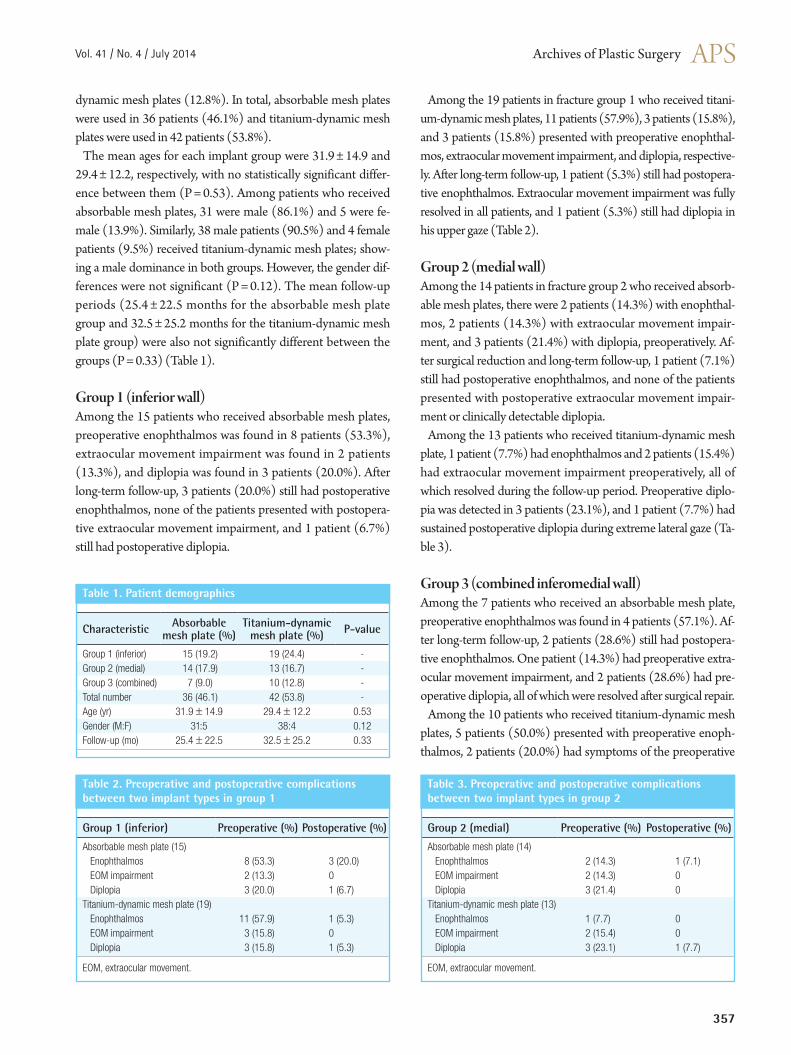
The mean ages for each implant group were 31.9 ± 14.9 and 29.4 ± 12.2, respectively, with no statistically significant differ-ence between them (P = 0.53). Among patients who received absorbable mesh plates, 31 were male (86.1%) and 5 were fe-male (13.9%). Similarly, 38 male patients (90.5%) and 4 female patients (9.5%) received titanium-dynamic mesh plates; show-ing a male dominance in both groups. However, the gender dif-ferences were not significant (P = 0.12). The mean follow-up periods (25.4 ± 22.5 months for the absorbable mesh plate group and 32.5 ± 25.2 months for the titanium-dynamic mesh plate group) were also not significantly different between the groups (P = 0.33) (Table 1).
Group 1 (inferior wall)Among the 15 patients who received absorbable mesh plates, preoperative enophthalmos was found in 8 patients (53.3%), extraocular movement impairment was found in 2 patients (13.3%), and diplopia was found in 3 patients (20.0%). After long-term follow-up, 3 patients (20.0%) still had postoperative enophthalmos, none of the patients presented with postopera-tive extraocular movement impairment, and 1 patient (6.7%) still had postoperative diplopia.
Among the 19 patients in fracture group 1 who received titani-um-dynamic mesh plates, 11 patients (57.9%), 3 patients (15.8%), and 3 patients (15.8%) presented with preoperative enophthal-mos, extraocular movement impairment, and diplopia, respective-ly. After long-term follow-up, 1 patient (5.3%) still had postopera-tive enophthalmos. Extraocular movement impairment was fully resolved in all patients, and 1 patient (5.3%) still had diplopia in his upper gaze (Table 2).
Group 2 (medial wall)Among the 14 patients in fracture group 2 who received absorb-able mesh plates, there were 2 patients (14.3%) with enophthal-mos, 2 patients (14.3%) with extraocular movement impair-ment, and 3 patients (21.4%) with diplopia, preoperatively. Af-ter surgical reduction and long-term follow-up, 1 patient (7.1%) still had postoperative enophthalmos, and none of the patients presented with postoperative extraocular movement impair-ment or clinically detectable diplopia.
Among the 13 patients who received titanium-dynamic mesh plate, 1 patient (7.7%) had enophthalmos and 2 patients (15.4%) had extraocular movement impairment preoperatively, all of which resolved during the follow-up period. Preoperative diplo-pia was detected in 3 patients (23.1%), and 1 patient (7.7%) had sustained postoperative diplopia during extreme lateral gaze (Ta-ble 3).
Group 3 (combined inferomedial wall)Among the 7 patients who received an absorbable mesh plate, preoperative enophthalmos was found in 4 patients (57.1%). Af-ter long-term follow-up, 2 patients (28.6%) still had postopera-tive enophthalmos. One patient (14.3%) had preoperative extra-ocular movement impairment, and 2 patients (28.6%) had pre-operative diplopia, all of which were resolved after surgical repair.
Among the 10 patients who received titanium-dynamic mesh plates, 5 patients (50.0%) presented with preoperative enoph-thalmos, 2 patients (20.0%) had symptoms of the preoperative
Characteristic Absorbable mesh plate (%)
Titanium-dynamic mesh plate (%) P-value
Group 1 (inferior) 15 (19.2) 19 (24.4) -Group 2 (medial) 14 (17.9) 13 (16.7) -Group 3 (combined) 7 (9.0) 10 (12.8) -Total number 36 (46.1) 42 (53.8) -Age (yr) 31.9±14.9 29.4±12.2 0.53Gender (M:F) 31:5 38:4 0.12Follow-up (mo) 25.4±22.5 32.5±25.2 0.33
Table 1. Patient demographics
Group 1 (inferior) Preoperative (%) Postoperative (%)
Absorbable mesh plate (15) Enophthalmos 8 (53.3) 3 (20.0) EOM impairment 2 (13.3) 0 Diplopia 3 (20.0) 1 (6.7)Titanium-dynamic mesh plate (19) Enophthalmos 11 (57.9) 1 (5.3) EOM impairment 3 (15.8) 0 Diplopia 3 (15.8) 1 (5.3)
EOM, extraocular movement.
Table 2. Preoperative and postoperative complications between two implant types in group 1
Group 2 (medial) Preoperative (%) Postoperative (%)
Absorbable mesh plate (14) Enophthalmos 2 (14.3) 1 (7.1) EOM impairment 2 (14.3) 0 Diplopia 3 (21.4) 0Titanium-dynamic mesh plate (13) Enophthalmos 1 (7.7) 0 EOM impairment 2 (15.4) 0 Diplopia 3 (23.1) 1 (7.7)
EOM, extraocular movement.
Table 3. Preoperative and postoperative complications between two implant types in group 2

Baek WI et al. Absorbable versus titanium mesh plate in blow-out fracture
358
extraocular movement impairment, and 3 patients (30.0%) had preoperative diplopia. After long-term follow-up, 1 patient (10.0%) had postoperative enophthalmos, 1 patient (10.0%) had extraocular movement impairment, and 1 patient (10.0%) still had diplopia (Table 4).
Comparative analysisIn every fracture group, the results of all chi-square tests and Fisher’s exact tests indicated that there was no significant differ-ence between the absorbable mesh plate and titanium-dynamic mesh plate with respect to the preoperative incidence of enoph-thalmos, extraocular movement impairment, or diplopia. The preoperative defect size of each group, measured by 3-dimen-sional computed tomography, were 2.72 ± 0.21 cm2 in group 1, 2.67 ± 0.14 cm2 in group 2, and 2.81 ± 0.20 cm2 in group 3, without statistically significant difference, too (P = 0.65).
After surgical repair, there were improvements in each compli-cation in every group (group 1, 2, and 3). Among them, howev-er, enophthalmos in group 1 showed statistical significance only, regardless of implant types.
In group 1, the difference in preoperative and postoperative in-cidence of enophthalmos was 8 to 3 patients (P = 0.03) for the absorbable mesh plate group and 11 to 1 patient (P = 0.002) for the titanium-dynamic mesh plate group, respectively (Fig. 1); these differences mark statistically significant improvements in enophthalmos incidence for both implants. In group 2 and 3, al-though there were also improvements in incidence of enophthal-mos in both implants, the result was not statistically significant.
Comparing extraocular movement impairment and diplopia, even though the incidences decreased in every groups, the im-provements were not statistically significant either.
DISCUSSION
The orbital wall is one of the most frequently damaged parts of the maxillofacial skeleton after midfacial trauma. Regardless of
the fracture site, blow-out fractures can cause various functional and aesthetic sequelae. Preventing these complications from be-coming long-term problems is very important, and it depends strongly on the materials used for bridging the orbital wall de-fects [11].
The prerequisites of an ideal material are good biocompatibili-ty, easy to manipulate, and strong mechanical strength to sup-port the orbital structure [12-15]. Remarkably, however, there are no uniformly accepted guidelines for selecting material for orbital reconstruction [16].
Various materials have been introduced for orbital wall recon-struction, each with its own advantages and disadvantages. Au-tologous materials, including periosteum [7], nasoseptal carti-lage [8], rib bone [9], and calvarium, have the advantages of re-sistance to infection, incorporation by the host into new bone, lack of host response against the graft, and little concern for late extrusion [17]. Autologous materials also have several disadvan-tages, including high risk of nerve and blood vessel injury, donor site morbidity, cosmetic disturbance, and an unpredictable de-gree of absorption [18,19].
Allogenic materials, such as lyophilized dura and lyophilized cartilage, showed good results during postoperative follow-up in some studies; however, it should not be used any more due to the risk of slow viral infections [20].
Recently, alloplastic materials have become the most widely used materials. Alloplastic materials are subdivided into absorb-able and non-absorbable mesh plates. Some surgeons prefer ab-sorbable mesh plates due to their ease of use and complete re-sorption, leaving no foreign material in human body [17]. How-
Group 3 (combined) Preoperative (%) Postoperative (%)
Absorbable mesh plate (7) Enophthalmos 4 (57.1) 2 (28.6) EOM impairment 1 (14.3) 0 Diplopia 2 (28.6) 0Titanium-dynamic mesh plate (10) Enophthalmos 5 (50.0) 1 (10.0) EOM impairment 2 (20.0) 1 (10.0) Diplopia 3 (30.0) 1 (10.0)
EOM, extraocular movement.
Table 4. Preoperative and postoperative complications between two implant types in group 3
Significant improvements in enophthalmos are observable after surgical repair in both implant types within group 1 (P<0.05). How-ever, both implant types showed no significant difference between themselves.
Fig. 1. Difference of enophthalmos incidence in group 1
Absorbable mesh plate Titanium-dynamic mesh plate
12
10
8
6
4
2
0
(n)
Preoperative Postoperative
P=0.29
P=0.002P=0.92
P=0.03
11
8
3
1

Vol. 41 / No. 4 / July 2014
359
ever, some studies have reported that the connective tissues around the absorbable mesh plate cannot replace the original bony structures completely after the thorough resorption of the plate, mainly in the center of the defect, which may result in pro-gressive enophthalmos [21]. Also, replacement by fibrosis may not support the orbital structure and can lead to persistent en-ophthalmos [22].
Titanium-dynamic mesh plates have also been used widely for various craniofacial fractures and are known to be biocompati-ble. It can be adopted to complex structures easily, and it can also be cut to shape as well [16]. However titanium plates are permanent foreign bodies. Several late-onset complications re-lated to the titanium-dynamic mesh plate have been reported, such as infection, extrusion, implant migration, residual diplo-pia, etc. [23]. Therefore, choosing an appropriate material to re-construct the orbital wall remains difficult and controversial.
Recently, various absorbable materials with different degrada-tion rates are introduced. The degradation rate depends on the proportion of monomers. According to some literatures and data about 85:15 poly (L-lactide-co-glycolide) mesh plate (RAPIDSORB Rapid Resorbable Fixation System, Synthes,
Oberdorf, Switzerland) in our study, it readily resorbs within 12 months. Therefore, we supposed at least 12 months as a long-term follow up. Moreover, we also found full absorption of the plate by 3-dimensional computed tomography during the long-term follow up (Fig. 2).
In our study, the preoperative incidence of enophthalmos was higher in group 1 than in the other groups, and regardless of im-plant type, most enophthalmos cases were relatively well cor-rected after orbital wall reconstruction. The reason of high inci-dence of preoperative enophthalmos for inferior orbital wall fracture cases, we suspect, might be due to the weight and in-creased tension of the globe to the orbital wall, especially the in-ferior orbital wall. The internal orbital contents include the weight of the globe, the extraocular musculature, orbital fat, neurovascular structures, the lacrimal apparatus, and even the musculocutaneous lids. According to the literature, the mean weight of the combined contents of the exenterated orbit, in-cluding the structures mentioned above, was found to be 42.97 ± 4.05 g, with a range of 37.80 to 51.03 g [24]. The nor-mal direct orbital tension is 4 mm Hg, and the direct orbital ten-sion after reconstruction of an orbital floor fracture usually in-
(A) Preoperative view of medial wall defect. Note on the herniated soft tissue through lamina papyracea (red circle). (B) Immediate postoperative view. Note on the absorbable plate and well-reducted soft tissue (red ar-row). (C) 13 months after orbital wall re-construction, the absorbable plate was fully resorbed, and the fibrous tissue substituted the medial orbital wall (red arrow).
Fig. 2. Coronal image of 3-dimensional computed tomography
A
B
C

Baek WI et al. Absorbable versus titanium mesh plate in blow-out fracture
360
creases to about 9.3 ± 3.1 mm Hg [25]. These factors might ex-plain the high incidence of preoperative enophthalmos in group 1 (inferior wall).
These factors also might be the reason of higher recurrence rate of enophthalmos after orbital wall reconstruction in the ab-sorbable mesh plate group compared to the titanium-dynamic mesh plate group. According to the results of our retrospective study, besides the obvious improvements in each complication type in every group (groups 1, 2, and 3) after orbital wall recon-struction, there was an overall tendency toward a lower recur-rence rate of enophthalmos in titanium-dynamic mesh plate re-cipients than in those who received an absorbable mesh plate as well; however, in long-term follow-up, the difference was not significant.
The significant difference in our study was the dramatic de-crease of enophthalmos incidence among the patients in group 1 after reconstruction, for both implant types. Furthermore, al-though the results from group 3 (combined wall) showed no statistically significant differences in our study, some similar re-sults are expected with a study of large number of patients, be-cause these injuries might lead to even larger orbital wall defects than isolated inferior orbital wall fractures do; which can cause higher incidence of preoperative enophthalmos.
Among the postoperative complicated patients, there was no patinet who consistently complained of the problems, except for only one patient with persistent diplopia in group 2. The pa-tient had underwent medial orbital wall reconstruction with ti-tanium-dynamic mesh plate and complained of postoperative diplopia. However, we did not undergo reoperation, because the patient was accompanied with objective restriction beyond 40 degrees of abduction from primary gaze, which means there was no diplopia in usual life except for extreme lateral gaze. Finally, we had no patient who had severe problem which required re-operation, fortunately.
A shortcoming of this study is the small number of patients in each group, especially in group 3 (combined wall). As we con-tinue to collect more medical data over several more years, we will have more statistical power to identify more significant dif-ferences.
Finally, our study does not suggest that one or the other of these implants is the best option for reconstructing orbital anat-omy after blow-out fractures. Even though we expected the sig-nificantly higher recurrent enophthalmos rate in patients with absorbable plate through complete absorption, both types showed good results without significant differences in long-term follow up. We can use both types of implants safely. Re-garding extraocular movement impairment and diplopia, both implants showed no significant differences either. This results
suggest that other factors, such as surgeon’s preference, patients’ request, or cost-effectiveness, can also be legitimate deciding points for choosing the implant as well.
In conclusion, both types of implants seem to be equally effec-tive and safe for orbital wall reconstruction, and they are both particularly effective with regard to improving enophthalmos in cases of inferior orbital wall fractures.
REFERENCES
1. Jank S, Emshoff R, Schuchter B, et al. Orbital floor recon-struction with flexible Ethisorb patches: a retrospective long-term follow-up study. Oral Surg Oral Med Oral Pathol Oral Radiol Endod 2003;95:16-22.
2. Kim IT, Choi JB. Normal range of exophthalmos values on orbit computerized tomography in Koreans. Ophthalmo-logica 2001;215:156-62.
3. Tanaka T, Morimoto Y, Kito S, et al. Evaluation of coronal CT findings of rare cases of isolated medial orbital wall blow-out fractures. Dentomaxillofac Radiol 2003;32:300-3.
4. Caranci F, Cicala D, Cappabianca S, et al. Orbital fractures: role of imaging. Semin Ultrasound CT MR 2012;33:385-91.
5. You JP, Kim DW, Jeon BJ, et al. Two-year follow-up on the use of absorbable mesh plates in the treatment of medial or-bital wall fractures. Arch Plast Surg 2013;40:728-34.
6. Buchel P, Rahal A, Seto I, et al. Reconstruction of orbital floor fracture with polyglactin 910/polydioxanon patch (ethisorb): a retrospective study. J Oral Maxillofac Surg 2005;63:646-50.
7. Dost P. Orbital floor reconstruction with autologous perios-teum transplant. Laryngorhinootologie 1996;75:57-8.
8. Lai A, Gliklich RE, Rubin PA. Repair of orbital blow-out fractures with nasoseptal cartilage. Laryngoscope 1998;108: 645-50.
9. Johnson PE, Raftopoulos I. In situ splitting of a rib graft for reconstruction of the orbital floor. Plast Reconstr Surg 1999; 103:1709-11.
10. Guerra MF, Perez JS, Rodriguez-Campo FJ, et al. Recon-struction of orbital fractures with dehydrated human dura mater. J Oral Maxillofac Surg 2000;58:1361-6.
11. Gierloff M, Seeck NG, Springer I, et al. Orbital floor recon-struction with resorbable polydioxanone implants. J Cranio-fac Surg 2012;23:161-4.
12. Courtney DJ, Thomas S, Whitfield PH. Isolated orbital blowout fractures: survey and review. Br J Oral Maxillofac Surg 2000;38:496-504.
13. Enislidis G. Treatment of orbital fractures: the case for treat-ment with resorbable materials. J Oral Maxillofac Surg 2004;

Vol. 41 / No. 4 / July 2014
361
62:869-72.14. Kontio R. Treatment of orbital fractures: the case for recon-
struction with autogenous bone. J Oral Maxillofac Surg 2004;62:863-8.
15. Potter JK, Ellis E. Biomaterials for reconstruction of the in-ternal orbit. J Oral Maxillofac Surg 2004;62:1280-97.
16. Dietz A, Ziegler CM, Dacho A, et al. Effectiveness of a new perforated 0.15 mm poly-p-dioxanon-foil versus titanium-dynamic mesh in reconstruction of the orbital floor. J Crani-omaxillofac Surg 2001;29:82-8.
17. Al-Sukhun J, Lindqvist C. A comparative study of 2 implants used to repair inferior orbital wall bony defects: autogenous bone graft versus bioresorbable poly-L/DL-Lactide [P(L/DL)LA 70/30] plate. J Oral Maxillofac Surg 2006;64:1038-48.
18. Banwart JC, Asher MA, Hassanein RS. Iliac crest bone graft harvest donor site morbidity. A statistical evaluation. Spine (Phila Pa 1976) 1995;20:1055-60.
19. Ahlmann E, Patzakis M, Roidis N, et al. Comparison of an-terior and posterior iliac crest bone grafts in terms of har-vest-site morbidity and functional outcomes. J Bone Joint
Surg Am 2002;84:716-20.20. Takashima S, Tateishi J, Taguchi Y, et al. Creutzfeldt-Jakob
disease with florid plaques after cadaveric dural graft in a Japanese woman. Lancet 1997;350:865-6.
21. Merten HA, Luhr HG. Resorbable synthetics (PDS foils) for bridging extensive orbital wall defects in an animal ex-periment comparison. Fortschr Kiefer Gesichtschir 1994; 39:186-90.
22. Tuncer S, Yavuzer R, Kandal S, et al. Reconstruction of trau-matic orbital floor fractures with resorbable mesh plate. J Craniofac Surg 2007;18:598-605.
23. Hwang K, Kim DH. Comparison of the supporting strength of a poly-L-lactic acid sheet and porous polyethylene (Med-por) for the reconstruction of orbital floor fractures. J Cra-niofac Surg 2010;21:847-53.
24. Haug RH, Nuveen E, Bredbenner T. An evaluation of the support provided by common internal orbital reconstruc-tion materials. J Oral Maxillofac Surg 1999;57:564-70.
25. Zhou H, Fan X, Xiao C. Direct orbital manometry in nor-mal and fractured orbits of Chinese patients. J Oral Maxillo-fac Surg 2007;65:2282-7.