Phytochemical-Based Nano-Pharmacotherapeutics for ... - MDPI
Upload
independentCategory
view
0download
0
www.elsevier.com/locate/phytochem
Phytochemistry 68 (2007) 1683–1691
PHYTOCHEMISTRY
Comparative phytochemical and morphological analyses of three ItalianPrimula species
Gelsomina Fico a,*, Graziella Rodondi a, Guido Flamini b, Daniele Passarella c,Franca Tome a
a Dipartimento di Biologia, Universita degli Studi di Milano, via Celoria 26, 20133 Milano, Italyb Dipartimento di Chimica Bioorganica e Biofarmacia, Universita degli Studi di Pisa, via Bonanno 33, 56126 Pisa, Italy
c Dipartimento di Chimica Organica e Industriale, Universita degli Studi di Milano, via Venezian 21, 20133 Milano, Italy
Received 4 December 2006; received in revised form 10 April 2007
Abstract
The taxonomy of alpine Primula species has long been in dispute because of high morphologic variability and several hybridisations.In Primula species, the trichome height and the colour of hair-tips are usually indicated as diacritic characters, but in our experience thisis not adequate. The present study, focused on Primula auricula, Primula daonensis and Primula hirsuta, therefore proposes the use ofother morphologic trichome parameters (size and dimensional ratio of stalk, neck and gland head). Phytochemical investigations aboutthe flavonoid composition (epicuticular and vacuolar) of leaves, as taxonomic markers, have also been performed. We report the isola-tion and identification of two new flavonol glycosides, isorhamnetin 3-O-(2,6-di-O-b-D-glucopyranosyl-b-D-glucopyranoside) (1) andkaempferol 3-O-(2-O-a-L-rhamnopyranosyl-6-O-b-D-xylopyranosyl-b-D-glucopyranoside) (2) and of eight known flavonoids. Size anddimensional ratio of the three trichome elements (stalk, neck and glandular head) are typical for each species analysed. The flavonoidprofile well characterise the entities under study. Three different profiles have been obtained with both vacuolar and epicuticularflavonoids.
The morphologic and phytochemical markers proposed in this work seem to be parameters which significatively discriminate the spe-cies under study.� 2007 Elsevier Ltd. All rights reserved.
Keywords: Primula auricula; Primula daonensis; Primula hirsuta; Primulaceae; Phytochemistry; Morphology; Taxonomy; Flavonoid; Trichome
1. Introduction
The Primula L. genus is the most important one in thePrimulaceae family and includes, considering the latestevaluations, 426 species, mainly located in the temperateand cold regions of the northern hemisphere and in thetropical mountains.
The systematic studies on European Primula L. genusstarted in the middle of 19th century, and are based onmorphological (Duby, 1844; Pax, 1889; Ludi, 1926; Smithand Fletcher, 1948), cytological (Bruum, 1932), caryologic
0031-9422/$ - see front matter � 2007 Elsevier Ltd. All rights reserved.
doi:10.1016/j.phytochem.2007.04.019
* Corresponding author. Tel.: +39 0250314770; fax: +39 0250314764.E-mail address: [email protected] (G. Fico).
(Wanner, 1943), biochemical (Mueller, 1915; Brunswik,1922; Hegnauer, 1969), cytotaxonomic (Kress, 1963,1989) and palynologic (Spanovschy, 1962) analyses.Recently the monographs of Richards (1993, 2003) investi-gates the Primula genus all over the world.
The systematics of the alpine Primula species belongingto the sect. auricula Duby has long been in dispute, becauseof high morphological variability, several hybridizationsand frequent description of new species and subspecies(Banfi and Ferlinghetti, 1993; Moser, 1998; Prosser andScortegagna, 1998; Jeßen and Lehmann, 2005).
Difficulties to distinguish among closely related Primula
sect. auricula species have recently prompted biomolecularstudies (Scudiero et al., 1980; Zhang and Kadereit, 2004)
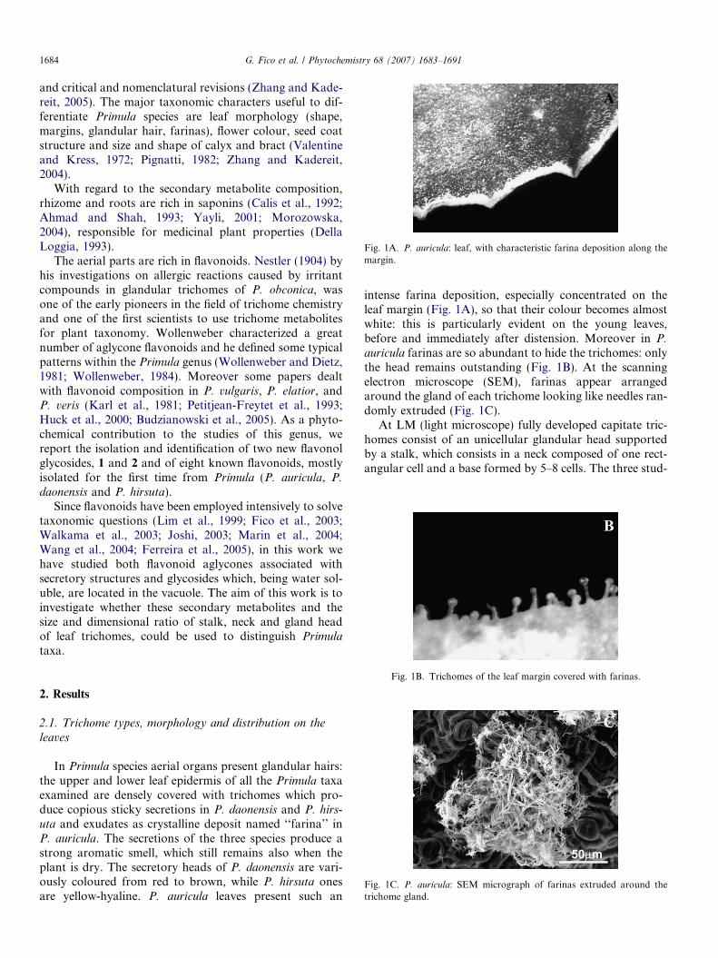
Fig. 1A. P. auricula: leaf, with characteristic farina deposition along themargin.
1684 G. Fico et al. / Phytochemistry 68 (2007) 1683–1691
and critical and nomenclatural revisions (Zhang and Kade-reit, 2005). The major taxonomic characters useful to dif-ferentiate Primula species are leaf morphology (shape,margins, glandular hair, farinas), flower colour, seed coatstructure and size and shape of calyx and bract (Valentineand Kress, 1972; Pignatti, 1982; Zhang and Kadereit,2004).
With regard to the secondary metabolite composition,rhizome and roots are rich in saponins (Calis et al., 1992;Ahmad and Shah, 1993; Yayli, 2001; Morozowska,2004), responsible for medicinal plant properties (DellaLoggia, 1993).
The aerial parts are rich in flavonoids. Nestler (1904) byhis investigations on allergic reactions caused by irritantcompounds in glandular trichomes of P. obconica, wasone of the early pioneers in the field of trichome chemistryand one of the first scientists to use trichome metabolitesfor plant taxonomy. Wollenweber characterized a greatnumber of aglycone flavonoids and he defined some typicalpatterns within the Primula genus (Wollenweber and Dietz,1981; Wollenweber, 1984). Moreover some papers dealtwith flavonoid composition in P. vulgaris, P. elatior, andP. veris (Karl et al., 1981; Petitjean-Freytet et al., 1993;Huck et al., 2000; Budzianowski et al., 2005). As a phyto-chemical contribution to the studies of this genus, wereport the isolation and identification of two new flavonolglycosides, 1 and 2 and of eight known flavonoids, mostlyisolated for the first time from Primula (P. auricula, P.
daonensis and P. hirsuta).Since flavonoids have been employed intensively to solve
taxonomic questions (Lim et al., 1999; Fico et al., 2003;Walkama et al., 2003; Joshi, 2003; Marin et al., 2004;Wang et al., 2004; Ferreira et al., 2005), in this work wehave studied both flavonoid aglycones associated withsecretory structures and glycosides which, being water sol-uble, are located in the vacuole. The aim of this work is toinvestigate whether these secondary metabolites and thesize and dimensional ratio of stalk, neck and gland headof leaf trichomes, could be used to distinguish Primula
taxa.
Fig. 1B. Trichomes of the leaf margin covered with farinas.
Fig. 1C. P. auricula: SEM micrograph of farinas extruded around thetrichome gland.
2. Results
2.1. Trichome types, morphology and distribution on the
leaves
In Primula species aerial organs present glandular hairs:the upper and lower leaf epidermis of all the Primula taxaexamined are densely covered with trichomes which pro-duce copious sticky secretions in P. daonensis and P. hirs-
uta and exudates as crystalline deposit named ‘‘farina’’ inP. auricula. The secretions of the three species produce astrong aromatic smell, which still remains also when theplant is dry. The secretory heads of P. daonensis are vari-ously coloured from red to brown, while P. hirsuta onesare yellow-hyaline. P. auricula leaves present such an
intense farina deposition, especially concentrated on theleaf margin (Fig. 1A), so that their colour becomes almostwhite: this is particularly evident on the young leaves,before and immediately after distension. Moreover in P.
auricula farinas are so abundant to hide the trichomes: onlythe head remains outstanding (Fig. 1B). At the scanningelectron microscope (SEM), farinas appear arrangedaround the gland of each trichome looking like needles ran-domly extruded (Fig. 1C).
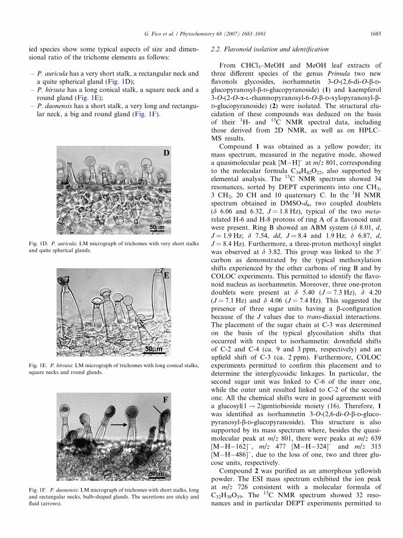
At LM (light microscope) fully developed capitate tric-homes consist of an unicellular glandular head supportedby a stalk, which consists in a neck composed of one rect-angular cell and a base formed by 5–8 cells. The three stud-
G. Fico et al. / Phytochemistry 68 (2007) 1683–1691 1685
ied species show some typical aspects of size and dimen-sional ratio of the trichome elements as follows:
– P. auricula has a very short stalk, a rectangular neck anda quite spherical gland (Fig. 1D);
– P. hirsuta has a long conical stalk, a square neck and around gland (Fig. 1E);
– P. daonensis has a short stalk, a very long and rectangu-lar neck, a big and round gland (Fig. 1F).
Fig. 1D. P. auricula: LM micrograph of trichomes with very short stalksand quite spherical glands.
Fig. 1E. P. hirsuta: LM micrograph of trichomes with long conical stalks,square necks and round glands.
Fig. 1F. P. daonensis: LM micrograph of trichomes with short stalks, longand rectangular necks, bulb-shaped glands. The secretions are sticky andfluid (arrows).
2.2. Flavonoid isolation and identification
From CHCl3–MeOH and MeOH leaf extracts ofthree different species of the genus Primula two newflavonols glycosides, isorhamnetin 3-O-(2,6-di-O-b-D-glucopyranosyl-b-D-glucopyranoside) (1) and kaempferol3-O-(2-O-a-L-rhamnopyranosyl-6-O-b-D-xylopyranosyl-b-D-glucopyranoside) (2) were isolated. The structural elu-cidation of these compounds was deduced on the basisof their 1H- and 13C NMR spectral data, includingthose derived from 2D NMR, as well as on HPLC–MS results.
Compound 1 was obtained as a yellow powder; itsmass spectrum, measured in the negative mode, showeda quasimolecular peak [M�H]� at m/z 801, correspondingto the molecular formula C34H42O22, also supported byelemental analysis. The 13C NMR spectrum showed 34resonances, sorted by DEPT experiments into one CH3,3 CH2, 20 CH and 10 quaternary C. In the 1H NMRspectrum obtained in DMSO-d6, two coupled doublets(d 6.06 and 6.32, J = 1.8 Hz), typical of the two meta-related H-6 and H-8 protons of ring A of a flavonoid unitwere present. Ring B showed an ABM system (d 8.01, d,J = 1.9 Hz; d 7.54, dd, J = 8.4 and 1.9 Hz; d 6.87, d,J = 8.4 Hz). Furthermore, a three-proton methoxyl singletwas observed at d 3.82. This group was linked to the 3 0
carbon as demonstrated by the typical methoxylationshifts experienced by the other carbons of ring B and byCOLOC experiments. This permitted to identify the flavo-noid nucleus as isorhamnetin. Moreover, three one-protondoublets were present at d 5.40 (J = 7.3 Hz), d 4.20(J = 7.1 Hz) and d 4.06 (J = 7.4 Hz). This suggested thepresence of three sugar units having a b-configurationbecause of the J values due to trans-diaxial interactions.The placement of the sugar chain at C-3 was determinedon the basis of the typical glycosilation shifts thatoccurred with respect to isorhamnetin: downfield shiftsof C-2 and C-4 (ca. 9 and 3 ppm, respectively) and anupfield shift of C-3 (ca. 2 ppm). Furthermore, COLOCexperiments permitted to confirm this placement and todetermine the interglycosidic linkages. In particular, thesecond sugar unit was linked to C-6 of the inner one,while the outer unit resulted linked to C-2 of the secondone. All the chemical shifts were in good agreement witha glucosyl(1! 2)gentiobioside moiety (16). Therefore, 1
was identified as isorhamnetin 3-O-(2,6-di-O-b-D-gluco-pyranosyl-b-D-glucopyranoside). This structure is alsosupported by its mass spectrum where, besides the quasi-molecular peak at m/z 801, there were peaks at m/z 639[M�H�162]�, m/z 477 [M�H�324]� and m/z 315[M�H�486]�, due to the loss of one, two and three glu-cose units, respectively.
Compound 2 was purified as an amorphous yellowishpowder. The ESI mass spectrum exhibited the ion peakat m/z 726 consistent with a molecular formula ofC32H38O19. The 13C NMR spectrum showed 32 reso-nances and in particular DEPT experiments permitted to

Table 1NMR data of new compounds 1 and 2
Position 1 (DMSO-d6) 2 (CD3OD)
1H NMR 13CNMR
1H NMR 13CNMR
Aglycone
2 155.2 166.83 133.3 135.44 176.9 180.25 161.0 162.26 6.06, d, 1.8 Hz 99.2 6.18, d, 2.0 Hz �98a
7 163.8 163.98 6.32, d, 1.8 Hz 94.1 6.39, d, 2.0 Hz �93a
9 156.6 159.410 103.4 106.910 122.0 124.020 8.01, d, 1.9 Hz 115.2 8.09, d, 8.9 Hz 117.030 146.8 6.91, d, 8.9 Hz 133.140 149.5 159.350 6.87, d, 8.4 Hz 113.2 6.91, d, 8.9 Hz 133.160 7.54, dd, 1.9,
8.4 Hz121.0 8.09, d, 8.9 Hz 117.0
OMe 3.82 s 55.7
3-Glc 3-Glc
1 5.40, d, 7.3 Hz 101.3 5.66, d, 7.5 Hz 101.32 3.28b 74.4 3.64, dd, 7.5, 8.9 Hz 80.83 3.22b 76.1 3.58, t, 8.9 Hz 79.64 3.08, t, 9.0 Hz 70.1 3.32–3.40, m 72.85 3.51b 76.1 3.42–3.47, m 78.36 3.40b 67.8 3.59, dd, 6.5,
11.8 Hz70.4
3.82, d, 11.4 Hz 3.93, dd, 1.8,11.8 Hz
600-Glc 200-Rha
1 4.20, d, 7.1 Hz 101.6 5.25, d, 1.3 Hz 103.52 3.03, m 82.3 4.03, dd, 1.3, 3.4 Hz 73.33 3.21b 75.7 3.82, dd, 3.4, 9.6 Hz 73.24 3.07, m 69.7 3.32–3.40, m 74.9c
5 2.61, m 76.1 4.06–4.15, m 70.86 3.34b 60.4 1.04, d, 6.2 Hz 18.4
3.50b
2000-Glc 600-Xyl
1 4.06, d, 7.4 Hz 104.4 4.10, d, 7.2 Hz 105.92 2.51d 74.4 3.03, dd, 7.2, 8.8 Hz 75.63 3.03, m 75.7 3.12, t, 8.8 Hz 78.24 2.98, m 69.7 3.32–3.40, m 71.9c
5 2.97, m 77.4 3.68, dd, 5.2,11.4 Hz
67.4
2.91, dd, 10.0,11.4 Hz
6 3.52c 61.03.64c
a Broad signals assigned on the basis of Hetcorr.b Signal superimposed by H2O, attributed by COLOC or HSQC.c Interchangeable signals.d Signal superimposed by DMSO-d6, attributed by COLOC or HSQC.
1686 G. Fico et al. / Phytochemistry 68 (2007) 1683–1691
recognize one CH3, two CH2, 20 CH and nine quaternaryC. In obtained DMSO-d6, the signals at d 5.66 (d,J = 7.5 Hz), 5.25 (d, J = 1.3 Hz) and at 4.10 (J = 7.2 Hz)revealed the presence of three sugar moieties that presentin two cases the b-configuration and in the other one thea-configuration. Complete assignments of each sugar pro-ton system were achieved by considering TOCSY and1H–1H COSY spectra. The presence of a glucose unitwas confirmed by the large vicinal coupling among ringprotons due to the trans-diaxial orientation. The 1HNMR spectrum showed two coupled doublets with a verysmall J-value, typical of the two meta-related H-6 and H-8protons of the A ring of the flavonoid unit and anAA 0BB 0 system (d 8.09 and 6.91, J = 8.9 Hz) that is dueto the B ring of the same flavonoid unit. The sugar moietywas found to be linked to C-3 as consequence of the samereason that we described above. By this way the flavonoidunit was recognized as kaempferol with the a glycosidicbond at position 3. The interglycosidic linkages were con-firmed by HMBC experiments. In particular the glucoseunit, that was directly connected to kaempferol, waslinked through position 2 to rhamnose and through posi-tion 6 to xylose.
The sugar moiety was almost superimposable to that ofcompound 1 and it was found to be linked to C-3 as abovedescribed. Therefore, compound 2 was identified askaempferol 3-O-(2-O-a-L-rhamnopyranosyl-6-O-b-D-xylo-pyranosyl-b-D-glucopyranoside) (Table 1).
Besides to 1 and 2, the known compounds, quercetin 3-O-(glucosyl(1! 2)gentiobioside) (3), kaempferol 3-O-neo-hesperidoside (4), isorhamnetin 3-O-neohesperidoside (5),tamarixin (6), isorhamnetin 3-O-glucopyranoside (7),isorhamnetin 3-O-(2-O-a-L-rhamnopyranosyl-6-O-b-D-xylopyranosyl-b-D-glucopyranoside) (8), quercetin 3-O-neohesperidoside (9), 7,2 0- dihydroxyflavone 7-O-b-D-glucopyranoside (10), were isolated and identified by spec-tral methods in comparison with literature data (Vidalet al., 1989; Kruthiventi and Krishnaswamy, 2000) (Tables2 and 3).
The chromatograms of the flavonoids in the exudatefrom P. auricula, P. daonensis and P. hirsuta are shownin Fig. 2A–C. Each chromatogram was characterized bythe presence of one main peak, with Rt respectively of35.34, 36.69 and 37.75 min.
The comparison of spectra of these peaks indicated thatthe three species are characterized by different compounds(Fig. 3A–C).
Traditionally the taxonomic value of trichomes, in diag-nostic key, to distinguish different Primula species is corre-lated to their presence/absence or their overall length(Pignatti, 1982), or density and colour of hair-tips (Tutinet al., 1993). Our LM analysis enhanced the differences inshape, size and dimensional ratio of the trichome elements:stalk, neck and glandular head, making the trichome micromorphology a diagnostic element more stringent than theoverall length of trichomes, or the hair-tip colour, to solvetaxonomic questions.
The phytochemical analysis has pointed out three differ-ent flavonoid profiles for the species under study (Table 4).The identified flavonol compounds are 3-O-glycosides, rep-resented by derivatives of kaempferol, quercetin, isorham-

Table 21H NMR data of known compounds 3–10
Position 3 4 5 6 7 8 9 10
Aglycone
3 6.90, s
5 7.24, br d,9.0 Hz
6 6.12, d,1.8 Hz
6.16, d, 1.9 Hz 6.18, d, 1.9 Hz 6.23, d,1.9 Hz
6.18, d, 2.0 Hz 6.02, d, 1.9 Hz 6.17, d, 1.8 Hz 7.56, br d,8.0 Hz
8 6.41, d,1.8 Hz
6.38, d, 1.9 Hz 6.38, d, 1.9 Hz 6.44, d,1.9 Hz
6.42, d, 2.0 Hz 6.21, d, 1.9 Hz 6.37, d, 1.8 Hz 7.53, br s
2’ 7.66, br s 8.01, d, 8.9 Hz 7.98, d, 1.8 Hz 7.66, br s 7.94, d, 1.9 Hz 7.91, d, 1.8 Hz 7.51, d, 2.1 Hz3’ 6.86, d, 8.9 Hz 7.62, br d,
8.0 Hz4’ 7.78, br t,
7.8 Hz5’ 6.80, d,
8.4 Hz6.86, d, 8.9 Hz 6.91, d, 8.5 Hz 7.00, d,
8.9 Hz6.87, d, 8.7 Hz 6.88, d, 8.5 Hz 6.81, d, 8.5 Hz 7.50, m
6’ 7.62 br d,8.4 Hz
8.01, d, 8.9 Hz 7.54, dd, 1.8,8.5 Hz
7.51, br d,8.9 Hz
7.50, dd, 1.9,8.8 Hz
7.43, dd, 1.8,8.5 Hz
7.59, dd, 2.1,8.5 Hz
8.02, br d,7.3 Hz
OMe 3.97, s 3.92, s 3.95, s 3.83, s
3-Glc 3-Glc 3-Glc 3-Glc 3-Glc 7-Glc
1 5.30, d,6.8 Hz
5.70, d, 7.2 Hz 5.89, d, 7.2 Hz 5.27 d, 7.4 Hz 5.29, d, 7.6 Hz 5.70, d, 7.4 Hz 5.64, d, 7.4 Hz 4.88 d, 7.0 Hz
2 3.26a 3.59a 3.63a 3.48a 3.50a 3.61a 3.60a 3.32a
3 3.24a 3.57a 3.52a 3.44a 3.44a 3.58a 3.53a 3.38a
4 3.09, t,8.7 Hz
3.53a 3.51a 3.37a 3.31a 3.48a 3.53a 3.24a
5 3.50a 3.26a 3.24a 3.22a 3.23a 3.60a 3.24a 3.42a
6 3.40a 3.50a 3.51a 3.69a 3.70a 3.71a 3.52a 3.49a
3.79, d,11.8 Hz
3.71, br d,11.1 Hz
3.76, br d,12.1 Hz
3.61a 3.58a 4.12, br d,11.1 Hz
3.73, br d,11.8 Hz
3.75, br d,11.5 Hz
600-Glc 200-Rha 200-Rha 200-Rha 200-Rha
1 4.14, d,7.4 Hz
5.21, br s 5.19, br s 5.02, br s 5.06, br s
2 3.00 m 4.02, br d,3.3 Hz
3.95, br d,3.0 Hz
4.00, br d,3.6 Hz
3.96, br d,3.1 Hz
3 3.21a 3.78, dd, 3.3,9.1 Hz
3.76, dd, 3.0,8.9 Hz
3.67a 3.76, dd, 3.1,8.9 Hz
4 3.02 m 3.30a 3.28a 3.33a 3.33a
5 2.65 m 4.00 m 3.97 m 3.92 m 3.99 m
6 3.36a 0.72, d, 6.1 Hz 0.62, d, 6.1 Hz 0.65, d, 6.1 Hz 0.75, d, 6.1 Hz3.49a
2000-Glc 600-Glc
1 4.00, d,7.5 Hz
4.05, d, 7.2 Hz
2 2.52b 3.01, dd, 7.2,8.9 Hz
3 3.00, m 3.10, t, 8.9 Hz4 2.98, m 3.15, t, 8.9 Hz5 2.99, m 2.86, m
6 3.48a 3.41a
3.58a
3.65a
a Signal superimposed by H2O, attributed by COLOC or HSQC.b Signal superimposed by DMSO-d6, attributed by COLOC or HSQC.
G. Fico et al. / Phytochemistry 68 (2007) 1683–1691 1687
netin and tamarixin and the sugar moieties include glucose,rhamnose, xylose and di- and three-saccharides based onthese sugars. The sole isolated flavone was 7,2 0-dihydroxy-flavone 7-O-glucopyranoside.
The first profile characterize P. auricula and shows thepresence of flavonols with three sugar units. The sugar moi-ety was represented by glucose, the aglycones were querce-tin and isorhamnetin.

Table 5Primula samples
Samples (according to Florad’Italia)
Locality HerbariumNo.
P. auricula Monte Alben (Bergamo –1650–1750 m)
Pa-101
P. daonensis Passo del Gavia (Brescia –2500 m)
Pd-101
P. hirsuta Alpe Gera (Sondrio –1700 m)
Ph-101
Table 313C NMR data of known compounds 3–10
Position 3 4 5 6 7 8 9 10
Aglycone
2 156.1 161.0 158.4 157.3 159.0 155.1 156.0 155.63 133.6 134.0 134.4 133.9 134.4 132.2 132.8 105.94 177.4 178.9 179.3 178.6 178.7 176.5 177.2 177.05 161.1 163.0 163.2 162.3 163.2 161.0 161.2 116.06 98.6 98.1 99.8 98.9 98.0 98.5 98.3 118.47 164.2 165.5 165.7 164.8 165.6 165.0 164.3 162.68 93.6 94.2 94.6 93.9 94.2 94.2 93.5 113.69 156.4 157.4 158.5 157.7 157.6 156.6 156.2 147.010 104.2 104.9 105.0 104.2 104.5 104.2 103.9 125.110 121.2 123.1 123.5 122.1 122.9 121.7 121.1 123.320 116.3 133.3 114.6 113.9 114.8 113.2 115.9 148.430 144.7 115.8 150.5 147.3 149.8 149.3 144.9 118.440 148.5 159.9 148.4 149.2 147.6 146.8 148.4 134.250 115.1 115.8 116.0 115.0 116.1 115.1 115.1 125.560 121.4 133.3 123.6 122.0 123.4 121.0 121.6 124.8OMe 57.0 56.9 57.1 55.8
3-Glc 3-Glc 3-Glc 3-Glc 3-Glc 3-Glc 3-Glc 7-Glc
1 100.9 100.3 100.8 101.3 101.1 100.7 100.5 101.22 74.2 79.9 80.4 74.4 74.3 82.3 79.3 73.33 76.1 79.0 78.9 76.7 76.5 77.5 77.4 75.94 69.6 71.7 71.9 70.6 70.3 70.5 70.6 69.85 76.1 78.2 78.4 77.0 77.2 76.1 77.2 77.36 68.1 62.4 62.6 61.3 61.0 67.8 60.9 60.8
600-Glc
200-Rha
200-Rha
200-Rha
1 101.5 103.1 102.8 102.5 103.02 82.1 72.4 72.4 68.6 71.83 75.7 72.3 72.4 68.2 71.74 69.4 74.1 74.0 71.7 73.05 76.0 69.9 69.9 65.3 68.26 60.5 17.6 17.4 17.0 17.2
2000-Glc
600-Glc
1 104.0 103.22 74.2 73.43 75.7 76.64 69.6 69.75 77.3 76.26 61.0 60.7
Table 4Flavonoid composition of Primula species
O
OOH
HO
OR1
R3
R2
R1 R2 R3
Primula auricula
b-D-glucopyranosyl(1! 2)gentiobioside (1) OH OCH3
b-D-glucopyranosyl(1! 2)gentiobioside (3) OH OH
O
O
Glc-O
OH
10
7,20-dihydroxyflavone 7-O-b-D-glucopyranoside
Primula daonensis
Neohesperidoside (5) OH OCH3
b-D-glucopyranoside (6) OCH3 OHb-D-glucopyranoside (7) OH OCH3
2-O-a-L-rhamnopyranosyl-6-O-b-D-glucopyranosyl-b-D-glucopyranoside (8)
OH OCH3
Neohesperidoside (9) OH OH
Primula hirsuta
2-O-a-L-rhamnopyranosyl-6-O-b-D-xylopyranosyl-b-D-glucopyranoside (2)
OH H
Neohesperidoside (4) OH H
1688 G. Fico et al. / Phytochemistry 68 (2007) 1683–1691
The second profile typified P. daonensis and it is charac-terised by the presence of flavonols with no, one, two andthree sugar units. The sugar moiety was represented by glu-cose and rhamnose, the aglycones were quercetin, isorham-netin and tamarixin.
The third profile typical of P. hirsuta is characterised bythe presence of flavonols with two and three sugar unitsrepresented by glucose, rhamnose and xylose; the aglyconewas exclusively kaempferol.
P. auricula, P. daonensis and P. hirsuta are therefore spe-cies having each one a well defined and exclusive composi-tion: these taxa presented in fact species-specificity in theirvacuolar profiles. The spectral data showed that also thecomposition of exudates and farina was different for thethree species.
Therefore, it can be concluded that the morphologicalresults are supported by the chemical data and permit tocharacterize the three Primula species.
On the basis of the above results, the mycromor-phological (regarding the fine structure of trichomes)and phytochemical investigations (regarding epicuticularand vacuolar flavonoids) can be therefore a useful toolfor future works concerning taxonomic studies on thisgenus.

Fig. 2. Chromatograms of P. auricula farina (A) and P. daonensis (B) andP. hirsuta (C) exudates.
G. Fico et al. / Phytochemistry 68 (2007) 1683–1691 1689
3. Experimental
3.1. Plant material
Three populations of Primula genus belonging to thespecies P. auricula L., P. daonensis Leiyb. and P. hirsuta
All., were collected in the Alps of Lombardia (Italy)during summer 2002 and have been determined accordingto Pignatti (1982). The voucher specimens are depositedin the Dipartimento di Biologia, Universita di Milano.Table 5 shows localities and identification numbers ofsamples.
3.2. Morphological analysis
For morphological observations, fresh young leaves(approx. 5–15 mm in lenght) were used. Epidermal tissues,cut from superior margin, were cleared using sodium hypo-chlorite 5% and observations were made using LEICA DMlight microscopy (LM).
For scanning electron microscopy (SEM), pieces of5 · 5 mm marginal leaves were fixed in 2.5% glutaralde-hyde in 50 mM cacodylate buffer at pH 7.2 for 3 h, washedin cacodylate buffer and dried at room temperature for aweek, mounted on aluminium stubs, coated with a thinlayer of gold and examined in a CAMBRIDGE SEM STE-REOSCAN 360.
3.3. Phytochemical analysis
The dried powdered leaves were defatted with n-hexaneand successively extracted with CHCl3, CHCl3-MeOH(9:1), and MeOH, each solvent for three times. The meth-anol extract was submitted to RP-HPLC on a MerckLiChrospher 100 RP-18 column (5 lm, 250 · 4 mm, flowrate 1.3 ml/min) with binary gradient elution (A: H2O(pH 3.5 with HCl); B: ACN; gradient: 0–10 min 88% A,10–15 min 82% A, 30 min 55% A, 35–42 min 100% B, min-imum re-equilibration time between two injections:10 min). The detection range was 250–360 nm. The concen-tration of samples was 100 mg/ml and the injection volumewas 15 ll.
The analytical chromatographic analyses were per-formed with a Merck-Hitachi L 6200 system with a photodiode array detector Hewlett Packard 1040, controlled byHP-Chemstation (Hewlett–Packard) software.
After removal of the solvent, the CHCl3:MeOH andMeOH extracts were separately chromatographed on aSephadex LH-20 column, using MeOH as eluent, to obtainfractions, combined together into further fractions accord-ing to TLC analyses [Silica 60 F254 gel-coated aluminiumsheets; eluent: n-BuOH–CH3COOH–H2O (60:15:25)]. Theflavonoid-containing fractions, selected using NTS-PEG(Naturstoffe Reagenz A-Polyethylenglycol) as reagent,were submitted to RP-HPLC on a C18 l-Bondapak column(300 · 7.8 mm, flow rate 2.5 ml min�1) with MeOH–H2O(40:60) to yield compound 1–10. In particular the new com-pounds 1 (10 mg) and 2 (7 mg) were obtained respectivelyby 26.6 g and 11.3 g of leaves (dry weight).
All the isolated compounds were submitted to NMRspectroscopic measurements with Bruker AC 400(400 MHz) apparatus using CD3OD or DMSO-d6 as sol-vents, and the chemical shifts were expressed in d (ppm)referring to solvent peaks: dH 3.31 or 2.49 and dC 49.0 or39.5, respectively. The HPLC–MS and UV–vis spectrawere performed on a HP 1090L instrument equipped witha Diode Array Detector, managed by a HP 9000 worksta-tion interfaced with a HP 1100 MSD API-electrosprayunit. Melting points (uncorrected) were determined witha Kofler apparatus.

Fig. 3. Spectrum of the main compounds present in farina and exudates of P. auricula(A), P. daonensis(B) and P. hirsuta (C).
1690 G. Fico et al. / Phytochemistry 68 (2007) 1683–1691
Epicuticular flavonoids have been extracted after freshleaves immersion in n-hexane, for 3 min. The extracts wereanalysed by HPLC and by spectral library comparison.
References
Ahmad, V.U., Shah, M.G., 1993. Macrophyllicinin, a saponin fromPrimula macrophylla. J. Nat. Prod. 56, 1580–1585.
Banfi, E., Ferlinghetti, R., 1993. Primula albenensis sp. nov.,una nuovaentita del sottogenere Auriculastrum nelle Prealpi Bergamasche (Alpisudorientali, Lombardia). Webbia 47, 203–212.
Brunswik, H., 1922. Mikrochemie der Flavonexkrete bei den Primulinae.Sitzber. Akad. Wiss. Wien, Math.-Nat. Kl. Abt. 1, 131–221.
Bruum, H.G., 1932. Cytological studies in Primula with special referenceto the relation between the kariology and taxonomy of the genus.Symb. Bot. Upsal. 1, 1–239.
Budzianowski, J., Morozowska, M., Wesolowska, M., 2005. Lipophilicflavones of Primula veris L. from field cultivation and in vitro cultures.Phytochemistry 66, 1033–1039.
Calis, I., Yuruker, A., Ruegger, H., Wright, A.D., Sticher, O., 1992.Triterpene saponins from Primula veris subsp. macrocalyx and Primula
elatior subsp. meyeri. J. Nat. Prod. 55, 1299–1306.Della Loggia, R., 1993. Piante Officinali per Infusi e Tisane. Organizz-
azione Editoriale Medicofarmaceutica, Milano.Duby, J.E., 1844. Primulaceae. In: De Candolle, A.P. (Ed.), Prodromus
Systematis Naturalis Regni Vegetabilis, vol. 8. Fortin, Masson etSociorum, Paris, pp. 33–74.
Ferreira, M.J.P., Brant, A.J.C., Alvarenga, S.A.V., Emerenciano, V.P.,2005. Neural networks in chemosystematic studies of Asteraceae: aclassification based on a dichotomic approach. Chemistry & Biodi-versity 2, 633–644.
Fico, G., Spada, A., Braca, A., Agradi, E., Morelli, I., Tome, F., 2003.RAPD analysis and flavonoid composition of Aconitum as an aid fortaxonomic discrimination. Biochem. Syst. Ecol. 31, 293–301.
Hegnauer, R., 1969. Chemotaxonomie der Pflanzen. Primulaceae. Bir-khauser Verlag, Basel und Stuttgart, pp. 387–403.
Huck, C.W., Huber, C.G., Ongania, K.-H., Bonn, G.K., 2000. Isolationand characterization of methoxylated flavones in the flowers ofPrimula veris by liquid chromatography and mass spectrometry. J.Chromatogr. A 870, 453–462.
Jeßen, S., Lehmann, L., 2005. Primula hirsuta subsp. valcuvianensis subsp.nov.-ein Lokalendemit der sudlichen Voralpen, Provinz Varese (sect.Auricula, Primulaceae). Sammelblatter Gebirgspflanzen 3.22.01.2.
Joshi, K., 2003. Leaf flavonoid patterns in Dipterocarpus and Hopea
(Dipterocarpaceae). Botan. J. Linn. Soc. 143, 43–46.Karl, C., Mueller, G., Pedersen, P.A., 1981. Flavonoids in the flowers of
Primula officinalis. Planta Med. 41, 96–99.Kress, A., 1963. Zytotaxonomische Untersuchungen an den Primeln der
Sektion Auricula Pax. Osterr. Bot. Z. 110, 53–102.Kress, A., 1989. Primulaceen-Studien 10: Chromosomenzahlungen an
Verschiedenen Primulaceen. Teil. C: Primula, sectio Auricula. Gro-benzell, Munich.
Kruthiventi, A.K., Krishnaswamy, N.R., 2000. Constituents of the flowersof Persea gratissima. Fitoterapia 71, 94–96.
Lim, C.E., Park, J.K., Park, C.W., 1999. Flavonoid variation of theAconitum jaluense complex (Ranunculaceae) in Korea. Plant Syst.Evol. 218, 125–131.
Ludi, W., 1926. Primulaceae. In: Hegi, G. (Ed.), Illustrierte Flora vonMitteleuropa, first ed. J.F. Lehmanns Verlag, Munchen, pp. 1715–1877.
Marin, P.D., Grayer, R.J., Grujic-Jovanovic, S., Kite, G.C., Veitch, N.C.,2004. Glycosides of tricetin methyl ethers as chemosystematic markersin Stachys subgenus Betonica. Phytochemistry 65, 1247–1253.
Morozowska, M., 2004. Vegetative development, flowering, fruiting andsaponin content in cultivated cowslip (Primula veris L.) plants. HerbaPolonica 50, 28–35.
Moser, D.M., 1998. Eine neuer Reliktendemit der Grigna Meridionale,Provincia di Como, Italien: Primula grignensis D.M. Moser (sect.Auricula, subsect. Erythrodrosum Schott). Candollea 53, 387–393.
Mueller, H., 1915. The occurrence of flavone as the farina of the Primula.J. Chem. Soc. 107, 872–877.
Nestler, A., 1904. Hautreizende Primeln. Untersuchungen uber dieEntstehung, Eigenschaften und Wircungen des Primelhautgiftes. Ver-lag Gebr. Borntrager, Berlin.
Pax, F., 1889. Monographische Ubersicht uber die Arten der GattungPrimula. Bot. Jahrb. 10, 75–241.
Petitjean-Freytet, C., Carnat, A., Lamaison, J.L., 1993. La fleur dePrimevere: etude comparee de Primula veris et Primula elatior. Plant.Med. Phytother. 26, 27–35.
Pignatti, S., 1982. Flora d’Italia. Edagricole, Bologna.Prosser, F., Scortegagna, S., 1998. Primula recubariensis, a new species of
Primula sect. Auricula Duby endemic to the SE Prealps, Italy.Willdenowia 28, 27–45.

G. Fico et al. / Phytochemistry 68 (2007) 1683–1691 1691
Richards, A.J., 1993. Primula. Timber Press, Portland, OR.Richards, A.J., 2003. Primula, second ed. Timber Press, Portland, Oregon.Scudiero, O., Andreis, C., De Luca, P., Ravazzi, C., Sala, E., 1980. A fine
restriction map of the ITS1 in species of Primula L. (Primulaceae).Delpinoa n.s. 35–36, 85–93.
Smith, W., Fletcher, H.R., 1948. The genus Primula sections Cuneifolia,Floribundae, Parry and Auricula. T. Roy. Soc. Edin.-Earth 61, 631–686.
Spanovschy, W., 1962. Die Bedeutung der Pollenmorphologie fur dieTaxonomie der Primulaceae–Primuloideae. Feddes Rep. Spec. Nov.Reg. Veg. 65, 149–213.
Tutin, T.G., Burges, N.A., Chater, A.O., Edmondson, J.R., Heywood,V.H., Moore, D.M., Valentine, D.H., Walters, S.M., Webb, D.A.(Eds.), 1993. Flora Europaea. Cambridge University Press, Cam-bridge.
Valentine, D.H., Kress, A., 1972. Primula. In: Tutin, T.G., Heywood,V.H., Burges, N.A., Moore, M., Valentine, D.H., Walters, S.M.,Webb, D.A. (Eds.), Flora Europaea. Cambridge University Press,Cambridge, pp. 15–20.
Vidal, O.E., Elias, R., Faure, F., Babadjamian, A., Crespin, F., Balansard,G., Boudon, G., 1989. Flavonol glycosides from Calendula officinalis
flowers. Planta Med. 55, 73–74.
Walkama, E., Salminen, J.-P., Koricheva, J., Pihlaja, K., 2003. Compar-ative analysis of leaf trichome structure and composition of epicutic-ular flavonoids in finnish birch species. Ann. Bot. 91, 643–655.
Wang, L.S., Hashimoto, F., Shiraishi, A., Aoki, N., Li, J.J., Sakata, Y.,2004. Chemical taxonomy of the Xibei tree peony from China by floralpigmentation. J. Plant Res. 117, 47–55.
Wanner, H., 1943. Zur Kariologie der Gattung Primula L. sect. Auricula
Duby. Planta 33, 637–652.Wollenweber, E., Dietz, V.H., 1981. Occurrence and distribution of free
flavonoid aglicones in plants. Phytochemistry 20, 869–932.Wollenweber, E., 1984. The systematic implication of flavonoid secreted
by plants. In: Rodriguez, E., Healey, P.L., Mehta, I. (Eds.), Biologyand Chemistry of Plant Trichomes. Plenum Press, New York, pp. 53–69.
Yayli, N., 2001. Triterpenoid saponin from Primula elatior subsp. meyeri.J. Asian Nat. Prod. Res. 3, 347–352.
Zhang, L.B., Kadereit, J.W., 2004. Classification of Primula sect. Auricula
(Primulaceae) based on two molecular data sets (IST,AFLPs), mor-phology and geographical distribution. Botan. J. Linn. Soc. 146, 1–26.
Zhang, L.B., Kadereit, J.W., 2005. Typification and synonymization inPrimula L. sect. Auricula Duby (Primulacaea). Taxon 54, 775–788.
Copyright © 2022 FDOKUMEN