Comparison of foliar terpenes between native and invasive Solidago gigantea
84
Gayana Bot. 65(1), 2008
COMPARATIVE FOLIAR EPIDERMAL MORPHOLOGY OF THE WESTAFRICAN SPECIES OF THE GENUS AFZELIA SMITH (LEGUMINOSAE:
CAESALPINIOIDEAE).
ESTUDIO COMPARATIVO DE LA EPIDERMIS DE LA HOJA DE ESPECIES DELGENERO AFZELIA SMITH (LEGUMINOSAE: CAESALPINIOIDEAE) DE AFRICA
OCCIDENTAL
Akeem Babalola Kadiri & James Dele Olowokudejo
University of Lagos, Department of Botany and Microbiology, Akoka Lagos, [email protected], [email protected]
ABSTRACT
The epidermal features of the leaves of the six West African species of Afzelia were examined and compared using light andscanning electron microscopy. The leaves are hypostomatic in all species and stomatal type is paracytic. Anticlinal wallsmay be undulate or straight-curved and stomatal index ranges from 17.48% in A. bella var. gracilior to 25.20% in A.bipindensis. Porrect and flattened scales distinguish the infra-specific taxa of A. bella while presence of unicellulartrichomes separates A. pachyloba from other species. The leaf surface is entirely covered by epicuticular wax depositsand the stomata are either raised or sunken in the genus. Using these foliar epidermal characters, an artificial key has beenprepared to facilitate identification of the species.
KEYWORDS: Afzelia, stomata, epidermal cell, morphology, taxonomy.
RESUMEN
Características de la epidermis de las hojas de seis especies de Afzelia del Africa occidental fueron examinadas y comparadasusando microscopios de luz y electrónico. Las hojas son hipostomáticas en todas las especies, y presentan estomasparacíticos. Paredes anticlinales pueden ser onduladas o recto-curvadas, y el índice estomático va de 17,48% en A. bellavar. gracilior a 25,20% en A. bipindensis. Escamas aplastadas y extendidas distinguen los taxa infraespecíficos de A. bella,mientras que la presencia de tricomas unicelulares separa a A. pachyloba de las demás especies. La superficie de las hojasestá enteramente cubierta por depósitos de cera epicuticular, y los estomas se presentan elevados o hundidos. Usandoestos caracteres epidérmicos foliares, se preparó una clave artificial para facilitar la identificación de las especies.
PALABRAS CLAVES: Afzelia, estomas, células epidérmicas, morfología, taxonomía.
INTRODUCTION
The genus Afzelia Smith is represented by sixspecies in West Africa, out of which one specieshas two varieties. Most of the species are welldistributed in Nigeria (Hutchinson & Dalziel 1958).The plant is economically useful. Burkill (1994)documented A. africana Sm., A. bella Harms, A.bipindensis Harms, A. bracteata T.Vogel and A.pachyloba Harms as the commercial species in WestAfrica. But A. bijuga A.Gray = Intsia bijuga (Celebr.)
Kuntze is insufficiently known (Hutchinson & Dalziel1958). The species are a good source of timber forconstruction works such as railways, buildings andcanoes, for making mortars and pestles, charcoal,burnt wood is for producing soap and they are a goodsource of dye. Medicinally, the bark and leaves areused as febrifuge and analgesic medicine; also, theyare useful in curing dermal infections and relievingkidney pains, the bark-decoction cures stomach-ache,controls hemorrhage in laboring women, cures heartand chest troubles, and it also has wound-healing
Gayana Bot. 65(1): 84-92, 2008 ISSN 0016-5301

85
properties. The bark is also aphrodisiac, the shootsand fruits are used to control menstrual problemsand promote penile erection. Root-decoction is usedto cure gonorrhea and can be used as poisoningantidote. The seeds are poisonous when maturedbut they are used for ornamental purposes. Thefoliage is good cattle fodder and the species are goodfor restocking savanna woodland (Burkill 1994).
The foliar epidermal features documented on thefamily Leguminosae by Metcalfe & Chalk (1950, 1979)were used as a guide in the present study towardsunderstanding the foliar endo-morphology of thegenus. Wood morphological characteristics; albeit,not investigated in the present account, of somespecies of the genus have been also being reportedby Germishuizen et al. (2005) and Okeke (1966).Therefore, this present investigation of the leafepidermal characters was undertaken using both lightand scanning electron microscopy to facilitate theunderstanding of the taxonomic relationship of thespecies of the genus and to aid recognition of thespecies even when they are available as leaf fragmentsand to provide useful taxonomic data on A. bijugawhich hitherto is a poorly reported taxon. Thetaxonomic relevance of foliar epidermal characters ofangiosperms has been well documented (Inamdar &Gangadhara 1977; Kotresha & Seetharam 1995, 2000;Ogundipe & Wujek 2004; Ogundipe & Akinrinlade1998; Olowokudejo & Pereira-Sheteolu 1988).
MATERIAL AND METHODS
Herbarium specimens obtained from the ForestryResearch Institute of Nigeria, Ibadan (FHI) were usedfor the study. Leaf epidermal preparations involvedcutting one to five centimetres square portions fromthe standard median portion of the leaf lamina nearthe mid-rib and then swelled by boiling in water forthirty minutes. The leaf pieces were later soaked inconcentrated trioxonitrate (v) acid (HNO3) in cappedspecimen bottles for about eight to twenty-four hoursto macerate the mesophyll. Tissue disintegration wasindicated by bubbles, and the epidermises weretransferred into Petri dishes containing water forcleansing and then, epidermises were separated withforceps and mounting needles. Tissue debris wascleared off the epidermises with a fine-hair brush andwashed in several changes of water. Drops of differ-ent grades of Ethanol: 50%, 70%, 75% up to 100%were added in turn to harden the cells. Preparationswere later stained with Safranin O in 50% alcohol forabout five minutes before mounting in glycerine onthe glass slide. The epidermises were mounted onthe glass slide with upper surfaces facing up andthen covered with cover-slips and ringed with nailvarnish to prevent dehydration.
Slides were examined with light microscope at x100, and x 400. Stomata and trichome indices werecalculated using the formulae of Stace (1965):
For SEM small pieces (7 mm2) of the leaf materialwere fixed on SEM stubs with double-sided tape,coated with gold in a sputter coater, and examinedand photographed in Jeol JSM 35 SEM. All werestudied at tilt angle of 00.
Summaries of the result are presented in Figures1-5 and Tables I-II. The voucher specimens studiedare A. africana- FHI 425 collected by Dalziel, J.M inKatagum district, Nigeria 1908; A. pachyloba- FHI38597, collected by Onochie C.F.A. in Okomu forestReserve, Nigeria in 1955; A. bipindensis- FHI 8889 wascollected by P.W. Richards & Co. in Okomu forest
reserve; A. bracteata- FHI 890 was collected byHutchinson and Dalziel in 1861; A. bella var. bella-FHI 14144 was collected by Chevalier, A. in Calabarprovince, Nigeria; A. bella var. gracilior- FHI 5815 wascollected in Kumasi, Ghana, by Andoh, J. E. in 1953 andA. bijuga FHI 2695 was collected in Cameroon.
RESULTS AND DISCUSSION
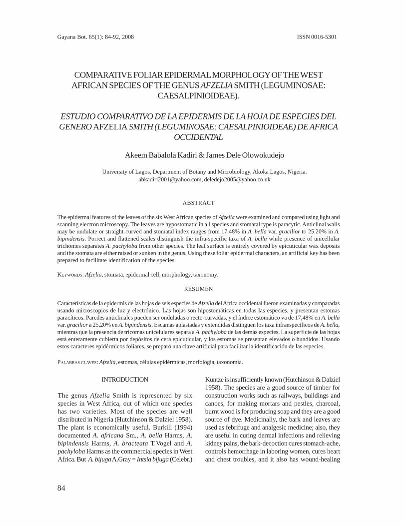
The leaf is generally hypostomatic, anticlinal wallsare usually straight, curved to undulate and cell shapeis either irregular or isodiametric (Figs. 1-3, Table I).
Stomata index = Stomata number X 100
Cell number per unit area + stomata number
Trichome index =
Trichome number X 100Cell number per unit area + trichome number
Foliar epidermal of Afzelia: KADIRI, A.B. & J. D. OLOWOKUDEJO
86
Gayana Bot. 65(1), 2008
FIGURE 1. Epidermides of the species of Afzelia in West Africa. a, b: A. africana; c, d: A. pachyloba; e, f: A. bipindensis. a,c, e: adaxial surface, b, d, f: abaxial surface. Scala = 50µm.
FIGURA 1. Epidermis de las especies de Afzelia en Africa occidental. a, b: A. africana; c, d: A. pachyloba; e, f: A. bipindensis.a, c, e: superficie adaxial, b, d, f: superficie abaxial. Escala = 50µm.
87
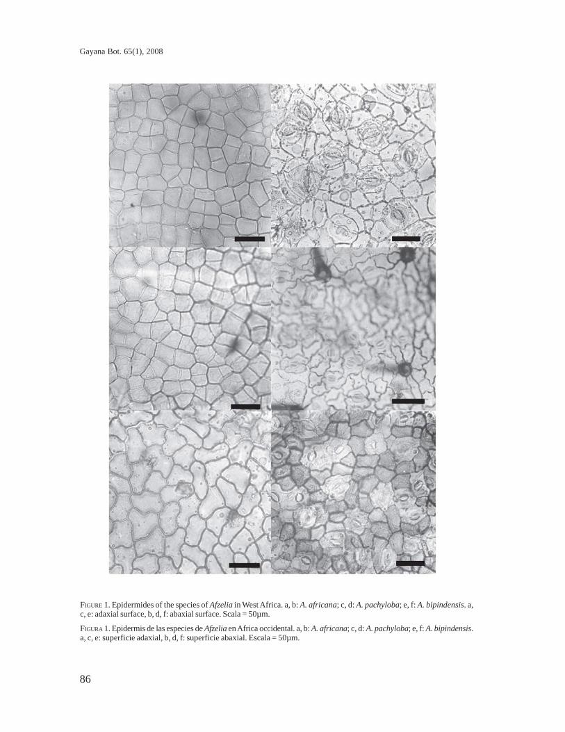
FIGURE 2. Epidermides of the species of Afzelia in West Africa. a, b: A. bracteata; c, d: A. bella var. bella; e, f: A. bellavar. gracilior. a, c, e: adaxial surface, b, d, f: abaxial surface. Scale = 50µm.
FIGURA 2. Epidermis de las especies de Afzelia en Africa occidental. a, b: A. bracteata; c, d: A. bella var. bella; e, f: A.bella var. gracilior. a, c, e: superficie adaxial, b, d, f: superficie abaxial. Escala = 50µm.
Foliar epidermal of Afzelia: KADIRI, A.B. & J. D. OLOWOKUDEJO
88
Gayana Bot. 65(1), 2008
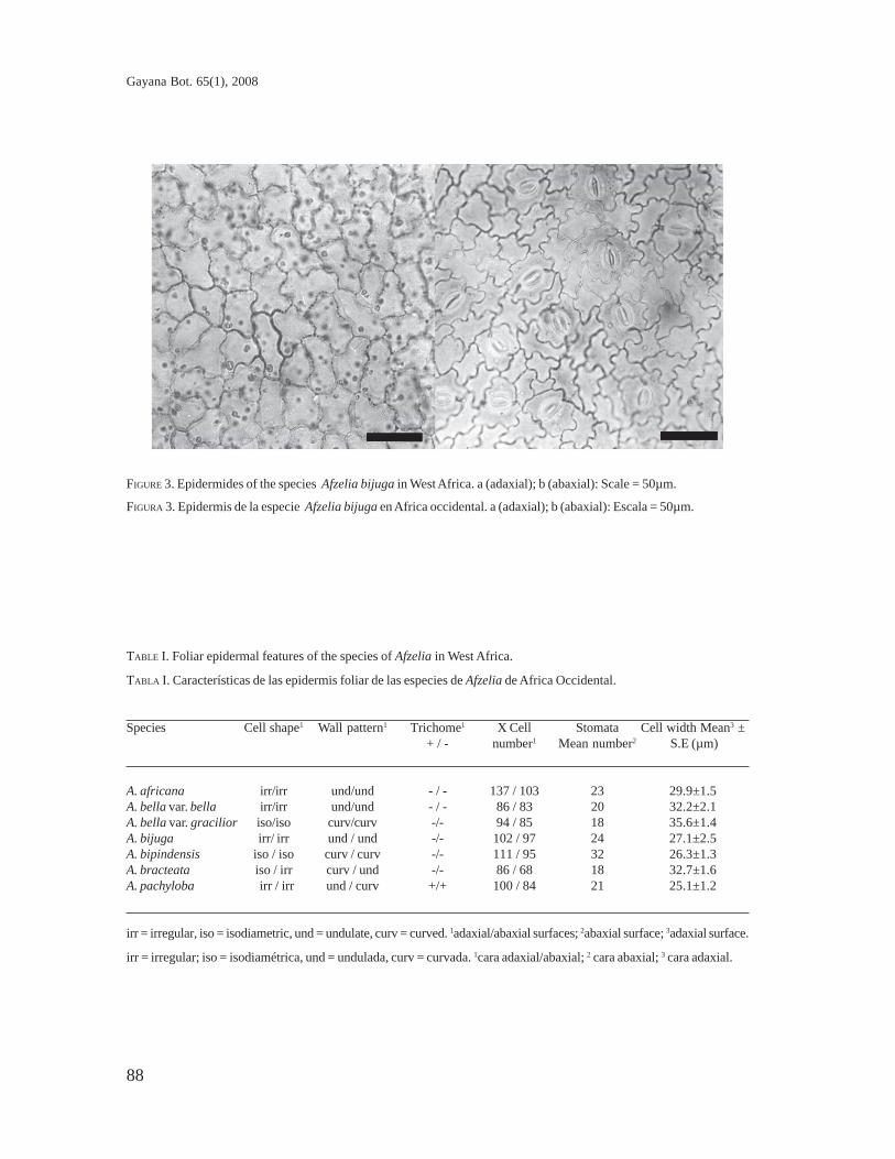
TABLE I. Foliar epidermal features of the species of Afzelia in West Africa.
TABLA I. Características de las epidermis foliar de las especies de Afzelia de Africa Occidental.
Species Cell shape1 Wall pattern1 Trichome1 X Cell Stomata Cell width Mean3 ±+ / - number1 Mean number2 S.E (µm)
A. africana irr/irr und/und - / - 137 / 103 23 29.9±1.5A. bella var. bella irr/irr und/und - / - 86 / 83 20 32.2±2.1A. bella var. gracilior iso/iso curv/curv -/- 94 / 85 18 35.6±1.4A. bijuga irr/ irr und / und -/- 102 / 97 24 27.1±2.5A. bipindensis iso / iso curv / curv -/- 111 / 95 32 26.3±1.3A. bracteata iso / irr curv / und -/- 86 / 68 18 32.7±1.6A. pachyloba irr / irr und / curv +/+ 100 / 84 21 25.1±1.2
irr = irregular, iso = isodiametric, und = undulate, curv = curved. 1adaxial/abaxial surfaces; 2abaxial surface; 3adaxial surface.
irr = irregular; iso = isodiamétrica, und = undulada, curv = curvada. 1cara adaxial/abaxial; 2 cara abaxial; 3 cara adaxial.
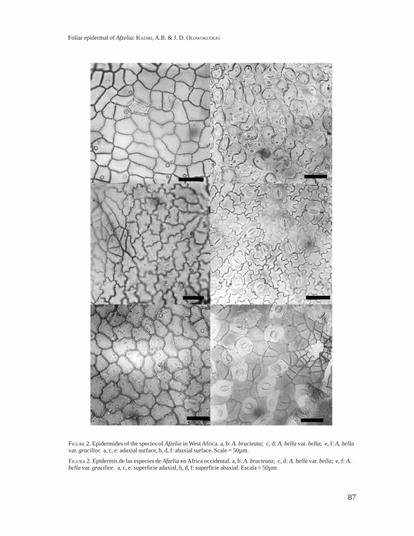
FIGURE 3. Epidermides of the species Afzelia bijuga in West Africa. a (adaxial); b (abaxial): Scale = 50µm.
FIGURA 3. Epidermis de la especie Afzelia bijuga en Africa occidental. a (adaxial); b (abaxial): Escala = 50µm.
89
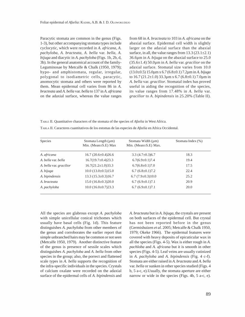
Paracytic stomata are common in the genus (Figs.1-3), but other accompanying stomata types includecyclocytic, which were recorded in A. africana, A.pachyloba, A. bracteata, A. bella var. bella, A.bijuga and diacytic in A. pachyloba (Figs. 1b, 2b, d,3b). In the general anatomical account of the family-Leguminosae by Metcalfe & Chalk (1950, 1979);hypo- and amphistomata, regular, irregular,polygonal to isodiametric cells, paracytic,anomocytic stomata and others were reported bythem. Mean epidermal cell varies from 86 in A.bracteata and A. bella var. bella to 137 in A. africanaon the adaxial surface, whereas the value ranges
from 68 in A. bracteata to 103 in A. africana on theabaxial surface. Epidermal cell width is slightlylarger on the adaxial surface than the abaxialsurface, in all, the value ranges from 13.3 (23.1±2.1)36.6µm in A. bijuga on the abaxial surface to 25.0(35.6±1.4) 50.0µm in A. bella var. gracilior on theadaxial surface. Stomatal size varies from 10.0(13.0±0.5) 15.0µm x 6.7 (6.8±0.1) 7.2µm in A. bijugato 16.7 (21.2±1.0) 33.3µm x 6.7 (6.8±0.1) 7.0µm inA. bella var. gracilior. Stomatal index has proveduseful in aiding the recognition of the species,its value ranges from 17.48% in A. bella var.gracilior to A. bipindensis in 25.20% (Table II).
TABLE II. Quantitative characters of the stomata of the species of Afzelia in West Africa.
TABLA II. Caracteres cuantitativos de los estomas de las especies de Afzelia en Africa Occidental.
Species Stomata Length (µm) Stomata Width (µm) Stomata Index (%)Min. (Mean±S.E) Max Min. (Mean±S.E) Max.
A. africana 16.7 (20.6±0.4)26.6 3.3 (4.7±0.3)6.7 18.3A. bella var. bella 16.7(19.7±0.4)23.3 6.7(6.9±0.1)7.4 19.4A. bella var. gracilior 16.7(21.2±1.0)33.3 6.7(6.8±0.1)7.0 17.5A. bijuga 10.0 (13.0±0.5)15.0 6.7 (6.8±0.1)7.2 22.4A. bipindensis 13.3 (15.3±0.3)16.7 6.7 (7.9±0.3)10.0 25.2A. bracteata 15.0 (16.8±0.3)20.0 6.7 (6.9±0.1)7.1 20.9A. pachyloba 10.0 (16.0±0.7)23.3 6.7 (6.9±0.1)7.1 20.0
All the species are glabrous except A. pachylobawith simple unicellular conical trichomes whichusually have basal cells (Fig. 1d). This featuredistinguishes A. pachyloba from other members ofthe genus and corroborates the earlier report thatsimple unbranched hairs may be common or not seen(Metcalfe 1950, 1979). Another distinctive featureof the genus is presence of sessile scales whichdistinguishes A. pachyloba and A. bella from otherspecies in the group; also, the porrect and flattenedscale types in A. bella supports the recognition ofthe infra-specific individuals in the species. Crystalsof calcium oxalate were recorded on the adaxialsurface of the epidermal cells of A. bipindensis and
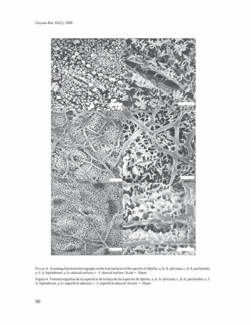
A. bracteata but in A. bijuga, the crystals are presenton both surfaces of the epidermal cell. But crystalhas not been reported before in the genus(Germishuizen et al. 2005; Metcalfe & Chalk 1950,1979; Okeke 1966). The epidermal features werecovered with heavy deposits of epicuticular wax inall the species (Figs. 4-5). Wax is either rough in A.pachloba and A. africana but it is smooth in otherspecies (Figs. 4-5). Leaf veins are usually cutinizedin A. pachyloba and A. bipindensis (Fig. 4 c-f).Stomata are either raised in A. bracteata and A. bellavar. bella or sunken in other species studied (Figs. 4b, 5 a-c, e).Usually, the stomata aperture are eithernarrow or wide in the species (Figs. 4b, 5 a-c, e).
Foliar epidermal of Afzelia: KADIRI, A.B. & J. D. OLOWOKUDEJO
90
Gayana Bot. 65(1), 2008
FIGURE 4. Scanning Electron micrographs of the leaf surfaces of the species of Afzelia. a, b: A. africana; c, d: A. pachyloba;e, f: A. bipindensis. a, b: adaxial surface, c - f: abaxial surface. Scale = 50µm.
Figura 4. Fotomicrografías de la superficie de la hoja de las especies de Afzelia. a, b: A. africana; c, d: A. pachyloba; e, f:A. bipindensis. a, b: superficie adaxial, c - f: superficie abaxial. Escala = 50µm.

91
FIGURE 5. Scanning Electron micrographs of the leaf surfaces of the species of Afzelia. a, b: A. bracteata; c, d: A. bella var.bella; e, f: A. bella var. gracilior. All abaxial surface. Scale = 50µm.
FIGURA 5. Fotomicrografías de la superficie de la hoja de Afzelia. a, b: A. bracteata; c, d: A. bella var. bella; e, f: A. bellavar. gracilior. Todas superficies abaxiales. Escala = 50µm.
Foliar epidermal of Afzelia: KADIRI, A.B. & J. D. OLOWOKUDEJO
92
Gayana Bot. 65(1), 2008
With these features, the genus can be distinguishedfrom other genera of the family by comparison of thevalues obtained on all the features studied. In the
same vein, the species too, can be delimited withease. Therefore, an artificial key is presented belowto separate the species.
ARTIFICIAL KEY TO THE SPECIES OF AFZELIA IN WEST AFRICA
1. Leaf pubescent, anticlinal wall undulate-curved on both surfaces. Stomatal index 20%……….…A. pachyloba1. Leaf glabrous, anticlinal wall undulate, straight- curved on either surface
2. Stomatal sunken, wax rough…………………………………...................................................…… A. africana2. Stomatal either sunken or raised, wax smooth.
3. Maximum stomata length not up to 25 µm, scale flattened…........................................A. bella var. bella3. Maximum stomata length above 25 µm, scale porrect....................................…..A. bella var. gracilior
4. Crystals of calcium oxalate present on both surfaces, epidermal cell width equal on bothsurfaces………....……………………………………………..................................................…A. bijuga
4. Crystals of calcium oxalate present on the adaxial surface only, epidermal cell width unequal oneither surface.
5. Stomata sunken .................................................................................................................A. bipindensis5. Stomata raised......................................................................................................................A. bracteata
CONCLUSION
The inter-specific taxonomic relationships existingin Afzelia can be investigated by the use of someendo-qualitative and quantitative characteristics ofthe leaf such as epidermal surfaces, epicuticular wax,stomatal size, crystals and stomatal orientation. Abijuga which is the least reported taxon is nowdocumented to possess glabrous leaf surfaces,crystals, irregular cell shape and undulate anticlinalwalls on both surfaces of the leaf epidermis. Thepresent study has also confirmed earlier reports ofworkers and new taxonomic data have been recordedfor the genus. Many of these features are bothspecific and generic constant, and they can be usedto facilitate the recognition of the species and helpin solving problems of adulteration in the commercialspecies even when the leaf is fragmentary.
BIBLIOGRAPHY
BURKILL, H. M. 1994. The Useful Plants of West TropicalAfrica (2nd Edition) Vol. 2. Royal Botanic Garden,Kew. pp. 21-25.
GERMISHUIZEN, G., N.R. CROUCH & G. CONDY. 2005. Afzeliaquanzensis. Flowering Plant of Africa 59: 74-83.
HUTCHINSON, J. & J.M. DALZIEL. 1958. Crown Agentsfor overseas Governments and Administrations.Flora of West Tropical Africa 1(2). London.828 pp.
INAMDAR, J. A. & M. GANGADHARA. 1977. Studies on thetrichomes of some Euphorbiaceae. FeddesRepertorium. 88: 103-111.
KOTRESHA, K. & Y.N. SEETHARAM. 1995. Epidermal studiesin some species of Bauhinia L. (Caesalpinioideae).Phytomorphology 45(1-2): 127-137.
KOTRESHA, K. & Y.N. SEETHARAM. 2000. Epidermalmicromorphology of some species of Cassia L.(Caesalpiniaceae). Phytomorphology 50(3-4):229-237.
METCALFE, C.R. & L. CHALK. 1950. Anatomy of theDicotyledons. Oxford University Press, Oxford.724 pp.
METCALFE, C.R. & L. CHALK. 1979. Anatomy of theDicotyledons (2nd ed.). Vol. 1. Oxford UniversityPress, Oxford. 276 pp.
OGUNDIPE, O.T. & O.O. AKINRINLADE. 1998. EpidermalMicromorphology of Some Species of AlbiziaDurazz (Mimosaceae). Phytomorphology 48 (2-3): 217-323.
OGUNDIPE, O.T. & D.E. WUJEK. 2004. Foliar anatomy ontwelve genera of Bignoniaceae (Lamiales). ActaBotanica Hungary 46: 290 - 312.
OKEKE, R.E. 1966. Comparative Anatomy of threeNigerian Afzelia species. Forest ProductsResearch Report 9: 8
OLOWOKUDEJO, J.D. & O. PEREIRA-SHETEOLU. 1988. Thetaxonomic value of epidermal characters in thegenus Ocimum (Lamiaceae) Phytomorphology 38(2-3):147-158.
STACE, C.A. 1965. Cuticular studies as an aid to planttaxonomy. Bulletin of British Museum (NaturalHistory) Botany 4: 3-78.
Recibido: 24.11.06Aceptado: 28.02.08
Copyright © 2022 FDOKUMEN