Vegetative propagation of mature dragon trees through epicormic ...
Salt-induced changes in the vegetative anatomyof Prosopis strombulifera (Leguminosae)
Herminda Reinoso, Laura Sosa, Lucía Ramírez, and Virginia Luna
Abstract: Seedlings of Prosopis strombulifera (Lam.) Benth. were grown hydroponically in Hoagland’s solution withaddition of 25 mmol/L NaCl every 48 h until final salt concentrations of 250, 500, and 700 mmol/L were reached.Control plants were grown without salt. Salinity induced anatomical changes in roots (young and mature zones), hypo-cotyls, young stems, and leaflets. The diameters of the young zone of roots of plants grown in increasing salt concen-trations were smaller than those of controls, with reduced number of cortex layers and reduced size of the vascularsystem. The roots from tolerant plants showed precocious suberization and (or) lignification of the endodermal cellsand early activity of the pericycle. Hypocotyl diameter was reduced along with a reduction in secondary phloem. Rootsand hypocotyls showed abundant phellem formation. The stem diameter of young tolerant plants was notably dimin-ished and less tissue lignification occurred. In stems and leaflets of treated plants, NaCl stimulated the production oftannins. In the leaflets, vascular bundles were similar in size. Groups of elongated parenchyma cells with many chloro-plasts surrounded the bundles. These results suggest that in the absence of secretory organs, the anatomical modifica-tions in this species are related to metabolic adaptations, such as an early development of the endodermal barrier forion exclusion, to allow survival in high salinity.
Key words: Prosopis strombulifera, anatomical changes, hydroponics, NaCl.
Résumé : Les auteurs ont cultivé des plantules de Prosopis strombulifera (Lam.) Benth. en hydroponique, utilisant lasolution de Hoagland. Il y ont ajouté 25 mmol/L de NaCl à toutes les 48 h, jusqu’à ce que les teneurs atteignent 250,500 et 700 mmol/L. Des plantes témoins ont été cultivées en absence de sel. La salinité induit des modifications anato-miques dans les racines (jeunes et matures), les hypocotyles, les jeunes tiges et les folioles. Les diamètres des partiesjeunes des racines des plants cultivés en présence accrues de sel, sont plus petits que chez les témoins, montrant unnombre réduit de couches corticales et une dimension amoindrie du système vasculaire. Les racines des plantes toléran-tes montrent une subérisation précoce et (ou) une lignification des cellules endodermiques ainsi qu’une activité hâtivedu péricycle. Le diamètre de l’hypocotyle ainsi que le phloème secondaire sont réduits. Les racines et les hypocotylesforment une abondance de phellème. Chez les jeunes plants tolérants, le diamètre de la tige est particulièrement réduit,et on y observe moins de tissus lignifiés. Chez les tiges et les folioles des plants traités, le NaCl stimule la productionsde tannins. Dans les folioles, les faisceaux vasculaires sont de même dimension. Des groupes de cellules allongées duparenchyme, contenant de nombreux chloroplastes, entourent les faisceaux. Ces résultats suggèrent qu’en absenced’organes sécréteurs, les modifications anatomiques, chez cette espèce, sont reliées à des adaptations écologiques tellesque le développement hâtif de la barrière endodermique pour exclure les ions, et permettre la survie en présence de sa-linité élevée.
Mots clés : Prosopis strombulifera, changements anatomiques, hydroponiques, NaCl.
[Traduit par la Rédaction] Reinoso et al. 628
Introduction
In Argentina, there are approximately 34 000 000 ha un-der conditions of excess water and salt, taking into accountonly humid environments and irrigated areas. This situation
represents extreme ecological conditions that drastically re-duce productivity. However, there are more than 300 speciesof halophytic plants in the world that have the potential tobe used for agricultural development, either improving eco-nomically important yet nontolerant species by means of ge-
Can. J. Bot. 82: 618–628 (2004) doi: 10.1139/B04-040 © 2004 NRC Canada
618
Received 29 July 2003. Published on the NRC Research Press Web site at http://canjbot.nrc.ca on 2 June 2004.
H. Reinoso. Laboratorio de Morfología Vegetal, Departamento de Ciencias Naturales, Universidad Nacional de Río Cuarto, RíoCuarto 5800, Argentina.L. Sosa. Laboratorio de Fisiología Vegetal, Facultad de Química, Bioquímica y Farmacia, Universidad Nacional de San Luis,San Luis, Argentina.L. Ramírez. Laboratorio de Genética Molecular y Mejoramiento de Plantas, Universidad Pública de Navarra, Pamplona, España.V. Luna.1 Laboratorio de Fisiología Vegetal, Departamento de Ciencias Naturales, Universidad Nacional de Río Cuarto,Río Cuarto 5800, Argentina.
1Corresponding author (e-mail: [email protected]).

© 2004 NRC Canada
Reinoso et al. 619
netic engineering (Glenn et al. 1999) or using them as forageor timber crops on otherwise barren soils (Bradbury andAhmad 1990).
In addition to producing metabolic changes, soil salinitycan also affect the general growth of the plant, particularlyits morphology and anatomy (Serrato Valenti et al. 1991). Itmay be inferred that plants that grow well in high-salinityenvironments have specific structural adaptations to alteredphysiological and biochemical mechanisms, while maintain-ing their reproductive capacity (Shannon et al. 1994). How-ever, knowledge of morphological and anatomical responsesto salinity is still incomplete.
Within Leguminosae subfamily Mimosoideae, there aresome genera that can be found in arid regions. One such ge-nus is Prosopis, which has many species that are importantarboreal and shrub-like components of saline zones in Amer-ica (Burkart 1937, 1952, 1976). These unique terrestrialspecies are able to grow in high-salinity conditions and,simultaneously, to fix nitrogen (Rhodes and Felker 1988).This is an unusual characteristic, because nitrogen fertiliza-tion otherwise decreases salt tolerance in halophytic plants(Grattan and Grieve 1999). The spiny shrub Prosopis strom-bulifera (Lam.) Benth. (Burkart 1976) is found in the sali-nized areas of Córdoba and San Luis, in central Argentina(Cantero et al. 1996). This species is distributed from theArizona desert (USA) to Patagonia (Argentina), where itmust tolerate prolonged drought periods. According to de laVega (1996), P. strombulifera can survive in hydroponicswith up to 1 mol/L NaCl, showing metabolic (enzymatic)changes in roots and leaves.
In a previous study we demonstrated that hydroponic-cultured plants of P. strombulifera could be considered toler-ant phenotypes, as there was growth increase of roots andstems with NaCl up to 450 mmol/L, compared with controlplants (Reinoso et al. 2000). These plants were more vigor-ous, had a higher leaf number with smaller leaflets, andharder and darker spines. The increase in salt concentrationabove 450 mmol/L decreased seedling growth.
As plant salt tolerance is a developmentally regulated fea-ture, some genotypes were unable to express tolerant character-istics in response to salt treatment. Therefore, 30% of plantsgrown in NaCl concentrations above 350 mmol/L showed par-ticular symptoms, such as a noticeable increase of root lengthand a general decrease of shoot growth (thick stems, thin andweak spines, general chlorosis, leaflet abscission, and leafletswelling). Because of their precocious senescence, very fewplants at the higher concentration (700 mmol/L NaCl) survivedto the end of the experiments (73 d). These plants are describedas “nontolerant” in the present work.
Investigation of anatomical changes in response to salinestress at the seedling stage may help us understand the inte-gration of morphological and physiological responses. Thus,the objective of the present work was to examine the effectsof increasing concentrations of NaCl (0–700 mmol/L) on thestructure of the root (young and older zone), hypocotyl,young stem, and leaflet from tolerant and nontolerant plantswithin the same population.
Materials and methods
Seeds of P. strombulifera were collected from southwest-
ern San Luis (near El Bebedero stream), Argentina. Geo-graphically, the location is 33°43′S, 66°37′W at 400–500 maltitude, with a temperate thermal regime (i.e., an annual av-erage temperature of 15–20 °C). This area belongs to thecarob tree forest located in a saline depression between theannual 300- to 400-mm isohyets in the El Monte Phyto-geographic Region. The soil has a franc-sandy texture,with abundant calcareous material and moderate salinity(8000 mho/cm2 (1 mho = 1 S) electrical conductivity at thesurface) (Anderson et al. 1970).
Pods were collected at random from 100 plants within thesame population, and pealed. Seeds were selected visuallyfor uniform size and healthy aspect. They were mechanicallyscarified and surface sterilized with 0.5% (m/m) sodiumhypochlorite for 1 min and then washed overnight underflowing water. Before sowing they were rinsed two or threetimes in distilled water and placed in Petri dishes with twolayers of water-saturated filter papers at 37 °C for 24 h (dela Vega 1996). Germinated seeds (with radicles 20 mm long)were cultured in hydroponics in two black trays per treat-ment per experiment (80 seedlings per tray,) with 10% offull-strength Hoagland’s solution. The seedlings were grownin a growth chamber under a 16 h light (200 µmol·m–2·s–1)(28 °C) : 8 h dark (20 °C) cycle and 80% relative humidity.After 76 h, seedlings were transferred to a 25% full-strengthHoagland’s solution (osmotic potential (Ψo) = –0.11 MPa).Each experiment (eight trays) had three replicates. The pHof the medium was adjusted to 6.0 in all cases. An aquariumtubing system with a peristaltic pump was used to provideaeration.
After 15 d, salt treatments were started by adding NaClpulses of 25 mmol/L every 48 h, until final salt concentra-tions of 250, 500, and 700 mmol/L (Ψo = –1, –1.88, and−2.6 MPa, respectively) were reached, corresponding toplants aged 35, 55, and 73 d, respectively. On each of thesedates, 15 control (no salt added) and 15 treated plants werecollected at random from each tray.
Samples taken from roots, stems, and leaves of theseplants were fixed in FAA (95% ethanol – glacial aceticacid – 37%–40% formaldehyde – water; 50:5:10:35, by vol-ume). In the case of roots and stems, samples were taken attwo different levels to analyze the structural differences be-tween young and adult segments of these organs. The youngroot zone was at 15 mm from the apex. To examine adultroots and hypocotyls, segments were taken, respectively, 5–6 mm below and 5–6 mm above the transition region (i.e.,where the root and stem meet). For young stems, sampleswere taken from below the second node of the main axes.Leaflets were taken from near the middle of the rachis of thefirst bipinnate leaf. The materials were then dehydrated in agraded ethanol series. Xylene was used as a transitional fluidprior to histowax (highly purified paraffin wax blended withpolymer additives) infiltration and embedding. A series oftransverse sections 10 µm thick were obtained using a rotarymicrotome. The sections were triple stained with hema-toxylin, safranin O, and fast green FCF (Johansen 1940).
To identify tannins, freehand sections were cut from freshmaterial and treated with ferric chloride (D’Ambrogio deArgüeso 1986) (data not shown).
To differentiate between lignin and suberin in the endo-dermis of young roots, paraffin was removed. Sections were

© 2004 NRC Canada
620 Can. J. Bot. Vol. 82, 2004
soaked in an alcoholic solution of phloroglucinol–HCl(D’Ambrogio de Argüeso 1986) and immediately observedin a Zeiss Axiophot fluorescence microscope using a Hg va-por lamp HBO100W and an excitation filter BP450–490combined with filters DS510 and LP 510. Phloroglucinol–HCl stains the aldehyde groups of lignin and suberin butquenches lignin’s autofluorescence and retains suberin’sautofluorescence (Baayen et al. 1996).
The histological preparations were assessed with a stan-dard Zeiss model 16 microscope, and photomicrographswere taken with a Zeiss Axiophot microscope using blackand white Kodak TMX100 print film.
Results
Low NaCl concentrations (up to 250 mmol/L) did notinduce structural changes in relation to control plants. Theanatomical descriptions in this section are therefore basedon observations made in plants collected at 500 and700 mmol/L NaCl (Ψo = –1.88 and –2.6 MPa, respectively)in the culture medium. Figure 1 shows a tolerant anda nontolerant treated plant collected from the 500-mmol/LNaCl treatment and their respective controls.
Anatomical characteristics of control plants
Young root zonesThese zones of the roots showed the same structure during
the 73 d of the experiment. Numerous wide-diameter, thin-walled root hairs were formed (Fig. 2). The cortex consistedof layers of seven or eight large, thin-walled parenchymacells, with small intercellular spaces. The pericycle adjacentto the endodermis consisted of two or three layers of thin-walled parenchyma cells with large nuclei and noticeablenucleoli (Fig. 3). The primary vascular tissues were repre-sented by three phloem groups alternating with three xylemstrands (Fig. 2).
Older root zonesOlder roots of 55- and 73-d-old plants showed a similar
diameter. The primary xylem in the center of the roots wasstar shaped and surrounded by secondary xylem, the cambialzone, and secondary phloem (Fig. 4). Three or four layers ofparenchyma originated from the pericycle external to thesecondary phloem. Several rows of phellem with tannin cellswere present.
HypocotylsHypocotyls from 55- and 73-d-old plants had the same
structural characteristics. A parenchymatous pith was sur-rounded by secondary xylem, forming a continuous cylinder(Fig. 5). A wide cambial zone separated the secondaryphloem from the secondary xylem. Four groups of highlylignified primary phloem fibers remained. Outside the corti-cal parenchyma a periderm, with similar characteristics tothose described for older root zones, had differentiated.
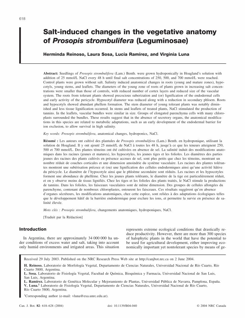
Fig. 1. General aspect of 55-d-old plants of Prosopis strombulifera. (A) Control plants. (B) Nontolerant plants from the 500 mmol/LNaCl treatment. (C) Tolerant plants collected from the 500 mmol/L NaCl treatment. Scale bar = 30 mm.

© 2004 NRC Canada
Reinoso et al. 621
Figs. 2–8. Transverse sections showing the anatomical characteristics of the young root zone, mature root zone, hypocotyl, young stem,and leaflet from control Prosopis strombulifera plants. Fig. 2. Young root zone from 73-d-old plants. Numerous thin-walled root hairsare present. The cortex is formed by seven or eight parenchyma layers. The innermost layer of the cortex, the endodermis (arrowhead),encloses the central cylinder. This root is triarch. Scale bar = 50 µm. Fig. 3. Detail of the root shown in Fig. 2. The pericycle enclos-ing the vascular tissue comprises two or tree layers of thin-walled parenchyma cells, indicating the initiation of secondary growth.Scale bar = 10 µm. Fig. 4. Older root zone section from a 73-d-old plant. The primary xylem in the center of the root is star shapedand surrounded by a developed secondary xylem. Scale bar = 100 µm. Fig. 5. Hypocotyl from a 73-d-old plant. The center of the or-gan is occupied by a thin-walled parenchymatous pith surrounded by secondary vascular tissues organized in a continuous cylinder.Scale bar = 100 µm. Fig. 6. Young stem from a 55-d-old plant. The vascular system is formed by a circle of open collateral vascularstrands separated by narrow areas of interfascicular parenchyma with tannin-containing cells. Scale bar = 50 µm. Fig. 7. Leaflet from a73-d-old plant with an isolateral organization. The stomata (arrows) are more abundant on the adaxial surface. The difference in diame-ter between the main vascular bundle and the lateral ones can also be seen. Scale bar = 50 µm. Fig. 8. Detail of the leaflet shown inFig. 7. Funnel-shaped cells with tannin (arrowhead) are located between the adaxial palisade tissue cells. Scale bar = 25 µm. ad,adaxial surface; ca, cambial zone; co, cortex; en, endodermis; fi, fibers; pe, pericycle; ph, phloem; pi, pith; pr, periderm; px,protoxylem; rh, root hairs; sp, secondary phloem; sx, secondary xylem.

Young stem zonesIn young stems from 55-d-old plants, the vascular system
consisted of a ring of open collateral vascular bundles sepa-rated by interfascicular areas with tannin-filled cells (Fig. 6).The cortex was formed by five chlorenchymatic subepider-mic strata, with some immersed tannin-filled cells. Primaryphloem fibers were present external to the functional phloem(Fig. 6).
Fewer tannin-filled cells were present in the cortex ofstems obtained from 73-d-old plants (data not shown). Thevascular bundles showed secondary tissues. The primaryphloem fiber caps outside the bundles were connected in theinterfascicular region, forming a continuous sclerenchymacylinder.
LeafletsThe anatomical features of leaflets obtained from 55- and
73-d-old plants were similar. In cross section, epidermalcells showed an elliptic shape, and a few of them containedtannins. Stomata were more abundant on the adaxial surface.Palisade tissue, which constituted the major part of the me-sophyll, occurred on either side of the spongy mesophyll.Cells towards the adaxial surface were long and narrow,while the ones towards the abaxial surface were short andwide (Fig. 7). A few funnel-shaped tannin-filled cells werelocated among the adaxial palisade cells (Fig. 8).
The vascular bundles were collateral and surrounded by abundle sheath. Fibers were present external to the functionalphloem of major veins (Fig. 7).
Anatomical characteristics of tolerant plants
Young root zonesThe roots collected at 500 mmol/L NaCl had a reduced di-
ameter because of fewer cortex layers and a reduction of thestele (Fig. 9). The cortex contained only four or five layersof parenchyma cells and an endodermis with thickened ra-dial and tangential cell walls. In Fig. 10, the fluorescence ofsuberin can be seen in some cells, while the rest of theendodermal cells appear lignified. The pericycle was an ac-
tive ring that comprised at least three layers of compressedand radially ordered cells (Fig. 11). The structure of theseroots was diarch (Fig. 9) and occasionally triarch. The xy-lem formed a solid central core of lignified metaxylem.
Roots from plants collected from the 700 mmol/L NaCltreatment underwent secondary growth. Secondary xylem el-ements were noticeable, and primary phloem was crushed.The periderm had started to form around the vascular cylin-der (Fig. 12), indicated by precocious meristematic activityof the pericycle in comparison with that of the controlplants. The cells in the periderm showed tannins in their cy-toplasm and cell walls. Cortical tissue remained external tothe developing periderm.
Older root zonesRoots from plants collected from the 500 mmol/L NaCl
treatment (data not shown) and 700 mmol/L NaCl treatmenthad a greater diameter than their respective controls, but thesecondary xylem showed a smaller development in bothcases. All plants had a reduced cambial zone. Outside thiszone voluminous parenchyma cells from the secondaryphloem and parenchyma cells originating from the pericyclewere developed. This, together with the greater developmentof the periderm, especially the phellem layers, caused the in-crease of the root diameter (Fig. 13).
HypocotylsHypocotyls from tolerant plants collected from the
500 mmol/L NaCl treatment (data not shown) and700 mmol/L NaCl treatment did not differ in their structuralcharacteristics, showing great phellem development, espe-cially in the regions between the primary phloem fibers.Here the cells had a greater radial expansion. Under thisprotective tissue, the organ adopted a quadrangular shape(Fig. 14). A narrow cambial zone produced scarce secondaryphloem formed by compact, small, and compressed cells.
Young stem zoneImportant features of young stems with these treatments
are diameter reduction, noticeable tannin accumulation in
© 2004 NRC Canada
622 Can. J. Bot. Vol. 82, 2004
Figs. 9–18. Transverse sections of the different organs from tolerant plants of Prosopis strombulifera showing the anatomical changesinduced by exposure to NaCl treatments. Fig. 9. Young root zone from a plant collected from the 500 mmol/L NaCl treatment showingthe noticeable reduction in diameter. The cortex contains four or five parenchyma layers. This root is diarch. Scale bar = 50 µm.Fig. 10. Young root zone from a plant collected from the 500 mmol/L NaCl treatment selectively showing the fluorescence of suberinin the endodermal cell walls after soaking the tissue in phloroglucinol–HCl (arrowhead). Scale bar = 60 µm. Fig. 11. Detail of the rootshown in Fig. 9. Endodermal cells with thickened radial and tangential walls. The pericycle has divided initiating secondary growthand comprises two or three layers of compressed and radially ordered cells. Scale bar = 10 µm. Fig. 12. Young root zone from a plantcollected from the 700 mmol/L NaCl treatment. Detail of the central cylinder. The secondary xylem elements are noticeable. Theperiderm has started to form around the vascular cylinder. Scale bar = 25 µm. Fig. 13. Older root zone section from a plant collectedfrom the 700 mmol/L NaCl treatment showing a greater diameter than control roots. Periderm and neighboring parenchyma are welldeveloped. The secondary xylem is less developed. Scale bar = 100 µm. Fig. 14. Hypocotyl from a tolerant plant collected from the700 mmol/L NaCl treatment. The organ has a quadrangular shape and is protected by a developed phellem. The limited developmentof secondary phloem is evident. Scale bar = 100 µm. Fig. 15. Young stem from a tolerant plant collected from the 500 mmol/L NaCltreatment showing the notably reduced diameter and greater tannin content compared with control. Scale bar = 50 µm. Fig. 16. Youngstem from a tolerant plant collected from the 700 mmol/L NaCl treatment showing tannin accumulation in parenchyma cells of cortexand pith. Scale bar = 50 µm. Fig. 17. Leaflet from a tolerant plant collected from the 500 mmol/L NaCl treatment showing a lowernumber of stomata (arrow) and more abundant tannic cells in the adaxial epidermis. Scale bar = 50 µm. Fig. 18. Leaflet from a toler-ant plant collected from the 700 mmol/L NaCl treatment showing groups of palisade cells converging in relation to the vascular bun-dles leaving elongated intercellular spaces (arrowheads). Scale bar = 50 µm. ad, adaxial surface; co, cortex; en, endodermis; mx,metaxylem; pe, pericycle; ph, phloem; pi, pith; pr, periderm; rh, root hairs; sp, secondary phloem; sx, secondary xylem.

cortical and (or) medullar parenchyma, vascular strand sizereduction, and absence of sclerenchyma tissue (Figs. 15 and16). In plants collected at 500 mmol/L NaCl, the epidermishad abundant tannin-filled cells. The cortical region wasformed by small chlorenchyma cells, homogeneous in shapeand size, arranged in a compact way and containing abun-dant tannins (Fig. 15). In stems from plants collected at700 mmol/L NaCl, only the most internal strata of the cortex
contained tannins. An accumulation of tannins in paren-chyma cells of the pith was also observed (Fig. 16).
LeafletsLeaflets from plants collected from the 500 mmol/L NaCl
treatment developed fewer stomata and abundant tannin-filled cells in the adaxial epidermis (Fig. 17). Parenchymacells in the palisade tissue tended to adopt a narrow and
© 2004 NRC Canada
Reinoso et al. 623

elongated shape with the same size and arrangement towardsboth leaflet surfaces. The main vascular bundle showed a re-duced diameter and a more compact cap with more abundantthick-walled fibers compared with the controls. More abun-dant cylindric tannin-filled cells also differentiated towardsthe adaxial surface in the mesophyll after salt treatments(Fig. 17).
In plants exposed to the 700 mmol/L NaCl treatment, themesophyll was formed by palisade tissue with very narrowand elongated cells rich in chloroplasts. Groups of these pa-renchyma cells converged in relation to the vascular bundles,leaving elongated intercellular spaces mainly in the inter-fascicular regions (Fig. 18). The diameter of the main vascu-lar bundle was reduced, and there was a slight increase inthe diameter of the lateral ones (Fig. 18).
Anatomical characteristics of the nontolerant plants
Young root zonesRoots collected from the 500 mmol/L (data not shown)
and 700 mmol/L NaCl treatments showed similar character-istics. Their central cylinder diameter was similar to that intolerant plant roots at 500 mmol/L NaCl. The epidermis hadremains of root hairs (Fig. 19). The cortex was thicker thanin tolerant plants because of the noticeable enlargement ofthe parenchyma cells. The endodermis had small and irregu-lar thin-walled cells, with only some of them showing Cas-parian strips (Fig. 20). The pericycle was represented byonly one layer of parenchyma cells, and only the xylem ele-ments maintained their integrity (Fig. 20).
Older root zonesRoots from plants collected from the 700 mmol/L NaCl
treatment showed profound alterations that had alreadystarted in the plants collected at 500 mmol/L (data notshown). In the center of the organ, as well as in vascular tis-sues (especially phloem), cellular destruction and tissue dis-organization occurred. The periderm had an abnormal andunequal development around the organ (Fig. 21).
HypocotylsHypocotyls from plants collected from the 500 and
700 mmol/L NaCl treatments had a large diameter becauseof the vacuolation undergone by the cortical layers of paren-chyma and by the enhanced development of the protective
tissues. The pith was disorganized, leaving large inter-cellular spaces (Fig. 22). The primary periderm was stillpresent, and a new periderm had started to form internally tothe primary phloem fibers (Fig. 23).
Young stem zonesStems from plants collected from the 500 and
700 mmol/L NaCl treatments did not show differences be-tween them. The most distinctive feature in these organswas the voluminous cortical and phloem parenchyma withenlarged and vacuolated cells. Phloem fibers were absent(Fig. 24).
LeafletsThe leaflets from plants collected from the 500 and
700 mmol/L NaCl treatments did not show differences be-tween them. The thickness of the mesophyll increasedapproximately 50% because of the enlargement of the chlor-enchyma cells, which showed intense vacuolation. This in-creased cellular volume did not leave intercellular spaces,rendering the mesophyll compact. Very few chloroplastscould be seen in these cells. In the mesophyll towards theadaxial surface, some tannin-filled cells were present(Fig. 25).
Discussion
Our results with P. strombulifera show that salt treatmentsinduced structural alterations in cells and tissues that modi-fied growth patterns at different levels of organization. Ana-tomical and cytological differences in roots, stems, andleaves occurred when plants were grown in the presence ofincreasing concentrations of NaCl in the medium, as re-ported for other species of this genus, such as Prosopistamarugo and Prosopis cineraria (Serrato Valenti et al.1991, 1992).
Anatomical changes in rootsRoot diameter of treated plants (tolerant and nontolerant)
near the apex was smaller than that in nontreated plants.This was noticeable from concentrations of 500 mmol/L andhigher. The smaller diameter was correlated with fewercortex layers and a reduction of the vascular system. Thispattern was maintained at higher concentrations (up to
© 2004 NRC Canada
624 Can. J. Bot. Vol. 82, 2004
Figs. 19–25. Transverse sections of different organs from nontolerant plants of Prosopis strombulifera showing the anatomical changesinduced in plants collected from the 700 mmol/L NaCl treatment. Fig. 19. Young root zone. The epidermis has remains of trichomes(arrowhead). Five or six parenchyma layers form the cortex. This root is diarch. Scale bar = 50 µm. Fig. 20. Detail of the root shownin Fig. 19. The pericycle is represented by only one layer of parenchyma cells. The endodermis has small and irregular thin-walledcells, only a few of them showing Casparian strips (arrowheads). Scale bar = 25 µm. Fig. 21. Older root zone showing profound alter-ations in vascular tissues. The periderm is abnormal and shows unequal development around the organ. Scale bar = 100 µm. Fig. 22.Hypocotyl showing a large diameter due to the intense vacuolation undergone by the cortical layers of parenchyma cells (arrowhead)and the enhanced development of the protective tissues. The parenchymatous pith has a disorganized aspect, leaving large intercellularspaces between its cells. Scale bar = 100 µm. Fig. 23. Detail of the hypocotyl shown in Fig. 22. The primary periderm is still present,but a new deeper periderm has formed towards the center of the organ. Scale bar = 50 µm. Fig. 24. Young stem. The cortex has largeparenchyma cells. The vascular bundles have abundant phloem parenchyma cells lacking fibers. Scale bar = 50 µm. Fig. 25. Leaflet.The enlargement of mesophyll cells is evident. These cells are arranged without leaving intercellular spaces (large arrowhead) and havelost most of their chloroplasts. Towards the adaxial surface of mesophyll, several cells with tannins (smaller arrowheads) are present.Scale bar = 50 µm. ad, adaxial surface; co, cortex; pe, pericycle; pi, pith; ph, phloem; pr, periderm; pr1, primary periderm; pr2, second-ary periderm; sp, secondary phloem; sx, secondary xylem.

700 mmol/L). This is in agreement with observations madeby Serrato Valenti et al. (1991) for P. tamarugo.
One of the developmental features that seems to charac-terize halophytic species is a marked reduction in the devel-opment of the cortex of primary roots (Hajibagheri et al.1985). These authors also observed an enhanced develop-ment of endodermis in roots of Suaeda maritima, with Cas-parian strips developing earlier in more saline environments.Roots from tolerant plants of P. strombulifera showed preco-cious suberization and (or) lignification of endodermal cells.The earlier development resulted in a discernible endo-
dermis, with Casparian strips found much closer to the roottip than in other species such as glycophytes. When a com-plete suberin lamella has developed, the endodermis as awhole forms the main barrier towards the radial transportof water (Schreiber et al. 1999). Consequently, these plantshave greater possibilities of efficiently filtering the soil solu-tion to prevent the passage of an excess of ions to the xylem.The fact that the roots from nontolerant plants had scarce orrare Casparian strips in their young segments may be thereason why these plants could not exclude ions, causingmetabolic disorders and foliar succulence (see next para-
© 2004 NRC Canada
Reinoso et al. 625

graph) that finally caused plant death. Another remarkablefeature of the precocious development of roots from tolerantplants is the activity of the pericycle initiating secondary tis-sues. The activity of the perycicle was severely injured bysalt treatment in nontolerant plants.
The most significant anatomical changes observed in themature zone of roots from tolerant plants was the increase inthe size of phloem parenchyma cells as well as the paren-chyma originating from the pericycle. Their vacuolationcould be interpreted as an adaptive mechanism to regulatethe internal concentration of solutes and protect the cyto-plasm from toxic levels of ions by storing them in the vacu-ole (Shannon et al. 1994).
The marked phellem development shown by the roots oftolerant plants may have some physiological and (or) adap-tive implications. In xerophytes, very thick cork develop-ment is usually related to a more efficient protection againstdrying out or other damage (Fahn and Cutler 1992). The for-mation of new periderm is common in mechanically injuredtissues (Oven et al. 1999). In this case, the stressing agent issalinity, so the development of this tissue might be a gener-alized stress response.
Anatomical changes in hypocotylsThe hypocotyl periderm of tolerant plants showed a gen-
eral response pattern similar to that described for roots. Thehypocotyls of the nontolerant plants, however, showed anoverproduction of periderm. This could be interpreted asplant investment of biopolymers into protective tissues be-fore death, considering that the salinity stress was lethalto them. This observation reinforces the idea of a stress-dependent stimulation of phellogen activity.
The most important modification in the hypocotyl struc-ture was a variation in shape, which in tolerant plants is ac-companied by a reduction in size. The quadrangular shape ofthese hypocotyls showed that salt affected vascular cambiumactivity by diminishing secondary phloem production. Inhypocotyls from nontolerant plants, the cambial activitywas difficult to evaluate because of the disorganization ofphloem and cortex. The additional formation of new peri-derm made it more difficult to adequately resolve the sec-ondary phloem limits.
Cambial activity is very sensitive to different stresses. Forexample, because of drought stress, in typical desert shrubsthe period of cambial activity coincides with the rainy sea-son (Fahn and Cutler 1992). Salinity stress caused a reducedproduction of xylem in Populus euphatica trees (Waisel1972). Our results with P. strombulifera confirmed thesegeneral observations.
Anatomical changes in the young zone of the stemsThe diameters of young stems in tolerant plants were no-
tably diminished, and tissues were less lignified. Similar ob-servations were made by Waisel (1972) and Saadeddin andDoddema (1986), who found that stems with abundant sec-ondary xylem constituted by highly lignified elements arecharacteristic of plants growing in habitats with low waterpotential. The facultative halophyte Atriplex prostrata alsoshowed smaller stem diameter and less lignified tissueswhen grown at 0.5% and 1% NaCl (Wang et al. 1997), sug-
gesting that those plants under salinity stress may stay injuvenile stages because of growth retardation.
Anatomical changes in the leafletsThe foliar anatomical features were remarkably different
in tolerant and nontolerant plants. The acquisition of succu-lence in plants growing under saline conditions is generallyconsidered an adaptive response, since it provides the struc-tural basis for the dilution of salts accumulated in the leaftissues (Waisel 1972; Cutter 1980; Fahn 1990). However, inP. strombulifera the acquisition of succulence was the resultof physiological and anatomical disturbances produced byNaCl salinity in 30% of the treated plants (nontolerant) thatwere not able to survive. It is possible that this response islinked to Cl– buildup, causing ionic imbalance and entry ofwater, as cited for other nontolerant species (Romero-Arandaet al. 1998).
The reduction in the number of chloroplasts in these leaf-lets mimics a glycophytic behavior. In several glycophytesgrowing in saline soils, Greenway and Mumn (1980) ob-served an intense foliar chlorosis, which negatively affectedthe photosynthetic rate. The cause of photosynthesis reduc-tion in nontolerant species growing in the presence of NaClis a matter of controversy. It seems to depend on specific iontoxicity caused by sodium (Lloyd et al. 1990), chloride(Romero-Aranda et al. 1998), or both (Lloyd et al. 1989).
Considering the mesophyll organization in tolerant leaf-lets, the tendency to homogeneity in vascular bundle size isremarkable. This could be interpreted as an adaptation tohelp an equitable transfer of water in the whole leaf, a strat-egy analogous to that present in some xerophytic species(Oppenheimer 1960; Fahn and Cutler 1992).
Fahn and Cutler (1992) consider that the arrangement ofthe assimilatory tissue in convergent radial groups of cellssurrounding each vein favors photosynthesis, and watertransport from the bundles toward the epidermis is more effi-cient when an adequate water supply is provided. The largeamount of chloroplasts and this particular arrangement ofthe assimilatory parenchyma are in agreement with the im-portant increase observed in the amount of photosyntheticpigments (Reinoso et al. 2000) and with the dry mass / freshmass index in leaves (Sosa et al. 2002), suggesting that pho-tosynthesis is not affected by salt in tolerant treated seed-lings of this species.
Tannin accumulationThe presence of increased tannin content in stems and
leaves of tolerant plants deserves special consideration.Phenolics have been considered of adaptive value in xero-phytic conditions. The accumulation of tannins increasedwith drought in Australian sclerophyllous plants (Fahn andCutler 1992) and in leaves of beech trees in xeric conditions(Grossoni et al. 1998). Tannins and lignin have been shownto increase in plants under elevated UV-B (Rozema et al.1997). The production of these UV-B absorbing phenoliccompounds is considered to be a hallmark of adaptation tothe terrestrial environment. Damage by UV-B to key targetsin plants can be largely avoided by effective wavelength-selective filtering of UV-B by phenolics. On the other hand,Rivero et al. (2001) found that plants of Lycopersicon escu-lentum and Citrullus lanatus under thermal stress accumu-
© 2004 NRC Canada
626 Can. J. Bot. Vol. 82, 2004

lated soluble phenolics by activating their biosynthesis andinhibiting their oxidation. Our results show that in P. strom-bulifera, NaCl stimulated the production of these com-pounds independently of light intensity and temperatureconditions. This is in agreement with the observation of a re-duction in tannin content in palisade cell layers of Kandeliacandel plants collected from a saline soil and grown infreshwater (Hwang and Chen 1995). As several studies sug-gest that condensed tannin accumulation occurs in responseto different stress conditions, these compounds may be in-volved in cell protective roles such as scavenging of reactiveoxygen species, which are known to produce a secondaryoxidative stress in biotic and abiotic stresses.
We found anatomical and histological differences betweencontrol, tolerant, and nontolerant treated seedlings, mainly inthe speed of tissue differentiation, tissue organization, andtannin accumulation. We propose that in the absence of se-cretory organs, anatomical modifications of tolerant plantsare related to metabolic changes that are the result of a com-plex and accurately tuned physiological regulation involvingion compartmentalization and compatible solute synthesis toprotect the cytoplasm in high salinity.
Further studies are being carried out to elucidate suchmechanisms and to test the proposition that salt-increasedtannin content may be in relation to some yet unknown cellstress protective function.
Acknowledgments
The study was supported with funds from ConsejoNacional de Investigaciones Científicas y Técnicas(CONICET) (PEI 0480/97), CONICOR (Consejo de Investi-gaciones de la Provincia de Córdoba) (research grant1655/99), and SECYT–UNRC (Secretaría de Ciencia yTécnica, Universidad Nacional de Río Cuarto, Argentina)(research grants 038/98-02 and 023/03) grants to V. Luna.
References
Anderson, D.L., Del Aguila, J.B., and Bernardon, A.E. 1970. Lasformaciones vegetales en la provincia de San Luis. Revista deInvestigación Agropecuaria, INSTA. Buenos Aires, Argentina.[In Spanish.]
Baayen, R.P., Ouellette, G.B., and Rioux, D. 1996. Compart-mentalization of decay in carnations resistant to Fusariumoxysporum f. sp. dianthi. Phytopathology, 86: 1018–1031.
Bradbury, M., and Ahmad, R. 1990. The effect of silicon on thegrowth of Prosopis juliflora growing in saline soil. Plant Soil,125: 71–74.
Burkart, A. 1937. Estudios morfológicos y etológicos en el géneroProsopis. Darwiniana, 3: 27–47. [In Spanish.]
Burkart, A. 1952. Las Leguminosas Argentinas. Acme Agency,Buenos Aires, Argentina. [In Spanish.]
Burkart, A. 1976. A monograph of the genus Prosopis (Legu-minosae subfam. Mimosoideae). Catalogue of the reconized spe-cies of Prosopis. J. Arnold Arbor. Harv. Univ. 57: 450–525.
Cantero, J.J., Cantero, A., and Cisneros, J.M. 1996. La vegetaciónde los paisajes hidro-halomórficos del centro de Argentina.Universidad Nacional de Río Cuarto, Córdoba, Argentina. [InSpanish.]
Cutter, E.G. 1980. Plant anatomy: experiment and interpretation,Part II. Organs. Edward Arnold, London.
D’Ambrigio de Argüeso, A. 1986. Manual de técnicas en histo-logía Vegetal. Ed. Hemisferio Surm Buenos Aires, Argentina.[In Spanish.]
de la Vega, A. 1996. Variabilidad genética y estructuración pobla-cional en especies del género Prosopis (Leguminosae). Recursobiótico para reforestar zonas áridas. Doctoral thesis, ETSIA,Universidad Pública de Navarra, España. [In Spanish.]
Fahn, A. 1990. Plant anatomy. 4th ed. Butterworth, Heinemann,Jordan Hill, Oxford.
Fahn, A., and Cutler, D. 1992. Xerophytes. Gebrüder BorntraegerBerlin, Stuttgart.
Glenn, E.P., Brown, J.J., and Blumwald, E. 1999. Salt toleranceand crop potential of Halophytes. Crit. Rev. Plant Sci. 18: 227–255.
Grattan, S.R., and Grieve, C.M. 1999. Handbook of plant and cropstress. Edited by M. Pessarakli. Dekker, New York.
Greenway, H., and Munns, R. 1980. Mechanisms of salt tolerancein nonhalophytes. Annu. Rev. Plant Physiol. 31: 149–190.
Grossoni, P., Bussotti, F., Tani, C., Gravano, E., Santorelli, S., andBottacci, A. 1998. Morpho-anatomical alterations in leaves ofFagus sylvatica L., and Quercus ilex L. in different environmen-tal stress condition. Chemosphere, 36: 919–924.
Hajibagheri, M.A., Yeo, A.R., and Flowers, T.J. 1985. Salt tole-rence in Suaeda maritime (L.) Dum. Fine structure and ionconcentrations in the apical region of roots. New Phytol. 99:331–343.
Hwang, Y.H., and Chen, S.C. 1995. Anatomical resonses in Kan-delia candel (L.) Druce seedling growing in the presence of dif-ferent concentrations of NaCl. Bot. Bull. Acad. Sin. (Taipei), 36:181–188.
Johansen, D.A. 1940. Plant microtechnique. 1st ed. McGraw-HillBook Company. Inc., New York.
Lloyd, J., Kriedemann, P.E., and Aspinall, A. 1989. Comparativesensitivity of “Prior Lisbon” lemon and “Valencia” orange treesto foliar sodium and chloride concentration. Plant Cell Environ.12: 529–540.
Lloyd, J., Kriedemann, P.E., and Aspinall, A. 1990. Contrast be-tween Citrus species in response to salinization an analysis ofphotosynthesis and water relations for different rootstocks com-binations. Physiol. Plant. 78: 236–246.
Oppenheimer, H.R. 1960. Adaptation to drought: xerophytism.Plant–water relationships in arid and semiarid conditions. AridZone Res. 15: 105–138.
Oven, P., Torelli, N., Shortle, W.C, and Zupan�i�, M. 1999. Theformation of a ligno-suberised layer and necrophylactic peri-derm in beech bark (Fagus sylvatica L.). Flora, 194: 137–144.
Reinoso, H., Sosa, L., Colombo, C., Ochoa, F., and Luna, V. 2000.Morphological and physiological responses of Prosopis strom-bulifera (Lam.) Benth., to increasing salt conditions. In Plant Bi-ology 2000: American Society of Plant Physiologists AnnualMeeting, San Diego, Calif., 15–19 July 2000. Edited by D.Bush, D. Cosgrove, R. Hangarter, R. Jorgensen, D. Delmer, P.Springer, W. Lucas, and D. Schnell. American Society of PlantPhysiologists, Baltimore, Md. pp. 210–202. [Abstr.]
Rhodes, D., and Felker, P. 1988. Mass screening of Prosopis (Mes-quite) seedlings for growth at seawater salinity concentrations.For. Ecol. Manage. 24: 169–176.
Rivero, R., Ruiz, J.M., García, P.C., López-Lefebre, L.R, Sánchez,E., and Romero, L. 2001. Resistance to cold and heat stress: ac-cumulation of phenolic compounds in tomato and watermelonplants. Plant Sci. 160: 315–321.
Romero-Aranda, R., Bondada, B.R., Syvertsen, J.P., and Grosser,J.W. 1998. Leaf characteristics and net gas exchange of diploidand autotetraploid Citrus. Ann. Bot. (Lond.), 79: 153–160.
© 2004 NRC Canada
Reinoso et al. 627

Rozema, J., van de Staaij, J., Björn, L.O., and Caldwell, M. 1997.UV-B as an environmental factor in plant life: stress and regula-tion. Trends Ecol. Evol. 12: 22–28.
Saadeddin, R., and Doddema, H. 1986. Anatomy of the “extreme”halophyte Arthrocnemum fruticosum (L.) Moq. in relation to itsphysiology. Ann. Bot. (Lond.), 57: 531–544.
Schreiber, L., Hartmann, K., Skrabs, M., and Zeier, J. 1999. Apo-plastic barriers in roots: chemical composition of endodermaland hypodermal cell walls. J. Exp. Bot. 50: 1267–1280.
Serrato Valenti, G., Ferro, M., Ferraro, D., and Riveros, F. 1991.Anatomical changes in Prosopis tamarugo Phil. Seedlings grow-ing at different levels of NaCl salinity. Ann. Bot. (Lond.), 68:47–53.
Serrato Valenti, G., Melone, L., Orsi, O., and Riveros, F. 1992. An-atomical changes in Prosopis cineraria (L.) Ducehil. Seedlingsgrowing at different levels of NaCl salinity. Ann. Bot. (Lond.),70: 399–404.
Shannon, M.C., Grieve, C.M., and Francois, L.E. 1994. Whole-plant response to salinity. In Plant–environment interactions.Edited by R.E. Wilkinson. Marcel Dekker, Inc, New York.pp. 199–244.
Sosa, L., Reginato, M., Reinoso, H., and Luna, V. 2002. Efectos decloruro y sulfato de Na sobre el crecimiento de plántulas deProsopis strombulifera. In Actas XI Reunión Latinoamericanade Fisiología Vegetal, Punta del Este, Uruguay, 22–25 October2002. El Copista, Córdoba, Argentina. [In Spanish.]
Waisel, Y. 1972. Biology of Halophytes. Academic Press, NewYork.
Wang, L.W, Showalter, A.M., and Ungar, I.A. 1997. Effect of sa-linity on growth, ion content, and cell wall chemistry in Atriplexprostata (Chenopodiaceae). Am. J. Bot. 84: 1247–1255.
© 2004 NRC Canada
628 Can. J. Bot. Vol. 82, 2004
Copyright © 2022 FDOKUMEN