Comparative cytotoxic and anti-tuberculosis activity of Aplysina caissara marine sponge crude...
7
Comparative cytotoxic and anti-tuberculosis activity of Aplysina caissara marine sponge crude extracts Luciana G. Azevedo a,b , Ana L. Muccillo-Baisch a,b , Daza de M.V.B. Filgueira a,b , Robert T. Boyle b , Daniela F. Ramos c , Andrea D. Soares d , Clea Lerner e , Pedro A. Silva c , Gilma S. Trindade a,b, ⁎ a Departamento de Ciências Fisiológicas, Fundação Universidade Federal do Rio Grande (FURG), Rio Grande, (96201-900), Brazil b Programa de Pós-graduação em Ciências Fisiológicas, Fisiologia Animal Comparada (FURG), Rio Grande, (96201-900), Brazil c Departamento de Patologia (FURG), Rio Grande, (96201-900), Brazil d Departamento de Oceanografia (FURG), Rio Grande, (96201-900), Brazil e Fundação Zoobotânica do Rio Grande do Sul, Porto Alegre, Brazil Received 28 March 2007; received in revised form 23 July 2007; accepted 24 July 2007 Available online 1 August 2007 Abstract Three crude extracts of Aplysina caissara, a marine sponge endemic to Brazil, were tested against a hepatoma cell line and Myco- bacterium tuberculosis. The results demonstrate that all extracts are toxic and capable of inhibiting cellular growth. Additionally, the extracts produced morphological aberrations and inhibited cell attachment to culture substrates. These effects were dose/time dependent. Our results also suggest that reactive oxygen species (ROS) production is not involved in the cytotoxic processes levied by the extracts employed in this study and that active metabolites are likely to be present in the polar fractions of the crude extracts. Finally, our results indicate that all three extracts exhibit a moderate anti-tuberculosis capacity, and that the removal of an extract's lipid fraction appears to diminish this activity. © 2007 Elsevier Inc. All rights reserved. Keywords: Aplysina caissara marine sponge; Anti-tuberculosis assay; Cytotoxic assay 1. Introduction Covering around 70% of the planet surface, the world's oceans possess a huge potential for the discovery of novel pharmaceuticals (Monks et al., 2002). According to the relevant marine literature, Porifera are the most widely studied phylum in this regard (Munro et al., 1999; Faulkner, 2002); thus it may come as no surprise that this group possesses the majority of the known marine invertebrate metabolites (Urban et al., 2000; Kijjoa and Sawangwong, 2004; Hunt and Vincent, 2006). Marine natural products are emerging as an alternative source for new drugs and could play an important role in the treatment of human diseases such as cancer and tuberculosis (TB). In this study, the special attention we pay to these two diseases is based upon the fact that cancer remains one of the leading causes of death in developing countries (Kraljevic et al., 2006) and TB is an infectious disease of large impact on public health throughout the world, causing around two million deaths annually (WHO, 2000). Therefore, the investigation of alter- native treatments for these diseases is relevant. A comparative study regarding the sources of bioactive compounds with pharmaceutical potential was published based upon statistical data from the US National Cancer Institute screening program, which indicated that marine invertebrates possess a much higher incidence of compounds with significant cytotoxic activity (Munro et al., 1999), with an emphasis leaning toward certain phyla, such as Porifera, Bryozoa and Chordata (Munro et al., 1999; Urban et al., 2000). Mayer and Gustafson (2006) published a review article Available online at www.sciencedirect.com Comparative Biochemistry and Physiology, Part C 147 (2008) 36 – 42 www.elsevier.com/locate/cbpc ⁎ Corresponding author. Departamento de Ciências Fisiológicas, Fundação Universidade Federal do Rio Grande (FURG), Rio Grande, (96201-900), Brazil. Tel./fax: +55 53 3233 6855/+55 53 3233 6850. E-mail address: [email protected] (G.S. Trindade). 1532-0456/$ - see front matter © 2007 Elsevier Inc. All rights reserved. doi:10.1016/j.cbpc.2007.07.007
Transcript of Comparative cytotoxic and anti-tuberculosis activity of Aplysina caissara marine sponge crude...
Available online at www.sciencedirect.com
ogy, Part C 147 (2008) 36–42www.elsevier.com/locate/cbpc
Comparative Biochemistry and Physiol
Comparative cytotoxic and anti-tuberculosis activity of Aplysina caissaramarine sponge crude extracts
Luciana G. Azevedo a,b, Ana L. Muccillo-Baisch a,b, Daza de M.V.B. Filgueira a,b,Robert T. Boyle b, Daniela F. Ramos c, Andrea D. Soares d, Clea Lerner e,
Pedro A. Silva c, Gilma S. Trindade a,b,⁎
a Departamento de Ciências Fisiológicas, Fundação Universidade Federal do Rio Grande (FURG), Rio Grande, (96201-900), Brazilb Programa de Pós-graduação em Ciências Fisiológicas, Fisiologia Animal Comparada (FURG), Rio Grande, (96201-900), Brazil
c Departamento de Patologia (FURG), Rio Grande, (96201-900), Brazild Departamento de Oceanografia (FURG), Rio Grande, (96201-900), Brazil
e Fundação Zoobotânica do Rio Grande do Sul, Porto Alegre, Brazil
Received 28 March 2007; received in revised form 23 July 2007; accepted 24 July 2007Available online 1 August 2007
Abstract
Three crude extracts of Aplysina caissara, a marine sponge endemic to Brazil, were tested against a hepatoma cell line and Myco-bacterium tuberculosis. The results demonstrate that all extracts are toxic and capable of inhibiting cellular growth. Additionally, theextracts produced morphological aberrations and inhibited cell attachment to culture substrates. These effects were dose/time dependent.Our results also suggest that reactive oxygen species (ROS) production is not involved in the cytotoxic processes levied by the extractsemployed in this study and that active metabolites are likely to be present in the polar fractions of the crude extracts. Finally, our resultsindicate that all three extracts exhibit a moderate anti-tuberculosis capacity, and that the removal of an extract's lipid fraction appears todiminish this activity.© 2007 Elsevier Inc. All rights reserved.
Keywords: Aplysina caissara marine sponge; Anti-tuberculosis assay; Cytotoxic assay
1. Introduction
Covering around 70% of the planet surface, the world'soceans possess a huge potential for the discovery of novelpharmaceuticals (Monks et al., 2002). According to the relevantmarine literature, Porifera are the most widely studied phylumin this regard (Munro et al., 1999; Faulkner, 2002); thus it maycome as no surprise that this group possesses the majority of theknown marine invertebrate metabolites (Urban et al., 2000;Kijjoa and Sawangwong, 2004; Hunt and Vincent, 2006).Marine natural products are emerging as an alternative sourcefor new drugs and could play an important role in the treatment
⁎ Corresponding author. Departamento de Ciências Fisiológicas, FundaçãoUniversidade Federal do Rio Grande (FURG), Rio Grande, (96201-900), Brazil.Tel./fax: +55 53 3233 6855/+55 53 3233 6850.
E-mail address: [email protected] (G.S. Trindade).
1532-0456/$ - see front matter © 2007 Elsevier Inc. All rights reserved.doi:10.1016/j.cbpc.2007.07.007
of human diseases such as cancer and tuberculosis (TB). In thisstudy, the special attention we pay to these two diseases is basedupon the fact that cancer remains one of the leading causesof death in developing countries (Kraljevic et al., 2006) andTB is an infectious disease of large impact on public healththroughout the world, causing around two million deathsannually (WHO, 2000). Therefore, the investigation of alter-native treatments for these diseases is relevant.
A comparative study regarding the sources of bioactivecompounds with pharmaceutical potential was publishedbased upon statistical data from the US National CancerInstitute screening program, which indicated that marineinvertebrates possess a much higher incidence of compoundswith significant cytotoxic activity (Munro et al., 1999), withan emphasis leaning toward certain phyla, such as Porifera,Bryozoa and Chordata (Munro et al., 1999; Urban et al.,2000). Mayer and Gustafson (2006) published a review article

37L.G. Azevedo et al. / Comparative Biochemistry and Physiology, Part C 147 (2008) 36–42
on anti-tumoral and cytotoxic marine compounds. In this review,they describe many marine sponges as sources of bioactivecompounds.
Additionally, it is important to evaluate marine sponges as apossible treatment for TB since certain sponge metabolites haveshown promising anti-TB activity (Nicholas et al., 2003; Mayerand Hamann, 2004, 2005; Souza, 2006). Chemotherapy is apowerful weapon to control TB (Silva et al., 2001), but theacquired resistance to anti-TB drugs has contributed to thereemergence of this disease (Rattan et al., 1998; Nachega andChaisson, 2003; WHO, 2006).
The order Verongida is known to possess secondary metab-olites that are bromotyrosine derivatives of widely varyingcomplexities (Hamann and Scheuer, 1993). In this order, manyspecies of the genus Aplysina possess compounds with anti-tumoral and anti-microbial activity. These anti-tumoral effectswere observed in the human colon tumor (HTC 116) cell line, inthe tumoral cell lines MCF-7 and CCRF-CEM (Rodriguez andPiña, 1993), in murine lymphoma L-1210 and human epidermoidcarcinoma KB cells (Kondo et al., 1994), in Ehrlich ascites tumor(EAT) cells and HeLa tumor cells (Koulman et al., 1996). Anti-microbial activity has been shown against Staphylococcus aureus,Pseudomonas aeruginosa and Escherichia coli (Rodriguez andPiña, 1993), against Mycobacterium tuberculosis (Encarnaciónet al., 2000) and against gram-positive and gram-negative marinebacteria including Alteromonas, Moraxella, and Vibrio spp.(Ebel et al., 1997).
The marine sponge Aplysina caissara (Pinheiro andHadju, 2001), an endemic Brazilian species of the orderVerongida, also possesses known bromotyrosine compounds(Saeki et al., 2002; Lira et al., 2006) such as Fistularin-3 and11-hydroxyaerothionin which display moderate antibiotic ac-tivity against E. coli, P. aeruginosa and S. aureus (Lira et al.,2006).
In light of all of these reported activities of pharmacologicalinterest, the aim of this work was to compare the cytotoxic and
Table 1Viability (%) of HTC cells exposed to different extracts and concentration
Extract Concentration(μg/mL)
Time (h)
0 24
Ac05 0 (96.71±0.42%) (9500 (94.89±0.79%) (91000 (93.46±1.08%) (82000 (96.69±0.74%) (2
Ac06 0 (95.92±0.61%) (925 (95.57±0.75%) (9100 (95.46±1.03%) (9400 (92.55±1.65%) (2
Ac06H 0 (96.61±0.49%) (25 (94.92±0.53%) (950 (95.47±0.75%) (9100 (95.13±1.07%) (9200 (94.06±1.07%) (8400 (94.06±1.39%) (2
Ac05 extract (0, 500, 1000 and 2000μg/mL) at 0, 24, 48 and 72h of exposure.Ac06 extra100, 200 and 400 μg/mL) at 0, 24, 48 and 72 h of exposure. ⁎ Significant difference w
anti-TB activity of the three crude extracts from marine spongeA. caissara.
2. Material and methods
2.1. Sponge collection
Samples of A. caissara were collected by SCUBA-divers atdepths of 15 m (first collection) and 7 m (second collection), onArvoredo Island, in the state of Santa Catarina (southeasternBrazil). The first collection was made in July of 2005 and thesesamples were directly immersed in ethanol. Samples from thesecond collection, in April of 2006, were immediately frozenunder dry ice (−78.5 °C). Both collections were sent to theLaboratório de Produtos Naturais do Departamento de CiênciasFisiológicas — FURG where the samples from the secondcollection were transferred from the dry ice to ethanol. Allsamples were maintained at −20 °C, and subsequentlyidentified by Dr. Clea Lerner. Some specimens were depositedin the Museu de Ciências Naturais, Fundação Zoobotânica ofRio Grande do Sul, Brazil — Porifera collection (MCNPOR).
2.2. Extract preparation
The sponge was extracted in ethanol (which was reserved) andthe remaining material was sequentially extracted four times bymaceration in methanol (0.3 g/mL), over 4 days. The methanolicsolution was reserved each of these days. After the fourth day, theethanol and methanol solutions were blended, filtered, concen-trated in a rotary evaporator and dried in a SpeedVac. A portion ofthe solution extracted from samples collected in 2006 waspartitioned against hexane (1:1, v/v) (100%), the lipid portion wasremoved, and then the polar fraction was dried in a SpeedVac.
Thus, for this study, three extracts were tested: Ac05collected in 2005, Ac06 collected in 2006 and Ac06H collectedin 2006 and partitioned against hexane.
48 72
7.27±0.64%) (96.86±0.64%) (96.82±0.39%)2.79±1.34%) (94.84±0.59%) (91.53±0.98%)4.78±2.04%)⁎ (67.41±2.30%)⁎ (53.50±3.75%)⁎
2.48±5.17%)⁎ (8.30±2.26%)⁎ (5.65±1.02%)⁎
6.98±0.39%) (96.99±0.83%) –7.76±0.48%) (95.91±0.68%) –6.55±0.84%) (93.06±1.0%) –5.16±3.54%)⁎ (5.32±1.37%)⁎ –97.9±0.53%) (96.99±0.37%) (94.72±0.89%)7.47±0.72%) (97.38±0.38%) (95.15±0.81%)7.68±0.49%) (97.42±0.41%) (92.97±1.06%)5.18±1.33%) (89.77±1.23%) (84.99±1.39%)0.55±3.64%)⁎ (30.97±4.91%)⁎ (22.84±8.20%)⁎
6.53±4.03%)⁎ (2.82±0.84%)⁎ (1.23±0.35%)⁎
ct (0, 25, 100 and 400μg/mL) at 0, 24 and 48 of exposure.Ac06Hextract (0, 25, 50,ith control cells (0 μg/mL) ( pb0.05).

Fig. 2. Total number of viable HTC cells exposed to different concentration ofAc06 extract (0, 25, 100 and 400 μg/mL) at 0, 24 and 48 h of exposure. Dataexpressed as means±standard error. ⁎ Significant difference with control cells(0 μg/mL) ( pb0.05).
38 L.G. Azevedo et al. / Comparative Biochemistry and Physiology, Part C 147 (2008) 36–42
2.3. Culture conditions
Hepatoma tissue culture cells (HTC) from Rattus norvegicuswere obtained from the Cell Bank Collection of Rio de Janeiro(RJCB), Brazil. The cells were maintained in culture medium(RPMI 1640, GIBCO) supplemented with 10% fetal bovineserum (GIBCO), sodium bicarbonate (0.2 g/L), L-glutamine(0.3 g/L), HEPES (25 mM), β-mercaptoethanol (5×10⁻⁵ M)and 1% antifungal and anti-microbial solution, in 25 cm2 cellculture flasks and kept at 37 °C.
2.4. Cytotoxic assay
For the experiments, the sponge extracts were dissolved in0.1% dimethyl sulfoxide (DMSO, Sigma-Aldrich) and the HTCcells (5×10⁵ cell/mL) were cultured in 24 well plates withmedium without β-mercaptoethanol. Cells treated with Ac05and Ac06H extracts were incubated for 0, 24, 48 and 72 h. Thefinal concentrations for Ac05 extract were 500, 1000 and2000μg/mL and forAc06Hwere 25, 50, 100, 200 and 400μg/mL.The incubation times for the Ac06 extract were 0, 24 and 48 h.The concentrations tested were 25, 100 and 400 μg/mL. Cellswere allowed to attach for 24 h before being treated with extracts.After exposure, the medium was removed and the cells werewashed with phosphate buffered saline (Ca 2+ and Mg 2+-free;PBS) and then the cells were mechanically removed. Deter-minations of viable cell number and cellular viability wereassessed by Trypan blue (0.08%) exclusion. The EC50
(estimated concentration that induced a loss of 50% cellviability) was determined after each time of exposure to Ac06Hextract only, considering cellular viability (%) and the totalnumber of viable cells. Additionally, the Ac06H extract wasused to analyze any morphological alterations occurring atconcentrations of 25, 50, 100, 200 and 400 μg/mL after 72 hexposure. The cells were examined using a Bioval XDS-1Binverted microscope (ocular WF 10X/20 and objective 40×).
Fig. 1. Total number of viable HTC cells exposed to different concentration ofAc05 extract (0, 500, 1000 and 2000 μg/mL) at 0, 24, 48 and 72 h of exposure.Data expressed as means±standard error. ⁎ Significant difference with controlcells (0 μg/mL) (pb0.05).
2.5. Assessment of intracellular ROS formation
Cellular suspensions (5×10⁵ cell/mL) were exposed toextracts of marine sponges in medium for 24 h. The cells werethen washed twice with PBS and incubated for 30 min at 37 °Cwith the fluorogenic compound 2′, 7′-dichlorofluorescin di-acetate (H2DCF-DA) at a final concentration of 40 μM, ac-cording to previously described methods (Myhre and Fonnum,2001). H2DCF-DA passively diffuses through cellular mem-branes and once inside, the acetates are cleaved by intracellularesterases. Thereafter, the non-fluorescent compound H2DCF isoxidized by ROS to the fluorescent compound DCF. Afterloading with H2DCF-DA, the cells were washed twice withPBS. Each treatment was performed in triplicate. Aliquots of160 μL of each sample (five replicates) were placed into anELISA plate and the fluorescence intensity was measured
Fig. 3. Total number of viable HTC cells exposed to different concentration ofAc06H extract (0, 25, 50, 100, 200 and 400 μg/mL) at 0, 24, 48 and 72 h ofexposure. Data expressed as means±standard error. ⁎ Significant difference withcontrol cells (0 μg/mL) ( pb0.05).

Table 2Estimated Ac06H extract concentration (μg/mL) that reduced 50% of the totalviable cell number and viability (EC50) in HTC cell line
Time EC50 (μg/mL) based in cell number EC50 (μg/mL) based in viability
24 h 308.45 303.74(276.66–349.67) (276.16–336.85)
48 h 178.82 172.10(162.00–197.25) (157.73–186.96)
72 h 159.71 156.60(145.43–175.72) (143.44–169.92)
Values between brackets represent the 95% confidence interval for each EC50
estimate.
39L.G. Azevedo et al. / Comparative Biochemistry and Physiology, Part C 147 (2008) 36–42
during 90 min at 37 °C, using a fluorimeter (Victor 2, PerkinElmer), with excitation and emission wavelengths of 485 and520 nm, respectively. ROS levels were expressed in terms offluorescence area, after fitting fluorescence data to a secondorder polynomial and integrating between 0 and 90 min in orderto obtain its area and expressed as FU min.
2.6. Anti-tuberculosis assay
The method chosen for the determination of antimycobacterialactivity was the Resazurin microtiter assay (REMA) (Palomino etal., 2002). Concisely, the assay was carried out in microplates (96wells) using resazurin as an indicator of cellular viability. Thecultivation medium employed was 7H9 (4.7 g of Middlebrook7H9 broth base [Difco-Becton Dickinson] enriched with 10%Oleic Acid, albumin, dextrose and catalase). The extracts were
Fig. 4. Photos of HTC cells submitted to Ac06H extract analyzed for the morpholconcentrations after 72 h exposure to extract in inverted microscopy (400×). The ar
weighed, dissolved in DMSO, and their antimycobacterialactivity was evaluated at an initial concentration of 400 μg/mLagainstM. tuberculosis H37Rv (ATCC 27294) and maintained inOgawa–Kudoh media for about 14 days, propagated frombacterial suspensions in sterile water containing 3 mm glassbeads. The suspension was homogenized by agitation and theturbidity was adjusted in agreement with tube one of theMcFarland scale (3.2×10³ cfu/mL). The strain inoculum wasprepared starting from that suspension, diluted 1:25 in Mid-dlebrook 7H9 broth (Becton Dickinson, Sparks, MD, USA)supplemented with 0.2% glycerol. The extracts from each spongewere assayed in triplicate. After 7 days of incubation, 20 μL ofresazurin (Applied Research Institute) solution was added to eachwell, incubated 48 h at 37 °C, and assessed for color development.A change from blue to pink indicated a reduction of resazurin andtherefore bacterial growth. The MIC was defined as the lowestdrug concentration that prevented this color change.
2.7. Statistical analysis
In the cytotoxic assay, each experiment was repeated in threeindependent trials, using triplicates in each trial. Data areexpressed as means±standard error and analyzed with ANOVAfollowed by unequal Tukey's multiple range test. p-Valuesbelow 0.05 were considered statistically significant. Values ofEC50 and their respective 95% confidence intervals were deter-mined according to the American Public Health Association(1976).
ogical alterations at 0 (1), 25 (2), 50 (3), 100 (4), 200 (5) and 400 (6) μg/mLrows in photos 3 and 4 demonstrated an increased amount of vacuoles.
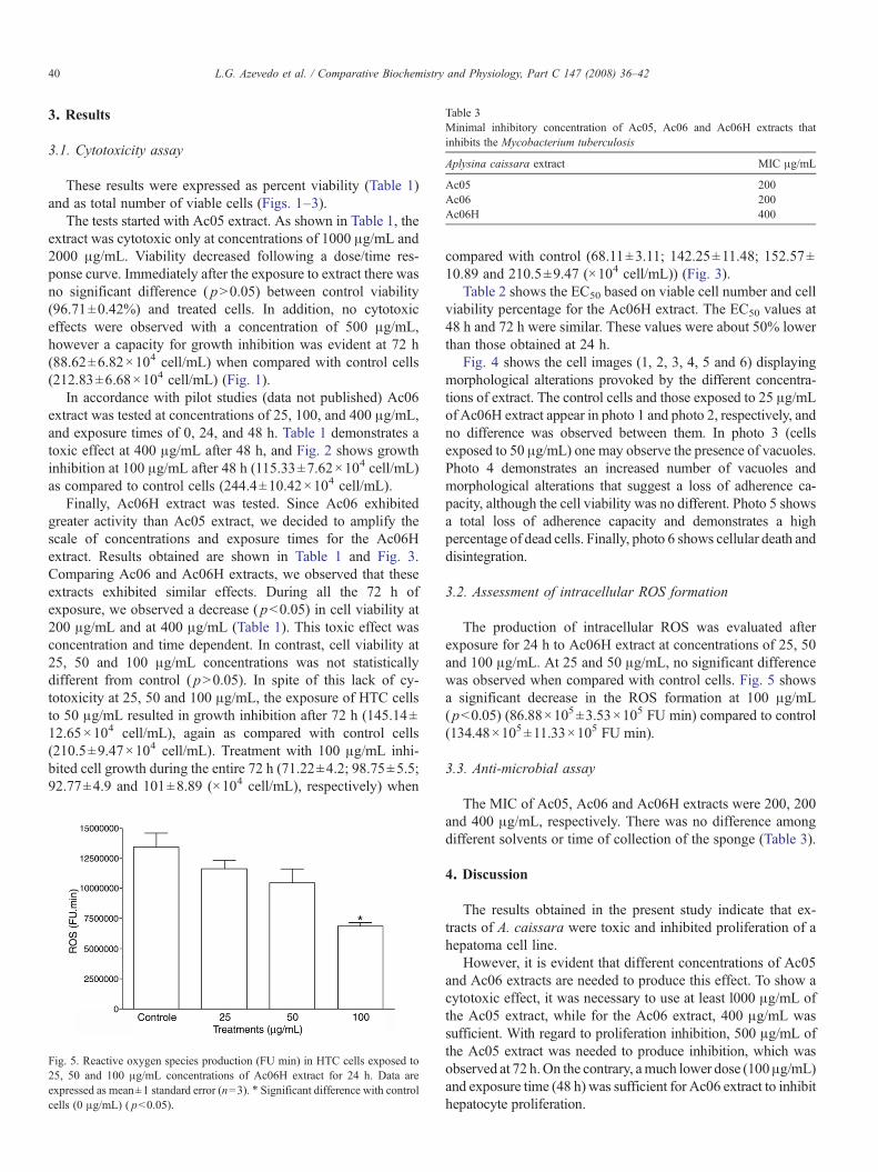
Table 3Minimal inhibitory concentration of Ac05, Ac06 and Ac06H extracts thatinhibits the Mycobacterium tuberculosis
Aplysina caissara extract MIC μg/mL
Ac05 200Ac06 200Ac06H 400
40 L.G. Azevedo et al. / Comparative Biochemistry and Physiology, Part C 147 (2008) 36–42
3. Results
3.1. Cytotoxicity assay
These results were expressed as percent viability (Table 1)and as total number of viable cells (Figs. 1–3).
The tests started with Ac05 extract. As shown in Table 1, theextract was cytotoxic only at concentrations of 1000 μg/mL and2000 μg/mL. Viability decreased following a dose/time res-ponse curve. Immediately after the exposure to extract there wasno significant difference ( pN0.05) between control viability(96.71±0.42%) and treated cells. In addition, no cytotoxiceffects were observed with a concentration of 500 μg/mL,however a capacity for growth inhibition was evident at 72 h(88.62±6.82×10⁴ cell/mL) when compared with control cells(212.83±6.68×10⁴ cell/mL) (Fig. 1).
In accordance with pilot studies (data not published) Ac06extract was tested at concentrations of 25, 100, and 400 μg/mL,and exposure times of 0, 24, and 48 h. Table 1 demonstrates atoxic effect at 400 μg/mL after 48 h, and Fig. 2 shows growthinhibition at 100 μg/mL after 48 h (115.33±7.62×10⁴ cell/mL)as compared to control cells (244.4±10.42×10⁴ cell/mL).
Finally, Ac06H extract was tested. Since Ac06 exhibitedgreater activity than Ac05 extract, we decided to amplify thescale of concentrations and exposure times for the Ac06Hextract. Results obtained are shown in Table 1 and Fig. 3.Comparing Ac06 and Ac06H extracts, we observed that theseextracts exhibited similar effects. During all the 72 h ofexposure, we observed a decrease ( pb0.05) in cell viability at200 μg/mL and at 400 μg/mL (Table 1). This toxic effect wasconcentration and time dependent. In contrast, cell viability at25, 50 and 100 μg/mL concentrations was not statisticallydifferent from control ( pN0.05). In spite of this lack of cy-totoxicity at 25, 50 and 100 μg/mL, the exposure of HTC cellsto 50 μg/mL resulted in growth inhibition after 72 h (145.14±12.65×10⁴ cell/mL), again as compared with control cells(210.5±9.47×10⁴ cell/mL). Treatment with 100 μg/mL inhi-bited cell growth during the entire 72 h (71.22±4.2; 98.75±5.5;92.77±4.9 and 101±8.89 (×104 cell/mL), respectively) when
Fig. 5. Reactive oxygen species production (FU min) in HTC cells exposed to25, 50 and 100 μg/mL concentrations of Ac06H extract for 24 h. Data areexpressed as mean±1 standard error (n=3). ⁎ Significant difference with controlcells (0 μg/mL) ( pb0.05).
compared with control (68.11±3.11; 142.25±11.48; 152.57±10.89 and 210.5±9.47 (×104 cell/mL)) (Fig. 3).
Table 2 shows the EC50 based on viable cell number and cellviability percentage for the Ac06H extract. The EC50 values at48 h and 72 h were similar. These values were about 50% lowerthan those obtained at 24 h.
Fig. 4 shows the cell images (1, 2, 3, 4, 5 and 6) displayingmorphological alterations provoked by the different concentra-tions of extract. The control cells and those exposed to 25 μg/mLof Ac06H extract appear in photo 1 and photo 2, respectively, andno difference was observed between them. In photo 3 (cellsexposed to 50 μg/mL) one may observe the presence of vacuoles.Photo 4 demonstrates an increased number of vacuoles andmorphological alterations that suggest a loss of adherence ca-pacity, although the cell viability was no different. Photo 5 showsa total loss of adherence capacity and demonstrates a highpercentage of dead cells. Finally, photo 6 shows cellular death anddisintegration.
3.2. Assessment of intracellular ROS formation
The production of intracellular ROS was evaluated afterexposure for 24 h to Ac06H extract at concentrations of 25, 50and 100 μg/mL. At 25 and 50 μg/mL, no significant differencewas observed when compared with control cells. Fig. 5 showsa significant decrease in the ROS formation at 100 μg/mL(pb0.05) (86.88×10⁵±3.53×10⁵ FU min) compared to control(134.48×10⁵±11.33×10⁵ FU min).
3.3. Anti-microbial assay
The MIC of Ac05, Ac06 and Ac06H extracts were 200, 200and 400 μg/mL, respectively. There was no difference amongdifferent solvents or time of collection of the sponge (Table 3).
4. Discussion
The results obtained in the present study indicate that ex-tracts of A. caissara were toxic and inhibited proliferation of ahepatoma cell line.
However, it is evident that different concentrations of Ac05and Ac06 extracts are needed to produce this effect. To show acytotoxic effect, it was necessary to use at least l000 μg/mL ofthe Ac05 extract, while for the Ac06 extract, 400 μg/mL wassufficient. With regard to proliferation inhibition, 500 μg/mL ofthe Ac05 extract was needed to produce inhibition, which wasobserved at 72 h.On the contrary, amuch lower dose (100μg/mL)and exposure time (48 h) was sufficient for Ac06 extract to inhibithepatocyte proliferation.

41L.G. Azevedo et al. / Comparative Biochemistry and Physiology, Part C 147 (2008) 36–42
At least 200 μg/mL of the Ac06H extract was needed to obtaina decrease in HTC viability. This extract also exhibited pro-liferation inhibition at lower concentrations (100 and 50 μg/mL).Comparing proliferation inhibition capacities between Ac06 andAc06H extracts at 100 μg/mL, we observed a quicker response(24 h earlier) with the Ac06H extract.
Regarding the potential difference between the extracts, at leasttwo proposals merit consideration: 1) the fact that the Ac05 extracthad been directly immersed in ethanol while the Ac06 extract wasimmediately frozen and after some time immersed in ethanol; 2)the collection of the sponges occurred in different seasons.
The first proposal is supported by literature that demonstratesthat interactions between sponge metabolites and alcoholsolvents affect metabolite activity. It has been reported thatdibromotyrosine metabolites of the sponge Aplysina aerophobacan degrade in the presence of alcohol–H2O mixtures, sinceenzymatic activity is not completely suppressed under suchconditions (Ebel et al., 1997). Nevertheless, similar experimentscarried out with extracts of the sponges A. insularis andA. archeri species demonstrated that dibromotyrosine metabo-lites of these sponges did not suffer degradation when stored inalcohol (Puyana et al., 2003).
The second proposal is based on the hypothesis that seasonalchanges can alter the production of active chemical compounds.This has been demonstrated in the Red Sea sponge Negombatamagnifica which possesses the compounds Latrunculin A andB. The concentrations of these compounds were analyzed inboth the summer and winter. The results of this study revealedthat the yield of latrunculins was higher in the winter (Khalifa etal., 2006). New experiments will be performed by our group tobetter understand these relationships between seasonal collec-tions of our yield of bioactive compounds.
In light of our more promising result with the Ac06, thisextract was partially purified by partitioning with hexane. Ac06and Ac06H extracts demonstrate similar activity, whichsuggests that the active metabolites are not in the lipid fraction(apolar fraction) of the extract since when this portion wasremoved with hexane, the extract did not diminish in activity.
In the future, new purification and isolation assays will beperformed with the Ac06H extract, as it may be possible thatA. caissara extracts possess greater activity and specificitywhen their individual components are isolated.
Acosta and Rodríguez (1992) discussed that cytotoxic effectsof chemical agents on cells may include altered morphology,failure of the cell to attach to surfaces, or changes in the rates ofcell processes such as growth, death and disintegration. Theseeffects were also observed in this study.
With regard to intracellular ROS formation, the results ob-tained in our study contribute to the controversy in the litera-ture. The bioactivity of the defense metabolites dienone andaeroplysinin-1 from the A. aerophoba sponge can be partlyexplained by their capacity to form free radicals that can destroycells; these metabolites show cytotoxicity against human cancercell lines (Koulman et al., 1996). This relationship betweencytotoxic effects and reactive oxygen species generation hasalso been suggested by Matsumoto et al. (2003) where theseauthors purposed that ascididemin isolated from a Didemnum
sp. tunicate was cytotoxic probably by direct iminoquinonereduction and reactive oxygen species generation. On the otherhand, the relevant literature reports that several sponge metab-olites exhibit antioxidant activity. Some examples include: thecompound 2-octaprenyl-1, 4-hydroquinone and 2-(24-hydroxy)-octaprenyl-1, 4-hydroquinone isolated from the sponge Irciniaspinosula, which exhibits a strong interaction with the 2,2-diphenyl-1-picrylhydrazyl radical (DPPH, a stable radical) andexhibited a significant anti-lipid peroxidation effect (Tzivelekaet al., 2002); also Takamatsu et al. (2003) revealed that differ-ent sponges possess metabolites such as (1S)-(+)-curcuphenol,aaptamine, isoaaptamine, and curcudiol, all with strong antiox-idant activity in the DPPH assay.
In this work, was demonstrated an antioxidant activity for theAc06H extract in the ROS assay.
Concerning anti-TB activity, our results indicate that allextracts possess a moderate activity. With regard to the Ac06extract, when compared with the results obtained in cytotoxicityassay, our expectations were that this extract would showgreater activity in the anti-TB assay when compared to Ac05extract. Contrary to this expectation, Ac05 and Ac06 extractspossessed the same MIC activity (200 μg/mL), showing thatindependent of the extraction methodology and collectionseason, the active compound(s) was present in both extracts. Inaddition, it was also noted that the Ac06H extract, partiallypurified by hexane extraction, suffered a decrease in its activitywhen compared with Ac06 extract. It is possible that the lipidportion removed by hexane may be involved in anti-TB activity.
Some authors have demonstrated anti-microbial activity ofisolated marine sponge compounds. In A. caissara, the productsFistularin-3 and 11-hydroxyaerothionin displayed moderateantibiotic activity against several pathogenic bacteria (Liraet al., 2006). The authors observed this effect from Fistularin-3against E. coli ATCC 25922 (MIC at 300 μg/mL) and oxacillinresistant S. aureus strain 8 (MIC at 600 μg/mL). It is possiblethat other isolates may show greater activity than crude extracts.In accordance with this prospect, Oliveira et al. (2006), screened500 crude extracts of marine invertebrates as inhibitors ofM. tuberculosis H37Rv and discovered that extracts fromAplysina cauliformis and Pachychalina sp. displayed antimy-cobacterial activity with an MIC value b200 μg/mL. However,these authors revealed that the known bromotyrosine alkaloidFistularin-3 from A. cauliformis was active against M. tubercu-losis with an MIC value of 8.03 μg/mL.
The results demonstrated in this present study suggest thatmarine sponge extracts possess a broad potential for pharma-cological applications.
Acknowledgments
This work was supported by grants from CNPq, CAPES andFundação Universidade Federal do Rio Grande.
References
Acosta, A.L., Rodríguez, A.D., 1992. 11-Oxoaerothionin: a cytotoxic antitumourbromotyrosine-derived alkaloid from the Caribbean marine sponge Aplysinalacunosa. Journal of Natural Products 55 (7), 1007–1012.
42 L.G. Azevedo et al. / Comparative Biochemistry and Physiology, Part C 147 (2008) 36–42
American Public Health Association, American WaterWorks Association, WaterPollution Control Federation, 1976. Standard Methods for the Examinationof Water and Wastewaters. American Public Health Association, Washing-ton, DC.
Ebel, R., Brenzinger, M., Kunze, A., Gross, H., Proksh, P., 1997. Woundactivation of prototoxins in marine sponge Aplysina aerophoba. Journal ofChemical Ecology 23 (5), 1451–1461.
Encarnación, R.D., Sandoval, E., Malmstrom, J., Christophersen, C., 2000.Calafianin, a bromotyrosine derivative from the marine sponge Aplysinagerardogreeni. Journal of Natural Products 63, 874–875.
Faulkner, D.J., 2002. Marine natural products. Natural Product Reports 19,1–48.
Hamann, M.T., Scheuer, P.J., 1993. Biogenetically diverse, bioactive constituentsof a sponge, order, Verongida: Bromotyramines and Sesquiterpene-Shikimatederived metabolites. Journal of Organic Chemistry 58, 6565–6569.
Hunt, B., Vincent, A.C.J., 2006. Scale and sustainability of marine biopros-pecting for pharmaceuticals. Ambio 35 (2), 57–64.
Khalifa, S., Ahmed, S., Mesbah, M., Youssef, D., Hamann, M., 2006. Quan-titative determination of latrunculins A and B in the Red sea sponge Ne-gombata magnifica by high performance liquid chromatography. Journal ofChromatography. B, Analytical Technologies in the Biomedical and LifeSciences 17; 832 (1), 47–51.
Kijjoa, A., Sawangwong, P., 2004. Drugs and cosmetics from the sea. MarineDrugs 2, 73–82.
Kondo, K., Nishi, J., Ishibashi, M., Kobayashi, J., 1994. Two new tryptophan-derived alkaloids from the Okinawan marine sponge Aplysina sp. Journal ofNatural Products 57, 1008–1011.
Koulman, A., Proksch, P., Ebel, R., Beekman, A.C., Uden,W.V., Konings, A.W.T.,Pedersen, J.A., Pras, N., Woerdenbag, H.J., 1996. Cytotoxicity and mode ofaction of aeroplysinin-1 and related dienone from the sponge Aplysinaaerophoba. Journal of Natural Products 59, 591–594.
Kraljevic, S., Sedic, M., Scott, M., Gehrig, P., Schlapbach, R., Pavelic, K., 2006.Casting light on molecular events underlying anti-cancer drug treatment:what can be seen from the proteomics point of view? Cancer TreatmentReviews 32, 619–629.
Lira, T.O., Berlinck, R.G.S., Nascimento, G.G.F., Hadju, E., 2006. Furtherdibromotyrosin-derived metabolites from the marine sponge Aplysinacaissara. Journal of the Brazilian Chemical Society 7 (7), 1233–1240.
Matsumoto, S.S., Biggs, J., Copp, B.R., Holden, J.A., Barrows, L.R., 2003.Mechanism of ascididemin-induced cytotoxicity. Chemical Research inToxicology 16, 113–122.
Mayer, A.M.S., Hamann, M.T., 2004. Marine pharmacology in 2000: marinecompounds with antibacterial, anticoagulant, antifungical, anti-inflammato-ry, antimalarial, antiplatelet, anti-tuberculosis, and antiviral activities;affecting the cardiovascular, immune, and nervous systems and othermiscellaneous mechanisms of action. Marine Biotechnology 6, 37–52.
Mayer, A.M.S., Hamann, M.T., 2005. Marine pharmacology in 2001–2002:marine compounds with anthelmintic, antibacterial, anticoagulant, antidia-betic, antifungal, anti-inflammatory, antimalarial, antiplatelet, antiprotozoal,antituberculosis and antiviral activities; affecting the cardiovascular,immune and nervous systems and other miscellaneous mechanisms ofaction. Comparative Biochemistry and Physiology. Part C, PharmacologyToxicology & Endocrinology 140, 265–286.
Mayer, A.M.S., Gustafson, K.R., 2006. Marine pharmacology in 2003–2004: anti-tumour and cytotoxic compounds. European Journal of Cancer 42, 2241–2270.
Monks, N.R., Lerner, C., Henriques, A.T., Farias, F.M., Schapoval, E.E.S.,Suyenaga, E.S., Rocha, A.B., Schwartsmann, G., Mothes, B., 2002.Anticancer, antichemotactic and antimicrobial activities of marine sponges
collected off coast of Santa Catarina, southern Brazil. Journal ofExperimental Marine Biology and Ecology 281, 1–12.
Munro, M.H.G., Blunt, J.W., Dumdei, E.J., Hickford, S.J.H., Lill, R.E., Li, S.,Battershill, C.N., Duckworth, A.R., 1999. The discovery and developmentof marine compounds with pharmaceutical potential. Journal of Biotech-nology 70, 15–25.
Myhre, O., Fonnum, F., 2001. The effect of aliphatic, naphthenic, and aromatichydrocarbons on production of reactive oxygen species and reactive nitrogenspecies in rat brain synaptosome fraction: the involvement of calcium, nitricoxide synthase, mitochondria and phospholipase A. Biochemical Pharmacol-ogy 62, 119–128.
Nachega, J.B., Chaisson, R.E., 2003. Tuberculosis drug resistance: a globalthreat. Clinical Infectious Diseases 36 (Suppl 1), S24–S30.
Nicholas, G.M., Eckman, L.L., Newton, G.L., Fahey, R.C., Ray, S., Bewley, C.A.,2003. Inhibition and kinetics of Mycobacterium tuberculosis and Mycobac-terium smegmatis mycotiol-S-conjugate amidase by natural products inhibi-tors. Bioorganic & Medicinal Chemistry 11, 6001–6608.
Oliveira, M.F., Oliveira, J.H.H.L., Galetti, F.C.S., Souza, A.O., Silva, C.L.,Hadju, E., Peixinho, S., Berlinck, R.G.S., 2006. Antimycobacterialbrominated metabolites from two species of marine sponges. Planta Medica72, 437–441.
Palomino, J.C., Martin, A., Camacho, M., Guerra, H., Swings, J., Portaels, F.,2002. Resazurin microtiter assay plate: simple and inexpensive method fordetection of drug resistance in Mycobacterium tuberculosis. AntimicrobialAgents and Chemotherapy 46 (8), 2720–2722 (Aug.).
Pinheiro, U.S., Hadju, E., 2001. Shallow-water Aplysina nardo (Aplysinidae,Verongida, Dermospogiae) from the São Sebastião Channel and its environs(tropical southwestern Atlantic), with the description of a new species and aliterature review of other Brazilian records of the genus. Revista Brasileirade Zoologia 18 (Suppl.1), 143–160.
Puyana, M., Fenical, W., Pawlik, J.R., 2003. Are there activated chemicaldefenses in sponges of genus Aplysina from Caribbean? Marine Ecology.Progress Series 246, 127–135.
Rattan, A., Kalia, A., Ahmad, N., 1998. Multidrug-resistant Mycobacteriumtuberculosis: molecular perspectives. Emerging Infectious Diseases 4 (2),195–209.
Rodriguez, A., Piña, I., 1993. The structures of Aplysinamisines I, II and III:new bromotyrosine-derived alkaloids from the Caribbean sponge Aplysinacauliformis. Journal of Natural Products 56 (6), 907–914.
Saeki, B.M., Granato, A.C., Berlinck, R.G.S., Magalhães, A., Schefer, A.B.,Ferreira, A.G., Pinheiro, U.S., Hadju, E., 2002. Two unprecedenteddibromotyrosine-derived alkaloids from the Brazilian endemic marinesponge Aplysina caissara. Journal of Natural Products 65, 796–799.
Silva, P.E.A., Osório, M., Reinhardt, M.C., Fonseca, L.S., Dellagostin, O.A.,2001. Drug resistance of strains of Mycobacterium tuberculosis isolated inBrazil. Microbes and Infection 3, 1111–1113.
Souza, M.V.N., 2006. Research article, marine natural products againsttuberculosis. The Scientific World Journal 6, 847–861.
Takamatsu, S., Hodges, T.W., Rajbhandari, I., Gerwick, W.H., Hamann, M.T.,Nagle, D.G., 2003. Marine natural products as novel antioxidant prototypes.Journal of Natural Products 66, 605–608.
Tziveleka, L.-A., Kourounakis, A.P., Kourounakis, P.N., Roussis, V., Vagias, C.,2002. Antioxidant potential of natural and synthesized hydroquinones.Bioorganic & Medicinal Chemistry 10, 935–939.
Urban, S., Hickford, S.J.H., Blunt, J.W., Munro, M.H.G., 2000. Bioactivemarine alkaloids. Current Organic Chemistry 4, 765–807.
WHO, 2000. World Health Organization Tuberculosis Fact Sheet. No. 104.WHO, 2006. World Health Organization Tuberculosis Fact Sheet. No. 104.


![Descriptors for Sponge Gourd [Luffa cylindrica (L.) Roem.]](https://static.fdokumen.com/doc/165x107/63187e763394f2252e02b92e/descriptors-for-sponge-gourd-luffa-cylindrica-l-roem.jpg)