Monosaccharides bound to hemoglobin in normal and diabetic individuals
Upload
independentCategory
view
2download
0
Comparative Analysis of Mycobacterial Truncated HemoglobinPromoters and the groEL2 Promoter in Free-Living and IntracellularMycobacteria
Sunil V. Joseph, G. K. Madhavilatha, R. Ajay Kumar, and Sathish Mundayoor
Department of Molecular Microbiology, Rajiv Gandhi Centre for Biotechnology, Trivandrum, India
The success of Mycobacterium tuberculosis depends on its ability to withstand and survive the hazardous environment inside themacrophages that are created by reactive oxygen intermediates, reactive nitrogen intermediates, severe hypoxia, low pH, andhigh CO2 levels. Therefore, an effective detoxification system is required for the pathogen to persist in vivo. The genome of M.tuberculosis contains a new family of hemoproteins named truncated hemoglobin O (trHbO) and truncated hemoglobin N(trHbN), encoded by the glbO and glbN genes, respectively, important in the survival of M. tuberculosis in macrophages. Myco-bacterial heat shock proteins are known to undergo rapid upregulation under stress conditions. The expression profiles of thepromoters of these genes were studied by constructing transcriptional fusions with green fluorescent protein and monitoring thepromoter activity in both free-living and intracellular milieus at different time points. Whereas glbN showed an early responseto the oxidative and nitrosative stresses tested, glbO gave a lasting response to lower concentrations of both stresses. At all timepoints and under all stress conditions tested, groEL2 showed higher expression than both trHb promoters and expression ofboth promoters showed an increase while inside the macrophages. Real-time PCR analysis of trHb and groEL2 mRNAs showedan initial upregulation at 24 h postinfection. The presence of the glbO protein imparted an increased survival to M. smegmatis inTHP-1 differentiated macrophages compared to that imparted by the glbN and hsp65 proteins. The comparative upregulationshown by both trHb promoters while grown inside macrophages indicates the importance of these promoters for the survival ofM. tuberculosis in the hostile environment of the host.
The success of Mycobacterium tuberculosis depends on its abilityto withstand and survive the hazardous environment inside
macrophages that are created by reactive oxygen intermediates(ROI), reactive nitrogen intermediates (RNI), severe hypoxia, lowpH, and high CO2 levels (5). It has already been reported thatactivation of macrophages induces the synthesis of an inducibleNO synthase (iNOS), resulting in the production of various RNI(7, 21). Activated macrophages can also produce an oxidativeburst which is responsible for the killing of intracellular M. tuber-culosis. ROI and RNI can damage DNA and other chemical moi-eties on which their propagation and protection depend, includ-ing Fe-S clusters, tyrosyl radicals, hemes, sulfhydryls, thioethers,and alkenes (22). Macrophages can produce superoxide (O2
�)and nitric oxide (NO) in almost equimolar amounts and thus canbe abundant generators of peroxynitrite (OONO�), a highly oxi-dizing agent (41, 45). NO limits the survival and growth of micro-bial pathogens by inhibiting enzymes like respiratory oxidases andiron sulfur centers of key enzymes such as aconitase (11, 13).
Since early events of M. tuberculosis infection involve entry andmultiplication within the bacteriostatic environment of macro-phages, which is characterized mainly by ROS and RNS, an effec-tive detoxification system is required for the pathogen to persist invivo. The genome of M. tuberculosis contains glbO and glbN genes,encoding truncated hemoglobin O (trHbO) and truncated hemo-globin N (trHbN), respectively, which belong to a new family ofhemoproteins that are extensively distributed in eubacteria, cya-nobacteria, protozoans, and plants (27, 29, 38). The amino acidsequences of trHbs are shorter and share very low similarity to thesequences of classical vertebrate hemoglobins and myoglobins.The mycobacterial trHb length varies from 215 amino acid resi-dues for M. vanbalenii trHbN to 121 amino acid residues for M.
smegmatis trHbN, with the majority being �136 amino acid resi-dues. Homology between members of different trHb classes isinsignificant (�20% identity) (3, 40). The genetic organization ofglbO is largely conserved across various mycobacterial species incomparison to that of glbN. Among mycobacteria, adaptation toobligate parasitism has resulted in the loss of trHb genes, as in thecase of M. leprae, which retained only glbO (4, 6, 27). Since M.leprae has retained only a minimalistic repertoire of functionalgenes, trHbO can be regarded as an essential product for intracel-lular parasitism, while trHbN appears to be dispensable.
The mechanism by which trHbN protects M. tuberculosis fromthe nitrosative stress and microbicidal activities of macrophagesrelies on its oxygen-dependent NO dioxygenase (NOD) activity.The oxygenated form of trHbN can very efficiently convert NO toharmless nitrate (25, 28). The inactivation of the glbN gene of M.bovis BCG impaired the ability of stationary-phase cells of theorganism to protect aerobic respiration from NO (24). Dikshitand colleagues (14, 25) and Ouellet et al. (24) showed that trHbNof M. tuberculosis has a potent ability to relieve the toxicity of NOand nitrosative stress in Escherichia coli and M. smegmatis. Theaerobic organism M. tuberculosis can use trHbO to collect anddeliver available O2 to the components of the electron transportchain in order to survive in the granulomas (16, 26). Ferrous ox-
Received 22 June 2012 Accepted 27 June 2012
Published ahead of print 6 July 2012
Address correspondence to Sathish Mundayoor, [email protected].
Copyright © 2012, American Society for Microbiology. All Rights Reserved.
doi:10.1128/AEM.01984-12
September 2012 Volume 78 Number 18 Applied and Environmental Microbiology p. 6499–6506 aem.asm.org 6499
ygenated M. tuberculosis trHbN and trHbO as well as M. lepraetrHbO have been shown to facilitate NO scavenging (2, 24). Ox-ygenated trHbN catalyzes fast NO oxidation compared to thespeed of catalysis for trHbO, which likely makes the NO detoxifi-cation role for trHbN higher and which shows that trHbO playsonly a marginal role in the protection of mycobacteria against NO(3). Mycobacterial trHbO is present during all stages of growth inthe host, and the expression of several oxidative defense genes wasfound to be constitutive (37). In addition, exposure to low NOconcentrations triggers the initiation of mycobacterial dormancy(44).
Heat shock proteins (HSPs) are generally responsible for pre-venting damage to proteins in response to high levels of heat. HSPsstabilize cellular proteins in response to diverse sources of stress orinjury. The mycobacterial Hsp family includes proteins such asDnaK, DnaJ, GrpE, GroES, GroEL, and other low-molecular-weight HSPs (31). HSPs also have a number of immunologicaleffects, including the induction of proinflammatory cytokines(23, 30, 36, 43). Mycobacterial HSPs may also participate in cyto-kine expression resulting from infection by M. tuberculosis (31). Atemperature increase from 37°C to 42°C induced elevated synthe-sis of three major proteins corresponding to the DnaK, GroEL,and GroES proteins of M. tuberculosis previously identified to beprominent antigens (42). In addition to its role as a heat shockprotein, GroEL functions as a chaperonin to assist in folding linearamino acid chains into their respective three-dimensional struc-ture.
Global analysis of gene expression in several pathogenic bacte-ria, including M. tuberculosis (9), has revealed that large-scalechanges occur upon in vitro exposure to environmental condi-tions that simulate the intracellular milieu (15, 19). The expres-sion pattern of mycobacterial trHb and groEL2 promoters undervarious oxidative and nitrosative stress conditions will give an ideaabout the expression of the corresponding genes inside the hostilemacrophage environment. Since little is known about the expres-sion and regulation of mycobacterial trHb and groEL2 promoters,we decided to study the response of the promoters by constructingtranscriptional fusions with green fluorescent protein (GFP) andmonitoring the promoter activity under various stress conditionsand also in THP-1 differentiated macrophages postinfection.Here we have compared the expression profile of mycobacterialtrHb and groEL2 promoters at different time points under bothfree-living and intracellular conditions. We have also looked intothe mRNA profiles of the genes in M. smegmatis and M. tubercu-losis and carried out survival assays in M. smegmatis.
MATERIALS AND METHODSBacterial strains, media, and growth conditions. M. smegmatis mc2155was grown in Luria-Bertani broth (USB) with 0.05% Tween 80 and platedin the same medium with 1.5% agar supplemented with kanamycin (25�g/ml) wherever appropriate. All transformations in Escherichia coli wereperformed with strain JM109. E. coli was grown in Luria-Bertani broth at37°C in shaking cultures. Kanamycin (25 �g/ml) was added to the me-dium whenever required. Plasmid pFPV27 was a kind gift from LalithaRamakrishnan, Department of Microbiology, University of Washington.
Cloning of mycobacterial glbO, glbN, and groEL2 promoters andpromoters with full genes. All promoter sequences were amplified fromM. tuberculosis H37Rv genomic DNA, which was isolated using a previ-ously published protocol (33). The sequences encompassing positions�240/�26, �410/�70, and �375/�18 relative to the start codons ofglbN, glbO, and groEL2, respectively, were PCR amplified using custom-
synthesized primers that incorporated 5= NotI and 3= BamHI restrictionsites (Table 1). The amplified products were then cloned into pFPV27 thathad previously been digested with the same restriction enzymes. The glbO,glbN, and groEL2 transcriptional fusions were termed as pFPVO, pFPVN,and pMRm, respectively. The glbO, glbN, and groEL2 genes, along with thepromoters, were also amplified from M. tuberculosis H37Rv genomicDNA and cloned into pFPV27, and these constructs were namedpFPVOpg, pFPVNpg, and pMRmpg, respectively. The sequences of all thecloned fragments were confirmed using BigDye Terminator cycle se-quencing chemistry for the ABI BioPrism apparatus (Applied Biosys-tems). Electrocompetent M. smegmatis mc2155 cells were electroporatedwith these plasmids as described previously (39).
Analysis of expression of transcriptional fusions under variousstress conditions. M. smegmatis cells containing pFPV27, pFPVO,pFPVN, and pMRm were grown for 24 h and were then inoculated in freshmedium at an optical density at 610 nm (OD610) of 0.05 and allowed togrow by shaking at 37°C for another 10 h. The cells were exposed todifferent physiological stresses, viz., 1 mM and 5 mM H2O2 for oxidativestress and 10 mM and 30 mM NaNO2 for nitrosative stress, and we alsochecked the influence of THP-1-derived macrophage cell lysates on theexpression of these transcriptional fusions. These concentrations of H2O2
and NaNO2 were used, as they had previously been determined to beminimally lethal to mycobacteria (34). The cultures were then incubatedat 37°C in a shaking incubator. One hundred microliters of each culturewas transferred to 96-well plates at 24 h, 48 h, and 72 h. The fluorescencewas measured in a Tecan fluorescent microplate reader at excitation/emission wavelengths of 488 nm/520 nm. Relative fluorescence units(RFU) were determined to be the number of fluorescence units/OD600.Background fluorescence due to read-through transcription of the tran-scriptional fusion vector was determined by measuring the RFU of M.smegmatis harboring pFPV27. All the experiments were carried out intriplicate.
Differentiation and in vitro infection of THP-1 cells. The monocyticcell line THP-1 was cultured in RPMI 1640 (RPMI) supplemented with10% fetal bovine serum. Cells (2.5 � 105) were seeded in 12-well platesand allowed to adhere and differentiate in the presence of phorbol myris-tate acetate (PMA; 25 ng/ml) at 37°C in a humidified atmosphere of 5%CO2 for 24 h. Cells were then washed three times with Hanks balanced saltsolution, and antibiotic-free culture medium was added. Adherent cellswere infected with M. smegmatis containing pFPV27, pFPVO, pFPVN,and pMRm at a multiplicity of infection (MOI) of 1:20 (macrophages tobacilli). After 4 h of infection, the cells were washed 3 times with warmRPMI to remove extracellular bacteria, and cells were incubated withcomplete RPMI containing 10 �g/ml gentamicin for 2 h to preventgrowth of extracellular bacteria. Subsequently, the cells were washed andmaintained in complete RPMI for the rest of the experiment.
Analysis of intracellular expression of transcriptional fusions. Toanalyze the intracellular expression of transcriptional fusions, macro-phage monolayers infected with M. smegmatis containing pFPV27,pFPVO, pFPVN, and pMRm were further incubated for 4 h, 24 h, and 48h. The intracellular mycobacteria were processed using a previously pub-lished protocol (10) with slight modifications. Briefly, the macrophagemonolayer was detached from 12-well plates using cell dissociation solu-tion (Sigma), collected by centrifugation (2,000 rpm for 5 min at 4°C),and broken by 20 passages through a 23-gauge needle. Intact cells andnuclei were removed by pelleting at 3,000 rpm for 5 min, and the super-
TABLE 1 Nucleotide sequences of primers used in this study
No. Primer Sequence
1 GlbO-F 5=-AAATATGCGGCCGCCTTCGTGCACCACGTCGGCC-3=2 GlbO-R 5=-GGATCCGCATAGAAACGCGACACGATCGC-3=3 GlbN-F 5=-AAATATGCGGCCGCCACCAAGATTCATCGTGGGTAG-3=4 GlbN-R 5=-CGGGATCCTTGCGCAAGCGTGACAGTAGTCC-3=5 GroEL2-F 5=-TTGCGGCCGCAACGGTGACCACAACGACGC-3=6 GroEL2-R 5=-CAATGGCCAAGACAATTGCGGGATCC-3=
Joseph et al.
6500 aem.asm.org Applied and Environmental Microbiology

natant containing bacteria was again centrifuged at 12,000 rpm for 10min. The pellet was then resuspended in 100 �l phosphate-buffered saline(PBS), the suspension was added to a 96-well plate, and the RFU andabsorbance were recorded as described in the previous section. All mea-surements were corrected for background fluorescence by subtracting theRFU for the control strain carrying plasmid pFPV27.
Extraction of RNA and RT-PCR. In 25-cm2 tissue culture flasks(Nunc), 40 � 105 cells were seeded and allowed to adhere and differentiatein the presence of PMA (25 ng/ml) at 37°C in a humidified atmosphere of5% CO2 for 24 h. Infection was performed with M. smegmatis containingpFPV27, pFPVOpg, pFPVNpg, and pMRmpg (MOI � 1:20). After 4 h, 24h, and 48 h of incubation, the culture supernatants were removed and themacrophage monolayer was washed twice with 1� PBS and detached witha cell scraper. The cell suspension was pelleted and resuspended in 350 �lof RA1 buffer (GE Healthcare) and 3.5 �l of �-mercaptoethanol, and thesuspension was transferred to a sterile 1.5-ml tube. The cells were lysed ina Mini-BeadBeater (2,500 rpm for 10 s; Biospec), after addition of 5-mmglass beads (Biospec). Total RNA was isolated using an RNA Spin minikit(GE Healthcare) following the manufacturer’s instructions. First-strandcDNA was synthesized from total RNA using a first-strand synthesis kit(Fermentas) with random oligonucleotides. The total RNA concentrationfor each sample was measured using a NanoDrop spectrophotometer(Thermo) at 260 nm. The quality of RNA was determined at a 260- and280-nm absorbance ratio (A260/A280). 16S rRNA was used as an endoge-nous control to normalize expression levels. Real-time PCR (RT-PCR)was done using gene-specific primers. The SYBR green method was used,and all reactions were performed in triplicate. Each reaction tube con-tained 10 �l iQSYBR green supermix (Bio-Rad), 300 nM gene-specificforward and reverse primers, and 50 ng of cDNA template, made up to afinal volume of 20 �l with nuclease-free water. The RT-PCR cycling con-ditions were 95°C for 3 min, followed by 40 cycles of 95°C for 10 s and58°C for 30 s, and the cycling was done using a Bio-Rad iCycler 5 appara-tus with an iQ multicolor real-time PCR detection system (Bio-Rad). Dataanalysis was performed with the iQ multicolor real-time PCR detectionsystem optical software system (Bio-Rad), version iQ5. Relative standardcurves for the target and endogenous control primer pairs were per-formed to verify that both PCR efficiencies were comparable.
Another experiment was done according to the above-described pro-tocol, where THP-1-derived macrophages were infected with M. tubercu-losis rather than recombinant M. smegmatis, and the levels of mycobacte-rial trHb and groEL2 mRNAs in THP-1-derived macrophages at 4 h, 24 h,and 48 h postinfection were analyzed with real-time PCR.
Confocal microscopy. In a 96-well glass-bottom tissue culture plate(Nunc), 50 � 104 cells were seeded and allowed to adhere and differentiatein the presence of PMA (25 ng/ml) at 37°C in a humidified atmosphere of5% CO2 for 24 h. Infection was performed with M. smegmatis containingpFPV27, pFPVO, pFPVN, and pMRm (MOI � 1:20). After 4 h, 24 h, and48 h of incubation, the cells were fixed with 4% paraformaldehyde in the darkfor 10 min at room temperature. Glycerol (90%) was added as the mountingmedium, and the cells were visualized in a confocal microscope.
Survival assay. Sixty thousand cells were seeded in a 96-well flat-bot-tom tissue culture plate (Tarson) and allowed to adhere and differentiatein the presence of PMA (25 ng/ml) at 37°C in a humidified atmosphere of5% CO2 for 24 h. Infection was performed with recombinant M. smegma-tis containing pFPV27, pFPVOpg, pFPVNpg, and pMRmpg (MOI �1:20). After 4 h of infection, the cells were washed three times with warmRPMI and treated with RPMI containing gentamicin (10 �g/ml) for 2 h at37°C to kill extracellular bacteria. Those monolayers in which survival for24 h and 48 h was to be measured were incubated in complete RPMIcontaining gentamicin (5 �g/ml) to prevent extracellular growth of bac-teria that might be released by premature lysis of macrophages. Cells induplicate wells were analyzed at 0 h, 24 h, and 48 h postinfection by adding200 �l of water and vigorously pipetting several times to ensure cell lysisand release of surviving intracellular bacteria. The lysates were serially dilutedin Luria-Bertani broth with 0.05% Tween 80 and plated onto Luria-Bertani
agar plates containing 0.05% Tween 80 and kanamycin (25 �g/ml). Thenumbers of CFU were counted after incubation at 37°C for 3 days, and themean value obtained was plotted along with the value for the control.
Statistical analysis. Results were expressed as the mean plus or minusthe standard deviation. Statistical comparison was performed using one-way analysis of variance. A probability (P) value of less than 0.05 wasconsidered statistically significant.
RESULTS
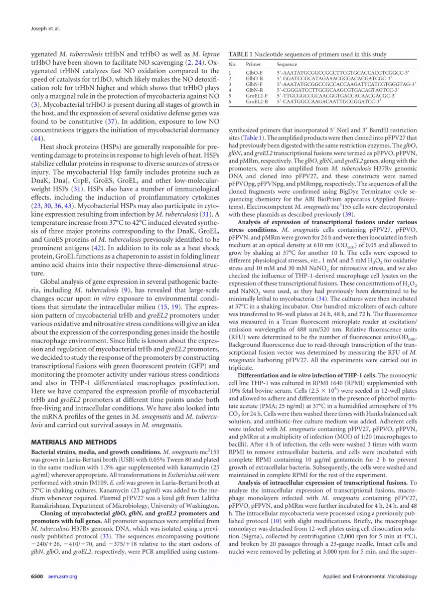
The activities of the promoters of the three M. tuberculosis genesglbO, glbN, and groEL2 were studied by subjecting recombinantM. smegmatis to various stress conditions (Fig. 1). Among thevarious stress conditions tested, 10 mM NaNO2 was the most po-tent inducer of the glbN promoter, and for glbO, it was 30 mMNaNO2, followed by 5 mM H2O2. glbN showed an early responseto both oxidative and nitrosative stress, with the highest being atthe 24-h time point. The glbO promoter responded more to thelower concentrations of both stresses, and the effects were morelasting. In the absence of any stress, the activities of glbN and glbOpromoters increased from the 24-h to 48-h time points, and theactivities of both showed a downward trend at 72 h. For the groEL2promoter, 30 mM NaNO2 elicited maximal expression at all thetime points tested. In the absence of any stress, the level of groEL2promoter expression was higher at the 24-h time point and de-creased from the 24-h to the 72-h time point, unlike the expres-sion profile shown by the trHb promoters.
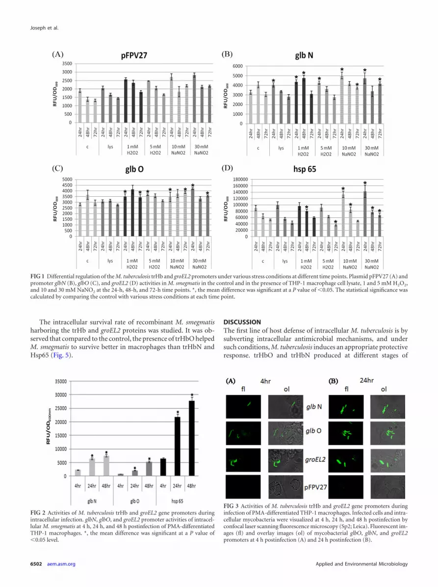
The activities of the mycobacterial trHb and groEL2 promotersin M. smegmatis were determined under intracellular conditionsin THP-1 differentiated macrophages (Fig. 2). All the promotersshowed considerable increases in activity with time, and the max-imal activity was at the 48-h time point. The glbN promotershowed higher activity than the glbO promoter at all time pointstested, but the activity of the groEL2 promoter was higher thanthat of both trHb promoters. This finding indicates that thegroEL2 promoter is stronger than both of the globin promotersunder intracellular conditions. Confocal microscopy of the in-fected THP-1-derived macrophages showed distinct fluorescencein phagocytosed M. smegmatis carrying glbO, glbN, and groEL2transcriptional fusions compared to the control (Fig. 3A and B).
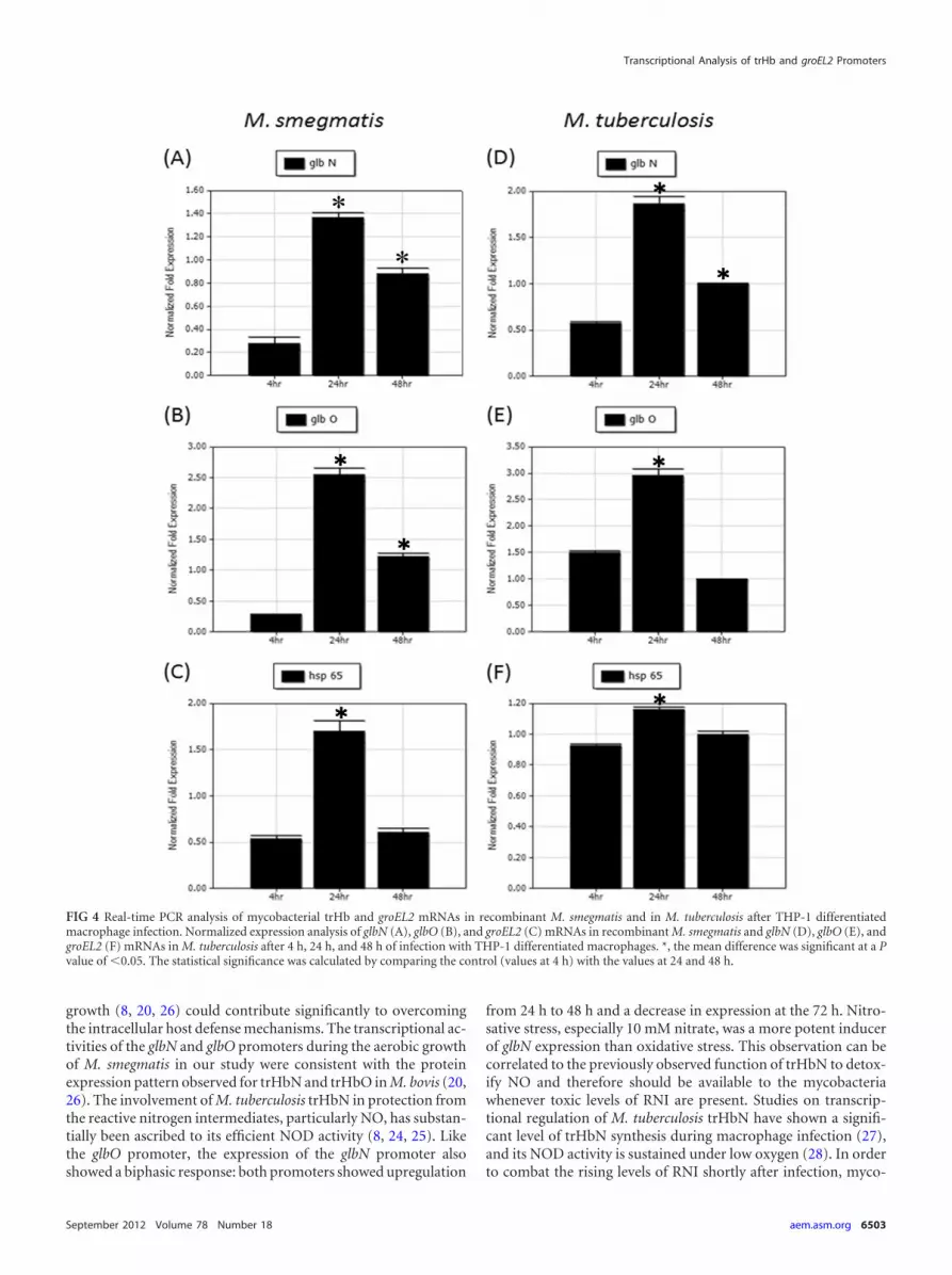
In order to analyze the levels of mycobacterial trHb and groEL2mRNAs in macrophages following infection, we cloned the fulllengths of both the mycobacterial trHb gene and groEL2 intopFPV27 along with their respective promoters. Total RNA was iso-lated from THP-1-derived macrophages infected with recombinantM. smegmatis at various time points after infection, and real-timePCR was carried out with gene-specific primers. Mycobacterial trHband groEL2 mRNAs showed an increase in expression from 4 h to 24h and showed a downward trend at 48 h. The extent of upregulationshown by both trHb mRNAs from 4 h to 24 h was higher than that ofgroEL2 mRNA at the same time points (Fig. 4A to C).
After the studies in recombinant M. smegmatis, we analyzed theexpression of mycobacterial trHb and groEL2 mRNAs in M. tuber-culosis H37Rv-infected THP-1-derived macrophages by real-timePCR. The expression profiles of mycobacterial trHb and groEL2 mR-NAs in THP-1-derived macrophages infected with recombinant M.smegmatis and M. tuberculosis H37Rv were comparable. Mycobacte-rial trHb and groEL2 mRNAs showed an upregulation from 4 h to 24h, and both showed a downward trend at 48 h (Fig. 4D to F). Alto-gether the expression of both trHb promoters and the groEL2 pro-moter showed an increase in expression when inside macrophages.
Transcriptional Analysis of trHb and groEL2 Promoters
September 2012 Volume 78 Number 18 aem.asm.org 6501
The intracellular survival rate of recombinant M. smegmatisharboring the trHb and groEL2 proteins was studied. It was ob-served that compared to the control, the presence of trHbO helpedM. smegmatis to survive better in macrophages than trHbN andHsp65 (Fig. 5).
DISCUSSIONThe first line of host defense of intracellular M. tuberculosis is bysubverting intracellular antimicrobial mechanisms, and undersuch conditions, M. tuberculosis induces an appropriate protectiveresponse. trHbO and trHbN produced at different stages of
FIG 1 Differential regulation of the M. tuberculosis trHb and groEL2 promoters under various stress conditions at different time points. Plasmid pFPV27 (A) andpromoter glbN (B), glbO (C), and groEL2 (D) activities in M. smegmatis in the control and in the presence of THP-1 macrophage cell lysate, 1 and 5 mM H2O2,and 10 and 30 mM NaNO2 at the 24-h, 48-h, and 72-h time points. *, the mean difference was significant at a P value of �0.05. The statistical significance wascalculated by comparing the control with various stress conditions at each time point.
FIG 2 Activities of M. tuberculosis trHb and groEL2 gene promoters duringintracellular infection. glbN, glbO, and groEL2 promoter activities of intracel-lular M. smegmatis at 4 h, 24 h, and 48 h postinfection of PMA-differentiatedTHP-1 macrophages. *, the mean difference was significant at a P value of�0.05 level.
FIG 3 Activities of M. tuberculosis trHb and groEL2 gene promoters duringinfection of PMA-differentiated THP-1 macrophages. Infected cells and intra-cellular mycobacteria were visualized at 4 h, 24 h, and 48 h postinfection byconfocal laser scanning fluorescence microscopy (Sp2; Leica). Fluorescent im-ages (fl) and overlay images (ol) of mycobacterial glbO, glbN, and groEL2promoters at 4 h postinfection (A) and 24 h postinfection (B).
Joseph et al.
6502 aem.asm.org Applied and Environmental Microbiology
growth (8, 20, 26) could contribute significantly to overcomingthe intracellular host defense mechanisms. The transcriptional ac-tivities of the glbN and glbO promoters during the aerobic growthof M. smegmatis in our study were consistent with the proteinexpression pattern observed for trHbN and trHbO in M. bovis (20,26). The involvement of M. tuberculosis trHbN in protection fromthe reactive nitrogen intermediates, particularly NO, has substan-tially been ascribed to its efficient NOD activity (8, 24, 25). Likethe glbO promoter, the expression of the glbN promoter alsoshowed a biphasic response: both promoters showed upregulation
from 24 h to 48 h and a decrease in expression at the 72 h. Nitro-sative stress, especially 10 mM nitrate, was a more potent inducerof glbN expression than oxidative stress. This observation can becorrelated to the previously observed function of trHbN to detox-ify NO and therefore should be available to the mycobacteriawhenever toxic levels of RNI are present. Studies on transcrip-tional regulation of M. tuberculosis trHbN have shown a signifi-cant level of trHbN synthesis during macrophage infection (27),and its NOD activity is sustained under low oxygen (28). In orderto combat the rising levels of RNI shortly after infection, myco-
FIG 4 Real-time PCR analysis of mycobacterial trHb and groEL2 mRNAs in recombinant M. smegmatis and in M. tuberculosis after THP-1 differentiatedmacrophage infection. Normalized expression analysis of glbN (A), glbO (B), and groEL2 (C) mRNAs in recombinant M. smegmatis and glbN (D), glbO (E), andgroEL2 (F) mRNAs in M. tuberculosis after 4 h, 24 h, and 48 h of infection with THP-1 differentiated macrophages. *, the mean difference was significant at a Pvalue of �0.05. The statistical significance was calculated by comparing the control (values at 4 h) with the values at 24 and 48 h.
Transcriptional Analysis of trHb and groEL2 Promoters
September 2012 Volume 78 Number 18 aem.asm.org 6503

bacteria have to respond immediately, and our finding of the high-est response at 24 h for the glbN promoter supports this. In addi-tion, macrophage cell lysate also induced higher levels of glbNpromoter expression at 24 h.
The physiological role of M. tuberculosis trHbO has primarilybeen related to O2 metabolism. trHbO is a membrane-associatedprotein which interacts with the CyoB subunit of the E. coli termi-nal oxidase cytochrome O complex and sustains aerobic respira-tion under microaerobic conditions by facilitating O2 delivery tocomponents of the electron transfer chain. Hence, trHbO washypothesized to be endowed with O2 uptake or delivery propertiesduring mycobacterial hypoxia and latency (16, 25). The findingthat the glbO promoter was upregulated by both H2O2 andNaNO2 stresses suggests the induction of trHbO biosynthesis dur-ing exposure to ROI and RNI. Also, the glbO response to the lowerconcentrations of both stresses was more lasting and better thanthe early response, which indicates the crucial involvement oftrHbO in mycobacterial survival in the presence of various stressesand for O2 delivery. trHbO is required for aerobic respiration, andits levels are enhanced by the general stresses that the bacteriawould come across once inside the phagosomes, which includeRNI, ROI, and hypoxia (26). Consistent with the observationmade above, we observed higher glbO promoter expression fromthe 24-h to the 48-h time point. Since 30 mM NaNO2 followed by5 mM H2O2 induced maximal glbO expression, this can be corre-lated with the poor protection against .NO toxicity by mycobac-terial trHbO compared to that by trHbN (28), indicating that theprimary role of trHbO is not in .NO oxidation.
The first promoters used to drive protein expression in myco-bacteria were derived from genes encoding the mycobacterial heatshock proteins GroEL2 and Hsp70, and these promoters wereused since heat shock genes are expressed at a high level. Stoveret al. (35) found that these promoters were able to drive the ex-pression of foreign genes to produce 10% or more of total myco-bacterial proteins, if used on an extrachromosomal plasmid,which was due to the deregulation of the promoter. In our study,the expression pattern shown by the groEL2 promoter was differ-ent from that shown by the globin promoters. The expression ofgroEL2 promoter activity was reduced from the 24-h time point tothe 72-h time point in the absence of any stress. Previously, Stover
et al. (35) showed upregulation of the groEL2 promoter under heat(42°C), acidic pH (pH 4), and oxidative (H2O2) stress. However,we report the response of the groEL2 promoter to oxidative andnitrosative stresses at different time points. Since nitrosativestress, especially 30 mM nitrite, induced the maximal expressionof the groEL2 promoter at all time points tested, it can be used asan effective inducer of the groEL2 promoter. Aravindhan et al. (1)recently reported a high level of induction of the groE promoterduring heat shock, followed by osmotic, dehydration, and oxida-tive stress conditions, and a moderate increase in the activity wasobserved under pH stress. Even though various stresses given un-der in vitro conditions are artificial and cannot be equated to thosethat occur under in vivo conditions, study of the differential reg-ulation of trHb and groEL2 promoters provides us with a clueabout the complex regulatory network that operates under in vivoconditions. Like glbN promoter expression, nitrosative stress in-duced early groEL2 promoter activity, with the maximum occur-ring at the 24-h time point.
While M. tuberculosis attempts to reside inside the macrophageto continue the infection indefinitely, the host tries to eliminatethe pathogen via the activation of microbicidal mechanisms. Tocombat this, mycobacteria upregulate both trHb promoters andthe groEL2 promoters, as shown by the significant increase in in-tracellular promoter activity from the 4-h time point to 48 h. Invitro experiments have demonstrated the induction of NO both inhuman alveolar macrophages (12) and in human monocytes andthe direct correlation between the growth inhibition of M. tuber-culosis and the presence of NO (32). Infection of iNOS-knockoutmice with M. tuberculosis indicated an extensive susceptibility ofthese mice, as measured by increases in bacterial loads and de-creased survival times (17, 18). Consistent with previous observa-tions (29), we observed that the glbN promoter exhibited a muchhigher activity than the glbO promoter, and both promoters werefound to be active after intracellular growth for about 48 h, with aslow but steady increase in activity. The role of superoxide radicalin the survival of M. tuberculosis inside the macrophages has alsobeen shown by using mice lacking the cytosolic p47 (phox) gene,which is essential for NADPH-dependent production of superox-ide radicals. phox�/� mice showed an increase in bacterial loadsduring the early period of infection with M. tuberculosis (22). Theresults obtained suggest that the stresses that M. tuberculosis facesduring growth inside macrophages keep the trHb gene promotersactive and that there might be a requirement for constant expres-sion of both glbO and glbN proteins during the intracellulargrowth of M. tuberculosis. However, we report the intracellularexpression profile of the M. tuberculosis groEL2 promoter at dif-ferent time points after infection. It has already been reported thatthe mycobacterial groEL2 promoter showed constitutive expres-sion under stress conditions like heat shock but that the groE pro-moter might be more active during infection (1). The mycobacte-rial groEL2 promoter emitted higher GFP fluorescence and wasthus stronger than both of the globin promoters at all time pointstested. Even though the expression of the glbN promoter was com-paratively higher than that of the glbO promoter in the intracellu-lar milieu, the survival assay indicated a survival advantage forrecombinant M. smegmatis bearing trHbO rather than trHbN.This could be due to the dual function carried out by the trHbOprotein, such as sustainment of aerobic respiration under mi-croaerobic conditions and slow oxidation of deleterious .NO (3).The presence of the Hsp65 protein did not provide any survival
FIG 5 Survival of recombinant M. smegmatis overexpressing mycobacterialtrHb and groEL2 proteins in THP-1 differentiated macrophages. (A) The re-trieved surviving intracellular bacteria were counted at 0 h, 24 h, 48 h, and 72h postinfection. *, the mean difference was significant at a P value of �0.05.The statistical significance was calculated by comparing the values for M. smeg-matis with the plasmid vector (pFPV27) with the values for M. smegmatis withthe recombinant vectors at each time point.
Joseph et al.
6504 aem.asm.org Applied and Environmental Microbiology
advantage to intracellular M. smegmatis, and it possibly plays littlerole in the intracellular survival of mycobacteria.
Within a few hours following phagocytosis, mycobacteria haveto encounter ROS and RNS. In order to scavenge these radicals,mycobacteria start to upregulate their stress-related gene expres-sion. Analysis of glbN, glbO, and groEL2 gene transcription in bothM. smegmatis and M. tuberculosis by real-time PCR analysisshowed that it was comparable and showed an upregulation fromthe 4-h to the 24-h time point, but the expression of these genesshowed downregulation at the subsequent 48-h time point. Thispattern of mRNA expression obtained can be correlated with theearly response shown by globin and trHb promoters against oxi-dative and nitrosative stresses.
M. tuberculosis has evolved the mechanisms needed to preciselyregulate the expression of promoters and, thereby, genes that al-low it to survive in the dynamic macrophage environment. Here,we have created transcriptional fusions of the mycobacterial trHbpromoters glbO and glbN and the mycobacterial groEL2 promoterwith GFP and analyzed the patterns of expression of these pro-moters under various stress conditions in both the free-living andintracellular milieus at different time points. The expression ofthese promoters was first studied under in vitro conditions thatsimulated the major environmental challenges faced by mycobac-teria. glbN showed an early response to the oxidative and nitrosa-tive stresses tested, and the glbO responses to the lower concentra-tions of both stresses were more lasting and better than the earlyresponse. At all time points and under all stress conditions tested,the activity of the groEL2 promoter was stronger than that of boththe glbO and glbN promoters, and nitrosative stress elicited thehighest expression of the groEL2 promoter. The intracellular ex-pression of both of the trHb promoters and the groEL2 promotershowed an increase when inside the macrophages. Among thetrHb promoters, the glbN promoter showed higher activity thanthe glbO promoter, but it showed less activity than the groEL2promoter. Real-time PCR analysis of total RNA isolated from re-combinant M. smegmatis- and M. tuberculosis H37Rv-infectedTHP-1-derived macrophages with gene-specific primers indi-cated comparable profiles of expression of both trHb promotersand the groEL2 promoter in the intracellular milieu. The levels ofboth trHb and groEL2 mRNAs showed an initial upregulation atthe 24-h time point, followed by a downregulatory trend at the 4-htime point. It was also observed that the presence of the glbOprotein imparted an increased survival advantage to M. smegmatiscompared with that of the glbN and hsp65 proteins. The compar-ative upregulation shown by both trHb promoters while growinginside macrophages and the survival assay indicate the importancethat these promoters have for survival of M. tuberculosis in thehostile environment inside the host.
ACKNOWLEDGMENT
S.V.J. and G.K.M. are grateful to the Council of Scientific and IndustrialResearch, Government of India, for financial support.
REFERENCES1. Aravindhan V, et al. 2009. Mycobacterium tuberculosis groE promoter
controls the expression of the bicistronic groESL1 operon and shows dif-ferential regulation under stress conditions. FEMS Microbiol. Lett. 292:42– 49.
2. Ascenzi P, et al. 2006. Nitric oxide scavenging by Mycobacterium lepraeGlbO involves the formation of the ferric heme-bound peroxynitrite in-termediate. Biochem. Biophys. Res. Commun. 339:450 – 456.
3. Ascenzi P, Bolognesi M, Milani M, Guertin M, Visca P. 2007. Mycobacte-rial truncated hemoglobins: from genes to functions. Gene 398:42–51.
4. Brosch R, Pym AS, Gordon SV, Cole ST. 2001. The evolution of myco-bacterial pathogenicity: clues from comparative genomics. Trends Micro-biol. 9:452– 458.
5. Casadevall A. 2008. Evolution of intracellular pathogens. Annu. Rev.Microbiol. 62:19 –33.
6. Cole ST, et al. 2001. Massive gene decay in the leprosy bacillus. Nature409:1007–1011.
7. Collins HL, Kaufmann SH. 2001. The many faces of host responses totuberculosis. Immunology 103:1–9.
8. Couture M, et al. 1999. A cooperative oxygen-binding hemoglobin fromMycobacterium tuberculosis. Proc. Natl. Acad. Sci. U. S. A. 96:11223–11228.
9. Cowley SC, Av-Gay Y. 2001. Monitoring promoter activity and proteinlocalization in Mycobacterium spp. using green fluorescent protein. Gene264:225–231.
10. Dhandayuthapani S, et al. 1995. Green fluorescent protein as a markerfor gene expression and cell biology of mycobacterial interactions withmacrophages. Mol. Microbiol. 17:901–912.
11. Gardner PR, Costantino G, Salzman AL. 1998. Constitutive and adaptivedetoxification of nitric oxide in Escherichia coli. Role of nitric-oxide di-oxygenase in the protection of aconitase. J. Biol. Chem. 273:26528 –26533.
12. Jagannath C, Actor JK, Hunter RL, Jr. 1998. Induction of nitric oxide inhuman monocytes and monocyte cell lines by Mycobacterium tuberculo-sis. Nitric Oxide 2:174 –186.
13. Lama A, et al. 2009. Role of pre-A motif in nitric oxide scavenging bytruncated hemoglobin, HbN, of Mycobacterium tuberculosis. J. Biol.Chem. 284:14457–14468.
14. Lama A, Pawaria S, Dikshit KL. 2006. Oxygen binding and NO scaveng-ing properties of truncated hemoglobin, HbN, of Mycobacterium smeg-matis. FEBS Lett. 580:4031– 4041.
15. Lee BY, Horwitz MA. 1995. Identification of macrophage and stress-induced proteins of Mycobacterium tuberculosis. J. Clin. Invest. 96:245–249.
16. Liu C, He Y, Chang Z. 2004. Truncated hemoglobin o of Mycobacteriumtuberculosis: the oligomeric state change and the interaction with mem-brane components. Biochem. Biophys. Res. Commun. 316:1163–1172.
17. MacMicking JD, et al. 1995. Altered responses to bacterial infection andendotoxic shock in mice lacking inducible nitric oxide synthase. Cell 81:641– 650.
18. MacMicking JD, et al. 1997. Identification of nitric oxide synthase as aprotective locus against tuberculosis. Proc. Natl. Acad. Sci. U. S. A. 94:5243–5248.
19. Mekalanos JJ. 1992. Environmental signals controlling expression of vir-ulence determinants in bacteria. J. Bacteriol. 174:1–7.
20. Mukai M, Savard PY, Ouellet H, Guertin M, Yeh SR. 2002. Uniqueligand-protein interactions in a new truncated hemoglobin from Myco-bacterium tuberculosis. Biochemistry 41:3897–3905.
21. Nathan C. 1992. Nitric oxide as a secretory product of mammalian cells.FASEB J. 6:3051–3064.
22. Nathan C, Shiloh MU. 2000. Reactive oxygen and nitrogen intermediatesin the relationship between mammalian hosts and microbial pathogens.Proc. Natl. Acad. Sci. U. S. A. 97:8841– 8848.
23. Ohashi K, Burkart V, Flohe S, Kolb H. 2000. Cutting edge: heat shockprotein 60 is a putative endogenous ligand of the Toll-like receptor-4complex. J. Immunol. 164:558 –561.
24. Ouellet H, et al. 2002. Truncated hemoglobin HbN protects Mycobacte-rium bovis from nitric oxide. Proc. Natl. Acad. Sci. U. S. A. 99:5902–5907.
25. Pathania R, Navani NK, Gardner AM, Gardner PR, Dikshit KL. 2002.Nitric oxide scavenging and detoxification by the Mycobacterium tuber-culosis haemoglobin, HbN in Escherichia coli. Mol. Microbiol. 45:1303–1314.
26. Pathania R, Navani NK, Rajamohan G, Dikshit KL. 2002. Mycobacte-rium tuberculosis hemoglobin HbO associates with membranes and stim-ulates cellular respiration of recombinant Escherichia coli. J. Biol. Chem.277:15293–15302.
27. Pawaria S, Lama A, Raje M, Dikshit KL. 2008. Responses of Mycobac-terium tuberculosis hemoglobin promoters to in vitro and in vivo growthconditions. Appl. Environ. Microbiol. 74:3512–3522.
28. Pawaria S, et al. 2007. Intracellular growth and survival of Salmonellaenterica serovar Typhimurium carrying truncated hemoglobins of Myco-bacterium tuberculosis. Microb. Pathog. 42:119 –128.
Transcriptional Analysis of trHb and groEL2 Promoters
September 2012 Volume 78 Number 18 aem.asm.org 6505

29. Pesce A, Nardini M, Milani M, Bolognesi M. 2007. Protein structure inthe truncated (2/2) hemoglobin family. IUBMB Life 59:535–541.
30. Pockley AG. 2003. Heat shock proteins as regulators of the immune re-sponse. Lancet 362:469 – 476.
31. Qamra R, Mande SC, Coates AR, Henderson B. 2005. The unusualchaperonins of Mycobacterium tuberculosis. Tuberculosis (Edinb.) 85:385–394.
32. Rich EA, et al. 1997. Mycobacterium tuberculosis (MTB)-stimulatedproduction of nitric oxide by human alveolar macrophages and relation-ship of nitric oxide production to growth inhibition of MTB. Tuber. LungDis. 78:247–255.
33. Sarojini S, Soman S, Radhakrishnan I, Mundayoor S. 2005. Identifica-tion of moaA3 gene in patient isolates of Mycobacterium tuberculosis inKerala, which is absent in M. tuberculosis H37Rv and H37Ra. BMC Infect.Dis. 5:81.
34. Springer B, et al. 2001. Silencing of oxidative stress response in Myco-bacterium tuberculosis: expression patterns of ahpC in virulent and avir-ulent strains and effect of ahpC inactivation. Infect. Immun. 69:5967–5973.
35. Stover CK, et al. 1991. New use of BCG for recombinant vaccines. Nature351:456 – 460.
36. Vabulas RM, et al. 2002. HSP70 as endogenous stimulus of the Toll/interleukin-1 receptor signal pathway. J. Biol. Chem. 277:15107–15112.
37. Voskuil MI, Bartek IL, Visconti K, Schoolnik GK. 2011. The response ofMycobacterium tuberculosis to reactive oxygen and nitrogen species.Front. Microbiol. 2:105.
38. Vuletich DA, Lecomte JT. 2006. A phylogenetic and structural analysis oftruncated hemoglobins. J. Mol. Evol. 62:196 –210.
39. Wards BJ, Collins DM. 1996. Electroporation at elevated temperaturessubstantially improves transformation efficiency of slow-growing myco-bacteria. FEMS Microbiol. Lett. 145:101–105.
40. Wittenberg JB, Bolognesi M, Wittenberg BA, Guertin M. 2002. Trun-cated hemoglobins: a new family of hemoglobins widely distributed inbacteria, unicellular eukaryotes, and plants. J. Biol. Chem. 277:871– 874.
41. Xia Y, Zweier JL. 1997. Superoxide and peroxynitrite generation frominducible nitric oxide synthase in macrophages. Proc. Natl. Acad. Sci.U. S. A. 94:6954 – 6958.
42. Young DB, Garbe TR. 1991. Heat shock proteins and antigens of Myco-bacterium tuberculosis. Infect. Immun. 59:3086 –3093.
43. Yu H, Yang YH, Rajaiah R, Moudgil KD. 2011. Nicotine-induceddifferential modulation of autoimmune arthritis in the Lewis rat involveschanges in interleukin-17 and anti-cyclic citrullinated peptide antibodies.Arthritis Rheum. 63:981–991.
44. Yukl ET, et al. 2011. Nitric oxide dioxygenation reaction in DevS and theinitial response to nitric oxide in Mycobacterium tuberculosis. Biochem-istry 50:1023–1028.
45. Zingarelli B, O’Connor M, Wong H, Salzman AL, Szabo C. 1996.Peroxynitrite-mediated DNA strand breakage activates poly-adenosinediphosphate ribosyl synthetase and causes cellular energy depletion inmacrophages stimulated with bacterial lipopolysaccharide. J. Immunol.156:350 –358.
Joseph et al.
6506 aem.asm.org Applied and Environmental Microbiology
Copyright © 2022 FDOKUMEN