Cloning of 16S rRNA genes amplified from normal and disturbed vaginal microflora suggests a strong...
11
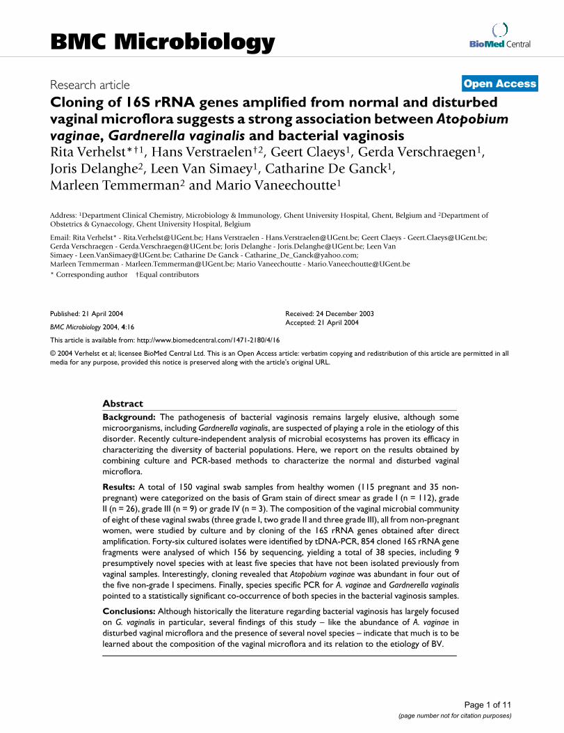
BioMed Central Page 1 of 11 (page number not for citation purposes) BMC Microbiology Open Access Research article Cloning of 16S rRNA genes amplified from normal and disturbed vaginal microflora suggests a strong association between Atopobium vaginae, Gardnerella vaginalis and bacterial vaginosis Rita Verhelst* †1 , Hans Verstraelen †2 , Geert Claeys 1 , Gerda Verschraegen 1 , Joris Delanghe 2 , Leen Van Simaey 1 , Catharine De Ganck 1 , Marleen Temmerman 2 and Mario Vaneechoutte 1 Address: 1 Department Clinical Chemistry, Microbiology & Immunology, Ghent University Hospital, Ghent, Belgium and 2 Department of Obstetrics & Gynaecology, Ghent University Hospital, Belgium Email: Rita Verhelst* - [email protected]; Hans Verstraelen - [email protected]; Geert Claeys - [email protected]; Gerda Verschraegen - [email protected]; Joris Delanghe - [email protected]; Leen Van Simaey - [email protected]; Catharine De Ganck - [email protected]; Marleen Temmerman - [email protected]; Mario Vaneechoutte - [email protected] * Corresponding author †Equal contributors Abstract Background: The pathogenesis of bacterial vaginosis remains largely elusive, although some microorganisms, including Gardnerella vaginalis, are suspected of playing a role in the etiology of this disorder. Recently culture-independent analysis of microbial ecosystems has proven its efficacy in characterizing the diversity of bacterial populations. Here, we report on the results obtained by combining culture and PCR-based methods to characterize the normal and disturbed vaginal microflora. Results: A total of 150 vaginal swab samples from healthy women (115 pregnant and 35 non- pregnant) were categorized on the basis of Gram stain of direct smear as grade I (n = 112), grade II (n = 26), grade III (n = 9) or grade IV (n = 3). The composition of the vaginal microbial community of eight of these vaginal swabs (three grade I, two grade II and three grade III), all from non-pregnant women, were studied by culture and by cloning of the 16S rRNA genes obtained after direct amplification. Forty-six cultured isolates were identified by tDNA-PCR, 854 cloned 16S rRNA gene fragments were analysed of which 156 by sequencing, yielding a total of 38 species, including 9 presumptively novel species with at least five species that have not been isolated previously from vaginal samples. Interestingly, cloning revealed that Atopobium vaginae was abundant in four out of the five non-grade I specimens. Finally, species specific PCR for A. vaginae and Gardnerella vaginalis pointed to a statistically significant co-occurrence of both species in the bacterial vaginosis samples. Conclusions: Although historically the literature regarding bacterial vaginosis has largely focused on G. vaginalis in particular, several findings of this study – like the abundance of A. vaginae in disturbed vaginal microflora and the presence of several novel species – indicate that much is to be learned about the composition of the vaginal microflora and its relation to the etiology of BV. Published: 21 April 2004 BMC Microbiology 2004, 4:16 Received: 24 December 2003 Accepted: 21 April 2004 This article is available from: http://www.biomedcentral.com/1471-2180/4/16 © 2004 Verhelst et al; licensee BioMed Central Ltd. This is an Open Access article: verbatim copying and redistribution of this article are permitted in all media for any purpose, provided this notice is preserved along with the article's original URL.
Transcript of Cloning of 16S rRNA genes amplified from normal and disturbed vaginal microflora suggests a strong...
BioMed CentralBMC Microbiology
ss
Open AcceResearch articleCloning of 16S rRNA genes amplified from normal and disturbed vaginal microflora suggests a strong association between Atopobium vaginae, Gardnerella vaginalis and bacterial vaginosisRita Verhelst*†1, Hans Verstraelen†2, Geert Claeys1, Gerda Verschraegen1, Joris Delanghe2, Leen Van Simaey1, Catharine De Ganck1, Marleen Temmerman2 and Mario Vaneechoutte1Address: 1Department Clinical Chemistry, Microbiology & Immunology, Ghent University Hospital, Ghent, Belgium and 2Department of Obstetrics & Gynaecology, Ghent University Hospital, Belgium
Email: Rita Verhelst* - [email protected]; Hans Verstraelen - [email protected]; Geert Claeys - [email protected]; Gerda Verschraegen - [email protected]; Joris Delanghe - [email protected]; Leen Van Simaey - [email protected]; Catharine De Ganck - [email protected]; Marleen Temmerman - [email protected]; Mario Vaneechoutte - [email protected]
* Corresponding author †Equal contributors
AbstractBackground: The pathogenesis of bacterial vaginosis remains largely elusive, although somemicroorganisms, including Gardnerella vaginalis, are suspected of playing a role in the etiology of thisdisorder. Recently culture-independent analysis of microbial ecosystems has proven its efficacy incharacterizing the diversity of bacterial populations. Here, we report on the results obtained bycombining culture and PCR-based methods to characterize the normal and disturbed vaginalmicroflora.
Results: A total of 150 vaginal swab samples from healthy women (115 pregnant and 35 non-pregnant) were categorized on the basis of Gram stain of direct smear as grade I (n = 112), gradeII (n = 26), grade III (n = 9) or grade IV (n = 3). The composition of the vaginal microbial communityof eight of these vaginal swabs (three grade I, two grade II and three grade III), all from non-pregnantwomen, were studied by culture and by cloning of the 16S rRNA genes obtained after directamplification. Forty-six cultured isolates were identified by tDNA-PCR, 854 cloned 16S rRNA genefragments were analysed of which 156 by sequencing, yielding a total of 38 species, including 9presumptively novel species with at least five species that have not been isolated previously fromvaginal samples. Interestingly, cloning revealed that Atopobium vaginae was abundant in four out ofthe five non-grade I specimens. Finally, species specific PCR for A. vaginae and Gardnerella vaginalispointed to a statistically significant co-occurrence of both species in the bacterial vaginosis samples.
Conclusions: Although historically the literature regarding bacterial vaginosis has largely focusedon G. vaginalis in particular, several findings of this study – like the abundance of A. vaginae indisturbed vaginal microflora and the presence of several novel species – indicate that much is to belearned about the composition of the vaginal microflora and its relation to the etiology of BV.
Published: 21 April 2004
BMC Microbiology 2004, 4:16
Received: 24 December 2003Accepted: 21 April 2004
This article is available from: http://www.biomedcentral.com/1471-2180/4/16
© 2004 Verhelst et al; licensee BioMed Central Ltd. This is an Open Access article: verbatim copying and redistribution of this article are permitted in all media for any purpose, provided this notice is preserved along with the article's original URL.
Page 1 of 11(page number not for citation purposes)
BMC Microbiology 2004, 4 http://www.biomedcentral.com/1471-2180/4/16
BackgroundBacterial vaginosis (BV) is considered to be the most com-mon cause of vaginal inflammation among both pregnantand non-pregnant women and prevalences between 4.9%and 36.0% have been reported from European and Amer-ican studies [1]. The etiology of this condition remainslargely unknown.
Nonetheless, the abundant literature, addressing bacterialvaginosis and ascending genital tract infection, has largelyfocused on the bacterial component and, in particular, ona few microorganisms, which are thought to play a pivotalrole in the pathology of bacterial vaginosis. Althoughprobably rather complex microbial community dynamicsare involved, culture-dependent studies indicate that alimited number of bacterial species, including Mobiluncusspp., Gardnerella vaginalis, Bacteroides spp., Prevotella spp.and Mycoplasma hominis, along with the relative absence ofLactobacillus spp., can be used as a sensitive index for bac-terial vaginosis. Consequently, according to Nugent et al.[2], the standard diagnosis of BV relies on the quantifica-tion of only three cellular types (Lactobacillus, Gardnerella,and Mobiluncus) on a Gram stained vaginal smear.
While conventional microbiological techniques are usefulas screening tools to identify women with BV, they do notenable prediction of the clinical burden associated withbacterial vaginosis. It has therefore been stated that betterunderstanding of the composition and dynamics of thevaginal microflora along with the factors associated withthe pathology of bacterial vaginosis is essential to improveour predictive abilities [1].
The analysis of complex bacterial communities has beenhampered by conventional culture-dependent methodsand by biochemical identification methods, since it leavesmany bacteria uncultured and unidentified. This mayprove especially true for vaginal microflora, both undernormal circumstances as well as in the setting of bacterialvaginosis, a condition primarily characterized by over-growth of anaerobic and fastidious microorganisms. Forexample, until recently, several important species, likeLactobacillus crispatus, L. gasseri and L. iners, were alllumped together into the L. acidophilus complex, whilepresent molecular techniques make it possible to differen-tiate between these closely related species [3-9]. Here wecombined culture with tDNA-PCR [4,10,11] and sequenc-ing of the 16S rRNA-gene for the identification of culturedorganisms.
Detailed information of complex microbial communitiescan also be acquired from the phylogenetic analysis of 16SrDNA sequences obtained directly from samples by PCRamplification, cloning, and sequencing, so that uncultiva-ble species are also included [12,13], although this
approach may be biased as well [14,15], as becomes alsoapparent from this study.
Here we report on the cloning and sequencing of 16SrRNA gene fragments, amplified directly from vaginalswabs, to examine the microbial diversity in the vaginalfluid of healthy women with different grades of vaginalmicroflora patterns as defined previously [16,17]. Finally,the data obtained by anaerobic culture and with cultureindependent techniques, lead us to carry out species spe-cific PCR for A. vaginae and G. vaginalis, such that the pres-ence of both species in differently graded samples couldbe established.
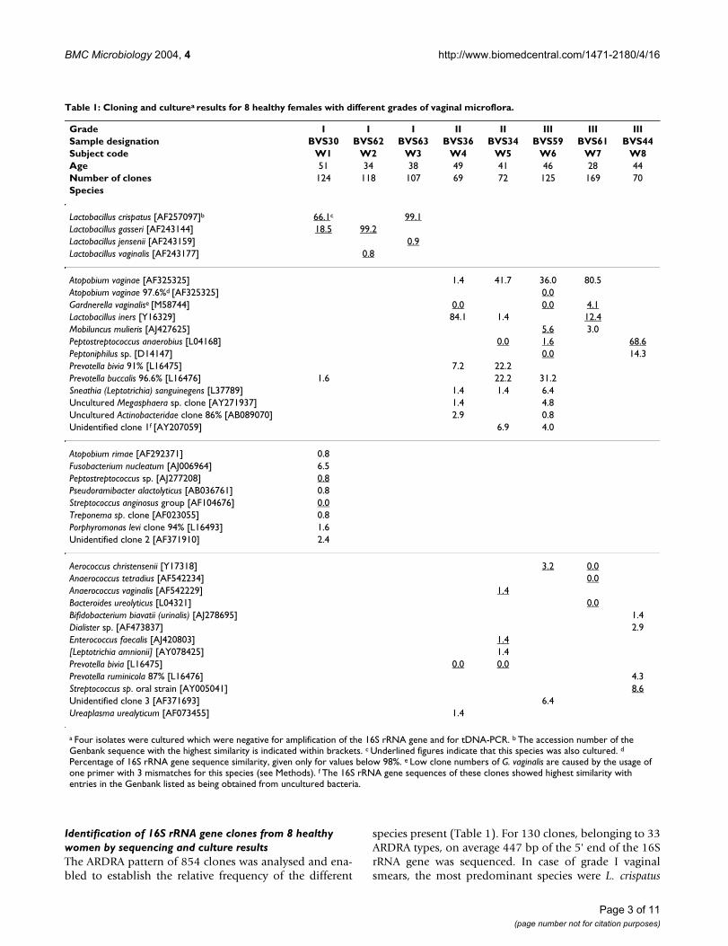
ResultsGrading of the Gram stained smears of the vaginal sam-ples for which cloning was carried out, according to thecriteria of Ison and Hay [17] assigned three specimens(W1-W3) to grade I, two (W4-5) to grade II and three(W6-8) to grade III. Table 1 lists the species identified bycloning and sequencing of cloned bacterial 16S rDNA-fragments, as well as by bacterial culture and tDNA-PCRfrom the vaginal microflora of these 8 healthy women.
Identification of cultured isolates from 8 healthy women by tDNA-PCR and 16S rRNA gene sequencingForty six isolates were obtained after anaerobic culture ofthe vaginal fluid from the 8 women in the cloning part ofthe study. Twenty nine isolates, belonging to the speciesBacteroides ureolyticus, Enterococcus faecalis, Lactobacilluscrispatus, L. jensenii, L. gasseri, L. vaginalis, Peptoniphilus sp.,Prevotella bivia, Streptococcus anginosus group and S. mitisgroup were identified by tDNA-PCR using a library con-taining tDNA-PCR fingerprints of well-identified strains.Six isolates belonging to the species Aerococcus christense-nii, Anaerococcus tetradius, Anaerococcus vaginalis, Lactoba-cillus iners, Mobiluncus mulieris and Peptostreptococcusanaerobius were identified by sequencing because theyproduced a tDNA pattern that initially was not present inthe database. These species were characterized by a spe-cific tDNA-PCR pattern that allowed unambiguous iden-tification after the new patterns were added to thedatabase. In summary, 40 of 46 isolates (87.0%) were cor-rectly identified by tDNA-PCR to the species level, includ-ing the species within the L. acidophilus complex. Of thesix isolates that could not be identified, one isolate, witha sequence that was 99% identical to that of a Peptostrepto-coccus sp. strain (CCUG 42997, AJ277208), could not beidentified unambiguously by tDNA-PCR because its pat-tern was identical to that of P. micros, one isolate – thatwas identified by sequencing as Atopobium vaginae – didnot yield a tDNA-pattern and another four isolates (8.7%)were tDNA-PCR negative and also 16S PCR negative.
Page 2 of 11(page number not for citation purposes)
BMC Microbiology 2004, 4 http://www.biomedcentral.com/1471-2180/4/16
Identification of 16S rRNA gene clones from 8 healthy women by sequencing and culture resultsThe ARDRA pattern of 854 clones was analysed and ena-bled to establish the relative frequency of the different
species present (Table 1). For 130 clones, belonging to 33ARDRA types, on average 447 bp of the 5' end of the 16SrRNA gene was sequenced. In case of grade I vaginalsmears, the most predominant species were L. crispatus
Table 1: Cloning and culturea results for 8 healthy females with different grades of vaginal microflora.
Grade I I I II II III III IIISample designation BVS30 BVS62 BVS63 BVS36 BVS34 BVS59 BVS61 BVS44Subject code W1 W2 W3 W4 W5 W6 W7 W8Age 51 34 38 49 41 46 28 44Number of clones 124 118 107 69 72 125 169 70Species
Lactobacillus crispatus [AF257097]b 66.1c 99.1Lactobacillus gasseri [AF243144] 18.5 99.2Lactobacillus jensenii [AF243159] 0.9Lactobacillus vaginalis [AF243177] 0.8
Atopobium vaginae [AF325325] 1.4 41.7 36.0 80.5Atopobium vaginae 97.6%d [AF325325] 0.0Gardnerella vaginalise [M58744] 0.0 0.0 4.1Lactobacillus iners [Y16329] 84.1 1.4 12.4Mobiluncus mulieris [AJ427625] 5.6 3.0Peptostreptococcus anaerobius [L04168] 0.0 1.6 68.6Peptoniphilus sp. [D14147] 0.0 14.3Prevotella bivia 91% [L16475] 7.2 22.2Prevotella buccalis 96.6% [L16476] 1.6 22.2 31.2Sneathia (Leptotrichia) sanguinegens [L37789] 1.4 1.4 6.4Uncultured Megasphaera sp. clone [AY271937] 1.4 4.8Uncultured Actinobacteridae clone 86% [AB089070] 2.9 0.8Unidentified clone 1f [AY207059] 6.9 4.0
Atopobium rimae [AF292371] 0.8Fusobacterium nucleatum [AJ006964] 6.5Peptostreptococcus sp. [AJ277208] 0.8Pseudoramibacter alactolyticus [AB036761] 0.8Streptococcus anginosus group [AF104676] 0.0Treponema sp. clone [AF023055] 0.8Porphyromonas levi clone 94% [L16493] 1.6Unidentified clone 2 [AF371910] 2.4
Aerococcus christensenii [Y17318] 3.2 0.0Anaerococcus tetradius [AF542234] 0.0Anaerococcus vaginalis [AF542229] 1.4Bacteroides ureolyticus [L04321] 0.0Bifidobacterium biavatii (urinalis) [AJ278695] 1.4Dialister sp. [AF473837] 2.9Enterococcus faecalis [AJ420803] 1.4[Leptotrichia amnionii] [AY078425] 1.4Prevotella bivia [L16475] 0.0 0.0Prevotella ruminicola 87% [L16476] 4.3Streptococcus sp. oral strain [AY005041] 8.6Unidentified clone 3 [AF371693] 6.4Ureaplasma urealyticum [AF073455] 1.4
a Four isolates were cultured which were negative for amplification of the 16S rRNA gene and for tDNA-PCR. b The accession number of the Genbank sequence with the highest similarity is indicated within brackets. c Underlined figures indicate that this species was also cultured. d
Percentage of 16S rRNA gene sequence similarity, given only for values below 98%. e Low clone numbers of G. vaginalis are caused by the usage of one primer with 3 mismatches for this species (see Methods). f The 16S rRNA gene sequences of these clones showed highest similarity with entries in the Genbank listed as being obtained from uncultured bacteria.
Page 3 of 11(page number not for citation purposes)

BMC Microbiology 2004, 4 http://www.biomedcentral.com/1471-2180/4/16
and L. gasseri. Smaller numbers of L. jensenii and L. vagina-lis were also present. All females with grade I smears (W1-W3) were colonized by two Lactobacillus species. W2 (age34) and W3 (age 38) were colonized by Lactobacillus spe-cies only, with one species – respectively L. gasseri and L.crispatus – represented by more than 99% of the clones. Inthe vaginal fluid of W1 (age 51) the most abundant spe-cies were L. crispatus (66.1%), L. gasseri (18.6%) and Fuso-bacterium nucleatum (6.5%). An additional 8 species werefound in low numbers, six only by cloning and one onlyby culture. Three of these species, representing 5.6% of theclones, showed less than 98% similarity with previouslypublished sequences. Culture results coincided largelywith cloning for the grade I samples, except for the nonLactobacillus species of W1 of which only two werecultured.
For the two grade II smears (W4 and W5) cloning revealedfour species, A. vaginae (resp. 1.5% and 41.6%), L. iners(resp. 84.1% and 1.4%), a P. bivia-like species (resp. 7.3%and 22.2%) and Sneathia sanguinegens (both 1.4%), thatwere present in both samples. P. bivia was also present inboth grade II microflora but it was found by culture only.Several other species were found by cloning only in eitherW4 or W5. For example, [Leptotrichia amnionii] was foundonly in W5. G. vaginalis and Peptostreptococcus anaerobiuswere found respectively in W4 and W5 by culture only andtwo species, Anaerococcus vaginalis and Enterococcus faeca-lis, were found by both cloning and culture in the micro-flora of W5.
Two women (W6 and W7) with a grade III vaginal smearwere colonized predominantly by A. vaginae (resp. 36.0and 80.5%), L. iners (absent and 12.4%), G. vaginalis(absent and 4.1%), M. mulieris (5.6 and 3.0%) and aPrevotella buccalis-like species (31.2% and absent), accord-ing to cloning. As for grade II samples, there was limitedcorrespondence with culture. For W6, an A. vaginae-likeorganism was cultured, but the sequence of this isolateshowed less than 98% similarity to the sequence of theclones. Furthermore, a large number of colonies of G. vag-inalis and Peptoniphilus sp. were present after anaerobicculture but no clones were obtained.
The most abundant species obtained by cloning the thirdgrade III vaginal sample (W8) were Peptostreptococcusanaerobius (68.6%)(also cultured), Peptoniphilus sp.(14.3%) and an unidentified Streptococcus sp. (8.6%)(alsocultured).
Total number of species identified and comparison between cloning and cultureOf the 38 species, 18 were discovered by cloning only, 5by culture only, and 8 by both cloning and culture. Threespecies were shown by both culture and cloning in some
samples, but only by culture in the remaining samples,another three species were shown by both cloning andculture in some samples, but only by cloning in theremaining samples and one species was found once byculture only and once by cloning only. The presence of allfour grade I Lactobacillus sp. was shown in grade I samplesby both cloning and culture, whereas L. iners was foundonly in three non-grade I samples, in all three by cloning– even abundantly in W4, but only in W7 by culture. G.vaginalis was cultured three times, but was found by clon-ing only once. A. vaginae was shown in four samples bycloning, but cultured only once. The P. bivia-like speciesand P. buccalis-like species were found in respectively twoand three samples by cloning but not by culture.
Possible novel species and generaOf the 38 species that were distinguished by culture andcloning in this study, only 76.3% demonstrated morethan 98% identity with previously known bacterial spe-cies (Table 1). Five of the eight vaginal samples containedpreviously unidentified species.
Anaerobic culture of the microflora of W6 revealed anAtopobium species that showed only 97.6% similarity withpreviously reported A. vaginae sequences (see below).
Two presumptively novel species within the genus Prevo-tella were found. For W4 and W5 (grade II), respectively7.3% and 22.2% of the clones had 91% similarity with aP. bivia sequence. For W1 (grade I), W5 and W6 (gradeIII), respectively 1.6%, 22.2% and 31.2% of the clonesshowed 96.6% similarity with a P. buccalis sequence.Respectively 2.9% and 0.8% of the clones of W4 (grade II)and W6 (grade III) were identical and had only 86% sim-ilarity with an uncultured termite Actinobacteridae bacte-rium (accession number AB089070).
Variation in Atopobium sequencesSixteen A. vaginae sequences are present in GenBank, onefrom a clinical isolate [18], one from the type strain [19]and 14 from cloned 16S rDNA fragments (Zhou, unpub-lished) (Figure 1). Another 17 sequences were obtained inthis study, one from an isolate from patient W6, two fromisolates from other patients (BVS38 and PB9) and 14 fromclones from four different patients, i.e. W4 (grade II), W5(II), W6 (III) and W7 (III) (Figure 1).
The sequence of all 7 clones from W5 was different fromthat of all other isolates and from the clones and the Gen-bank sequences, except from one Genbank clonesequence (AY269023). The sequence of all clones fromthe three other patients and from the two isolates fromsamples BVS38 and PB9 were identical to the previouslypublished clinical isolate [18] and the type strain [19].The sequence of the cultured isolate from patient W6 was
Page 4 of 11(page number not for citation purposes)
BMC Microbiology 2004, 4 http://www.biomedcentral.com/1471-2180/4/16
somewhat intermediate between both above sequences,although the sequence of the clones of the same patientwere identical to that of the type strain. Specific amplifica-tion with both the ato167f primer and the ato154f primergave positive signals for the vaginal sample of W6, indicat-ing that indeed both types of sequence were present.
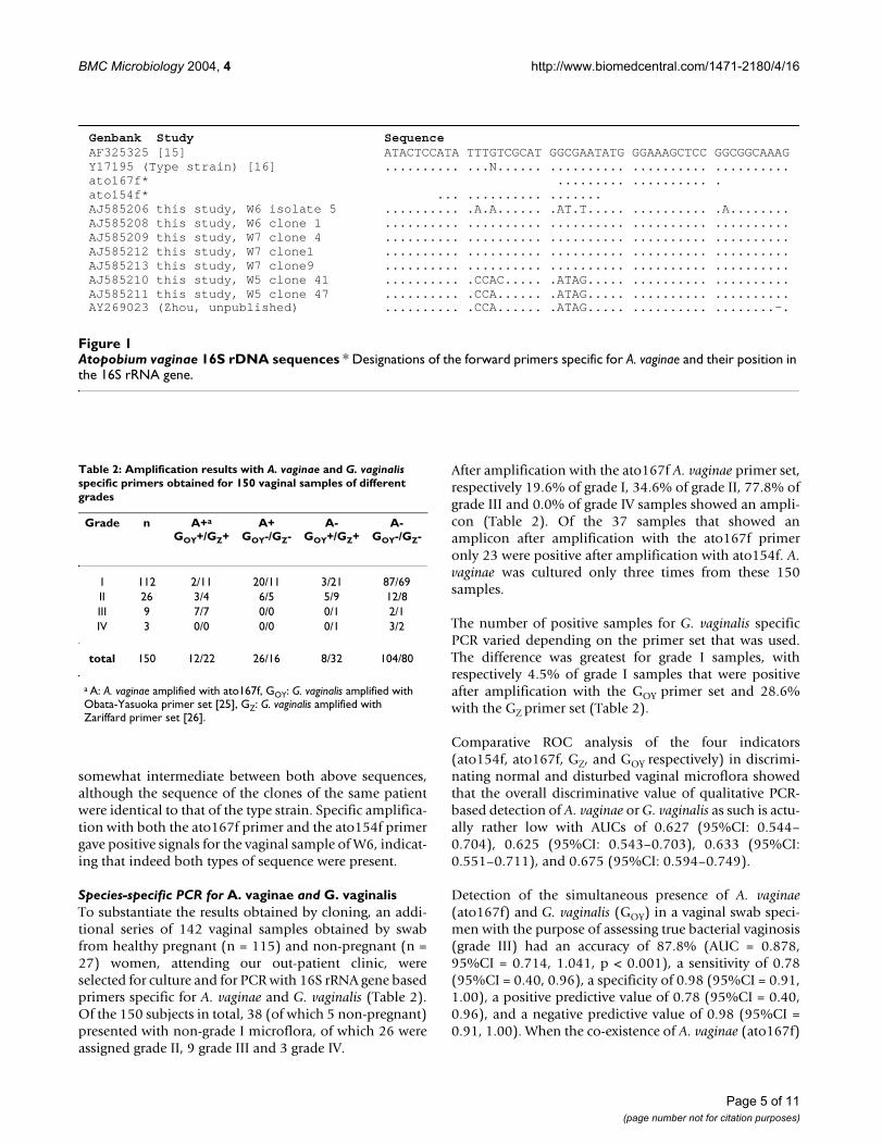
Species-specific PCR for A. vaginae and G. vaginalisTo substantiate the results obtained by cloning, an addi-tional series of 142 vaginal samples obtained by swabfrom healthy pregnant (n = 115) and non-pregnant (n =27) women, attending our out-patient clinic, wereselected for culture and for PCR with 16S rRNA gene basedprimers specific for A. vaginae and G. vaginalis (Table 2).Of the 150 subjects in total, 38 (of which 5 non-pregnant)presented with non-grade I microflora, of which 26 wereassigned grade II, 9 grade III and 3 grade IV.
After amplification with the ato167f A. vaginae primer set,respectively 19.6% of grade I, 34.6% of grade II, 77.8% ofgrade III and 0.0% of grade IV samples showed an ampli-con (Table 2). Of the 37 samples that showed anamplicon after amplification with the ato167f primeronly 23 were positive after amplification with ato154f. A.vaginae was cultured only three times from these 150samples.
The number of positive samples for G. vaginalis specificPCR varied depending on the primer set that was used.The difference was greatest for grade I samples, withrespectively 4.5% of grade I samples that were positiveafter amplification with the GOY primer set and 28.6%with the GZ primer set (Table 2).
Comparative ROC analysis of the four indicators(ato154f, ato167f, GZ, and GOY respectively) in discrimi-nating normal and disturbed vaginal microflora showedthat the overall discriminative value of qualitative PCR-based detection of A. vaginae or G. vaginalis as such is actu-ally rather low with AUCs of 0.627 (95%CI: 0.544–0.704), 0.625 (95%CI: 0.543–0.703), 0.633 (95%CI:0.551–0.711), and 0.675 (95%CI: 0.594–0.749).
Detection of the simultaneous presence of A. vaginae(ato167f) and G. vaginalis (GOY) in a vaginal swab speci-men with the purpose of assessing true bacterial vaginosis(grade III) had an accuracy of 87.8% (AUC = 0.878,95%CI = 0.714, 1.041, p < 0.001), a sensitivity of 0.78(95%CI = 0.40, 0.96), a specificity of 0.98 (95%CI = 0.91,1.00), a positive predictive value of 0.78 (95%CI = 0.40,0.96), and a negative predictive value of 0.98 (95%CI =0.91, 1.00). When the co-existence of A. vaginae (ato167f)
Atopobium vaginae 16S rDNA sequencesFigure 1Atopobium vaginae 16S rDNA sequences * Designations of the forward primers specific for A. vaginae and their position in the 16S rRNA gene.
Genbank Study Sequence AF325325 [15] ATACTCCATA TTTGTCGCAT GGCGAATATG GGAAAGCTCC GGCGGCAAAGY17195 (Type strain) [16] .......... ...N...... .......... .......... ..........ato167f* ......... .......... .ato154f* ... .......... .......AJ585206 this study, W6 isolate 5 .......... .A.A...... .AT.T..... .......... .A........AJ585208 this study, W6 clone 1 .......... .......... .......... .......... ..........AJ585209 this study, W7 clone 4 .......... .......... .......... .......... ..........AJ585212 this study, W7 clone1 .......... .......... .......... .......... ..........AJ585213 this study, W7 clone9 .......... .......... .......... .......... ..........AJ585210 this study, W5 clone 41 .......... .CCAC..... .ATAG..... .......... ..........AJ585211 this study, W5 clone 47 .......... .CCA...... .ATAG..... .......... ..........AY269023 (Zhou, unpublished) .......... .CCA...... .ATAG..... .......... ........-.
Table 2: Amplification results with A. vaginae and G. vaginalis specific primers obtained for 150 vaginal samples of different grades
Grade n A+a
GOY+/GZ+A+
GOY-/GZ-A-
GOY+/GZ+A-
GOY-/GZ-
I 112 2/11 20/11 3/21 87/69II 26 3/4 6/5 5/9 12/8III 9 7/7 0/0 0/1 2/1IV 3 0/0 0/0 0/1 3/2
total 150 12/22 26/16 8/32 104/80
a A: A. vaginae amplified with ato167f, GOY: G. vaginalis amplified with Obata-Yasuoka primer set [25], GZ: G. vaginalis amplified with Zariffard primer set [26].
Page 5 of 11(page number not for citation purposes)

BMC Microbiology 2004, 4 http://www.biomedcentral.com/1471-2180/4/16
and G. vaginalis was assessed using the GZ-primer set, theoverall performance of the assay was significantly lower.
DiscussionTo our knowledge, no cloning study addressing the com-position of the microflora of vaginal samples and bacte-rial vaginosis samples has been published thus far. Bymeans of culture and by cloning and sequencing of 16SrRNA genes, amplified directly from the vaginal samples,we studied the vaginal microflora of 8 healthy womenwith different grades of bacterial vaginosis according toNugent [2], as modified by Ison and Hay [16,17].
The frequency of the different clones could be assessed byperforming ARDRA of the cloned 16S rRNA genes andcounting the number of clones belonging to each ARDRAtype. Representative clones for each ARDRA type weresubsequently sequenced to determine the species identity.Five bacterial species, i.e. Atopobium rimae, Bifidobacteriumbiavatii (urinalis), Dialister sp., [Leptotrichia amnionii] andSneathia sanguinegens, had never been recovered from thevagina. Twenty two percent of the clones, belonging to 9putative species, showed less than 98% homology to anyof the 16S rRNA gene sequences present in GenBank, indi-cating that several bacterial species, indigeneous to thefemale genital tract, remain to be characterized.
In this study, cloning confirmed that the vaginal micro-flora of healthy women is dominated by a limited numberof Lactobacillus species. For the women with grade I micro-flora, the two species L. crispatus and L. gasseri, alone or incombination, accounted for 85–99% of the clones. Thetwo younger women, 34 and 38 years, had almost purecultures of L. crispatus resp. L. gasseri, in association withlow numbers of resp. L. jensenii (< 1%) and L. vaginalis (<1%) and without any other bacteria. In our ongoing stud-ies we find this to be the case for 61.5% of the grade I sam-ples (unpublished data). The finding of monocultures ofL. crispatus and L. gasseri is also in correspondence withrecent reports, which point to the predominance of onlythree to four Lactobacillus species in normal vaginal micro-flora [7,20]. Antonio et al. [3] considered L. crispatus, L.jensenii and L. gasseri as the indicator species for normalvaginal microflora, also because most strains of these spe-cies are hydroxyperoxide producers in opposition to otherLactobacillus spp. It should be noted in this context thatanother group could not substantiate any protective effectof hydroxyperoxide producing lactobacilli [21].
In the 51-year-old woman (W1), with a combination ofboth L. crispatus and L. gasseri, some additional non Lacto-bacillus species were present in low numbers, with Fusobac-terium nucleatum as the most predominant (i.e. 6.5%).This may be in accordance with another observation,
namely that postmenopausal healthy women frequentlypresent with BV-like microscopy [5,22].
Of the five women with non-grade I microflora (two withgrade II and three with grade III), one presented with pre-dominant Peptostreptococcus clones (68%), a picture thatwas very different from that observed in the other women.However, most striking was the observation that for theother four patients between 1.5% and 80.0% of the cloneswere identified as Atopobium vaginae, a species previouslyknown only from a single isolate from the vagina of ahealthy woman [19] and from a clinically important iso-late, described as the causative agent in a case of a pelvicinflammatory disease (PID) following transvaginal oocyterecruitment [18]. Interestingly, Zhou et al. [20] reporteddominating Atopobium sp. in a woman, judged to carry anormal vaginal microflora. The authors concluded thatAtopobium may be present as part of the normal microfloraof the vagina [23]. This warrants a more detailed discus-sion of this recently described species.
The genus Atopobium was introduced to accommodate thespecies Lactobacillus minutus, Lactobacillus rimae and Strep-tococcus parvulus [24]. Atopobium species have beendescribed to produce major amounts of lactic acid [19], acharacteristic reminiscent of lactobacilli, although no spe-cial reference to this property was made in the descriptionof A. vaginae [19]. Atopobium species are anaerobic, Gram-positive elliptical cocci or rod-shaped organisms occur-ring singly, in pairs or as short chains. The variable cellmorphology of A. vaginae makes that this species mayreside perfectly camouflaged and as a consequence unde-tectable among the mixture of other species present ingrade II and III bacterial communities. Also the fact that A.vaginae is fastidious and forms small pinhead colonies canexplain why this species was not yet established as part ofthe vaginal microflora, when using classical microbiology.In this study, we could culture the species from only threeout of 150 vaginal specimens.
Based on our findings with A. vaginae specific PCR, whichrecovered this species in 19.6% of the 112 grade I speci-mens, it appears as if A. vaginae may be a constituent –presumably in low numbers – of the human vagina, pos-sibly attaining replicative dominance in association withdecreasing lactobacillary grading. This hypothesis couldbe substantiated with quantitative PCR and the develop-ment of selective culture media.
The striking fact that Gardnerella vaginalis, predominantlypresent in bacterial vaginosis samples [e.g. [25,26]], wasnot found by cloning as carried out in this study, can beexplained as the consequence of a methodological bias,because the forward primer used for cloning (10f) con-tained 3 mismatches for G. vaginalis. On the other hand,
Page 6 of 11(page number not for citation purposes)

BMC Microbiology 2004, 4 http://www.biomedcentral.com/1471-2180/4/16
the use of these non G. vaginalis 'universal' primers mayhave facilitated to establish the high prevalence of A. vagi-nae. To assess the relative importance of both species,cloning should be carried out with different sets of prim-ers. Amplification of grade I, grade II and grade III sampleswith species specific primers indicated that G. vaginaliswas present in respectively 28.6%, 50.0 % and 88.9% ofthe samples, while Atopobium was present in respectively19.6%, 34.6% and 77.8% of the vaginal microflora.
Since any pairwise comparison of the four ROC plots didnot show any significant differences between the AUCs, itis also apparent that each of the four indicators understudy (ato154f, ato167f, GZ, and GOY respectively)presents with a comparably limited accuracy in discrimi-nating normal and disturbed vaginal microflora. This maybe explained by the fact that, although both species arepresent in about 80% of the true bacterial vaginosis sam-ples, their presence in grade I samples is not uncommon.This lack of accuracy for G. vaginalis was also previouslyassessed in culture-dependent studies [27,28].
The co-existence of A. vaginae and G. vaginalis in womenwith grade III microflora together with their simultaneousabsence in grade I samples (Table 2) is striking. The simul-taneous presence of both species therefore is highly pre-dictive for BV.
Lactobacillus iners, was present among the clones in threeof the five non-grade I samples, even in large numbers,and absent from the three grade I samples. This specieswas only recently recognized as a separate Lactobacillusspecies [29]. In the original description of this relativelyasaccharolytic Lactobacillus species, the only vaginalisolate among the 9 strains from females came from vagi-nal discharge [29]. This species may have been largelyoverlooked in culture based studies [30-32] since it doesnot grow on Lactobacillus selective media, including MRSand Rogosa-Sharp medium [29]. Using molecular tech-niques L. iners was reported as one of the most frequentlyencountered vaginal Lactobacillus species [8,9] present innormal and BV microflora [5].
Leptotrichia sp. are slow-growing, gram-negative anaerobicorganisms of the oral cavity and genital tract. [L. amnionii]is an extremely fastidious organism, which most likelyexplains why this organism was hitherto virtually unde-tectable by conventional culture-based microbiologicaltechniques. Of interest is that this particular Leptotrichiaspecies was isolated by Shukla et al. [33] from the amni-otic fluid of a woman with second trimester fetal loss,which led the authors to conclude that [L. amnionii] is pre-sumably indigenous to the genital tract and acts as anopportunist under particular clinical conditions, includ-
ing pregnancy. This study demonstrates for the first timethe presence of [L. amnionii] in the vagina.
Sneathia sanguinegens (formerly Leptotrichia sanguinegens)was found in this study to be present in three patients withnon-grade I vaginal microflora, in moderate numbers. S.sanguinegens has been isolated from human blood andamniotic fluid [34] and has been associated with severalcases of pregnancy-associated bacteraemia, i.e. postpar-tum fever in four patients and neonatal sepsis in twopatients [35]. This study demonstrates the presence of S.sanguinegens in the vagina for the first time.
One strain from W6 and 14.3% of the clones from W8were identified as Peptoniphilus indolicus based on morethan 98% similarity with a Genbank sequence with acces-sion number D14147 corresponding with the type strainCCUG 17639 of this species. This type strain however hastwo Genbank entries (also AY153430) with low similarity[36]. Unfortunately our isolate was not stored so furtherphenotypic identification was not possible. Because of theambiguous Genbank sequences, we chose to designateour isolate and clones as Peptoniphilus sp.
Thus far, identification of organisms cultured from com-plex microbial biota like the vagina, especially under thecondition of vaginosis, has also been hampered by thelimitations of conventional biochemical and phenotypicidentification methods. However, using a DNA-basedmethod, namely tDNA-PCR [11], it is convenient toappropriately identify most of these organisms, once alibrary, based on tDNA-PCR of well characterized organ-isms, has been constructed. Also, newly unknowns can befirst identified by 16S rRNA gene sequence determination,whereafter it is possible to use the corresponding tDNAfingerprint for future identification of the organisms ofthe same species.
tDNA-PCR, which consists of the amplification of thespacer regions between tRNA genes, has been shown toyield mainly species-specific DNA fingerprints [10]. Com-bined with capillary electrophoresis, tDNA-PCR has beenshown to be a semi-automated, digital DNA-fingerprint-ing method that enables rapid and discriminatory identi-fication of species from very diverse phylogenetic groups,including Lactobacillus [4,11].
ConclusionsIn summary, the use of tDNA-PCR based identification ofcultured organisms in combination with cloning of 16SrRNA genes, amplified directly from vaginal swabs, ena-bled us to characterize the vaginal microflora in a detailedmanner, and to compare the composition of the normalvaginal microflora with the bacterial vaginosis microflora.The presence of A. vaginae in 4 of the 5 women with bac-
Page 7 of 11(page number not for citation purposes)

BMC Microbiology 2004, 4 http://www.biomedcentral.com/1471-2180/4/16
terial vaginosis grade II-III microflora is an unexpectedand previously not reported finding, which may shed newlight on the etiology of this condition. There is also theambiguous position of L. iners, which apparently is notindicative of a normal microflora, but may point to someintermediate condition.
Furthermore, our findings warrant more detailed studiesof the ability of species like L. iners and A. vaginae to pro-duce lactic acid, hydrogen peroxide and of their cellularand colonial morphology and biochemical characteris-tics, as well as their interaction with lactobacilli and G.vaginalis. A selective medium for these organisms mightbe a welcome tool for further studies, given the fact thatuntil now they have been largely overlooked by culturemethods.
Finally, the discrepancies between culture, cloning andspecific PCR, together with the diversity of the recoveredspecies, their unequal distribution over different vaginalsamples, the elucidation of several species – includingpresumptively new species – previously not associatedwith the vagina, indicate that much is to be learned aboutthe composition of the vaginal microflora and its relationto the etiology of BV.
MethodsStudy population, sample collection and gradingFor a total of 150 healthy women of reproductive age,attending our out-patient clinic, of which 115 were preg-nant, the health condition with regard to the compositionof the vaginal bacterial community was assessed micro-scopically after Gram stain, according to the criteria ofHay and Ison [17] and vaginal samples were taken.Briefly, specimens were considered grade I (normal) whenonly Lactobacillus morphotypes were present, grade II(intermediate) when both Lactobacillus and other mor-photypes were present, grade III (BV) when only nonLactobacillus morphotypes were seen and grade IV whenonly Gram positive cocci were seen.
Sampling was carried out as follows. After placement of anon-lubrificated speculum two sterile cotton swabs wereinserted into the vaginal vault. The swabs were rotatedagainst the vaginal wall at the midportion of the vault andwere carefully removed to prevent contamination with thevulva and introitus microflora. One swab was returned toa sterile tube (dry swab) (Copan, Brescia, Italy), for thepurpose of DNA-extraction. The other swab was placedinto Amies transport medium (Nuova Aptaca, Canelli,Italy) and was used for making a smear for the purpose ofgrading according to the Hay and Ison criteria [17] and foranaerobic culture. Both swabs were processed in themicrobiology laboratory within 4 h.
Based on smear results, eight non-pregnant women(mean age 41.4 years, range 28–51 years) from the stud-ied population, attending our out-patient clinic for a rou-tine gynaecological visit, were selected for cloning of the16S rRNA genes present in the vaginal microflora. The cul-ture results are reported only for these eight women.
To substantiate the results obtained by cloning and cul-ture, PCR with species specific primers for A. vaginae andG. vaginalis and culture were carried out for the completepopulation.
Culture and identification of cultured isolates by tDNA-PCRFor eight non-pregnant women, the swab on Amies trans-port medium was streaked onto tryptic soy agar supple-mented with 5% sheep blood (Becton Dickinson,Franklin Lakes, NJ) and incubated anaerobically at 37°Cupon arrival at the microbiology laboratory. After 4 daysof incubation, all the isolates with different colony mor-phology were selected for identification. DNA wasextracted by simple alkaline lysis: one colony was sus-pended in 20 µl of 0.25% sodium dodecyl sulfate-0.05 NNaOH, heated at 95°C for 15 min and diluted with 180µl of distilled water. tDNA-PCR and capillary electro-phoresis were carried out as described previously [4,11].The species to which each isolate belonged was deter-mined by comparing the tDNA-PCR fingerprint obtainedfrom each isolate with a library of tDNA-PCR fingerprintsobtained from reference strains, using an in-house soft-ware program [11]. The library of tDNA-PCR fingerprintsand the software are available on request.
DNA extraction of vaginal swab samplesFor DNA extraction from the dry vaginal swabs, theQIAamp DNA mini kit (Qiagen, Hilden, Germany) wasused according to the manufacturer's recommendations,with minor modifications. The dry swab specimen fromeach patient was swirled for 15 s in 400 µl of lysis buffer(20 mM Tris-HCl, pH 8.0; 2 mM EDTA; 1.2% Triton).Fifty units of mutanolysin (25 U/µl) (Sigma, Bornem, Bel-gium) were added and the samples were incubated for 30min at 37°C. After the addition of 20 µl Proteinase K (20mg/ml) and 200 µl AL buffer (Qiagen), samples wereincubated for 30 min at 56°C. Next, 200 µl of ethanol wasadded and DNA was purified by adding the lysate to theQiagen columns as described by the manufacturer.Finally, the total bacterial DNA was eluted with 100 µl ofAE buffer (Qiagen). DNA-extracts were stored at -20°Cand were used for the purpose of cloning experiments andspecies specific PCR.
Cloning of amplified mixtures of 16S rDNATo amplify the 5' part of the bacterial 16S rRNA genes byPCR, primers 10f (5' AGTTTGATCCTGGCTCAG) and
Page 8 of 11(page number not for citation purposes)
BMC Microbiology 2004, 4 http://www.biomedcentral.com/1471-2180/4/16
534r (5' ATTACCGCGGCTGCTGG) [37], which target thedomain Bacteria, were used. It should be noted that theforward primer contains mismatches for G. vaginalis atpositions 1 (A/G), 5 (T/C) and 9 (C/T). A 50 µl PCR mix-ture contained 0.1 µM of each primer, 25 µl of Promegamaster mix (Promega, Madison, WI), 5 µl of DNA extractand distilled water. Thermal cycling consisted of an initialdenaturation of 5 min at 94°C, followed by three cycles of1 min at 94°C, 2 min at 50°C and 1 min at 72°C, fol-lowed by 35 cycles of 20 sec at 94°C, 1 min at 50°C and1 min 72°C, with a final extension of 10 min at 72°C, andcooling to 10°C. PCR products were purified using theQIAquick PCR Purification Kit (Qiagen). Cloning wasdone using the Qiagen PCR Cloning Kit (UA-cloning,Qiagen), whereby the purified amplicons were ligatedinto the pDrive cloning vector and transformed into Qia-gen EZ Competent E. coli Cells, as specified by themanufacturer.
Screening for clones with different 16S rRNA gene inserts by ARDRAFrom each sample, 100 to 200 ampicillin-resistant trans-formants, recognizable as white colonies on Luria-Bertani(LB) agar containing IPTG (Roche, Basel, Switzerland), X-Gal (Roche) and 100 µg ampicillin/ml after overnightincubation at 37°C, were selected for further analysis. Sin-gle colonies were picked from the agar plates and trans-ferred with sterile tips to the wells of a 96 well plate filledwith LB Broth supplemented with 100 µg ampicillin/mland incubated for 3 h. To avoid sequencing of all clones,the 16S rRNA gene inserts were differentiated initiallyfrom each other by means of Amplified rDNA RestrictionAnalysis (ARDRA)[38]. One µl of bacterial culture wasadded to a final volume of 20 µl PCR mix, containing 0.2µM of two plasmid-targeted primers, QF (5'TACGTATCG-GATCCAGAATTC) and QR (5'CGAGAAGCTTGTCGAC-GAATT), 10 µl of Promega master mix and distilled water.The position of primers QF and QR was chosen as suchthat their distance towards the insert was the same inorder to result in the same ARDRA pattern regardless theorientation of the insert in the cloning vector. Cyclingconditions were the same as described above for the 16SrRNA gene. Ten microliter of the amplified products weredigested with 10 U of the restriction endonuclease BstUIin the appropriate enzyme buffer and incubated for 3 h at60°C. The DNA restriction fragments were separated in a2.5% agarose electrophoresis gel, containing 2% Meth-aphor (FMC Bioproducts, Rockland, ME) and 0.5% MPagarose (Roche, Basel, Switzerland) in the presence ofethidium bromide (50 ng/ml). The gels were photo-graphed and the obtained ARDRA fingerprints were com-pared visually. For each ARDRA type, the 16S rRNA geneinsert of at least one representative clone was sequenced,thus minimizing the number of sequence reactions thathad to be carried out.
Sequencing of 16S rRNA genes from isolates and clonesFor 8 healthy women, the 16S rRNA gene was amplifiedand sequenced from selected clones and from culturedisolates that could not be identified by tDNA-PCR, usingprimers 10f and 534r, as described above. For the clones,1 µl of the QF-QR amplified PCR product was diluted1000 times with distilled water and re-amplification ofthe 16S rRNA gene was done with the primers 10f and534r. For the cultured isolates, a 5 µl aliquot of the alka-line lysate was added to a 50 µl PCR mixture. The ampli-fication products were then purified with the QiaquickPCR purification kit, according to the manufacturer'sinstructions. Sequencing was done using the ABI Big Dyecycle sequencing reaction kit with AmpliTaq FS DNApolymerase (Applied Biosystems, Foster City, CA.) withprimer 534r. Sequencing reaction products were analyzedon an ABI 310 genetic analyzer (Applied Biosystems).Inspection of the electropherograms was done with Chro-mas http://www.technelysium.com.au/chromas14x.htmland with the BioEdit package [39]. Comparison of thesequences of the inserts and isolates to the 16S rRNA genesequences in GenBank was done using the BLAST software[40]. Clones or isolates with DNA sequences sharing morethan 98% identity with known sequences were assigned tothat phylotype.
Species specific PCR for Gardnerella vaginalisG. vaginalis species-specific primers as designed byZariffard et al. (GZ)[26] and Obata-Yasuoka et al. (GOY)[25] were used. Briefly, a 20 µl PCR mixture containedrespectively 0.05 and 0.4 µM primers, 10 µl of Promegamaster mix (Promega, Madison, WI), 2 µl of Qiagen DNA-extract of the samples and distilled water. Thermal cyclingwith GZ primers consisted of an initial denaturation of 10min at 94°C, followed by 50 cycles of 5 s at 94°C, 45 s at55°C and 45 s at 72°C, and a final extension of 10 min at72°C. Thermal cycling with the GOY primers was per-formed by an initial denaturation of 1 min at 94°C wasfollowed by 40 cycles of 1 min at 94°C, 1 min at 60°Cand 1 min at 70°C and a final extension of 7 min at 72°C.During the first ten cycles the annealing temperature waslowered by 0.5°C per cycle. Five microliter of the ampli-fied product of each PCR was visualized on a 2% agarosegel.
Species specific PCR for Atopobium vaginaeTwo primer sets that allowed amplification of the 16SrRNA gene of A. vaginae and that lacked homology withnon-target bacteria as determined by searching the GeneBank database using BLAST software [40], were designed.The selected primers, ato167f 5' (GCGAATAT-GGGAAAGCTCCG), ato154f 5' (ATATTTGTCGCAT-GGCGAAT) and ato587r 5'(GAGCGGATAGGGGTTGAGC), were analysed for sec-ondary structures using NetPrimer (Premier Biosoft Inter-
Page 9 of 11(page number not for citation purposes)
BMC Microbiology 2004, 4 http://www.biomedcentral.com/1471-2180/4/16
national, Palo Alto, CA). A 20 µl PCR mixture contained0.2 µM of primers (respectively ato167f or ato154f andato587r), 10 µl of Promega master mix (Promega, Madi-son, WI), 2 µl of Qiagen DNA-extract of the samples anddistilled water. Thermal cycling consisted of an initialdenaturation of 5 min at 94°C, followed by three cycles of1 min at 94°C, 2 min at 58°C and 1 min at 72°C, fol-lowed by 35 cycles of 20 sec at 94°C, 1 min at 58°C and1 min 72°C, with a final extension of 10 min at 72°C, andcooling to 10°C. Five microliter of the amplified productwas visualized on a 2% agarose gel. The primers amplifieda DNA-fragment of respectively 420 and 433 basepairsfrom A. vaginae and showed no cross reactivity to otherorganisms, including A. rimae (data not presented).
StatisticsTo assess the relative accuracy of PCR-based detection ofG. vaginalis or A. vaginae, we applied comparative receiver-operating-characteristic (ROC) analysis under the non-parametric assumption and compared the accuracies ofthe isolated markers (amplification with ato154f,ato167f, GOY and GZ) in allocating subjects to a particularGram stain category (grades I to IV).
Subsequently, we compared the accuracy of PCR-basedcombined detection of G. vaginalis (using the two differ-ent primer sets designated GZand GOY) and A. vaginae(using ato167f) to assess the vaginal microflora status(according to Gram stain category) by non-parametricROC-analysis.
Accuracy in these analyses is expressed as the area-under-the-curve (AUC) in the ROC-plot, the 95% confidenceinterval (CI) to the AUC, and the p-value to the 95% CI.We also calculated the estimated sensitivity, specificity,positive and negative predictive values (PPV and NPV)and the 95% confidence intervals to these measures. Sta-tistical significance was accepted at the α = 0.05-level.
Analyses were carried out using the statistical softwarepackages EpiCalc2000 v.1.02 and SPSS v.11.0.
Nucleotide sequence accession numbersOut of 156 16S rRNA gene sequences, obtained from 26isolates and from 130 clones, two sequences of A. vaginae(-like) species and 23 sequences of uncultured bacteriumclones were submitted to GenBank and were assignedaccession no. AJ585206 to AJ585213 and no. AJ619698 toAJ619714.
Authors' contributionsRV, GC, GV and MV participated in the development ofthe study design, the analysis of the study samples, thecollection, analysis and interpretation of the data, and inthe writing of the report. HV and MT participated in the
development of the study design, the collection of thestudy samples, the collection, analysis and interpretationof the data, and in the writing of the report. JD partici-pated in the analysis and interpretation of the data and inthe writing of the report. LVS and CDG participated in theanalysis of the study samples. All authors read andapproved the final manuscript.
AcknowledgementsThis work was supported through a research grant by the Marguerite-Marie Delacroix Foundation. As the main funding source, the Marguerite-Marie Delacroix Foundation was not involved in the development of the study design, the collection, analysis, and interpretation of the data, in the writing of the report nor in the decision to submit the paper for publication.
References1. Morris M, Nicoll A, Simms I, Wilson J, Catchpole M: Bacterial vagi-
nosis: a public health review. Br J Obstet Gynaecol 2001,108:439-450.
2. Nugent RP, Krohn MA, Hillier SL: Reliability of diagnosing bacte-rial vaginosis is improved by a standardized method of gramstain interpretation. J Clin Microbiol 1991, 29:297-301.
3. Antonio MA, Hawes SE, Hillier SL: The identification of vaginalLactobacillus species and the demographic and microbiologiccharacteristics of women colonized by these species. J InfectDis 1999, 180:1950-1956.
4. Baele M, Vaneechoutte M, Verhelst R, Vancanneyt M, Devriese LA,Haesebrouck F: Identification of Lactobacillus species usingtDNA-PCR. J Microbiol Methods 2002, 50:263-271.
5. Burton JP, Reid G: Evaluation of the bacterial vaginal flora of 20postmenopausal women by direct (Nugent score) andmolecular (polymerase chain reaction and denaturing gradi-ent gel electrophoresis) techniques. J Infect Dis 2002,186:1770-1780.
6. Burton JP, McCormick JK, Cadieux PA, Reid G: Digoxigenin-labelled peptide nucleic acid to detect lactobacilli PCRamplicons immobilized on membranes from denaturing gra-dient gel electrophoresis. Lett Appl Microbiol 2003, 36:145-149.
7. Pavlova SI, Kilic AO, Kilic SS, So JS, Nader-Macias ME, Simoes JA, TaoL: Genetic diversity of vaginal lactobacilli from women in dif-ferent countries based on 16S rRNA gene sequences. J ApplMicrobiol 2002, 92:451-459.
8. Tarnberg M, Jakobsson T, Jonasson J, Forsum U: Identification ofrandomly selected colonies of lactobacilli from normal vagi-nal fluid by pyrosequencing of the 16S rDNA variable V1 andV3 regions. APMIS 2002, 110:802-810.
9. Vasquez A, Jakobsson T, Ahrne S, Forsum U, Molin G: VaginalLactobacillus flora of healthy Swedish women. J Clin Microbiol2002, 40:2746-2749.
10. Welsh J, McClelland M: Genomic fingerprints produced by PCRwith consensus tRNA gene primers. Nucleic Acids Res 1991,19:861-866.
11. Baele M, Baele P, Vaneechoutte M, Storms V, Butaye P, Devriese LA,Verschraegen G, Gillis M, Haesebrouck F: Application of tDNA-PCR for the identification of enterococci. J Clin Microbiol 2000,38:4201-4207.
12. Suau A, Bonnet R, Sutren M, Godon JJ, Gibson GR, Collins MD, DoreJ: Direct analysis of genes encoding 16S rRNA from complexcommunities reveals many novel molecular species withinthe human gut. Appl Environ Microbiol 1999, 65:4799-807.
13. Paster BJ, Boches SK, Galvin JL, Ericson RE, Lau CN, Levanos VA,Sahasrabudhe A, Dewhirst FE: Bacterial diversity in human sub-gingival plaque. J Bacteriol 2001, 183:3770-83.
14. Hugenholtzt P, Huber T: Chimeric 16S rDNA sequences ofdiverse origin are accumulating in the public databases. Int JSyst Evol Microbiol 2003, 53:289-293.
15. von Wintzingerode F, Gobel UB, Stackebrandt E: Determination ofmicrobial diversity in environmental samples: pitfalls ofPCR-based rRNA analysis. FEMS Microbiol Rev 1997, 21:213-29.
Page 10 of 11(page number not for citation purposes)
http://www.ncbi.nlm.nih.gov/entrez/query.fcgi?cmd=Retrieve&db=PubMed&dopt=Abstract&list_uids=1706728
http://www.ncbi.nlm.nih.gov/entrez/query.fcgi?cmd=Retrieve&db=PubMed&dopt=Abstract&list_uids=1706728
http://www.ncbi.nlm.nih.gov/entrez/query.fcgi?cmd=Retrieve&db=PubMed&dopt=Abstract&list_uids=1706728
http://www.ncbi.nlm.nih.gov/entrez/query.fcgi?cmd=Retrieve&db=PubMed&dopt=Abstract&list_uids=2017367

BMC Microbiology 2004, 4 http://www.biomedcentral.com/1471-2180/4/16
Publish with BioMed Central and every scientist can read your work free of charge
"BioMed Central will be the most significant development for disseminating the results of biomedical research in our lifetime."
Sir Paul Nurse, Cancer Research UK
Your research papers will be:
available free of charge to the entire biomedical community
peer reviewed and published immediately upon acceptance
cited in PubMed and archived on PubMed Central
yours — you keep the copyright
Submit your manuscript here:http://www.biomedcentral.com/info/publishing_adv.asp
BioMedcentral
16. Hay PE, Taylor-Robinson D, Lamont RF: Diagnosis of bacterialvaginosis in a gynaecology clinic. Br J Obstet Gynaecol 1992,99:63-66.
17. Ison CA, Hay PE: Validation of a simplified grading of Gramstained vaginal smears for use in genitourinary medicineclinics. Sex Transm Infect 2002, 78:413-415.
18. Geiβdörfer W, Bohmer C, Pelz K, Schoerner C, Frobenius W,Bogdan C: Tuboovarian abscess caused by Atopobium vaginaefollowing transvaginal oocyte recovery. J Clin Microbiol 2003,41:2788-2790.
19. Rodriguez Jovita M, Collins MD, Sjödén B, Falsen E: Characteriza-tion of a novel Atopobium isolate from the human vagina:description of Atopobium vaginae sp. nov. Int J Syst Bacteriol 1999,49:1573-1576.
20. Zhou X, Bent SJ, Schneider MG, Davis CC, Islam MR, Forney LJ:Characterization of vaginal microbial communities based onterminal restriction fragment length polymorphisms (T-RFLP) and sequencing of 16S rRNA genes. 103rd ASM GeneralMeeting [http://www.asmusa.org/memonly/abstracts/AbstractView.asp?AbstractID=81240]. Washington D.C 18–22 May2003
21. Rosenstein IJ, Fontaine EA, Morgan DJ, Sheehan M, Lamont RF, Tay-lor-Robinson D: Relationship between hydrogyen peroxide-producing strains of lactobacilli and vaginosis-associated bac-terial species in pregnant women. Eur J Clin Microbiol Infect Dis1997, 16:517-522.
22. Cauci S, Driussi S, De Santo D, Penacchioni P, Iannicelli T, LanzafameP, De Seta F, Quadrifoglio F, de Aloysio D, Guaschino S: Prevalenceof bacterial vaginosis and vaginal flora changes in peri- andpostmenopausal women. J Clin Microbiol 2002, 40:2147-2152.
23. Zhou X, Bent SJ, Schneider MG, Davis CC, Forney LJ: Is Atopobiumvaginae a predominant member in vaginal microbial com-munities of healthy women. 103rd ASM General Meeting[http:www.asmusa.org/memonly/abstracts/AbstractView.asp?AbstractID=81143]. Washington D.C 18–22 May 2003
24. Collins MD, Wallbanks S: Comparative sequence analyses of the16S rRNA genes of Lactobacillus minutus, Lactobacillus rimaeand Streptococcus parvulus: proposal for the creation of a newgenus Atopobium. FEMS Microbiol Lett 1992, 74:235-2340.
25. Obata-Yasuoka M, Ba-Thein W, Hamada H, Hayashi H: A multiplexpolymerase chain reaction-based diagnostic method for bac-terial vaginosis. Obstet Gynecol 2002, 100:759-764.
26. Zariffard MR, Saifuddin M, Sha BE, Spear GT: Detection of bacte-rial vaginosis-related organisms by real-time PCR for lacto-bacilli, Gardnerella vaginalis and Mycoplasma hominis. FEMSImmunol Med Microbiol 2002, 34:277-281.
27. Hillier SL: Diagnostic microbiology of bacterial vaginosis. Am JObstet Gynecol 1993, 169:455-459.
28. Aroutcheva AA, Simoes JA, Behbakht K, Faro S: Gardnerella vagina-lis isolated from patients with bacterial vaginosis and frompatients with healthy vaginal ecosystems. Clin Infect Dis 2001,33:1022-1027.
29. Falsen E, Pascual C, Sjoden B, Collins MD: Phenotypic and phylo-genetic characterization of a novel Lactobacillus species fromhuman sources: description of Lactobacillus iners sp. nov. Int JSyst Bacteriol 1999, 49:217-221.
30. Reid G, McGroarty JA, Tomeczek L, Bruce AW: Identification andplasmid profiles of Lactobacillus species from the vagina of100 healthy women. FEMS Immunol Med Microbiol 1996, 15:23-26.
31. Eschenbach DA, Davick PR, Williams BL, Klebanoff SJ, Young-SmithK, Critchlow CM, Holmes KK: Prevalence of hydrogen perox-ide-producing Lactobacillus species in normal women andwomen with bacterial vaginosis. J Clin Microbiol 1989,27:251-256.
32. Song YL, Kato N, Matsumiya Y, Liu CX, Kato H, Watanabe K: Iden-tification of and hydrogen peroxide production by fecal andvaginal lactobacilli isolated from Japanese women and new-born infants. J Clin Microbiol 1999, 37:3062-3064.
33. Shukla SK, Meier PR, Mitchell PD, Frank DN, Reed KD: Leptotrichiaamnionii sp. nov., a novel bacterium isolated from the amni-otic fluid of a woman after intrauterine fetal demise. J ClinMicrobiol 2002, 40:3346-3349.
34. Collins MD, Hoyles L, Tornqvist E, von Essen R, Falsen E: Charac-terization of some strains from human clinical sources whichresemble "Leptotrichia sanguinegens": description of Sneathia
sanguinegens sp. nov., gen. nov. Syst Appl Microbiol 2001,24:358-361.
35. Hanff PA, Rosol-Donoghue JA, Spiegel CA, Wilson KH, Moore LH:Leptotrichia sanguinegens sp. nov., a new agent of postpar-tum and neonatal bacteremia. Clin Infect Dis 1995, 20 Suppl2:S237-S239.
36. Song Y, Liu C, McTeague M, Finegold SM: 16S ribosomal DNAsequence-based analysis of clinically significant gram-posi-tive anaerobic cocci. J Clin Microbiol 2003, 41:1363-9.
37. Muyzer G, de Waal EC, Uitterlinden AG: Profiling of complexmicrobial populations by denaturing gradient gel electro-phoresis analysis of polymerase chain reaction-amplifiedgenes coding for 16S rRNA. Appl Environ Microbiol 1993,59:695-700.
38. Vaneechoutte M, De Beenhouwer H, Claeys G, Verschraegen G, DeRouck A, Paepe N, Elaichouni A, Portaels F: Identification of Myco-bacterium species by using amplified ribosomal DNA restric-tion analysis. J Clin Microbiol 1993, 31:2061-2065.
39. Hall TA: BioEdit: a user-friendly biological sequence align-ment editor and analysis program for Windows 98/98/NT.Nucleic Acids Symp Ser 1999, 41:95-98.
40. Altschul SF, Gish W, Miller W, Myers EW, Lipman DJ: Basic localalignment search tool. J Mol Biol 1990, 215:403-410.
Page 11 of 11(page number not for citation purposes)
http://www.ncbi.nlm.nih.gov/entrez/query.fcgi?cmd=Retrieve&db=PubMed&dopt=Abstract&list_uids=1547176
http://www.ncbi.nlm.nih.gov/entrez/query.fcgi?cmd=Retrieve&db=PubMed&dopt=Abstract&list_uids=1547176
http://www.ncbi.nlm.nih.gov/entrez/query.fcgi?cmd=Retrieve&db=PubMed&dopt=Abstract&list_uids=9272387
http://www.ncbi.nlm.nih.gov/entrez/query.fcgi?cmd=Retrieve&db=PubMed&dopt=Abstract&list_uids=9272387
http://www.ncbi.nlm.nih.gov/entrez/query.fcgi?cmd=Retrieve&db=PubMed&dopt=Abstract&list_uids=9272387
http://www.ncbi.nlm.nih.gov/entrez/query.fcgi?cmd=Retrieve&db=PubMed&dopt=Abstract&list_uids=1382033
http://www.ncbi.nlm.nih.gov/entrez/query.fcgi?cmd=Retrieve&db=PubMed&dopt=Abstract&list_uids=8357044
http://www.ncbi.nlm.nih.gov/entrez/query.fcgi?cmd=Retrieve&db=PubMed&dopt=Abstract&list_uids=8871112
http://www.ncbi.nlm.nih.gov/entrez/query.fcgi?cmd=Retrieve&db=PubMed&dopt=Abstract&list_uids=2915019
http://www.ncbi.nlm.nih.gov/entrez/query.fcgi?cmd=Retrieve&db=PubMed&dopt=Abstract&list_uids=2915019
http://www.ncbi.nlm.nih.gov/entrez/query.fcgi?cmd=Retrieve&db=PubMed&dopt=Abstract&list_uids=7548563
http://www.ncbi.nlm.nih.gov/entrez/query.fcgi?cmd=Retrieve&db=PubMed&dopt=Abstract&list_uids=7548563
http://www.ncbi.nlm.nih.gov/entrez/query.fcgi?cmd=Retrieve&db=PubMed&dopt=Abstract&list_uids=7548563
http://www.ncbi.nlm.nih.gov/entrez/query.fcgi?cmd=Retrieve&db=PubMed&dopt=Abstract&list_uids=7683183
http://www.ncbi.nlm.nih.gov/entrez/query.fcgi?cmd=Retrieve&db=PubMed&dopt=Abstract&list_uids=7683183
http://www.ncbi.nlm.nih.gov/entrez/query.fcgi?cmd=Retrieve&db=PubMed&dopt=Abstract&list_uids=7683183
http://www.ncbi.nlm.nih.gov/entrez/query.fcgi?cmd=Retrieve&db=PubMed&dopt=Abstract&list_uids=8396586
http://www.ncbi.nlm.nih.gov/entrez/query.fcgi?cmd=Retrieve&db=PubMed&dopt=Abstract&list_uids=8396586