bacterial endospore detection using photoluminescence from ...
APPLIED AND ENVIRONMENTAL MICROBIOLOGY, Sept. 2007, p. 5731–5741 Vol. 73, No. 180099-2240/07/$08.00�0 doi:10.1128/AEM.01251-07Copyright © 2007, American Society for Microbiology. All Rights Reserved.
Dynamics of Vaginal Bacterial Communities in Women Developing BacterialVaginosis, Candidiasis, or No Infection, Analyzed by PCR-Denaturing
Gradient Gel Electrophoresis and Real-Time PCR�
Beatrice Vitali,1 Ciro Pugliese,1 Elena Biagi,1 Marco Candela,1 Silvia Turroni,1 Gert Bellen,2Gilbert G. G. Donders,2,3 and Patrizia Brigidi1*
Department of Pharmaceutical Sciences, CIRB-Center for Biotechnology, University of Bologna, Bologna, Italy1;Femicare Clinical Research for Women, Tienen, Belgium2; and Departments of Obstetrics and Gynecology,
General Hospital Heilig Hart, Tienen, and University Hospital Gasthuisberg,Catholic University of Leuven, Leuven, Belgium3
Received 6 June 2007/Accepted 12 July 2007
The microbial flora of the vagina plays a major role in preventing genital infections, including bacterialvaginosis (BV) and candidiasis (CA). An integrated approach based on PCR-denaturing gradient gel electro-phoresis (PCR-DGGE) and real-time PCR was used to study the structure and dynamics of bacterial com-munities in vaginal fluids of healthy women and patients developing BV and CA. Universal eubacterial primersand Lactobacillus genus-specific primers, both targeted at 16S rRNA genes, were used in DGGE and real-timePCR analysis, respectively. The DGGE profiles revealed that the vaginal flora was dominated by Lactobacillusspecies under healthy conditions, whereas several potentially pathogenic bacteria were present in the flora ofwomen with BV. Lactobacilli were the predominant bacterial population in the vagina for patients affected byCA, but changes in the composition of Lactobacillus species were observed. Real-time PCR analysis allowed thequantitative estimation of variations in lactobacilli associated with BV and CA diseases. A statisticallysignificant decrease in the relative abundance of lactobacilli was found in vaginal fluids of patients with BVcompared to the relative abundance of lactobacilli in the vaginal fluids of healthy women and patients with CA.
The microbial inhabitants of the human vagina constitute afinely balanced ecosystem, with the vaginal environment con-trolling the colonizing bacteria and the microflora in turn con-trolling the vaginal environment. This dynamic microbial com-munity plays a pivotal role in preventing colonization byundesirable organisms, including those responsible for bacte-rial vaginosis (BV), candidiasis (CA), urinary tract infections,aerobic vaginitis, and sexually transmitted diseases (15, 20, 29,32, 39). In women of childbearing age, the vaginal ecosystem isdominated by Lactobacillus spp., but a diverse array of otherbacteria can be present in much lower numbers (24, 25). Lac-tobacilli are involved in maintaining the normal vaginal micro-flora by preventing overgrowth by pathogenic and opportunis-tic organisms (30). They may act by stimulating the immunesystem, by competing with other microorganisms for the ad-herence to the vaginal epithelium, and by producing lactic acid,bacteriocins, and hydrogen peroxide. This microbial metabo-lite represents one of the most effective protectors againstbacteria, specifically against catalase-negative bacteria, which,in most cases, account for BV (5). It has been observed that 70to 95% of lactobacilli present in the vaginal flora of healthywomen produce hydrogen peroxide. This percentage forwomen affected by BV drops to 5% (8, 16).
The characterization of the composition and ecology of thevaginal microbial ecosystems in healthy women and in patients
affected by infectious diseases, such as BV and CA, is impor-tant for an understanding of the etiology of these diseases andthe development of efficient therapies. BV, widely believed tobe the most common vaginal disorder affecting women of re-productive age, is defined as an alteration of the vaginal bac-terial morphotypes characterized by an overgrowth of severalanaerobic and microaerophilic bacteria, gram-positive cocci,and a Mycoplasma sp. and by a decreased prevalence of Lac-tobacillus species (17, 23). Incidence rates of BV range from 5to 50% among both nonpregnant and pregnant women (22).CA is an infection by Candida spp., mostly Candida albicans.Yeast overgrowth can modify the normal vaginal flora. Up to75% of women experience genital CA during their lifetime,and 5 to 8% have chronic, recurring CA, defined as four ormore episodes in a 12-month period (19, 33).
In recent years, molecular techniques based on the analysisof rRNA gene sequences have been developed, providing pow-erful tools to reveal the phylogenetic diversity of the microor-ganisms found within complex ecosystems and understandcommunity dynamics (12, 24, 41). PCR-denaturing gradient gelelectrophoresis (PCR-DGGE) represents a rapid and reliabletechnique that has been used successfully to identify the bac-terial compositions of different ecological niches, including thevaginal ecosystem (9, 10, 13, 14, 35). Sequencing of different16S rRNA gene fragments allows the determination of whichbacterial species are most common among the human speci-mens. Real-time PCR with genus- and species-specific primerscan provide a quantification of selected bacteria present incomplex microbial communities by measuring the amount ofPCR products in each cycle as fluorescence (6, 11, 26, 27, 31).
In the present study, we utilized PCR-DGGE with universal
* Corresponding author. Mailing address: Department of Pharma-ceutical Sciences, University of Bologna, Via Belmeloro 6, 40126 Bo-logna, Italy. Phone: 39 051 2099743. Fax: 39 051 2099734. E-mail:[email protected].
� Published ahead of print on 20 July 2007.
5731
on January 3, 2015 by guesthttp://aem
.asm.org/
Dow
nloaded from

TA
BL
E1.
Stru
ctur
eof
vagi
nalm
icro
flora
asan
alyz
edby
PCR
-DG
GE
and
real
-tim
ePC
R
Patie
ntA
ge(y
r)C
linic
alhi
stor
yV
isit
Clin
ical
eval
uatio
nM
icro
bial
popu
latio
n16
Srr
nop
eron
s(m
ean
�SD
)L
acto
baci
llus
rela
tive
abun
danc
eL
acto
baci
llus
Tot
aleu
bact
eria
148
BV
IN
IL
acto
baci
llus
sp.
4.8E
�09
�1.
1E�
091.
2E�
10�
4.9E
�09
0.4
IIC
AL
acto
baci
llus
sp.
4.7E
�09
�2.
3E�
098.
7E�
09�
4.1E
�09
0.5
III
NI
Lac
toba
cillu
ssp
.8.
3E�
09�
1.4E
�09
6.4E
�10
�1.
6E�
100.
12
48B
V,C
AI
BV
Lep
totr
ichi
aam
nion
ii,A
topo
bium
vagi
nae,
Gar
dner
ella
vagi
nalis
,unc
ultu
red
Pre
vote
llasp
.,un
cultu
red
Chl
orofl
exib
acte
rium
,unc
ultu
red
Meg
asph
aera
sp.,
uncu
lture
dC
lost
ridiu
msp
.,St
aphy
loco
ccus
sp.
6.4E
�09
�8.
0E�
083.
2E�
10�
3.7E
�08
0.2
IIB
VL
epto
tric
hia
amni
onii,
Ato
pobi
umva
gina
e,G
ardn
erel
lava
gina
lis,u
ncul
ture
dP
revo
tella
sp.,
uncu
lture
dC
hlor
oflex
ibac
teriu
m,u
ncul
ture
dM
egas
phae
rasp
.,un
cultu
red
Clo
strid
ium
sp.,
Stap
hylo
cocc
ussp
.
9.1E
�08
�1.
1E�
082.
1E�
10�
5.9E
�08
0.0
III
BV
Ato
pobi
umva
gina
e,G
ardn
erel
lava
gina
lis,u
ncul
ture
dP
revo
tella
sp.,
uncu
lture
dC
hlor
oflex
ibac
teriu
m,
uncu
lture
dM
egas
phae
rasp
.,un
cultu
red
Clo
strid
ium
sp.,
Stap
hylo
cocc
ussp
.
1.2E
�09
�3.
3E�
087.
3E�
10�
1.0E
�09
0.0
348
NI
IN
IL
acto
baci
llus
iner
s4.
8E�
07�
1.7E
�06
2.1E
�09
�1.
9E�
070.
0II
NI
Lac
toba
cillu
sin
ers
1.4E
�10
�1.
3E�
092.
2E�
10�
3.7E
�08
0.6
III
NI
Lac
toba
cillu
sin
ers
1.3E
�10
�1.
5E�
082.
2E�
10�
7.7E
�08
0.6
446
CA
IC
AL
acto
baci
llus
sp.
3.9E
�10
�1.
0E�
104.
9E�
10�
4.1E
�09
0.8
IIC
AL
acto
baci
llus
iner
s,L
acto
baci
llus
sp.,
Lac
toba
cillu
sva
gina
lis,C
lost
ridiu
msp
.,U
reap
lasm
asp
.9.
9E�
09�
7.0E
�09
1.1E
�10
�2.
8E�
090.
9
III
NI
Lac
toba
cillu
sin
ers,
Lac
toba
cillu
sva
gina
lis,C
lost
ridiu
msp
.,U
reap
lasm
asp
.3.
9E�
09�
2.5E
�09
4.1E
�09
�1.
5E�
081.
0
545
NI
IN
IL
acto
baci
llus
sp.,
Gar
dner
ella
vagi
nalis
7.7E
�10
�1.
5E�
106.
3E�
10�
5.6E
�09
1.2
IIN
IL
acto
baci
llus
sp.,
Gar
dner
ella
vagi
nalis
4.4E
�10
�7.
7E�
097.
5E�
10�
3.0E
�10
0.6
III
NI
Lac
toba
cillu
ssp
.,G
ardn
erel
lava
gina
lis9.
6E�
10�
1.6E
�10
9.1E
�10
�6.
6E�
091.
16
43B
VI
NI
Lac
toba
cillu
sac
idop
hilu
s,L
acto
baci
llus
iner
s1.
9E�
10�
6.9E
�08
5.2E
�10
�4.
8E�
100.
4II
NI
Lac
toba
cillu
sac
idop
hilu
s,L
acto
baci
llus
iner
s3.
0E�
10�
7.9E
�08
6.4E
�10
�2.
4E�
090.
5II
IN
IL
acto
baci
llus
acid
ophi
lus,
Lac
toba
cillu
sin
ers
2.3E
�10
�2.
1E�
091.
6E�
11�
4.5E
�10
0.1
73
BV
IN
IL
acto
baci
llus
iner
s5.
3E�
10�
2.4E
�09
6.2E
�10
�3.
9E�
090.
9II
BV
Lep
totr
ichi
aam
nion
ii,L
epto
tric
hia
sang
uine
gens
,G
ardn
erel
lava
gina
lis,u
ncul
ture
dP
revo
tella
sp.,
uncu
lture
dC
lost
ridiu
msp
.
4.4E
�07
�1.
3E�
072.
0E�
11�
1.1E
�10
0.0
III
BV
�C
AL
acto
baci
llus
iner
s5.
1E�
10�
8.3E
�09
6.5E
�10
�2.
5E�
080.
88
41C
AI
NI
Lac
toba
cillu
sin
ers
1.8E
�10
�1.
3E�
083.
5E�
10�
1.1E
�10
0.5
IIN
IL
acto
baci
llus
iner
s5.
2E�
09�
3.5E
�08
1.9E
�10
�1.
7E�
100.
3II
IN
IL
acto
baci
llus
iner
s1.
4E�
10�
2.1E
�09
1.5E
�11
�1.
1E�
110.
19
41B
V,C
AI
BV
�C
AL
acto
baci
llus
iner
s,G
ardn
erel
lava
gina
lis1.
8E�
11�
1.2E
�11
1.8E
�11
�1.
3E�
101.
0II
NI
Lac
toba
cillu
sin
ers
2.9E
�10
�8.
9E�
093.
2E�
10�
3.4E
�09
0.9
III
CA
Lac
toba
cillu
sin
ers
5.2E
�10
�3.
2E�
101.
3E�
11�
2.5E
�09
0.4
1041
CA
IN
IL
acto
baci
llus
gass
eri,
Lac
toba
cillu
sac
idop
hilu
s,L
acto
baci
llus
iner
s,L
acto
baci
llus
vagi
nalis
7.4E
�09
�2.
2E�
086.
5E�
10�
3.3E
�10
0.1
IIN
IL
acto
baci
llus
gass
eri,
Lac
toba
cillu
sac
idop
hilu
s,L
acto
baci
llus
iner
s,L
acto
baci
llus
vagi
nalis
3.1E
�09
�6.
5E�
081.
6E�
10�
3.5E
�09
0.2
III
NI
Lac
toba
cillu
sga
sser
i,L
acto
baci
llus
acid
ophi
lus,
Lac
toba
cillu
sin
ers,
Lac
toba
cillu
sva
gina
lis8.
6E�
09�
5.6E
�08
2.5E
�10
�2.
0E�
090.
3
1140
BV
,CA
IN
IL
acto
baci
llus
acid
ophi
lus
1.4E
�10
�7.
4E�
074.
2E�
10�
1.1E
�10
0.3
IIN
IL
acto
baci
llus
acid
ophi
lus
1.6E
�10
�9.
4E�
084.
3E�
10�
8.4E
�09
0.4
III
NI
Lac
toba
cillu
sac
idop
hilu
s1.
6E�
10�
2.7E
�08
4.5E
�11
�1.
7E�
110.
0
5732 VITALI ET AL. APPL. ENVIRON. MICROBIOL.
on January 3, 2015 by guesthttp://aem
.asm.org/
Dow
nloaded from

1240
CA
IN
IL
acto
baci
llus
gass
eri,
Ato
pobi
umva
gina
e3.
4E�
10�
1.5E
�10
7.4E
�10
�2.
4E�
090.
5II
NI
Lac
toba
cillu
sga
sser
i2.
3E�
09�
8.3E
�07
4.4E
�09
�2.
4E�
080.
5II
IB
VL
acto
baci
llus
gass
eri,
Ato
pobi
umva
gina
e,V
eilo
nella
mon
tpel
liere
nsis
,Pre
vote
llasp
.,St
rept
ococ
cus
sp.
1.6E
�10
�8.
2E�
091.
2E�
11�
6.0E
�09
0.1
1338
CA
IN
IL
acto
baci
llus
sp.
1.1E
�10
�7.
4E�
072.
1E�
10�
7.2E
�08
0.5
IIC
AL
acto
baci
llus
sp.
9.9E
�09
�4.
0E�
081.
4E�
10�
1.9E
�09
0.7
III
NI
Lac
toba
cillu
ssp
.7.
1E�
09�
3.9E
�09
2.0E
�10
�1.
4E�
090.
414
38B
V,C
AI
NI
Lac
toba
cillu
sga
sser
i,un
cultu
red
Gar
dner
ella
sp.,
Gar
dner
ella
vagi
nalis
1.6E
�10
�7.
6E�
093.
4E�
10�
1.4E
�09
0.5
IIN
IL
acto
baci
llus
gass
eri,
uncu
lture
dG
ardn
erel
lasp
.,G
ardn
erel
lava
gina
lis1.
2E�
10�
4.0E
�09
1.7E
�10
�1.
6E�
080.
7
III
NI
Lac
toba
cillu
sga
sser
i,un
cultu
red
Gar
dner
ella
sp.,
Gar
dner
ella
vagi
nalis
5.1E
�09
�1.
1E�
072.
9E�
10�
1.1E
�08
0.2
1538
CA
IB
VL
acto
baci
llus
sp.,
Lac
toba
cillu
sin
ers
2.7E
�10
�1.
6E�
103.
1E�
10�
1.6E
�09
0.9
IIN
IL
acto
baci
llus
sp.
1.9E
�10
�1.
3E�
103.
4E�
10�
5.7E
�08
0.6
III
CA
Lac
toba
cillu
ssp
.,L
acto
baci
llus
iner
s3.
7E�
10�
7.6E
�09
3.2E
�10
�4.
2E�
081.
216
37C
AI
CA
Lac
toba
cillu
sac
idop
hilu
s,L
acto
baci
llus
iner
s2.
0E�
10�
2.6E
�08
2.0E
�10
�5.
1E�
091.
0II
NI
Lac
toba
cillu
sga
sser
i,L
acto
baci
llus
acid
ophi
lus,
Lac
toba
cillu
sin
ers
8.6E
�09
�1.
6E�
085.
9E�
09�
3.7E
�09
1.4
III
NI
Lac
toba
cillu
sac
idop
hilu
s1.
8E�
10�
6.1E
�08
2.5E
�10
�1.
2E�
100.
717
37B
V,C
AI
NI
Lac
toba
cillu
sac
idop
hilu
s,L
acto
baci
llus
iner
s8.
1E�
10�
2.2E
�10
1.4E
�11
�9.
8E�
100.
6II
NI
Lac
toba
cillu
sac
idop
hilu
s,L
acto
baci
llus
iner
s,L
acto
baci
llus
vagi
nalis
3.2E
�10
�3.
8E�
093.
1E�
10�
7.8E
�09
1.0
III
BV
Lac
toba
cillu
sac
idop
hilu
s,L
acto
baci
llus
iner
s,G
ardn
erel
lava
gina
lis3.
2E�
10�
2.6E
�10
2.0E
�11
�3.
1E�
100.
2
1837
CA
IC
AL
acto
baci
llus
iner
s4.
8E�
10�
1.8E
�09
5.7E
�10
�1.
1E�
100.
8II
NI
Lac
toba
cillu
sin
ers
2.6E
�10
�1.
7E�
104.
5E�
10�
6.0E
�09
0.6
III
NI
Lac
toba
cillu
sin
ers,
Shig
ella
boyd
ii4.
6E�
10�
1.0E
�10
4.5E
�10
�3.
9E�
091.
019
36C
AI
NI
Lac
toba
cillu
ssp
.1.
2E�
10�
2.8E
�08
1.3E
�10
�3.
5E�
090.
9II
CA
Lac
toba
cillu
ssp
.1.
0E�
10�
2.3E
�09
1.9E
�10
�1.
1E�
090.
5II
IN
IL
acto
baci
llus
sp.
1.1E
�10
�8.
0E�
081.
3E�
10�
1.3E
�09
0.9
2035
BV
,CA
IN
IL
acto
baci
llus
iner
s3.
8E�
10�
1.6E
�10
9.3E
�10
�3.
4E�
100.
4II
NI
Lac
toba
cillu
sin
ers,
Lac
toba
cillu
sac
idop
hilu
s,M
egas
phae
rasp
.1.
2E�
11�
2.6E
�10
2.3E
�11
�1.
3E�
110.
5
III
BV
Lac
toba
cillu
sin
ers,
Lac
toba
cillu
sac
idop
hilu
s,L
acto
baci
llus
plan
taru
m,M
egas
phae
rasp
.,un
cultu
red
Clo
strid
ium
bact
eriu
mcl
one,
Stap
hylo
cocc
ussp
.
3.2E
�06
�2.
2E�
062.
5E�
09�
1.4E
�09
0.0
2133
NI
IC
AL
acto
baci
llus
iner
s5.
8E�
09�
1.8E
�09
7.8E
�09
�7.
5E�
080.
7II
CA
Lac
toba
cillu
sin
ers
2.2E
�10
�4.
0E�
092.
9E�
10�
5.7E
�08
0.7
III
NI
Lac
toba
cillu
sin
ers,
Lac
toba
cillu
sga
sser
i,G
ardn
erel
lava
gina
lis6.
5E�
09�
6.8E
�08
9.3E
�09
�2.
9E�
090.
7
2233
CA
IN
IL
acto
baci
llus
acid
ophi
lus,
Lac
toba
cillu
sva
gina
lis2.
1E�
10�
3.5E
�09
8.3E
�10
�3.
8E�
090.
3II
NI
Lac
toba
cillu
sac
idop
hilu
s,L
acto
baci
llus
vagi
nalis
5.8E
�09
�1.
6E�
095.
6E�
10�
7.8E
�09
0.1
III
NI
Lac
toba
cillu
sac
idop
hilu
s,L
acto
baci
llus
vagi
nalis
7.7E
�09
�1.
0E�
096.
4E�
10�
2.7E
�09
0.1
2331
CA
IN
IL
acto
baci
llus
acid
ophi
lus
8.6E
�09
�3.
1E�
081.
9E�
10�
9.9E
�08
0.4
IIN
IL
acto
baci
llus
acid
ophi
lus
8.4E
�09
�1.
2E�
081.
7E�
10�
1.9E
�09
0.5
III
NI
Lac
toba
cillu
sac
idop
hilu
s1.
2E�
10�
2.5E
�07
2.1E
�10
�9.
4E�
080.
624
27N
II
BV
Gar
dner
ella
vagi
nalis
8.2E
�07
�4.
8E�
071.
3E�
11�
3.2E
�10
0.0
IIN
IL
acto
baci
llus
iner
s2.
5E�
10�
3.4E
�09
7.7E
�10
�2.
1E�
090.
3II
IN
IL
acto
baci
llus
iner
s9.
0E�
10�
2.1E
�10
2.7E
�11
�4.
2E�
090.
3
Con
tinue
don
follo
win
gpa
ge
VOL. 73, 2007 DYNAMICS OF VAGINAL BACTERIAL COMMUNITIES 5733
on January 3, 2015 by guesthttp://aem
.asm.org/
Dow
nloaded from

primers for the bacterial 16S rrn operon to explore the com-position of normal vaginal communities and assess changesrelated to BV and CA. In addition to using this qualitativeapproach, we quantified changes in Lactobacillus populationsamong women with different clinical profiles by using real-timePCR with 16S rRNA gene-targeted genus-specific primers.
MATERIALS AND METHODS
Study population. Before patient recruitment, the study was considered andapproved by the ethical committee of the Heilig Hart Hospital of Tienen, Bel-gium. After written consent had been obtained, a total of 26 randomly selectedpremenopausal women (23 to 48 years of age; mean, 38) who had no symptomsor signs of vaginal and urinary tract infection were recruited by telephone fromthe visitor list of the routine gynecology outpatient departments of the HeiligHart Hospital, Tienen, and the Gasthuisberg University Hospital, Leuven, Bel-gium. None of the subjects had received oral or local antimicrobial therapywithin the previous 2 weeks, required any other prescribed therapy, or was usingspermicidal products. The recruits underwent examination once a month duringthree consecutive months. At each visit, a sterile speculum was inserted and asample of vaginal fluid was taken from the upper lateral vaginal vault with awooden Ayer’s spatula for immediate saline microscopy, measurement of pH,and assessment for color and smell. All slides were taken in duplicate and laterreviewed by a research assistant who was blinded to the patients’ data or previousmicroscopy data. Any discrepancies were discussed, and a decision was madeafter a third examination of the slides. Digital photographs of all the importantfindings in each slide were taken. Standardized vaginal rinsings with 2 ml ofsaline were collected for molecular studies by flushing and reaspirating the fluid
FIG. 1. DGGE profiles of healthy women (NI group). A singleDGGE profile was reported for each woman in the NI group. The bandscorrespond to bacterial taxa and GenBank accession numbers as follows:Lactobacillus iners, 3a (AB279893), 6b (AB279894), 8a (AB279895), and10c (AB279896); Lactobacillus sp., 5a (AB279907) and 26a (AB279908);Lactobacillus acidophilus, 6a (AB279917), 10b (AB279918), 11a(AB279919), 22a (AB279920), 23a (AB279921), and 25b (AB279922);Lactobacillus gasseri, 10a (AB279926), 14a (AB279927), and 25a(AB279928); Lactobacillus vaginalis, 10d (AB279933) and 22b(AB279934); Gardnerella vaginalis, 5b (AB279938), 14c (AB279939), and25c (AB279940); uncultured Gardnerella sp., 14b (AB279952).T
AB
LE
1—C
ontin
ued
Patie
ntA
ge(y
r)C
linic
alhi
stor
yV
isit
Clin
ical
eval
uatio
nM
icro
bial
popu
latio
n16
Srr
nop
eron
s(m
ean
�SD
)L
acto
baci
llus
rela
tive
abun
danc
eL
acto
baci
llus
Tot
aleu
bact
eria
2524
CA
IN
IL
acto
baci
llus
acid
ophi
lus,
Lac
toba
cillu
sga
sser
i,G
ardn
erel
lava
gina
lis9.
4E�
09�
1.3E
�09
1.6E
�10
�3.
6E�
090.
6
IIN
IL
acto
baci
llus
acid
ophi
lus,
Lac
toba
cillu
sga
sser
i,G
ardn
erel
lava
gina
lis1.
6E�
10�
1.1E
�09
4.1E
�10
�8.
2E�
090.
4
III
NI
Lac
toba
cillu
sac
idop
hilu
s,L
acto
baci
llus
gass
eri,
Gar
dner
ella
vagi
nalis
2.3E
�10
�2.
6E�
096.
0E�
10�
7.7E
�09
0.5
2623
NI
IN
IL
acto
baci
llus
sp.
3.6E
�09
�1.
3E�
094.
1E�
10�
1.9E
�10
0.1
IIN
IL
acto
baci
llus
sp.
1.7E
�10
�2.
0E�
088.
0E�
10�
3.5E
�10
0.2
III
NI
Lac
toba
cillu
ssp
.2.
1E�
10�
5.9E
�09
1.0E
�11
�1.
0E�
100.
2
5734 VITALI ET AL. APPL. ENVIRON. MICROBIOL.
on January 3, 2015 by guesthttp://aem
.asm.org/
Dow
nloaded from

through a needle in the left, central, and right upper vaginal vaults. The vaginalrinsings were subsequently stored at �80°C until use. BV was diagnosed accord-ing to Amsel’s criteria (3), with the modification that the criterion of clue-cell-positive microscopy results along with a typical granular vaginal bacterial flora inany case had to be present. The diagnosis of CA was performed by microscopyand culture on Sabouraud dextrose agar (Difco, Detroit, MI). According to thisclinical evaluation, women were split into four subject groups: healthy subjectsretaining a normal infection index (the NI group) (n � 11) and patients affectedon this or subsequent visits by BV (n � 5), by colonization with Candida spp. (theCA group) (n � 7), or by both BV and CA (the BV-CA group) (n � 3) (Table1). Women were classified as being in the NI group if all visits resulted in an NI,classified as being in the BV or CA group if at least one visit resulted in adiagnosis of BV or CA, respectively, and classified as being in the BV-CA groupif at least one visit resulted in a diagnosis of BV and at least one visit resulted ina diagnosis of CA. The clinical history of each woman was also taken into accountand reported in Table 1.
Extraction of bacterial DNA from vaginal rinsings. One milliliter of vaginalrinsings was centrifuged at 10,000 rpm for 15 min, and the pellets were washedthree times in saline at 40°C. Each pellet was resuspended in 180 �l of enzymaticlysis buffer (20 mM Tris-HCl, pH 8, 2 mM EDTA, 1.2% Triton X-100, 20 mg/mllysozyme), and total bacterial DNA was extracted by using the DNeasy tissue kit(QIAGEN, Hilden, Germany) according to the manufacturer’s instructions.
PCR-DGGE analysis. Amplification of the V2-V3 region of the bacterial 16SrRNA gene was carried out using the universal eubacterial primers HDA1-GC(containing a GC clamp) and HDA2 (38), supplied by M-Medical, Milan, Italy.
The amplification reactions were performed in a T Gradient thermal cycler(Biometra, Gottingen, Germany). Dynazyme II (Celbio, Milan, Italy) was used asa thermostable polymerase under conditions suggested by the supplier. Thereaction mixture contained 0.5 �M of each primer, 200 �M of each deoxynucleo-side triphosphate, 0.5 U of Dynazyme II, and 4 �l of the bacterial DNA templatein a final volume of 50 �l. The thermal cycling program consisted of the followingtime and temperature profile: 95°C for 5 min; 30 cycles of 95°C for 30 s, 56°C for30 s, and 72°C for 60 s; and 72°C for 8 min. Samples (5 �l) of the amplifiedproducts (200 bp) were subjected to gel electrophoresis in 2% agarose gels andvisualized by ethidium bromide staining.
DGGE analysis was performed using the D-Code universal mutation detectionsystem apparatus (Bio-Rad, Hercules, CA) with 20-cm by 20-cm by 0.75-mm gels.The sequence-specific separation of the PCR fragments was obtained in 8%(wt/vol) polyacrylamide (acrylamide-N,N�bisacrylamide; 40:3 [wt/vol]) gels in 0.5 �TAE buffer (20 mM Tris, 10 mM glacial acetic acid, 0.5 mM EDTA, pH 8). Thedenaturing gels contained a 30% to 50% gradient of urea and formamide in-creasing in the direction of electrophoresis. A 100% denaturing solution con-tained 40% (vol/vol) formamide and 7 M urea. A stacking gel containing 8%(wt/vol) polyacrylamide was applied onto the denaturing gel. A volume of 8 to 16�l of PCR samples was loaded onto the stacking gel. Electrophoresis was con-ducted at a constant voltage of 130 V and a temperature of 60°C for approxi-mately 6 to 7 h. Following electrophoresis, the gel was silver stained by using theprotocol of Bassam et al. (7).
Sequencing of the V2-V3 region of the 16S rRNA gene. DNA fragments ofinterest were excised from the denaturing gels with a sterile scalpel, washed once
FIG. 2. DGGE profiles of patients developing BV. Three DGGE courses corresponding to the three monthly gynecologic visits were reported.The bands correspond to bacterial taxa and GenBank accession numbers as follows: Lactobacillus iners, 17b (AB279897), 20e (AB279898), and 24b(AB279899); Lactobacillus acidophilus, 17a (AB279923) and 20d (AB279924); Lactobacillus plantarum, 20a (AB279937); Lactobacillus gasseri, 12a(AB279929) and 20b (AB279930); Lactobacillus vaginalis, 17c (AB279935); Atopobium vaginae, 2q (AB279953) and 12g (AB279954); Gardnerellavaginalis, 2m (AB279941), 2o (AB279942), 2p (AB279943), 17d (AB279944), 17e (AB279945), and 24c (AB279946); Leptotrichia amnionii, 2a(AB279955), 2b (AB279956), and 2d (AB279957); Leptotrichia sanguinegens, 24a (AB279960); uncultured Prevotella sp., 2c (AB279962), 2f(AB279963), 2g (AB279964), and 2h (AB279965); Prevotella sp., 12b (AB279968) and 12e (AB279969); uncultured Megasphaera sp., 2l(AB279970), 20f (AB279971), and 20g (AB279972); uncultured Chloroflexi bacterium, 2i (AB279973); uncultured Clostridium sp., 2n (AB279974);Streptococcus sp., 12c (AB279977); Staphylococcus sp., 2e (AB279978) and 20c (AB279979); Veillonella montpellierensis, 12d (AB279980) and 12f(AB279981).
VOL. 73, 2007 DYNAMICS OF VAGINAL BACTERIAL COMMUNITIES 5735
on January 3, 2015 by guesthttp://aem
.asm.org/
Dow
nloaded from

in 1 � PCR buffer, and incubated in 20 �l of the same buffer overnight at 4°C.Four microliters of the buffer solution was used as the template for PCR.Reamplification of the V2-V3 region was conducted as described above byemploying the primers HDA1 (without the 5� GC clamp) and HDA2. Thereamplified fragments were purified by using the QIAquick PCR purification kit(QIAGEN) and then subjected to automated sequence analysis of both DNAstrands with HDA1 and HDA2 primers. BigDye terminators (ABI-PerkinElmer,Foster City, CA) were used with a PRISM 377 sequencer (ABI). The sequenceidentities were determined by comparison with rRNA gene sequences depositedin the GenBank database by using the BLAST algorithm (2).
Real-time quantitative PCR. Quantitative PCR was performed with a Light-Cycler instrument (Roche, Mannheim, Germany), and SYBR green I fluoro-phore was used to correlate the amount of PCR product with the fluorescencesignal. Total bacterial DNA extracted from vaginal fluids was amplified with theuniversal primer set HDA1/HDA2 (38) and the Lactobacillus genus-specificprimer set Bact-0011f/Lab-0677r (21), which amplifies a 700-bp region inside the16S rRNA genes. Three subsamples of each DNA extract were amplified in afinal volume of 20 �l containing 4 mM of MgCl2, 0.5 �M of each primer, 2 �l ofLightCycler-FastStart DNA Master SYBR green I (Roche), and 2 �l of eithertemplate or water (no-template control). The thermal cycling conditions usedwere as follows: an initial denaturation step at 95°C for 10 min followed by 40cycles of denaturation at 95°C for 15 s, primer annealing at 56°C (HDA1/HDA2)or 63°C (Bact-0011f/Lab-0677r) for 25 s, extension at 72°C for 40 s, and anadditional incubation step at 85°C for 5 s for fluorescence acquisition. For eachstep, the temperature transition rate was 20°C s�1. Melting curve analysis forPCR product identification was obtained by heating at 20°C s�1 to 95°C, coolingat 20°C s�1 to 60°C with a 15-s hold, and then slowly heating at 0.2°C s�1 to 99°C.Fluorescence readings were collected continuously during this heating to mon-itor the denaturation of PCR products.
Quantification of rrn operons of total eubacteria and lactobacilli was done by
using standard curves made with known concentrations of Lactobacillus acidoph-ilus NCFM genomic DNA, which has a size of 1.994 Mb and four rrn operons (1).Chromosomal DNA of L. acidophilus NCFM was extracted by using the DNeasytissue kit (QIAGEN) and serially diluted from 105 to 102 molecules �l�1. Resultsobtained by PCR were converted to the average estimate of total eubacterial andLactobacillus sp. rrn operons present in 1 ml of vaginal rinsing, and standarddeviations (SD) were calculated. The ratio of Lactobacillus sp. rrn operons tototal eubacterial 16S rRNA gene copies (relative abundance) was evaluated foreach vaginal sample.
Statistical analysis. Differences in the numbers of total eubacterial and lac-tobacillus rrn operons and relative abundances of lactobacilli among NI, BV, andCA groups were analyzed. Data were assessed for normal distribution by usingthe Kolmogorov-Smirnov test, and the Kruskal-Wallis test was performed tosimultaneously compare the analysis groups. When significant differences (P �0.05) were obtained, multiple-comparison Dunn’s procedure was used to isolatethe groups that differed from the others. Statistical analysis was carried out withSigmaStat software (Systat Sofware Inc., San Jose, CA).
Nucleotide sequence accession numbers. The nucleotide sequences of theV2-V3 DGGE fragments have been deposited in the DDBJ nucleotide sequencedatabase under accession numbers AB279893 to AB279983.
RESULTS
Qualitative analysis of vaginal bacterial communities byPCR-DGGE. The DGGE profiles of the four subject groups(NI, BV, CA, and BV-CA) are shown in Fig. 1 to 4, and asummary of the identifications obtained by BLAST search ofthe GenBank database with the 16S V2-V3 region sequences is
FIG. 3. DGGE profiles of patients developing CA. Three DGGE courses corresponding to the three monthly gynecologic visits were reported.The bands correspond to bacterial taxa and GenBank accession numbers as follows: Lactobacillus sp., 1a (AB279909), 1b (AB279910), 1c(AB279911), 1d (AB279912), 4b (AB279913), 13a (AB279914), and 19a (AB279915); Lactobacillus iners, 4c (AB279900), 16c (AB279901), 18a(AB279902), and 21b (AB279903); Lactobacillus gassseri, 16a (AB279931) and 21a (AB279932); Lactobacillus acidophilus, 16b (AB279925);Lactobacillus vaginalis, 4d (AB279936); Gardnerella vaginalis, 21c (AB279947) and 21d (AB279948); uncultured Ureaplasma sp., 4a (AB279982);uncultured Clostridium sp., 4e (AB279976); Shigella boydii, 18b (AB279983).
5736 VITALI ET AL. APPL. ENVIRON. MICROBIOL.
on January 3, 2015 by guesthttp://aem
.asm.org/
Dow
nloaded from
presented in Table 1. It should be noted that universal eubac-terial primers were used for PCR-DGGE analysis; hence, onlyabundant populations were represented in this study.
For each healthy woman, identical DGGE patterns wereobtained at the three monthly gynecologic visits, indicating thatthe dominant bacterial populations remained remarkably sta-ble over time in absence of pathological events. Thus, a singleprofile for each woman belonging to the NI subject group isshown in Fig. 1. According to the data reported in previousstudies (9, 12, 24, 41), a low level of bacterial diversity ap-peared to characterize the communities of healthy vaginas.Lactobacilli represented the dominant inhabitants of the nor-mal vaginal tract, as all the women in the NI group had at leastone sequence homologous to a sequence of a Lactobacillusspecies. In patients 3, 8, 11, 23, and 26, only one dominantDGGE fragment was detected, and it was related to Lactoba-cillus iners, L. acidophilus, and Lactobacillus sp. These lacto-bacilli were also found in the other healthy women, in whichmore than one dominant DGGE fragments were visualized.Other Lactobacillus species detected in these women wereLactobacillus gasseri and Lactobacillus vaginalis. Interestingly,the microorganism Gardnerella vaginalis, which is commonlyassociated with BV, was found in vaginal samples of threehealthy women, confirming that its presence should not bestrictly linked to a pathological condition.
L. iners, L. acidophilus, Lactobacillus plantarum, L. gasseri,and L. vaginalis remained the predominant species in the va-ginas of the women in the BV subject group when the clinicalevaluation registered an NI (Fig. 2). The PCR-DGGE patternsof women during visits during which BV was diagnosed (BVvisits) showed a number of dominant fragments that washigher than that for women during NI visits, indicating thathigh bacterial diversity is associated with BV status, in accor-dance to what has been previously reported (9, 14, 36, 40).Generally, we observed a correlation between the BV condi-tion and a relative lack of lactobacilli, accompanied by a pro-found increase in the quantity of other vaginal anaerobic bac-teria related to Gardnerella vaginalis, Atopobium vaginae,Leptotrichia amnioni, Leptotrichia sanguinegens, Prevotella sp.,Megasphaera sp., an uncultured Chloroflexi bacterium, an un-cultured Clostridium sp., Streptococcus sp., Staphylococcus sp.,and Veillonella montpellierensis. Subject 2, who had the clinicalsymptoms for BV at every visit, presented the most complexand diversified vaginal microbiota. Minor variations in DGGEprofiles were observed between samples from BV and NI visitsfor subjects 12, 17, 20, and 24, for whom BV was diagnosed inone visit only. Notably, Gardnerella vaginalis was found in allvaginal samples corresponding to BV visits except for the thirdvisit for subjects 20 and 12.
PCR-DGGE analysis of vaginal bacterial flora for womenaffected by CA led to the interesting result of the predominantpresence of Lactobacillus species (Fig. 3). We did not findchanges in DGGE profiles between vaginal fluids from womenduring CA and NI visits, and Lactobacillus sp., L. iners, L.gasseri, L. acidophilus, and L. vaginalis were the most frequentspecies detected. Other microorganisms related to Gardnerellavaginalis, Ureaplasma sp., Clostridium sp., and Shigella boydiiwere identified. However, these species cannot be associatedwith CA status, because they were found in vaginal fluidsrecovered from women during both NI and CA visits.
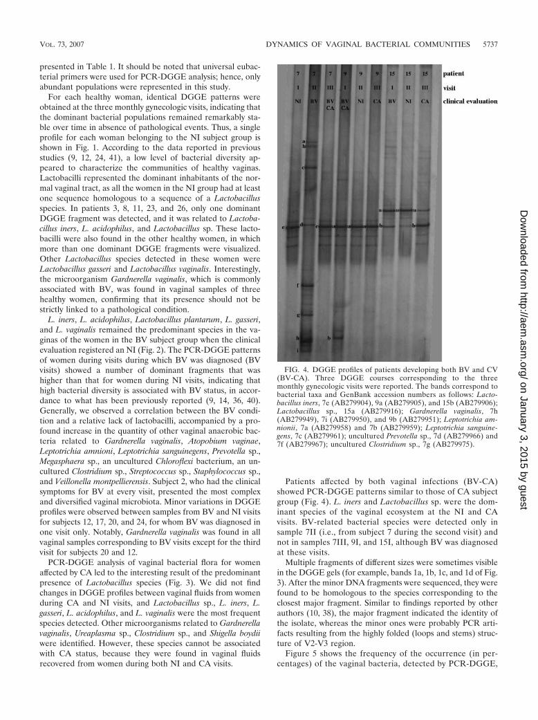
Patients affected by both vaginal infections (BV-CA)showed PCR-DGGE patterns similar to those of CA subjectgroup (Fig. 4). L. iners and Lactobacillus sp. were the dom-inant species of the vaginal ecosystem at the NI and CAvisits. BV-related bacterial species were detected only insample 7II (i.e., from subject 7 during the second visit) andnot in samples 7III, 9I, and 15I, although BV was diagnosedat these visits.
Multiple fragments of different sizes were sometimes visiblein the DGGE gels (for example, bands 1a, 1b, 1c, and 1d of Fig.3). After the minor DNA fragments were sequenced, they werefound to be homologous to the species corresponding to theclosest major fragment. Similar to findings reported by otherauthors (10, 38), the major fragment indicated the identity ofthe isolate, whereas the minor ones were probably PCR arti-facts resulting from the highly folded (loops and stems) struc-ture of V2-V3 region.
Figure 5 shows the frequency of the occurrence (in per-centages) of the vaginal bacteria, detected by PCR-DGGE,
FIG. 4. DGGE profiles of patients developing both BV and CV(BV-CA). Three DGGE courses corresponding to the threemonthly gynecologic visits were reported. The bands correspond tobacterial taxa and GenBank accession numbers as follows: Lacto-bacillus iners, 7e (AB279904), 9a (AB279905), and 15b (AB279906);Lactobacillus sp., 15a (AB279916); Gardnerella vaginalis, 7h(AB279949), 7i (AB279950), and 9b (AB279951); Leptotrichia am-nionii, 7a (AB279958) and 7b (AB279959); Leptotrichia sanguine-gens, 7c (AB279961); uncultured Prevotella sp., 7d (AB279966) and7f (AB279967); uncultured Clostridium sp., 7g (AB279975).
VOL. 73, 2007 DYNAMICS OF VAGINAL BACTERIAL COMMUNITIES 5737
on January 3, 2015 by guesthttp://aem
.asm.org/
Dow
nloaded from

in relation to the clinical status. Samples 7III and 9I werenot included in this analysis and in the subsequent statisticalevaluations because of the simultaneous presence of BV andCA conditions. The histograms indicate the increased com-plexity of vaginal ecology under BV conditions, character-ized by the appearance of several anaerobic species and thedecrease of L. vaginalis, L. gasseri, L. acidophilus, and Lac-tobacillus sp. The figure also shows that the composition ofthe vaginal bacterial flora did not undergo significantchanges as a consequence of Candida infection, even if adiverse distribution of Lactobacillus species appeared. Inparticular, we observed increased frequencies of L. iners andLactobacillus sp. and decreased frequencies of L. acidophi-lus, L. gasseri, and L. vaginalis. Notably, H2O2 is commonlyproduced by strains of L. acidophilus, L. gasseri, and L.vaginalis, while it is an uncommon metabolite among strainsof L. iners (5, 34, 41).
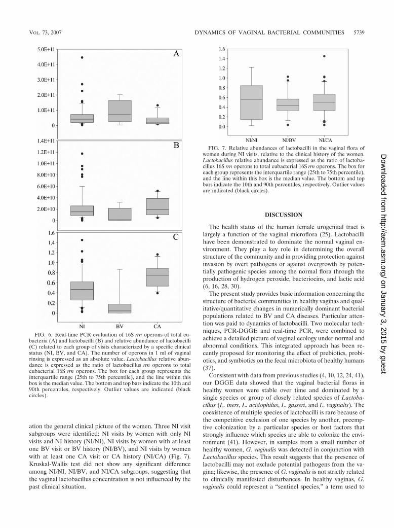
Quantification of vaginal lactobacilli by real-time PCR.Quantitative variations in vaginal lactobacillus populations re-lated to BV or CA were evaluated by real-time PCR analysis.For each vaginal fluid sample for each woman enrolled in thetrial, the numbers of 16S rrn operons of total eubacteria andlactobacilli were determined by using universal and genus-specific primers, respectively (Table 1). The reliability of thereal-time PCR data was supported by calibration curves withan r of 0.99 (data not shown) and acceptable SD. Relativeabundances of lactobacilli were calculated in order to assesswhether variations in lactobacillus populations were linked tochanges in the total concentration of bacteria (Table 1). Inaccordance with the PCR-DGGE results, no evident differ-ences in rRNA gene copy numbers of lactobacilli, as well as intheir relative abundances, were observed between vaginal flu-ids of women during CA and NI visits, while decreases in bothparameters occurred in cases in which BV was diagnosed.
However, in samples from subjects 12 and 17 during BV visits,the reduction in the relative abundance of lactobacilli did notreflect a decline in the corresponding rRNA genes. Notably,vaginal fluids collected at the third visit for subject 7, the firstvisit for subject 9, and the first visit for subject 15 (patientsbelonging to the BV-CA subject group) presented a high num-ber of Lactobacillus 16S rRNA gene copies and a high relativeabundance of lactobacilli in spite of the BV diagnosis. Theseresults are in agreement with the PCR-DGGE profiles, whichshowed the predominance of lactobacilli in the vaginal tracts ofthese patients. Therefore, the colonization patterns of lacto-bacilli in these women developing either BV or CA, or BV andCA at the same time, differ from the normal BV pattern, butthey rather resemble the typical pattern observed for womenaffected by CA.
To test the significance of quantitative changes in Lactoba-cillus populations related to BV and CA infections, the num-bers of 16S rRNA gene copies of total eubacteria and lacto-bacilli and the relative abundance of lactobacilli in samplesfrom women during NI, BV, or CA visits (plotted in Fig. 6)were analyzed by a Kruskal-Wallis test due to nonnormal datadistribution. The only significant difference among the groupswas found for relative abundances of lactobacilli (P � 0.001).Dunn’s procedure confirmed the existence of a significant dif-ference in relative abundances of lactobacilli (P � 0.05) foreach possible comparison (BV versus NI, CA versus NI, andCA versus BV). Figure 6C highlights the reduction of therelative abundance of lactobacilli in the vaginal flora of womenduring BV visits compared to those in the vaginal flora ofwomen during NI and CA visits and the increase in the sameparameter for women during CA visits compared to womenduring NI visits.
The relative abundance of lactobacilli in the vaginal flora ofwomen during NI visits was evaluated, taking into consider-
FIG. 5. Frequency of the occurrence of bacterial species in relation to clinical status of the subject. Frequencies were calculated as the ratios(percentage) of the number of visits characterized by the presence of each species to the total number of visits belonging to each visit group (NI,BV, and CA).
5738 VITALI ET AL. APPL. ENVIRON. MICROBIOL.
on January 3, 2015 by guesthttp://aem
.asm.org/
Dow
nloaded from
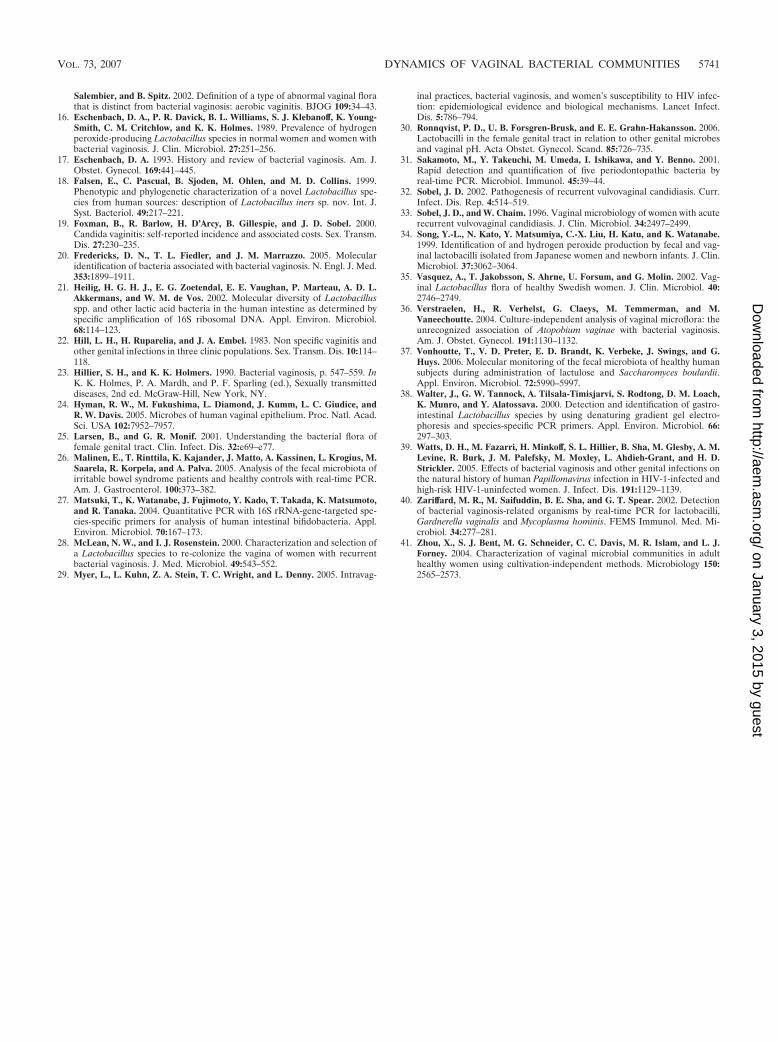
ation the general clinical picture of the women. Three NI visitsubgroups were identified: NI visits by women with only NIvisits and NI history (NI/NI), NI visits by women with at leastone BV visit or BV history (NI/BV), and NI visits by womenwith at least one CA visit or CA history (NI/CA) (Fig. 7).Kruskal-Wallis test did not show any significant differenceamong NI/NI, NI/BV, and NI/CA subgroups, suggesting thatthe vaginal lactobacillus concentration is not influenced by thepast clinical situation.
DISCUSSION
The health status of the human female urogenital tract islargely a function of the vaginal microflora (25). Lactobacillihave been demonstrated to dominate the normal vaginal en-vironment. They play a key role in determining the overallstructure of the community and in providing protection againstinvasion by overt pathogens or against overgrowth by poten-tially pathogenic species among the normal flora through theproduction of hydrogen peroxide, bacteriocins, and lactic acid(6, 16, 28, 30).
The present study provides basic information concerning thestructure of bacterial communities in healthy vaginas and qual-itative/quantitative changes in numerically dominant bacterialpopulations related to BV and CA diseases. Particular atten-tion was paid to dynamics of lactobacilli. Two molecular tech-niques, PCR-DGGE and real-time PCR, were combined toachieve a detailed picture of vaginal ecology under normal andabnormal conditions. This integrated approach has been re-cently proposed for monitoring the effect of prebiotics, probi-otics, and symbiotics on the fecal microbiota of healthy humans(37).
Consistent with data from previous studies (4, 10, 12, 24, 41),our DGGE data showed that the vaginal bacterial floras inhealthy women were stable over time and dominated by asingle species or group of closely related species of Lactoba-cillus (L. iners, L. acidophilus, L. gasseri, and L. vaginalis). Thecoexistence of multiple species of lactobacilli is rare because ofthe competitive exclusion of one species by another, preemp-tive colonization by a particular species or host factors thatstrongly influence which species are able to colonize the envi-ronment (41). However, in samples from a small number ofhealthy women, G. vaginalis was detected in conjunction withLactobacillus species. This result suggests that the presence oflactobacilli may not exclude potential pathogens from the va-gina; likewise, the presence of G. vaginalis is not strictly relatedto clinically manifested disturbances. In healthy vaginas, G.vaginalis could represent a “sentinel species,” a term used to
FIG. 6. Real-time PCR evaluation of 16S rrn operons of total eu-bacteria (A) and lactobacilli (B) and relative abundance of lactobacilli(C) related to each group of visits characterized by a specific clinicalstatus (NI, BV, and CA). The number of operons in 1 ml of vaginalrinsing is expressed as an absolute value. Lactobacillus relative abun-dance is expressed as the ratio of lactobacillus rrn operons to totaleubacterial 16S rrn operons. The box for each group represents theinterquartile range (25th to 75th percentile), and the line within thisbox is the median value. The bottom and top bars indicate the 10th and90th percentiles, respectively. Outlier values are indicated (blackcircles).
FIG. 7. Relative abundances of lactobacilli in the vaginal flora ofwomen during NI visits, relative to the clinical history of the women.Lactobacillus relative abundance is expressed as the ratio of lactoba-cillus 16S rrn operons to total eubacterial 16S rrn operons. The box foreach group represents the interquartile range (25th to 75th percentile),and the line within this box is the median value. The bottom and topbars indicate the 10th and 90th percentiles, respectively. Outlier valuesare indicated (black circles).
VOL. 73, 2007 DYNAMICS OF VAGINAL BACTERIAL COMMUNITIES 5739
on January 3, 2015 by guesthttp://aem
.asm.org/
Dow
nloaded from

refer to an indigenous species that is particularly sensitive tochanges in biological, physical, or chemical characteristics ofthe environment and responds through an increase in popula-tion size (12). The identification of potential sentinel speciescan help to predict whether there is an increased risk of anindividual contracting an infection due to shifts in the compo-sition of the microbial communities or changes in the abun-dance of specific populations. The sentinel species conceptmay be also used for prevention and early diagnosis of BVdisturbance, which is characterized by the outgrowth of severalpotentially pathogenic microorganisms normally present but atmuch lower numbers (9, 14, 20, 40). Actually, the patientsaffected by BV enrolled in this clinical trial showed a relativescarcity of lactobacilli, together with an increased complexityof vaginal communities dominated by anaerobic bacteria re-lated to Gardnerella vaginalis, Atopobium vaginae, Leptotrichiasp., Prevotella sp., Megasphaera sp., Chloroflexi, Clostridium sp.,Streptococcus sp., Staphylococcus sp., and Veillonella sp. Allthese organisms have already been identified as components ofthe abnormal vaginal flora under conditions of BV disturbance(9, 20), except for Chloroflexi, Streptococcus sp., and Veillonellasp., which represent newly recognized populations not previ-ously associated with BV. By contrast, we did not find anabnormal bacterial vaginal ecosystem in patients affected byCA, as the predominant presence of lactobacilli was observedwith our DGGE-based qualitative analysis. However, a differ-ent frequency of particular Lactobacillus species was found inrespect to the frequency detected under healthy conditions. Inparticular, we observed a decrease in H2O2-producing species(L. acidophilus, L. gasseri, and L. vaginalis) and an increase ofnon-H2O2-producing L. iners. This finding suggests some hy-potheses regarding the role of lactobacilli in protecting womenfrom Candida infection: (i) women with a vaginal flora domi-nated by H2O2-producing Lactobacillus species have a minorrisk to contract CA; (ii) H2O2, rather than other metabolitesproduced by lactobacilli, such as lactic acid, could be respon-sible for the control of Candida overgrowth (33). In patientsprone to developing CA as well as BV, the DGGE pattern ofCA-associated lactobacilli was dominant, while the DGGEpattern of BV-associated bacteria was not detected in thisgroup with mixed infections.
An important advantage of the DGGE technique is the abilityto detect several bacterial populations not readily culturable. Forexample, L. iners does not grow on the major selective media forisolation of Lactobacillus, including MRS and Rogosa media (18).Likewise, the strict anaerobes Atopobium vaginae, Megasphaera,and Leptotrichia require specialized media and often grow slowly.The finding of these organisms as members of vaginal flora dem-onstrates how cultivation-based methods can be misleading.
Since DGGE can be considered a semiquantitative tool formonitoring bacterial populations, additional analysis with real-time PCR was carried out to obtain a quantitative estimationof changes in lactobacillus concentrations associated with BVand CA diseases. An analogous approach was used by Bartoschet al. (6) to monitor changes in intestinal bacterial communitystructure among different groups of elderly people. A statisti-cally significant decrease in Lactobacillus relative abundanceduring BV visits was registered compared to the relative abun-dances during NI and CA visits, confirming the qualitativeobservations derived from DGGE analysis. Notably, the rela-
tive abundance of lactobacilli was significantly increased forwomen during CA visits, further supporting our hypothesisthat the predisposition to Candida infection is not correlatedwith the predominance of lactobacilli but to the presence ofparticular Lactobacillus species. We also investigated the pos-sibility of a link between the clinical history and the relativeabundance of lactobacilli in vaginal fluids of women during NIvisits, but no significant correlations were detected.
In conclusion, it has been demonstrated for the first timethat the integrated use of DGGE and real-time PCR repre-sents a successful strategy to monitor the qualitative and quan-titative changes of vaginal bacterial communities correlated todifferent infectious diseases, such as BV and CA. In particular,the major findings obtained in this study are the following: (i)Lactobacillus species are the major constituents in vagina ofhealthy women and patients developing CA but not in patientsdeveloping BV; (ii) new components of vaginal microbiota inBV have been discovered; (iii) women prone to developing CAmainly host non-H2O2-producing Lactobacillus species; and(iv) in patients developing a mixed BV and CA infection, thecolonization pattern of lactobacilli resembles the CA pattern.
REFERENCES
1. Altermann, E., W. M. Russell, M. A. Azcarate-Peril, R. Barrangou, B. L.Buck, O. McAuliffe, N. Souther, A. Dobson, T. Duong, M. Callanan, S. Lick,A. Hamrick, R. Cano, and T. R. Klaenhammer. 2005. Complete genomesequence of the probiotic lactic acid bacterium Lactobacillus acidophilusNCFM. Proc. Natl. Acad. Sci. USA 102:3906–3912.
2. Altschul, S. F., T. L. Madden, A. A. Schaffer, J. Zhang, Z. Zhang, V. Miller,and D. J. Lipman. 1997. Gapped BLAST and PSI-BLAST: a new generationof protein database search program. Nucleic Acids Res. 25:3389–3402.
3. Amsel, R., P. A. Totten, C. A. Spiegel, C. K. Chen, D. Eschenbach, and K. K.Holmes. 1983. Nonspecific vaginitis: diagnostic criteria and microbial andepidemiological associations. Am. J. Med. 74:14–22.
4. Antonio, M. A., S. E. Hawes, and S. L. Hillier. 1999. The identification ofvaginal Lactobacillus species and the demographic and microbiologic char-acteristics of women colonized by these species. J. Infect. Dis. 180:1950–1956.
5. Aroutcheva, A., D. Gariti, M. Simon, S. Shott, J. Faro, J. A. Simoes, A.Gurguis, and S. Faro. 2001. Defense factors of vaginal lactobacilli. Am. J.Obstet. Gynecol. 185:375–379.
6. Bartosch, S., F. Alemu, G. T. Macfarlane, and M. E. T. McMurdo. 2004.Characterization of bacterial communities in feces from healthy elderly vol-unteers and hospitalized patients by using real-time PCR and effects ofantibiotic treatment on the fecal microbiota. Appl. Environ. Microbiol. 70:3575–3581.
7. Bassam, B. J., G. Caetano-Anolles, and P. M. Gresshoff. 1991. Fast andsensitive silver staining of DNA in polyacrylamide gels. Anal. Biochem.196:80–83.
8. Beigi, R. H., H. C. Wiesenfeld, S. L. Hillier, T. Straw, and M. A. Krohn. 2005.Factors associated with absence of H2O2-producing Lactobacillus amongwomen with bacterial vaginosis. J. Infect. Dis. 191:924–929.
9. Burton, J. P., and G. Reid. 2002. Evaluation of the bacterial vaginal flora of20 postmenopausal women by direct (Nugent score) and molecular (poly-merase chain reaction and denaturing gradient gel electrophoresis) tech-niques. J. Infect. Dis. 186:1770–1780.
10. Burton, J. P., P. A. Cadieux, and G. Reid. 2003. Improved understanding ofthe bacterial vaginal microbiota of women before and after probiotic instil-lation. Appl. Environ. Microbiol. 69:97–101.
11. Convert, M., G. Martinetti Lucchini, M. Dolina, and J. C. Piffaretti. 2005.Comparison of LightCycler PCR and culture for detection of group B strep-tococci from vaginal swabs. Clin. Microbiol. Infect. 11:1022–1026.
12. Coolen, M. J. L., E. Post, C. C. Davis, and L. J. Forney. 2005. Character-ization of microbial communities found in the human vagina by analysis ofterminal restriction fragment length polymorphisms of 16S rRNA genes.Appl. Environ. Microbiol. 71:8729–8737.
13. Devillard, E., J. P. Burton, J. Hammon, D. Lam, and G. Reid. 2004. Novelinsight into the vaginal microflora in postmenopausal women under hor-mone replacement therapy as analyzed by PCR-denaturing gradient gelelectrophoresis. Eur. J. Obstet. Gynecol. Reprod. Biol. 117:76–81.
14. Devillard, E., J. P. Burton, and G. Reid. 2005. Complexity of vaginal micro-flora as analyzed by PCR denaturing gradient gel electrophoresis in a patientwith recurrent bacterial vaginosis. Infect. Dis. Obstet. Gynecol. 13:25–31.
15. Donders, G. G. G., A. Vereecken, E. Bosmans, A. Dekeersmaecker, G.
5740 VITALI ET AL. APPL. ENVIRON. MICROBIOL.
on January 3, 2015 by guesthttp://aem
.asm.org/
Dow
nloaded from
Salembier, and B. Spitz. 2002. Definition of a type of abnormal vaginal florathat is distinct from bacterial vaginosis: aerobic vaginitis. BJOG 109:34–43.
16. Eschenbach, D. A., P. R. Davick, B. L. Williams, S. J. Klebanoff, K. Young-Smith, C. M. Critchlow, and K. K. Holmes. 1989. Prevalence of hydrogenperoxide-producing Lactobacillus species in normal women and women withbacterial vaginosis. J. Clin. Microbiol. 27:251–256.
17. Eschenbach, D. A. 1993. History and review of bacterial vaginosis. Am. J.Obstet. Gynecol. 169:441–445.
18. Falsen, E., C. Pascual, B. Sjoden, M. Ohlen, and M. D. Collins. 1999.Phenotypic and phylogenetic characterization of a novel Lactobacillus spe-cies from human sources: description of Lactobacillus iners sp. nov. Int. J.Syst. Bacteriol. 49:217–221.
19. Foxman, B., R. Barlow, H. D’Arcy, B. Gillespie, and J. D. Sobel. 2000.Candida vaginitis: self-reported incidence and associated costs. Sex. Transm.Dis. 27:230–235.
20. Fredericks, D. N., T. L. Fiedler, and J. M. Marrazzo. 2005. Molecularidentification of bacteria associated with bacterial vaginosis. N. Engl. J. Med.353:1899–1911.
21. Heilig, H. G. H. J., E. G. Zoetendal, E. E. Vaughan, P. Marteau, A. D. L.Akkermans, and W. M. de Vos. 2002. Molecular diversity of Lactobacillusspp. and other lactic acid bacteria in the human intestine as determined byspecific amplification of 16S ribosomal DNA. Appl. Environ. Microbiol.68:114–123.
22. Hill, L. H., H. Ruparelia, and J. A. Embel. 1983. Non specific vaginitis andother genital infections in three clinic populations. Sex. Transm. Dis. 10:114–118.
23. Hillier, S. H., and K. K. Holmers. 1990. Bacterial vaginosis, p. 547–559. InK. K. Holmes, P. A. Mardh, and P. F. Sparling (ed.), Sexually transmitteddiseases, 2nd ed. McGraw-Hill, New York, NY.
24. Hyman, R. W., M. Fukushima, L. Diamond, J. Kumm, L. C. Giudice, andR. W. Davis. 2005. Microbes of human vaginal epithelium. Proc. Natl. Acad.Sci. USA 102:7952–7957.
25. Larsen, B., and G. R. Monif. 2001. Understanding the bacterial flora offemale genital tract. Clin. Infect. Dis. 32:e69–e77.
26. Malinen, E., T. Rinttila, K. Kajander, J. Matto, A. Kassinen, L. Krogius, M.Saarela, R. Korpela, and A. Palva. 2005. Analysis of the fecal microbiota ofirritable bowel syndrome patients and healthy controls with real-time PCR.Am. J. Gastroenterol. 100:373–382.
27. Matsuki, T., K. Watanabe, J. Fujimoto, Y. Kado, T. Takada, K. Matsumoto,and R. Tanaka. 2004. Quantitative PCR with 16S rRNA-gene-targeted spe-cies-specific primers for analysis of human intestinal bifidobacteria. Appl.Environ. Microbiol. 70:167–173.
28. McLean, N. W., and I. J. Rosenstein. 2000. Characterization and selection ofa Lactobacillus species to re-colonize the vagina of women with recurrentbacterial vaginosis. J. Med. Microbiol. 49:543–552.
29. Myer, L., L. Kuhn, Z. A. Stein, T. C. Wright, and L. Denny. 2005. Intravag-
inal practices, bacterial vaginosis, and women’s susceptibility to HIV infec-tion: epidemiological evidence and biological mechanisms. Lancet Infect.Dis. 5:786–794.
30. Ronnqvist, P. D., U. B. Forsgren-Brusk, and E. E. Grahn-Hakansson. 2006.Lactobacilli in the female genital tract in relation to other genital microbesand vaginal pH. Acta Obstet. Gynecol. Scand. 85:726–735.
31. Sakamoto, M., Y. Takeuchi, M. Umeda, I. Ishikawa, and Y. Benno. 2001.Rapid detection and quantification of five periodontopathic bacteria byreal-time PCR. Microbiol. Immunol. 45:39–44.
32. Sobel, J. D. 2002. Pathogenesis of recurrent vulvovaginal candidiasis. Curr.Infect. Dis. Rep. 4:514–519.
33. Sobel, J. D., and W. Chaim. 1996. Vaginal microbiology of women with acuterecurrent vulvovaginal candidiasis. J. Clin. Microbiol. 34:2497–2499.
34. Song, Y.-L., N. Kato, Y. Matsumiya, C.-X. Liu, H. Katu, and K. Watanabe.1999. Identification of and hydrogen peroxide production by fecal and vag-inal lactobacilli isolated from Japanese women and newborn infants. J. Clin.Microbiol. 37:3062–3064.
35. Vasquez, A., T. Jakobsson, S. Ahrne, U. Forsum, and G. Molin. 2002. Vag-inal Lactobacillus flora of healthy Swedish women. J. Clin. Microbiol. 40:2746–2749.
36. Verstraelen, H., R. Verhelst, G. Claeys, M. Temmerman, and M.Vaneechoutte. 2004. Culture-independent analysis of vaginal microflora: theunrecognized association of Atopobium vaginae with bacterial vaginosis.Am. J. Obstet. Gynecol. 191:1130–1132.
37. Vonhoutte, T., V. D. Preter, E. D. Brandt, K. Verbeke, J. Swings, and G.Huys. 2006. Molecular monitoring of the fecal microbiota of healthy humansubjects during administration of lactulose and Saccharomyces boulardii.Appl. Environ. Microbiol. 72:5990–5997.
38. Walter, J., G. W. Tannock, A. Tilsala-Timisjarvi, S. Rodtong, D. M. Loach,K. Munro, and Y. Alatossava. 2000. Detection and identification of gastro-intestinal Lactobacillus species by using denaturing gradient gel electro-phoresis and species-specific PCR primers. Appl. Environ. Microbiol. 66:297–303.
39. Watts, D. H., M. Fazarri, H. Minkoff, S. L. Hillier, B. Sha, M. Glesby, A. M.Levine, R. Burk, J. M. Palefsky, M. Moxley, L. Ahdieh-Grant, and H. D.Strickler. 2005. Effects of bacterial vaginosis and other genital infections onthe natural history of human Papillomavirus infection in HIV-1-infected andhigh-risk HIV-1-uninfected women. J. Infect. Dis. 191:1129–1139.
40. Zariffard, M. R., M. Saifuddin, B. E. Sha, and G. T. Spear. 2002. Detectionof bacterial vaginosis-related organisms by real-time PCR for lactobacilli,Gardnerella vaginalis and Mycoplasma hominis. FEMS Immunol. Med. Mi-crobiol. 34:277–281.
41. Zhou, X., S. J. Bent, M. G. Schneider, C. C. Davis, M. R. Islam, and L. J.Forney. 2004. Characterization of vaginal microbial communities in adulthealthy women using cultivation-independent methods. Microbiology 150:2565–2573.
VOL. 73, 2007 DYNAMICS OF VAGINAL BACTERIAL COMMUNITIES 5741
on January 3, 2015 by guesthttp://aem
.asm.org/
Dow
nloaded from
Copyright © 2022 FDOKUMEN