Cloning and regulation of the major histocompatibility class I alpha gene in the teleost fish...
9
Short sequence report Cloning and regulation of the major histocompatibility class I alpha gene in the teleost fish gilthead seabream Alberto Cuesta, Jose ´ Meseguer, Maria Angeles Esteban * Fish Innate Immune System Group, Department of Cell Biology, Faculty of Biology, University of Murcia, 30100 Murcia, Spain Received 31 May 2006; revised 28 July 2006; accepted 3 August 2006 Available online 16 August 2006 Keywords: MHC class I alpha; Antigen presentation; Up-regulation; Teleost fish; Gilthead seabream Major histocompatibility complex (MHC) genes encode key molecules in the immune response. Antigens properly processed are expressed in the membrane bound to either class I or class II MHC proteins and then recognized by lymphocytes through the cell-surface T-lymphocyte receptors (TCR). However, contact with the CD8 (in cytotoxic T lymphocytes) or CD4 (in helper T lymphocytes) co-receptors are necessary for triggering the adaptive immune response leading to pathogen elimination [1]. MHC genes have been cloned in several species, representing all the vertebrate groups. From an evolutionary point of view, the adaptive immune response appears in fish, and the presence of MHC and MHC-related genes has been evidenced in all species studied. In teleost or bony fish MHC genes are not clustered, unlike in cartilaginous fish and tetrapods, indicating differences in evolution [2,3]. Therefore, a study of the teleost fish MHC genes is essential for understanding the importance and significance of this genetic divergence; at the same time, the knowledge will add to the recent advances made in fish immunology. Since the early 1990s when fish MHC genes were first cloned, numerous sequences have been published in many bony fish species but mainly for evo- lutionary purposes [3,4]. Interestingly, little information exists regarding their regulation and function. Thus, further characterization of the fish MHC genes at different levels will be of great help in understanding the innateeadaptive immune system interactions. Class I MHC is a non-covalently bound heterodimer composed of an MHC class Ia chain (about 45 kDa) and b2- microglobulin (about 12 kDa). MHC class Ia is expressed on all the nucleated cells and generally presents endogenous antigens to cytotoxic T lymphocytes. The heavy chain, Ia, is encoded by a polymorphic gene containing a leader pep- tide, 3 extracellular domains (a1, a2 and a3) and transmembrane and cytoplasmic regions. However, recent findings point to the presence of soluble MHC class Ia proteins without a transmembrane region [5]. Genomic studies have demonstrated the variable copy numbers in the genome of teleost fish (from 3 to 17) as well as polymorphism in the peptide-binding region (PBR) within the a1 and a2 domains [4,6,7]. However, little information exists about MHC functioning in fish. At gene level, the expression of MHC class I (either Ia chain or b2-microglobulin) may be affected by physical factors (e.g. temperature), particulated antigens (viruses, bacteria, etc.) or endogenous soluble proteins (cytokines, growth factors, etc.) [8e10]. Moreover, the use of anti-fish MHC class Ia antibodies has revealed that its expression pattern is similar to that in mammals, including the immune system cells, and that it may be * Corresponding author. Tel.: þ34 968367665; fax: þ34 968363963. E-mail address: [email protected] (M. Angeles Esteban). 1050-4648/$ - see front matter Ó 2006 Elsevier Ltd. All rights reserved. doi:10.1016/j.fsi.2006.08.005 Fish & Shellfish Immunology 22 (2007) 718e726 www.elsevier.com/locate/fsi
Transcript of Cloning and regulation of the major histocompatibility class I alpha gene in the teleost fish...

Fish & Shellfish Immunology 22 (2007) 718e726www.elsevier.com/locate/fsi
Short sequence report
Cloning and regulation of the major histocompatibility class Ialpha gene in the teleost fish gilthead seabream
Alberto Cuesta, Jose Meseguer, Maria �Angeles Esteban*
Fish Innate Immune System Group, Department of Cell Biology, Faculty of Biology, University of Murcia, 30100 Murcia, Spain
Received 31 May 2006; revised 28 July 2006; accepted 3 August 2006
Available online 16 August 2006
Keywords: MHC class I alpha; Antigen presentation; Up-regulation; Teleost fish; Gilthead seabream
Major histocompatibility complex (MHC) genes encode key molecules in the immune response. Antigens properlyprocessed are expressed in the membrane bound to either class I or class II MHC proteins and then recognized bylymphocytes through the cell-surface T-lymphocyte receptors (TCR). However, contact with the CD8 (in cytotoxicT lymphocytes) or CD4 (in helper T lymphocytes) co-receptors are necessary for triggering the adaptive immuneresponse leading to pathogen elimination [1]. MHC genes have been cloned in several species, representing all thevertebrate groups. From an evolutionary point of view, the adaptive immune response appears in fish, and the presenceof MHC and MHC-related genes has been evidenced in all species studied. In teleost or bony fish MHC genes are notclustered, unlike in cartilaginous fish and tetrapods, indicating differences in evolution [2,3]. Therefore, a study of theteleost fish MHC genes is essential for understanding the importance and significance of this genetic divergence; at thesame time, the knowledge will add to the recent advances made in fish immunology. Since the early 1990s when fishMHC genes were first cloned, numerous sequences have been published in many bony fish species but mainly for evo-lutionary purposes [3,4]. Interestingly, little information exists regarding their regulation and function. Thus, furthercharacterization of the fish MHC genes at different levels will be of great help in understanding the innateeadaptiveimmune system interactions.
Class I MHC is a non-covalently bound heterodimer composed of an MHC class Ia chain (about 45 kDa) and b2-microglobulin (about 12 kDa). MHC class Ia is expressed on all the nucleated cells and generally presents endogenousantigens to cytotoxic T lymphocytes. The heavy chain, Ia, is encoded by a polymorphic gene containing a leader pep-tide, 3 extracellular domains (a1, a2 and a3) and transmembrane and cytoplasmic regions. However, recent findingspoint to the presence of soluble MHC class Ia proteins without a transmembrane region [5]. Genomic studies havedemonstrated the variable copy numbers in the genome of teleost fish (from 3 to 17) as well as polymorphism inthe peptide-binding region (PBR) within the a1 and a2 domains [4,6,7]. However, little information exists aboutMHC functioning in fish. At gene level, the expression of MHC class I (either Ia chain or b2-microglobulin) maybe affected by physical factors (e.g. temperature), particulated antigens (viruses, bacteria, etc.) or endogenous solubleproteins (cytokines, growth factors, etc.) [8e10]. Moreover, the use of anti-fish MHC class Ia antibodies has revealedthat its expression pattern is similar to that in mammals, including the immune system cells, and that it may be
* Corresponding author. Tel.: þ34 968367665; fax: þ34 968363963.
E-mail address: [email protected] (M. �Angeles Esteban).
1050-4648/$ - see front matter � 2006 Elsevier Ltd. All rights reserved.
doi:10.1016/j.fsi.2006.08.005
719A. Cuesta et al. / Fish & Shellfish Immunology 22 (2007) 718e726
regulated in different fish species [6,11e13]. However, fish MHC class I needs to be studied in greater depth before wecan fully understand its role in the fish immune response. Therefore, we aimed herein to clone the MHC class I alphachain in the Mediterranean teleost gilthead seabream (Sparus aurata L.) and study its regulation.
Specimens (125 g bw) of the hermaphroditic protandrous seawater teleost gilthead seabream (S. aurata L.)(CULMAREX S.A., Murcia, Spain) were kept in running seawater aquaria at 20� 2 �C and fed daily (1% biomass).This study was approved by the Bioethical Committee of the University of Murcia. Brain, liver, gut, gills, head-kidney(HK), spleen, thymus and testis were obtained by dissection and immediately frozen in TRIzol Reagent (Gibco). Leu-cocytes from peripheral blood (PBLs), peritoneal exudate (PE) and HK were isolated and adjusted in sRPMI [RPMI-1640 (Gibco) culture medium containing 2% FCS (Gibco) and 0.35% NaCl]. Isolated leucocytes and cultured tumorcells (seabream fin, SAF-1, ECACC-00122301; mouse lymphoma, L-1210, ATCC CCL-219) were pelleted and alsoresuspended in TRIzol reagent. HK leucocytes from 3 different fish (107 per fish) were treated in vitro with medium(controls), heat-killed Vibrio anguillarum R-82 (5 bacterial cells per leucocyte) (pathogenic for seabream), heat-killedSaccharomyces cerevisiae S288C (2 yeast cells per leucocyte), concanavalin A (ConA; 5 mg/ml; Sigma), ConAþ LPS(lipopolysaccharide; 5 mg/ml and 10 mg/ml, respectively; Sigma), phytohemagglutinin (PHA; 10 mg/ml; Sigma), son-icated and RNase treated SAF-1 or L-1210 tumor cells (10 leucocytes per tumor cell), CpG ODN (50-TCCAT-GACGTTCCTGATGCT-30; 50 mg/ml; Eurogentec) or poly I:C (25 mg/ml; Sigma). The mixed leucocyte reaction(MLR) was carried out by co-incubation of leucocytes from 3 specimens (107 leucocytes of each fish) in triplicate.After 4 h of incubation, leucocytes from the replicas were washed, pooled and resuspended in TRIzol reagent.
Total RNA was isolated from TRIzol reagent frozen samples following the manufacturer’s instructions. First strandcDNAwas synthesized by reverse transcription of 1e5 mg of total RNA using the ThermoScript� RNAse H� ReverseTranscriptase (Invitrogen) with an oligo-dT18 primer (Invitrogen). Spleen cDNA was used in a first PCR amplificationwith degenerate primers (Table 1) designed against known fish MHC class I alpha sequences. PCR reactions werecarried out using Taq polymerase (Invitrogen) and the amplification was performed in a MasterCycler GradientPCR: 95 �C for 5 min, 35 cycles of 95 �C for 45 s, 55 �C for 45 s, 72 �C for 45 s, and followed by 72 �C for10 min. PCR products were separated on a 1% agarose gel containing 0.5 mg/ml ethidium bromide and visualisedunder UV light. PCR fragments were purified from the gel and ligated into the pGEM-T Easy vector (Promega).Following transfection into competent Escherichia coli DH-5a cells, recombinants were identified through blue-whitecolor selection in ampicillin Luria Broth Base plates, containing 40 mg/ml of X-Gal. Isolated plasmid DNA wassequenced using an ABI PRISM 377 sequencer. Based on the first partial sequence of seabream MHC class I alpha,specific primers were then designed (Table 1) and used to obtain the 30 and 50 ends by rapid amplification of cDNAends (RACE). For the 50RACE, an oligo-dT-synthesized spleen cDNA was treated with E. coli RNase H (Gibco),purified with the Concert Rapid PCR Purification System (Gibco) and finally tailed with poly(A) at the 50 end usingterminal deoxynucleotidyl transferase (TdT; Gibco). To obtain both ends, this cDNA was used as template in RT-PCR
Table 1
Oligonucleotide primers used in the study
Name Nucleotide sequence (50 / 30) Position (bp)
MHC-F1 ACTCBHTGAAGTWTTTCTDCAC 64
MHC-F2 GWGGATGWAYGGMTGTGAGTGG 341
MHC-R1 TCTGGAGGAGAGACACYGAKGG 632
MHC-R2 TBABACCWGAGAGMTGAAACAC 856
SpauCIAF5 TGGACCACGGAGAGATCCTCC 730
SpauCIAF7 ACGGAGGATATTCAGACTGTCTGTG �75
SpauCIAF9 TGGAGGAAAGATGGAGAGGAGC 696
SpauCIF11 ATGAGGTTCTTGGTGTTTCTGG 1
SpauCIAR5 TGGAGCGATCCATGTCTCTGC 462
SpauCIAR8 ACAGCATCCATTGTTCCAGTGC 1169
SpauCIAR9 TCCAGTTTGGTTGAAGCGTGG 324
b-ActinF ATCGTGGGGCGCCCCAGGCACC
b-ActinR CTCCTTAATGTCACGCACGATTTC
RACET GGCCACGCGTCGACTAGTAC(T)16
RACE GGCCACGCGTCGACTAGTAC
Degeneracy: B¼C, G or T; H¼A, C or T; W¼A or T; D¼A, C or T; Y¼C or T; M¼A or C; K¼G or T.

720 A. Cuesta et al. / Fish & Shellfish Immunology 22 (2007) 718e726
reactions with RACET or RACE universal primers and specific primers (Table 1). All the sequences were analysed forsimilarity with other known sequences using the BLAST program [14] within the ExPASy Molecular Biology server(http://us.expasy.org), which was also used to predict the protein structure. Multiple sequence alignments were carriedout using the CLUSTALW program [15], within the European Bioinformatics Institute. Phylogenetic and molecularevolutionary analyses were conducted using MEGA version 2.1 [16]. A phylogenetic tree was constructed using thea3 nucleotide sequences of known MHC genes.
For expression studies, 1 mg of total RNA obtained from brain, liver, gut, gills, HK, spleen, thymus and testis,PBLs, PE leucocytes, SAF-1 as well as from treated HK leucocytes was retrotranscribed into cDNA and the 10-fold diluted samples were tested for Spau-UAA mRNA expression. PCR reactions were carried out, using 1 ml of tem-plate, with SpauCIAF11 and SpauCIAR5 primers as follows: 95 �C for 2 min, 30 cycles of 95 �C for 45 s, 56 �C for45 s, 72 �C for 45 s, and finally 72 �C for 10 min. As controls, cDNA of the constitutively expressed b-actin gene wasamplified (26 cycles) using specific primers (Table 1). PCR products were separated on a 1% agarose gel containing0.5 mg/ml ethidium bromide. Photographs were taken with the GL 100 Imaging system (Kodak) and band intensitywas measured by the 1D Image Analysis Software v3.6 (Kodak). Band intensity was measured and normalized tothat of b-actin.
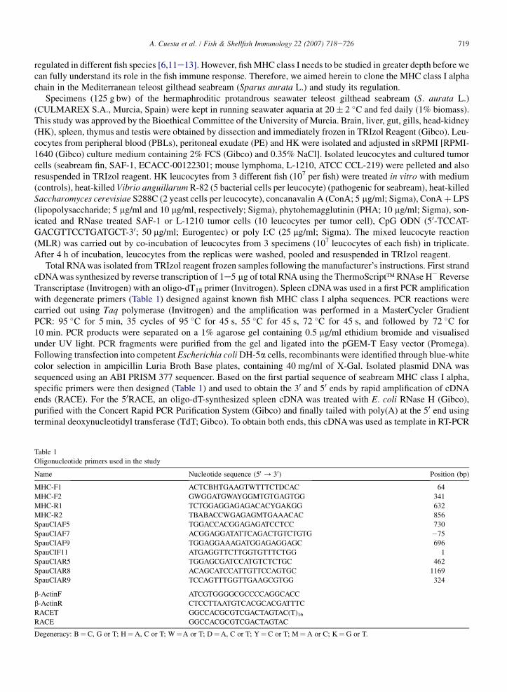
BLAST of the first sequence fragments obtained with degenerate primers were seen to be true gilthead seabreamMHC class I alpha. By means of RACE and specific primers we obtained the complete ORF of 2 full-length cDNAalleles of the Spau-UAA gene, called Spau-UAA*01 (Fig. 1) (GenBank accession number: DQ211540), and Spau-UAA*02 (GenBank accession number: DQ211541), according to the proposed nomenclature [17]. Both alleles hada 96 bp of 50-UTR and a 459 bp of 30-UTR and a coding sequence of 1125 bp codifying for a putative protein of374 amino acids (Figs. 1 and 2). The predicted ORF contains a putative 57 bp leader peptide, a 267 bp a1 domain,a 279 bp a2 domain, a 309 bp a3 domain and 210 bp of CP/TM/CYT region which agrees with the length of otherpublished sequences for teleosts. The 30-UTR region contains the canonical polyadenylation signal. Further analysisled us to construct a phylogenetic tree (Fig. 3) using the nucleotide sequences of the a3 region (software MEGA) [16].The neighbor-joining tree constructed showed 2 main branches in which all the teleost fish clustered together, whilethe cluster with elasmobranches and human MHC class I appeared in the other branch. Moreover, both seabreamalleles cluster tightly and in the same branch as other teleosts belonging to the superorders Acanthopterygii, Paracan-thopterygii and Protacanthopterygii. In the other branch of the tree, however, and evolving earlier are the teleosts ofthe superorder Ostariophysii.
At protein level, the predicted proteins for the 2 alleles have 374 amino acids (Figs. 1 and 2) divided into a leaderpeptide of 19 residues, 3 extracellular domains (a1 with 89, a2 with 93 and a3 with 103 amino acids) and a 70 residuesconnective peptide/transmembrane/cytoplasmic (CP/TM/CYT) region. Protein sequences were aligned to other ver-tebrate MHC class I alpha proteins (Fig. 2) and showed all the features expected for an MHC class Ia chain. That is,the presence of cysteines involved in the a2 and a3 intra-domain disulphide bridge (C119, C183, C219 and C278),a potential N-glycosylation site (NQT, position 105e107) and conserved residues important for contact (peptide,b2-microglobulin or CD8). Based on the alignment (Fig. 2), the cysteines and the N-glycosylation site are presentand the positions completely conserved in all the vertebrates analysed so far. The important residues for contactingthe peptide and b2-microglobulin are fairly well conserved in all fish while the similarity in the CD8 contact regionis lower.
Regarding the gene expression, Spau-UAA transcript was detected in all the assayed tissues, isolated leucocytes andalso in the seabream tumor cell line SAF-1 (Fig. 4). Afterwards, we evaluated the possible regulation of the geneexpression in isolated head-kidney leucocytes. Incubation with the non-pathogenic yeast S. cerevisiae (band intensity0.6) or with CpG ODNs (band intensity 0.85) decreased the expression of the seabream MHC class Ia with respect tocontrol leucocytes (band intensity 1). On the other hand, MLR or incubation of HK leucocytes with the seabream path-ogenic bacterium V. anguillarum, tumor cells (SAF-1 or L-1210), mitogens (ConA, LPS or PHA) or poly I:C led to anup-regulation of the Spau-UAA mRNA expression.
Teleost fish are the first vertebrate group to show a complete immune system with innate and adaptive responseslargely homologous with the mammalian immune system. Apart from these similarities, the presence of the MHC andof different lymphocyte subsets has been demonstrated in fish by functional data including antibody production, acuteallograft rejection, delayed hypersensitivity and mixed leucocyte reactions [18e20]. Cloning strategies have identi-fied, moreover, the presence of immune-related genes encoding IgM, TCR, CD3, CD4, CD8, Rag, MHC class I, b2-microglobulin, MHC class II, MHC class I/II loading pathways, cytokines or chemokines [3,19e22]. To date,

721A. Cuesta et al. / Fish & Shellfish Immunology 22 (2007) 718e726
unfortunately, the scarcity of fish cell lines and proper antibodies still hampers progress in fish immunology, especiallyas regards how leucocytes function and interact.
In this study we have evaluated the regulation of the Mediterranean gilthead seabream MHC class I alpha gene.Firstly, we have cloned the seabream MHC class I alpha gene, named Spau-UAA following the accepted nomenclature[17]. By means of RT-PCR and RACE we have obtained 2 alleles of the Spau-UAA gene. They consisted of a codingsequence of 1125 bp encoding a putative protein of 374 amino acids and showing high similarities with publishedMHC class Ia sequences. Alignment with other known protein sequences revealed that predicted Spau-UAA haveconserved the main residues important for defining bona fide MHC class Ia proteins. The 2 cloned seabream MHCclass Ia genes are polymorphic in the a1 and a2 domains but are very well conserved in the a3 and CP/TM/CYTregions. Apart from the cysteines involved in intra-domain disulphide bridges, residues contacting with the antigen
Fig. 1. Full-length sequence of the gilthead seabream Spau-UAA*01 allele. The start and stop codons, and the putative polyadenylation signal in
the 30-UTR, are marked in bold. GenBank accession number is DQ211540.

722 A. Cuesta et al. / Fish & Shellfish Immunology 22 (2007) 718e726
Fig. 2. Amino acid sequence comparison between most of the known vertebrate MHC class I alpha. Alignment is based on the Spau-UAA*01
sequence. Identities are indicated by dots. Dashes denote gaps introduced to maximize alignments. Symbols indicate conserved residues important
for peptide binding (PBR; *), lymphocyte CD8 (#) and b2-microglobulin ($) contact. Potential N-linked glycosylation sites are in bold. Sequence
accession numbers are as follows: Spau-UAA*01 (Sparus aurata, DQ211540), Spau-UAA*02 (DQ211541), Stvi (Stizostedion vitreum,
AAL11412), Paol (Paralichthys olivaceus, BAD13368), Auha (Aulonocara hansbaenschi, AAD37812), Orla (Oryzias latipes, BAB83849),
Taru (Takifugu rubripes, AAC41238), Gamo (Gadus morhua, AAL14532), Pore (Poecilia reticulata, CAA90791), Sasa (Salmo salar,
AAN75116), Ongo (Oncorhynchus gorbuscha, BAA09553), Onmy (Oncorhynchus mykiss, AAG25199), Cyca (Cyprinus carpio, CAA62497),
Ctid (Ctenopharyngodon idella, AAS76087), Dare (Danio rerio, NP_ 956879), Icpu (Ictalurus punctatus, AAG29241), Bain (Barbus intermedius,
CAD44955), Trsc (Triakis scyllium, AAB97332), Gici (Ginglymostoma cirratum, AAC60347) and HLA-B (Homo sapiens, CAC15502).

723A. Cuesta et al. / Fish & Shellfish Immunology 22 (2007) 718e726
and b2-microglobulin are fairly well conserved in all the MHC class Ia proteins while the contact site with CD8 hasless similarity. Allele sequence similarity suggests that the alleles come from the same gene but further studies areneeded. In the cytoplasmic tail, they contain potentially phosphorylable serines needed to be functional, althoughthe position is not conserved [5,11]. Based on this alignment, we constructed a phylogenetic tree with the a3cDNA sequences and using the software MEGA [16]. One of the branches contains the vertebrates with clusteredMHC genes, for example HLA and elasmobranch fish. In the other branch, all the teleost fish appear clustered, indi-cating that they evolved from a common ancestor but with many chromosome deletions and duplications affecting theMHC and MHC-related genes [23e25]. Within the bony fish, the tree shows the proposed evolutionary lineage forfish. Thus, the older fish belonging to the superorder Ostariophysii (Cypriniformes and Siluriformes) appears ina branch while in the other one, the Acanthopterygii, Paracanthopterygii and Protacanthopterygii fish appear jointlyclustered. Seabream, within the order Perciformes, seems to have evolved later in the teleost lineages.
Although evolutionary aspects of the MHC have been described in depth, study of the MHC tissue or cell distri-bution and gene/protein regulation is in its infancy. We have found that Spau-UAA gene is expressed in all the studied
Spau-UAA*01
Spau-UAA*02
Stvi
Paol
Auha
Orla
Pore
Taru
Gamo
Sasa
Ongo
Onmy
Icpu
Dare
Cyca
Ctid
Bain
HLA
Trsc
Gici
100
100
85
100
100
100
100
100
100
9181
97
100
100
63
51
78
0.05
Fig. 3. Neighbor-joining tree constructed with the nucleotide sequences of the a3 region of known MHC class I alpha. Genetic distances were
calculated based on nucleotide differences (p-distance) with complete deletion of gaps. The number at each node indicates the percentage of
bootstrapping after 1000 replications. Sequences are: Spau, DQ211540 and DQ211541; Stvi, AY057458; Paol, AB126918; Auha, AF038549;
Orla, BA000027; Taru, AF001217; Gamo, AF414205; Pore, PRUAA30; Sasa, AF504022; Ongo, ONHO92HH; Onmy, AF287485; Cyca,
CCUA1MRN; Ctid, AY391782; Dare, NM 200585; Icpu, AY008848; Bain, BIN506999; Trsc, AF034331; Gici, AF028557; and HLA,
HSA300181.

724 A. Cuesta et al. / Fish & Shellfish Immunology 22 (2007) 718e726
tissues, isolated leucocytes and tumor cells, as described in other fish and mammals [3,5,9,12]. Moreover, down- orup-regulation of fish MHC gene expression, either MHC class I or II, has been well documented in vitro or in vivo afterexposure to yeasts, bacteria, viruses, parasites, mitogens and cytokines [9,10,26e34]. Data seem to confirm that infish, as in mammals, the regulation of the MHC is at the transcription level, while the cell-surface MHC protein closelyresembles mRNA levels [35,36]. Furthermore, although MHC class I is known to present endogenous antigens, theycan also do so with exogenous antigens [3]. In this study, we exposed HK leucocytes to mixed leucocyte reaction,particulate antigens or mitogens. Our results demonstrate the down-regulation in head-kidney leucocytes incubatedwith CpG ODNs and yeast cells but up-regulation in MLR and when incubated with V. anguillarum, tumor cells,and mitogens. The use of CpG ODNs has shown potent immunomodulatory functions [34] but none has driven theregulation of MHC class I gene expression. Moreover, some ODNs have up-regulated the expression of MHC classII genes while others have shown down-regulatory properties. Although both the yeast and the bacterium used hereinare phagocytable particles by monocyteemacrophages and acidophilic granulocytes, the differences in the Spau-UAAgene expression could reside in the great pathogenic potential of V. anguillarum. On the other hand, tumor cells (SAF-1 and L-1210) susceptible to gilthead seabream HK non-specific cytotoxicity cells also up-regulates the MHC class Iaexpression. Finally, the use of poly I:C and MLR would be useful to understand the immune response because of mim-icking virus infections and allografts, respectively. In this sense, fish MHC class I expression has been seen to beaffected by particulated antigens, such as bacteria [10] viruses [9] or parasites [32]. Strikingly, mitogens (ConA,LPS and PHA) and poly I:C up-regulated the expression of Spau-UAA gene of the head-kidney leucocytes but failedto do the same with the expression of seabream MHC class IIa [33]. Unfortunately, little information exists about thisaspect in fish. For example, bacterial LPS has been used to modulate the MHC class I expression in fish tumor celllines [9,26]. However, they speculate about a possible indirect effect through liberation of the TNFa and IFNg cyto-kines, as happens in mammals [35]. However, this hypothesis might be contradictory, taking into account that
Fig. 4. Spau-UAA mRNA expression in various tissues, isolated leucocytes and tumor cells from gilthead seabream. RT-PCR was performed using
the specific primers SpauCIAF11and SpauCIAR5 and amplified for 30 cycles. b-actin gene was amplified for 26 cycles as internal control. (A)
Constitutive expression of Spau-UAA. B, brain; L, liver; G, gut; Gi, gill; HK, head-kidney; S, spleen; T, thymus; PELs, peritoneal exudate
leucocytes; PBLs, peripheral blood leucocytes; SAF-1, fin seabream tumor cells; Te, testis; NT, no template. (B) Expression of Spau-UAA in
HK leucocytes stimulated in vitro for 4 h. C, control; Sc, Saccharomyces cerevisiae S288C, Va, Vibrio anguillarum R-82; ConA, concanavalin
A; LPS, lipopolysaccharide; PHA, phytohemagglutinin; MLR, mixed leucocyte reaction. Band intensities were normalized to that of the b-actinand then the value of the control was taken as 1.

725A. Cuesta et al. / Fish & Shellfish Immunology 22 (2007) 718e726
seabream TNFa gene expression is not modulated by LPS [37]. Concomitant to MHC, studies related to the ontogenyand function of T cytotoxic cells and, concretely, of CD8þ T lymphocytes in fish are still lacking [19,25].
To conclude, we have cloned the gilthead seabream MHC class I alpha gene (Spau-UAA), which possesses all thecharacteristics and conserved residues to be considered a true MHC class I alpha gene. It is expressed in all the seab-ream tissues and also in seabream tumor cells as occurs in mammals. Finally, Spau-UAA expression in head-kidneyleucocytes is down-regulated by yeast cells and CpG ODNs and up-regulated in MLR and by exposure to pathogenicbacteria, tumor cells, poly I:C and mitogens (ConA, PHA or ConAþ LPS).
Acknowledgements
This work was funded by grants from the European Commission (QLK2-CT2002-00722 and SSP8-CT2003-501984). A. Cuesta has a postdoctoral fellowship from Fundacion CajaMurcia. The authors wish to thank Dr. E.Chaves-Pozo for providing the testis cDNA sample and Drs. A.E. Toranzo and J.L. Barja for the bacterium V.anguillarum.
References
[1] Klein J. The natural history of the major histocompatibility complex. New York: Willey; 1986.
[2] Sato A, Figueroa F, Murray BW. Nonlinkage group of major histocompatibility complex class I and class II loci in bony fishes. Immuno-
genetics 2000;51:108e16.
[3] Dixon B, Stet RJM. The relationship between major histocompatibility receptors and innate immunity in teleostean fish. Developmental and
Comparative Immunology 2001;25:683e99.
[4] Grimholt Y, Lie O. The major histocompatibility complex in fish. Revue Scientifique et Technique 1998;17:121e7.
[5] Fujiki K, Booman M, Chin-Dixon E, Dixon B. Cloning and characterization of cDNA clones encoding membrane-bound and potentially
secreted major histocompatibility class I receptors from walleye (Stizostedion vitreum). Immunogenetics 2001;53:760e9.
[6] Antao AB, Chinchar VG, McConnell TJ, Miller NW, Clem LW, Wilson MR. MHC class I genes of the channel catfish: sequence analysis and
expression. Immunogenetics 1999;49:303e11.
[7] Persson AC, Stet RJM, Pilstrom L. Characterisation of MHC class I and beta-2-microglobulin sequences in Atlantic cod revealing an
unusually high number of expressed class I genes. Immunogenetics 1999;50:49e59.
[8] Rodrigues PN, Dixon B, Roelofs J, Rombout JH, Egberts E, Pohajdak B, et al. Expression and temperature-dependent regulation of the
beta2-microglobulin (Cyca-B2m) gene in a cold-blooded vertebrate, the common carp (Cyprinus carpio L.). Developmental Immunology
1998;5:263e75.
[9] Koppang EO, Dannevig BH, Lie Ø, Rønningen K, Press CML. Expression of MHC class I and II mRNA in a macrophage-like cell line
(SHK-1) derived from Atlantic salmon, Salmo salar L., head kidney. Fish & Shellfish Immunology 1999;9:473e89.
[10] Park KC, Osborne JA, Tsoi SC, Brown LL, Johnson SC. Expressed sequence tags analysis of Atlantic halibut (Hippoglossus hippoglossus)
liver, kidney and spleen tissues following vaccination against Vibrio anguillarum and Aeromonas salmonicida. Fish & Shellfish Immunology
2005;18:393e415.
[11] van Erp SHM, Dixon B, Figueroa F, Egberts E, Stet RJM. Identification and characterization of a novel MHC class I gene from carp
(Cyprinus carpio L.). Immunogenetics 1996;44:49e61.
[12] Dijkstra JM, Kollnr B, Aoyagi K, Sawamoto Y, Kuroda A, Ototake M, et al. The rainbow trout classical MHC class I molecule Onmy-
UBA*1 is expressed in similar cell types as mammalian classical MHC class I molecules. Fish & Shellfish Immunology 2003;14:1e23.
[13] Fischer U, Dijkstra JM, Kollner B, Kiryu I, Koppang EO, Hordvik I, et al. The ontogeny of MHC class I expression in rainbow trout (Onco-
rhynchus mykiss). Fish & Shellfish Immunology 2005;18:49e60.
[14] Altschul SF, Gish W, Miller W, Myers E, Lipman DJ. Basic local alignment search tool. Journal of Molecular Biology 1990;215:403e10.
[15] Thompson JD, Higgins DJ, Gibson TJ, Clustal W. Improving the sensitivity of progressive multiple sequence alignment through sequence
weighting, position-specific gap penalties and weight matrix choice. Nucleic Acid Research 1994;22:4673e80.
[16] Kumar S, Tamura K, Jakobsen IB, Nei M. MEGA2: molecular evolutionary genetics analysis software. Bioinformatics 2001;17:1244e5.
[17] Klein J, Bontrop RE, Dawkins RL, Erlich HA, Gyllensten UB, Heise ER, et al. Nomenclature of the major histocompatibility complexes of
different species: a proposal. Immunogenetics 1990;31:217e9.
[18] Manning MJ. Biology of farmed fish. Sheffield: Sheffield Academic Press; 1998. p. 180e221.
[19] Nakanishi T, Fischer U, Dijkstra JM, Hasegawa S, Somamoto T, Okamoto N, et al. Cytotoxic T cell function in fish. Developmental and
Comparative Immunology 2002;26:131e9.
[20] Fischer U, Utke K, Somamoto T, Kollner B, Ototake M, Nakanishi T. Cytotoxic activities of fish leucocytes. Fish & Shellfish Immunology
2006;20:209e26.
[21] Dijkstra JM, Somamoto T, Moore L, Hordvik I, Ototake M, Fischer U. Identification and characterization of a second CD4-like gene in
teleost fish. Molecular Immunology 2006;43:410e9.
[22] Moore LJ, Somamoto T, Lie KK, Dijkstra JM, Hordvik I. Characterisation of salmon and trout CD8a and CD8b. Molecular Immunology
2005;42:1225e34.

726 A. Cuesta et al. / Fish & Shellfish Immunology 22 (2007) 718e726
[23] Shum BP, Guethlein L, Flodin LR, Adkison MA, Hedrick RP, Nehring RB, et al. Modes of salmonid MHC class I and II evolution differ
from the primate paradigm. Journal of Immunology 2001;166:3297e308.
[24] Miller KM, Kaukinen KH, Schulze AD. Expansion and contraction of major histocompatibility complex genes: a teleostean example.
Immunogenetics 2002;53:941e63.
[25] Hansen JD, Strassburger P, Thorgaard GH, Young WP, Du Pasquier L. Expression, linkage, and polymorphism of MHC-related genes in
rainbow trout, Oncorhynchus mykiss. Journal of Immunology 1999;163:774e86.
[26] Knight J, Stet RJ, Secombes CJ. Modulation of MHC class II expression in rainbow trout Oncorhynchus mykiss macrophages by TNFa and
LPS. Fish & Shellfish Immunology 1998;8:545e53.
[27] Koppang EO, Lundin M, Press CML, Rønningen K, Lie Ø. Differing levels of MHC class II b chain expression in a range of tissues from
vaccinated and non-vaccinated Atlantic salmon (Salmo salar L.). Fish & Shellfish Immunology 1998;8:183e96.
[28] Brubacher JL, Secombes CJ, Zou J, Bols NC. Constitutive and LPS-induced gene expression in a macrophage-like cell line from the rainbow
trout (Oncorhynchus mykiss). Developmental and Comparative Immunology 2000;24:565e74.
[29] Saeij JP, de Vries BJ, Wiegertjes GF. The immune response of carp to Trypanoplasma borreli: kinetics of immune gene expression and
polyclonal lymphocyte activation. Developmental and Comparative Immunology 2003;27:859e74.
[30] Lindenstrøm T, Secombes CJ, Buchmann K. Expression of immune response genes in rainbow trout skin induced by Gyrodactylus derjavini
infections. Veterinary Immunology and Immunopathology 2004;97:137e48.
[31] Takano T, Iwahori A, Hirono I, Aoki T. Development of a DNA vaccine against hirame rhabdovirus and analysis of the expression of
immune-related genes after vaccination. Fish & Shellfish Immunology 2004;17:367e74.
[32] Chang MX, Nie P, Liu GY, Song Y, Gao Q. Identification of immune genes in grass carp Ctenopharyngodon idella in response to infection of
the parasitic copepod Sinergasilus major. Parasitology Research 2005;96:224e9.
[33] Cuesta A, Esteban MA, Meseguer J. Granulocytes as antigen-presenting cells. Cloning, distribution and up-regulation of the teleost fish
MHC class II alpha. Molecular Immunology 2006;43:1275e85.
[34] Carrington AC, Secombes CJ. A review of CpGs and their relevance to aquaculture. Veterinary Immunology and Immunopathology
2006;112:87e101.
[35] Glimcher LH, Kara CJ. Sequences and factors: a guide to MHC class-II transcription. Annual Review of Immunology 1992;10:13e49.
[36] Koppang EO, Hordvik I, Bjerkas I, Torvund J, Aune L, Thevarajan J, et al. Production of rabbit antisera against recombinant MHC class II
beta chain and identification of immunoreactive cells in Atlantic salmon (Salmo salar). Fish & Shellfish Immunology 2003;14:115e32.
[37] Garcıa-Castillo J, Pelegrın P, Mulero V, Meseguer J. Molecular cloning and expression analysis of tumor necrosis factor alpha from a marine
fish reveal its constitutive expression and ubiquitous nature. Immunogenetics 2002;54:200e7.