Cis-regulatory elements affecting the Nanos gene promoter in the germline stem cells
7
Journal of Biotechnology 145 (2010) 323–329 Contents lists available at ScienceDirect Journal of Biotechnology journal homepage: www.elsevier.com/locate/jbiotec Cis-regulatory elements affecting the Nanos gene promoter in the germline stem cells Ijaz Ali a,b,∗ , Muti ur Rehman b , Farzana Rashid c , Sanaullah Khan d , Aqib Iqbal a , Xia Laixin a , Naeem ud din Ahmed e , A. Zahoor Swati a a Institute of Biotechnology and Genetic Engineering, AUP, Pakistan b State Key Laboratory of Biomembrane and Membrane Biotechnology, Institute of Zoology, Chinese Academy of Sciences, China c Beijing University of Chemical Technology, Beijing, China d Kohat University of Science and Technology, Pakistan e Department of Zoology, IC University, Peshawar article info Article history: Received 1 July 2009 Received in revised form 5 December 2009 Accepted 9 December 2009 Keywords: Cis-acting elements Germline stem cells Nos promoter abstract Drosophila Nanos gene plays an important role in stem cell maintenance and body patterning. With the purpose of understanding the cis-regulatory machinery involved in the transcription of the nanos gene in the germline stem cells, we examined its promoter fragment from +97 to −708 relative to the transcription start site and identified enhancer elements located between position −108 and +97. Experiments with transgenic flies revealed that the minimal promoter (from −108 to +20) is sufficient in the germline stem cells for the GFP expression in transgenic Drosophila. Moreover, the flag-tagged nanos protein blotting experiments revealed that a short promoter fragment plus some sequences of the nos 5 UTR spanning −108 to +97 could efficiently drive the expression of the flag-tagged [Nos-mRNA-nos3 UTR] transgene in transgenic flies indicating that the cis-regulatory elements located between positions −108 and +97 of the nanos promoter are sufficient to fully transcribe the nanos mRNA. Deletion of the identified cis-acting sequences from the promoter rendered it non-functional as it could no longer transcribe the nanos mRNA in transgenic flies thus revealing the importance of these sequences for the transcription of the nanos gene. © 2009 Elsevier B.V. All rights reserved. 1. Introduction Drosophila Nanos (Nos) is evolutionarily conserved and has a widespread role in germline development (Wang and Lehmann, 1991; Tsuda et al., 2003). Nos is required in pole cells for their proper migration into the embryonic gonads (Kobayashi et al., 1996; Forbes and Lehmann, 1998) and is involved in maintain- ing transcriptional quiescence (Deshpande et al., 1999). Nos is expressed in the early germarium where it is needed for continued egg chamber production and is also required for the maintenance of the germline stem cells (Wang and Lehmann, 1991; Bhata, 1999). Transcription of nanos is initiated during the first larval instar and is restricted to the germline throughout development. The activation of gene expression is controlled at different lev- els, including positioning genes to transcriptionally active domains in the nucleus, increasing chromatin accessibility, recruitment of ∗ Corresponding author at: Institute of Biotechnology and Genetic Engineering, N-W.F.P Agricultural University Peshawar, Pakistan. Tel.: +92 3339544062; fax: +92 9218102. E-mail address: [email protected] (I. Ali). transcription complexes, transcription elongation, and posttran- scriptional mechanisms (Kosak and Groudine, 2004; Leach et al., 2003). Although considerable research work has been done in order to elucidate the posttranscriptional regulation of nos (Gavis and Lehmann, 1994), the cis-regulatory transcriptional control of the nanos gene expression in the germline stem cells as well as somatic cells of Drosophila has drawn very little attention. Previous studies have indicated that the transcription of nanos is necessary for a vari- ety of functions through out the fly development, including GSCs maintenance (Bhata, 1999). Therefore it is utmost important to know about the cis-regulatory elements controlling the expression of nos in various tissues including the GSCs. The nos promoter (Nosp) has established biotechnological appli- cations and is being used in a variety of constructs to drive the expression of transgenes in GSCs as well as somatic cells. Earlier studies indicated that the nos promoter is fully functional in the germline stem cells (Van Doren et al., 1998; Chen and McKearin, 2003) and sequences almost 1 kbp upstream to the TSS (tran- scriptional start site) plus some sequences (250 bp) of the 5 UTR were sufficient for the reporter gene expression in GSCs (Tracey et al., 2000). Experiments with transgenic Drosophila revealed that a short promoter fragment (from −108 to +97) is sufficient and 0168-1656/$ – see front matter © 2009 Elsevier B.V. All rights reserved. doi:10.1016/j.jbiotec.2009.12.011
-
Upload
independent -
Category
Documents
-
view
1 -
download
0
Transcript of Cis-regulatory elements affecting the Nanos gene promoter in the germline stem cells

Ci
IXa
b
c
d
e
a
ARRA
KCGN
1
w1p1ieeo1iTei
Nf
0d
Journal of Biotechnology 145 (2010) 323–329
Contents lists available at ScienceDirect
Journal of Biotechnology
journa l homepage: www.e lsev ier .com/ locate / jb io tec
is-regulatory elements affecting the Nanos gene promotern the germline stem cells
jaz Ali a,b,∗, Muti ur Rehmanb, Farzana Rashidc, Sanaullah Khand, Aqib Iqbala,ia Laixina, Naeem ud din Ahmede, A. Zahoor Swatia
Institute of Biotechnology and Genetic Engineering, AUP, PakistanState Key Laboratory of Biomembrane and Membrane Biotechnology, Institute of Zoology, Chinese Academy of Sciences, ChinaBeijing University of Chemical Technology, Beijing, ChinaKohat University of Science and Technology, PakistanDepartment of Zoology, IC University, Peshawar
r t i c l e i n f o
rticle history:eceived 1 July 2009eceived in revised form 5 December 2009ccepted 9 December 2009
eywords:
a b s t r a c t
Drosophila Nanos gene plays an important role in stem cell maintenance and body patterning. With thepurpose of understanding the cis-regulatory machinery involved in the transcription of the nanos gene inthe germline stem cells, we examined its promoter fragment from +97 to−708 relative to the transcriptionstart site and identified enhancer elements located between position −108 and +97. Experiments withtransgenic flies revealed that the minimal promoter (from −108 to +20) is sufficient in the germline stemcells for the GFP expression in transgenic Drosophila. Moreover, the flag-tagged nanos protein blotting
′
is-acting elementsermline stem cellsos promoterexperiments revealed that a short promoter fragment plus some sequences of the nos 5 UTR spanning−108 to +97 could efficiently drive the expression of the flag-tagged [Nos-mRNA-nos3′UTR] transgene intransgenic flies indicating that the cis-regulatory elements located between positions −108 and +97 ofthe nanos promoter are sufficient to fully transcribe the nanos mRNA. Deletion of the identified cis-actingsequences from the promoter rendered it non-functional as it could no longer transcribe the nanos mRNAin transgenic flies thus revealing the importance of these sequences for the transcription of the nanos
gene.. Introduction
Drosophila Nanos (Nos) is evolutionarily conserved and has aidespread role in germline development (Wang and Lehmann,
991; Tsuda et al., 2003). Nos is required in pole cells for theirroper migration into the embryonic gonads (Kobayashi et al.,996; Forbes and Lehmann, 1998) and is involved in maintain-
ng transcriptional quiescence (Deshpande et al., 1999). Nos isxpressed in the early germarium where it is needed for continuedgg chamber production and is also required for the maintenancef the germline stem cells (Wang and Lehmann, 1991; Bhata,999). Transcription of nanos is initiated during the first larval
nstar and is restricted to the germline throughout development.he activation of gene expression is controlled at different lev-ls, including positioning genes to transcriptionally active domainsn the nucleus, increasing chromatin accessibility, recruitment of
∗ Corresponding author at: Institute of Biotechnology and Genetic Engineering,-W.F.P Agricultural University Peshawar, Pakistan. Tel.: +92 3339544062;
ax: +92 9218102.E-mail address: [email protected] (I. Ali).
168-1656/$ – see front matter © 2009 Elsevier B.V. All rights reserved.oi:10.1016/j.jbiotec.2009.12.011
© 2009 Elsevier B.V. All rights reserved.
transcription complexes, transcription elongation, and posttran-scriptional mechanisms (Kosak and Groudine, 2004; Leach et al.,2003). Although considerable research work has been done in orderto elucidate the posttranscriptional regulation of nos (Gavis andLehmann, 1994), the cis-regulatory transcriptional control of thenanos gene expression in the germline stem cells as well as somaticcells of Drosophila has drawn very little attention. Previous studieshave indicated that the transcription of nanos is necessary for a vari-ety of functions through out the fly development, including GSCsmaintenance (Bhata, 1999). Therefore it is utmost important toknow about the cis-regulatory elements controlling the expressionof nos in various tissues including the GSCs.
The nos promoter (Nosp) has established biotechnological appli-cations and is being used in a variety of constructs to drive theexpression of transgenes in GSCs as well as somatic cells. Earlierstudies indicated that the nos promoter is fully functional in thegermline stem cells (Van Doren et al., 1998; Chen and McKearin,
2003) and sequences almost 1 kbp upstream to the TSS (tran-scriptional start site) plus some sequences (250 bp) of the 5′UTRwere sufficient for the reporter gene expression in GSCs (Traceyet al., 2000). Experiments with transgenic Drosophila revealed thata short promoter fragment (from −108 to +97) is sufficient and
324 I. Ali et al. / Journal of Biotechnology 145 (2010) 323–329
F y +1. Essential enhancer regions for the GFP or nos-mRNA transcription are blue while redl nesis in this study. Bold underlined letters indicate the binding site for the transcriptionf uences are bold italic (red) while the binding site of transcription factor ‘Brinker’ is showni the reader is referred to the web version of this article.)
ftralaeac
2
2
mfacw
2
wDrpl(Kt
Table 2Nos promoter internal deletion constructs.
S/no Constructs Deletions
1. P[Nosp-�D33:GFP] From −108 to −752. P[Nosp-�D4:GFP] From −108 to −13. P[Nosp-� D5:GFP] From −108 to −404. P[Nosp-� D6:GFP] From −40 to −1
TN
ig. 1. The nos promoter from −708 to +97. Transcription start site is represented betters and figures indicate important positions used to make constructs for transgeactor ‘Zen’ while the black bold letters show DRE (DREF binding element). E-box seqn brown letters. (For interpretation of the references to color in this figure legend,
or the wild type-like expression of GFP in the GSCs as well as forhe nos-mRNA expression. Moreover, this study reports various cis-egulatory elements affecting the activity of the promoter in GSCsnd somatic cells. It has been demonstrated that many basic helix-oop-helix (bHLH) transcription factors bind to the E-box motifnd regulate gene expression (Okada et al., 2004). The deletionxperiments also confirmed that E-box sequences do not affect thectivity of the nos promoter in the germline stem cells or somaticells.
. Materials and methods
.1. Fly strains
Stocks were maintained at 25 ◦C on standard corn-eal/agar/molasses medium. White eye w1118 flies were used
or micro-injections and screening of transgenic flies. Double bal-nce w Sp/CyO; Dr/Tm3 flies were used for mapping the transgene’shromosomal location. Flies carrying P[Otep-3xflag-Ote] transgeneere used as flag-control for Western blotting.
.2. DNA constructions
A 708 bp fragment upstream of the Drosophila nanos gene alongith 97 bp of the 5′ UTR sequences (Fig. 1) was PCR amplified fromrosophila w1118genomic DNA and double digested with specific
estriction enzymes. The CaSpeR4 based p-element transformation
lasmid P[Bam:GFP] (Chen and McKearin, 2003), was manipu-ated to make P[nosp-708:GFP] with an 805 bp fragment of nospfrom −708 to +97, relative to the transcription start site). ThepnI and AgeI sites of the plasmid P[nosp-708:GFP] were used
o generate a series of reporter constructs with 5′ deletions, 3′
able 1os promoter 5′ and 3′ deletion constructs along with established and tested fly lines.
S/no 5′ deletion constructs Fly lines
1. P[Nosp-708:GFP] 62. P[Nosp-504:GFP] 33. P[Nosp-207:GFP] 34. P[Nosp-108:GFP] 65. P[Nosp-105:GFP] 56. P[Nosp-85:GFP] 47. P[Nosp-75:GFP] 68. P[Nosp-40:GFP] 69. P[Nosp-12:GFP] 6
Fig. 2. Internal deletion scheme for the nos promoter.
deletions (Table 1) and internal deletions (Table 2) from the nospromoter. The restriction sites (Age1-HindIII, Not1 and BamHI) ofthe plasmid P[Otep-3XFlag-Ote] were used to make another series ofconstructs named P[nosp-3xFlag-nos-mRNA-nos3′UTR]. These con-structs were driven by various terminally and internally deleted
(KpnI-AgeI) fragments of the nos promoter (Table 3). Terminal orinternal deletions were made according to previously describedprocedures (Chen and McKearin, 2003, also see Fig. 2 for schemat-ics). The plasmids were named according to the nomenclature ofS/no 3′ deletion constructs Fly lines
1. P[Nosp+97:GFP] 62. P[Nosp+84:GFP] 53. P[Nosp+52:GFP] 64. P[Nosp+20:GFP] 55. P[Nosp-32:GFP] 6
I. Ali et al. / Journal of Biotechnology 145 (2010) 323–329 325
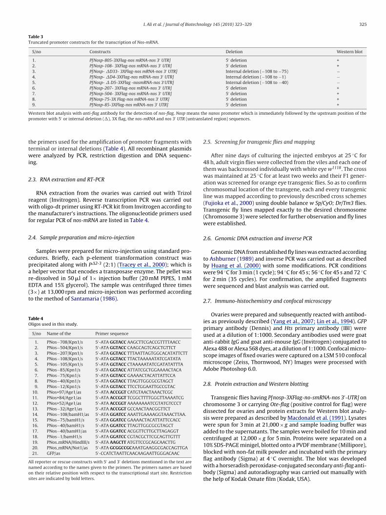
Table 3Truncated promoter constructs for the transcription of Nos-mRNA.
S/no Constructs Deletion Western blot
1. P[Nosp-805-3XFlag-nos mRNA-nos 3′ UTR] 5′ deletion +2. P[Nosp-108- 3XFlag-nos mRNA-nos 3′ UTR] 5′ deletion +3. P[Nosp- �D33- 3XFlag-nos mRNA-nos 3′ UTR] Internal deletion (−108 to −75) −4. P[Nosp- �D4-3XFlag-nos mRNA-nos 3′ UTR] Internal deletion (−108 to −1) −5. P[Nosp- � D5-3XFlag -nosmRNA-nos 3′UTR] Internal deletion (−108 to −40) −6. P[Nosp-207- 3XFlag-nos mRNA-nos 3′ UTR] 5′ deletion +7. P[Nosp-504- 3XFlag-nos mRNA-nos 3′ UTR] 5′ deletion +8. P[Nosp-75-3X Flag-nos mRNA-nos 3′ UTR] 5′ deletion +9. P[Nosp-85-3XFlag-nos mRNA-nos 3′ UTR] 5′ deletion +
W ans thp transl
ttwi
2
rwtf
2
cparE(t
TO
Anos
estern blot analysis with anti-flag antibody for the detection of nos-flag. Nosp meromoter with 5′ or internal deletion (�), 3X flag, the nos-mRNA and nos 3′ UTR (un
he primers used for the amplification of promoter fragments witherminal or internal deletions (Table 4). All recombinant plasmidsere analyzed by PCR, restriction digestion and DNA sequenc-
ng.
.3. RNA extraction and RT-PCR
RNA extraction from the ovaries was carried out with Trizoleagent (Invitrogen). Reverse transcription PCR was carried outith oligo-dt primer using RT-PCR kit from Invitrogen according to
he manufacturer’s instructions. The oligonucleotide primers usedor regular PCR of nos-mRNA are listed in Table 4.
.4. Sample preparation and micro-injection
Samples were prepared for micro-injection using standard pro-edures. Briefly, each p-element transformation construct wasrecipitated along with P�2-3 (2:1) (Tracey et al., 2000); which ishelper vector that encodes a transposase enzyme. The pellet was
e-dissolved in 50 �l of 1× injection buffer (20 mM PIPES, 1 mMDTA and 15% glycerol). The sample was centrifuged three times3×) at 13,000 rpm and micro-injection was performed accordingo the method of Santamaria (1986).
able 4ligos used in this study.
S/no Name of the Primer sequence
1. PNos−708/Kpn1/s 5′-ATA GGTACC AAGCTTCGACCGTTTTAACC2. PNos−504/Kpn1/s 5′-ATA GGTACC CAAGCAGTCAGCTGTTCT3. PNos−207/Kpn1/s 5′-ATA GGTACC TTTAATTAGTGGCACATATTCTT4. PNos−108/Kpn1/s 5′-ATA GGTACC TTACTAAAAATATCGATATA5. PNos−105/Kpn1/s 5′-ATA GGTACC CTAAAAATATCGATATATTTA6. PNos−85/Kpn1/s 5′-ATA GGTACC ATTATCGCTGGAAAACTACA7. PNos−75/Kpn1/s 5′-ATA GGTACC GAAAACTACATTATTCCA8. PNos−40/Kpn1/s 5′-ATA GGTACC TTAGTTGGCGCGTAGCT9. PNos−12/Kpn1/s 5′-ATA GGTACC TTCCTGGAATTGCCGTAC
10. PNos+97/Age1/as 5′-ATA ACCGGT CATGTAACTAAACTCGC11. PNos+84/Age1/as 5′-ATA ACCGGT TCGGCTTTTGGGTTAAAATCG12. PNos+52/Age1/as 5′-ATA ACCGGT AAAAAAAATCGTATGTCCCT13. PNos−32/Age1/as 5′-ATA ACCGGT GCCAACTAACGGTTCT14. PNos−108/bamH1/as 5′-ATA GGATCC AAATTGAAAAGGTAAACTTAA.15. PNos−75/bamH1/s 5′-ATA GGATCC GAAAACTACATTATTCCACC16. PNos−40/bamH1/s 5′-ATA GGATCC TTAGTTGGCGCGTAGCT17. PNos−40/bamH1/as 5′-ATA GGATCC ACGGTTCTTGCTTAGAGGT18. PNos−1/bamH1/s 5′-ATA GGATCC CGTACGCTTCGCAGTTGTTT19. PNos mRNA/HindIII/s 5′-ATA AAGCTT ATGTTCCGCAGCAACTTG20. PNos mRNA/Not1/as 5′-ATA GCGGCCGCAAATGAAGGCGACCAGTTGA21. GFP/as 5′-CCATCTAATTCAACAAGAATTGGGACAAC
ll reporter or rescue constructs with 5′ and 3′ deletions mentioned in the text areamed according to the names given to the primers. The primers names are basedn their relative position with respect to the transcriptional start site. Restrictionites are indicated by bold letters.
e nanos promoter which is immediately followed by the upstream position of theated region) sequences.
2.5. Screening for transgenic flies and mapping
After nine days of culturing the injected embryos at 25 ◦C for48 h, adult virgin flies were collected from the viles and each one ofthem was backcrossed individually with white eye w1118. The crosswas maintained at 25 ◦C for at least two weeks and their F1 gener-ation was screened for orange eye transgenic flies. So as to confirmchromosomal location of the transgene, each and every transgenicline was mapped according to previously described cross schemes(Fujioka et al., 2000) using double balance w Sp/CyO; Dr/Tm3 flies.Transgenic fly lines mapped exactly to the desired chromosome(Chromosome 3) were selected for further observation and fly lineswere established.
2.6. Genomic DNA extraction and inverse PCR
Genomic DNA from established fly lines was extracted accordingto Ashburner (1989) and inverse PCR was carried out as describedby Huang et al. (2000) with some modifications. PCR conditionswere 94 ◦C for 3 min (1 cycle); 94 ◦C for 45 s; 56 ◦C for 45 s and 72 ◦Cfor 2 min (35 cycles). For confirmation, the amplified fragmentswere sequenced and blast analysis was carried out.
2.7. Immuno-histochemistry and confocal microscopy
Ovaries were prepared and subsequently reacted with antibod-ies as previously described (Yang et al., 2007; Lin et al., 1994). GFPprimary antibody (Dennis) and Hts primary antibody (IBI) wereused at a dilution of 1:1000. Secondary antibodies used were goatanti-rabbit IgG and goat anti-mouse IgG (Invitrogen) conjugated toAlexa 488 or Alexa 568 dyes, at a dilution of 1:1000. Confocal micro-scope images of fixed ovaries were captured on a LSM 510 confocalmicroscope (Zeiss, Thornwood, NY) Images were processed withAdobe Photoshop 6.0.
2.8. Protein extraction and Western blotting
Transgenic flies having P[nosp-3XFlag-no-smRNA-nos 3′-UTR] onchromosome 3 or carrying Ote-flag (positive control for flag) weredissected for ovaries and protein extracts for Western blot analy-sis were prepared as described by Macdonald et al. (1991). Lysateswere spun for 3 min at 21,000 × g and sample loading buffer wasadded to the supernatants. The samples were boiled for 10 min andcentrifuged at 12,000 × g for 5 min. Proteins were separated on a
10% SDS-PAGE minigel, blotted onto a PVDF membrane (Millipore),blocked with non-fat milk powder and incubated with the primaryflag antibody (Sigma) at 4 ◦C overnight. The blot was developedwith a horseradish peroxidase-conjugated secondary anti-flag anti-body (Sigma) and autoradiography was carried out manually withthe help of Kodak Omate film (Kodak, USA).
326 I. Ali et al. / Journal of Biotechnology 145 (2010) 323–329
Fig. 3. Effect of 5′ deletions from the nos promoter on GFP expression in the germline stem cells. Arrows point to the tip of germaria where GSCs are located. Arrowheadsi (red)t 03:GF− reade
3
3f
rrtcPoans−Dtspa
3
aith+
ndicate expression of GFP in somatic cells. The ovaries were reacted with anti-Htso drive GFP in the germline are (A) −708/+97:GFP, (B) −207/+97:GFP, (C) −108/+112/+97:GFP. (For interpretation of the references to color in this figure legend, the
. Results
.1. GFP expression pattern in GSCs is affected by 5′ deletionsrom the nos promoter
Confocal microscopy on the ovaries from transgenic fliesevealed strong GFP fluorescence in GSCs in the case of flies car-ying the P[nosp-708:GFP] transgene (Fig. 3A). To know abouthe transcriptional importance of E-box sequences, 5′ deletiononstructs were made with or without these sequences, namely[nosp-504:GFP] and P[nosp-207:GFP]. Strong GFP fluorescence wasbserved in GSCs in the case of all transgenic fly lines carrying thebovementioned transgenes. Then we started 5′ deletions from theos promoter and made a series of P[nosp:GFP] 5′ deletion con-tructs with promoter fragment −207 followed by −108, −105,85,− 75, −40, and −12, relative to the transcription start site.eleted fragments until −108 did not show any significant reduc-
ion or change in the expression pattern of GFP in the germlinetem cells (Fig. 3B and C), while all deletions made downstream thisosition had GFP fluorescence significantly altered or completelybolished in the GSCs (Fig. 3D–H).
.2. Effect of 3′ nosp deletions on GFP expression pattern in GSCs
Using the P[nosp:GFP] p-element transformation plasmid,
nother series of 3′ promoter deletion constructs were made start-ng with +97 followed by +84, +52, +20 and −32, relative to theranscription start site (Table 1). The 3′ deletions from the promoterad no significant effect on GFP fluorescence in GSCs upto position20, relative to transcription start site while further deletions uptoor DAP1 (blue) and anti-GFP (green) antibodies. Various promoter fragments usedP, (D) −105/+97:GFP, (E) −85/+97:GFP, (F) −75/+97:GFP, (G) −40/+97:GFP and (H)r is referred to the web version of this article.)
position −32 of the nos promoter did not show any GFP fluorescencein GSCs (Fig. 4).
3.3. Internal deletions from nosp and GFP expression pattern inGSCs
After having determined the effect of 5′ and 3′ deletions on GFPtranscription in GSCs, we began with internal deletions from thenos promoter. Initially, we deleted a relatively longer fragment(−108 to −1) from nosp which completely abolished GFP fluores-cence in stem cells (Fig. 5A) showing that this region of the nospromoter contains enhancers that are vital for the nos gene expres-sion in the GSCs. After that we deleted another fragment spanningfrom −108 to −40 and noticed again that there was no GFP fluores-cence in GSCs (Fig. 5B). Deletion of another 33 bp fragment (−108 to−75) from the nos promoter was also sufficient for the transcriptionof GFP to be diminished in GSCs (Fig. 5C). To confirm the importanceof upstream promoter sequences present close to the transcriptionstart site, we made another deletion from −40 to −1 and observedthat the GFP expression was blocked again in this case (Fig. 5D).
3.4. A short promoter fragment is sufficient for the expression ofnos-mRNA
In order to carry out the sufficiency test for the nos promoter
fragment (−108 to +97) to drive the transcription of nos-mRNA andto know about the importance of the cis-acting elements presentin the promoter, we constructed a series of 5′ and internal dele-tions nosp constructs and examined their capability to transcribethe transgene P[nosp-nosmRNA-3xflag-nos 3′-UTR] (Table 3). We
I. Ali et al. / Journal of Biotechnology 145 (2010) 323–329 327
Fig. 4. Effect of nosp 3′ deletions on GFP in the germline stem cells. Arrows indicate GFP positive or GFP negative germline stem cells. The ovaries were reacted with anti-Hts(red) or DAP1 (blue) and anti-GFP (green) antibodies. Nos promoter 3′ deleted fragments used to drive GFP are (A) −708/+97:GFP, (B) −708/+84:GFP, (C) −708/+52:GFP and(D) −708/−32:GFP. (For interpretation of the references to color in this figure legend, the reader is referred to the web version of this article.)
Fig. 5. Internal deletions from the nos promoter and its effect on GFP expression in the germline stem cells. GFP expression pattern for constructs with internally deletedo 1, (Bs anti-r this a
ntEdmtldflmm
Ffimw1P
r mutated promoter fragments is shown in the following order (A) from −108 to −tem cells in the germaria is indicated by the arrows. The ovaries were reacted witheferences to color in this figure legend, the reader is referred to the web version of
oticed that the promoter fragment from −708 to +97 could fullyranscribe nos-mRNA (Fig. 6C). To investigate the importance of-box like sequences for the transcription of the nos-mRNA, weesigned a construct having all the E-box sequences with a pro-oter fragment from −504 to +97, and another one from −207
o +97 lacking in these sequences and established transgenic flyines for each. Both of the promoter fragments did not show any
ifference in their transcriptional capability of nos-mRNA in sixy lines tested for each of the constructs (Table 3). We madeore 5′ and internal deletions using the same parent constructentioned earlier in this text and observed their transcriptionalig. 6. Nos-Flag fusion protein detection by Western blotting. Shown in thegure are Western blots for nos:flag driven by various truncated nos pro-oter fragments in the following order (A) Ote-3xflag-Ote (positive control), (B)1118 (negative control), (C) P[Nosp-708-3XFlag-nos-mRNA-nos 3′UTR], (D) P[Nosp-08-3XFlag-nosmRNA-nos3′UTR], (E) P[Nosp-75-3XFlag-nosmRNA-nos 3′UTR] and (F)[Nosp-�(−108 to −75)-3XFlag-nos mRNA-nos3′UTR].
) from −108 to −40, (C) from −108 to −75 and (D) from −40 to −1. The location ofHts (red) or DAP1 (blue) and anti-GFP (green) antibodies. (For interpretation of therticle.)
importance for the nos-mRNA. The results revealed that all con-structs with promoter fragment −108 or upstream could transcribethe P[nosp-nosmRNA-3xflag-nos 3′-UTR] transgene while in the caseof constructs with promoter fragments downstream −108 or withinternal deletions (−108 to −75 and −108 to −40), we were unabledetect nos-flag fusion protein in all the fly lines checked by West-ern blotting (see Fig. 6 and Table 3). It indicated that nos-mRNA wasnot transcribed due to lack of vital cis-regulatory elements presentin the deleted regions which were identified earlier. It further con-firmed that the shortest promoter fragment that can drive wildtype-like expression of the nos-mRNA spans from −108 to +97.
4. Discussion
Drosophila Nanos is evolutionarily conserved and has awidespread role in germline development and is involved in
GSCs maintenance (Wang and Lehmann, 1991; Bhata, 1999). Geneexpression is controlled by a variety of mechanisms includingrecruitment of transcription complexes (Kosak and Groudine,2004; Leach et al., 2003) wherein these factors bind with spe-cific nucleotide sequences located in the promoter region or the
3 echno
UoacuetvPtaetciedtrw−eanatpasnpmettGsonTtalttsoploaemtwwatafilsdip
28 I. Ali et al. / Journal of Biot
TRs of a particular gene and ultimately would turn a gene on orff. Promoter region is important for the transcriptional regulationnd contain the driving machinery for the expression of a typi-al eukaryotic gene. Earlier studies indicated that sequences 1 kbpstream to the nos transcription start site were sufficient for thexpression of the transgene (Tracey et al., 2000). Other studies men-ioned earlier in this text have also used the nos promoter to drivearious transgenes in GSCs. Initial experiments with flies carrying[nosp-708:GFP] gave strong fluorescence in GSCs which revealedhat the 805 bp nos promoter fragment (from −708 to +97) containsll the regulatory elements responsible for the germline-specificxpression of the transgene. Computational analysis indicated thathere are E-box like sequenes in nosp. Deletion experiments con-luded that E-box sequences in nosp do not play any regulatory rolen GSCs. So as to define the 5′ limits of the nos promoter that couldfficiently drive GFP expression in the GSCs, we made a series of 5′
eletions from the promoter and noticed that the expression pat-ern did not change significantly until we reached position −105,elative to the transcription start site. The GFP expression patternas the same in the case of a number of constructs ranging from708 to −108, which indicated the fact that important cis-actinglements responsible for the expression of the transgene in GSCsre located downstream −108. The expression pattern altered sig-ificantly in the case of −105 as it was not restricted to GSCs onlynd it was completely abolished when we reached position −75 ofhe nos promoter. These results indicate that there are sequencesresent between position −108 and −75 of the nos promoter, whichre vital for the expression of the transgene in GSCs. Two more con-tructs with truncated promoter fragments (−40 and −12) also didot show any GFP fluorescence in transgenic Drosophila. The nosromoter 3′ truncation revealed that deletion of the promoter frag-ent until position +20 did not have any significant effect on the
xpression of GFP in GSCs. Fly lines established for 3′ promoter dele-ions constructs indicated that sequences from −40 to +20 relativeo the transcription start site are essential for GFP expression. TheFP expression pattern for constructs having a series of 3′ deletioneries (+97, +84, +52 and +20) is almost the same with no inhibitoryr enhancing effect, indicating that sequences downstream +20 areot essentially required for the transgenic GFP expression in GSCs.hese experiments indicated that the 60 bp sequence (from posi-ion −40 to +20) is important for the transgenic GFP expressionnd may have binding sites for vital transcription factors regu-ating nosp. We also noticed that all 3′ deletions restricted GFPo GSCs and cystoblasts and no expression was observed beyondhat point in somatic cells which indicates that sequences down-tream +20 may be important for the GFP expression in tissuesther than GSCs. These terminal deletions revealed that the com-lete driving machinery for the transgenic GFP expression in GSCs
ay between position −108 and +20 of the nanos promoter. Thesebservations were further substantiated by the fact that we wereble to get wild type-like expression of GFP in GSCs from the short-st promoter fragment mentioned earlier. The 5′ and 3′ deletionsarginalized the functional minimal promoter to lay between posi-
ion −108 and +20 relative to the transcription start site, but weanted to know whether internal deletions from the promoterould have any effect on GFP expression in transgenic fly lines,
nd whether this fragment is both sufficient and enough for theransgenic GFP expression in the GSCs? We established fly lines forseries of internal deletion constructs starting with deletion of a
ragment from −108 to −1 from the entire nosp. GFP expressionn GSCs was completely abolished for this construct in all the fly
ines established. It indicated that this region contains enhancerequences necessary for the transgenic GFP expression. We furthereleted a 33 bp fragment (−108 to −75) from the promoter andn fly lines established for this construct GFP expression was com-letely abolished in the germline which brought us even closer to
logy 145 (2010) 323–329
finding important cis-regulatory sequences in nosp. These experi-ments indicated the presence of enhancers between position −108and −1, relative to the transcription start site. In order to figureout if this region contains a single or multiple enhancers, we mademore internal deletions from the promoter and observed its effecton GFP expression in the germline. Deletion of a promoter frag-ment from −40 to −1 again abolished GFP expression indicatingthat there are other essential sequences in this region too, thoughnot capable of driving expression independently. This observationis consistent with other studies where enhancer function is dis-rupted upon loss of a single activator or repressor site (Arnostiet al., 1996; Small et al., 1992). The blotting experiments for flieshaving the deletion constructs further substantiated the previousobservation with GFP expression as nos-flag fusion protein wasnot detectable in ovarian extracts of transgenic Drosophila havingthe same deletion construct (Fig. 6 and Table 3). The 5′ deletionseries earlier indicated that sequences from −108 to −75 are nec-essary for the GFP expression in the germline. We did not detectthe nos-flag fusion protein in the case of the construct with dele-tion of the 33 bp fragment between position −108 and −75, whichmeans that this region of the promoter contain cis-regulatory ele-ments essential for the expression of nos-mRNA. Initial blottingexperiments with flies having p[nosp-3xflag-nosmRNA-nos 3′UTR]transgene indicated that the promoter fragment from −708 to +97could fully transcribe the nos-mRNA. So as to know about the min-imal promoter which is sufficient and enough for the nos geneexpression, we made a series of internal and 5′ deletions from thepromoter and observed its effect on the transcription of nos-mRNAin transgenic Drosophila. We observed that the promoter fragment−108 and upstream sequences could effectively transcribe the nos-mRNA based transgene, while deletions downstream −108 couldnot do the same. The nos promoter internal deletions also revealedthe same picture which further strengthened previous observa-tions made in case of transgenic GFP expression in the GSCs. Theseexperiments further indicated that the shortest fragment that couldtranscribe nos-mRNA spans from position −108 to +97 of the nospromoter. Analysis of the truncated promoter further revealed thepresence of specific binding sites for several transcription factorslocated in the previously identified vital promoter region. Thesesites include Zen (Doyle et al., 1986) (from −104 to −97), Brinker(Jazwinska et al., 1999) located at position −35 to −30, and DREF(DNA replication related element factor) which binds specifically tothe 8 bp palindromic DRE sequence spanning position −98 to −91(Fig. 1). DRE has also been associated with other factors includingBeaf-32 (Hart et al., 1999). The DNA replication-related element(DRE) is a regulatory element frequently found in the promoters ofDrosophila genes (Hirose et al., 1993). There are DRE-like sequencesin the 5′-flanking regions of many genes other than DNA replicationand proliferation-related genes, suggesting that DREF plays multi-ple roles in vivo (Yoshida et al., 2004). Drosophila Dpp pathwayis negatively regulated by eukaryotic translation initiation factor4A (Li and Li, 2006) which is a target of DREF transcription factor(Ida et al., 2007). Recent evidence confirmed that elF4A controlsgermline stem cells self-renewal (Shen et al., 2009). Internal dele-tion (from 108 to −75) and 5′ promoter deletion (−75) experimentsreveal that sequences between position −108 and −75 are not onlyvital for the expression of GFP in the germline stem cells but alsofor the transcription of nos mRNA. The same region of the pro-moter contains binding sites for DREF and ZEN; target of the highlevel dpp (Lin et al., 2006) (Fig. 1) indicating a possibly importanttranscriptional role for these factors. All of the observations with
P[nosp:GFP] and P[nosp-3XFlag-nosmRNA-nos3′UTR] transgenes ledto the conclusion that the minimal nos promoter that can effectivelytranscribe GFP in GSCs lay between position −108 and +20 and thatthe vital cis-acting elements for the transcription of transgenic GFPor nos-mRNA are present in between positions −108 and +97.
echno
A
Eagp
R
A
A
B
C
D
D
F
F
G
H
H
H
I
I. Ali et al. / Journal of Biot
cknowledgements
The first author would like to highly acknowledge the Higherducation Commission of Pakistan for their support, Jiang Xiaoyangnd Xiao Shen for providing P[Otep-3Xflag-Ote] construct and trans-enic fly lines. The author is also thankful to Dr. Dahua Chen forroviding him the opportunity to work in his lab.
eferences
rnosti, D.N., Barolo, S., Levine, M., Small, S., 1996. The eve stripe2 enhancer employsmultiple modes of transcriptional synergy. Development 122, 205–214.
shburner, M., 1989. Drosophila: A Laboratory Manual. Cold Spring Harbor Labora-tory Press, Cold Spring Harbor, NY, pp. 106–107.
hata, K.M., 1999. The posterior determinant Gene nanos Is required for the main-tenance of the adult germline stem cells during Drosophila oogenesis. Genetics151, 1479–1492.
hen, D., McKearin, D.M., 2003. A discrete transcriptional silencer in the bam genedetermines asymmetric division of the Drosophila germline stem cell. Develop-ment 130, 1159–1170.
oyle, H.J., Harding, K., Hoey, T., Levine, M., 1986. Transcripts encoded by a home-obox gene are restricted to dorsal tissues of Drosophila embryos. Nature 323,76–79.
eshpande, G., Calhoun, G., Yanowitz, J.L., Schedl, P.D., 1999. Novel functions ofnanos in down regulating mitosis and transcription during the development ofthe Drosophila germline. Cell 99, 271–281.
orbes, A., Lehmann, R., 1998. Nanos and Pumilio have critical roles in the devel-opment and function of Drosophila germline stem cells. Development 125,679–690.
ujioka, M., Jaynes, J.B., Bejsovec, A., Weir, M., 2000. Developmental biology proto-cols. In: Rocky, S.T., Cecilia, W.L. (Eds.), Production of transgenic Drosophila, vol.2. Humana Press Inc., New York, pp. 353–363.
avis, E.R., Lehmann, R., 1994. Translational regulation of nanos by RNA localization.Nature 369, 315–318.
art, C.M., Cuvier, O., Laemmli, U.K., 1999. Evidence for an antagonistic relationshipbetween the boundary element-associated factor BEAF and the transcriptionfactor DREF. Chromosoma 108 (6), 375–383.
irose, F., Yamaguchi, M., Handa, H., Inomata, Y., Matsukage, A., 1993. Novel 8-basepair sequence (Drosophila DNA replication-related element) and specific bindingfactor involved in the expression of Drosophila genes for DNA polymerase ˛ andproliferating cell nuclear antigen. J. Biol. Chem. 268, 2092–2099.
uang, A.M., Rehm, E.J., Rubin, G.M., 2000. Recovery of DNA sequences flanking P-element insertions: inverse PCR and plasmid rescue. In: Sullivan, M., Ashburner,R., Hawley, S. (Eds.), Drosophila Protocols. Cold Spring Harbor Laboratory Press,Cold Spring Harbor, pp. 429–437.
da, H., Yoshida, H., Nakamura, K., Yamaguchi, M., 2007. Identification of eIF4A as atarget of DREF transcription factor. Exp. Cell Res. 313 (20), 4208–4220.
logy 145 (2010) 323–329 329
Jazwinska, A., Kirov, N., Wieschaus, E., Roth, S., Rushlow, C., 1999. The Drosophilagene brinker reveals a novel mechanism of Dpp target gene regulation. Cell 96(4), 563–573.
Kobayashi, S., Yamada, M., Asaoka, M., Kitamura, T., 1996. Essential role of the pos-terior morphogen nanos for germline development in Drosophila. Nature 380,708–711.
Kosak, S., Groudine, T.M., 2004. Gene order and dynamic domains. Science 306,644–647.
Leach, K.M., Vieira, K.F., Kang, S.H., Aslanian, A., Teichmann, M., Roeder, R.G., Bungert,J., 2003. Characterization of the human �-globin downstream promoter region.Nucleic Acids Res. 31, 1292–1301.
Li, J., Li, W.X., 2006. A novel function of Drosophila eIF4A as a negative regulatorof Dpp/BMP signalling that mediates SMAD degradation. Nat. Cell Biol. 8 (12),1407–1414.
Lin, H., Yue, L., Allan, C., 1994. The Drosophila fusome, a germline-specific organelle,contains membrane skeletal proteins and functions in cyst formation. Develop-ment 120, 947–956.
Lin, M.C., Park, J., Kirov, N., Rushlo, C., 2006. Threshold response of C15 to the Dppgradient in Drosophila is established by the cumulative effect of Smad and Zenactivators and negative cues. Development 133, 4805–4813.
Macdonald, P.M., Luk, S.K., Kilpatrick, M., 1991. Protein encoded by the exuperantiagene is concentrated at sites of bicoid mRNA accumulation in Drosophila nursecells but not in oocytes or embryos. Genes Dev. 5, 2455–2466.
Okada, Y., Matsuura, E., Tozuka, Z., Nagai, R., Watanabe, A., Matsumoto, K., Yasui, K.,Jackman, R.W., Nakano, T., Doi, T., 2004. Upstream stimulatory factors stimulatetranscription through the E-box motifs in the PF4 gene in megakaryocytes. Blood104, 2027–2034.
Santamaria, P., 1986. Injecting eggs. In: Roberts, D.B. (Ed.), Drosophila: A PracticalApproach, pp. 159–173.
Shen, R., Weng, C., Yu, J., Xie, T., 2009. eIF4A controls germline stem cell self-renewalby directly inhibiting BAM function. PNAS 106, 11623–11628.
Small, S., Blair, A., Levine, M., 1992. Regulation of even-skipped stripe2 in theDrosophila embryo. EMBO J. 11, 4047–4057.
Tracey, W.D., Ning, X., Klingler, M., Kramer, S.G., Gergen, J.P., 2000. Quanti-tative analysis of gene function in the Drosophila embryo. Genetics 154,273–284.
Tsuda, M., Sasaoka, Y., Kiso, M., Abe, K., Haraguchi, S., Kobayashi, S., Saga, Y., 2003.Conserved Role of nanos Proteins in Germ Cell Development. Science 301,1239–1241.
Van Doren, M., Williamson, A.L., Lehmann, R., 1998. Regulation of zygoticgene expression in Drosophila primordial germ cells. Curr. Biol. 8, 243–246.
Wang, C., Lehmann, R., 1991. Nanos is the localized posterior determinant inDrosophila. Cell 66, 637–647.
Yang, L., Duan, R., Chen, D., Wang, J., Jin, P., 2007. Fragile X mental retardationproteinmodulates the fate of germline stem cells in Drosophila. Hum. Mol. Genet. 16,1814–1820.
Yoshida, H., Kwon, E., Hirose, F., Otsuki, K., Yamada, M., Yamaguchi, M., 2004. DREFis required for EGFR signalling during Drosophila wing vein development. GenesCells 9, 935–944.