Chiral capillary electrophoresis–mass spectrometry of 3,4-dihydroxyphenylalanine: Evidence for its...
13
Chiral Capillary Electrophoresis – Mass Spectrometry of Tetrahydroisoquinoline-derived Neurotoxins: Observation of Complex Stereoisomerism Hao Wu, Baiqing Yuan, and Yi-Ming Liu Department of Chemistry and Biochemistry, Jackson State University, 1400 Lynch, St, Jackson, MS 39110, USA Abstract Previous studies have shown that certain 1,2,3,4-tetrahydroisoquinoline derivatives (TIQs) are neurotoxins inducing Parkinsonism. Further, individual enantiomers of these toxins such as (R/S)- N-methylsalsolinol ((R/S)-NMSal) possess distinct neurotoxicological properties. In this work, a chiral capillary electrophoresis (CE) method with electrospray ionization-tandem mass spectrometric (ESI-MS/MS) detection was developed for the quantification of TIQ enantiomers. Enantioseparation was achieved with sulfated β-cyclodextrin (sulfated β-CD) as chiral selector. To avoid any potential contamination of MS ionization source by the non-volatile chiral selector, partial filling technique was deployed in the CE separation. TIQ derivatives, including (R/S)-6,7- dihydroxy-1-methy-TIQ (salsolinol, Sal), (R/S)-1-benzyl-TIQ (BTIQ), and (R/S)-NMSal, were base-line resolved with resolution values (R) ranging from 3 (for Sal) to 4.5 (for BTIQ), which were much better than those reported previously by HPLC methods. ESI-MS/MS detection of the resolved TIQ enantiomers was specific and sensitive (LOD = 1.2 μM for Sal enantiomers). The proposed chiral CE-MS/MS method was used to study in vitro formation of (R/S)-NMSal. It was found that NMSal was formed from the incubation of epinine (a dopamine metabolite) with acetaldehyde (a metabolite of alcohol). More interestingly, four isomers of NMSal were separated and detected in the incubation solution. They were identified as (R)-e.e-NMSal, (R)-e.a-NMSal, (S)-e.e-NMSal, and (S)-e.a-NMSal. This was the first lab evidence that this parkinsonian neurotoxin exists in multiple isomeric forms. Keywords Chiral capillary electrophoresis; mass spectrometry; neurotoxin; tetrahydroisoquinoline; N- methylsalsolinol; stereoisomer 1. Introduction It is well documented that some tetrahydroisoquinoline derivatives such as N- methylsalsolinol (NMSal) cause neurotoxicological damages similar to those caused by 1- methyl-4-phenyl-1,2,3,6-tetrahydropyridine (MPTP) [1-3]. MPTP is a well known synthetic neurotoxin that causes Parkinsonism in humans, monkeys, and various animals [4]. © 2011 Elsevier B.V. All rights reserved. Correspondence to: Yi-Ming Liu. Publisher's Disclaimer: This is a PDF file of an unedited manuscript that has been accepted for publication. As a service to our customers we are providing this early version of the manuscript. The manuscript will undergo copyediting, typesetting, and review of the resulting proof before it is published in its final citable form. Please note that during the production process errors may be discovered which could affect the content, and all legal disclaimers that apply to the journal pertain. NIH Public Access Author Manuscript J Chromatogr A. Author manuscript; available in PMC 2012 May 20. Published in final edited form as: J Chromatogr A. 2011 May 20; 1218(20): 3118–3123. doi:10.1016/j.chroma.2011.03.026. NIH-PA Author Manuscript NIH-PA Author Manuscript NIH-PA Author Manuscript
-
Upload
independent -
Category
Documents
-
view
5 -
download
0
Transcript of Chiral capillary electrophoresis–mass spectrometry of 3,4-dihydroxyphenylalanine: Evidence for its...

Chiral Capillary Electrophoresis – Mass Spectrometry ofTetrahydroisoquinoline-derived Neurotoxins: Observation ofComplex Stereoisomerism
Hao Wu, Baiqing Yuan, and Yi-Ming LiuDepartment of Chemistry and Biochemistry, Jackson State University, 1400 Lynch, St, Jackson,MS 39110, USA
AbstractPrevious studies have shown that certain 1,2,3,4-tetrahydroisoquinoline derivatives (TIQs) areneurotoxins inducing Parkinsonism. Further, individual enantiomers of these toxins such as (R/S)-N-methylsalsolinol ((R/S)-NMSal) possess distinct neurotoxicological properties. In this work, achiral capillary electrophoresis (CE) method with electrospray ionization-tandem massspectrometric (ESI-MS/MS) detection was developed for the quantification of TIQ enantiomers.Enantioseparation was achieved with sulfated β-cyclodextrin (sulfated β-CD) as chiral selector. Toavoid any potential contamination of MS ionization source by the non-volatile chiral selector,partial filling technique was deployed in the CE separation. TIQ derivatives, including (R/S)-6,7-dihydroxy-1-methy-TIQ (salsolinol, Sal), (R/S)-1-benzyl-TIQ (BTIQ), and (R/S)-NMSal, werebase-line resolved with resolution values (R) ranging from 3 (for Sal) to 4.5 (for BTIQ), whichwere much better than those reported previously by HPLC methods. ESI-MS/MS detection of theresolved TIQ enantiomers was specific and sensitive (LOD = 1.2 μM for Sal enantiomers). Theproposed chiral CE-MS/MS method was used to study in vitro formation of (R/S)-NMSal. It wasfound that NMSal was formed from the incubation of epinine (a dopamine metabolite) withacetaldehyde (a metabolite of alcohol). More interestingly, four isomers of NMSal were separatedand detected in the incubation solution. They were identified as (R)-e.e-NMSal, (R)-e.a-NMSal,(S)-e.e-NMSal, and (S)-e.a-NMSal. This was the first lab evidence that this parkinsonianneurotoxin exists in multiple isomeric forms.
KeywordsChiral capillary electrophoresis; mass spectrometry; neurotoxin; tetrahydroisoquinoline; N-methylsalsolinol; stereoisomer
1. IntroductionIt is well documented that some tetrahydroisoquinoline derivatives such as N-methylsalsolinol (NMSal) cause neurotoxicological damages similar to those caused by 1-methyl-4-phenyl-1,2,3,6-tetrahydropyridine (MPTP) [1-3]. MPTP is a well known syntheticneurotoxin that causes Parkinsonism in humans, monkeys, and various animals [4].
© 2011 Elsevier B.V. All rights reserved.Correspondence to: Yi-Ming Liu.Publisher's Disclaimer: This is a PDF file of an unedited manuscript that has been accepted for publication. As a service to ourcustomers we are providing this early version of the manuscript. The manuscript will undergo copyediting, typesetting, and review ofthe resulting proof before it is published in its final citable form. Please note that during the production process errors may bediscovered which could affect the content, and all legal disclaimers that apply to the journal pertain.
NIH Public AccessAuthor ManuscriptJ Chromatogr A. Author manuscript; available in PMC 2012 May 20.
Published in final edited form as:J Chromatogr A. 2011 May 20; 1218(20): 3118–3123. doi:10.1016/j.chroma.2011.03.026.
NIH
-PA Author Manuscript
NIH
-PA Author Manuscript
NIH
-PA Author Manuscript

Therefore, study on TIQs' neurotoxicity has been intensive [5-7]. Very importantly, it hasbeen found that many chiral TIQ compounds exhibit enantioselective neurotoxicity, that isthe two enantiomers possess distinct neurotoxicological properties [8-10]. For example, (R)-enantiomer of NMSal was found 1000 times more potent to induce Parkinsonism in rat thanthe (S)-enantiomer [2, 11]. Study on TIQs' enantioselective neurotoxicity requires sensitivequantification of these toxic compounds with stereochemical selectivity.
Analytical methods based on high-performance liquid chromatography (HPLC) [12-17],capillary electrophoresis (CE) [18-21], gas chromatography-mass spectrometry (GC-MS)[22-226], and HPLC-MS [27-29] have been developed for enantiomeric quantification ofTIQs. Since many TIQs such as Sal and NMSal are highly hydrophilic and easily oxidizedin basic solutions, the cumbersome sample pretreatment and pre-column derivatizationprocedures required in GC-MS analysis can be problematic causing a significant loss of theanalytes. The chiral HPLC-MS methods reported previously allowed a facile determinationof Sal enantiomers without pre-column derivatization. However, efforts to achieve a chiralseparation of other TIQ neurotoxins including MNSal and BTIQ on the β-cyclodextrinbonded silica column failed in our previous studies [29].
Compared with HPLC, CE offers advantages including high separation efficiency, shortseparation time, and the compatibility with small sample volume/mass. In recent decademuch interest has been given to coupling of CE with MS for chiral analysis. Extensivereviews on this topic were given [30 - 31]. A major challenge remains the potentialcontamination of MS ionization source by the non-volatile chiral selector and other additivesin CE running buffer. Several chiral selectors including chiral crown ethers [32 - 33],proteins [34], chiral micelles [35], and cyclodextrins [36-37] were used in chiral CE-MSanalysis. In search for an effective assay of TIQ enantiomers, a CE-MS method with highseparation efficiency, peak identification capability, and assay sensitivity was developed. Inthis work, sulfated β-cyclodextrin (β-CD) was selected as the chiral selector since it was themost effective chiral selector for resolving Sal enantiomers by using HPLC based on ourprevious investigations. In addition, since sulfated β-CD is negatively charged, it migratesaway from the MS ionization source in a CE-MS separation. To avoid any potentialcontamination by the non-volatile chiral selector, partial filling technique was deployed.Three most extensively studied TIQ neurotoxins, i.e. Sal, NMSal, and BTIQ were selectedas the model analytes. By using the chiral analytical method, in vitro formation of NMSalfrom incubation of epinine (a dopamine metabolite) with acetaldehyde (an alcoholmetabolite) was investigated. Taking advantage of the mass-specific detection, stableisotope labeled chemicals could be used to facilitate the CE peak identification in the studyof this important neurotoxin.
2. Materials and Methods2.1. Materials
Racemic Sal, racemic 1-BTIQ, dopamine (DA), epinine, acetaldehyde, acetaldehyde-2,2,2-d3, ammonium acetate, acetic acid and sulfated sodium salt of β-Cyclodextrin (sulfated β-CD) were purchased from Sigma–Aldrich (St. Louis, MO, USA). (+)-(R)- /(-)-(S)-Salenantiomers were prepared from racemic Sal as described in our previous work [19]. Milli-Q water (Millipore) was used through out the work. Prior to CE analysis, all samples and therunning buffer were filtered through a nylon 0.22 μm syringe filter.
2.2. CE-MS ApparatusThe CE-MS system consisted of an Agilent 7100 capillary electrophoresis system and aThermoFinnigan mass spectrometer (LCQ DECA). A CE-MS adapter kit from Agilent
Wu et al. Page 2
J Chromatogr A. Author manuscript; available in PMC 2012 May 20.
NIH
-PA Author Manuscript
NIH
-PA Author Manuscript
NIH
-PA Author Manuscript

Technologies was used for the coupling. All CE operations including capillary flush, chiralselector loading, sample injection, and separation were automated. The mass spectrometerwas equipped with an ESI source and a syringe pump. It was operated in a positive ionmode. Multiple stage mass spectrometry (MS/MS) experiments were performed to isolateand fragment the targeted ions. The operating conditions of the MS detector were optimizedwith a solution of Sal (1.0 μM) infused into the ESI-MS system with a syringe pump at aflow rate of 2μL /mL. Parameters were optimized using the Autotune Program. Data werecollected and analyzed by using Xcalibur.
2.3. Chiral CE-MS AssayCapillary was flushed with the CE running buffer for 3 min, and then the chiral selectorsolution was introduced into the capillary by pressure injection at 100 mbar for 50 s. Asample solution was injected at 50 mbar for 12 s. The capillary inlet end was placed in theCE running buffer vial and separation was started by applying a positive voltage. At thesame time MS detection began (sheath liquid was automatically turned on by the massspectrometer).
CE conditions: column, 50μm ID /360μm OD × 75 cm long fused-silica capillary; CErunning buffer, 20 mM acetic acid /ammonium acetate buffer at pH 5.5; chiral selectorsolution, 1.0 mM sulfated β-CD in CE running buffer; CE voltage, positive 25.0 kV; columntemperature, 20 °C.
MS conditions: sheath liquid, 50% methanol in water containing 0.1% acetic acid at 2μL /min; spray voltage, 4 kV; capillary temperature, 220°C; sheath gas, 20 arbitrary units (au);auxiliary gas, 0 au. For SRM experiments, normalized collision energy was set at 30 with anisolation width of 2.0 u, and the activation time was set at 30 ms.
2.4. In vitro study of NMSal formationEpinine was dissolved in 50mM phosphate buffered saline (PBS) (pH 7.4). The solution wasmixed with acetaldehyde. The final concentrations were 10 mM and 30 mM for epinine andacetaldehyde, respectively. The mixture was incubated at 37°C for 2h and then centrifugedat 10000×g for 10min. Supernatant was collected for NMSal quantification. Incubations ofepinine with acetaldehyde-d3 were carried out similarly.
3. Results and Discussion3.1. Chiral CE-MS of TIQs
In our previous work on chiral CE separation of TIQs, a very complex running buffercontaining β-CD as the chiral selector had to be used, which did not allow its coupling withMS detection [18]. Ammonium acetate buffer was selected as the background electrolyte inthis work because it was volatile and would not contaminate the MS ionization source.Volatile chiral selectors such as certain chiral crown ethers are ideal for chiral CE-MSanalysis. However, our efforts to resolve Sal enantiomers by using chiral crown ethers (e.g.2-hydroxymethyl-18-crown-6) came out with no success. From our computational studies,cyclodextrins form inclusion complexes with Sal, NMSal, and BTIQ with stereochemicalpreferences in terms of the stabilization energy [38]. Sulfated β-CD, although non-volatile,is negatively charged, and thus migrates against the EOF or away from the MS detector inthe proposed CE-MS set-up. Therefore, it was selected as the chiral selector. Further, partialfilling technique [32, 39-40] was deployed to ensure that sulfated β-CD would not get intothe MS ionization source. Results from studying various separation conditions with Salenantiomers as the model solutes are described as following.
Wu et al. Page 3
J Chromatogr A. Author manuscript; available in PMC 2012 May 20.
NIH
-PA Author Manuscript
NIH
-PA Author Manuscript
NIH
-PA Author Manuscript

The pH of CE running buffer affects the magnitude of EOF and the apparent chargenumbers on the chiral selector and the analytes. It, therefore, affects the separation results. Inthe tested pH range from 3 to 7, the best resolution of Sal enantiomers was achieved at pH5.5. The migration time of Sal increased as the running buffer pH decreased. When it was <3, Sal would not be eluted out. Concentration of sulfated β-CD in the range of 0.1 -10.0 mMwas investigated. Sal enantiomers were base-line resolved with sulfated β-CD concentrationat 1.0 mM or above. At high sulfated β-CD concentrations (e.g. 5mM), better resolutionswere obtained, but the migration times increased significantly (to about 30mins). To obtainboth an acceptable separation resolution and good separation efficiency, sulfated β-CD at1mM was used for further studies. Sheath liquid was used for the coupling. The compositionof sheath liquid was investigated to obtain the best MS detection sensitivity. Mixtures ofwater and methanol or acetonitrile at various ratios containing acetic acid were tested. It wasfound that better MS signals were obtained with water /methanol (50:50 v/v) with 0.1%acetic acid. Acetic acid was added to enhance the formation of positive ions in ESI. Theflow rate of sheath liquid was set at 2μl /min. An unstable ESI spray was observed at a flowrate of 1μl /min. Under the selected experimental conditions, enantiomers of Sal, BTIQ, andNMSal were all base-line separated and sensitively detected. Typical electropherogramsfrom the separations and the MS2 spectra are shown in Figure 1. The resolution values (R)were 3 for Sal enantiomers and 4.5 for BTIQ enantiomers, which were much better thanthose previously reported by using HPLC-MS methods [27-29]. Moreover, the chiral CEseparation was completed within 15 min.
3.2. Analytical figures of meritThe present chiral CE-MS method was evaluated for enantiomeric quantification of Sal interms of the response linearity, limit of detection, and reproducibility. Five point calibrationcurves were prepared by analyzing authentic Sal racemate solutions at concentrations of 5.0,10.0, 25.0, 50.0, and 100 μM. Peak heights were used for the quantification. Linearregression analysis on the results of peak heights versus concentrations yielded thefollowing calibration equations:
where H is peak height and C the concentration (μM). A good linearity was obtained forboth calibration curves with correlation coefficients > 0.985. From the calibration curves,limits of detection (LOD) were estimated to be 1.2 μM for (R)-Sal and 1.5μM for (S)-Sal(signal / noise ratio =3). The reproducibility of analyte response (RSD %) in terms of peakheight and migration time were studied by separating a Sal racemate solution (10.0 μM eachenantiomer) for 6 times. The RSDs of peak height and migration time for both enantiomerswere found <1.7% and 2.4% (n= 6), respectively.
3.3. In vitro formation of NMSalThe proposed chiral CE-MS/MS method was used to study in vitro formation of NMSal, aparkinsonian neurotoxin. There are several possible synthetic pathways of NMSal asoutlined in Fig. 2. Dopamine is an important neurotransmitter. Loss of dopaminergicneurons is a key pathological feature of Parkinson's disease. Condensation of dopamine withacetaldehyde (a metabolite of alcohol) forms Sal that can be converted to NMSal by N-
Wu et al. Page 4
J Chromatogr A. Author manuscript; available in PMC 2012 May 20.
NIH
-PA Author Manuscript
NIH
-PA Author Manuscript
NIH
-PA Author Manuscript

methyltransferase [1]. Another possible synthetic pathway of NMSal is the condensation ofepinine and acetaldehyde. Epinine can be formed from dopamine via N-methylation [41].Tothe best of our knowledge, little study of NMSal formation from epinine and acetaldehydehas been done so far. In this work, epinine was incubated with acetaldehyde in mimicphysiological conditions for 2 hrs. The resultant incubation solutions were analyzed toquantify NMSal enantiomers by the chiral CE-MS method. Although detection of twoenantiomers of NMSal was expected, four CE peaks were actually observed in the TICelectropherogram of m/z 194 ion, protonated molecular ion [M+H]+ of NMSal, as shown inFig. 3. Further MS/MS investigations revealed that the MS2 spectra for the first and the thirdpeaks were identical, and those for the second and the fourth peaks were identical. The twodifferent MS2 spectra are shown in Figure 3b & 3c. Although the two spectra were notidentical they contained almost the same product ions with only differences in relativeabundances. This indicated that the four species detected were likely four isomers of NMSal.Its chemical structure was determined from the MS2 spectrum. Ions m/z 177, 163, 165, 151,145, and 137 in the spectrum were all assigned as the product ions of [NMSal+H]+ m/z 194.The proposed fragmentation pathways are shown in Fig. 4. The structure elucidation wasfurther verified by using NMSal-d3 obtained from incubation of epinine and acetaldehyde-d3(CD3COCOH). Again, four CE peaks were obtained from the separation with MS detectionset for m/z 197, [M+H]+ of NMSal-d3. The MS2 spectra for the first and the third peaks wereidentical (Fig. 5a), and those for the second and the fourth peaks (Fig. b5) were identical.Most product ions appearing in the spectrum were in consistent with the fragmentationpathways of NMSal shown in Fig. 4: C11H13D2NO+ m/z 179 corresponding to C11H15NO+
m/z 177; C10H9D2O2+ m/z 168 to C10H11O2
+ m/z 163; C9H8D3NO2+ m/z 168 to
C9H11NO2+ m/z 165; C10H8D3O2
+ m/z 154 to C10H11O2+ m/z 151; and finally C8H9O2
+ m/z137 was observed from both NMSal and NMSal-d3.
Above results indicate that NMSal exists in for isomeric forms. It should be pointed out thatonly two isomers (enantiomers) of Sal were detected in the incubation solution fromdopamine and acetaldehyde (see Fig 2 for the reaction), and the enantiomeric ratio wasfound to be 1, i.e. racemic Sal was produced from the incubation. We believe that the methylgroup substitution at the nitrogen atom in the piperidine structure of NMSal leads to thecomplex stereoisomerism. The four NMSal isomers detected are two pairs of enantiomerswhose MS spectra are identical. The two conformational isomeric forms result from the twodifferent orientations the methyl group assumes (either axial or equatorial). When the methylgroup occupies an equatorial position, the isomer is more stable than that when it occupiesan axial position. This is why two small peaks (axial conformation) and two large peaks(equatorial conformation) were observed in the electropherogram (Fig. 3a). By comparingthe two MS2 spectra (Fig 3b & 3c), it can be seen that the relative abundance of ion m/z 165from equatorial isomer is much larger than that from axial isomer. These results support theproposed chemical structure of this ion (Fig. 4). Based on the above analysis, the separatedfour NMSal isomers were identified as (R)-e.e-NMSal, (R)-e.a-NMSal, (S)-e.e-NMSal, and(S)-e.a-NMSal. Their chemical structures are shown in Fig 6. This is the first demonstrationthat NMSal exists in four isomeric forms. The complex stereoisomerism should be takeninto consideration when studying the neurotoxicity of this important neurotoxin.
4 ConclusionsA chiral CE-MS/MS method was developed for enantiomeric quantification oftetrahydroisoquinoline-derived neurotoxins. The method had much better chiral separationefficiency compared with the HPLC-based methods previously reported. Analysisoperations, including capillary flush, chiral selector loading, sample injection, separation,and MS detection were all automated. The assay was proved to be sensitive, selective,reproducible, and easy to carry out. Study of in vitro formation of NMSal, a parkinsonian
Wu et al. Page 5
J Chromatogr A. Author manuscript; available in PMC 2012 May 20.
NIH
-PA Author Manuscript
NIH
-PA Author Manuscript
NIH
-PA Author Manuscript

neurotoxin, showed that NMSal was formed from incubation of epinine (a dopaminemetabolite) with acetaldehyde (a metabolite of alcohol). More interestingly, four NMSalisomers were separated and identified in the incubation solution by using the proposedhighly selective chiral CE-MS/MS method. This was the first lab evidence that NMSalexists in multiple isomeric forms.
AcknowledgmentsSupport from US National Institutes of Health (SC1 GM089557) is gratefully acknowledged.
References1. Nagatsu T. Neurosci Res. 1997; 29:99. [PubMed: 9359458]2. Naoi M, Maruyama W, Akao Y, Yi H. Neurotoxicology and Teratology. 2002; 24:579. [PubMed:
12200189]3. Lorenc-Koci E, Antkiewicz-Michaluk L, Kamińska A, Lenda T, Zieba B, Wierońska J, Smiałowska
M, Schulze G, Rommelspacher H. Neurosci. 2008; 156:973.4. Langston JW, Ballard P, Tetrud JW, Irwin I. Science. 1983; 219:979. [PubMed: 6823561]5. Yi H, Akao Y, Maruyama W, Chen K, Shih J, Naoi M. J Neurochem. 2006; 96:541. [PubMed:
16336631]6. DeCuypere M, Lu Y, Miller DD, LeDoux MS. J Neurochem. 2008; 107:1398. [PubMed: 19013830]7. Lee J, Ramchandani VA, Hamazaki K, Engleman EA, McBride WJ, Li TK, Kim HY. Alcohol Clin
Exp Res. 2010; 34:242. [PubMed: 19951298]8. Storch A, Ott S, Hwang YI, Ortmann R, Hein A, Frenzel S, Matsubara K, Ohta S, Wolf HU,
Schwarz J. Biochem Pharmacol. 2002; 63:909. [PubMed: 11911843]9. Maruyama W, Narabayashi H, Dostert P, Naoi M. J Neural Transm. 1996; 103:1069. [PubMed:
9013394]10. Maruyama W, Naoi M, Kasamatsu T, Hashizume Y, Takahashi T, Kohda K, Dostert P. J
Neurochem. 1997; 69:322. [PubMed: 9202326]11. Naoi M, Maruyama W, Dostert P, Hashizume Y, Nakahara D, Takahashi T. M Ota Brain Res.
1996; 709:285.12. Stammel W, Thomas H. Anal l Lett. 1993; 26:2513.13. Baum SS, Rommelspacher H. J Chromatogr B. 1994; 660:235.14. Stammel W, Woesle B, Thomas H. Chirality. 1995; 7:10.15. Deng Y, Maruyama W, Yamamura H, Kawai M, Dostert P, Naoi M. Anal Chem. 1996; 68:2826.
[PubMed: 8794919]16. Deng Y, Maruyama W, Kawai M, Dostert P, Yamamura H, Takahashi T, Naoi M. J Chromatogr B.
1997; 689:313.17. Kenneth M, Chris S, John M. Enantiomer. 2000; 5:377. [PubMed: 11126878]18. Quan Z, Song YR, Peters G, Wu MS, Sheng YH, Hwang HM, Liu YM. Anal Sci. 2005; 21:115.
[PubMed: 15732469]19. Quan Z, Song Y, Saulsberry A, Sheng Y, Liu YM. J Chromatogr Sci. 2005; 43:121. [PubMed:
15842750]20. Ilisz I, Fodor G, Iványi R, Szente L, Tóth G, Péter A. J Chromatogr B. 2008; 875:273.21. Jiang CX, Tong MY, Breitbach S Zachary, Armstrong DW. Electrophoresis. 2009; 30:3897.
[PubMed: 19938182]22. Haber H, Henklein P, Georgi M, Melzig MF. J Chromatogr B. 1995; 672:179.23. Haber H, Stender N, Mangholz A, Ehrenreich H, Melzig MF. J Chromatogr B. 1999; 735:299.24. Musshoff F, Schmidt P, Dettmeyer R, Priemer F, Jachau K, Madea B. Forensic Sci Intl. 2000;
113:359.25. Liu YM, Gordon P, Green S, Sweedler JV. Anal Chim Acta. 2000; 420:81.
Wu et al. Page 6
J Chromatogr A. Author manuscript; available in PMC 2012 May 20.
NIH
-PA Author Manuscript
NIH
-PA Author Manuscript
NIH
-PA Author Manuscript

26. Musshoff F, Lachenmeier DW, Kroener L, Schmidt P, Dettmeyer R, Madea B. Cell Mol Biol(Paris, France, Print). 2003; 49:837.
27. Lee J, Huang B, Yuan Z, Kim HY. Anal Chem. 2007; 79:9166. [PubMed: 17973500]28. Rojkovicova T, Mechref Y, Starkey JA, Wu G, Bell RL, McBride WJ, Novotny MV. J Chromatogr
B. 2008; 863:206.29. Cai M, Liu YM. Rapid Comm Mass Spectrom. 2008; 22:4171.30. Simó C, García-Cañas V, Cifuentes A. Electrophoresis. 2010; 3:1442.31. Rudaz S, Veuthey JL, Schappler J. Chromatogr Sci Series. 2010; 100:363.32. Tanaka Y, Otsuka K, Terabe S. J Chromatogr A. 2000; 875:323. [PubMed: 10839152]33. Moini M, Schultz CL, Mahmood H. Anal Chem. 2003; 75:6282. [PubMed: 14616012]34. Fanali S, Desiderio C, Schulte G, Heitmeier S, Strickmann D, Chankvetadze B, Blaschke G. J
Chromatogr A. 1998; 800:69.35. Akbay C, Rizvi SAA, Shamsi SA. Anal Chem. 2005; 77:1672. [PubMed: 15762571]36. Schulte G, Heitmeier S, Chankvetadze B, Blaschke G. J Chromatogr A. 1998; 800:77.37. Giuffrida A, León C, García-Cañas V, Cucinotta V, Cifuentes A. Electrophoresis. 2009; 30:1734.
[PubMed: 19360784]38. Hwang HM, Quan Z, Liu YM. Intl J Quan Chem. 2009; 109:81.39. Grard S, Morin PH, Dreux M, Ribet JP. J Chromatogr A. 2001; 926:3. [PubMed: 11554417]40. Shamsi SA. Electrophoresis. 2002; 23:4036. [PubMed: 12481292]41. Laduron P. Nat New Biol. 1972; 238:212. [PubMed: 4506203]
Wu et al. Page 7
J Chromatogr A. Author manuscript; available in PMC 2012 May 20.
NIH
-PA Author Manuscript
NIH
-PA Author Manuscript
NIH
-PA Author Manuscript

Fig 1.Separation of Sal and BTIQ enantiomers by the proposed chiral CE-MS/MS method: (a)TIC of m/z 180 from Sal separation; (b) MS2 of m/z 180 from (a); (c) TIC of m/z 224 fromBTIQ separation; and (d) MS2 of m/z 224 from (c). Separation conditions: running buffer,20 mM ammonium acetate buffer (pH 5.5) containing 1 mM sulfated β-CD with the use ofpartial filling technique; capillary, 50 μm id × 75 cm length; and voltage applied, positive 25kV. ESI-MS detection conditions: sheath liquid, 50% methanol (v/v) with 0.1% acetic acidat 2 μL /min; ESI spray voltage, 4 kV; detection mode, positive. Ananlyte concentration: 25μM for each enantiomer.
Wu et al. Page 8
J Chromatogr A. Author manuscript; available in PMC 2012 May 20.
NIH
-PA Author Manuscript
NIH
-PA Author Manuscript
NIH
-PA Author Manuscript

Fig 2.Potential biosynthetic pathways of NMSal, a parkinsonian neurotoxin: the condensation ofdopamine (a neurotransmitter) with acetaldehyde (an alcohol metabolite) produces Sal thatcan be converted to NMSal by N-methytransferase; and the condensation of epinine that canbe formed from dopamine via N-methylation with acetaldehyde produces NMSal.
Wu et al. Page 9
J Chromatogr A. Author manuscript; available in PMC 2012 May 20.
NIH
-PA Author Manuscript
NIH
-PA Author Manuscript
NIH
-PA Author Manuscript

Fig 3.Chiral CE-MS analysis of a sample solution from incubating epinine with acetaldehyde: (a)TIC electropherogram of m/z 194; (b) MS2 of m/z 194 at 9.93 min (peak 1); and (c) MS2 ofm/z 194 at 11.04 min (peak 2). CE and MS detection condition were as in Figure 1.Incubation conditions: [Epinine] = 10 mM; [acetaldehyde] = 30 mM; medium, PBS pH 7.4;temperature, 37 °C; time, 3 hours. Incubation solution was diluted 10 times before analysis.MS2 spectra for peak 1 and peak 3 were identical, and those for peak 2 and peak 4 wereidentical.
Wu et al. Page 10
J Chromatogr A. Author manuscript; available in PMC 2012 May 20.
NIH
-PA Author Manuscript
NIH
-PA Author Manuscript
NIH
-PA Author Manuscript

Fig 4.Proposed CID fragmentation pathways of NMSal.
Wu et al. Page 11
J Chromatogr A. Author manuscript; available in PMC 2012 May 20.
NIH
-PA Author Manuscript
NIH
-PA Author Manuscript
NIH
-PA Author Manuscript
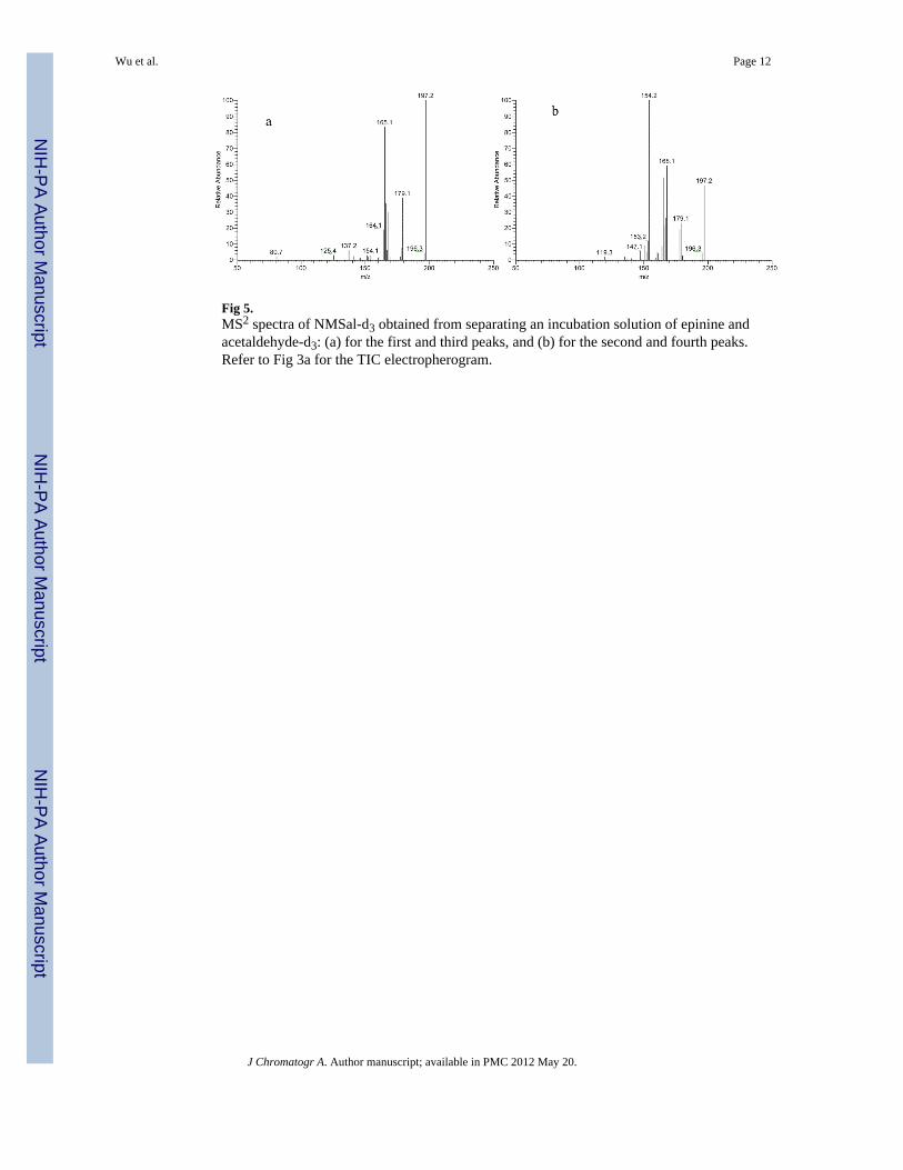
Fig 5.MS2 spectra of NMSal-d3 obtained from separating an incubation solution of epinine andacetaldehyde-d3: (a) for the first and third peaks, and (b) for the second and fourth peaks.Refer to Fig 3a for the TIC electropherogram.
Wu et al. Page 12
J Chromatogr A. Author manuscript; available in PMC 2012 May 20.
NIH
-PA Author Manuscript
NIH
-PA Author Manuscript
NIH
-PA Author Manuscript

Fig 6.Chemical structures of NMSal isomers formed from incubation of epinine withacetaldehyde. They were eluted into 4 peaks in the chiral CE-MS separation and identifiedby MS/MS.
Wu et al. Page 13
J Chromatogr A. Author manuscript; available in PMC 2012 May 20.
NIH
-PA Author Manuscript
NIH
-PA Author Manuscript
NIH
-PA Author Manuscript