Application of Coordination Compounds with Transition Metal ...
Upload
khangminh22Category
view
0download
0
CHEMOPATHOLOGICAL STUDIES W I T H COMPOUNDS OF ARSENIC.
IV. THE CHARACTER AND DISTRIBUTION OF RENAL INJURY PRO- DUCED BY ARSENICALS AS INDICATED BY TILE PROCESSES
OF REPAIR.
BY LOUISE PEARCE, M.D., AI'CD WADE H. BROWN, M.D.
(From the Laboratories of The Rockefeller Institute for Medical Research.)
PLATES 71 TO 76.
(Received for publication, January 31, 1916.)
In previous papers 1 attention was directed to some of the striking differences in the gross and histological changes produced in the kidneys of dogs by lethal doses of certain compounds of arsenic. Upon the basis of these observations it was pointed out that arseni- cals could produce a so called tubular as well as a vascular injury of the kidney and that innumerable combinations of these two funda- mental forms of tissue injury were obtainable through the use of arsenical compounds of different chemical constitution.
In order to obtain additional information as to the character and location of the specific renal injury produced by these arsenicals, as well as a knowledge of the subsequent processes of organic repair, we have extended our experiments to a study of tissue changes in the kidneys of animals given sublethal doses of these substances.
EXPERIMENTAL.
In the following experiments guinea pigs were used, as in our experi- ence the kidneys of these animals are as free from spontaneous lesions as those of any animal available for the purpose. However, it should be mentioned in this connection that in the kidneys of some guinea pigs we have found certain pathological changes that seem to be associ-
1Pearce, L., and Brown, W. H., Jour. Exper. Med., 1915, xxii, 517,525. 443
Dow
nloaded from http://rupress.org/jem
/article-pdf/23/4/443/1173999/443.pdf by guest on 28 May 2022
444 RENAL INJURY AND PROCESSES OF REPAIR
ated with the presence of a renal parasite3 The relative importance of this finding in interpreting the various lesions in the processes of repair will be considered in the discussion.
In our previous experiments 1 dogs were used because the compara- tively large size of their kidneys showed gross changes advanta- geously. However, as we stated then, the histological changes were controlled by experiments carried out with guinea pigs. The gross and microscopic changes in the kidneys of both dogs and guinea pigs following lethal and sublethal doses of these arsenicals were practi- cally identical with but one exception, that of arsenophenylglycine, to which we shall refer at greater length in discussing the results ob- tained with this compound.
Sterile solutions of arsenious acid, arsenic acid, sodium cacodylate, salvarsan, neosalvarsan, arsacetin, arsenophenylglycine, and atoxyl were injected intraperitoneally into adult male guinea pigs. With each compound, one sublethal dose was given and the animal killed after 2 to 5 days. In addition, with the exception of the salvarsan experiment, other guinea pigs were given two to five small doses of the drug and killed in 3 to 19 days. In a few instances the animal died within 24 hours after receiving a repeated dose. In these guinea pigs an acute lesion complicates the one of longer duration. We were guided in the determination of the size of the sublethal dose by our knowledge of the character and extent of the renal injury caused by lethal amounts of the arsenical in question. We tried always to in- ject a non-fatal dose, yet one large enough to cause definite renal injury.
Arsenious Acid.
The acute injury to the kidney produced by large doses of arseni- ous acid is primarily one of congestion and dilatation of the blood vessels with hemorrhage; the tubular epithelium is degenerated with occasional slight cell necrosis and usually there is a voluminous granular precipitate in the glomerular capsule and the tubules. The restoration to normal after a moderate dose of arsenious acid is very rapid and there is but slight evidence of the previous injury (Fig. 1). This consists of large, rather irregular glomeruli with somewhat
t Pearce, L., .Tour. Exper. Med., 1916, xxiil, 431.
Dow
nloaded from http://rupress.org/jem
/article-pdf/23/4/443/1173999/443.pdf by guest on 28 May 2022
LOUISE PEARCE AND WADE H. BROWN 445
shrunken tufts and a distinct although slight increase in tuft nuclei. About the base of the glomeruli are small accumulations of fibroblasts and round Cells (Fig. 1) which occasionally slightly invade the adjacent
• labyrinthine tissue. Nowhere have we seen any distortion or altera- tion in the usual architecture in either the cortex or the medulla. The tubular epithelium shows only slight degenerative changes with no evidences of tubular necrosis or regeneration.
Practically the same microscopic picture is found in Guinea Pigs 1, 2, 3, and 4 of this series (Table I), irrespective of the difference in size and number of doses given or the length of survival of the animal. The changes in Guinea Pig 2 are relatively more marked than in the others, although they are essentially of the same type.
TABLE I .
Arsenious AcM.
Animal No.
Day ofexperiment and dose of druginmg. per k~o of bodyweight.
I 2 5 6 11
5 3 3 10
10 10 10 8 5
Doses. Days survived. Termination.
3 Killed. 8 t~
12 Died.
6 "
The proliferative changes ill the kidney, therefore, after sublethal injections of arsenious acid are extremely slight and are confined to slight increase of nuclei in a somewhat shrunken glomerular tuft and to a slight accumulation of fibroblasts and round cells about the base of the glomeruli. There are no mitoses in the tubular epithelium, indicating a previous injury of this tissue with subsequent regener- ation.
Arsenic Acid.
The gross and microscopic pictures of the kidneys of guinea pigs poisoned with lethal doses of arsenic acid are practically the same as those of arsenious acid. Vascular injury is the predominating change and there is but slight tubular necrosis. However, the study of the kidneys of guinea pigs that have received sublethal doses of arsenic
Dow
nloaded from http://rupress.org/jem
/article-pdf/23/4/443/1173999/443.pdf by guest on 28 May 2022
446 RENAL INJURY AND PROCESSES OF R E P A I R
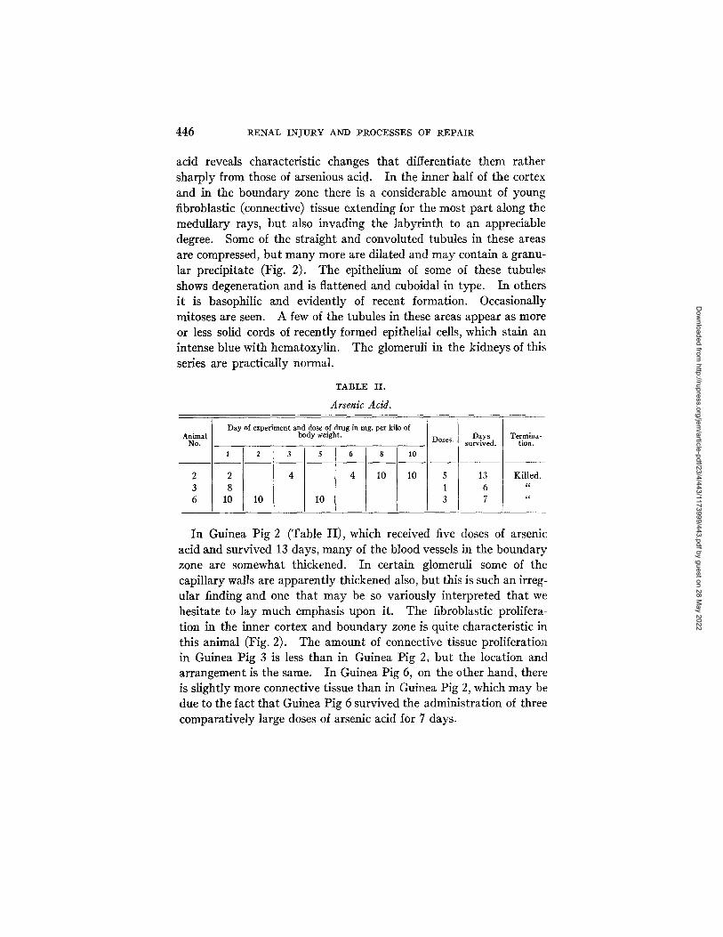
acid reveals characteristic changes that differentiate them rather sharply from those of arsenious acid. In the inner half of the cortex and in the boundary zone there is a considerable amount of young fibroblastic (connective) tissue extending for the most part along the medullary rays, but also invading the labyrinth to an appreciable degree. Some of the straight and convoluted tubules in these areas are compressed, but many more are dilated and may contain a granu- lar precipitate (Fig. 2). The epithelium of some of these tubules shows degeneration and is flattened and cuboidal in type. In others it is basophilic and evidently of recent formation. Occasionally mitoses are seen. A few of the tubules in these areas appear as more or less solid cords of recently formed epithelial cells, which stain an intense blue with hematoxylin. The glomeruli in the kidneys of this series are practically normal.
TABLE I I .
Arsenic Acid.
Animal No.
Day of experiment and dose of drug in mg. per kilo of body weight.
1 2 3 5
2 4
8
lO 10 10
6 8 10
4 10 10
Doses. survived.
5
1
3
Day s Termina- tion.
13 Killed.
6 "
7
In Guinea Pig 2 (Table II), which received five doses of arsenic acid and survived 13 days, many of the blood vessels in the boundary zone are somewhat thickened. In certain glomeruli some of the capillary walls are apparently thickened also, but this is such an irreg- ular finding and one that may be so variously interpreted that we hesitate to lay much emphasis upon it. The fibroblastic prolifera- tion in the inner cortex and boundary zone is quite characteristic in this animal (Fig. 2). The amount of connective tissue proliferation in Guinea Pig 3 is less than in Guinea Pig 2, but the location and arrangement is the same. In Guinea Pig 6, on the other hand, there is slightly more connective tissue than in Guinea Pig 2, which may be due to the fact that Guinea Pig 6 survived the administration of three comparatively large doses of arsenic acid for 7 days.
Dow
nloaded from http://rupress.org/jem
/article-pdf/23/4/443/1173999/443.pdf by guest on 28 May 2022
LOUISE PEARCE AND WADE H. BROWN 447
In contrasting arsenious and arsenic acid, which resemble each other so closely in the character of the acute renal lesion, it appears that there are distinct differences in their action which are revealed in the process of repair. With arsenious acid, proliferative changes are al- most wanting and are practically exclusively confined to the slight increase in nuclei of the glomerular tuft and to slight fibroblastic accumulation about the base of the glomeruli. With arsenic acid, on the other hand, proliferative activity of fibroblasts is appreciably more marked, and is associated with distortion of the tubules espe- cially in the inner half of the cortex and boundary zone and with dis- tinct (although slight) regeneration of tubular epithelium.
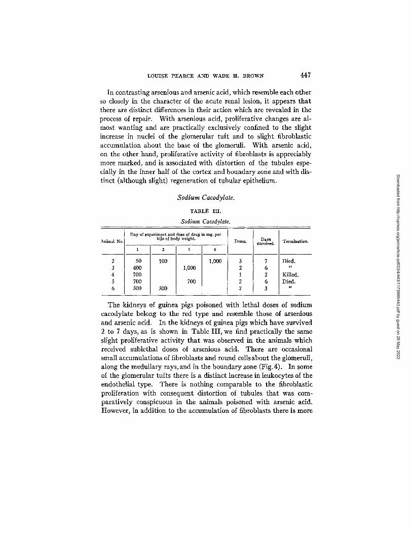
Sodium Cacodylate.
TABLE III.
Sodium Cacodylate. i
Animal No.
Day of experiment and dose of drug in rag. per kilo of body weight.
2 50 3 400 4 700 5 700 6 500
2
100
500
1,000
700
6
1,000
Doses. D a y s survived. Termination.
Died.
Killed. Died.
tc
The kidneys of guinea pigs poisoned with lethal doses of sodium cacodylate belong to the red type and resemble those of arsenious and arsenic acid. In the kidneys of guinea pigs which have survived 2 to 7 days, as is shown in Table III, we find practically the same slight proliferative activity that was observed in the animals which received sublethal doses of arsenious acid. There are occasional small accumulations of fibroblasts and round cells about the glomeruli, along the medullary rays, and in the boundary zone (Fig. 4). In some of the glomerular tufts there is a distinct increase in leukocytes of the endothelial type. There is nothing comparable to the fibroblastic proliferation with consequent distortion of tubules that was com- paratively conspicuous in the animals poisoned with arsenic acid. However, in addition to the accumulation of fibroblasts there is more
Dow
nloaded from http://rupress.org/jem
/article-pdf/23/4/443/1173999/443.pdf by guest on 28 May 2022
448 RENAL INJURY AND PROCESSES OF REPAIR
necrosis of the tubular epithelium than with either arsenious or arsenic acid, and mitoses may be found in both the convoluted and the straight tubules.
There is but little difference, histologically, in the kidneys of the various guinea pigs of this series (Table III). In Guinea Pig 2 there are perhaps more and slightly larger accumulations of fibroblasts. Mitoses are found in the tubular epithelium of all five animals, but they ~re more numerous in Guinea Pig 6.
There is scarcely any fibroblastic proliferation, therefore, in the animals poisoned with sublethal doses of sodium cacodylate as well as with arsenious acid,--in this respect differing from those poisoned with arsenic acid. But, in addition, there is distinct reparative activity on the part of the tubular epithelium, indicating that this tissue has been injured by the drug, and in this respect differing from guinea pigs poisoned with sublethal doses of both arsenious and arsenic acids, in which the tubular epithelium is but little affected.
Salvarsan.
We have but one instance in which a sublethal dose of salvarsan was given and the animal allowed to live for more than 24 hours. This guinea pig received 150 rag. of the drug per kilo of body weight and was killed 48 hours after its administration.
The acute salvarsan kidney is a red kidney. In this one example of an early stage in the process of repair the glomeruli are large and rather irregular. The tufts on the whole are not shrunken and there is an accumulation of endothelial leukocytes in some of them. Many of the walls of the tuft capillaries are hyaline and distinctly thick- ened. About the base of some of the glomeruli there is a collection of fibroblasts and round cells which in some instances extends slightly outward into the labyrinth between the convoluted tubules. In the boundary zone and along the inner portion of the medullary rays are small irregular patches of young fibroblasts (Fig. 5), but there is no distortion of the tubular structures. The tubular epithelium is markedly degenerated but there is no definite epithelial necro- sis, and since we have seen no mitoses we may infer that the tubular epithelium was not seriously injured. I t should be stated in this
Dow
nloaded from http://rupress.org/jem
/article-pdf/23/4/443/1173999/443.pdf by guest on 28 May 2022
L O U I S E P E A R C E A N D W A D E I I . B R O W N 449
connection, however, that this animal received the dose of salvarsan intraperitoneally and that the compound was absorbed relatively slowly, for when the animal was killed 2 days after injection, the abdominal cavity contained a quantity of unabsorbed drug.
The evidences of repair, seen in the kidneys of guinea pigs after salvarsan injection, are therefore chiefly the changes in the glomeruli and the irregular proliferation of fibroblasts about the glomeruli and in the boundary zone.
Neosalvarsan.
The acute neosalvarsan kidney resembles those of arsenious acid and salvarsan. In Guinea Pig 1, killed on the 2nd day of survival, as shown in Table IV, we find definite signs of a proliferative activity
TABLE IV.
Neosalvarsan.
Animal No.
Day of experiment and dose of drug in rag. per kilo of
body weight. Doses. Days survived. Termination.
1 5
1 300 l 2 Killed. 2 190 500 2 6 Died.
in the kidney in addition to the acute lesion which is still present. About the base of the glomeruli are small accumulations of fibro- blasts and round cells. In addition there is a slight diffuse infiltration of the same character along the medullary rays in both cortex and medulla which in some areas invades the adjacent labyrinthine tissue. The glomeruli themselves are extremely irregular; some are of normal size, while others are markedly shrunken with a contracted tuft filling approximately three-quarters of the capsular space. The tubular epithelium is considerably degenerated and in the inner portion of the cortex there is some individual cell disintegration of the epithelium of the loops of Henle and a slight degree of necrosis. A few mitotic figures are seen.
The changes in Guinea Pig 2 are of the same general character as those in Guinea Pig 1, although of a lesser degree. This may b'e
Dow
nloaded from http://rupress.org/jem
/article-pdf/23/4/443/1173999/443.pdf by guest on 28 May 2022
450 RENAL INJURY AND PROCESSES OF REPAIR
due to the much smaller initial dose. The capsules of Bowman and the capillary walls of some of the glomerular tufts are slightly hyaline and thickened. There are small, irregular patches of fibro- blasts and round cells about the glomeruli, along the medullary rays, and in the boundary zone. The tubular epithelium is degenerated with occasional slight necrosis. No mitoses are seen.
The changes in the process of repair after neosalvarsan resemble in general those after salvarsan, differing only in the greater irreg- ularity of the glomeruli and the somewhat greater degree of tubular necrosis which is most marked in the inner portion of the cortex. After both salvarsan and neosalvarsan there is a patchy irregular proliferation of fibroblasts in the boundary zone and cortex, but there is no distortion of the tubules as with arsenic acid.
A r s a c e t i n .
TABLE V.
Arsacetin.
Animal No.
1
2
3
Day of experiment and dose of drug in mg. per kilo of body weight.
1 .5 13 16
125 250
100 200
300
200 200
Doses. survived.
2 4 1
Days Termination.
6 Killed. 19 "
2
The kidneys of dogs and guinea pigs which have received lethal doses of arsacetin are pale and show a most extensive tubular necrosis with a subordinate vascular injury. In the reaction from a non- fatal dose of this compound there is an exceedingly active and prompt regeneration of the epithelium, especially in the loops of Henle, and to a somewhat less extent in the convoluted tubules and the tubules of the medulla (Figs. 6, 7, and 8). The glomerular capillaries are some- what dilated and there may be a slight and irregular accumulation of fibroblasts and polyblasts about the base of the glomeruli, along the medullary rays, and in the boundary zone. In Guinea Pig 3 (Table V), killed on the 2nd day, there is already marked activity of the tubular epithelium throughout the cortex and to a less extent in
Dow
nloaded from http://rupress.org/jem
/article-pdf/23/4/443/1173999/443.pdf by guest on 28 May 2022
LOUISE PEARCE AND WADE H. BROWN 451
the medulla. There is practically no vascular alteration and no fibro- blastic proliferation in this animal. Guinea Pig 1 was killed on the 6th day after having received two doses of arsacetin. The kidney sections of this animal show a moderate regeneration of the tubular epithelium, almost exclusively confined to the loops of Henle and the medullary tubules. About the base of some of the glomeruli there is a very slight degree of fibroblastic proliferation. Four moderate sized doses of arsacetin were given to Guinea Pig 2 in 16 days, and the animal was killed on the 19th day (Table V). In this animal the regenerative activity of the tubular epithelium has reached a remark- able degree. Mitotic figures are found in practically every microscopic field (4 mm. Zeiss objective; No. 4 ocular) and they are particularly numerous in the straight tubules of the cortex and medulla (Figs. 6, 7, and 8). Many of the tubules along the medullary rays show entirely new epithelium and some of these tubules are almost solid masses of new cells. Degeneration with some necrosis and desquamation of cells is still present in some of the convoluted tubules and limbs of Henle and there are many hyaline casts in the boundary zone and medulla. In the boundary zone, along the medullary rays, and about the base of some of the glomeruli there is a rather diffuse proliferation of fibroblasts and infiltration of polyblasts which irregularly invade the labyrinth in some areas. Several of the renal tubules of this animal contain the parasite which we have referred to and which must be taken into consideration in interpreting the proliferative changes about the glomeruli.
Atoyxl.
The acute lesion in the kidneys of dogs and guinea pigs poisoned with lethal amounts of atoxyl is predominantly tubular, but the vas- cular changes are by no means inconspicuous. In guinea pigs which have received sublethal doses of atoxyl, there is a rapid a t t empt at regeneration of the tubular epithelium which is shown in both animals of this series (Table VI). Many of the straight tubules are dilated and lined with low cuboidal basophilic staining cells (Fig. 9), and there are numerous mitoses in both cortical and medullary epithelium (Figs. 9, 10, and 11). There is a well marked leukocytic exudate
Dow
nloaded from http://rupress.org/jem
/article-pdf/23/4/443/1173999/443.pdf by guest on 28 May 2022
452 RENAL INJURY AND PROCESSES OF REPAIR
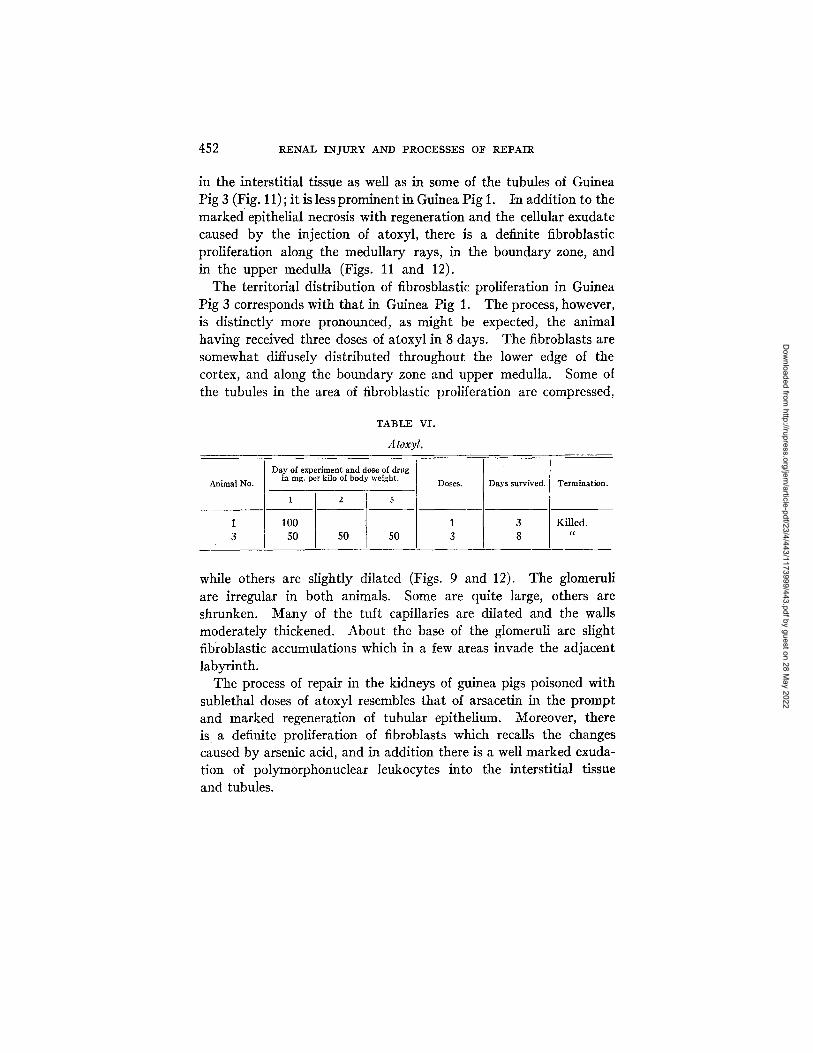
in the interstitial tissue as well as in some of the tubules of Guinea Pig 3 (Fig. 11) ; it is less prominent in Guinea Pig 1. tn addition to the marked epithelial necrosis with regeneration and the cellular exudate caused by the injection of atoxyl, there is a definite fibroblastic proliferation along the medullary rays, in the boundary zone, and in the upper medulla (Figs. 11 and 12).
The territorial distribution of fibrosblastic proliferation in Guinea Pig 3 corresponds with that in Guinea Pig 1. The process, however, is distinctly more pronounced, as might be expected, the animal having received three doses of atoxyl in 8 days. The fibroblasts are somewhat diffusely distributed throughout the lower edge of the cortex, and along the boundary zone and upper medulla. Some of the tubules in the area of fibroblastic proliferation are compressed,
TABLE V I .
A toxyl.
Day of experiment and dose of drug I Animal No. [ _ _ i n rag, per kilo of body weight. [ Doses.
1 2 5
31 1050 50 50 31
Days survived. Termination.
Killed.
while others are slightly dilated (Figs. 9 and 12). The glomeruli are irregular in both animals. Some are quite large, others are shrunken. Many of the tuft capillaries are dilated and the walls moderately thickened. About the base of the glomeruli are slight fibroblastic accumulations which in a few areas invade the adjacent labyrinth.
The process of repair in the kidneys of guinea pigs poisoned with sublethal doses of atoxyl resembles that of arsacetin in the prompt and marked regeneration of tubular epithelium. Moreover, there is a definite proliferation of fibroblasts which recalls the changes caused by arsenic acid, and in addition there is a well marked exuda- tion of polymorphonuclear leukocytes into the interstitial tissue and tubules.
Dow
nloaded from http://rupress.org/jem
/article-pdf/23/4/443/1173999/443.pdf by guest on 28 May 2022
LOUISE PEARCE AND WADE H. BROWN 453
Arsenophenylglycine.
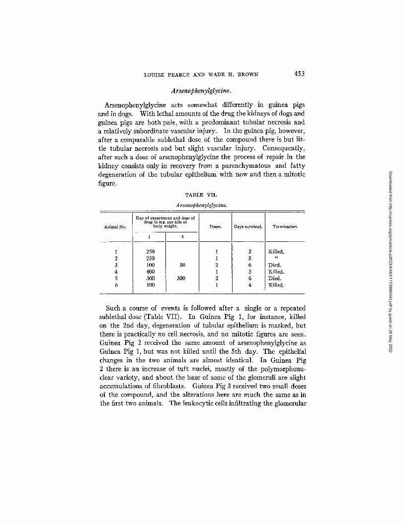
Arsenophenylglycine acts somewhat differently in guinea pigs and in dogs. With lethal amounts of the drug the kidneys of dogs and guinea pigs are both pale, with a predominant tubular necrosis and a relatively subordinate vascular injury. In the guinea pig, however, after a comparable sublethal dose of the compound there is but lit- tle tubular necrosis and but slight vascular injury. Consequently, after such a dose of arsenophenylglycine the process of repair in the kidney consists only in recovery from a parenchymatous and fat ty degeneration of the tubular epithelium with now and then a mitotic figure.
TABLE VII.
Arsenophenylglycine.
Animal No.
Day of experiment and dose of drug in mg, per kilo of
body weight.
1 5
250 250 100 50 400 300 300 500
Doses. Days survived. Termination.
Killed. gc
Died. Killed. Died. Killed.
Such a course of events is followed after a single or a repeated sublethal dose (Table VII). In Guinea Pig 1, for instance, killed on the 2nd day, degeneration of tubular epithelium is marked, but there is practically no cell necrosis, and no mitotic figures are seen. Guinea Pig 2 received the same amount of arsenophenylglycine as Guinea Pig 1, but was not killed until the 5th day. The epithelial changes in the two animals are almost identical. In Guinea Pig 2 there is an increase of tuft nuclei, mostly of the polymorphonu- clear variety, and about the base of some of the glomeruli are slight accumulations of fibroblasts. Guinea Pig 3 received two small doses of the compound, and the alterations here are much the same as in the first two animals. The leukocytic cells infiltrating the glomerular
Dow
nloaded from http://rupress.org/jem
/article-pdf/23/4/443/1173999/443.pdf by guest on 28 May 2022
454 RENAL INJURY AND PROCESSES 0]~ REPAIR
tuft are mostly eosinophilic; there is a very irregular thickening of the wall of the glomerular capillaries and an extremely irregular and slight distribution of round cells and fibroblasts about the base of some of the glomeruli, along the straight vessels, and in the laby- rinth. The tubular epithelium is swollen and granular and there is some necrosis of a disintegrative character in the epithelium of the outer cortex. An occasional mitotic figure is seen.
The acute changes in the kidneys of guinea pigs after sublethal doses of arsenophenylglycine consist almost entirely of parenchyma- tous and fatty degeneration of the tubular epithelium with but little cell necrosis, differing in this respect from a similar sublethal injury in the kidneys of dogs. The processes of repair after such an injury are, therefore, comparatively simple and do not involve any appre- ciable degree of cell regeneration. There is only a slight and very irregular interstitial fibroblastic proliferation.
DISCUSSION.
A study of the processes of repair in the kidneys of guinea pigs poisoned with sublethal doses of certain arsenical compounds fur- nishes additional information as to the character and location of the acute injury. The idea, which we have previously suggested, namely, that all arsenicals do not produce a purely vascular type of renal in- jury, is further substantiated by this series of experiments in which the regeneration of tubular epithelium plays a relatively conspicuous part. The participation of the epithelial tissue, however, is by no means the dominant feature in the recovery after sublethal doses of all arsenical compounds, but only after certain particular ones. In others, the acute injury is mainly vascular, and the reaction of the epithelial structures is but slight. Further, the interstitial proliferation of fibroblasts which occurs in a marked degree after injections of various arsenicals may be especially pronounced in the repair following a drug which, in lethal doses, causes but little vascular disturbance, as, for instance, arsacetin. Here, the initial and domi- nant injury is epithelial; there is only slight demonstrable alteration of the vascular structures. In the process of recovery, however, after the administration of sublethal doses of arsacetin, there may be quite
Dow
nloaded from http://rupress.org/jem
/article-pdf/23/4/443/1173999/443.pdf by guest on 28 May 2022
LOUISE PEARCE AND WADE H. BROWN 455
a marked proliferation of young fibroblasts diffusely distributed (Table V, Guinea Pig 2, and Fig. 8). This is also seen in the repair of the kidney following injections of atoxyl, although here the picture is more complicated, for atoxyl affects the vascular as well as the epithelial tissue of the kidney. The distribution of the proliferating fibroblasts after a more purely vascular injury such as that pro- duced by arsenious or arsenic acid is confined more sharply to the boundary zone, with radiations along the medullary rays to the glomeruli. On the other hand, after arsacetin, the fibroblastic pro- liferafion is more diffusely distributed throughout the cortex, al- though it may be more numerous in the boundary zone and along the medullary rays.
Taking all these observations into consideration, therefore, it would seem that a toxic agent like arsenic, may, in one or another of its various combinations, injure the vascular, epithelial, or interstitial (connective) tissue of the kidney, if we may judge of such an initial injury by the subsequent processes of repair. The relative dis- tribution and extent of the initial injury may be difficult to determine until one studies various stages of the recovery of the kidney, in which the injured tissues are regenerating. This is particularly true in the case of injury of the connective tissue of the kidney.
After a sublethal dose of arsenious acid which produces an al- most pure type of vascular injury in the kidneys, the return to nor- mal is very rapid and there is only a slight fibroblastic proliferation about the base of the glomeruli to indicate a previous injury. In- jections of sublethal amounts of salvarsan also cause a proliferation of fibroblasts of a more interstitial and rather patchy character, some- what greater in extent and amount than with arsenious acid. On the other hand, with arsenic acid, there is a relatively large amount of fibroblastic proliferation, especially in the boundary zone, resulting in the compression or dilatation of some of the tubules, and in addition a slight although definite regeneration of tubular epithelium. Sodium cacodylate in sublethal amounts causes essentially the same slight fibroblastic changes seen after arsenious acid, but a well marked regeneration of tubular epithelium as well. With all these four com- pounds, arsenious and arsenic acid, salvarsan, and sodium cacodylate, there are slight but fairly regular glomerular changes, consisting of
Dow
nloaded from http://rupress.org/jem
/article-pdf/23/4/443/1173999/443.pdf by guest on 28 May 2022

456 RENAL INJURY AND PROCESSES OF REPAIR
a slightly swollen tuft and an increase in the tuft nuclei. With neosalvarsan, however, the glomeruli are extremely irregular, some being very large, others contracted and shrunken. There is a slight interstitial fibroblastic proliferation, comparable to that of salvarsan and a slight regeneration of tubular epithelium. After sublethal injections of atoxyl, there is very marked regenerative activity of the tubular epithelium, a leukocytic exudate, and a definite and rather diffuse interstitial proliferation of fibroblasts with dilatation or com- pression of adjacent tubules. Following a sublethal injection of arsacetin, there is a conspicuous and prompt regeneration of tubular epithelium and a relatively diffuse interstitial proliferation of fibro- blasts. Unfortunately, we must omit the experiments of arsenophe- nylglycine on the guinea pig in this consideration, for the reasons stated above.
I t would seem, therefore, that arsenicals which produce an injury that is primarily vascular may lead to only a slight subsequent proliferation of tissue (arsenious acid). However, other arsenicals which produce an acute injury that is vascular, may lead to distinct interstitial proliferation (axsenic acid, salvarsan, and neosalvarsan). Moreover, an arsenical that produces an acute injury that is primarily vascular may also produce injury of the tubular epithelium which in the stages of repair dominates the picture to the exclusion of any extensive interstitial proliferation (sodium cacodylate).
On the other hand, arsenicals that produce primarily an injury of tubular epithelium cause a marked subsequent regeneration of this tissue, and may cause in addition an interstitial fibroblasfic prolifera- tion (arsacetin). But an arsenical compound that produces a marked vascular injury in addition to the picture of tubular necrosis may lead to a relatively marked interstitial proliferation as well as the extensive epithelial regeneration (atoxyl).
In connection with the fibroblasfic proliferation described in the processes of repair in these kidneys, we must refer to the presence of a parasite in the kidneys of some guinea pigs3 This parasite is only occasionally found in guinea pigs, but we believe that it may cause irregular accumulations of fibroblasts and round cells about the base of some of the glomeruli and in the neighboring labyrinthine tissue. These accumulations resemble those seen after sublethal injection
Dow
nloaded from http://rupress.org/jem
/article-pdf/23/4/443/1173999/443.pdf by guest on 28 May 2022

LOUISE PEARCE AND WADE H. BROWN 457
of arsenious acid, but they are much more irregular. Therefore, we wish to be particularly cautious in a final interpretation of our results and to take into consideration the possibility that some of the fibroblastic proliferation we have seen in the kidneys of guinea pigs after sublethal injections of these arsenicals may be due in part to this renal parasite.
We have shown that after acute lethal injury with various arsenicals, two types of kidneys could be distinguished, both grossly and his- tologically, namely, the red and the pale. Further, in the reaction after a sublethal injury inflicted with these compounds these two main types of kidney alteration may still be differentiated by the processes of repair. The administration of those compounds which produce a predominantly pale kidney is followed by a most prompt and pronounted regeneration of tubular epithelium with a varying degree of diffuse fibroblastic proliferation. The compounds that produce a predominantly red kidney are followed by proliferation of fibroblasts with but slight regeneration of the tubular epithelium. The distribution of the fibroblasts in the reparative stages ap- parently corresponds in some degree to the initial injury. After a more or less predominant vascular injury the fibroblasts are usually found about the glomeruli, along the medullary rays, and in the boundary zone. With other compounds that show little evidence of an acute vascular injury, the connective tissue injury may be more pronounced, and in recovery from such an injury the fibroblasts are diffusely distributed throughout the cortex and medulla in a typical interstitial manner. But, just as there are innumerable combinations of vascular and tubular and probably connective tissue injury in the acute arsenic kidneys, so there are innumerable combinations of the different tissue elements in the processes of repair. Hence, we suggest that the character and distribution of renal injury produced by arsenical compounds as indicated by the processes .~f repair are bound up in the chemical constitution of these compounds. Further, that each particular compound as far as the kidney is concerned, acts as a more or less specific toxic agent, as shown by the character and distribution of the renal lesions.
Dow
nloaded from http://rupress.org/jem
/article-pdf/23/4/443/1173999/443.pdf by guest on 28 May 2022
458 RENAL INJURY AND PROCESSES OF REPAIR
SU'M.MARY.
1. The processes of repair in the kidneys of guinea pigs af ter sub- lethal doses of certain arsenical compounds indicate tha t all arsenicals do not produce a purely vascular type of renal injury.
2. While some arsenicals produce a predominant ly vascular injury and others produce a predominant ly tubular injury, bo th these tissue elements are undoubted ly always affected, al though in varying pro- portion. In addition, the interstit ial connective tissue is probably always affected. The diffuse proliferation of this tissue m a y be relatively conspicuous in the processes of repair af ter arsenicals tha t cause bu t slight vascular injury.
3. All red kidneys do not necessarily show identical pictures dur- ing the processes of repair; the same is true of pale kidneys.
4. The mode of action of an arsenical compound as a renal toxic
agent is bound up with the chemical const i tut ion of the compound.
EXPLANATION OF PLATES.
The illustrations are all from untouched microphotographs.
PLATE 71.
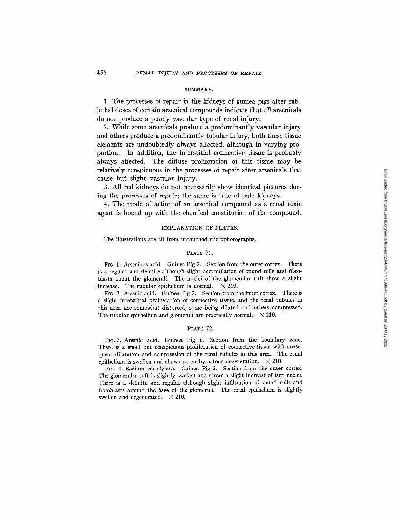
Fla. 1. Arsenious acid. Guinea Pig 2. Section from the outer cortex. There is a regular and definite although slight accumulation of round cells and fibro- blasts about the glomeruli. The nuclei of the glomerular tuft show a slight increase. The tubular epithelium is normal. X 210.
FIG. 2. Arsenic acid. Guinea Pig 2. Section from the inner cortex. There is a slight interstitial proliferation of connective tissue, and the renal tubules in this area are somewhat distorted, some being dilated and others compressed. The tubular epithelium and glomeruli are practically normal. X 210.
PLATE 72.
FIG. 3. Arsenic acid. Guinea Pig 6. Section from the boundary zone. There is a small but conspicuous proliferation of connective tissue with conse- quent dilatation and compression of the renal tubules in this area. The renal epithelium is swollen and shows parenchymatous degeneration. X 210.
FIG. 4. Sodium cacodylate. Guinea Pig 2. Section from the outer cortex. The glomerular tuft is slightly swollen and shows a slight increase of tuft nuclei. There is a definite and regular although slight infiltration of round cells and fibroblasts around the base of the glomeruli. The renal epithelium is slightly swollen and degenerated. X 210.
Dow
nloaded from http://rupress.org/jem
/article-pdf/23/4/443/1173999/443.pdf by guest on 28 May 2022

THE JOURNAL OF EXPERIMENTAL MEDICINE VOL. X X I I I . PLATE 71.
(Pearce and Brown: Renal Injury and Repair.)
Dow
nloaded from http://rupress.org/jem
/article-pdf/23/4/443/1173999/443.pdf by guest on 28 May 2022

THE JOURNAL OF EXPERIMENTAL MEDICINE VOL. X X I I I . PLATE 7?.
(Pearce and Brown: Renal Injury and Repair.)
Dow
nloaded from http://rupress.org/jem
/article-pdf/23/4/443/1173999/443.pdf by guest on 28 May 2022

THE JOURNAL OF EXPERIMENTAL MEDICINE VOL. XX I I I . PLATE 73.
(Pearce and Brown; Renal Injury and Repair.I
Dow
nloaded from http://rupress.org/jem
/article-pdf/23/4/443/1173999/443.pdf by guest on 28 May 2022

THE JOURNAL OF EXPERIMENTAL MEDICINE VOL. X X I I I . PLATE 74.
CPearce and l~rown: Renal Injury and Repair.)
Dow
nloaded from http://rupress.org/jem
/article-pdf/23/4/443/1173999/443.pdf by guest on 28 May 2022

THE JOURNAL OF EXPERIMENTAL MEDICINE VOL. X X I l l . PLATE 75.
(Pearce: and Brown: Renal Injury and Repair.)
Dow
nloaded from http://rupress.org/jem
/article-pdf/23/4/443/1173999/443.pdf by guest on 28 May 2022
THE JOURNAL OF EXPERIMENTAL MEDICINE VOL. X X l l l . PLATE 76.
(Pearce and Brown: Renal Injury and Repair.)
Dow
nloaded from http://rupress.org/jem
/article-pdf/23/4/443/1173999/443.pdf by guest on 28 May 2022

LOUISE PEARCE AND WADE H. BROWN 459
PLATE 73.
FIG. S. Salvarsan. Guinea Pig 1. Section from the boundary zone. There is an irregular patchy proliferation of connective tissue. The renal epithelium is practically normal. X 210.
FIG. 6. Arsacetin. Guinea Pig 2. Section from the outer cortex. There is a marked degeneration and regeneration of the tubular epithelium with numer- ous mitotic figures. There is a granular precipitate in the lumen of the tubules which appear slightly dilated. The glomeruli are normal in appearance. There is a slight diffuse proliferation of fibroblasts. X 210.
PLATE 74.
FIG. 7. Arsacetin. Guinea Pig 3. Section from the midcortex. There is a moderate degeneration and marked regeneration of the tubular epithelium with numerous mitotic figures. There is a granular precipitate in the lumen of the tubules. × 210.
FIG. 8. Arsacetin. Guinea Pig 2. Section from the upper medulla. There is marked regeneration of the tubular epithelium with numerous mitotic figures. There is a slight but definite interstitial proliferation of fibroblasts. × 210.
PLATE 75.
FIG. 9. Atoxyl. Guinea Pig 3. Section from the inner cortex. There is a conspicuous dilatation of some of the tubules with a granular precipitate in the lumen. The tubular epithelium shows regeneration with numerous mitotic figures. The glomeruli appear normal. X 210.
FIG. 10. Atoxyl. Guinea Pig 1. Section from the outer cortex. There are two adjacent mitotic figures in the epithelium of a convoluted tubule. X 1,470.
PLA~ 76.
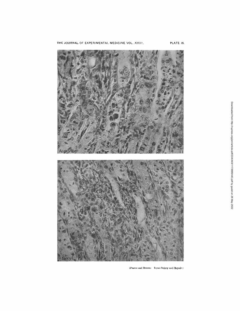
FIG. 11. Atoxyl. Guinea Pig 3. Section from the boundary zone. There is a conspicuous leukocytic infiltration in the tubules and interstitial tissue. The tubular epithelium shows numerous mitotic figures. There is some interstitial proliferation of the connective tissue. × 378
FIc. 12. Atoxyl. Guinea Pig 3. Section from the boundary zone. There is a diffuse interstitial proliferation of connective tissue. The tubular epithe- lium shows several mitotic figures. × 378.
Dow
nloaded from http://rupress.org/jem
/article-pdf/23/4/443/1173999/443.pdf by guest on 28 May 2022
Copyright © 2022 FDOKUMEN