Chemical and in Vitro Assessment of Alaskan Coastal Vegetation Antioxidant Capacity
8
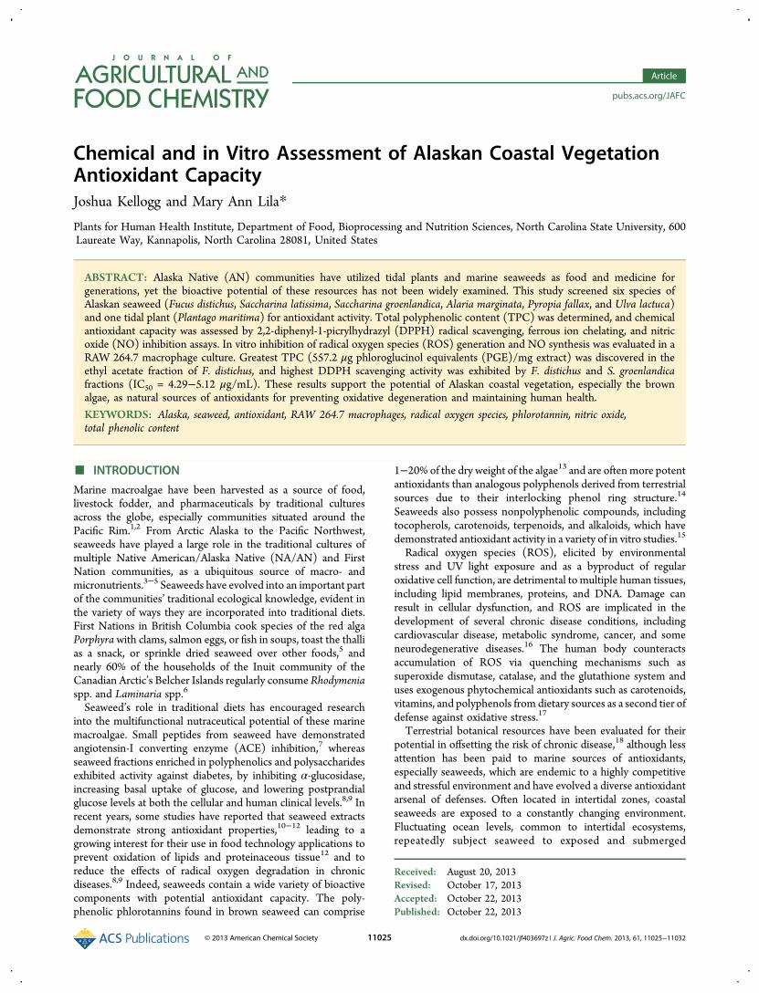
Chemical and in Vitro Assessment of Alaskan Coastal Vegetation Antioxidant Capacity Joshua Kellogg and Mary Ann Lila* Plants for Human Health Institute, Department of Food, Bioprocessing and Nutrition Sciences, North Carolina State University, 600 Laureate Way, Kannapolis, North Carolina 28081, United States ABSTRACT: Alaska Native (AN) communities have utilized tidal plants and marine seaweeds as food and medicine for generations, yet the bioactive potential of these resources has not been widely examined. This study screened six species of Alaskan seaweed (Fucus distichus, Saccharina latissima, Saccharina groenlandica, Alaria marginata, Pyropia fallax, and Ulva lactuca) and one tidal plant (Plantago maritima) for antioxidant activity. Total polyphenolic content (TPC) was determined, and chemical antioxidant capacity was assessed by 2,2-diphenyl-1-picrylhydrazyl (DPPH) radical scavenging, ferrous ion chelating, and nitric oxide (NO) inhibition assays. In vitro inhibition of radical oxygen species (ROS) generation and NO synthesis was evaluated in a RAW 264.7 macrophage culture. Greatest TPC (557.2 μg phloroglucinol equivalents (PGE)/mg extract) was discovered in the ethyl acetate fraction of F. distichus, and highest DDPH scavenging activity was exhibited by F. distichus and S. groenlandica fractions (IC 50 = 4.29−5.12 μg/mL). These results support the potential of Alaskan coastal vegetation, especially the brown algae, as natural sources of antioxidants for preventing oxidative degeneration and maintaining human health. KEYWORDS: Alaska, seaweed, antioxidant, RAW 264.7 macrophages, radical oxygen species, phlorotannin, nitric oxide, total phenolic content ■ INTRODUCTION Marine macroalgae have been harvested as a source of food, livestock fodder, and pharmaceuticals by traditional cultures across the globe, especially communities situated around the Pacific Rim. 1,2 From Arctic Alaska to the Pacific Northwest, seaweeds have played a large role in the traditional cultures of multiple Native American/Alaska Native (NA/AN) and First Nation communities, as a ubiquitous source of macro- and micronutrients. 3−5 Seaweeds have evolved into an important part of the communities’ traditional ecological knowledge, evident in the variety of ways they are incorporated into traditional diets. First Nations in British Columbia cook species of the red alga Porphyra with clams, salmon eggs, or fish in soups, toast the thalli as a snack, or sprinkle dried seaweed over other foods, 5 and nearly 60% of the households of the Inuit community of the Canadian Arctic’s Belcher Islands regularly consume Rhodymenia spp. and Laminaria spp. 6 Seaweed’s role in traditional diets has encouraged research into the multifunctional nutraceutical potential of these marine macroalgae. Small peptides from seaweed have demonstrated angiotensin-I converting enzyme (ACE) inhibition, 7 whereas seaweed fractions enriched in polyphenolics and polysaccharides exhibited activity against diabetes, by inhibiting α-glucosidase, increasing basal uptake of glucose, and lowering postprandial glucose levels at both the cellular and human clinical levels. 8,9 In recent years, some studies have reported that seaweed extracts demonstrate strong antioxidant properties, 10−12 leading to a growing interest for their use in food technology applications to prevent oxidation of lipids and proteinaceous tissue 12 and to reduce the effects of radical oxygen degradation in chronic diseases. 8,9 Indeed, seaweeds contain a wide variety of bioactive components with potential antioxidant capacity. The poly- phenolic phlorotannins found in brown seaweed can comprise 1−20% of the dry weight of the algae 13 and are often more potent antioxidants than analogous polyphenols derived from terrestrial sources due to their interlocking phenol ring structure. 14 Seaweeds also possess nonpolyphenolic compounds, including tocopherols, carotenoids, terpenoids, and alkaloids, which have demonstrated antioxidant activity in a variety of in vitro studies. 15 Radical oxygen species (ROS), elicited by environmental stress and UV light exposure and as a byproduct of regular oxidative cell function, are detrimental to multiple human tissues, including lipid membranes, proteins, and DNA. Damage can result in cellular dysfunction, and ROS are implicated in the development of several chronic disease conditions, including cardiovascular disease, metabolic syndrome, cancer, and some neurodegenerative diseases. 16 The human body counteracts accumulation of ROS via quenching mechanisms such as superoxide dismutase, catalase, and the glutathione system and uses exogenous phytochemical antioxidants such as carotenoids, vitamins, and polyphenols from dietary sources as a second tier of defense against oxidative stress. 17 Terrestrial botanical resources have been evaluated for their potential in offsetting the risk of chronic disease, 18 although less attention has been paid to marine sources of antioxidants, especially seaweeds, which are endemic to a highly competitive and stressful environment and have evolved a diverse antioxidant arsenal of defenses. Often located in intertidal zones, coastal seaweeds are exposed to a constantly changing environment. Fluctuating ocean levels, common to intertidal ecosystems, repeatedly subject seaweed to exposed and submerged Received: August 20, 2013 Revised: October 17, 2013 Accepted: October 22, 2013 Published: October 22, 2013 Article pubs.acs.org/JAFC © 2013 American Chemical Society 11025 dx.doi.org/10.1021/jf403697z | J. Agric. Food Chem. 2013, 61, 11025−11032
Transcript of Chemical and in Vitro Assessment of Alaskan Coastal Vegetation Antioxidant Capacity
Chemical and in Vitro Assessment of Alaskan Coastal VegetationAntioxidant CapacityJoshua Kellogg and Mary Ann Lila*
Plants for Human Health Institute, Department of Food, Bioprocessing and Nutrition Sciences, North Carolina State University, 600Laureate Way, Kannapolis, North Carolina 28081, United States
ABSTRACT: Alaska Native (AN) communities have utilized tidal plants and marine seaweeds as food and medicine forgenerations, yet the bioactive potential of these resources has not been widely examined. This study screened six species ofAlaskan seaweed (Fucus distichus, Saccharina latissima, Saccharina groenlandica, Alaria marginata, Pyropia fallax, and Ulva lactuca)and one tidal plant (Plantago maritima) for antioxidant activity. Total polyphenolic content (TPC) was determined, and chemicalantioxidant capacity was assessed by 2,2-diphenyl-1-picrylhydrazyl (DPPH) radical scavenging, ferrous ion chelating, and nitricoxide (NO) inhibition assays. In vitro inhibition of radical oxygen species (ROS) generation and NO synthesis was evaluated in aRAW 264.7 macrophage culture. Greatest TPC (557.2 μg phloroglucinol equivalents (PGE)/mg extract) was discovered in theethyl acetate fraction of F. distichus, and highest DDPH scavenging activity was exhibited by F. distichus and S. groenlandicafractions (IC50 = 4.29−5.12 μg/mL). These results support the potential of Alaskan coastal vegetation, especially the brownalgae, as natural sources of antioxidants for preventing oxidative degeneration and maintaining human health.
KEYWORDS: Alaska, seaweed, antioxidant, RAW 264.7 macrophages, radical oxygen species, phlorotannin, nitric oxide,total phenolic content
■ INTRODUCTION
Marine macroalgae have been harvested as a source of food,livestock fodder, and pharmaceuticals by traditional culturesacross the globe, especially communities situated around thePacific Rim.1,2 From Arctic Alaska to the Pacific Northwest,seaweeds have played a large role in the traditional cultures ofmultiple Native American/Alaska Native (NA/AN) and FirstNation communities, as a ubiquitous source of macro- andmicronutrients.3−5 Seaweeds have evolved into an important partof the communities’ traditional ecological knowledge, evident inthe variety of ways they are incorporated into traditional diets.First Nations in British Columbia cook species of the red algaPorphyra with clams, salmon eggs, or fish in soups, toast the thallias a snack, or sprinkle dried seaweed over other foods,5 andnearly 60% of the households of the Inuit community of theCanadian Arctic’s Belcher Islands regularly consume Rhodymeniaspp. and Laminaria spp.6
Seaweed’s role in traditional diets has encouraged researchinto the multifunctional nutraceutical potential of these marinemacroalgae. Small peptides from seaweed have demonstratedangiotensin-I converting enzyme (ACE) inhibition,7 whereasseaweed fractions enriched in polyphenolics and polysaccharidesexhibited activity against diabetes, by inhibiting α-glucosidase,increasing basal uptake of glucose, and lowering postprandialglucose levels at both the cellular and human clinical levels.8,9 Inrecent years, some studies have reported that seaweed extractsdemonstrate strong antioxidant properties,10−12 leading to agrowing interest for their use in food technology applications toprevent oxidation of lipids and proteinaceous tissue12 and toreduce the effects of radical oxygen degradation in chronicdiseases.8,9 Indeed, seaweeds contain a wide variety of bioactivecomponents with potential antioxidant capacity. The poly-phenolic phlorotannins found in brown seaweed can comprise
1−20% of the dry weight of the algae13 and are oftenmore potentantioxidants than analogous polyphenols derived from terrestrialsources due to their interlocking phenol ring structure.14
Seaweeds also possess nonpolyphenolic compounds, includingtocopherols, carotenoids, terpenoids, and alkaloids, which havedemonstrated antioxidant activity in a variety of in vitro studies.15
Radical oxygen species (ROS), elicited by environmentalstress and UV light exposure and as a byproduct of regularoxidative cell function, are detrimental to multiple human tissues,including lipid membranes, proteins, and DNA. Damage canresult in cellular dysfunction, and ROS are implicated in thedevelopment of several chronic disease conditions, includingcardiovascular disease, metabolic syndrome, cancer, and someneurodegenerative diseases.16 The human body counteractsaccumulation of ROS via quenching mechanisms such assuperoxide dismutase, catalase, and the glutathione system anduses exogenous phytochemical antioxidants such as carotenoids,vitamins, and polyphenols from dietary sources as a second tier ofdefense against oxidative stress.17
Terrestrial botanical resources have been evaluated for theirpotential in offsetting the risk of chronic disease,18 although lessattention has been paid to marine sources of antioxidants,especially seaweeds, which are endemic to a highly competitiveand stressful environment and have evolved a diverse antioxidantarsenal of defenses. Often located in intertidal zones, coastalseaweeds are exposed to a constantly changing environment.Fluctuating ocean levels, common to intertidal ecosystems,repeatedly subject seaweed to exposed and submerged
Received: August 20, 2013Revised: October 17, 2013Accepted: October 22, 2013Published: October 22, 2013
Article
pubs.acs.org/JAFC
© 2013 American Chemical Society 11025 dx.doi.org/10.1021/jf403697z | J. Agric. Food Chem. 2013, 61, 11025−11032

conditions, with alternative high/low ultraviolet (UV) light andoxygen levels that necessitate adaptive defense mechanisms.19,20
Algal phlorotannins provide UV protection, with biosynthesis ofthe metabolites up-regulated by high levels of incident radiation;phlorotannins are even exuded into the surrounding sea mediumto further bolster protection against UV radiation.21−23
Furthermore, seaweeds have been shown to combat herbivorouspredation by constitutive and inducible defensive cocktailsdesigned to reduce consumption of damaged tissues or toincrease the exposure of assaulting herbivores to predators.22,24
These abiotic and biotic stresses have elicited the complexphytochemical makeup of seaweeds. The same bioactivephytochemicals accumulated by the seaweed to resist environ-mental stressors, once ingested, are relevant to human healthmaintenance.22,25 Increasing water temperatures have pushedseaweed communities beyond their ability to acclimate, leadingto shifts in distribution patterns as species are forced into morepolar waters while new, previously nonindigenous, speciesexpand their geographic ranges.26,27 Indeed, the degree ofshifting has raised some alarm that continued oceanic warmingcould force seaweed species, and their genetic and phytochemicalbiodiversity, beyond the point where sustained retreat is possible,raising the potential for global extinctions.28
The waters around Alaska hold an abundant diversity ofmacroalgae29 used by AN cultures for generations, but littleresearch has demonstrated their mechanisms for healthprotection. In this study, six species of seaweed and one tidalterrestrial plant harvested from the southeastern coast of Alaskain early summer were surveyed for their antioxidant potential.
■ MATERIALS AND METHODSChemicals. Unless otherwise noted, all chemicals were of reagent or
microbiological grade and obtained from Sigma-Aldrich (St. Louis, MO,USA).Sample Material. Several algal species and one coastal perennial
plant were collected from the coastal area surrounding Sitka, AK, in June2012. These samples included four Phaeophyta (brown algae) species[Fucus distichus (commonly known as bladderwrack), Saccharinalatissima (sugar wrack), Saccharina groenlandica (kelp), and Alariamarginata (winged kelp)]; one species of Rhodophyta (red algae)[Pyropia fallax (laver)]; a species of Chlorophyta (green algae) [Ulvalactuca (sea lettuce)]; and the terrestrial Plantaginaceae species Plantagomaritima (goosetongue).3 Identifications were verified by the authors inthe field with assistance from the Sitka Sound Science Center (Sitka,AK). Freshly collected seaweeds (at least three different individualplants per species) were washed with fresh seawater to removeparticulates, salts, and epiphytes that might have been attached to thesurface of the thalli, frozen, and transported overnight to the laboratory,where the samples were frozen at −80 °C, lyophilized, and kept at −80°C until extract preparation.Extract Preparation. Four grams of each freeze-dried sample were
powdered using a grinding mill (IKA, Wilmington, NC, USA), and thepowder was suspended in 200 mL of 80% aqueous methanol and shakenfor 24 h on an orbital shaker at 250 rpm and 23 °C in the dark. Theextract liquid was filtered through Whatman no. 1 filter paper and thepowder re-extracted a second time. The two extracts were combined andevaporated under reduced pressure to remove excess solvent. Theresulting aqueous residue was diluted to 200 mL with deionized waterand sequentially partitioned with hexane, ethyl acetate, and 1-butanol (3× 200 mL), yielding four crude fractions including the aqueous residue(H, E, B, and W, respectively). Solvents were removed via rotaryevaporation, and all fractions were lyophilized and held at −80 °C untilanalysis. For all chemical and in vitro assays, samples were reconstitutedin an aqueous methanol or ethanol solvent system to the appropriateconcentration for testing.
Microplate Assay for Total Phenolics.The total phenolic content(TPC) of seaweed fractions was quantified using a microplate-adaptedFolin−Ciocalteu protocol.30 Briefly, each well of a 96-well plate wascharged with 75 μL of deionized water, 25 μL of the sample or standard,and 25 μL of Folin−Ciocalteu reagent, diluted 1:1 with water. Thereaction mixture was mixed and held for 6 min, after which 100 μL of7.5% Na2CO3 was added to each well. The plate was incubated in thedark for 90 min, and then the absorbance was read at 765 nm on aMolecular Devices M3 microplate reader (Molecular Devices Inc.,Sunnyvale, CA, USA). Phloroglucinol (15.5 − 500 μg/mL) was used tocreate a standard curve for calculation of the phenolic content, inphloroglucinol equivalents (PGE, R2 = 0.999).
Chemical Antioxidant Analysis. Field Antioxidant Screening.Fractions were first analyzed for potential antioxidant activity using aqualitative assay protocol developed for field screening.31,32 Each well ofa 96-well plate was charged with 200 μL of an ABTS solution (7 mM2,2′-azinobis(3-ethylbenzothiazoline-6-sulfonic acid) (ABTS) and 200mM potassium persulfate), and 10 μL of sample was introduced to eachwell. The degree of color loss was analyzed visually and ascribed aqualitative value ranging from 0 (no color change from the negativecontrol, no antioxidant activity) to 3 (completely colorless solution, highantioxidant activity). All assays were run in triplicate.
DPPH Activity. To determine the scavenging activity of the fractionsagainst the 2,2-diphenyl-1-picrylhydrazyl radical (DPPH), a rapidmicroplate assay was adapted fromHerald et al.30 In summary, 200 μL ofDPPH solution (150 μM in 80% methanol, prepared fresh daily) wascharged to the wells of a 96-well plate. Serial dilutions of the fractions(from an initial concentration of 1 mg/mL) were prepared in 80%methanol, 25 μL was added to the well, and the plate was incubated atroom temperature in the dark for 2 h, after which the absorbance wasread at 515 nm on a microplate reader.
A calibration curve was made with DPPH, from 4 to 150 μM, whichwas used to calculate the inhibition percentage of DPPH in the reactionmixture (R2 = 0.999). The percentage inhibition of DPPH was plottedagainst the log10 sample concentration to obtain an IC50 value, whichrepresented the seaweed fraction concentration required to scavenge50% of the DPPH radical in the reaction.
Ferrous Ion Chelation. The ferrous ion chelating ability was assayedaccording to the method of Wang et al.11 with minor modifications. Aworking solution of 135 μL of distilled water and 5 μL of FeCl2 (2 mM)was prepared fresh, and 100 μL of extract or sample (1 mg/mL in 80%methanol) was introduced. The reaction was initiated by adding 10 μLof ferrozine (5 mM) and incubating for 10 min at room temperature.The absorbance at 562 nm was measured on a microplate reader.
Nitric Oxide Radical Inhibition. The ability of seaweed fractions toinhibit nitric oxide radical formation was determined according to theprocedure of Oliviera et al.33 In a 96-well plate, 100 μL of 20mM sodiumnitroprusside was incubated with 100 μL of sample (prepared at 1 mg/mL in 80% methanol) for 60 min at room temperature, under light.Subsequently, 100 μL of Griess reagent (1% sulfanilamide and 0.1%naphthylethylenediamine in 2% phosphoric acid) was added, and themixture was incubated at room temperature for 10 min and then read ona microplate reader at 562 nm. Absorbances were compared against acalibration curve created with serial dilutions of sodium nitrite (R2 =0.998).
In Vitro ROS and NO Inhibition.Macrophage Cell Culture. RAW264.7 macrophages (American Type Culture Collection, Rockville, MD,USA) were maintained at a subconfluent density at 37 °C in a 5% CO2atmosphere during culturing and treatment. All cells were cultured inDulbecco’s modified Eagle medium (DMEM) with 100 units/mLpenicillin−streptomycin, 10 mM sodium pyruvate, and 10% fetal bovineserum (FBS).
Cytotoxicity Assay. All extracts were assayed for decreases in cellvitality. CellTiter 96AQueous One Solution (Promega, Madison, WI,USA) was used to quantify the number of viable cells according to themanufacturer’s recommendations. Briefly, 20 μL of CellTiter96AQueous One Solution was charged to each well containing 100μL of DMEM without FBS, and the plates were incubated at 37 °C and5% CO2 atmosphere for 2 h. The absorbance was measured on amicroplate reader at 515 nm and compared against a vehicle-treated
Journal of Agricultural and Food Chemistry Article
dx.doi.org/10.1021/jf403697z | J. Agric. Food Chem. 2013, 61, 11025−1103211026
control. None of the seaweed fractions significantly decreased cellviability (defined as cell counts <80% of control, data not shown).
Fluorescent ROS Assay. For determining in vitro ROS generation, afluorescent dye protocol was adapted from Choi et al.34 RAW 264.7
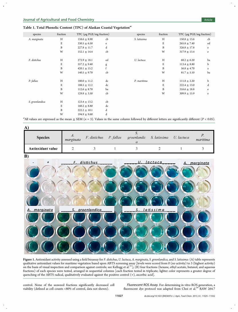
Table 1. Total Phenolic Content (TPC) of Alaskan Coastal Vegetationa
species fraction TPC (μg PGE/mg fraction) species fraction TPC (μg PGE/mg fraction)
A. marginata H 158.6 ± 8.90 cb S. latissima H 150.8 ± 13.6 cbE 330.5 ± 6.50 e E 265.8 ± 7.40 edB 227.8 ± 11.7 d B 326.8 ± 17.8 eW 152.1 ± 14.4 cb W 317.9 ± 13.4 e
F. distichus H 272.9 ± 18.1 ed U. lactuca H 68.2 ± 6.20 baE 557.2 ± 9.40 g E 113.4 ± 8.80 bB 420.1 ± 15.2 f B 56.8 ± 4.70 aW 140.5 ± 9.70 cb W 81.7 ± 5.10 ba
P. fallax H 180.0 ± 11.2 dc P. maritima H 111.8 ± 2.20 bE 188.5 ± 12.2 dc E 222.4 ± 13.0 dB 112.6 ± 8.70 ba B 316.6 ± 18.8 eW 129.8 ± 3.50 cb W 309.9 ± 15.9 e
S. groenlandica H 123.4 ± 13.2 cbE 160.2 ± 8.80 dcB 222.2 ± 10.1 dW 194.9 ± 9.60 d
aAll values are expressed as the mean ± SEM (n = 3). Values in the same column followed by different letters are significantly different (P < 0.05).
Figure 1. Antioxidant activity assessed using a field bioassay for F. distichus,U. lactuca, A. marginata, S. groenlandica, and S. latissima: (A) table representsqualitative antioxidant values for maritime vegetation based upon ABTS screening assay [levels were scored from 0 (no activity) to 3 (highest activity)on the basis of visual inspection and comparison against controls; see Kellogg et al.31]; (B) four fractions (hexane, ethyl acetate, butanol, and aqueousfractions) of each species were tested, arranged in sequential columns [each fraction tested in triplicate; lighter color represents a greater degree ofquenching of the ABTS radical, qualitatively evaluated against the positive control (+), ascorbic acid].
Journal of Agricultural and Food Chemistry Article
dx.doi.org/10.1021/jf403697z | J. Agric. Food Chem. 2013, 61, 11025−1103211027
macrophage cells were seeded at a concentration of 4 × 10⁵ cells/wellinto a 24-well plate and incubated overnight at 37 °C. Cells were chargedwith 500 μL of 50 μM 2′,7′-dichlorodihydrofluorescein diacetate acetylester (H2DCFDA, Molecular Probes, Eugene, OR, USA), preparedfresh daily in sterile phosphate-buffered saline (PBS) for 30 min.Fluoresecent medium was aspirated, and cells were exposed to 25 μL ofextract/fraction (100 μg/mL final concentration) and 1 μL oflipopolysaccharide (LPS, from Escherichia coli 026:B6) and incubatedfor 24 h, after which the fluorescence of 2′,7′-dichlorofluorescein (DCF)was measured at 485 nm (excitation) and 515 nm (emission) on amicroplate reader. The known antioxidant dexamethasone (DEX) wasused as a positive control. The experiments were performed with threeindependent replications, each replication assayed at least in duplicate.Nitric Oxide Assay. The production of nitrite, the stable end-product
of NO generation in activated macrophages, was assayed by acolorimetric assay. To 100 μL of cell culture medium was added 100μL of Griess reagent (1% sulfanilamide and 0.1% naphthylethylenedi-amine in 5% phosphoric acid), and the mixture was incubated at roomtemperature for 10 min. The absorbance at 540 nm was read on amicroplate reader. The nitrite concentration was calculated using asodium nitrite standard curve.Statistics. All assays were performed at least in triplicate. Results are
presented as the mean of triplicate runs ± SEM. Statistical analysis wasconducted using repeated-measures ANOVA followed by Tukey’s test(Prism 6.0, GraphPad Inc., La Jolla, CA, USA), with statistical
significance determined at the P < 0.05 or P < 0.01 level. Half-maximalinhibitory concentration (IC50) data were compiled after logarithmictransformation and expressed as the geometric mean with 95%confidence intervals.
■ RESULTS
Total Phenolic Content. The total phenolic content of thefractionated marine vegetation, determined by the Folin−Ciocalteu colorimetric analysis, is given in Table 1. The greenalgae U. lactuca demonstrated the lowest TPC across the fourfractions (56.8−113.4 μg PGE/mg), compared to the fourspecies of Alaskan brown algae (123.4−557.2 μg PGE/mg). Theethyl acetate (557.2 ± 9.7 μg PGE/mg) and butanol (420.1 ±15.2 μg PGE/mg) fractions of F. distichus had the greatest TPCcontent of all the samples, higher than a previous study of therelated species F. vesiculosus, which was measured at 37.4 ± 0.6%TPC.11 The terrestrial goosetongue, P. maritima, showed levelsof phenolics (111.8−316.6 μg PGE/mg) that fell in the middlerange of values for the species tested.
Antioxidant Capacity: Chemical Assays. The antioxidantcapacity of the marine vegetation extracts, based upon thepreliminary screening assay, were assigned qualitative values
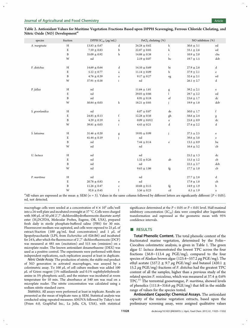
Table 2. Antioxidant Values for Maritime Vegetation Fractions Based upon DPPH Scavenging, Ferrous Chloride Chelating, andNitric Oxide (NO) Developmenta
species fraction DPPH IC50 (μg/mL) FeCl2 chelating (%) NO inhibition (%)
A. marginata H 13.83 ± 0.67 d 24.28 ± 0.62 k 30.6 ± 3.1 edE 7.59 ± 0.83 b 22.87 ± 0.85 k 33.1 ± 2.6 edB 35.09 ± 0.92 h 14.88 ± 0.38 i 10.9 ± 2.0 cbaW nd 2.19 ± 0.07 bc 19.7 ± 1.5 dcb
F. distichus H 14.69 ± 0.64 d 14.10 ± 0.69 hi 27.9 ± 2.8 dE 5.12 ± 0.77 a 11.14 ± 0.09 h 37.9 ± 2.1 eB 4.76 ± 0.39 a 9.17 ± 0.27 eg 32.4 ± 2.1 edW 37.91 ± 0.58 i nd 26.1 ± 2.7 d
P. fallax H nd 11.64 ± 1.61 g 39.2 ± 2.1 eE nd 29.03 ± 0.86 l 29.7 ± 2.2 edB nd 8.95 ± 0.18 ef 23.6 ± 1.7 dcW 50.84 ± 0.03 k 18.21 ± 0.85 j 19.9 ± 1.8 dcb
S. groenlandica H nd 6.07 ± 0.87 de 50.0 ± 1.7 fE 24.85 ± 0.13 f 12.28 ± 0.56 gh 58.6 ± 2.4 gB 4.29 ± 0.19 a 0.09 ± 0.012 a 25.8 ± 0.9 dcW 39.81 ± 0.03 i 4.43 ± 0.21 d 27.4 ± 2.2 d
S. latissima H 31.46 ± 0.20 g 19.95 ± 0.99 j 37.5 ± 2.5 eE 41.44 ± 0.19 j nd 39.6 ± 3.8 eB nd 7.44 ± 0.14 e 13.5 ± 0.9 baW nd nd 16.4 ± 3.2 cb
U. lactuca H nd nd 25.2 ± 1.3 dcE nd 1.32 ± 0.26 ab 15.5 ± 1.2 cbB nd nd 22.5 ± 2.7 dcbW nd 9.63 ± 1.06 fg 17.7 ± 1.0 cb
P. maritima H nd nd 27.7 ± 2.8 dE 20.78 ± 0.83 e nd 17.9 ± 1.8 cbB 11.26 ± 0.47 c 10.68 ± 0.15 fg 14.9 ± 1.9 bW 92.8 ± 0.45 l 3.56 ± 0.23 cd 6.3 ± 1.9 a
aAll values are expressed as the mean ± SEM (n = 3). Values in the same column followed by different letters are significantly different (P < 0.05).nd, not detected.
Journal of Agricultural and Food Chemistry Article
dx.doi.org/10.1021/jf403697z | J. Agric. Food Chem. 2013, 61, 11025−1103211028
from 0 to 3 on the basis of color development, comparing eachfraction against positive and negative controls (1 mM ascorbicacid and 60% ethanol, respectively). All seven crude extractsexhibited antioxidant activity (Figure 1), and subsequentfractions were also assayed under the same conditions (Figure1 displays representative fractions from several species). Allcrude extracts demonstrated some degree of antioxidant activity.The brown algae generally exhibited higher levels of radicalquenching. The majority of fractions displayed antioxidantactivity by quenching the ABTS radical; the ethyl acetate andbutanol fractions showed higher activity than hexane or aqueousfractions.As shown in Table 2, the fractionated extracts of marine
vegetation varied in their ability to scavenge the DPPH radical.Two seaweed species, the red alga P. fallax and the green U.lactuca, had no detectable antioxidant activity in the DPPH assay.Congruent with the screening assay, fractions from the fourbrown algae, A. marginata, F. distichus, S. groenlandica, and S.latissima, each demonstrated radical scavenging in a dose-dependent manner over a concentration range of 1−100 μg/mL.Of the four, F. distichus was the most active, with ethyl acetate-soluble and butanol-soluble fractions at IC50 values of 5.12± 0.77and 4.76 ± 0.39 μg/mL, respectively. The butanol fraction of S.groenlandica (4.29 ± 0.19 μg/mL) and the ethyl acetate fractionof A. marginata (7.59 ± 0.83 μg/mL) also exhibited capacity forscavenging DPPH radicals. These values were similar inmagnitude to that of the commercial antioxidant ascorbic acid(3.28 ± 0.11 μg/mL) and were also comparable to those of theclosely related F. vesiculosus,11 with ethyl acetate and butanolfractions at IC50 values of 3.76 ± 0.22 and 4.77 ± 0.25 μg/mL,respectively. The ethyl acetate fraction (20.78 ± 0.83 μg/mL)and butanol fraction (11.26± 0.47 μg/mL) of Plantago maritimaalso demonstrated moderate antioxidant efficacy. The mildactivity of some aqueous fractions (F. distichus, P. fallax, and S.groenlandica had IC50values of 37.91, 50.84, and 39.81 μg/mL,respectively) could be attributed to the presence of sulfatedpolysaccharides that are found in all three major taxa ofalgae.35−37
In addition, the TPC levels of Alaskan seaweed had a strongpositive correlation with DPPH radical scavenging capability (R2
= 0.662, P < 0.001). Very high and significant positivecorrelations, up to R2 = 0.99, have been documented betweenpolyphenolic content and antioxidant capability of seaweeds inprevious studies.12,38 The moderate correlation found in thecurrent study may suggest that other active, nonpolyphenoliccomponents could have synergistic effects on radical scavenging,as shown in Heo et al.’s 2005 study, in which fractions obtainedfrom the brown algae Ecklonia cava and Sargassum coreanumpossessed low DPPH radical scavenging activity despite testingfor comparative TPC levels as other, higher activity, extracts.39
Lipid and cellular oxidation may be amplified by interactionwith transition metal cations, which play an important part inaging and age-related chronic diseases,40,41 and the ability ofantioxidants to chelate these ions can diminish the cumulativeeffects of transition metal-mediated free radical and oxidativedamage.42 The species’ fractions demonstrated various degreesof ferrous ion chelation capacity (Table 2). The ethyl acetatefraction of P. fallax (29.03 ± 0.86%) had the greatest chelatingability, whereas the hexane (24.28 ± 0.62%) and ethyl acetate(22.87 ± 0.85%) fractions of A. marginata also exhibited highlevels of chelating. These levels were consistent with otherspreviously reported11,12 when sample concentration is adjustedfor. Although the ferrous ion chelation of the fractions was notcorrelated with TPC (R2 = 0.08), this lack of parallel response hasbeen seen in other seaweed studies,12 as well as in extracts ofmalting barley,43 for which TPC had poor correlations with bothDPPH and cation radical scavenging activities.Nitric oxide radical (NO) inhibition levels, as assayed by
sodium nitrite production from light-induced sodium nitroprus-side decomposition, varied between extract fractions (Table 2).Two fractions derived from S. groenlandica demonstrated thegreatest NO scavenging ability. The hexane (50.0 ± 1.7%inhibition) and ethyl acetate (58.6 ± 2.4%) fractions showed>50% NO inhibitory activity. Other brown algae fractions, fromF. distichus, A. marginata, and S. latissima, also had strong levels ofNO inhibition (19.7−39.5%). Within each species of brown alga,the ethyl acetate fraction demonstrated the highest degree of NO
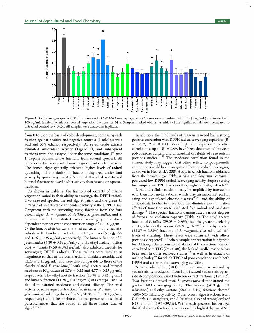
Figure 2. Radical oxygen species (ROS) production in RAW 264.7 macrophage cells. Cultures were stimulated with LPS (1 μg/mL) and treated with100 μg/mL fractions of Alaskan coastal vegetation fractions for 24 h. Samples marked with an asterisk (∗) are significantly different compared tountreated control (P < 0.05). All samples were assayed in triplicate.
Journal of Agricultural and Food Chemistry Article
dx.doi.org/10.1021/jf403697z | J. Agric. Food Chem. 2013, 61, 11025−1103211029

inhibition (33.1, 37.9, 58.6, and 39.6% inhibition for A.marginata, F. distichus, S. groenlandica, and S. latissima,respectively). The red alga P. fallax was on the same magnitudeas brown algae. The green alga U. lactuca and tidal plant P.maritima had lower ability to inhibit NO formation than the redor brown algae (6.3−27.7%). These species all showed greatestintraspecies inhibition with the hexane fraction (39.2% for P.fallax, 25.2% for U. lactuca, and 27.7% for P. maritima).In Vitro ROS and NO Inhibition. Cellular antioxidant
activities of Alaskan coastal resources were determined using anin vitro system to gauge reduction of radical-mediated oxidationin a RAW 264.7 macrophage culture. ROS production in theRAW 264.7 cells was monitored through quantitativefluorescence signaling. Introduced H2DCFDA penetrated theouter membrane of viable cells, where it was deacetylated bycytosolic esterases and subsequently reacted with ROSwithin thecell to yield the highly fluoresecent DCF. Lipopolysaccharide(LPS, 1 μg/mL) induced a 2.5-fold overproduction of ROS in thecells compared to nonstimulated cells. Figure 2 shows the effectof Alaskan seaweed and plant fractions on the inhibition ofintracellular LPS-induced ROS generation. The positive controlDEX lowered ROS production back to nominal levels.Examination of the cytotoxicity of the prepared fractions inRAW 264.7 macrophages by MTT indicated that, at 100 μg/mL,none of the Alaskan vegetative fractions affected the viability ofRAW 264.7 cells (data not shown). Therefore, inhibition of LPS-induced oxidative stress (via ROS or NO production) was notthe result of a cytotoxic effect.Alaskan seaweeds had varying levels of intracellular ROS
inhibition (Figure 2). ROS levels after treatment by the fractionsof the green alga U. lactuca remained statistically higher than thecontrol levels, indicating insufficient antioxidant activity at thecellular level to inhibit LPS stimulation. Three of the fourfractions of the red alga P. fallax showed significantly higher ROSlevels over the control, with only the aqueous fraction (W)having a large inhibition of ROS production. The brown algae, F.distichus,A. marginata, S. groenlandica, and S. latissima, all effecteda significant reduction (P < 0.05) on cellular ROS levels; in fact,only three fractions (F. distichus-W, A. marginata-W, and S.
latissima-H) did not reduce ROS production back to controllevels. The terrestrial goosetongue also had significant ROSinhibition across all four fractions, lowering the ROS generationof the cells to their nonstimulated state. The ethyl acetatefractions appeared to be lower than the other fractions inreducing ROS production, although these comparisons were notstatistically significant. These activities were only moderatelycorrelated with total phenolic content (R2 = 0.450, P < 0.001),indicating that potentiating interactions and/or synergiesbetween polyphenols and other phytochemicals could havecontributed to ROS inhibition.The oxidative nitric oxide pathway culminates in the
generation of nitrite, which can be quantitatively measuredfrom culture media using the Greiss reagent. NO production wasmonitored in RAW264.7 cells stimulated by LPS for 24 h. LPS (1μg/mL) evoked a 16.6-fold induction of nitrite production versusthe naive control, and this induction was inhibited by the positivecontrol, DEX, to baseline levels (P < 0.001). The Alaskanvegetative fractions reduced NO production to various degrees(Figure 3). All except the butanol fraction of U. lactuca reducedNO production by a statistically significant amount (P < 0.001);however, several species and fractions were unable to reduce theNO levels back to nonstimulated levels. In general, the brownalgae fractions lowered NO production to basal levels, and theaqueous fraction of F. distichus, the ethyl acetate fraction of A.marginata, and the butanol fraction of S. latissima had the mostactivity of all fractions tested. Fractions from goosetonguereduced NO levels as well, but not as effectively as the seaweed(comparison P < 0.034). The NO reduction levels achieved byAlaskan vegetation exhibited a very strong positive correlationwith in vitro ROS inhibition capability (R2 = 0.844, P < 0.001),supporting the observation that antioxidant activity and NOinhibition share common metabolic pathways. However, therewas only moderate correlation with TPC (R2 = 0.192, P < 0.019).
■ DISCUSSION
This study demonstrated the antioxidant capacities of edibleAlaskan intertidal vegetative extracts/fractions based on chemicalassays as well as in vitro scavenging effects on ROS levels and
Figure 3.Nitric oxide (NO) production in LPS-stimulated RAW 264.7 macrophage cells. Cultures were cotreated with 100 μg/mL fractions of Alaskancoastal vegetation fractions for 24 h. Samples marked with an asterisk (∗) are significantly different compared to untreated control (P < 0.05). Allsamples were assayed in triplicate.
Journal of Agricultural and Food Chemistry Article
dx.doi.org/10.1021/jf403697z | J. Agric. Food Chem. 2013, 61, 11025−1103211030

reduction of NO production by RAW 264.7 cells. The brownalgae species, F. distichus, A. marginata, S. groenlandica, and S.latissima, all demonstrated high levels of chemical antioxidantactivities (based upon DPPH, NO, and FeCl2 assays), as well ashigh in vitro capacities of scavenging ROS and decreasing NOproduction. Phlorotannins, oligomer derivatives of phlorogluci-nol, are the predominant polyphenol component of brownalgae,44 and all species tested had high TPC levels. Thecorrelation observed between TPC and chemical (DPPH R2 =0.662) and in vitro (R2 = 0.450) antioxidant activity indicatedthat polyphenolics are a significant contributor to the antioxidantcapabilities of these seaweeds, although this correlation waslower than expected compared against other studies on TPC-enriched seaweed fractions,45 suggesting that other nonphenoliccompounds could also play a role in antioxidant protection. Thesole terrestrial plant, P. maritima, had high levels of polyphenolicsand moderate levels of chemical antioxidant activity, butexhibited high levels of in vitro ROS and NO inhibition,suggesting that this tidal plant also is a strong potential source ofantioxidant phytochemicals.The red alga P. fallax did not contain phlorotannins, had
moderate levels of phenolics, and exhibited variable abilities toquench radicals in the chemical assays. With the exception of theaqueous fraction, there was little significant activity in the in vitroROS assay, or nitric oxide suppression. The green alga U. lactucaregistered greatly diminished in vitro antioxidant capacitycompared to the other species tested. This result concurredwith the low levels of antioxidant activity observed in the DPPHand NO inhibition assays as well as the low TPC levels for thegreen alga.In general, these species, which form part of the traditional
diets of Pacific Northwest cultures, exhibited effective protectionagainst oxidation in chemical and in vitro systems. As such, theymay have the potential to confer protection when incorporatedinto the diet, offsetting many oxidative-damage relatedconditions, such as obesity, diabetes, and cardiovascular disease.Nitric oxide is an inflammatory mediator, and thus theseseaweeds have the potential to also act as anti-inflammatoryagents by reducing inflammatory markers and decreasing theproduction of pro-inflammatory cytokines. This is an arena forfuture experimentation, as marine polyphenols have not beenwidely studied for their anti-inflammatory capacity with respectto suppression of NO and other cytokines.45 Additional researchis being undertaken to determine which specific phytochemicalsconfer antioxidant protection, including further isolation andstructure elucidation to better understand the mechanisms thatunderpin the observed effects.
■ AUTHOR INFORMATIONCorresponding Author*(M.A.L.) E-mail: [email protected]. Phone: (704) 250-5407. Fax: (704) 250-5409.FundingThis project is supported with funding from the University ofNorth Carolina General Administration and North CarolinaState University’s Plants for Human Health Institute.NotesThe authors declare no competing financial interest.
■ ACKNOWLEDGMENTSWe thank Renae Matheson and the Southeast Alaska RegionalHealth Consortium (SEARHC) for assistance in collecting and
screening the species used in this study. We acknowledge theexpertise and guidance of Dr. Deborah Esposito and RennettaRoberts of the Plants for Human Health Institute for theirtechnical help in the cell culture and in vitro assays.
■ REFERENCES(1) Jimenez-Escrig, A.; Gomez-Ordonez, E.; Ruperez, P. Brown andred seaweeds as potential sources of antioxidant nutraceuticals. J. Appl.Phycol. 2012, 24, 1123−1132.(2) Widjaja-Adhi Airanthi, M. K.; Hosokawa, M.; Miyashita, K.Comparative antioxidant activity of edible Japanese brown seaweeds. J.Food Sci. 2011, 76, C104−C111.(3) Garza, D. Common Edible Seaweeds in the Gulf of Alaska; Alaska SeaGrant College Program: Fairbanks, AK, 2005.(4) Turner, N. C.; Bell, M. A. M. The ethnobotany of the SouthernKwakiutl Indians of British Columbia. Econ. Bot. 1973, 27, 257−310.(5) Turner, N. J. The ethnobotany of edible seaweed (Porphyraabbottae and related species; Rhodophyta: Bangiales) and its use by FirstNations on the Pacific Coast of Canada. Can. J. Bot. 2003, 81, 283−293.(6)Wein, E. E.; Freeman, M. M. R.; Makus, J. C. Use of and preferencefor traditional foods among the Belcher Island Inuit. Arctic 1996, 49,256−264.(7) Sato, M.; Hosokawa, T.; Yamaguchi, T.; Nakano, T.; Muramoto,K.; Kahara, T.; Funayama, K.; Kobayashi, A.; Nakano, T. Angiotensin I-converting enzyme inhibitory peptides derived from wakame (Undariapinnatif ida) and their antihypertensive effect in spontaneously hyper-tensive rats. J. Agric. Food Chem. 2002, 50, 6245−6252.(8) Kim, M. S.; Kim, J. Y.; Choi, W. H.; Lee, S. S. Effects of seaweedsupplementation on blood glucose concentration, lipid profile, andantioxidant enzyme activities in patients with type 2 diabetes mellitus.Nutr. Res. Pract. 2008, 2, 62−67.(9) Zhang, J.; Tiller, C.; Shen, J.; Wang, C.; Girouard, G. S.; Dennis, D.;Barrow, C. J.; Miao, M.; Ewart, H. S. Antidiabetic properties ofpolysaccharide- and polyphenolic-enriched fractions from the brownseaweed Ascophyllum nodosum. Can. J. Physiol. Pharmacol. 2007, 85,1116−1123.(10) O’Sullivan, A. M.; O’Callaghan, Y. C.; O’Grady, M. N.;Queguineur, B.; Hanniffy, D.; Troy, D. J.; Kerry, J. P.; O’Brien, N. M.In vitro and cellular antioxidant activities of seaweed extracts preparedfrom five brown seaweeds harvested in spring from the west coast ofIreland. Food Chem. 2011, 126, 1064−1070.(11) Wang, T.; Jonsdottir, R.; Liu, H.; Gu, L.; Kristinsson, H. G.;Raghavan, S.; Olafsdottir, G. Antioxidant capacities of phlorotanninsextracted from the brown algae Fucus vesiculosus. J. Agric. Food Chem.2012, 60, 5874−5883.(12) Wang, T.; Jo nsdo ttir, R.; Olafsdo ttir, G. Total phenoliccompounds, radical scavenging and metal chelation of extracts fromIcelandic seaweeds. Food Chem. 2009, 16, 240−248.(13) Ragan, M. A.; Glombitza, K. W. Phlorotannins, brown algalpolyphenols. Prog. Phycol. Res. 1986, 4.(14) Heo, S. J.; Ko, S. C.; Cha, S. H.; Kang, D. H.; Park, H. S.; Choi, Y.U. Effect of phlorotannins isolated from Ecklonia cava on melanogenesisand their protective effect against photo-oxidative stress induced by UV-B radiation. Toxicol. in Vitro 2009, 23, 1123−1130.(15) Hu, C. C.; Lin, J. T.; Lu, F. J.; Chou, F. P.; Yang, D. J.Determination of carotenoids in Dunaliella salina cultivated in Taiwanand antioxiant capacity of the algal carotenoid extract. Food Chem. 2008,109, 439−446.(16) Aruoma, O. I. Free radicals, oxidative stress, and antioxidants inhuman health and disease. J. Am. Oil Chem. Soc. 1998, 75, 199−212.(17) Kaliora, A. C.; Dedoussis, G. V. Z.; Schmidt, H. Dietaryantioxidants in preventing atherogenesis. Atherosclerosis 2006, 187, 1−17.(18) Scalbert, A.; Manach, C.; Morand, C.; Remesy, C. Dietarypolyphenols and the prevention of diseases. CRC Crit. Rev. Food Sci.2005, 45, 287−306.
Journal of Agricultural and Food Chemistry Article
dx.doi.org/10.1021/jf403697z | J. Agric. Food Chem. 2013, 61, 11025−1103211031
(19) Peteiro, C.; Freire, O. Effect of water motion on the cultivation ofthe commercial seaweed Undaria pinnatif ida in a coastal bay of Galicia,Northwest Spain. Aquaculture 2011, 314, 269−276.(20) Blanchette, C. A. Size and survival of intertidal in plants inresponse to wave action: a case study with Fucus gardneri. Ecology 1997,78, 1563−1578.(21) Pavia, H.; Cervin, G.; Lindgren, A.; Aberg, P. Effects of UV-Bradiation and simulated herbivory on phlorotannins in the brown algaAscophyllum nodosum. Mar. Ecol.−Prog. Ser. 1997, 157, 139−146.(22) Bischof, K.; Gomez, I.; Molis, M.; Hanelt, D.; Karsten, U.; Luder,U.; Roleda, M. Y.; Zacher, K.; Wiencke, C. Ultraviolet radiation shapesseaweed communities. Rev. Environ. Sci. Biotechnol. 2006, 5, 141−166.(23) Schoenwaelder, M. A. E.; Wiencke, C.; Clayton, M. N.;Glombitza, K. W. The effect of elevated UV radiation on Fucus spp.(Fucales, Phaeophyta) zygote and embryo development. Plant Biol.2003, 5, 366−377.(24) Oliveira, A. S.; Sudatti, D. B.; Fujii, M. T.; Rodrigues, S. V.;Pereira, R. C. Inter- and intrapopulation variation in the defensivechemistry of the red seaweed Laurencia dendroidea (Ceramiales,Rhodophyta). Phycologia 2013, 52, 130−136.(25) Bunsom, C.; Prathep, A. Effects of salinity, light intensity andsediment on growth, pigments, agar production and reproduction inGracilaria tenuistipitata from Songkhla Lagoon in Thailand. Phycol. Res.2012, 60, 169−178.(26) Díez, I.; Muguerza, N.; Santolaria, A.; Ganzedo, U.; Gorostiaga, J.M. Seaweed assemblage changes in the eastern Cantabrian Sea and theirpotential relationship to climate change. Estuarine, Coastal Shelf Sci.2012, 99, 108−120.(27) Harley, C. D. G.; Anderson, K. M.; Demes, K. W.; Jorve, J. P.;Kordas, R. L.; Coyle, T. A.; Graham, M. H. Effects of climate change onglobal seaweed communities. J. Phycol. 2012, 48, 1064−1078.(28) Wernberg, T.; Russell, B. D.; Thomsen, M. S.; Gurgel, C. F. D.;Bradshaw, C. J. A.; Poloczanska, E. S.; Connell, S. D. Seaweedcommunities in retreat from ocean warming.Curr. Biol. 2011, 21, 1828−1832.(29) Lindberg, M. R.; Lindstrom, S. C. Field Guide to Seaweeds ofAlaska; Alaska Sea Grant College Program, University of Alaska:Fairbanks, AK, 2010; p 188.(30) Herald, T. J.; Gadgil, P.; Tilley, M. High-throughput micro plateassays for screening flavonoid content and DPPH-scavenging activity insorghum bran and flour. J. Sci. Food Agric. 2012, 92, 2326−2331.(31) Kellogg, J.; Wang, J.; Flint, C.; Ribnicky, D.; Kuhn, P.; deMejia, E.G.; Raskin, I.; Lila, M. A. Alaskan wild berry resources and human healthunder the cloud of climate change. J. Agric. Food Chem. 2010, 58, 3884−3900.(32) Kellogg, J.; Joseph, G.; Andrae-Marobela, K.; Sosome, A.; Flint,C.; Komarnytsky, S.; Fear, G.; Struwe, L.; Raskin, I.; Lila, M. A. Screens-to-nature: opening doors to traditional knowledge and hands-on scienceeducation. NACTA J. 2010, 54, 41−48.(33) Oliveira, A. P.; Silva, L. R.; Ferreres, F.; de Pinho, P. G.; Valentao,P.; Silva, B. M.; Pereira, J. A.; Andrade, P. B. Chemical assessment and invitro antioxidant capacity of Ficus carica latex. J. Agric. Food Chem. 2010,58, 3393−3398.(34) Choi, S.-Y.; Hwang, J.-H.; Ko, H.-C.; Park, J.-G.; Kim, S.-J.Nobiletin from citrus fruit peel inhibits the DNA-binding activity of NF-κB and ROS production in LPS-activated RAW 264.7 cells. J.Ethnopharm. 2007, 113, 149−155.(35) Rocha de Souza, M. C.; Marques, C. T.; Dore, C. M. G.; Ferreirada Silva, F. R.; Rocha, H. A. O.; Leite, E. L. Antioxidant activities ofsulfated polysaccharides from brown and red seaweeds. J. Appl. Phycol.2007, 19, 153−160.(36) Ruperez, P.; Ahrazem, O.; Leal, J. A. Potential antioxidant capacityof sulfated polysaccharides from the edible brown seaweed Fucusvesiculosus. J. Agric. Food Chem. 2002, 50, 840−845.(37) Shao, P.; Chen,M.; Pei, Y.; Sun, P. In vitro antioxidant activities ofdifferent sulfated polysaccharides from chlorophytan seaweeds Ulvafasciata. Int. J. Biol. Macromol. 2013, 59, 295−300.
(38) Jimenez-Escrig, A.; Jimenez-Escrig, I.; Pulido, R.; Saura-Calizto, F.Antioxidant activity of fresh and processed edible seaweeds. J. Sci. FoodAgric. 2001, 81, 530−534.(39) Heo, S. J.; Park, E. J.; Lee, K. W.; Jeon, Y.-J. Antioxidant activitiesof enzymatic extracts from brown seaweeds. Bioresour. Technol. 2005, 96,1613−1623.(40) Salih, A. M.; Price, J. F.; Smith, D. M.; Dawson, L. E. Lipidoxidation in turkey meat as influenced by salt, metal cations, andantioxidants. J. Food Qual. 1989, 12, 71−83.(41) Zhang, Y.; Zou, B.; Wang, K.; Pan, Y.; Liang, H.; Yi, X.; Wang, H.Antioxidant activities and transition metal ion chelating studies of somehydroxyl Schiff base derivatives.Med. Chem. Res. 2012, 21, 1341−1346.(42) Hipkiss, A. R. Glycation, ageing and carnosine: are carnivorousdiets beneficial. Mech. Ageing Dev. 2005, 126, 1034−1039.(43) Zhou, H. F.; Fan, W.; Dong, J. J.; Lu, J.; Chen, J.; Shan, L. J.Evaluation of antioxidant activities and total phenolic contents of typicalmalting barley varieties. Food Chem. 2008, 107, 296−304.(44) Steevensz, A. J.; MacKinnon, S. L.; Hankinson, R.; Craft, C.;Connan, S.; Stangel, D. B.; Melanson, J. E. Profiling phlorotannins inbrown macroalgae by liquid chromatography-high resolution massspectrometry. Phytochem. Anal. 2012, 23, 547−553.(45) Kim, A. R.; Shin, T. S.; Lee, M. S.; Park, J. Y.; Park, K. E.; Yoon, N.Y.; Kim, J. S.; Choi, J. S.; Jang, B. C.; Byun, D. S.; Park, N. K.; Kim, H. R.Isolation and identification of phlorotannins from Ecklonia stoloniferawith antioxidant and anti-inflammatory properties. J. Agric. Food Chem.2009, 57, 3483−3489.
Journal of Agricultural and Food Chemistry Article
dx.doi.org/10.1021/jf403697z | J. Agric. Food Chem. 2013, 61, 11025−1103211032