perd sa virulence et dans quelle mesure la virulence est liee a ...
Upload
independentCategory
view
3download
0
Virology ] (]]]]) ]]]–]]]
Contents lists available at SciVerse ScienceDirect
Virology
0042-68
http://d
n Corr
College
Paul, M
E-m1 Cu
Goat, In
Mathur2 Cu
Univers3 Cu
Univers
PleasPichi
journal homepage: www.elsevier.com/locate/yviro
Characterization of virulence-associated determinants in the envelopeglycoprotein of Pichinde virus
Naveen Kumar 1, Jialong Wang 2, Shuiyun Lan, Shamika Danzy 2, Lisa McLay Schelde 3,Jill Seladi-Schulman 2, Hinh Ly, Yuying Liang n
Department of Veterinary and Biomedical Sciences, University of Minnesota, Twin Cities, MN 55108, USA
a r t i c l e i n f o
Article history:
Received 24 February 2012
Returned to author for revisions
6 July 2012
Accepted 12 July 2012
Keywords:
Virulence
Arenavirus
Pichinde virus
Glycoprotein
Entry
22/$ - see front matter & 2012 Elsevier Inc. A
x.doi.org/10.1016/j.virol.2012.07.009
espondence to: Department of Veterinary an
of Veterinary Medicine, 1988 Fitch Avenue,
N 55108, USA. Fax: þ1 612 625 0204.
ail address: [email protected] (Y. Liang).
rrent address: Division of Animal Health, Cen
dian Council of Agricultural Research, Mak
a, UP, India.
rrent address: Department of Microbiolog
ity. Atlanta, GA, USA.
rrent address: Department of Pathology and
ity. Atlanta, GA, USA.
e cite this article as: Kumar, N., etnde virus. Virology (2012), http://dx
a b s t r a c t
We use a small animal model, based on guinea pigs infected with a non-pathogenic Pichinde virus
(PICV), to understand the virulence mechanisms of arenavirus infections in the hosts. PICV P2 strain
causes a mild febrile reaction in guinea pigs, while P18 causes severe disease with clinical and
pathological features reminiscent of Lassa hemorrhagic fever in humans. The envelope glycoproteins
(GPC) of P2 and P18 viruses differ at positions 119, 140, and 164, all localized to the receptor-binding
G1 subunit. We found that lentiviral pseudotyped virions (VLPs) bearing P18 GPC show more efficient
cell entry than those with P2 GPC, and that the E140 residue plays a critical role in this process.
Infection of guinea pigs with the recombinant viruses containing the E140K change demonstrated that
E140 of GPC is a necessary virulence determinant of P18 infections, possibly by enhancing the ability of
virus to enter target cells.
& 2012 Elsevier Inc. All rights reserved.
Introduction
Lassa virus (LASV), an Old World (OW) arenavirus, causesendemic Lassa fever in West Africa with estimated 300,000–500,000 infections and 5000 deaths annually (Khan et al., 2008;McCormick and Fisher-Hoch, 2002). In South America, severalNew World (NW) arenaviruses, such as Junin (JUNV) andMachupo (MACV), cause sporadic hemorrhagic fever diseases(Buchmeier et al., 2007). Novel arenaviruses that can causehemorrhagic fever diseases have also been identified in recentyears (Briese et al., 2009; Delgado et al., 2008), highlighting theurgency to develop much-needed preventive and therapeuticmeasures. Efforts to develop antiviral approaches, however, havebeen hampered by the lack of in-depth understanding of themechanisms of arenavirus replication and pathogenesis due tothe strict requirement of Biosafety-Level 4 (BSL-4) containment towork with these live pathogenic arenaviruses.
ll rights reserved.
d Biomedical Sciences,
295 AS/VM Building., Saint
tral Institute of Research on
hdoom, P.O. Farah 281122,
y and Immunology, Emory
Laboratory Medicine, Emory
al., Characterization of viru.doi.org/10.1016/j.virol.201
Pichinde virus (PICV) is a NW arenavirus that is non-pathogenicin humans and can be safely studied at BSL-2. Its natural host is theColumbian rice rat Oryzomys albigularis (Trapido and Sanmartin,1971). Whereas a low number of passages of PICV in inbred guineapigs produce an avirulent virus that causes a brief febrile reaction,extended passage in inbred guinea pigs has led to the isolation ofhighly pathogenic strains that cause severe disease characterized byfever, severe weight loss, terminal shock syndrome, and death,similar to those found in human Lassa fever (Aronson et al., 1994;Jahrling et al., 1981; Lucia et al., 1990; McCormick et al., 1987; Qianet al., 1994). Due to the difficulty in maintaining inbred strain 13guinea pigs, Aronson and her colleagues have shown that similardistinct disease patterns were also observed in outbred Hartleyguinea pigs infected by a low passge (e.g., strain P2) and a highpassage (e.g., strain P18) PICV strains (Zhang et al., 2001). PICVinfection in guinea pigs, therefore, represents a safe, convenient, andeconomical small animal model to investigate the pathogenesis ofarenavirus hemorrhagic fevers (Aronson et al., 1994; Cosgriff et al.,1987; Jahrling et al., 1981; Schaeffer et al., 1993). Understanding thevirulence mechanisms by which the two closely related PICV strainscause distinct disease outcomes in guinea pigs is expected to shedimportant lights into those of pathogenic arenaviruses in humans.We have identified several amino acid differences in the viral codingsequences, including three mutations in the viral envelop glycopro-tein (GPC) (Lan et al., 2008). We propose that these GPC mutationsmay be important virulence determinants.
Arenavirus envelope glycoprotein is translated as a precursorprotein that is cleaved by signal peptidase and the SKI-1/S1P
lence-associated determinants in the envelope glycoprotein of2.07.009

- GPC
- G1/G2
- actin
P18
P2
P18
-E14
0K
P18
-T11
9S
P18
-V16
4I
P18
-T11
9S/E
140K
P18
-T11
9S/V
164I
P18
-E14
0K/V
164I
P18
-trip
le-m
ut
cont
rol
HIV-1 p24
G1/G2VLP
celllysates
1.00
1.02
1.02
1.06
1.03
1.06
1.10
1.01
1.01
1.00
1.01
1.01
1.01
1.02
1.06
1.10
1.00
1.01
normalized G1/G21.
00
1.01
1.01
1.06
1.00
1.01
0.99
1.01
1.00
Fig. 1. Characterization of the GPC-mediated cell entry by GPC-pseudotyped
lentiviral VLPs. (A) Expressions of P2, P18, and P18 mutant GPC proteins in the
transfected 293T cells. Western blot analysis was conducted using guinea pig anti-
PICV serum. (B) Detection of G1/G2 and HIV-1 p24 in the purified VLPs by western
blot analysis. Intensity of each band was quantified by the Image J analysis
software and normalized to that of P18.
N. Kumar et al. / Virology ] (]]]]) ]]]–]]]2
protease into three subunits: 58-aa stable signal peptide (SSP) aswell as the canonical receptor-binding and transmembrane fusionsubunits (G1 and G2, respectively) (Buchmeier et al., 2007).Arenavirus SSP is unique in that it is a component of the matureGPC complex and plays important roles not only in regulating theintracellular trafficking and proteolytic maturation of the GPCcomplex, but also in pH-induced membrane fusion activity andvirus entry (Agnihothram et al., 2006; York and Nunberg, 2006,2009). G1 is variable among arenaviruses and determines recep-tor specificity. So far two arenavirus receptors have been identi-fied, the alpha-dystroglycan used by OW and NW Clade Carenaviruses such as LASV and LCMV (Cao et al., 1998;Spiropoulou et al., 2002), and transferrin receptor 1 used by NWClade B arenaviruses such as JUNV and MACV(Abraham et al.,2009; Radoshitzky et al., 2007). The receptor for NW Clade Aarenaviruses such as PICV is unknown. The G2 subunit contains afusion peptide and a transmembrane (TM) domain, and isinvolved in fusion activity (Buchmeier et al., 2007).
Avirulent P2 GPC differs from virulent P18 by three residues atpositions 119, 140, and 164, all localized to the receptor-bindingG1 subunit (Lan et al., 2008). In this study, we characterized theeffects of these GPC mutations on viral entry using a pseudotypedlentivirus system, and on viral virulence in guinea pigs usingrecombinant PICV mutants (Lan et al., 2009). Our studies revealthat a single amino acid E140 in GPC increases the GPC-depen-dent virus entry in vitro and is also a necessary virulencedeterminant of P18 infections in vivo.
Table 1Efficiency of various GPC-incorporated VLPs in
transducing guinea pig fibroblast JH4 cells.
GPC constructs (GFP-positive) %n
P18 40.0372.14
P2 0.0570.005
P18-T119S 34.1370.78
P18-E140K 8.9470.64
P18-V164I 41.470.17
P18-T119S/E140K 10.2270.23
P18-T119S/V164I 25.5770.45
P18-E140K/V164I 9.6570.69
P18-T119S/E140K/V164I 0.0770.009
n Results shown are the averages of at least
three independent experiments with standard
deviation.
Results
P18 GPC protein mediates more efficient cell entry than P2 GPC
protein in a pseudotyped virus system
Studies from our laboratory as well as others have shown thatP18 PICV causes severe hemorrhagic fever-like disease in guineapigs, in contrast to the avirulent P2 strain (Lan et al., 2009; Zhanget al., 1999; Zhang et al., 2001). The molecular mechanismsunderlying these differences in pathogenesis are unknown. Wehave shown that the P2 and P18 genomes differ by severalencoded amino acids, including three missense mutations locatedwithin the receptor-binding G1 subunit of the GPC protein(Lan et al., 2008), which we hypothesize to affect the efficiencyof GPC-mediated cell entry. To compare entry efficiency mediatedby P2 and P18 GPC, we generated lentivirus-like particles contain-ing P2 or P18 GPC proteins using an established three-plasmidsystem (Naldini et al., 1996). Briefly, 293T cells were transfectedwith: pCMV-DR8.2, a plasmid containing the HIV genome exceptfor the packaging signal and envelope gene; pHR0, a plasmidcontaining a GFP expression cassette and HIV packaging signal;and a pCAGGS expression vector expressing either P2 or P18GPC proteins. Pseudotyped viral-like particles (VLPs) incorporat-ing the respective GPC protein were harvested from the cell-culture supernatants. P2 and P18 GPC proteins were expressed atsimilar levels in transfected 293T cells (Fig. 1A) and showedsimilar cleavage of precursor GPC into mature G1 and G2 (Fig. 1A).Purified GPC-pseudotyped VLPs contained similar ratios of G1/G2to HIV-1 capsid (p24) protein (Fig. 1B), demonstrating that P2 orP18 GPCs were incorporated into VLPs with similar efficiencies.Upon transduction of guinea pig fibroblast JH4 cells with similaramounts of the VLPs, GFP expression was efficiently and consis-tently detected in cells transduced using P18 VLPs. In contrast,GFP expression was rare in cells transduced using P2 VLPs. Flowcytometric analysis determined transductions efficiencies of 40%and 0.05%, respectively, in P18 and P2 VLP-transduced cultures(Table 1). Similar results were obtained in multiple cell lines,
Please cite this article as: Kumar, N., et al., Characterization of viruPichinde virus. Virology (2012), http://dx.doi.org/10.1016/j.virol.201
including 293T, human endothelial cells SLK, human foreskinfibroblast (HFF) cells, Chinese hamster ovarian (CHO) cells,human lung epithelial cells A549, and mouse fibroblast cells3T3 (data not shown), indicating that P18 GPC is significantlymore efficient for cell entry independent of host cell species orcell type.
Amino acid E140 is critical in enhancing pseudotyped virus-mediated
cell entry
To identify the P18-specific residue(s) responsible forincreased cell entry, we compared the entry efficiency of VLPsbearing one or more P2 mutations in JH4 cells (Table 1). A singleP2-specific mutation at residue 140 (E140K), but not at 119(T119S) or 164 (V164I), caused a significant reduction in VLPentry efficiency from 40% to 9%, suggestive of a critical role ofE140 in enhancing GPC-mediated cell entry. It is noteworthy thatthe E140K VLPs were still more efficient than P2 GPC-containingVLPs in transducing cells (9% vs. 0.05%), implying that the othertwo amino acid substitutions may also participate at some levelsto optimize cell entry. Indeed, the T119S/V164I double mutant
lence-associated determinants in the envelope glycoprotein of2.07.009
% s
urvi
val (
n=9)
survival curve
days post-infection
0
25
50
75
100
80
90
100
110
120
130
140
% o
f ini
tial b
ody
wei
ght
days post-infection0 2 4 6 8 10 12 14 16 18
0 2 4 6 8 10 12 14 16 18
rect
al te
mpe
ratu
re (C
)
38.0
38.5
39.0
39.5
40.0
40.5
41.0
41.5
days post-infection0 2 4 6 8 10 12 14 16 18
rP2rP18 rP18-triple-mut rP18-E140K
temperature
Body Weight
****
****
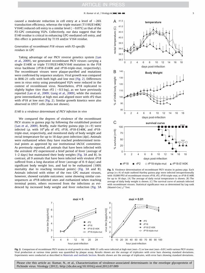
Fig. 3. Virulence determination of recombinant PICV strains in guinea pigs. Each
group (n¼9) of male outbred Hartley guinea pigs were infected intraperitoneally
with 10,000 PFU of recombinant viruses rP18, rP2, rP18-triple-mut, or rP18-E140K
for up to 18 days. (A) The average of daily rectal temperature is shown. (B) The
average of daily body weight is shown. (C) The survival curve of animals infected
with recombinant viruses. Statistical significance was as determined by Log-rank
(Mantel-Cox) w2 Test.
N. Kumar et al. / Virology ] (]]]]) ]]]–]]] 3
caused a moderate reduction in cell entry at a level of �26%transduction efficiency, whereas the triple mutant (T119S/E140K/V164I) reduced cell entry to a similar level (�0.07%) as that of theP2-GPC containing VLPs. Collectively, our data suggest that theE140 residue is critical in enhancing GPC-mediated cell entry, andthis effect is potentiated by T119 and/or V164 residue.
Generation of recombinant P18 viruses with P2-specific
residues in GPC
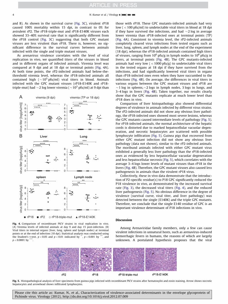
Taking advantage of our PICV reverse genetics system (Lanet al., 2009), we generated recombinant PICV viruses carrying asingle E140K or triple T119S/E140K/V164I mutation in the P18virus backbone (rP18-E140K and rP18-triple-mut, respectively).The recombinant viruses were plaque-purified and mutationswere confirmed by sequence analysis. Viral growth was comparedin BHK-21 cells with both high and low moi (Fig. 2). Differencesseen in virus entry using pseudotyped VLPs were reduced in thecontext of recombinant virus. Nonetheless, rP18 replicated toslightly higher titer than rP2 (�0.5 log), as we have previouslyreported (Lan et al., 2009; Liang et al., 2009), while the mutantsgrew intermediately at high moi and aligned more with rP2 thanwith rP18 at low moi (Fig. 2). Similar growth kinetics were alsoobserved in U937 cells (data not shown).
E140 is a virulence determinant of PICV infection in vivo
We compared the degrees of virulence of the recombinantPICV strains in guinea pigs by following the established protocol(Lan et al., 2009). Briefly, male Hartley guinea pigs (n¼9) wereinfected i.p. with 104 pfu of rP2, rP18, rP18-E140K, and rP18-triple-mut, respectively, and monitored daily of body weight andrectal temperature for up to 18 days post-infection (dpi). Animalswere euthanized when they have reached predetermined term-inal points as approved by our institutional IACUC committee.As previously reported, all animals that have been infected withthe avirulent rP2 experienced a brief period of fever (average of1–2 days) but maintained their body weights (Fig. 3A and B). Incontrast, all 9 animals that have been infected with virulent rP18suffered from a long duration of fever (average of 8–9 days) andsignificant body weight loss, and had to be euthanized (100%mortality due to reaching terminal points) (Fig. 3A and B).Animals infected with either of the two GPC mutant viruses,however, showed variable outcomes: some showing similar con-sequences as rP18-infected ones and euthanized when reachingterminal points, others recovered from the infections as evi-denced by increased body weight and fever reduction (Fig. 3A
rP2rP18rP18 E140K rP18-triple-mut
2
3
4
5
6
7
0 4 8 12 16 20 24hour post-infection
viru
s tit
er [l
og(p
fu/m
l)]
moi = 5
viru
s tit
er [l
og(p
fu/m
l)]
2
3
4
5
6
7
1
8moi = 0.01
rP2rP18rP18 E140K rP18-triple-mut
0 10 20 30 40 50 60hour post-infection
70 80 90 100
Fig. 2. Comparison of recombinant PICV strains in viral growth in vitro. BHK-21 cells were infected at high moi (moi¼5) or low moi (moi¼0.01) with various PICV strains.
Viral production at various time points was quantified by plaque assay. Results shown are the average of triplicates with error bars showing standard deviations.
Experiments were conducted as described in Materials and methods Section. Results shown are the average of triplicates, with error bars showing standard deviations.
Please cite this article as: Kumar, N., et al., Characterization of virulence-associated determinants in the envelope glycoprotein ofPichinde virus. Virology (2012), http://dx.doi.org/10.1016/j.virol.2012.07.009
N. Kumar et al. / Virology ] (]]]]) ]]]–]]]4
and B). As shown in the survival curve (Fig. 3C), virulent rP18caused 100% mortality within 15 dpi, in contrast to 0% foravirulent rP2. The rP18-triple-mut and rP18-E140K viruses eachshowed 33–40% survival rate that is significantly different fromthe rP18 control (Fig. 3C) suggesting that both GPC mutantviruses are less virulent than rP18. There is, however, no sig-nificant difference in the survival curves between animalsinfected with the single and triple mutant viruses.
As arenavirus virulence correlates with the level of viralreplication in vivo, we quantified titers of the viruses in bloodand in different organs of infected animals. Viremia level wascompared at 9 dpi and at 18 dpi or terminal points (Fig. 4A).At both time points, the rP2-infected animals had below-the-threshold viremia level, whereas the rP18-infected animals allcontained high (�105 pfu/ml) viral titers in blood. Animalsinfected with the GPC mutant viruses (rP18-E140K and rP18-triple-mut) had �2 log lower viremia (�103 pfu/ml) at 9 dpi than
******* *******
*** ***
************
*******
***
*******
****** **
viru
s tit
er [l
og(p
fu/g
)]
Liver Lung
spleen Lymph node
viremia (9 dpi) viremia (TP or 18 dpi)
viru
s tit
er [l
og(p
fu/m
l)]
01234567
01234567
0
2
4
6
8
0
2
4
6
8
0
2
4
6
8
0
2
4
6
8
viru
s tit
er [l
og(p
fu/g
)]
rP2rP18 rP18-triple-mut rP18-E140K
Fig. 4. Comparison of recombinant PICV strains in viral replication in vivo.
(A) Viremia levels of infected animals at day 9 and day 15 post-infection. (B)
Viral titers in internal organs (liver, lung, spleen and lymph nodes) at terminal
points or at the end of infection (18 dpi). Statistical analysis was conducted using
the Student’s t-test. po0.05 and po0.01 indicated by **, po0.001 by ***, and
po0.0001 by ****.
Fig. 5. Histopathological analysis of liver specimens from guinea pigs infected with rec
hepatocytes and arrowhead shows infiltrated lymphocytes.
Please cite this article as: Kumar, N., et al., Characterization of viruPichinde virus. Virology (2012), http://dx.doi.org/10.1016/j.virol.201
those with rP18. These GPC mutants-infected animals had verylow (o100 pfu/ml) to undetectable viral titers in blood at 18 dpiif they have survived the infections, and had �2 log in averagelower viremia than rP18-infected ones at terminal points (TP)(Fig. 4A). Consistent to viremia level, the rP2-infected animalscompletely cleared virus infections from tested organs such asliver, lung, spleen, and lymph nodes at the end of the experiment(18 dpi), whereas the rP18-infected animals contained high titersof viruses, ranging from 106 pfu/g in lymph nodes to 108 pfu/g inlivers, at terminal points (Fig. 4B). The GPC mutants-infectedanimals had very low (o1000 pfu/g) to undetectable viral titersin the tested organs at 18 dpi if they have survived from theinfections, and had significantly lowered viral titers in organsthan rP18-infected ones even when they have succumbed to theinfections (Fig. 4B). On average, the differences in viral titers invarious organs between the GPC mutant viruses and rP18 are�1 log in spleens,�2 logs in lymph nodes, 3 logs in lungs, and3–4 logs in livers (Fig. 4B). Taken together, our results clearlyshow that the GPC mutants replicate at much lower level thanrP18 does in vivo.
Comparison of liver histopathology also showed differentialdegrees of virulence in animals infected by different virus strains.The rP2-infected animals did not show any obvious liver pathol-ogy, the rP18-infected ones showed most severe lesions, whereasthe GPC mutants caused intermediate levels of pathology (Fig. 5).For rP18-infected animals, the normal architecture of the hepaticcords is distorted due to marked hepatocellular vacuolar degen-eration, and necrotic hepatocytes are scattered with possiblelymphocyte infiltration (Fig. 5). Guinea pigs that recovered fromeither GPC mutant infection did not show any obvious liverpathology (data not shown), similar to the rP2-infected animals.The moribund animals infected with either GPC mutant virusexhibited a generally less liver pathology than the rP18-infectedones as evidenced by less hepatocellular vacuolar degenerationand less hepatocellular necrosis (Fig. 5), which correlates with theaverage 3–4 logs lower levels of mutant viruses than rP18 in thelivers (Fig. 4B). Therefore, the GPC mutant viruses also caused lesspathogenesis in animals than the virulent rP18 virus.
Collectively, these in vivo data demonstrate that the introduc-tion of P2-specific residue(s) to P18 GPC significantly reduced theP18 virulence in vivo, as demonstrated by the increased survivalrate (Fig. 3), the decreased viral titers (Fig. 4), and the reducedliver pathogenesis (Fig. 5). No obvious difference in the degree ofvirulence (survival curve, viral titer, and liver pathology) wasdetected between the single (E140K) and the triple GPC mutants.Therefore, we conclude that the single E140 residue of GPC is animportant virulence determinant of P18 infections in vivo.
Discussion
Among Arenaviridae family members, only a few can causevirulent infections in unnatural hosts, such as arenavirus-inducedhemorrhagic fevers in humans, the reasons of which are largelyunknown. A postulated hypothesis proposes that the viral
ombinant PICV strains after hematoxylin and eosin staining. Arrow shows necrotic
lence-associated determinants in the envelope glycoprotein of2.07.009

N. Kumar et al. / Virology ] (]]]]) ]]]–]]] 5
genome may encode critical determinants for virulence. We use asafe small animal model, in which two PICV strains derived fromlow (P2) and high (P18) passages in guinea pigs can respectivelycause avirulent and virulent infections with opposing outcomes inthe animals, to characterize the virulence-associated determi-nants in arenavirus genome. Compared to the variable mortalityrates of outbred guinea pigs infected with virulent PICV inprevious studies (Jahrling et al., 1981; Scott and Aronson, 2008),we have observed more consistent high mortality rate andhemorrhagic fever symptoms in our infected animals (Figs. 3–5),which is likely due to different experimental settings between ourstudy and others. Knowledge of virulence mechanisms learnedfrom the PICV-guinea pig model can be extrapolated to otherpathogenic arenaviruses in humans. Through site-directed muta-genesis and comprehensive characterization of the mutantviruses in this study, we have shown that the E140 residue withinthe receptor-binding GP1 subunit is an essential virulence deter-minant of P18 infection of guinea pigs, as the E140K substitutioncan significantly reduce viral replication and pathogenesis in vivo(Figs. 3–5). We have also provided evidence to suggest that themolecular mechanism by which the E140 residue enhances viralreplication and virulence is possibly due to its ability to enhancecell entry efficiency as demonstrated in the pseudotyped lenti-viral system (Table 1).
Using pseudotyped VLPs, we have shown that GPC of virulentP18 virus can mediate a much more efficient cell entry than thatof avirulent P2 into a variety of cell lines (data not shown).The correlation of cell entry efficiency and viral virulence in vivohas led us to believe that GPC-mediated cell entry may be animportant virulence mechanism of arenavirus infection in vivo. AsP2 and P18 GPC proteins differ at three amino acids, 119, 140, and164, within the G1 subunit that mediates specific binding to hostreceptor(s) (Lan et al., 2008), we hypothesize that these residuescan affect virus–cell binding to influence the virus entry effi-ciency. By systematic mutagenesis, we learn that the K140Esubstitution with a change from positive to negative side chainof the amino acid during the P2-P18 adaptation is essential inenhancing cell entry, which can be further augmented by theS119T or I164V substitution (Table 1). However, the exactmolecular mechanism by which these amino acid substitutionsenhance GPC-mediated cell entry requires a better understandingof PICV GPC interaction with its host receptor(s) at the atomiclevel, information of which is not available at the moment. Theentry receptor of PICV as well as other members of Clade A NWarenaviruses has not yet been identified, although we haveproduced preliminary data to suggest that PICV does not primar-ily use a-DG or TfR1 for entry (unpublished data).
Additionally, we have provided evidence to demonstrate theimportant role of the E140 residue in mediating PICV replicationand virulence in vivo. We have shown that a single amino acidsubstitution of E140 with the P2-specific residue K in the genomeof virulent rP18 virus has improved survival rate of infectedanimals (Fig. 3). Furthermore, this E140K single substitution hasled to significantly lower level of viral replication in vivo by atleast 2 logs in blood and by 1–4 logs in various organs (Fig. 4), andsubsequently less liver pathogenesis (Fig. 5), even in the mor-ibund animals. Our in vivo data have provided evidence for E140as one of the important virulence determinants of P18 infection inguinea pigs, which correlates well with its role in increasing GPC-dependent virus entry in vitro. It is noteworthy that compared tothe pronounced 1–4 log difference in viral replication in vivo(Fig. 4), the GPC mutant viruses replicate only slightly less thanthe parental rP18 virus by o0.5 log in vitro (Fig. 2A). This couldbe explained by the differences between the in vivo and in vitroexperimental conditions, among which host immunity may playan important role in amplifying the differential infectivity of viral
Please cite this article as: Kumar, N., et al., Characterization of viruPichinde virus. Virology (2012), http://dx.doi.org/10.1016/j.virol.201
strains in vivo. As early target cells of arenavirus infections aremacrophages and DCs, which are immune cells important forboth innate and adaptive immune responses, differential entryefficiency into these early target cells due to GPC mutations notonly can lead to intrinsically different viral replication levels butmay also affect host immunity that in return leads to even greaterdifferences in viral growth in vivo. Regardless of the mechanisms,our data show that the GPC-mediated differential cell entry intohost target cells leads to the differences in viral growth in vivo.
This GPC-dependent virulence mechanism is also supported bya recent report demonstrating that a single mutation in LCMVGPC is necessary and sufficient for receptor binding, dendritic cellinfection, and long-term persistence (Sullivan et al., 2011). It isimportant to note, however, that mutations in GPC alone did notreduce PICV viral virulence to the level of rP2 (Figs. 3–5)suggesting the contribution of other possible viral virulencefactors besides GPC. We have recently produced evidence todemonstrate that both viral L polymerase and nucleoprotein(NP) may also play important roles in mediating virus virulence((Liang et al., 2009) and our unpublished data). Efforts to char-acterize the virulence-associated mutations in the NP and Lproteins are currently underway. Taken together, our studysuggests that virulent arenavirus infections are a complex processthat requires the synergetic functions of different viral proteins,including but not necessarily limited to GPC, to increase viralentry and RNA synthesis, and to effectively evade host immunedetection (Fan et al., 2010; Martinez-Sobrido et al., 2006; Qi et al.,2010).
Materials and methods
Cells
293T (human kidney epithelial), SLK (human endothelial),human foreskin fibroblast (HFF), A549 (human embryonic lungepithelial) and NIH 3T3 (mouse embryonic fibroblast) were grownin DMEM supplanted with 10% fetal bovine serum (FBS). JH4(guinea pig lung fibroblast) and Chinese hamster ovary epithelial(CHO) were grown in DMEM F12 supplemented with 10% FBS.Vero cells were grown in MEM supplemented with 10% FBS.Human monocytic U937 cells were grown in RPMI-1640 supple-mented with 10% FBS.
GPC protein vector construction, mutagenesis, and expression
We amplified the GPC genes from P2, and P18 virion RNAs andsubcloned each into pCAGGS expression vectors. The resultedplasmids are named pCAGGS-P2-GPC, and pCAGGS-P18-GPC.Single, double, and triple mutations were introduced into theP18 GPC protein at three residues (T119S, E140K, and V164I) byPCR-mediated site-directed mutagenesis and confirmed bysequencing. Expression of various GPC proteins was examinedby transfection of 293T cells with the individual expressionvectors, followed by Western blot analysis using anti-PICV guineapig sera.
Generation of pseudotyped lentivirus-like particles (VLPs)
293T cells were transfected with pHR’GFP, pCMVRD8.91, andthe individual GPC expression vectors using Lipofectamine 2000(Invitrogen). Supernatants of the transfected cells were collectedat different time points up to 96 h post-transfection and filteredthrough a 0.45 mm filter. After purification through sucrosegradient ultracentrifugation, the pseudotyped VLPs were resus-pended in DMEM, aliquoted, and stored at �80 1C. The amount of
lence-associated determinants in the envelope glycoprotein of2.07.009
N. Kumar et al. / Virology ] (]]]]) ]]]–]]]6
VLPs was normalized by the HIV-1 p24 levels as determined byWestern blot analysis using an anti-HIV-1 p24 antibody, which iskindly provided by Dr. E. Hunter (Emory University).
Quantification of GPC-mediated cell entry efficiency of VLPs
Target cells were infected with pseudotyped VLPs at moi of1 in the presence of 2 mg/ml polybrene for 2 h, and replaced withfresh media. GFP protein expression was detected at 72 h post-infection (hpi), either under a fluorescent microscope or by flowcytometric analysis. The efficiency of cell entry was measured bythe percentage of cells expressing the GFP protein (GFPþ%).
Generation of recombinant viruses
Respective GPC mutations (single E140K and triple T119S/E140K/V164I mutations) were introduced to the plasmid encod-ing the full-length anti-sense S segment of P18 virus byPCR-mediated mutagenesis and confirmed by sequencing. Recom-binant PICV viruses were generated as described before (Lan et al.,2009). Briefly, plasmid encoding either WT or mutant P18 Ssegment, together with plasmid encoding P18 L segment, wastransfected into BSRT7-5 cells that stably express T7 polymerase.Recombinant viruses were plaque purified from the supernatantsof the transfected cells, sequence verified by RT-PCR, and ampli-fied in BHK-21 cells. Viral titer was determined by plaque assay.
Determination of viral growth kinetics in vitro
BHK-21 cells were infected with WT or mutant recombinantPICV viruses at moi of 5 or 0.01 for 1 h. After PBS wash for threetimes, cells were replaced with fresh media. At various timespost-infection, viral titers in the supernatants were determinedby plaque assay. Viral production at various time points wassimilarly quantified by plaque assay as described above.
Guinea pig experiments
The experimental procedures for guinea pigs followed theapproved protocol of the Institutional Animal Care and UseCommittee (IACUC) of the Emory University. Briefly, healthy400 g–500 g male outbreed Hartley guinea pigs were housed toacclimatize for 5–7 days after which each group (n¼9) of animalswere injected intraperitoneally with 10,000 PFU of rP18, rP2,rP18-triple-mut (T119S/E140K/V164I) and rP18-E140K, respec-tively. Rectal temperature and body weight were measured dailyfor up to 18 days. Blood was drawn from the marginal ear vein atdifferent days post-infection (dpi). Guinea pigs were declaredmoribund and euthanized if body weight decreased by 30% whencompared to the control group or if animals reached other setterminal points as approved by Emory University IACUC. Virustitration in serum and in tissue specimens was determined byplaque assay in vero cells as described before (Lan et al., 2009).Histopathological analysis was conducted after hematoxylin andeosin staining of formalin-fixed tissue section. Statistical analysesof the survival curve by Log-rank (Mantel-Cox) w2 Test wereconducted using GraphPad prism 5 software. Statistical signifi-cance of viral titers in serum and in different tissue sample wasanalyzed by ‘‘t’’ test.
Acknowledgments
We thank Dr. Johnson (National Animal Disease Center, Ames,IA) for consultation on histopathological analyses of the guineapig liver samples, Dr. Aronson (University of Texas Medical
Please cite this article as: Kumar, N., et al., Characterization of viruPichinde virus. Virology (2012), http://dx.doi.org/10.1016/j.virol.201
Branch) for providing P2 and P18 viruses, Dr. Hunter (EmoryUniversity) for providing anti-HIV-1 p24 antibody, and Dr. Nun-berg (University of Montana) for discussion and sage advice. Thiswork was supported by the NIH grant nos. AI083409 to YL, andR56AI091805 and R01AI093580 to HL.
References
Abraham, J., Kwong, J.A., Albarino, C.G., Lu, J.G., Radoshitzky, S.R., Salazar-Bravo, J.,Farzan, M., Spiropoulou, C.F., Choe, H., 2009. Host-species transferrin receptor1 orthologs are cellular receptors for nonpathogenic new world clade Barenaviruses. PLos Pathog. 5, e1000358.
Agnihothram, S.S., York, J., Nunberg, J.H., 2006. Role of the stable signal peptideand cytoplasmic domain of G2 in regulating intracellular transport of the Juninvirus envelope glycoprotein complex. J. Virol. 80, 5189–5198.
Aronson, J.F., Herzog, N.K., Jerrells, T.R., 1994. Pathological and virological featuresof arenavirus disease in guinea pigs. Comparison of two Pichinde virus strains.Am. J. Pathol. 145, 228–235.
Briese, T., Paweska, J.T., McMullan, L.K., Hutchison, S.K., Street, C., Palacios, G.,Khristova, M.L., Weyer, J., Swanepoel, R., Egholm, M., Nichol, S.T., Lipkin, W.I.,2009. Genetic detection and characterization of Lujo virus, a new hemorrhagicfever-associated arenavirus from southern Africa. PLos Pathog. 5, e1000455.
Buchmeier, M.J., De La Torre, J.C., Peters, C.J., 2007. Arenaviridae: the viruses andtheir replication. In: Knipe, D.M., Howley, P.M. (Eds.), Fields Virology, 5th ed.Lippincott Williams & Wilkins, Philadelphia, PA, pp. 1791–1827.
Cao, W., Henry, M.D., Borrow, P., Yamada, H., Elder, J.H., Ravkov, E.V., Nichol, S.T.,Compans, R.W., Campbell, K.P., Oldstone, M.B., 1998. Identification of alpha-dystroglycan as a receptor for lymphocytic choriomeningitis virus and Lassafever virus. Science 282, 2079–2081.
Cosgriff, T.M., Jahrling, P.B., Chen, J.P., Hodgson, L.A., Lewis, R.M., Green, D.E.,Smith, J.I., 1987. Studies of the coagulation system in arenaviral hemorrhagicfever: experimental infection of strain 13 guinea pigs with Pichinde virus. Am.J. Trop. Med. Hyg. 36, 416–423.
Delgado, S., Erickson, B.R., Agudo, R., Blair, P.J., Vallejo, E., Albarino, C.G., Vargas, J.,Comer, J.A., Rollin, P.E., Ksiazek, T.G., Olson, J.G., Nichol, S.T., 2008. Chaparevirus, a newly discovered arenavirus isolated from a fatal hemorrhagic fevercase in Bolivia. PLos Pathog. 4, e1000047.
Fan, L., Briese, T., Lipkin, W.I., 2010. Z proteins of New World arenaviruses bindRIG-I and interfere with type I interferon induction. J. Virol. 84, 1785–1791.
Jahrling, P.B., Hesse, R.A., Rhoderick, J.B., Elwell, M.A., Moe, J.B., 1981. Pathogenesisof a pichinde virus strain adapted to produce lethal infections in guinea pigs.Infect. Immun. 32, 872–880.
Khan, S.H., Goba, A., Chu, M., Roth, C., Healing, T., Marx, A., Fair, J., Guttieri, M.C.,Ferro, P., Imes, T., Monagin, C., Garry, R.F., Bausch, D.G., 2008. New opportu-nities for field research on the pathogenesis and treatment of Lassa fever.Antiviral Res. 78, 103–115.
Lan, S., McLay, L., Aronson, J., Ly, H., Liang, Y., 2008. Genome comparison ofvirulent and avirulent strains of the Pichinde arenavirus. Arch. Virol. 153,1241–1250.
Lan, S., McLay Schelde, L., Wang, J., Kumar, N., Ly, H., Liang, Y., 2009. Developmentof infectious clones for virulent and avirulent pichinde viruses: a model virusto study arenavirus-induced hemorrhagic fevers. J. Virol. 83, 6357–6362.
Liang, Y., Lan, S., Ly, H., 2009. Molecular determinants of Pichinde virus infection ofguinea pigs—a small animal model system for arenaviral hemorrhagic fevers.Ann. N. Y. Acad. Sci. 1171 (1), E65–E74.
Lucia, H.L., Coppenhaver, D.H., Harrison, R.L., Baron, S., 1990. The effect of anarenavirus infection on liver morphology and function. Am. J. Trop. Med. Hyg.43, 93–98.
Martinez-Sobrido, L., Zuniga, E.I., Rosario, D., Garcia-Sastre, A., de la Torre, J.C.,2006. Inhibition of the type I interferon response by the nucleoprotein of theprototypic arenavirus lymphocytic choriomeningitis virus. J. Virol. 80,9192–9199.
McCormick, J.B., Fisher-Hoch, S.P., 2002. Lassa fever. Curr. Top. Microbiol. Immunol.262, 75–109.
McCormick, J.B., King, I.J., Webb, P.A., Johnson, K.M., O’Sullivan, R., Smith, E.S.,Trippel, S., Tong, T.C., 1987. A case-control study of the clinical diagnosis andcourse of Lassa fever. J. Infect. Dis. 155, 445–455.
Naldini, L., Blomer, U., Gallay, P., Ory, D., Mulligan, R., Gage, F.H., Verma, I.M.,Trono, D., 1996. In vivo gene delivery and stable transduction of nondividingcells by a lentiviral vector. Science 272, 263–267.
Qi, X., Lan, S., Wang, W., Schelde, L.M., Dong, H., Wallat, G.D., Ly, H., Liang, Y., Dong,C., 2010. Cap binding and immune evasion revealed by Lassa nucleoproteinstructure. Nature 468, 779–783.
Qian, C., Jahrling, P.B., Peters, C.J., Liu, C.T., 1994. Cardiovascular and pulmonaryresponses to Pichinde virus infection in strain 13 guinea pigs. Lab. Anim. Sci.44, 600–607.
Radoshitzky, S.R., Abraham, J., Spiropoulou, C.F., Kuhn, J.H., Nguyen, D., Li, W.,Nagel, J., Schmidt, P.J., Nunberg, J.H., Andrews, N.C., Farzan, M., Choe, H., 2007.Transferrin receptor 1 is a cellular receptor for New World haemorrhagic feverarenaviruses. Nature 446, 92–96.
Schaeffer Jr., R.C., Bitrick Jr., M.S., Connolly, B., Jenson, A.B., Gong, F., 1993. Pichindevirus-induced respiratory failure due to obstruction of the small airways:structure and function. Exp. Lung Res. 19, 715–729.
lence-associated determinants in the envelope glycoprotein of2.07.009

N. Kumar et al. / Virology ] (]]]]) ]]]–]]] 7
Scott, E.P., Aronson, J.F., 2008. Cytokine patterns in a comparative model ofarenavirus haemorrhagic fever in guinea pigs. J. Gen. Virol. 89, 2569–2579.
Spiropoulou, C.F., Kunz, S., Rollin, P.E., Campbell, K.P., Oldstone, M.B., 2002.New World arenavirus clade C, but not clade A and B viruses, utilizes alpha-dystroglycan as its major receptor. J. Virol. 76, 5140–5146.
Sullivan, B.M., Emonet, S.F., Welch, M.J., Lee, A.M., Campbell, K.P., de la Torre, J.C.,Oldstone, M.B., 2011. Point mutation in the glycoprotein of lymphocyticchoriomeningitis virus is necessary for receptor binding, dendritic cell infec-tion, and long-term persistence. Proc. Nat. Acad. Sci. U. S. A. 108, 2969–2974.
Trapido, H., Sanmartin, C., 1971. Pichinde virus, a new virus of the Tacaribe groupfrom Colombia. Am. J. Trop. Med. Hyg. 20, 631–641.
Please cite this article as: Kumar, N., et al., Characterization of viruPichinde virus. Virology (2012), http://dx.doi.org/10.1016/j.virol.201
York, J., Nunberg, J.H., 2006. Role of the stable signal peptide of Junin arenavirusenvelope glycoprotein in pH-dependent membrane fusion. J. Virol. 80,7775–7780.
York, J., Nunberg, J.H., 2009. Intersubunit interactions modulate pH-inducedactivation of membrane fusion by the Junin virus envelope glycoproteinGPC. J. Virol..
Zhang, L., Marriott, K., Aronson, J.F., 1999. Sequence analysis of the small RNAsegment of guinea pig-passaged Pichinde virus variants. Am. J. Trop. Med. Hyg.61, 220–225.
Zhang, L., Marriott, K.A., Harnish, D.G., Aronson, J.F., 2001. Reassortant analysis ofguinea pig virulence of pichinde virus variants. Virology 290, 30–38.
lence-associated determinants in the envelope glycoprotein of2.07.009
Copyright © 2022 FDOKUMEN