Rare TERT Promoter Mutations Present in Benign and ... - MDPI
Upload
independentCategory
view
6download
0
ELSEVIER Molecular Brain Research 27 (1994) 205-214
MOLECULAR BRAIN
RESEARCH
Research report
Characterization of the promoter region and flanking sequences of the neuron-specific gene RC3 (neurogranin)
Miguel A. Ifiiguez a, Beatriz Morte a, Angeles Rodriguez-Pefia a, Alberto Mufioz a, Dan Gerendasy b, j. Gregor Sutcliffe h, Juan Bernal a.,
a Instituto de Investigaciones Biomddicas, CSIC, Facultad de Medicina, UAM, Arturo Duperier 4, 28029 Madrid, Spain b Department of Molecular Biology, Scripps Research Institute, La Jolla, CA 92037, USA
Accepted 19 July 1994
Abstract
RC3 encodes a thyroid hormone-dependent, calmodulin-binding, protein kinase C substrate (neurogranin, p17) present in the dendritic spines of discrete neuronal populations in the forebrain. Its physiological role could be related to synaptic plasticity, memory, and other processes. In the present work we have isolated and sequenced 2.4 kbp of genomic DNA upstream from the origin of transcription and determined its nucleotide sequence. The major features of the RC3 promoter are the absence of TATA and CAAT boxes and the presence of an Initiator sequence surrounding the cap site. By sequence analysis we identified several cis-acting regulatory elements, among them response elements for retinoic acid and steroid (glucocorticoids/pro- gesterone) hormone receptors. An oligonucleotide containing the retinoic acid responsive element bound to retinoic acid receptors specifically in vitro and conferred retinoic acid regulation to a heterologous promoter after transfection in COS-7 cells. Retinoic acid and dexamethasone, respectively, increased activity of the RC3 promoter in neuroblastoma cells when a deletion construct containing the retinoic acid and the glucocorticoid responsive elements was cotransfected with retinoic acid receptor or glucocorticoid receptor expression vectors. When added together all-trans retinoic acid and dexamethasone had additive effects. Despite the fact that RC3 expression in vivo is thyroid hormone-dependent, no evidence for the presence of a thyroid hormone responsive element was found within the 2.4 kbp flanking region analyzed and thyroid hormone did not increase reporter activity after cotransfection of suitable constructs with thyroid hormone receptor expression vectors. Our results suggest that the expression of RC3 in vivo could be subject to complex physiological signals, including retinoids and steroid hormones in addition to thyroid hormones.
Keywords: Gene expression; Initiator element; Thyroid hormone; Glucocorticoid; Retinoic acid; Dendrite spine
I. Introduction
The brain-specific m R N A RC3, and its protein product are enriched in regions of the forebrain, mainly cerebral cortex, striatum, and hippocampus [27]. RC3 expression has been shown both by in situ hybridiza- tion and immunohistochemical methods to occur exclu- sively in neurons. The RC3 protein (also called neuro- granin and p17) is a 78 amino acid, protein kinase C substrate that binds calmodulin in its dephosphorylated state. Both the calmodulin-binding and PKC-phos-
* Corresponding author. Fax: (1) (341) 585 4587. E-mail: [email protected].
0169-328X/94/$07.00 © 1994 Elsevier Science B.V. All rights reserved SSDI 0 1 6 9 - 3 2 8 X ( 9 4 ) 0 0 1 5 9 - 6
phorylation domains are homologous to the corre- sponding domains of GAP-43, a neuronal protein lo- cated in the axons during development and regenera- tion [2]. In contrast to GAP-43, the accumulation of RC3 is postnatal and its localization is postsynaptic. In the neostriatum, the protein accumulates in the den- dritic spines of medium and large size neurons [28]. The role of RC3 is presently unknown, but its cellular and anatomical localization, along with its time of developmental onset, are consistent with a function within the post-synaptic second messenger cascade as- sociated with Long Term Potentiation (LTP) [12]. This phenomenon, which is believed by many to constitute the physiological basis of memory, is one consequence of biochemical events set in motion by the influx and
206 M.A. lfiiguez et al. / Molecular Brain Research 27 (1994) 205-214
mobilization of Ca 2+ [13,14]. Because RC3 is a PKC substrate, interacts with calmodulin and, thereby, indi- rectly with other Ca 2÷- and calmodulin-dependent en- zymes such as CaM kinase II, RC3 may also be central to other Ca 2÷ dependent events (not necessarily exclu- sive of LTP) that contribute to dendritic spine develop- ment and synaptic plasticity [11,19].
Previous results from our laboratory have shown that the expression of RC3 at the mRNA and protein levels is under the influence of thyroid hormone in vivo [8,9,16]. Thyroid hormone is known to exert profound influences on brain development and function, includ- ing effects on the dendritic spines of telencephalic neurons. RC3 is dependent on thyroid hormone, not only during maturation but also, and in a reversible way, in adult rats. This contrasts with other brain parameters that are dependent upon thyroid hormone only during the first 10-20 days after birth, and is similar to the response shown by the number and distribution of dendritic spines, which are also re- versibly affected by hypothyroidism in mature animals [7,21]. It is thus tempting to speculate that RC3 is a molecular correlate of the effects of thyroid hormone, and perhaps other physiological signals, on dendritic spines. It is therefore likely that RC3 expression is significant for dendritic spine formation and synaptic plasticity, and that physiological regulation of its ex- pression might be relevant to some pathological pro- cesses. For these reasons we became interested in the isolation and sequencing of genomic clones containing the 5' flanking region of RC3 and the identification of regulatory sequences. We found a number of putative regulatory regions, among them a RARE and a GRE, suggesting that RC3 is under complex, multihormonal control.
2. Materials and methods
2.1. Isolation and characterization of RC3 genomic clones
To isolate RC3 genomic clones, we screened a rat genomic library in the EMBL-3 SP 6 / T 7 vector (Clontech) with a 297 nt probe from the 5' end of the cDNA obtained after BamHI-XbaI digestion of the cDNA cloned in the BamHI site of pr iG 327 [27]. From one million clones we isolated four positives of 15-20 kbp in length. Restriction mapping of the clones was performed by partial digestion and Southern blot hybridization with labeled SP6 and T7 oligonu- cleotides. After hybridization with cDNA probes, we selected a 2.7 kbp fragment for double-stranded DNA sequencing after subcloning in pBluescript.
2.2. Plasmids and oligonucleotides
The following plasmids were used in this study: As chloramfeni- col acetyl transferase (CAT) vectors we used the pBLCAT-2, in which expression of the reporter enzyme is under control of the thymidine kinase promoter, or the promoterless pBLCAT-3 plasmid.
Retinoid receptor expression vectors consisted of the cDNAs encod- ing human retinoic acid receptor (RAR) a or/3 or human retinoid X receptor (RXR) a or /3 cloned in the plasmid pSG-5, and were supplied by Dr H. Stunnenberg (European Molecular Biology Labo- ratory, Heidelberg, Germany). T 3 receptor (T3R) expression vectors were the rat T3R~-I or mouse T3R/3-1 cDNAs cloned in pCDM8, and were a gift from Dr P.R. Larsen (Brigham and Womens Hospi- tal, Boston, MA). The oligonucleotides used in this study were synthesized as two complementary strands with 5' extensions based on the sequences of the RC3 R A R E as shown in this paper, the R A R E present in the promoter region of the retinoic acid receptor /32 gene [5] [ (RAR/32RARE or, simply/32), or the T3RE present in the long terminal repeat of Moloney murine leukemia virus [23] (Mo). For comparison in electrophoretic mobility shift assays we also used an oligonucleotide consisting of the palindromic T3RE. The sequences of these nucleotides were as follows: RC3RARE: 5' GATCCATTTAAAGGGCACAAATAGGTCACGATTAG 3'; R A R / 3 2 R A R E : 5' T C G A C G G G T A G G G T T C A C C G A A A G - T T C A C T C G C 3', MoMLV T3RE: 5' T C G A C A G G G T C A T T T C A G G T C C T T G C 3'; TREpaI: 5' A G C I ' T C A G G T C A T G A C C q ' G A 3'. The nonspecific oligonucleotide used for the gel retardation experi- ments was a 43-mer oligonucleotide from the promoter of rat prote- olipid protein with the following sequence: 5' G A T C C A A G A G T T T T G A C T G G C T G A T T T C C A G T T T G T - G A T A A T G 3'
For transactivation experiments a single copy of the R A R E s or T3REs shown above were subcloned in the pBLCAT2 vector. To study the activity of the RC3 promoter, a genomic fragment spanning nucleotides - 2 1 3 4 to + 156 was subcloned into the EcoRI-BamHI sites of pBLCAT3. Plasmid R C 3 ( - 7 1 5 / + 156)CAT was generated by t reatment with exonuclease IlL Plasmid R C 3 ( - 4 6 6 / + 156)CAT was obtained after deletion of an EcoRI-Pst l fragment from R C 3 ( - 2 1 3 4 / + 156) and religation. Plasmid R C 3 ( - 2 1 3 4 / - 715) CAT was generated after digestion with Pstl and BamHl, and religation. Identity of all clones was verified by sequencing.
2.3. Electrophoretic mobility shift assays
To study interaction of R A R or T3R with DNA, we used nuclear extracts from HeLa cells expressing either T3R, RAR, or RXR, through infection with vaccinia vectors. These nuclear extracts were generously supplied by Dr H. Stunnenberg. Annealed, double- s t randed oligonucleotides with 5' extensions were labeled by filling in with Klenow DNA polymerase and [32p]dCTP. Nuclear extract was incubated in 13 tzl of binding buffer [(15% glycerol, 5 mM MgCI2, 50 mM KC1, 20 mM HEPES pH 7.9, 5 mM DTT and 0.3 m g / m l poly(dI-dC)] for 15 min on ice. 40-100 fmol of labeled probe was added and incubation was continued for another 15 min. Competing oligonucleotides were added in a 50-fold molar excess at the same time as the probe. The p r o t e i n - D N A complexes formed during the binding reaction were separated on 6% polyacrylamide gels in 0.25 × TBE in the cold at 200 V. The gels were prerun for 1 h a 200 V also in the cold. After the run, the gels were dried and exposed to X-ray films.
2.4. Transfections and chloramphenicol acetyl transferase assays
Cells for transfection [(COS-7, or Neuro-2a (N2a)] were grown and maintained in Dulbecco's modified Eagle's medium supple- mented with 10% fetal bovine serum. The cells were plated at a density of 250,000 cells per 6 cm plate the day before transfection. The cells were transfected using the calcium phosphate procedure [43] with 3 /xg of the appropriate CAT construct, 0.3 p.g of an expression vector containing the nuclear receptor and 1.5/.~g (COS-7)
M.A. lfiiguez et al. / Molecular Brain Research 27 (1994) 205-214
or 3 / z g (N2a) of the plasmid pCHI10 (Pharmacia LKB Biotechnol- ogy Inc.). This plasmid contains the structural gene for/3-galactosi- dase under control of SV40 early promoter and was used as an internal control for transfection efficiency. 16-18 h after D N A addition the cells were washed with PBS and replenished with medium containing serum depleted of retinoids and thyroid hor- mones by t rea tment with charcoal and Dowex resin. Where appro- priate, all-trans retinoic acid (Sigma Chemical Co., St. Louis, MO) was added at concentrat ions between 0.05 and 10/zM, and T 3 at 150 nM, and the cells were incubated for a further 24 h before harvesting for determination of/3-galactosidase and C AT activities [20].
207
A RC3 cDNA
B ol Xb ORF poly(A) poly(A) *- i I I
probe
0.1 kb
B RC3 genomic clones
3 . R e s u l t s
Xh Xb Xb E XbE Xb Xb E Xh I II I I I I I ! |
RC3:6
Xh E Xb E Xb Xb E Xh I 1 I I I I I I
3.1. Structure of RC3 promoter and upstream regulatory elements
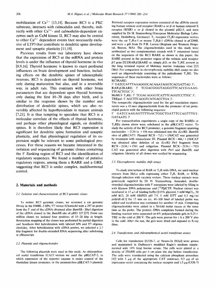
We screened a AEMBL-3 rat genomic library with a BamHI-XbaI fragment from the 5' end of RC3 cDNA (Fig. 1A). From 106 recombinant phages we obtained 4 isolates representing 3 different clones whose respec- tive restriction maps are shown in Fig. lB. The restric- tion fragments respectively named RC3 : 6 and RC3 : 2.7 of 6 and 2.7 kpb in length, were selected on the basis of hybridization with the RC3 cDNA probe and sub- cloned in the pBKS ÷ vector (Fig. 1C). To determine which sequences corresponded to cDNA, we hy- bridized Southern blots containing aliquots of these clones digested with EcoRI, PstI and BamHI, with a 26mer complementary to nucleotides 122 to 148 from the 5'-UT region of the cDNA. This oligonucleotide had previously been used in primer-extension analysis (see below) to determine the 5' end of the cDNA. A PstI-BamHI fragment from RC3:6 and a PstI-XhoI fragment from RC3:2.7 hybridized to the probe. The orientation of RC3 : 2.7 was determined by PCR analy- sis using as primers the 26-mer and oligonucleotides corresponding to regions of either the T7 or T3 pro- moters in the vector. We obtained a single 2.5 kbp fragment amplified using as primer the T7 sequence, indicating that this clone contained the 5' region of the gene (result not shown).
The sequence of this clone is shown in Fig. 2. It contained the 5' end of the transcribed region of the gene as shown by an exact correspondence of nu- cleotide sequence with the reported cDNA sequence [27]. The origin of transcription has been shown previ- ously by primer extension analysis [27] and corresponds to the + 1 nucleotide shown in Figs. 1C and 2. Follow- ing the 5' untranslated region there was an open read- ing frame containing the first 5 amino acids (met-asp- c-ys-cys-thr). The peptide sequence was interrupted by an intron which according to preliminary estimates is larger than 2 kbp.
The region upstream from the RC3 transcriptional origin contained no TATA box. Instead, an initiator
Xh Xb E Xb E Xb Xb E Xh I I I I I I l l l
1 kb RC3:2.7
C Subclones in pBKS
Xb E P B Xh T 7 1 t t i l l i T 3
pRC3:6
E P +1 Xh T7 i I I p T3
oi pRC3:2.7 1 Hyhridizalion with cDNA probe (ol)
1 kb
Fig. 1, Cloning of RC3 gene flanking region. A: structure of RC3 cDNA. The open reading frame (ORF) is represented by the shaded area. The probe used for genomic library screening is indicated, and was obtained after digestion of the cDNA clone with BamHI (B), which cleaves within the polylinker of the cloning vector, and XbaI (Xb). Sites for polyadenylation are shown (poly(A)). The arrow under 'o1' represents the approximate location and 5'-3' orientation of a 26-met oligonucleotide used as a cDNA probe in Southern blots and as a primer for PCR reactions aimed at determining the orientation of the clones after subcloning in pBKS. B: restriction maps of the three independent genomic clones obtained after library screening. The shaded areas labelled RC3:6 and RC3:2.7 correspond to 6 and 2.7 kbp fragments subcloned in pBKS. C: structure of the RC3 : 6 and RC3:2.7 fragments after subcloning in pBKS. The shaded areas are restriction fragments which showed hybridization with the 26-mer oligonucleotide (ol) in Southern blots. The approximate location of the + 1 nucleotide is shown, as well as the position of the T3 and T7 promoters of the pBKS vector. Restriction enzymes are as follows, E, EcoRI; B, BamHI; P, PstI; Xb, XbaI; Xh, XhoI.
element (Inr) [24] surrounding the cap site was found, flanked by putative Spl sites. The initiator sequence resembles that of the terminal deoxynucleotidyl trans- ferase (TdT) initiator, with the difference that in the latter the cap site corresponds to adenine, whereas in RC3 the first nucleotide would be guanine. According to recent studies [10], the RC3 initiator contains fea- tures of strong initiators, such as a T at + 3, but the presence of a G at + 1 would render it less active, with a predicted activity of about 10% that of the TdT initiator. However, the sequence +4CCAG'I-T+9 con- forms exactly to the initiator consensus sequence
208 M.A. lfiiguez et al. /Molecular Brain Research 27 (1994) 205-214
-2274 CCCGGGCTGC
-2214 CTCTTTGCCC PU box
-2154 GAGGAAGTTG
-2094 CCTAGGACAT
AGGAATTCCT CAGCTCTAAT TTGGGAAAAG CACAGTCCAG ATCCGATATC
CTCTGTTTCC CCCTTACGTC TCTTCTCTGG GTTCTGGAGT AAAAAGCACA
GCACAGCCAG AGGTCCAGAT GTGTTTGTCA TTTAGGCAAA GACCTTAGTT
CCTGCCATTT GGTGGAGGTG GGGGGACTAA AGATGTGCCA GAGACAGATT
-2034 GGGGAACAGA TTTGCTGTTA CAGGGGTGGG TGGGTCCCAA ACATGTTGGG AGAGACACCC AP2
-1974 CTACACCCCA ACCCTGGAAT CCAGCCAGCC ACCAGCTCAG CTTGGAGGCC CAAGCTGGCT
-1914 AATCATGCCC CCAACCTGAG TGGACACTGG CATATCCCTC CCACTCATTC TCCCTTTAAC half-site
-1854 AAGGAAAGCC ATGACCCAGT CCCACTGTGG CTATCCATTG CTATCCATTG CTGCTTGGCT
-1794 GTTTGAGGTC CCCTGTCCTA CCCAGAGCCA CCAGGTATTC CTTCCTCTCT AAGCAGTAGC
-1734 TGTCCTTTCC TGTCCCTCCT TAGAGAACTT AGCTGAGGAG GGCTTGAGTT TTCTCAAAGG C-mOS
-1674 GGTCCCAACC ACCTGGGTAT CATGCAACAC AGTCAGCTAT ACTGCCCTGA CAGGCTGGGA SRE
-1614 GGTTAAGAGA CCACAACAAG ATCAAGCTGC CTACCCCAGA TGTCCCTTTC CATATCTCTG
TCTCTCCAAA CCACATCCTT CCCCAGCATG AGCTCAAAGG GCCAAAACCC
CCTACACTGC TTGAGTTGAC AGCATCCTTC TTCTTCCTGA GAGCAAGTGC API
AACTTTTGTC TTATTTTGAG GCAATGAATC AAAAGGTTAG GAAGTCTAGT
GGATGGGTGG GTGTGCTTAA GTCAGAACCA GCATATAAAT ACATTCAAAG half-site half-site
GATCATAC, GT CATGGGTAGA TACTAAATAG ATACCTACCT GGTCAGTGAT
ACTTGGAACT AGTCAAAGCA CTGAGAATAA GGGGCTATTG AATGCCCAGT
ACATATCTGC ACCAATACTT CCACAGCTCA GGAAACATCA CGGAAGAAGG
GTTTAAGAAC CAGAAGGGAA GTGAGGTATG GAATTATGAC TTCCAGTCAT
TAGTATTCCT GAGTTCACAA CAGCTGGATC ACCTGCCCAA GGATCTGCAC AP2
CCATCAACAC TCTGCCATGG AAAGAGGAGG GGCTTATGAG GTCCCACCCC c -myb
TCTATATACA GTTAAGGGTT GGCAAGGTAG GGAGAGACAT TTGCTTCAGT
CTACTATTAT AGCCTACGAT GCTGTAAACA AGCCCTTACC TGTGCTCCTG
TAATAAAATT CAATAGTTTC CTTCCCTTCC CCGCAAAAAA AGACATGACA
-774 ATAAAAGAGA TACTTGTGGC AAGAATGAGG GTTGTCAGGA GTGGGAGGGG GCAAAAGAGG half-site RARE
-714 GTAAC~T C, AACATTATT TAAAGC, C, CAC AAATAGGTCA CGATTAAAAC AATAATTATG GRE
-654 TGGAATCAAT ATGTTCTAGT ACAGTGTTTT CGGTAAAAAT TTTAAAAAGA AAGCACTTAA
-594 TGCAGACATT GCCAGCCGGG AGAACGCAGG CAATTTGATC TATGTGCTAA CGAACAGGAG
-534 GGCTACAATT TACCTGCTTC GCAGGGTGTG TGTACCCGGC CCGTGTAGTT GTTTTTGCCT
-474 ATTGTGTCTG CAGTGTCTGT ATGTGGGAAG ATAACCACAG CCTGGGAATA TATATGAATG
-414 CACGAGACTT GGAGCACAGC GTCTGTGTCA CTATGTGCCC ATCATGTAGG CTGCATATCA
-354 TCAGCTTTCT CTGGAGCTAG AGTGAGGAGG GCACCTCTTT GCAGAGAGGG GAGATAGAAG
-294 AAGTAATGTT TGGGACTCTG GGCTGTGAAA GTATAGGGAG GTGGGTGCGG TGCGTGGTTT sp1
-234 TCTATCTATG GGCGGTAGGG CGTCGTGCGC CCAGATGTGT ATTTGTGCCC TGGATGTTGG
-174 GGCCTTGTGC CTACTCCGAC GTACATCTGC CTGGCAGTCT GCGATTTAGA AAGCAGGGGG PU box
-114 GGTTGAATTT GGGAGATGGG GTGTGTAGGA ~TGGATG GAAGTGTGTA GGTGGAGAGG Spl +I Inr
-54 TTGGGGCACC AAAGCGAGCG AGGCGC4%GAG AGTCGAGAGA GGGAGTGGTC CTC~CTCC~ Spl Y box
+8 TTCTCCCCC, C CCACCCTACA GAAAGTGTCT CCTGATTGGC TTTGAGGCCG CAGGGCTCAG
+68 GTTACATTCG CAAGAGTTGC GGAGCGCGGG AGACCGGACC CAAGAGGAGA GAGGCTGGTT
+128 CTGCACCGAT TCTGTGCTGG TCCGGGAGTG CCCGACAGCC CCTGAACTAC CACCCAGCAT
+188 TGTACAAACC CACCCCCACT CTGAGCCAGG CTCCACCCCA GCCAAGGACC CTCAACACCG MetAspCysCysThr Intron
t248 GCAATGGACT GCT~CGgt gagtccatac ttgaactcag cttggtctgg ggcaaggggc
+308 gcccgagaac ccctgggggt gagggaggga tggaagtagg tgcggaagac ctaggagccc
+368 tgtggggtag gatggtgctg agatatattg gatggtggaa agcacctgcc tgatcctcga
-1554 CTTCCCAGCC NF-EI
-1494 GCTGATAACC API
-1434 AAGGGTGACT
-1374 GTGAGGAGTG
-1314 TGAATAGACA NF-EI
- 1254 C.ATAAAAGAC
-1194 CATAGAGAGG
-1134 TGTGGGAAGA
-1074 GACTTGGTGA
-1014 AGGATTGGGC
-954 ACCCTGAAGA
-894 GGTGTAGTCA
- 834 TAAACAACAC
M.A. I~iguez et aL / Molecular Brain Research 27 (1994) 205-214
2 3
RC3* RAR~2RARE* RC3*
J32 RC3 Mo NS - 132 RC3 NS
TREp* RC3*
- RC3
i iii!ii!ili ili ilili i
209
RARQt + + + + + + + + + + + T R ~ + + + +
RXRo~ + + + + + + R X R a +
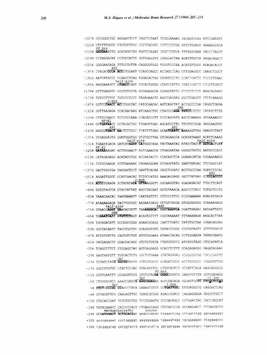
Fig. 3. Band shift assay demonstrating the binding of receptor proteins to the putative RC3 retinoic acid response element. Formation of protein-DNA complexes was examined after electrophoresis and autoradiography. Labeled oligonucleotides representing the RC3 retinoic acid responsive element (RC3*), the retinoic acid responsive element of the retinoic acid receptor f12 gene (RARfl2RARE*) or the palindromic thyroid hormone responsive element (TREp*), are shown at the top of the diagram. These oligonucleotides were incubated with HeLa cell nuclear extracts derived from HeLa cells (wt) or from extracts derived from HeLa cells that were engineered to express high levels of the retinoic acid receptor t~ (RARt~), the retinoid X receptor a (RXRtY) or the thyroid hormone receptor a (TaRa), in various combinations, as indicated at bottom of figure. The unlabeled oligonucleotides used to compete with the labeled oligonucleotides are displayed near the top of the diagram; /32, RAR/32 RARE; Rca, RC3 RARE; Mo, moloney murine leukemia virus TARE; NS, nonspecific oligonucleotide.
PyPyA+INTPyPy. It is known that initiators can be shifted upstream or downstream from the origin of transcription [10] with the A located between - 5 and + 5 (double underline in Fig. 2). In the case of RC3, the close proximity of the downstream Spl site could shift the origin of transcription several bases upstream of the initiator. Upstream of the initiator there were a number of consensus sequences for transcription fac- tors, including AP-2, AP-1, c-mos, SRE, and NF-E1 sites.
Since RC3 is influenced in vivo by thyroid hormone we searched for the sequence AGGTCA, conforming the half-site of direct repeats [26] found in type II
nuclear receptor responsive elements (i.e., the recep- tors for 1,25(OH)2vitamin Da, retinoic acid and thyroid hormone, Ta). In addition to three isolated half-sites of uncertain significance, the sequence _69tAGGGCA - caaatAGGTCA_673 was an almost perfect consensus sequence for a retinoic acid response element (RARE) consisting of a direct repeat of the motif AGGTCA with a gap of five nucleotides. Retinoic acid receptors (RARs) bind to their cognate responsive elements in DNA as heterodimers with RXR, which is also a receptor for 9-cis-retinoic acid. RXR is also the het- erodimeric partner of thyroid hormone receptors (T3Rs) , vitamin D 3 receptor, and other ligand-depend-
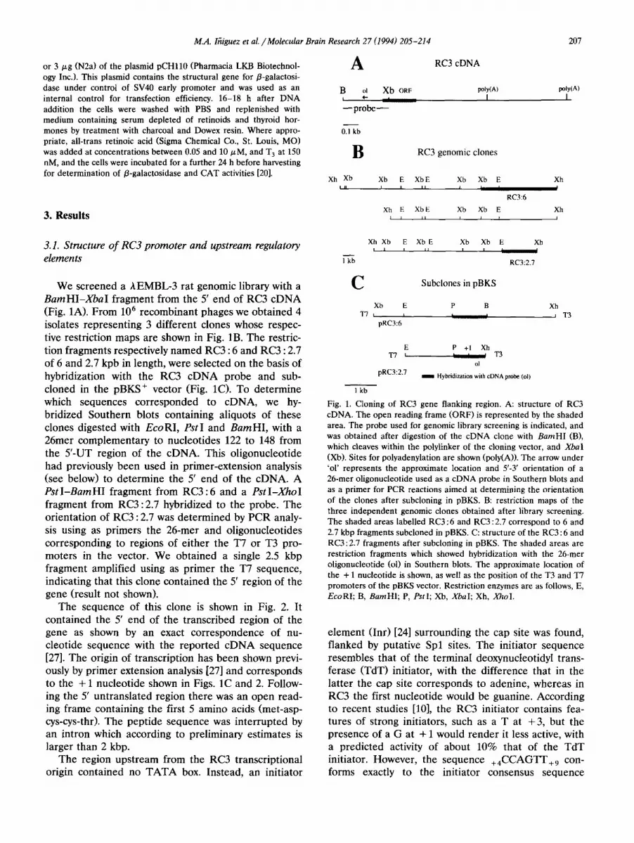
Fig. 2. Nucleotide sequence of pRC3 : 2.7. Consensus sequences for transcriptional regulators are in bold face. RARE, retinoic acid responsive element; GRE, glucocorticoid response element. The transcriptional start site is shown by + 1 above the corresponding nucleotide. Putative regulatory sequences (PU box, AP-2, c-mos, serum response element (SRE), NF-E1, AP-1, c-myb) are highlighted. They were identified using the Quest program from Intelligenetics. The sequence labeled 'half site' corresponds to the consensus half recognition site for type II nuclear receptors. Within the initiator (Inr) sequence, the double underlined nucleotides represent two alternative positions as the center of the initiator. The amino terminal aminoacid sequence of the RC3 protein is shown. The partial sequence of the first intron is depicted in lower case.
210 M.A. lhiguez et al. /Molecular Brain Research 27 (1994) 205 214
ent or -independent transcription factors [25]. The heterodimers recognize response elements organized as direct repeats of the motif AGGTCA spaced by a variable number of nucleotides, which are four for T3R-RXR and five for RAR-RXR.
3.2. Characterization of a retinoic acid responsit,e ele- ment (RARE)
RA:
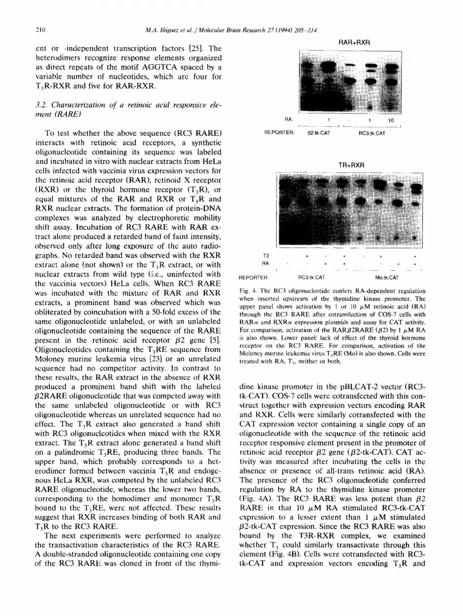
REPORTER: To test whether the above sequence (RC3 RARE) interacts with retinoic acid receptors, a synthetic oligonucleotide containing its sequence was labeled and incubated in vitro with nuclear extracts from HeLa cells infected with vaccinia virus expression vectors for the retinoic acid receptor (RAR), retinoid X receptor (RXR) or the thyroid hormone receptor (T3R), or equal mixtures of the RAR and RXR or T3R and RXR nuclear extracts. The formation of protein-DNA complexes was analyzed by electrophoretic mobility shift assay. Incubation of RC3 RARE with RAR ex- tract alone produced a retarded band of faint intensity, observed only after long exposure of the auto radio- graphs. No retarded band was observed with the RXR extract alone (not shown) or the T3R extract, or with nuclear extracts from wild type (i.e., uninfected with the vaccinia vectors) HeLa cells. When RC3 RARE was incubated with the mixture of RAR and RXR extracts, a prominent band was observed which was obliterated by coincubation with a 50-fold excess of the same oligonueleotide unlabeled, or with an unlabeled oligonucleotide containing the sequence of the RARE present in the retinoic acid receptor /32 gene [5]. Oligonucleotides containing the T3RE sequence from Moloney murine leukemia virus [23] or an unrelated sequence had no competitor activity. In contrast to these results, the RAR extract in the absence of RXR produced a prominent band shift with the labeled /32RARE oligonucleotide that was competed away with the same unlabeled oligonucleotide or with RC3 oligonucleotide whereas un unrelated sequence had no effect. The T3R extract also generated a band shift with RC3 oligonucleotides when mixed with the RXR extract. The T3R extract alone generated a band shift on a palindromic T3RE, producing three bands. The upper band, which probably corresponds to a het- erodimer formed between vaccinia T3R and endoge- nous HeLa RXR, was competed by the unlabeled RC3 RARE oligonucleotide, whereas the lower two bands, corresponding to the homodimer and monomer T3R bound to the T3RE, were not affected. These results suggest that RXR increases binding of both RAR and T3R to the RC3 RARE.
The next experiments were performed to analyze the transactivation characteristics of the RC3 RARE. A double-stranded oligonucleotide containing one copy of the RC3 RARE was cloned in front of the thymi-
RAR+RXR
i
1 1 10 L . . . . . . . . . . J ~ . . . . . . . . . .
[}2:tk:CAT RC3:tk:CAT
TR+RXR
T 3 + + + +
RA + + + + L . . . . . J L _ __ J
REPORTER: RC3:tk:CAT Mo:tk:CAT
Fig. 4. The RC3 oligonucleotide confers RA-dependent regulation when inserted upstream of the thymidine kinase promoter. The upper panel shows activation by 1 or 10 /zM retinoic acid (RA) through the RC3 RARE after cotransfection of COS-7 cells with RARa and RXRa expression plasmids and assay for CAT activity. For comparison, activation of the RAR/32RARE (/32) by 1 /~M RA is also shown. Lower panel: lack of effect of the thyroid hormone receptor on the RC3 RARE. For comparison, activation of the Moloney murine leukemia virus T3RE (Mo) is also shown. Cells were treated with RA, T3, neither or both.
dine kinase promoter in the pBLCAT-2 vector (RC3- tk-CAT). COS-7 cells were cotransfected with this con- struct together with expression vectors encoding RAR and RXR. Cells were similarly cotransfected with the CAT expression vector containing a single copy of an oligonucleotide with the sequence of the retinoic acid receptor responsive element present in the promoter of retinoic acid receptor /32 gene (/32-tk-CAT). CAT ac- tivity was measured after incubating the cells in the absence or presence of all-trans retinoic acid (RA). The presence of the RC3 oligonucleotide conferred regulation by RA to the thymidine kinase promoter (Fig. 4A). The RC3 RARE was less potent than /32 RARE in that 10 /xM RA stimulated RC3-tk-CAT expression to a lesser extent than 1 /~M stimulated /32-tk-CAT expression. Since the RC3 RA RE was also bound by the T3R-RXR complex, we examined whether T 3 could similarly transactivate through this element (Fig. 4B). Cells were cotransfected with RC3- tk-CAT and expression vectors encoding T3R and
M.A. lfiiguez et aL / Molecular Brain Research 27 (1994) 205-214 211
RXR. For comparison we used a construct in pBLCAT-2 containing a single copy of the T3RE pre- sent in the long terminal repeat of Moloney murine leukemia virus (Mo-tk-CAT). Cotransfection with T 3 R
and R X R did not confer regulation by either T3, RA, or both. In contrast, the plasmid Mo-tk-CAT was acti- vated by T 3 in the same experiments. Thus, though T3R-RXR was able to bind to the RC3 RARE, the complex formed was not functional.
3.3. Transactivation by retinoic acid and steroid hor- mones in the context o f the RC3 promoter
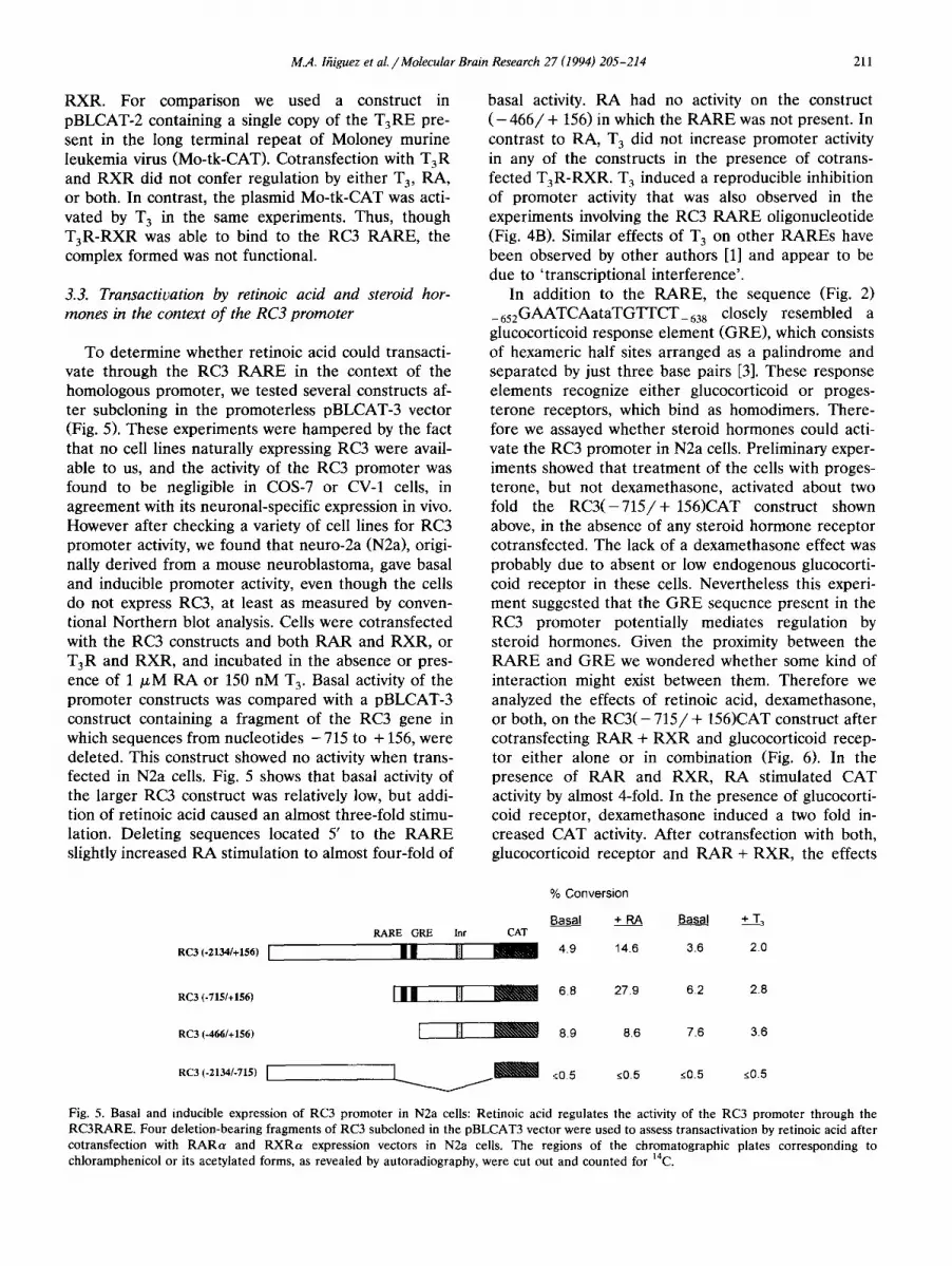
To determine whether retinoic acid could transacti- vate through the RC3 R A R E in the context of the homologous promoter, we tested several constructs af- ter subcloning in the promoterless pBLCAT-3 vector (Fig. 5). These experiments were hampered by the fact that no cell lines naturally expressing RC3 were avail- able to us, and the activity of the RC3 promoter was found to be negligible in COS-7 or CV-1 cells, in agreement with its neuronal-specific expression in vivo. However after checking a variety of cell lines for RC3 promoter activity, we found that neuro-2a (N2a), origi- nally derived from a mouse neuroblastoma, gave basal and inducible promoter activity, even though the cells do not express RC3, at least as measured by conven- tional Northern blot analysis. Cells were cotransfected with the RC3 constructs and both R A R and RXR, or TaR and RXR, and incubated in the absence or pres- ence of 1 /.~M R A or 150 nM T 3. Basal activity of the promoter constructs was compared with a pBLCAT-3 construct containing a fragment of the RC3 gene in which sequences from nucleotides - 715 to + 156, were deleted. This construct showed no activity when trans- fected in N2a cells. Fig. 5 shows that basal activity of the larger RC3 construct was relatively low, but addi- tion of retinoic acid caused an almost three-fold stimu- lation. Deleting sequences located 5' to the R A R E slightly increased RA stimulation to almost four-fold of
basal activity. R A had no activity on the construct ( - 4 6 6 / + 156) in which the R A R E was not present. In contrast to RA, T 3 did not increase promoter activity in any of the constructs in the presence of cotrans- fected T3R-RXR. T 3 induced a reproducible inhibition of promoter activity that was also observed in the experiments involving the RC3 R A R E oligonucleotide (Fig. 4B). Similar effects of T 3 o n other RAREs have been observed by other authors [1] and appear to be due to ' transcriptional interference' .
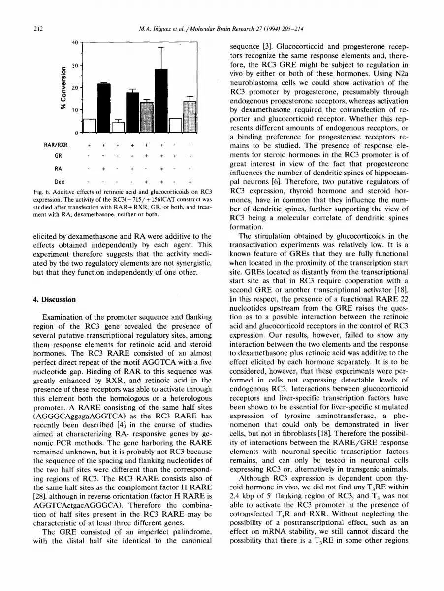
In addition to the RARE, the sequence (Fig. 2) _652GAATCAataTGT-FCT_63s closely resembled a glucocorticoid response element (GRE), which consists of hexameric half sites arranged as a palindrome and separated by just three base pairs [3]. These response elements recognize either glucocorticoid or proges- terone receptors, which bind as homodimers. There- fore we assayed whether steroid hormones could acti- vate the RC3 promoter in N2a ceils. Preliminary exper- iments showed that t reatment of the cells with proges- terone, but not dexamethasone, activated about two fold the R C 3 ( - 7 1 5 / + 156)CAT construct shown above, in the absence of any steroid hormone receptor cotransfected. The lack of a dexamethasone effect was probably due to absent or low endogenous glucocorti- coid receptor in these cells. Nevertheless this experi- ment suggested that the G R E sequence present in the RC3 promoter potentially mediates regulation by steroid hormones. Given the proximity between the R A R E and G R E we wondered whether some kind of interaction might exist between them. Therefore we analyzed the effects of retinoic acid, dexamethasone, or both, on the R C 3 ( - 7 1 5 / + 156)CAT construct after cotransfecting R A R + R X R and glucocorticoid recep- tor either alone or in combination (Fig. 6). In the presence of R A R and RXR, RA stimulated CAT activity by almost 4-fold. In the presence of glucocorti- coid receptor, dexamethasone induced a two fold in- creased CAT activity. After cotransfection with both, glucocorticoid receptor and R A R + RXR, the effects
% Conversion
Basal + RA Basal + T 3 RARE GRE lnr CAT
RC3 (-2134/+156) I II I [I 1 4.9 14.6 3.6 2.0
RC3 (-7~5/*t56) I I I [il ~ 6.8 27.9 6.2 2.8
RC3 (-466/+156) [ - - ~ 8.9 8.6 7.6 3,6
RC3 (-2134/-715) [ ] [ ~ ~;0.5 <0.5 <0.5 ~0.5
Fig. 5. Basal and inducible expression of RC3 promoter in N2a cells: Retinoic acid regulates the activity of the RC3 promoter through the RC3RARE. Four deletion-bearing fragments of RC3 subcloned in the pBLCAT3 vector were used to assess transactivation by retinoic acid after cotransfection with RARa and RXRa expression vectors in N2a cells. The regions of the chromatographic plates corresponding to chloramphenicol or its acetylated forms, as revealed by autoradiography, were cut out and counted for 14C.
212 M.A. lfiiguez et al. / Molecular Brain Research 27 (1994) 205-214
40
3O r- ._o
Q) • 20 t -
O U
~ ~o
RAR/RXR + + + + + +
GR + + + + + +
RA + + +
Dex + + +
Fig. 6. Additive effects of retinoic acid and glucocorticoids on RC3 expression. The activity of the RC3( - 7 1 5 / + 156)CAT construct was studied after transfection with R A R + RXR, GR, or both, and treat- ment with RA, dexamethasone, neither or both.
elicited by dexamethasone and RA were additive to the effects obtained independently by each agent. This experiment therefore suggests that the activity medi- ated by the two regulatory elements are not synergistic, but that they function independently of one other.
4. Discussion
Examination of the promoter sequence and flanking region of the RC3 gene revealed the presence of several putative transcriptional regulatory sites, among them response elements for retinoic acid and steroid hormones. The RC3 RARE consisted of an almost perfect direct repeat of the motif AGGTCA with a five nucleotide gap. Binding of RAR to this sequence was greatly enhanced by RXR, and retinoic acid in the presence of these receptors was able to activate through this element both the homologous or a heterologous promoter. A RARE consisting of the same half sites (AGGGCAggagaAGGTCA) as the RC3 RARE has recently been described [4] in the course of studies aimed at characterizing RA- responsive genes by ge- nomic PCR methods. The gene harboring the RARE remained unknown, but it is probably not RC3 because the sequence of the spacing and flanking nucleotides of the two half sites were different than the correspond- ing regions of RC3. The RC3 RARE consists also of the same half sites as the complement factor H RARE [28], although in reverse orientation (factor H RARE is AGGTCActgacAGGGCA). Therefore the combina- tion of half sites present in the RC3 RARE may be characteristic of at least three different genes.
The GR E consisted of an imperfect palindrome, with the distal half site identical to the canonical
sequence [3]. Glucocorticoid and progesterone recep- tors recognize the same response elements and, there- fore, the RC3 G RE might be subject to regulation in vivo by either or both of these hormones. Using N2a neuroblastoma cells we could show activation of the RC3 promoter by progesterone, presumably through endogenous progesterone receptors, whereas activation by dexamethasone required the cotransfection of re- porter and glucocorticoid receptor. Whether this rep- resents different amounts of endogenous receptors, or a binding preference for progesterone receptors re- mains to be studied. The presence of response ele- ments for steroid hormones in the RC3 promoter is of great interest in view of the fact that progesterone influences the number of dendritic spines of hippocam- pal neurons [6]. Therefore, two putative regulators of RC3 expression, thyroid hormone and steroid hor- mones, have in common that they influence the num- ber of dendritic spines, further supporting the view of RC3 being a molecular correlate of dendritic spines formation.
The stimulation obtained by glucocorticoids in the transactivation experiments was relatively low. It is a known feature of GREs that they are fully functional when located in the proximity of the transcription start site. GREs located as distantly from the transcriptional start site as that in RC3 require cooperation with a second GRE or another transcriptional activator [18]. In this respect, the presence of a functional RARE 22 nucleotides upstream from the G RE raises the ques- tion as to a possible interaction between the retinoic acid and glucocorticoid receptors in the control of RC3 expression. Our results, however, failed to show any interaction between the two elements and the response to dexamethasone plus retinoic acid was additive to the effect elicited by each hormone separately. It is to be considered, however, that these experiments were per- formed in cells not expressing detectable levels of endogenous RC3. Interactions between glucocorticoid receptors and liver-specific transcription factors have been shown to be essential for liver-specific stimulated expression of tyrosine aminotransferase, a phe- nomenon that could only be demonstrated in liver cells, but not in fibroblasts [18]. Therefore the possibil- ity of interactions between the R A R E / G R E response elements with neuronal-specific transcription factors remains, and can only be tested in neuronal cells expressing RC3 or, alternatively in transgenic animals.
Although RC3 expression is dependent upon thy- roid hormone in vivo, we did not find any T3RE within 2.4 kbp of 5' flanking region of RC3, and T 3 was not able to activate the RC3 promoter in the presence of cotransfected T3R and RXR. Without neglecting the possibility of a posttranscriptional effect, such as an effect on mRNA stability, we still cannot discard the possibility that there is a T3RE in some other regions
M.A. lfiiguez et aL / Molecular Brain Research 27 (1994) 205-214 213
of the gene, further upstream of the DNA sequence analyzed by us, or even downstream from the origin of transcription. There are examples of T3RE located in intronic sites, such as in the third intron of the growth hormone gene [22]. It is also possible that the action of T 3 is mediated indirectly through interactions of the receptor with transcriptional regulatory proteins, simi- lar to the interactions with Jun and Fos [29]. Another possibility for T 3 regulation is through control of RXR, which acts as a common partner of T3R and RAR [25]. It has been shown that thyroid hormone controls the expression of RXRb in the rat brain [16]. Since binding of RAR to the RC3 RARE was greatly facilitated by RXR, intracellular changes of this receptor in selected neuronal targets could result in a lower activation by endogenous retinoids and, therefore, in a decreased RC3 expression.
In summary, the data reported in this paper suggest that the RC3 gene is subject to a complex network of regulatory signals, in addition to thyroid hormone, which includes steroid hormones and retinoids. The degree and cell specificity of this regulation in vivo remains to be assessed.
Acknowledgements
This work has been supported by grants from DGI- CYT PM92-0025, FIS 94/0273, Fundaci6n Salud 2000, and NIH grant NS 22111. We would like to acknowl- edge Dr H. Stunnenberg for his generous gift of vac- cinia extracts and expression plasmids encoding retinoid and glucocorticoid receptors and for his continuing support and suggestions; Dr P.R. Larsen, for TaR expression plasmids; and Ms Gloria Chacon and Ms Patria Danielson for excellent technical assistance.
References
[1] Barettino, D., Bugge, T.H., Bartunek, P., Vivanco-Ruiz, M., Sonntagbuck, V., Beug, H., Zenke, M. and Stunnenberg, H., Unliganded T3R , but not its oncogenic variant, v-erbA, sup- presses RAR-dependent transactivation by titrating out RXR, EMBO J., 12 (1993) 1343-1354.
[2] Baudier, J., Deloulme, J.C., Van Dorsselaer, A., Black, D. and Matthes, W.D., Purification and characterization of abrain- specific protein kinase C substrate, neurogranin (p17). Identifi- cation of a consensus aminoacid sequence between neurogranin and neuromodulin (Gap43) that corresponds to the protein kinase C phosphorylation site and the calmodulin-binding do- main, J. Biol. Chem., 266 (1991) 229-237.
13] Beato, M., Gene regulation by steroid hormones, Cell, 56 (1989) 335-344.
[4] Costa-Giomi, M.P., Gaub, M.P., Chambon, P. and Abarzfia, P., Characterization of a retinoic acid responsive element isolated by whole genome PCR, Nucl. Acid Res., 20 (1992) 3223-3232.
[5] de Th6, H., Vivanco-Ruiz, M.D.M., Tiollais, P., Stunnenberg, H. and Dejean, A., Identification of a retinoic acid responsive
element in the retinoic acid receptor/3 gene, Nature, 343 (1990) 177-180.
[6l Gould, E., Woolley, C.S., Frankfurt, M. and McEwen, B.S., Gonadal steroids regulate dendritic spine density in hippocam- pal pyramidal cells in adulthood, J. Neurosci., 10 (1990) 1286- 1291.
17] Gould, E., Allan, M.D. and McEwen, B.S., Dendritic spine density of adult hippocampal pyramidal cells is sensitive to thyroid hormone, Brain Res., 525 (1990) 327-329.
[8] Ifiiguez, M.A., Rodriguez-Pefia, A., Ibarrola, N., Morreale de Escobar, G. and Bernal, J., Adult rat brain is sensitive to thyroid hormone: regulation of RC3/neurogranin mRNA, J. Clin. In- cest., 90 (1992) 554-558.
[9] Ifiiguez, M.A., Rodriguez-Peha, A., Ibarrola, N., Aguilera, M., Mufioz, A. and Bernal, J., Thyroid hormone regulation of RC3, a brain-specific gene encoding a protein kinase-C substrate, Endocrinology, 133 (1993) 467-473.
[10] Javahery, R., Khachi, A., Lo, K., Zenzie-Gregory, B. and Smale, S., DNA sequence requirements for transcriptional initiator activity in mammalian cells, Mol. Cell. Biol., 5 (1994) 153-177.
[11] Kelly, P.T., Calmodulin-dependent protein kinase II. Multifunc- tional roles in neuronal differentiation and synaptic plasticity, Mol. Neurobiol., 5 (1991) 153-177.
[12] Klann, E., Chert, S.-J. and Sweatt, J.D., Increased phosphoryla- tion of a 17-kDa protein kinase C substrate (p17) in long-term potentiation, J. Neurochem., 58 (1992) 1576-1579.
[13] Linden, D.J. and Routtenberg, A., The role of protein kinase C in long-term potentiation: a testable model, Brain Res. Rev., 14 (1989) 279-296.
[14] Madison, D.V., Malenka, R.C. and Nicoll, R.A., Mechanisms underlying long-term potentiation of synaptic transmission, Anna. Rec. Neurosci., 14 (1991) 379-397.
[15] Mano, H., Mori, R., Ozawa, T., Takeyama, K.-L, Yoshizawa, Y., Kojima, R., Arao, Y., Masushige, S. and Kato, S., Positive and negative regulation of retinoid X receptor gene expression by thyroid hormone in the rat. Transcriptional and post-transcrip- tional controls by thyroid hormone, J. Biol. Chem., 269 (1994) 1591-1594.
[16] Mufioz, A., Rodriguez-Pefia, A., Perez-Castillo, A., Ferreiro, B., Sutcliffe, J.G. and Bernal, J., Effects of neonatal hypothyroidism on rat brain gene expression, Mol. Endocrinol., 5 (1991) 273-280.
[17] Mufioz-Cfinoves, P., Vik, D.P. and Tack, B.F., Mapping of a retinoic acid responsive element in the promoter region of the complement factor H gene, J. Biol. Chem., 265 (1990) 20065- 20068.
[18] Nitsch, D., Boshart, M. and Schutz, G., Activation of the tyro- sine aminotransferase gene depends on synergy between liver- specific and hormone-responsive elements, Proc. Natl. Acad. Sci. USA, 90 (1993) 5479-5483.
[19] Olds, J.L. and Alkon, D.A., A role for protein kinase C in associative learning, New Biol., 3 (1991) 27-35.
[20] Rosenthal, N., Identification of regulatory elements of cloned genes with functional assays. In S.L. Berger and A.R. Kimmel (Eds.), Methods in Enzymology, Vol. 152, Academic Press, Lon- don, 1987, pp. 704-720.
[21] Ruiz-Marcos, A., Cartagena, P., Garcla, A., Escobar del Rey, F. and Morreale de Escobar, G., Rapid effects of adult-onset hypothyroidism on dendritic spines of pyramidal cells of the rat cerebral cortex, Exp. Brain Res., 73 (1988) 583-588.
[22] Sap, J., de Magistris, L., Stunnenberg, H. and Vennstr6m, B., A major thyroid hormone response element in the third intron of the rat growth hormone gene, EMBO J., 9 (1990) 887-896.
[23] Sap, J., Mufioz, A., Schmitt, J., Stunnenberg, H. and Vennstr6m, B., Repression of transcription mediated at a thyroid hormone response element, Nature, 340 (1989) 242-244.
[24] Smale, S. and Baltimore, D., The initiator as a transcriptional control element, Cell, 57 (1989) 103-113.
214 M.A. lfiiguez et aL / Molecular Brain Research 27 (1994) 205-214
[25] Stunnenberg, H.G., Mechanisms of transactivation by retinoic acid receptors, Bioessays, 15 (1989) 309-315.
[26] Umesono, K., Muramaki, K.K., Thompson, C.C. and Evans, R.M., Direct repeats as selective response elements for the thyroid hormone, retinoic acid, and vitamin D3 receptors, Cell, 65 (1991) 1255-1266.
[27] Watson, J.B., Battenberg, E.F., Wong, K.K., Bloom, F.E. and Sutcliffe, J.G., Subtractive cDNA cloning of RC3, a rodent cortex-enriched mRNA encoding a novel 78 residue protein, Z Neurosci. Res., 26 (1990) 397-408.
[28] Watson, J.B., Sutcliffe, J.G. and Fisher, R.S., Localization of the protein kinase C phosphorylation/calmodulin-binding substrate in dendritic spines of neostriatal neurons, Proc. Natl. Acad. Sci. USA, 89 (1992) 8581-8585.
[29] Zahng, X.K., Wills, K.N., Hussman, M., Hermann, T. and Pfahl, M., Novel pathway for thyroid hormone receptor action through interactions with jun and fos oncogene activities, Mol. Cell. Biol., 11 (1991) 6016-6025.
Copyright © 2022 FDOKUMEN