Characterization of SH groups in porin of bovine heart mitochondria. Porin cysteines are localized...
9
bur. J. Riochcm. 202, 903-911 (1991) (:> FEES 1991 Characterization of SH groups in porin of bovine heart mitochondria Porin cysteines are localized in the channel walls Vito Dc PLNTO', Jalal Ahmad Al JAMAL', R o h d BENZ', Giuseppe GENCHI and Ferdinand0 PALMIERI' ' Department of Pharmaco-Biology, Laboratory of Biochemistry and Molecular Biology, University of Bari ' Lehrstuhl fur Biotechnologie, Universitiit Wiirzburg, Wiirzburg, Federal Republic of Germany (Received June 12/July 29, 1991) - EJB 91 0766 and CNR Unit for thc Study of Mitochondria, Bari, Italia Porin from bovine heart mitochondria contains probably two cysteines (Cys126 and Cys230 in human porin, Kayser, H., Kratzin, H. D., Thinnes, F. P., Gotz, H., Schmidt, W. E., Eckart, K. & Hilschmann, N. (1989) Bid. Chem. Hoppe-Seyler 370,1265 - 1278). Reduced and oxidized forms of these cysteines were investigated in purified protein and in intact mitochondria using the agents dithioerythritol, cuprous(I1) phenantroline, diamide and performic acid. Furthermore, intact mitochondria were labelled with the sulfhydryt-alkylating agents N-[ ''C]ethylmaleimide, eosin-5-maleimide and N-(I-pyreny1)-maleimide. Affinity chromatography of bovine heart porin was performed with cysteine-specific material. The results can be summarized as follows: (1) Porin has one reduced and two oxidized forms of apparent molecular masses between 30 and 35 kDa. The native form of porin is the reduced 35 kDa form. The oxidized forms only appear after denaturation with SDS. (2) The 35-kDa reduced and the 33.5-kDa oxidized forms of porin show the same pore-forming properties after reconstitution of the protein into lipid bilayer membranes. (3) Labelling of cysteines by eosin-5-maleimide and N-( 1-pyreny1)-maleimide suggested their location at a boundary between the water-phase and the lipid-phase. liicubation of intact mitochondria with N-ethylmaleimide prior to eosin-5-maleimide and N-(1-pyreny1)maleimide treatment resulted in the inhibition of the fluorescent labelling. Among the cysteines present in the primary structure, Cys126 is the most sensitive to N-ethylmaleimide binding. (4) Bovine heart mitochondrial porin covalently bound to Affj-Gel 501 (with a 1.75 nm long spacer), but not to Thiopropyl-Sepharose 6B (with a 0.51 nm spacer). This suggests that at least one of the cysteines is localized between 0.51 nm and 1.75 nm deep in the protein micelle. The outer mitochondrial membrane has special per- meability properties: it allows the free diffusion of hydrophilic metabolites below a well-defined exclusion limit [l -31. The protein responsible for its permeability properties is the mito- chondrial porin, also known as the voltage-dependent anion selective channel. The general diffusion channel of the outer mitochondrial membrane is slightly anion selective at low transmembrane potentials. Voltages larger than 20 mV result in a shift of the pore into closed states, which have completely different permeability properties than the open state [4, 51. The channel-forming protein or porin is a polypeptide of molecular mass ranging between 29 kDa and 37 kDa in differ- ent species. All mitochondria studied so far contain this pore- forming protein [l, 2, 6 - 91. Two rapid methods have been established for the purifi- cation of functional mitochondrial porin to homogcneity [lo]. One method uses the detergent Triton X-100 [I 11 and the other the detergent lauryl-(dimethy1)-amineoxide (LDAO) [12]. In comparison with Triton X-100, LDAO has a much shorter Correspondence to F. Palmieri, Dipartimenlo Farmaco-Biologico, Laboratorio di Biochimica, Universiti di Bari, Trav. 200 v. Re David, 4, 1-70125 Bari, ltalia Ahhreviaiions. LDAO; lauryl(dimcthyl)amineoxide. polar head group. As a consequence part of the hydrophilic domains of the LDAO-solubilized porin are exposed to the surrounding water, as shown by its ability to interact with cation-exchange chromatographic matrices [12, 131. Further- more, mitochondrial porin purified in the presence of LDAO showed better preserved functional features [12]. The primary structures of porin from Sacchuromyces cerevisiue [14], Neurospora c'rassu [I 51 and human B-lymphocytes [16] have been determined. They are not particularly hydrophobic. Porin from human B-lymphocytes shows approximately 29% and 24% identity to sequences of porins of N . crassa and S. cerrvisiae, respectively. Partial sequences of bovine heart and rat kidney mitochondrial porins show over 90% identity to the human sequence [16]. Despite a possible small homology, immunological cross-reactivity in the N-terminal region of porin has been found among initochondrd p o r k purified from organisms as distantly related as insect and human [17]. Disulfide bridges are the most important covalent bonds involved in the stabilization of the tertiary and quaternary structures of the protein. The characterization of sulfhydryl groups in protein and the assignment of disulfide bridges provides valuable information for determining the structure of a protein. In mitochondria the presence of free SH groups seems to be essential for the activity of most metabolite carrier
-
Upload
independent -
Category
Documents
-
view
7 -
download
0
Transcript of Characterization of SH groups in porin of bovine heart mitochondria. Porin cysteines are localized...
bur. J . Riochcm. 202, 903-911 (1991) (:> FEES 1991
Characterization of SH groups in porin of bovine heart mitochondria Porin cysteines are localized in the channel walls
Vito Dc PLNTO', Jalal Ahmad Al JAMAL', R o h d BENZ', Giuseppe GENCHI and Ferdinand0 PALMIERI' ' Department of Pharmaco-Biology, Laboratory of Biochemistry and Molecular Biology, University of Bari
' Lehrstuhl fur Biotechnologie, Universitiit Wiirzburg, Wiirzburg, Federal Republic of Germany
(Received June 12/July 29, 1991) - EJB 91 0766
and CNR Unit for thc Study of Mitochondria, Bari, Italia
Porin from bovine heart mitochondria contains probably two cysteines (Cys126 and Cys230 in human porin, Kayser, H., Kratzin, H. D., Thinnes, F. P., Gotz, H., Schmidt, W. E., Eckart, K. & Hilschmann, N. (1989) Bid. Chem. Hoppe-Seyler 370,1265 - 1278). Reduced and oxidized forms of these cysteines were investigated in purified protein and in intact mitochondria using the agents dithioerythritol, cuprous(I1) phenantroline, diamide and performic acid. Furthermore, intact mitochondria were labelled with the sulfhydryt-alkylating agents N-[ ''C]ethylmaleimide, eosin-5-maleimide and N-(I-pyreny1)-maleimide. Affinity chromatography of bovine heart porin was performed with cysteine-specific material. The results can be summarized as follows:
(1) Porin has one reduced and two oxidized forms of apparent molecular masses between 30 and 35 kDa. The native form of porin is the reduced 35 kDa form. The oxidized forms only appear after denaturation with SDS.
(2) The 35-kDa reduced and the 33.5-kDa oxidized forms of porin show the same pore-forming properties after reconstitution of the protein into lipid bilayer membranes.
(3) Labelling of cysteines by eosin-5-maleimide and N-( 1-pyreny1)-maleimide suggested their location at a boundary between the water-phase and the lipid-phase. liicubation of intact mitochondria with N-ethylmaleimide prior to eosin-5-maleimide and N-(1-pyreny1)maleimide treatment resulted in the inhibition of the fluorescent labelling. Among the cysteines present in the primary structure, Cys126 is the most sensitive to N-ethylmaleimide binding.
(4) Bovine heart mitochondrial porin covalently bound to Affj-Gel 501 (with a 1.75 nm long spacer), but not to Thiopropyl-Sepharose 6B (with a 0.51 nm spacer). This suggests that at least one of the cysteines is localized between 0.51 nm and 1.75 nm deep in the protein micelle.
The outer mitochondrial membrane has special per- meability properties: it allows the free diffusion of hydrophilic metabolites below a well-defined exclusion limit [l -31. The protein responsible for its permeability properties is the mito- chondrial porin, also known as the voltage-dependent anion selective channel. The general diffusion channel of the outer mitochondrial membrane is slightly anion selective at low transmembrane potentials. Voltages larger than 20 mV result in a shift of the pore into closed states, which have completely different permeability properties than the open state [4, 51. The channel-forming protein or porin is a polypeptide of molecular mass ranging between 29 kDa and 37 kDa in differ- ent species. All mitochondria studied so far contain this pore- forming protein [l, 2, 6 - 91.
Two rapid methods have been established for the purifi- cation of functional mitochondrial porin to homogcneity [lo]. One method uses the detergent Triton X-100 [I 11 and the other the detergent lauryl-(dimethy1)-amineoxide (LDAO) [12]. In comparison with Triton X-100, LDAO has a much shorter
Correspondence to F. Palmieri, Dipartimenlo Farmaco-Biologico, Laboratorio di Biochimica, Universiti di Bari, Trav. 200 v. Re David, 4, 1-70125 Bari, ltalia
Ahhreviaiions. LDAO; lauryl(dimcthyl)amineoxide.
polar head group. As a consequence part of the hydrophilic domains of the LDAO-solubilized porin are exposed t o the surrounding water, as shown by its ability to interact with cation-exchange chromatographic matrices [12, 131. Further- more, mitochondrial porin purified in the presence of LDAO showed better preserved functional features [12]. The primary structures of porin from Sacchuromyces cerevisiue [14], Neurospora c'rassu [I 51 and human B-lymphocytes [16] have been determined. They are not particularly hydrophobic. Porin from human B-lymphocytes shows approximately 29% and 24% identity to sequences of porins of N . crassa and S. cerrvisiae, respectively. Partial sequences of bovine heart and rat kidney mitochondrial porins show over 90% identity to the human sequence [16]. Despite a possible small homology, immunological cross-reactivity in the N-terminal region of porin has been found among initochondrd p o r k purified from organisms as distantly related as insect and human [17].
Disulfide bridges are the most important covalent bonds involved in the stabilization of the tertiary and quaternary structures of the protein. The characterization of sulfhydryl groups in protein and the assignment of disulfide bridges provides valuable information for determining the structure of a protein. In mitochondria the presence of free SH groups seems to be essential for the activity of most metabolite carrier
904
proteins from the inner mitochondrial membrane charac- terized till now. Inhibition by sulfhydryl reagents was shown for purified proteins in the reconstituted system for phosphate [IS, 191, oxoglutarate [20, 211, dicarboxylate [22], citrate [23], carnitine [24] and aspartate/glutamate [25] transporters.
In this paper we have characterized the sulfhydryl groups of porin from bovine heart mitochondria. Purified porin or intact initochondria in which porin could be immunodetected were subjected to chemical reagents which are able to oxidize or reduce sulfhydryl groups. Sulfhydryl alkylating agents and different chromatographic matrices specific for SH groups were used to assay the accessibility of the cysteine residues of porin. The oxidized and reduced form of porin were reconsti- tuted into lipid bilayer membranes. Both forms showed vir- tually the same channel-forming properties, typical of bovine heart porin. The localization of cysteines in the porin-channel is discussed.
MATERIALS AND METHODS
Mu trviuls
Hydroxyapatite (Bio-gel HTP) and Affi-Gel 501 were obtained from Bio-Rad; LDAO, Triton X-100, dithio- erythritol, N-ethylmaleimide, acrylamide and N,W-methy- lenebisacrylamide from Serva. Celite 535 was purchased from Roth, eosin-5-maleimide froin Molecular Probes, N-( 1- pyreny1)-maleimide, diamide and 1 ,lo-phenantroline from Sigma. Thiopropyl-Sepharose 6B was obtained from Pharmacia. ['4C]dicyclohexylcarbodiirnide from CEA, and N-[14C]-ethylmaleimide from Amersham.
Preparation Of' porin
The two recent methods developed for the purification of porin from mammalian tissues [lo- 121 were used. The porin purified in Triton X-100 was obtained as follows: bovine heart mitochondria were lysed by osmotic shock; after centrifuga- tion, the pellet, mainly composed of mitochondrial mem- branes, was solubilized by 3% Triton X-100, 10 mM Tris/ HCI, pH 7.2 and 0.1 mM EDTA at a final concentration of 5 mg protein/ml. After 30 min at O"C, the solubilization mixture was centrifuged at 40000 x g for 15 min and the supernatant loaded onto a dry hydroxyapatite/celite column (ratio 2 : 1). The elution was performed with the solubilization buffer. A volume identical to that of the solubilized protein was eluted from the column. It contained the porin at high purity [Ill.
The LDAO method was applied to the mitochondrial membrane pellet prepared as described above. The pellet was solubilized by 2% LDAO, 10 mM Tris/HCl, pH 7.2 and 0.1 mM EDTA at a final concentration of 5 mg protein/ml. After 30 min at O"C, the solubilization mixture was centri- fuged at 40000 x g for 15 min and the supernatant loaded onto a dry hydroxyapatite/celite column (ratio 2: 1). After the absorption of the solubilized material, the column was washed by applying 6 vol. of the same solubilization buffer. The column was then eluted with 8 vol. of the solubilization buffer supplemented with 5 mM K P , and 50 mM KCI, pH 7.2 and the fractions containing porin were collected [12].
Reduction und oxidution of SH groups
Modification of porin was performed in the same medium in which the protein was purified (3% Triton X-100, 10 mM Tris/HCI, pH 7.2,O.l mM EDTA or 2% LDAO, 10 mM Tris/
HCI, pH 7.2,O.l mM EDTA, 5 mM KP,, 50 mM KC1) at room temperature. The concentrations of the modifying agents were 0.25 mM CuzS04 plus 1.3 mM 1,lO-phenantroline, 1 mM diamide, 2 mM N-ethylmaleimide plus 20 mM EDTA, 0.2% SDS, 50 mM dithioerythritol. Incubation times were 30 min for cuprous(I1) phenantroline and diamide, and 15 min for SDS, dithioerythritol and N-ethylmaleimide in EDTA. The order of addition of the reagents to the samples is described in Results. The reactions were stopped by the addition of N-ethylmaleimide in EDTA. The protein samples were precipi- tated with a five-fold excess of acetone at -20°C for 1 h, centrifuged, the pellet resuspended in SDS-sample buffer without reducing agents and immediately run in SDS/gel electrophoresis. Performic acid was prepared by mixing 1 ml 30% HzOz with 9 ml 88% HCOOH. After 1 h, this solution was added to acetone-precipitated pellets of purified porin. The reaction was stopped by adding a 20-fold excess of water and freeze-drying the samples immediately. The lyophilized samples were dissolved in SDS-sample buffer without reduc- ing agents and run in SDS/gel electrophoresis.
Modification of freshly prepared bovine heart mitochon- dria were performed as described for the purified porin. The mitochondria were resuspended in 250 mM sucrose, 10 mM Tris/HCl, pH 8.0 and 0.05 mM EDTA. Modifying agent con- centrations and incubation times were as described for purified porin. Immediately after modifications, mitochondria were dissolved in SDS-sample buffer without reducing agents and aliquots containing the same amount of protein immediately run in SDS/gel electrophoresis.
Dicyclohexylcarhodiimide, N-ethylmaleimide, N- (I-pyrenyl) -maleimide and eosin-5-maleimide labelling
Bovine heart mitochondria prepared by standard pro- cedures were incubated (5 mg protein/ml) with [14C]dicyclo- hexylcarbodiiniide (1 nmol/mg protein) for 16 - 20 h at 4 "C in 0.25 M sucrose, 1 mM EDTA, 10 mM Tris/HCl, pH 7.4 [26]. N-['4C]ethylmaleimide labelling of bovine heart mitochondria was performed by incubating mitochondria (5 mg protein/ml) with 2 to 12 nmol/mg protein of N-['4C]ethylmaleimide for 2 min at 4" C. The reaction was stopped by the addition of a 50-fold excess of dithioerithrytol. For labelling of mitochon- dria with eosin-5-maleimide or N-( I-pyreny1)-maleimide, the compounds were dissolved in 10 mM stock solutions. The labelling of bovine heart mitochondria was performed for 0.5-1 hour at 4°C in the dark, at the concentrations indi- cated. The reaction was stopped with a 50-fold excess of dithioerythritol. In competition experiments, mitochondria were first incubated with various concentrations of N- ethylmaleimide for 2 min at 4°C. The reaction was stopped with a %-fold excess of dithioerythritol. N-ethylmaleimide- labelled mitochondria were washed twice with the sucrose medium to eliminate residual dithioerythritol and then incu- bated with 0.5 mM eosin-5-maleimide or with 0.4 mM N-(I- pyreny1)-maleimide.
Peptide mapping by CNBr cleavage
The method used is essentially the same described in [27]. Purified porin labelled with eosin-5-maleimide or N-ethyl- maleimide was subjected to standard SDS/gel electrophoresis. The fluorescent bands were identified under ultraviolet light and cut out from the gel. On each band 25 p1 of a solution containing 1 mg CNBr/pl acetonitrile, 200 p1 0.125 M Tris/ HC1, pH 6.8 and 200 pl 0.6 M HCI were added. Tubes were
905
tightly capped and incubated at 37°C for 30 min in the dark. After incubation the reaction mixture was removed and the gel slices rinsed three times for 1 h with 1 ml 0.125 mM Tris/ HCI, pH 6.8, containing 0.1% SDS and 1 mM EDTA. The gel bands were loaded in the wells of a gel for peptides and immediately run.
SDS/gel electrophoresis
Polyacrylamide slab gel electrophoresis of acetone-precipi- tated samples was carried out according to the procedure of Laemmli [28]. The separation gel contained routinely 14% acrylamide with a ratio acrylamide/bisacrylamide of 30: 0.8. 17.5% acrylamide with a ratio acrylamide/bisacrylamide of 30 : 0.2 was used to achieve a better resolution in the 30 - 35- kDa region. For peptide analysis, the discontinuous Tricine system of Schiigger and von Jagow [29] was employed. We used a 16.5% T, 6% C (bisacrylammide/acrylanimide ratio), with 10% glycerol in the separating gel. Samples were dis- solved in SDS-sample buffer, containing 2.5% SDS, 10% glycerol, 62.5 mM Tris/HCl, pH 6.8 and where indicated, 50 mM dithioerythritol. The molecular mass markers used were the Bio-Rad low molecular mass markers (92.5,66.2,45, 31, 21.5 and 14.4 kDa) and the molecular-mass SDS-17 kit from Sigma, containing CNBr fragments of myoglobin (17, 14.4, 10.7 (fragment “1 + 3”, always found in the peptide mixture [29]), 8.2, 6.2 and 2.5 kDa).
Immunohlotting experiments
The antiserum against bovine heart porin was obtained from rabbit [30]. The antiserum against the acetylated 18 N-terminal amino acids of human porin was a kind gift of F. Thinnes (Gottingen). The proteins separated on SDS/gel electrophoresis were transferred to nitrocellulose [31, 321, in- cubated with the antisera and then with an anti-(rabbit Ig horse-radish-peroxidase-linked) antibody (purchased from Amersham). The peroxidase reaction was performed using 20 ml of a mixture of 0.05% 4-chloro-1-naphtol, 16% meth- anol, 0.5% bovine serum albumin in 0.14 M NaCI, 0.01 M phosphate, pH 7.0, with the final addition of 12 1.11 30% Hz02.
Reconstitution experiments
Black lipid bilayers were formed as described previously [33 ] . The instrumentation consisted of a Teflon chamber with two aqueous compartments of 5 ml connected by small circu- lar holes (surface area about 0.1 mm’). Membranes were formed by painting onto the holes a 1% solution of diphytanoyl phosphatidylcholine (Avanti Biochemicals, Birmingham AL) In n-decane. The aqueous salt solutions were used unbuffered and were approximately pH 6. The single- channel fluctuations were measured with a pair of calomel electrodes with salt bridges switched in series with a voltage source and a current-to-voltage converter based on a Burr Brown operational amplifier. The amplified signal was monitored with a storage oscilloscope and recorded with a tape or a strip chart recorder. The temperature was maintained at 25 c.
Other method.\
Eosin-5-maleimide and N-( 1-pyreny1)-maleiniide labelling was visualized by exposing the gel immediately after electro- phoresis to ultraviolet light. Fluorographs were obtained by
photographing the fluorescent emission with the help of cutoff filters. N-[’4C]ethylmaleimide labelling was detected by fluorography in Coomassie-brillant-blue-stained gels with the aid of Kodak X-OMAT films. Mitochondria1 protein was measured by the biuret method, using KCN to account for turbidity due to phospholipids [34]. Purified protein was mea- sured by the Lowry method modified for the presence of detergent 1351.
RESULTS
Heterogeneity of purqied porin
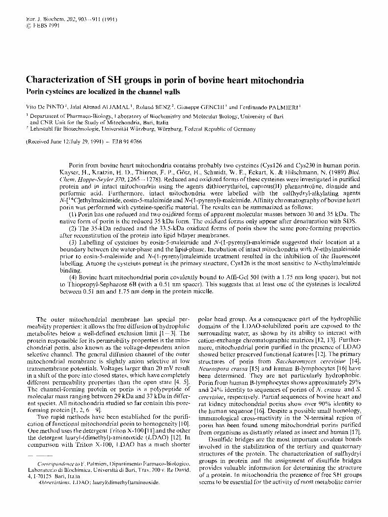
Since mammalian porin has two sulfhydryl groups [16] which may influence its electrophoretic mobility, and since porin may be present as contaminant in preparations of mito- chondrial substrate carriers having a molecular inass between 30 kDa and 37 kDa (for a review see [36]), it is essential to recognize precisely the porin mobility in the electro- phoretograms. To investigate this problem in detail we performed the experiments presented in Fig. 1. Different re- ducing and oxidizing agents were used to investigate the re- duction state of sulphydryl groups present in bovine heart porin purified in Triton X-100. Cuprous(I1)phenantroline and azodicarboxylic acid, and the strong oxidizing agent performic acid were used to oxidize cysteines. Their reduction was performed by dithioerythritol. Equal aliquots of the same porin preparation were incubated with the reagents indicated, added in the order shown in the figure (for more details see Materials and Methods). The protein samples were then acetone-precipitated, dissolved in SDS-sample buffer without the reducing agent and immediately run on SDS/gel electro- phoresis. Surprisingly, we observed at least three bands in the molecular mass region of 30 - 35 kDa. The apparent molec- ular masses of these bands were 35 kDa (band l) , 33.5 kDa (band 2) and 30 kDa (band 3). Band 1 corresponded to the electrophoretic mobility of purified porin [8, 261. Cuprous(1- I)phenantroline, introduced by Kobashi [37], is one of the most effective agents used to catalyze oxidative S-S bridge formation. The set of experiments performed with cuprous(1- 1)phenantroline (Fig. 1A) indicates that band 3 is the only new band which clearly appeared after chemical treatment. But this band became only visible after SDS treatment of the porin. This means that the protein has to be unfolded to allow contact between porin cysteines to form a disulfide bridge. After treatment with azodicarboxylic acid [38] two bands ap- peared. These bands (bands 2 and 3) were again only formed when porin was unfolded by SDS (Fig. 1 A, lanes 5 and 8). N-ethylmaleimide protected against the action of cuprous(I1)phenantroline (lanes 2 and 3) and of azodicarbox- ylic acid (lanes 6 and 7) by alkylating the SH groups of the porin before the subsequent unfolding by SDS during the preparation of the samples for electrophoresis. In lane 5 porin was unfolded by the SDS-sample buffer and oxidized by the previously added azodicarboxylic acid. In lane 8, 0.2% SDS and azodicarboxylic acid were left to incubate with porin for 30 min when the reaction was blocked by the addition of N-ethylmaleimide. After oxidation of cysteine to cysteic acid by performic acid, only band 2 was detected on the gels (Fig. 1 B). With 10 mM dithioerythritol added to porin after treatment with cuprous(1I)phenantroline or azodicarboxylic acid, we observed only the typical band of 35 kDa (band 1). Usually in control samples, run in the absence of any oxidizing or reducing reagent, the three bands were clearly visible (lane 0 in Fig. 1B). Sometimes, however, expecially with freshly
906
0 CUP NEM SDS DA DA NEM SDS M DTE SDS SDS NEM CUP CUP NEM DA DA CUP DA
NEM NEM NEM NEM DTE DTE
D I E Performic Acid 0 IOminIh 3h 7h
A
Fig. 1. Influence of reducing and oxidizing agents on the electrophoretic puttern qfporin purified in Triton X-100. (A) Bovine heart porin purified in Triton X-100 was incubated at room temperature with the indicated sequences of reagents. At the end, porin samples were acetone- precipitated, redissolved in SDS-sample buffer without dithioerythritol and run on SDS/gel electrophoresis. Other details in Material and Methods. (B) Bovine heart porin purified in Triton X-100 was acetone-prccipitated, the pellet redissolved and incubated with performic acid. The reaction was stopped at the indicated times by freeze-drying the samples. They were, then, redissolved in SDS-sample buffer without dithioerythritol and run on SDS/gel electrophoresis. CUP, cuprous(l1) phenantroline; DA, azodicarboxylic acid; NEM, N-ethylmaleimidc plus EDTA; DTE, dithioerythritol; 0, control, with no added reagent. The arrows indicate the three bands in which porin was distributed
0 CUP NEM SDS DA DA NEM SDS DTESDS SDS
NEM NEM NEM NEM DTE DTE
NEM CUP CUP NEM DA DA CUP DA
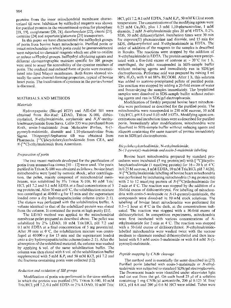
Fig. 2. Influence of reducing and oxidizing agents on the electrophoretic pattern of porin in intact mitochondria. Bovine heart mitochondria were incubated with the indicated sequences of reagents. At the end, mitochondria were dissolved in SDS-sample buffer without added dithioerythritol and immediately run on SDS/gel electrophoresis. The
bovine heart mitochondria (Fig. 2) . In this case, mitochondria were dissolved in SDS-sample buffer after chemical modifi- cation and immediately run on SDS-gel in the absence of reducing chemicals. The gel was then transferred to nitrocellu- lose and immunodetected with an antiserum against purified bovine heart porin (not shown) or with an antiserum against the 18-amino-acids long N-terminal of human porin, which has a very high degree of specificity for mammalian porins [I71 (Fig. 2). The experiments shown in Fig. 2 confirmed the results obtained with the purified porin. In intact mitochon- dria, porin is present as a single band of 35 kDa (band 1). Bands 2 and 3 were only observed when mitochondria were exposed to the combined action of SDS and oxidizing agents.
It should be pointed out that interchain disulfide bridges between two or more polypeptide chains can be ruled out, because we never detected higher molecular mass aggregates on the gels. Furthermore it seems unlikely that the pore- forming complex contains intrachain disulfide bridges, since the native form of porin in mitochondria in the absence of reducing agents is band 1 (Fig. 2 lane 0).
The cysteines qfporin are in an environment uccessible to hydrophylic and hydrophobic reagents
proteins-were transferred to nitrocellulose andimmunodecorated with an antiserum raised against the N terminus of human porin at a , : looo dilution, Abbreviations as in the legend to Fig, The arrOwS indicate the three bands in which porin was distributed in the absence of the reducing agent
To investigate the environment of the sulfhydryl groups present in bovine heart porin we modified intact mitochondria by means of eosin-5-maleimide as a reagent specific for cysteines localized in hydrophilic regions [39] and N-(1- pyreny1)-maleimide as a reagent specific for cysteines localized in hydrophobic regions [40]. Under our experimental con- ditions, both eosin-5-maleimide and N-( 1 -pyrenyl)-maleimide as well as N-ethylmaleimide were able to label the mitochon- drial porin. Fig. 3 is a fluorograph of bovine heart mitochon- dria incubated with N-[14C]ethylmaleimide and with [14C]di-
prepared porin samples, just a smear below the 35-kDa band was visible (lane 0 in Fig. 1 A).
The same set of experiments described above for the pure porin (Fig. 1 A) were repeated with freshly prepared, intact
907
PORIN MITOCHONDRIA
INCUBATION NPM [@I INCUBATION NPM [pM]
1 0 3 0 60' 50 100 400 10' 30 60' 50 100 400 TIME TIME
35 kDa
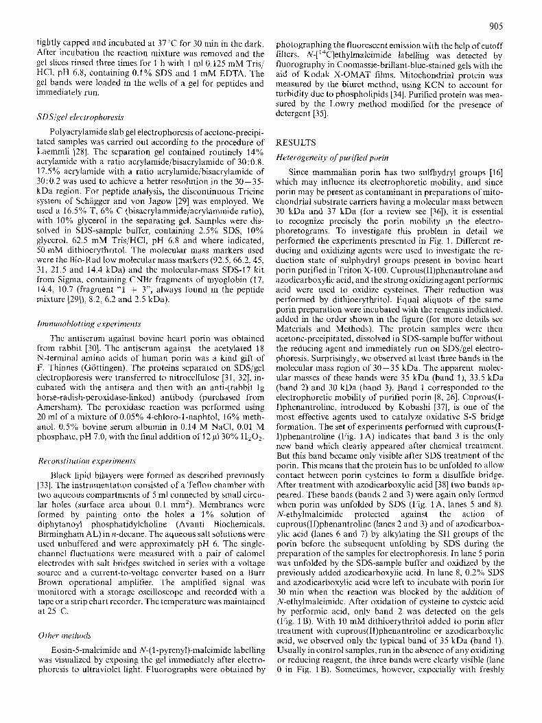
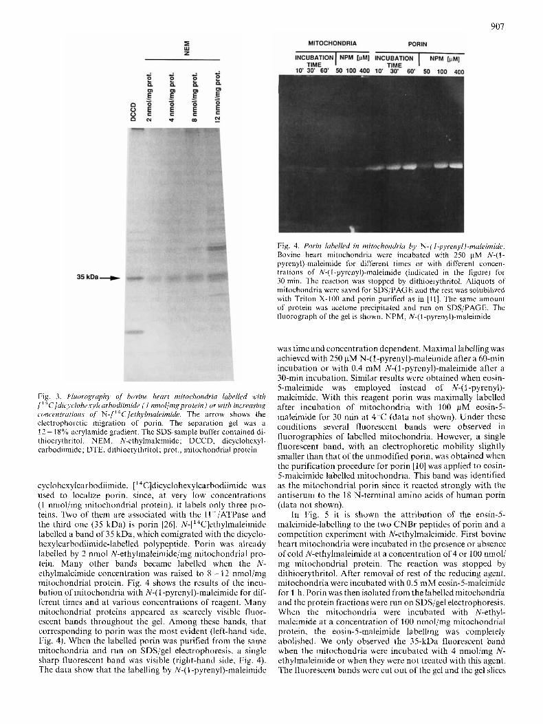
Fig. 3. Nuovogvuphy of bovine heart mitochondria labelled with [ 14C]dic~~lnh~xylcuv/70diin~ide ( I nmollmgprotein) or with increusing concentrutions of N-[ '4C]e t~ iy lmule imid~. The arrow shows the electrophoretic migration of porin. The separation gel was a 12- 18% acrylamide gradient. The SDS-sdmpk buffer contained di- thiocrythritol. NEM, N-ethylmaleimide; DCCD, dicyclohexyl- carbodiimide; DTE, dithioerythritol; prot., mitochondrial protein
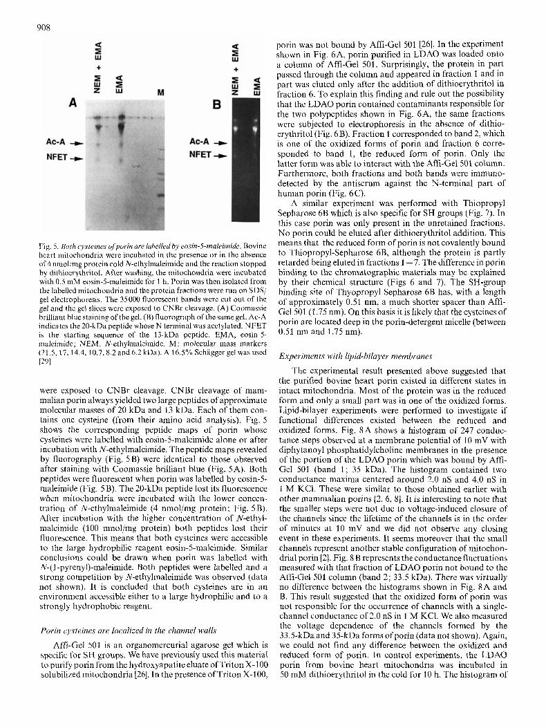
cyclohexylcarbodiimide. ['4C]dicyclohexylcarbodii~nide was used to localize porin, since, at very low concentrations (1 nmol/iiig mitochondrial protein), it labels only three pro- teins. Two of them are associated with the H+/ATPase and the third one (35 kDa) is porin [26]. N-['4C]ethylmaleimide labelled a band of 35 kDa, which comigrated with the dicyclo- hexylcarbodiimide-labelled polypeptide. Porin was already labelled by 2 nmol N-ethylmaleimide/mg mitochondrial pro- tein. Many other bands became labelled when the N- ethylmaleimide concentration was raised to 8 - 12 nniol/mg mitochondrial protein. Fig. 4 shows the results of the incu- bation of mitochondria with N-( 1 -pyrenyl)-maleimide for dif- ferent times and at various Concentrations of reagent. Many mitochondrial proteins appeared as scarcely visible fluor- escent bands throughout the gel. Among these bands, that corresponding to porin was the most evident (left-hand side, Fig. 4). When the labelled porin was purified from the same mitochondria and run on SDS/gel electrophoresis, a single sharp fluorescent band was visible (right-hand side, Fig. 4). The data show that the labelling by N-(I -pyrenyl)-maleimide
6 PORlN
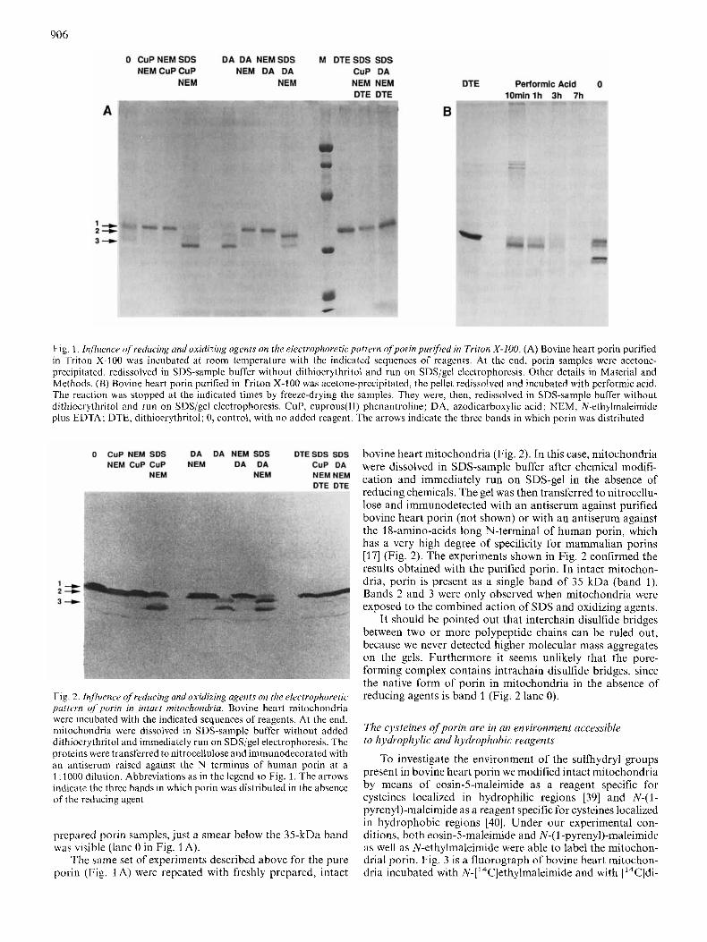
Fig. 4. Porin Iubelked in mitochondriu by N-( I-pyrenyl)-muleimide. Bovine heart mitochondria were incubated with 250 pM N-(l- pyreny1)-maleiniide for different times or with different concen- trations or N-( 1 -pyrenyl)-maleimide (indicated in the figure) for 30 min. 'The reaction was stopped by dithioerythritol. Aliquots of mitochondria were saved for SDS/PAGE and the rest was solubili7ed with Triton X-100 and porin purified as in [ I l l . The same amount of protein was acetone precipitated and run on SDS/PAGE. Thc lluorograph of the gel is shown. NPM, N-(I-pyreny1)-maleimide
was time and concentration dependent. Maximal labelling was achieved with 250 pM N-(1-pyreny1)-maleimide after a 60-min incubation or with 0.4 mM N-(1-pyreny1)-maleimide after a 30-min incubation. Similar results were obtained when eosin- 5-maleimide was employed instead of N-(I -pyrenyl)- maleimide. With this reagent porin was maximally labelled after incubation of mitochondria with 100 pM eosin-5- maleimide for 30 min at 4 'C (data not shown). Under these conditions several fluorescent bands were observed in fluorographies of labelled mitochondria. However, a single fluorescent band, with an electrophoretic mobility slightly smaller than that of the unmodified porin, was obtained when the purification procedure for porin [lo] was applied to eosin- 5-maleimide labelled mitochondria. This band was identified as the mitochondrial porin since it reacted strongly with the antiserum to the 18 N-terminal amino acids of human porin (data not shown).
In Fig. 5 it is shown the attribution of the eosin-5- maleimide-labelling to the two CNBr peptides of porin and a competition experiment with N-ethylmaleimide. First bovine heart mitochondria were incubated in the presence or absence of cold N-ethylmaleimide at a concentration of 4 or 100 nmol/ mg mitochondrial protein. The reaction was stopped by dithioerythritol. After removal of rest of the reducing agent, mitochondria were incubated with 0.5 m M eosin-5-nialeimide for 1 h. Porin was then isolated from the labelled mitochondria and the protein fractions were run on SDS/gel electrophoresis. When the mitochondria were incubated with N-ethyl- maleimide at a concentration of 3 00 nmol/mg mitochondrial protein, the eosin-5-maleimide labelling was completely abolished. We only observed the 35-kDa fluorescent band when the mitochondria were incubated with 4 nmol/mg N- ethylmaleimide or when they were not treated with this agent. The fluorescent bands were cut out ofthe gel and the gel slices
908
A
Ac-A -D
NFET +
+
M B
Ac-A + NFET -+
+
Fig. 5. Both cysteines ofporin are libelledby eosin-5-maleimide. Bovine heart mitochondria were incubated in the presence or in the absence of 4 nmol/mg protein cold N-ethylmaleimide and the reaction slopped by dithioerythritol. After washing, the mitochondria were incubated with 0.5 mM eosin-5-maleimide for 1 h. Porin was then isolated from the labelled mitochondria and the protein fractions were run on SDS/ gel electrophoresis. The 35000 fluorescent bands were cut out of the gel and the gel slices wcre exposed to CNBr cleavage. (A) Coomassie brilliant blue staining of the gel. (B) fluorograph of the same gel. Ac-A indicates the 20-kDa peptide whose N lerminal was acetylated. NFET is the starting sequence of the 13-kDa peptide. EMA, eosin-5- malcimide; NEM, N-ethylmaleimide. M : inolecular mass markers (21 .5, 17,14.4,10.7,8.2 and 6.2 kDa). A 16.5% Schigger gel was used 1291
were exposed to CNBr cleavage. CNBr cleavage of mam- malian porin always yielded two large peptides of approximate molecular masses of 20 kDa and 13 kDa. Each of them con- tains one cysteine (from their amino acid analysis). Fig. 5 shows the corresponding peptide maps of porin whose cysteines were labelled with eosin-5-maleimide alone or after incubation with N-ethylmaleimide. The peptide maps revealed by fluorography (Fig. 5 s ) were identical to those observed after staining with Coomassie brilliant blue (Fig. 5A). Both peptides were fluorescent when porin was labelled by eosin-5- maleimide (Fig. 5B). The 20-kDa peptide lost its fluorescence when mitochondria were incubated with the lower concen- tration of N-ethylmaleimide (4 nmoljmg protein; Fig. 5 B). After incubation with the higher concentration of N-ethyl- maleimide (100 nmol/mg protein) both peptides lost their fluorescence. This means that both cysteines were accessible to the large hydrophilic reagent eosin-5-maleimide. Similar conclusions could be drawn when porin was labelled with N-(I -pyrenyl)-maleimide. Both peptides were labelled and a strong competition by N-ethylmaleimide was observed (data not shown). It is concluded that both cysteines are in an environment accessible either to a large hydrophilic and to a strongly hydrophobic reagent.
Porin cysieines are localized in the channel walls
Affi-Gel 501 is an organomercurial agarose gel which is specific for SH groups. We have previously used this material to purify porin from the hydroxyapatite eluate of Triton X-100 solubilized mitochondria [26]. In the presence of Triton X-100,
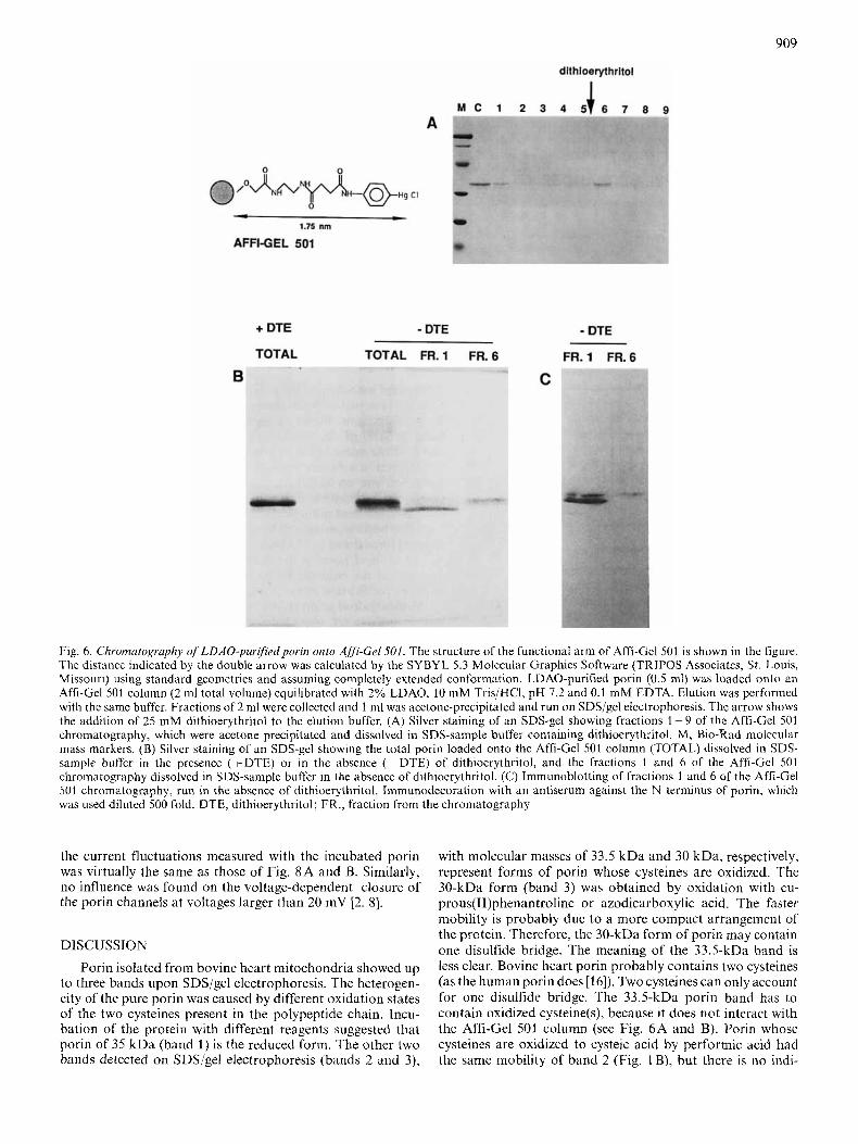
porin was not bound by Affi-Gel 501 [26]. In the experiment shown in Fig. 6A, porin purified in LDAO was loaded onto a column of Affi-Gel 501. Surprisingly, the protein in part passed through the column and appeared in fraction 1 and in part was eluted only after the addition of dithioerythritol in fraction 6. To explain this finding and rule out the possibility that the LDAO porin contained contaminants responsible for the two polypeptides shown in Fig. 6A, the same fractions were subjected to electrophoresis in the absence of dithio- erythritol (Fig. 6B). Fraction 1 corresponded to band 2, which is one of the oxidized forms of porin and fraction 6 corre- sponded to band 1, the reduced form of porin. Only the latter form was able to interact with the Affi-Gel501 column. Furthermore, both fractions and both bands were immuno- detected by the antiserum against the N-terminal part of human porin (Fig. 6C).
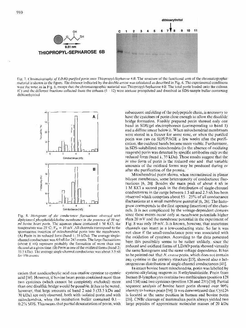
A similar experiment was performed with Thiopropyl Sepharose 6B which is also specific for SH groups (Fig. 7). In this case porin was only present in the unretained fractions. No porin could be eluted after dithioerythritol addition. This means that the reduced form of porin is not covalently bound to Thiopropyl-Sepharose 6B, although the protein is partly retarded being eluted in fractions 1 - 7. The difference in porin binding to the chromatographic materials may be explained by their chemical structure (Figs 6 and 7). The SH-group binding site of Thyopropyl Sepharose 6B has, with a length of approximately 0.51 nm, a much shorter spacer than Affi- Gel 501 (1.75 nm). On this basis it is likely that the cysteines of porin are located deep in the porin-detergent micelle (between 0.51 nm and 1.75 nm).
Experiments with lipid-bilayer membranes
The experimental result presented above suggested that the purified bovine heart porin existed in different states in intact mitochondria. Most of the protein was in the reduced form and only a small part was in one of the oxidized forms. Lipid-bilayer experiments were performed to investigate if functional differences existed between the reduced and oxidized forms. Fig. 8A shows a histogram of 247 conduc- tance steps observed at a membrane potential of 10 mV with diphytanoyl phosphatidylcholine membranes in the presence of the portion of the LDAO porin which was bound by Affi- Gel 501 (band 1; 35 kDa). The histogram contained two conductance maxima centered around 2.0 nS and 4.0 nS in 1 M KCI. These were similar to those obtained earlier with other mammalian porins [2, 6, 81. It is interesting to note that the smaller steps were not due to voltage-induced closure of the channels since the lifetime of the channels is in the order of minutes at 10 mV and we did not observe any closing event in these experiments. It seems moreover that the small channels represent another stable configuration of mitochon- drial porin [2]. Fig. 8 B represents the conductance fluctuations measured with that fraction of LDAO porin not bound to the Affi-Gel 501 column (band 2; 33.5 kDa). There was virtually no difference between the histograms shown in Fig. 8A and B. This result suggested that the oxidized form of porin was not responsible for the occurrence of channels with a single- channel conductance of 2.0 nS in 1 M KCl. We also measured the voltage dependence of the channels formed by the 33.5-kDa and 35-kDa forms of porin (data not shown). Again, we could not find any difference between the oxidized and reduced form of porin. In control experiments, the LDAO porin from bovine heart mitochondria was incubated in 50 mM dithioerythritol in the cold for 10 h. The histogram of
909
dithioerythritol
Fig. 6. Chromatogruphy ofLDAO-purifiedpvrin onlo Affi-Cel501. The structure of the functional arm of Affi-Gel 501 is shown in the figure. The distance indicated by the double arrow was calculated by the SYBYL 5.3 Molecular Graphics Software (TRIPOS Associates, S t . Louis, Missouri) using standard geometries and assuming complcteiy extended conformation. LDAO-purified porin (0.5 ml) was loaded onto an Affi-Gel 501 column (2 ml total volume) equilibrated with 2% LDAO, 10 mM Tris/HCl, pH 7.2 and 0.1 mM EDTA. Elution was performed with the same buffer. Fractions of2 ml were collected and 1 ml was acetone-precipitated and run on SDS/gel electrophoresis. The arrow shows the addition of 25 niM dithioerythritol to the elution buffer. (A) Silver staining of an SDS-gel showing fractions 1-9 of the Affi-Gel 501 chromatography, which were acetone precipitated and dissolved in SDS-sample buffer containing dithioerythritol. M, Bio-Rad molecular mass markers. (B) Silver staining of an SDS-gel showing the total porin loaded onto the Affi-Gel 502 column (TOTAL) dissolved in SDS- sample buffer in the presencc (+DTE) or in the absence (-DTE) of dithiocrythritol, and the fractions 1 and 6 of the Affi-Gcl 501 chromatography dissolved in SDS-sample buffcr in the absence of dithioerythritol. (C) lmmunoblotting of fractions 1 and 6 of the Affi-Gel 501 chromatography, run in the absence of dithioerythritol. Immunodccoration with an antiserum against the N terminus of porin, which was used diluted 500 fold. DTE, dithioerythritol; FR. , fraction from the chromatography
the current fluctuations measured with the incubated porin was virtually the same as those of Fig. 8A and B. Similarly, no influence was found on the voltage-dependent closure of the porin channels at voltages larger than 20 mV [2, 81.
DISCUSSION Porin isolated from bovine heart mitochondria showed up
to three bands upon SDS/gel electrophoresis. The heterogen- eity of the pure porin was caused by different oxidation states of the two cysteines present in the polypeptide chain. Incu- bation of the protein with different reagents suggested that porin of 35 kDa (band 1) is the reduced form. The other two bands detected on SDS/gel electrophoresis (bands 2 and 3),
with molecular masses of 33.5 kDa and 30 kDa, respectively, represent forms of porin whose cysteines are oxidized. The 30-kDa form (band 3 ) was obtained by oxidation with cu- prous(I1)phenantroline or azodicarboxylic acid. The faster mobility is probably due to a more compact arrangement of the protein. Therefore, the 30-kDa form of porin may contain one disulfide bridge. The meaning of the 33.5-kDa band is less clear. Bovine heart porin probably contains two cysteines (as the human porin does [16]). Two cysteines can only account for one disulfide bridge. The 33.5-kDa porin band has to contain oxidized cysteine(s), because it does not interact with the Affi-Gel 501 column (see Fig. 6A and B). Porin whose cysteines are oxidized to cysteic acid by perforinic acid had the same mobility of band 2 (Fig. lB), but there is no indi-
910
OH
dlthloerythrltol
C 1 2 3 4 5 6 7 t 8 9 1 0 1 1
0.51 nm
THIOPROPYL-SEPHAROSE 6B
Fig. 7. Chromatography of LDAO-pur$iedpnrin onto Thir,propyl-Sephuro.se 6 B. The structure of the functional arm of the chromatographic material is shown in the figure. The distance indicated by the double arrow was calculated as described in Fig. 6. The experimental conditions were the same as in Fig. 6, except that the chromatographic material was Thiopropyl-Sepharose 6B. The total porin loaded onto the column (C) and thc different fractioiis collected from the column (1 - 12) were acetone precipitated and dissolved in SDS-sample buffer containing dithioerhytritol
?.,
30 I - ). x 20
& m I?
10
0 0 2 4 6
Conductance (nS)
0 2 4 6
Conductance (nS)
Fig. 8. Histogram of the conductance , fli~cttiatiuns ohserved with diphytanojbl phosphatidyl~holine membranes in the presence of 10 ng/ mf bovine heart porin. The aqueous phase contained 1 M KCI, the temperature was 2 5 T ; V , = 10 mV. All channels correspond to the spontaneous inscrtion of mitochondria1 porin into the membranes. (A) Porin in its reduced form (band 1 ; 35 kDa). The average singlc- channel conductance was 4.0 nS for 247 events. The large fluctuations (about 8 nS) represent probably the formation of more than one channel at a givcn time. (B) Porin in one of the oxidized forms (band 2; 33.5 kna). The average single-channel conductance was about 3.9 nS for 196 events
cation that azodicarboylic acid can oxidize cysteine to cysteic acid [38]. However, if bovine heart porin contained more than two cysteines (which cannot be completely excluded) more than one disulfide bridge would be possible. It has to be noted, however, that large amounts of band 2 and 3 (33.5 kDa and 30 kDa) are only observed, both with [solated porin and with mitochondria, when the incubation buffer contained 0.1 - 0.2°/0 SDS. This means that partial denaturation of porin, with
subsequent unfolding of the polypeptide chain, is necessary to have the cysteines of porin close enough to allow the disulfide bridge formation. Freshly prepared porin showed only one band in SDS/gel electrophoresis (corresponding to band 1) and a diffuse smear below it. When mitochondrial membranes were stored in a freezer for some time, or when the purified porin was run on SDSjPAGE a few weeks after the purifi- cation, the oxidized bands became more visible. Furthermore, in SDS-solubilized mitochondria (in the absence of oxidizing reagents) porin was detected by specific antibodies only as the reduced form (band 1, 35 kDa). These results suggest that the in vivo form of porin is the reduced one and that variable amounts of the oxidized forms may be produced during or after the purification of the protein. .
Mitochondria1 porin shows, when reconstituted in planar bilayer membranes, some heterogeneity of conductance fluc- tuations [6, 261. Besides the main peak of about 4 nS in 1 M KCI a second peak in the distribution of single-channel conductances in the range between 1.5 nS and 2.5 nS has been observed which comprises about 15 -25% of all conductance fluctuations at a small membrane potential [6, 261. The histo- gram corresponds to the first opening (insertion) of the chan- nels. It is not complicated by the voltage-dependent closures since these events occur only at membrane potentials higher than 20 mV and the membrane potential in the experiment of Fig. 8 was only 10 mV. It is known, however, that sometimes channels can insert in a low-conducting state. So far it was not clear if the small-conductance pore was associated with the oxidation of cysteines. According to the data presented here this possibility seems to be rather unlikely, since the reduced and oxidized forms of LDAO porin showed virtually the same histograms and the same voltage dependence. It has to be pointed out that N . crussaporin, which does not contain any cysteine in the primary structure [15], showed also a het- erogeneous distribution of single-channel conductances [41].
In intact bovine heart mitochondria, porin was labelled by cysteine-alkylating reagents as N-ethylmaleimide. Porin from human B-lymphocytes contains two methionines (position 128 and 154) and two cysteines (position 126 and 231 j [16]. Partial sequence analysis of bovine heart porin showed over 90% identity to human porin [16]. It was demonstrated that Cys126 is located at the same position in human and bovine porin [16]. CNBr cleavage of mammalian porin always yielded two large peptides of approximate molecular masses of 20 kDa

91 1
and 13 kDa. The large peptide (20 kDa) has been found to be blocked and it is the N-terminal part of porin. The 13-kDa peptide showed the starting sequence: NFET after Met154 [16]. Each of these peptides from bovine heart porin contains one cysteine (from their amino acid analysis). This means that Cys126 must be part of the 20-kDa peptide and another cysteine of the 13-kDa fragment. With this information in mind the fluorescent reagents eosin-5-maleimide and N-(1- pyreny1)-maleimide were employed to probe the accessibility of the cysteine residues of porin. Porin was labelled by maleimides in mitochondria and the fluorescent protein was purified by standard procedure. CNBr cleavage of the fluor- escent porin showed that both peptides and thus both those cysteines, could be labelled by eosin-5-maleiinide or by N-(I- pyreny1)-maleimide. This means that both cysteines are lo- calized in an environment accessible to water-soluble and to hydrophobic reagents. The fluorescence of the 20-kDa peptide could be climinated by incubation with a low concentration of N-ethylmaleimide. The corresponding cysteine (Cysl26) is therefore the most reactive to N-ethylmaleimide. The porin channcl contains two possible localizations for water-access- ible amino acid residues: the external surface, surrounding the mouth of the pore and the walls of the pore itself, facing the water inside the channel. Only the latter possibility is allowed to amino acid residues able to react both with hydrophobic and with hydrophilic reagents. To confirm this conclusion we used commercially available chromatographic matrices (Affi- Gel 501 and Thiopropyl-Sepharose 6B), as probes for SH groups. Both are specific for free SH groups but have spacer arms with different lengths (0.51 nm and 1.75 nm). The LDAO porin was bound only by the material with the longer arm. The length of the arm is a likely reason for the difference in porin binding to the chromatographic materials. Conse- quently it may be suggested that at least one of the two cysteines is localized between 0.51 nm and I .75 nm away from the surfacc of the protein-detergent micelle. The other one should have a similar localization or it is located even deeper in the protein-detergent micelle. As it can be labelled by eosin- 5-maleimide it may face the water phase anywhere inside of the channel.
The authors thank D r 1'. Schilitz (Frciburg. F.R.G.) for the peptide analysis of the CNBr fragments and Angela Schmid (Wiirzburg, F.R.G.) for the help with the membrane cxperiments. This work was supported by the target project Biolechnology and Bioinstrumentation of thc C'onsiglio Nazionule delk Riccrchc, by the Miriistero ddlu Universitu c della Ricerca Scientificu e Tee-nologica, and by the Deutsche ~orschung.~gemeinschuft (SFB 176) and by the Fond der Chemisehen Industrir.
REFERENCES I . 2. 3.
4. 5.
6.
Colombini, M. (1979) Nature 279, 643 -645. Benz, R. (1985) Crit. Rev. Biochem. 19, 145-190. Dihamic, M. (ed.) Multi-Author Review (1990) Experientiu 46,
Colombini. M. (1980) Ann. N . Y . Acud. Sci. 341, 552-563. Benz, R., Kottke, M. & Brdiczka, D. (1990) Biochim. Biophys.
Roos, N., Bcnz, R. & Brdiczka, D. (1982) Biochim. Biophys. Actu
131 - 201.
act^ 1022. 311 -318.
686. 204-214.
7. Linden, M., Gellerfoi-s, P. & Nelson, B. D. (1982) Biochem. J .
8. De Pinto, V., Ludwig, O., Krause, .I., Benz, R. & Palmieri, F.
9. Smack, J . K. & Colombini, M. (1985) Plant fil.vsioI. 79, 1094-
10. Palmieri, F. and De Pinto, V. (1989) J . Bioenerg. Bzomemhr. 21,
11. De Pinto, V., Prezioso, G. & Palmieri, F. (1987) Biochim. Biophys.
12. Dc Pinto, V., Benz, R. & Palmicri, F. (1989) Eur. J . Biochem.
13. De Pinto, V., Al Jamal, J. A.. Benz, R. & Palmieri, F. (1990)
14. Mihara, K . & Sato, K. (1985) EMBO J . 4, 769-774. 15. Kleene, R., Pfanner, N., Pfaller, R., Link, T. A., Sebald, W.,
Neupert, W. & Tropschung, M. (1987) E M B O J . 6, 2627- 2633.
16. Kayser, H., Kratzin, 13. I]., Thinnes, F. P., Gutz, H., Schmidt, W. E., Eckart, K. & Hilschmann, N. (1989) Biol. Chrm. Hoppe- Seylrr 370, 1265- 1278.
17. De Pinto, V., Zara, V., Benz, K., Gnoni, G. & Palmicri, F. (2992) Biochim. Biophys. Actu, 1061, 279-286.
18. Wohlrab, H. (1980) J . Bid . Chcm. 255, 8170-8173. 19. Kolbe, H. V. J . , Bottrich, J. , Genchi, G., Palmieri, F. &
Kadenbach, B. (1981) FEBS Lett. 124,265-269. 20. Bisaccia, F., Indiveri, C. & Palmieri, F. (1985) Biochim. Biophys.
21. Zara, V. & Palmieri, F. (1988) FERS Lett. 236, 493-496. 22. Bisaccia, F., Indiveri, C. & Palmieri, I-'. (1988) Biockinz. Bioph)*.s.
23. Bisaccia, F., I k Palma, A. & Palmicri, F. (1989) Biochim. Bio-
24. Indiveri, C., Tonazzi, A. & Palmieri, F. (1990) Biochim. Biiiphys.
25. Dicrks, T.. Salentin, A,, Heberger, C. & Krlmer, R . (1990) Bio-
26. De Pinto, V.. Tommasino, M., Bcnz, R. & Palmieri, F. (1985)
27. Plaxton, W. C. & Moorheati, G. B. G. (1989) A n d . Biochem. 178,
28. Laemmli, U. K. (1974) Nature 227, 680-685. 29. Schigger. H. & von Jagow, G . (1987) Anal. Biochem. 166, 368-
30. De Pinto, V., Hcnz, R., Caggese, C. & Palmieri, F. (1989) Biochim.
31. Rott, R. & Nelson, N. (1981) J . Biol. Cham. 256, 9224-9228. 32. Towbin, H.. Staehelin, 'T'. & Gordon, J . (1979) Proc. Nut / . Acud.
33. Benz, R., .lanko, K., Boos, W. & Laugcr, P. (1978) Biochim.
34. Kroger. A. & Kiingcnberg, M. (1966) Biochenz. Z . 344, 317-
35. Kusov, Y. Y . & Kalinchuk, N. A. (1978) Anal. Biochem. 88,256-
36. KrCmer, R. & Palmieri. 1:. (1989) Biochim. Biophys. Actu 974,
37. Kobashi, K. (1968) Biochim. Biophys. Actu 158, 239-245. 38. Kosower, N. S., Kosower. E. M., Wertheim, B. & Correa, W. S.
(1 969) Biochem. Biopliys. Res. Commun. 37, 593 - 596. 39. Houstek, J . & Pcdersen, P. (1985) J . Biof. Chrm. 260,6288 -6295. 40. Weltman, J. K . , Szaro, K. P., Frackelton, R., Dowbcn, R. M.,
Bunting, J. R. & Cathou, R . E. (1973) J . Biol. Cliem. 248, 31 73 - 31 77.
41. Frcitag, l i . , Neupcrt, W. & Henz, R. (1982) Emr. .1. Riochem. 123, 629 - 638.
208, 77 - 82.
(1987) Biochim. Biophys. Acta 894, 109-119.
1097.
43 7 - 425.
Actu 905, 499- 502.
183, 179-187.
FEBS Lett 274, 122-126.
Arts 810, 362-369.
Acts 933, 229 -240.
phys. Act0 Y77, 171 -176.
Acts 1020. 81 -86.
chim. Biophys. Actu 1028, 268 -280.
BiocJiirn. Biojdiys. Acta 813, 230 -242.
391 - 393.
379.
Biophys. Acta 087, 1 -- 7.
Sci. U S A 76,4350-4354.
Biophys. Act(E511, 305--319.
326.
262.
123.