Characterization of Pinin, A Novel Protein Associated with the ...
16
Characterization of Pinin, A Novel Protein Associated with the Desmosome--Intermediate Filament Complex Pin Ouyang* and Stephen P. Sugrue* *Department of Anatomy, Chang Gung Medical College, Kwei-San, Tau-Yuan, China; and *Department of Anatomy and Cell Biology, University of Florida College of Medicine, Gainesville, Florida 32610-0235 Abstract. We have identified a protein named pinin that is associated with the mature desmosomes of the epithelia (Ouyang, P., and S.P. Sugrue. 1992. J. Cell Biol. 118:1477-1488). We suggest that the function of pinin is to pin intermediate filaments to the desmo- some. Therefore, pinin may play a significant role in re- inforcing the intermediate filament-desmosome com- plex. cDNA clones coding for pinin were identified, using degenerative oligonucleotide probes that were based on the internal amino acid sequence of pinin for the screening of a cDNA library. Immunoblotting of expressed recombinant proteins with the monoclonal 08L antibody localized the 08L epitope to the carboxyl end of the protein. Polyclonal antibodies directed against fusion proteins immunoidentified the 140-kD protein in tissue extracts. Immunofluorescence analy- sis, using the antifusion protein antibody, demonstrated pinin at lateral epithelial boundaries, which is consis- tent with desmosomal localization. The conceptual translation product of the cDNA clones contained three unique domains: (a) a serine-rich domain; (b) a glutamine-proline, glutamine-leucine repeat domain; and (c) an acidic domain rich in glutamic acid. Al- though the 3' end of the open reading frame of the clone for pinin showed near identity to a partial cDNA isolated for a pig neutrophil phosphoprotein (Bellavite, P., F. Bazzoni, et al. 1990. Biochem. Biophys. Res. Com- mun. 170:915-922), the remaining sequence demon- strated little homology to known protein sequences. Northern blots of mRNA from chicken corneal epithe- lium, MDCK cells, and various human tissues indicated that pinin messages exhibit tissue-specific variation in size, ranging from 3.2 to 4.1 kb. Genomic Southern blots revealed the existence of one gene for pinin, sug- gesting alternative splicing of the mRNA. Expression of the full-length cDNA clones in human 293 cells and monkey COS-7 cells demonstrated that a 140-kD im- munoreactive species on Western blots corresponded to pinin. Pinin cDNA transfected into the transformed 293 cells resulted in enhanced cell--cell adhesion. Im- munofluorescence staining revealed that the expressed pinin protein was assembled to the lateral boundaries of the cells in contact, which is consistent with the stain- ing pattern of pinin in epithelial cells. ESMOSOMES (Macula adherens) are intimately in- volved in the structural and functional integration of adjacent epithelial cells. They serve as rein- forcement sites of cell-cell adhesion, as well as points for lateral anchorage of the intermediate scaffold of the epi- thelial cell (Staehelin, 1974; Arnn and Staehelin, 1981). Ultrastructurally, they appear as symmetrically arranged disc-shaped structures of a varying diameter (0.1-2 ~m). The space between the interacting membranes is 20-30 nm, which often exhibits a central electron-dense core, pre- sumably consisting of the overlapping domains of the Please address all correspondence to Stephen P. Sugrue, Department of Anatomy and Cell Biology, University of Florida College of Medicine, 1600 SW Archer Road, Gainesville, FL 32610-0235. Tel.: (352) 392-3432; Fax: (352) 392-3431; E-mail: [email protected]. The present address for Pin Ouyang is Department of Anatomy, Chang Gung Medical College, 259 Wen-Hwa 1st Road, Kwei-San, Tau-Yuan, Taiwan, China, 327. transmembrane glycoproteins of the desmosome. On each cytoplasmic side of the interacting membranes, there are trilaminar plaques that appear to anchor the looping bun- dles of intermediate filaments (IFt; for reviews see Buxton and Magee, 1992; Buxton et al., 1993; Garrod, 1993; Legan et al., 1992). Biochemical and molecular analyses have led to the identification of several constitutive proteins, in- cluding desmoplakin, plakoglobin, and the transmembrane cadherin-like glycoproteins desmoglein and desmocollin. (Mueller and Franke, 1983; Cowin et al., 1985 1986; Green et al., 1990; Holton et al., 1990; Collins et al., 1991; Wheeler et al., 1991; Wiche et al., 1991; Green et al., 1992). Significant differences in the composition of desmosomes of various tissues have also been reported. Isoforms of desmoglein (Dsgl-3) and desmocollin (Dscl-3) have been 1. Abbreviations used in this paper: GST, gtutathione S-transferase; IF, in- termediate filament; MDBK, Madin-Darby bovine kidney (cells). © The RockefellerUniversity Press, 0021-9525/96/11/1027/16 $2.00 The Journal of Cell Biology, Volume 135, Number 4, November 1996 1027-1042 1027
-
Upload
khangminh22 -
Category
Documents
-
view
1 -
download
0
Transcript of Characterization of Pinin, A Novel Protein Associated with the ...
Characterization of Pinin, A Novel Protein Associated with the Desmosome--Intermediate Filament Complex Pin Ouyang* and Stephen P. Sugrue*
*Department of Anatomy, Chang Gung Medical College, Kwei-San, Tau-Yuan, China; and *Department of Anatomy and Cell Biology, University of Florida College of Medicine, Gainesville, Florida 32610-0235
Abstract. We have identified a protein named pinin that is associated with the mature desmosomes of the epithelia (Ouyang, P., and S.P. Sugrue. 1992. J. Cell Biol. 118:1477-1488). We suggest that the function of pinin is to pin intermediate filaments to the desmo- some. Therefore, pinin may play a significant role in re- inforcing the intermediate filament-desmosome com- plex. cDNA clones coding for pinin were identified, using degenerative oligonucleotide probes that were based on the internal amino acid sequence of pinin for the screening of a cDNA library. Immunoblotting of expressed recombinant proteins with the monoclonal 08L antibody localized the 08L epitope to the carboxyl end of the protein. Polyclonal antibodies directed against fusion proteins immunoidentified the 140-kD protein in tissue extracts. Immunofluorescence analy- sis, using the antifusion protein antibody, demonstrated pinin at lateral epithelial boundaries, which is consis- tent with desmosomal localization. The conceptual translation product of the cDNA clones contained three unique domains: (a) a serine-rich domain; (b) a glutamine-proline, glutamine-leucine repeat domain;
and (c) an acidic domain rich in glutamic acid. Al- though the 3' end of the open reading frame of the clone for pinin showed near identity to a partial cDNA isolated for a pig neutrophil phosphoprotein (Bellavite, P., F. Bazzoni, et al. 1990. Biochem. Biophys. Res. Com- mun. 170:915-922), the remaining sequence demon- strated little homology to known protein sequences. Northern blots of mRNA from chicken corneal epithe- lium, MDCK cells, and various human tissues indicated that pinin messages exhibit tissue-specific variation in size, ranging from 3.2 to 4.1 kb. Genomic Southern blots revealed the existence of one gene for pinin, sug- gesting alternative splicing of the mRNA. Expression of the full-length cDNA clones in human 293 cells and monkey COS-7 cells demonstrated that a 140-kD im- munoreactive species on Western blots corresponded to pinin. Pinin cDNA transfected into the transformed 293 cells resulted in enhanced cell--cell adhesion. Im- munofluorescence staining revealed that the expressed pinin protein was assembled to the lateral boundaries of the cells in contact, which is consistent with the stain- ing pattern of pinin in epithelial cells.
ESMOSOMES (Macula adherens) are intimately in- volved in the structural and functional integration of adjacent epithelial cells. They serve as rein-
forcement sites of cell-cell adhesion, as well as points for lateral anchorage of the intermediate scaffold of the epi- thelial cell (Staehelin, 1974; Arnn and Staehelin, 1981). Ultrastructurally, they appear as symmetrically arranged disc-shaped structures of a varying diameter (0.1-2 ~m). The space between the interacting membranes is 20-30 nm, which often exhibits a central electron-dense core, pre- sumably consisting of the overlapping domains of the
Please address all correspondence to Stephen P. Sugrue, Department of Anatomy and Cell Biology, University of Florida College of Medicine, 1600 SW Archer Road, Gainesville, FL 32610-0235. Tel.: (352) 392-3432; Fax: (352) 392-3431; E-mail: [email protected].
The present address for Pin Ouyang is Department of Anatomy, Chang Gung Medical College, 259 Wen-Hwa 1st Road, Kwei-San, Tau-Yuan, Taiwan, China, 327.
transmembrane glycoproteins of the desmosome. On each cytoplasmic side of the interacting membranes, there are trilaminar plaques that appear to anchor the looping bun- dles of intermediate filaments (IFt; for reviews see Buxton and Magee, 1992; Buxton et al., 1993; Garrod, 1993; Legan et al., 1992). Biochemical and molecular analyses have led to the identification of several constitutive proteins, in- cluding desmoplakin, plakoglobin, and the transmembrane cadherin-like glycoproteins desmoglein and desmocollin. (Mueller and Franke, 1983; Cowin et al., 1985 1986; Green et al., 1990; Holton et al., 1990; Collins et al., 1991; Wheeler et al., 1991; Wiche et al., 1991; Green et al., 1992). Significant differences in the composition of desmosomes of various tissues have also been reported. Isoforms of desmoglein (Dsgl-3) and desmocollin (Dscl-3) have been
1. Abbreviat ions used in this paper: GST, gtutathione S-transferase; IF, in- termediate filament; MDBK, Madin-Darby bovine kidney (cells).
© The Rockefeller University Press, 0021-9525/96/11/1027/16 $2.00 The Journal of Cell Biology, Volume 135, Number 4, November 1996 1027-1042 1027
found in several desmosome-containing tissues and be- tween layers of the same tissue (Parrish et al., 1986; Angst et al., 1990; Koch et al., 1991; Legan et al., 1992; Arne- mann et al., 1993; Buxton et al., 1993; Theis et al., 1993). Other desmosomal plaque-associated molecules have been reported in limited subsets of epithelial tissues. These include a shorter spliced form of desmoplakin (des- moplakin II; Mueller and Franke, 1983; Cowin et al., 1985; Angst et al., 1990; Green et al., 1990); plakophilin (for- merly band-6-protein), a "new" member of the plakoglo- bin~armadillo gene family (Hatzfeld et al., 1994; Heid et al., 1994); desmocalmin, a Ca++-binding protein (Tsukita and Tsukita, 1985); plectin, the large IF associated protein (Wiche et al., 1991; Wiche et al., 1993); and IFAP 300 (Skalli et al., 1994).
Although many of the molecular constituents of the desmosome have now been characterized, key questions remain concerning the molecular organization of the des- mosome, the mechanism of desmosomal assembly and dis- assembly, and the modulation of the desmosome during essential activities of the epithelial cell.
We have identified a phosphoprotein with an Mr ~140,000, as judged by SDS-PAGE and Western blotting, which was found to be associated with all mature desmosomes (Ouy- ang and Sugrue, 1992). This molecule, which was identi- fied by mAb 08L, is now referred to as pinin. The 08L anti- body stained the intracellular side of lateral epithelial cell margins near the cytoplasmic face of the desmosomal complex in the vicinity of intermediate filament conver- gence onto the desmosome. The 08L antigen did not local- ize to the desmosomal plaque proper; rather, it was local- ized to the periphery of the plaque. Examination of the assembly of pinin to desmosomal complexes in cells grown at low confluence or in low calcium conditions revealed the pinin to be recruited to preformed, morphologically identifiable desmosomes. The presence of 08L immunore- activity at the desmosome correlated with the establish- ment of a highly organized desmosome-IF complex. These observations led us to conclude that the 08L protein was not integral to the desmosome proper, but rather may be involved in the organization and/or stabilization of the more mature or definitive desmosome-IF complex.
Here, we present data regarding the purification, molec- ular cloning, and expression of pinin. Sequence analysis of eDNA clones suggests that pinin is a new protein with little or no overall homology to other desmosomal or IF-associ- ated proteins. Northern blot analyses and the identifica- tion of pinin immunoreactivity within nerve cells tempt us to speculate that pinin may represent one family of mole- cules involved in IF membrane assemblies. Results from transfections of pinin eDNA suggest a key role for pinin in the stabilization of epithelial cell-cell adhesion.
Materials and Methods
Reagents DME and FCS were purchased from ICN Biomedicals, Inc. (Costa Mesa, CA). Hanks' medium and other supplements for cell culture, unless other- wise described, were purchased from Irvine Scientific (Santa Ana, CA). PMSF, leupeptin, chemostatin, and pepstatin were purchased from Sigma Chem. Co. (St. Louis, MO). The MDCK cell line was kindly provided by Dr. Karl Matlin (Harvard Medical School, Boston, MA). All molecular bi-
ology reagents, including restriction enzymes, were purchased from Boeh- ringer Mannheim Corp. (Indianapolis, IN), unless otherwise stated.
Cell Culture MDCK cell line of passages 10-60, human 293 transformed embryonic kid- ney epithelial cells, and COS-7 African Green monkey kidney cells that constitutively express SV-40 large T antigen were maintained in DME and supplemented with 10% FCS, 2 mM glutamine, and 200 U/ml each of streptomycin and penicillin G. Cells were passed with 0.1% trypsin and 0.04% EDTA in Hanks' medium.
Purification of Pinin MDCK cells were sequentially extracted with CSK buffer (10 mM Pipes, 300 mM sucrose, 150 mM NaCl, 3 mM CaCI2, I mM EDTA, 0.1 mM DTF, 0.5% Triton X-100, pH 6.8, l mM PMSF, 1 p,g/ml each of pepstatin, leu- peptin and chemostatin), and 1.5 M KCI in 10 mM Tris, pH 7.4. The KCI- soluble fractions were dialyzed extensively against 10 mM Tris, pH 7.4, followed by centrifugation. The precipitate contained >90% of pinin based on immunoblotfing. Then 10 mM Tris containing 7 M urea was added to the precipitate, which dissolved pinin. The urea-soluble fraction was then filtered through a 0.22-p~m filter and subjected to gel filtration on a 1.5x 120-cm Sephacryl-400 column (Pharmacia LKB Biotechnology, Piscataway, N J) with a flow rate of 5 ml/h. Fractions containing pinin were pooled and applied to a DEAE filter disk (FMC Corp. BioProducts, Rockland, ME). Protein bound to DEAE support was eluted with a linear salt-gradient from 0.1 to 0.5 M NaCl, following washing in 0.1 M Tris with 0.05 M NaCl. The 08L-positive fractions were subsequently identified by immunoblotting and were pooled. Pooled fractions were concentrated with filters (Centricon; Amicon, Beverly, MA). Samples were resolved by 6% SDS-PAGE and transferred to nitrocellulose filter paper. The band corresponding to pinin was excised and prepared for trypsin digestion and microsequencing (Bill Lane, Microchemistry Laboratory, Harvard Uni- versity).
Screening of cDNA Libraries An oriented MDCK eDNA library constructed in UNI-ZAP XR vector (Stratagene, La Jolla, CA) was kindly provided by Dr. Marino Zerial Eu- ropean Molecular Biology Laboratory (EMBL) Heidelberg, Germany. This library was screened with an oligonucleotide probe (po36) based on the amino acid sequence derived from one of the tryptie fragments (36,V- E-L-A-Q-L-Q-E-E-W-N-E-H-N-A-K). The sequence of the 256-fold de- generate oligonucleotide probe 19036 was as follows: 5'-GTIGA(A/G)(C/ T)TIGCICAGCTICAGGA(A/G)GA(A/G)TGGAA (T/C)GA(A/G)CA(T/ C)AA(T/C)GCIAA-3' . A total of 300,000 phage plaques were screened. Duplicate filters were prehybridized at 60°C overnight in 6× standard sa- line citrate (SSC), 1× SSPE, 2 × Denhardt 's solution, and 0.25% SDS con- taining 100 v,g/ml boiled salmon sperm DNA. Hybridization was per- formed under the same conditions as prehybridization with the addition of polynucleotide kinase 32P-labeled po36. Filters were then washed for 1 h at 60°C with 2x SSC and 0.05% SDS, and were then exposed to X-OMAT film (Eastman Kodak Co., Rochester, NY). The cDNA library was re- screened with the random-primed 32p-labeled 1.6-kb EcoR1 fragment of po36-5. Filters to be probed with the DNA fragment were prehybridized in 50% formamide, 5 × SSPE, and 5 x Denhardt 's solution with 100 v,g/ml salmon sperm DNA. Hybridization was carried out for 18 h in the same solution at 60°C. Filters were washed four times in 0.2 × SSC and 0.05% SDS at 60°C for 15 min.
A human placenta cDNA library, which was oligo(dT) and random primed (HL3007b; CLONTECH, Palo Alto, CA), was used to isolate clones sshp6A and sshp6B via screening with MDCK clone ssl3. In addi- tion, a bovine kidney cell line (MDBK) library (BL3001b, Clontech, Palo Alto, CA) was used to identify clones bk5 and bkl6.
5' RACE of MDCK cDNA MDCK cell total RNA was prepared according to the single-step method (Chomczynski and Sacchi, 1987). First-strand eDNA was constructed in the presence of Superscript reverse transcriptase (Stratagene) by priming total RNA with a specific primer gsp13 located 230 bp downstream from the 5' end of ss13. Single-strand ligation of cDNAs with an oligonucle- otide anchor was performed using T4 ligase at 22°C overnight. The liga- tion products were then used as templates for PCR. PCR was carried out for 35 cycles consisting of 94°C for 45 s, 60°C for 45 s, and 72°C for 1 min.
The Journal of Cell Biology, Volume 135, 1996 1028
Reactions were primed with the nested ss13-specific primer, gsp23, which is located 110 bp upstream of gspl3, and a primer complementary to the anchor sequence. The PCR product was confirmed by Southern blot and sequencing.
5' end-anchored human placenta DNA purchased from Clontech (5' RACE Ready eDNA) was used to generate the 5' end of human pinin by the same procedure described above.
DNA Sequencing eDNA inserts were rescued from the UNI-ZAP XR vector according to the manufacturer's protocol (Stratagene). Inserts from gt11-derived clones were excised from k DNA preps and ligated into pBluescript SK(+). Phage- mid DNA, prepared by Magic miniprep (Promega, Madison, WI), was used directly for double-stranded DNA sequencing with Sequenase II (United States Biochem Corp., Cleveland, OH) using universal primers and then a series of selected primers 17-18 nucleotides in length. The se- lection of primers was based on GC content and location ~50 nucleotides proximal to termination of previous sequence. (The MDBK cDNAs were sequenced at the DNA sequencing core of University of Florida Interdis- ciplinary Center for Biotechnology Research).
Expression and Purification of Recombinant Pinin eDNA synthesis was carried out by priming the ssl3 template with specific oligonucleotides linked to sequence of the restriction enzyme sites. BamH1 was used on sense primers and EcoR1 was used on antisense primers (lowercase below). Sequences were selected to generate polypep- tides between 10 and 14 kD. Peptide 1 spanning amino acid residues 11- 111 was generated with sense primer (ctctcggatcccAATATI'CGCAAGC'F- CACC) and antisense primer (gggaat tcccgACGTGTGCGCTCTITG- GAG); peptide 2, amino acids 187-303, was generated with sense primer (ctctcggatcccAAACAGACAGAACTGCGG) and antisense primer (gg- gaattcccgTI 'CCTCTCGCTGAGCCAC); peptide 3, amino acids 452-580, was generated with sense primer (ctctcggatcccAGAGAATCTGAGCCC- CAG) and antisense primer (gggaattcccgTrTGCTATCTGAATGGAC); and peptide 4, amino acids 566-702, was generated with sense primer ctctcggatcccCTGAGGTGACAGAGAGCC and antisense gggaattcccgGAT- GTATCCCTTCGTTCCG. An additional, larger peptide 12, amino acids 452-702 was generated by priming with the sense primer of peptide 3 and antisense primer of peptide 4. The PCR was performed for 20 cycles of 94°C for 30 s, 55°C for 30 s, and 72°C for 1 rain using Vent ~ DNA poly- merase (New England Biolabs Inc., Beverly, MA). PCR products were re- solved on 1% agarose, 1% NuSieve gels, excised and cleaned, then di- gested with restriction endonucleases BamHI and EcoRI and ligated into the pGEX-1 vector, which contains the carboxyl terminus of glutathione S-transferase (GST), 27.5 kD, under control of the tac promoter. The bac- teria that were transformed successfully with recombinant vector and ver- ified by the restriction digestion of plasmid minipreps were induced with IPTG and 0.l mM in Luria broth with ampicillin for 3 h at 37°C. Bacteria pelleted from 1.5 ml of the culture were then lysed in sample buffer and boiled for 3 min, and were applied to 10% SDS-PAGE gels. Purifications of expressed proteins were accomplished by absorption of fusion proteins from bacterial cell lysates to glutathione-Sepharose, followed by elution with 5 mM glutathione in 50 mM Tris-HCl, pH 8.0.
Antibody Production Fusion protein containing peptide 3 was used as the immunogen. Poly- cional antibodies were produced in rabbits by BABCO. (Berkeley Anti- body Co., Berkeley, CA). The initial inoculation, containing 300 p,g of fu- sion protein and four boost injections of 120 Ixg, was given at 3-wk intervals. 2 wk later, final boost rabbit serum was harvested. Western blot and immunofluorescence analyses were carried out with the reactive and preimmune sera.
Northern Blots RNA was prepared by disruption of embryonic day 18 chick corneal epi- thelia and MDCK cells in 6 M guanidinium isothiocyanate, followed by centrifugation through cesium chloride. Poly(A) RNA was isolated by chromatograph over oligo-(dT) cellulose. RNA, 5 p,g poly-A-RNA or 8 Ixg of total RNA (MDCK), were separated by electrophoresis through 2.2 M formaldehyde and 1.0% agarose and blotted to nylon paper (Hybond; Amersham, Arlington Heights, IL). These filters and human multiple tis- sue Northern blot filters (CLONTECH) were prehybridized in 50% for-
mamide, 5× SSC, 5x Denhardt 's solution, 0.2% SDS, and 200 ~g/ml salmon sperm DNA for 10 h at 42°C. Next, they were hybridized in the same solution with 32p-labeled 1.6-kb EcoRI fragment or 280-bp EcoRI- Accl fragment of ssl3 at 42°C for 16 h. Filters were then washed twice in 2x SSC with 0.1% SDS for 30 min at room temperature and I x SSC with 0.1% SDS at 55°C for 2 h, and were exposed to Kodak XAR-5 film at -80°C with an intensifying screen.
Genomic Southern Blots Genomic DNA was isolated from human peripheral blood. The genomic DNA was then digested with restriction enzyme EcoR1, HindlII, or PstI, and was transferred to nitrocellulose. After prehybridization incubation, the blots were probed with the 1.5-kb EcoRI fragment of the human eDNA sshp(6A).
Construction of Mammalian Expression Vectors and Transfections Full-length eDNA with 5' untranslated region stretch of 11 bp (CA- G A G A G A A G A T G - - ) was ligated into pCDNA3 (Invitrogen, San Diego, CA). The fidelity of products was confirmed by DNA sequencing using in- ternal and flanking primers. DNA was transfected into monkey kidney- derived COS-7 cells and 293 cells (an embryonic kidney cell line) using the calcium phosphate method (Graham and Eb, 1973). Positive transfectants were selected with G418 (0.6 mg/ml effective concentration; GIBCO BRL, Gaithersburg, MD).
Preparation and Immunoblotting of Whole-cell Extracts MDCK cells, stable transfectants containing pinin DNA, as well as control vector DNA transfectants and nontransfectants, were extracted as de- scribed by Ouyang and Sugrue, 1992. Samples containing 30 tzg of protein were loaded and run on 8% SDS-polyacrylamide gels. The gels were transferred to nitrocellulose and immunoblotting was carried out as de- scribed previously (Ouyang and Sugrue, 1992). The primary antibodies were used at dilutions of 1:1,000 of polyclonal serum 3A and 1:10 for am- monium sulfate-purified 08L mAb. Primary antibodies were detected by a 1:1,000 dilution of peroxidase-coupled secondary antibody (Boehringer Mannheim, Indianapolis, IN). The peroxidase was then visualized by 0.5 mg/ml diaminobenzidine or ECL reagent (Amersham).
Immuno f luorescence
For immunohistochemistry, cells were grown on glass coverslips. Cells were washed in PBS and fixed in -20°C acetone for 2 min. After washes in PBS, cells were incubated in primary antibodies: mAb (08L) hybridoma supernatant was used at 1:20 dilution and polyclonal m3A was diluted 1:200. Desmoplakin multiepitope antibody cocktail was used to visualize desmoplakin (DP 2.15, DP 2.17, DP 2.20 IgG1; American Research Prod- ucts, Inc., Belmont, MA). Rat mAb to ZO1 (R40.76) was generously pro- vided by Dr. Dan Goodenough (Department of Cell Biology, Harvard Medical School). Primary incubations were carried out for 1 h at room temperature. Secondary antibodies were used at a dilution of 1:200: goat anti-mouse and goat anti-rabbit conjugated to either FITC (Boehringer Mannheim) or to Texas red (Cappel Organon Teknika Corp., West Ches- ter, PA). Controls included the incubation of fixed cells in preimmune rabbit serum for polyclonal antibody and conjugated secondary antibodies only.
Results
Identification of cDNA for Pinin
We previously showed that unlike the majority of charac- terized desmosomal proteins, pinin is extractable in high salt-containing buffers (Ouyang and Sugrue, 1992). West- ern blots of two-dimensional gels revealed the existence of multiple isoforms of pinin, with isoelectric point ranging from 5.9 to 6.4. Taking advantage of the solubility prop- erty and the observed pI of pinin, we purified pinin through the use of differential extractions, standard chro-
Ouyang and Sugrue Pinin and the Desmosome-intermediate Filament Complex 1029

a
M D C K I I i I I I I I
I I I ~L I ' __1 S I EcoRI Haell Pvull X Accl
gap13 ancho r~ l l ~ - ~ gap 23
rc16 MDCK full length
po 36 -5
I I I
A~lPvul I
po 3 6 - 9 ss 13
H u m a n p l a c e n t a sshp 6A
anchor ~ggg:6~AA" 176
~ r'np 17
Human fuji length
I I
sshp 6B
M D B K
bk16
MDBK full length
I I
bk5
L V
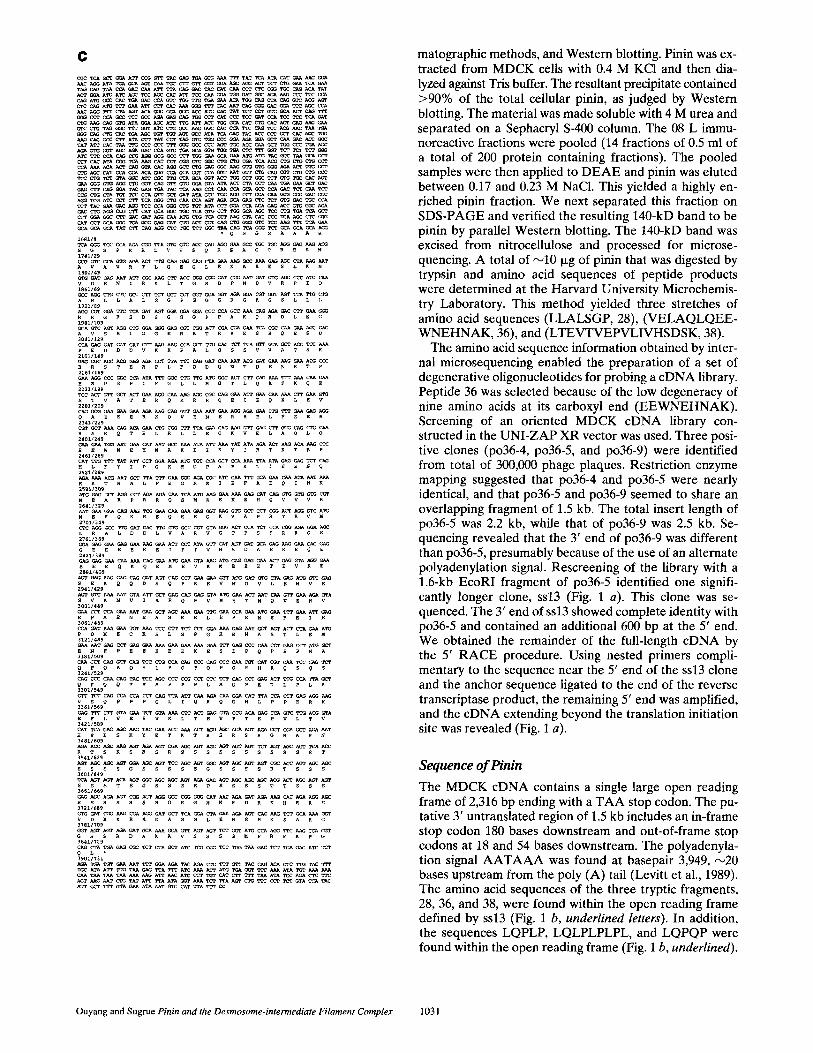
Figure 1. (a) Alignment of the overlapping cDNA clones identi- fied for pinin from MDCK, human placenta, and MDBK. The first two clones identified were po36-5 and po36-9, which repre- sented identical open reading frames of the 3' end and use of dif- ferent polyadenylation sites. The sequence derived from the ex- treme 5' end of the longer ssl3 cDNA clone was used to amplify the 5' end from cDNA that had been modified by the addition of an anchor sequence. Screening of the human placenta kgtl l li- brary yielded two clones covering most of the coding sequence. The two clones were separated by an internal EcoRI site. The 5' end of the placental cDNA was also identified by RACE. Over- lapping MDBK clones were identified. These clones accounted for the full-length open reading frame and a large 3' untranslated stretch. (b) The nucleotide and predicted amino acid sequence of MDCK cDNA for pinin. The amino acid sequences obtained from microsequeneing fragments that were derived from trypsin digestion of 08L are 26, 28, and 38 (underlined), and those de- rived from V8 digestions are V~, V2, and V3 (underlined). The 3' untranslated domain contains polyadenylation sequences AATAAA (underlined). The one used for ssl3 is shown at 3759 (double un- derline), and that used in clone po36-5 is shown at residue 2653 (double underline). (c) The nucleotide and predicted amino acid sequence of MDBK cDNA.
b i/1
M ^ 61/3
V A V R T . . . . . V ~ E Q L E K A K E $ . . . . . . . . . . g A ~ 121/23
D E N I R K L P I Q A T G R D P N D V R 181/43
P G O G R G R G S L S , ...... pp~
G G G P P A K Q R 3ol/ea
V S Z ~ G O e R R T e e Z S R Q E S D e 361 /103
E D D D V K K P A L Q S S V V A T S K ~
~ ~ ~ ~ ~ A ~ C ~ T ~ T A ~ T ~ ~ G ~ ~ R T R R D L I Q D Q N M D E K G K Q R N ~8 t /143
~ . o ~ ~ o ~ ~ ~ ~ 0 ~ S T V sai/163 pT ACT ~ p ~ T . . . . . . . . . . p . . . . . .
K R R Q E I E Q L E V Q A E 601/183
~ ~ V E N E R R E L . . . . . . . . . ~ E R R A K . . . . . 661/203
L L E ~ K ~ 721/223
A K I I K Y I R T K T K P H L F 781 /243
Y I L R M C P A T Q K I E E S Q R K 841/263 .... ~g ........... ~ ATA ~ T ~ A ~ ~
E Q 2 N K M E ~01/28~
~ C ~ A ~ A ~ A A ~ ~ ~ ~ ~ T ~ ~ A R p R R Q S M K E H Q V V R N E E 961/303
K A E Q E E G K V A ~ ~ ~ R E E E L E E 1021/323 ~ T ~ ~ T ~ T ~ A ~ A T A ~ ~ A ~ ~ ~ ~ ~ ~ ~ATA G N Q H N D V E I E E A G E E E E K E I 1 0 ~ / 3 4 3
G I V H S D A E K E Q E E E E Q K Q E M i14~/363
~ ~ A ~ ~ ~ A ~ A ~ ~ ~ ~ ~ ~ ~ T A ~ E V K I E E E T E V R E S E K Q Q D S Q 1201/383 C ~ ~ ~ A ~ T ~ C T A ~ A ~ ~ G T A ~ ~ ~ T ~ A A ~ P E E V M D V L E M L L H V A V K N V I 1261/403
z ~ ~ v s e T ~ 0 ~ ~ A ~ C ~ r ~ s v z ~ s e ~ Z 13211423 A ~ ~ ~ ~ G C ~ A ~ ~ ~ ~ O ~ T ~ ~ ~ T S X E L E P E M E ? E V E P D K E C K I~SI/443
S L S P V R E N A S A L E E M E N E P E l a 4 1 / 4 6 3
K R E R E S E P Q P E P V R H L O P L P 1501/483
1561/503 ~ A C ~ C ~ ~ ~ c c c c ~ ~
L Q P Q L Q L Q L P Q p Q S Q S Q B 1621/523
L O L P L P L P P Q P Q V Q A 16ai/543
S Q P Q A V L Q P ~ ~ S Q P E T L P W 1741/563 ~ A ~ G ~ A ~ ~ ~ ~ ~ ~ A ~ ~ ~ ~ T ~ A ~ A ~ L A V L Q A P V Q V I Q B Q O H L L p ~
1S01/5~3
1861160] ATA~A~T
R S R S R G R A 1921/623
s s s s s s s s s 19811643
s s s s s B s s 2041/663 TCC AGT AGC AGC TCC AC-C ACA AGT GGC AGC AGC ACG AGA GAT AGT AGC AC, C AGC ACT AC T S S S S S S T S o S S R R D S S S S T T 2101/583
GGC CCG GGA CAT AAC AGA CAT AGA AAG CAC S R S H N R D R K H
2 1 6 1 / 7 0 3
p e s ~ L E R S H K s 2221/723 TCA AAA GG~T GGT AOT AGT AGA C~T ACA AAA GGA TCA AAG ~AT AAG AAT TCC CGG TCC GAC S X S o s s R D T K o s x D K N S R S D 2~aI/743 AOA A~ AGG TCT ATA TCA GAG AGT AGT CGA TCA GGC AAA ACA TCT TCA AGA AGT GAA ~ R K R S I S E S S R S G K R S S R S E R 23411763 GAC CGA AAA TCA GAC AGO AAA GAC AAA AQG CGT ~ T~ AAG AAO CCA GGC ~'IT C~ A~ D R K s D R X v K R R • CTA TTC TTT GCC GCA GAA GAT TTC T~ ATG AGT AAA ~X~ ATT ACC ~,FT CCT TGT AAG GAG GAT GCT GCC TTA AGA ATT GCA TGT TGT AKA AAA TCT TTT TTG GAA AAT ACA GAC TGT ~ TTT ACC AGA CAT TCT TGT ACT gTFf TGC ATA ATT TT~ TAK C~ TTA ~ ATC AAA APT ATG TGA GGT TCC AAA ATA TGT AAA AAT TAT AAT A~T AAA AAA AGA TTA ACA TCC CTT GTC ATC TTT TTT AAA TAT CCT ATA CAC TTC AGT AAG AAT CTG TAT ATT TTA ATA GGT AAA TCT TTA CGC TCC TGT TCC C~'~ CAA ATT CT~ TAT CAT ACA ~q~3 CTT TTC GTC AGA AAT AAA TCG %~C-C
ATT TCT TTC ATT AGT TTT CGG AAT CGT CCT CCG TTQ ACA CTT GTA TAA TA~ ATT ACC CTC TTG ATA TAT CST TTT C~G CTT TTA TCA CTA CAT ACT GAA CGT CAT TAG AAT GTC T~Ff GAA GGG TPG ATT ACT ACT AAT CAA CTA T~ TCG TCT ~pG TAT ~A AGA AAA TAA TAA AAT AGT TGG TCG ACT ATT CTT C'Fp CTA CTA TAT GAT GTT TGT C.A~ A&T AC.p TGC TCC TCC TGC TGT GTA SCA ArC ATC TTG TTG A~r AAT A~? AAT A~A ~TA TTA TeA CCC TCC TTC TGA OTA ATT ACT ACT ACT A~'p TCA GAC GTC TIT APT ACT ACA TCA TC4% A~T AAT CAT C(~T AC.A ACA GCT GGT AGT ATA TC.A AGA ATA GTT GCT C'FP TTT TAT ATG AC.A AAC GCG GGG A~A TGC TTA GAT TAA AAC AAG GC4% GAT TAG AAA AAA ATA GTA AAT GAT TCT GA~ AAC AAA CTA TGT TCC CCA AAA G~ T~ AAA GAT C~ ACT ATG ACA AGC Tt~C A~K~ TT~ CCT AAG GCA AAT AAA AGT GAG C.AA ACC TrC ~AA TGA CAT TCT AAT CTA CTG TTC A~ ATC TAC ~C~A T~A ACG TCG ~ CAT AGT TIG TTC TCA C~A TCA ~T ACT ATT TTT TCT AAT CTC CCT TCT T?T ;UtT CTA AC.C ACT TCC CCC T~T ~ TCA TAT ATA A~T CAT ATT T~ TAG ATA A~ ~ TAT CAT TTG GTT GC.C ATT TTC "FfC AT~ ATT ATA GGA (~A TCA TTC ATT T~ CTC CA~ GGC TAC TTG T~T C.AT ATA CCA TAT ACA TCT ATT C~AA GAA AAT AAT CAC TCT CTA GGG C*AG GGA GGT ~X~A AAA GTA TAT TCT AAA CTT GGG TTT TTG AGT TTG TG~ TCT T~-T CTT AAC Ti-p TGT G~.~ GCT CTA ACT ACA TGC CAA TAT GTG TTC TCA AGA GTT TTT GTT AA~ TAT TCT ATG AAA GTT TAC AGA AT~ A~ GAA G'Ff CAT CTA CAC TTG AAT CTG T/%A GCA AC.A TAA CAC ACA AGT GTA CCA AGT CAT TAT TAA CTT TGT TGT TTT ATA AAT TTG TAT GAA TTT GGA GTA TCT ~ ~CC ATT ACT ATA TAT GTG CAA ATA AAT GTG GCT TAG ACT TGT GAA AAA AAA ~u%A AAA AAA AAA A~
T h e J o u r n a l o f C e l l B i o l o g y , V o l u m e 1 3 5 , 1 9 9 6 1 0 3 0
C
A~ A~ ~C
~N~NNNNN . . . . . . . . . . . . . .
T A T A ~ C T ~ ~ ~ ~ ~ ~ ~ ~ ~ ~ ~ ~ C ~
~ A ~ A ~ T ~ T ~
~ A ~ ~ ~ ~ ~ ATA ~ A ~ ~ ~ ~ ~ ~
~ T ~ ~ C ~ ~ A T A ~ ~ G ~
~ ~ ~ ~ ~ ~T ~ C~ A~ ~ ~ ~ ~ ~A ~T ~ ~ A~ ~ ~
1 6 8 1 / 9
S G S ~ R R L V V S Q R E A C C R E K M 1741/29
1 8 0 1 / 4 9
R ~ I Q V D E N I R K L T G R D 1861/69
A R L L a L S e e S G C e S L L ~ 1921/89
R R G V S D S G G G e P ~ O L ~ G 1981 /109
A V S R L G O E R R T R R 2041/129
p E D D D V K K p A L Q S 2101/149
~ R S T E R P L F Q D Q ~ T ~ E K E T P 2 1 6 1 / 1 5 9 ~ C C C ~ ~ A T A ~ ~ ~ ~ A ~ A ~ ~ ~ ~ ~ C ~ E R P G P I F G L L M G T L Q K F K Q E 2 2 2 1 / 1 8 9
S T V A T E R Q K R R Q E I E Q K L E V 2281/209
Q A E E E R K Q V E N E R R E L F E E R 23411229
~ A K Q T ~ L ~ O K V 2 ~ 0 1 / 2 4 9
E E W N E H N A K I I K Y I R T K T ~ P 24~I/269 ~ T ~ ~TATA~CCT~ ~ A ~ ~ T C ~ A A T A ~ G T~
25~11Z89 . . . . . . . . . . . . . . . . . . . . . . . . ~ p R K T ~ A L F E G R R I ~ F A E Q I
M E A p R R Q S M K E K E H Q V V 2641/329
N E E Q K S E Q E E G K V A p R T R V M
2761/369
G E E E K E I p I V H S D A E K 2 8 2 1 / 3 8 ~
Q E M E V K M ~ E E T E V ~ 28SI/409
S E K Q Q D S Q P E E V M D V L E M V E 2941/429
3001/~9
E ~ $ E N E A S K E L E P E M E F E I E 3061/~69 ~ T ~ ~ ~ ~ ~ ~ ~ ~ ~ ~ ~ T ~ ~ A ~ A ~ A ~ P D K E C K S L S P G K E N A S T L E M 3121/489
E N R ~ E E K E E K E S E P Q P E p M A 3181/509 ~ c ~ ~ ~ ~ ~ C ~ C C C ~ C ~ C ~ T ~ ~ ~ ~ Q P Q A Q S L P Q P Q e Q R H R Q S Q S 32~x/5~ ~ ¢ C C ~ ~ A C ~ C ~ C ~ ~ ~ ~ c ~ ~ ~ ~ Q P Q Q Y S S P P P L S O ~ E T L P ~ A 3301/549
~ ~ C ~ C ~ T ~ A A ~ ~ ~ ~ T ~ A ~ ~ ~ ~ V S Q ~ P P Q L I Q R Q ~ H L P p E R K 33~I1569 ~ G ~ ~ A ~ ~ A ~ ~ ~ ~ A C ~ A ~ C ~ ~ A ~ A E F L V E S V K L T E V p T E P V L T V 3421/589 ~ T ~ ~ ~ ~ T A C ~ A ~ A ~ ~ ~ A ~ ~ ~ ~ ~ T H S E S K Y E T K T R S R S R G R A R N 34811609
a T S ~ S R S R S S S S S S S S S S S ~ 3541/629
S S S S G S S S S S G S S S S R T S S S
S S S T S G S S S R D S S S S T T S S S 366~16~9
E S R S R S R G R G H N R D R K H R R S 37211689 ~ T ~ ~ ~ ~ T ~ ~ ~ A ~ A ~ E ~ ~ ~ ~ V D R K R R D A S G L E R S H K S A K G 37811709
~ A ~ A ~ T ~ A ~ ~ ~ A ~ ~ ~ A ~ ~ ~ ~ C ~ G S S R D A K A V S S S G M p R F K p G 3841/729 ~ G ~ A ~ ~ ~ T ~ A ~ A ~ ~ C ~ T ~ ~ ~ ~ A ~ Q S " 3901/731 ~ ~ ~ ~ ~T ~ ~ ~ TAC ~ ~ ~ G~ TAC ~ ~ ~ ~ T~ ~
T~ T~ T~ ~ ~G A~ ~ A~ ~ ~ ~T ~ ~ T~ ATA ~ ~ ~ ~ ~T ~ TAT A~ ~A ATA ~ ~ ~ ~A ~ ~ ~ ~ ~ ~A ~ T~
A~ ~ ~ GTA ~ ATA ~T ~ ~T ~A ~
matographic methods, and Western blotting. Pinin was ex- tracted from MDCK cells with 0.4 M KCI and then dia- lyzed against Tris buffer. The resultant precipitate contained >90% of the total cellular pinin, as judged by Western blotting. The material was made soluble with 4 M urea and separated on a Sephacryl S-400 column. The 08 L immu- noreactive fractions were pooled (14 fractions of 0.5 ml of a total of 200 protein containing fractions). The pooled samples were then applied to D E A E and pinin was eluted between 0.17 and 0.23 M NaCI. This yielded a highly en- riched pinin fraction. We next separated this fraction on SDS-PAGE and verified the resulting 140-kD band to be pinin by parallel Western blotting. The 140-kD band was excised from nitrocellulose and processed for microse- quencing. A total of ,--,10 Ixg of pinin that was digested by trypsin and amino acid sequences of peptide products were determined at the Harvard University Microchemis- try Laboratory. This method yielded three stretches of amino acid sequences (LLALSGP, 28), (VELAQLQEE- WNEHNAK, 36), and (LTEVTVEPVLIVHSDSK, 38).
The amino acid sequence information obtained by inter- nal microsequencing enabled the preparation of a set of degenerative oligonucleotides for probing a cDNA library. Peptide 36 was selected because of the low degeneracy of nine amino acids at its carboxyl end (EEWNEHNAK). Screening of an oriented MDCK cDNA library con- structed in the UNI-ZAP XR vector was used. Three posi- tive clones (po36-4, po36-5, and po36-9) were identified from total of 300,000 phage plaques. Restriction enzyme mapping suggested that po36-4 and po36-5 were nearly identical, and that po36-5 and po36-9 seemed to share an overlapping fragment of 1.5 kb. The total insert length of po36-5 was 2.2 kb, while that of po36-9 was 2.5 kb. Se- quencing revealed that the 3' end of po36-9 was different than po36-5, presumably because of the use of an alternate polyadenylation signal. Rescreening of the library with a 1.6-kb EcoRI fragment of po36-5 identified one signifi- cantly longer clone, ssl3 (Fig. 1 a). This clone was se- quenced. The 3' end of ssl3 showed complete identity with po36-5 and contained an additional 600 bp at the 5' end. We obtained the remainder of the full-length cDNA by the 5' RACE procedure. Using nested primers compli- mentary to the sequence near the 5' end of the ssl3 clone and the anchor sequence ligated to the end of the reverse transcriptase product, the remaining 5' end was amplified, and the cDNA extending beyond the translation initiation site was revealed (Fig. 1 a).
Sequence of Pinin
The MDCK cDNA contains a single large open reading frame of 2,316 bp ending with a TAA stop codon. The pu- tative 3' untranslated region of 1.5 kb includes an in-frame stop codon 180 bases downstream and out-of-frame stop codons at 18 and 54 bases downstream. The polyadenyla- tion signal A A T A A A was found at basepair 3,949, ~20 bases upstream from the poly (A) tail (Levitt et al., 1989). The amino acid sequences of the three tryptic fragments, 28, 36, and 38, were found within the open reading frame defined by ss13 (Fig. 1 b, underlined letters). In addition, the sequences LQPLP, LQLPLPLPL, and LQPQP were found within the open reading frame (Fig. 1 b, underlined).
Ouyang and Sugrue Pinin and the Desmosome-intermediate Filament Complex 1031
~ o ~ g y H A V A V R T b Q E ~ £ K ~ K E S L K N V D E ~ I R r L T G R D ~ N D V R B I
. . . . . . . I . . . . . . . . . . . . . . . . . . . . . . . . . . : . . . . . . . . . . . . . I " ~ K P I N I ~ M A V V R L Q ~ Q L E K A I E S L K N V D E N I K L T ~ R D P ~ D V R p l l o H ~ P Z N I N M A ~ ' ~ L O E O L £ K & X E S L K N V n E ~ I R R L T G R D p N n V R P I 4 0
~ o r i c y Q A R L L A L S G P G G G R G R G S L L L R R G F S D S G G G P P A K Q R D L E
. . . . . . i o . . . . . . . . . . . . . . . . . . . . . . . . . . . . . g . . . . o . . . . {.o ~ N p I N z N Q A R L L A L S G P G G C R G R G S L L L a R G F S D S G G P P A K Q R O L E 8 0 H U m ~ t P I N C N O A R L L A L S G P G G G R G R G S L L L R R G F S D S G G P P A K O R D L E 7 9
~ o ¢ i ¢ y G A V E R L G G E R R T R R E S R Q E S D p E D D D V K K P A L Q S S V V A T S
. . . . . . . I . . . . . . . . . . . . . . . i R o . . . . . . . . . . . . . . Q . . . . . . fizz0 H~)BKpZNZN C A V S R L G G ~ R R T R R E R Q E S D p E D D D V X K P A L Q S S V V A T 120 H ~ P I N I N G A V S R L G G ~ R R T R ~ R Q E S D ~ S D D D V K K P A L O S S V V A T 119
M a j o r i t y K B R - T R R O L ~ O D O N M D £ K G K - Q R N R R [ F G ~ L M G T L O K F X Q
~ B K p I N I N S T ~ R ~ L ~ Q D O N ~ D E K P R J P G ' P I Z F G L L M G r L Q K ~ K 160 Humi~ p Z N I N I X E m - I T R R D L I O D O N M D E X G K I - [ Q R N g R I F G L L M G T L O g P ~ I S ~
~ o r i c y E S T V A T E R Q K R R Q Z l E Q K L E V O A E Z g R K Q V E N E R R E L ~ £ E
. . . . . . . I . . . . . . . . . . . . . . . . . . . . 0 . . . . . . 0 . . . . . . . . . . . I" ~ N P Z N I N E S T V ~ T E R Q R R Q E I E Q K L E V O A E E E R X Q V E N ~ R R E L F ~ 2 0 D H ~ p I N Z N E S T V A T E R O R R O E I E O K L K V O A E E E R K O V E N ~ R R K L F ~ I ~ 7
~ j o r i ~ y R R A K Q T E L R L L E Q K V E L A ~ L Q E E W N E H N A K I I K Y I R T K T K
~ p I N I N R R A K Q T ~ L ~ L L E Q ~ V E L A Q L O E ~ W N E ~ N A R ~ K ¥ I R T K T 240 H ~ n p I N I N R R A K O T E L R L L E O K V E L A O L O R E W N K H N A K I I K Y I R T K T 237
~ j ~ r i t y P H L F Y I P G R M C P A T Q g L Z E E S Q R K M ~ A L F E G R R I E F A E Q I
~ K P m l N e H L ¥I PGRMCPA K L I E E S O R K ~ N A L t G R R I F A E O 27~ Humen PININpHLPYYPGRMCPATOKLIEESORKMNALF G R R I F A ~ ° 27~
~ o r ~ c y N X M E A R P R R Q S M K E K E H U V V - R N E E Q K A E O E E G X V A Q R E E
~PININ N K M ~ A R P R R O S M K ~ K E M Q V V V R N ~ K ~ E O ~ E G K V A Ii~ H~.PZHINNKMEAR~OSMKEK~HOVV-RNE~IHIKAEQEEGKVAOREEI316
~orlty ELXETGNQN . . . . . N O V E Z S E A G E E ~ E K E I G [ V H S D A E K Z
~BK pIN~N VN L ~A LDD L V A RVGT P S P RRG SIG g E E Z ~ E I~I V ~ ~ O A E K ~l 3S9 H~nPININ[~:~VJETONOHI . . . . . INDVSIEEAGEEEEKEIGIVH DAE~ ~51
~jori~y Q E E E E Q K Q E M E V K H E E E T E V R E S E K Q Q D S U P E E V H D V L E M
~ K p I ~ I N Q E E E E O X Q E H E V K M E E ~ T E V R ~ S E K O Q D S O P E E V M D V ~ E 399 H ~ n P I N I N O E E E E O K O E M E V K H E E E T E V R E S E K O O D S O P E E V M D V L E 1~I
Majority V E X V X V K N V I A E Q E V M E T N Q V E S V E P S E N E A S K E L E P E M E
I ~ C K ~ I N Z N I ~ - ~ H I ~ ~ H ~ A ~ V K N V I A E Q E U H E T N O V E S V E P S E N E ~ S K - E L E P E H ~ 432
H~nPININ N ~ - VIA OEVMETNIRIVEgVEPSENEASKELEPE 429
~)or~y F E I E P D K E C K S L S p G K g M A S A L E M ~ N E P E E K E E K E S E P Q P
~nKpININ F E I ~ p D K E C K S L S P G K E N ~ S ~ L ~ M E ~ E , ~ P - - ~ E K E E K E S E p Q p 4 ? 7 H~n BININ E P D K E C K S L S P G K E NIVIS A blDL~JK~ S DI E K E E 469
~ i o r ~ C y E e V A Q p O A Q S O P Q p X X X X O X E p O P Q L Q P E p X - O p Q L O - - -
MDBK P I N I N [ ~ : ~ ~ Q O ~ J L M A Q P O A ° S L . . . . . . . . . . . . . . . . . . . . . . . . . . 492 H ~ n P I N I N A O p O A O S Q L O S ~ S t k P O P O L O P ~ P I A - I O P O L O I - - - 506
M a j o r i t y . . . . . . . . . X O p Q L ° L . . . . . . ° p Q X X X Q X Q S Q p Q A V L Q p
OpQpQSQSQPIOPOLOLJPLPLPL p Q V A Q ~ ~ ~ a x w ~ i ~ . . . . . . . . . . . . . . . . . . . . . .
~ j o r i c y X P X S O P E T ~ L A V ~ O P ~ P O V X O e O G . L L ~ E ~ E r P V E S V K
½ ~ n p INZN H ~ I P S O P E I D ~ S I L A V L O P I T I ~ 0 VIT~._~J Hi_C,L~J F I L P E R K IOIF v V E S V ~1 562
M a j o r i t y L T E V p V E e V L T V H S ~ S K X ~ T K T ~ S R S R G R A R ~ R T S K S R S R
~ K p I N I N T I D T K T K T R S R S R G R A R N ~ T S K S R S R ~ S 3 Z
H u ~ n p I ~ I N S T a S R S R G R R N R T ~ S R S R 602
~ ) o r i c y S S S S S S S S S S S T S S S S G S S S S S G S S S S R S S S S $ S S S T S G S
~ e ~ e t ~ Z N S S S S S S S S S S S T S S S S ~ S S S G s s s s ~ s s s s s s ~ s e s ~ 2 a H~nPINIMSSSSSSSSSSSTSSSSGSSS S G S S S S R S S S S S S S S T S G S ~ 2
Ha)ortCy $ S R D $ S S S T T S S S E S R S R g R G R G H N R D R ~ R ~ R ~ V D R K R R D
. . . . . . . ~ R b . . . . . ~ . . . . . . . . . . . . . . . . . . . . . . . . . . . . . . 17t~ M D B K P I N I N S S R D S S S S T S S S E S R S R S R G R G H N R D R K H R R S V D R K R R O 6 6 8 H u ~ n p ~ N I N RDSSSST S S S E S R S R S R G R G ~ N R D R X H R R S V D R K R R D ~ 8 2
Major/ty T S G L E R S H K S S K G O S S R D T K G S K D R H S R ~ D R K R S I S E S S R
~ a K pZNIn A S G L S R S H K S ~ O G S S R 0 ~ - ~ K I ^ . . . . . . . . . . . . . . . . V l S S 692 H ~ n p I M I N S G L E R S H K S S K G C S S R D T K G S K D K P t S R S D R X R S I S E $ S 722
H a ~ Q r i t y S G K R S S R S E R O R K S D R K D K R R -
MDCK p I N [ N S G ~ ~ S R S E R D . ~ K S D R K D K R RI ~74 MDBK prn~u S G I H P ~ F K P C Q . . . . . . . . . L ~¢4 f l ~ n p I N I N S G K R S S R S E R D R K S D R K D K R K i 7~4
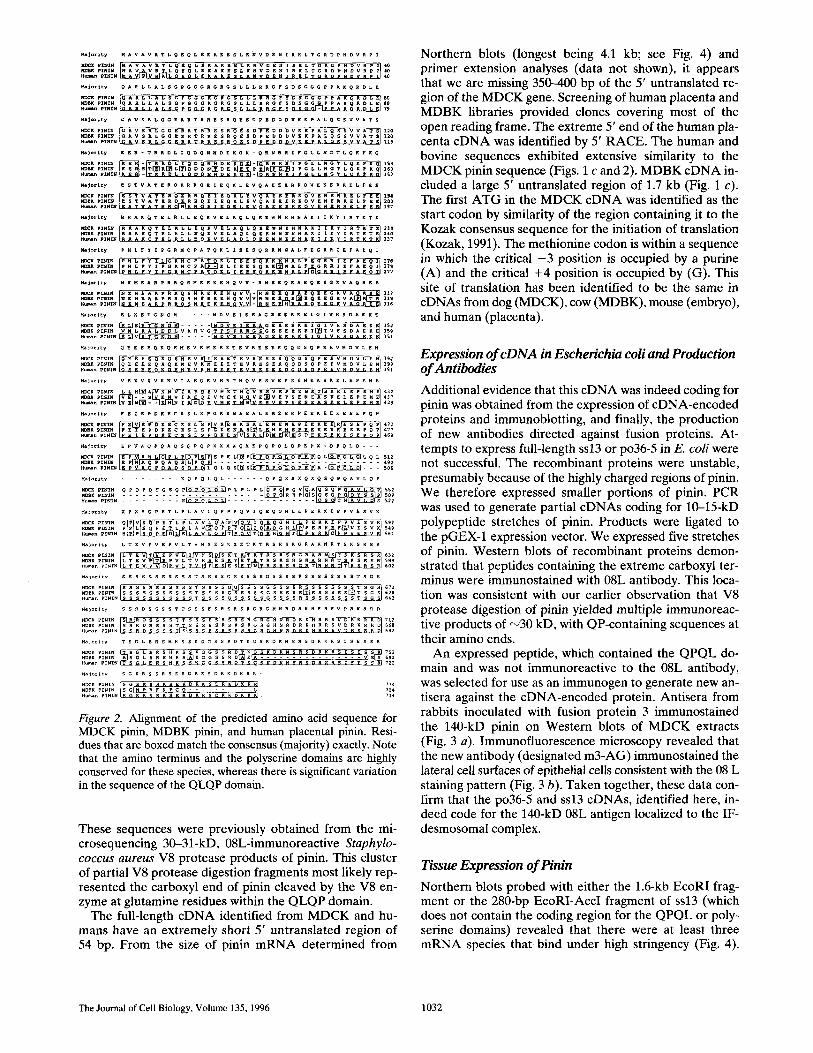
Figure 2. Alignment of the predicted amino acid sequence for MDCK pinin, MDBK pinin, and human placental pinin. Resi- dues that are boxed match the consensus (majority) exactly. Note that the amino terminus and the polyserine domains are highly conserved for these species, whereas there is significant variation in the sequence of the QLQP domain.
These sequences were previously obtained from the mi- crosequencing 30-31-kD, 08L-immunoreactive Staphylo- coccus aureus V8 protease products of pinin. This cluster of partial V8 protease digestion fragments most likely rep- resented the carboxyl end of pinin cleaved by the V8 en- zyme at glutamine residues within the QLQP domain.
The full-length cDNA identified from MDCK and hu- mans have an extremely short 5' untranslated region of 54 bp. From the size of pinin mRNA determined from
Northern blots (longest being 4.1 kb; see Fig. 4) and primer extension analyses (data not shown), it appears that we are missing 350--400 bp of the 5' untranslated re- gion of the MDCK gene. Screening of human placenta and MDBK libraries provided clones covering most of the open reading frame. The extreme 5' end of the human pla- centa cDNA was identified by 5' RACE. The human and bovine sequences exhibited extensive similarity to the MDCK pinin sequence (Figs. 1 c and 2). MDBK eDNA in- cluded a large 5' untranslated region of 1.7 kb (Fig. 1 c). The first ATG in the MDCK cDNA was identified as the start codon by similarity of the region containing it to the Kozak consensus sequence for the initiation of translation (Kozak, 1991). The methionine codon is within a sequence in which the critical - 3 position is occupied by a purine (A) and the critical +4 position is occupied by (G). This site of translation has been identified to be the same in cDNAs from dog (MDCK), cow (MDBK), mouse (embryo), and human (placenta).
Expression of cDNA in Escherichia coli and Production of Antibodies
Additional evidence that this cDNA was indeed coding for pinin was obtained from the expression of eDNA-encoded proteins and immunoblotting, and finally, the production of new antibodies directed against fusion proteins. At- tempts to express full-length ssl3 or po36-5 in E. coli were not successful. The recombinant proteins were unstable, presumably because of the highly charged regions of pinin. We therefore expressed smaller portions of pinin. PCR was used to generate partial cDNAs coding for 10-15-kD polypeptide stretches of pinin. Products were ligated to the pGEX-1 expression vector. We expressed five stretches of pinin. Western blots of recombinant proteins demon- strated that peptides containing the extreme carboxyl ter- minus were immunostained with 08L antibody. This loca- tion was consistent with our earlier observation that V8 protease digestion of pinin yielded multiple immunoreac- tive products of ,-~30 kD, with QP-containing sequences at their amino ends.
An expressed peptide, which contained the QPQL do- main and was not immunoreactive to the 08L antibody, was selected for use as an immunogen to generate new an- tisera against the eDNA-encoded protein. Antisera from rabbits inoculated with fusion protein 3 immunostained the 140-kD pinin on Western blots of MDCK extracts (Fig. 3 a). Immunofluorescence microscopy revealed that the new antibody (designated m3-AG) immunostained the lateral cell surfaces of epithelial cells consistent with the 08 L staining pattern (Fig. 3 b). Taken together, these data con- firm that the po36-5 and ssl3 cDNAs, identified here, in- deed code for the 140-kD 08L antigen localized to the IF- desmosomal complex.
Tissue Expression of Pinin
Northern blots probed with either the 1.6-kb EcoRI frag- ment or the 280-bp EcoRI-AccI fragment of ss13 (which does not contain the coding region for the Q P Q L or poly- serine domains) revealed that there were at least three mRNA species that bind under high stringency (Fig. 4).
The Journal of Cell Biology. Volume 135. 1996 1032
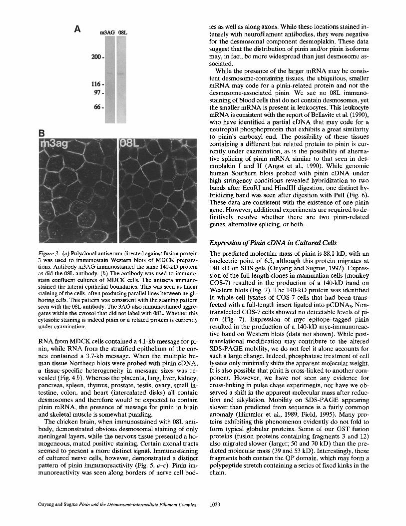
Figure 3. (a) Polyclonal antiserum directed against fusion protein 3 was used to immunostain Western blots of MDCK prepara- tions. Antibody m3AG immunostained the same 140-kD protein as did the 08L antibody. (b) The antibody was used to immuno- stain confluent cultures of MDCK cells. The antisera immuno- stained the lateral epithelial boundaries. This was seen as linear staining of the cells, often producing parallel lines between neigh- boring cells. This pattern was consistent with the staining pattern seen with the 08L antibody. The 3AG also immunostained aggre- gates within the cytosol that did not label with 08L. Whether this cytosolic staining is indeed pinin or a related protein is currently under examination.
RNA from MDCK cells contained a 4.1-kb message for pi- nin, while RNA from the stratified epithelium of the cor- nea contained a 3.7-kb message. When the multiple hu- man tissue Northern blots were probed with pinin cDNA, a tissue-specific heterogeneity in message sizes was re- vealed (Fig. 4 b). Whereas the placenta, lung, liver, kidney, pancreas, spleen, thymus, prostate, testis, ovary, small in- testine, colon, and heart (intercalated disks) all contain desmosomes and therefore would be expected to contain pinin mRNA, the presence of message for pinin in brain and skeletal muscle is somewhat puzzling.
The chicken brain, when immunostained with 08L anti- body, demonstrated obvious desmosomal staining of only meningeal layers, while the nervous tissue presented a ho- mogeneous, muted positive staining. Certain axonal tracts seemed to present a more distinct signal. Immunostaining of cultured nerve cells, however, demonstrated a distinct pattern of pinin immunoreactivity (Fig. 5, a-c). Pinin im- munoreactivity was seen along borders of nerve cell bod-
ies as well as along axons. While these locations stained in- tensely with neurofilament antibodies, they were negative for the desmosomal component desmoplakin. These data suggest that the distribution of pinin and/or pinin isoforms may, in fact, be more widespread than just desmosome as- sociated.
While the presence of the larger mRNA may be consis- tent desmosome-containing tissues, the ubiquitous, smaller mRNA may code for a pinin-related protein and not the desmosome-associated pinin. We see no 08L immuno- staining of blood cells that do not contain desmosomes, yet the smaller mRNA is present in leukocytes. This leukocyte mRNA is consistent with the report of BeUavite et al. (1990), who have identified a partial cDNA that may code for a neutrophil phosphoprotein that exhibits a great similarity to pinin's carboxyl end. The possibility of these tissues containing a different but related protein to pinin is cur- rently under examination, as is the possibility of alterna- tive splicing of pinin mRNA similar to that seen in des- moplakin I and II (Angst et al., 1990). While genomic human Southern blots probed with pinin cDNA under high stringency conditions revealed hybridization to two bands after EcoRI and HindlII digestion, one distinct hy- bridizing band was seen after digestion with PstI (Fig. 6). These data are consistent with the existence of one pinin gene. However, additional experiments are required to de- finitively resolve whether there are two pinin-related genes, alternative splicing, or both.
Expression o f Pinin c D N A in Cultured Cells
The predicted molecular mass of pinin is 88.1 kD, with an isoelectric point of 6.5, although this protein migrates at 140 kD on SDS gels (Ouyang and Sugrue, 1992). Expres- sion of the full-length clones in mammalian cells (monkey COS-7) resulted in the production of a 140-kD band on Western blots (Fig. 7). The 140-kD protein was identified in whole-cell lysates of COS-7 cells that had been trans- fected with a full-length insert ligated into pCDNA3. Non- transfected COS-7 cells showed no detectable levels of pi- nin (Fig. 7). Expression of myc epitope-tagged pinin resulted in the production of a 140-kD myc-immunoreac- tive band on Western blots (data not shown). While post- translational modification may contribute to the altered SDS-PAGE mobility, we do not feel it alone accounts for such a large change. Indeed, phosphatase treatment of cell lysates only minimally shifts the apparent molecular weight. It is also possible that pinin is cross-linked to another com- ponent. However, we have not seen any evidence for cross-linking in pulse chase experiments, nor have we ob- served a shift in the apparent molecular mass after reduc- tion and alkylation. Mobility on SDS-PAGE appearing slower than predicted from sequence is a fairly common anomaly (Himmler et al., 1989; Field, 1995). Many pro- teins exhibiting this phenomenon evidently do not fold to form typical globular proteins. Some of our GST fusion proteins (fusion proteins containing fragments 3 and 12) also migrated slower (larger; 50 and 70 kD) than the pre- dicted molecular mass (39 and 53 kD). Interestingly, these fragments both contain the QP domain, which may form a polypeptide stretch containing a series of fixed kinks in the chain.
Ouyang and Sugrue Pinin and the Desmosome-intermediate Filament Complex 1033

Figure 4. Northern blot analysis of tissue expression of 08L messages revealed that there exists at least three mRNA species that bind 08L DNA under high stringency. (a) Probing with the 1.5-kb EcoRI fragment showed that RNA from chick corneal epithelia contained a 3.7-kb message, whereas MDCK cells contained 4.1-kb message. (b) Various human tissue mRNA are probed with the EcoRI 1.6-kb fragment of po36-5 (left panel) and a 5' fragment containing the EcoRI-Acc1280 bp (right panel). Note the appearance of three mRNA sizes in the range of 3.2-4.1 kb, with a tissue-specific variation of expression. The 08L-positive tissues, such as the brain, placenta, liver, kidney, and pancreas contain the 4.1- and the 3.7-kb messages, whereas the heart, lung, and muscle contain predominantly a 3.2-kb mes- sage.
Transfection of pinin into chicken embryonic fibroblasts resulted in the cytosolic accumulation of pinin immunore- activity, but expressing cells demonstrated little change in their morphology. We next examined the distribution of expressed pinin in the context of an epithelial cell. We se- lected human embryonic kidney-derived 293 cells because they demonstrated expression of epithelial proteins such as cadherins and desmoplakin with little or no pinin ex- pression (Fig. 8, a and a'). Examination of the 293 cells that had received the full-length pinin revealed expressed pinin along the lateral borders of 293 cells in close contact. The 293 ceUs that were transfected with pinin cDNA and immunostained for pinin and desmoplakin revealed that expressed pinin was found in association with desmoplakin (Fig. 8, b and b '--d and d'). While the cells expressing the cDNA also exhibited cytosolic accumulations of pinin, there were clearly areas of colocalization, especially at cell-cell contact sites. However, we did not observe significant amounts of pinin assembled at the cell periphery in the ab- sence of desmoplakin.
The expression of pinin in 293 cells leads to a striking change in the cell/tissue morphology. Non-pinin-express- ing 293 cells are often seen as spindle shaped, which ex- hibit limited cell-cell interactions even at very high cell densities. 293 cells transfected with pinin cDNA and then selected with G418, however, exhibited extensive cell-cell contacts and grew in culture as islands (Fig. 9). We have isolated four independent clones of cells expressing pinin. After nine passages of these cells, the epithelial phenotype and growth characteristics of pinin-expressing cells is con- sistent. EM of these clones revealed that the entire array of epithelial cell junctions is enhanced (Figs. 10 and 11).
While desmoplakin immunostaining of cultures of non- transfected 293 cells revealed moderate staining (Fig. 8 a'), morphologically recognizable desmosomes were quite rare in these cells, and those that were found appeared imma- ture and somewhat delicate (Fig. 11 b). Examination of pi- nin-expressing 293 cells revealed fairly well-formed inter- cellular junctional specializations with numerous small desmosomes (Fig. 11, c-f). The desmosomes in the trans- fected cells, while small, exhibited well-formed plaques and numerous associated intermediate size filaments (Fig. 11, c-f). Immunostaining pinin-transfected cells with anti- body against the tight junction component ZO1 (Steven- son et al., 1986) revealed deposition of ZO1 along lateral cell borders, while the untransfected 293 cells showed little ZO1 staining and no zonula staining pattern (Fig. 12). While double immunostaining for pinin and ZO1 in trans- fected cells demonstrated some overlap, the pinin immuno- staining was more extensive and showed more interrup- tions than that for ZO1.
D i s c u s s i o n
We have previously demonstrated that young desmo- somes containing desmoglein, desmoplakin, plakoglobin, and associated IF do not exhibit immunoreactivity for pi- nin. On the other hand, more mature, well-formed, and better organized desmosomes do contain pinin. Therefore, we considered pinin to be a novel protein that is nonessen- tial for desmosomes per se, but perhaps important in the stabilization and organization of the desmosome-IF com- plex. We set out to gain more insight into the possible functions of pinin and its role in desrnosome regulation.
The Journal of Cell Biology, Volume 135, 1996 1034
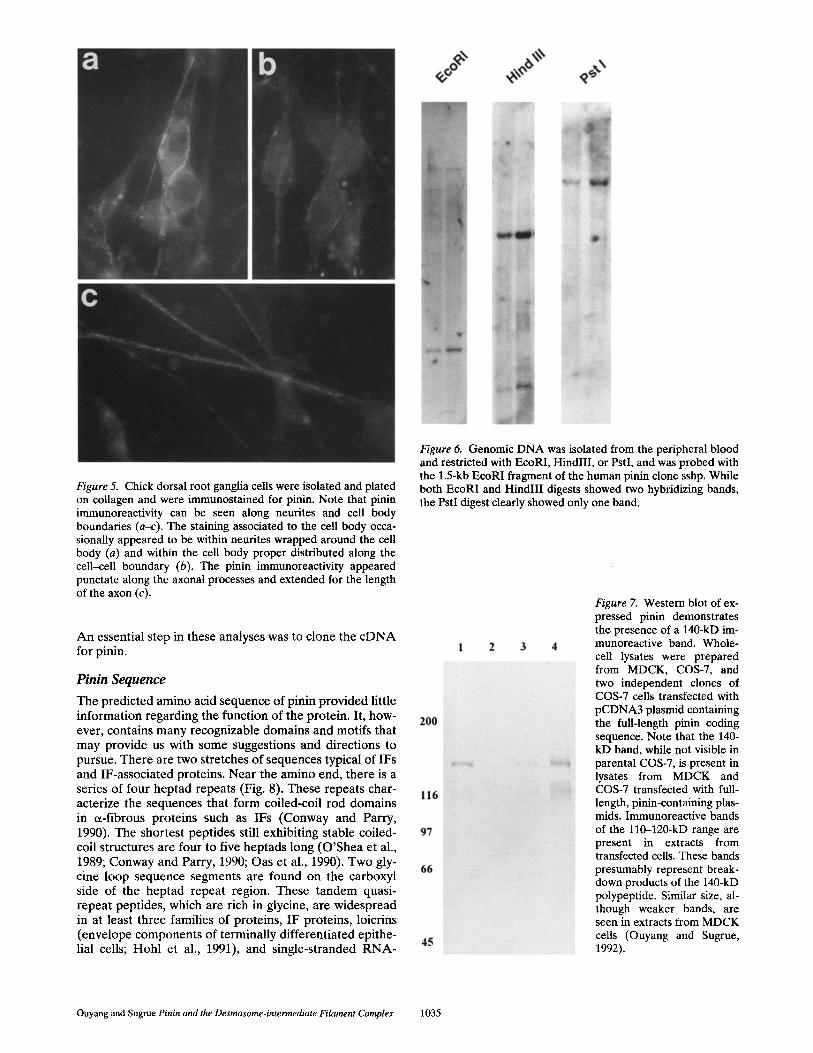
Figure 5. Chick dorsal root ganglia cells were isolated and plated on collagen and were immunostained for pinin. Note that pinin immunoreactivity can be seen along neurites and cell body boundaries (a-c). The staining associated to the cell body occa- sionally appeared to be within neurites wrapped around the cell body (a) and within the cell body proper distributed along the cell-cell boundary (b). The pinin immunoreactivity appeared punctate along the axonal processes and extended for the length of the axon (c).
An essential step in these analyses was to clone the c D N A for pinin.
Pin in Sequence
The predicted amino acid sequence of pinin provided little information regarding the function of the protein. It, how- ever, contains many recognizable domains and motifs that may provide us with some suggestions and directions to pursue. There are two stretches of sequences typical of IFs and IF-associated proteins. Near the amino end, there is a series of four heptad repeats (Fig. 8). These repeats char- acterize the sequences that form coiled-coil rod domains in oL-fibrous proteins such as IFs (Conway and Parry, 1990). The shortest peptides still exhibiting stable coiled- coil structures are four to five heptads long (O 'Shea et al., 1989; Conway and Parry, 1990; Oas et al., 1990). Two gly- cine loop sequence segments are found on the carboxyl side of the heptad repeat region. These tandem quasi- repeat peptides, which are rich in glycine, are widespread in at least three families of proteins, IF proteins, loicrins (envelope components of terminally differentiated epithe- lial cells; Hohl et al., 1991), and single-stranded R N A -
Figure 6. Genomic DNA was isolated from the peripheral blood and restricted with EcoRI, HindlII, or PstI, and was probed with the 1.5-kb EcoRI fragment of the human pinin clone sshp. While both EcoRI and HindlII digests showed two hybridizing bands, the PstI digest clearly showed only one band.
Figure Z Western blot of ex- pressed pinin demonstrates the presence of a 140-kD im- munoreactive band. Whole- cell lysates were prepared from MDCK, COS-7, and two independent clones of COS-7 cells transfected with pCDNA3 plasmid containing the full-length pinin coding sequence. Note that the 140- kD band, while not visible in parental COS-7, is present in lysates from MDCK and COS-7 transfected with full- length, pinin-containing plas- mids. Immunoreactive bands of the ll0-120-kD range are present in extracts from transfected cells. These bands presumably represent break- down products of the 140-kD polypeptide. Similar size, al- though weaker bands, are seen in extracts from MDCK ceils (Ouyang and Sugrue, 1992).
Ouyang and Sugrue Pinin and the Desmosome-intermediate Filament Complex 1035
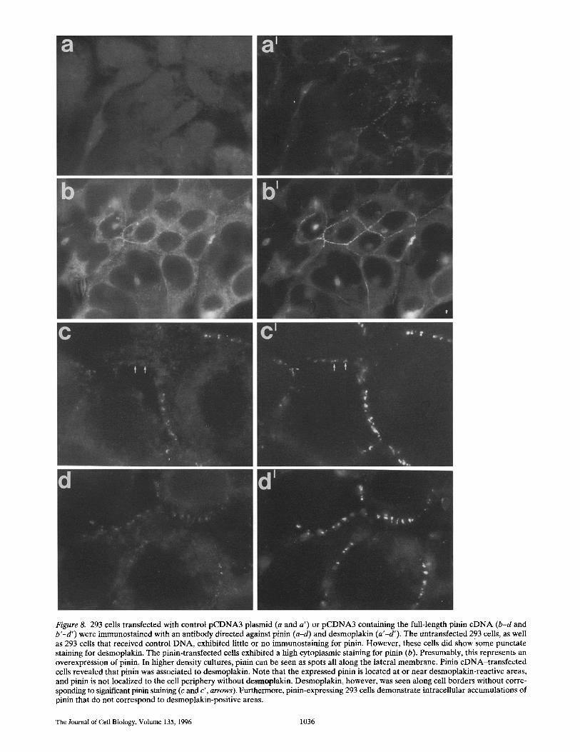
Figure 8. 293 cells transfected with control pCDNA3 plasmid (a and a ') or pCDNA3 containing the full-length pinin cDNA (b-d and b'-d') were immunostained with an antibody directed against pinin (a-d) and desmoplakin (a'-d'). The untransfected 293 cells, as well as 293 cells that received control DNA, exhibited little or no immunostaining for pinin. However, these cells did show some punctate staining for desmoplakin. The pinin-transfected cells exhibited a high cytoplasmic staining for pinin (b). Presumably, this represents an overexpression of pinin. In higher density cultures, pinin can be seen as spots all along the lateral membrane. Pinin cDNA-transfected cells revealed that pinin was associated to desmoplakin. Note that the expressed pinin is located at or near desmoplakin-reactive areas, and pinin is not localized to the cell periphery without desmoplakin. Desmoplakin, however, was seen along cell borders without corre- sponding to significant pinin staining (c and c', arrows). Furthermore, pinin-expressing 293 cells demonstrate intraceilular accumulations of pinin that do not correspond to desmoplakin-positive areas.
The Journal of Cell Biology, Volume 135, 1996 1036
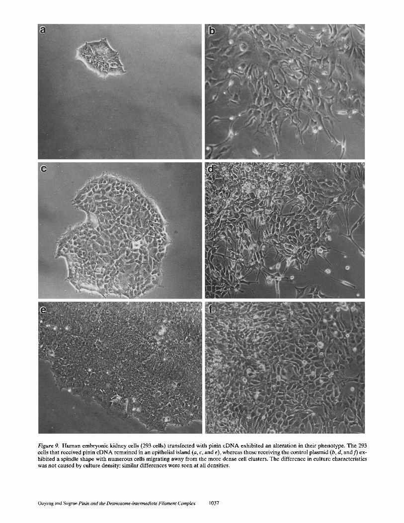
Figure 9. Human embryonic kidney cells (293 cells) transfected with pinin cDNA exhibited an alteration in their phenotype, The 293 cells that received pinin c D N A remained in an epithelial island (a, c, and e), whereas those receiving the control plasmid (b, d, and f) ex- hibited a spindle shape with numerous cells migrating away from the more dense cell clusters. The difference in culture characteristics was not caused by culture density; similar differences were seen at all densities.
Ouyang and Sugrue Pinin and the Desmosome-intermediate Filament Complex 1037

Figure 10. EM of pinin-transfected 293 cells showed enhanced cell-cell adhesion with more extensive cell junctions. Untransfected 293 cells grown at a high cell density demonstrated cell adhesion with limited cell junctions (a). Even at high densities, the 293 cells retained a low profile. Pinin-transfected 293 cells demonstrated cell junctional complexes and a more typical epithelial polarity (b). Lateral cell surfaces displayed numerous junctions, including desmosomes (b', as outlined in b).
binding proteins (Steinert et al., 1991). Glycine loops are expected to be highly flexible, and may participate in in- teractions with neighboring glycine loops on the same or adjacent proteins. They have been postulated to form the basis for adaptable intracytoskeleton interactions (Stein- ert et al., 1991). Residues 635-700 comprise a serine-rich domain. Serines comprise 48 of the 65 residues. The poly- serine domain may be highly flexible and may represent a "hot spot" for the phosphorylation of pinin. The deduced sequence of pinin contains motifs recognized by Ca÷/cal - modulin protein kinases (RXXS), c-AMP--dependent kinases (XRRXSX), c-GMP--dependent protein kinase (XSRX), protein kinase C (XRXXSX), and casein kinase 2 (XSXXEX; Kemp and Pearson, 1990). These potential phosphoryla- tion sites are clustered at two locations: (a) around the poly- serine domain; and (b) near the amino-terminal end of the negatively charged, glutamic acid-rich domain. We have previously shown that pinin exhibits multiple isoforms on two-dimensional gel analyses because of phosphorylation.
A comparison of the eDNA and protein sequence of pi- nin with other sequences available from databases (Gen- Bank/EMBL/DDBJ, Swiss Prot) revealed a striking ho- mology of the 3' end of pinin cDNA to a eDNA identified from a pig neutrophil expression library. This eDNA was identified with a polyclonal antibody directed against a 32-kD phosphoprotein that was involved in a phosphoryla- tion cascade of neutrophils (Bellavite et al., 1990). Within a 279-amino acid stretch of pinin, 196 amino acids were identical to those found in the 32 kD neutrophil phos- phoprotein. At this time, it is not clear as to whether the eDNA identified from the pig neutrophil codes for the 32- kD phosphoprotein or a larger neutrophil protein sharing an antigenic site. While we have not noted any immuno- staining of blood cells with antibodies directed against pi- nin, cDNA probes derived from the 5' end of pinin eDNA recognize a 3.4-kb band in Northern blots of peripheral blood leukocytes. These data may indicate that pinin and
the neutrophil protein are members of a protein family in- volved in phosphorylation events that take place at the cy- toskeleton-membrane interface.
Other than the pig neutrophil eDNA, no significant se- quence homologies were detected on homology searches. However, there was some weak homology of the glutamic acid-rich domain to proteins such as trichohyalin, caldes- mon, and myosin. The significance of these small homolo- gous stretches is not yet evident. When the cDNA and protein sequence of pinin was directly compared to des- mosomal-assoeiated proteins such as desmoplakin, desmo- collins, plakoglobin, and ptectin (with the exception of the heptad repeats), few if any other significant homologies were observed. There were, however, certain regions that exhibited the B-turn and charge characteristics of the 13- residue repeat found in filagrin, another IF-associated pro- tein (Rothnagel et al., 1987; Rothnagel and Steinert, 1990; Mack, 1993).
Possible Functions of Pinin
Clearly, pinin is predominantly found associated to des- mosomes. Nevertheless, we have shown that it is also present within cultured nerve cells, and in fact, Northern blots suggest that it may be expressed at significant levels in the brain. While we do not yet have detailed informa- tion as to its distribution in nerve cells, it is surely not asso- ciated to desmosomes. Therefore, we must keep in mind that pinin may have a more general role within epithelial cells than that at the desmosome.
While expressing pinin eDNA in fibroblasts yielded lit- tle information, expressing it in transformed epithelial cells (human 293 cells) resulted in a dramatic change in cell/tissue architecture. The recipient cells exhibited an in- creased cell-cell adhesion and an enhanced epithelial cell polarity. We suggest that this increased adhesion may be caused by the stabilization of the desmosomal adhesion
The Journal of Cell Biology, Volume 135, 1996 1038
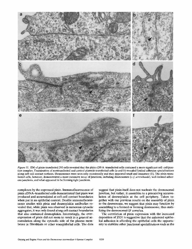
Figure 11. EM of pinin-transfected 293 cells revealed that the pinin cDNA-transfected cells contained a more significant cell-ceU junc- tion complex. Examination of nontransfected and control plasmid-transfected cells (a and b) revealed limited adhesion specializations along cell-cell contact surfaces. Desmosomes were seen only occasionally and they appeared small and immature (b). The pinin-trans- fected cells, however, demonstrated a more extensive array of junctions, including desmosomes (c-f, arrowheads), well-formed adher- ens junctions, and what appeared to be forming tight junctions.
complexes by the expressed pinin. Immunofluorescence of pinin cDNA-transfected cells demonstrated that pinin was produced and accumulated at cell-cell contact boundaries when put in an epithelial context. Double immunofluores- cence studies with pinin and desmoplakin antibodies re- vealed that, while pinin was observed in numerous cytosolic aggregates, it was only found along cell contact boundaries that also contained desmoplakin. Interestingly, the over- expression of pinin did not seem to result in a general ac- cumulation along the cytosolic side of the plasma mem- brane in fibroblasts or other nonepithelial cells. The data
suggest that pinin itself does not nucleate the desmosomal junction, but rather, it assembles to a preexisting accumu- lation of desmoplakin at the cell periphery. Taken to- gether with our previous results on the assembly of pinin to the desmosome, we suggest that pinin may function by assembling to a formed or forming desmosome, thus stabi- lizing the desmosomal-IF complex.
The correlation of pinin expression with the increased deposition of ZO1 is suggestive that the enhanced epithe- lial adhesion is affording the epithelial cells the opportu- nity to stabilize other junctional specializations such as the
Ouyang and Sugrue Pinin and the Desmosome-intermediate Filament Complex 1039

Figure 12. Examination of pinin-transfected cells by immunostaining with tight junction-associated protein ZO1 revealed increased deposition of ZO1 along lateral epithelial borders. Untransfected 293 ceils grown at high cell densities showed little immunostaining for ZO1 (A), whereas pinin-transfected 293 cells showed ZO1 deposits along lateral cell borders (B). Double immunostaining of pinin- transfected ceils for pinin (C) and ZO1 (D) revealed linear ZO1 staining near the more extensive punctate staining for pinin.
tight and adherens junctions. ZO1 has been localized to the zonula adherens and to the tight junction (Itoh et al., 1993); therefore, ZO1 in the transformed cells could be lo- cated at either or both of the junctions. EM revealed an in- crease in both tight and adherens junctions along with the increase in desmosomes. While pinin does not overlap with ZO1 in epithelial tissues in vivo (Ouyang and Sugrue, 1992), it will be of great importance to determine whether or not pinin interacts with ZO1 or other nondesmosomal junctional components in transfectant cells.
The serine block, flanked by numerous kinase recogni- tion motif sites, suggests that at least the carboxyl portion of pinin may serve as a substrate for serine/threonine ki- nases. It has been postulated that phosphorylation may play an important role in cell-cell adhesion (Tsukita et al., 1991; Volberg et al., 1991, 1992, 1994; Takeichi et al., 1992; Stappenbeck et al., 1994), as well as IF and IF-associated protein assembly and function (Nigg et al., 1986; Sihag et al., 1988; Shea et al., 1990; Foisner et al., 1991; Nixon and Sihag, 1991; Peter et al., 1991; Hennekes et al., 1993). In addition, other desmosomal components, such as the
desmoplakins and desmocollins, have been demonstrated to be phosphorylated on serines (Stappenbeck et al., 1994). Citi and co-workers have showed that serine phosphoryla- tion by protein kinase C may be a key step in desmosome disassembly (Citi, 1992; Citi et al,, 1994; Denisenko et al., 1994). In desmosome assembly experiments, we have shown that pinin is recruited to an existing desmosome. If pinin is involved in the maintenance of desmosomal stabil- ity, then it may be reasonable to speculate that phosphory- lation of pinin at key sites may induce a conformational change in the molecule, thus weakening its association to the desmosomal complex and thereby lowering the stabil- ity of the desmosome. It will be of great interest to deter- mine the role of specific phosphorylation of pinin domains in the assembly and disassembly of desmosomes and sta- ble epithelial adhesion.
Here, we have presented data regarding the character- ization of a novel desmosome-associated molecule, pinin. The identification of the cDNA for pinin as genuine was supported by the predicted amino acids that were deduced from the cDNA sequences containing all three tryptic
The Journal of Cell Biology, Volume 135, 1996 1040
fragments derived from the purified protein. In addition, the original 08L mAb reacted with fusion proteins that were expressed in vitro. Moreover, antiserum that was col- lected from animals injected with this recombinant protein identified the 140-kD pinin on Western blots and immuno- stained the lateral epithelial surfaces. Expression of the full-length cDNA clones in 293 cells demonstrated that pinin was produced and assembled along the lateral cell surface, where it was localized near desmoplakin. Further- more, cells receiving pinin cDNA exhibited enhanced cell- cell adhesion. We believe that investigation into the func- tion(s) of pinin and related proteins in cell adhesion and IF organization will contribute significantly to our current knowledge of epithelial cell-cell adhesion.
The authors acknowledge Ms. Summer Carter for technical, photographic, and editorial help.
This work was supported by National Institues of Health grant EY07883 to S.P. Sugrue and National Science Council R.O.C. NSC84- 2331-B-182-50 to P. Ouyang.
Received for publication 5 January 1996 and in revised form 2 September 1996.
References
Angst, B.D., L.A. Nilles, and K.J. Green. 1990. Desmoplakin II expression is not restricted to stratified epithelia. J. Cell Sci. 97:247-257.
Amemann, J., K.H. Sullivan, A.L. Magee, I.A. King, and R.S. Buxton. 1993. Stratification-related expression of isoforms of the desmosomal cadherins in human epidermis. J. Cell. Sci. 104:741-750.
Arnn, J., and L.A. Staehelin. 1981. The structure and function of spot desmo- somes. Int. J. Dermatol. 20(5):330-339.
Bellavite, P., F. Bazzoni, M.A. Cassatella, K.J. Hunter, and J.V. Bannister. 1990. Isolation and characterization of a eDNA clone for a novel serine-rich neutrophil protein. Biochem. Biophys. Res. Commun. 170(2):915-922.
Buxton, R.S., P. Cowin, W.W. Franke, D.R. Garrod, K.J. Green, I.A. King, P.J. Koch, A.L. Magee, D.A. Rees, J.R. Stanley, et aL 1993. Nomenclature of the desmosomal cadherins. J. Cell Biol. 121(3):481-483.
Buxton, R.S., and A.I. Magee. 1992. Structure and interactions of desmosomal and other cadherins. Semin. Cell. Biol. 3(3):157-167.
Chomczynski, P., and N. Sacchi. 1987. Single-step method of RNA isolation by acid guanidinium thiocyanate-phenol-chloroform extraction. Anal, Bio- chem. 162(1):156-159.
Citi, S. 1992. Protein kinase inhibitors prevent junction dissociation induced by low extraceUular calcium in MDCK epithelial cells. Z Cell Biol. 117(1):16%178.
Citi, S., T. Volberg, A.D. Bershadsky, N. Denisenko, and B. Geiger. 1994. Cy- toskeletal involvement in the modulation of cell-cell junctions by the protein kinase inhibitor H-7. J. Cell Sci. 107:683~692.
Collins, J.E., P.K. Legan, T.P. Kenny, J. MacGarvie, J .L Holton, and D.R. Gar- rod. 1991. Cloning and sequence analysis of desmosomal glycoproteins 2 and 3 (desmocollins): cadherin-like desmosomal adhesion molecules with hetero- geneous cytoplasmic domains. Z Cell Biol. 113(2):381-391.
Conway, J.F., and D.A. Parry. 1990. Structural features in the heptad substruc- ture and longer range repeats of two-stranded alpha-fibrous proteins. Int. J. Biol. Macromol. 12(5):328--334.
Cowin, P., H.P. Kapprell, and W.W. Franke. 1985. The complement of desmo- somal plaque proteins in different cell types. J. Cell Biol. 101(4):1442-1454.
Cowin, P., H.P. Kapprell, W.W. Franke, J. Tamkun, and R.O. Hynes. 1986. Pla- koglobin: a protein common to different kinds of intercellular adhering junc- tions. Cell. 46(7):1063-1073.
Denisenko, N., P. Burighel, and S. Citi. 1994. Different effects of protein kinase inhibitors on the localization of junctional proteins at cell-cell contact sites. J. Cell Sci. 107:969-981.
Field, C.M., and B.M. Alberts. 1995. Anillin, a contractile ring protein that cy- cles from the nucleus to the cell cortex. J. Cell Biol. 131(1):165-178.
Foisner, R., P. Traub, and G. Wiche. 1991. Protein kinase A- and protein kinase C-regulated interaction of plectin with lamin B and vimentin. Proc. Natl. Acad. Sci. USA. 88(9):3812-3816.
Garrod, D.R. 1993. Desmosomes and hemidesmosomes. Curr. Opin. Cell Biol. 5(1):30-40.
Graham, F.L, and A.v.d. Eb. 1973. A new technique for the assay of infectivity of human adenovirus 5 DNA. Virology. 52(2):456-467.
Green, K.J., D.A. Parry, P.M. Steinert, M.L. Virata, R.M. Wagner, B.D. Angst, and L.A. Nilles. 1990. Structure of the human desmoplakins. Implications for function in the desmosomal plaque. J. Biol. Chem. 265(19):11406-11407.
Green, K.J., M.L. Virata, G.W. Elgart, J.R. Stanley, and D.A. Parry. 1992. Comparative structural analysis of desmoplakin, bullous pemphigoid anti-
gen and plectin: members of a new gene family involved in organization of intermediate filaments. Int. J. Biol. Macromol. 14(3):145-153.
Hatzfeld, M., G.I. Kristjansson, U. Plessmann, and K. Weber. 1994. Band 6 pro- tein, a major constituent of desmosomes from stratified epithelia, is a novel member of the armadillo multigene family. J. Cell Sci. 107:2259-2270.
Held, H.W., A. Schmidt, R. Zimbelmann, S. Schafer, S. Winter-Simanowski, S. Stumpp, M. Keith, U. Figge, M. Schnolzer, and W.W. Franke. 1994. Cell type-specific desmosomal plaque proteins of the plakoglobin family: plako- philin I (band 6 protein). Differentiation. 58(2):113-131.
Hennekes, H., M. Peter, K. Weber, and E.A. Nigg. 1993. Phosphorylation on protein kinase C sites inhibits nuclear import of lamin B2. J. Cell Biol. 120(6):1293-1304.
Himmler, A., D. Drechsel, M.W. Kirscbner, and D.W. Martin, Jr. 1989. Tau consists of a set of proteins with repeated C-terminal microtubule-binding domains and variable N-terminal domains. Mol. Cell. Biol. 9(4):1381-1388.
Hohl, D., T. Mehrel, U. Lichti, M.L. Turner, D.R. Roop, and P.M. Steinert. 1991. Characterization of human loricrin. Structure and function of a new class of epidermal cell envelope proteins. Z Biol. Chem. 266(10):66264i636.
Holton, J.L., T.P. Kenny, P.K. Legan, J.E. Collins, J.N. Keen, and D.R. Garrod. 1990. Desmosomal glycoproteins 2 and 3 (desmocollins) show N-terminal similarity to calcium-dependent cell-cell adhesion molecules. J. Cell Sci. 113: 381-391.
Kemp, B.E., and R.B. Pearson. 1990. Protein kinase recognition sequence mo- tifs. Trends Biochem. Sci. 15(9):342-346.
Koch, P.J., M.D. Goldschmidt, M.J. Walsh, and R. Zimbelmann. 1991. Amino acid sequence of bovine muzzle epithelial desmocollin derived from cloned cDNA: a novel subtype of desmosomal cadherins. Differentiation. 47(1):29-36.
Kozak, M. 1991. An analysis of vertebrate mRNA sequences: intimations of translational control. J. Cell Blot, 115(4):887-903.
Legan, P.K., J.E. Collins, and D.R. Garrod. 1992. The molecular biology of des- mosomes and hemidesmosomes: what's in a name? Bioessays. 14(6):385-393.
Levitt, N., D. Briggs, A. Gil, and N.J. Proudfoot. 1989. Definition of an efficient synthetic poly(A) site. Genes Dev. 3(7):1019-1025.
Mack, J.W., A.C. Steven, and P.M. Steinert. 1993. The mechanism of interac- tion of filaggrin with intermediate filaments. The ionic zipper hypothesis. J. Mol. Biol. 232:50-66.
Mueller, H., and W.W. Franke. 1983. Biochemical and immunological charac- terization of desmoplakins I and II, the major polypeptides of the desmo- somal plaque. Z Mot. Biol. 163(4):647~671.
Nigg, E.A., B.M. Sefton, S.J. Singer, and P.K. Vogt. 1986. Cytoskeletal organi- zation, vinculin-phosphorylation, and fibronectin expression in transformed fibroblasts with different cell morphologies. Virology. 151(1):50-65.
Nixon, R.A., and R.K. Sihag. 1991. Neurofilament phosphorylation: a new look at regulation and function. Trends Neurosci. 14( 11 ):501-506.
O'Shea, E.K., R. Rutkowski, and P.S. Kim. 1989. Evidence that the leucine zip- per is a coiled coil. Science (Wash. DC). 243(4890):538-542.
Oas, T.G, LP. Mclntosh, E.K. O'Shea, F.W. Dahlquist, and P.S. Kim. 1990. Secondary structure of a leucine zipper determined by nuclear magnetic res- onance spectroscopy. Biochemistry. 29(12):2891-2894.
Ouyang, P., and SP. Sugrue. 1992. Identification of an epithelial protein related to the desmosome and intermediate filament network. J. Cell Biol. 118(6): 1477-1488.
Parrish, E.P., D.R. Garrod, D.L. Mattey, L. Hand, P.V. Steart, and R.O. Weller. 1986. Mouse antisera specific for desmosomal adhesion molecules of suprabasal skin cells, meninges, and meningioma. Proc. Natl. Acad. Sci. USA. 83(8):2657-2661.
Peter, M., E. Heitlinger, M. Haner, U. Aebi, and E.A. Nigg. 1991. Disassembly of in vitro formed lamin head-to-tail polymers by CDC2 kinase. E M B O (Eur Mol. BioL Organ.) J. 10(6):1535-1544.
Rothnagel, J.A., and P.M. Steinert. 1990. The structure of the gene for mouse filaggrin and a comparison of the repeating units. J. Biol. Chem. 265(4): 1862-1865.
Rothnagel, J.A., T. Mehrel, W.W. Idler, D.R. Roop, and P.M. Steinert. 1987. The gene for mouse epidermal filaggrin precursor. Its partial characteriza- tion, expression, and sequence of a repeating filaggrin unit. J. Biol. Chem. 262(32):15643-15648.
Shea, T.B., R.K. Sihag, and R.A. Nixon. 1990. Dynamics of phosphorylation and assembly of the high molecular weight neurofilament subunit in NB2al dl neuroblastoma. J. Neurochem. 55(5):1784-1792.
Sihag, R.K., A.Y. Jeng, and R.A. Nixon. 1988. Phosphorylation of neurofila- ment proteins by protein kinase C. FEBS Lett. 233(1):181-185.
Skalli, O., J.C. Jones, R. Gagescu, and R.D. Goldman. 1994. IFAP 300 is com- mon to desmosomes and hemidesmosomes and is a possible linker of inter- mediate filaments to these junctions. J. Cell Biol. 125(1):159-170.
Staehelin, L.A. 1974. Structure and function of intercellular junctions. Int. Rev. Cytol. 39(191):191-283.
Stappenbeck, T.S., J.A. Lamb, C.M. Corcoran, and K.J. Green. 1994. Phosphor- ylation of the desmoplakin COOH terminus negatively regulates its interac- tion with keratin intermediate filament networks. J. Biol. Chem. 269(47): 29351-29354.
Steinert, P.M., J.W. Mack, B.P. Korge, S.Q. Gan, S.R. Haynes, and A.C. Steven. 1991. Glycine loops in proteins: their occurrence in certain interme- diate filament chains, loricrins and single-stranded RNA binding proteins. Int. J. BioL Macromol. 13(3):130-139.
Takeichi, M., S. Hirano, N. Matsuyoshi, and T. Fujimori. 1992. Cytoplasmic
Ouyang and Sugrue Pinin and the Desmosome-intermediate Filament Complex 1041
control of eadherin-mediated cell-cell adhesion. Cold Spring Harb Syrup. Quant. BioL 57(327):327-334.
Theis, D.G., P.J. Koch, and W.W. Franke. 1993. Differential synthesis of type 1 and type 2 desmocollin mRNAs in human stratified epithelia. Int. J. Dev. Biol. 37(1):101-110.
Tsukita, S., K. Oishi, T. Akiyama, Y. Yamanashi, T. Yamamoto, and S. Tsukita. 1991. Specific proto-oncogenie tyrosine kinases of sre family are enriched in cell-to-cell adherens junctions where the level of tyrosine phosphorylation is elevated. J. Cell Biol. 113(4):867--879.
Tsukita, S., and S. Tsukita. 1985. Desmocalmin: a ealmodulin-binding high mo- lecular weight protein isolated from desmosomes. J. Cell Biol. 101(6):2070-- 2080.
Volberg, T., B. Geiger, S. Citi, and A.D. Bershadsky. 1994. Effect of protein ki- nase inhibitor H-7 on the contractility, integrity, and membrane anchorage of the microfilament system. Cell MotiL Cytoskeleton. 29(4):321-338.
Volberg, T., B. Geiger, R. Dror, and Y. Zick. 1991. Modulation of intercellular adherens-type junctions and tyrosine phosphorylation of their components in RSV-transformed cultured chick lens cells. Cell Regulation. 2(2):105-120.
Volberg, T., Y. Zick, R. Dror, I. Sabanay, C. Gilon, A. Levizki, and B. Geiger. 1992. The effect of tyrosine-specific protein phosphorylation on the assem- bly of adherens-type junctions. EMBO (Eur. Mol. Biol. Organ.) J. 11(5): 1733-1742.
Wheeler, G.N., A.E. Parker, C.L. Thomas, P. Ataliotis, D. Poynter, J. Arne- mann, A.J. Rutman, S.C. Pidsley, F.M. Watt, D.A. Rees, et al. 1991. Desmo- somal glycoprotein DGI, a component of intercellular desmosome junctions, is related to the cadherin family of cell adhesion molecules. Proc. Natl. Acad. Sci. USA. 88(11):4796~800.
Wiche, G., B. Beeker, K. Luber, G. Weitzer, MJ. Castanon, R. Hauptmann, C. Stratowa, and M. Stewart. 1991. Cloning and sequencing of rat plectin indi- cates a 466-kD polypeptide chain with a three-domain structure based on a central a-helical coiled coil. Z Cell Biol. 114(1):83-99.
Wiehe, G., D. Gromov, A. Donovan, M.J. Castanon, and E. Fuchs. 1993. Ex- pression of pleetin mutant eDNA in cultured cells indicates a role of COOH- terminal domain in intermediate filament association. J. Cell Biol. 121(3): 607--619.
The Journal of Cell Biology, Volume 135, 1996 1042