Characterization of Delhi iron pillar rust by X-ray diffraction, Fourier transform infrared...
17
Characterization of Delhi iron pillar rust by X-ray diraction, Fourier transform infrared spectroscopy and Mo¨ssbauer spectroscopy R. Balasubramaniam a, *, A.V. Ramesh Kumar b a Department of Materials and Metallurgical Engineering, Indian Institute of Technology, Kanpur 208 016, India b Electrochemistry and Corrosion Division, Defense Materials, Stores Research and Development Establishment, Kanpur 208 013, India Received 19 February 1999; accepted 21 March 2000 Abstract Rust samples obtained from the region just below the decorative bell capital of the Delhi iron pillar (DIP) have been analyzed by X-ray diraction (XRD), Fourier transform infrared spectroscopy (FTIR) and Mo¨ssbauer spectroscopy. The identification of iron hydrogen phosphate hydrate in the crystalline form by XRD was unambiguous. Very weak diraction from the oxyhydroxides/oxides of iron was observed indicating that these phases are most likely to be present in the amorphous form in the rust. The present XRD analysis of rust obtained from an inaccessible area of the DIP has also been compared with earlier analyses of DIP rust obtained from regions accessible to the public. FTIR indicated that the constituents of the scale were g-, a-, d-FeOOH, Fe 3x O 4 and phosphate, and that the scale was hydrated. The unambiguous identification of the iron oxides/oxyhydroxides in the FTIR spectrum implied that they are present in the amorphous state, as XRD did not reveal these phases. The FTIR results have also been compared with earlier FTIR spectroscopic results of atmospheric rust formation. Mo¨ssbauer spectroscopy indicated that the rusts contained g-FeOOH, superparamagnetic a-FeOOH, d-FeOOH and magnetite, all in the amorphous form. The Mo¨ssbauer spectrum also confirmed that iron in the crystalline iron hydrogen phosphate hydrate, whose presence was confirmed by XRD, was in the ferric state indicating that it was a stable end corrosion product. 7 2000 Elsevier Science Ltd. All rights reserved. 0010-938X/00/$ - see front matter 7 2000 Elsevier Science Ltd. All rights reserved. PII: S0010-938X(00)00045-7 Corrosion Science 42 (2000) 2085–2101 www.elsevier.com/locate/corsci * Corresponding author.
Transcript of Characterization of Delhi iron pillar rust by X-ray diffraction, Fourier transform infrared...

Characterization of Delhi iron pillar rust byX-ray di�raction, Fourier transform infraredspectroscopy and MoÈ ssbauer spectroscopy
R. Balasubramaniama,*, A.V. Ramesh Kumarb
aDepartment of Materials and Metallurgical Engineering, Indian Institute of Technology, Kanpur 208 016,
IndiabElectrochemistry and Corrosion Division, Defense Materials, Stores Research and Development
Establishment, Kanpur 208 013, India
Received 19 February 1999; accepted 21 March 2000
Abstract
Rust samples obtained from the region just below the decorative bell capital of the Delhiiron pillar (DIP) have been analyzed by X-ray di�raction (XRD), Fourier transforminfrared spectroscopy (FTIR) and MoÈ ssbauer spectroscopy. The identi®cation of ironhydrogen phosphate hydrate in the crystalline form by XRD was unambiguous. Very weak
di�raction from the oxyhydroxides/oxides of iron was observed indicating that these phasesare most likely to be present in the amorphous form in the rust. The present XRD analysisof rust obtained from an inaccessible area of the DIP has also been compared with earlier
analyses of DIP rust obtained from regions accessible to the public. FTIR indicated thatthe constituents of the scale were g-, a-, d-FeOOH, Fe3ÿxO4 and phosphate, and that thescale was hydrated. The unambiguous identi®cation of the iron oxides/oxyhydroxides in the
FTIR spectrum implied that they are present in the amorphous state, as XRD did notreveal these phases. The FTIR results have also been compared with earlier FTIRspectroscopic results of atmospheric rust formation. MoÈ ssbauer spectroscopy indicated that
the rusts contained g-FeOOH, superparamagnetic a-FeOOH, d-FeOOH and magnetite, allin the amorphous form. The MoÈ ssbauer spectrum also con®rmed that iron in the crystallineiron hydrogen phosphate hydrate, whose presence was con®rmed by XRD, was in the ferricstate indicating that it was a stable end corrosion product. 7 2000 Elsevier Science Ltd. All
rights reserved.
0010-938X/00/$ - see front matter 7 2000 Elsevier Science Ltd. All rights reserved.
PII: S0010 -938X(00)00045 -7
Corrosion Science 42 (2000) 2085±2101
www.elsevier.com/locate/corsci
* Corresponding author.
Keywords: Delhi iron pillar; Rust characterization; X-ray di�raction; Fourier transform infrared
spectroscopy; MoÈ ssbauer spectroscopy; Phosphate; Amorphous iron oxyhydroxides
1. Introduction
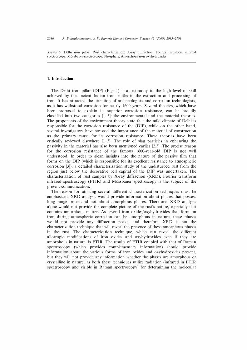
The Delhi iron pillar (DIP) (Fig. 1) is a testimony to the high level of skillachieved by the ancient Indian iron smiths in the extraction and processing ofiron. It has attracted the attention of archaeologists and corrosion technologists,as it has withstood corrosion for nearly 1600 years. Several theories, which havebeen proposed to explain its superior corrosion resistance, can be broadlyclassi®ed into two categories [1±3]: the environmental and the material theories.The proponents of the environment theory state that the mild climate of Delhi isresponsible for the corrosion resistance of the (DIP), while on the other hand,several investigators have stressed the importance of the material of constructionas the primary cause for its corrosion resistance. These theories have beencritically reviewed elsewhere [1±3]. The role of slag particles in enhancing thepassivity in the material has also been mentioned earlier [2,3]. The precise reasonfor the corrosion resistance of the famous 1600-year-old DIP is not wellunderstood. In order to glean insights into the nature of the passive ®lm thatforms on the DIP (which is responsible for its excellent resistance to atmosphericcorrosion [3]), a detailed characterization study of the undisturbed rust from theregion just below the decorative bell capital of the DIP was undertaken. Thecharacterization of rust samples by X-ray di�raction (XRD), Fourier transforminfrared spectroscopy (FTIR) and MoÈ ssbauer spectroscopy is the subject of thepresent communication.
The reason for utilizing several di�erent characterization techniques must beemphasized. XRD analysis would provide information about phases that possesslong range order and not about amorphous phases. Therefore, XRD analysisalone would not provide the complete picture of the rust's nature, especially if itcontains amorphous matter. As several iron oxides/oxyhydroxides that form oniron during atmospheric corrosion can be amorphous in nature, these phaseswould not provide any di�raction peaks, and therefore, XRD is not thecharacterization technique that will reveal the presence of these amorphous phasesin the rust. The characterization technique, which can reveal the di�erentallotropic modi®cations of iron oxides and oxyhydroxides even if they areamorphous in nature, is FTIR. The results of FTIR coupled with that of Ramanspectroscopy (which provides complementary information) should provideinformation about the various forms of iron oxides and oxyhydroxides present,but they will not provide any information whether the phases are amorphous orcrystalline in nature, as both these techniques utilize radiation (infrared in FTIRspectroscopy and visible in Raman spectroscopy) for determining the molecular
R. Balasubramaniam, A.V. Ramesh Kumar / Corrosion Science 42 (2000) 2085±21012086

vibration (due to bending and stretching of the bonds) between iron and oxygen[4]. As the molecular vibration in all known iron oxides and hydroxides are wellcharacterized and available in the literature, comparison of the FTIR spectra fromthe DIP rust with standards would provide the exact nature of the phases of ironoxides and oxyhydroxides present. IR spectroscopy has been successfully utilized
Fig. 1. The Delhi iron pillar before the construction of the iron grill cage around the stone platform.
R. Balasubramaniam, A.V. Ramesh Kumar / Corrosion Science 42 (2000) 2085±2101 2087

to identify iron oxide/oxyhydroxide phases under atmospheric corrosion [5,6] andaqueous corrosion [7] conditions.
MoÈ ssbauer spectroscopy can be used to obtain further insights on the structural(i.e. amorphous or crystalline) and chemical aspects of the rusts. MoÈ ssbauerspectroscopy is a powerful tool to understand the amorphous versus crystallinitycontroversy in the case of oxides/oxyhydroxides [8]. In case the phase thatconstitutes the rust is crystalline, it would lead to alignment of magnetic ®elds inthe individual grains of the phase and provide a characteristic hyper®ne splittingwith characteristic magnetic ®elds. For example, in the case of crystallinemagnetite, two sextets (10-line pattern) are to be expected in the MoÈ ssbauerspectrum at ambient temperatures. However, in case this phase is amorphous, themagnetic ®elds will not be aligned in the individual grains and this would result incollapse of magnetic ®elds or as a result of which a doublet would be obtainedinstead of a sextet [8±10]. A collapsed sextet would indicate the ®nenanocrystalline nature of magnetite. Therefore, MoÈ ssbauer spectroscopy can beadvantageously utilized to probe the crystal nature (i.e. amorphous or crystalline)of the phase. The use of MoÈ ssbauer spectroscopy for elucidation of the chemicalnature of corrosion products is also well established [8,11±13]. Another advantageof utilizing MoÈ ssbauer spectroscopy is that the asymmetry in any peak of centraldoublet indicates the contribution due to some other species (phase). For example,Fe2+ ions show high positive value for isomer shift and quadrupole splittingresulting in asymmetry in the central doublet in which a higher intense ®rst peak(from left) is obtained. The second peak of ferrous doublet will be in the morepositive region of the spectrum [14].
2. Experimental
Rust samples were collected using a plastic scraper from several di�erentlocations in the region just below the decorative bell capital (Fig. 2(a)).Observation of the same region, nearly a year after the samples were collected(Fig. 2(b)), did not reveal any evidence for rust removal, thereby indicating thatthe passive ®lm is self-healing. This is the region where the rust layer on theexposed surface of the pillar is maximum [15] and therefore, this allowed thecollection of a signi®cant amount of rust suitable for characterization by severaltechniques. Moreover, this is the region where the rust is the oldest as this portionof the pillar is inaccessible to the public. A portion of the rust sample collectedfrom each location was ®rst ground into ®ne powder and mounted in between twothin polymer foils. A part of the same ground powder was also analyzed usingFTIR. The polymer foil containing the DIP rust was used for MoÈ ssbauerspectroscopic analysis of the phases present.
The polymer foils were mounted in a di�ractometer such that the ¯at foil wasplaced horizontally and the incident beam could sample the specimen through thewhole range of 2y studied. Several di�raction patterns were obtained using Cu Ka
radiation at di�erent scan speeds and scanning angle steps. The present paper
R. Balasubramaniam, A.V. Ramesh Kumar / Corrosion Science 42 (2000) 2085±21012088
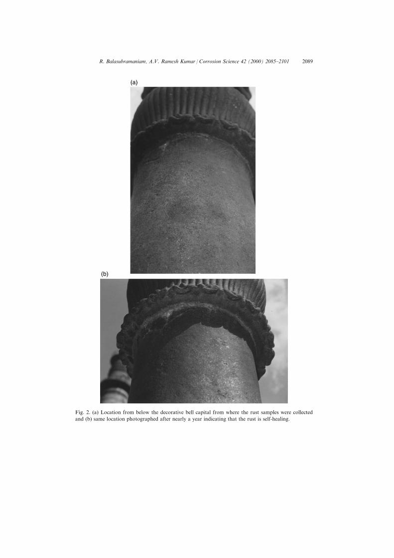
Fig. 2. (a) Location from below the decorative bell capital from where the rust samples were collected
and (b) same location photographed after nearly a year indicating that the rust is self-healing.
R. Balasubramaniam, A.V. Ramesh Kumar / Corrosion Science 42 (2000) 2085±2101 2089
reports the di�raction pattern taken at a scan rate of 38 per min (i.e. 0.058 per s),with the scan being conducted between 2y � 10±808 in steps of 0.058.
A small portion of the rust was ®nely ground and then pressed (in vacuum) inthe form of a disc using spectroscopically pure dry KBr. The FTIR spectrum wasrecorded at room temperature using a Nicolet Magna 750 Series 2, USA FTIRsystem.
In order to study the rust by MoÈ ssbauer spectroscopy, the polymer foils weremounted on a sample holder of a MoÈ ssbauer spectrometer in transmissiongeometry. The spectrometer controller model was S-600 of Austin ScienceAssociates, USA with linear motor of ASA, USA which was coupled with aNorland Ortec Multichannel analyzer and proportional counter (Kr at 1 atm and3% CO2) manufactured by Ranger Corporation, USA and these were used tocount the 14.4 KeV g-ray. The source used was 400 MBq 57Co source inPalladium matrix (Amersham, UK). The MoÈ ssbauer spectrum was recorded in a¯y back mode at room temperature in 1024 channels, which gives a singlespectrum. The MoÈ ssbauer data was ®tted to Lorentzian approximation using mm/sirus v.2.7 MoÈ ssbauer data handling program [16]. The MoÈ ssbauer parametersreported are with respect to a-Fe standard.
3. Results and discussion
3.1. X-ray di�raction
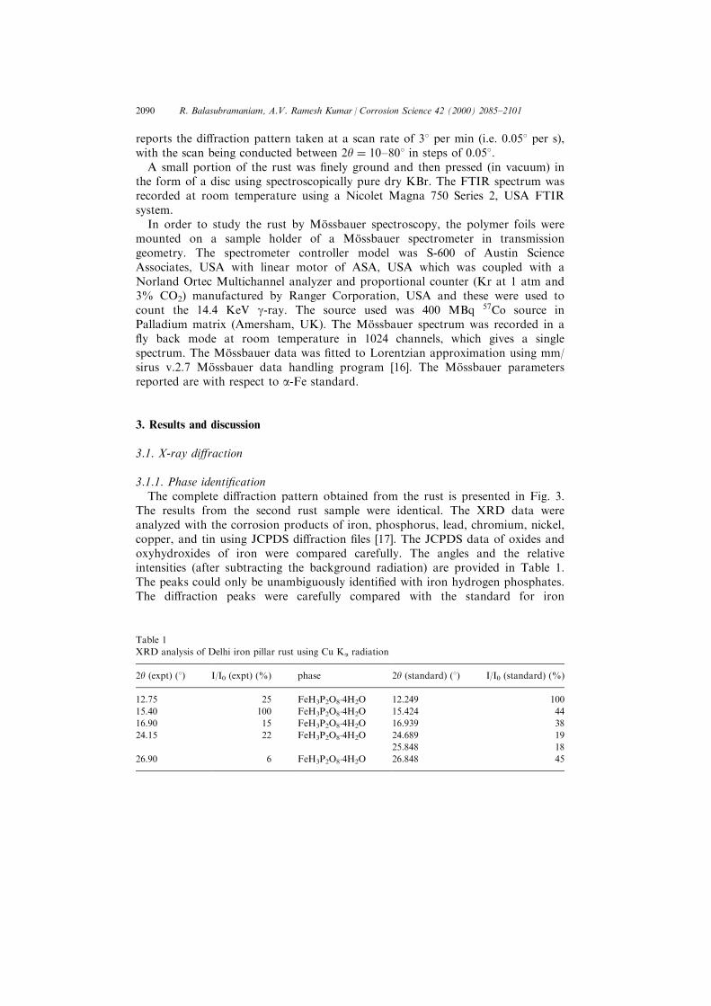
3.1.1. Phase identi®cationThe complete di�raction pattern obtained from the rust is presented in Fig. 3.
The results from the second rust sample were identical. The XRD data wereanalyzed with the corrosion products of iron, phosphorus, lead, chromium, nickel,copper, and tin using JCPDS di�raction ®les [17]. The JCPDS data of oxides andoxyhydroxides of iron were compared carefully. The angles and the relativeintensities (after subtracting the background radiation) are provided in Table 1.The peaks could only be unambiguously identi®ed with iron hydrogen phosphates.The di�raction peaks were carefully compared with the standard for iron
Table 1
XRD analysis of Delhi iron pillar rust using Cu Ka radiation
2y (expt) (8) I/I0 (expt) (%) phase 2y (standard) (8) I/I0 (standard) (%)
12.75 25 FeH3P2O8�4H2O 12.249 100
15.40 100 FeH3P2O8�4H2O 15.424 44
16.90 15 FeH3P2O8�4H2O 16.939 38
24.15 22 FeH3P2O8�4H2O 24.689 19
25.848 18
26.90 6 FeH3P2O8�4H2O 26.848 45
R. Balasubramaniam, A.V. Ramesh Kumar / Corrosion Science 42 (2000) 2085±21012090

phosphate (FePO4�2H2O Ð ®le number 33-0667) and several other iron hydrogenphosphate hydrates (FeH3P2O8�4H2O Ð ®le number 45-0500, FeH6P3O9�H2O Юle number 31-0623, FeH2P3O10�(3/2)H2O Ð ®le number 28-0486 andFeH3P2O6�3H2O Ð ®le number 31-0624) [17]. All the peaks could be identi®edwith the phase FeH3P2O8�4H2O. The 2y values and corresponding intensity of thepeaks from the standard di�raction ®le of FeH3P2O8�4H2O are also provided inTable 1. It is seen that the high intensity peak of the phase FeH3P2O8�4H2Oappears as a signi®cant peak at 2y of 12.758. The experimental peak occurring at2y of 15.408 is again a signi®cant peak of the phase FeH3P2O8�4H2O. The possiblereasons for the experimentally determined peak at 15.408 being the highestintensity peak rather than the peak at 12.758 are (a) di�erent degrees of hydration,(b) slight di�erences in composition, and (c) orientation e�ects in the DIP rustsample as compared to the standard. Contribution to the 100% experimental peak(at 2y of 15.408) due to di�raction from other planes is also a possibility as thestandard pattern shows the presence of three di�raction peaks clustered very nearto this angle. A similar situation is obtained for the di�raction peak occurring at2y of 24.158, as there are two signi®cant peaks (of theoretical intensities 19 and18%) of the phase FeH3P2O8�4H2O occurring near this di�raction peak (Table 1).Therefore, there is no ambiguity in the identi®cation of the iron hydrogenphosphate hydrate phase (FeH3P2O8�4H2O) in the DIP rust as the experimentally
Fig. 3. X-ray di�raction pattern of the DIP rust showing the intensities in the region 2y � 10ÿ 808:
R. Balasubramaniam, A.V. Ramesh Kumar / Corrosion Science 42 (2000) 2085±2101 2091

obtained relative intensities can also be matched to the standard JCPDSdi�raction data of this phase.
As the rust obtained is very old, it is not surprising that the stable crystallineform of iron hydrogen phosphate hydrate [18] was identi®ed. However, it shouldbe borne in mind that the presence of the precursor to this phase, i.e. Fe3(PO4)2 inthe rust could not be ruled out as it is amorphous [18] in nature and would nothave provided di�raction peaks. Its relative amount is not important as it is theprecursor to the much more stable crystalline form of iron hydrogen phosphatehydrate, which has been unambiguously identi®ed in the rust by XRD.
The presence of FeH3P2O8�4H2O is also theoretically favored as it is composedof FePO4 and H3PO4, whose standard free energies for formation is extremelynegative at room temperature. At 298.15 K and 1 bar pressure, the standard freeenergy of formation of FePO4�2H2O is ÿ1657.5 kJ/mol, while that for crystallineH3PO4 is ÿ1119.2 kJ/mol [19]. In comparison, the standard free energies offormation of the oxides and oxyhydroxides of iron are much higher [19]. On thetopic of thermodynamics, it is interesting to note the experimental result ofUrasova et al. [20] who found that phosphate is stable even at P concentration aslow as 0.24 wt% in the Fe-P-O system. Therefore, the identi®cation of ironhydrogen phosphate hydrate is reliable as the laws of thermodynamics are exactand conclusions based on thermodynamics are sound and reliable.
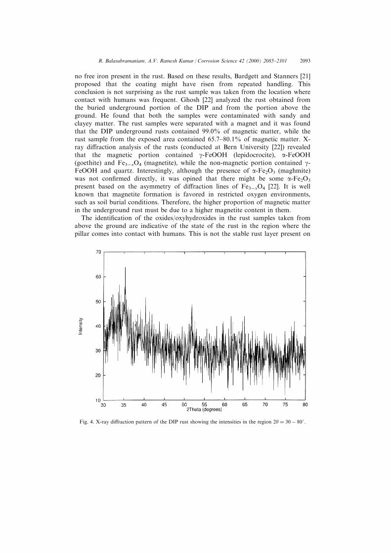
From the above analysis, it would apparently appear that the rust of the ironpillar is composed of only iron phosphates. This may not be true as very carefulanalysis of the XRD pattern revealed weak peak(s) in the region of 2y � 30ÿ 358(Fig. 4). When the di�raction data of the several oxides and oxyhydroxides ofiron were analyzed, it was noticed that the maximum intensity peaks in the case ofseveral iron oxides and oxyhydroxides are concentrated in the 2y range between30 and 358. It, therefore, appears from the very weak di�raction intensitiesobtained in this range that there should be a very small amount of crystalline ironoxides or oxyhydroxides present in the rust.
3.1.2. Comparison with earlier XRD studiesEarlier XRD studies of rusts from the DIP are summarized below in order to
compare the present results with them. The rust samples that were obtained in thepresent case were taken from the surface of the pillar just below the decorativebell capital and this rust must be the oldest rust on the pillar. Moreover, this rusthas been undisturbed because this location of the pillar is inaccessible to thepublic. Therefore, this important aspect must be borne in mind while viewing theearlier rust analyses as all of them were conducted on rusts obtained from thelower regions of the pillar. The rust forming in the lower regions cannot be in theundisturbed condition because of the custom of the visitors who try to encircletheir arms around the pillar with their backs against the pillar in the belief that ifsuch an act can be successfully completed, it brings good luck.
A minute sample of the rust scraped from the area about 5 feet from theground was found essentially amorphous and contained clayey and siliceousmatter [21]. X-ray ¯uorescent analysis of the same rust con®rmed that there was
R. Balasubramaniam, A.V. Ramesh Kumar / Corrosion Science 42 (2000) 2085±21012092
no free iron present in the rust. Based on these results, Bardgett and Stanners [21]proposed that the coating might have risen from repeated handling. Thisconclusion is not surprising as the rust sample was taken from the location wherecontact with humans was frequent. Ghosh [22] analyzed the rust obtained fromthe buried underground portion of the DIP and from the portion above theground. He found that both the samples were contaminated with sandy andclayey matter. The rust samples were separated with a magnet and it was foundthat the DIP underground rusts contained 99.0% of magnetic matter, while therust sample from the exposed area contained 65.7±80.1% of magnetic matter. X-ray di�raction analysis of the rusts (conducted at Bern University [22]) revealedthat the magnetic portion contained g-FeOOH (lepidocrocite), a-FeOOH(goethite) and Fe3ÿxO4 (magnetite), while the non-magnetic portion contained g-FeOOH and quartz. Interestingly, although the presence of a-Fe2O3 (maghmite)was not con®rmed directly, it was opined that there might be some a-Fe2O3
present based on the asymmetry of di�raction lines of Fe3ÿxO4 [22]. It is wellknown that magnetite formation is favored in restricted oxygen environments,such as soil burial conditions. Therefore, the higher proportion of magnetic matterin the underground rust must be due to a higher magnetite content in them.
The identi®cation of the oxides/oxyhydroxides in the rust samples taken fromabove the ground are indicative of the state of the rust in the region where thepillar comes into contact with humans. This is not the stable rust layer present on
Fig. 4. X-ray di�raction pattern of the DIP rust showing the intensities in the region 2y � 30ÿ 808:
R. Balasubramaniam, A.V. Ramesh Kumar / Corrosion Science 42 (2000) 2085±2101 2093

the pillar. However, the above observation provide information about the rustlayer in the region that was in human contact. Interestingly, a sample was cutfrom the bottom exposed region of the pillar for metallographic analysis [22] andthe rust formed on this freshly exposed surface after 1.5 years of exposure wasstudied by XRD by Lahiri et al. [23]. They identi®ed g-FeOOH (lepidocrocite)and a-FeOOH (goethite) in the rust, with the major phase being g-FeOOH(lepidocrocite) based on the di�raction intensities. While the above studies provideinformation about the state of the rust in the ground level region of the pillar, theXRD results provided in the present study provides information about the state ofthe stable oldest rust on the pillar. Nevertheless, the information from the abovestudies can be utilized to construct the sequence of events leading to the formationof the protective passive ®lm on the DIP [24].
3.2. Fourier transform infrared spectroscopy
It is established that IR spectroscopy can be utilized to understand the processof rusting of steels. Misawa et al. [5,6] studied the mechanism of atmosphericrusting and the protective amorphous rust on low allow steel by using IRabsorption spectroscopy. It has been pointed out by them that the absorptionband at higher wavenumber region is due to OH stretching and at lowerwavenumber is because of Fe±O lattice vibration. Misawa et al. [5,6] attributedthe following peaks as key absorption bands: 890 cmÿ1 for a-FeOOH, 1020 cmÿ1
for g-FeOOH, 580 cmÿ1 for magnetite and 470 cmÿ1 for d-FeOOH. Ishii et al.[25] also con®rmed that the Fe±O stretching vibration in Fe3ÿxO4 corresponds toa wave number 570 cmÿ1. Moreover, they also showed that if this peak is sharp,it indicates the purity level and the prevalence of very small amount of defects[25]. Raman et al. [26] applied IR absorption spectroscopy to the study ofatmospheric rust and they have provided standard spectra for di�erent phases.Music et al. [7] used Fourier transform infrared spectroscopy for the analysis ofrust formed by corrosion of steel in aqueous solutions. Yamashita et al. [27]studied the rust layer, formed on weathering steel for a quarter of a century in anindustrial region, by IR absorption spectroscopy where the bands obtained wererelatively broad.
3.2.1. Phase identi®cationThe FTIR spectrum obtained from one DIP rust sample is provided in Fig. 5.
The spectrum obtained from another rust sample was identical and therefore isnot presented. In order to precisely identify the peaks that are due to KBr andother IR active molecules (i.e. control specimen) present in the atmosphere, thespectrum was recorded at ambient room atmosphere and not in an inertatmosphere. The FTIR spectrum of the control sample is provided in Fig. 6. Thefollowing discussion will address the identi®cation of phases present in the DIPrust based on the two spectra presented above.
On comparing the DIP rust and control spectra, it is clear that the peaks thatoccur in the control sample in the region around 1500, 2920 and 2850 cmÿ1 arise
R. Balasubramaniam, A.V. Ramesh Kumar / Corrosion Science 42 (2000) 2085±21012094

from C=O and O±H stretchings [4,28] which are present in the atmosphere.When these peaks are eliminated from analysis in the DIP rust FTIR spectrum,only certain other signi®cant peaks require to be identi®ed. The broad band seenin the region 3000±3500 cmÿ1 is due to hydration of the rust. It is known that O±H stretching leads to a strong peak between 3000 and 3700 cmÿ1, while O±Hbending to a medium band between 1200 and 1500 cmÿ1 [4,28]. Therefore, thisimplies hydration of corrosion product(s) in the DIP rust.
It can be noted from Fig. 5 that the presence of g-FeOOH, a-FeOOH and d-FeOOH is con®rmed by the appearance of peaks at 1020 cmÿ1 (g-FeOOH), 890cmÿ1 (a-FeOOH) and 470 cmÿ1 (d-FeOOH) [5,6]. The DIP rust FTIR spectrumalso reveals that d-FeOOH is the major component of the rust as the peak isrelatively intense compared to the others. This phase forms due to catalytic actionof elements like Cu, P, and Ni that are added for weathering resistance [5].Interestingly, the peak due to magnetite (Fe3ÿxO4) was present at 580 cmÿ1. Thisindicates the relatively more protective nature of the DIP rust. It is known thatmagnetite formed during atmospheric corrosion will be non-stoichiometric and asthe non-stoichiometry increases, a rapid decrease in electrical conductivity isobserved in turn due to localization of electrons to regions encompassing availableoctahedral Fe pairs [29]. The present FTIR results of the DIP rust are compared
Fig. 5. FTIR spectrum from the DIP rust.
R. Balasubramaniam, A.V. Ramesh Kumar / Corrosion Science 42 (2000) 2085±2101 2095

with that of the rust on a Gupta-period corrosion resistant iron clamp, which wasanalyzed earlier [30]. The FTIR spectrum from the Eran rust (Figure 3 of Ref.[30]) revealed characteristic peaks due to g-, a- and d-FeOOH. Moreover, theexistence of magnetite (that was likely doped with P ions) in the crystalline formwas identi®ed in contrast to the amorphous nature of magnetite in DIP's rust. Inorder to appreciate this di�erence, it is important to keep in mind theenvironmental conditions in which the Eran rust and DIP rust were obtained. TheEran iron clamp was removed from a stone block where it was buried inside. Thestone block was part of the oldest temple at Eran which was built byChandragupta II Vikramaditya [31], the Chandra mentioned in the DIP's oldestSanskrit inscription [32]. The major di�erence between the Eran and DIP rust wasthat the former was obtained from a restricted environment in a stone block(thereby implying restricted oxygen supply to the surface of the iron clamp)whereas, the rust on the DIP was obtained from a surface exposed to theatmosphere. Under the condition of restricted oxygen supply, it is well establishedthat the rust that forms on iron is magnetite. Therefore, it is not surprising thatmagnetite was obtained in the crystalline form in the Eran iron clamp. On theother hand, the DIP rust is exposed to the environment where it undergoesconstant wetting and drying cycles and in such a case, it is observed that the
Fig. 6. FTIR spectrum from control specimen.
R. Balasubramaniam, A.V. Ramesh Kumar / Corrosion Science 42 (2000) 2085±21012096

magnetite in the rust is amorphous in nature. The reason for the presence ofmagnetite in the amorphous form has been addressed elsewhere [24].
An interesting aspect that was found from the FTIR spectrum was that therewas a distinct band resulting from the presence of some phosphate phase(s) in theregion 1250±900 cmÿ1 [4]. The wavenumber for ionic phosphate is 1030±1120cmÿ1, while for covalent phosphate the band should occur at 920±1050 cmÿ1 andfor P=O bonds, the spectra occur at 1200±1250 cmÿ1 [4,28]. Although it isdi�cult to tell whether the ionic phosphate that is providing the signal in thespectra is due to H3PO4 or FePO4 or both, FTIR spectroscopy, nevertheless,®rmly proves the existence of phosphates in the DIP rust.
3.3. MoÈssbauer spectroscopy
MoÈ ssbauer spectra of the rust samples obtained from two di�erent locations inthe region just below the decorative bell capital of the DIP are presented in Figs.7 and 8. The continuous line in the spectra in these ®gures are computer ®tted.The dots are experimental points. On comparing the MoÈ ssbauer spectra in Figs. 7and 8, it is evident that both the rust samples exhibit a similar nature becauseboth exhibit a collapsed sextet and a central doublet. As the results from two rustsamples (taken from di�erent locations in the top regions of the pillar) wereidentical, the validity of the characterization is established. Calculation ofMoÈ ssbauer parameters showed a low value for isomer shift (IS) i.e. 0.08 mm/s inthe ®rst sample and 0.15 mm/s in the second sample. However, quadrupolesplitting (QS) values (i.e. 0.55 mm/s and 0.58 mm/s for samples 1 and 2,
Fig. 7. MoÈ ssbauer spectrum obtained from the ®rst DIP rust sample.
R. Balasubramaniam, A.V. Ramesh Kumar / Corrosion Science 42 (2000) 2085±2101 2097

respectively) matched with literature values reported for oxyhydroxides [12±14].Ferric phosphate shows an IS value of about 0.30 mm/s [33]. The deviation fromthe standard reported valued of isomer shifts could be due to the modi®edenvironment of the species responsible for the doublet. FTIR showedcharacteristic peaks for g-FeOOH, a-FeOOH and d-FeOOH indicating that theywere in the pure form. However, a broad band was obtained for magnetite andphosphate indicating that they either exist in a di�erent form or are incorporatedwith ions [26]. The sextet was tried to be ®t for a-FeOOH with a magnetic ®eld of313 KOe �IS � 0:40 mm/s and QS � 0:09 mm/s [34]) but good ®tting could not beobtained. Therefore, the collapsed sextet is due to magnetite that is in ®nenanocrystalline or amorphous form [35].
In order to understand the nature of the rust, the MoÈ ssbauer spectrum obtainedfrom the DIP rust from one location was analyzed in detail (Fig. 7). The samerust was earlier analyzed by XRD and FTIR. The central doublet showed a highvalue of 0.58 mm/s and 0.72 mm/s for full width at half maxima for individualpeaks from left to right, respectively. A similar high value was also observed inthe second sample. From these observations, it is clear that many species arebeing depicted as the central doublet in the MoÈ ssbauer spectrum. Singh et al. [8]showed at room temperature that g-FeOOH, a-FeOOH and Fe3ÿxO4 (allexhibiting superparamagnetic behavior) existed in rust samples on weatheringsteels and only a central doublet was obtained at room temperature. Secondly, ifonly g-FeOOH, d-FeOOH and superparamagnetic a-FeOOH were present, itshould have shifted the central doublet to more positive velocity region resultingin an IS value of about 0.40 mm/s. As the IS obtained in the DIP rust samples
Fig. 8. MoÈ ssbauer spectrum obtained from the second DIP rust sample.
R. Balasubramaniam, A.V. Ramesh Kumar / Corrosion Science 42 (2000) 2085±21012098

were 0.08 mm/s and 0.15 mm/s, it indicated the presence of magnetite in additionto the other species.
In both samples, an asymmetry of the right peak in the central doublet isobserved which is generally not obtained. Meisel et al. [36], while studying rustconversion by phosphoric acid, noticed a singlet at about 0.42 mm/s, which wasattributed to FeH3(PO4)2�2.5H2O. The asymmetric second peak is also at 0.42mm/s and this could be due to the merging of singlet along with the doublet dueto other species. Therefore, MoÈ ssbauer spectroscopy also supports the XRDresults obtained earlier, i.e. the identi®cation of ferric hydrogen phosphatehydrate.
The rust from an ancient corrosion resistant Gupta period iron clamp has beenearlier analyzed by MoÈ ssbauer spectroscopy [30]. Similar to the DIP rust, theEran rust also showed a central doublet. Moreover, the Eran rust's centraldoublet exhibited asymmetry (larger intense left peak) which was due to thepresence of ferrous phosphates. There was also a shoulder at more positivevelocity region, which again con®rmed that iron was in the ferrous state in thephosphate present in that rust [30]. In the case of the DIP rust, a similarasymmetry (i.e. higher intense left peak of central doublet) is absent (see Figs. 7and 8) and there is no shoulder (in the positive velocity region). Theseobservations clearly indicate that the iron in the phosphate is not in the ferrousstate. This is an important result as this also indicates that the precursor phase (inwhich the iron is in the +2 oxidation state and also amorphous in nature [18]) tothe crystalline iron hydrogen phosphate hydrate is absent in the DIP rust. Thiscon®rms the XRD results, which unambiguously concluded that the phosphatewas crystalline iron hydrogen phosphate hydrate (FeH3P2O8�4H2O representedalternatively as FePO4�H3PO4�4H2O). In this phosphate, it can be clearly noticedthat iron is in the ferric state.
4. Conclusions
The oldest rust from the DIP was characterized by XRD, FTIR and MoÈ ssbauerspectroscopy. XRD analyses con®rmed the presence of crystalline hydrated ironhydrogen phosphate, i.e. FeH3P2O8�4H2O (which can also be viewed asFePO4�H3PO4�4H2O). The presence of a very small fraction of iron oxides/oxyhydroxides in the crystalline form was also indicated. The results of the FTIRstudy indicated that the constituents of the scale were g-, a-, d-FeOOH, magnetiteand phosphate. The FTIR study also provided that the scale was hydrated. AsXRD analysis did not reveal the presence of the hydrated iron oxides andoxyhydroxides, their identi®cation in the FTIR spectrum implied that they arepresent in the amorphous state. MoÈ ssbauer spectroscopic analysis indicated thatthe rust contained g-FeOOH, a-FeOOH, d-FeOOH and magnetite in theamorphous form. This information was indirectly indicated by XRD, while theFTIR spectra could not provide this information. Therefore, MoÈ ssbauerspectroscopic study of the DIP rust con®rms the amorphous nature of the
R. Balasubramaniam, A.V. Ramesh Kumar / Corrosion Science 42 (2000) 2085±2101 2099
protective passive ®lm, barring the phase iron hydrogen phosphate hydrate. TheMoÈ ssbauer spectra also revealed that the iron in the crystalline iron hydrogenphosphate hydrate, whose presence was con®rmed by XRD analysis, was in theferric state.
Acknowledgements
The authors would like to thank the Archaeological Survey of India forpermission to study the Delhi iron pillar and Director, Defense Materials andStores Research and Development Establishment (DMSRDE), Kanpur for his co-operation.
References
[1] G. Wranglen, The rustless iron pillar at Delhi, Corrosion Science 10 (1970) 761±770.
[2] R. Balasubramaniam, Studies on the corrosion resistance of the Delhi iron pillar, NML Technical
J. 37 (1995) 123±145.
[3] R. Balasubramaniam, Mixed potential theory analysis of the corrosion resistance of the Delhi
iron pillar, Trans. Indian Inst. Metals 50 (1997) 23±35.
[4] D.A. Skoog, J.J. Leary, in: Principles of Instrumental Analysis, 4th ed., Harcourt Brace College
Publishers, New York, 1992, pp. 252±309.
[5] T. Misawa, T. Kyuno, W. Suetaka, S. Shimodaira, The mechanism of atmospheric rusting and
the e�ect of Cu and P on the rust formation of low alloy steels, Corrosion Science 11 (1971) 35±
48.
[6] T. Misawa, K. Asami, K. Hashimoto, S. Shimodaira, The mechanism of atmospheric rusting and
the protective amorphous rust on low alloy steel, Corrosion Science 14 (1974) 279±289.
[7] S. Music, M. Gotic, S. Popovic, X-ray di�raction and fourier transform infrared analysis of the
rust formed by the corrosion of steel in aqueous solutions, J. Mater. Sci. 28 (1993) 5744±5751.
[8] A.K. Singh, R. Ericsson, L. Haggstrom, J. Gullman, MoÈ ssbauer and X-ray di�raction phases
analysis of rusts from atmospheric test sites with di�erent environments in Sweden, Corrosion
Science 25 (1985) 931±945.
[9] T. Shinjo, MoÈ ssbauer e�ect in antiferromagnetic ®ne particles, J. Physical Society of Japan 21
(1966) 917±922.
[10] F. van der Woude, A.J. Dekker, MoÈ ssbauer E�ect in a-FeOOH, Phys. Stat. Sol. 13 (1066) 181±
193.
[11] M.J. Graham, M. Cohen, Analysis of iron corrosion products using MoÈ ssbauer spectroscopy,
Corrosion 32 (1976) 432±438.
[12] H. Leidheiser Jr., I. Czako-Nagy, A MoÈ ssbauer spectroscopic study of rust formed during
simulated atmospheric corrosion, Corrosion Science 24 (1984) 569±577.
[13] A. Vertes, I. Czako-Nagy, MoÈ ssbauer spectroscopy and its application to corrosion studies,
Elctrochim. Acta 34 (1989) 721±758.
[14] J. Dannon, Fe: metals, alloys and inorganic compound, in: V.I. Goldanaskii, R.H. Herber (Eds.),
Chemical applications of MoÈ ssbauer spectroscopy, Academic Press, New York, 1968, pp. 160±
262.
[15] R. Balasubrmaniam, On the presence of lead in the Delhi iron pillar, Bull. Metals Museum 29-I
(1998) 19±39.
[16] L. Bottyan, Gy. Fodag, K. Kulesar, D.L. Nagy, Central Research Institute of Physics, Budapest,
Hungary.
R. Balasubramaniam, A.V. Ramesh Kumar / Corrosion Science 42 (2000) 2085±21012100

[17] JCPDS Powder Di�raction Files, PCPDFWIN Software, Joint Committee on Powder Di�raction
Standards Ð International Centre for Di�raction Data, Swarthmore, USA, 1996.
[18] E.L. Ghali, R.J.A. Potoin, The mechanism of phosphating of steel, Corrosion Science 12 (1972)
583±594.
[19] T.L. Woods, R.M. Garrels, in: Thermodynamic Values at Low Temperature for Natural
Inorganic Materials, Oxford University Press, Oxford, 1987, pp. 100±106.
[20] V.A. Urasova, N.P. Levenets, A.M. Samarin, Non metallic inclusions in the Fe±P±O system,
Izvest. Akad. Nauk SSSR, Metally 6 (1966) 24±30.
[21] W.E. Bardgett, J.F. Stanners, Delhi iron pillar Ð A study of the corrosion aspects, J. Iron and
Steel Inst. 210 (1963) 3±10 and NML Technical J. 5 (1963) 24±30.
[22] M.K. Ghosh, The Delhi iron pillar and its iron, NML Technical J. 5 (1963) 31±45.
[23] A.K. Lahiri, T. Banerjee, B.R. Nijhawan, Some observations on corrosion-resistance of ancient
Delhi iron pillar and present-time adivasi iron made by primitive methods, NML Tech. J. 5
(1963) 46±54.
[24] R. Balasubramaniam, On the corrosion resistance of the Delhi iron pillar, Corrosion Science, 42
(2000) 2103±2129.
[25] M. Ishii, M. Nakahira, Infrared absorption spectra and cation distributions in (Mn,Fe)3O4, Solid
State Commn. 11 (1972) 209±212.
[26] A. Raman, B. Kuban, K. Razvan, The application of IR spectroscopy to the study of
atmospheric rust systems: I standard spectra and illustrative applications to identify rust phases in
natural corrosion, Corrosion Science 32 (1991) 1295±1306.
[27] M. Yamashita, H. Miyuki, Y. Matsuda, H. Nagano, T. Misawa, The long term growth of the
protective rust layer formed on weathering steel by atmospheric corrosion during a quarter of a
century, Corrosion Science 36 (1994) 283±299.
[28] R.A. Nyquist, R.A. Kagel (Eds.), IR Spectra of Inorganic Compounds, Academic Press, New
York, 1971.
[29] J.M. Daniels, A. Rosenewaig, MoÈ ssbauer spectroscopy of stoichiometric and non-stoichiometric
magnetite, J. Phys. Chem. Solids 30 (1969) 1561±1571.
[30] A.V. Ramesh Kumar, R. Balasubramaniam, Corrosion product analysis of ancient corrosion
resistant Indian iron, Corrosion Science 40 (1998) 1169±1178.
[31] K.D. Bajpai, Coins from Eran excavations: a chronological analysis, in: D.W. Macdowall, S.
Sharma, S. Garg (Eds.), Indian Numismatics: History, Art and Culture, vol. 1, Agam Kala
Prakashan, New Delhi, 1990, pp. 79±92.
[32] R. Balasubramaniam, Identity of Chandra and Vishnupadagiri of the Delhi iron pillar inscription:
numismatic, archaeological and literary evidence, Bull. Metals Museum 34 (2000) 42±64.
[33] A.N. Nigam, R.P. Tripathi, K. Dhoot, The e�ect of phosphoric acid on rust studied by
MoÈ ssbauer spectroscopy, Corrosion Science 30 (1990) 799±803.
[34] M. Stratmann, K. Ho�man, In situ MoÈ ssbauer spectroscopic study of reaction with rust layers,
Corrosion Science 29 (1989) 1329±1352.
[35] W. Kundig, H. Bommel, G. Constabalis, R.H. Lindquist, Some properties of supported small a-Fe2O3 particles determined with the MoÈ ssbauer e�ect, Physical Review 142 (1966) 327±333.
[36] W. Meisel, H.J. Guttmann, P. Gutlich, In¯uence of phosphoric acid on steel and on its corrosion
products: a MoÈ ssbauer spectroscopic approach, Corrosion Science 23 (1983) 1373±1379.
R. Balasubramaniam, A.V. Ramesh Kumar / Corrosion Science 42 (2000) 2085±2101 2101