Characterization by scanning tunneling microscopy of the oxygen induced restructuring of Au(111
12
surface science ELSEVIER Surface Science 355 (1996) 1-12 Characterization by scanning tunneling microscopy of the oxygen induced restructuring of Au(111) J. Chevrier 1, L. Huang, P. Zeppenfeld *, G. Comsa lnstitut far Grenzflachenforschung und Vakuumphysik, KFA-Forschungszentrum Jfilich GmbH, D-52425 Jfilich, Germany Received 8 August 1995; accepted for publication 8 January 1996 Abstract The morphology of the Au( 111 ) surface exposed to oxygen under high pressure ( Po2 = 1 bar) and high temperature (500 < T < 800°C) during a prolonged period of time (between 1/2 and 24 h) was investigated by scanning tunneling microscopy (STM). At the atomic scale the surface exhibits a (v/3 x x/~)R30° structure which is ascribed to atomic oxygen being strongly chemisorbed at the gold surface. In addition, on a larger scale, a long-range superstructure with hexagonal symmetry and a periodicity varying between 60 and 80 A is observed. This superstructure is interpreted as a moir6 type pattern arising from the periodic height modulation induced at the surface by a small lattice mismatch and a slight rotation between the topmost two gold layers. The long-range superstructure exhibits local distortions which can be explained by the high sensitivity of moir6 patterns to the exact lattice mismatch and to the rotation angle between the two layers. The observation of small reconstructed areas forming on the Au(lll) surface upon chemisorption of oxygen indicates that the restructuring proceeds via a nucleation and growth process. Keywords: Chemisorption; Gold; Gold oxide; Low index single crystal surfaces; Oxidation; Scanning tunneling microscopy; Surface structure, morphology, roughness, and topography 1. Introduction The interaction of oxygen with transition metal surfaces is a long-standing problem which has received considerable attention as evidenced by the large number of studies on this topic published so far. A summary of the experimental results for nickel, copper and silver surfaces is presented in a recent review [1] together with theoretical con- cepts which can explain some of the general trends observed for the chemisorption of oxygen on these * Corresponding author. E-mail: [email protected]. 1 Permanent address: ESRF, BP 220, F-38043 Grenoble Cedex 9, France. 0039-6028/96/$15.00 © 1996 Elsevier Science B.V. All rights reserved PII S0039-6028 (96) 00374-3 surfaces. A property common to metals like nickel, copper and silver is the large heat of formation of the bulk oxide (AHcu20 ~ -340 kJ mol-1 02 and AHAgzO~-55 kJ mo1-1 02) as can be deduced from the Ellingham diagram in Ref. [2]. A strong interaction of oxygen with the surfaces of these metals is thus expected and, as illustrated in the case of the (111) surfaces of Ni, Cu and Ag [3-6], studies of the surface structures induced by oxygen have, indeed, confirmed the strong chemisorption of oxygen in these systems. In particular, the heat of adsorption of oxygen on the Ag(lll) surface has been reported to be AH~-170kJ mo1-1 02 [7], i.e., much larger than the heat of formation of the bulk oxide. This leads the author in Ref. [7] to conclude that:
Transcript of Characterization by scanning tunneling microscopy of the oxygen induced restructuring of Au(111

surface science
ELSEVIER Surface Science 355 (1996) 1-12
Characterization by scanning tunneling microscopy of the oxygen induced restructuring of Au(111)
J. Chevrier 1, L. Huang, P. Zeppenfeld *, G. Comsa lnstitut far Grenzflachenforschung und Vakuumphysik, KFA-Forschungszentrum Jfilich GmbH, D-52425 Jfilich, Germany
Received 8 August 1995; accepted for publication 8 January 1996
Abstract
The morphology of the Au( 111 ) surface exposed to oxygen under high pressure ( Po2 = 1 bar) and high temperature (500 < T < 800°C) during a prolonged period of time (between 1/2 and 24 h) was investigated by scanning tunneling microscopy (STM). At the atomic scale the surface exhibits a (v/3 x x/~)R30 ° structure which is ascribed to atomic oxygen being strongly chemisorbed at the gold surface. In addition, on a larger scale, a long-range superstructure with hexagonal symmetry and a periodicity varying between 60 and 80 A is observed. This superstructure is interpreted as a moir6 type pattern arising from the periodic height modulation induced at the surface by a small lattice mismatch and a slight rotation between the topmost two gold layers. The long-range superstructure exhibits local distortions which can be explained by the high sensitivity of moir6 patterns to the exact lattice mismatch and to the rotation angle between the two layers. The observation of small reconstructed areas forming on the A u ( l l l ) surface upon chemisorption of oxygen indicates that the restructuring proceeds via a nucleation and growth process.
Keywords: Chemisorption; Gold; Gold oxide; Low index single crystal surfaces; Oxidation; Scanning tunneling microscopy; Surface structure, morphology, roughness, and topography
1. Introduction
The interaction of oxygen with transition metal surfaces is a long-standing problem which has received considerable attention as evidenced by the large number of studies on this topic published so far. A summary of the experimental results for nickel, copper and silver surfaces is presented in a recent review [1] together with theoretical con- cepts which can explain some of the general trends observed for the chemisorption of oxygen on these
* Corresponding author. E-mail: [email protected].
1 Permanent address: ESRF, BP 220, F-38043 Grenoble Cedex 9, France.
0039-6028/96/$15.00 © 1996 Elsevier Science B.V. All rights reserved PII S0039-6028 (96) 00374-3
surfaces. A property common to metals like nickel, copper and silver is the large heat of formation of the bulk oxide (AHcu20 ~ -340 kJ mol-1 02 and AHAgzO~-55 kJ mo1-1 02) as can be deduced from the Ellingham diagram in Ref. [2]. A strong interaction of oxygen with the surfaces of these metals is thus expected and, as illustrated in the case of the (111) surfaces of Ni, Cu and Ag [3-6], studies of the surface structures induced by oxygen have, indeed, confirmed the strong chemisorption of oxygen in these systems.
In particular, the heat of adsorption of oxygen on the Ag( l l l ) surface has been reported to be A H ~ - 1 7 0 k J mo1-1 02 [7], i.e., much larger than the heat of formation of the bulk oxide. This leads the author in Ref. [7] to conclude that:
2 J. Chevrier et al./Surface Science 355 (1996) 1-12
"oxygen atoms are much more stable bonded to the surface of metallic silver than in bulk Ag20".
The large difference of AH between the bulk and the surface in the case of silver raises the question about the interaction of oxygen with gold surfaces. Indeed, it is well known that gold does not form any stable bulk oxide. Only metastable gold oxides have been reported, such as the auric oxide Au203 which decomposes at temperatures above 140°C [8,9]. The heat of formation of this oxide appears to be poorly known; the values quoted in the literature range from -3.7 [9] to +160kJ/mol [ 10]. However, even if the bulk oxide is thermody- namically unstable, the particular bonding situa- tion at the surface might still allow a strong chemisorption of oxygen at the gold surface. In fact, several studies [8-13] have provided information on the interaction of oxygen with gold surfaces. These studies were performed under ultra- high vacuum (UHV) conditions and low oxygen partial pressure (po2< 10 -4 mbar). According to these references, the oxygen chemisorption on gold surfaces appears to be very sensitive to the exact experimental conditions and the cleanliness of the sample. In particular, it was concluded that Ca or Si impurities could play an important role in the chemisorption of oxygen on gold [9,11,12].
On the "clean" gold surfaces, it seems that both high temperature and high pressure are needed in order to chemisorb a significant amount of oxygen [8,13]. This conforms with a recent observation by reflection electron microscopy (REM) of a (x~xx/3)R30 ° structure on Ag( l l l ) induced by chemisorption of oxygen [6]: this surface structure was only obtained after prolonged annealing ( ~ 48 h) in oxygen ( Po2 = 1 bar) at high temperature (T> 800 K).
In this context, we have investigated the effect of the chemisorption of oxygen on the morphology of the Au( l l l ) surface using STM. To this end, Au( l l l ) surfaces have been exposed to a high pressure of oxygen (Po2= 1 bar) at high temper- atures (500_<T<800°C). The aim of the present study is: (i) to provide evidence for the chemisorp- tion of oxygen on the Au( l l l ) surface; (ii) to characterize the structural changes at the surface induced by the presence of oxygen atoms; and (iii) to gain some insight in the kinetics of the
oxygen chemisorption and the restructuring of the Au( 111 ) surface.
2. Experimental
The STM used in these experiments is a modified "beetle" type STM [14]. It is operated at room temperature in air or in a neutral gas atmosphere. All topographs presented here were obtained in constant current mode. Typical tunneling currents ranged from 1 to 10nA and the bias voltage applied to the tip was between 100 and 400 mV. Most images were recorded using an electronic high-pass filter on the signal. In this way the average inclination of the sample is suppressed and the full gray scale can be used to display the details of the surface morphology. Another effect caused by the high-pass filter is the impression of an apparent illumination of the surface from the left. The magnitude of the surface corrugation has been extracted from images recorded without the high- pass filter.
Experiments have been performed on two different types of gold samples. The first type are gold films (about 2000 ,~ thick) deposited on glass by evaporation at room temperature [ 15]. In order to improve the adhesion of the gold films to the glass substrate, a 20-A layer of chromium was deposited on the clean glass prior to the evapora- tion of gold. Without further processing, STM topographs reveal that the gold films consist of small grains with a typical diameter of about 100-500 A. After short annealing in a gas flame, the average grain size is considerably increased and the 23 x x/-3 herringbone surface reconstruc- tion, characteristic of the clean Au(111) surface [ 16], is observed on the large terraces.
The so-prepared gold films were then annealed in an oxygen atmosphere for 30 min at 800°C in a quartz tube inserted into a furnace. A constant oxygen pressure of 1 bar was maintained by flowing oxygen through the quartz tube during the whole processing at high temperature and also during cooling of the sample. After this treatment, the STM analysis always showed a completely trans- formed microstructure. Large facets were found with sizes comparable to the maximum scan width

J. Chevrier et al./Surface Science 355 (1996) 1-12 3
of 6000 A. The presence of triangular terraces on these facets with a characteristic angle of 60 ° between adjacent steps revealed that these gold facets were indeed (111) oriented. After the annea- ling in oxygen at high temperature, the presence of oxygen but also small amounts of chromium (at the surface or, possibly, between the individual grains) could be detected by Auger spectroscopy.
The second type of sample is a mechanically polished gold single crystal with (111) surface orientation. The initial preparation of the crystal consisted in annealing the sample in a furnace under oxygen atmosphere, first at 1000°C for 10 min, then at 800°C for 1 h followed by annealing in an argon atmosphere at 800°C for 30 min. On the so-prepared Au( l l l ) sample, large terraces exhibiting the 23 × x/~ herringbone reconstruction were observed by STM. Subsequently, the Au(111) crystal was annealed in the same furnace as the gold films using the same oxygen pressure of 1 bar. Many different sample preparations at various temperatures ranging between 500 and 800°C were carried out. We have also investigated the presence of water on the oxygen chemisorption on the gold single crystal (cf. Ref. [6]). To this end, oxygen was flowed through a water reservoir before entering the furnace.
Compared to the gold films the use of the single crystal Au(l l 1) sample presents clear advantages. No chromium or other impurities due to the contact with the glass support are present. Furthermore, annealing of the gold films simulta- neously induces grain coarsening, appearance of (111) facets and chemisorption of oxygen. In the case of the Au( l l l ) single crystal, chemisorption of oxygen is expected to be the single dominant process. The results obtained on both types of samples, gold films and Au(111) single crystal, are presented and discussed in the following.
3. Results
3.1. Characterization of the surface structure
Fig. 1 depicts the characteristic morphology of the Au( l l l ) surface obtained after annealing the gold single crystal in oxygen at T = 800°C for 3 h.
m Fig. 1. STM topograph (1850x 1850A,) of the single crystal A u ( l l l ) surface after annealing in oxygen (po2= 1 bar for 3 h at 800°C). An area exhibiting a long-range superstructure with hexagonal symmetry is located on the upper terrace close to a monatomic step edge. Tip bias: Vt = 120 mV, tunneling current: It = 2.0 nA.
An area of about 700 ,~ in diameter located on the upper terrace and close to a monatomic step edge exhibits a distinctive long-range superstructure with hexagonal symmetry. The boundaries of the restructured area form straight lines at 120 ° angles and the step edge protrudes outwards onto the lower terrace (probably as a result of the restructur- ing). Furthermore, the surface of the region exhibit- ing the hexagonal superstructure appears to be slightly lowered with respect to the surrounding upper terrace. However, a conclusion on the topog- raphy cannot be drawn from this result. The observed lowering of the surface height could likely be due to a different electronic structure of the two regions. In fact, under usual tunneling conditions, oxygen atoms chemisorbed on various metal surfaces including AI, Cu, Ni and Pt are imaged as depressions rather than protrusions (see, e.g., Ref. [1]). Fig. 2 presents STM images recorded from a restructured Au(111) single crystal surface obtained after even longer heating of the sample (24 h) at 800°C in oxygen saturated with water. The four images (a)-(e) are zoom-in images at increasing magnification of the same surface area and reveal the details of the structure at different length scales. Fig. 2a (890 x 890 ~,) shows the same long-range hexagonal superstructure as in the restructured part in Fig. 1, but extending over a

4 J. Chevrier et al./Surface Science 355 (1996) 1 12
Fig. 2. STM topographs of the single crystal Au(111) surface after annealing in oxygen (24 h at 800°C, Po2 = 1 bar). (a) 890 x 890 ,~ area exhibiting the hexagonal superstructure with periodicity D ~ 6 5 ,~. (b)-(e) Zoom-in images of the central area in (a), revealing the details of the surface structure at the atomic scale. With increasing magnification the (x/3 x x/3)R30 ° structure with lattice parameter x/3 × aAu ~ 5 ,~ becomes apparent. (a, b) lit = 100 mV, I t = 1.7 nA; (c, d) V t = 100 mV, I t = 3.8 nA.
much larger area. The measured periodicity D of this structure is about 65 A. The presence of this hexagonal superstructure on the A u ( l l l ) single crystal is consistent with our previous observation of a similar oxygen-induced restructuring of gold thin films on glass [ 15]. Surprisingly, however, the periodicity here is somewhat smaller than the value D~80 ,~ observed with the thin films [15]. The superstructure is very regular and not disturbed by step edges (the bright band in Fig. 2a). Images of the surface structure with atomic resolution are presented in Figs. 2b-2d. They reveal another structure (again with hexagonal symmetry) but at the atomic scale. The corresponding periodicity of about 5 ~, is close to x/3 times the distance aAu = 2.88 A between neighboring gold atoms in the Au(111) surface. The identical characteristic spac-
ing was also measured on the gold films deposited on glass after similar oxygen treatment [15]. We conclude that chemisorption of oxygen induces the same (V/3 × V/3)R30 ° surface structure on the Au(111) single crystal surface as on the (111) facets of the gold films. A (x/~ × x/3)R30 ° structure on the A u ( l l l ) surface exposed to oxygen at high temperature (but much lower oxygen pressure) has also been observed by low energy electron diffrac- tion (LEED) in Ref. [13]. Furthermore, a (x/~× x/3)R30 ° structure induced by chemisorbed oxygen was reported for N i ( l l l ) [3] and Ag ( l l l ) [6]. The comparison with the A g ( l l l ) case is even more intriguing, since in this case also a long- range hexagonal superstructure was observed with a periodicity of about 70 A and a corrugation perpendicular to the surface of ,-~ 1 ~, [6]. From

J. Chevrier et al. / Surface Science 355 (1996) 1-12 5
the image in Fig. 3 (recorded without the high pass filter) the corrugation associated with the super- structure induced by oxygen on the Au(111) sur- face is found to be about 0.5 ~,. Therefore, the morphologies obtained after exposure to oxygen at high temperature on both the Au( l l l ) single crystal and gold thin films deposited on glass, closely resemble those observed on the Ag( l l l ) surface after a similar surface treatment.
To further corroborate the role of oxygen adsorption in the restructuring of the Au( l l l ) surface, gold samples exhibiting the hexagonal superstructure as shown in Fig. 2, were heated in a hydrogen atmosphere. This treatment completely destroyed the surface reconstruction induced by the previous annealing in oxygen, thereby leaving the surface in a rather rough, disordered state. The
(b) 0.06 ,~
v g 004 ~ i / , ~ t
,J, L , , , , , , , 0 10 20 30 40 50 60
distance (nm)
Fig. 3. (a) STM topograph (670 x 670 ,~) of the superstructure induced by oxygen on a A u ( l l l ) thin film sample. This image was recorded without high pass filter, i.e., the gray scale directly refers to the tip-sample distance (Vt=400 mV, l t=2 .0nA) . (b) Height profile along the white line indicated in (a), yielding a superstructure periodicity D ~ 80 ~, and a corrugation ampli- tude of about 0.5 ~..
removal of the surface restructuring by annealing in a reducing atmosphere has also been reported for Ag( l l l ) [6]. Again, the results obtained for the Au(111) and the Ag(111) surface are very similar.
3.2. Moir~ pattern analysis of the long-range hexagonal superstructure
As described above, the long-range hexagonal superstructure on the Au(111) surface is observed after annealing the gold samples in oxygen at high temperatures. The question arises, how chemi- sorbed oxygen atoms (also responsible for the formation of the (V/-3 x x/~)R30 ° structure) can induce this additional long-range periodic super- structure. A closer examination of the superstruc- ture reveals that the situation is more complex than expected for a rigorously defined hexagonal lattice with large unit cell. This is evident from the STM images in Figs. 4 and 5, where the straight atomic steps and the well-defined 60 ° angles between them can serve as a reference for evaluat- ing the regularity of the pattern. Comparing Figs. 4a and 4b, it is obvious that the "rows" formed by the superstructure (i.e., the lines connecting neigh- boring bright or dark spots along the high symme- try directions) do not have a fixed crystallographic orientation. Indeed, in Fig. 4a an angle of about 20 ° is measured between these rows and the step edge (running along the Au(ll0)-direction), whereas in Fig. 4b the rows are almost parallel to the surface steps. Furthermore, Figs. 4 and 5a reveal that the pattern itself can be significantly distorted. First, the rows of the superstructure are not exactly straight but can slightly meander on length scales larger than the periodicity. Second, the overall symmetry is not really hexagonal. For instance, the left step edge on the terrace at the lower part of Fig. 5a makes an angle of about 10 ° with respect to the rows of the superstructure, whereas the pattern is almost parallel to the right step edge. Hence, the angle between the two corre- sponding symmetry directions of the superstructure is about 70 ° (instead of 60 ° as expected for a perfectly hexagonal pattern). On the other hand, the atomic (x/~ × x/~)R30 ° structure is undistorted, hexagonal and always oriented 30 ° with respect to

J. Chevrier et al./Surface Science 355 (1996) 1-12
Fig. 4. STM topographs of the Au(111) surface after annealing of a gold thin film sample in oxygen ( 1/2 h at 800°C, Po2 = 1 bar). Both images (1200 × 1200 ,~) show a superstructure with a periodicity of D ~ 80 .~. The straight step edges are oriented along the close packed ( l l0)-direct ions forming angles of exactly 60 °. With these step edges as a reference, the orientation and local distortion of the superstructure becomes apparent. In (a) the angle between the "rows" of the superstructure and a nearby surface step is about 20 ° (as indicated by the arrows) (V t=400 mV, I t = 1.0 nA). In (b) the superstructure "rows" are almost parallel to the steps (Vt=400 mV, I t= 5.4 nA).
the surface steps, independent of the overall shape or orientation of the superstructure (see Fig. 5b). Finally, even the periodicity D of the superstructure is not a well-defined constant. This follows from the different values we have measured on the (111) facets of gold films on glass (D ,~ 80 A) and on the Au(111) single crystal surface (D ~ 65 A), although in both cases the same (x/3x x/3)R30 ° atomic structure is observed. Even more surprising, Fig. 6 shows an example of a reconstructed area on the
Fig. 5. STM topographs of the Au(111) surface after annealing of a gold thin film in oxygen (1/2h at 800°C, po2=l bar). (a) As can be inferred from the different orientation of the superstructure "rows" with respect to the left and right step edge of the triangular-shaped terrace, the symmetry of the structure is not perfectly hexagonal: the angle between the different "rows" indicated in the image is about 70 ° instead of 60" (1360×1360~., Vt=400 mV, I t = l . l n A ) . (b) Zoom-in (340 x 340 ~,) of the region marked by the white square in (a). Note that the angle between the step edge and the orientation of the atomic structure is still 30 ° , as expected for a (x/3x ~/3)R30 ° lattice, independent of the orientation and distortion of the long-range superstructure (Vt=400 mV, It = 2.0 nA).
single crystal A u ( l l l ) surface exhibiting two locally different periods of the superstructure. The abrupt change of the periodicity leads to the rather sharp boundaries between the two regions in Fig. 6. Although, on the basis of our experiments, we could not identify the reason for the different periodicities and the sharp interface between them,

J. Chevrier et al./Surface Science 355 (1996) 1-12
Fig. 6. STM topograph ( l l 0 0 x 1100.~) of the single crystal Au( 111 ) surface after annealing in oxygen (1 h at 700°C, Po2 = 1 bar). At the center of an area exhibiting the superstructure with a periodicity of about 60 ~,, a well-defined region (roughly 500~, in diameter) is found with a completely different periodicity of about 80 A (lit =400 mV, I t = 5.4 nA).
we may infer that the superstructure is extremely sensitive to local inhomogeneities on the surface.
All these experimental observations are charac- teristic to the geometrical properties of moir6 pat- terns which have often been invoked to explain the long-range surface structures induced by adsor- bates [17-20] . Bao et al. [6] have also used this geometrical analysis to interpret the superstructure observed on the A g ( l l l ) surface after exposure to oxygen.
The moir6 patterns considered here are modula- tion patterns resulting from the superposition of two two-dimensional lattices with the same symme- try but different lattice parameters (a vs. a') and/or different orientation. As shown in Fig. 7, the super- position of two hexagonal lattices with a small lattice mismatch m-(a'-a)/a generates a hexago- nal superstructure on a scale much larger than the lattice periodicity.
It is straightforward to show that the periodicity D and the rotation angle • of the moir6 pattern are related to the lattice mismatch m and the rotation angle 0 in the following way:
/£a
D - x / (K_ c o s 0) 2 -~- sin 2 0' (1)
sin 0 tan O - - - - (2)
K--COS O'
Fig. 7. Moir6 patterns produced by the superposition of two hexagonal lattices with a lattice mismatch m = 4 % . In (a) the two lattices are aligned (rotation angle 0=0°), whereas in (b) they are rotated relative to each other by 0 = 1'. As a result, the orientation of the moir6 pattern changes from • = 0 ° in (a) to • =23.5 ° in (b).
where K - 1 + m= a'/a. These relationships, which are also illustrated in Fig. 8, allow a quantitative analysis of the moir6 pattern. Since the periodicity D is a monotonically decreasing function of the rotation angle 0, reaching its maximum value ( l / m + 1).a for 0 = 0 ° (Fig. 8a), a periodicity, as in the present case, of 21 x aAu ~60 A or larger can only be achieved if the lattice misfit between the topmost two layers is smaller than 5%. Note, however, that the sign of the misfit (i.e., whether the topmost layer is compressed or expanded) cannot be determined from the size of the moir6 periodicity D alone. We can further conclude from Fig. 8b that, for a lattice misfit below 5%, a small misorientation 0 of the order of 1 c' results in a rotation of the moir6 pattern • by more than 20 °. For instance, in order to induce the large (clock- wise) rotation • =23.5 ° of the moir6 pattern in Fig. 7b, the two lattices have to be rotated only by an angle 0= 1 ° against each other. At the same time, the periodicity D is also slightly changed: the period of the rotated moir6 is about 10% smaller than in the case where the two lattices are aligned (Fig. 7a). Likewise, a variation of the misfit by
8 J. Chevrier et al./Surface Science 355 (1996) 1-12
30
25 a
-~ 20
o 15 ' r -
~" 10
0 E 5
0
t ' I ' I '
"%, t - - ~ , t - - - m = o . ~
. , , I}. . . . . . m=O.04
' . . . . . . m=O.1
I ~ I
5 10
rotation angle 0 (o)
15
eo ...... -____'-____'___~
s ~ ...--.
1-. J • 1 " ( u 4 0 ~ ,' . . ' " - - m = O
it .: .,.s ,..~ . . . . l ~ i i im=o.o2 • t ." .I ~..-" - .... m=O.o4 2 0 4 . ' ~ , . / . 1 " ' " " - - - - - m = 0 . 1
I . ' , , . 1 " . . . . . . m=O.2 rb . ' / . . , - "
0 ~ ' " ' I , I i
0 5 10 15
rotation angle O (°)
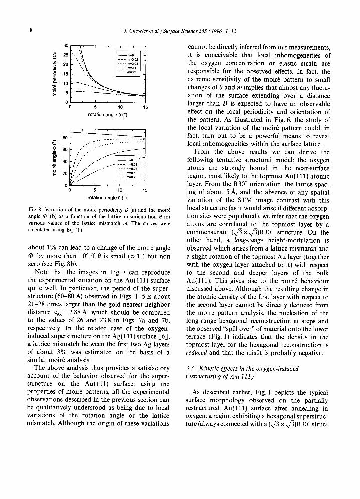
Fig. 8. Variation of the moir6 periodicity D (a) and the moir6 angle • (b) as a function of the lattice misorientation 0 for various values of the lattice mismatch m. The curves were calculated using Eq. (1).
about 1% can lead to a change of the moir6 angle by more than i0 ° if 0 is small (,~1 °) but non
zero (see Fig. 8b). Note that the images in Fig. 7 can reproduce
the experimental situation on the Au(111) surface quite well. In particular, the period of the super- structure (60-80 ~,) observed in Figs. 1-5 is about 21-28 times larger than the gold nearest neighbor distance aAu=2.88 ,~, which should be compared to the values of 26 and 23.8 in Figs. 7a and 7b, respectively. In the related case of the oxygen- induced superstructure on the Ag(111) surface [6], a lattice mismatch between the first two Ag layers of about 3% was estimated on the basis of a similar moir6 analysis.
The above analysis thus provides a satisfactory account of the behavior observed for the super- structure on the A u ( l l l ) surface: using the properties of moir6 patterns, all the experimental observations described in the previous section can be qualitatively understood as being due to local variations of the rotation angle or the lattice mismatch. Although the origin of these variations
cannot be directly inferred from our measurements, it is conceivable that local inhomogeneities of the oxygen concentration or elastic strain are responsible for the observed effects. In fact, the extreme sensitivity of the moir6 pattern to small changes of 0 and m implies that almost any fluctu- ation of the surface extending over a distance larger than D is expected to have an observable effect on the local periodicity and orientation of the pattern. As illustrated in Fig. 6, the study of the local variation of the moir6 pattern could, in fact, turn out to be a powerful means to reveal local inhomogeneities within the surface lattice.
From the above results we can derive the following tentative structural model: the oxygen atoms are strongly bound in the near-surface region, most likely to the topmost Au(111) atomic layer. From the R30 ° orientation, the lattice spac- ing of about 5 ~,, and the absence of any spatial variation of the STM image contrast with this local structure (as it would arise if different adsorp- tion sites were populated), we infer that the oxygen atoms are correlated to the topmost layer by a commensurate (x/~ x v/3)R30° structure. On the other hand, a long-range height-modulation is observed which arises from a lattice mismatch and a slight rotation of the topmost Au layer (together with the oxygen layer attached to it) with respect to the second and deeper layers of the bulk Au( l l l ) . This gives rise to the moir6 behaviour discussed above. Although the resulting change in the atomic density of the first layer with respect to the second layer cannot be directly deduced from the moir6 pattern analysis, the nucleation of the long-range hexagonal reconstruction at steps and the observed "spill over" of material onto the lower terrace (Fig. 1) indicates that the density in the topmost layer for the hexagonal reconstruction is reduced and that the misfit is probably negative.
3.3. Kinetic effects in the oxygen-induced restructuring of Au( l l I)
As described earlier, Fig. 1 depicts the typical surface morphology observed on the partially restructured A u ( l l l ) surface after annealing in oxygen: a region exhibiting a hexagonal superstruc- ture (always connected with a (x/3 × x/-3)R30 ° struc-

J. Chevrier et aL /Surface Science 355 (1996) 1-12 9
ture at the atomic scale) forms a compact island surrounded by a rather disordered and rough surface. Occasionally, we have observed the co- existence of such a restructured island with the 23 x x/~ reconstruction of the clean Au( l l l ) sur- face. This surface morphology strongly suggests that the formation of the long-range hexagonal reconstruction and the necessary atomic rearrange- ments within the Au surface proceeds via nucle- ation and subsequent growth of such restructured areas during chemisorption of oxygen at high temperature.
Such a nucleation and growth scenario is cor- roborated by experiments on the Au( l l l ) single crystal sample prepared under very different condi- tions. As an example, Fig. 9 shows an STM topo- graph of an Au( l l l ) surface annealed in oxygen at much lower temperature (T= 500°C) for 1 h. In the center of a fiat terrace, a small reconstructed area whose lateral size is about 200 A can be distinguished [21]. After subsequent annealing of the same surface in an oxygen atmosphere satu- rated with water at T = 800°C for 24 h, the restruc- turing of the Au( l l l ) surface becomes almost complete. Fig. 10 shows a large scale image (6100x 6100A) of the so-prepared surface. The entire region exhibits a very regular hexagonal superstructure with a period of 65 ,~ (as can be seen from the zoom-in image shown in the inset in
m Fig. 9. STM topograph (385 x 385 .~) of the single crystal A u ( l l l ) surface after annealing in oxygen (1 h at 500°C, po2= 1 bar). The superstructure is observed over an area of about 200 ~, on an otherwise flat terrace (Vt =400 mV, It = 3.0 nA).
\
Fig. 10. STM topograph (6100 x 6100 A,) of the A u ( l l l ) single crystal surface after annealing in a water-saturated oxygen atmosphere po2=l bar) for 24h at 800°C (Vt=200 mV, I t= 5.0 nA). This image only shows a small part of a completely reconstructed area which extends over a length of several microns. The central region indicated by the white square in the image was scanned at higher magnification and is shown in the inset in the top right corner, revealing the details of the hexagonal superstructure with periodicity D ~ 65 ~.
the top right corner). In fact, surface regions over length scales of several microns have been found to be completely reconstructed.
These observations demonstrate that the nucle- ation and growth of the oxygen-induced recon- struction of the Au( l l l ) surface strongly depend on the ambient conditions. Thus, it appears that the oxygen chemisorption proceeds via an acti- vated process which ultimately leads to the forma- tion of the (V/3 x x/-3)R30 ° structure together with the long-range hexagonal superstructure.
It has been suggested [9,11] that impurities could facilitate the reaction of oxygen with the Au( l l l ) surface. Also, based on theoretical argu- ments, Norskov et al. [1,22] have described how impurities on surfaces of transition metals might act as centers for the nucleation of oxide islands and thereby enhance the effective interaction of oxygen with the surface. Although such an effect could account for the above observations, its direct experimental confirmation is beyond the grasp of the present study. On the basis of experimental results obtained under well-defined UHV condi- tions [23], we can say, however, that no signature of oxygen adsorption or formation of a

10 J. Chevrier et al./Surface Science 355 (1996) 1-12
(x~ × x/-3) R30° structure could be observed on a carefully prepared clean Au( l l l ) surface after exposure to oxygen at po2= 10 -6 Torr and T= 900 K for several hours. This result is consistent with earlier reports 1-11,12] but disagrees with a recent LEED study of oxygen adsorption on Au( l l l ) 1,13].
In this context, the difference observed between the behavior of the gold thin films and the gold single crystal is particularly noteworthy. In the case of gold thin films deposited on glass, large ( 111 ) facets are obtained after annealing in oxygen, which can grow much larger than 1000 A (as seen for instance in Figs. 4 and 5). After the oxygen treatment, these (111) facets are always completely reconstructed 1-15]. The lateral extension of the superstructure is only limited by the size of the facet. As mentioned above, the gold films were evaporated on a thin chromium layer deposited on glass substrates containing silicon and calcium. At least silicon and calcium have been claimed to trigger the chemisorption of oxygen on gold sur- faces [9,11,12]. A similar observation was reported by Bonzel et al. for the "oxide formation" on P t ( l l l ) 1,24]. In this case, the appearance of the "Pt oxide" was always correlated with the presence of oxide forming impurities (mainly Si) segregating to the surface during oxygen exposures at high temperature.
The enhanced concentration of impurities in the gold films could explain why the restructuring is more easily obtained on these samples than on the Au(111) single crystal. However, during annealing of the gold films, grain coarsening, nucleation of facets, and chemisorption of oxygen all take place at the same time. Therefore, the formation of the reconstruction might as well be correlated with the appearance of large (111) facets, for instance, due to a more efficient chemisorption of oxygen at the edge of the growing terraces. The complexity of each of the above processes prevents a definite analysis of the formation of the oxygen induced reconstruction in the case of the gold films.
The important role of surface defects or impuri- ties on the nucleation of the oxygen-induced restructuring is also evident from the experiments on the Au( 111 ) single crystal. After repeated cycles of annealing in oxygen at high temperatures the
Au(111) sample became more and more resistant to the nucleation of the long-range hexagonal superstructure. The characteristic hexagonal pat- tern was found only at very few places, whereas most terraces exhibited a "modified herringbone" structure as shown in Fig. 11. This morphology is clearly derived from the reconstruction pattern observed on the clean Au(111) surface, but shows a locally enhanced contrast. Indeed, some of the area between neighboring reconstruction lines appears darker than on the "clean" Au(111) surface (see, for instance, the region marked by the white circle in Fig. 11). This enhancement of the contrast depends on the tip voltage used for imaging. Such a behavior is characteristic for adsorbates ("chemi- cal contrast") and, hence, corroborates the presence of oxygen at the surface. Preliminary experiments seem to indicate that the oxygen is more weakly bonded in this modified herringbone structure than in the (x/~ x x/3)R30 ° structure. For instance, we have found that, in contrast to the very stable (x/3 x V/3)R30 ° structure, the modified herringbone structure deteriorates under ambient conditions and can be irreversibly destroyed by the scanning STM tip, if a tunneling gap impedance below 1 Mf~ is used for imaging. Investigations to further characterize the nature of the modified herringbone structure and its possible relation to the nucleation
Fig. 11. STM topograph (535 x 535 A; Vt= 180 mV, It=4.0 nA) of the "modified herringbone" structure obtained after annealing the Au( 111 ) single crystal surface in a water-saturated oxygen atmosphere ( p o = l bar) for 1 h at 700°C. Prior to this treatment, the sample had already undergone several similar heating cycles in an oxygen atmosphere.

J. Chevrier et al./ Surface Science 355 (1996) 1-12 11
and growth of the oxygen-induced long-range hex- agonal reconstruction are currently in progress.
While the presence of impurities strongly affects the nucleation of the oxygen-induced structures on A u ( l l l ) , we were not able to detect Ca or Si impurities in the Auger and XPS spectra obtained from the restructured single crystal sample, so that the concentration of these impurities must be rather small. Therefore, the appearance of extended, regular patterns such as the (x/c3 x x/~)R30 ° structure and the long-range hexag- onal superstructure cannot be attributed to the formation of a complex surface compound involv- ing a large amount of impurities. Instead, the observed structures appear to be intrinsically induced by the chemisorption of oxygen on A u ( l l l ) .
4. Conclusion
Although gold is known not to form any stable bulk oxide, we have shown in the present STM study that chemisorption of oxygen takes place on the Au( 111 ) surface during annealing in an oxygen atmosphere at high temperature. The chemisorp- tion of oxygen is correlated with the appearance of (x/3 x x/~)R30 ° surface structure. In addition, the A u ( l l l ) surface exhibits a hexagonal super- structure with a period ranging between 60 and 80 ~, and a corrugation close to 0.5 ,~. This super- structure can be satisfactorily described as the result of a moir6 pattern induced by an in plane lattice mismatch and a slight rotation between the two topmost layers of the gold surface. The high sensitivity of the moir6 pattern to local surface strain could explain the observed distortions of the hexagonal superstructure as well as the variation of the periodicity.
The observation of localized areas of the (x/~ x x/~)R30 ° phase with the characteristic long-range hexagonal superstructure indicates that the restruc- turing of the surface upon exposure to oxygen proceeds via a nucleation and growth process. The experiments indicate that surface defects or impuri- ties might play a crucial role in the nucleation of the oxygen-induced surface reconstruction.
Many questions are left open by the present
STM which require further experimental work. For instance, a precise determination of the surface lattice parameter and the rotation angle of the surface layer with respect to the bulk would allow a quantitative test of the moir6 pattern analysis. Also, a direct experimental determination of the adsorption site and the type of bonding of the oxygen atoms to the surface [6,25] appears highly desirable: as suggested on the basis of theoretical arguments presented in Refs. [1,22], knowing the oxygen adsorption site might help to under- stand the mechanism which controls the surface restructuring.
Acknowledgements
J.C. gratefully thanks the Alexander von Humboldt foundation for a research fellowship. L.H. acknowledges the financial support from the Deutscher Akademischer Austauschdienst (DAAD). We have benefited from the technical help of U. Linke and B. Schumacher and from stimulating discussions with U. Linke and S. Horch. The experiments on the interaction of oxygen with the Au(111) surface under UHV con- ditions were performed in collaboration with M.A. Krzyzowski. We are also grateful to F.P. Coenen for conducting the Auger and XPS experiments.
References
[1] F. Besenbacher and J.K. Norskov, Progr. Surf. Sci. 44 (1993) 5.
12] D.R. Gaskell, in Physical Metallurgy, Vol. 1, 3rd ed., Eds. R.W. Cahn and P. Haasen (North-Holland, Amsterdam, 1983) ch. 6, p. 271.
1"3] H. Ibach and H. Bruchman, Phys. Rev. Lett. 44 (1980) 36; M.A. Mendez, W. Oed, A. Fricke, L. Hammer, K. Heinz and K. MOiler, Surf. Sci. 253 (1991) 99.
[4] A.R. Kortan and R.L. Park, Phys. Rev. B 23 (1981) 6340. [5] F. Jensen, F. Besenbacher and I. Stensgaard, Surf. Sci.
269/270 (1992) 400. 1"6] X. Bao, J.V. Barth, G. Lempfuhl, R. Schuster, Y. Uchida,
R. Schl6gl and G. Ertl, Surf. Sci. 284 (1993) 14; X. Bao, M. Muhler, B. Pettinger, R. Schl6gl and G. Ertl, Catalysis Lett. 22 (1993) 215.
1-7] C.T. Campbell, Surf. Sci. 157 (1985) 43. 1"81 M.A. Chesters and G.A. Somorjai, Surf. Sci. 52 (1975) 21.

12 J. Chevrier et al./ Surface Science 355 (1996) 1-12
[9] N.D.S. Canning, D. Outka and R.J. Madix, Surf. Sci. 141 (1984) 240.
[10] C.R. Aita and N.C. Tran, J. Vac. Sci. Technol. A 9 (1991) 1498.
[11] J.J. Pireaux, M. Chtaib, J.P. Delrue, P.A. Thiry, M. Liehr and R. Caudano, Surf. Sci. 141 (1984) 211; J.J. Pireaux, M. Liehr, P.A. Thiry, J.P. Delrue and R. Caudano, Surf. Sci. 141 (1984) 221.
[12] P. L6gar6, L. Hilaire, M. Sotto and G. Maire, Surf. Sci. 91 (1980) 175.
[13] J. Cao, N. Wu, S. Qi, K. Feng and M.S. Zei, Chin. Phys. Lett. 6 (1989) 92.
[14] K.H. Besocke, Surf. Sci. 181 (1987) 145. [15] L. Huang, J. Chevrier, P. Zeppenfeld and G. Comsa,
Appl. Phys. Lett. 66 (1995) 935. [16] J.V. Barth, H. Brune, G. Ertl and R.J. Behm, Phys. Rev.
B 42 (1990) 9307. [17] K. Takayanagi, Ultramicroscopy 8 (1982) 145. [18] F. Grey and J. Bohr, Europhys. Lett. 18 (1992) 717. [19] T.A. Land, Th. Michely, R.J. Behm, J.C. Hemminger and
G. Comsa, Surf. Sci. 264 (1992) 261.
[20] T. Wiederholt, H. Brune, J. Wintterlin, R.J. Behm and G. Ertl, Surf. Sci. 324 (1995) 91.
[21] Fig. 9 depicts one of the rare cases where the reconstruc- tion nucleates on a fiat and apparently clean part of the surface. Usually, the hexagonal superstructure starts to develop at surface imperfections such as step edges (Fig. 1) or dislocations.
[22] B. Chakraborty, S. Holloway and J.K. Norskov, Surf. Sci. 152/153 (1985) 660.
[23] In this experiment, the (111) surface of a gold single crystal was probed in situ by thermal helium atom scattering. Specular helium reflection showed no sign of oxygen adsorption and no diffraction signature corre- sponding to a (v/3x x/~)R30 ° or any other oxygen- induced surface structure could be detected by LEED or helium diffraction.
[24] H.P. Bonzel, A.M. Franken and G. Pirug, Surf. Sci. 104 (1981) 625.
[25] B. Pettinger, X. Bao, I.C. Wilcock, M. Muhler and G. Ertl, Phys. Rev. Lett. 72 (1994) 1561.