2016 - Beyond Meaning: Prospections of Suprematist Semiotics
Upload
khangminh22Category
view
0download
0
Velchover Iridology - Chapter 7 Iris common diseases Semiotics
A significant number of eye diseases, and changes of the vision is the result of many pathological processes developing in the various systems and organs of an infected person. Ophthalmic tests help diagnose disorders of the brain, facio-cranial structures, a number of internal organs and the endocrine system. In this case, the main target of the diagnostic studies in ophthalmology is a classic of the eye, a minor - the front of the eye, including the iris.
Today ophthalmologists consider individual symptoms of the iris, indicating a particular neyrosomati-cal pathology. These include Argyll syndrome Roberts-on - with tabes, paralytic immobility of the pupil - in cerebral meningitis and encephalitis, healing and imperforate pupil - in congenital toxoplasmosis, Horner's syndrome and Petit - in paralysis and irritation of the cervical sympathetic nerve, yellowish appearance pink papules and Gum - syphilis, greasy, yellowish bumps precipitates - tuberculosis, rubeosis symptom - in diabetes, and others. However, such a limited "arsenal" iridodiagno-acoustic tests is in clear disagreement with the anatomical and physiological significance of the iris. Studies in recent years show that the general and local changes in the iris play in general semiotics diseases no less important than the changes in the retina. Unfortunately, these studies are scarce and not sufficiently known. In the following chapters, we will make an attempt to analyze the physiological significance and diagnostic value of some of the most characteristic signs iris associated with disturbances in certain organs and systems. Of course, we are aware that any sign of iridology as topostabil-tion (related to iridotopografii) and Topola-stable (independent of their location), is the projection of the failed group of autonomic fibers that innervate specific area or organ. This is not the visualization if the "output" on the iris of the internal organ, and some resultant multiple neurological circuits. Therefore, decoding of a mark on the iris is a very responsible and delicate operation, which should prepare yourself every beginner iridologist.
IRIDOGENETIC TYPES Taken for granted that the world is impossible to find two people with the exact same person. This is particularly true in relation to the eyes, as the iris of each person completely unique. It is so individual that it could do a great service in criminology, as her picture is a hundred times richer and more accurate of any fingerprints. Nevertheless, of the infinite number of combinations of structural iris reflecting constitutional human characteristics, it is possible to allocate some of the most common types of it (Table 1).

Table 1. The frequency of different types of iris in people with different color eyes (%)
Types of iris Number Tseєt eyes examined by the radial radial radial GOVERNMENTAL homogeneous lacunar Blue 300 83.6 1.7 14.7 Blue 87 73.0 - 27.0 Grey 195 86.5 - 13.5 Light brown 244 44.8 48.3 6.9 Brown 126 13.2 85.8 1.0 Those ions Brown 125 - 90.0 10.0
Some people view the iris is open fan, made up of thin, well-matched fibers - trabeculae. We call this type of radial (Fig. 47, see insert). In people with light eyes, he found an average of 2.5 times more often than the dark-eyed: 83% - at first, at 32% the second. According to our observations, the radial type of iris is a sign of a good constitution and properties of healthy people.
The second type of iris - for a xed l o-d o m o r e n n s d, is characterized by a combination of a radial pattern with a dense homogeneous colored ciliary circle (Fig. 48, see insert). Observed this type almost exclusively in dark-eyed people, with one in four people with dark brown eyes indicated a homogenous version of the second type, in which there is no radial striations, but remains homogeneous form dense gustopigmentirovannoy iris. Just like the previous one, radial homogeneous type of iris is a sign of a good constitution and observed in healthy people.
The third type of iris - angular l akunarny - is presented in the form of thinning of the stroma with scattered leaf-shaped depressions - lacunae (Fig. 49, see insert). In individual subjects iris is a thin, ground breaking plate with a random pattern of trabeculae and crypts. This type occurs in people with different color eyes with a frequency of 1 to 27%. The specified type of iris characteristic of people with a weaker constitution and tendency to dysfunction and disease.
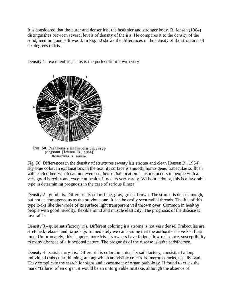
It is considered that the purer and denser iris, the healthier and stronger body. B. Jensen (1964) distinguishes between several levels of density of the iris. He compares it to the density of the solid, medium, and soft wood. In Fig. 50 shows the differences in the density of the structures of six degrees of iris.
Density 1 - excellent iris. This is the perfect tin iris with very
Fig. 50. Differences in the density of structures sweaty iris stroma and clean [Iensen B., 1964]. sky-blue color. In explanations in the text. its surface is smooth, homo-gene, trabeculae so flush with each other, which can not even see their radial location. This iris occurs in people with a very good heredity and excellent health. It occurs very rarely. Without a doubt, this is a favorable type in determining prognosis in the case of serious illness.
Density 2 - good iris. Different iris color: blue, gray, green, brown. The stroma is dense enough, but not as homogeneous as the previous one. It can be easily seen radial threads. The iris of this type looks like the whole of its surface light transparent veil thrown over. Common in healthy people with good heredity, flexible mind and muscle elasticity. The prognosis of the disease is favorable.
Density 3 - quite satisfactory iris. Different coloring iris stroma is not very dense. Trabeculae are stretched, relaxed and tortuosity. Immediately we can assume that the authorities have lost their tone. Unfortunately, this happens more iris. Its owners have fatigue, low resistance, susceptibility to many diseases of a functional nature. The prognosis of the disease is quite satisfactory.
Density 4 - satisfactory iris. Different iris coloration, density satisfactory, consists of a long individual trabecular thinning, among which are visible cracks. Numerous cracks, usually oval. They complicate the search for signs and assessment of organ pathology. If found to crack the mark "failure" of an organ, it would be an unforgivable mistake, although the absence of
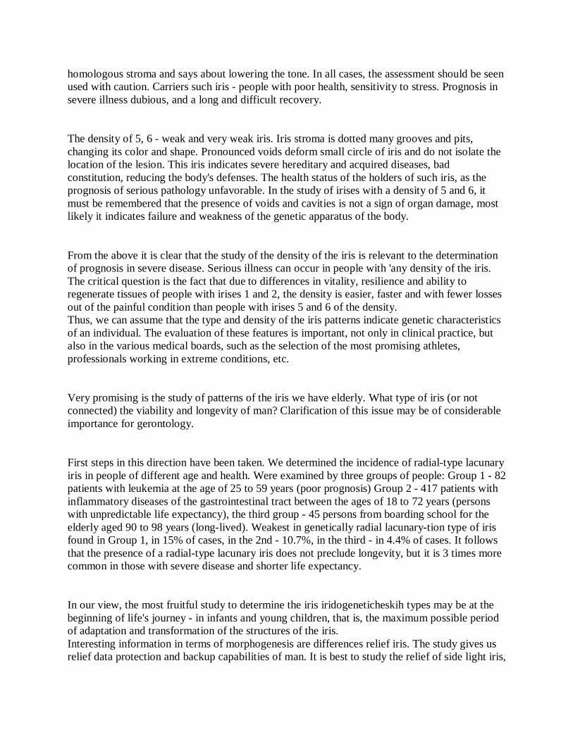
homologous stroma and says about lowering the tone. In all cases, the assessment should be seen used with caution. Carriers such iris - people with poor health, sensitivity to stress. Prognosis in severe illness dubious, and a long and difficult recovery.
The density of 5, 6 - weak and very weak iris. Iris stroma is dotted many grooves and pits, changing its color and shape. Pronounced voids deform small circle of iris and do not isolate the location of the lesion. This iris indicates severe hereditary and acquired diseases, bad constitution, reducing the body's defenses. The health status of the holders of such iris, as the prognosis of serious pathology unfavorable. In the study of irises with a density of 5 and 6, it must be remembered that the presence of voids and cavities is not a sign of organ damage, most likely it indicates failure and weakness of the genetic apparatus of the body.
From the above it is clear that the study of the density of the iris is relevant to the determination of prognosis in severe disease. Serious illness can occur in people with 'any density of the iris. The critical question is the fact that due to differences in vitality, resilience and ability to regenerate tissues of people with irises 1 and 2, the density is easier, faster and with fewer losses out of the painful condition than people with irises 5 and 6 of the density. Thus, we can assume that the type and density of the iris patterns indicate genetic characteristics of an individual. The evaluation of these features is important, not only in clinical practice, but also in the various medical boards, such as the selection of the most promising athletes, professionals working in extreme conditions, etc.
Very promising is the study of patterns of the iris we have elderly. What type of iris (or not connected) the viability and longevity of man? Clarification of this issue may be of considerable importance for gerontology.
First steps in this direction have been taken. We determined the incidence of radial-type lacunary iris in people of different age and health. Were examined by three groups of people: Group 1 - 82 patients with leukemia at the age of 25 to 59 years (poor prognosis) Group 2 - 417 patients with inflammatory diseases of the gastrointestinal tract between the ages of 18 to 72 years (persons with unpredictable life expectancy), the third group - 45 persons from boarding school for the elderly aged 90 to 98 years (long-lived). Weakest in genetically radial lacunary-tion type of iris found in Group 1, in 15% of cases, in the 2nd - 10.7%, in the third - in 4.4% of cases. It follows that the presence of a radial-type lacunary iris does not preclude longevity, but it is 3 times more common in those with severe disease and shorter life expectancy.
In our view, the most fruitful study to determine the iris iridogeneticheskih types may be at the beginning of life's journey - in infants and young children, that is, the maximum possible period of adaptation and transformation of the structures of the iris. Interesting information in terms of morphogenesis are differences relief iris. The study gives us relief data protection and backup capabilities of man. It is best to study the relief of side light iris,

by moving the beam. The surface of the iris does not look smooth and flat, but a conglomeration of bumps and depressions, something resembling volcanic craters. From the center (or pupil) iris surface rises to an elevation of independent rings, the shape of which is very variable. G edge surface elevation ciliary zone descends in the form of a gentle slope to the outer edge of the iris. There are several types of terrain. We describe the seven varieties of terrain and interpretation G. Jausas (1974) (Fig. 51):
Fig. 51. Types of relief iris. I - normal, 2 - disc-shaped, 3 - flattened lateral 4 - edge-teroobrazny 5 - rounded, thickened, 6 - flat 7 - locally deformed.
1 - normal. Have an average size of the ring tops autonomous and uniform internal and on-ruyachnym slopes. Indicates the balance of vitality and good prognosis in diseases. 2 - m foam. Etched characterized pupillary zone in the middle. Occurs in hypertension, bradycardia, sweating and diarrhea. 3 - A flattening of o-l ateralny. Etched characterized slope ciliary zone, demonstrating the sympathetic hypofunction. 4 - crateriform. Different steep slope the foremost pupil zone. Found in endocrine and humoral disorders. 5 - Rounding of y tolschenny. The surface of the iris as a swollen, Fuchs angle (formed by the

pupillary zone and self-ring) is missing. Occurs in hypertension and polyphagia. 6 - flat. Characterized by the complete disappearance of the independent rings. Indicates a low level of resistance and poor prognosis in the case of serious illness. 7 - Local on-d eformirovanny. Indicates the presence of severe chronic illness.
Experience iridology Research shows that the most complete information about a particular trait can be obtained from the complex study of the issue. This applies to the evaluation of the genetic characteristics of a person. On the constitution of the individual we propose that instead of one or two signs, and on a number of important features of a 10-point scale (Table 2). Good signs are assessed morphogenetic to individual.
Table 2. Ten-point system for evaluation of the constitutional Symptom Assessment Rating Badge Badge Total. .. The density of the iris 1 or 2 + 3.6 Relief 1 + 2-7 - There is no deformation of the pupil - Slagging Autonomous there + is - tion of the ring Rupture and deformation auto-no is - mous rings Brown spots there + is - Toxic no spot + is - Adaptive rings no + is - No lymphatic rosary + is - Dystrophic rim there + is -
Rating:

sign (+), bad - (-). When fading out of the grade, which can range from 0 to 10 points, are taken into account only positive signs.
Ideally, if there are 10 positive signs constitution of man can be estimated to be 10 points. However, such individuals are extremely rare. Only rarely observed and those with the constitution, estimated at 0 - 1 point. The above ratings system can be applied in the selection of medical commissions for support integrative iridogeneticheskogo tion test, supplementing the genetic studies. B. Jensen (1982), for example, believes that the iris is the only structure that maps the birth defects that are passed by inheritance to the fourth generation inclusive.
More complex is the question of hereditary transmission of local signs of disease or predisposition to it. Spending iridoskopiyu people composed together in a close relationship, we have found many similarities, their color and structure of iris, which may be one of the most striking of hereditary traits. Hereditary pathology, on the conclusion of iridology, iris structure is characterized by change, particularly leafy hollows and gaps in its stroma, which is rarely performed material substratum of dark color. Through early detection of it (preferably in a child) on the iris, and the administration of appropriate treatment and the preventive measures can maintain the health of a person for a long time.
Iridoskopicheski determine the transmission of hereditary pathological features from one or both parents. In the event that there is a transfer of both father and mother, inherited organ inferiority in the child will be expressed much coarser than that of either parent. It is as if a summation of congenital deficiency of organ function or system. However, as mentioned above, the detection of a congenital defect in the iris is not enough to pathology of the body. Until that time, it is considered only as a poor background, which can develop a particular disease.
Iridology examination at members of 18 families, we found a hereditary transfer of local signs of iris from parents to children in 50% of cases. And at the same child one lesion in the iris can be inherited from the father, the other - from the mother (Fig. 52, see insert).
At the I International Conference on iridotronike in 1980 E. Wojnar introduced large pictorial material on the individual families. Altogether, they examined three thousand people. Analysis of the data showed that the type of iris structure inherited from the parents, but it is not like a full copy of one of them. The authors found that the structure of the iris of any person genetically composed of two parts. A child inherits from a parent or structure type ciliary zone (peripheral part) or pupil (interior). What zpachenie has a gear - it is not entirely clear.
According to V. Bourdiol (1975), right-handed projections of the iris homolateral, the true left-handed - geterolateral-nye: the right half of the body is shown in the left iris, left side - on the
right. Following the example of his teacher, R. Schmidt (1960), R. Bourdiol found the right eye muya ^ magnitude representation store it possible paternal genetic contribution, the left - the mother. In women, the opposite. The author argues that these manifestations are absolutely correct in the transmission main topographic labile dyschromia and age spots.
We have not had sufficient opportunity to check the material above the laws, so we can not express their attitude to the view, and R. E. Wojnar Voig-diol. One thing is certain, iridogenetike studies should be conducted on a large scale, and certainly in collaboration with the physiology and genetics.
Iris color Relatively recently obtained reliable information on the origin of the eye color of a man found his anatomical basis. It turned out that the different color of eyes - blue, gray, brown - due to different content in the stroma of the iris pigment cells - melanocytes. For a small amount of melanin pigment-e - blue eyes, with a moderate - brown, with a significant - black. It has been proven that the eye color is a genetic trait passed down by dominant or recessive. To some extent, it also depends on age. It is noticed that most of the infants appear pas light with light eyes. But in the early mestsy life in most children's eyes become darker. Many Western scholars suggest that the basis for such changes are frequent vaccinations, childhood diseases, the excessive use of drugs. In our opinion, the iris darkening with age, mainly due to the maturation of the pigmented structures of the eye.
Kayue too is the color of the eyes in a human life? Inadequate research on this issue, we examine in more detail.
The earliest, from the early XX century., Idealistic views on the importance of eye color belong J. Peczeli (1886) and N. Liljequist (1897), who believed that the constitution and temperament of each person depend on the influence of the planets, to which they were born. In their view, sky azure color of the eyes is the most perfect particular to people with good health, while the brown color indicates the presence of hereditary or acquired disease. J. Peczeli and N. Liljequist developed a classification of colors the human eye, in which they were located, "from best to worst" in the following order: "perfect" eye-blue and dark blue, then light brown, medium korichievye and finally, blue-gray and brownish- green. Of course, no evidence to support this view, J. Peczeli and N. Liljequist result could not.
In what color eyes began to study na objective basis. Most original views can be called F. Vida and J. Deck (1954), who proposed a three functional types of iris, "lymph", people with blue iris, "hematogenous" - with brown and "mixed" - with sulfur. Considered by these authors are regarded by them as a type of structure with different reaction and the ability to self-adapt.
We believe that the eye color differences can be explained by anatomical and physiological characteristics of the iris. Blue eye color is due to a thin layer of slightly pigmented melanocytes,
brown - a layer of medium thickness and moderate pigmentation, black - a thick layer of melanocytes and intense pigmentation. And any man inherent pigment cells are not only in the iris, but in the course of the entire middle of the eye, causing a color as the retina. This pigment optical filters shield can vary significantly for each person in the service of a strictly individual basis for specific reactions to light. This means that under the same conditions, ie, with the same light, an iris and the retina, the light will be blue-eyed people is much greater than that of the brown-eyed.
According to some, the same light source is in the cerebral cortex of light-eyed people is much stronger activating effect than people with dark eyes. Moreover, it is assumed that even a dream (their duration, nature, etc.) to some extent dependent on the color of the eyes.
Noting this important physiologically fact, scientists have been unable, however, to explain the origins of its occurrence. The question remains: why, other things being equal, the central nervous system in people with blue eyes are more sensitive to light than dark-eyed people? Our research biomicroscopic eye allowed, it seems, to identify the cause of this phenomenon. It turned out that in response to the same bright light, the pupils blue-eyed people were whiter than wide (at 1.5-4% by area) than people with brown eyes. Perhaps this is because the more delicate iris blue-eyed people have more mild neuro-motor apparatus and, therefore, less than the dark-eyed individuals, by constriction of the pupil. Although differences in the area of the pupils at the holders of blue and brown irises are small, they can not affect the amount of light passing through the pupil flow on the intensity of bioenergetic processes in the brain and the body in general.
Consequently, the color of the eyes - not an abstract concept, but purely materialistic and physiological, for now it is clear that the owners of blue eyes are not just blue-eyed people, but in terms of light energy are people, in a sense "left out" because own poor light filters (thinning layer of melanocytes) and have reduced the protective function of the eyes.
On the contrary, brown-eyed, and especially black-eyed people have a strong light filters that can protect them from the intense light.
With this interpretation is fully consistent environmental data on the distribution of people in the direction of the poles to the equator. On a global scale, even without the correction factors on migration and inheritance, the blue eye color is dominant to the inhabitants of the Nordic countries, Brown - mid-south and black - in equatorial countries. It is interesting to note that many of the people of Central Africa can see additional pigment halo or, as we say, the pigment crown, located outside from the area of the iris in the limb [Velhover E. et al, 1982]. Similar pigmentation of the conjunctiva was observed Krylov and TM Sobolev (1986). They found her at students from Africa to 90% of students of the Middle East in 50% of students from South-East Asia - in 30% of cases. Students from Latin America conjunctival pigmentation occurs only in people of Asian and African descent.
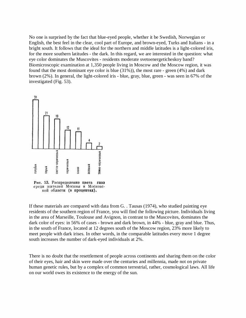
No one is surprised by the fact that blue-eyed people, whether it be Swedish, Norwegian or English, the best feel in the clear, cool part of Europe, and brown-eyed, Turks and Italians - in a bright south. It follows that the ideal for the northern and middle latitudes is a light-colored iris, for the more southern latitudes - the dark. In this regard, we are interested in the question: what eye color dominates the Muscovites - residents moderate svetoenergeticheskoy band? Biomicroscopic examination at 1,350 people living in Moscow and the Moscow region, it was found that the most dominant eye color is blue (31%)), the most rare - green (4%) and dark brown (2%). In general, the light-colored iris - blue, gray, blue, green - was seen in 67% of the investigated (Fig. 53).
If these materials are compared with data from G. . Tausas (1974), who studied painting eye residents of the southern region of France, you will find the following picture. Individuals living in the area of Marseille, Toulouse and Avignon, in contrast to the Muscovites, dominates the dark color of eyes: in 56% of cases - brown and dark brown, in 44% - blue, gray and blue. Thus, in the south of France, located at 12 degrees south of the Moscow region, 23% more likely to meet people with dark irises. In other words, in the comparable latitudes every move 1 degree south increases the number of dark-eyed individuals at 2%.
There is no doubt that the resettlement of people across continents and sharing them on the color of their eyes, hair and skin were made over the centuries and millennia, made not on private human genetic rules, but by a complex of common terrestrial, rather, cosmological laws. All life on our world owes its existence to the energy of the sun.
That it divided the globe into latitude belt or by creating the conditions for the life of various organisms: plants, animals, and people. We must assume that people adapt to lighting zones occurred with considerable losses and very slow.
Distribution of eye-color large and very large among residents of Moscow and Moscow region (in percent). radiation took people with strong pigment "coating" (dark-skinned and dark-eyed.) The exception to this rule are the indigenous inhabitants of the Far North and Alaska. Eskimo, Nenets and Chukchi in contrast to the blue-eyed Mr. Danes, Swedes and Britons have dark eyes, though, and live in the same geographical latitude. However, this does not contradict, but rather confirms the effectiveness of environmental laws, as identical in latitude habitats Danes and Eskimos quite unequal in terms of bioenergy. The climate is characterized by cold far north and the bright snow for a long time. Snow covering the ground in these places, like the vast mirror reflecting 95% of the sun's rays into the biosphere, while the snow-free land represents only 10-20% of the solar energy. Particularly intense white light flooded the plains in the spring. At this time, some of the people with the naked eye there is a kind of snow blindness or eye burns. Even the locals a hard time adapting to the dazzling sunlight. This is evidenced by a very ancient custom of some northern nations, including Inuit, were made for special wooden eye protection goggles with narrow slits. Thus, the harsh conditions of the North, combined with sufficient light intensity, have developed among the natives of this place stronger safeguards: brown eyes, black hair and dark skin. There is no doubt that the blue-dwellers, such as the British Isles, would feel is not quite comfortable. However, some Western scientists are still preaching the theory of the "best and worst colors"
Considering the perfect blue eyes. The absurdity of such theories is obvious.
If we reject the entrenched view of the purely aesthetic eye color around us and get on the physiological point of view, a new perspective can be brought address a number of medical and biological problems. First of all it concerns the very important issue of acclimatization.
So, in theory, move-eyed man from north to south, from the usual climate conditions, the intensity of light radiation, with possible 'reactions overdrive: nervousness, a tendency to vascular spasm, hypertensive crisis, etc. In contrast, dark-eyed man with a move south to north may cause the appearance of deactivation reactions, expressed in weakness, weakness, depressed mood, etc. However, these reactions are usually mild and not at all. Indeed, in the adaptation of the eye to the light energy is involved not only light-protective filters iris and pupils are constantly moving and capable of regrouping melanocytes eyes. But unlike the permanent daylight filter these two controls to age considerably weakened by human aging is accompanied by a significant decrease in the reaction of pupils to light. That's why older people are much worse than the young, carry light acclimatization and move to another place. The above three factors are the main light-protective mechanisms of the brain and eyes. Thanks to them, is the adaptation of the body to the surrounding light environment.
On public relations are the light climate on the one hand, and a set of light-shielding factors - on

the other to a large extent depend on the reactivity and the vitality of any subject. At equilibrium, the parties established in the body energy balance, and the person feels well. If observed energy imbalance, which may occur from the prevalence of visual stimulus over the forces of light protection or vice versa, then you feel good as a rule is violated. Of course, against this background faster the 'breakthrough' defense mechanisms and the development of certain diseases. A specific example of such a "breakthrough" is to strengthen the nervous excitability, observed in many people in the active work of the sun. It is possible that an energy imbalance associated with the appearance of migraine and hypertensive crises, and possibly diencephalic paroxysms.
According to D. Zigelmaier (1971) and some other iridolo-gov, the differences in the incidence of some diseases are caused by uneven color of eyes, and, consequently, different adaptation to ambient light environment. Insufficient adaptation to light stimuli can be explained by a very interesting and it is unclear in the recent past fact: in England and Sweden pulmonary tuberculosis often sick person, with brown eyes, in southern Germany, and Italy - with the blue. One can assume that the disease in the first, to some extent related to the deactivation of the light, the second - with a light overdrive.
We studied the susceptibility to light 640 Moscow residents aged 20 to 40 years old, with normal-i niє of view. There were 400 subjects roil i lubye eyes, 240 - brown. J 1 = II? The study consisted of 10-se-I "I-IV I-IV nanosecond light eyes bright" light from a distance of 20 cm sensitivity and result revealed that pain tolerance in the light of many people with different eye color. times more common in light-eyed Luden (15%) than in people with dark eyes (0.8%). Directly related to the "blue-eyed and brown-eyed problem" is by M. Millodot (1976). He was interested in a long time noted by a fact: wearing contact lenses more often brings trouble goluboglazsh people than black-eyed. M. Millodot suggested that the sensitivity of the cornea depends on the color of the eyes. To test this hypothesis, he had a very demonstrative study. Volunteers (156) (English, negros, Indians, Chinese), who had good vision and is the same age, the cornea is gradually increasing pressure as long as the subjects did not start to feel it. The studies found that the cornea, blue-eyed people in 2 times more sensitive cornea brown-eyed and 4 times more sensitive cornea eyed (Fig. 54). Consequently, sensitivity to pressure and pain is most pronounced in people with light eyes. It may be that associated with the giperchuvstvitelnostgo revealed British interesting phenomenon to achieve the therapeutic effect of drugs dose for black-eyed patients should be greater than for patients with blue eyes.

K. Chen and E. Poth (1929) found that a person of the Caucasian peoples are more sensitive to Mydriatics than the Chinese, and the latter are more sensitive than negros. Among Caucasians Mydriatics more effective in individuals with light iris than the dark [Howard N. J., Lee, TR, 1927; Obianwu N. O., et al., 1965]. These differences are usually registered with the introduction midriatiche-adrenergic skih funds and less with drugs that enhance the pupil due to violation of cholinergic transmission.
On the difference between responses to miotocs depending on the degree of pigmentation of the iris is less well known, although N. MeIi-kian et al. (1971) showed that people with a strong pigmentation of the skin, iris, trabecular network and fundus less susceptible to mechanical damage to the eyeball and the hypotensive effect of 4% solution of pilocarpine, than people with weak pigmentation.
A similar conclusion was also American researchers L. Harris and M. Galin (1971), who studied the hypotensive response to pilocarpine in patients with glaucoma. The researchers found that in order to achieve an equivalent therapeutic effect in patients with blue eyes should bury pilocarpine weaker concentration (1%) than brown-eyed (4%), and especially black-eyed (8%).
It must be assumed that, for this reason, cleansing the body of toxins during fasting occurs differently in individuals with different eye color. According to our clinical and the laboratory
and iridograficheskim observations, the light-eyed people endogenous cleansing recorded in 65%, the brown-eyed - in 52% of cases.
The data shown is a particular expression of modern concepts of pharmacokinetics, according to which people are divided on the body's ability to "recycle" drugs into 2 groups: fast and slow "atsetillyatory." To obtain the desired therapeutic effect, first-fast "atsetillyatoram" requires 2-fold lower dose of drugs than the second.
Our studies have shown that the color of the iris plays a role in the origin of not all, but only of certain diseases. To answer this question in Alma-Ata, ie, in the zone of intense light conditions were examined 617 patients: 300 - with endemic goiter, 187 - with gastric ulcer and 130 - with angina. It was found that the detection rate of goiter in patients with blue and brown eyes almost identical. Perhaps this is because the disease is pain-free and is not accompanied by stress reactions. Another matter ulcer and angina - diseases more "aggressive" proceeding with severe pains and high voltage defenses. They were observed more frequently in people with blue eyes. "Breakthrough" of the disease they have, compared to the brown-eyed people, registered 1.5 times more frequently in ulcer disease and 2.5 times more often in angina So, the energy imbalance due to the conflict situation in the "light-light protection ' is not a pathological condition, and the basis on which to develop a particular disease. With equal light and equal imbalance greater chance emergence of diseases characterized by pain and more profound changes in the body.
On the basis of some particulars, A. Maubach (1952) stated that there is a relationship between eye color and susceptibility to disease, supposedly people with gray and greenish-brown eyes more susceptible to other cancers. However, when multiple checks specified point of view has not been confirmed. It has been proven that the color of the iris is no regard to the specific, including cancer, disease has not. A striking illustration of lower photoresist features, and along with it the life of the whole organism, is a pigment deficiency in humans and animals albinos. Such individuals have melapotsity, which contain only colorless shadow of melanin granules. VroYahdennoe lack of melanin pigment from birth leads to partial blindness, photophobia, and susceptibility to many diseases. Albino inherent low tyrosine levels, weak synthesis chamber teholaminov and very little physical activity in stressful situations.
Lack of melanin content in the body and its derivative of tyrosine observed in fenilnirovinogradnoy mental retardation, or disease Felling. For patients with this form of mental retardation is characterized by a thin white skin, light hair and eyes, microcephaly, profound mental hypoplasia, seizures and fits of anger. As albinism and Felling disease are rare pathologies studied in humans is not enough. The most complete information on the subject is found in studies conducted on the fruit fly Drosophila. In all cases, when the fruit fly carries the gene that determines the absence of pigment in the eyes, and at the same time strictly regularly changes its color of internal organs and, most importantly, reduced fertility and life flies.
It is known that the occurrence of tumors of the eye in cattle is in direct connection with congenital depigmentation of the eyelids, proptosis and intense ultraviolet radiation.
Extrapolation of the anatomical and functional bases pigmentation eye on other systems and functions of the body, particularly the skin, allows a better understanding of the universal function of pigmentation. Consider some of the facts and conditions.
In humans and many animals protection from intense light irradiation provides a shielding layer of melanin pigment and keratin of the stratum corneum, which either absorb all wavelengths of light, or filtered particularly dangerous UV rays. In response to prolonged exposure to sunlight, a person with fair skin tan produced by the increased formation of keratin and especially melanin. Tanned skin man missing only 5% of the UV rays with a wavelength of 300 nm, while the skin nezagorevshego - 25%. People with dark skin, almost all UV rays are absorbed by melanin, which is available for them in large numbers. This is due to adequate protection from the high doses of radiation energy specific to those areas of the world, home to people with hyperpigmented skin In-governmental.
The differences between the two extremes of the body (pigmentation and depigmentation) clearly revealed in photosensitivity. It is known that under the influence of hematoporphyrin in animals and humans is increased sensitivity to light, the so-called photosensitivity. If you enter the blood-porphyrin under the skin of animals with white and black fur, and then put them into the light, then the first developing inflammation, shortness of breath, seizures, and even "light death" in the latter nothing happens. Photosensitized poor pigment "white" die, photosensitized rich pigment "black" survive. This fact clearly expressed diametrically opposite positions in the relationship "- Light protection".
Relevant to the issue of pigmentation has background adaptation of the growing organism, established LA Zakharova and Kornilova MB (1981). In their experiments, they showed that the larvae of the common frog, grown on a black background, with the 20th stage of development are far ahead in the growth and intensity of pigmentation of the larvae reared on a white background. The number of dermal and epidermal melanophores per unit area at the "chernofonovyh" larvae was significantly greater than that of "belofonovyh." Interestingly, the highest differences were found in the calculation of the epidermal melanophores concerning eksteroretseptivnym structures.
In our view, lens, and thus energozaschitnoy function has not only exteroreceptors melanin, but also internal, interoretseptivnye melanin. The latter is, and perhaps not coincidentally, the most important line in the central nervous system, the brain stem. There are 3 major pigment groups: black substance, bluish gray wing place (triangle vagus nerve). In addition to the granular pigment balls - "situational quencher" that appear in the lesions in severe, debilitating disease, these three formations are like stationary bioenergy filter-absorber. Of their operation, as well as the exterior of the pigment layer of the retina, iris and skin depends on the level of total
bioenergy body. We think it would be interesting from the point of view of general pathology, to analyze a large section material morphology and biochemistry of the substantia nigra of the brain in people who were killed at different ages and from different causes. Poorly understood is the question of the physiological role of a limited accumulation of pigment in the skin: freckles, senile nevi, moles, etc.
Freckles in the form of dots and spots poured under the influence of sunlight on a background of pale, and so little pigmented skin. This economical, forced Election light barrier observed in individuals with very low stocks of melanin. Transient skin changes observed in women during pregnancy. Brown spots of yellow or brown in color they appear in certain locations on the face, breasts, abdomen in the midline. May pigment "shields" in pregnancy protects against light (energy) affektsy important areas such as the brain stem, breast and point-"heralds" middle line of the abdomen.
With approximately the same positions should be considered and the emergence of the elderly age spots, and in particular nevi brown. If freckles occurring in young people are, in most cases, the formation time, the acquired brown spots in the elderly tend to be persistent and progressive. With age-spots "patch" covers all of the new skin. It is hard to say which is more favorable age: one in which the skin remains clean, or one in which they are full of "patches." Who knows, there are spots peculiar meter malfunction, indicating gross "failure" in the body? Perhaps, on the contrary: they are not gross changes, and the easiest to manage to protect the well-developed "service melanins"? If you take one of the most prominent forms of human pigment pathology - Addison's disease, it is in her intensive skin pigmentation separate increasingly combined with the benign course of the disease and on the contrary., Clinical variants with weak pigmentation occur generally heavier.
Question of local pathology pigmentation and aging is not yet resolved, but now with some degree of probability we can say nevi in older people as a positive phenomenon, as a "safe conduct", which gives the body in perpetuity to their most weakened bodies.
According to G. Harrison et al. (1977), melanin is not only prevents damage, but also the mutagenic effect of ultraviolet radiation. This is evidenced by the following facts. Skin cancer in Latin America is mostly found in white immigrants and hardly registered Aboriginal, and it is localized equally often in open and protected clothing on the body.
These and other facts are strong evidence of the relationship of iris eye color with the constitution and the adaptation status of the organism.
Unusual in terms of bioenergy is heterochromia or iris eye different colors when a person, for example, one blue eye and one brown (Fig. 55, see insert). Heterochromia occurs rarely, to our knowledge, in 0.5% of cases. People having this abnormality of function is not compromised. Anomaly affects the other-in the perception of light. When heterochromia it different for the left and right hemispheres of the brain. Therefore, bioenergy and reactivity in the right and left side
of the body are also different. Heterochromia eye can be compared with two windows, one of which is fully glazed, and the other only half.
Pathogenetically heterochromia is largely due to the sympathetic innervation. S. PaIHs (1982) writes that melanocytes are moved to the choroid and iris during embryonic development under the control of the sympathetic nervous system. The latter has a significant impact on the formation of melanin and thus the color of the iris. In the absence of sympathetic influence (birth trauma, etc.) iris pigmentation may not be enough to one side or another [Rusetsky II, 1958; Makley T. A., Abbott, K., 1965; Ehinger B. et al., 1969 ]. Heterochromia can occur as an isolated anomaly, does not necessarily affect the sympathetic way. Finally, the reason may be heterophthalmia local disease. In this case, often arise in determining slozhposti hand pathology (remembering the different effects of diffuse iris melanoma, on the one hand, and its atrophy due to iritis or glaucoma - on the other.) To variations heterochromia in the broadest sense can be attributed differences in the color of eyes and hair, that is, differences vested eyed brown-eyed brunettes and blondes. This phenomenon has not been studied in the physiological and energetic aspects.
In contrast, a rare true heterochromia internal or central, heterochromia occurs frequently and is characterized by darker pupillary zone compared with the ciliary (Fig. 56, see insert). The color intensity of the central circle - yellow blue-eyed and dark-brown in brown-eyed - notes, usually on both sides. Pathogenetically it indicates pathology of the gastrointestinal tract.
First discovered inside heterochromia N. Liljequist (1903). Watching the dynamics of eye pediatric patients and learning their way of life, he drew attention to the fact that those children who are in families for better sleep regularly gave black poppy okolozrachkovaya part of the iris acquired a more pronounced color. The children who did not use poppy color changes are not detected. Thus was established a projection area of the stomach and intestines on the iris. Most iridology [Liljequist N., 1903; Roberts F., 1962; Kriege, T., 1969, and others] believe that the main cause of internal heterochromia are constipation and bowel disease, as well as a massive medication, most of which (7/12) has a place of its action the stomach and intestines.
It is known that the overall color of the iris changes throughout life clearly - in infancy, little noticeable - in the young and middle age, more markedly (fades and fades) - at the oldest. It is essential that any enlightenment iris stroma, often occurring under the influence of natural factors of treatment is a positive process, and vice versa, any darkening of the iris indicates a negative shift in the body.
Situation is more difficult to change in the color of individual sections of the iris. Whether there are spots of color in people in adulthood? Leading Iridology answer this question in the affirmative, but the documentary evidence no one has been cited. No reasoned argument in favor of the color transformation with us.
At present, we have five observations based solely on history. We present two of them.
VP, 48, an engineer. Past medical history: scarlet fever, gastritis. At birth, both his eyes were bright blue, a 5-year old - blue-green. BH years after moving from Krasnodar to Moscow drew attention to the brown spot that appears on the iris of the left eye. Initially, it brings with waves, later observed constantly. At the age of 39-44 years living and working abroad, this time pale spot and got both eyes blue. In 45 years, he returned to Moscow, found a dramatic change in the color of the left eyes in a short time he became completely brown. True heterochromia remains stable in subsequent years (see Fig. 55).
O. L., 21, art director. Postponed diseases: measles, flu, whooping cough, pneumonia. To 12 years, the two eyes are clear, greenish. Then on the right in the medial part of the iris appeared brown dots. She gradually increased in size and has now reached a significant size and sector of the form (Fig. 57, see insert).
Of course, that medical history information that is not supported by objective data, evidence of local color transformations can not. Requires long-term monitoring of dynamic irises people with essential use of recording equipment in order to get the answer to the question - when and how the color changes occur.
The study of light protection iris is just beginning. However, obvious is the theoretical and practical sense, which can get the work done in this direction. In the light adaptation, they should lead to a clear recommendation related to the movement of people through the meridians of the globe. In preliminary findings can be said that before sending the person to travel necessary to consider not only the state of his health, but also the potential light-protective features: size of the pupils, the liveliness of the reaction of pupils to light, density and color of the iris. To reduce the stress response to light all arrivals to the south-eyed northerners can recommend wearing safety glasses with tinted windows. This measure is particularly important for older people who, without doctor's permission and without good reason not to move from north to south, from one latitude to another light. Conversely, for removing inhibitory responses arising from dark-eyed people when moving from south to north, to recommend a number of measures stimulating-translational nature: stay in the open air, taking small doses of adaptogens (Eleutherococcus, pantokrina), etc.
The magnitude and shape of the pupil Investigation of the pupils and their reflections which indicate the status of autonomic nervous regulation is the subject of special attention not only eye doctors, but other doctors - neurologists, psychiatrists, therapists, TB specialists. Currently, the interest in studying the size and shape of pupils is of increasing importance, as these concepts are related adaptation to light energy. From the literature it is known that the iris and light aperture - pupil - have permanent effects on the internal organs and the external
environment. Vegetative apparatus of the eye automatically adjusts light clearance holes, thus ensuring a balance between the ambient light environment and the internal needs of the body. Light output, the perceived light-sensitive structures of the eye, is transformed into bioelectrical kind of energy, which has an activating effect on the centers of the brain and spinal cord. Bioelectric effects are also autonomic nervous system and the endocrine glands responsible for hemodynamics, metabolism, trophic and other vital processes in the body.
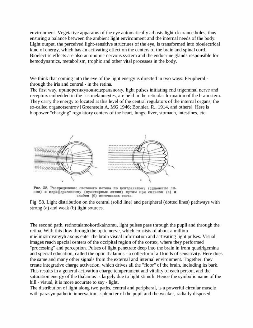
We think that coming into the eye of the light energy is directed in two ways: Peripheral - through the iris and central - in the retina. The first way, иридоретикуловисцеральному, light pulses initiating end trigeminal nerve and receptors embedded in the iris melanocytes, are held in the reticular formation of the brain stem. They carry the energy to located at this level of the central regulators of the internal organs, the so-called organotsentrov [Greenstein A. MG 1946; Bonnier, R., 1914, and others]. Here is biopower "charging" regulatory centers of the heart, lungs, liver, stomach, intestines, etc.
Fig. 58. Light distribution on the central (solid line) and peripheral (dotted lines) pathways with strong (a) and weak (b) light sources.
The second path, retinotalamokortikalnomu, light pulses pass through the pupil and through the retina. With this flow through the optic nerve, which consists of about a million mielinizirovanyyh axons enter the brain visual information and activating light pulses. Visual images reach special centers of the occipital region of the cortex, where they performed "processing" and perception. Pulses of light penetrate deep into the brain in front quadrigemina and special education, called the optic thalamus - a collector of all kinds of sensitivity. Here does the same and many other signals from the external and internal environment. Together, they create integrative charge activation, which drives all the "floor" of the brain, including its bark. This results in a general activation charge temperament and vitality of each person, and the saturation energy of the thalamus is largely due to light stimuli. Hence the symbolic name of the hill - visual, it is more accurate to say - light. The distribution of light along two paths, central and peripheral, is a powerful circular muscle with parasympathetic innervation - sphincter of the pupil and the weaker, radially disposed

muscle to the sympathetic innervation - dplatatorom pupil. In its configuration both muscles resemble a wheel hub rather spoked wheels. While reducing sphincter antagonistically reduce dilator, in general, "wheel" works in unison as a single muscle band. Thanks to him, the correction flux is always going to respect the interests of the central lumen, as the light energy in the first place and more than adequate volume needs central, not peripheral pathway (Fig. 58).
If you calculate the energy "needs" of internal organs, it appears that they are relatively small. This is because the internal organs have autonomous stable instability and the slow flow of nervous and metabolic reactions. In contrast, many-sided activity of the brain and the muscular system characterized by intense nervous and metabolic reactions. They require high energy costs, determining the primacy of processes of higher nervous activity of the function of many organs. Mitotic innervation by preganglionic fibers coming from the pupil-motor neuron nucleus Edinger-Westphal, the activation level is regulated by nerve cells in the pretectal area. Post-ganglion fibers to the sphincter of the pupil away from the ciliary ganglion of the first to the rear of the eyeball. Mitotic reaction, like other parasympathetic function was significantly differentiated, concentrated n approaches somatic type. W. Gaskell (1916) considered the sphincter muscle of the pupil as it is intermediate between smooth and skeletal. Mitotic reflex response is a protective, conservative, and at the same time differentiating. It refers to the instantaneous phasic reactions occurring at 0.3-0.8 s.
Mydriatic innervation by neurons tsilio-spinal center, located at the segment level Ce-Di) 2 spinal cord. Axons pass in the superior cervical sympathetic ganglion, within which synapse with post-ganglionic neurons. Last follow along the carotid arteries in the eye n orbit, where in a short pass tsiliarpye nerves that innervate the pupil dilator. Mydriatic response is indicative reaction generalized to many signals and responses to afferent stimulation, as well as emotions in the formation of which involve autonomic and cortical centers. It refers to the slow, tonic reactions occurring in 10-20 with [Schmidt R., Thews G., 1985]. Should indicate that the clinically accepted for mydriasis pupil diameter greater than 6 mm miosis - 2 mm or less. Until now, ophthalmologists and neurologists can not give a clear explanation of the act of mydriasis. This question is not easy, because the change of parameters of the same stimulus (light) cause a reaction of two oppositely acting muscles of the sphincter which supposedly only constricts the pupil, and the dilator - only expands. But is it really?
If pupil dilation - the sympathetic reaction, it is unlikely that it will be able to implement a unilateral tender radial fibers and dilator, very vaguely reminiscent of the muscle. Probably dominant neuromotor apparatus iris reacts to changes of light, is the sphincter of the pupil. Leads to a reduction in its coma, relaxation - to mydriasis, while creating a tonic dilator muscle tension with ever-present tendency to increase the pupil. A similar function of the tonic tension in the office are chapter for two more smooth, sympathetically innervated muscles: the orbital muscles responsible for vystoyanie eyes from their sockets, and the muscle of the upper eyelid, which determines the width of the individual tone of his eye slit. Formed as a trio of muscle sympathetic provide some tonic trends, and in case of their violation - pathological syndromes Horner and Petit.

The dominant role in the expansion of the pupil parasympathetic nervous system and the sphincter muscles can be judged by the emotional mydriasis in experimental animals, which remains after cutting sympathetic and disappears after section of the oculomotor nerve.
The role of the parasympathetic nervous system in both mitotic and in mydriatic reaction is demonstrated by the use of funds is not the sympathetic neurotransmitter, and parasympathetic series: cholinomimetic (pilocarpine, karbaholin) and antiho-linesteraznyh agents (physostigmine, army hloroftalm) - to narrow the pupils and anticholinergic agents (atropine, scopolamine, homatropine) - to expand. As for the neurotransmitter contained in sympathetic endings iris - norepinephrine and epinephrine, they drip into the eye in normal and concentrated solutions mydriatic effect in healthy people does not matter. In response to the introduction of substances adrenomimeticheskim normal pupil is not expanding [Pallis C, 1985].
Interestingly, the blocking drugs peripheral holinoreaktivnye system, cause not only midri DNase, and expansion of the entire iris. According to our observations, burying homatropine in the eye pupil expands on average 2.25 times, the pupillary zone, by 1.3 times and narrows the ciliary zone of 2.5. In this case, the horizontal diameter of the iris is increased by 0,1-0,55 mm or 0,8-4,6% (Fig. 59, see insert).
Pupil size during the life of undergoing significant changes, ranging from 1 to 8 mm. It is estimated that the light flux entering the eye with the smallest pupil diameter (1 to 2 mm), 16 times less than its maximum diameter (8 mm). For children in the first year of life is characterized by cramps, then pupils increased to a maximum value in the children and young age, gradually decrease in mature and old and over are miotichvymi in old age. Of course, the fact that the physiological miosis infants is quite different from miosis observed in the elderly.
Size and shape of the pupils studied by us in the precise experiment. The sample of 750 people, including 390 men and 360 women aged 10 to 69 years. With various diseases were 652 people, healthy - 98. Evaluating the individual iris stroma and the pupils themselves held projection-biomicroscopic method for measuring at the average 1400-1500 cm2. This allowed analyzed Rowan material with a very high degree of accuracy is much higher than usual for the clinic visual assessment of pupils (Table 3)
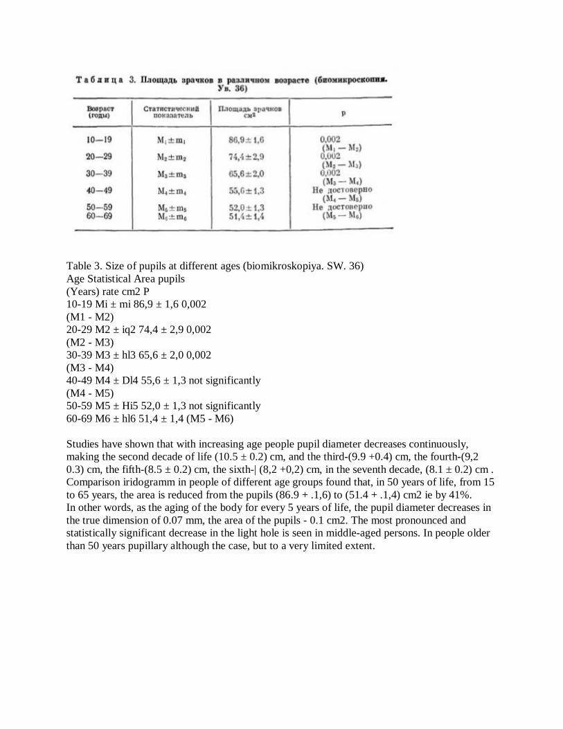
Table 3. Size of pupils at different ages (biomikroskopiya. SW. 36) Age Statistical Area pupils (Years) rate cm2 P 10-19 Mi ± mi 86,9 ± 1,6 0,002 (M1 - M2) 20-29 M2 ± iq2 74,4 ± 2,9 0,002 (M2 - M3) 30-39 M3 ± hl3 65,6 ± 2,0 0,002 (M3 - M4) 40-49 M4 ± Dl4 55,6 ± 1,3 not significantly (M4 - M5) 50-59 M5 ± Hi5 52,0 ± 1,3 not significantly 60-69 M6 ± hl6 51,4 ± 1,4 (M5 - M6) Studies have shown that with increasing age people pupil diameter decreases continuously, making the second decade of life (10.5 ± 0.2) cm, and the third-(9.9 +0.4) cm, the fourth-(9,2 0.3) cm, the fifth-(8.5 ± 0.2) cm, the sixth-| (8,2 +0,2) cm, in the seventh decade, (8.1 ± 0.2) cm . Comparison iridogramm in people of different age groups found that, in 50 years of life, from 15 to 65 years, the area is reduced from the pupils (86.9 + .1,6) to (51.4 + .1,4) cm2 ie by 41%. In other words, as the aging of the body for every 5 years of life, the pupil diameter decreases in the true dimension of 0.07 mm, the area of the pupils - 0.1 cm2. The most pronounced and statistically significant decrease in the light hole is seen in middle-aged persons. In people older than 50 years pupillary although the case, but to a very limited extent.
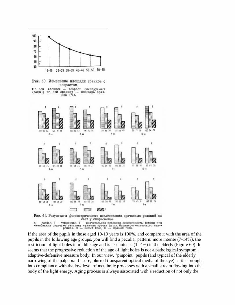
If the area of the pupils in those aged 10-19 years is 100%, and compare it with the area of the pupils in the following age groups, you will find a peculiar pattern: more intense (7-14%), the restriction of light holes in middle age and is less intense (1 -4%) in the elderly (Figure 60). It seems that the progressive reduction of the age of light holes is not a pathological symptom, adaptive-defensive measure body. In our view, "pinpoint" pupils (and typical of the elderly narrowing of the palpebral fissure, blurred transparent optical media of the eye) as it is brought into compliance with the low level of metabolic processes with a small stream flowing into the body of the light energy. Aging process is always associated with a reduction of not only the

exchange, kinetic, and thought processes, and functions of the various eksteroretseptivnyh systems. In the conducted GOVERNMENTAL previous studies we were able to show that the restriction of light holes leads to the reduction of bioenergetic activity of the brain. This is evidenced by the fact that the light from the same source makes older persons action (mean age 61 Fig. 60. Change in pupils a year) a weak activation of the decline of bioenergy is to reduce the light energy flowing through the narrowed pupil brain elderly. All this suggests the possibility of dynamic observation of pupil size in a particular category of dispensary patients, in which it will be possible to assess the level of light adaptation and the degree of aging.
The value of the pupils not only depends on the person's age, but also on many other factors. It changes constantly, every second and minute for all human life. During the day, in a state of hyperactivity, emotional stress and fear pupils dilate, during sleep, rest when tired and unwell - narrowed. Perhaps not much exaggeration to say that in the eyes of everyone tentatively "recorded" his energetic and emotional potential. Wide pupils indicate a high bio-energy, narrow - a low. In the study of the innervation of the pupil should be considered "anxiety pupil» (hippus), is to constantly change its diameter, a variety of amplitude and frequency. It is observed in individual subjects and is clearly visible in the IRI-doskopii. "Anxiety pupil" demonstrates the dynamism of autonomic innervation of the pupil, excessive fluctuations which show lability and autonomic instability. Between "anxiety pupil" and the type of light pupillary reflex is a connection: the more "concern pupil", the faster and expressed zhennee reaction of the pupil to light and vice versa.
The study of pupil size in men and women with dark heads did not show any differences. However, most light-eyed young and middle-aged eyes were wider than the light-eyed men of the same age. These differences were small and were in the true measurement on average 0.24 mm in diameter. Maybe some large pupils in light-eyed women due to their age.
In assessing the reaction of pupils to light examining the correlation between their size and structural characteristics of the iris. It was found that the type of iris (radial, radial homo-gene, lacunar) and the number of spots present in it have no effect on the amount of light holes.
Studies have shown that the value of the pupils to a large extent depends on the state of the other structures of iris: pupillary zone and the pulmonary circulation. It has been found that people with a pure pupillary zone pupillary diameter 0.33 mm larger than that of humans, the pupillary zone that looks worn and hyperpigmented. A similar relationship was revealed when analyzing the state of the pulmonary circulation. People with a "pure" small circle of the pupil diameter was 0.47 mm (!) Than in those with diffuse and "intoxicated" small circle.
As mentioned, the pupillary zone is the projection of the gastrointestinal tract, pulmonary circulation - a projection of the autonomic nervous system. Adaptive-trophic changes, we found in these areas of iris in patients with a relatively narrow pupils, show high light depending on the functional state of bioenergy of the autonomic nervous system and gastrointestinal tract.

Differentiated assessment of the reaction of pupils to light, carried out by the photometric method, opens the possibility to determine the level of reactivity and automated startle stem of the brain. It plays a significant role in the speed of motor responses, or torpid instantaneous response, which may be of paramount importance for the people of certain occupations (pilots, athletes, etc.).
We examined 18 athletes-runners of Children's Specialized Schools: 5 boys and 13 girls aged 14 - 16 years. The results of photometric calculations are shown in Fig. 61, which are shown in descending order of the numerical values of light responses of the surveyed athletes. Based on these data, in addition to the quantitative data, we can obtain some information about the qualitative aspects of reactivity. Highest first column indicates the immediacy and power of reaction, first low and high subsequent columns - the number of low-key response, low columns - on the weak reaction, etc.

In Fig. 62 schematically examples of high optical-cerebral (case 2) and low reactivity (observation 17). Is interesting to note that the athlete Peninsula, which has, to our knowledge, a high level of reactivity (case 2), and was the most productive in the professional relationship.
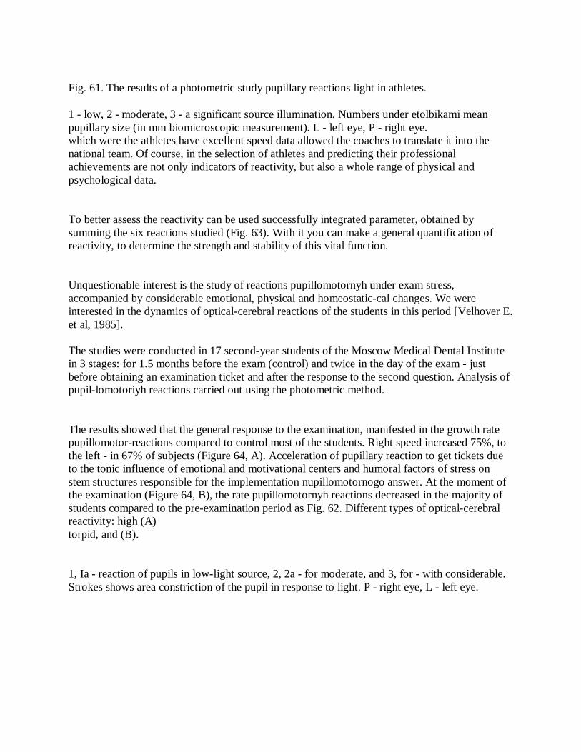
Fig. 61. The results of a photometric study pupillary reactions light in athletes.
1 - low, 2 - moderate, 3 - a significant source illumination. Numbers under etolbikami mean pupillary size (in mm biomicroscopic measurement). L - left eye, P - right eye. which were the athletes have excellent speed data allowed the coaches to translate it into the national team. Of course, in the selection of athletes and predicting their professional achievements are not only indicators of reactivity, but also a whole range of physical and psychological data.
To better assess the reactivity can be used successfully integrated parameter, obtained by summing the six reactions studied (Fig. 63). With it you can make a general quantification of reactivity, to determine the strength and stability of this vital function.
Unquestionable interest is the study of reactions pupillomotornyh under exam stress, accompanied by considerable emotional, physical and homeostatic-cal changes. We were interested in the dynamics of optical-cerebral reactions of the students in this period [Velhover E. et al, 1985]. The studies were conducted in 17 second-year students of the Moscow Medical Dental Institute in 3 stages: for 1.5 months before the exam (control) and twice in the day of the exam - just before obtaining an examination ticket and after the response to the second question. Analysis of pupil-lomotoriyh reactions carried out using the photometric method.
The results showed that the general response to the examination, manifested in the growth rate pupillomotor-reactions compared to control most of the students. Right speed increased 75%, to the left - in 67% of subjects (Figure 64, A). Acceleration of pupillary reaction to get tickets due to the tonic influence of emotional and motivational centers and humoral factors of stress on stem structures responsible for the implementation nupillomotornogo answer. At the moment of the examination (Figure 64, B), the rate pupillomotornyh reactions decreased in the majority of students compared to the pre-examination period as Fig. 62. Different types of optical-cerebral reactivity: high (A) torpid, and (B).
1, Ia - reaction of pupils in low-light source, 2, 2a - for moderate, and 3, for - with considerable. Strokes shows area constriction of the pupil in response to light. P - right eye, L - left eye.
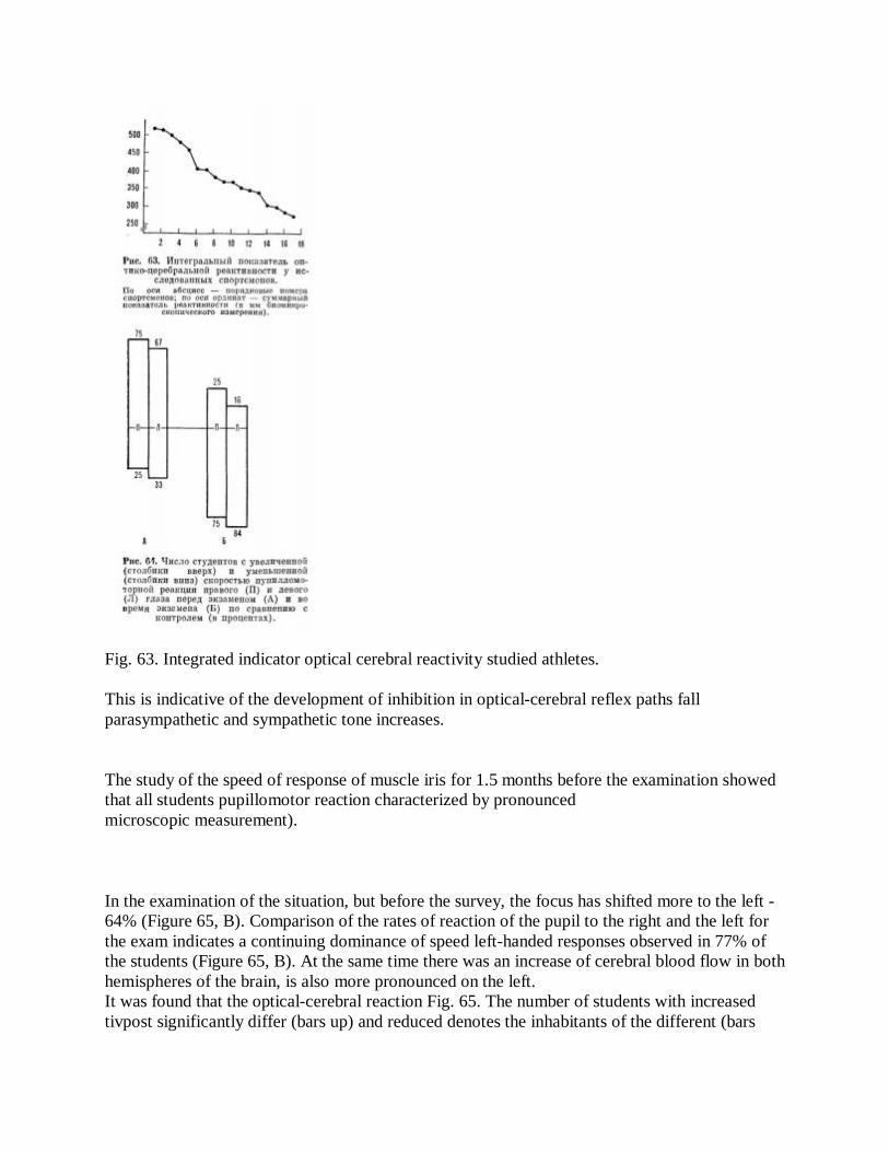
Fig. 63. Integrated indicator optical cerebral reactivity studied athletes.
This is indicative of the development of inhibition in optical-cerebral reflex paths fall parasympathetic and sympathetic tone increases.
The study of the speed of response of muscle iris for 1.5 months before the examination showed that all students pupillomotor reaction characterized by pronounced microscopic measurement).
In the examination of the situation, but before the survey, the focus has shifted more to the left - 64% (Figure 65, B). Comparison of the rates of reaction of the pupil to the right and the left for the exam indicates a continuing dominance of speed left-handed responses observed in 77% of the students (Figure 65, B). At the same time there was an increase of cerebral blood flow in both hemispheres of the brain, is also more pronounced on the left. It was found that the optical-cerebral reaction Fig. 65. The number of students with increased tivpost significantly differ (bars up) and reduced denotes the inhabitants of the different (bars
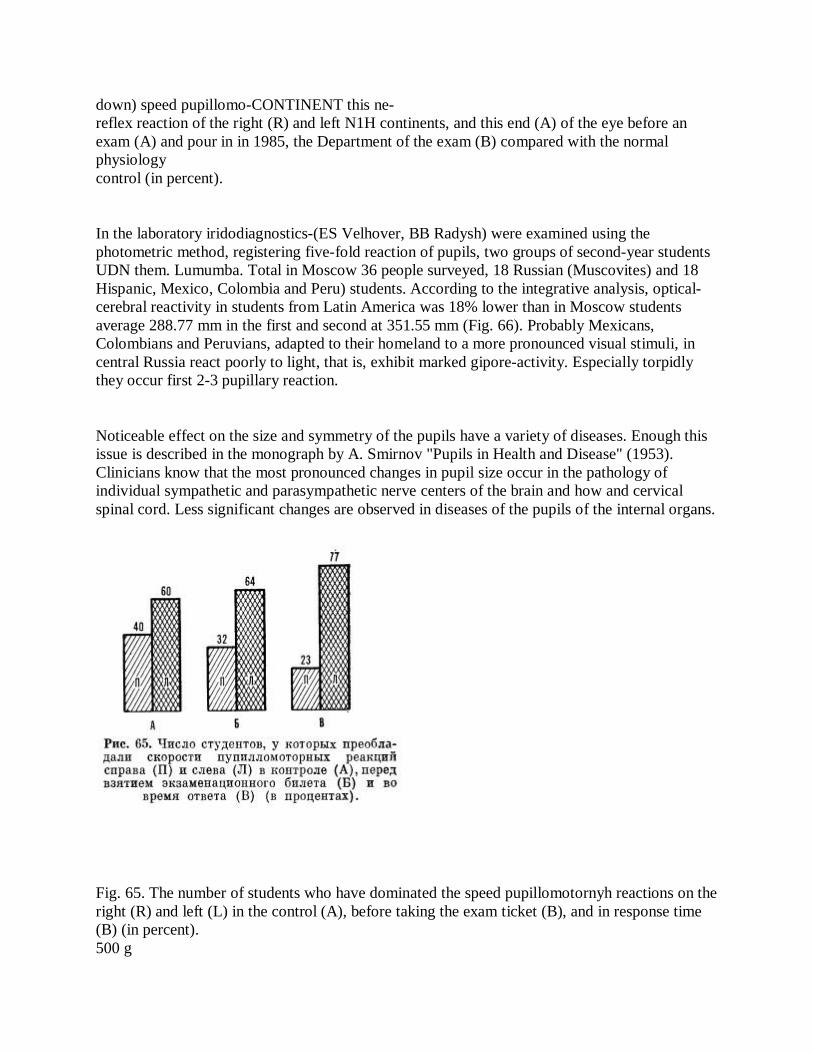
down) speed pupillomo-CONTINENT this ne- reflex reaction of the right (R) and left N1H continents, and this end (A) of the eye before an exam (A) and pour in in 1985, the Department of the exam (B) compared with the normal physiology control (in percent).
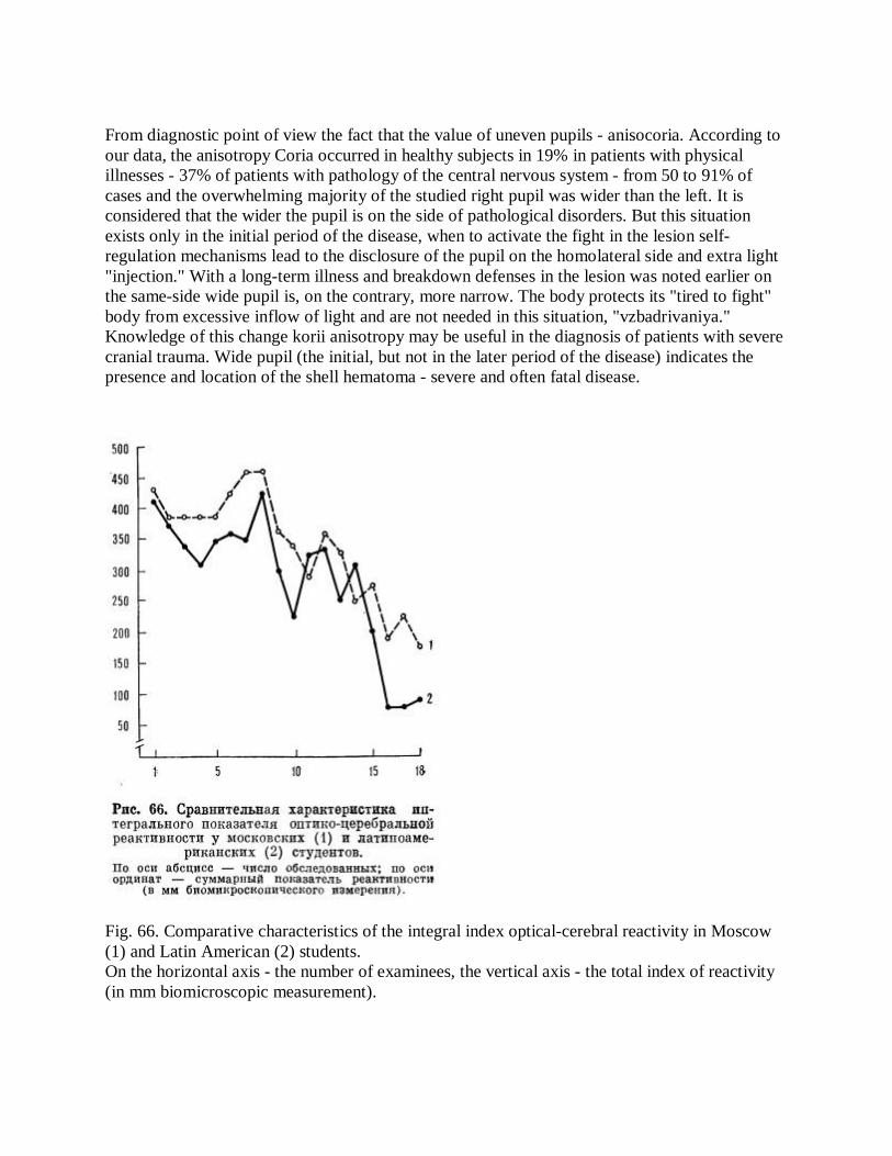
In the laboratory iridodiagnostics-(ES Velhover, BB Radysh) were examined using the photometric method, registering five-fold reaction of pupils, two groups of second-year students UDN them. Lumumba. Total in Moscow 36 people surveyed, 18 Russian (Muscovites) and 18 Hispanic, Mexico, Colombia and Peru) students. According to the integrative analysis, optical-cerebral reactivity in students from Latin America was 18% lower than in Moscow students average 288.77 mm in the first and second at 351.55 mm (Fig. 66). Probably Mexicans, Colombians and Peruvians, adapted to their homeland to a more pronounced visual stimuli, in central Russia react poorly to light, that is, exhibit marked gipore-activity. Especially torpidly they occur first 2-3 pupillary reaction.
Noticeable effect on the size and symmetry of the pupils have a variety of diseases. Enough this issue is described in the monograph by A. Smirnov "Pupils in Health and Disease" (1953). Clinicians know that the most pronounced changes in pupil size occur in the pathology of individual sympathetic and parasympathetic nerve centers of the brain and how and cervical spinal cord. Less significant changes are observed in diseases of the pupils of the internal organs.
Fig. 65. The number of students who have dominated the speed pupillomotornyh reactions on the right (R) and left (L) in the control (A), before taking the exam ticket (B), and in response time (B) (in percent). 500 g
From diagnostic point of view the fact that the value of uneven pupils - anisocoria. According to our data, the anisotropy Coria occurred in healthy subjects in 19% in patients with physical illnesses - 37% of patients with pathology of the central nervous system - from 50 to 91% of cases and the overwhelming majority of the studied right pupil was wider than the left. It is considered that the wider the pupil is on the side of pathological disorders. But this situation exists only in the initial period of the disease, when to activate the fight in the lesion self-regulation mechanisms lead to the disclosure of the pupil on the homolateral side and extra light "injection." With a long-term illness and breakdown defenses in the lesion was noted earlier on the same-side wide pupil is, on the contrary, more narrow. The body protects its "tired to fight" body from excessive inflow of light and are not needed in this situation, "vzbadrivaniya." Knowledge of this change korii anisotropy may be useful in the diagnosis of patients with severe cranial trauma. Wide pupil (the initial, but not in the later period of the disease) indicates the presence and location of the shell hematoma - severe and often fatal disease.
Fig. 66. Comparative characteristics of the integral index optical-cerebral reactivity in Moscow (1) and Latin American (2) students. On the horizontal axis - the number of examinees, the vertical axis - the total index of reactivity (in mm biomicroscopic measurement).

Very difficult estimate of pupils with different coma. Perhaps the origins of understanding to be found in the features of a bioenergy off consciousness. When thyrotoxic, epileptic, eclamptic, liver, and other gipohloremichesky komah eyes wide. Their width indicates the great need of the organism to light energy. It can be assumed that patients with these komami are willing to accept a massive flow of light. When uremic, diabetic and alimentary dystrophy komah pupils, however, are narrow. This indicates a lower body's need for light energy. Perhaps patients with komami not require increased activation, content with a small influx of light and, therefore, low bioenergetic charge.
To study the correlation between the size of the holes and the light kind of disease, we selected several groups of patients of approximately the same age (40-50 years), sex, and with the same eye color. As a control, with the same characters, was under the supervision of a group of healthy subjects (Table 4). Studies have shown that higher levels of light adaptation observed in bronchial asthma, cholecystitis and ulcers. The lowest level of light adaptation, and therefore a lower degree exteroanterocone and interore-tseptivnoy activity occur in cancer.
Thus, in a variety of situations, both in normal and pathological conditions, regulatory light sympathetic and parasympathetic centers provide the optimal amount of individual pupils, ie the optimal flow of light. And all this is done automatically, regardless of the will and desires.
Table 4. The dependence of the diameter of the pupil of the type of disease.
The number of disease pupil diameter (cm) patients right left Stomach cancer 52 8.1 8.1 Duodenal ulcer 74 8.7 8.3 Chronic cholecystitis 68 27 8.5 8.6 Asthma 9.0 8.8 Healthy (control) 40 10.7 10.6
Found that for visual assessment of the pupil in the correct format is typical inaccuracy. Of the
750 subjects visual deformation of the pupil is fixed at 3%, biomicroscopic - in 37% of cases. Change the configuration of the pupils were different, they were observed in one eye or in both simultaneously. In total, we have identified nine types of strain relief: oval-vertical, horizontal oval, oval-diagonal upper oval-diagonal lower, locally flattened top, locally flattened bottom, locally flattened medial, lateral locally flattened multiforme. In Fig. 67 shows the frequency of occurrence of certain kinds of deformation of the right and left pupils. The most common type of oval-vertical deformation and least likely - oval-horizontal and multiforme.
T. Kriege (1971) believes that the oval shape of the pupils in most cases points to an inherited or acquired predisposition to apoplexy states. According to the author, the oval shape of the horizontal pupils observed in individuals who are prone to heart attacks, asthma, depression and psychosis. Oval vertical pupils are a sign of approaching death. T. Kriege writes that when an "vertical ovals" in both iris death occurs during the four days, with the discovery of "vertical oval" in one iris - within 4 weeks. Our studies indicate complete failure prediction "signs" T. Kriege. Oval-shape vertical pupils, two-way and one-sided, we found 34% of the patients, or in 255 patients who were treated over a variety of neurological diseases and therapeutic. However, none of these patients for 24-45 days in bolpitse and in the next period after discharge from her sudden death did not come.
Based on the summary of calculations performed on eight sectors, it has been the direction of the maximum and minimum displacement of the pupil. As a result of this kind of foaming topographic model deformapii pupils (Figure 68). It can be seen that the change in shape of the pupils are unequal uniformly, by type of sector contraction. In this case, the tidal forces operate mainly in the horizontal plane. They lead to more frequent narrowing of the pupil in the sectors of the iris, which projected the heart, lungs and other vital organs.
If we consider the frequency of occurrence of pupillary constriction on the relevant sections of the iris and the projection in strictly descending order, will have the following picture: the right pupil narrows to ezofagotrahealnoy-pharynx region in 50% of cases, cardiopulmonary - 40%, in the brain - 16% in on-Chechnya-genital - 12%, in the liver and oral - 10%, in the neck and the back and vesico areas - in 2% of cases in the left pupil narrows ezofagotrahealnoy-pharynx region in 36% of cases, cardiopulmonary - 28%, in the brain - in 22%, and in the spleen and oral - in 18% of renal re-nitalnoy - 12%, in the neck and the back and vesico areas - in 8% of cases.

Fig. 67. The prevalence of certain kinds of deformations of the right and left pupils (in percent).
Fig. 68. Topography deformation pupils (statistics for the eight sectors).
Sectoral contraction of the pupil on the site-specific data is accompanied by an increase in the area of the iris stroma in the projection areas of the lungs, heart and other organs. This leads to a change in light intensity: the central path (through the pupil and the retina), the flow rate decreases, the peripheral (through the iris) - increases. The physiological significance of such svetoenerge-optical inversion can be explained as follows. Progressive disorder in any organ neurotrophic cause changes in the relevant section of the iris, which weakens the function dilator. Operating in this sector, the sphincter of the pupil has a dominant position. Anatomically, it affects a flattening of the pupil and the iris stroma increase in the area at the site. Physiologically said restructuring means more svetoenergeticheskoy activity expanded at a site iris proektsionpo related to the affected organ.
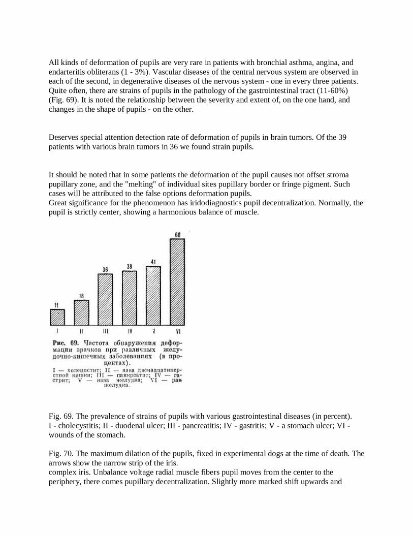
All kinds of deformation of pupils are very rare in patients with bronchial asthma, angina, and endarteritis obliterans (1 - 3%). Vascular diseases of the central nervous system are observed in each of the second, in degenerative diseases of the nervous system - one in every three patients. Quite often, there are strains of pupils in the pathology of the gastrointestinal tract (11-60%) (Fig. 69). It is noted the relationship between the severity and extent of, on the one hand, and changes in the shape of pupils - on the other.
Deserves special attention detection rate of deformation of pupils in brain tumors. Of the 39 patients with various brain tumors in 36 we found strain pupils.
It should be noted that in some patients the deformation of the pupil causes not offset stroma pupillary zone, and the "melting" of individual sites pupillary border or fringe pigment. Such cases will be attributed to the false options deformation pupils. Great significance for the phenomenon has iridodiagnostics pupil decentralization. Normally, the pupil is strictly center, showing a harmonious balance of muscle.
Fig. 69. The prevalence of strains of pupils with various gastrointestinal diseases (in percent). I - cholecystitis; II - duodenal ulcer; III - pancreatitis; IV - gastritis; V - a stomach ulcer; VI - wounds of the stomach. Fig. 70. The maximum dilation of the pupils, fixed in experimental dogs at the time of death. The arrows show the narrow strip of the iris. complex iris. Unbalance voltage radial muscle fibers pupil moves from the center to the periphery, there comes pupillary decentralization. Slightly more marked shift upwards and
inwards. It is important to note that the location of the pupil is not due to excessive voltage, certain muscle groups, and their attenuation. Therefore, the displacement of the pupil up to look for abnormalities in the lower areas of the projection of the kidneys, reproductive organs, the displacement inwards - in the projection areas of the heart, the aorta and the bronchopulmonary system. However, it should be noted that the interpretation of the phenomenon of pupillary decentralization should take into account other pathological manifestations in the iris stroma. The presence of bias is not supported by other signs of iris, indicates only the positive tone of the functional ^ enie relevant authorities.
It has been mentioned that the regulation of admission of light energy produced in the body automatically throughout a person's life. Dying is always accompanied by an increase in the broadest pupils. Undoubtedly, this again is protective, but, unfortunately, the last measure of the body, increasing the inflow of light trying to save the life of fading mechanisms. NK Bogolepov (1962) argues that the expansion of pupils is as cardinal signs of death as cessation of breathing and heart rate. In his view, any moribund patient with the missing heart and breathing, but with narrow pupils is not hopeless, and requires resuscitation.
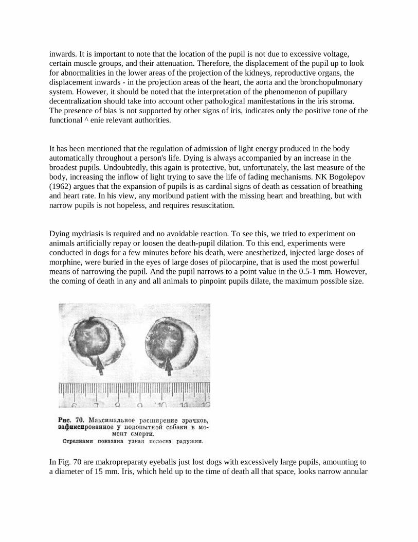
Dying mydriasis is required and no avoidable reaction. To see this, we tried to experiment on animals artificially repay or loosen the death-pupil dilation. To this end, experiments were conducted in dogs for a few minutes before his death, were anesthetized, injected large doses of morphine, were buried in the eyes of large doses of pilocarpine, that is used the most powerful means of narrowing the pupil. And the pupil narrows to a point value in the 0.5-1 mm. However, the coming of death in any and all animals to pinpoint pupils dilate, the maximum possible size.
In Fig. 70 are makropreparaty eyeballs just lost dogs with excessively large pupils, amounting to a diameter of 15 mm. Iris, which held up to the time of death all that space, looks narrow annular

strip shifted to the periphery. Expanding pupils, co-death, after a few hours (corpse) narrowed to medium size.
Physiologists believe that after the heart and diaphragm muscle sphincter and dilator of the pupil are the most mobile and capable of working muscles. G using them from birth to death are functioning eyes - these truly critical hole of the human body.
INDEPENDENT RING In iridology area between two zones of iris - pupillary and ciliary body - called autonomous ring, as it is a projection of the autonomic nervous system. Diagnostic value of this area is extremely high, first, because it is an indicator of all visceral systems, and secondly, because it is the main point of reference for topical iridodiagnostics. Is extremely important for autonomous ring automatic analysis of the state of the iris using iridotronnyh devices. It is noticed that in the iris ring newborn independent almost indistinguishable, and vice versa, it is clearly expressed in the elderly. R. Bourdiol (1975) believes that the structure of autonomous ring has a double origin: embryological (from fetal membranes) and vascular (small blood vessels of the circle).
Standalone ring looks flat or a broken line, slightly raised above the deep mesodermal sheet by forming the line of major trabeculae. Traben-specified molecules include quite a powerful system of blood vessels - small arterial circle of the iris. Standalone ring - education is dynamic, because it can shrink and expand depending on the ever-changing values of pupillary zone and the pupil. In expanding pupil pupillary zone much narrower and the front surface of the iris descends steeply to the pupillary margin, which makes the autonomous inspection of the ring. If you decrease the pupil is enlarged pupil zone, resulting in a line of standalone ring becomes more clear and distinct. Therefore biomicroscopic study of autonomous ring is best done with narrow pupils, using a bright light source.
G. Iausas (1974) believes that the height and width of the ring can be judged independent of the sympathetic nervous system function.
Fig. 71. The most common forms of autonomous ring and - flat, b - gear, in - retracted, d - extended.
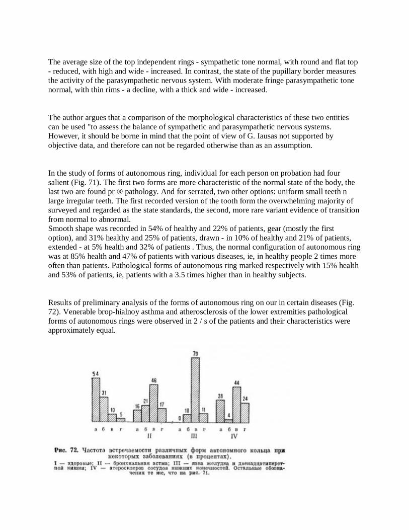
The average size of the top independent rings - sympathetic tone normal, with round and flat top - reduced, with high and wide - increased. In contrast, the state of the pupillary border measures the activity of the parasympathetic nervous system. With moderate fringe parasympathetic tone normal, with thin rims - a decline, with a thick and wide - increased.
The author argues that a comparison of the morphological characteristics of these two entities can be used "to assess the balance of sympathetic and parasympathetic nervous systems. However, it should be borne in mind that the point of view of G. Iausas not supported by objective data, and therefore can not be regarded otherwise than as an assumption.
In the study of forms of autonomous ring, individual for each person on probation had four salient (Fig. 71). The first two forms are more characteristic of the normal state of the body, the last two are found pr ® pathology. And for serrated, two other options: uniform small teeth n large irregular teeth. The first recorded version of the tooth form the overwhelming majority of surveyed and regarded as the state standards, the second, more rare variant evidence of transition from normal to abnormal. Smooth shape was recorded in 54% of healthy and 22% of patients, gear (mostly the first option), and 31% healthy and 25% of patients, drawn - in 10% of healthy and 21% of patients, extended - at 5% health and 32% of patients . Thus, the normal configuration of autonomous ring was at 85% health and 47% of patients with various diseases, ie, in healthy people 2 times more often than patients. Pathological forms of autonomous ring marked respectively with 15% health and 53% of patients, ie, patients with a 3.5 times higher than in healthy subjects.
Results of preliminary analysis of the forms of autonomous ring on our in certain diseases (Fig. 72). Venerable brop-hialnoy asthma and atherosclerosis of the lower extremities pathological forms of autonomous rings were observed in 2 / s of the patients and their characteristics were approximately equal.

Fig. 72. The incidence of various forms of autonomous ring at some diseases (in percent). I - healthy; II - asthma; III - gastric and duodenal ulcers; IV - atherosclerosis of the lower extremities. The remaining symbols are the same as in Fig. 71.
Fig. 73. The cardinal sign of strain duodenal bulb - local retraction ring e autonomous sector "5.00-7.00". Nakov. The bulk of the ring strain independent of bronchial asthma treated topically to the projection area "light - the bronchi," in atherosclerosis of the lower extremities, a projection area "of the brain - the pelvic organs."
The situation is different with gastric ulcer and duodenal ulcer. Pathological forms of autonomous ring made in this disease 90%, is very characteristic sign of duodenal ulcer was a local autonomous retraction ring in both eyes on the meridian "5.00-7.00" (Fig. 73). Indrawing configuration was very different. In 116 patients, it was observed in both the iris in 14 patients - only on the left, and 10 - just right. Specific symptoms observed by us in 95% of cases, confirmed Gast-microscopic and radiological detection of deformation duodenal bulb [Velhover ES, Pichkhadze RS, Sherstnev VP. 1980]. In healthy people, he was observed in 3% of patients with gastritis - in 10% of cases. Moreover, X-ray of the 5 patients (3 with an ulcer on the front and 2 at | the lesser curvature of the bulb), in which the local retraction on iridogrammah not. Coincided with data gastroscopy, a symptom of deformation of the duodenal bulb was found in 4 patients. This gives us reason to believe that the local autonomous vtya-tion ring in sector "5.00-7.00" from both sides is a cardinal sign of strain duodenal bulb (Fig. 74, see insert).

For changes in the configuration of the ring many autonomous iri-long detect bowel stricture (Figure 75), diverticulosis (Fig. 76) and some other lesions of the gastrointestinal tract. We observed two patients with lateral displacement of the small intestine caused by tumor growth. A characteristic feature of these patients was iridogramm flattening inside standalone ring, indicating disturbances in the projection areas of the small intestine.
In some cases the line is independent of the ring is interrupted laterally spaced cracks. Observation R. Voig-diol (1975), an autonomous crack ring show marked changes in the spine.
Very often transverse cracks are located at the top of the Autonomous ring, which can be clearly seen by biomicroscopy in side illumination. This kind of signs are most common in the axon forms of osteoarthritis of the cervical spine. Seems very promising diagnostics iris prolapse of the transverse colon, less omissions and displacement of the stomach and other organs [Maubach A., 1952; Iensen B., 1964; Kriege, T., 1971, and others].
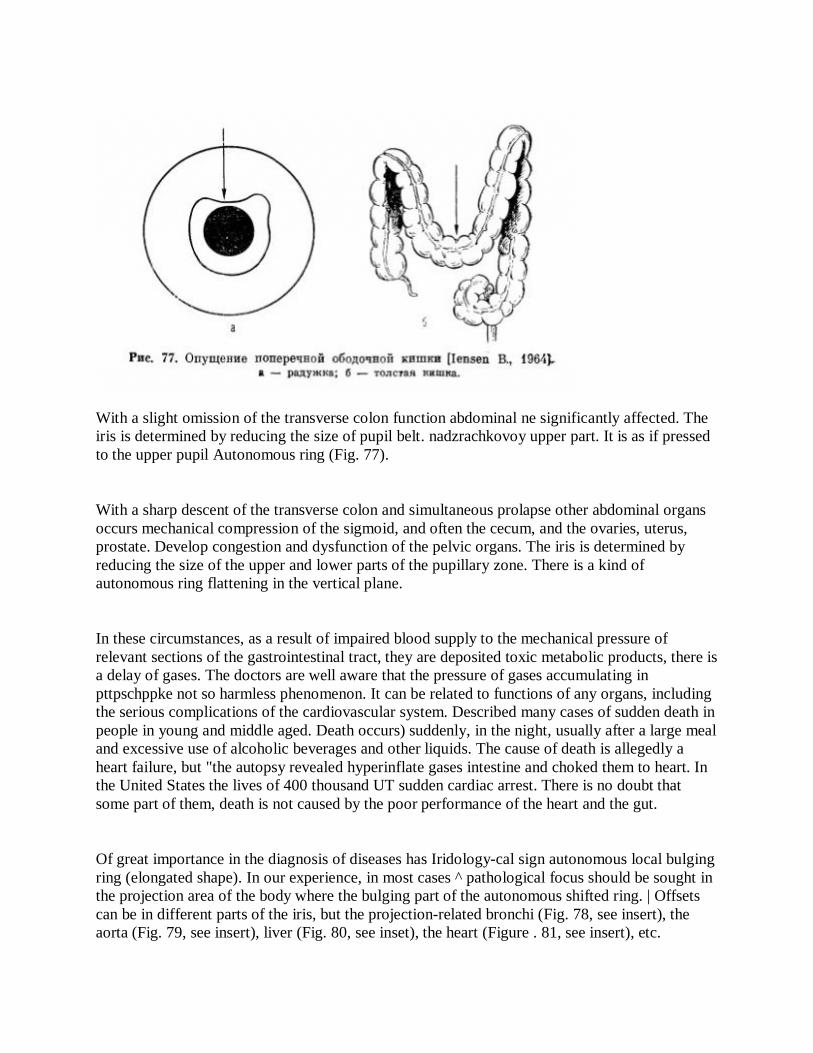
With a slight omission of the transverse colon function abdominal ne significantly affected. The iris is determined by reducing the size of pupil belt. nadzrachkovoy upper part. It is as if pressed to the upper pupil Autonomous ring (Fig. 77).
With a sharp descent of the transverse colon and simultaneous prolapse other abdominal organs occurs mechanical compression of the sigmoid, and often the cecum, and the ovaries, uterus, prostate. Develop congestion and dysfunction of the pelvic organs. The iris is determined by reducing the size of the upper and lower parts of the pupillary zone. There is a kind of autonomous ring flattening in the vertical plane.
In these circumstances, as a result of impaired blood supply to the mechanical pressure of relevant sections of the gastrointestinal tract, they are deposited toxic metabolic products, there is a delay of gases. The doctors are well aware that the pressure of gases accumulating in pttpschppke not so harmless phenomenon. It can be related to functions of any organs, including the serious complications of the cardiovascular system. Described many cases of sudden death in people in young and middle aged. Death occurs) suddenly, in the night, usually after a large meal and excessive use of alcoholic beverages and other liquids. The cause of death is allegedly a heart failure, but "the autopsy revealed hyperinflate gases intestine and choked them to heart. In the United States the lives of 400 thousand UT sudden cardiac arrest. There is no doubt that some part of them, death is not caused by the poor performance of the heart and the gut.
Of great importance in the diagnosis of diseases has Iridology-cal sign autonomous local bulging ring (elongated shape). In our experience, in most cases ^ pathological focus should be sought in the projection area of the body where the bulging part of the autonomous shifted ring. | Offsets can be in different parts of the iris, but the projection-related bronchi (Fig. 78, see insert), the aorta (Fig. 79, see insert), liver (Fig. 80, see inset), the heart (Figure . 81, see insert), etc.
The phenomenon of local bulging (FLV) Autonomous ring in the projection area of the heart, we studied carefully [Velhover ES, A. Karasev, Radysh BB, 1985, 1986]. Bulging found in the lateral sections of both irises. The sample of 677 patients and 232 healthy people aged 13-16 years. FLV was observed in healthy subjects: the right-to 9.5%, on the left - in 26.3% of cases. In patients with heart disease (acute myocardial infarction, myocardial infarction, angina, rheumatic heart disease): Right - 28,6 - 31,8%, from left to 80,3-83,7% of cases. Thus was established the high frequency of the FLV in patients with heart disease, 3 times higher than that in healthy (differences statistically significant, p <0.05-0.001). Processing of iridium-graphic materials (color slides) of the patients with myocardial | attack on the computer brand «Ibas» has provided interesting data. It was found that the optical density of the projection area of the heart on the iris in patients with myocardial infarction is significantly different on the structural characteristics from those of the healthy controls. For registogramm patients have a wide frequency range of optical density is 2-3 times greater than in healthy people.
Comparing iridology findings with clinical and instrumental examinations, we have g conclusion that dislocation figures Autonomous ring-type local bulging correlated with the presence of hypertension in different cavities of the heart. Based on ECG, echocardiography and roentgenocardiography, we found that myocardial hypertrophy and dilatation of the heart is reflected in the iris as a standalone FLV ring. These results have provided new insights into the localization of the individual parts of the heart on the iris. The right iris reflects the state of the right heart. Left - left heart. The ratio of the frequency of finding FLV the left and right iris averaged 3: 1, ie, to the left of the ring strain autonomous observed in 3 times more often than the right. Hypertension in the right atrium was shown offset Autonomous ring in sector "9.30-9.50" in the right ventricle - in the sector, "8.50-9.30". Increased pressure in the left atrium, accompanied by the appearance FLV sector "2.00-2.30" in the left ventricle - in the sector, "2.30-3.20".
Pathogenetic mechanisms of dilatation of the heart cavities originally anticipated compensatory hypertrophy. For differentiation of these processes of reflection on the iris, we have identified four independent degree of deformation of the ring: light, medium, coarse and tear Autonomous ring. Usually mild consistent compensatory hypertrophy, the average - the initial stages of dilation, rough - pronounced dilatation. Autonomous ring gap was seen in the investigated patients in individual cases and to reflect together with marked dilatation of the heart cavities formation processes, heart aneurysm.
Research results indicate the feasibility of autonomous registration FLV ring at screening Iridology heart disease. This - the first verified iridology symptom heart disease, which can be used not only in clinical practice, but m for mass preventive examinations of the population.

Considerable interest in the study of mental illness using the iridodiagnostics. These are the results of studies 280 patients with schizophrenia. At the most "of the patients were set following changes in the brain area iris: friable nature of the stroma, the presence of gaps, a large number of adaptation arcs and rings [Velhover ES, Elisha J. M., 1983]. However, the most typical symptom of schizophrenia iridology was bulging ring autonomous zone in the brain iris observed in 75% of cases [Velhover ES, Elisha J. M., Sineok S., 1986]. There were various options for deployment of autonomous ring. In some cases it was a double bulging without breaks Autonomous ring - a symptom of the "horns", while others - bulging with damaged integrity of autonomous ring in sector "11.30-12.40" (Fig. 82).
Experience shows that the gap symptom Autonomous ring "indicates irreversible pathology relevant parts of the autonomic nervous system. We observed two men aged 40 and 46 years with a break autonomous "rings verhnemedialnom quadrant of the iris. Both registered the sudden death of the background of relatively prosperous state of health. It is quite clear that the conclusions of Picacho, two observations should be done. Based on these, we can only speculate how serious dolzhep be search for the causes and physiological nature breaks Autonomous ring. B. Iensen (1984) believes that the location of the ring breaks autonomous use. Changes in schizophrenia is independent of the ring [Velhover ES, Elisha J. M., Sineok S., 1983].
And - the phenomenon of double bulging Autonomous ring (a symptom of "horns"), b - a phenomenon bulging with breaking Autonomous ring in sector "11.30-12.40" suggest hypothyroidism relevant authorities, which may, even celebrated in the embryonic stage of development.
In some cases the border autonomous rings may be difficult, so it is unclear in places like "melting." A similar pattern was observed in 16% of patients aged 40-50 years old, many of whom had cancer.
In 56% of the investigated paintings Autonomous ring right and left eyes were markedly
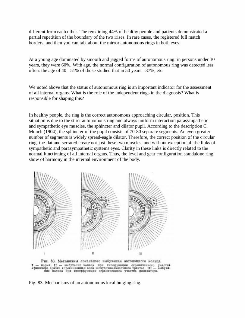
different from each other. The remaining 44% of healthy people and patients demonstrated a partial repetition of the boundary of the two irises. In rare cases, the registered full match borders, and then you can talk about the mirror autonomous rings in both eyes.
At a young age dominated by smooth and jagged forms of autonomous ring: in persons under 30 years, they were 60%. With age, the normal configuration of autonomous ring was detected less often: the age of 40 - 51% of those studied that in 50 years - 37%, etc.
We noted above that the status of autonomous ring is an important indicator for the assessment of all internal organs. What is the role of the independent rings in the diagnosis? What is responsible for shaping this?
In healthy people, the ring is the correct autonomous approaching circular, position. This situation is due to the strict autonomous ring and always uniform interaction parasympathetic and sympathetic eye muscles, the sphincter and dilator pupil. According to the description C. Munch (1904), the sphincter of the pupil consists of 70-80 separate segments. An even greater number of segments is widely spread-eagle dilator. Therefore, the correct position of the circular ring, the flat and serrated create not just these two muscles, and without exception all the links of sympathetic and parasympathetic systems eyes. Clarity in these links is directly related to the normal functioning of all internal organs. Thus, the level and gear configuration standalone ring show of harmony in the internal environment of the body.
Fig. 83. Mechanisms of an autonomous local bulging ring.
I - rate; II - bulging ring with hypothyroidism limited portion of the sphincter of the pupil (the projection area of the gastrointestinal tract); III - bulging ring with hyperthyroidism limited area dilator.
But it is necessary in any organ or system of the body, in the intestines, lungs, etc. - appear pathological focus, as there is increased flow of impulses, which focused on the impact on a particular segment of the iris. During long-term and persistent changes in the organs and, therefore, when, long-existing vistseroiridalnyh impulses occur as pigmentation disorders and neuro-muscular elements iris. There disharmony yaervnoy autonomous system, fragmentary weakening and loss of function of the sympathetic and parasympathetic muscles of the eye, which inevitably leads to a "break-up" forms of autonomous ring. It takes the form of anomalous: irregularly toothed, busy, etc.
In Fig. 83 schematically illustrates the mechanism of abnormal configuration standalone ring - elongated shape. This mechanism is very complex and ambiguous. There are two possibilities of neuromuscular relationships. The first is that the bulging portion of the ring is an autonomous part of the pupillary zone of iris, the projection associated with abnormal • hearth in the gastro-intestinal tract. The defeat of the proceeding by loss and indicated in the diagram "minus" sign. Sphincter of the pupil in this area dramatically reduced, marked deformity of the pupil and discoloration in the stroma and the relief of pupillary zone. "Plus" indicated peripheral iris area, on which a relatively strong dilator. Autonomous regional bulging ring toward periphery is influenced dilator having a more pronounced tone, that is, as a result of the difference between the two opposing forces of the eye muscles.
Violation of the second type is because vybu-damped autonomous area 'of the ring does not shift laterally ciliary zone, and as bv is coming at him. This occurs when the projection of the lesion is located outside from the ring and there is a stand-alone syndrome irritation. The corresponding projection won the iris, in our example - the zone of the aorta, indicated in the diagram a "plus", is a more active part of dilator, which tightens to his inner dilator fibers and functionally less active fragment Autonomous ring.
Thus, the local autonomous bulging ring can be observed in two situations: first, when hypothyroidism limited portion of the sphincter of the pupil (the projection area of the stomach and intestines), and secondly, the limited area of hyperactivity dilator (flat area to other bodies in the iris).
In iridology is important not only to assess the form of autonomous ring, but his appearance or. more precisely, of purity. Distinguish between pure and so-called intoxicated avtopompye ring (Fig. Si, 85, see insert). Pure ring characterized by clear, distinct, and, as a rule, thin borders. These rings are found almost exclusively in healthy people. For the majority of patients, and a

small part of healthy characteristic of the ring is intoxicated, the distinguishing feature of which are unclear or pigmented border. These rings typically lose a linear form and turn into wide, raised above the surrounding stroma strip resembling the parapet. Some call them iridologp autonomous dual-circuit rings, suggesting that they are based on vascular congestion phenomenon. Such rings are painted in a darker color. Depending on the width of the intensity of color n moyasno say slagging autonomous rings (1st, 2nd and 3rd degree). In plow understanding slagging phenomenon indicates trophic disorders in the corresponding zone of iris, indicating dysfunction of the autonomic nervous system. , This primarily affected the activities of the Border sympathetic.
Fig. 86. The incidence of various types of autonomous ring in healthy (1) and patients (2) people. I - pure; II - Slagging I degree; III - Slagging II degree; IV - Slagging III degree. The vertical axis - the number of cases detected autonomous rings (%).
In Fig. 86 shows the frequency of detection of different types of autonomous rings in healthy and sick people. What Slagging Autonomous ring detected in the majority of patients, is not unexpected. What is interesting fact - 34% of intoxicated autonomous rings 1 st and 2 nd degree, found in healthy people, do not speak in favor of their health and require appropriate diagnostic and therapeutic measures.
ADAPTATION RINGS
Adaptive iris rings are known as ophthalmologists ring contraction. They are located on the periphery of the iris in the form of curved or circular depressions in the stroma, concentrically surrounding the pulmonary circulation (Fig. 87, see insert). Ring contraction, or contraction of the ring can be light and dark, narrow and wide, smooth and sinuous.
Some iridology, in particular G. Iausas (1974), call them signs of deepening, or "cardiac rings." When considered with sufficient increase in pigmentation disorders can be seen, but not of the trabeculae. The impression is that the iris is at this point though is cracked, that pigment layers lose their flexibility and unity, have ceased to follow the progress of trabeculae. According to ML Krasnov (1952) and NB Shulpina (1974), the occurrence of these structures due to the activities of neuromotor apparatus of the eye, with contraction and expansion of the surface layers of the iris.
However, the reduction of the ring as a simple fold of iris is not quite correct. R. Bourdiol (1975) believes that an analysis of this phenomenon it is necessary to take into account four factors: 1) according to histology, there is no front of the boundary layer at the level mentioned folds, and 2) the folds are not all people, whereas the narrowing and dilatation of the pupil is an important function of each iris, 3) called folds have the right, and some even finished form, and 4) folding does not increase significantly with mydriasis and decreases with myositis.
In special literature iridology ring reductions called nerve rings. According to V. Iensen (1970), their presence in the iris indicates excitation and spasms of one or more organs from trauma, inflammation, mental stress or drug overload. If the fibers of the stroma in the nerve ring is deeply impressed, then it means that the corresponding body is excited and is prone to severe cramps. G. Iausas (1958) found that the nerve ring is especially common in heart disease, and, on this basis, they proposed the term heart rings.
The authors found that of the 100 carriers of nerve rings 66 have heart disease, 27 - neurosis kardialgicheskie syndromes and 7 - purely nervous disorders. Moreover, 93% of "core" under carefully conducted survey revealed a number of complaints that point to heart disease: pain in the heart, arrhythmia, shortness of breath, etc.
R. Bourdiol (1969) found the nerve ring in all patients spazmofiliey. Each patient, carrier rings, clinically and by electromyography showed positive symptoms chvostek and "hands obstetrician." R. Schnabel (1959) and R. Bourdiol (1974) wrote that the concentric rings of an indicator spazmofilii, nervous irritability, labile neyrovege-tatiki manifesting convulsions, spasms and contractions. They often occur in manic individuals and expressed "neurotic." In psychotic states, disease-Freed Reich and some other hereditary degenerative diseases acquire not concentric rings, and oval.
On the nerve ring is described as a phenomenon relevant to the changing metabolic stability, which invariably leads to a decrease in tissue elasticity, and with age - to the sclerotic changes in the body.
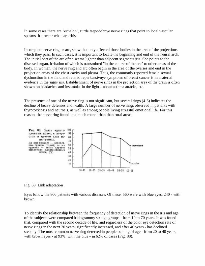
In some cases there are "echelon", turtle tsepodobnye nerve rings that point to local vascular spasms that occur when arteritis.
Incomplete nerve ring or arc, show that only affected those bodies in the area of the projections which they pass. In such cases, it is important to locate the beginning and end of the neural arch. The initial part of the arc often seems lighter than adjacent segments iris. She points to the diseased organ, irritation of which is transmitted "in the course of the arc" to other areas of the body. In women, the nerve ring and arc often begin in the area of the ovaries and end in the projection areas of the chest cavity and pleura. Thus, the commonly reported female sexual dysfunction in the field and related reperkussivnye symptoms of breast cancer is its material evidence in the signs iris. Establishment of nerve rings in the projection area of the brain is often shown on headaches and insomnia, in the light-- about asthma attacks, etc.
The presence of one of the nerve ring is not significant, but several rings (4-6) indicates the decline of heavy defenses and health. A large number of nerve rings observed in patients with thyrotoxicosis and neurosis, as well as among people living stressful emotional life. For this reason, the nerve ring found in a much more urban than rural areas.
Fig. 88. Link adaptation Eyes follow the 800 patients with various diseases. Of these, 560 were with blue eyes, 240 - with brown.
To identify the relationship between the frequency of detection of nerve rings in the iris and age of the subjects were compared iridogrammy six age groups - from 10 to 70 years. It was found that, compared with the second decade of life, and regardless of the color eye detection rate of nerve rings in the next 20 years, significantly increased, and after 40 years - has declined steadily. The most common nerve ring detected in people coming of age - from 20 to 40 years, with brown eyes - at 93%, with the blue - in 62% of cases (Fig. 88).
The establishment of the greatest number of nerve rings in the most active in the labor and psycho-emotional for a minimum period of life and nerve rings during progressive aging shows, first, that it does not imply (but does not exclude) a direct link between the disease and the presence of the nerve ring , otherwise they would come up to elderly age, with its many diseases and disorders. Second, open up interesting possibilities for a very simple and accurate assessment of human emotional stress. Such an assessment is always necessary in a mental practice, especially in cases where for outward calm and balance of an individual hiding labile and hypersensitive nature.
Curves shown in Fig. 88, clearly illustrate the difference in the presence of nerve rings in people with blue and brown eyes. This difference is very stable, as noted in all age groups. From a total of 240 patients with dark-eyed nerve rings were found in 84% of the 560 blue-eyed - only 37% of cases. Thus was established a definite relationship in the incidence of nerve rings of color human eyes. In people with dark eyes, they met 2.3 times more likely than blue-eyed. Perhaps this suggests that the neuromotor apparatus and associated feature light adaptation developed in the former more than the latter. Confirmation is often observed differences in life-, relatively high reactivity of dark-eyed people than blue-eyed.
Similar to our data published G. Iausas (1974), found the following incidence of neural arches with residents in the south of France: for men with light iris - 33%, with a dark-60% of women with light iris - 43%, with the dark - 66%. It follows that the presence of nerve rings and arcs often found in women than in men.
Analysis of the data showed that the nerve rings and arcs are located exclusively in the peripheral regions of the iris, ciliary its belt. Very rarely they are in the pupillary zone of the projection associated with the gastrointestinal tract. Of the 800 surveyed patients only 2 were found neural arch in the vicinity of the pupil.
Nerve rings and arcs detected in both patients and healthy people, most of them were from 1 to 3 rings. Less frequent with 4 persons and rarely with more rings. Unclosed concentric grooves, or arc, located in different places in their number, they got over the circular grooves or rings.
To determine the location of the nerve ring of iris ciliary zone we divided by differing in size, but similar in the functional significance of the projection area, according to the topographic pattern B. Iensen. I topically zone corresponds to the representation of the brain; II - maxillo-li-tsevoy area; III - the back; IV - the liver, kidneys and reproductive organs; V-of the heart and lungs; VI - the neck. Calculation of curved and circular grooves on the iris was made based on "Dial division", which allowed to determine the disposition of nerve rings in the zones of the iris in the average (Figure 89).
The most common nerve rings and arcs were found in areas IhV, the projection corresponding to the brain, lungs I1 heart. These particularly active in the field of functional otnoshenii1 had an

average of 2.03 and 2.01 nerve rings. The smallest number of nerve rings located in zones III and VI, the projection associated with the back and neck. These functionally less active sections of the body had an average of 1.78 and 1.8 of nerve rings. It is interesting to note that the area of the greatest number of rings (area of the brain) was not the biggest in size, just as the area of the smallest number (area behind) was not the small.
When comparing the nerve ring to the state of the pulmonary circulation iris projection associated with autonomic nervous system *, was a direct correlation between the number of nerve rings and clean the small circle. When thickened and intoxicated small circle or ring off, the nerve ring were found in 43% more than in pure stand-alone " ring. This means that to alter the function of the autonomic nervous system is increasing steadily and the number of nerve rings.
Fig. 89. The number of rings adaptation in different sectors iris (the average value).
To study the association between nerve rings with various diseases were divided into 3 groups of patients: asthma, holetsistopankreatitom and gastric cancer. The control group comprised 43 healthy people. The age of the subjects in all groups were similar and ranged from 30 to 50 years (Table 5).

Table 5. The relationship between the frequency of detection of nerve ring and the kind of disease.
The disease is the number of subjects frequency of detection of nerve ring (%)
Asthma 40,100
Pancreatic 46, 65
Stomach cancer 24 19
Healthy (control) 43 33
In healthy people the nerve ring identified rare - one in three. They were located in all zones of the iris, had a whitish hue and sufficient width and depth. The presence of several rings often coincides with the state of irritability and emotional instability, with the presence of neurotic symptom. Characteristically, the vast majority of healthy people furrows in the iris are concentric closed, that is, had the form of rings.
In bronchial asthma, nerve rings were found in 100% of patients. Most often, they were not closed, and had form arcs and semirings. Localized these arcs and semicircles mainly in the zone V, corresponding to the light projection.
When holetsistopankreatit nerve rings and arcs were observed in 65% of patients. Basically, they were located in areas I and IV, the projection associated with the brain, biliary and pancreatic systems.
In gastric cancer nerve ring occurred in 19% of patients, ie, much less than in healthy people and those with other diseases. This figure is very significant for the quantitative characterization of the phenomenon of nerve rings. Assuming that the number of nerve rings in the iris increases in
direct proportion to the painful process, it related to cancer care number (19%) did not confirm the legitimacy of this assumption, as patients in this group can not be the "health healthy". Interestingly, the nerve ring in cancer patients differed from nerve rings in other patients, not only quantitatively but also qualitatively. Concentric closed rings were observed in them is extremely rare, but often detected subtle, subtle arc. These thread-like, as if melting into the ciliary sulcus belt were pathognomonic for terminal cancer. They were the kind of evidence-activity and arrest the decline of the body's defenses. More correct "to say hyporeactivity because melting arc reflects the struggle of the body, weak and decaying, but still struggle.
What is the physiological significance and diagnostic value of nerve rings?
It is believed that the basis for the formation of nerve arcs and rings is a continuous "game" neyromotorov iris. This process is very complex and, we believe, are unequal in tonic regard. Certain emphasis in it belongs to the sympathetic nervous system and the pupil dilator. That reduction dilator while relaxing the sphincter of the pupil leads to reduction of furrows in the stroma of ciliary zone, is a zone of their permanent location. Dilatation of the pupil under the dilator, education and the relatively thin flattened radially, collects tissue ciliary zone in concentric folds. Summed over time frequent and sharp mydriasis alternating with moments and periods of contraction, contribute to the formation of nerve arcs and rings. They occur in the tissues innervated by sympathetic nerves, representing a non-specific reactive phenomenon. Strong emotions, physical and mental stress, stress, aches and pains and irritants, stimulating the sympathetic nervous system, cause the development of nerve rings.
One of the main stimuli of sympathetic nervous system is the light, the intensity and the parameter changes which largely depends on the "game" neyromotorov. Perhaps, therefore, a brown-eyed people, most of whom live in the south, the nerve ring occur more often than people with light eyes.
We have stated that in some cases there are annular grooves in the other - arched. What is the cause of time-differences? We believe that the mechanisms responsible for the formation of neural arcs and rings, varied combination and concluded both in the external and the internal environment of the body. If the factors stimulating the sympathetic nervous system (pain impulses, light and other stimuli), are long and hard enough on the person, not burdened with disease - there totality dilator reduction, resulting in the formation of the nerve ring. This is a protective measure of the organism, its generalized adaptive response. The source of these reactions are primarily used by environmental stimuli. If the same factors at * strength and durability to act on a person with a particular focal pathology (a majority) - strongly reduced dilator not all, and some of its segments, resulting in the neural arch. As well as the ring, nervous arc reflect the protective properties of the body, but not generalized, and local character. The source of their curves are vnutrisredo-irritants, mainly lesions.
Research results show that on a number of attributes - mind, shape, location, nerve arcs and rings can objectively judge the general and local voltage apparatus sympathetic eyes. These features can be seen on the state of reactivity, emotional expression, and the level-scale adaptive-protective changes in the body.
Iridology Most agree that the owners of the nerve rings - susceptible subjects who apparently do not show their emotions, and hide them inside. Usually, this is an extremely sensitive and reserved people.
The study of neural arcs and rings as a physiological and diagnostic tests can provide useful information, which will serve to further improve clinical and sports medicine.
As for terminology, in our understanding, existing in the literature as "nerve ring" and "ring contraction" is not disclosed adequately described the essence of the phenomenon. More complete and appropriate name would be "adaptive ring", which we propose to introduce in place of the existing ones.
Pupillary Border
Pupillary border or fringe of pigment, is the inner edge of the iris. It is formed by the ectodermal layer of the pigment epithelium, which is out of the iris around the pupil forming w original pigment "necklace." Pupillary rim develops from the front of the embryonic optic vesicle fetus. Typically, the pupillary rim looks like a beautiful velvet rims, dark brown or black. It is very sensitive to light stimuli cause the oscillates, as if constantly "breathing". In the state of pupillary miosis border is much better expressed. Therefore, the study of its method is preferably carried out at biomicroscopy pupillary constriction, using a bright light source. In order to obtain accurate data in the study of the dynamics of the pupillary border must for all subsequent measurements to achieve the same size of the pupil.
In some cases, outside from the pupillary border can see the contours of the rainbow sphincter, which is a light-gray, sometimes yellowish-gray strips of 1 mm surrounds pupillary border. If the anatomy of the pupillary border is well understood, then it can not be said about its function and role in the general semiotics diseases. In this section we will try to present a number of known and new facts with which to reveal the physiological significance of the pupillary border and other pigmented eyes.
It is known that penetrates the eyeball light is a complex energy flow, which consists of
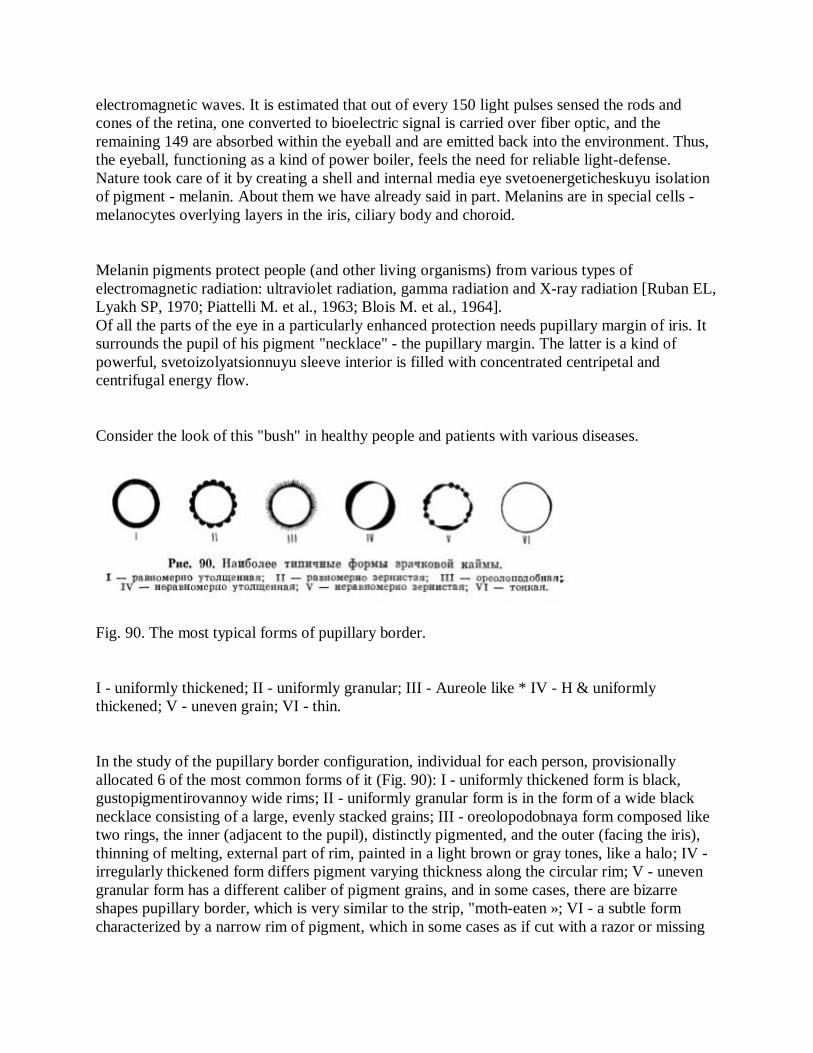
electromagnetic waves. It is estimated that out of every 150 light pulses sensed the rods and cones of the retina, one converted to bioelectric signal is carried over fiber optic, and the remaining 149 are absorbed within the eyeball and are emitted back into the environment. Thus, the eyeball, functioning as a kind of power boiler, feels the need for reliable light-defense. Nature took care of it by creating a shell and internal media eye svetoenergeticheskuyu isolation of pigment - melanin. About them we have already said in part. Melanins are in special cells - melanocytes overlying layers in the iris, ciliary body and choroid.
Melanin pigments protect people (and other living organisms) from various types of electromagnetic radiation: ultraviolet radiation, gamma radiation and X-ray radiation [Ruban EL, Lyakh SP, 1970; Piattelli M. et al., 1963; Blois M. et al., 1964]. Of all the parts of the eye in a particularly enhanced protection needs pupillary margin of iris. It surrounds the pupil of his pigment "necklace" - the pupillary margin. The latter is a kind of powerful, svetoizolyatsionnuyu sleeve interior is filled with concentrated centripetal and centrifugal energy flow.
Consider the look of this "bush" in healthy people and patients with various diseases.
Fig. 90. The most typical forms of pupillary border.
I - uniformly thickened; II - uniformly granular; III - Aureole like * IV - H & uniformly thickened; V - uneven grain; VI - thin.
In the study of the pupillary border configuration, individual for each person, provisionally allocated 6 of the most common forms of it (Fig. 90): I - uniformly thickened form is black, gustopigmentirovannoy wide rims; II - uniformly granular form is in the form of a wide black necklace consisting of a large, evenly stacked grains; III - oreolopodobnaya form composed like two rings, the inner (adjacent to the pupil), distinctly pigmented, and the outer (facing the iris), thinning of melting, external part of rim, painted in a light brown or gray tones, like a halo; IV - irregularly thickened form differs pigment varying thickness along the circular rim; V - uneven granular form has a different caliber of pigment grains, and in some cases, there are bizarre shapes pupillary border, which is very similar to the strip, "moth-eaten »; VI - a subtle form characterized by a narrow rim of pigment, which in some cases as if cut with a razor or missing

entirely. Such a "cut-off" type of rims we did not put in an independent form, and considered as a kind of thin shape border.
As an example, two forms of the pupillary border: uniformly thickened and "moth-eaten" (Figure 91, 92, see insert).
Evenly and uniformly thickened granular forms were observed in 67% of healthy people and were regarded by us as an expression of the norm. They are indicators of the relative prosperity of the body in the "light - light barrier." The other 4 form together a cardinal sign - a local loss of pigment pupillary border. There were these forms primarily in human patients (69%), which gave us reason to take them to the pathological (Fig. 93). It should be emphasized that oreolopodobnaya form is often observed in patients with diffuse gastric mucosal lesion (subatrophic and atrophic gastritis). The subtle form of recorded at many debilitating chronic diseases as well as in cancer. Patients with people she met 7 times more likely than healthy. According to R. Schnabel (1959), a partial defect of the pupillary border in the upper part indicates dysfunction of the central nervous system, the over-sensitivity of the brain.
Fig. 93. The prevalence of different forms of the pupillary border of healthy and sick people (in percentage), i - healthy, 2 - patients. The notation is the same as in Fig. 90. slit of such a defect is found in individuals who have inherited a tendency to brain damage.
Of interest is the average size of the pupil border in various forms. Under normal forms they were 4.8 mm, oreolopodobnoy (with two rings) - 4.7 mm, irregularly thickened form - 1.9 mm,
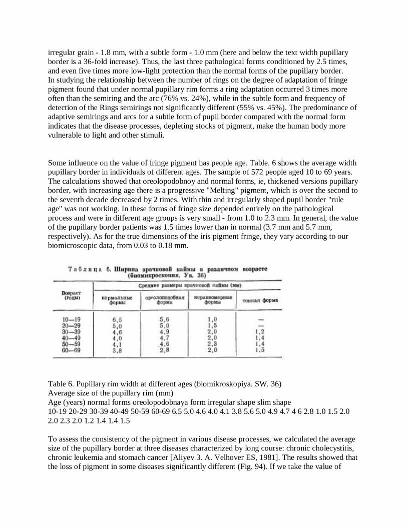
irregular grain - 1.8 mm, with a subtle form - 1.0 mm (here and below the text width pupillary border is a 36-fold increase). Thus, the last three pathological forms conditioned by 2.5 times, and even five times more low-light protection than the normal forms of the pupillary border. In studying the relationship between the number of rings on the degree of adaptation of fringe pigment found that under normal pupillary rim forms a ring adaptation occurred 3 times more often than the semiring and the arc (76% vs. 24%), while in the subtle form and frequency of detection of the Rings semirings not significantly different (55% vs. 45%). The predominance of adaptive semirings and arcs for a subtle form of pupil border compared with the normal form indicates that the disease processes, depleting stocks of pigment, make the human body more vulnerable to light and other stimuli.
Some influence on the value of fringe pigment has people age. Table. 6 shows the average width pupillary border in individuals of different ages. The sample of 572 people aged 10 to 69 years. The calculations showed that oreolopodobnoy and normal forms, ie, thickened versions pupillary border, with increasing age there is a progressive "Melting" pigment, which is over the second to the seventh decade decreased by 2 times. With thin and irregularly shaped pupil border "rule age" was not working. In these forms of fringe size depended entirely on the pathological process and were in different age groups is very small - from 1.0 to 2.3 mm. In general, the value of the pupillary border patients was 1.5 times lower than in normal (3.7 mm and 5.7 mm, respectively). As for the true dimensions of the iris pigment fringe, they vary according to our biomicroscopic data, from 0.03 to 0.18 mm.
Table 6. Pupillary rim width at different ages (biomikroskopiya. SW. 36) Average size of the pupillary rim (mm) Age (years) normal forms oreolopodobnaya form irregular shape slim shape 10-19 20-29 30-39 40-49 50-59 60-69 6.5 5.0 4.6 4.0 4.1 3.8 5.6 5.0 4.9 4.7 4 6 2.8 1.0 1.5 2.0 2.0 2.3 2.0 1.2 1.4 1.4 1.5 To assess the consistency of the pigment in various disease processes, we calculated the average size of the pupillary border at three diseases characterized by long course: chronic cholecystitis, chronic leukemia and stomach cancer [Aliyev 3. A. Velhover ES, 1981]. The results showed that the loss of pigment in some diseases significantly different (Fig. 94). If we take the value of

fringe pigment in healthy people over 100%, in patients with chronic cholecystitis, its value is 72%, in patients with chronic leukemia, 63% of patients with gastric cancer 21%. Excessive depletion of melanin reserves in cancer is the result of a sharp weakening of the adaptive-defenses and very profound changes in the body. It is important to note that the majority of patients in stage IV gastric cancer pupillary rim width was less than 1 mm, or not defined at all.
At the same time, the number of cancer patients with early stage disease and only a good general state of health and appearance of the pupillary rim sizes were normal. Seems appropriate to examine the issue at a large clinical material and prolonged catamnesis, it is possible that the state of the pupillary border may be a reliable predictor.
Known from the literature that the Lens feature natural melanin is universal, and the safeguarding of its mechanism of action in the various forms of life, including in microorganisms. There is reason to believe that this is the function of melanin in the evolution was the most ancient and that its elements have appeared in prebiotic structures. Melanin is always located in the cell wall, making the outer layer, "opaque" for electrons.
To indicate melanoma ninsoderzhaschey pigment cells in the literature use the two terms: "melanocyte" and "melanoma formation." The term "melanocyte" is used to refer to a fully differentiated, cells produce melanin in all vertebrates, including humans. The term "melanophores" is usually applied to melanocytes of cold-blooded vertebrates. Undifferentiated pigment cells called "melanoblast". As for the microbial cells, their inclusion pigment bear the name "chromatophores." LA Drachev et al. (1975) found that the chromatophores of non-sulfur purple photosynthetic bacteria have a device to transform solar energy into a transmembrane electric potential difference. Undoubtedly, the melanocytes of the human body have a higher than bacterial chromatophores electrogenic activity.
Experimental research has shown that melanofo-ry eyes are receptors that respond to the reflected light stimulus. Moreover, fish and amphibians found a direct reaction to light melanophores [Golichenkov VA, 1980, and others]. About receptor activity melanophore reptiles according to data A. Korotkov (1972), found on the surface of the pigment cells are many nerve endings receptor type.
The structure of melanins are carbon, nitrogen, hydrogen, sulfur and other elements. In them there are at least 17 amino acids. Contain melanin free radicals by 2-3 orders of magnitude than other biopolymers. The presence of stable free radicals, obviously, causes the electron-acceptor and radioprotective properties. Recent research found that, in addition to the Lens features melanins have phagocytic-lysosomal, thermoregulatory and antimicrobial activity, as well as some anti-tumor activity [Lyakh SP, 1968 Ruban EL, Lyakh SP, 1970 ; Piattelli M. et al., 1963; Ioung, 1973, and others]. From the results of AE Kaplan and LM Malova (1979) that in vitro iris black rabbits
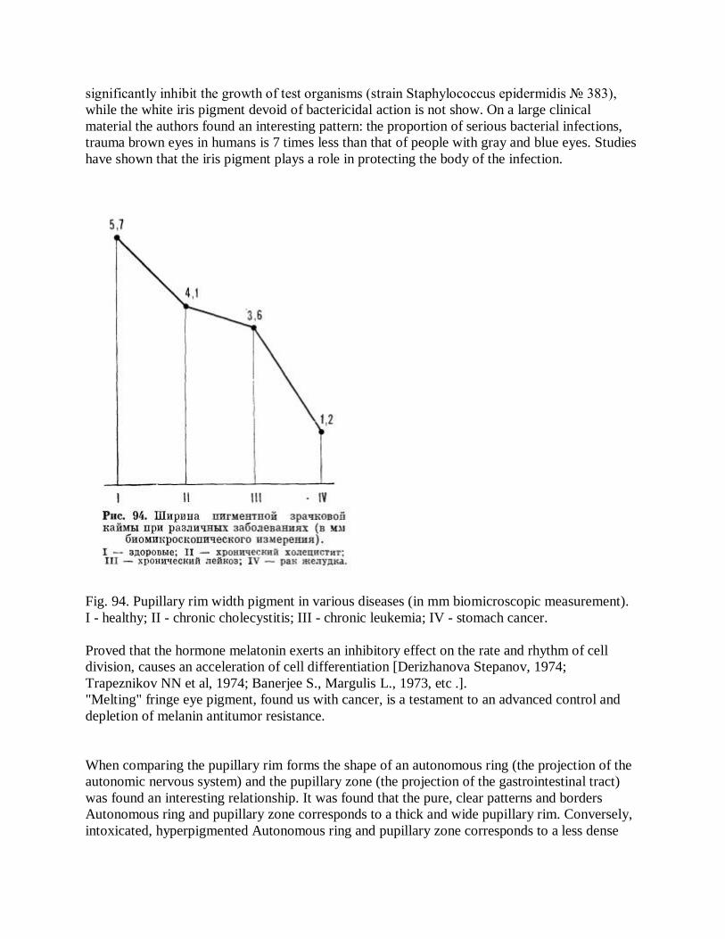
significantly inhibit the growth of test organisms (strain Staphylococcus epidermidis № 383), while the white iris pigment devoid of bactericidal action is not show. On a large clinical material the authors found an interesting pattern: the proportion of serious bacterial infections, trauma brown eyes in humans is 7 times less than that of people with gray and blue eyes. Studies have shown that the iris pigment plays a role in protecting the body of the infection.
Fig. 94. Pupillary rim width pigment in various diseases (in mm biomicroscopic measurement). I - healthy; II - chronic cholecystitis; III - chronic leukemia; IV - stomach cancer. Proved that the hormone melatonin exerts an inhibitory effect on the rate and rhythm of cell division, causes an acceleration of cell differentiation [Derizhanova Stepanov, 1974; Trapeznikov NN et al, 1974; Banerjee S., Margulis L., 1973, etc .]. "Melting" fringe eye pigment, found us with cancer, is a testament to an advanced control and depletion of melanin antitumor resistance.
When comparing the pupillary rim forms the shape of an autonomous ring (the projection of the autonomic nervous system) and the pupillary zone (the projection of the gastrointestinal tract) was found an interesting relationship. It was found that the pure, clear patterns and borders Autonomous ring and pupillary zone corresponds to a thick and wide pupillary rim. Conversely, intoxicated, hyperpigmented Autonomous ring and pupillary zone corresponds to a less dense
and more narrow pupillary rim. This provision illustrates well the index of average width of fringe pigment, which in its pure pupillary zone was equal at 36-fold increase of 5.6 mm, with intoxicated I degree - 5.1 mm, intoxicated II degree - 2.3 mm, intoxicated III degree - 2.2 mm. In true measuring these dimensions correspond 0.156, 0.142, 0.064 and 0.061 mm. Fundamentally, this means that the "cleaner" the human body, the more melanin reserves to save it, the greater the width of the pupillary border. In contrast, the more "deposits" toxins in the body, the less melanin protection and already pupillary rim.
In analyzing the relationship between the color of the iris and the pupil border form we found that at a young age, light-eyed people pupillary rim width is 1.5-2 times higher than in people with dark eyes. After 35-40 years, the difference gradually disappears. The question arises as to why young people with blue eyes pigment fringe wider. We think that the answer to this question must be sought in the size of pupils. It has been mentioned that the size of the pupil and, therefore, the integrative potential of the light passing through them flow more blue-eyed people than the dark-eyed. To protect the light-eyed people from a possible "svetoenergeti-cal burn", nature is forced to "construct" their pupils around more massive pigment "necklace."
The most favorable conditions for this construction there at a young age, when a person is not only a good health, but large amounts of the pigment melanin.
Inflammatory and degenerative signs
It is very common and very important signs of the iris are splitting and deepening in its stroma. In iridology literature they are called lacunae (from Lat. Lacuna - depression, failure) or crypt (Greek krypte - deepening, a tunnel). Some authors use both terms at the same time, attributing some properties lacunae and crypts of the others. We believe that the terms with the same value should not be entered in the same classification. So going into the stroma of the iris, we offer one-to-one call - gaps. Lacunae, iris defects, always point to organic lesion 2 reflexively related departments: land iris and "Corresponding" with him body. Indentations in the iris occur in various destructive processes - inflammation, degeneration, trauma. Distinguish five parameters that evaluate iris gaps. Consider their role in semiotics and diagnostics.
The size of the gaps. Estimating the size of the gaps in iridology is relative. Fundamentally, we may assume that the greater the defect in the iris, the more extensive pathological focus in the body, and vice versa. However, this does not mean the absolute dependence of the gap on the severity of the disease. A lot depends on the location of the defect on the iris. If, for example, a small gap is localized in the projection area of the medulla oblongata, the corresponding clinical picture will be much harder than that observed in the presence of large gaps in the projection area of the complex is functionally less important - the back muscles.
The depth of the gaps. Distinguish between superficial and deep gaps. And their depth depends on the severity of inflammatory or degenerative process. The more pronounced the process, the

greater the depth of a lacuna. This fact can be used in semiotics and diagnostics as well as on degree depressions in the iris can indirectly judge the acute, subacute and chronic stages of disease. Form gaps. Observed a wide variety of configuration gaps. In the classification of I. Deck (1965) there are 11 forms of gaps: 1) the point 2) slotted 3) lanceolate, 4) diamond, and 5) leaf-6) Separation of 7) asparagus-shaped, 8) meduzopodobnaya, 9) sotopodobnaya, 10) open , 11) multiple. The different shapes can have the bottom of gaps that can be radially striated, silvery needles, slatted, monotonous, etc. Most of the authors argue that the linear and angular edge defects show "debut" of the pathological process, while the rounded edges of the point significant duration of the disease and the more coarse destructive changes in the body. The appearance of a light shaft at the edge of the gap indicates the completion of the pathological process.
Often, the individual fibers or several grouped together fibers appear thicker, whitish and tortuous. These fibers are transformed R. Schnabel (1959), a symptom called "curl" and explained their appearance as a result of the inflammatory process (Fig. 95, see insert).
Color gaps. Colouring depressions in the iris is of great diagnostic value. It is proved that the light-colored gaps indicates acute inflammatory process, the dark - the chronic. Iridology many believe that if the contrast between the center of the gap in the form of a point, less zonal illumination or darkening, it indicates the severity of the injury. According to R. Schmidt (1960), a coloring of gaps is one of the hallmarks of cancer.
Localization of gaps. As described above, the location of gaps has significant diagnostic value. According to our observations, most of the gaps is in the ciliary zone. Their most favorite place is the region adjacent to the autonomous ring outside.
Topography gaps gives an indication of what the projection area on the iris, and therefore what the internal organs affected.
Fundamentally we can assume that centrally located gap indicates pathology underlying formations (Central nervous system, parenchymal organs, etc.), while the peripheral gaps point to skin lesions, peripheral vascular and lymphatic system. Of interest in this connection some lacunar genotypes and phenotypes marked I. Deck (1954). These combinations are called lacunae author spitseobraznym, pankreotriadnym types and cardiorenal (Fig. 96).
When inflammation of the pancreas lacunar structure as it is framed by the lower sector of the ring in both stand-alone and - "Daisy" (abdominal or spitseobrazny type), b - pankreotriadny: I - pankreolakuny, II - nasal space, sinuses, adenoids, hyperplasia, III - bronchopulmonary space, organ weakness in - Cardio-Renal: I - the heart; II - kidney.

Fig. 96. Iridotopologicheskie genotypes [Deck I., 1954]. Topography of gap characters in pancreatitis [Deck I., 1965]. iris. Visually, these formations have scalloped shape (Fig. 97).
In assessing the gaps of Iridology, there are 2 opinions. F. Vi-da and I. Deck (1954), G. Iausas (1958) and V. Iensen (1964) consider the iris defects in morphological parameters, with particular emphasis on the size, depth and shape of the gaps. W. Schnabel (1959), L. Vannier (1951) and M. Verdun (1961) considered the main criterion for evaluation of the defect on the iris no morphological and chromatic changes. They believe that the basis for the formation of gaps is a complex restructuring of iris pigment.
We think that both views are legitimate, since the formation of gaps is accompanied by breaking various, including pigment, layers of the iris. On this occasion, R. Bourdiol (1975) notes that poor blue iris pigment dominated morphological changes in rich dark iris pigment - color.
Changes in the structure and color of the iris inflammation and de-generation of systems and to some extent connected with the activity of the pathological process and the time factor. Iridology is well known that seem to judge the gaps stage of the inflammatory process (Fig. 98).

Stage of acute inflammation characterized by swelling and splitting of radial fibers iris. Violated strict linear trabeculae. They become wavy, spiral, sometimes layered. This type of iris, we called radially-wavy, we noted 20% of patients with light eyes and in 3% of patients with darker. This kind of stroma R. Bourdiol compares with soaked felt, M. Verdun - with clouds. F. Vida and I. Deck - a tree, from which all the fallen leaves. It should be emphasized that the blue-eyed people wavy trabeculae are painted white, the brown-eyed - in yellow.
When coarser radial fibers splitting these areas become dark in color with light iris and be-* vyatsya depigmented in the dark (Fig. 99, 100, see insert) "Pathogenetic they mean weakening the body's resistance and increased blood flow to the appropriate authorities. If an acute inflammation in the short term ends in recovery, all changes in the iris regress.
The increase in the acute inflammatory process with a. If acute inflammation grows and deepens, it leads to a dramatic swelling of trabeculae. They get longer and wavy. Separate cleft, increasing in size, shape surface gaps. The edges of these gaps have a polygonal shape, and their shallow bottom to be covered "bars" of the bright filaments.
Subacute stage of inflammation characterized by the appearance of pigment on the sides and at the bottom of the gaps. Sami gap deepened and become darker. Stage of chronic inflammation seen in the gradual increase of the gaps both in width and in depth. Lacunae lose initially polygonal shape, rounded. They are painted in a dark color intensity due to increased pigmentation.

Fig. 98. Signs of organic lesions [Bourdiol R., 1975].
The four phases of pathological changes in the dynamics of the iris: A - an acute stage of inflammation, B - the growth of acute inflammation; B - subacute stage of inflammation, D - chronic stage of inflammation. Unlike other authors, W. Iensen (1964) believes that iridoskopicheski can ascertain the presence of an "open", according to inflammatory sites and 'closed', encapsulated. In this regard, B. Iensen concludes that if there is an acute inflammation of the outflow secretion in any cavity, such an "open" the inflammatory process is reflected in the iris characteristic white spot with clefts, very reminiscent of a flame of fire (Fig. 101, ). If the acute inflammatory process is the type of "closed", ie, has no natural drainage outlet for the inflammatory secretions, it will have a different view and a gap in the iris. It will be surrounded by a white capsule, inside which one can distinguish the white walls and dark foci, projection associated with cysts and other limited areas <Fig. 101, b).
Gap signs of chronic inflammation of the iris in its configuration will be like the preceding one. It also should distinguish between "open" and "closed" chronic lesions and corresponding gaps. Only in these cases, the gaps will be dark or black. From a theoretical point of view, the interpretation of B. Iensen is not well founded. When

considered lacunar signs in the same plane, you feel about communicating and closed cavities. Our studies show that determine the nature of the boundaries of the inflammatory process in the iris signs can not.
Moreover, the gap in the iris are not a specific sign of inflammation. G is just as likely as during inflammation, they may be found in degenerative and other destructive processes. It must be remembered clinicians conducting the differential diagnosis of diseases.
Fig. 101. Signs of acute inflammation [Iensen B., 1964]. a - "open" b - a "closed".
As an example, the results of our observations in the neurological clinic. Were examined 150 patients with various diseases: cerebral arachnoiditis, brain tumors, multiple sclerosis, epilepsy, hereditary degenerative disease and cerebrovascular accidents. Gaps in the projection area "brain" have been observed in patients examined at different rates, most often with hereditary degenerative disease (85%) and epilepsy (71%), more rarely with cerebral arachnoiditis (Fig. 102). However, the size, shape, depth, and other structural features of the gaps did not differ in different diseases. It is interesting to note that the light-eyed patients had more gaps than dark-eyed - in the ratio 3:2. More logically and reasonably explains V. Iensen (1970) marks the evolution of gap iris at different stages of inflammation and degeneration. In the author borrowed from Fig. 103 clearly

shows the "patogeneticheskaya ladder gaps" - grading defects iris stroma associated with the increase and the pathological changes.
Dima P. (1977) points out that we should not identify so-called catarrhal sign lacunar iris with large dents (pits) that characterize this type of iris. The bright iris catarrhal sign differs from the surrounding stroma in color, acquiring, usually shaped gap. In the dark iris catarrhal sign is like a deep crack - very dark, almost black line embedded in the iris.
G. Iausas (1958) wrote that the cracks in brown iris (the equivalent of lacunae) have a radial direction. They often pachinayutsya the pupil and intersect the pupillary zone and autonomous ring. In these cases, they suggest autonomic dysfunction. If the crack is in the iris zakanchivaetsya mild, and coloring it a few lighter, which means (by analogy with open lacuna in bright eyes) that the disease continues. If the crack ends abruptly, and coloring it in sharp contrast with the surrounding stroma, we can say (by analogy with closed lacuna in bright eyes) on the completion of inflammation and scarring. Like most iridology, we believe that the sign of a "closed loopholes" or "clearly breaks off crack" should be interpreted as scarring in the corresponding internal organ or part of the body. This is an important iri-dodiagnostichesky symptom. Despite the fact that the "open" and "closed" gaps differ on the dynamics of the process, pathogenetically they express similar changes: hypothyroidism and the fall of vitality in certain tissues of the body.
M. Kibler and L. Sterzing (1956) noted that a significant portion of gaps and other signs located near the spiral corrugated trabeculae. So wavy border demarcation trabeculae is undeniable role in iridology, helping recognize the disease almost all organs, except for liver, spleen, and skin. Principle it can be said that the weaker the body, the greater the number of gaps and wave observed in trabecular iris.
Fig. 102. The frequency of detection of gaps in various cerebral diseases of the brain (in percent).
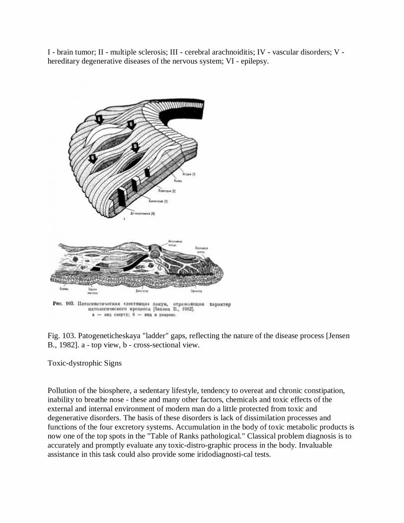
I - brain tumor; II - multiple sclerosis; III - cerebral arachnoiditis; IV - vascular disorders; V - hereditary degenerative diseases of the nervous system; VI - epilepsy.
Fig. 103. Patogeneticheskaya "ladder" gaps, reflecting the nature of the disease process [Jensen B., 1982]. a - top view, b - cross-sectional view. Toxic-dystrophic Signs
Pollution of the biosphere, a sedentary lifestyle, tendency to overeat and chronic constipation, inability to breathe nose - these and many other factors, chemicals and toxic effects of the external and internal environment of modern man do a little protected from toxic and degenerative disorders. The basis of these disorders is lack of dissimilation processes and functions of the four excretory systems. Accumulation in the body of toxic metabolic products is now one of the top spots in the "Table of Ranks pathological." Classical problem diagnosis is to accurately and promptly evaluate any toxic-distro-graphic process in the body. Invaluable assistance in this task could also provide some iridodiagnosti-cal tests.

Toxic radiance. Fairly common sign of iris are so-called sunshine, or spokes. They are radially directed from the center to the periphery of the area of the gastrointestinal tract to the area of the skin. "Sunlight" are dark in color and resemble a wedge scars (Fig. 104, see insert).
P. Schmidt (1960) and R. Bourdiol (1974) prefer to call them "asthenic radial cracks." According to observations of R. Bourdiol, the presence of these characters are always shows the weakness of certain organs and systems, such as being on the level of "12.00", they determine psychasthenia, etc.
The presence of light in the iris indicates toxic effects on the gastrointestinal tract, tend to spread to other organs and tissues. If, along with the sun in the iris reveals adaptive ring, it indicates the exhaustion of the nervous system and the weakening of restoration, reparation, and processes. According to a number of iridology and observations, the sun's rays are usually found in the projection area of the brain. Patients with sign sunlight often complain of headaches, dizziness, loss of productivity of mental work, etc. They unsuccessfully assigned medication. Experience has shown that diet and regular bowel cleansing have in such cases is much more effective than conventional treatment (galvanic collar by Shcherbakov, darsonvalization the head, etc.).
With long flowing inflammatory diseases develop pronounced toxic-dystrophic changes, accompanied by acidosis. Metabolic disorder with displacement scheniem pH toward the acidic environment causes a number of general changes in the iris. The projection area of the gastrointestinal tract and autonomous ring covered with a whitish bloom. From this bright belt radially to the periphery of the white rays diverge. They are few in number, raised above the rest of the relief iris and separated from each other. These filaments can affect both single and multiple organ sectors. B. Iensen (1964) called these eyes "eyes rheumatic acids." White radiance observed in patients with rheumatic diseases, especially common in subacute stage or exacerbation of a chronic process (Fig. 105, see insert). Individuals with this iris often complain of pain in the joints, in the course of nerves and the spine. Is a common complaint of irritability and emotional lability. It should be noted that the phenomenon of white radiance can be seen only in people with light eyes. In the brown-eyed people of similar changes in the iris were observed.
According to F. Roberts (1962), the white radiance is particularly common in children with skin rashes, rhinitis, tracheitis, bronchitis, diarrhea, fevers. One of the causes of the phenomenon of white radiance author considers malnutrition in children. Dystrophic rim. It is black, often dark smoke ring, located on the periphery, at the root of the iris. It is located in the zone, the projection associated with the skin. Dystrophic rim has a different width and rough conical shape. From the periphery of the rim very often waste within a few tracks conical configuration is incorrect, with the top facing the pupil (Fig. 106, see insert). According to our data, dystrophic rim of varying severity observed in all patients and a significant part of healthy.

Dystrophic rim appears in inflammatory diseases and intoxication, that is, in all cases where there is an excessive accumulation in the body of toxic substances and drugs. The more is distinct dystrophic rim, the more severe toxemia and certainly working hard metabolic apparatus of the skin. The appearance of dystrophic rim indicates an excess of toxic material and slag retention in those organs in the projection sector which appeared rim. Bezel indicates incomplete activity and decrease blood flow to the organs and tissues, segmental associated with the area of skin. If dystrophic rim light is detected in the area - this means that the lungs filled with toxic products of metabolism that are due to lowering the cleaning function of the skin, kidneys, and other organs can not be removed from the body. Thus, the appearance of dystrophic rim on the iris serves as a measure of "contamination of the skin." That's the mark of iridology judge hypo and anhidrosis. Dima P. (1958) wrote that the media clearly pronounced dystrophic rim are very sensitive to cold. Often, they are predisposed to bleeding, rheumatism, benign and malignant tumors. Very black and compact dystrophic rim indicates a high content of the internal organs of toxic material.
A number of iridology believe that black dystrophic rim may indicate psoriasis and tuberculosis [Dima P., 1958; Kriege T., 1971; Jensen B., 1984]. Based on this, these patients should examine the respiratory system, and especially the top of the lungs.
In some cases, flat area of skin becomes radial branching consisting of a dark or smoky filaments extending from the peripheral ring to the pupil. Branching this resembles a crown or garland. Specialists believe that the minor branching dystrophic rim appears at a time when the acute process eliminated, but the causative factor remains. Small branching can be connected to the solar rays, while creating a thick cloth. According to V. Ferrandiz (1978), these compounds are a sign of neurosis expressed toxic and hereditary origin. When small branching detected in projection areas of the brain, it is called stagnant and degenerative changes in the brain department, which may clinically manifest as dizziness, a feeling of heaviness in the head, the oppression of mental activity, migraine tendency to drowsiness, fatigue, epilepsy, etc.
Iridology note that vaccination and duration of administration of drugs cause darkening of dystrophic rim. This primarily refers to dermatitis, eczema and other skin diseases, which are used in the treatment means (antibiotics, corticosteroids, and others), which suppress the cleansing function of the excretory system of the skin. If the turbidity of the projection area of the skin is observed in the segment of the ovaries, then we can expect a violation of their function, which is expressed in a number of menopausal symptoms.
In severe toxic states dystrophic rim dark and covered with a kind of white specks. At the same time it becomes wider and closer to the periphery of the pupil. It is as if the flushing of the iris pattern. Sometimes towards the outer rim of dystrophic zone bowel develops a second, inner bezel. Iridodiagnosty Some argue that the convergence of the two rings is extremely toxic

adverse prognostic factor for patients and portends death. We do not share this view and believe that predict death by degenerative rim or any other signs of iris pevozmozhno. In some cases, dystrophic rim is hereditary (Fig. 107, see insert). Those born with the podymchatym-ring on the periphery of the iris, for the most part characterized by low body resistance. P. Dime-ing (1977) states that children born with dark dystrophic rim during the first two years of life, there is a particular skin disease.
Dr. Lindlar (1979) describes the following observation. He examined very painful, weakened child first months of life, which is poorly developed and not gain weight. Of inquiry revealed that the mother of Kuta child is afraid to bathe him and make some fresh air. On examination, the patient was found iris very deep dystrophic rim. The child was prescribed regular rubdown, sun and air baths. After a few months of hygienic measures brought results: The child grew stronger, weight gain corresponded age norm, stopped hurting, dystrophic rim was less pronounced.
Lymphatic rosary. The lymphatic system is a giant drainage network of the body and part of the venous vascular system, in close connection with which it develops and functions. It performs several important functions: transport, hematopoietic, assimilation, neutralizing, etc. M. Archer (1978) reported that the recently discovered another property of the lymphatic system - delivery of hormones to their own actions. All this has led physiologists consider the lymphatic system of one of the most important in the body.
Years of research Iridology found that the lymphatic system is projected onto the iris as a narrow ring located inward from the area of the skin. Most distinctly flat area and lymphatic mikrotsirkulyator-tion (network peripheral capillaries) systems is presented in a topographic pattern B. Iensen (see Fig. 44).
In healthy people, a well-functioning lymph vessels and nodes of the specified area on the iris looks clean and evenly colored. Disorders of metabolism and overload the body of toxic products in the projection system in the iris limfotsirkulyatornoy appear peculiar changes, referred to as the lymphatic rosary (Fig. 108, see insert). It is white, zhemchuzhnopodobnye point or small clouds resembling pieces of wool. They have different size and shape, often as a rosary, are located next to each other. Sometimes, in the white clouds lymphatic rosary visible pink or brown inclusions - specks. Iris fibers with increased in size and are so close to each other that badly looked pigment relief. Depending on the topics of involved lymph system rosary on the iris may appear in the form of a closed circle, or limited to specific, E plots of white dots and clouds. If, for example, worsening of lung lymph flow, the elements of the lymphatic rosary will be localized to the lateral part of the iris, and if worse lymph in the tonsils and pharynx, the lymphatic rosary will appear in the medial part of the iris, etc.
The most common symptom of lymphatic rosary occurs in diathesis, metabolic disorders,

rheumatism, gallstones. In some cases, the "flakes" on the iris may occur in children with poor elimination products vaccination [Iausas G., 1958]. Similar changes are also observed in people engaged in hard physical labor: blacksmiths, porters, some athletes. A role in the formation of lymphatic rosary is insufficient and photo-ascorbic acid in the body Lie.
Lymphatic Rosary is a fairly common symptoms, especially in people with blue eyes. They recorded it in 16% of cases, while the brown-eyed - just 2%.
In a study of 1,300 people (healthy and patients with various diseases), we found two features. The first feature is that the lymphatic rosary in otherwise healthy young people met with the same frequency as that of many of middle-aged and elderly. In healthy it was celebrated in 15% of cases, patients with pulmonary, cardiovascular, neurological, gastrointestinal and skin diseases - from 9 to 22%, on average, a 15-J6% of cases. Therefore, lymphatic rosary as an early symptom of lymphatic insufficiency occurs not only at "formed diseases," but also in a latent, pre-clinical, period. The presence of this symptom means less antitoxic function of the lymphatic system and the general body resistance.
According to statistics from G. Iausas (1983), the frequency of detection of lymphatic rosary in French in two or more times higher than that of the surveyed Muscovites blue-eyed people - 42%, in the dark-eyed - 17%.
Observations show that the people who have revealed lymphatic rosary predisposed to acute respiratory viral diseases, and are sensitive to weather changes.
The second feature of the lymphatic rosary was that he had met with cancer is 3 times more likely (46%) than in other diseases. The projection area of lymphatic and circulator-tion systems in cancer patients had the appearance of large, sometimes merging with each rosary dirty gray or yellow. This lymphatic rosary is qualitatively different from other diseases peculiar light variations. Rough dirty dull lymphatic rosary beads in cancer patients indicate serious "damage" the lymphatic system and drastic depletion of the body's defenses. In the projection iris area, which was detected lymphatic rosary in 7h cancer patients registered another pathological symptom - ring sodium.
Ring of sodium. Some iridology believe that administration of large amounts of salt, baking soda and such drugs as sodium salicylate, may appear in the iris ring of sodium. It is of various shades of whitish, opaque or transparent, narrow or wide (Fig. 109, see insert). Ring is formed in a part of the sclera, which covers the cornea and is as it were, over the iris. Sodium ring is always at the periphery of the iris, in the zone corresponding to the lymphatic and microcirculatory system. When interpreting these rings, one of which relates to the cornea, and the other - to the iris iridology should seek help from an ophthalmologist who owns biomicroscopic method of research.
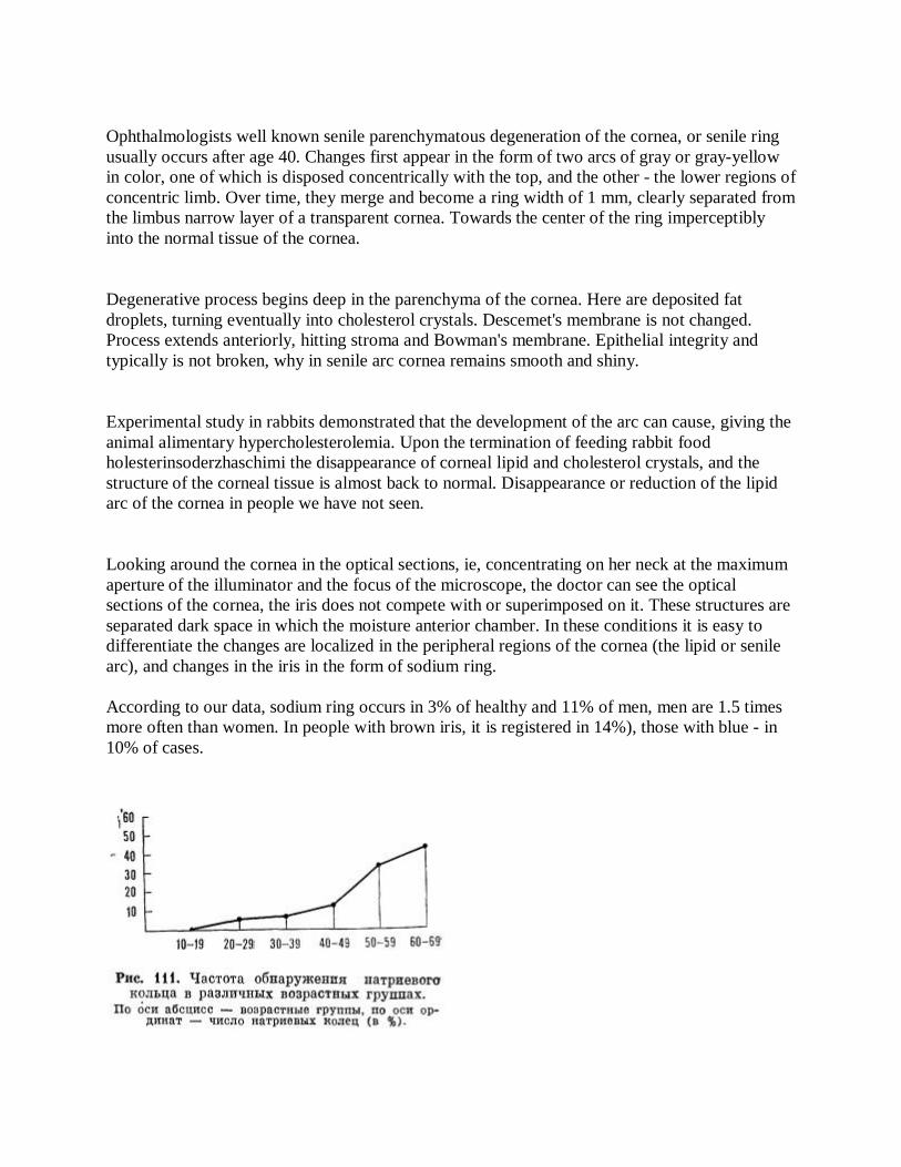
Ophthalmologists well known senile parenchymatous degeneration of the cornea, or senile ring usually occurs after age 40. Changes first appear in the form of two arcs of gray or gray-yellow in color, one of which is disposed concentrically with the top, and the other - the lower regions of concentric limb. Over time, they merge and become a ring width of 1 mm, clearly separated from the limbus narrow layer of a transparent cornea. Towards the center of the ring imperceptibly into the normal tissue of the cornea.
Degenerative process begins deep in the parenchyma of the cornea. Here are deposited fat droplets, turning eventually into cholesterol crystals. Descemet's membrane is not changed. Process extends anteriorly, hitting stroma and Bowman's membrane. Epithelial integrity and typically is not broken, why in senile arc cornea remains smooth and shiny.
Experimental study in rabbits demonstrated that the development of the arc can cause, giving the animal alimentary hypercholesterolemia. Upon the termination of feeding rabbit food holesterinsoderzhaschimi the disappearance of corneal lipid and cholesterol crystals, and the structure of the corneal tissue is almost back to normal. Disappearance or reduction of the lipid arc of the cornea in people we have not seen.
Looking around the cornea in the optical sections, ie, concentrating on her neck at the maximum aperture of the illuminator and the focus of the microscope, the doctor can see the optical sections of the cornea, the iris does not compete with or superimposed on it. These structures are separated dark space in which the moisture anterior chamber. In these conditions it is easy to differentiate the changes are localized in the peripheral regions of the cornea (the lipid or senile arc), and changes in the iris in the form of sodium ring. According to our data, sodium ring occurs in 3% of healthy and 11% of men, men are 1.5 times more often than women. In people with brown iris, it is registered in 14%), those with blue - in 10% of cases.
Fig. 111. The frequency of detection of sodium rings in different age groups. On the horizontal axis - the age group, the vertical axis - the number of sodium rings (in%).
Changes similar to the sodium ring, can be observed in neonates (embryotoxon) and at a young age. In Fig. 110 (see inset) shows a sodium arc, revealed at the dispensary examination, the 13-year old healthy boy.
In many diseases (cardiovascular, pulmonary, neurological, gastrointestinal), the frequency of detection of sodium rings ranging from 11 to 16%. In patients with sodium ring found 2.5 times more often (36%). However, be aware that the population of our cancer patients were on average 1.5-2 times older than the other patients, so the search for the sources of more frequent finding in cancer sodium rings in the nature of the cancer process is possible only with a large assumption, since the age factor plays an no less important.
Very interesting in this regard is the observation of sodium ring in the various age groups. The results of the analysis show that the aging of the body's sodium number of rings has steadily increased from zero in the second decade to 44% in the seventh decade of life (Fig. 111).
In a study of a large group of healthy subjects and patients with cerebral atherosclerosis were installed following iridoklinicheskie correlates [Velhover ES, 1973]. The severity of sodium-lipid ring was directly proportional to the stage of cerebral atherosclerosis, age of patients, systolic pressure, blood prothrombin levels and inversely proportional to the blood filling of the brain. Dependence on the detection of sodium ring diastolic pressure and cholesterol levels (except stage I cerebral atherosclerosis) is very uncertain.
There are different points of view on the formation of a sodium ring. In classical ophthalmology, as stated above, it is called senile arc and explain its origin fatty degeneration of Bowman's membrane and the surface layers of the corneal stroma. We think that fat ne-rerozhdenie - a phenomenon not primary, and secondary, that the annular degeneration of the iris and cornea occurs as a single process that is associated with the violation of sodium, and other lipid metabolism in the body, particularly in the large pool of blood round the iris. In this area, anastomose front and long posterior ciliary arteries that provide blood flow to the iris and cornea. The blood supply in the latter is the type of diffusion episclera branches anterior ciliary arteries.
Thus, sodium ring, more sodium-lipid ring, as it is in two tiers: the front - in the marginal parts of the cornea and more backward - in the marginal parts of the iris. So where do I begin boundary haze? Probably with changes in acid-base balance in the direction of an acidic environment.
A progressive increase in sodium rings in old age is parallel with the increase of acidosis. There may be a relationship between these two phenomena. The argument in favor of this assumption

is that the sodium animals and plants due to its alkaline properties can act as a powerful converter acidotic environment. Therefore it during the deposition of large blood vessels of the circle of iris can be seen as a massive neutralization of acid in the iris, and therefore, as a special sign of significant acidosis. Sodium chloride in such neutralization is not involved. That's why we do not agree with the interpretation of a number of iridology, consider the root cause of the formation of sodium ring high salt intake. Table salt, or sodium chloride, leads to sclerosis of vessels and poor nutrition in all organs, including the iris. Sodium chloride provides the background against which the active participation of phosphate and sodium sulfate (elements in plant and animal foods) develops boundary haze, or sodium ring.
Ring of sodium in the iris indicates the development process of hardening of the arteries and veins long before clinical symptoms appear obliteration of the vessels. Circulation with deteriorating appear general fatigue, reduced vision, sensitivity to cold, pain in the legs, etc.
Such phenomena are observed frequently in people with sedentary lifestyles. Most obliteration of vessels and lack of blood flow seen in the lower extremities. This is probably one of the earliest signs of aging and the inevitable companion of rheumatism, arthritis, gout, certain skin disorders and diseases of veins - phlebitis.
The appearance of the iris ring soda worth paying attention to doctors because it precedes in time the development of clinical symptoms of vascular obliteration, can serve as an indicator of the earliest signs of the deterioration of blood flow and increase acidosis. Toxic and age spots
The spots are the most characteristic signs of the iris. Their presence always indicates serious pathological changes in the body. Distinguish toxic stains and pigment origin.
Toxic stains. These spots can be seen by visual inspection. They are large, crisp, cut or angular edges and a homogeneous structure. Usually they will be several. Painted toxic spots in dark colors: dark brown, dark red, rarely yellow-orange. Whatever the spot, they talk about the weakness of the body's defenses. On examination, it seems that they are inserted into the stroma of the iris, as well-fitted to the parquet floor tiles.
According to G. Iausas (1958), toxic stains occur in about 50% of cases and more often in women than in men: women with light eyes - in 63%, with the dark - in 44%. in men with light eyes - 55%, with dark - in 31% of cases. In 1936, as mentioned by the author started to deal with the Iranian-long, toxic stains were more commonly found in polychrome and light iris and very rarely in dark-colored. After two decades, according to him, has no such selectivity: toxic spots
equally frequent in both the light and the dark iris. G. Iausas believes that this is due to the urbanization of life, distribution of stress factors, infections and intoxication.
Iridology many believe that these spots indicate the poisoning of the body by various exogenous and endogenous toxins. Some researchers see them as signs of a precancerous condition.
Endogenous hyperpigmentation appears in the form of individual white clouds or a zone adjacent to the autonomous ring outside. Location white "slag" in this area (ring 4), which is the projection area of great vessels, a sign of severe autoimmune reaction of the body (Fig. 112, see insert). Figuratively speaking, this mat now in the middle of the iris is like a "field after the battle." It belongs to topolabilnym signs that under the influence of medication-cleansing therapy may be partially or completely disappear. You must use the natural drainage and intense stimulation of isolation: strict separation zochno-diet therapy, sauna, vacuum massage and other kinetic therapies.
Exogenous hyperpigmentation characterized by yellow or light brown color and is located in all parts of the iris. It is associated with impaired elimination of substances entering the body: high doses of drugs, alcohol, tobacco, drugs, and herbal decoctions and infusions. Insufficient excretion of exogenous substances can provoke whitish color shift radially going trabeculae in yellow or reddish color.
We assume that the delay and the accumulation of toxic products in some parts of the vascular system of the eye - a very real phenomenon, and therefore real and formation of toxic spots on the iris. This is evidenced by a form of spots that are in the form of strips and polygonal shapes as it fit into the groove move the iris vessels.
Of particular interest are spot-sectors. According to our observations, they are found in 2% of patients with various diseases, most innate. There are indications that some of them are associated with intoxication in children.
The analysis showed that the most intense color stain-sectors indicated in the pupillary zone, ie, in the basin of the smallest blood vessels. In the slow blood flow in them are more likely to settle in the circulating blood of toxic substances. On these sites, apparently begins the emergence of toxic stain-sectors. Illustrate this assumption iridogrammy 2, which shows the various options toxic pigment sectors: emerging and mature (Fig. 113, 114, see insert).
Toxic brown spots should be distinguished from stains associated with excessive consumption of iodine and iron supplements. Last mainly located in the projection of the gastrointestinal tract, have fuzzy, sometimes filamentous shape and similar to toxic stains color: reddish-brown and yellowish-red with "iodine" spots and purplish-brown and rusty-brown with "iron." In contrast to

these medicinal inclusions, toxic stains and are characterized by the lack of clear boundaries to be eating threads. However, the literature and our own experience does not allow us to make any claims about the nature of the considered extremely toxic stains and spots-sectors. It may be that in their education and other factors are involved: genetic, by environmental and other
F. Roberts (1962) pointed to two possible factors responsible for the accumulation of pigment in certain areas of the iris: 1) constipation and bowel disease, and 2) intoxication quinine, sulfur, iodine, iron, etc. The question is today almost unexplored. Spots. In addition to toxic spots, iris faces many other pigmented lesions. They have a different color, size, shape, density and shape (Fig. 115, see insert). Brown spots appear less coarse, less sharp and less colored than toxic. Boundaries of their never clearly radial, and have more or less rounded form. Many of them are very important topikodiagnosticheskim iris signs and are therefore of considerable practical interest.
Most thoroughly studied spots R. Bourdiol (1975). He divides spots into 5 groups: 1) bright pigments, 2) dark pigments, 3) brown-red pigments, 4) red pigments, 5) pigments such as "prezentnogo (fine-grained) of tobacco." Bright pigments can be of different colors and shades. In iris blue are very common yellow-gold spots and fields, in the iris brown - greenish-rusty. Their location shows the weakness of the body. Yellow-gold frame in the autonomous rings indicate weakness of the ligamentous apparatus and radicular spine. Generalized distribution of yellow-golden pigment in the ciliary zone is marked by a number of neuroses and psychoses.
Dirty-yellow spots resembling crushed gravel, observed in some intoxications. In the brown-eyed people, they have a greenish tint. If these spots are located in the lower segments of the iris, they indicate renal toxicity.
Light yellow pigments consisting of tiny grains, curls or rays, as well as yellow-green pigments formed large granules or jelly-like mass, evidence of infection of the relevant authorities. This may be syphilitic, gonorrheal and tuberculous infection. However, most often yellow-green pigments found in purulent foci and septicemia.
Dark pigments, unlike the toxic stains are composed of a homogeneous, swollen mass of pigment-like, by R. Schnabel (1959), and cauliflower. Their edges are usually distinct and rounded. According to the majority of iridology, dark spots and dark pigments, surrounded by a bright belt, are a sign of the cancer lesion.
Black spots on F. Roberts (1962), observed in many diseases: ulcers, cancer, gastritis, tuberculosis, worm infestations, kidney and gallstones, and so bright and belt marks in the area of age spots and gaps F. Roberts is a sign of scar tissue as a result of the transactions and other

damaging effects. He believes that the scar tissue is almost completely absorbed in 10 years, so the white mark on the iris fade.
Biomicroscopic R. Bourdiol distinguishes two types of dark pigments bear and felt. Bear color - brown-red in color, with a "plucked" edges, something resembling the skin of a bear. Bear meets pigment tumors of the pelvic organs. Felt color - dark brown in color, consists of randomly arranged thin filaments resembling felt. Felt pigment found in the projection are pupillary zone, where it ever stay the course and obscure radial fibers iris. The presence of this pigment is indicative of digestive tract tumors.
Brownish-red pigments can be 3 types: Ezhov,, brown and mesh. Finding them in a particular area of the iris indicates a genetic predisposition of the body to certain infections.
Detection of red pigments is of great diagnostic and topographical value. No matter what their size, they always indicate hemorrhagic syndromes. Very pathognomonic sign for hemophilia is the presence of pigments in a tuft of wool or helical strands of red.
Pigments such as "tobacco prezentnogo» P. Schmidt (1974) has emphasized the diagnostic value. They take the form of scattered or grouped small beads bright, red-brown or dark brown. When the location of the pigment of the "prezentnogo tobacco" in the field of autonomous ring can think about the defeat of the pancreas, at the location of the pupil in the district - a chronic colitis and intestinal paresis. R. Schnabel (1959) described two types of these spots.
Type I - the smallest of the pigment of the "prezentnogo tobacco", visible only with biomicroscopy. They consist of dark, fine-grained particles, resembling crushed black pepper. Like many other dark pigments, they are characteristic of tumor states. The smallest group of pigments such as "prezentnogo tobacco" equivalent bearish pigment and just as he was found in prostate cancer, breast cancer and uterine cancer.
Type II - large groups of pigment such as "prezentnogo tobacco", visible with a simple magnifying lens. They consist of a light brownish-red or coarse particles, resembling grains of cinnamon. These pigments are solely in the ciliary zone. They always have a diagnostic value, although not in all cases indicate the topography of defeat.
R. Schnabel has allocated 5 forms location pigment type II: 1) triangular, 2) stripline, and 3) the form of "fish tail", and 4) a circular, 5) kuchkoobraznuyu. R. Bourdiol (1975) added a classification of R. Schnabel (1959), the addition of the 19 differential diagnostic forms of the pigment type II (Fig. 116):

a) triangular shape - triangle, facing the center of the base, indicating an infection of organs, projected in the area of iris; b) triangular shape - triangle, facing towards the periphery of the base points to disorders of innervation, projected in this site. This mark in verhnevisochnom sector is a symptom of glaucoma and cataracts;
Fig. 116. Different localization of large groups of the pigment of the "tobacco prezentnogo» [Bourdiol R., 1975J. Explanations in the text. c) a thin strip, located radially and consisting of 2-3 fibers. Topographic values has no points to the weakness of the ligamentous apparatus with arthralgia; d) wide strip, located radially. Topographic values no means brain disorders in cases of multiple sclerosis and syringomyelia; e) two narrow parallel strips indicate increasing parasympathetic tone the digestive tract. Observed with heartburn, diarrhea, flatulence in the intestines and so on; f) two narrow parallel strips starting from projections autonomous rings indicate cerebral vascular disorders: headache, dizziness, loss of consciousness;

g) v-shaped "fish tail" refers to the neurotic and autonomic disorders; h) form swallows indicate microbial organ failure corresponding projection area; i) form the arch indicates vascular pathology; j) form forceps points to defeat the vertebrae; a) wide circular bar in the middle of the ciliary zone indicates an organic lesion of the sector; 1) narrow circular bar in the middle of the ciliary zone indicates functional disorders of the body; m) circular band around the autonomous ring indicates visceral parasimpatikotonii; n) limbic circular strip indicates a worsening function of the venous and lymphatic systems; o) kuchkoobraznaya form with clusters of pigment located on the tops of cloves Autonomous ring. Topographic value has to indicate pancreatitis, diabetes and obesity; p) kuchkoobraznaya form with clusters of pigment, located at the base of teeth Autonomous ring. Topographic values has no points to endocrine disorders; q) kuchkoobraznaya form with clusters of pigment located in the central part of the ciliary belt parallels. Topographic values has no points to vertebral arthrosis; d) kuchkoobraznaya form with clusters of pigment located on the limb at intervals of 1-1.5 mm. Topographic values not indicate the presence of psychosis; s) Chaotic kuchkoobraznaya form indicates the presence of menopause and androgenic disorders.
Brown spots in the representation R. Bourdiol, to their interests for professionals iridolo-gov. However, not all of them have compelling evidence in diagnostic and in Topographically. We reject as untenable the dark spots in cancer, yellow-green spots in purulent infection, red spots with hemorrhages and some characters' prezentnogo tobacco. "
In principle, we do not agree with R. Bourdiol iridium and other log files that signs of iris, including age spots, can be used to establish the etiologic diagnosis. Not all the spots, especially single and large, indicate for the presence of disease. One spot in itself nothing to say. However, if it is located near or in the thickness of the other iridology signs (in the depth of the gap between the split trabeculae, etc.), it certainly becomes a diagnostic sense.
Most iridology note that in healthy people, especially in childhood and adolescence, the iris looks clean and clear. Considered to be firmly established that the healthier the body, the cleaner, one-color and denser iris. The patients and the elderly, it becomes dull, sometimes grubby, usually multi-colored, with some spots and fields. Write about it, not only iridium-diagnosticians, and ophthalmologists. Not counting sites enlightenment pigmented lesions with any eye color undergo certain changes in color - from light gold to dark brown and black. Therefore, the difference is easy to spot these blue-eyed people, and not always easy for people with dark eyes.
We present the results of our studies of age spots • in terms of topographical significance. Outset that we analyzed only distinctly pigmented spots and intentionally did not include the so-called land of enlightenment.
More than 5 thousand observations healthy and sick people give us reason to believe that the number of dark spots on the iris with age and the onset of disease is greatly increased. The vast majority of healthy children aged 7 to 11 years, we found no pronounced pigmentation. And only 4% of them met the unit (sometimes multiple) spots on various parts of the iris. A different picture is observed in healthy middle-aged and elderly people - the number and prevalence of dark spots on the iris of their markedly increased.
It seems that the progressive increase in the number of age spots, similar to senile arc of the cornea, is linked to the age factor. However, this is not the case. We watched and children with a variety of pigmentation in the iris - they had been ill for many diseases. At the same time we were able to meet some elderly people with absolutely clean iris, which in life does not hurt.
It would be better, perhaps, to assume that the cause of age spots is a painful process, but with age, when there is a "cumulative effect" of disease, only increases their number.
An illustration of this is the following example.
Of the large number of patients observed in the advisory department of one of the hospitals in different experts, we were 4 age groups of patients with a similar disease to 50 people each.
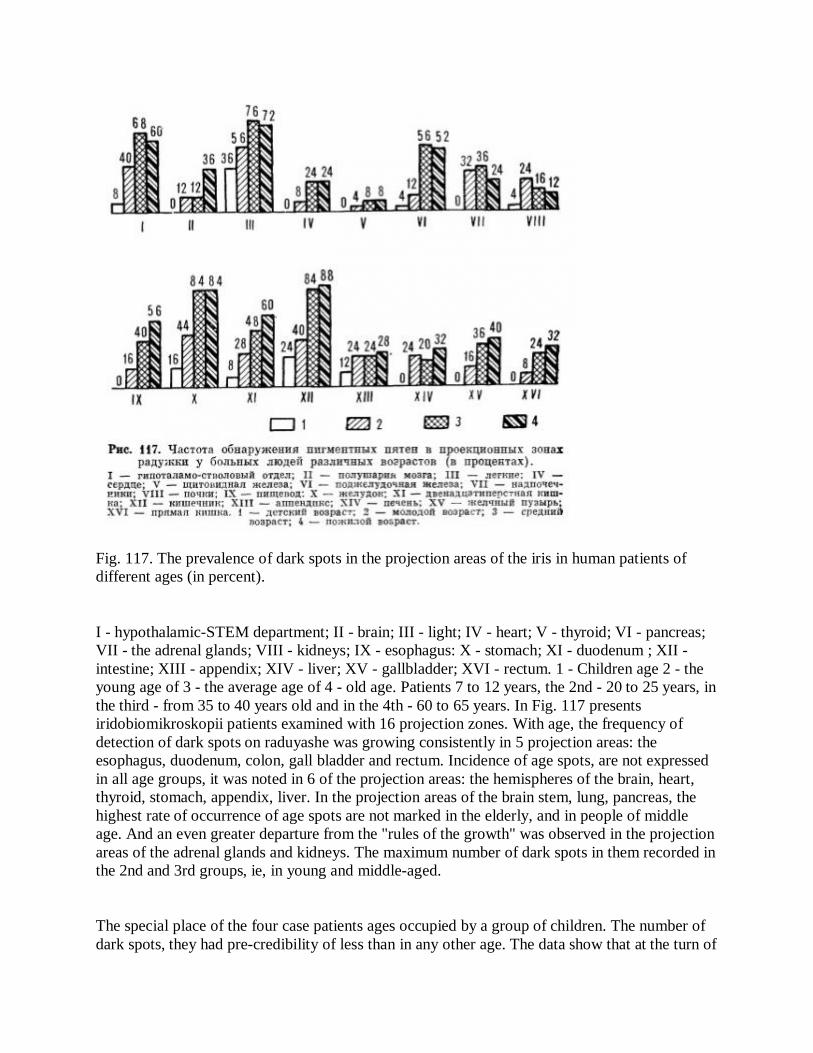
Fig. 117. The prevalence of dark spots in the projection areas of the iris in human patients of different ages (in percent).
I - hypothalamic-STEM department; II - brain; III - light; IV - heart; V - thyroid; VI - pancreas; VII - the adrenal glands; VIII - kidneys; IX - esophagus: X - stomach; XI - duodenum ; XII - intestine; XIII - appendix; XIV - liver; XV - gallbladder; XVI - rectum. 1 - Children age 2 - the young age of 3 - the average age of 4 - old age. Patients 7 to 12 years, the 2nd - 20 to 25 years, in the third - from 35 to 40 years old and in the 4th - 60 to 65 years. In Fig. 117 presents iridobiomikroskopii patients examined with 16 projection zones. With age, the frequency of detection of dark spots on raduyashe was growing consistently in 5 projection areas: the esophagus, duodenum, colon, gall bladder and rectum. Incidence of age spots, are not expressed in all age groups, it was noted in 6 of the projection areas: the hemispheres of the brain, heart, thyroid, stomach, appendix, liver. In the projection areas of the brain stem, lung, pancreas, the highest rate of occurrence of age spots are not marked in the elderly, and in people of middle age. And an even greater departure from the "rules of the growth" was observed in the projection areas of the adrenal glands and kidneys. The maximum number of dark spots in them recorded in the 2nd and 3rd groups, ie, in young and middle-aged.
The special place of the four case patients ages occupied by a group of children. The number of dark spots, they had pre-credibility of less than in any other age. The data show that at the turn of

children and young adults is a natural breakthrough in pigment, then retained only the tendency to further increase the number of dark spots.
Attention is drawn to a large detection rate of pigment spots in the zones of adrenal and pancreatic cancer in patients of middle and old age. This is an unexpected and extremely important for each clinician fact. From the results of this study follows another interesting situation. The greatest number of dark spots observed in areas corresponding to the brain stem, stomach, intestines, lungs, ie to a highly sensitive function and have a direct link with the various eksteroretsep-tive stimuli. Conversely, the least number of spots observed in the projection areas, corresponds to the heart, kidneys and other parenchymal organs with their reduced sensitivity activity and the absence of direct contact with exteroanterocone retsentorami. There is correlation between the data and doskopii iri-known statistics that out of all types have the highest prevalence of disease is pulmonary and gastro-intestinal diseases.
Very characteristic changes in the iris in patients with rheumatism. Many of them after the acute stage were found scattered Punctulata include white, yellow-brown or dark brown. In a smaller or larger grains, sometimes as much as flakes were located in the stroma of the ciliary zone. In some cases they were placed evenly throughout the iris, others concentrated in separate groups outside from a standalone ring. This phenomenon of "grain" was observed in 68% of patients with rheumatic disease and in 14% of healthy individuals (the differences are statistically significant, p <0.001). We can not claim that the phenomenon of "grain" is peculiar only to patients with rheumatism, as a number of data, it was observed, but with less persistent, brucellosis, malaria, and certain forms of tuberculosis.
To map projection zones iris certain organs and body parts were held iri-doskoppcheskie study of patients with well-defined organ pathology. It is possible to evaluate the utility of topographical dividing iris scheme F. Vida and I. Deck (1954). Table. 7 shows the results of clinical iridoskopi-cal comparisons in healthy individuals and patients with various diseases.
From the data table shows that of the 11 groups of patients in 9 frequency of detection of pigment spots in their respective areas of iris was significantly higher than in healthy (differences statistically significant). This demonstrates the value topikodiagnosticheskoy iridoskopicheskih data.
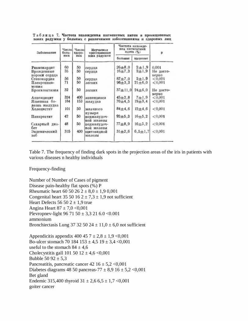
Table 7. The frequency of finding dark spots in the projection areas of the iris in patients with various diseases n healthy individuals
Frequency-finding
Number of Number of Cases of pigment Disease pain-healthy flat spots (%) P Rheumatic heart 60 50 26 2 ± 8,0 ± 1,9 0,001 Congenital heart 35 50 16 2 ± 7,3 ± 1,9 not sufficient Heart Defects 56 50 2 ± 1,9 true Angina Heart 87 ± 7,0 <0,001 Plevropnev-light 96 71 50 ± 3,3 21 6.0 <0.001 ammonium Bronchiectasis Lung 37 32 50 24 ± 11,0 ± 6,0 not sufficient
Appendicitis appendix 400 45 7 ± 2,8 ± 1,9 <0,001 Bo-ulcer stomach 70 184 153 ± 4,5 19 ± 3,4 <0,001 useful to the stomach 84 ± 4,6 Cholecystitis gall 101 50 12 ± 4,6 <0,001 Bubble 50 92 ± 5,3 Pancreatitis, pancreatic cancer 42 16 ± 5,2 <0,001 Diabetes diagrams 48 50 pancreas-77 ± 8,9 16 ± 5,2 <0,001 Bet gland Endemic 315,400 thyroid 31 ± 2,6 6,5 ± 1,7 <0,001 goiter cancer

Under these pathological processes, such as angina, pleuropneumonia, gastric ulcer, cholecystitis, pancreatitis, the frequency of finding pigmentation was especially significant - from 70 to 95%. A small number (45%) iridoskopicheskih findings of appendicitis was probably due to errors in diagnosis (reperkus-aggressive "appendicitis") and short duration of pain in some patients.
F. Roberts (1962) argued that when the true appendicitis in the projection area of the appendix can be seen a large triangle, with psevdoappenditsite - one white line. According to the author, iridology sign "big triangle" can help surgeons in the diagnosis and thus reduce the number of unnecessary appendectomies.
In most patients with "silent" over the process painless (endemic goiter, bronchiectasis, congenital heart disease) spots on the iris did not arise. Very rarely, in 15-35% of cases, they were found in bronchial asthma, cystic lung disease and kidney failure, congenital anomalies of great vessels, liver cirrhosis and tumors. Pigmentation in the projection area of the brain gipotalamostvolovogo observed 2-3 times more often than in the area of the cerebral hemispheres, and they are found not only in cerebral pathology, but also in many visceral diseases. Unlike A. Maubach (1952), we deny the dependence of the structure of pigment spots on the nature and etiology of a disease. Fundamentally we can talk about the degree of organ damage: small - with light and surface spots, rough - in dark and big.
Violations of pigmentation is the phenomenon of "melopodobnoy iris." In light-eyed people, he expressed a whitish-blue or whitish-gray iris and veiling of trabeculae (Fig. 118, see insert). Such iris, we say that they are like greased cream. In dark-eyed people changes occur amber, cloud-like coating. The phenomenon of "melopodobnoy iris" refers to the excessive accumulation of blood lactic acid and other acids, ie acidosis. In this case, because of history is commonly known that people with a large number of acid-eat foods (meat, eggs, white bread, refined sugar, tea, coffee), and often suffer from colds.
Based on the results of the research can be argued that the appearance of age spots on the iris of their sex, age and surgery does not. A large number of observations (315 appendectomies, 300 thyroidectomy, 46-Pneumonectomy and suturing of septal heart defects) strongly suggests that the formation of age spots surgery and removal of the body have no effect. Probably operative trauma, to be applied in anesthesia and other modern methods of anesthesia, does not create the flow of pain impulses sent to the projection zone of the iris, and therefore does not cause any changes in it. The very same exception malfunctioning as a permanent source of sensory input, by contrast, "relieving tension" with iridonevralnyh tract, promotes regression of the former to the violation. That's why were doomed attempt to prove the existence of a number of experimental iridonevralnyh ties ectomy various organs.

Our study iridology is not limited by the parallels with the clinic, the confirmation or non-confirmation of the clinical diagnosis. In some cases, the study found themselves in an iris scan for a more "subtle" than conventional survey methods. Some examples. In b-tion C, 31, for months lechivshegosya from neurasthenia, iridium-scopic were signs of bilateral pleural process, caught in the further refinement of tuberculous pleurisy. In b-tion K., 36 years old, unsuccessfully lechivsheysya heart of neurosis was found pigmented in the projection of the lower part of the left lung. When the diagnosis of subsequent clinical identified the left-side nizhnedolevaya interstitial pneumonia. In b-tion D., 26 years old, complaining about a long wheezing, iridoskopicheski found pigmented in the projection area of the left lower lung. Later, when hospital examination, was diagnosed with polycystic lower lobe and reed segment of the left lung, the about which the patient was successfully operated on.
B-tion G., 30 years old, applied to iridology office complaining of frequent pain in the upper abdomen, heartburn, nausea, general weakness growing. When iridology examination was diagnosed as gastritis and holetsistopankreatit known patient on previously held regular research. In addition, in the projection area of left kidney were found in the iris pigmented and the symptom of "stacks", showing the pelvic-ureteral occlusion. It was suggested at the urologist survey, from which the patient because of the absence of any symptoms of kidney decided to abstain. 1 month after iridoskopii patient was admitted to the urology clinic with a diagnosis of renal colic. When contrasting uro-graphy in the upper third of the left ureter stone revealed a 5 mm X HZ.
The following clinical and radiological monitoring at the patient • anticipated presence of the process in the lower lobe of the left lung. Iridoskopicheski process localized to the left in the projection of the lower parts of the pleura, the lung parenchyma is. Produced by the operation has proved the process to determine the topical iridoskopicheskim data. Give a description of the case.
B-tion H., 7 years. Diagnosis: echinococcosis lower lobe of the left lung. Chest X-ray: in the lower lobe of the left lung is defined oval formation with clear smooth contours. Normal heart size and pulsation. Conclusion: Ovarian lower lobe of the left lung may hydatid (Fig. 119). When iridoskopii patient discovered adaptive ring, more pronounced in the lateral sections. The left iris is determined by dark pigmented corresponding to the projection of the lower portions of the pleura (Fig. 120). Patients underwent an operation where in the lower left lung between the parietal and visceral pleura found cyst the size of a fist. Lung parenchyma with a cyst was not connected. Perform routine echinococcectomy.
Investigation of age spots and gaps is of great importance for the development and improvement iridodiagnostics. Detection of these signs in the iris implies lesions in the body, so it is responsible to relate to assessing the status of the iris projection zones, each of which needs to be carefully examined and clarified on the basis of modern diagnostic techniques. Summarizing, we
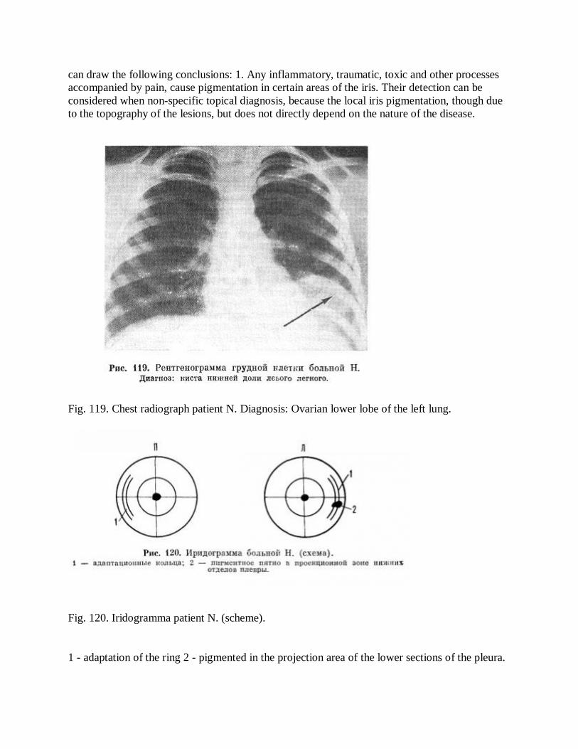
can draw the following conclusions: 1. Any inflammatory, traumatic, toxic and other processes accompanied by pain, cause pigmentation in certain areas of the iris. Their detection can be considered when non-specific topical diagnosis, because the local iris pigmentation, though due to the topography of the lesions, but does not directly depend on the nature of the disease.
Fig. 119. Chest radiograph patient N. Diagnosis: Ovarian lower lobe of the left lung.
Fig. 120. Iridogramma patient N. (scheme).
1 - adaptation of the ring 2 - pigmented in the projection area of the lower sections of the pleura.

2. Rarely matches the clinical diagnosis of a local pigmentation in the iris due to the lack of pain impulses and is observed at many of the "little by little" occurring diseases: a) different variants of congenital abnormalities (polycystic bodies, strictures, diverticula, coarctation, etc.), b) pain-free, "glow" throughout the process (tumors, cysts, polyps), c) poor defeat receptors parenchymal organs, and d) in the pathology of the conducting system and spinal cord. It is possible that factors other than pain and "receptive saturation" sick body, the formation of age spots is influenced by anatomical and physiological features of the dilator. Heerfordt (1900) and A. Szili (1902) found that the dilator (and sphincter) developed from the cells of the outer layer of the optic cup. But while in the epithelial cells of the sphincter completely transformed into the muscle, the dilator it happened only in one of the cells, while in another part of the preserved epithelial character and pigmentation. Thus, the form and function of "extender" pupil depends on the level of differentiation. At a low degree of differentiation (in the iris radial and radial-homogenous types) Lens feature eye is the usual way - spots on the iris appear immediately. With a high degree of differentiation (in the iris radial lacunar type) safety function is weakened eyes - local pigmentation in the iris occur gradually, or much later.
Based on this study it can be assumed that the first type of structure dilator is more common, as the phenomenon of "age spots" is observed in the majority of patients examined.
Copyright © 2022 FDOKUMEN