Chapter 24 Lecture Outline - Pcmac
114
Chapter 24 Lecture Outline Copyright © McGraw-Hill Education. All rights reserved. No reproduction or distribution without the prior written consent of McGraw-Hill Education. See separate PowerPoint slides for all figures and tables pre-inserted into PowerPoint without notes and animations. 24-1
-
Upload
khangminh22 -
Category
Documents
-
view
1 -
download
0
Transcript of Chapter 24 Lecture Outline - Pcmac

Chapter 24 Lecture Outline
Copyright © McGraw-Hill Education. All rights reserved. No reproduction or distribution without the prior written
consent of McGraw-Hill Education.
See separate PowerPoint slides for all figures and tables pre-inserted into PowerPoint
without notes and animations.
24-1

Chapter 24
Digestive System
24-2

24.1 Anatomy of the Digestive System
• Digestive tract: also called alimentary tract or canal – GI tract: technically refers to
stomach and intestines • Accessory organs
– Primarily glands, secrete fluids into tract
• Regions – Mouth or oral cavity with salivary
glands and tonsils – Pharynx (throat) with tubular
mucous glands – Esophagus with tubular mucous
glands – Stomach with many different
kinds of glands that are tubular – Small intestine (duodenum,
ileum, jejunum) with liver, gallbladder and pancreas as major accessory organs
– Large intestine including cecum, colon, rectum and anal canal with mucous glands
– Anus
24-3
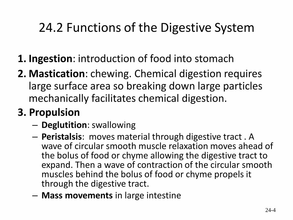
24.2 Functions of the Digestive System
1. Ingestion: introduction of food into stomach
2. Mastication: chewing. Chemical digestion requires large surface area so breaking down large particles mechanically facilitates chemical digestion.
3. Propulsion – Deglutition: swallowing – Peristalsis: moves material through digestive tract . A
wave of circular smooth muscle relaxation moves ahead of the bolus of food or chyme allowing the digestive tract to expand. Then a wave of contraction of the circular smooth muscles behind the bolus of food or chyme propels it through the digestive tract.
– Mass movements in large intestine
24-4

Peristalsis
24-5
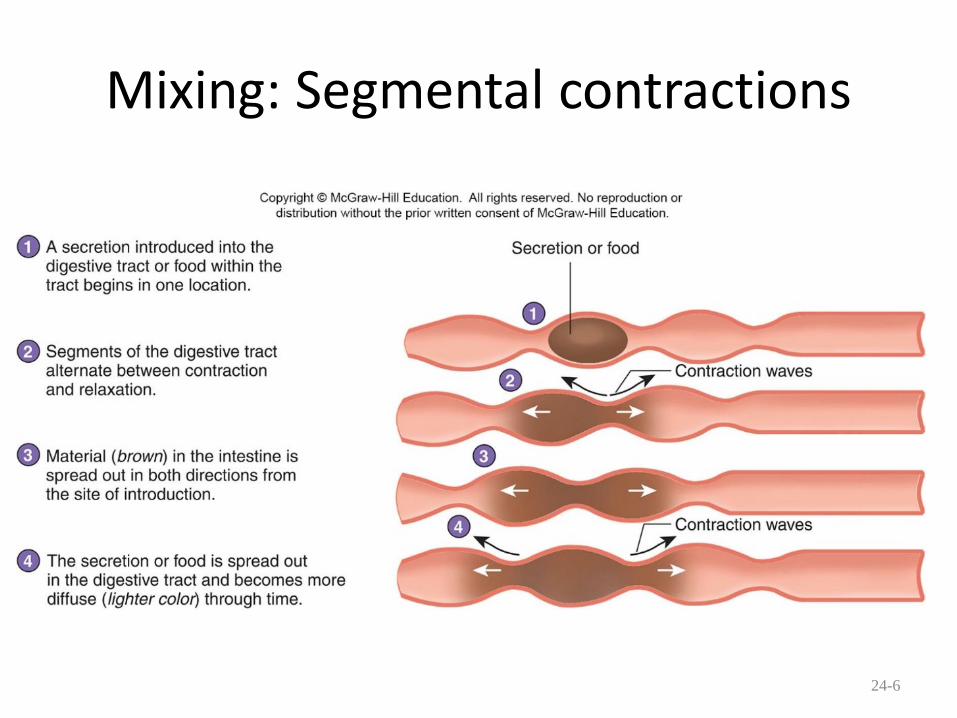
Mixing: Segmental contractions
24-6

Functions, cont.
5. Secretion: lubricate, liquefy, digest – Mucus: secreted along entire digestive tract, lubricates food and
lining, coats lining and protects from mechanical digestion, from acid and from digestive enzymes.
– Water: liquefaction makes food easier to digest and absorb
– Bile: emulsifies fats
– Enzymes: chemical digestion
6. Digestion: Mechanical and chemical
7. Absorption: Movement from tract into circulation or lymph
8. Elimination: Waste products removed from body; feces. Defecation
24-7
Functions of the Digestive System
24-8

Functions of the Digestive System (2)
24-9

24.3 Histology of the Digestive Tract
•Mucosa. Innermost layer, consisting of mucous epithelium (stratified squamous in mouth, oropharynx, esophagus and anal canal), simple columnar epithelium in the rest of the tract.
– Loose connective tissue: lamina propria – Muscularis mucosae: smooth muscle
•Submucosa. Thick C.T. layer with nerves, blood vessels, small glands. Parasympathetic submucosal plexus.
24-10

Histology of the Digestive Tract
• Muscularis: 2 or 3 layers of smooth muscle, two of which are circular and longitudinal. Exception: esophagus where the upper 1/3 is striated. This layer also contains the myenteric plexus. The myenteric and submucosal plexi together are called the enteric or intramural plexus. Important in control of movement and secretion
• Serosa or adventitia: Connective tissue. Where serosa is present, called visceral peritoneum. Where adventitia is present, connective tissue blends with connective tissue of surrounding structures
24-11

Histology of the Digestive Tract (2)
24-12

24.4 Regulation of the Digestive System
• Chemical regulation – Production of hormones to be
discussed later
• Gastrin, secretin
– Production of paracrine chemicals like histamine
• Help local reflexes in ENS control the conditions of the internal environment of the digestive tract such as pH levels
• Nervous regulation – Local: enteric nervous system
• Types of neurons: sensory, motor, interneurons
• Coordinates peristalsis and regulates local reflexes
– General: coordination with the CNS. May initiate reflexes because of sight, smell, or taste of food. Parasympathetic primarily. Sympathetic input inhibits muscle contraction, secretion, and decrease of blood flow to the digestive tract.
24-13

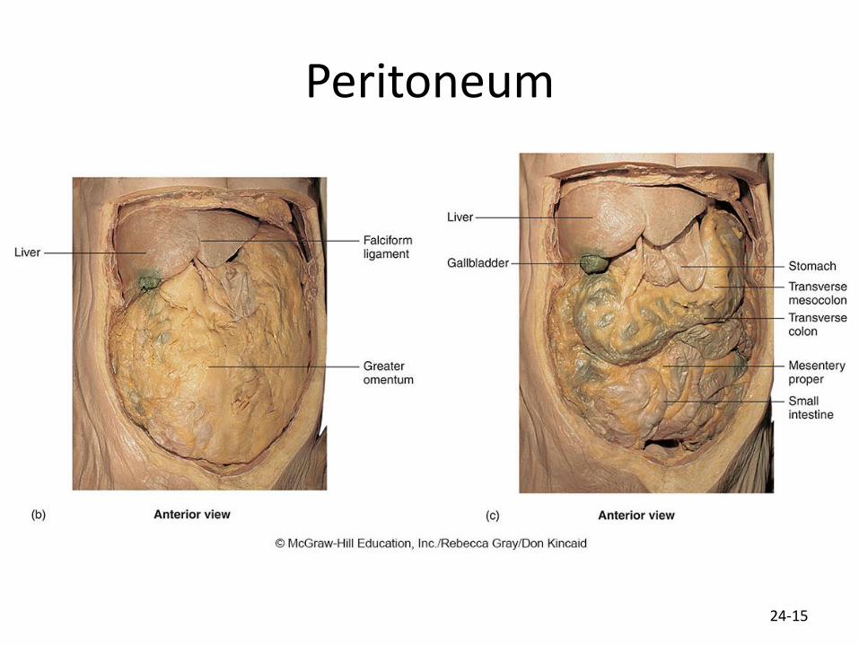
24.5 Peritoneum • Peritoneum
– Visceral: Covers organs – Parietal: Covers interior surface of body
wall – Retroperitoneal: Certain organs
covered by peritoneum on only one surface and are considered behind the peritoneum; e.g., kidneys, pancreas, duodenum
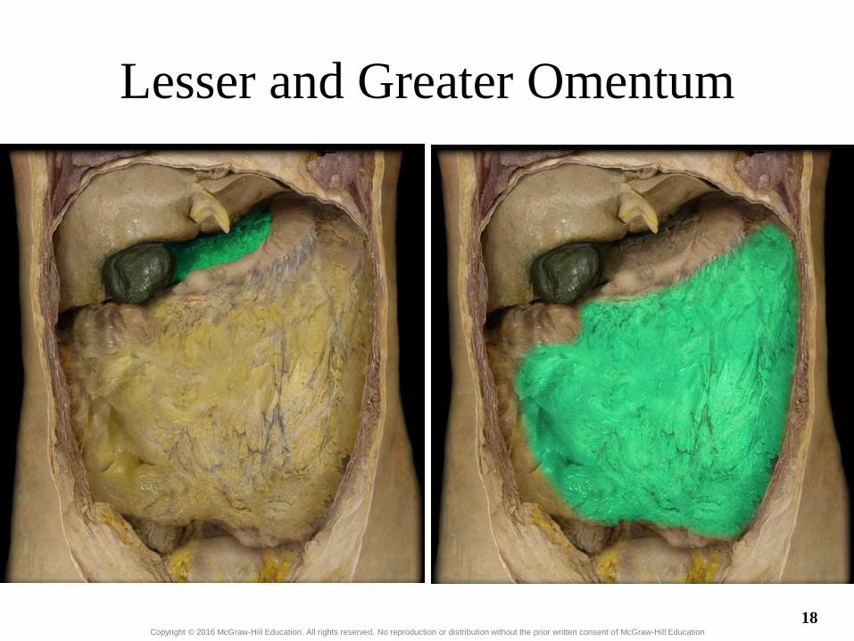
• Mesenteries: two layers of peritoneum with thin layer of loose C.T. between – Routes by which vessels and nerves
pass from body wall to organs – Greater omentum: connects greater
curvature of the stomach to the transverse colon.
– Lesser omentum: connects lesser curvature of the stomach and the proximal part of the duodenum to the liver and diaphragm.
– Transverse mesocolon, sigmoid mesocolon, mesoappendix.
• Ligaments – Coronary: between liver and diaphragm – Falciform: between liver and anterior
abdominal wall
24-14
Peritoneum
24-15

Peritoneum (2)
16
Visceral
peritoneum
Parietal
peritoneum
Peritoneal
cavity
Lesser
omentum
Mesentery
of small
intestine
Greater
omentum
Peritoneum
Copyright © McGraw-Hill Education. All rights reserved. No reproduction or distribution without the prior written consent of McGraw-Hill Education.

Copyright © 2016 McGraw-Hill Education. All rights reserved. No reproduction or distribution without the prior written consent of McGraw-Hill Education
Mesentery
17
Copyright © 2016 McGraw-Hill Education. All rights reserved. No reproduction or distribution without the prior written consent of McGraw-Hill Education
Lesser and Greater Omentum
18

Copyright © 2016 McGraw-Hill Education. All rights reserved. No reproduction or distribution without the prior written consent of McGraw-Hill Education
Falciform Ligament
19

24.6 Oral Cavity
• Bounded by lips anteriorly, fauces (opening into pharynx) posteriorly – Vestibule: space between
lip/cheeks and alveolar processes with teeth
– Oral cavity proper: medial to alveolar processes
– Lined with moist stratified squamous epithelium
24-20

Lips and Cheeks
• Both structures important in mastication and speech
• Lips (labia): orbicularis oris muscle within. Keratinized stratified squamous exterior is thin and color of blood in dermis gives a red/pink color. – Labial frenula (mucous folds) extend from alveolar processes of
maxilla and mandible to the upper and lower lips, respectively.
– Many facial muscles act to move lips
• Cheeks: lateral walls of oral cavity – Buccinator muscle
– Buccal fat pad
24-21
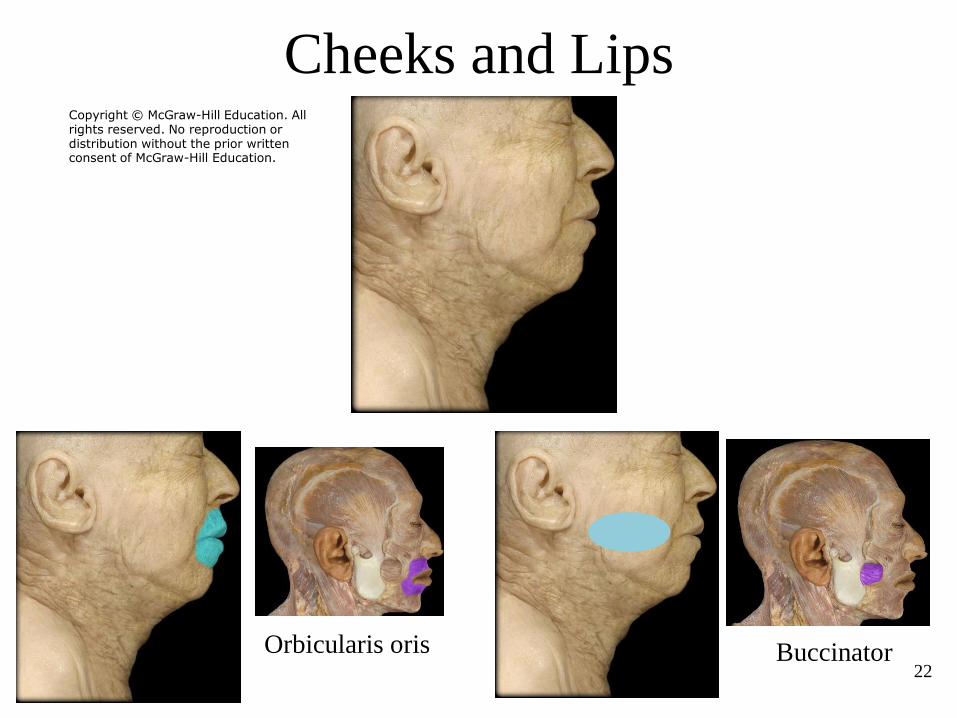
Cheeks and Lips
22
Orbicularis oris Buccinator
Copyright © McGraw-Hill Education. All rights reserved. No reproduction or distribution without the prior written consent of McGraw-Hill Education.

Palate and Palatine Tonsils
• Palate
– Hard palate: anterior, supported by maxilla and palatine bone
– Soft palate: posterior, consists of skeletal muscle and connective tissue
– Uvula: projects from posterior of soft palate
• Palatine tonsils: lateral walls of fauces
24-23

Palate and Palatine Tonsil (2)
24
Hard palate Soft palate
Palatine tonsil
Uvula
Copyright © McGraw-Hill Education. All rights reserved. No reproduction or distribution without the prior written consent of McGraw-Hill Education.
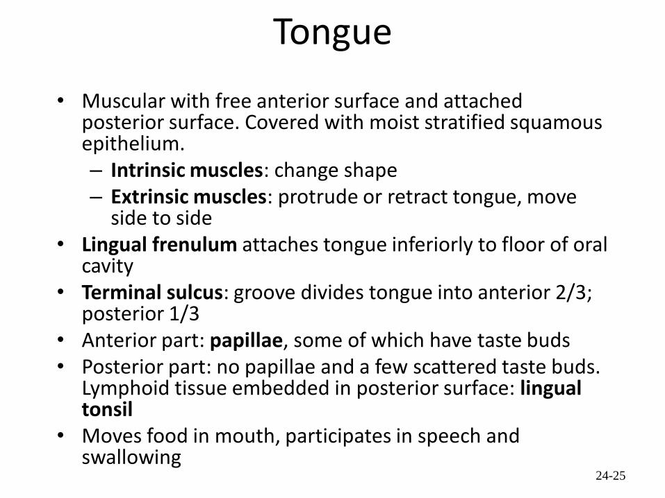
Tongue
• Muscular with free anterior surface and attached posterior surface. Covered with moist stratified squamous epithelium. – Intrinsic muscles: change shape – Extrinsic muscles: protrude or retract tongue, move
side to side • Lingual frenulum attaches tongue inferiorly to floor of oral
cavity • Terminal sulcus: groove divides tongue into anterior 2/3;
posterior 1/3 • Anterior part: papillae, some of which have taste buds • Posterior part: no papillae and a few scattered taste buds.
Lymphoid tissue embedded in posterior surface: lingual tonsil
• Moves food in mouth, participates in speech and swallowing
24-25

Tongue (2)
26 Copyright © McGraw-Hill Education. All rights reserved. No reproduction or distribution without the prior written consent of McGraw-Hill Education.

Teeth
• Two sets – Primary, deciduous, milk:
Childhood
– Permanent or secondary: Adult (32)
• Types – Incisors, canines, premolars
and molars
24-27

Teeth (2) • Involved in mastication and
speech • Anatomic crown: enamel-
covered part of tooth; clinical crown is section of tooth above gum line
• Neck: enameled part of tooth below gum line
• Enamel: outermost layer of anatomical crown. Non-living; acellular. Protective.
• Dentin: living, cellular, calcified tissue. In the root, dentin is covered by cellular bone-like structure that helps hold tooth in the socket.
• Pulp cavity filled with blood vessels, nerves, and connective tissue
• Periodontal ligaments: hold tooth in socket.
• Gingiva: dense, fibrous C.T. covered by stratified squamous epithelium.
24-28

Teeth (3)
29 Copyright © McGraw-Hill Education. All rights reserved. No reproduction or distribution without the prior written consent of McGraw-Hill Education.

Copyright © 2016 McGraw-Hill Education. All rights reserved. No reproduction or distribution without the prior written consent of McGraw-Hill Education
Teeth and Gingiva
30

Mastication
• Chewing: incisors and canines bite or cut off food; molar-type teeth grind food
• Muscles involved: masseter, temporalis, medial and lateral pterygoids. – Elevate mandible: temporalis, masseter, medial pterygoids – Depress mandible: lateral pterygoids – Protraction and lateral and medial excursion: pterygoids and
masseter – Retraction- temporalis
• Mastication reflex: medulla oblongata, but descending pathways from cerebrum provide conscious control. Controls basic movements involved in chewing
24-31

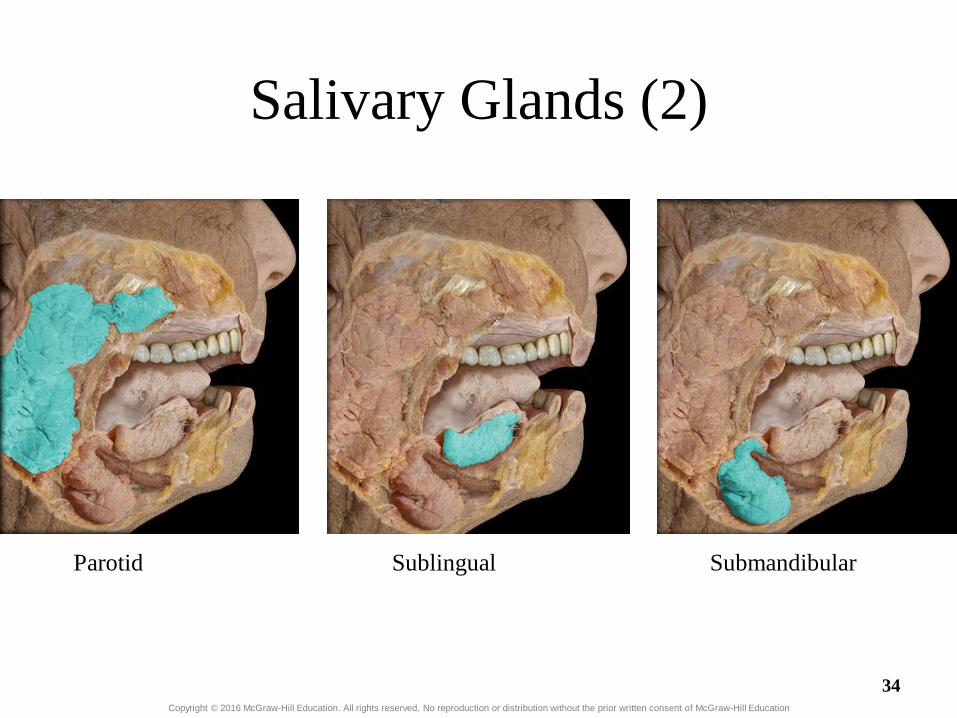
Salivary Glands • Three pairs of multicellular
glands – Parotid: largest. Serous. Just
anterior to the ear. Parotid duct crosses over masseter, penetrates buccinator, and enters the oral cavity adjacent to the 2nd upper molar
– Submandibular: mixed, but more serous than mucous. Posterior half of inferior border of mandible. Duct enters oral cavity on either side of lingual frenulum
– Sublingual: smallest. Mixed, but primarily mucous. Each has 10-12 ducts that enter the floor of the oral cavity.
• Lingual glands. Small, coiled tubular glands on surface of tongue.
24-32

Saliva
• Compound alveolar salivary glands. Produce saliva – Prevents bacterial infection – Lubrication – Contains salivary amylase that breaks
down starch into disaccharides maltose and isomaltose.
– Helps to form bolus for swallowing – Parasympathetic input causes salivary
production
24-33
Copyright © 2016 McGraw-Hill Education. All rights reserved. No reproduction or distribution without the prior written consent of McGraw-Hill Education
Salivary Glands (2)
Parotid Sublingual Submandibular
34

Muscles of Mastication
35
Temporalis Masseter
Medial
pterygoid
Lateral
pterygoid
Copyright © McGraw-Hill Education. All rights reserved. No reproduction or distribution without the prior written consent of McGraw-Hill Education.
24.7 Swallowing
• Pharynx – Posterior walls of
oropharynx and laryngopharynx contains group of muscles called pharyngeal constrictors that contribute to swallowing
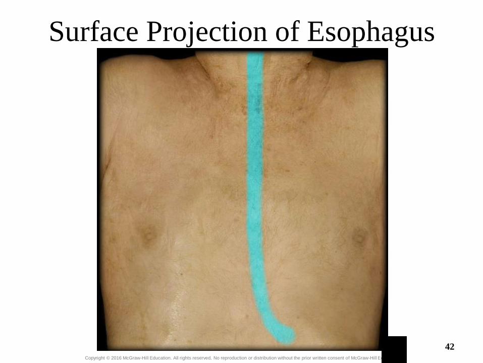
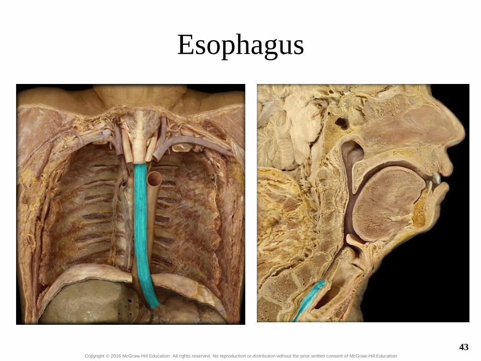
• Esophagus – Transports food from
pharynx to stomach – Passes through esophageal
hiatus (opening) of diaphragm and ends at stomach • Hiatal hernia: widening of
hiatus
– Sphincters • Upper. Striated • Lower. Smooth
– Mucosa is moist stratified squamous epithelium. Produces thick layer of mucus.
24-36

Swallowing (Deglutition)
• Three phases – Voluntary: bolus of food moved by tongue from oral
cavity to pharynx. – Pharyngeal: reflex. Controlled by swallowing center in
medulla oblongata. Soft palate elevates, upper esophageal sphincter relaxes, elevated pharynx opens the esophagus, food pushed into esophagus by pharyngeal constrictors’ successive contraction from superior to inferior. Epiglottis is tipped posteriorly due to pressure of the bolus, larynx elevated to prevent food from passing into larynx.
– Esophageal: reflex. Stretching of esophagus causes enteric NS to initiate peristalsis of muscles in the esophagus.
24-37

Three Phases of Swallowing
24-38

Copyright © 2016 McGraw-Hill Education. All rights reserved. No reproduction or distribution without the prior written consent of McGraw-Hill Education
Pharynx
Nasopharynx Oropharynx Laryngopharynx
39

Copyright © 2016 McGraw-Hill Education. All rights reserved. No reproduction or distribution without the prior written consent of McGraw-Hill Education
Pharynx (2)
Oropharynx Nasopharynx Laryngopharynx
40

Copyright © 2016 McGraw-Hill Education. All rights reserved. No reproduction or distribution without the prior written consent of McGraw-Hill Education
Pharynx (3)
Pharyngeal constrictors
Superior esophageal sphincter 41
Copyright © 2016 McGraw-Hill Education. All rights reserved. No reproduction or distribution without the prior written consent of McGraw-Hill Education
Surface Projection of Esophagus
42
Copyright © 2016 McGraw-Hill Education. All rights reserved. No reproduction or distribution without the prior written consent of McGraw-Hill Education
Esophagus
43

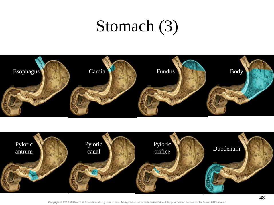
24.8 Stomach • Openings
– Gastroesophageal (cardiac): to esophagus
– Pyloric: to duodenum
• Parts – Cardiac – Fundus – Body – Pyloric: antrum and canal
• Greater and lesser curvatures: attachment sites for omenta
• Sphincters – Cardiac (lower esophageal) – Pyloric
24-44

Copyright © 2016 McGraw-Hill Education. All rights reserved. No reproduction or distribution without the prior written consent of McGraw-Hill Education
Stomach
45

Copyright © 2016 McGraw-Hill Education. All rights reserved. No reproduction or distribution without the prior written consent of McGraw-Hill Education
Stomach Curvatures
Greater curvature
Lesser curvature
46

Copyright © 2016 McGraw-Hill Education. All rights reserved. No reproduction or distribution without the prior written consent of McGraw-Hill Education
Stomach (2)
Esophagus Cardia Fundus
Body Pyloris Duodenum
47
Copyright © 2016 McGraw-Hill Education. All rights reserved. No reproduction or distribution without the prior written consent of McGraw-Hill Education
Stomach (3)
Esophagus Cardia Fundus Body
Pyloric
antrum
Pyloric
canal
Pyloric
orifice Duodenum
48

Copyright © 2016 McGraw-Hill Education. All rights reserved. No reproduction or distribution without the prior written consent of McGraw-Hill Education
Stomach Wall Musculature
Stomach
musculature
Lower
esophageal
sphincter
Pyloric
sphincter
49

Histology of the Stomach • Layers
– Serosa or visceral peritoneum – Muscularis: three layers
• Outer longitudinal • Middle circular • Inner oblique
– Submucosa
– Mucosa
• Rugae: folds in stomach when empty. Mucosa and submucosa.
24-50

Histology of the Stomach (3)
• Gastric pits: openings for gastric glands. Lined with simple columnar epithelium
• Cells of gastric pits – Surface mucus: mucus that protects stomach lining from
acid and digestive enzymes – Mucous neck: mucus – Parietal: hydrochloric acid and intrinsic factor – Chief: pepsinogen – Endocrine: regulatory hormones
• Enterochromaffin-like cells: histamine that stimulates acid secretion
• Gastrin-containing cells: secrete gastrin • Somatostatin-containing cells: secrete somatostatin
that inhibits gastrin and insulin secretion
24-51
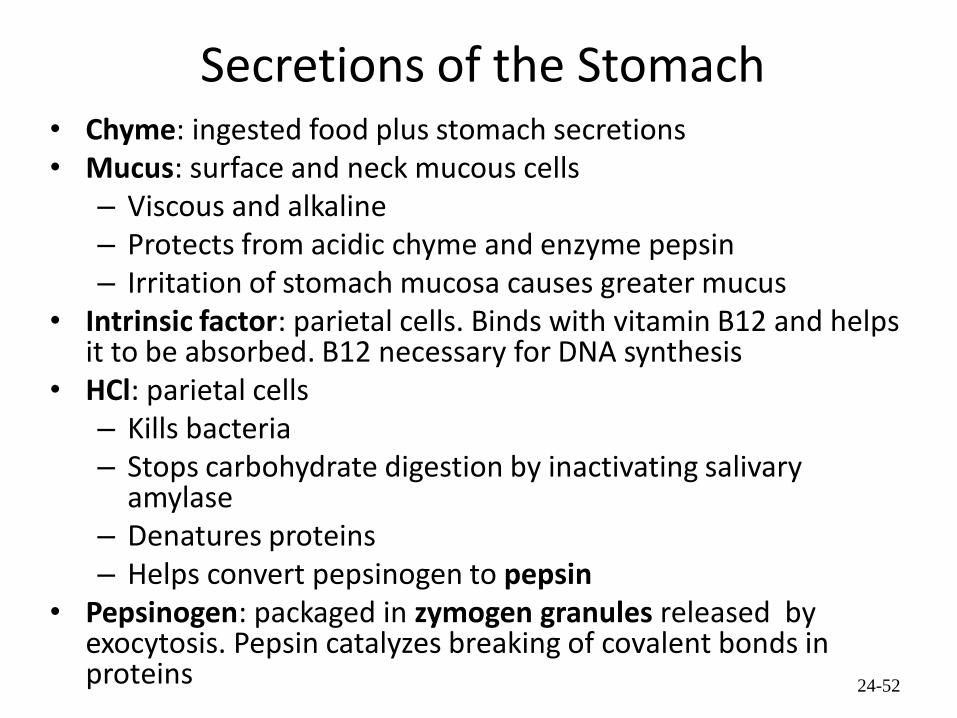
Secretions of the Stomach • Chyme: ingested food plus stomach secretions • Mucus: surface and neck mucous cells
– Viscous and alkaline – Protects from acidic chyme and enzyme pepsin – Irritation of stomach mucosa causes greater mucus
• Intrinsic factor: parietal cells. Binds with vitamin B12 and helps it to be absorbed. B12 necessary for DNA synthesis
• HCl: parietal cells – Kills bacteria – Stops carbohydrate digestion by inactivating salivary
amylase – Denatures proteins – Helps convert pepsinogen to pepsin
• Pepsinogen: packaged in zymogen granules released by exocytosis. Pepsin catalyzes breaking of covalent bonds in proteins 24-52

Hydrochloric Acid Production
1. Carbon dioxide (CO2) diffuses into the parietal cell.
2. Carbon dioxide combines with water (H2O) in an enzymatic reaction that is catalyzed by carbonic anhydrase (CA) to form carbonic acid (H2CO3).
3. Carbonic acid dissociates into a bicarbonate ion (HCO3
–) and a hydrogen ion (H+).
4. Bicarbonate ions are transported back into the bloodstream. An antiporter in the plasma membrane exchanges HCO3
– for a chloride ion (CI–).
5. A H+- K+ pump moves H+ into the duct of the gastric gland and K+ into the parietal cell.
6. Chloride ions diffuse into the gastric gland duct.
24-53

Cephalic Phase
• The taste or smell of food, tactile sensations of food in the mouth, or even thoughts of food stimulate the medulla oblongata.
• Parasympathetic action potentials are carried by the vagus nerves to the stomach, where enteric plexus neurons are activated.
• Postganglionic neurons stimulate secretion by parietal and chief cells (HCl and pepsin) and stimulate the secretion of the hormone gastrin and histamine.
• Gastrin is carried through the circulation back to the stomach where it and histamine stimulate further secretion of HCl and pepsin.
24-54

Gastric Phase • Distention of the stomach activates a parasympathetic reflex.
Action potentials are carried by the vagus nerves to the medulla oblongata.
• Medulla oblongata stimulates further secretions of the stomach. • Distention also stimulates local reflexes that amplify stomach
secretions.
24-55

Intestinal Phase 1. Chyme in the duodenum with a pH less than 2 or containing lipids inhibits
gastric secretions by three mechanisms 2. Sensory input to the medulla from the duodenum inhibits the motor
input from the medulla to the stomach. Stops secretion of pepsin and HCl.
3. Local reflexes inhibit gastric secretion 4. Secretin, and cholecystokinin produced by the duodenum decrease
gastric secretions in the stomach.
24-56

Functions of the Gastrointestinal Hormones
24-57

Movements of the Stomach
• Combination of mixing waves (80%) and peristaltic waves (20%)
• Both esophageal and pyloric sphincters are closed.
24-58

24.9 Small Intestine
• Site of greatest amount of digestion and absorption of nutrients and water
• Divisions – Duodenum- first 25 cm
beyond the pyloric sphincter.
– Jejunum- 2.5 m
– Ileum- 3.5 m. Peyer’s patches or lymph nodules
24-59
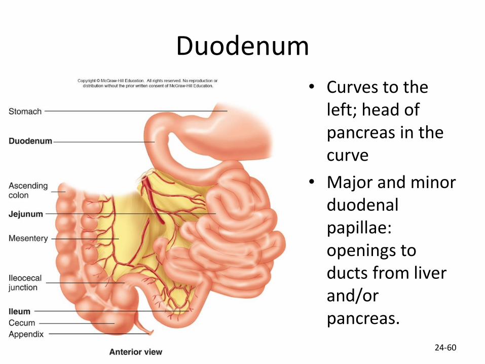
Duodenum
• Curves to the left; head of pancreas in the curve
• Major and minor duodenal papillae: openings to ducts from liver and/or pancreas.
24-60

Copyright © 2016 McGraw-Hill Education. All rights reserved. No reproduction or distribution without the prior written consent of McGraw-Hill Education
Gross Anatomy of Small Intestine
61

Copyright © 2016 McGraw-Hill Education. All rights reserved. No reproduction or distribution without the prior written consent of McGraw-Hill Education
Duodenum and Ileum
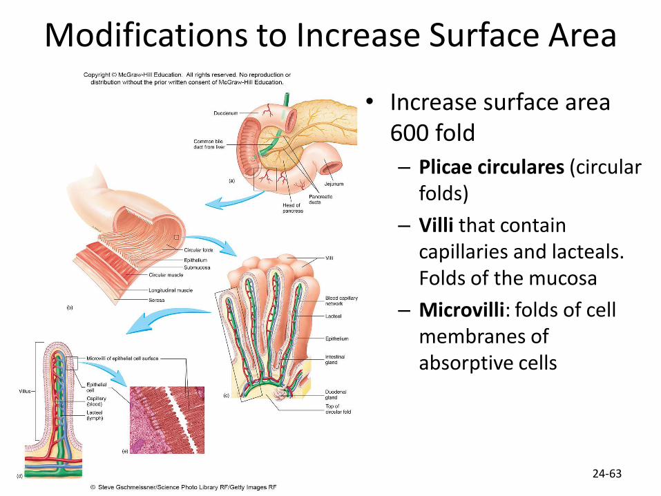
Modifications to Increase Surface Area
• Increase surface area 600 fold
– Plicae circulares (circular folds)
– Villi that contain capillaries and lacteals. Folds of the mucosa
– Microvilli: folds of cell membranes of absorptive cells
24-63

Mucosa and Submucosa of the Duodenum
• Cells and glands of the mucosa – Absorptive cells: cells with microvilli, produce digestive
enzymes and absorb digested food
– Goblet cells: produce protective mucus
– Endocrine cells: produce regulatory hormones
– Granular cells (paneth cells): may help protect from bacteria
• Intestinal glands (crypts of Lieberkühn): tubular glands in mucosa at bases of villi
• Duodenal glands (Brunner’s glands): tubular mucous glands of the submucosa. Open into intestinal glands
24-64

Jejunum and Ileum
• Gradual decrease in diameter, thickness of intestinal wall, number of circular fold, and number of villi the farther away from the stomach
• Major site of nutrient absorption
• Peyer’s patches: lymphatic nodules numerous in mucosa and submucosa
• Ileocecal junction: where ilium meets large intestine. Ileocecal sphincter and ileocecal valve
24-65

Secretions of the Small Intestine
• Fluid primarily composed of water, electrolytes and mucus. • Mucus
– Protects against digestive enzymes and stomach acids • Digestive enzymes: bound to the membranes of the
absorptive cells – Disaccharidases: Break down disaccharides to
monosaccharides – Peptidases: Hydrolyze peptide bonds – Nucleases: Break down nucleic acids
• Duodenal glands – Stimulated by vagus nerve, secretin, chemical or tactile
irritation of duodenal mucosa
24-66

Movement in the Small Intestine
• Mixing and propulsion over short distances
• Segmental contractions mix
• Peristalsis propels
• Ileocecal sphincter remains slightly contracted until peristaltic waves reach it; it relaxes, allowing chyme to move into cecum
• Cecal distention causes local reflex and ileocecal valve constricts – Prevents more chyme from entering cecum
– Increases digestion and absorption in small intestine by slowing progress of chyme
– Prevents backflow
24-67

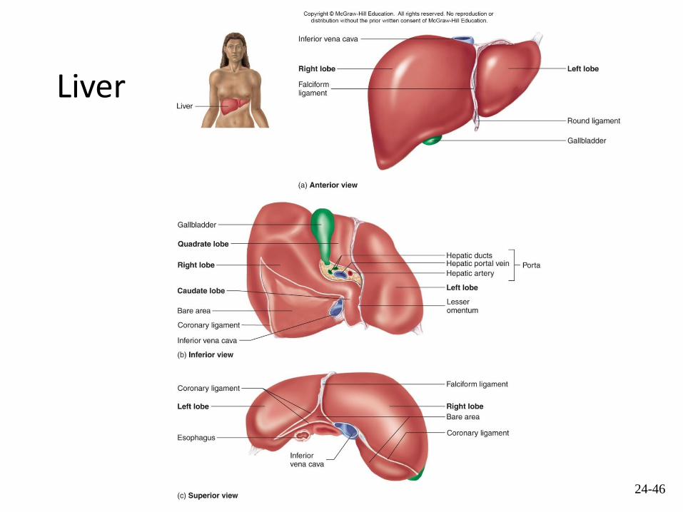
24.10 Liver • Lobes
– Major: Left and right
– Minor: Caudate and quadrate
• Porta: on inferior surface. Vessels, ducts, nerves, exit/enter liver – Hepatic portal vein, hepatic artery, hepatic nerve plexus enter
– Lymphatic vessels, two hepatic ducts exit
• Ducts – Right and left hepatics unite to form
– Common hepatic
– Cystic: from gallbladder
– Common bile: union of cystic duct and common hepatic duct 24-68
Liver
24-46

Liver
and
Gall
Bladder
Rt. & Lt. Hepatic
Ducts
Common Hepatic
Duct
Bile Duct Pancreatic Duct
Duodenal
Papilla
Copyright © McGraw-Hill Education. All rights reserved. No reproduction or distribution without the prior written consent of McGraw-Hill Education.

Rt. & Lt. Hepatic ducts
Common Hepatic duct
Cystic duct Gallbladder
Hepatopancreatic
ampulla Major duodenal papilla
Pancreatic duct
Bile duct
Biliary Apparatus
Copyright © McGraw-Hill Education. All rights reserved. No reproduction or distribution without the prior written consent of McGraw-Hill Education.
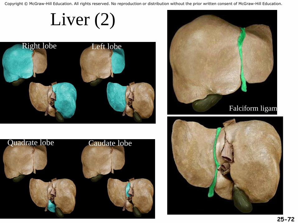
Liver (2)
Right lobe Left lobe
Quadrate lobe Caudate lobe
Falciform ligament
25-72
Copyright © McGraw-Hill Education. All rights reserved. No reproduction or distribution without the prior written consent of McGraw-Hill Education.
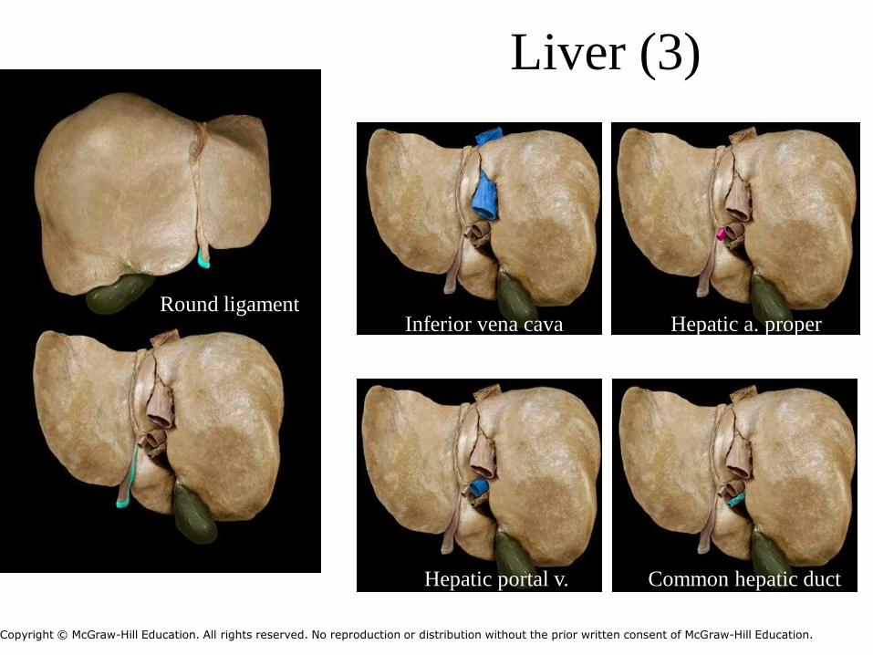
Round ligament Inferior vena cava
Hepatic portal v.
Hepatic a. proper
Common hepatic duct
Liver Liver (3)
Copyright © McGraw-Hill Education. All rights reserved. No reproduction or distribution without the prior written consent of McGraw-Hill Education.

Liver, Gallbladder, Pancreas and Ducts
1. The hepatic ducts, which carry bile from the liver lobes, combine to form the common hepatic duct.
2. The common hepatic duct combines with the cystic duct from the gallbladder to form the common bile duct.
3. The common bile duct and the pancreatic duct combine to form the hepatopancreatic ampulla.
4. The hepatopancreatic ampulla empties bile and pancreatic secretions into the duodenum at the major duodenal papilla.
5. The accessory pancreatic duct empties pancreatic secretions to the duodenum at the minor duodenal papilla.
24-74

Histology of the Liver
• Connective tissue septa branch from the porta into the interior
– Divides liver into lobules
– Nerves, vessels and ducts follow the septa
• Lobules: portal triad at each corner
– Three vessels: hepatic portal vein, hepatic artery, hepatic duct
– Central vein in center of lobule
• Central veins unite to form hepatic veins that exit liver and empty into inferior vena cava
24-75

Histology of the Liver (2) • Hepatic cords: radiate out from
central vein. Composed of hepatocytes
• Hepatic sinusoids: between cords, lined with endothelial cells and hepatic phagocytic (Kupffer) cells
• Bile canaliculus: between cells within cords
• Hepatocyte functions – Bile production – Storage – Interconversion of nutrients – Detoxification – Phagocytosis – Synthesis of blood components

Functions of the Liver
• Bile production: 600-1000 mL/day. Bile salts (bilirubin), cholesterol, fats, fat-soluble hormones, lecithin – Neutralizes and dilutes stomach acid – Bile salts emulsify fats. Most are reabsorbed in the ileum. – Secretin (from the duodenum) stimulates bile secretions,
increasing water and bicarbonate ion content of the bile • Storage
– Glycogen, fat, vitamins, copper and iron. Hepatic portal blood comes to liver from small intestine.
24-77

Functions of the Liver (2) • Nutrient interconversion
– Amino acids to energy producing compounds – Hydroxylation of vitamin D. Vitamin D then travels to
kidney where it is hydroxylated again into its active form • Detoxification
– Hepatocytes remove ammonia and convert to urea • Phagocytosis
– Kupffer cells phagocytize worn-out and dying red and white blood cells, some bacteria
• Synthesis – Albumins, fibrinogen, globulins, heparin, clotting factors
24-78

Blood and Bile Flow Through the Liver (2)
1. The hepatic artery carries oxygenated blood from the aorta through the porta of the liver. Hepatic artery branches become part of the portal triads. Blood from the hepatic artery branches enters the hepatic sinusoids and supplies hepatocytes in the hepatic cords with oxygen.
2. The hepatic portal vein carries nutrient-rich, deoxygenated blood from the intestines through the porta of the liver. Hepatic portal vein branches become part of the portal triads. Blood from the hepatic portal vein branches enters the hepatic sinusoids and supplies hepatocytes in the hepatic cords with nutrients.
3. Blood in the hepatic sinusoids that comes from the hepatic artery and hepatic portal vein picks up plasma proteins, processed molecules, and waste products produced by the hepatocytes of the hepatic cords. The hepatic sinusoids empty into central veins. The central veins connect to hepatic veins, which connect to the inferior vena cava.
4. Bile produced by hepatocytes in the hepatic cords enters bile canaliculi, which connect to hepatic duct branches that are part of the portal triads.
5. The hepatic duct branches converge to form the left and right hepatic ducts, which carry bile out the porta of the liver.
24-79
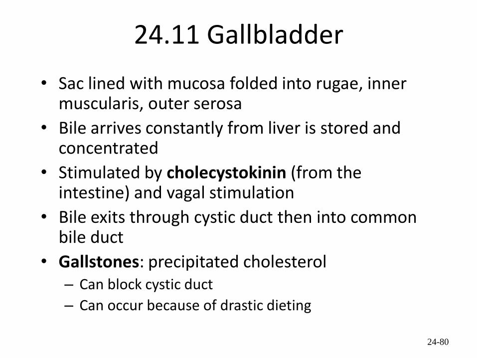
24.11 Gallbladder
• Sac lined with mucosa folded into rugae, inner muscularis, outer serosa
• Bile arrives constantly from liver is stored and concentrated
• Stimulated by cholecystokinin (from the intestine) and vagal stimulation
• Bile exits through cystic duct then into common bile duct
• Gallstones: precipitated cholesterol – Can block cystic duct
– Can occur because of drastic dieting
24-80
Copyright © 2016 McGraw-Hill Education. All rights reserved. No reproduction or distribution without the prior written consent of McGraw-Hill Education
Gallbladder
81
Copyright © McGraw-Hill Education. All rights reserved. No reproduction or distribution without the prior written consent of McGraw-Hill Education.

Control of Bile Secretion and Release
24-82

24.12 Pancreas
• Pancreas both endocrine and exocrine
• Head, body and tail
• Endocrine: pancreatic islets. Produce insulin, glucose, and somatostatin
• Exocrine: groups acini (grape-like cluster) form lobules separated by septa.
• Intercalated ducts lead to intralobular ducts lead to interlobular ducts lead to the pancreatic duct.
• Pancreatic duct joins common bile duct and enters duodenum at the hepatopancreatic ampulla controlled by the hepatopancreatic ampullar sphincter
24-83

Pancreas
24-56

Copyright © 2016 McGraw-Hill Education. All rights reserved. No reproduction or distribution without the prior written consent of McGraw-Hill Education
Pancreas (2)
Head Body Tail Pancreatic duct
Pancreas

Copyright © 2016 McGraw-Hill Education. All rights reserved. No reproduction or distribution without the prior written consent of McGraw-Hill Education
Pancreas (3)
Head Neck Body Tail
86
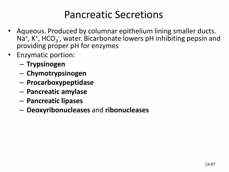
Pancreatic Secretions
• Aqueous. Produced by columnar epithelium lining smaller ducts. Na+, K+, HCO3
-, water. Bicarbonate lowers pH inhibiting pepsin and providing proper pH for enzymes
• Enzymatic portion: – Trypsinogen – Chymotrypsinogen – Procarboxypeptidase – Pancreatic amylase – Pancreatic lipases – Deoxyribonucleases and ribonucleases
24-87

Pancreatic Secretions (2)
• Interaction of duodenal and pancreatic enzymes – Enterokinase from the duodenal mucosa and attached to the
brush border activates trypsinogen to trypsin. – Trypsin activates chymotrypsinogen to chymotrypsin. – Trypsin activates procarboxypeptidase to carboxypeptidase.
• Trypsin, chymotrypsin, and carboxypeptidase digest proteins: proteolytic.
• Pancreatic amylase continues digestion of starch. • Pancreatic lipase digests lipids. • Deoxyribonucleases and ribonucleases digest DNA and
ribonucleic acid, respectively.
24-88

Bicarbonate Ion Production in Pancreas
24-89
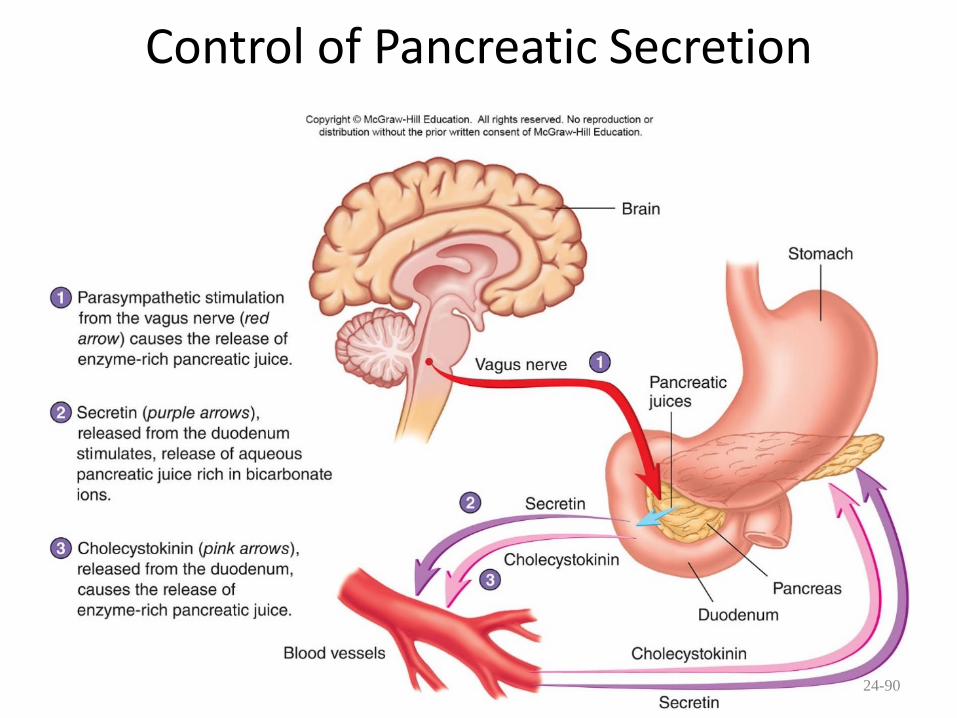
Control of Pancreatic Secretion
24-90

24.13 Large Intestine
• Extends from ileocecal junction to anus
• Consists of cecum, colon, rectum, anal canal
• Movements sluggish (18-24 hours); chyme converted to feces.
• Absorption of water and salts, secretion of mucus, extensive action of microorganisms.
• 1500 mL chyme enter the cecum, 90% of volume reabsorbed yielding 80-150 mL of feces 24-91

Ileocecal
junction Cecum
Vermiform
appendix
Ascending
colon Transverse
colon
Descending
colon
Sigmoid
colon
Rt. Colic
flexure
Lt. colic
flexure
Teniae
coli
92
Large
Intestine
Copyright © McGraw-Hill Education. All rights reserved. No reproduction or distribution without the prior written consent of McGraw-Hill Education.

Colon X-ray
Cecum Ascending colon Transverse colon
Descending colon Sigmoid colon
Rectum
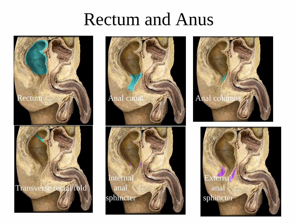
Rectum and Anus
Rectum Anal canal
External
anal
sphincter
Transverse rectal fold
Internal
anal
sphincter
Anal columns

Haustra Teniae coli
Omental (epiploic) appendages
95
Copyright © McGraw-Hill Education. All rights reserved. No reproduction or distribution without the prior written consent of McGraw-Hill Education.
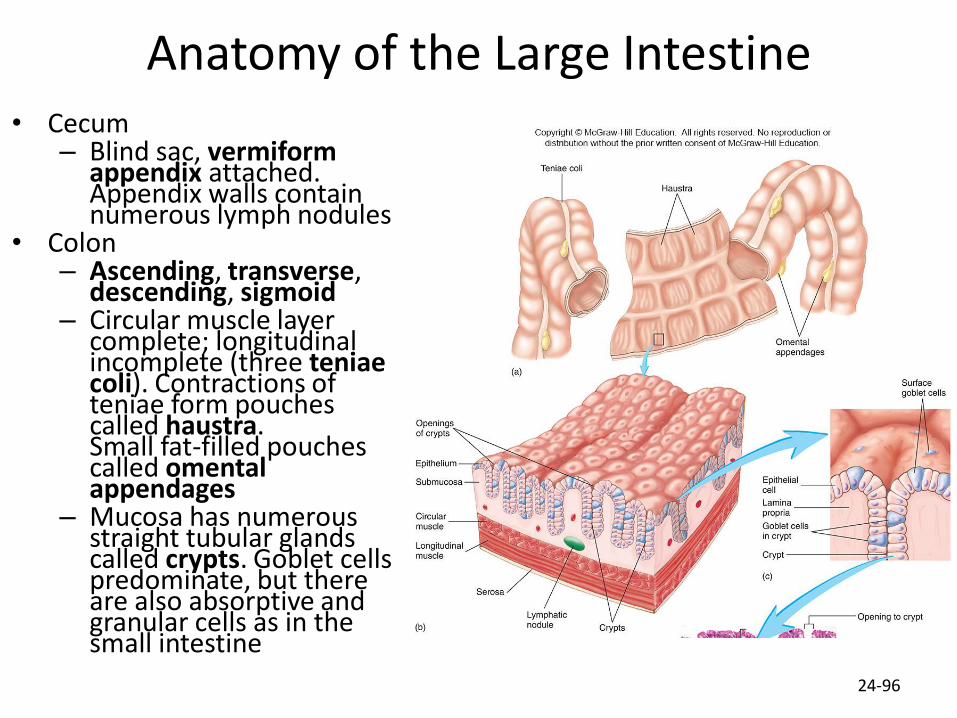
Anatomy of the Large Intestine • Cecum
– Blind sac, vermiform appendix attached. Appendix walls contain numerous lymph nodules
• Colon – Ascending, transverse,
descending, sigmoid – Circular muscle layer
complete; longitudinal incomplete (three teniae coli). Contractions of teniae form pouches called haustra. Small fat-filled pouches called omental appendages
– Mucosa has numerous straight tubular glands called crypts. Goblet cells predominate, but there are also absorptive and granular cells as in the small intestine
24-96

Anatomy of the Large Intestine (2)
• Rectum – Straight muscular tube,
thick muscular tunic • Anal canal- superior
epithelium is simple columnar; inferior epithelium is stratified squamous – Internal anal sphincter
(smooth muscle) – External anal sphincter
(skeletal muscle) – Hemorrhoids: Vein
enlargement or inflammation
24-97

Secretions of the Large Intestine
• Mucus provides protection – Parasympathetic stimulation increases rate of goblet cell secretion
• Pumps: bacteria produce acid and the following remove acid from the epithelial cells that line the large intestine – Exchange of bicarbonate ions for chloride ions – Exchange of sodium ions for hydrogen ions
• Bacterial actions produce gases (flatus) from particular kinds of carbohydrates found in legumes and in artificial sugars like sorbitol
• Bacteria produce vitamin K which is then absorbed • Feces consists of water, undigested food (cellulose),
microorganisms, sloughed-off epithelial cells
24-98
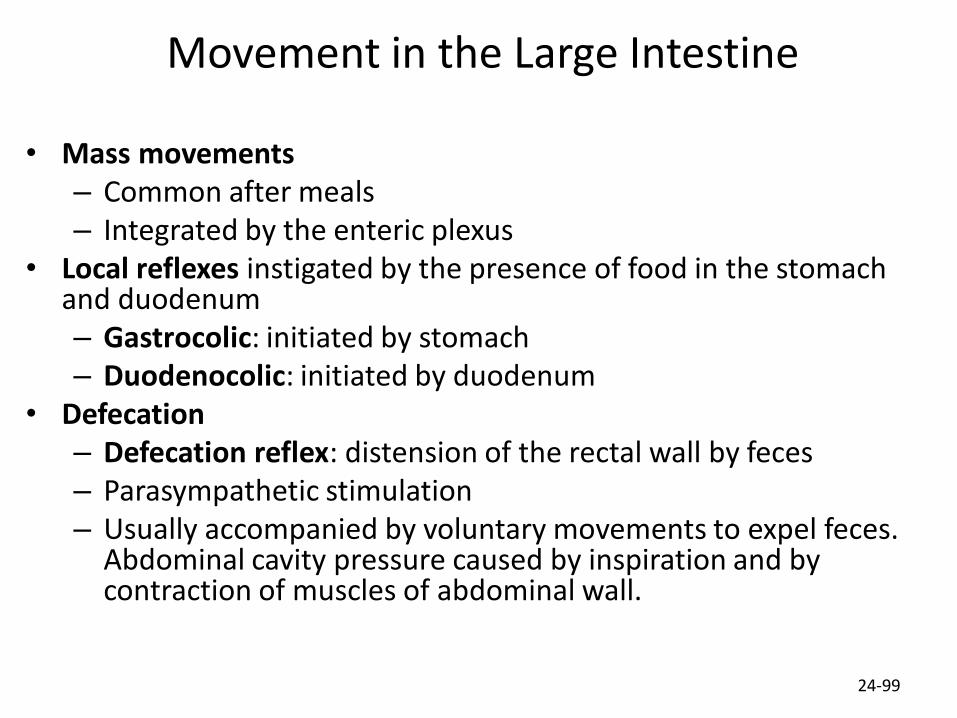
Movement in the Large Intestine
• Mass movements – Common after meals – Integrated by the enteric plexus
• Local reflexes instigated by the presence of food in the stomach and duodenum – Gastrocolic: initiated by stomach – Duodenocolic: initiated by duodenum
• Defecation – Defecation reflex: distension of the rectal wall by feces – Parasympathetic stimulation – Usually accompanied by voluntary movements to expel feces.
Abdominal cavity pressure caused by inspiration and by contraction of muscles of abdominal wall.
24-99

Movement in the Large Intestine (2) 1. The thought or smell of food, distention of the stomach,
and the movement of chyme into the duodenum can stimulate the gastrocolic and duodenocolic reflexes (green arrows).
2. The gastrocolic and duodenocolic reflexes stimulate mass movements in the colon, which propel the contents of the colon toward the rectum (orange arrow).
3. Distention of the rectum by feces stimulates local defecation reflexes. These reflexes cause contractions of the colon and rectum (brown arrow), which move feces toward the anus.
4. Local reflexes cause relaxation of the internal anal sphincter (brown arrow).
5. Distention of the rectum by feces stimulates parasympathetic reflexes. Action potentials are propagated to the defecation reflex center located in the spinal cord (yellow arrow).
6. Action potentials stimulate contraction of the colon and rectum and relaxation of the internal anal sphincter (purple arrows).
7. Action potentials are propagated through ascending nerve tracts to the brain (blue arrow).
8. Descending nerve tracts from the brain regulate the defecation reflex center (pink arrow).
9. Action potentials from the brain control the external anal sphincter (purple arrow).
24-100
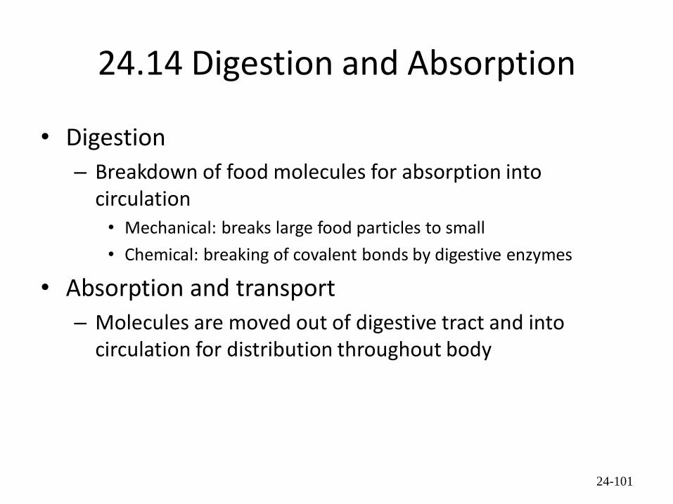
24.14 Digestion and Absorption
• Digestion
– Breakdown of food molecules for absorption into circulation • Mechanical: breaks large food particles to small
• Chemical: breaking of covalent bonds by digestive enzymes
• Absorption and transport
– Molecules are moved out of digestive tract and into circulation for distribution throughout body
24-101

Digestion Summary
24-68

Carbohydrates: Hydrolyzed into Monosaccharides
• Glucose is transported to cells requiring energy; insulin influences rate of transport
• Monosaccharide (glucose)
transport 1. Glucose is absorbed by
symport with Na+ into intestinal epithelial cells.
2. Symport is driven by a sodium gradient established by a Na+–K+ pump.
3. Glucose moves out of the intestinal epithelial cells by facilitated diffusion.
4. Glucose enters the capillaries of the intestinal villi and is carried through the hepatic portal vein to the liver.
24-103

Transport of Lipids Across Intestinal Epithelium
Lipid transport 1. Bile salts surround fatty acids and
monoglycerides to form micelles.
2. Micelles attach to the plasma membranes of intestinal epithelial cells, and the fatty acids and monoglycerides pass by simple diffusion into the intestinal epithelial cells.
3. Within the intestinal epithelial cell, the fatty acids and monoglycerides are converted to triglycerides; proteins coat the triglycerides to form chylomicrons, which move out of the intestinal epithelial cells by exocytosis.
4. The chylomicrons enter the lacteals of the intestinal villi and are carried through the lymphatic system to the general circulation.
24-104

Lipids
• Include triglycerides, phospholipids, steroids, fat-soluble vitamins
• Bile salts surround fatty acid and glycerol to form micelles
• Chylomicrons are 90% triglyceride, 5% cholesterol, 4% phospholipid, 1% protein.
• Chylomicrons enter blood stream and travel to adipose tissue. In blood, triglycerides converted back into fatty acids and glycerol where they are transported into the adipose cells, then converted back into triglycerides.
24-105
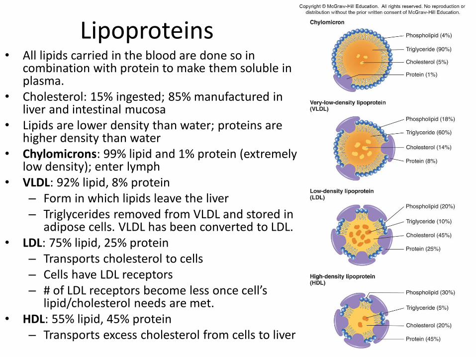
Lipoproteins • All lipids carried in the blood are done so in
combination with protein to make them soluble in plasma.
• Cholesterol: 15% ingested; 85% manufactured in liver and intestinal mucosa
• Lipids are lower density than water; proteins are higher density than water
• Chylomicrons: 99% lipid and 1% protein (extremely low density); enter lymph
• VLDL: 92% lipid, 8% protein – Form in which lipids leave the liver – Triglycerides removed from VLDL and stored in
adipose cells. VLDL has been converted to LDL. • LDL: 75% lipid, 25% protein
– Transports cholesterol to cells – Cells have LDL receptors – # of LDL receptors become less once cell’s
lipid/cholesterol needs are met. • HDL: 55% lipid, 45% protein
– Transports excess cholesterol from cells to liver
24-106

Transport of LDL into Cells
24-107
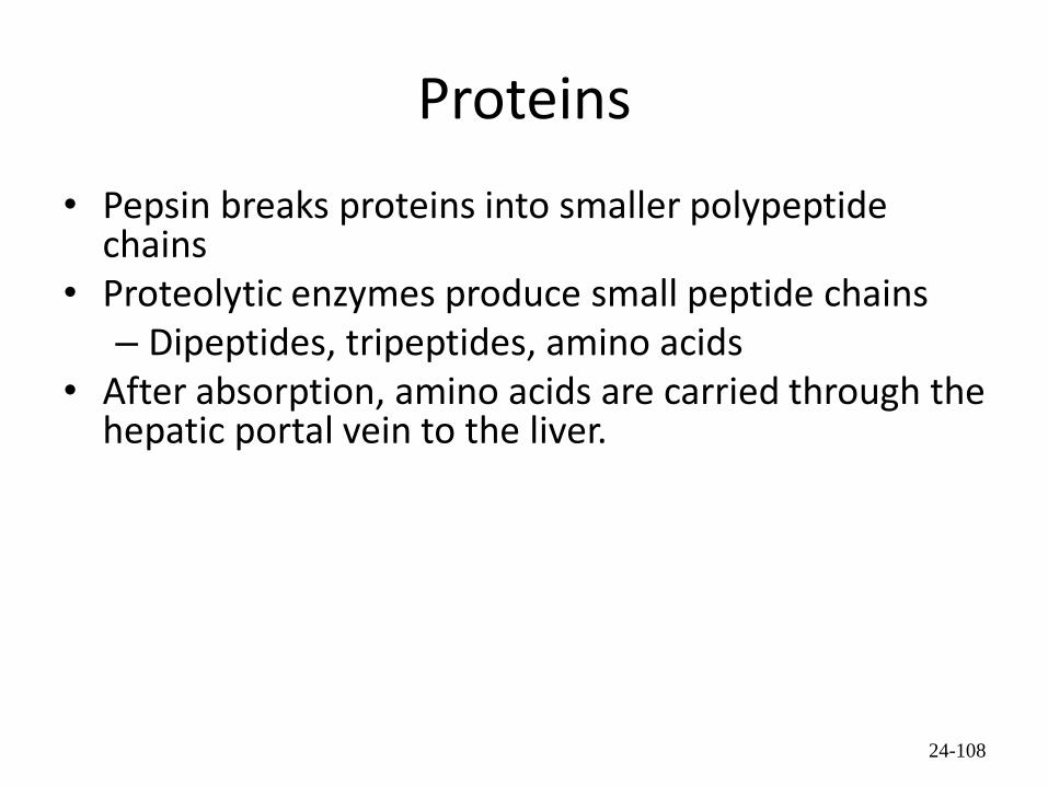
Proteins
• Pepsin breaks proteins into smaller polypeptide chains
• Proteolytic enzymes produce small peptide chains – Dipeptides, tripeptides, amino acids
• After absorption, amino acids are carried through the hepatic portal vein to the liver.
24-108
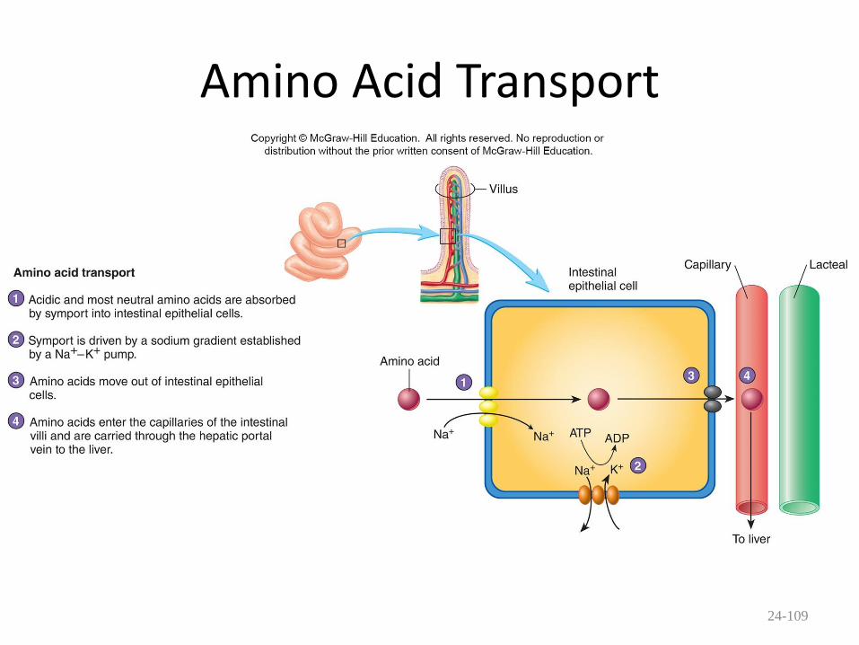
Amino Acid Transport
24-109

Water and Ions
• Water: can move in either direction across wall of small intestine depending on osmotic gradients
• Ions: sodium, potassium, calcium, magnesium, phosphate are actively transported
24-110

24.15 Effects of Aging
• Decrease in mucus layer, connective tissue, muscles and secretions
• Increased susceptibility to infections and toxic agents, increase in incidences of ulcerations and cancers
24-111

Diseases and Disorders
Digestive System
24-112

Diseases and Disorders
Digestive System (2)
24-113

Diseases and Disorders
Digestive System (3)
24-114