Bioavailability of nanoparticulate hematite to Arabidopsis thaliana
Upload
independentCategory
view
0download
0
REVIEWS
Challenges for Research on Polyphenols from Foodsin Alzheimer’s Disease: Bioavailability, Metabolism,
and Cellular and Molecular Mechanisms
MANJEET SINGH,† MADELEINE ARSENEAULT,† THOMAS SANDERSON,†
VEN MURTHY,§ AND CHARLES RAMASSAMY*,†,§
INRS-Institut Armand-Frappier, 531 Boulevard des Prairies, Laval, Quebec, Canada H7V 1B7, andINAF, Universite Laval, Department of Medical Biology, Faculty of Medicine, Laval University,
Quebec, Canada
Polyphenols are the most abundant antioxidants in diet. Indeed, fruits, vegetables, beverages (tea,wine, juices), plants, and some herbs are loaded with powerful antioxidant polyphenols. Despite theirwide distribution, research on human health benefits truly began in the mid-1990s (Scalbert, A.;Johnson, I. T.; Saltmarsh, M. Am. J. Clin. Nutr. 2005, 81, S15S-217S). Phenolic compounds havebeen receiving increasing interest from consumers and manufacturers because numerous epide-miological studies have suggested associations between consumption of polyphenol-rich foods orbeverages and the prevention of certain chronic diseases such as cancers and cardiovascular diseases(Manach, C.; Mazur, A.; Scalbert, A. Curr. Opin. Lipidol. 2005, 16, 77-84; Duthie, S. J. Mol. Nutr.Food Res. 2007, 51, 665-674). Furthermore, in the past 10 years, research on the neuroprotectiveeffects of dietary polyphenols has developed considerably. These compounds are able to protectneuronal cells in various in vivo and in vitro models through different intracellular targets (Ramassamy,C. Eur. J. Pharmacol. 2006, 545, 51-64). However, it is not at all clear whether these compoundsreach the brain in sufficient concentrations and in a biologically active form to exert beneficial effects.On the other hand, it has become clear that the mechanisms of action of these polyphenols go beyondtheir antioxidant activity and the attenuation of oxidative stress. Therefore, there is a need for moreresearch on their intracellular and molecular targets as special pathways underlying distinct polyphenol-induced neuroprotection. The focus of this review is aimed at presenting the role of some polyphenolsfrom fruits, vegetables, and beverages in neuroprotection and particularly in Alzheimer’s diseaseand the research challenges in this area.
KEYWORDS: Antioxidant; neuroprotection; catechins; resveratrol; curcumin; berries; pomegranate; NRf2;
MAPKs
I. OXIDATIVE STRESS IN AGING AND ALZHEIMER’SDISEASE (AD)
I.1. Oxidatively Induced Damage in Aging Brain. Freeradicals can de defined as molecules or molecular fragmentscontaining one or more unpaired electrons in atomic ormolecular orbitals. These unpaired electrons give a considerabledegree of reactivity to free radicals. Reactive oxygen species(ROS), as well as reactive nitrogen species (RNS), are productsof normal cellular metabolism. Both reactive species are nowwell-recognized as playing a dual role in both deleterious and
beneficial effects, especially in numerous signal transductionmechanisms. Radicals derived from oxygen represent the mostimportant class of radical species generated in living systems.For instance, superoxide anion (O2
•-) is considered to be theprimary ROS and can further interact with other molecules togenerate secondary ROS, either through enzyme- or metal-catalyzed processes. These anions are mostly produced in themitochondria by complexes I and III of the electron transportchain. The hydroxyl radical (OH•) has a high reactivity, makingit a very toxic radical with a very short half-life. Hydroxylradicals are mostly produced through the Fenton reaction, whichrequires Fe2+. Additional ROS that can be formed in living
* Corresponding author (e-mail [email protected]).† INRS-Institut Armand-Frappier.§ Laval University.
J. Agric. Food Chem. 2008, 56, 4855–4873 4855
10.1021/jf0735073 CCC: $40.75 2008 American Chemical SocietyPublished on Web 06/17/2008

systems are peroxyl radicals (ROO•), particularly through thereaction of hydroxyl radicals with unsaturated lipids.
Nitric oxide (NO•) is a small molecule that contains a singleunpaired electron and is therefore a free radical. NO• is generatedin biological tissues by nitric oxide synthases (NOSs), whichmetabolize arginine to citrulline with the formation of NO•. NO•
is an abundant reactive radical that acts as an important oxidativebiological signaling molecule in diverse physiological phenom-ena, including neurotransmission, smooth muscle relaxation,immune regulation, and blood pressure regulation, etc. Anoverproduction of RNS causes nitrosative stress. This may occurwhen the generation of reactive nitrogen species exceeds thesystem’s ability to neutralize and eliminate them. Nitrosativestress may lead to unwanted nitrosylation reactions that alterthe structure and functions of certain proteins.
At high concentrations, ROS and RNS can be importantmediators of damage to nucleic acids, lipids, and proteins. Inthe brain, these damages contribute to aging and age-associatedneurodegenerative diseases such as Alzheimer’s disease (AD)or Parkinson’s disease.
Brain aging is associated with the accumulation of theseoxidative-induced damages, likely due to the imbalance betweenantioxidant defenses and intracellular generation of ROS. Theoverall rationale implication of oxidative stress in aging brainis based on the following premises: (a) the brain contains highlevels of unsaturated fatty acids, which are vulnerable tooxidation (it is particularly high in 20:4 and 22:6 fatty acids);(b) the brain consumes high amounts of oxygen (about 20% ofthe total amount used in the body); (c) levels of antioxidantsare lower in the brain; and (d) the brain contains highconcentrations of transition metals such as Fe2+ that are keycatalysts of oxidative-induced damages.
In the brain, oxidative damage to DNA occurs continuously,resulting in damaged nucleotides and strand breaks. It has beenestimated that 10000 oxidative interactions occur between DNAand endogenously generated free radicals per human cell perday. One of the most widely studied base lesions is 8-hydroxy-2′-deoxyguanosine (8-OHdG), a hydroxyl radical-damagedguanine nucleoside. Several studies have demonstrated that thelevel of 8-OHdG is elevated in old brains, with a 4-fold increasewhen compared to young brains (5). In mitochondria, higherlevels of oxidative damage and DNA mutations have beenascribed to the location of the DNA near the inner mitochondrialmembrane sites where superoxide anions are mainly formed.
Accumulation of oxidized proteins in brain is widely con-sidered a hallmark of aging (6–8) with increases of nearly 2-and 4-fold in humans and rats, respectively (6). Oxidativelyinduced damage to proteins can affect virtually all amino acids,with sulfur-containing amino acids and aromatic amino acidsbeing the most susceptible (9). The amount of oxidized proteinis influenced not only by its rate of formation, but also by itsrate of degradation. In a functioning system oxidized proteinsare degraded by the proteasomal/protease system, which isresponsible for removing damaged proteins that form insolubleaggregates (10). During aging, proteasome activity also declines(10), and this could contribute to the elevation of oxidizedproteins. Lipid peroxidation yields a large number of compoundssuch as malondialdehyde (MDA), 4-hydroxynonenal (HNE), F2-isoprostanes, and acrolein. The most widely studied are theactive aldehydes and isoprostanes. A set of isoprostanes thatcould be uniquely formed in brain from peroxidation ofdocosahexanoic acid are neuroprostanes (11). HNE is the majorproduct formed from peroxidation of ω-6-conjugated fatty acids,such as arachidonic acid and linoleic acid (12). HNE is
biologically active, causing gene induction (antioxidant genes)and cytotoxicity; it can react with many biological moleculesincluding various amino acids, proteins, and bases in DNA.
Lipid peroxidation has been reported to be elevated in thebrain with age. MDA was increased in the cytoplasm of neuronsand astrocytes in normal aging, but was rarely detected in normalyoung subjects. In the hippocampus, neuronal and glial MDAdeposition was marked in the CA4 region but mild in CA1 (13).Morevover, the lipid peroxidation products, MDA, HNE, andacrolein, have been reported to react with DNA and proteins toproduce further damage in aged brains (14). Age-relatedmodifications of multiple biomolecules by oxidative damagesmay affect brain health and function in the long run. Thesechanges observed in normal aging are exacerbated in variousneurodegenerative diseases related to aging.
I.2. Oxidative Damage to Lipids, Proteins, and NucleicAcids in Mild Cognitive Impairment and in AD. Aging maybe regarded as the major risk factor for all forms of dementiaand particularly for AD, affecting up to 18 million peopleworldwide (15). According to the Alzheimer’s Society, thisnumber could reach 34 million by 2025 (15). Its prevalencedoubles approximately every five years after the age of 60, with1 in 10 individuals over 65 years and nearly half of those over85 being affected by the disease. AD causes one of the greatestthreats to the future of the healthcare system, with the anticipateddemographic shift to an aging population.
AD is multifactorial, with a complex combination of geneticand nongenetic components. The early-onset familial formrepresents only a small fraction of all AD cases (e5%) andtypically presents itself with age of onset younger than 65 years,whereas the nongenetic or sporadic form represents the majorityof AD cases. To date, mutations on three genes have beenreported to cause the early-onset familial form of AD. Theseinclude the genes coding for the amyloid precursor protein(APP) on chromosome 21, presenilin 1 on chromosome 14, andpresenilin 2 on chromosome 1 (16–18). Although these AD-causing mutations occur in three different genes located on threedifferent chromosomes, they all share a common biochemicalpathway, that is, the altered production of the amyloid � peptide(A�), which leads to neuronal death and dementia.
A� is released after the cleavage of APP by �- andγ-secretases, respectively. �-Secretase has been identified as anaspartic protease, and γ-secretase can cleave APP at theC-terminal end of A� at different sites, giving rise to A�peptides that are 39-43 amino acids long. The exact locationof C-terminal cleavage is critical, because generation of the moreamyloidogenic peptides (such as A�1-42) is strongly correlatedwith AD development.
On the other hand, the late-onset form of AD, representingthe vast majority of all AD cases, has one common genetic riskfactor, the ε4 allele of the apolipoprotein E gene (APOE) locatedon chromosome 19q13 (19–21). The increased risk with the ε4allele of the apolipoprotein E has been consistently replicatedin a large number of studies across many ethnic groups. Unlikethe mutations in the known early-onset familial form genes, theε4 allele of the apolipoprotein E is neither necessary norsufficient to cause AD but instead operates as a genetic riskfactor by decreasing the age of onset in a dose-dependentmanner.
ApoE is a plasma glycoprotein with a molecular mass of 34kDa, synthesized mainly by the liver, neurons and astrocytesin the brain, and other cell types including macrophages andmonocytes. A polymorphism of APOE in human serum has beendescribed by isoelectric focusing, which determined the threemajor isoforms of ApoE (ApoE2, ApoE3, and ApoE4). A single
4856 J. Agric. Food Chem., Vol. 56, No. 13, 2008 Singh et al.

locus with three alleles (E2, E3, and E4) is responsible for thispattern. The ApoE2, ApoE3, and ApoE4 isoforms differ inamino acid sequence at two sites (residues 112 and 158), withapoE2 containing cysteine residues and apoE4 containingarginine residues at both sites, whereas apoE3 contains cysteineand arginine at positions 112 and 158, respectively. ApoE is amajor apolipoprotein in the brain involved in the redistributionof lipids through the low-density lipoprotein and relatedreceptors (5). Apolipoprotein E4 has been reported to increaseA� production as well as inhibit clearance (22, 23).
Although a large number of genes have been shown to beassociated with AD (24), the exact nature of their relation toAD development is still unclear. In addition to these geneticcomponents, there is considerable evidence that oxidative stressis an early and critical event in the pathogenesis of AD (25–28).For instance, isoprostanes, derived from free radical oxidationof docosahexaenoic acid, are increased in brain cortex (28–30).Interestingly, F-2-isoprostanes, prostaglandin-like compoundsderived from free radical-catalyzed peroxidation of arachidonicacid, are also elevated in the plasma, urine, and cerebrospinalfluid of patients with AD (32). Moreover, we and others havedemonstrated that the level of lipid peroxidation in the AD brainis dependent on the apoE genotype and level (30–33). Increasedlevels of HNE and acrolein in different brain areas were alsodescribed in the hippocampus/parahippocampal gyrus andcerebellum in subjects with mild cognitive impairment ascompared to control subjects (35). Both byproducts of lipidperoxidation are known to be neurotoxic and can affect neuronalfunctions (36, 37).
Protein carbonyls and 3-nitrotyrosine, which are the markersof protein oxidation, are also elevated in AD (38, 39). Proteincarbonyls are present in both tangle- and non-tangle-bearingneurons (40), in the frontal lobe or hippocampus (41, 42).Nitrotyrosine and dityrosine cross-linked proteins are elevated8- and 3-fold, respectively, in the hippocampus and neocorticalregions of AD brain as compared to age-matched controls (43).By using redox proteomics, specific elevation of oxidativelymodified proteins has been identified in the hippocampus andthe parietal lobe of the AD brain, such as R-enolase, heat shockcognate 71 (HSC 71), creatine kinase BB (CK BB), glutaminesynthase (GS), and ubiquitin carboxy-terminal hydrolase L-1(UCHL-1) (44, 45).
It has been found that levels of multiple oxidized bases fromnuclear and mitochondrial DNA in the AD brain were signifi-cantly higher in the frontal, parietal, and temporal lobes ascompared to control brain regions (46). Moreover, mitochondrialDNA had approximately 10-fold higher levels of oxidized basesthan nuclear DNA. These data are consistent with higher levelsof oxidative stress in mitochondria. 8-OHdG, a widely studiedbiomarker of DNA damage, was approximately 10-fold higherthan other oxidized base adducts in both AD and controlsubjects. DNA from the temporal lobe showed the mostoxidative damage, whereas the cerebellum was only slightlyaffected in AD brains. These results suggest that oxidativedamage to mitochondrial DNA may contribute to the neurode-generation observed in AD. DNA repair mechanisms have acritical role in protecting the genome. Several studies haveshown a decline in the repair of 8-OHdG in AD (47, 48). RNAoxidation is also an important event in the pathogenesis of ADas up to 50% of mRNAs purified from AD frontal cortices wereoxidized (49) and RNA oxidation occurred to a large extent inthose neurons that are especially vulnerable to degeneration inAD (50).
Further evidence of oxidative stress in AD is the modification
of antioxidant activity in the brain. For instance, we havedemonstrated that catalase activity is higher and glutathione levelis lower in AD (30). The glutathione transferase activity, whichis responsible for HNE clearance, is decreased in several regionsof the AD brain including the hippocampus (51).
There is now substantial evidence indicating that oxidativedamage to the brain is an early event in the pathogenesis ofAD as lipid and protein as well as DNA and RNA oxidationsare elevated in mild cognitive impairment (MCI) (52, 53), acondition that is a transistion phase between control andAD (54, 55). MCI patients suffer from a decline in cognitionwithout signs of dementia, with activities of daily livingrelatively unaffected. Pathologically, MCI has also been char-acterized by using magnetic resonance imaging technology, toshow measurable atrophy in the hippocampus and entorhinalcortex (56, 57), both neurodegenerated areas in AD. Thesestudies establish oxidative damage as an early event in thepathogenesis of AD that can serve as a therapeutic target toslow the progression or perhaps the onset of the disease (54).
The A� peptide, which is responsible for senile plaqueformation in AD brain, has been reported to generate hydrogenperoxide from molecular oxygen through electron transferinteractions involving bound redox-active metal ions (58–61).A� has high affinity for both copper and zinc (62), and bothA� and APP display strong copper reductase activity, generatingCu+ from Cu2+.
The original amyloid cascade hypothesis claimed that thefibrilized form of A�� (fA�) was the main component of senileplaques (63). Because many processes of AD were not explainedby fA�, there is still no clear consensus on the precise natureof the toxic form of A�, but recent attention has focused onearly protein assemblies [protofibrils, soluble oligomers, A�-derived diffusible ligands (ADDLs), or globular neurotoxins].Therefore, the effects of A� could depend on its aggregationstate (64). On the basis of these findings, it has been proposedthat A� acts through a biphasic neurotoxic mechanism that isconformation dependent.
Besides its ability to induce oxidative stress, A� could alsolead to activation of some redox-sensitive transcription factorssuch as NF-κB, extracellular protein regulated protein kinase(ERK), c-Jun N-terminal kinase (JNK), and p38 of mitogen-activated protein kinases (MAPKs) pathways. We and othershave demonstrated that activation of these stress MAPKs mayeventually lead to cell death (65, 66). These pathways arepotential targets of polyphenolic compounds from fruits andvegetables. Altogether, these studies and others demonstrate thatoxidative stress is involved in the pathophysiology of AD;therefore, intake of antioxidants may be beneficial in theprevention of AD.
II. CLASSIFICATION, BIOAVAILABILITY, ANDMETABOLISM OF POLYPHENOLS
II.1. Classification of Polyphenols. One of the majordifficulties of elucidating the beneficial effects of polyphenolsis the large number of polyphenolic compounds found in fruits,vegetables, and beverages and the even larger numbers of theirmetabolites (Figure 1). Moreover, their bioavailability differsfrom one consumer to another in addition to intraindividualresponse occurring with physiopathological conditions.
For a number of reasons it is extremely difficult to estimatethe daily average intake of polyphenols. One is the considerablediversity of the chemical structures of polyphenols, making theestimation of their content in food complex. Moreover, polyphe-nol intake depends on analytical methods, variation of content
Reviews J. Agric. Food Chem., Vol. 56, No. 13, 2008 4857

in particular foods, geographical distribution, and seasonalvariations, as well as habits of people, that is, tea, coffee, andwine consumption. For instance, Halvorsen et al. have screenedthe total antioxidant capacity of a variety of dietary fruits andvegetables by the ferric reducing antioxidant power assay(FRAP) (68) and found that the content of polyphenols in fruitsand vegetables varies with geographical region and cultivar(Table 1). Wu et al. have quantified the levels of anthocyaninsin 100 common foods in the U.S. market and found that theconcentrations of anthocyanins varied significantly among them,from 0.6 to 390 mg/100 g (69). On the basis of the intake datafrom NHANES 2001-2002 (National Health and NutritionExamination Survey), they estimated that the consumption oftotal anthocyanins in the United States was around 12.5 mg/day (see Table 2) (69). This is 10 times higher than the intakeof vitamin C and 100 times higher than the intake of vitaminE. The main dietary sources are fruits, vegetables and plant-derived beverages such as tea and red wine. The average intakeof all flavonoids was found to be around 13 mg/day. Polyphenolscan be broadly divided into two categories, flavonoids andnonflavonoid polyphenols. Nonflavonoids and phenolic acidsare abundant in foods. Flavonoids, the target class of polyphe-nols, may be divided into different subclasses according to thedegree of oxidation of the heterocyclic ring: anthocyanins,flavonols, flavans, flavanol, flavones, and isoflavones. In general,they are hydroxylated, methoxylated, and/or glycosylatedderivatives (except catechins) (71, 72). The linked sugar is often
glucose or rhamnose. The number of sugar moieties is com-monly one, but could be two or three, and there are severalpositions of substitution on the polyphenol.
Quercetin, present in many fruits, vegetables, and beverages,is the main flavonol in our diet, and its mean intake wasestimated around 16 mg/day (73). Anthocyanins are pigmentsof red fruits such as berries, grapes, and strawberries, and theircontents could vary from 0.15 to 4.5 mg/g in fresh fruit. Themain flavanols are catechins. These compounds are abundantin tea, and an infusion of green tea could contain 1 g/L catechins,whereas in black tea, their content is reduced to about half ofthis value due to their oxidation into more complex polyphenolsduring fermentation (74). Other sources of catechins are redwines (34) and chocolate (67) (Table 3). Flavones are lesscommon and were identified in sweet red pepper (luteolin) andcelery (apigenin) (73). Flavanones are mainly found in citrusfruits, hesperidin from oranges being the most widely consumed.The main source of isoflavones is soy, which contains around1 mg of genistein and daidzein/g of dry bean (75). Bothisoflavones have received considerable attention due to theirestrogenic properties (76). Proanthocyanidins are polymericflavanols and are usually present in plants. They are responsiblefor the astringency of food, and common sources are apples,pears, grapes, red wine, tea, and chocolate. Stilbenes are notwidely found in food plants. Nevertheless, the stilbene resvera-trol found in red wine has recently received great attention forits anticarcinogenic properties and for its neuroprotective effect.
Figure 1. Main groups of polyphenols with their individual compounds and food sources.
4858 J. Agric. Food Chem., Vol. 56, No. 13, 2008 Singh et al.

Lignans are mainly present in flaxseed and flaxseed oil. Theirpresence has been identified in plasma and in urine, and theycould be metabolized by the gut microflora. Lignans arerecognized as phytoestrogens due to their estrogen-like effect.Other unknown dietary polyphenols could also be generated afterfood fermentation, storage, or cooking. Usually, phenolic acidsand non-flavonoid compounds account for about one-third ofthe total phenols, whereas flavonoids account for two-thirds.
II.2. Bioavailability and Metabolism. Bioavailability ofpolyphenols varies widely from one compound to another. Itdepends on their chemical structure, which determines theirabsorption rate through the gastroinstestinal tract, metabolism,and, therefore, biological activities. Most polyphenolics arepoorly absorbed from the intestine and are highly metabolized,or rapidly eliminated. For instance, the maximal plasmaconcentrations of flavonoids are low, usually not more than 1µmol/L, with a maximum level attained 1-2 h after ingestion.Therefore, the maintenance of a high concentration in plasmarequires repeated ingestion of the polyphenols over time.Furthermore, the biological activities of the metabolites maydiffer from the parent compounds. Therefore, extensive researchregarding their bioavailability and metabolism is required if theirhealth effects are to be understood.
To determine whether certain polyphenols do in fact provideneuroprotection upon dietary exposure, it is important tounderstand how these compounds are absorbed by the body andwhere they are possibly further metabolized to biologically
active or inactive metabolites. Polyphenols present as aglyconescan be absorbed from the small intestine. However, most ofthem are present in the form of esters, glycosides, or polymersand are not easily absorbed in their natural form (72). Glyco-sylation influences chemical, physical, and biological propertiesof the flavonoids and their absorption. It is generally acceptedthat the breakdown of these conjugates to aglycones by acidhydrolysis in the stomach and by microflora in the gut is requiredto produce the bioactive components that are readily bioavailableto the body. However, relatively little is known about the abilityof these aglycone polyphenolics to reach the target cells or whatthe influence of further metabolism in the body has on theirspectra of biological activities. There are numerous sitesimportant for the metabolism of dietary polyphenols, includingthe gastrointestinal tract, the liver, and various other tissues suchas the skin and brain.
After hydrolysis of polyphenolic glucosides in the gas-trointestinal tract, the aglycones are absorbed by the intestinalenterocytes. Here they undergo extensive glucuronide conjuga-tion by UDP-glucuronyl transferase (UDP-GT) during theirtransfer from the gut to the portal vein (77–81). These conjugatesof polyphenolics are the predominant form, present in the liverand other organs of the body. Polyphenolics containing acatechol (catechins, quercetin) moiety also undergo methylationby catechol-O-methyltransferase (COMT) (82). Other routes ofmetabolism in the gut are related to the antioxidant activitiesof the polyphenolics. Particularly, catechol-containing com-
Table 1. Total Antioxidant Concentrations of Fruits Determined by the FRAP Assaya
sample Ammol/100 g sample B
mmol/100 g sample C
mmol/100 g
overallmean
Fruitsgrape Carmel, Israel (n ) 3) 2.42 Chiquita, Chile (n ) 3) 1.02 Del Monte, Chile (n ) 3) 0.9 1.45orange Spain 1.5 Outspan, Holland (n ) 3) 1.08 Zenta (n ) 3) 0.83 1.14plum Red Beauty, Ciruella, Spain (n ) 3) 1.42 Herman, Norway (n ) 3) 1.02 Forlimpopoli, Italy (n ) 3) 0.73 1.06pineapple Del Monte, Costa Rica (n ) 3) 1.36 Ivory Coast (n ) 3) 0.39 Del Monte, Costa Rica (n ) 3) 1.36 1.04lemon Dana, Spain (n ) 3) 1.03 Dana, Spain (n ) 3) 1.05 Dana, Spain (n ) 3) 0.99 1.02kiwi fruit yellow, Zespri, New Zealand (n ) 3) 1.29 green, Zespri, New Zealand 1.02 France (n ) 3) 0.43 0.91grapefruit red, Dole, Honduras (n ) 3) 0.81 yellow, Jaffa, Israel (n ) 3) 0.82 red, Dole, Honduras (n ) 3) 0.87 0.83fig Smyrna, Turkey (n ) 3) 0.81 Smyrna, Turkey (n ) 3) 0.75 Smyrna, Turkey (n ) 3) 0.64 0.73papaya Mali (n ) 1) 0.34 Dana, Brazil (n ) 3) 0.75 Dana, Brazil (n ) 3) 0.76 0.62apricot USA (n ) 3) 0.52 USA (n ) 3) 0.51 USA (n ) 3) 0.52 0.52mango red, OJ, Pakistan (n ) 3) 0.37 red, Dole, Brazil (n ) 3) 0.33 yellow, La Bamba, Mexico (n ) 3) 0.36 0.35apple Golden Delicious, New Zealand (n ) 3) 0.15 Granny Smith, New Zealand 0.51 Gala, Italy (n ) 3) 0.22 0.29pear Holland (n ) 3) 0.2 Holland (n ) 3) 0.19 Norway (n ) 3) 0.16 0.18horned melon Kiviano, New Zealand (n ) 3) 0.05 Pattern, Mali (n ) 1) 0.15 yellow, Mali (n ) 1) 0.29 0.16
VegetablesBrussels sprout Spain (n ) 3) 1.31 Content, Norway (n ) 3) 0.74 Spain (n ) 3) 1.37 1.14spinach Vikong 290, Norway (n ) 3) 1.1 Italy (n ) 3) 0.96 Italy (n ) 3) 0.87 0.98asparagus Agro Paracas, Peru (n ) 3) 0.79 Agro Paracas, Peru (n ) 3) 0.8 Agro Paracas, Peru (n ) 3) 0.97 0.85celery Mali (n ) 3) 0.8 0.8artichoke heart Italy (n ) 3) 0.71 Italy (n ) 3) 0.67 Italy (n ) 3) 0.69 0.69onion red, Italy (n ) 3) 0.7 yellow, Italy (n ) 3) 0.63 Red Baron, Norway (n ) 3) 0.67 0.67broccoli Norway (n ) 3) 0.35 Spain (n ) 3) 0.63 Spain (n ) 3) 0.77 0.58avocado Spain (n ) 3) 0.6 Israel (n ) 3) 0.18 Spain (n ) 3) 0.44 0.41Savoy cabbage Taler, Norway (n ) 3) 0.4 Norway (n ) 3) 0.41 Norway (n ) 3) 0.43 0.41radish France (n ) 3) 0.39 France (n ) 3) 0.42 Holland (n ) 3) 0.39 0.4tomato cherry tomato, Holland (n ) 3) 0.34 plum tomato, Spain (n ) 3) 0.24 Mali (n ) 2) 0.34 0.31garlic, dried Holland (n ) 3) 0.24 USA (n ) 3) 0.23 USA (n ) 3) 0.24 0.24cauliflower Freemont, Norway (n ) 3) 0.13 Alverda, Norway (n ) 3) 0.22 Spain (n ) 3) 0.35 0.23garlic Holland (n ) 3) 0.19 Senegal (n ) 3) 0.25 Mali (n ) 3) 0.18 0.21maize Carmel, Israel (n ) 3) 0.21 Spain (n ) 3) 0.26 Mali (n ) 1) 0.1 0.19
Dried Fruitsapricot Diva, Turkey (n ) 3) 3.27 Sunsweet, California (n ) 3) 3.23 Diva, Turkey (n ) 3) 3.23 3.24prune Diva, California (n ) 3) 1.95 Sunsweet, California (n ) 3) 2.17 Sunsweet, California (n ) 3) 3.69 2.6raisin SunMaid, USA (n ) 3) 0.92 Asteche, Spain (n ) 3) 0.92 Korints, USA (n ) 3) 0.57 0.8fig Smyrna, Italy (n ) 3) 0.71 Smyrna, Italy (n ) 3) 0.78 Smyrna, Italy (n ) 3) 0.79 0.76
a Samples A-C represent separate samples of the same dietary plant obtained from different sources such as goegraphical location or manufacturers. The number ofitems analyzed is indicated in parentheses. Modified from Halvorsen et al. (68).
Reviews J. Agric. Food Chem., Vol. 56, No. 13, 2008 4859
pounds such as catechins and quercetin may undergo oxidationin their role as antioxidants to form quinone-like structures thatare detoxified by glutathione conjugation or broken down tosmaller phenolic compounds (78), as has been observed inhuman skin fibroblasts (81). Once polyphenolics reach the liver,any remaining aglycone will undergo glucuronidation or sul-fation and methylated polyphenolics may undergo demethylation.
The absorption of polyphenols also depends on the molecularweight. Because of their large molecular weight and their
hydrosolubility, proanthocyanidins are poorly absorbed in thesmall intestine and are rapidly metabolized and eliminated (83).
Thus, the bioavailability and pharmacokinetics of polyphe-nolics are governed by a plethora of factors, that is, their nativeform (glycosylated/aglycone), the type of sugar moiety present,and their physiochemical properties. Moreover, some of themetabolites still possess inherent biological activities.
For instance, quercetin is readily taken up by Caco-2 humancolon cancer cells, whereas its dietary forms 4′-monoglucosideand 3,4′-diglucoside are not. Studies have shown that of ingestedquercetin <2% showed up in plasma. However, it is possiblethat quercetin remains in the epithelial cells of the gut, exertingits antioxidant effects locally to protect against colon cancer(84–87). Quercetin would then be metabolized in situ withoutreaching the plasma in significant concentrations. A studyfollowing the absorption and metabolism of radiolabeled quer-cetin-4′-glucoside in rats (7.6 mg/kg) found that >85% remainedin the gut (either in its contents or its tissue) and about 6% wasabsorbed into the rest of the body, with about 3% found inplasma and 2% in liver (88). Most of the absorbed quercetinwas in the form of diglucuronides, but more than 20 differentglucuronidated, methylated, and/or sulfated metabolites wereidentified. Virtually no quercetin or its metabolites were detectedin brain. This study demonstrates the great importance of thegastrointestinal tract and the relatively lesser important role ofthe liver in the metabolism of polyphenolic compounds, suchas quercetin (79, 88). It also indicates that systemic bioavail-ability of quercetin is low and that bioavailability to the brainis almost negligible. It should be pointed out, however, thatthis is a rat study and that the uptake and metabolism ofquercetin-4′-glucoside was followed for only 5 h. It could besuggested that greater concentrations of some quercetin me-tabolites may be found in certain tissues at later time points,although the results of Graf et al. (88) indicate that mostmetabolite concentrations in liver, for example, were decreasingafter the first hour.
II.3. Uptake and Metabolism in the Brain. Oxidative stressand damage to brain macromolecules is an important processin neurodegenerative diseases. The antioxidant properties ofmany polyphenols is purported to provide neuroprotection. Itis, however, not at all clear whether most of these compoundsreach the brain in sufficient concentrations and in a biologicallyactive form to have any beneficial effects. It is generally assumedthat glucuronides have difficulty entering the cell and thatconversion back to the unconjugated form by glucuronidasesis required for cellular bioavailability. Clearly, it is the balancebetween these two forms at the target site that determines thebiological effectiveness of these polyphenolic compounds.
Although numerous studies have reported flavonoid-mediatedneuroprotection, there is little information about the interactionof flavonoids or their metabolites with the blood-brain barrier(BBB). The BBB is formed by the endothelium of brainmicrovessels, under the inductive influence of associated cells,especially astrocytes (89). Other transporters involved in theregulation of substrates across the blood-brain interface includethe multidrug resistance-associated proteins (MRPs). The fla-vonoid epigallocatechin gallate, a polar polyphenol, has beenreported to enter the brain after a gastric administration of[3H]epigallocatechin gallate (90). The citrus flavonoids narin-genin and hesperitin readily cross the BBB, whereas the lesslipophilic glucuronide or glycoside conjugates have greaterdifficulty (91). It also appears that methylated flavonoids crossthe BBB more readily than their phenolic counterparts (92).
Table 2. Estimation of Daily Consumption of Anthocyanins from Fruits,Vegetables, and Beverages [Modified from Wu et al. (69)]
food anthocyanin (mg/100 g) daily consumption (mg)
Fruits (Raw)apple 0.6 0.7blackberry 245 0.03blueberry 365 3.39cherry, sweet 122 0.56cranberry 140 0.17grape 36.7 1.77nectarine 6.8 0.02peach 4.8 0.12plum 71.8 0.64raspberry 390 0.93strawberry 21.1 0.41subtotal 8.75
Vegetables (Raw)eggplant 85.7 0.13cabbage, red 322 0.82lettuce, red leaf 2.2 0.01red radish 100 0.14onion 12.1 0.96bean, black 44.5 0.13subtotal 2.19
Nutspistachio 7.5 0.004subtotal 0.004
Beveragesgrape juice 14.0 0.93wine 10.7 0.66subtotal 1.68
total 12.53
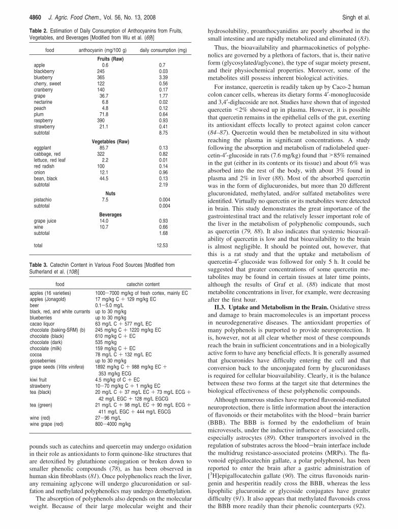
Table 3. Catechin Content in Various Food Sources [Modified fromSutherland et al. (108)]
food catechin content
apples (16 varieties) 1000-7000 mg/kg of fresh cortex, mainly ECapples (Jonagold) 17 mg/kg C + 129 mg/kg ECbeer 0.1-5.0 mg/Lblack, red, and white currants up to 30 mg/kgblueberries up to 30 mg/kgcacao liquor 63 mg/L C + 577 mg/L ECchocolate (baking-SRM) (b) 245 mg/kg C + 1220 mg/kg ECchocolate (black) 610 mg/kg C + ECchocolate (dark) 535 mg/kgchocolate (milk) 159 mg/kg C + ECcocoa 78 mg/L C + 132 mg/L ECgooseberries up to 30 mg/kggrape seeds (Vitis vinifera) 1892 mg/kg C + 988 mg/kg EC +
353 mg/kg ECGkiwi fruit 4.5 mg/kg of C + ECstrawberry 10-70 mg/kg C + 1 mg/kg ECtea (black) 20 mg/L C + 37 mg/L EC + 73 mg/L ECG +
42 mg/L EGC + 128 mg/L EGCGtea (green) 21 mg/L C + 98 mg/L EC + 90 mg/L ECG +
411 mg/L EGC + 444 mg/L EGCGwine (red) 27-96 mg/Lwine grape (red) 800-4000 mg/kg
4860 J. Agric. Food Chem., Vol. 56, No. 13, 2008 Singh et al.

III. ANTIOXIDANT ACTIVITIES OF POLYPHENOLS FROMFOODS
Flavonoids are the most widely studied class of polyphenolswith respect to their antioxidant and biological activities. Theyhave powerful antioxidant activities in vitro, being able toscavenge a wide range of ROS and RNS and chlorine species,such as superoxide, hydroxyl and peroxyl radicals, and hy-pochlorous and peroxynitrous acid. They can also chelate metalions (75).
There are various methods for assessing the total antioxidantcapacity of dietary fruits and vegetables. Among them are theequivalent antioxidant capacity assay (TEAC) using 6-hydroxy-2,5,7,8-tetramethylchroman-2-carboxylic acid (Trolox), a hy-drosoluble analogue of R-tocopherol, as reference (93), theferric-reducing ability of plasma assay (FRAP) (94), and thewidely used oxygen radical absorbance capacity assay (ORAC)(95). Halvorsen et al. (68) have measured the total antioxidantactivity in a variety of dietary plants used worldwide by FRAPassay. They reported a large variation in their ability to reduceFe3+ with a >1000-fold difference among total antioxidants invarious dietary plants. Vegetables such as kale, chili pepper,red cabbage, parsley, artichoke, Brussels sprouts and spinachcontained high antioxidants. Analyses of fruits demonstratedthat pomegranate, grape, plum, pineapple, date, and kiwi havepotent antioxidant activity (in decreasing order); most berries(e.g., various small fruits such as dog rose, crowberry, blueberry,strawberry, blackberry, raspberry) as well contained very highconcentrations of antioxidants. Notably, most members of thecitrus family (e.g., lemon, clementine, orange, grapefruit, andlime) also contained high amounts of antioxidants. However,the classification could differ from one assay to another. Forinstance, sulfur-containing compounds could not be detectedby FRAP assay. Recently, Halvorsen and co-workers (96)analyzed the total concentration of redox active compounds in1113 food samples obtained from the U.S. Department ofAgriculture National Food and Nutrient Analysis Program. Theantioxidant analysis was also based on the reduction of ferricions using 2,4,6-tripyridyl-s-triazine (TPTZ). Of the 50 foodproducts highest in antioxidant concentrations, 13 were spices,8 were in the fruit and vegetable category, 5 were berries, 5were chocolate-based, 5 were breakfast cereals, and 4 were nutsor seeds (Table 4). On the basis of typical serving sizes,blackberries, walnuts, strawberries, artichokes, cranberries,brewed coffee, raspberries, pecans, blueberries, ground cloves,grape juice, and unsweetened baking chocolate were at the topof the ranked list. Of the dried herbs tested, oregano, sage,peppermint, thyme, and lemon balm contained very high levelsof antioxidants as did spices such as clove.
The evaluation of phenolic compounds in commercial fruitjuices and fruit drinks revealed that purple grape juice containedthe largest number of individual phenolic compounds and alsothe highest concentration of total phenolics (97). The maincomponents were flavan-3-ols, anthocyanins, and hydroxycin-namates, which accounted for 93% of the total phenolic content.In contrast, white grape, pineapple, and tomato juices had thelowest total phenolic content. Interestingly, their antioxidantlevel was related to phenolic content (97). These data suggestthat the consumption of fruit juices could have a positive impacton health. Recently, the Kame project indicated that long-termfruit juice consumption can provide protection against AD (98).
IV. POLYPHENOL INTAKE AND RISK OF DEMENTIA
Many epidemiological studies have documented the influenceof dietary habits and antioxidants on the incidence of neuro-
degenerative disorders such as AD, but these analyses haveyielded inconsistent results. For instance, the Honolulu-AsiaAging study, a longitudinal study of elderly Japanese-Americanmen, examined the association between midlife dietary intakeof antioxidants and the incidence of late-life dementia and itssubtypes in 2459 men with a follow-up between 1991 and 1999.This analysis concluded that intakes of �-carotene, flavonoids,and vitamins E and C did not modify the risk of dementia (99).Flavonoid intake was estimated using mean intake of tea (greenand black); information on wine drinking by type (red or white)was not available, but this is presumed to be a minor source offlavonoids in this population. Another limitation of this studyis that nutrient intakes were determined from a single 24 hdietary recall that may not be representative of usual foodconsumption, in contrast to other studies such as the Rotterdamstudy in which dietary intake was estimated with a semiquan-titative food frequency questionnaire. With a total of 5395
Table 4. 50 Foods with the Highest Antioxidant Contents per Serving Size[Modified from Halvorsen et al. (68)]
productantioxidant content
(mmol/100 g)
cloves, ground 125.549oregano leaf, dried 40.299ginger, ground 21.571cinnamon, ground 17.647turmeric powder 15.679walnuts 13.126basil leaf, dried 12.307mustard seed, yellow, ground 10.527curry powder 9.98pecans 9.668chocolate, baking, unsweetened 8.876paprika 8.601chili powder 8.372parsley, dried 7.43molasses, dark 4.9pepper, black 4.444artichokes, prepared 4.237chocolate, dark 4.188blackberries 3.99whole-grain cereal 3.412cranberries 3.289pudding mix, chocolate, cook-and-serve 3.026bran cereal 2.925power bar, chocolate flavor 2.757chocolates, sugar-free 2.567raspberries 2.334strawberries 2.159blueberries 2.154cabbage, red, cooked 2.153wine, red 2.135barley malt syrup, organic 2.121prunes 2.018cherries, sour 1.814peppers, red, cooked 1.64chocolate cookies with vanilla creme filling 1.604Cocoa Krispies cereal 1.558chocolate chip cookies 1.524mustard, yellow, prepared 1.501milk chocolate candy 1.483pistachios 1.426plums 1.33kiwi fruit 1.325corn flakes 1.255coffee 1.249spinach, frozen 1.226flaxseed, ground or milled 1.125rice and corn cereals 1.121toasty peanut crackers 1.101cupcakes, chocolate 1.059grape juice 1.011
Reviews J. Agric. Food Chem., Vol. 56, No. 13, 2008 4861

participants and a mean follow-up of 6 years, this analysis foundthat high intake of vitamins C and E may lower the risk of AD(100). A suggestion of a protective effect of vitamin E againstAD was also reported in the Chicago Health and Aging Projectafter 3.9 years of follow-up (101). Interestingly, the protectiveassociation of vitamin E was observed only in persons who wereApoE4 negative. A protective association between flavonoidintake and dementia was found in the so-called PAQUID study,a 5- and 10-year follow-up study of a cluster sample of 1640subjects in the southwestern departments of Gironde andDordogne in France. This study suggested that an average intakeof 14.3 ( 5.85 mg/day of dietary flavonoid was associated withless cognitive decline in subjects aged 65 years or older (102).The PAQUID study also showed that people drinking three tofour glasses of wine per day had 80% decreased incidence ofdementia and AD compared to those who drank less or did notdrink at all (103). The consumption of fruit and vegetable juicescontaining high concentrations of polyphenols, at least threetimes per week, may play an important role in delaying the onsetof AD, particularly in ApoE4 carriers (98). Recently, anassociation between Mediterranean diet (MeDi) and lower riskof AD has been reported. A case-control study nested within acommunity-based cohort (194 AD patients vs 1790 non-AD)in New York indicated that higher adherence to a Mediterraneandiet was associated with lower risk for AD (103, 104). Inaddition, higher adherence to the MeDi is associated with lowermortality in AD (105). This diet consists of high amounts offruits, vegetables, cereals, and fish, mild to moderate amountsof alcohol, and low amounts of red meat and dairy products.An increase of the consumption of fruits and vegetables is alsoassociated with a reduced risk of stroke, a risk factor for AD.He and co-workers carried out a meta-analysis and found thatindividuals with more than five servings per day had asignificantly reduced risk of stroke (106).
These clinical and epidemiological studies suggest that theconsumption of flavonoids and polyphenols from fruits, veg-etables, or beverages could reduce the risk of AD. However,the relationship between polyphenol consumption and risk ofdementia needs further investigations. One of the limitationsof most studies was that dietary assessement were collectedcloser to the onset of dementia (aged 65 years and more), oncethe oxidative stress level is already high and most neurons aredegenerated, except for the Honolulu-Asia study, in whichsubjects were, on average, aged 52.4 ( 4.2 years. Numerous invivo and in vitro studies performed in animal models or cellculture demonstrated that the antioxidant activity of phenoliccompounds is unlikely to be the sole explanation for theirprotective cellular effects.
V. MECHANISMS OF ACTION OF DIFFERENTPOLYPHENOLS
In the following paragraphs, we will review some mechanismsand targets of the most consumed polyphenols and particularlytheir beneficial effects against the A�-induced toxicity and theirneuroprotective effects.
V.1. Green Tea Catechins. Green tea is rich in flavonoids(30% of dry weight of a leaf) (107), with the main compoundsbeing epigallocatechin-gallate (EGCG), (-)-epigallocatechin(EGC), (-)-epicatechin (EC), and (-)-epicatechin-3-gallate(ECG) (Figure 2). Catechin intake has been associated with awide variety of beneficial health effects (108). All of thecatechins have a wide variety of biological actions pertainingto their chemical structure, but the different mechanismsunderlying these actions have not been fully elucidated. Their
antioxidants and free radical scavenging activities mainlycontribute to their beneficial effects. These flavonoids haveantioxidant potencies in the order of EGCG > ECG > EGC >EC (109). Their free radical scavenging abilities relate to thegallate moiety esterified at position 3 of the C ring, the catecholgroup (3,4-dihydroxyl groups) on the B ring, and the hydroxylgroup at positions 5 and 7 on the A ring. The galloylatedcatechins are more active antioxidants due to their higherphospholipid/water partition coefficients (110). Moreover, thefree radical scavenger property increases with the number ofhydroxyl groups the catechin possesses. For instance, EGCGand EC possess eight and five hydroxyl groups, respectively,and the antioxidant activity of EGCG is higher than that of EC.Furthermore, their antioxidant abilities are higher than those ofR-tocopherol or vitamins C and E.
Catechins can exert their antioxidant activity through variousmechanisms, one being by chelating metal ions such ascopper(II) and iron (II), and therefore prevent the generation ofpotentially damaging free radicals. Thus, reduction of the freeiron pool by EGCG chelation may lead to the suppression ofthe translation of APP mRNA. Accordingly, Levites et al. havedemonstrated that prolonged administration of EGCG to miceinduced a reduction in holo-APP levels in the hippocampus(135). This result was supported by those obtained in cell culturemodels with a concomitant decrease in A� levels.
Catechins may also exert their antioxidant effects throughthe ultrarapid electron transfer to ROS-induced radical sites onDNA or by forming stable semiquinone free radicals. Moreover,after the oxidation of catechins by free radicals, a dimerizedproduct is formed with an increased iron-chelating potential andability to scavenge superoxide anions. The prevention ofoxidative-induced damage by catechins is very effective ascatechins can inhibit the ROS-induced damage by a wide varietyof initiators including hydrogen peroxide, iron, paraquat, orradiolysis. Antioxidant properties of catechins were also ob-served in different in vivo models. For instance, rats receivinggreen tea extracts orally exhibited higher levels of antioxidantenzymes such as glutathione peroxidase and reductase, super-oxide dismutase, and catalase (111). These effects on antioxidantlevels were also investigated in human. It has been evidencedthat after 42 days of the consumption of 2 cups of green tea,containing approximately 250 mg of total catechins, a significantincrease in plasma total antioxidants was observed, whereas theplasma peroxide level decreased (112). Catechins also decreasedoxidative stress by inhibiting the activity of xanthine oxidase(113), a ROS-generating system. Catechins can also protectlipids from oxidation in the liver, serum, and brain (111). Forinstance, it has been demonstrated that catechins could protectagainst lipid peroxidation induced by 6-hydroxydopamine,hydrogen peroxide, and iron (114). These antioxidant effectsare observed in vitro with concentrations ranging from 1 to 50µM. However, with higher concentrations (100-500 µM) andin the presence of copper(II) or iron(III), EGCG exacerbatedoxidative stress, cytotoxicity, and DNA damage induced byhydrogen peroxide (115–117).
EGCG could also modulate apoptosis pathways to protectcells against oxidative stress. The effects of catechins onapoptotic pathways could be divergent. For instance, on PC12cells EGCG, at low doses (1-10 µM), could inhibit caspase-3activity or activate the PI3K/Akt pathway, which promote cellsurvival (118). On the other hand, catechins could also modulateapoptosis by altering the expression of anti-apoptotic and pro-apoptotic genes. In SH-SY5Y neuronal cells, EGCG preventedthe expression of pro-apoptotic genes Bax and Bad while
4862 J. Agric. Food Chem., Vol. 56, No. 13, 2008 Singh et al.

inducing the anti-apoptotic genes Bcl-2, Bcl-w, and Bcl-X in6-hydroxydopamine-induced apoptosis (119, 120). EGCG pro-motes cell survival by restoring the protein kinase C activity, acritical regulator of cell proliferation and survival. However, athigh doses, EGCG (50-500 µM) can induce pro-apoptoticproperties by increasing Bax, Bad, and caspase-6 activity whiledecreasing Bcl-x and Bcl-2 activity.
There is substantial evidence that catechins can exert anti-inflammatory effects. This could be due to their abilities toscavenge NO, the peroxynitrite anion, or to reduce the activity
of NO synthase (121, 122) with EGCG being the most effective(123). The neuronal nNOS and the inducible iNOS isoformscould also be inhibited by catechins (124). This effect ofcatechins likely involved the inhibition of the activation of thetranscription factor NF-κB as the κB sequence is present in thepromoter region of the iNOS gene (125). On the contrary,catechins could induce the endothelial isoform eNOS activity,a vasodilatator-inducing enzyme. This activity contributes tothe anti-inflammatory effects of catechins (126). Anothermechanism of action proposed may be the presence of the
Figure 2. Chemical structures of different catechins. Catechins have a three-ring structure with two or three hydroxyls on the B ring and with or withouta gallate group at the C3 position of the C ring.
Reviews J. Agric. Food Chem., Vol. 56, No. 13, 2008 4863

antioxidant response element (ARE) on the promoter of theeNOS gene, and catechins could bind to the ARE and activateeNOS (127). These effects of EGCG on NOS activities alsocontribute to the anti-ischemic effect of EGCG (108). The anti-inflammatory effect of EGCG has also been studied in manycell types through the regulatory effect of EGCG on cytokinesecretion. For instance, Kim et al. (128) have demonstrated thatEGCG was able to inhibit the production of IL-1 and attenuatethe expression of cyclooxygenase-2 induced by IL-1 and A�or the activation of NF-κB and MAPK pathways induced byIL-1 and A� (128, 129).
The prevention of cerebrovascular diseases or stroke by greentea has been evidenced during a 4-year follow-up study with5910 individuals. The incidence of cerebral hemorrhage andstroke were 2-fold higher in those who consumed less than fivecups than in those who consumed five cups or more daily (130).An inverse correlation between black tea consumption and theincidence of stroke was also replicated in a cohort of 552 menaged 50-69 years and followed up for 15 years (131). However,this inverse association was not observed in the Zutphen Study(132).
Although there is no significant outcome relative to teaconsumption in AD case control, there are several in vitro studiesshowing that green tea extract may protect neurons from A�-induced damages (133–136). Over the past decade, intense focushas been placed on the processes of APP proteolysis and A�metabolism as possible targets for AD therapy. Various syntheticand naturally occurring compounds have been analyzed for theirefficacy in the modulation of these pathological events. Amongthem, EGCG is able to regulate the proteolytic processing ofAPP both in vitro and in vivo (134). In neuronal cell cultures,it could promote the nonamyloidogenic R-secretase pathway(134). In primary neuronal cells derived from a transgenic mousemodel overexpressing the human APP containing the AD-linkedK670M/M671L double mutation (Swedish mutation), EGCG,at 20 µM, significantly reduced A� peptide generation (A�1-40
and A�1-42) by 38%, with purified EGCG being more potentthan green tea (137). These results were strengthened byexperiments on N2a cells stably transfected with “Swedish”mutant APP, where EGCG treatment led to marked elevationin active R-desintegrin and metalloprotease (ADAM 10) pro-teins, ultimately leading to the nonamyloidogenic APP process-ing pathway (138).
Green tea catechins, especially ECGC, also reduced theactivation of a number of signaling pathways such as p38 andJNK of MAPKs (128) and induce the phosphorylation of proteinkinase C (135, 139) and phosphatidylinositol-3-kinase (PI-3kinase)-Akt (118), and these modulations may mediate someof the neuroprotective effects of EGCG. Protein kinaseC (134, 135) plays a central role in neuronal cell survival, andloss in its activity is frequently observed in neuronal insultssuch as in the presence of A� peptide accumulation and otherneurotoxins (140). In neuronal cell lines and primary cells inculture, EGCG prevented the decline of ERK1/2 induced by6-hydroxydopamine or by oxidized low-density lipoproteins(135, 141). MAPKs are also involved in the regulation of theexpression of pro-apoptotic and anti-apoptotic genes. EGCG-treated SH-SY5Y neuroblastoma cells have decreased expressionof the pro-apoptotic genes Bax, Bad, cell cycle inhibitor Gadd45,Fas ligand, and tumor necrosis factor-mediated apoptosis ligandTRAIL (134, 142).
All of the catechins are rapidly absorbed and widelydistributed (143), and the peak concentration in plasma isreached 1.4-2.4 h after ingestion (144). For instance, Wistar
rats exposed to epicatechin (oral gavage of 100 mg/kg/day) hadsteady-state plasma levels of about 5 µM for epicatechin andjust over 1 µM for 3′-O-methylepicatechin, as well as almost50 µM for glucuronidated epicatechin and 20 µM for glucu-ronidated 3- O-methylepicatechin (77). Very low concentrationsof glucuronidated 4′-O-methylcatechin were also detected. LC-MS analysis demonstrated the presence of very small quantitiesof epicatechin, 3′-O-methylepicatechin, and the 5-O- and/or 7-O-glucuronides of epicatechin in the brain of these rats. It is likelythat the glucuronides were formed in situ in neuronal cells, asthey are considered to cross the BBB very poorly, and it isknown that uridine diphosphate-glucuronyl transferase (UDP-GT) activity is present in the central nervous system of rats(145) and humans (146). For example, cis- and trans-resveratrol,polyphenolics found in wine, were 3-O-glucuronidated in ratbrain tissue and rat astrocytes (147). Similar to the study withepicatechin, no B-ring 4′-O-glucuronide was found. The totalconcentration of epicatechin and metabolites was estimated tobe about 0.4 nmol/g of brain tissue, but the investigators statethat the concentrations were too low to quantify accurately (148).Some in vivo studies have shown that 0.33% of EGCGadministration can reach the brain (90) and that frequentconsumption of green tea enables the body to maintain a highlevel of catechins. Another study exposing rats to grape extractknown to contain numerous polyphenolics including epicat-echins also found no trace of epicatechins or its methylated andglucuronidated metabolites in brain (149). A study more relevantfor human exposure to epicatechins followed six subjects(scheduled for lumbar puncture) after the ingestion of a 300mL boiling water infusion of 7 g of green Kenyan tea (150).The average intake of (-)-epicatechins was 53 µmol ofepicatechin, 149 µmol of epigallocatechin, 206 µmol of epi-gallocatechin-3-O-gallate, and 97 µmol of epicatechin-3-O-gallate. After 1 h, plasma levels of the epicatechins were readilydetectable with total epicatechin concentrations amounting toabout 1.6 µM; however, nothing was found in cerebral spinalfluid. Each 200 mL cup of green tea contains approximately200 mg of catechins, with 88 mg of EGCG (151). However,the quantities of catechins are inconsistent with various brandsand origins of green tea (152). This makes it difficult to knowexactly the amount of green tea required daily to provide aneuroprotective effect.
In conclusion, in addition to their known antioxidant proper-ties, catechins can target other pathways to exert their neuro-protective effect. These studies demonstrated that catechinscould protect different cell types against various cytotoxiccompounds independently of their free radical scavenger proper-ties but through some emerging pathways that have attractedmuch attention recently (Figure 3). However, current epide-miological and clinical evidence correlating catchin intake andthe incidence of AD is inconsistent. It is not clear whetherepicatechins are capable of entering the brain in concentrationssufficiently high to be able to exert their beneficial effects.
V.2. Curcumin. Curcumin is a major chemical componentof turmeric (Curcuma longa) and is used as a spice to give aspecific flavor and yellow color to Indian curries and in foodpreservation. Interestingly, the prevalence of AD in people aged70-79 years in India is 4.4-fold less than in the United States(153). Turmeric is derived from the rhizome, or root, of theplant. There is substantial in vitro evidence indicating thatcurcumin has antioxidant, anti-inflammatory, and antiamyloidactivities (154). For instance, curcumin could inhibit lipidperoxidation (155), activate glutathione S-transferase (156), orinduce heme oxygenase-1 (HO-1) (157). HO-1 induction occurs
4864 J. Agric. Food Chem., Vol. 56, No. 13, 2008 Singh et al.

through the antioxidant response element (ARE) (158). Cur-cumin could also chelate the redox active metals iron (Fe2+)and copper (Cu2+) (159).
Inflammation is thought to be implicated in the pathophysi-ology of AD. Some epidemiological studies have consistentlydemonstrated an association between the use of nonsteroidalanti-inflammatory drugs and a subsequent decreased risk of thedevelopment of AD (160–162). Curcumin has been shown tohave anti-inflammatory effects as it is a good inhibitor oflipoxygenase and COX-2 (163, 164), both enzymes beingresponsible for the synthesis of the pro-inflammatory leuko-trienes, prostaglandins, and thromboxanes. Curcumin is also asuppressor of iNOS and a potent inhibitor of NF-κB and AP-1activation (165–167). This mechanism is likely involved in theinhibition of the expression of inflammatory cytokines COX-2and iNOS, as these transcription factors are well-known toregulate these inflammatory factors. All of these factors (IL-1,TNFR, COX-2, iNOS, JNK, NF-κB) are also implicated in A�toxicity.
Aggregation of A� into fibrils and the subsequent formationof amyloid plaques are crucial steps in the pathogenesis of AD.It has been found that curcumin inhibits the formation andextension of A� fibrils and destabilizes preformed A� fibrils ina dose-dependent fashion between 0.1 and 1 µM (168).Curcumin could also bind to fibrillar A� regardless of thespecific A� sequence.
In light of the spectrum of activities, curcumin represents ahopeful approach for delaying or preventing the progression ofAD. Therefore, the effects of curcumin have been tested in
several animal models for AD. When fed to aged Tg2576 micewith advanced amyloid accumulation, curcumin reduced A�levels and plaques (169). In this study, low (160 ppm) and highdoses (5000 ppm) of curcumin significantly lowered oxidizedproteins and IL-1�, whereas low doses reduced plaque burden.Subsequent in vivo studies using multiphoton microscopydemonstrated that curcumin could cross the BBB, targetingsenile plaques in Tg2576 mice and disrupting existing plaques(170).
However, preclinical data from animal models and phase Iclinical studies performed with human volunteers and patientshave demonstrated low systemic bioavailability following oralintake. The absorption, distribution, metabolism, and excretionof curcumin in rodents has been widely described. These studiessupport the notion that curcumin undergoes a rapid and efficientmetabolism that severely curtains its availability. An ingesteddose (1 g/kg) administered to rats resulted in about 75% of thespice’s metabolites being detected in feces (171). Intestinalmetabolism, particularly glucuronidation and sulfation, of cur-cumin might explain its poor systemic availability (172). Themetabolites were characterized mainly as glucuronides oftetrahydrocurcumin and hexahydrocurcumin.
Altogether, curcumin, a highly lipophilic compound, canprotect cells against A� toxicity by preventing A� peptideaggregation and reducing plaque burden, through its antioxidantand anti-inflammatory activities and the inhibition of cellsignaling pathways at multiple levels. However, curcuminundergoes rapid metabolism, and the bioavailability of the parentcompound is low (172). Therefore, more investigations arenecessary on curcumin or on related compounds to gather moreinformation on biomarkers of AD pathology in addition toclinical trials data.
V.3. Resveratrol. Resveratrol is a non-flavonoid polyphenolicfound in grapes, red wine, and berries. The concentration ofresveratrol in red wine is in the range of 1.5-3 mg/L (173).There are two isomeric forms of resveratrol, the biologicallyinactive cis-resveratrol and the most biologically active trans-resveratrol (trans-3,4,5-trihydroxystilbene). This compound hasbeen the focus of a number of studies demonstrating itsantioxidant, anti-inflammatory, antimutagenic, and anticarcino-genic effects (173–175). Interestingly, several epidemiologicalstudies indicate an inverse correlation of wine consumption andincidence of AD (176–178).
In several in vitro studies, resveratrol has been recognizedfor its powerful antioxidant properties. At the cellular level,resveratrol could protect PC12 cells against A�-induced toxicityand prevent the accumulation of intracellular ROS (179).Resveratrol can also protect SH-SY5Y neuroblastoma cells andprimary hippocampal neuronal/glial cells from H2O2, NO, andA�-induced toxicity (139, 180–182). In cultured PC12 cells,resveratrol also increased the HO-1 activity, and similarly incortical mouse neurons an up-regulation of HO-1 gene expres-sion via the activation of NF-E2-related factors 2 (NRf2) (183)was observed (184). Interestingly, resveratrol exhibited itsneuroprotective effects when it was used in pretreatment, incotreatment, or in post-treatment.
The inhibition of A� secretion by resveratrol could also beimplicated in this neuroprotective effect because the secretionof A� is reduced in two cell lines, HEK 293 and N2a, transfectedwith APP695 (185). This effect was not mediated by �- andγ-secretase activities but may be through the elevation of thedegradation of A� peptide. However, resveratrol did not affectA�-degrading enzymes such as neprilysin, endothelin-convertingenzyme-1 or -2, and insulin-degrading enzyme (185).
Figure 3. Potential pathways involved in the neuroprotective mechanismsof EGCG. Modified from Mandel et al. (206).
Reviews J. Agric. Food Chem., Vol. 56, No. 13, 2008 4865

It is well-known that reducing food intake or caloric restrictionextends lifespan in a wide range of species. Recently, it hasbeen found that resveratrol can mimic dietary restriction andtrigger sirtuin proteins (186). The sirtuin enzymes are aphylogenetically conserved family of enzymes that catalyzeNAD-dependent protein deacetylation. In yeast, sir2 is essentialfor lifespan extension by caloric restriction and a variety of otherstresses, including increased temperature, amino acid restriction,and osmotic shock (70, 187). Activators of sirtuins can be akey to extending lifespan and overcoming a variety of stressesin higher organisms. Among 18 small molecules that canincrease human sirt1 activity, resveratrol induced the highestactivity of sirt1 and increased the lifespan of yeast by nearly70% (188). Analysis of the structure-activity relationshipsuggests that the hydroxylated trans-stilbene ring structure isessential for activation of sirt1. However, the mechanisms thatlink resveratrol to the activation of sirt1 and the subsequentprotection of neurons against A� remain unknown. Nevertheless,resveratrol-induced sirt1 has been found to repress p53 activityand to suppress apoptotic activities of FOXO proteins, therebyprotecting neurons against apoptosis-induced by A�.
The NF-κB pathway is also a target of resveratrol. In a recentstudy with mixed neuron/glial cultures from Sprague-Dawleyrat, it has been demonstrated that resveratrolsby inducing sirt1activationscould inhibit the NF-κB signaling in microglia andastrocytes with a neuroprotective effect against A�-inducedtoxicity (183). NF-κB signaling controls the expression of bothiNOS and cathepsin B, two factors that mediate apoptosis. InPC12 cells, A� induces the degradation of IκBR, the inhibitorysubunit of NF-κB activation, and increases the nuclear trans-location of p65. The activation of NF-κB was reversed whencells were treated with resveratrol (25 µM) (189). We haverecently demonstrated that sirt1 could be activated by flavonoidsand that this activation is associated with the NF-κB inhibitionand protection against A� toxicity (65). Thus, modulation ofdifferent sirtuins by phenolic compounds could provide animportant arsenal to overcome variety of stresses that compro-mise neuronal survival in different neurodegenerative diseasessuch as AD.
After oral administration of resveratrol, it is rapidly metabo-lized (within 2 h, with a peak in <30 min) to glucuronide acidand sulfate conjugates in the liver and intestinal cells (173).More than 90% of total resveratrol, given as pure aglycone,circulates in the plasma in the conjugated form, and glucu-ronidation predominates the metabolism of resveratrol. Theseresults indicate that the circulating forms of resveratrol arepredominantly modified metabolites and not the original agly-cone. Therefore, the antioxidant and anti-inflammatory activitiesand the effect on cell signaling of the original aglyconecompound seem to be considerably diminished due to itsextensive and rapid metabolism. However, the biologicalactivities of the circulating form and their functions remain tobe determined, particularly their implication in neuroprotectiveeffects. Indeed, resveratrol appears to reach the brain as wasshown in one rat study exposing 250 g males to 50 mg/kg [3H]-trans-resveratrol by oral gavage (77). The concentrations foundin the whole brain after 2 and 18 h were about 0.03 and 0.01%,respectively, of the original dose, suggesting that distributionto the brain is minimal.
In summary, it is clear that the neuroprotective effect ofresveratrol implicates different pathways which may be criticalto neuronal protection in AD. In addition to its antioxidanteffects, the efficacy of resveratrol against A� toxicity alsoinvolves several transduction pathways or the modulation of
glia/astrocyte functions. All of these functions may playsynergistic roles in treating AD. However, pharmacokineticstudies indicate that resveratrol is rapidly metabolized in liverand intestinal epithelials cells. Therefore, the efficacy ofresveratrol in the treatment of AD will also depend on thebioavailability of its metabolites and their biologicalactivities.
V.4. Effects of Polyphenols from Berries and Pomegranateon Cognitive Performance. Berries are rich sources of phenoliccompounds such as phenolic acids as well as anthocyanins,proanthocyanidins, and other flavonoids (e.g., ellagitannins). Thecontent of phenolics in berries is affected by the degree ofmaturity at harvest, by the cultivar, and by the pre- andpostharvest environnments. For instance, the total phenolic acidcontent ranged from 2845 to 5418 mg/kg (190) with hydroxy-cinnamic acids constituting from 68.9 to 85% of the total; morethan 20 phenolic acids could be identified in berries. Inblueberries (Vaccinium ashei reade), catechin is the majorflavonoid, reaching 387 mg/100 g of fresh weight; epicatechinconcentrations ranged from 34 to 129 mg/100 g of fresh weight,and total anthocyanins ranged from 84 to 113 mg/100 g of freshweight (191). It has been estimated that 1.20 g of totalanthocyanins was present in human serum after a consumptionof 100 g of blueberries, and maximal level was reached 4 hafter the consumption. Interestingly, a significant positivecorrelation between serum anthocyanin content and postprandialantioxidant status has been observed (192). This absorptioncould have some positive effects in the brain through severalprocesses, as has been demonstrated in various animal studies.Dietary supplementation for 8 weeks with blueberry extractsreversed cognitive deficits in Morris water maze performancetest in 19-month-old rats (193). However, the effect of blueberryextracts on cognitive functions might involve more than justtheir antioxidant actions. For example, aged rats on a blueberryextract diet had significantly lower levels of NF-κB than agedrats on a control diet (194). It has been described that the agedrat control diet group had significantly higher average NF-κBlevels than young rats (194). These results are in accordancewith the known effect of flavonoids on cell signaling such ason the activity of NF-κB (65, 195). Additional evidence wasseen in a recent study with the double-transgenic mice modelof AD overexpressing APP and presenilin 1, in which geneticmutations promote the production of the A� peptide and thehallmark of AD-like senile plaques in several regions. Whenthese mice were supplemented with blueberry extract (2% ofdiet) at 4 months and continued until 12 months of age, theirperformance in a Y-maze cognitive performance test was similarto that of nontransgenic mice and significantly better than thatof nonsupplemented transgenic mice (196). However, examina-tion of the brains of these mice revealed that supplementationof blueberry extract did not affect the A� peptide productionor deposition or the number of plaques. These data suggest thatthe impairment of cognitive functions observed in these trans-genic mice may not necessarily be the result of deposition ofthe A� peptide. In these mice supplemented with blueberryextract, the concentrations of hippocampal ERK as well asstriatal and hippocampal PKCR were higher than in transgenicmice supplemented with control diet. Both protein kinase C andERK have been shown to be involved in early and late stagesof memory formation (197). These results indicate that blueberryextract supplementation might prevent cognitive and motordeficits through various neuronal signaling pathways. Further-more, short-term blueberry supplementation increases hippoc-ampal plasticity (198). Diet supplemented with blueberry extractcould also protect the brain against apoptosis as rats receiving
4866 J. Agric. Food Chem., Vol. 56, No. 13, 2008 Singh et al.

blueberry extracts had significantly lower caspase-3 activity inthe ischemic hemisphere (199). Taken together, these studiesdemonstrate that blueberry extract-supplemented diets couldprotect neuronal loss and prevent the decrease of cognitivefunctions against various insults through antioxidant, anti-apoptotic, and regulation of cell signaling mechanisms.
Pomegranates (Punica granatum L.) contain very high levelsof polyphenols compared with other fruits and vegetables(200–202), and the most important polyphenols are ellagic acid,punicalagin, and hydrolyzable tannins such as ellagitannins andgallotannins.
Recently, the administration of pomegranate juice (PJ) toTg2576 transgenic mice expressing the APP695 human genefrom 6 to 12.5 months of age exhibited improvements in cuedand spatial learning tasks as compared to a sugar water controlgroup (203). Additionally, PJ-treated mice had a significantlyreduced burden of plaque load and soluble A�1-42 in thehippocampus. Grape juice is also a rich source of flavonoidsthat include catechins, epicathechins, quercetin, anthocyanins,and proanthocyanidins (3). When aged Fisher 344 rats weregiven 10 or 50% of grape juice from 19 to 21 months of age,their performance motor functions in rod walk and cognitiveperformance on the Morris water maze were improved (204).
Several dietary supplements with either spinach or strawberryextracts have also been reported to reduce some neurologicaldeficits in aged animal models (113, 199).
VI. CHALLENGES FOR RESEARCH ON POLYPHENOLS INNEURODEGENERATIVE DISEASES
Hundreds of polyphenols with potent antioxidant activity havebeen shown to have neuroprotective effects in vitro and inanimal studies, but only a few compounds, for example,curcumin, have progressed successfully into active clinical trialsin neurodegenerative diseases. Most reports on the beneficialeffects of polyphenols are based on in vitro and in vivo studieseither in cell cultures or in animal models, where there is notextensive neuronal damage. On the contrary, in human clinicaltrials, the patients already suffer from extensive neuronal lossand damage. Therefore, the important question arises whetherpolyphenols should be tested for therapeutic efficacy or as agentsthat can further slow the progression of disease. Second, fewstudies are available to conclusively prove that polyphenols cancross the BBB to exert their protective effects. More data andstudies are required to validate that polyphenols can cross theBBB in sufficient quantity to exert their biological andpharmacological actions. Because most of the neurodegenerativediseases require a lengthy incubation time before clinicalmanifestation, it is worthwhile to conduct epidemiologicalstudies regarding polyphenol intake and progression of diseases.In this regard the variation between geographical distributionsof various neurological disorders should be compared with foodpolyphenolic composition data across various countries andethnicities. These data could throw light on why AD is moreprevalent in certain regions and its correlation with local dietaryhabits, especially with regard to polyphenol intake. Anotheraspect of polyphenols that warrants further detailed investigationis synergistic and antagonistic activities in combination withother biological antioxidants. For example, ascorbate andcatechin have been shown to have a synergistic effect asascorbate could protect catechin from oxidation (205), leadingto the hypothesis that polyphenol antioxidants may be part of abroader antioxidant network of the organism. Another rapidlydeveloping aspect of free radicals research is their participationin the process of mediating and regulating cellular functions
without causing unwarranted oxidative stress. It may be possiblethat dietary polyphenols continuously participate in the regula-tion of cellular functions independent of their antioxidantproperties. In addition, we cannot exclude the possibility thatpolyphenolic compounds may regulate the expression of somegenes coding for antioxidant enzymes and thus help the neuralcells to cope with increased oxidative stress.
VII. CONCLUSIONS
Polyphenols from fruits and vegetables seem to be invaluablepotential agents in neuroprotection by virtue of their ability toinfluence and modulate several cellular processes such assignaling, proliferation, apoptosis, redox balance, and dif-ferentiation. Although abundant in fruits, vegetables, tea, wine,and medicinal plants, more detailed studies are required todetermine their absorption, bioavailibity, and ability to crossthe BBB. Their neuroprotective activity in various models ofneurodegenerative diseases in vitro and in vivo have beendocumented, but it would be unwise to extrapolate these resultsto the human situation without proper clinical trials in patientssuffering from irreversible and extensive neuronal loss. Inaddition, most cell culture or animal studies have been conductedon a short-term basis. Therefore, more long-term studies shouldbe undertaken to determine their beneficial effects in slowlydeveloping neurodegenerative disorders such as AD. In viewof their multiple biological activities, polyphenols hold greatpromise as potential therapeutic/prophylactic agents in neuro-degenerative diseases. Further studies are also required tounderstand the effect of ROS on basic cellular and molecularfunctions of the various nerve cells in the brain and how this inturn affects the physiopathology of neurodegenerative diseases.Also, the impact of ROS on the production of differentneurotrophins, neurotransmitters, and steroids (glucocorticoids)in the brain and their possible modulation by polyphenols isworth examining as it will open up new vistas for the treatmentof neurodegenerative diseases.
LITERATURE CITED
(1) Scalbert, A.; Johnson, I. T.; Saltmarsh, M. Polyphenols: anti-oxidants and beyond. Am. J. Clin. Nutr. 2005, 81, 215S–217S.
(2) Manach, C.; Mazur, A.; Scalbert, A. Polyphenols and preventionof cardiovascular diseases. Curr. Opin. Lipidol. 2005, 16, 77–84.
(3) Duthie, S. J. Berry phytochemicals, genomic stability and cancer:evidence for chemoprotection at several stages in the carcino-genic process. Mol. Nutr. Food Res. 2007, 51, 665–674.
(4) Ramassamy, C. Emerging role of polyphenolic compounds inthe treatment of neurodegenerative diseases: a review of theirintracellular targets. Eur. J. Pharmacol. 2006, 545, 51–64.
(5) Hamilton, M. L.; Van Remmen, H.; Drake, J. A.; Yang, H.; Guo,Z. M.; Kewitt, K.; Walter, C. A.; Richardson, A. Does oxidativedamage to DNA increase with age? Proc. Natl. Acad. Sci. U.S.A.2001, 98, 10469–10474.
(6) Smith, C. D.; Carney, J. M.; Starke-Reed, P. E.; Oliver, C. N.;Stadtman, E. R.; Floyd, R. A.; Markesbery, W. R. Excess brainprotein oxidation and enzyme dysfunction in normal aging andin Alzheimer disease. Proc. Natl. Acad. Sci. U.S.A. 1991, 88,10540–10543.
(7) Floyd, R. A.; Hensley, K. Oxidative stress in brain aging.Implications for therapeutics of neurodegenerative diseases.Neurobiol. Aging 2002, 23, 795–807.
(8) Poon, H. F.; Calabrese, V.; Calvani, M.; Butterfield, D. A.Proteomics analyses of specific protein oxidation and proteinexpression in aged rat brain and its modulation by L-acetylcar-nitine: insights into the mechanisms of action of this proposedtherapeutic agent for CNS disorders associated with oxidative
Reviews J. Agric. Food Chem., Vol. 56, No. 13, 2008 4867

stress. Antioxid. Redox Signal. 2006, 8, 381–394.(9) Berlett, B. S.; Stadtman, E. R. Protein oxidation in aging, disease,
and oxidative stress. J. Biol. Chem. 1997, 272, 20313–20316.(10) Widmer, R.; Ziaja, I.; Grune, T. Protein oxidation and degradation
during aging: role in skin aging and neurodegeneration. FreeRadical Res, 2006, 40, 1259–1268.
(11) Reich, E. E.; Markesbery, W. R.; Roberts, L. J.; Swift, L. L.;Morrow, J. D.; Montine, T. J. Brain regional quantification ofF-ring and D-/E-ring isoprostanes and neuroprostanes in Alzhe-imer’s disease. Am. J. Pathol. 2001, 158, 293–297.
(12) Esterbauer, H.; Schaur, R. J.; Zollner, H. Chemistry andbiochemistry of 4-hydroxynonenal, malonaldehyde and relatedaldehydes. Free Radical Biol. Med. 1991, 11, 81–128.
(13) Dei, R.; Takeda, A.; Niwa, H.; Li, M.; Nakagomi, Y.; Watanabe,M.; Inagaki, T.; Washimi, Y.; Yasuda, Y.; Horie, K.; Miyata,T.; Sobue, G. Lipid peroxidation and advanced glycation endproducts in the brain in normal aging and in Alzheimer’s disease.Acta Neuropathol. (Berlin) 2002, 104, 113–122.
(14) Poon, H. F.; Calabrese, V.; Scapagnini, G.; Butterfield, D. A.Free radicals and brain aging. Clin. Geriatr. Med. 2004, 20, 329–359.
(15) Mount, C.; Downton, C. Alzheimer disease: progress or profit.Nat. Med. 2006, 12, 780–784.
(16) Chartier-Harlin, M. C.; Crawford, F.; Houlden, H.; Warren, A.;Hughes, D.; Fidani, L.; Goate, A.; Rossor, M.; Roques, P.; Hardy,J.; et al. Early-onset Alzheimer’s disease caused by mutationsat codon 717 of the �-amyloid precursor protein gene. Nature1991, 353, 844–846.
(17) Levy-Lahad, E.; Wasco, W.; Poorkaj, P.; Romano, D. M.;Oshima, J.; Pettingell, W. H.; Yu, C. E.; Jondro, P. D.; Schmidt,S. D.; Wang, K.; et al. Candidate gene for the chromosome 1familial Alzheimer’s disease locus. Science 1995, 269, 973–977.
(18) Sherrington, R.; Rogaev, E. I.; Liang, Y.; Rogaeva, E. A.;Levesque, G.; Ikeda, M.; Chi, H.; Lin, C.; Li, G.; Holman, K.Cloning of a gene bearing missense mutations in early-onsetfamilial Alzheimer’s disease. Nature 1995, 375, 754–760.
(19) Poirier, J.; Davignon, J.; Bouthillier, D.; Kogan, S.; Bertrand,P.; Gauthier, S. Apolipoprotein E polymorphism and Alzheimer’sdisease. Lancet 1993, 342, 697–699.
(20) Strittmatter, W. J.; Saunders, A. M.; Schmechel, D.; Pericak-Vance, M.; Enghild, J.; Salvesen, G. S.; Roses, A. D. Apolipo-protein E: high-avidity binding to �-amyloid and increasedfrequency of type 4 allele in late-onset familial Alzheimerdisease. Proc. Natl. Acad. Sci. U.S.A. 1993, 90, 1977–1981.
(21) Strittmatter, W. J.; Saunders, A. M.; Schmechel, D.; Pericak-Vance, M.; Enghild, J.; Salvesen, G. S.; Roses, A. D. Apolipo-protein E: high-avidity binding to �-amyloid and increasedfrequency of type 4 allele in late-onset familial Alzheimerdisease. Proc. Natl. Acad. Sci. U.S.A. 1993, 90, 1977–1981.
(22) Beffert, U.; Aumont, N.; Dea, D.; Lussier-Cacan, S.; Davignon,J.; Poirier, J. Apolipoprotein E isoform-specific reduction ofextracellular amyloid in neuronal cultures. Brain Res. Mol. BrainRes. 1999, 68, 181–185.
(23) Stadtman, E. R. Protein oxidation and aging. Science 1992, 257,1220–1224.
(24) Chai, C. K. The genetics of Alzheimer’s disease. Am. J.Alzheimers Dis. Other Dementia 2007, 22, 37–41.
(25) Onyango, I. G.; Khan, S. M. Oxidative stress, mitochondrialdysfunction, and stress signaling in Alzheimer’s disease. Curr.Alzheimer Res. 2006, 3, 339–349.
(26) Markesbery, W. R. The role of oxidative stress in Alzheimerdisease. Arch. Neurol. 1999, 56, 1449–1452.
(27) Christen, Y. Oxidative stress and Alzheimer disease. Am. J. Clin.Nutr. 2000, 71, 621S–629S.
(28) Nourooz-Zadeh, J.; Liu, E. H.; Yhlen, B.; Anggard, E. E.;Halliwell, B. F4-isoprostanes as specific marker of docosa-hexaenoic acid peroxidation in Alzheimer’s disease. J. Neuro-chem. 1999, 72, 734–740.
(29) Ramassamy, C.; Averill, D.; Beffert, U.; Bastianetto, S.; Theroux,L.; Lussier-Cacan, S.; Cohn, J. S.; Christen, Y.; Davignon, J.;Quirion, R.; Poirier, J. Oxidative damage and protection byantioxidants in the frontal cortex of Alzheimer’s disease is relatedto the apolipoprotein E genotype. Free Radical Biol. Med. 1999,27, 544–553.
(30) Ramassamy, C.; Averill, D.; Beffert, U.; Theroux, L.; Lussier-Cacan, S.; Cohn, J. S.; Christen, Y.; Schoofs, A.; Davignon, J.;Poirier, J. Oxidative insults are associated with apolipoproteinE genotype in Alzheimer’s disease brain. Neurobiol. Dis. 2000,7, 23–37.
(31) Pratico, D.; Clark, C. M.; Lee, V. M.; Trojanowski, J. Q.; Rokach,J.; FitzGerald, G. A. Increased 8,12-iso-iPF2R-VI in Alzheimer’sdisease: correlation of a noninvasive index of lipid peroxidationwith disease severity. Ann. Neurol. 2000, 48, 809–812.
(32) Montine, K. S.; Reich, E.; Neely, M. D.; Sidell, K. R.; Olson,S. J.; Markesbery, W. R.; Montine, T. J. Distribution of reducible4-hydroxynonenal adduct immunoreactivity in Alzheimer diseaseis associated with APOE genotype. J. Neuropathol. Exp. Neurol.1998, 57, 415–425.
(33) Montine, K. S.; Olson, S. J.; Amarnath, V.; Whetsell, W. O.,Jr.; Graham, D. G.; Montine, T. J. Immunohistochemicaldetection of 4-hydroxy-2-nonenal adducts in Alzheimer’s diseaseis associated with inheritance of APOE4. Am. J. Pathol. 1997,150, 437–443.
(34) Frankel, E. N.; Kanner, J.; German, J. B.; Parks, E.; Kinsella,J. E. Inhibition of oxidation of human low-density lipoproteinby phenolic substances in red wine. Lancet 1993, 341, 454–457.
(35) Williams, T. I.; Lynn, B. C.; Markesbery, W. R.; Lovell, M. A.Increased levels of 4-hydroxynonenal and acrolein, neurotoxicmarkers of lipid peroxidation, in the brain in mild cognitiveimpairment and early Alzheimer’s disease. Neurobiol. Aging2006, 27, 1094–1099.
(36) Lovell, M. A.; Xie, C.; Markesbery, W. R. Acrolein is increasedin Alzheimer’s disease brain and is toxic to primary hippocampalcultures. Neurobiol. Aging 2001, 22, 187–194.
(37) Qin, Z.; Hu, D.; Han, S.; Reaney, S. H.; Di Monte, D. A.; Fink,A. L. Effect of 4-hydroxy-2-nonenal modification on R-synucleinaggregation. J. Biol. Chem. 2007, 282, 5862–5870.
(38) Sultana, R.; Perluigi, M.; Butterfield, D. A. Protein oxidationand lipid peroxidation in brain of subjects with Alzheimer’sdisease: insights into mechanism of neurodegeneration fromredox proteomics. Antioxid. Redox Signal. 2006, 8, 2021–2037.
(39) Butterfield, D. A. Proteomics: a new approach to investigateoxidative stress in Alzheimer’s disease brain. Brain Res. 2004,1000, 1–7.
(40) Smith, M. A.; Perry, G.; Richey, P. L.; Sayre, L. M.; Anderson,V. E.; Beal, M. F.; Kowall, N. Oxidative damage in Alzheimer’s.Nature 1996, 382, 120–121.
(41) Pamplona, R.; Dalfo, E.; Ayala, V.; Bellmunt, M. J.; Prat, J.;Ferrer, I.; Portero-Otin, M. Proteins in human brain cortex aremodified by oxidation, glycoxidation, and lipoxidation. Effectsof Alzheimer disease and identification of lipoxidation targets.J. Biol. Chem. 2005, 280, 21522–21530.
(42) Aksenov, M. Y.; Aksenova, M. V.; Butterfield, D. A.; Geddes,J. W.; Markesbery, W. R. Protein oxidation in the brain inAlzheimer’s disease. Neuroscience 2001, 103, 373–383.
(43) Hensley, K.; Maidt, M. L.; Yu, Z.; Sang, H.; Markesbery, W. R.;Floyd, R. A. Electrochemical analysis of protein nitrotyrosineand dityrosine in the Alzheimer brain indicates region-specificaccumulation. J. Neurosci. 1998, 18, 8126–8132.
(44) Castegna, A.; Aksenov, M.; Thongboonkerd, V.; Klein, J. B.;Pierce, W. M.; Booze, R.; Markesbery, W. R.; Butterfield, D. A.Proteomic identification of oxidatively modified proteins inAlzheimer’s disease brain. Part II: dihydropyrimidinase-relatedprotein 2, R-enolase and heat shock cognate 71. J. Neurochem.2002, 82, 1524–1532.
4868 J. Agric. Food Chem., Vol. 56, No. 13, 2008 Singh et al.

(45) Castegna, A.; Aksenov, M.; Aksenova, M.; Thongboonkerd, V.;Klein, J. B.; Pierce, W. M.; Booze, R.; Markesbery, W. R.;Butterfield, D. A. Proteomic identification of oxidatively modifiedproteins in Alzheimer’s disease brain. Part I: creatine kinase BB,glutamine synthase, and ubiquitin carboxy-terminal hydrolaseL-1. Free Radical Biol. Med. 2002, 33, 562–571.
(46) Wang, J.; Xiong, S.; Xie, C.; Markesbery, W. R.; Lovell, M. A.Increased oxidative damage in nuclear and mitochondrial DNAin Alzheimer’s disease. J. Neurochem. 2005, 93, 953–962.
(47) Weissman, L.; Jo, D. G.; Sorensen, M. M.; de Souza-Pinto, N. C.;Markesbery, W. R.; Mattson, M. P.; Bohr, V. A. Defective DNAbase excision repair in brain from individuals with Alzheimer’sdisease and amnestic mild cognitive impairment. Nucleic AcidsRes. 2007, 35, 5545–5555.
(48) Markesbery, W. R.; Lovell, M. A. DNA oxidation in Alzheimer’sdisease. Antioxid. Redox Signal. 2006, 8, 2039–2045.
(49) Shan, X.; Lin, C. L. Quantification of oxidized RNAs inAlzheimer’s disease. Neurobiol. Aging 2006, 27, 657–662.
(50) Nunomura, A.; Perry, G.; Pappolla, M. A.; Wade, R.; Hirai, K.;Chiba, S.; Smith, M. A. RNA oxidation is a prominent featureof vulnerable neurons in Alzheimer’s disease. J. Neurosci. 1999,19, 1959–1964.
(51) Lovell, M. A.; Xie, C.; Markesbery, W. R. Decreased glutathionetransferase activity in brain and ventricular fluid in Alzheimer’sdisease. Neurology 1998, 51, 1562–1566.
(52) Lovell, M. A.; Markesbery, W. R. Oxidatively modified RNAin mild cognitive impairment. Neurobiol. Dis. 2008, 29 (2), 169–175.
(53) Lovell, M. A.; Markesbery, W. R. Oxidative damage in mildcognitive impairment and early Alzheimer’s disease. J. Neurosci.Res. 2007, 85, 3036–3040.
(54) Markesbery, W. R.; Lovell, M. A. Damage to lipids, proteins,DNA, and RNA in mild cognitive impairment. Arch. Neurol.2007, 64, 954–956.
(55) Butterfield, D. A.; Sultana, R. Redox proteomics identificationof oxidatively modified brain proteins in Alzheimer’s diseaseand mild cognitive impairment: insights into the progression ofthis dementing disorder. J. Alzheimer’s Dis. 2007, 12, 61–72.
(56) Devanand, D. P.; Pradhaban, G.; Liu, X.; Khandji, A.; De Santi,S.; Segal, S.; Rusinek, H.; Pelton, G. H.; Honig, L. S.; Mayeux,R.; Stern, Y.; Tabert, M. H.; de Leon, M. J. Hippocampal andentorhinal atrophy in mild cognitive impairment: prediction ofAlzheimer disease. Neurology 2007, 68, 828–836.
(57) Du, A. T.; Schuff, N.; Amend, D.; Laakso, M. P.; Hsu, Y. Y.;Jagust, W. J.; Yaffe, K.; Kramer, J. H.; Reed, B.; Norman, D.;Chui, H. C.; Weiner, M. W. Magnetic resonance imaging of theentorhinal cortex and hippocampus in mild cognitive impairmentand Alzheimer’s disease. J. Neurol. Neurosurg. Psychiatry 2001,71, 441–447.
(58) Huang, X.; Atwood, C. S.; Hartshorn, M. A.; Multhaup, G.;Goldstein, L. E.; Scarpa, R. C.; Cuajungco, M. P.; Gray, D. N.;Lim, J.; Moir, R. D.; Tanzi, R. E.; Bush, A. I. The A � peptideof Alzheimer’s disease directly produces hydrogen peroxidethrough metal ion reduction. Biochemistry 1999, 38, 7609–7616.
(59) Butterfield, D. A. Amyloid �-peptide (1-42)-induced oxidativestress and neurotoxicity: implications for neurodegeneration inAlzheimer’s disease brain. A review. Free Radical Res. 2002,36, 1307–1313.
(60) Behl, C.; Davis, J. B.; Lesley, R.; Schubert, D. Hydrogenperoxide mediates amyloid beta protein toxicity. Cell 1994, 77,817–827.
(61) Tabner, B. J.; El-Agnaf, O. M.; Turnbull, S.; German, M. J.;Paleologou, K. E.; Hayashi, Y.; Cooper, L. J.; Fullwood, N. J.;Allsop, D. Hydrogen peroxide is generated during the very earlystages of aggregation of the amyloid peptides implicated inAlzheimer disease and familial British dementia. J. Biol. Chem.2005, 280, 35789–35792.
(62) Wang, L.; Colon, W. Effect of zinc, copper, and calcium on thestructure and stability of serum amyloid A. Biochemistry 2007,46, 5562–9.
(63) Hardy, J. A.; Higgins, G. A. Alzheimer’s disease: the amyloid
cascade hypothesis. Science 1992, 256, 184–185.(64) Tamagno, E.; Bardini, P.; Guglielmotto, M.; Danni, O.; Tabaton,
M. The various aggregation states of �-amyloid 1-42 mediatedifferent effects on oxidative stress, neurodegeneration, andBACE-1 expression. Free Radical Biol. Med. 2006, 41, 202–212.
(65) Longpre, F.; Garneau, P.; Christen, Y.; Ramassamy, C. Protectionby EGb 761 against beta-amyloid-induced neurotoxicity: in-volvement of NF-κB, SIRT1, and MAPKs pathways andinhibition of amyloid fibril formation. Free Radical Biol. Med.2006, 41, 1781–1794.
(66) Tamagno, E.; Parola, M.; Guglielmotto, M.; Santoro, G.; Bardini,P.; Marra, L.; Tabaton, M.; Danni, O. Multiple signaling eventsin amyloid �-induced, oxidative stress-dependent neuronalapoptosis. Free Radical Biol. Med. 2003, 35, 45–58.
(67) Arts, I. C.; Hollman, P. C.; Kromhout, D. Chocolate as a sourceof tea flavonoids. Lancet 1999, 354, 488.
(68) Halvorsen, B. L.; Holte, K.; Myhrstad, M. C.; Barikmo, I.;Hvattum, E.; Remberg, S. F.; Wold, A. B.; Haffner, K.;Baugerod, H.; Andersen, L. F.; Moskaug, O.; Jacobs, D. R., Jr.;Blomhoff, R. A systematic screening of total antioxidants indietary plants. J. Nutr. 2002, 132, 461–471.
(69) Wu, X.; Beecher, G. R.; Holden, J. M.; Haytowitz, D. B.;Gebhardt, S. E.; Prior, R. L. Concentrations of anthocyanins incommon foods in the United States and estimation of normalconsumption. J. Agric. Food Chem. 2006, 54, 4069–4075.
(70) Anderson, R. M.; Latorre-Esteves, M.; Neves, A. R.; Lavu, S.;Medvedik, O.; Taylor, C.; Howitz, K. T.; Santos, H.; Sinclair,D. A. Yeast life-span extension by calorie restriction is inde-pendent of NAD fluctuation. Science 2003, 302, 2124–2126.
(71) Rice-Evans, C. A.; Miller, N. J.; Paganga, G. Structure-antioxidant activity relationships of flavonoids and phenolic acids.Free Radical Biol. Med. 1996, 20, 933–956.
(72) Scalbert, A.; Williamson, G. Dietary intake and bioavailabilityof polyphenols. J. Nutr. 2000, 130, 2073S–2085S.
(73) Hertog, M. G.; Hollman, P. C.; Katan, M. B.; Kromhout, D.Intake of potentially anticarcinogenic flavonoids and theirdeterminants in adults in The Netherlands. Nutr. Cancer 1993,20, 21–29.
(74) Yang, G. Y.; Liu, Z.; Seril, D. N.; Liao, J.; Ding, W.; Kim, S.;Bondoc, F.; Yang, C. S. Black tea constituents, theaflavins,inhibit 4-(methylnitrosamino)-1-(3-pyridyl)-1-butanone (NNK)-induced lung tumorigenesis in A/J mice. Carcinogenesis 1997,18, 2361–2365.
(75) Mira, L.; Fernandez, M. T.; Santos, M.; Rocha, R.; Florencio,M. H.; Jennings, K. R. Interactions of flavonoids with iron andcopper ions: a mechanism for their antioxidant activity. FreeRadical Res. 2002, 36, 1199–1208.
(76) Adlercreutz, H.; Mazur, W. Phyto-oestrogens and Westerndiseases. Ann. Med. 1997, 29, 95–120.
(77) Abd El-Mohsen, M.; Bayele, H.; Kuhnle, G.; Gibson, G.;Debnam, E.; Kaila Srai, S.; Rice-Evans, C.; Spencer, J. P.Distribution of [3H]trans-resveratrol in rat tissues following oraladministration. Br. J. Nutr. 2006, 96, 62–70.
(78) Corona, G.; Tzounis, X.; Assunta Dessi, M.; Deiana, M.;Debnam, E. S.; Visioli, F.; Spencer, J. P. The fate of olive oilpolyphenols in the gastrointestinal tract: implications of gastricand colonic microflora-dependent biotransformation. Free Radi-cal Res. 2006, 40, 647–658.
(79) Crespy, V.; Morand, C.; Besson, C.; Cotelle, N.; Vezin, H.;Demigne, C.; Remesy, C. The splanchnic metabolism of fla-vonoids highly differed according to the nature of the com-pound. Am. J. Physiol. Gastrointest. LiVer Physiol. 2003, 284,G980-G988.
(80) Spencer, J. P.; Chowrimootoo, G.; Choudhury, R.; Debnam, E. S.;Srai, S. K.; Rice-Evans, C. The small intestine can both absorband glucuronidate luminal flavonoids. FEBS Lett. 1999, 458,224–230.
(81) Spencer, J. P.; Kuhnle, G. G.; Williams, R. J.; Rice-Evans, C.Intracellular metabolism and bioactivity of quercetin and its invivo metabolites. Biochem. J. 2003, 372, 173–181.
Reviews J. Agric. Food Chem., Vol. 56, No. 13, 2008 4869

(82) Spencer, J. P.; Schroeter, H.; Shenoy, B.; Srai, S. K.; Debnam,E. S.; Rice-Evans, C. Epicatechin is the primary bioavailableform of the procyanidin dimers B2 and B5 after transfer acrossthe small intestine. Biochem. Biophys. Res. Commun. 2001, 285,588–593.
(83) Manach, C.; Williamson, G.; Morand, C.; Scalbert, A.; Remesy,C. Bioavailability and bioefficacy of polyphenols in humans. I.Review of 97 bioavailability studies. Am. J. Clin. Nutr. 2005,81, 230S–242S.
(84) Erlund, I.; Kosonen, T.; Alfthan, G.; Maenpaa, J.; Perttunen,K.; Kenraali, J.; Parantainen, J.; Aro, A. Pharmacokinetics ofquercetin from quercetin aglycone and rutin in healthy volunteers.Eur. J. Clin. Pharmacol. 2000, 56, 545–553.
(85) Manach, C.; Morand, C.; Crespy, V.; Demigne, C.; Texier, O.;Regerat, F.; Remesy, C. Quercetin is recovered in human plasmaas conjugated derivatives which retain antioxidant properties.FEBS Lett. 1998, 426, 331–336.
(86) Moon, J. H.; Nakata, R.; Oshima, S.; Inakuma, T.; Terao, J.Accumulation of quercetin conjugates in blood plasma after theshort-term ingestion of onion by women Am. J. Physiol. Regul.Integr. Comp. Physiol. 2000, 279, R461-R467.
(87) Murota, K.; Shimizu, S.; Chujo, H.; Moon, J. H.; Terao, J.Efficiency of absorption and metabolic conversion of quercetinand its glucosides in human intestinal cell line Caco-2. Arch.Biochem. Biophys. 2000, 384, 391–7.
(88) Graf, B. A.; Mullen, W.; Caldwell, S. T.; Hartley, R. C.; Duthie,G. G.; Lean, M. E.; Crozier, A.; Edwards, C. A. Dispositionand metabolism of [2-14C]quercetin-4′-glucoside in rats. DrugMetab. Dispos. 2005, 33, 1036–1043.
(89) Abbott, N. Astrocyte-endothelial interactions and blood-brainbarrier permeability. J. Anat. 2002, 200, 527.
(90) Suganuma, M.; Okabe, S.; Oniyama, M.; Tada, Y.; Ito, H.; Fujiki,H. Wide distribution of (-)-epigallocatechin gallate, a cancerpreventive tea polyphenol, in mouse tissue. Carcinogenesis 1998,19, 1771–1776.
(91) Youdim, K. A.; Dobbie, M. S.; Kuhnle, G.; Proteggente, A. R.;Abbott, N. J.; Rice-Evans, C. Interaction between flavonoids andthe blood-brain barrier: in vitro studies. J. Neurochem. 2003,85, 180–192.
(92) Youdim, K. A.; Shukitt-Hale, B.; Joseph, J. A. Flavonoids andthe brain: interactions at the blood-brain barrier and theirphysiological effects on the central nervous system. Free RadicalBiol. Med. 2004, 37, 1683–1693.
(93) Miller, N. J.; Sampson, J.; Candeias, L. P.; Bramley, P. M.; Rice-Evans, C. A. Antioxidant activities of carotenes and xanthophylls.FEBS Lett. 1996, 384, 240–242.
(94) Benzie, I. F.; Strain, J. J. The ferric reducing ability of plasma(FRAP) as a measure of ”antioxidant power”: the FRAP assay.Anal. Biochem. 1996, 239, 70–76.
(95) DeLange, R. J.; Glazer, A. N. Phycoerythrin fluorescence-basedassay for peroxy radicals: a screen for biologically relevantprotective agents. Anal. Biochem. 1989, 177, 300–306.
(96) Halvorsen, B. L.; Carlsen, M. H.; Phillips, K. M.; Bohn, S. K.;Holte, K.; Jacobs, D. R., Jr.; Blomhoff, R. Content of redox-active compounds (ie, antioxidants) in foods consumed in theUnited States. Am. J. Clin. Nutr. 2006, 84, 95–135.
(97) Mullen, W.; Marks, S. C.; Crozier, A. Evaluation of phenoliccompounds in commercial fruit juices and fruit drinks. J. Agric.Food Chem. 2007, 55, 3148–3157.
(98) Dai, Q.; Borenstein, A. R.; Wu, Y.; Jackson, J. C.; Larson, E. B.Fruit and vegetable juices and Alzheimer’s disease: the KameProject. Am. J. Med. 2006, 119, 751–759.
(99) Laurin, D.; Masaki, K. H.; Foley, D. J.; White, L. R.; Launer,L. J. Midlife dietary intake of antioxidants and risk of late-lifeincident dementia: the Honolulu-Asia Aging Study. Am. J.Epidemiol. 2004, 159, 959–967.
(100) Engelhart, M. J.; Geerlings, M. I.; Ruitenberg, A.; van Swieten,J. C.; Hofman, A.; Witteman, J. C.; Breteler, M. M. Dietaryintake of antioxidants and risk of Alzheimer disease. JAMA-J. Am. Med. Assoc. 2002, 287, 3223–3229.
(101) Morris, M. C.; Evans, D. A.; Bienias, J. L.; Tangney, C. C.;Bennett, D. A.; Aggarwal, N.; Wilson, R. S.; Scherr, P. A.Dietary intake of antioxidant nutrients and the risk of incidentAlzheimer disease in a biracial community study. JAMA-J. Am.Med. Assoc. 2002, 287, 3230–3237.
(102) Letenneur, L.; Proust-Lima, C.; Le Gouge, A.; Dartigues, J.;Barberger-Gateau, P. Flavonoid intake and cognitive decline overa 10-year period. Am. J. Epidemiol. 2007, 165 (12), 1364–1371.
(103) Scarmeas, N.; Stern, Y.; Tang, M. X.; Mayeux, R.; Luchsinger,J. A. Mediterranean diet and risk for Alzheimer’s disease. Ann.Neurol. 2006, 59, 912–921.
(104) Scarmeas, N.; Stern, Y.; Mayeux, R.; Luchsinger, J. A. Mediter-ranean diet, Alzheimer disease, and vascular mediation. Arch.Neurol. 2006, 63, 1709–1717.
(105) Scarmeas, N.; Luchsinger, J. A.; Mayeux, R.; Stern, Y. Mediter-ranean diet and Alzheimer disease mortality. Neurology 2007,69, 1084–1093.
(106) He, F. J.; Nowson, C. A.; MacGregor, G. A. Fruit and vegetableconsumption and stroke: meta-analysis of cohort studies. Lancet2006, 367, 320–326.
(107) Graham, H. N. Green tea composition, consumption, andpolyphenol chemistry. PreV. Med. 1992, 21, 334–350.
(108) Sutherland, B. A.; Rahman, R. M.; Appleton, I. Mechanisms ofaction of green tea catechins, with a focus on ischemia-inducedneurodegeneration. J. Nutr. Biochem. 2006, 17, 291–306.
(109) Guo, Q.; Zhao, B.; Li, M.; Shen, S.; Xin, W. Studies on protectivemechanisms of four components of green tea polyphenols againstlipid peroxidation in synaptosomes. Biochim. Biophys. Acta 1996,1304, 210–222.
(110) Caturla, N.; Vera-Samper, E.; Villalain, J.; Mateo, C. R.; Micol,V. The relationship between the antioxidant and the antibacterialproperties of galloylated catechins and the structure of phos-pholipid model membranes. Free Radical Biol. Med. 2003, 34,648–662.
(111) Skrzydlewska, E.; Augustyniak, A.; Michalak, K.; Farbiszewski,R. Green tea supplementation in rats of different ages mitigatesethanol-induced changes in brain antioxidant abilities. Alcohol2005, 37, 89–98.
(112) Erba, D.; Riso, P.; Criscuoli, F.; Testolin, G. Malondialdehydeproduction in Jurkat T cells subjected to oxidative stress.Nutrition 2003, 19, 545–548.
(113) Bickford, P. C.; Gould, T.; Briederick, L.; Chadman, K.; Pollock,A.; Young, D.; Shukitt-Hale, B.; Joseph, J. Antioxidant-rich dietsimprove cerebellar physiology and motor learning in aged rats.Brain Res. 2000, 866, 211–217.
(114) Guo, S.; Bezard, E.; Zhao, B. Protective effect of green teapolyphenols on the SH-SY5Y cells against 6-OHDA inducedapoptosis through ROS-NO pathway. Free Radical Biol. Med.2005, 39, 682–95.
(115) Elbling, L.; Weiss, R. M.; Teufelhofer, O.; Uhl, M.; Knasmueller,S.; Schulte-Hermann, R.; Berger, W.; Micksche, M. Green teaextract and (-)-epigallocatechin-3-gallate, the major tea catechin,exert oxidant but lack antioxidant activities. FASEB J. 2005, 19,807–809.
(116) Furukawa, A.; Oikawa, S.; Murata, M.; Hiraku, Y.; Kawanishi,S. (-)-Epigallocatechin gallate causes oxidative damage toisolated and cellular DNA. Biochem. Pharmacol. 2003, 66, 1769–1778.
(117) Oikawa, S.; Furukawaa, A.; Asada, H.; Hirakawa, K.; Kawanishi,S. Catechins induce oxidative damage to cellular and isolatedDNA through the generation of reactive oxygen species. FreeRadical Res. 2003, 37, 881–890.
(118) Koh, S. H.; Kim, S. H.; Kwon, H.; Kim, J. G.; Kim, J. H.; Yang,K. H.; Kim, J.; Kim, S. U.; Yu, H. J.; Do, B. R.; Kim, K. S.;Jung, H. K. Phosphatidylinositol-3 kinase/Akt and GSK-3mediated cytoprotective effect of epigallocatechin gallate onoxidative stress-injured neuronal-differentiated N18D3 cells.Neurotoxicology 2004, 25, 793–802.
(119) Levites, Y.; Youdim, M. B.; Maor, G.; Mandel, S. Attenuationof 6-hydroxydopamine (6-OHDA)-induced nuclear factor-κB(NF-κB) activation and cell death by tea extracts in neuronal
4870 J. Agric. Food Chem., Vol. 56, No. 13, 2008 Singh et al.

cultures. Biochem. Pharmacol. 2002, 63, 21–29.(120) Weinreb, O.; Mandel, S.; Amit, T.; Youdim, M. B. Neurological
mechanisms of green tea polyphenols in Alzheimer’s andParkinson’s diseases. J. Nutr. Biochem. 2004, 15, 506–516.
(121) Nagai, K.; Jiang, M. H.; Hada, J.; Nagata, T.; Yajima, Y.;Yamamoto, S.; Nishizaki, T. (-)-Epigallocatechin gallate pro-tects against NO stress-induced neuronal damage after ischemiaby acting as an antioxidant. Brain Res. 2002, 956, 319–322.
(122) Tedeschi, E.; Menegazzi, M.; Yao, Y.; Suzuki, H.; Forstermann,U.; Kleinert, H. Green tea inhibits human inducible nitric-oxidesynthase expression by down-regulating signal transducer andactivator of transcription-1R activation. Mol. Pharmacol. 2004,65, 111–120.
(123) Paquay, J. B.; Haenen, G. R.; Stender, G.; Wiseman, S. A.;Tijburg, L. B.; Bast, A. Protection against nitric oxide toxicityby tea. J. Agric. Food Chem. 2000, 48, 5768–5772.
(124) Chan, M. M.; Fong, D.; Ho, C. T.; Huang, H. I. Inhibition ofinducible nitric oxide synthase gene expression and enzymeactivity by epigallocatechin gallate, a natural product from greentea. Biochem. Pharmacol. 1997, 54, 1281–1286.
(125) Lin, Y. L.; Lin, J. K. (-)-Epigallocatechin-3-gallate blocks theinduction of nitric oxide synthase by down-regulating li-popolysaccharide-induced activity of transcription factor nuclearfactor-κB. Mol. Pharmacol. 1997, 52, 465–472.
(126) Lorenz, M.; Wessler, S.; Follmann, E.; Michaelis, W.; Dusterhoft,T.; Baumann, G.; Stangl, K.; Stangl, V. A constituent of greentea, epigallocatechin-3-gallate, activates endothelial nitric oxidesynthase by a phosphatidylinositol-3-OH-kinase-, cAMP-de-pendent protein kinase-, and Akt-dependent pathway and leadsto endothelial-dependent vasorelaxation. J. Biol. Chem. 2004,279, 6190–6195.
(127) Yu, R.; Jiao, J. J.; Duh, J. L.; Gudehithlu, K.; Tan, T. H.; Kong,A. N. Activation of mitogen-activated protein kinases by greentea polyphenols: potential signaling pathways in the regulationof antioxidant-responsive element-mediated phase II enzymegene expression. Carcinogenesis 1997, 18, 451–456.
(128) Kim, S. J.; Jeong, H. J.; Lee, K. M.; Myung, N. Y.; An, N. H.;Mo Yang, W.; Kyu Park, S.; Lee, H. J.; Hong, S. H.; Kim, H. M.;Um, J. Y. Epigallocatechin-3-gallate suppresses NF-κB activationand phosphorylation of p38 MAPK and JNK in human astro-cytoma U373MG cells J. Nutr. Biochem. 2007, 32 (10), 1720–1725.
(129) Heo, H. J.; Lee, C. Y. Epicatechin and catechin in cocoa inhibitamyloid beta protein induced apoptosis. J. Agric. Food Chem.2005, 53, 1445–1448.
(130) Sato, Y.; Nakatsuka, H.; Watanabe, T.; Hisamichi, S.; Shimizu,H.; Fujisaku, S.; Ichinowatari, Y.; Ida, Y.; Suda, S.; Kato, K.;et al. Possible contribution of green tea drinking habits to theprevention of stroke. Tohoku J. Exp. Med. 1989, 157, 337–343.
(131) Keli, S. O.; Hertog, M. G.; Feskens, E. J.; Kromhout, D. Dietaryflavonoids, antioxidant vitamins, and incidence of stroke: theZutphen study. Arch. Intern. Med. 1996, 156, 637–642.
(132) Arts, I. C.; Hollman, P. C.; Feskens, E. J.; Bueno de Mesquita,H. B.; Kromhout, D. Catechin intake might explain the inverserelation between tea consumption and ischemic heart disease:the Zutphen Elderly Study. Am. J. Clin. Nutr. 2001, 74, 227–232.
(133) Choi, Y. T.; Jung, C. H.; Lee, S. R.; Bae, J. H.; Baek, W. K.;Suh, M. H.; Park, J.; Park, C. W.; Suh, S. I. The green teapolyphenol (-)-epigallocatechin gallate attenuates �-amyloid-induced neurotoxicity in cultured hippocampal neurons. Life Sci.2001, 70, 603–614.
(134) Levites, Y.; Amit, T.; Youdim, M. B.; Mandel, S. Involvementof protein kinase C activation and cell survival/cell cycle genesin green tea polyphenol (-)-epigallocatechin 3-gallate neuro-protective action. J. Biol. Chem. 2002, 277, 30574–30580.
(135) Levites, Y.; Amit, T.; Mandel, S.; Youdim, M. B. Neuropro-tection and neurorescue against Abeta toxicity and PKC-dependent release of nonamyloidogenic soluble precursor proteinby green tea polyphenol (-)-epigallocatechin-3-gallate. FASEBJ. 2003, 17, 952–954.
(136) Bastianetto, S.; Yao, Z. X.; Papadopoulos, V.; Quirion, R.Neuroprotective effects of green and black teas and their catechingallate esters against �-amyloid-induced toxicity. Eur. J. Neu-rosci. 2006, 23, 55–64.
(137) Rezai-Zadeh, K.; Shytle, D.; Sun, N.; Mori, T.; Hou, H.;Jeanniton, D.; Ehrhart, J.; Townsend, K.; Zeng, J.; Morgan, D.;Hardy, J.; Town, T.; Tan, J. Green tea epigallocatechin-3-gallate(EGCG) modulates amyloid precursor protein cleavage andreduces cerebral amyloidosis in Alzheimer transgenic mice.J. Neurosci. 2005, 25, 8807–8814.
(138) Obregon, D. F.; Rezai-Zadeh, K.; Bai, Y.; Sun, N.; Hou, H.;Ehrhart, J.; Zeng, J.; Mori, T.; Arendash, G. W.; Shytle, D.;Town, T.; Tan, J. ADAM10 activation is required for green tea(-)-epigallocatechin-3-gallate-induced R-secretase cleavage ofamyloid precursor protein. J. Biol. Chem. 2006, 281, 16419–16427.
(139) Bastianetto, S.;Brouillette, J.;Quirion, R. Neuroprotective effectsof natural products: interaction with intracellular kinases, amyloidpeptides and a possible role for transthyretin Neurochem. Res.2007, 18 (9), 587–596.
(140) Maher, P. How protein kinase C activation protects nerve cellsfrom oxidative stress-induced cell death. J. Neurosci. 2001, 21,2929–2938.
(141) Schroeter, H.; Spencer, J. P.; Rice-Evans, C.; Williams, R. J.Flavonoids protect neurons from oxidized low-density-lipopro-tein-induced apoptosis involving c-Jun N-terminal kinase (JNK),c-Jun and caspase-3. Biochem. J. 2001, 358, 547–557.
(142) Kalfon, L.; Youdim, M. B.; Mandel, S. A. Green tea polyphenol(-) -epigallocatechin-3-gallate promotes the rapid protein kinaseC- and proteasome-mediated degradation of Bad: implicationsfor neuroprotection. J. Neurochem. 2007, 100, 992–1002.
(143) Hollman, P. C.; Tijburg, L. B.; Yang, C. S. Bioavailability offlavonoids from tea. Crit. ReV. Food Sci. Nutr. 1997, 37, 719–738.
(144) Yang, C. S.; Chen, L.; Lee, M. J.; Balentine, D.; Kuo, M. C.;Schantz, S. P. Blood and urine levels of tea catechins afteringestion of different amounts of green tea by human volunteers.Cancer Epidemiol. Biomarkers PreV. 1998, 7, 351–354.
(145) Ghersi-Egea, J. F.; Walther, B.; Decolin, D.; Minn, A.; Siest,G. The activity of 1-naphthol-UDP-glucuronosyltransferase inthe brain. Neuropharmacology 1987, 26, 367–372.
(146) Wahlstrom, A.; Winblad, B.; Bixo, M.; Rane, A. Human brainmetabolism of morphine and naloxone. Pain 1988, 35, 121–127.
(147) Sabolovic, N.; Heurtaux, T.; Humbert, A. C.; Krisa, S.; Magdalou,J. cis- and trans-Resveratrol are glucuronidated in rat brain, olfactorymucosa and cultured astrocytes. Pharmacology 2007, 80, 185–192.
(148) Abd El Mohsen, M. M.; Kuhnle, G.; Rechner, A. R.; Schroeter,H.; Rose, S.; Jenner, P.; Rice-Evans, C. A. Uptake andmetabolism of epicatechin and its access to the brain after oralingestion. Free Radical Biol. Med. 2002, 33, 1693–1702.
(149) Tsang, C.; Auger, C.; Mullen, W.; Bornet, A.; Rouanet, J. M.;Crozier, A.; Teissedre, P. L. The absorption, metabolism andexcretion of flavan-3-ols and procyanidins following the ingestionof a grape seed extract by rats. Br. J. Nutr. 2005, 94, 170–181.
(150) Zini, A.; Del Rio, D.; Stewart, A. J.; Mandrioli, J.; Merelli, E.;Sola, P.; Nichelli, P.; Serafini, M.; Brighenti, F.; Edwards, C. A.;Crozier, A. Do flavan-3-ols from green tea reach the humanbrain? Nutr. Neurosci. 2006, 9, 57–61.
(151) Lee, M. J.; Wang, Z. Y.; Li, H.; Chen, L.; Sun, Y.; Gobbo, S.;Balentine, D. A.; Yang, C. S. Analysis of plasma and urinarytea polyphenols in human subjects. Cancer Epidemiol. Biomar-kers PreV. 1995, 4, 393–399.
(152) Lakenbrink, C.; Lapczynski, S.; Maiwald, B.; Engelhardt, U. H.Flavonoids and other polyphenols in consumer brews of tea andother caffeinated beverages. J. Agric. Food Chem. 2000, 48,2848–2852.
(153) Ganguli, M.; Chandra, V.; Kamboh, M. I.; Johnston, J. M.;Dodge, H. H.; Thelma, B. K.; Juyal, R. C.; Pandav, R.; Belle,S. H.; DeKosky, S. T. Apolipoprotein E polymorphism andAlzheimer disease: The Indo-US Cross-National Dementia Study.
Reviews J. Agric. Food Chem., Vol. 56, No. 13, 2008 4871

Arch. Neurol. 2000, 57, 824–830.(154) Menon, V. P.; Sudheer, A. R. Antioxidant and anti-inflammatory
properties of curcumin. AdV. Exp. Med. Biol. 2007, 595, 105–125.
(155) Wei, Q. Y.; Chen, W. F.; Zhou, B.; Yang, L.; Liu, Z. L. Inhibitionof lipid peroxidation and protein oxidation in rat liver mito-chondria by curcumin and its analogues. Biochim. Biophys. Acta2006, 1760, 70–77.
(156) Nishinaka, T.; Ichijo, Y.; Ito, M.; Kimura, M.; Katsuyama, M.;Iwata, K.; Miura, T.; Terada, T.; Yabe-Nishimura, C. Curcuminactivates human glutathione S-transferase P1 expression throughantioxidant response element. Toxicol. Lett. 2007, 170, 238–247.
(157) Motterlini, R.; Foresti, R.; Bassi, R.; Green, C. J. Curcumin, anantioxidant and anti-inflammatory agent, induces heme oxyge-nase-1 and protects endothelial cells against oxidative stress. FreeRadical Biol. Med. 2000, 28, 1303–1312.
(158) Hayes, J. D.; McMahon, M. Molecular basis for the contributionof the antioxidant responsive element to cancer chemoprevention.Cancer Lett 2001, 174, 103–113.
(159) Baum, L.; Ng, A. Curcumin interaction with copper and ironsuggests one possible mechanism of action in Alzheimer’sdisease animal models. J. Alzheimer’s Dis. 2004, 6, 367–377.(discussion 443-449)
(160) Andersen, K.; Launer, L. J.; Ott, A.; Hoes, A. W.; Breteler,M. M.; Hofman, A. Do nonsteroidal anti-inflammatory drugsdecrease the risk for Alzheimer’s disease? The Rotterdam Study.Neurology 1995, 45, 1441–1445.
(161) Hayden, K. M.; Zandi, P. P.; Khachaturian, A. S.; Szekely, C. A.;Fotuhi, M.; Norton, M. C.; Tschanz, J. T.; Pieper, C. F.;Corcoran, C.; Lyketsos, C. G.; Breitner, J. C.; Welsh-Bohmer,K. A. Does NSAID use modify cognitive trajectories in theelderly? The Cache County study. Neurology 2007, 69, 275–282. (corporate name: Cache County Investigators)
(162) Scharf, J. M.; Daffner, K. R. NSAIDs in the prevention ofdementia: a Cache-22. Neurology 2007, 69, 235–236.
(163) Rao, C. V. Regulation of COX and LOX by curcumin. AdV.Exp. Med. Biol. 2007, 595, 213–226.
(164) Sandur, S. K.; Ichikawa, H.; Pandey, M. K.; Kunnumakkara,A. B.; Sung, B.; Sethi, G.; Aggarwal, B. B. Role of pro-oxidantsand antioxidants in the anti-inflammatory and apoptotic effectsof curcumin (diferuloylmethane). Free Radical Biol. Med. 2007,43, 568–580.
(165) Bengmark, S. Curcumin, an atoxic antioxidant and natural NFκB,cyclooxygenase-2, lipooxygenase, and inducible nitric oxidesynthase inhibitor: a shield against acute and chronic diseases.JPEN-J. Parenter. Enteral. Nutr. 2006, 30, 45–51.
(166) Singh, S.; Aggarwal, B. B. Activation of transcription factor NF-κB is suppressed by curcumin (diferuloylmethane). J. Biol. Chem.1995, 270, 24995–25000.
(167) Shishodia, S.; Singh, T.; Chaturvedi, M. M. Modulation oftranscription factors by curcumin. AdV. Exp. Med. Biol. 2007,595, 127–148.
(168) Yang, F.; Lim, G. P.; Begum, A. N.; Ubeda, O. J.; Simmons,M. R.; Ambegaokar, S. S.; Chen, P. P.; Kayed, R.; Glabe, C. G.;Frautschy, S. A.; Cole, G. M. Curcumin inhibits formation ofamyloid beta oligomers and fibrils, binds plaques, and reducesamyloid in vivo. J. Biol. Chem. 2005, 280, 5892–5901.
(169) Lim, G. P.; Chu, T.; Yang, F.; Beech, W.; Frautschy, S. A.;Cole, G. M. The curry spice curcumin reduces oxidative damageand amyloid pathology in an Alzheimer transgenic mouse.J. Neurosci. 2001, 21, 8370–8377.
(170) Garcia-Alloza, M.; Borrelli, L. A.; Rozkalne, A.;Hyman, B.T.; Bacskai, B. J. Curcumin labels amyloid pathology in vivo,disrupts existing plaques, and partially restores distorted neuritesin an Alzheimer mouse model J. Neurochem. 2007, 102 (4),1095–1104.
(171) Wahlstrom, B.; Blennow, G. A study on the fate of curcumin inthe rat. Acta Pharmacol. Toxicol. (Copenhagen) 1978, 43, 86–92.
(172) Sharma, R. A.; Steward, W. P.; Gescher, A. J. Pharmacokineticsand pharmacodynamics of curcumin. AdV. Exp. Med. Biol. 2007,
595, 453–470.(173) de la Lastra, C. A.; Villegas, I. Resveratrol as an anti-
inflammatory and anti-aging agent: mechanisms and clinicalimplications. Mol. Nutr. Food Res. 2005, 49, 405–430.
(174) Jang, M.; Cai, L.; Udeani, G. O.; Slowing, K. V.; Thomas, C. F.;Beecher, C. W.; Fong, H. H.; Farnsworth, N. R.; Kinghorn,A. D.; Mehta, R. G.; Moon, R. C.; Pezzuto, J. M. Cancerchemopreventive activity of resveratrol, a natural product derivedfrom grapes. Science 1997, 275, 218–220.
(175) Soleas, G. J.; Diamandis, E. P.; Goldberg, D. M. Resveratrol: amolecule whose time has come? And gone? Clin. Biochem. 1997,30, 91–113.
(176) Lindsay, J.; Laurin, D.; Verreault, R.; Hebert, R.; Helliwell, B.;Hill, G. B.; McDowell, I. Risk factors for Alzheimer’s disease:a prospective analysis from the Canadian Study of Health andAging. Am. J. Epidemiol. 2002, 156, 445–453.
(177) Orgogozo, J. M.; Dartigues, J. F.; Lafont, S.; Letenneur, L.;Commenges, D.; Salamon, R.; Renaud, S.; Breteler, M. B. Wineconsumption and dementia in the elderly: a prospective com-munity study in the Bordeaux area. ReV. Neurol. (Paris) 1997,153, 185–192.
(178) Truelsen, T.; Thudium, D.; Gronbaek, M. Amount and type ofalcohol and risk of dementia: the Copenhagen City Heart Study.Neurology 2002, 59, 1313–1319. (corporate name: CopenhagenCity Heart Study)
(179) Jang, J. H.; Surh, Y. J. Protective effects of resveratrol onhydrogen peroxide-induced apoptosis in rat pheochromocytoma(PC12) cells. Mutat. Res. 2001, 496, 181–190.
(180) Savaskan, E.; Olivieri, G.; Meier, F.; Seifritz, E.; Wirz-Justice,A.; Muller-Spahn, F. Red wine ingredient resveratrol protectsfrom �-amyloid neurotoxicity. Gerontology 2003, 49, 380–383.
(181) Han, Y. S.; Zheng, W. H.; Bastianetto, S.; Chabot, J. G.; Quirion,R. Neuroprotective effects of resveratrol against beta-amyloid-induced neurotoxicity in rat hippocampal neurons: involvementof protein kinase C. Br. J. Pharmacol. 2004, 141, 997–1005.
(182) Bastianetto, S.; Zheng, W. H.; Quirion, R. Neuroprotectiveabilities of resveratrol and other red wine constituents againstnitric oxide-related toxicity in cultured hippocampal neurons.Br. J. Pharmacol. 2000, 131, 711–720.
(183) Chen, J.; Zhou, Y.; Mueller-Steiner, S.; Chen, L. F.; Kwon, H.;Yi, S.; Mucke, L.; Gan, L. SIRT1 protects against microglia-dependent amyloid-� toxicity through inhibiting NF-κB signaling.J. Biol. Chem. 2005, 280, 40364–40374.
(184) Zhuang, H.; Kim, Y. S.; Koehler, R. C.; Dore, S. Potentialmechanism by which resveratrol, a red wine constituent, protectsneurons. Ann. N. Y. Acad. Sci. 2003, 993, 276–286 (discussion287-298) .
(185) Marambaud, P.; Zhao, H.; Davies, P. Resveratrol promotesclearance of Alzheimer’s disease amyloid-� peptides. J. Biol.Chem. 2005, 280, 37377–37382.
(186) Baur, J. A.; Sinclair, D. A. Therapeutic potential of resveratrol:the in vivo evidence. Nat. ReV. Drug DiscoV. 2006, 5, 493–506.
(187) Swiecilo, A.; Krawiec, Z.; Wawryn, J.; Bartosz, G.; Bilinski, T.Effect of stress on the life span of the yeast SaccharomycescereVisiae. Acta Biochim. Pol. 2000, 47, 355–364.
(188) Howitz, K. T.; Bitterman, K. J.; Cohen, H. Y.; Lamming, D. W.;Lavu, S.; Wood, J. G.; Zipkin, R. E.; Chung, P.; Kisielewski,A.; Zhang, L. L.; Scherer, B.; Sinclair, D. A. Small moleculeactivators of sirtuins extend Saccharomyces cereVisiae lifespan.Nature 2003, 425, 191–196.
(189) Jang, J. H.; Surh, Y. J. Protective effect of resveratrol on�-amyloid-induced oxidative PC12 cell death. Free Radical Biol.Med. 2003, 34, 1100–1110.
(190) Zadernowski, R.; Naczk, M.; Nesterowicz, J. Phenolic acidprofiles in some small berries. J. Agric. Food Chem. 2005, 53,2118–2124.
(191) Sellappan, S.; Akoh, C. C.; Krewer, G. Phenolic compounds andantioxidant capacity of Georgia-grown blueberries and blackber-ries. J. Agric. Food Chem. 2002, 50, 2432–2438.
(192) Mazza, G.; Kay, C. D.; Cottrell, T.; Holub, B. J. Absorption ofanthocyanins from blueberries and serum antioxidant status in
4872 J. Agric. Food Chem., Vol. 56, No. 13, 2008 Singh et al.

human subjects. J. Agric. Food Chem. 2002, 50, 7731–7737.(193) Andres-Lacueva, C.; Shukitt-Hale, B.; Galli, R. L.; Jauregui, O.;
Lamuela-Raventos, R. M.; Joseph, J. A. Anthocyanins in agedblueberry-fed rats are found centrally and may enhance memory.Nutr. Neurosci. 2005, 8, 111–120.
(194) Goyarzu, P.; Malin, D. H.; Lau, F. C.; Taglialatela, G.; Moon,W. D.; Jennings, R.; Moy, E.; Moy, D.; Lippold, S.; Shukitt-Hale, B.; Joseph, J. A. Blueberry supplemented diet: effects onobject recognition memory and nuclear factor-κ B levels in agedrats. Nutr. Neurosci. 2004, 7, 75–83.
(195) Martinez-Florez, S.; Gutierrez-Fernandez, B.; Sanchez-Campos,S.; Gonzalez-Gallego, J.; Tunon, M. J. Quercetin attenuatesnuclear factor-κB activation and nitric oxide production ininterleukin-1�-activated rat hepatocytes. J. Nutr. 2005, 135,1359–1365.
(196) Joseph, J. A.; Denisova, N. A.; Arendash, G.; Gordon, M.;Diamond, D.; Shukitt-Hale, B.; Morgan, D. Blueberry supple-mentation enhances signaling and prevents behavioral deficitsin an Alzheimer disease model. Nutr. Neurosci. 2003, 6, 153–162.
(197) Micheau, J.; Riedel, G. Protein kinases: which one is the memorymolecule? Cell. Mol. Life Sci. 1999, 55, 534–548.
(198) Lau, F. C.; Shukitt-Hale, B.; Joseph, J. A. The beneficial effectsof fruit polyphenols on brain aging. Neurobiol. Aging 2005, 26(Suppl. 1), 128–132.
(199) Wang, Y.; Chang, C. F.; Chou, J.; Chen, H. L.; Deng, X.; Harvey,B. K.; Cadet, J. L.; Bickford, P. C. Dietary supplementation withblueberries, spinach, or spirulina reduces ischemic brain damage.Exp. Neurol. 2005, 193, 75–84.
(200) Kelawala, N. S.; Ananthanarayan, L. Antioxidant activity ofselected foodstuffs. Int. J. Food Sci. Nut.r 2004, 55, 511–516.
(201) Wang, R. F.; Xie, W. D.; Zhang, Z.; Xing, D. M.; Ding, Y.;Wang, W.; Ma, C.; Du, L. J. Bioactive compounds from theseeds of Punica granatum (pomegranate). J. Nat. Prod. 2004,67, 2096–2098.
(202) Xu, J.; Guo, C. J.; Yang, J. J.; Wei, J. Y.; Li, Y. F.; Pang, W.;Jiang, Y. G.; Cheng, S. [Intervention of antioxidant systemfunction of aged rats by giving fruit juices with differentantioxidant capacities]. Zhonghua Yu Fang Yi Xue Za Zhi 2005,39, 80–83.
(203) Hartman, R. E.; Shah, A.; Fagan, A. M.; Schwetye, K. E.;Parsadanian, M.; Schulman, R. N.; Finn, M. B.; Holtzman, D. M.Pomegranate juice decreases amyloid load and improves behaviorin a mouse model of Alzheimer’s disease. Neurobiol. Dis. 2006,24, 506–515.
(204) Shukitt-Hale, B.; Carey, A.; Simon, L.; Mark, D. A.; Joseph,J. A. Effects of Concord grape juice on cognitive and motordeficits in aging. Nutrition 2006, 22, 295–302.
(205) Lotito, S. B.; Fraga, C. G. Catechins delay lipid oxidation andR-tocopherol and �-carotene depletion following ascorbatedepletion in human plasma. Proc. Soc. Exp. Biol. Med. 2000,225, 32–38.
(206) Mandel, S.; Weinreb, O.; Reznichenko, L.; Kalfon, L.; Amit, T.Green tea catechins as brain-permeable, non toxic iron chelatorsto “iron out iron” from the brain. J. Neural Transm. Suppl. 2006,249, 57.
Received for review November 30, 2007. Revised manuscript receivedFebruary 18, 2008. Accepted March 5, 2008.
JF0735073
Reviews J. Agric. Food Chem., Vol. 56, No. 13, 2008 4873
Copyright © 2022 FDOKUMEN