Enhancement of Oral Bioavailability of Irbesartan by Niosomal Formulation
12
Available online on www.ajper.com Asian Journal of Pharmaceutical Education and Research Vol -3, Issue-4, October-December 2014 ISSN: 2278-7496 AJPER October-December 2014, Vol 3, Issue 4 (122-133) RESEARCH ARTICLE Enhancement of Oral Bioavailability of Irbesartan by Niosomal Formulation Niharika Thakur 1 , Piush Khare 1 , Azad Khan 2 , Amit Kumar Srivastava* 3 1 Truba Institute of Pharmacy, Bhopal, (M.P.), India 2 ITS Paramedical (Pharmacy) College, Muradnagar Ghaziabad, (U.P.), India 3 Sapience Bioanalytical Research Lab, Bhopal, (M.P.), India Article Received on 12 August 2014 Accepted on 28 September 2014 Abstract: The purpose of this research was to prepare the Irbesartan niosome in a trial to improve its oral bioavaibility. Niosome are vesicles mainly consisting of nonionic surfactant. These NSVs were prepared by the conventional film hydration method. The mixture consisted of cholesterol, span-80 & chloroform in the molar ratio 65:60:5 respectively. The entrapment ~10% of Irbesartan used in the hydration, for hydration phosphate buffer pH 7.5 solution was used, the vesicles have an average size of 0.95m, the most probable size of 0.8m and average size range 0.4 to 2.2 m, most of the noisome have unilamellar or spherical shape. The niosomal formulation significantly retard release compared with free drug. The in-vivo study revealed that niosomal dispersion significantly improved oral bioavaibility of Irbesartan in rabbit, after a single oral dose of 40mg/kg. The average relation bioavaibility of the drug from the niosomal dispersion in relation to the free solution was 2.55 indicating more than two folds increase in drug bioavaibility. In conclusion, the niosomal formulation would be an improving delivery system for Irbesartan, with improved bioavaibility and prolonged drug release profiles. KeyWords: Niosome, Oral bioavaibility, Irbesartan. *Correspondence for Author: Amit Kumar Srivastava Sapience Bioanalytical Research Laboratory, C-51, Indrapuri Bhopal (M.P.), India. Pin code-462021 Email: [email protected] Contact no: +918819023499
-
Upload
independent -
Category
Documents
-
view
0 -
download
0
Transcript of Enhancement of Oral Bioavailability of Irbesartan by Niosomal Formulation
Available online on www.ajper.com
Asian Journal of Pharmaceutical Education and Research
Vol -3, Issue-4, October-December 2014
ISSN: 2278-7496
AJPER October-December 2014, Vol 3, Issue 4 (122-133)
RESEARCH ARTICLE
Enhancement of Oral Bioavailability of Irbesartan by Niosomal Formulation
Niharika Thakur1, Piush Khare1, Azad Khan2, Amit Kumar Srivastava*3
1Truba Institute of Pharmacy, Bhopal, (M.P.), India 2ITS Paramedical (Pharmacy) College, Muradnagar Ghaziabad, (U.P.), India
3Sapience Bioanalytical Research Lab, Bhopal, (M.P.), India
Article Received on 12 August 2014
Accepted on 28 September 2014
Abstract: The purpose of this research was to prepare the Irbesartan niosome in a trial to improve its oral bioavaibility. Niosome are vesicles mainly consisting of nonionic surfactant. These NSVs were prepared by the conventional film hydration method. The mixture consisted of cholesterol, span-80 & chloroform in the molar ratio 65:60:5 respectively. The entrapment ~10% of Irbesartan used in the hydration, for hydration phosphate buffer pH 7.5 solution was used, the vesicles have an average size of 0.95m, the most probable size of 0.8m and average size range 0.4 to 2.2 m, most of the noisome have unilamellar or spherical shape. The niosomal formulation significantly retard release compared with free drug. The in-vivo study revealed that niosomal dispersion significantly improved oral bioavaibility of Irbesartan in rabbit, after a single oral dose of 40mg/kg. The average relation bioavaibility of the drug from the niosomal dispersion in relation to the free solution was 2.55 indicating more than two folds increase in drug bioavaibility. In conclusion, the niosomal formulation would be an improving delivery system for Irbesartan, with improved bioavaibility and prolonged drug release profiles. KeyWords: Niosome, Oral bioavaibility, Irbesartan.
*Correspondence for Author: Amit Kumar Srivastava
Sapience Bioanalytical Research Laboratory, C-51, Indrapuri Bhopal (M.P.), India.
Pin code-462021
Email:
[email protected] Contact no: +918819023499
Srivastava et al. Enhancement of Oral Bioavailability of Irbesartan by Niosomal Formulation
AJPER October-December 2014, Vol 3, Issue 4 (122-133)
Introduction:
Irbesartan is an nonpeptide tetrazole derivative and angiotensin II receptor (AT1 subtype)
antagonist used to treat hypertension, diabetic neuropathy, and in the reduction of renal disease
progression in patients with Type 2 diabetes1, 2. This drug is a specific competitive antagonist of
AT1 receptor with a much greater affinity (more than 8500-fold) for the AT1 receptor than for
the AT2 receptor. According to Biopharmaceutical Classification System (BCS) classification
Irbesartan belongs to BCS class II, having poor solubility in water and biological fluids with high
permeability which ultimately results into its poor bioavailability (26%)
after oral administration3,4. Aqueous solubility of a drug can be a critical limitation to its oral
absorption. Solubility and dissolution are the main parameters for the therapeutic outcome of a
drug and to attain desired concentration of drug in systemic circulation for pharmacological
response5-7. For the enhancement of oral bioavailability of poorly soluble drugs remnants one of
the most challenging aspects of drug development. Although salt formation, solubilisation and
particle size reduction have commonly been used to enhance dissolution rate and so oral
absorption and bioavailability of such drugs, there are some practical restrictions of these
techniques8,9.
Niosomes are the surfactant vesicles prepared from different nonionic surfactants. These are
spherical lipid bilayers capable of entrapping water soluble molecules within an aqueous domain
or alternatively lipid molecules within lipid bilayers. They may be unilamellar or multilamellar
depending upon the approach used for their preparation. In recent years, niosomes have been
broadly studied for the prospective to serve as carriers for delivery of drugs, antigens, hormones,
and other bioactive agents. Niosomes are nonionic surfactant vesicles that are well recognized as
drug delivery vehicles. Niosomes can carry hydrophilic drugs by encapsulation are quite stable,
and require no special conditions for production or storage. Preliminary studies indicate that
niosomes may increase the absorption of certain drugs from the gastrointestinal tract following
oral ingestion10, 11.
In the present study, Irbesartan loaded niosomes were formulated and evaluated for their in vitro
as well as in vivo characteristics in an attempt to improve the oral bioavailability of the drug. The
in vivo evaluation of Irbesartan niosomes in comparison with free drug solution was conducted
in rats after a single oral dose.
Srivastava et al. Enhancement of Oral Bioavailability of Irbesartan by Niosomal Formulation
AJPER October-December 2014, Vol 3, Issue 4 (122-133)
2. MATERIALS AND METHODS
2.1. Materials
Materials used for the preparation of Irbesartan niosome were Irbesartan, recived as gift sample
from Nicholas Piramal India Ltd. Mumbai, Cholesterol, Span 80, Chloroform and PBS,
Trichloroacetic acid, Perchloric acid, Acetonitrile, Triton X-100 All other chemicals except pure
drug Irbesartan were of analytical grade and purchased from Himedia laboratories pvt.ltd
Mumbai and High purity laboratory chemical Mumbai.India.
2.2. Preparation of Irbesartan Niosomes12
The nonionic surfactant vesicles were prepared by the conventional thin film hydration method.
Cholesterol, span 80 and chloroform (49.5 mg CHOL, 150 mg span 80 and 10 ml chloroform) in
a molar ratio of 65:60:5. Both of this content dissolved in chloroform (10 ml). The lipid mixture
was added to a 100ml round bottom flask, and the solvent was evaporated under reduced
pressure at a temperature of 40-45ºC by a rotary evaporator until a thin lipid film was deposited
on the wall of the flask. The excess organic solvent was removed by leaving the flask in a
desiccator under vacuum overnight. The lipid film was hydrated with 5ml of the aqueous phase
containing 20mg Irbesartan. The hydration was continued for 1 hour, while the flask was kept
rotating at 40-450C. It was essential to prepare the vesicles at a temperature above the gel-liquid
transition temperature. The niosomal suspension was further hydrated at room temperature for 2
hours in order to complete the swelling process. The hydrated niosomes were sonicated for 20
minutes in a bath type sonicator. This niosomal dispersion containing both free and entrapped
drug was used for in vivo study. Niosomes were separated from un-entrapped drug by gel
permeation chromatography. The niosomal fraction was diluted with the eluent to obtain a total
lipid concentration of 5 mg/ml. This purified niosomal dispersion was used for in vitro study.
2.3. Particle Size Determination
The freshly purified niosomal dispersion was scanned and imaged using an optical microscope
attached to video camera (Panasonic, Japan) with a magnification power of X40.
2.4. Determination of Entrapment Efficiency13
An aliquot of the freshly purified niosomal dispersion (5 mg/ml) was diluted with 10% Triton X-
100 in a ratio of 1:99 v/v. The detergent dissolved the niosomes and yielded a clear solution. The
Srivastava et al. Enhancement of Oral Bioavailability of Irbesartan by Niosomal Formulation
AJPER October-December 2014, Vol 3, Issue 4 (122-133)
resultant solution was analyzed for Irbesartan concentration using the described high
performance liquid chromatography (HPLC) method to calculate the amount of entrapped
Irbesartan. The percentage of entrapped Irbesartan was calculated by applying the following
equation:
%푬풏풕풓풂풑풎풆풏풕 =(푨푬 − ퟏퟎퟎ)
(푨푰)
Where,
AE is the amount of entrapped drug, and
AI is the initial amount of drug in the aqueous phase
2.5. In-Vitro Release Study14
The release of Irbesartan from niosomes was studied by employing the dialysis method. The
dialysis sacks (cellulose tubing, 35/100 mm flat width/length and left to soak in normal saline for
24 hours before use. A 3ml sample, either of the freshly purified niosomal dispersion or of free
Irbesartan solution in normal saline, was transferred to the dialysis sacks. The concentration of
Irbesartan in each of the 2 samples was ~80μg/ml (determined according to the calculated
entrapment efficiency of the niosomal dispersion). The sack was placed in 200ml magnetically
stirred normal saline at 370C. Two milliliter samples were withdrawn at specified time intervals
of 0.5, 1, 2, 3, 4, 5, and 6 hours and replaced by fresh medium, and drug content was determined
according to the described HPLC method.
2.6. In-Vivo Study
2.6.1. Experimental Design15
New Zealand White Rabbits (2.0-2.5 kg) of either sex were selected for study. In-vivo animal
studies were conducted in accordance with the protocol approved by the Institutional Animal
Ethical Committee of Truba Institute of Pharmacy, Bhopal, (M.P.). Animals were housed under
standard conditions with room temperature of 21±2°C, Relative humidity of 65% and 12:12 hour
light dark cycle and starved for 18 hours before the experiment with free access to water. The
animals were divided into three groups, each group containing three animals. The first group was
treated as control. Second and third groups were treated with a single oral dose of 40 mg/kg of
Srivastava et al. Enhancement of Oral Bioavailability of Irbesartan by Niosomal Formulation
AJPER October-December 2014, Vol 3, Issue 4 (122-133)
free Irbesartan solution and the freshly prepared unpurified niosomal dispersion containing both
the free (89%) and the entrapped drug (11%) by oral route. Blood samples (1 ml) were collected
directly from marginal ear vein of each animal with a 24-G, 1-in. needle and collected directly in
fresh Eppendorf tubes containing small quantity of EDTA at 0, 0.25, 0.5, 0.75, 1.0, 1.5, 2.0, 4.0,
6.0, 8.0, 10.0, and 12.0 hours after administration of free drug solution and at 0, 0.25, 0.5, 0.75,
1.0, 1.5, 2.0, 4.0, 6.0, 9.0, 12.0, 15.0, and 24.0 hours after administration of the niosomal
Irbesartan dispersion. Eppendorf tubes containing blood with EDTA were placed in centrifuge
apparatus and centrifuged for 15 minutes at 5000rpm. After 15 minutes Eppendorf tubes were
removed from centrifuge apparatus and supernatant was collected in fresh Eppendorf tubes then
equal quantity of acetonitrile was mixed in the supernatant and again centrifuged for 15 minutes
at 5000rpm. After 15 minutes Eppendorf tubes were removed from centrifuge apparatus and the
supernatant (Plasma) was collected in fresh eppendorf tubes leaving coagulated proteins. This
collected plasma was stored at 2-8ºC for further in-vivo study.
2.6.2. HPLC Analysis of Irbesartan
The concentrations of Irbesartan were measured in plasma samples using the HPLC technique
described by Peh and Yuen16 with the slight modification. An aliquot (250 μL) of plasma was
mixed with 100 μL of 30% trichloroacetic acid and the mixture was vortexes for 30 seconds and
centrifuged for 25 minutes. A 50μL sample of the clear supernatant was injected onto the HPLC
system. A waters HPLC system was used consisting of 600-controller, fluorescence detector in
combination with a data module integrator chromatographic separation accomplished using a C-
18 column, made up of stainless steel with a guard per column of same packing material. The
column effluent was monitored at an excitation wavelength of 250 nm and an emission
wavelength of 370 nm, and the eluent flow rate was 1.2 ml min−1. The mobile phase consisted of
acetonitrile in 0.02M disodium hydrogen orthophosphate buffer adjusted to pH 2.5 with
perchloric acid in the ratio of 60:40v/v. Calibration curves were constructed in rabbit plasma by
spiking the blank samples with the standard amounts of drug Irbestarn. Peak areas were used in
the determination of drug concentrations in the analyzed samples. The data were acquired and
processed. The obtained chromatograms showed no interfering peaks, and the retention time of
Irbesartan was 4.185 minutes. The calibration curves were linear over the range of 100 to 6000
mg/ml. The sensitivity of the assay under these conditions was 50 mg/ml in rabbit plasma.
Srivastava et al. Enhancement of Oral Bioavailability of Irbesartan by Niosomal Formulation
AJPER October-December 2014, Vol 3, Issue 4 (122-133)
Interlay precision was determined by assaying 5 samples; the coefficients of variation were
19.5% and 5.1% at concentrations of 100 mg/ml and 600 mg/ml, respectively.
2.6.3. Pharmacokinetic Analysis
Pharmacokinetic parameters were calculated from the individual plasma concentration-time
curves for Irbesartan after the oral administration of its free solution or niosomal dispersion. The
values of peak height (Cmax) and peak time (Tmax) were obtained directly from individual
plasma drug concentration time curves. The areas under the plasma concentration time curves
(AUC0) were estimated by the linear trapezoidal rule. The ratio of individual AUC0 values of
niosomal Irbeatarn suspension to those of from Irbesartan solution (relative bioavailability) was
calculated to assess the extent of absorption from each formulation. The terminal elimination rate
constants (K) were calculated by applying linear regression on the log concentration vs. time
curve (3-4 points).
3. RESULTS AND DISCUSSION
3.1. Size Distribution
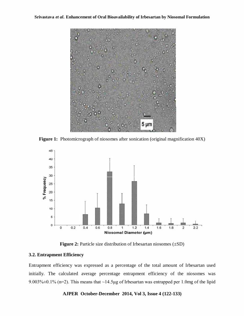
Niosomes appeared as large unilamellar vesicles with spherical shape. Sonication may be
responsible for the breakdown of the multilamellar vesicles to form unilamellar ones. Particle
size analysis of the freshly prepared niosomes shows that the average size is ~0.95μm and the
most probable size is 0.8μm (Figure 2). The use of high cholesterol content in the formulation of
Irbesartan niosomes may lead to large vesicle size. McIntosh et al17 found that cholesterol
increases the width of lipid bilayers and consequently increases the vesicle size. Yoshioka et al18
found that the mean size of the niosomes showed a regular increase with increasing the
hydrophilic lipophilic balance (HLB) of the surfactant because surface free energy decreases
with increasing hydrophobicity.
This result was in good agreement with that obtained in the present study, where the average size
of Irbesartan niosomes is 0.95μm. The particle size distribution of the prepared niosomes reflects
a wide size range of 0.4 to 2.2μm as shown in Figure 2. This finding may be owing to the
influence of certain preparation conditions such as the hydration time and the degree of shaking.
Srivastava et al. Enhancement of Oral Bioavailability of Irbesartan by Niosomal Formulation
AJPER October-December 2014, Vol 3, Issue 4 (122-133)
Figure 1: Photomicrograph of niosomes after sonication (original magnification 40X)
Figure 2: Particle size distribution of Irbesartan niosomes (±SD)
3.2. Entrapment Efficiency
Entrapment efficiency was expressed as a percentage of the total amount of Irbesartan used
initially. The calculated average percentage entrapment efficiency of the niosomes was
9.003%±0.1% (n=2). This means that ~14.5μg of Irbesartan was entrapped per 1.0mg of the lipid
Srivastava et al. Enhancement of Oral Bioavailability of Irbesartan by Niosomal Formulation
AJPER October-December 2014, Vol 3, Issue 4 (122-133)
phase. This result may be explained by the high cholesterol content (~50% of the total lipids).
Yoshioka et al. reported that entrapment efficiency was increased with increasing cholesterol
content when niosomes were prepared by changing the molar ratio of nonionic surfactant to
cholesterol. The authors found that vesicles prepared with span 80 (HLB-4.7) showed the most
efficient entrapment compared with those prepared with other spans because of its highest phase
transition temperature of ~500C. The large size of the prepared unilamellar niosomes may also be
responsible for the high drug entrapment.
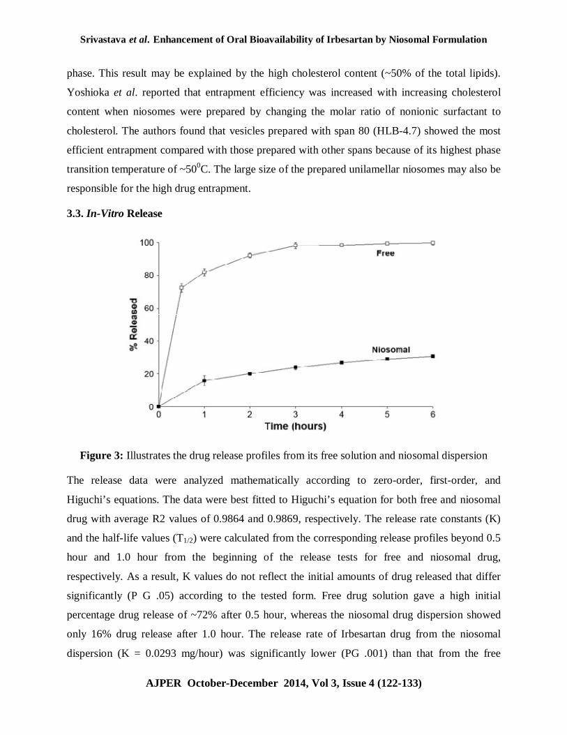
3.3. In-Vitro Release
Figure 3: Illustrates the drug release profiles from its free solution and niosomal dispersion
The release data were analyzed mathematically according to zero-order, first-order, and
Higuchi’s equations. The data were best fitted to Higuchi’s equation for both free and niosomal
drug with average R2 values of 0.9864 and 0.9869, respectively. The release rate constants (K)
and the half-life values (T1/2) were calculated from the corresponding release profiles beyond 0.5
hour and 1.0 hour from the beginning of the release tests for free and niosomal drug,
respectively. As a result, K values do not reflect the initial amounts of drug released that differ
significantly (P G .05) according to the tested form. Free drug solution gave a high initial
percentage drug release of ~72% after 0.5 hour, whereas the niosomal drug dispersion showed
only 16% drug release after 1.0 hour. The release rate of Irbesartan drug from the niosomal
dispersion (K = 0.0293 mg/hour) was significantly lower (PG .001) than that from the free
Srivastava et al. Enhancement of Oral Bioavailability of Irbesartan by Niosomal Formulation
AJPER October-December 2014, Vol 3, Issue 4 (122-133)
solution (K = 0.0654 mg/hour). The drug release from the free solution began to plateau after 3
hours, whereas, the release from the niosomal dispersion was continued for 6 hours without
reaching plateau. These results pointed to sustained release characteristics with a Higuchi pattern
of drug release, where niosomes act as reservoir system for continuous delivery of drug. This
slow release pattern of entrapped drug may indicate the high stability of the niosomal
formulation. Presence of cholesterol in a high percentage and the use of span 80 in the niosomal
formulation may explain this high stability of the niosomal membrane.
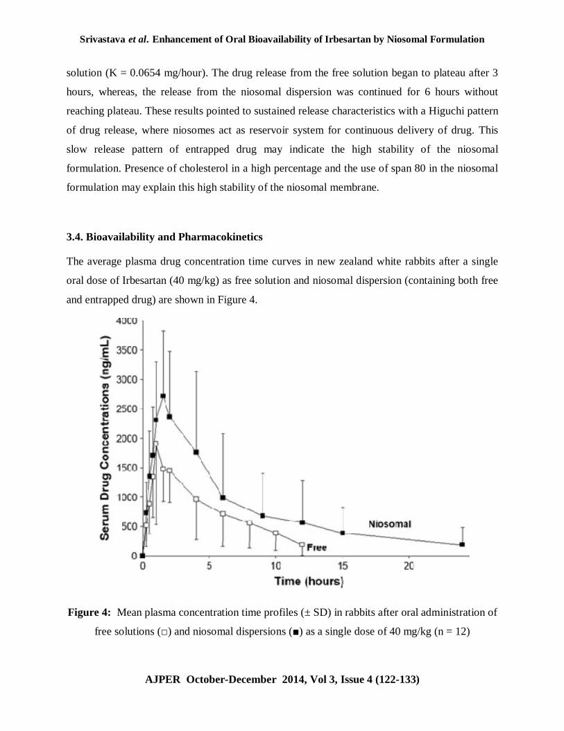
3.4. Bioavailability and Pharmacokinetics
The average plasma drug concentration time curves in new zealand white rabbits after a single
oral dose of Irbesartan (40 mg/kg) as free solution and niosomal dispersion (containing both free
and entrapped drug) are shown in Figure 4.
Figure 4: Mean plasma concentration time profiles (± SD) in rabbits after oral administration of
free solutions (□) and niosomal dispersions (■) as a single dose of 40 mg/kg (n = 12)
Srivastava et al. Enhancement of Oral Bioavailability of Irbesartan by Niosomal Formulation
AJPER October-December 2014, Vol 3, Issue 4 (122-133)
The sustained release effect was also investigated by the in vitro release study. A possible
explanation for this sustained release effect is that niosomes act as a carrier and a slow release
vehicle. The drug is carried by the niosomes through the epithelium into deeper layers of the
mucosa, where the encapsulated drug is slowly released.
This sustained release effect can improve the bioavailability of drugs with slow and limited
absorption and narrow absorption windows. The significant increase of Cmax values may be
owing to enhanced absorption of the free drug included in the tested unpurified niosomal
formulation (containing both the free and niosomal drug). The individual AUC0→∞ values for the
niosomal dispersions were compared with those for the free drug solutions to determine the
relative bioavailability and the mean ratio was found to be 2.55 (±1.82). This result indicated that
more than 2 fold increase in the oral bioavailability of Irbesartan was achieved by the niosomal
formulation. The oral bioavailability of Irbesartan is low (15% to 30%), highly variable in
humans, and is species dependent. Studies of the mechanisms of oral absorption of Irbesartan
have produced conflicting results. The existence of a saturable process in the oral absorption of
Irbesartan by mice,and dogs has been proposed based on a decline in the fraction of dose
absorbed with rising dosage levels. The same suggestion has been made for humans based on a
decline in percentage Irbesartan urinary recovery with increasing dosage. In the present study,
the niosomal dispersion enhanced the bioavailability up to 2.5-fold despite the low content of
entrapped Irbesartan (10%) in comparison with free Irbesartan (89%). This effect may be
explicable in view of the poor oral bioavailability of Irbesartan in its conventional forms (15%-
30%). The improved oral bioavailability may be owing to the lipophilic nature of the niosomal
formulation and the effect of the nonionic surface-active agent on the permeability of the
gastrointestinal membrane. Improved portioning of the lipophilic system to the mucosa, a direct
effect of the surface active agent (span 80) on the barrier function of the mucosa, and prolonged
localization of the drug-loaded niosomes at the site of absorption may be possible reasons for the
improved bioavailability. The present in vivo results support previously published in vitro
findings indicating that passive diffusion is the main mechanism underpinning the intestinal
absorption Irbesartan.
Srivastava et al. Enhancement of Oral Bioavailability of Irbesartan by Niosomal Formulation
AJPER October-December 2014, Vol 3, Issue 4 (122-133)
4. CONCLUSION
The prepared Irbesartan niosomes have unilamellar spherical shape with an average size of 0.95
μm and percentage drug entrapment of 10%. The niosomal formulation showed sustained release
characteristics with Higuchi pattern of drug release. In vivo study in rabbits revealed that more
than 2-fold increase in the oral bioavailability and MRT was achieved by the niosomal
formulation. So, the prepared niosomes could be promising delivery systems Irbesartan with
sustained drug release profiles.
REFERENCES
1. Powell, JR; Reeves, RA; Marino, MR; Cazaubon, C and Nisato, D, “A review of the new
angiotensin II-receptor antagonist irbesartan”, Cardiovascular Drug Reviews, 1998; 16,
169-194.
2. Ellis, ML and Patterson, JH, “A new class of antihypertensive therapy: angiotensin II
receptor antagonists”, Pharmacotherapy, 1996; 16, 849-860.
3. R. Lobenberg and G. L. Amidon, “Modern bioavailability, bioequivalence and
biopharmaceutics classification system. New scientific approaches to international
regulatory standards,” European Journal of Pharmaceutics and Biopharmaceutics, 2000;
50(1), 3-12.
4. WHO Prequalification of Medicines Programme, “General notes on Biopharmaceutics
Classification System (BCS)-based biowaiver applications,” Guidance Document, 2011.
5. V.Manimaran, N.Damodharan, M.Mothilal, K.Rajkumar, Ruby Mamachan Chalackal,
Enhancement of dissolution rate of glibenclamide by solid dispersion technology,
International Journal of Current Pharmaceutical Research, 2010; 2(3), 14-17.
6. K. Dua, K. Pabreja, and M. V. Ramana: Preparation, Characterization and In-Vitro
Evaluation of Aceclofenac Solid Dispersions. ARS Pharmaceutica 2010; 51(1): 57-76.
7. Meera, C.S., A.B. Sayyad and S.D. Sawant, Review on various techniques of solubility
enhancement of poorly soluble drugs with special emphasis on solid dispersion. Journal
of Pharmacy Research, 2010; 3, 2494-2501.

Srivastava et al. Enhancement of Oral Bioavailability of Irbesartan by Niosomal Formulation
AJPER October-December 2014, Vol 3, Issue 4 (122-133)
8. Y. Xie, G. Li, X. Yuan, Z. Cai, and R. Rong, “Preparation and in vitro evaluation of solid
dispersions of total flavones of hippophae Rhamnoides L,” AAPS Pharm Sci Tech.,
2009; 10(2), 631-640.
9. D. K. Sharma, and S. B. Joshi, “Solubility enhancement strategies for poorly water-
soluble drugs in solid dispersions: A review,” Asian Journal of Pharmaceutical Sciences,
2007; 1(1), 9-19.
10. G. P. Kumar and P. Rajeshwarrao, “Nonionic surfactant vesicular systems for effective
drug delivery: an overview,” Acta Pharmaceutica Sinica B, 2011; 1(4), 208-219.
11. S. Biswal, P. N. Murthy, J. Sahu, P. Sahoo, and F. Amir, “Vesicles of non-ionic
surfactants (niosomes) and drug delivery potential,” International Journal of
Pharmaceutical Sciences and Nanotechnology, 2008; 1(1) 1-8.
12. Kandasamy Ruckmani and Veintramuthu Sankar, Formulation and Optimization of
Zidovudine Niosomes, AAPS PharmSciTech. Sep 2010; 11(3): 1119–1127.
13. Elbary A, El-laithy HM, Tadros MI. Sucrose stearate based proniosome-derived
Niosomes for the nebulisable delivery of cromolyn sodium. International Journal of
Pharmaceutics. 2008; 357, 189-198.
14. Anupriya kapoor, R.Gahoi, D.Kumar, In-vitro drug release profile of Acyclovir from
Niosomes formed with different Sorbitan esters, Asian Journal of Pharmacy & Life
Science, 2011; 1(1), 64-70.
15. Pratap S. Jadon, Virendra Gajbhiye, Rajesh S. Jadon, Kavita R. Gajbhiye, and Narayanan
Ganesh, Enhanced Oral Bioavailability of Griseofulvin via Niosomes, AAPS Pharm Sci
Tech. 2009; 10(4): 1186-1192.
16. Peh KK, Yuen KH. Simple high-performance liquid chromatographic method for the
determination of acyclovir in human plasma using fluorescence detection. Journal of
Chromatography B. 1997; 693, 241-244.
17. Yoshioka T, Stermberg B and Florence AT, Preparation and properties of vesicles
(niosomes) of sobitan monoesters (Span 20, 40, 60,and 80) and a sorbitan triester (Span
85). International Journal of Pharmaceutics, 1994: 105, 1-6.
18. McIntosh TJ., The effect of cholesterol on the structure of phosphatidylcholine bilayers.
Biochimica et Biophysica Acta (BBA), 1978; 513(1), 43-58.