The DNLZ/HEP zinc-binding subdomain is critical for regulation of the mitochondrial chaperone HSPA9
Upload
independentCategory
view
1download
0
INFECTION AND IMMUNITY, Apr. 2003, p. 2130–2141 Vol. 71, No. 40019-9567/03/$08.00�0 DOI: 10.1128/IAI.71.4.2130–2141.2003Copyright © 2003, American Society for Microbiology. All Rights Reserved.
CesD2 of Enteropathogenic Escherichia coli Is a Second Chaperonefor the Type III Secretion Translocator Protein EspD
Bianca C. Neves,1 Rosanna Mundy,1 Liljana Petrovska,1 Gordon Dougan,1 Stuart Knutton,2and Gad Frankel1*
Centre for Molecular Microbiology and Infection, Department of Biological Sciences, Imperial College London,London SW7 2AZ,1 and Institute of Child Health, University of Birmingham,
Birmingham B4 6NH,2 United Kingdom
Received 23 September 2002/Returned for modification 15 November 2002/Accepted 20 December 2002
Enteropathogenic Escherichia coli (EPEC) and enterohemorrhagic E. coli are extracellular pathogens thatemploy a type III secretion system to export translocator and effector proteins, proteins which facilitatescolonization of the mucosal surface of the intestine via formation of attaching and effacing (A/E) lesions. Thegenes encoding the proteins for A/E lesion formation are located on a pathogenicity island, termed the locusof enterocyte effacement (LEE), which contains eae encoding intimin as well as the type III secretion system andeffector genes. Many type III secreted proteins are stabilized and maintained in a secretion-competent con-formation in the bacterial cytosol by specific chaperone proteins. Three type III chaperones have beendescribed thus far within the EPEC LEE region: CesD, for the translocator proteins EspB and EspD; CesT, forthe effector proteins Tir and Map; and CesF, for EspF. In this study we report the characterization of CesD2(previously Orf27), a second LEE-encoded chaperone for EspD. We show specific CesD2-EspD protein inter-action which appears to be necessary for proper EspD secretion in vitro and pathogenesis in vivo as demon-strated in the A/E-lesion-forming mouse pathogen Citrobacter rodentium.
Enteropathogenic Escherichia coli (EPEC) is a commoncause of infant diarrhea, particularly in developing countries(42). EPEC infection is associated with formation of a typicalmicroscopic lesion on intestinal epithelial cells, the attachingand effacing (A/E) lesion (41). A/E lesions are characterized bydestruction of cellular microvilli and intimate attachment ofbacteria to cup-like pedestals at the apical enterocyte cellmembrane (31), triggered by activation of a number of signaltransduction pathways and rearrangement of cytoskeletal pro-teins (46). A/E lesions are induced by other enteric pathogens,such as enterohemorrhagic E. coli (EHEC) (54). EHEC infec-tion, which is frequently associated with outbreaks in the de-veloped world, can cause severe diarrhea, hemorrhagic colitis,and hemolytic-uremic syndrome (42). EPEC and EHEC arethe most prominent members among the A/E-lesion-causingpathogens, which are also represented by animal pathogens,including rabbit EPEC (45) and Citrobacter rodentium, whichcauses transmissible murine colonic hyperplasia (47).
The A/E lesion phenotype is encoded by a chromosomalpathogenicity island termed the locus of enterocyte effacement(LEE) region (37). The LEE consists of 41 open readingframes (11, 16), encoding a transcriptional regulator (Ler)(40); type III secretion system (TTSS) proteins (Esc and Sep)(16, 25); type III translocator (EspA, EspB, EspD) (12, 30, 34)and effector (EspF, EspG, Map, Tir) (14, 28, 29, 38, 39) pro-teins; an outer membrane adhesin, intimin (26); and a numberof proteins of unknown function (16).
TTSSs deliver effector proteins through the bacterial innerand outer membranes, with no periplasmic intermediate, andthrough the plasma membrane into the eukaryotic cell cytosol(23). The structural TTSS proteins are highly conserved amongpathogenic bacteria, and many show an intriguing similarity toproteins involved in flagellar biosynthesis (23). The TTSS hasbeen purified from Salmonella enterica serovar Typhimurium,Shigella flexneri (33, 51) and most recently from EPEC (8, 49).It consists of a multiple ring structure and a needle projection.In contrast to the high degree of sequence similarity betweenthe structural TTSS proteins, the effector, secreted, proteinsshow a high level of variation from one system to another.These proteins vary greatly in size, structure, and function andaccount for the species-specific pathogenicity phenotypes as-sociated with different bacterial infections (23).
In EPEC there are seven LEE-encoded type III secretedproteins—EspA, EspB, EspD, EspF, EspG, Map, and Tir (12,14, 28–30, 34, 38, 39). EspA, EspB, and EspD are translocatorproteins, necessary for A/E lesion formation and signalingwithin host cells (19). EspA is the main or only component ofa transiently expressed filamentous surface organelle (32),which is required for the translocation of other translocatorand effector proteins (28, 32). In contrast, immunofluorescenceassays and a calmodulin-dependent adenylate cyclase reportersystem have demonstrated that EspB is translocated into thehost cell, where it is distributed between the cytosol andplasma membrane compartments (61). The presence of EspBin the cytosolic fraction suggests that it may also have, inaddition to its translocator activities, an effector function (52).Indeed, it has been shown that EspB causes changes in hostcell morphology and reorganization of stress fibers, acting pos-sibly as a cytoskeletal toxin (52).
The third translocator protein, EspD, is inserted into a tryp-
* Corresponding author. Mailing address: Centre for Molecular Mi-crobiology and Infection, Flowers Building, Department of BiologicalSciences, Imperial College, London SW7 2AZ, United Kingdom.Phone: 44-(0)20-7594-5253. Fax: 44-(0)20-7594-3069. E-mail: [email protected].
2130

sin-sensitive location in the host cell plasma membrane, at sitesof bacterial contact, but is not translocated into the cytoplasm(55). Based on its homology to YopB, it is believed that EspDis the main component of the TTSS translocation pore in theplasma membrane (19). Indeed, we have recently shown thatEspD exhibits intermolecular interaction which involved itscarboxy-terminal coiled-coil domain (7). A radical disruptionof this region, although did not affect EspA filament produc-tion, caused reduced EspA filament-mediated cell attachmentand EspD-mediated A/E lesion formation (7). The fact that anespD mutant secretes low levels of EspA and produces trun-cated EspA filaments (32) suggests that EspD might be aminor component of the filaments. Indeed, the current modelof the EPEC TTSS predicts interaction between EspA fila-ments and the EspD translocation pores (19), interactionwhich allows protein translocation.
Many of the type III secreted proteins are dependent onspecific chaperones for stabilization in the bacterial cytosolprior to secretion and prevention of premature interactionswith secreted proteins and/or with parts of the secretion andtranslocation machinery (4, 58). In addition, chaperones havebeen shown to be required for the exportation of translocatorand effector proteins. Three chaperones encoded within theLEE region have been described thus far. The first one de-scribed was CesD, a chaperone for EspB and EspD (56). CesDis a 17.5-kDa protein which was shown to interact specificallywith EspD. In a CesD-deficient mutant, EspD secretion wasabolished and the amount of EspB secreted was reduced, butlittle effect on secretion of EspA was observed (56). Despite
sharing features that are common to other type III secretionchaperones, CesD is distinct due to its membrane localization(56). CesT was the second reported LEE-encoded chaperone(1, 13). CesT, 15 kDa, is also a bivalent chaperone that bindsto, and is required for translocation of, both Tir (1, 13) andMap (6). CesF, the most recently reported EPEC TTSS chap-erone, is a 14-kDa protein which specifically interacts withEspF (15). In this study we characterize the gene product ofthe LEE region orf27, a protein with unknown function whichexhibits features common to TTSS chaperones (16). We dem-onstrate that Orf27 is a second secretion partner for EspD andconsequently have renamed it CesD2.
MATERIALS AND METHODS
Bacterial strains, plasmids, and growth conditions. The bacterial strains usedin this study are listed in Table 1. Plasmids used and constructs generated in thisstudy are listed in Table 2. Unless otherwise stated, bacterial strains were grownin Luria-Bertani (LB) broth at 37°C. Growth media were supplemented withampicillin (50 to 100 �g/ml), chloramphenicol (30 �g/ml), kanamycin (30 �g/ml),or nalidixic acid (100 �g/ml) as required.
Molecular techniques. Cloning which required PCR amplification was per-formed with the proofreading DNA polymerase Deep Vent (New England Bio-labs), unless stated otherwise, and all the clones were verified by sequencing. Thesynthetic oligonucleotides used as primers in the PCR and DNA sequencingprocedures were obtained from Pharmacia (United Kingdom) and are listed inTable 3. Automated sequencing was performed at the Advanced BiotechnologyCentre, Imperial College of Science, Technology, and Medicine, Faculty ofMedicine. DNA analysis was performed with Gene Jockey II and programsavailable at http://www.expasy.ch/.
Cloning, expression, and purification of histidine-tagged CesD2. The 408-bpDNA fragment encoding orf27 (cesD2) was amplified by PCR, using the primer
TABLE 1. Bacterial strains used in this study
Strain Description or genotype Source or reference
E. coli TG1 supE hsd �5thi �(lac-proAB) F�[traD36 proAB� lacIq lacZ�M15] StratageneE. coli BL21(DE3)pLysS F� ompT hsdSB(rB
� mB�) gal dcm (DE3) pLysS (Cmr) Novagen
E. coli JM109 recA1 supE44 endA1 hsdR17 gyrA96 relA1 thi �(lac-proAB) New England BiolabsE. coli E2348/69 EPEC prototype strain 36C. rodentium Wild-type, nalidixic acid-resistant strain (formerly C. freundii biotype 4280) 47C. rodentium ICC172 E2348/69 �cesD2::aphT This studyC. rodentium ICC173 C. rodentium cesD2::aphT This study
TABLE 2. Plasmids used in this study
Plasmid Description or genotype Source or reference
pET 28a PT7lac N-terminal His6 expression vector NovagenpGEM-T-Easy ColE1-based vector for cloning of PCR products PromegapET3d PT7 expression vector NovagenpMal-c2X Ptac malE N-terminal fusion expression vector New England BiolabspACYC184 Origin of replication from p15A; low copy number; Cmr Tcr New England BiolabspICC232 pET3d-CesD2, unmodified CesD2 This studypICC233 pMAL-c2X-CesD2; full-length CesD2 fused to MBP in N-terminal end This studypICC234 pET28a-CesD2; full-length CesD2 fused to His6 in the N-terminal end This studypICC240 PCR-amplified 4.071-kbp DNA fragment containing cesD2; cloned into pGEM-T-Easy This studypICC241 In-frame deletion subclone of pICC240; missing bp 97 to 351 of cesD2 This studypICC260 �cesD2::aphT in pGEM-T-Easy This studypKD46 Low-copy-number plasmid encoding the phage � Red recombinase, expressing �, �, and
exo from the arabinose-inducible ParaB
9
pSB315 Plasmid containing cloned aphT kanamycin cassette 21pICC243 PCR-amplified 1.208-kbp DNA fragment containing cesD2CR and cloned in pGEM-T-Easy This studypICC244 Insertion subclone of pICC243; cesD2CR::aphT This studypICC245 pACYC184-CesD2CR This studypICC246 pACYC184-HisCesD2EPEC This study
VOL. 71, 2003 CesD2 OF EPEC 2131
pair cesD2-F (NdeI) and cesD2-R (BamHI) and EPEC genomic DNA from theprototype strain E2348/69 (36) as the DNA template. The PCR product wasdigested with NdeI and BamHI and cloned into NdeI/BamHI-digested pET28-a,generating plasmid pICC234 (Table 2). A His6-CesD2 fusion was expressed inBL21(DE3)pLysS(pICC234). An overnight culture was diluted 1:100 in 100 ml ofLB broth, supplemented with kanamycin (30 �g/ml), chloramphenicol (30 �g/ml), and 0.2% glucose. The culture was grown to an optical density at 600 nm(OD600) of 0.4 to 0.8, at 37°C with shaking, and was induced with the addition of1.0 mM IPTG (isopropyl-�-D-thiogalactopyranoside). Induction was performedfor 4 h at 30°C, with shaking. Bacterial cells were pelleted by centrifugation at8,000 g for 20 min and resuspended in cold binding buffer (5 mM imidazole,0.5 M NaCl, 20 mM Tris-HCl [pH 7.9]). Bacterial suspensions were sonicatedand cell debris was removed by centrifugation at 45,000 g for 30 min. The cellextracts were filtered through a 0.45-�m-pore-size filter device prior to thefollowing purification procedures. His6-CesD2 was purified as recommended bythe manufacturer (Novagen). Briefly, the bacterial lysate obtained as describedabove was loaded onto a 2.5-ml nickel-charged column, previously equilibratedwith binding buffer. The column was then washed with 25 ml of binding buffer,7.5 ml of wash buffer 1 (30 mM imidazole, 0.5 M NaCl, 20 mM Tris-HCl [pH7.9]), and 7.5 ml of wash buffer 2 (60 mM imidazole, 0.5 M NaCl, 20 mMTris-HCl [pH 7.9]). The bound protein was eluted with 15 ml of elution buffer(500 mM imidazole, 0.5 M NaCl, 20 mM Tris-HCl [pH 7.9]). Fractions (1 ml)were collected and monitored by sodium dodecyl sulfate–12% polyacrylamidegel electrophoresis (SDS–12% PAGE).
Column capture assay. His-tag columns containing bound His6-CesD2 orHis-T7 tag (negative control), were used to test the ability of recombinant CesD2to bind the secreted forms of EspA, EspB, and EspD. The procedure wasperformed as described for the purification steps, involving two additional stageswhich consisted of loading 25 ml of culture supernatant from E2348/69, grown inDulbecco’s modified Eagle medium (DMEM), onto the columns and an extrawashing step followed by elution as for the purification procedures. Fractions (1ml) were collected, separated by SDS–12% PAGE, and analyzed by Westernblotting in order to verify copurification of any of the secreted proteins with therecombinant CesD2, or the control column His6-T7.
Generation of a nonpolar cesD2 E2348/69 mutant. PCR was performed usingprimers LEE-4K-F and LEE-4K-R to obtain a 4,071-bp DNA fragment of theLEE region containing cesD2, from strain E2348/69, which was ligated intopGEM-T-Easy vector (Promega), creating plasmid pICC240 (Table 2). InversePCR was used to construct an in-frame deletion within cesD2 using primersCesD2-INV-F (SmaI) and CesD2-INV-R (SmaI) and pICC240 as the template.Digestion with SmaI and self-ligation excised nucleotides 97 to 351 and added aSmaI site to the deletion locus (Fig. 1B), creating plasmid pICC241 (Table 2).For DNA amplification, the Expand High Fidelity PCR System (Roche, Mann-heim, Germany) was employed, which included a hot start of 2 min at 94°C, andthe preparation of two master mixes. For a 100-�l reaction mixture, mix 1 (50 �l)contained 400 �M concentrations of the deoxynucleoside triphosphates, 600 nMconcentrations of upstream and downstream primers, and 10 ng of template(genomic DNA from strain E2348/69), while mix 2 (50 �l) contained the ExpandHF buffer with 15 mM MgCl2 and 2.6 U of Expand High Fidelity PCR Systemenzyme mix. For primers LEE-4K-F and LEE-4K-R, cycling was performed asfollows: 94°C for 2 min (hot start); 10 cycles of 94°C for 30 s, 65°C for 30 s, and68°C for 6 min; 20 cycles of 94°C for 30 s, 65°C for 30 s, and 68°C for 6 min with15-s increase; and a final extension at 68°C for 7 min. The same PCR cycling
conditions were used for the inverse PCR, using primers CesD2-INV-F (SmaI)and CesD2-INV-R (SmaI), except for the annealing temperature, which, in thiscase, was 60°C. An aphT cassette (21) with a HincII site in the correct readingframe was digested out of plasmid pSB315 and cloned into the SmaI-digestedplasmid pICC241. The orientation of the cassette was verified by PCR, usingprimers CesD2-F (NdeI) and AphT-R, which tested one of the junction frag-ments. Primers LEE-4K-F and LEE-4K-R were used to amplify a fragment ofapproximately 4.8 kbp from plasmid pICC260, which contained the disruptedcesD2 gene and its flanking regions. One microgram of the amplified fragmentwas treated with DpnI, purified, and transformed by electroporation intoE2348/69 carrying a Red helper plasmid, pKD46 (Table 2) (9). Transformantswere first selected for Kmr and then tested for ampicillin sensitivity to confirmloss of the helper plasmid. The �cesD2::aphT EPEC derivative was namedICC172 (Table 1). In order to trans-complement ICC172, pICC234 was doubledigested with XbaI/BamHI and a 509-bp DNA fragment, which contained theentire cesD2 and a sequence coding for an N-terminal His6 tag, was ligated intothe BamHI site of pACYC184, generating plasmid pICC246. Prior to ligation,both vector and insert were previously filled in with Klenow DNA polymerase I(New England Biolabs), according to the manufacturer’s instructions. Fragmentorientation was verified by PCR, using primers pACYC-F and CesD2-R(BamHI).
Generation of a nonpolar cesD2 C. rodentium mutant. PCR was used togenerate a 1,208-bp fragment containing the C. rodentium cesD2 gene (11), usingprimers CesD2CR(�400)-F and CesD2CR(�808)-R (Table 3) and genomic DNAfrom a wild-type C. rodentium strain (47). The amplified fragment was insertedinto pGEM-T Easy vector, creating plasmid pICC243. aphT cassette (21) wasdigested out of pSB315 with EcoRI and ligated into EcoRI-cut pICC243, gen-erating an insertion at position 76 of cesD2 (Fig. 1C) and creating pICC244. Theorientation of the cassette was verified by PCR with primers CesD2CR(�1)-F(NcoI) and AphT-R (Table 3). A 2,200-bp fragment containing the disruptedcesD2 was generated by PCR using plasmid pICC244 as the template, withprimers CesD2CR(�400)-F and CesD2CR(�808)-R, and treated with DpnI. Onemicrogram of the PCR product was electrotransformed into the C. rodentiumstrain carrying the Red helper plasmid pKD46 (Table 2), and this was followedby screening for recombinants as described above. The generated cesD2::aphT C.rodentium strain was named ICC173 (Table 1). The cesD2 (CR27) (11) gene wasalso cloned into plasmid pACYC184 (Table 2), to be used in the trans-comple-mentation experiments. A fragment of 453 bp containing the full-length cesD2and 45 bp of upstream sequence was amplified from C. rodentium genomic DNA,with primers CesD2CR(�45)-F (BamHI) and CesD2CR(�408)-R (SalI) and wasligated into BamHI/SalI-double-digested pACYC184, generating pICC245.
Preparation of EPEC-secreted proteins. Analysis of culture supernatants ofEPEC-secreted proteins was performed as described previously (32). Bacteriawere grown in 20 ml of DMEM at 37°C with shaking to an OD600 of 1.0. Thebacterial cells were pelleted by centrifugation at 8,000 g for 10 min, andsupernatants were passed through filters with a pore size of 0.45 �m. Phenyl-methylsulfonyl fluoride (PMSF) (50 �g/ml; Sigma), aprotinin (0.5 �g/ml; Sigma),and EDTA (0.5 mM) were added (all final concentrations), and the Esp proteinswere concentrated 100-fold with centrifugal filter devices (Millipore Corpora-tion, Bedford, Mass.). A volume of concentrated samples was loaded and sepa-rated by SDS–12% PAGE followed by Western blotting.
Bacterial cell fractionation. (i) Periplasm extraction. All the EPEC cultures(100 ml) were grown to an OD600 of 1.0 in DMEM. The periplasmic contents
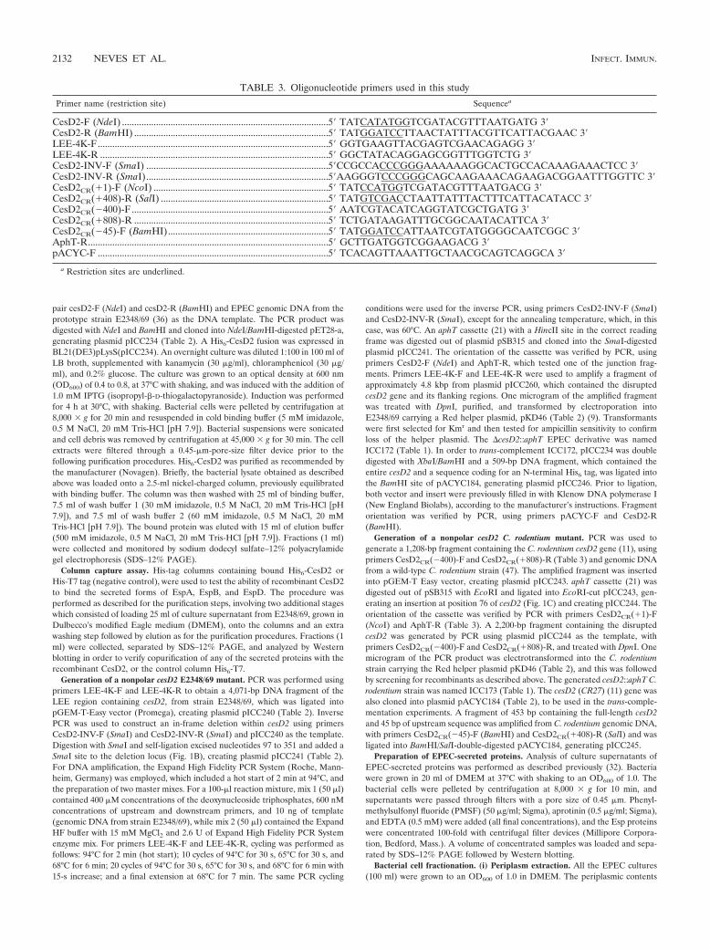
TABLE 3. Oligonucleotide primers used in this study
Primer name (restriction site) Sequencea
CesD2-F (NdeI) .....................................................................................5� TATCATATGGTCGATACGTTTAATGATG 3�CesD2-R (BamHI) ................................................................................5� TATGGATCCTTAACTATTTACGTTCATTACGAAC 3�LEE-4K-F...............................................................................................5� GGTGAAGTTACGAGTCGAACAGAGG 3�LEE-4K-R ..............................................................................................5� GGCTATACAGGAGCGGTTTGGTCTG 3�CesD2-INV-F (SmaI) ...........................................................................5�CCGCCACCCGGGAAAAAAGGCACTGCCACAAAGAAACTCC 3�CesD2-INV-R (SmaI)...........................................................................5�AAGGGTCCCGGGCAGCAAGAAACAGAAGACGGAATTTGGTTC 3�CesD2CR(�1)-F (NcoI) ........................................................................5� TATCCATGGTCGATACGTTTAATGACG 3�CesD2CR(�408)-R (SalI) .....................................................................5� TATGTCGACCTAATTATTTACTTTCATTACATACC 3�CesD2CR(�400)-F .................................................................................5� AATCGTACATCAGGTATCGCTGATG 3�CesD2CR(�808)-R ................................................................................5� TCTGATAAGATTTGCGGCAATACATTCA 3�CesD2CR(�45)-F (BamHI)..................................................................5� TATGGATCCATTAATCGTATGGGGCAATCGGC 3�AphT-R...................................................................................................5� GCTTGATGGTCGGAAGACG 3�pACYC-F ...............................................................................................5� TCACAGTTAAATTGCTAACGCAGTCAGGCA 3�
a Restriction sites are underlined.
2132 NEVES ET AL. INFECT. IMMUN.
were collected following osmotic shock as follows. Bacterial cells were pelletedby centrifugation at 4,000 g for 20 min, at 4°C. The supernatant was removed,the pellets were washed twice and resuspended in 2.5 ml of ice-cold 20 mMTris-HCl (pH 7.5). Two ml of ice-cold 40% sucrose and 150 �l of 500 mM EDTAwere subsequently added, followed by gentle agitation at 4°C for 20 min. Cellswere once more spun down as before, the supernatant was discarded, and pelletswere immediately resuspended in 1 ml of ice-cold deionized water, followinggentle agitation at 4°C for 20 min. The suspension was centrifuged at 8,000 g
for 20 min at 4°C. The resulting supernatant containing periplasmic proteins wasthen collected (P fraction).
(ii) Preparation of cytoplasmic and membrane fractions. The pellet from theperiplasmic extraction was then fractionated as described previously (24), withmodifications. The pellet was initially resuspended in 8 ml of lysis buffer (10 mMTris-HCl [pH 7.5], 0.5 mM PMSF, aprotinin [0.5 �g/ml]), freeze-thawed, andpassed twice through a cell disrupter (Constant Cell Disruption System). Cellenvelopes and unbroken bacteria were removed by centrifuging twice (5,000 g
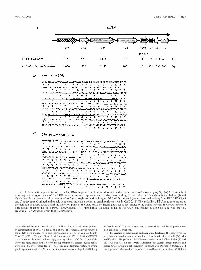
FIG. 1. Schematic representation of LEE4, DNA sequence, and deduced amino acid sequence of cesD2 (formerly orf27). (A) Overview (notto scale) of the organization of the LEE4 operon. Arrows represent each of the open reading frames, with their length indicated below. (B andC) Nucleotide and amino acid sequences of espB (carboxyl-terminal region), cesD2 (orf27), and escF (amino terminal region) from EPEC E2348/69and C. rodentium. Outlined amino acid sequences indicate a potential amphipathic -helix in CesD2. (B) The underlined DNA sequence indicatesthe deletion in EPEC �cesD2 and the insertion point of the aphT cassette. Highlighted sequences indicate the points wherein the SmaI sites wereintroduced for construction of EPEC �cesD2::aphT. (C) Highlighted sequence indicates the EcoRI site where the aphT cassette was inserted,creating a C. rodentium strain that is cesD2::aphT.
VOL. 71, 2003 CesD2 OF EPEC 2133
for 10 min at 4°C). The supernatant, containing soluble proteins (cytosolic) andinsoluble proteins (inner and outer membranes), was removed and ultracentri-fuged for 1 h at 50,0000 g, at 4°C, to pellet the membranes. The supernatantcontaining cytoplasmic proteins (C fraction) was collected and concentrated to0.4 ml with centrifugal filter devices (Millipore Corporation). The membranepellet was washed once with lysis buffer and resuspended in 0.4 ml of Sarkosylbuffer (100 mM NaCl, 10 mM Tris-HCl [pH 8.0], 0.5 mM PMSF, aprotinin [0.5�g/ml], 0.5% N-lauroylsarcosine [Sigma]). Under this condition the inner mem-brane is dissolved but not the outer membrane. Following centrifugation at50,000 g for 1 h, at 4°C, the supernatant containing the inner membraneproteins (IM fraction) was collected. The remaining pellet was washed once withSarkosyl buffer, under the same conditions as described above, and dissolved in0.4 ml of 1 SDS loading buffer (OM fraction). Equivalent amount of proteinswere loaded after normalization in relation to the volume of the original bacte-rial cultures.
Enrichment and purity of each fraction were estimated by detection of pro-teins known to be localized within each of the compartments. Enrichment of thecytoplasm was determined by immunoblotting using a mouse polyclonal anti-serum against �-galactosidase (1:2,000; Sigma), a well-characterized cytoplasmicenzyme (35). Enrichment of the IM fraction was determined by detection of Etk(24) with a specific rabbit polyclonal antiserum (1:2,000). Maltose binding pro-tein (antiserum diluted 1:2,000, New England Biolabs) was used as a marker forperiplasmic material (27), while intimin (26), an outer membrane adhesin ofEPEC (antiserum diluted 1:2,000 [2]), was used as a marker for the OM fraction.
Immunoblot analysis. Proteins separated by SDS-PAGE were transferredelectrophoretically onto nitrocellulose membranes (0.45-�m pore size; Bio-Rad)and immunoblotted according to the method of Towbin et al. (53). Proteins wereblotted using a Bio-Rad Wet Blot apparatus, the membranes were blockedovernight in 3% bovine serum albumin (Sigma) and washed thoroughly withphosphate-buffered saline (PBS) containing 0.05% Tween 20 (PBST). Primaryantisera were diluted in PBST, and the concentrations used are indicated foreach experiment. Secondary antibodies, goat anti-rabbit or anti-mouse (1:5,000dilution, Sigma), were conjugated with horseradish peroxidase. An ECL Westernblotting analysis system (Amersham Life Science) was used according to themanufacturer’s instructions. The immunoblots were then exposed to a high-performance chemiluminescence film, Hyperfilm ECL (Amersham Life Sci-ence), and the was film developed using a Fuji X-ray film developer.
Fluorescence actin staining (FAS). Subconfluent cultures of HEP-2 cells onglass coverslips were placed in wells of a 24-well tissue culture plate containing1 ml of HEPES-buffered minimal essential medium containing 2% fetal calfserum. Ten microliters of an overnight bacterial broth culture was added to eachwell, and the cells were incubated for 1, 3, and 6 h at 37°C; in 6-h assays freshmedium was added after 3 h. After three washes in PBS to remove nonadheringbacteria, cells were fixed in 4% buffered formalin for 30 min. Washed cells werepermeabilized by treating coverslips with 0.1% Triton X-100 in PBS for 5 min.After three PBS washes, coverslips were incubated with a solution of fluoresceinisothiocyanate-phalloidin (5 �g/ml; Sigma) in PBS for 20 min to stain filamen-tous cell actin. Bacteria were immunostained using an antibody to wholeE2348/69 bacteria and a secondary Alexa-594 antibody (Molecular Probes).Coverslips were washed a further three times in PBS and mounted in Citifluormountant (agar). Specimens were examined using a Leica TCS SP2 spectralconfocal microscope.
Challenge of mice with C. rodentium. Male, specific-pathogen-free C3H/Hejmice (4 to 5 weeks old) were purchased from Harlan Olac (Bicester, UnitedKingdom). All mice were housed in individual ventilated cages with free accessto food and water. Bacterial inocula were prepared by culturing bacteria over-night at 37°C in LB broth containing nalidixic acid (100 �g/ml) plus chloram-phenicol (30 �g/ml) (wild-type C. rodentium carrying pACYC184), LB brothcontaining kanamycin (30 �g/ml) (C. rodentium ICC173), or LB broth containingkanamycin plus chloramphenicol (30 �g/ml) (C. rodentium ICC173 carryingpICC245 or pICC246). After incubation, bacteria were harvested by centrifuga-tion and resuspended in equal volumes of PBS. Mice were then orally inoculatedwith 0.2 ml of a bacterial suspension using a gavage needle (50). The viable countof each inoculum was determined by retrospective plating on LB agar containingappropriate antibiotics.
The murine infection assay was performed twice. In the first set of experi-ments, five mice were used per strain and two mice were used as uninfectedcontrols. In the second set of experiments, three mice were used per group andagain two mice were used as uninfected controls. There was one case of fatalityamong the mice infected with the wild-type strain.
Mesurement of pathogen burden. Mice were killed 13 days postinfection, andthe distal 8 cm of colon was aseptically removed and weighed, after removal offecal pellets. Colons were then homogenized mechanically using a Seward 80
stomacher (Seward Medical, London, England), and the numbers of viablebacteria in colonic homogenates were determined by viable count on LB agarcontaining appropriate antibiotics as described previously (50).
Statistical analysis. All statistical analysis was carried out using the two-tailedStudent’s t test.
Histological and immunofluorescence analysis. Distal colons of noninfectedmice and those of mice infected with wild-type, ICC173, or complemented stainswere snap-frozen and stored at �70°C until cutting. Cryosections (6 �m thick)were cut, air dried for 1 h, fixed with acetone for 10 min, and processed forhistological analysis or immunofluorescence analysis. For histological analysiscryosections were stained with hematoxylin and eosin and analyzed using a NikonEclipse E600 microscope.
For immunofluorescence analysis cryosections were blocked using 1% bovineserum albumin followed by the addition of a rabbit universal intimin sera (3) andwere incubated for 1 h at room temperature. After extensive washing, cryosec-tions were incubated for 45 min with goat anti-rabbit immunoglobulin G labeledwith fluorescein isothiocyanate (Sigma). Host cell F-actin was stained withTRITC (tetramethyl rhodamine isothiocyanate)-conjugated phalloidin (Sigma).Specimens were mounted in Mowiol (Aldrich) and viewed through an invertedZeiss LSM 510 Meta confocal microscope.
RESULTS
Binding of native secreted EspD to His6-Orf27. Type IIIsecretion chaperones form a heterogeneous family of proteinsthat have little or no similarity at the amino acid level but aregenerally small (14- to 19-kDa), cytoplasmic, acidic proteinswith predicted carboxyl-terminal amphipathic -helix and of-ten adjacent to or in the proximity of the gene coding for thetarget protein (4, 23, 58). Although no function has been de-termined for orf27 of the LEE region, its deduced gene prod-uct has all the characteristics of a putative type III secretionchaperone (16). Orf27 is encoded within the polycistronicLEE4 operon (16, 40), which encodes the TTSS translocator(EspA, EspD, EspB, and EscF) and effector (EspF) proteins(Fig. 1A). Orf27 has a calculated molecular mass of 15.8 kDaand pI 5.3. Although it does not show primary sequence sim-ilarity to any protein currently in the databases, its carboxyl-terminal region contains a putative amphipathic -helix, pro-posed to mediate interactions with target proteins (Fig. 1B).Based on these observations, we investigated whether Orf27had the ability to directly interact with any of the translocatorproteins encoded upstream on the LEE4, i.e., EspA, EspD,and EspB.
Preliminary results obtained from microarray-based tran-scriptional analyses in our laboratory have demonstrated thatorf27 is expressed in the early exponential growth phase (M.Batchelor, unpublished data). To examine whether the orf27gene produces a detectable protein product, the full-lengthorf27 gene was cloned into pET3-d (pICC232) and expressedin BL21(DE3)pLysS, producing an unmodified recombinantOrf27 peptide. A protein band with an apparent molecularmass of 13 kDa, slightly smaller than that predicted from theamino acid sequence (15.8 kDa), was visualized by SDS-PAGE(data not shown). In addition, the entire Orf27 peptide wasexpressed as a histidine-tagged N terminus fusion protein(His6-Orf27) in E. coli BL21(DE3)pLysS from plasmidpET28a (pICC234). To investigate whether recombinantOrf27 binds any of the native translocator proteins fromEPEC, columns containing bound His6-Orf27 or His6-T7 wereoverlaid with 25 ml of filtered culture supernatant from E2348/69, grown in DMEM under conditions favorable for LEE geneexpression. Following elution of His6-T7 or His6-Orf27 with
2134 NEVES ET AL. INFECT. IMMUN.
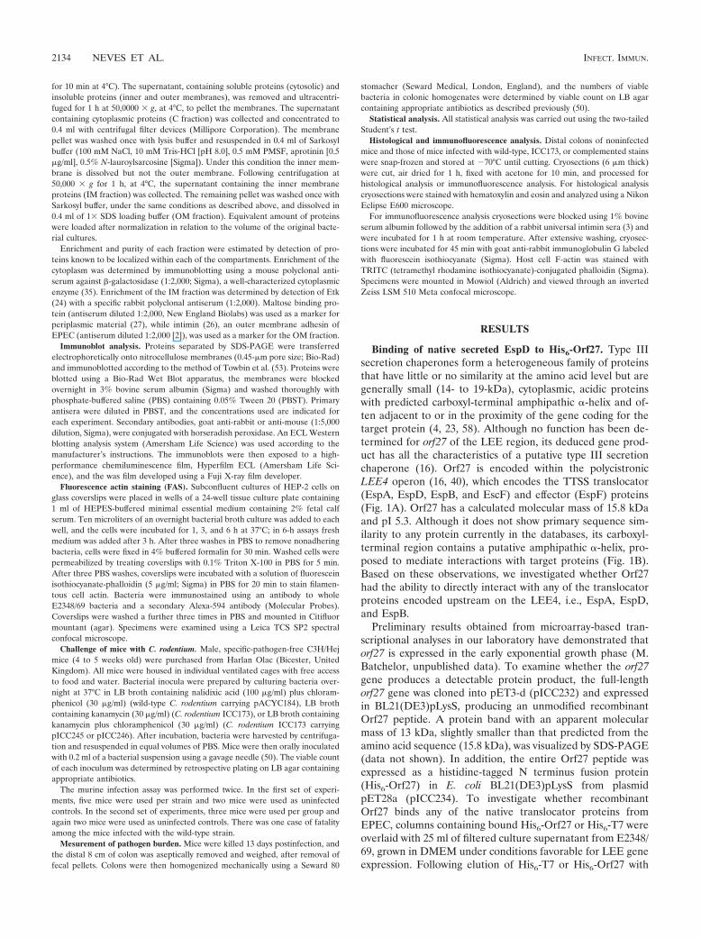
the imidazole buffer, 1-ml fractions were collected and probedwith either His6, EspA, EspB, or EspD antisera by Westernblotting. The results showed that EspD was coeluted with His6-Orf27 (Fig. 2). Neither EspA nor EspB bound to the column,as they were not detected within the eluted His6-Orf27 frac-tions (data not shown). The control, His6-T7 column, did notbind any of the tested proteins from the culture supernatant(Fig. 2). These results showed specific Orf27-EspD proteininteraction and suggested that Orf27, like CesD, might displayan EspD chaperone activity. Consequently, we renamed Orf27CesD2.
A nonpolar mutation in EPEC cesD2 affects the level ofintracellular and secreted EspD but not A/E lesion formationin vitro. In order to determine the role of CesD2 during in-fection in vitro, we disrupted its gene in the prototype EPECstrain E2348/69 and subjected the mutant to a variety of phe-notypic characterizations. For the mutagenesis, a 4-kbp DNAfragment containing cesD2 was cloned. Inverse PCR was thenperformed to generate an in-frame internal deletion (Fig. 1B)in which a kanamycin resistance cassette aphT (21) was in-serted (pICC260). The disrupted cesD2 was amplified, and thePCR product was transformed into E2348/69 carrying plasmidpKD46 (encoding the Red recombinase, which enhances therate of recombination of linear DNA) (9). Allelic exchange wasselected, generating a �cesD2::aphT EPEC, strain ICC172.The nonpolar nature of the mutation was confirmed by thedetection of wild-type levels of EspF, which is encoded down-stream cesD2 (Fig. 1; data not shown), in Western blots.
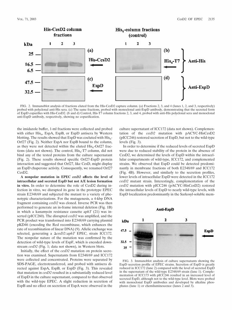
Initially, the effect of the cesD2 mutation on protein secre-tion was examined. Supernatants from E2348/69 and ICC172were collected and concentrated. Proteins were separated bySDS-PAGE, electrotransferred, and probed with antisera di-rected against EspA, EspB, or EspD (Fig. 3). This revealedthat mutation in cesD2 resulted in a substantially reduced levelof EspD in the culture supernatant, compared to that observedwith the wild-type EPEC. A slight reduction in secretion ofEspB and no effect on secretion of EspA were observed in the
culture supernatant of ICC172 (data not shown). Complemen-tation of the cesD2 mutation with pACYC-HisCesD2(pICC246) restored secretion of EspD, but not to the wild-typelevels (Fig. 3).
In order to determine if the reduced levels of secreted EspDwere due to reduced stability of the protein in the absence ofCesD2, we determined the levels of EspD within the intracel-lular compartments of wild-type, ICC172, and complementedstrains. We observed that EspD could be detected predomi-nantly in membrane fractions of both E2348/69 and ICC172(Fig. 4B). However, and similarly to the secretion profiles,lower levels of intracellular EspD were detected in the ICC172cesD2 mutant strain. Interestingly, complementation of thecesD2 mutation with pICC246 (pACYC-HisCesD2) restoredthe intracellular levels of EspD to nearly wild-type levels, withEspD localization predominantly in the Sarkosyl-soluble mem-
FIG. 2. Immunoblot analysis of fractions eluted from the His-CesD2 capture column. (a) Fractions 2, 3, and 4 (lanes 1, 2, and 3, respectively)probed with polyclonal anti-His sera. (c) The same fractions, probed with monoclonal anti-EspD antibody, demonstrating that the secreted formof EspD copurifies with His-CesD2. (b and d) Control, His-T7 column fractions 2, 3, and 4, probed with anti-His polyclonal sera and monoclonalanti-EspD antibody, respectively, showing no copurification.
FIG. 3. Immunoblot analysis of culture supernatants showing theEspD secretion profile of EPEC strains. Secretion of EspD is greatlyreduced in ICC172 (lane 2) compared with the level of secreted EspDin the supernatant of the wild-type E2348/69 strain (lane 1). Comple-mentation of ICC173 with pICC246 resulted in an increased level ofsecreted EspD, although not to the wild-type level. Blots were probedwith monoclonal EspD antibodies and developed by alkaline phos-phates (lane 1) or chemiluminescence (lanes 2 and 3).
VOL. 71, 2003 CesD2 OF EPEC 2135
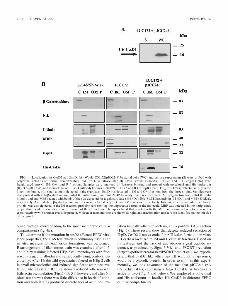
brane fraction, corresponding to the inner membrane cellularcompartment (Fig. 4B).
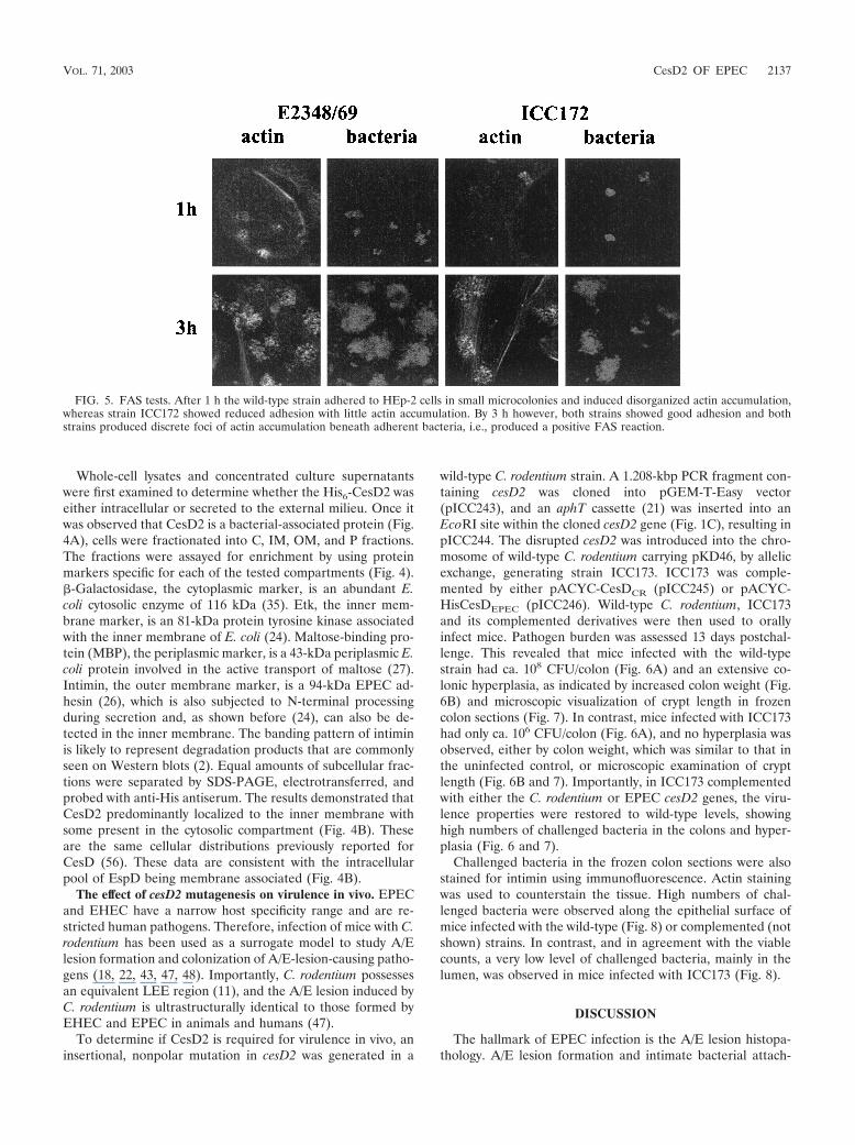
To determine if the mutation in cesD2 affected EPEC viru-lence properties, the FAS test, which is commonly used as anin vitro measure for A/E lesion formation, was performed.Rearrangement of filamentous actin was examined after 1, 3,and 6 h by staining infected HEp-2 cell monolayers with fluo-rescein-tagged phalloidin and subsequently using confocal mi-croscopy. After 1 h the wild-type strain adhered to HEp-2 cellsin small microcolonies and induced significant actin accumu-lation, whereas strain ICC172 showed reduced adhesion withlittle actin accumulation (Fig. 5). By 3 h, however, and after 6 h(data not shown) there was little difference in levels of adhe-sion and both strains produced discrete foci of actin accumu-
lation beneath adherent bacteria, i.e., a positive FAS reaction(Fig. 5). These results show that despite reduced secretion ofEspD, CesD2 is not essential for A/E lesion formation in vitro.
CesD2 is localized to IM and C cellular fractions. Based onits features and the lack of any obvious signal peptide se-quence, as predicted by SignalP V1.1 and iPSORT prediction(http://hypothesiscreator.net/iPSORT/predict.cgi), we hypoth-esized that CesD2, like other type III secretion chaperones,would be a cytosolic protein. In order to confirm this experi-mentally, we took advantage of the fact that pICC246 (pACYC-HisCesD2), expressing a tagged CesD2, is biologicallyactive in vivo (Fig. 4 and below). We employed a polyclonalanti-His antiserum to localize His-CesD2 in different EPECcellular compartments.
FIG. 4. Localization of CesD2 and EspD. (A) Whole ICC172(pICC246) bacterial cells (WC) and culture supernatant (S) were probed withpolyclonal anti-His antiserum, demonstrating that CesD2 is intracellular.(B) EPEC strains E2348/69, ICC172, and ICC172(pICC246) werefractionated into C, IM, OM, and P fractions. Samples were analyzed by Western blotting and probed with polyclonal anti-His antiserum(ICC172-pICC246) and monoclonal anti-EspD antibody (strains E2348/69, ICC172, and ICC172-pICC246). His6-CesD2 was detected mostly in theinner membrane, with small amount detected in the cytoplasm. EspD was detected in IM and OM fractions from the three strains. Samples werealso probed with anti-�-galactosidase, anti-Etk, anti-intimin, and anti-MBP to verify fraction enrichment. Anti-�-galactosidase, anti-Etk, anti-intimin, and anti-MBP reacted with bands of the size expected for �-galactosidase (116 kDa), Etk (81.2 kDa), intimin (94 kDa), and MBP (43 kDa),respectively. As predicted, �-galactosidase and Etk were detected only in C and IM fractions, respectively. Intimin, which is an outer membraneprotein, was also detected in the IM fraction, probably representing the unprocessed form of the molecule. MBP was detected in the periplasmicpreparation, while it was also present in some of the C fractions. The upper band that reacted with the MBP antiserum is likely to represent across-reactivity with another cytosolic protein. Molecular mass markers are shown at right, and fractionation markers are identified on the left sideof the panel.
2136 NEVES ET AL. INFECT. IMMUN.
Whole-cell lysates and concentrated culture supernatantswere first examined to determine whether the His6-CesD2 waseither intracellular or secreted to the external milieu. Once itwas observed that CesD2 is a bacterial-associated protein (Fig.4A), cells were fractionated into C, IM, OM, and P fractions.The fractions were assayed for enrichment by using proteinmarkers specific for each of the tested compartments (Fig. 4).�-Galactosidase, the cytoplasmic marker, is an abundant E.coli cytosolic enzyme of 116 kDa (35). Etk, the inner mem-brane marker, is an 81-kDa protein tyrosine kinase associatedwith the inner membrane of E. coli (24). Maltose-binding pro-tein (MBP), the periplasmic marker, is a 43-kDa periplasmic E.coli protein involved in the active transport of maltose (27).Intimin, the outer membrane marker, is a 94-kDa EPEC ad-hesin (26), which is also subjected to N-terminal processingduring secretion and, as shown before (24), can also be de-tected in the inner membrane. The banding pattern of intiminis likely to represent degradation products that are commonlyseen on Western blots (2). Equal amounts of subcellular frac-tions were separated by SDS-PAGE, electrotransferred, andprobed with anti-His antiserum. The results demonstrated thatCesD2 predominantly localized to the inner membrane withsome present in the cytosolic compartment (Fig. 4B). Theseare the same cellular distributions previously reported forCesD (56). These data are consistent with the intracellularpool of EspD being membrane associated (Fig. 4B).
The effect of cesD2 mutagenesis on virulence in vivo. EPECand EHEC have a narrow host specificity range and are re-stricted human pathogens. Therefore, infection of mice with C.rodentium has been used as a surrogate model to study A/Elesion formation and colonization of A/E-lesion-causing patho-gens (18, 22, 43, 47, 48). Importantly, C. rodentium possessesan equivalent LEE region (11), and the A/E lesion induced byC. rodentium is ultrastructurally identical to those formed byEHEC and EPEC in animals and humans (47).
To determine if CesD2 is required for virulence in vivo, aninsertional, nonpolar mutation in cesD2 was generated in a
wild-type C. rodentium strain. A 1.208-kbp PCR fragment con-taining cesD2 was cloned into pGEM-T-Easy vector(pICC243), and an aphT cassette (21) was inserted into anEcoRI site within the cloned cesD2 gene (Fig. 1C), resulting inpICC244. The disrupted cesD2 was introduced into the chro-mosome of wild-type C. rodentium carrying pKD46, by allelicexchange, generating strain ICC173. ICC173 was comple-mented by either pACYC-CesDCR (pICC245) or pACYC-HisCesDEPEC (pICC246). Wild-type C. rodentium, ICC173and its complemented derivatives were then used to orallyinfect mice. Pathogen burden was assessed 13 days postchal-lenge. This revealed that mice infected with the wild-typestrain had ca. 108 CFU/colon (Fig. 6A) and an extensive co-lonic hyperplasia, as indicated by increased colon weight (Fig.6B) and microscopic visualization of crypt length in frozencolon sections (Fig. 7). In contrast, mice infected with ICC173had only ca. 106 CFU/colon (Fig. 6A), and no hyperplasia wasobserved, either by colon weight, which was similar to that inthe uninfected control, or microscopic examination of cryptlength (Fig. 6B and 7). Importantly, in ICC173 complementedwith either the C. rodentium or EPEC cesD2 genes, the viru-lence properties were restored to wild-type levels, showinghigh numbers of challenged bacteria in the colons and hyper-plasia (Fig. 6 and 7).
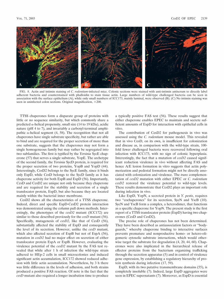
Challenged bacteria in the frozen colon sections were alsostained for intimin using immunofluorescence. Actin stainingwas used to counterstain the tissue. High numbers of chal-lenged bacteria were observed along the epithelial surface ofmice infected with the wild-type (Fig. 8) or complemented (notshown) strains. In contrast, and in agreement with the viablecounts, a very low level of challenged bacteria, mainly in thelumen, was observed in mice infected with ICC173 (Fig. 8).
DISCUSSION
The hallmark of EPEC infection is the A/E lesion histopa-thology. A/E lesion formation and intimate bacterial attach-
FIG. 5. FAS tests. After 1 h the wild-type strain adhered to HEp-2 cells in small microcolonies and induced disorganized actin accumulation,whereas strain ICC172 showed reduced adhesion with little actin accumulation. By 3 h however, both strains showed good adhesion and bothstrains produced discrete foci of actin accumulation beneath adherent bacteria, i.e., produced a positive FAS reaction.
VOL. 71, 2003 CesD2 OF EPEC 2137

ment are mediated by close interaction between the outermembrane adhesin intimin and the translocated intimin recep-tor, Tir, which is believed to be delivered to the host cellplasma membrane via the TTSS needle complex and throughEspA filaments and an EspD-associated translocation pore
(19). Similar to translocator proteins in other TTSSs, EspDwas reported to require a chaperone, CesD, for its propersecretion (56). Here we report that EspD is unique among theTTSS translocator proteins, in that it requires a second chap-erone, CesD2, for stabilization and secretion.
FIG. 6. Virulence of C. rodentium strains. (A) Data depict the number of C. rodentium CFU recovered from colonic tissues of individual mice,on day 13 postinfection. Mice infected with the wild-type strain had a high pathogen burden. In contrast, mice infected with the cesD2-mutant strain(ICC173) had lower bacterial counts, although the difference was not statistically significant (P � 0.05). In mutant strains complemented with eitherpICC245 or pICC246, the CFU level was restored to wild-type level. (B) The distal 8 cm of the colons were weighed 13 days postchallenge. Theweights of colons from mice infected with the wild-type (WT) strain or complemented mutant strains, carrying either pICC245 or pICC246, wereequivalent and significantly greater than those of colons from mice infected with the mutant strain and uninfected controls (P � 0.1). There wasno significant difference between the colon weights of uninfected mice and mice infected with ICC173 (P � 0.05).
FIG. 7. Hematoxylin and eosin-stained colonic frozen sections from C3H/Hej mice experimentally infected with C. rodentium. (A) Colonicsections from an uninfected mouse, showing normal colonic architecture. (B) Crypt hyperplasia and inflammation in a mouse infected with wild-type C.rodentium. (C) Infection with cesD2-minus derivative (ICC173), similarly to the uninfected control, revealed neither hyperplasia nor inflammatoryresponse. (D and E) Complementation of ICC173 with pICC245 (D) or pICC246 (E) restored both hyperplastic and inflammatory responses. Originalmagnification, 200.
2138 NEVES ET AL. INFECT. IMMUN.
TTSS chaperones form a disparate group of proteins withlittle or no sequence similarity, but which commonly share apredicted -helical propensity, small size (14 to 19 kDa), acidicnature (pH 4 to 7), and invariably a carboxyl-terminal amphi-pathic -helical segment (4, 58). The recognition that not allchaperones have single substrate specificity, but rather are ableto bind and are required for the proper secretion of more thanone substrate, suggests that the chaperones may not form asingle homogeneous family but may rather be segregated intotwo subfamilies. The first is typified by the Yersinia SycE chap-erone (57) that serves a single substrate, YopE. The archetypeof the second family, the Yersinia SycD protein, is required forthe proper secretion of two proteins, YopB and YopD (44).Interestingly, CesD2 belongs to the SycE family, since it bindsonly EspD, while CesD belongs to the SycD family as it haschaperone activity for both EspB and EspD (56). Importantly,CesD and CesD2 are unique not only because they chaperoneand are required for the stability and secretion of a singletranslocator protein, EspD, but also because they are locatedmainly within the bacterial inner membrane.
CesD2 shows all the characteristics of a TTSS chaperone.Indeed, direct and specific EspD-CesD2 protein interactionwas demonstrated using the column pull down methods. Inter-estingly, the phenotypes of the cesD2 mutant (ICC172) aresimilar to those described previously for the cesD mutant (56).Specifically, mutagenesis of cesD2, like that of CesD (56),substantially affected the stability of EspD and consequentlythe level of its secretion. However, unlike the cesD mutant,which also affected secretion of EspB but not of EspA (56),mutation in cesD2 had no major effect on secretion of eithertranslocator protein EspA or EspB. However, evaluating thevirulence potential of the cesD2 mutant by the FAS test re-vealed that while after 1 h of infection the wild-type strainadhered to HEp-2 cells in small microcolonies and inducedsignificant actin accumulation, ICC172 showed reduced adhe-sion with little actin accumulation. Nevertheless, by 3 h therewas little difference in the levels of adhesion, and both strainsproduced a positive FAS reaction. Of note is the fact that thecesD mutant also required a longer incubation time to produce
a typically positive FAS test (56). These results suggest thateither chaperone enables EPEC to maintain and secrete suf-ficient amounts of EspD for interaction with epithelial cells invitro.
The contribution of CesD2 for pathogenesis in vivo wasassessed using the C. rodentium mouse model. This revealedthat in vivo CesD, on its own, is insufficient for colonizationand disease as, in comparison with the wild-type strain, 100-fold fewer challenged bacteria were recovered following oralinfection with ICC173, with no sign of colonic hyperplasia.Interestingly, the fact that a mutation of cesD2 caused signif-icant reduction virulence in vivo without affecting FAS andhence A/E lesion formation in vitro suggests that actin poly-merization and pedestal formation might not be directly asso-ciated with colonization and virulence. The trans complemen-tation of cesD2 mutation with either EPEC or C. rodentiumcesD2 restored the virulence potential to wild-type levels.These results demonstrate that CesD2 plays an important roleduring infection in vivo.
Like EspD, YopN, a secreted protein in Y. pestis, requirestwo “cochaperones” for its secretion, SycN and YscB (10).SycN and YscB form a complex, a heterodimer, that functionsas a specific chaperone for YopN. The present study is the firstreport of a TTSS translocator protein (EspD) having two chap-erones (CesD and CesD2).
The precise role of chaperones has not been determined.They have been described as antiassociation factors or “body-guards,” whereby chaperone binding to interactive surfacesprevents premature and nonproductive homo- or hetero-oli-gomeric cytosolic substrate interactions, which would other-wise target the substrate for degradation (4, 20, 44, 60). Chap-erones were also implicated in the hierarchical release ofeffector proteins from the bacterium organizing traffickingthrough the secretion apparatus (5) and in control of virulencegene expression, by establishing a regulatory hierarchy of pro-tein synthesis during infection (17, 59).
EspD, with its two putative transmembrane domains (55), iscompletely insoluble (7). Indeed, large EspD aggregates wereseen in EPEC supernatants (7). Moreover, as EspD is essential
FIG. 8. Actin and intimin staining of C. rodentium-infected mice. Colonic sections were stained with anti-intimin antiserum to directly labeladherent bacteria and counterstained with phalloidin to stain tissue actin. Large numbers of wild-type challenged bacteria can be seen inassociation with the surface epithelium (A), while only small numbers of ICC173, mainly luminal, were observed (B). (C) No intimin staining wasseen in uninfected colon sections. Original magnification, 200.
VOL. 71, 2003 CesD2 OF EPEC 2139
for EspA filament biosynthesis (possibly by having a cap-likeactivity), it is likely to be the first protein to be secreted oncethe TTSS needle complex had been assembled. The reasonwhy EspD requires two chaperones is not clear. However, thismight be needed to keep EspD in a soluble state, to preventEspD-EspD (7) and possible EspD-EspA protein interactionsfrom occurring prematurely, while giving EspD an advantagein secretion over other translocator and effector proteins. Weare currently testing these hypotheses experimentally.
ACKNOWLEDGMENTS
This work was supported by the Wellcome Trust and the BBSRC.B.C.N. was supported by a fellowship from CNPy Brazil.
REFERENCES
1. Abe, A., M. de Grado, R. A. Pfuetzner, C. Sanchez-Sanmartin, R. Devinney,J. L. Puente, N. C. Strynadka, and B. B. Finlay. 1999. EnteropathogenicEscherichia coli translocated intimin receptor, Tir, requires a specific chap-erone for stable secretion. Mol. Microbiol. 33:1162–1175.
2. Adu-Bobie, J., G. Frankel, C. Bain, A. G. Goncaleves, L. R. Trabulsi, G.Douce, S. Knutton, and G. Dougan. 1998. Detection of intimin , �, �, and , four intimin derivatives expressed by attaching and effacing microbialpathogens. J. Clin. Microbiol. 36:662–668.
3. Batchelor, M., S. Knutton, V. Huter, M. Zanial, G. Dougan, and G. Frankel.1999. Development of a universal intimin antiserum and PCR primers.J. Clin. Microbiol. 37:3822–3827.
4. Bennett, J. C., and C. Hughes. 2000. From flagellum assembly to virulence:the extended family of type III export chaperones. Trends Microbiol. 8:202–204.
5. Boyd, A. P., I. Lambermont, and G. R. Cornelis. 2000. Competition betweenthe Yops of Yersinia enterocolitica for delivery into eukaryotic cells: role ofthe SycE chaperone binding domain of YopE. J. Bacteriol. 182:4811–4821.
6. Creasey, E. A., R. M. Delahay, A. A. Bishop, R. K. Shaw, B. Kenny, S.Knutton, and G. Frankel. 2002. CesT is a bivalent enteropathogenic Esch-erichia coli chaperone required for translocation of both Tir and Map. Mol.Microbiol. 47:209–221.
7. Daniell, S. J., R. M. Delahay, R. K. Shaw, E. L. Hartland, M. J. Pallen, F.Booy, F. Ebel, S. Knutton, and G. Frankel. 2001. The coiled coil domain ofenteropathogenic Escherichia coli type III secreted protein EspD is involvedin EspA filament-mediated cell attachment and hemolysis. Infect. Immun.69:4055–4064.
8. Daniell, S. J., N. Takahashi, R. Wilson, D. Friedberg, I. Rosenshine, F. P.Booy, R. K. Shaw, S. Knutton, G. Frankel, and S. Aizawa. 2001. The fila-mentous type III secretion translocon of enteropathogenic Escherichia coli.Cell. Microbiol. 3:865–871.
9. Datsenko, K. A., and B. L. Wanner. 2000. One-step inactivation of chromo-somal genes in Escherichia coli K12 using PCR products. Proc. Natl. Acad.Sci. USA 97:6640–6645.
10. Day, J. B., and G. V. Plano. 1998. A complex composed of SycN and YscBfunctions as a specific chaperone for YopN in Yersinia pestis. Mol. Microbiol.30:777–788.
11. Deng. W., Y. Li, B. A. Vallance, and B. B. Finlay. 2001. Locus of enterocyteeffacement from Citrobacter rodentium: sequence analysis and evidence forhorizontal transfer among attaching and effacing pathogens. Infect. Immun.69:6323–6335.
12. Donnenberg, M. S., J. Yu, and J. B. Kaper. 1993. A second chromosomalgene necessary for intimate attachment of enteropathogenic Escherichia colito epithelial cells. J. Bacteriol. 175:4670–4680.
13. Elliott, S. J., M. S. Dubois, S. W. Hutcheson, L. A. Wainwright, M. Batch-elor, G. Frankel, S. S. Knutton, and J. B. Kaper. 1999. Identification ofCesT, a chaperone for the type III secretion of Tir in EnteropathogenicEscherichia coli. Mol. Microbiol. 33:1176–1189.
14. Elliott, S. J., E. O. Krejany, J. L. Mellies, R. M. Robins-Browne, C.Sasakawa, and J. B. Kaper. 2001. EspG, a novel type III system-secretedprotein from enteropathogenic Escherichia coli with similarities to VirA ofShigella flexneri. Infect. Immun. 69:4027–4033.
15. Elliott, S. J., C. B. O’Connell, A. Koutsouris, C. Brinkley, M. S. Donnenberg,G. Hecht, and J. B. Kaper. 2002. A gene from the locus of enterocyteeffacement that is required for enteropathogenic Escherichia coli to increasetight-junction permeability encodes a chaperone for EspF. Infect. Immun.70:2271–2277.
16. Elliott, S. J., L. A. Wainwright, T. K. McDaniel, K. G. Jarvis, Y. K. Deng,L. C. Lai, B. P. McNamara, M. S. Donnenberg, and J. B. Kaper. 1998. Thecomplete sequence of the locus of enterocyte effacement (LEE) from en-teropathogenic Escherichia coli E2348/69. Mol. Microbiol 28:1–4.
17. Francis, M. S., S. A. Lloyd, and H. Wolf-Watz. 2001. The type III secretionchaperone LcrH co-operates with YopD to establish a negative, regulatory
loop for control of Yop synthesis in Yersinia pseudotuberculosis. Mol. Micro-biol. 42:1075–1093.
18. Frankel, G., A. D. Phillips, M. Novakova, H. Field, D. C. Candy, D. B.Schauer, G. Douce, and G. Dougan. 1996. Intimin from enteropathogenicEscherichia coli restores murine virulence to a Citrobacter rodentium eaeAmutant: induction of an immunoglobulin A response to intimin and EspB.Infect. Immun. 64:5315–5325.
19. Frankel, G., A. D. Phillips, I. Rosenshine, G. Dougan, J. B. Kaper, and S.Knutton. 1998. Enteropathogenic and enterohaemorrhagic Escherichia coli:more subversive elements. Mol. Microbiol. 30:911–921.
20. Frithz-Lindsten, E., R. Rosqvist, L. Johansson, and A. Forsberg. 1995. Thechaperone-like protein YerA of Yersinia pseudotuberculosis stabilizes YopEin the cytoplasm but is dispensable for targeting to the secretion loci. Mol.Microbiol. 16:635–647.
21. Galan, J. E., C. Ginocchio, and P. Costeas. 1992. Molecular and functionalcharacterization of the Salmonella invasion gene invA: homology of InvA tomembers of a new protein family. J. Bacteriol. 174:4338–4349.
22. Ghaem-Maghami, M., C. P. Simmons, S. Daniell, M. Pizza, D. Lewis, G.Frankel, and G. Dougan. 2001. Intimin-specific immune responses preventbacterial colonization by the attaching-effacing pathogen Citrobacter roden-tium. Infect. Immun. 69:5597–5605.
23. Hueck, C. J. 1998. Type III protein secretion systems in bacterial pathogensof animals and plants. Microbiol. Mol. Biol. Rev. 62:379–433.
24. Ilan, O., Y. Bloch, G. Frankel, H. Ullrich, K. Geider, and I. Rosenshine.1999. Protein tyrosine kinases in bacterial pathogens are associated withvirulence and production of exopolysaccharide. EMBO J. 18:3241–3248.
25. Jarvis, K. G., J. A. Giron, A. E. Jerse, T. K. McDaniel, M. S. Donnenberg,and J. B. Kaper. 1995. Enteropathogenic Escherichia coli contains a putativetype III secretion system necessary for the export of proteins involved inattaching and effacing lesion formation. Proc. Natl. Acad. Sci. USA 92:7996–8000.
26. Jerse, A. E., J. Yu, B. D. Tall, and J. B. Kaper. 1990. A genetic locus ofenteropathogenic Escherichia coli necessary for the production of attachingand effacing lesions on tissue culture cells. Proc. Natl. Acad. Sci. USA87:7839–7843.
27. Kellermann, O., and S. Szmelcman. 1974. Active transport of maltose inEscherichia coli K12. Involvement of a “periplasmic” maltose binding pro-tein. Eur. J. Biochem. 47:139–149.
28. Kenny, B., R. DeVinney, M. Stein, D. J. Reinscheid, E. A. Frey, and B. B.Finlay. 1997. Enteropathogenic E. coli (EPEC) transfers its receptor forintimate adherence into mammalian cells. Cell 91:511–520.
29. Kenny, B., and M. Jepson. 2000. Targeting of an enteropathogenic Esche-richia coli (EPEC) effector protein to host mitochondria. Cell. Microbiol.2:579–590.
30. Kenny, B., L. C. Lai, B. B. Finlay, and M. S. Donnenberg. 1996. EspA, aprotein secreted by enteropathogenic Escherichia coli, is required to inducesignals in epithelial cells. Mol. Microbiol. 20:313–323.
31. Knutton, S., D. R. Lloyd, and A. S. McNeish. 1987. Adhesion of entero-pathogenic Escherichia coli to human intestinal enterocytes and culturedhuman intestinal mucosa. Infect. Immun. 55:69–77.
32. Knutton, S., I. Rosenshine, M. J. Pallen, I. Nisan, B. C. Neves, C. Bain, C.Wolff, G. Dougan, and G. Frankel. 1998. A novel EspA-associated surfaceorganelle of enteropathogenic Escherichia coli involved in protein translo-cation into epithelial cells. EMBO J. 17:2166–2176.
33. Kubori, T., Y. Matsushima, D. Nakamura, J. Uralil, M. Lara-Tejero, A.Sukhan, J. E. Galan, and S. I. Aizawa. 1998. Supramolecular structure of theSalmonella typhimurium type III protein secretion system. Science 280:602–605.
34. Lai, L. C., L. A. Wainwright, K. D. Stone, and M. S. Donnenberg. 1997. Athird secreted protein that is encoded by the enteropathogenic Escherichiacoli pathogenicity island is required for transduction of signals and forattaching and effacing activities in host cells. Infect. Immun. 65:2211–2217.
35. Lee, C., P. Li, H. Inouye, E. R. Brickman, and J. Beckwith. 1989. Geneticstudies on the inability of �-galactosidase to be translocated across theEscherichia coli cytoplasmic membrane. J. Bacteriol. 171:4609–4616.
36. Levine, M. M., E. J. Berquist, D. R. Nalin, D. H. Waterman, R. B. Hornick,C. R. Young, S. Stoman, and B. Rowe. 1978. Escherichia coli that causediarrhoea but do not produce heat-labile or heat-stable enterotoxins and arenon-invasive. Lancet i:119–122.
37. McDaniel, T. K., K. G. Jarvis, M. S. Donnenberg, and J. B. Kaper. 1995. Agenetic locus of enterocyte effacement conserved among diverse enterobac-terial pathogens. Proc. Natl. Acad. Sci. USA 92:1664–1668.
38. McNamara, B. P., and M. S. Donnenberg. 1998. A novel proline-rich protein,EspF, is secreted from enteropathogenic Escherichia coli via the type IIIexport pathway. FEMS Microbiol. Lett. 166:71–78.
39. McNamara, B. P., A. Koutsouris, C. B. O’Connell, J. P. Nougayrede, M. S.Donnenberg, and G. Hecht. 2000. Translocated EspF protein from entero-pathogenic Escherichia coli disrupts host intestinal barrier function. J. Clin.Investig. 107:621–629.
40. Mellies, J. L., S. J. Elliott, V. Sperandio, M. S. Donnenberg, and J. B. Kaper.1999. The Per regulon of enteropathogenic Escherichia coli: identification ofa regulatory cascade and a novel transcriptional activator, the locus of en-
2140 NEVES ET AL. INFECT. IMMUN.
terocyte effacement (LEE)-encoded regulator (Ler). Mol. Microbiol. 33:296–306.
41. Moon, H. W., S. C. Whipp, R. A. Argenzio, M. M. Levine, and R. A. Gian-nella. 1983. Attaching and effacing activities of rabbit and human entero-pathogenic Escherichia coli in pig and rabbit intestines. Infect. Immun. 41:1340–1351.
42. Nataro, J. P., and J. B. Kaper. 1998. Diarrheagenic Escherichia coli. Clin.Microbiol. Rev. 11:142–201.
43. Newman, J. V., B. A. Zabel, S. S. Jha, and D. B. Schauer. 1999. Citrobacterrodentium espB is necessary for signal transduction and for infection oflaboratory mice. Infect. Immun. 67:6019–6025.
44. Neyt, C., and G. R. Cornelis. 1999. Role of SycD, the chaperone of theYersinia Yop translocators YopB and YopD. Mol. Microbiol. 31:143–156.
45. Robins-Browne, R. M., A. M. Tokhi, L. M. Adams, V. Bennett-Wood, A. V.Moisidis, E. O. Krejany, and L. E. O’Gorman. 1994. Adherence character-istics of attaching and effacing strains of Escherichia coli from rabbits. Infect.Immun. 62:1584–1592.
46. Rosenshine, I., S. Ruschkowski, M. Stein, D. J. Reinscheid, S. D. Mills, andB. B. Finlay. 1996. A pathogenic bacterium triggers epithelial signals to forma functional bacterial receptor that mediates actin pseudopod formation.EMBO J. 15:2613–2624.
47. Schauer, D. B., and S. Falkow. 1993. Attaching and effacing locus of aCitrobacter freundii biotype 4280 that causes transmissible murine colonichyperplasia. Infect. Immun. 61:2486–2492.
48. Schauer, D. B., and S. Falkow. 1993. The eae gene of Citrobacter freundiibiotype 4280 is necessary for colonization in transmissible murine colonichyperplasia. Infect. Immun. 61:4654–4661.
49. Sekiya, K., M. Ohishi, T. Ogino, K. Tamano, C. Sasakawa, and A. Abe. 2001.Supermolecular structure of the enteropathogenic Escherichia coli type IIIsecretion system and its direct interaction with the EspA-sheath-like struc-ture. Proc. Natl. Acad. Sci. USA 98:11638–11643.
50. Simmons, C. P., N. S. Goncalves, M. Ghaem-Maghami, M. Bajaj-Elliott, S.Clare, B. Neves, G. Frankel, G. Dougan, and T. T. MacDonald. 2002. Im-paired resistance and enhanced pathology during infection with a noninva-sive, attaching-effacing enteric bacterial pathogen. Citrobacter rodentium, inmice lacking IL-12 or IFN-gamma. J. Immunol. 15:1804–1812.
51. Tamano, K., S. Aizawa, E. Katayama, T. Nonaka, S. Imajoh-Ohmi, A. Ku-wae, S. Nagai, and C. Sasakawa. 2000. Supramolecular structure of theShigella type III secretion machinery: the needle part is changeable in lengthand essential for delivery of effectors. 19 EMBO J. 3876–3887.
52. Taylor, K. A., P. W. Luther, and M. S. Donnenberg. 1999. Expression of theEspB protein of enteropathogenic Escherichia coli within HeLa cells affectsstress fibers and cellular morphology. Infect. Immun. 67:120–125.
53. Towbin, H., T. Staehelin, and J. Gordon. 1979. Electrophoretic transfer ofproteins from polyacrylamide gels to nitrocellulose sheets: procedure andsome applications. Proc. Natl. Acad. Sci. USA 76:4350–4354.
54. Tzipori, S., I. K. Wachsmuth, C. Chapman, R. Birden, J. Brittingham, C.Jackson, and J. Hogg. 1986. The pathogenesis of hemorrhagic colitis causedby Escherichia coli O157:H7 in gnotobiotic piglets. J. Infect. Dis. 154:712–714.
55. Wachter, C., C. Beinke, M. Mattes, and M. A. Schmidt. 1999. Insertion ofEspD into epithelial target cell membranes by infecting enteropathogenicEscherichia coli. Mol. Microbiol. 31:1695–1707.
56. Wainwright, L. A., and J. B. Kaper. 1998. EspB and EspD require a specificchaperone for proper secretion from enteropathogenic Escherichia coli. Mol.Microbiol. 27:1247–1260.
57. Wattiau, P., and G. R. Cornelis. 1993. SycE, a chaperone-like protein ofYersinia enterocolitica involved in the secretion of YopE. Mol. Microbiol.8:123–131.
58. Wattiau, P., S. Woestyn, and G. R. Cornelis. 1996. Customized secretionchaperones in pathogenic bacteria. Mol. Microbiol. 20:255–262.
59. Williams, A. W., and S. C. Straley. 1998. YopD of Yersinia pestis plays a rolein negative regulation of the low-calcium response in addition to its role intranslocation of Yops. J. Bacteriol. 180:350–358.
60. Woestyn, S., M. P. Sory, A. Boland, O. Lequenne, and G. R. Cornelis. 1996.The cytosolic SycE and SycH chaperones of Yersinia protect the region ofYopE and YopH involved in translocation across eukaryotic cell mem-branes. Mol. Microbiol. 20:1261–1271.
61. Wolff, C., I. Nisan, E. Hanski, G. Frankel, and I. Rosenshine. 1998. Proteintranslocation into HeLa cells by infecting enteropathogenic Escherichia coli.Mol. Microbiol. 28:143–155.
Editor: J. T. Barbieri
VOL. 71, 2003 CesD2 OF EPEC 2141
Copyright © 2022 FDOKUMEN