Cerebral glycolysis: a century of persistent misunderstanding ...
18
REVIEW ARTICLE published: 19 November 2014 doi: 10.3389/fnins.2014.00360 Cerebral glycolysis: a century of persistent misunderstanding and misconception Avital Schurr* Department of Anesthesiology and Perioperative Medicine, University of Louisville School of Medicine, Louisville, KY, USA Edited by: Pierre J. Magistretti, École Polytechnique Fédérale de Lausanne, Switzerland Reviewed by: Juan P. Bolanos, University of Salamanca-Consejo Superior de Investigaciones Científicas, Spain Igor Allaman, École Polytechnique Fédérale de Lausanne, Switzerland (in collaboration with Pierre J. Magistretti) *Correspondence: Avital Schurr, Department of Anesthesiology and Perioperative Medicine, University of Louisville School of Medicine, Louisville, KY 40202, USA e-mail: [email protected] Since its discovery in 1780, lactate (lactic acid) has been blamed for almost any illness outcome in which its levels are elevated. Beginning in the mid-1980s, studies on both muscle and brain tissues, have suggested that lactate plays a role in bioenergetics. However, great skepticism and, at times, outright antagonism has been exhibited by many to any perceived role for this monocarboxylate in energy metabolism. The present review attempts to trace the negative attitudes about lactate to the first four or five decades of research on carbohydrate metabolism and its dogma according to which lactate is a useless anaerobic end-product of glycolysis. The main thrust here is the review of dozens of scientific publications, many by the leading scientists of their times, through the first half of the twentieth century. Consequently, it is concluded that there exists a barrier, described by Howard Margolis as “habit of mind,” that many scientists find impossible to cross. The term suggests “entrenched responses that ordinarily occur without conscious attention and that, even if noticed, are hard to change.” Habit of mind has undoubtedly played a major role in the above mentioned negative attitudes toward lactate. As early as the 1920s, scientists investigating brain carbohydrate metabolism had discovered that lactate can be oxidized by brain tissue preparations, yet their own habit of mind redirected them to believe that such an oxidation is simply a disposal mechanism of this “poisonous” compound. The last section of the review invites the reader to consider a postulated alternative glycolytic pathway in cerebral and, possibly, in most other tissues, where no distinction is being made between aerobic and anaerobic glycolysis; lactate is always the glycolytic end product. Aerobically, lactate is readily shuttled and transported into the mitochondrion, where it is converted to pyruvate via a mitochondrial lactate dehydrogenase (mLDH) and then is entered the tricarboxylic acid (TCA) cycle. Keywords: cerebral energy metabolism, glycolysis, lactate, mitochondrial LDH, NAD-NADH recycling, habit of mind INTRODUCTION More than 70 years ago, the identity and sequence of the reac- tions of glycolysis, also known as the Embden-Meyerhof pathway, were elucidated. Nevertheless, for the past 25 years investigators in the field of brain energy metabolism have been hotly debat- ing the details of that sequence. A somewhat similar debate first took place among exercise physiologists and biochemists when Brooks (1985) published results showing that lactic acid (lac- tate) is the glycolytic product and the oxidative substrate during sustained exercise. Soon thereafter, a few studies by neurosci- entists questioned the status quo in our understanding of how the brain handles increased energy requirements during stimu- lation. First, Fox and Raichle (1986) demonstrated a focal phys- iological uncoupling between cerebral blood flow and oxidative metabolism upon somatosensory stimulation in humans. Two years later Fox et al. (1988) showed that during focal physio- logic neural activity the consumption of glucose is non-oxidative. Simultaneously, Schurr et al. (1988) demonstrated the ability of brain (hippocampal) slices to maintain normal synaptic func- tion with lactate as the sole oxidative energy substrate. Many scientists in the field were surprised by these findings, while others discounted them (Chih et al., 2001; Dienel and Hertz, 2001; Chih and Roberts, 2003; Dienel and Cruz, 2004; Hertz, 2004; Fillenz, 2005). Despite the allowance of time necessary for new findings to overcome “habits of mind” (Margolis, 1993) or the incommen- surability of “new” and “old” paradigms (Kuhn, 1996), the great debate has not subsided. Hence, lines have been drawn between two camps; one, still a majority, which discounts any key role for lactate in brain (and muscle) energy metabolism and another, a growing minority, which holds lactate as an important, and at times, crucial, oxidative substrate for energy production in the brain (and other tissues). The unusual longevity of this debate is somewhat surprising. Being on the minority side of it, I have been intrigued by both its persistence and its emotional flair. The drive to settle the unre- solved issues that continue to sustain this debate has prompted the following review of the recorded research on energy metabolism through the formative years of the field of biochemistry during the first half of the twentieth century. The aim of this review has been to uncover the basis and reasoning for lactate’s long-lasting negative reputation among scientists and clinicians that has pre- vented its “rehabilitation” and thus its consideration as an integral www.frontiersin.org November 2014 | Volume 8 | Article 360 | 1
-
Upload
khangminh22 -
Category
Documents
-
view
0 -
download
0
Transcript of Cerebral glycolysis: a century of persistent misunderstanding ...
REVIEW ARTICLEpublished: 19 November 2014doi: 10.3389/fnins.2014.00360
Cerebral glycolysis: a century of persistentmisunderstanding and misconceptionAvital Schurr*
Department of Anesthesiology and Perioperative Medicine, University of Louisville School of Medicine, Louisville, KY, USA
Edited by:
Pierre J. Magistretti, ÉcolePolytechnique Fédérale deLausanne, Switzerland
Reviewed by:
Juan P. Bolanos, University ofSalamanca-Consejo Superior deInvestigaciones Científicas, SpainIgor Allaman, École PolytechniqueFédérale de Lausanne, Switzerland(in collaboration with Pierre J.Magistretti)
*Correspondence:
Avital Schurr, Department ofAnesthesiology and PerioperativeMedicine, University of LouisvilleSchool of Medicine, Louisville, KY40202, USAe-mail: [email protected]
Since its discovery in 1780, lactate (lactic acid) has been blamed for almost any illnessoutcome in which its levels are elevated. Beginning in the mid-1980s, studies on bothmuscle and brain tissues, have suggested that lactate plays a role in bioenergetics.However, great skepticism and, at times, outright antagonism has been exhibited by manyto any perceived role for this monocarboxylate in energy metabolism. The present reviewattempts to trace the negative attitudes about lactate to the first four or five decadesof research on carbohydrate metabolism and its dogma according to which lactate is auseless anaerobic end-product of glycolysis. The main thrust here is the review of dozensof scientific publications, many by the leading scientists of their times, through the firsthalf of the twentieth century. Consequently, it is concluded that there exists a barrier,described by Howard Margolis as “habit of mind,” that many scientists find impossible tocross. The term suggests “entrenched responses that ordinarily occur without consciousattention and that, even if noticed, are hard to change.” Habit of mind has undoubtedlyplayed a major role in the above mentioned negative attitudes toward lactate. As earlyas the 1920s, scientists investigating brain carbohydrate metabolism had discovered thatlactate can be oxidized by brain tissue preparations, yet their own habit of mind redirectedthem to believe that such an oxidation is simply a disposal mechanism of this “poisonous”compound. The last section of the review invites the reader to consider a postulatedalternative glycolytic pathway in cerebral and, possibly, in most other tissues, where nodistinction is being made between aerobic and anaerobic glycolysis; lactate is alwaysthe glycolytic end product. Aerobically, lactate is readily shuttled and transported into themitochondrion, where it is converted to pyruvate via a mitochondrial lactate dehydrogenase(mLDH) and then is entered the tricarboxylic acid (TCA) cycle.
Keywords: cerebral energy metabolism, glycolysis, lactate, mitochondrial LDH, NAD-NADH recycling, habit of mind
INTRODUCTIONMore than 70 years ago, the identity and sequence of the reac-tions of glycolysis, also known as the Embden-Meyerhof pathway,were elucidated. Nevertheless, for the past 25 years investigatorsin the field of brain energy metabolism have been hotly debat-ing the details of that sequence. A somewhat similar debate firsttook place among exercise physiologists and biochemists whenBrooks (1985) published results showing that lactic acid (lac-tate) is the glycolytic product and the oxidative substrate duringsustained exercise. Soon thereafter, a few studies by neurosci-entists questioned the status quo in our understanding of howthe brain handles increased energy requirements during stimu-lation. First, Fox and Raichle (1986) demonstrated a focal phys-iological uncoupling between cerebral blood flow and oxidativemetabolism upon somatosensory stimulation in humans. Twoyears later Fox et al. (1988) showed that during focal physio-logic neural activity the consumption of glucose is non-oxidative.Simultaneously, Schurr et al. (1988) demonstrated the ability ofbrain (hippocampal) slices to maintain normal synaptic func-tion with lactate as the sole oxidative energy substrate. Manyscientists in the field were surprised by these findings, while others
discounted them (Chih et al., 2001; Dienel and Hertz, 2001; Chihand Roberts, 2003; Dienel and Cruz, 2004; Hertz, 2004; Fillenz,2005). Despite the allowance of time necessary for new findingsto overcome “habits of mind” (Margolis, 1993) or the incommen-surability of “new” and “old” paradigms (Kuhn, 1996), the greatdebate has not subsided. Hence, lines have been drawn betweentwo camps; one, still a majority, which discounts any key role forlactate in brain (and muscle) energy metabolism and another, agrowing minority, which holds lactate as an important, and attimes, crucial, oxidative substrate for energy production in thebrain (and other tissues).
The unusual longevity of this debate is somewhat surprising.Being on the minority side of it, I have been intrigued by bothits persistence and its emotional flair. The drive to settle the unre-solved issues that continue to sustain this debate has prompted thefollowing review of the recorded research on energy metabolismthrough the formative years of the field of biochemistry duringthe first half of the twentieth century. The aim of this review hasbeen to uncover the basis and reasoning for lactate’s long-lastingnegative reputation among scientists and clinicians that has pre-vented its “rehabilitation” and thus its consideration as an integral
www.frontiersin.org November 2014 | Volume 8 | Article 360 | 1
Schurr Glycolysis at 75 - a dogma reconsidered
part of oxidative energy metabolism. Suspicions that lactate’s illreputation has contributed greatly to its dismissal as anything, butuseless end-product of anaerobic energy metabolism, led me tosearch for recorded hints to discount any such suspicions. Uponreading through the troves of research papers of the past, it isclear that one cannot separate the science from the scientists whopractice it. Disagreements among investigators in the fields ofmuscle and brain energy metabolism had already existed in theearly decades of the twentieth century. Lactate, by the majority ofinterpretations of research results, had been considered for a longtime to be a product that must be disposed of in order to achievenormalization of tissue functioning. This is despite findings byseveral investigators of brain energy metabolism in the 1920 and1930s, who demonstrated the ability, especially of brain gray mat-ter, to oxidize lactate. With an emphasis on glycolysis, this paperattempts to sort out as many as possible conceptions and miscon-ceptions about (brain) energy metabolism in the formative yearsof modern biochemistry. A plausible explanation is proposed forhow that great leap in knowledge, which occurred over sevendecades ago, and the research that led to it, have shaped minds andbeliefs both then and now. Guidance from the wisdom of threephilosophers, Barber (1961), Kuhn (1996) and Margolis (1993)has been instrumental in this attempt to understand how scien-tific concepts and beliefs have determined both the direction andthe pace of scientific progress in the field of energy metabolism.
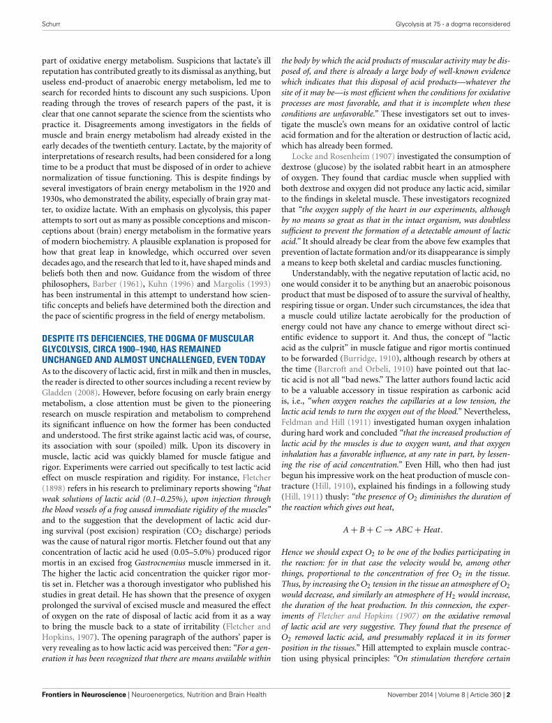
DESPITE ITS DEFICIENCIES, THE DOGMA OF MUSCULARGLYCOLYSIS, CIRCA 1900–1940, HAS REMAINEDUNCHANGED AND ALMOST UNCHALLENGED, EVEN TODAYAs to the discovery of lactic acid, first in milk and then in muscles,the reader is directed to other sources including a recent review byGladden (2008). However, before focusing on early brain energymetabolism, a close attention must be given to the pioneeringresearch on muscle respiration and metabolism to comprehendits significant influence on how the former has been conductedand understood. The first strike against lactic acid was, of course,its association with sour (spoiled) milk. Upon its discovery inmuscle, lactic acid was quickly blamed for muscle fatigue andrigor. Experiments were carried out specifically to test lactic acideffect on muscle respiration and rigidity. For instance, Fletcher(1898) refers in his research to preliminary reports showing “thatweak solutions of lactic acid (0.1–0.25%), upon injection throughthe blood vessels of a frog caused immediate rigidity of the muscles”and to the suggestion that the development of lactic acid dur-ing survival (post excision) respiration (CO2 discharge) periodswas the cause of natural rigor mortis. Fletcher found out that anyconcentration of lactic acid he used (0.05–5.0%) produced rigormortis in an excised frog Gastrocnemius muscle immersed in it.The higher the lactic acid concentration the quicker rigor mor-tis set in. Fletcher was a thorough investigator who published hisstudies in great detail. He has shown that the presence of oxygenprolonged the survival of excised muscle and measured the effectof oxygen on the rate of disposal of lactic acid from it as a wayto bring the muscle back to a state of irritability (Fletcher andHopkins, 1907). The opening paragraph of the authors’ paper isvery revealing as to how lactic acid was perceived then: “For a gen-eration it has been recognized that there are means available within
the body by which the acid products of muscular activity may be dis-posed of, and there is already a large body of well-known evidencewhich indicates that this disposal of acid products—whatever thesite of it may be—is most efficient when the conditions for oxidativeprocesses are most favorable, and that it is incomplete when theseconditions are unfavorable.” These investigators set out to inves-tigate the muscle’s own means for an oxidative control of lacticacid formation and for the alteration or destruction of lactic acid,which has already been formed.
Locke and Rosenheim (1907) investigated the consumption ofdextrose (glucose) by the isolated rabbit heart in an atmosphereof oxygen. They found that cardiac muscle when supplied withboth dextrose and oxygen did not produce any lactic acid, similarto the findings in skeletal muscle. These investigators recognizedthat “the oxygen supply of the heart in our experiments, althoughby no means so great as that in the intact organism, was doubtlesssufficient to prevent the formation of a detectable amount of lacticacid.” It should already be clear from the above few examples thatprevention of lactate formation and/or its disappearance is simplya means to keep both skeletal and cardiac muscles functioning.
Understandably, with the negative reputation of lactic acid, noone would consider it to be anything but an anaerobic poisonousproduct that must be disposed of to assure the survival of healthy,respiring tissue or organ. Under such circumstances, the idea thata muscle could utilize lactate aerobically for the production ofenergy could not have any chance to emerge without direct sci-entific evidence to support it. And thus, the concept of “lacticacid as the culprit” in muscle fatigue and rigor mortis continuedto be forwarded (Burridge, 1910), although research by others atthe time (Barcroft and Orbeli, 1910) have pointed out that lac-tic acid is not all “bad news.” The latter authors found lactic acidto be a valuable accessory in tissue respiration as carbonic acidis, i.e., “when oxygen reaches the capillaries at a low tension, thelactic acid tends to turn the oxygen out of the blood.” Nevertheless,Feldman and Hill (1911) investigated human oxygen inhalationduring hard work and concluded “that the increased production oflactic acid by the muscles is due to oxygen want, and that oxygeninhalation has a favorable influence, at any rate in part, by lessen-ing the rise of acid concentration.” Even Hill, who then had justbegun his impressive work on the heat production of muscle con-tracture (Hill, 1910), explained his findings in a following study(Hill, 1911) thusly: “the presence of O2 diminishes the duration ofthe reaction which gives out heat,
A + B + C → ABC + Heat.
Hence we should expect O2 to be one of the bodies participating inthe reaction: for in that case the velocity would be, among otherthings, proportional to the concentration of free O2 in the tissue.Thus, by increasing the O2 tension in the tissue an atmosphere of O2
would decrease, and similarly an atmosphere of H2 would increase,
iments of Fletcher and Hopkins (1907) on the oxidative removalof lactic acid are very suggestive. They found that the presence ofO2 removed lactic acid, and presumably replaced it in its formerposition in the tissues.” Hill attempted to explain muscle contrac-tion using physical principles: “On stimulation therefore certain
Frontiers in Neuroscience | Neuroenergetics, Nutrition and Brain Health November 2014 | Volume 8 | Article 360 | 2
the duration of the heat production. In this connexion, the exper-
Schurr Glycolysis at 75 - a dogma reconsidered
molecules are thrown into solution, which before stimulation werelightly connected in some physical or chemical way with other bod-ies, so as to be inactive. The presence of these chemical moleculessets up a tension, possibly at certain colloidal membranes in thefiber: the tension falls again, owing to the diffusion of these chem-ical molecules into the general free space in the fiber, away from thesensitive membranes; the molecules are then oxidized, or replaced intheir original positions, under the action of O2, with an evolution ofheat proportional to the amount of those bodies present.”
In another study, Fletcher (1911) went after conflicting “evi-dence” regarding the chemical action involved in the formationof lactic acid in muscle and in other cells. He also justified hisefforts since “it has been urged by several observers in recent yearsthat considerable and continued production of d-lactic acid (theold nomenclature of L-lactic acid) maybe found during autoly-sis (aseptic or antiseptic) of minced or crushed muscle long afterthe extinction of irritability and destruction of structure.” Not sur-prising, “observers” were making a connection between muscledamage, its death and the production of lactic acid. Fletcher con-cluded from his studies that “the evidence hitherto produced of anautolytic production of lactic acid by muscle cannot be accepted.”Somewhat surprising conclusion in Fletcher’s study is that “noglycolytic enzyme leading to lactic acid formation appears to existin muscle. After the addition of dextrose to intact surviving mus-cle, or to preparations of disintegrated muscle, no increase of lacticacid is found in the absence of bacteria.” Peters (1913), using Hill’scalorimeter for heat production measurements combined withthose of lactic acid production confirmed both Hill’s (1911) andFletcher and Hopkins’s (1907) findings, concluding that “heatproduction and lactic acid liberation in fatiguing amphibian mus-cle are extremely intimately connected.” Later, Fletcher (1913)repeated his amphibian muscle studies with several mammalianmuscles, essentially concluding that muscles from both of thesesources are similar in their survival respiration and lactic acid pro-duction. That very year, Hill (1913) published results on muscleheat production using a newly designed and constructed “thermo-electric apparatus with which it was possible to estimate very rapidlythe rise of temperature of muscle, if necessary to within a millionthof a degree.” In the summary of his paper Hill suggested “that theprocesses of muscular contraction are due to liberation of lactic acidfrom some precursor, and that the lactic acid increases the tension insome colloidal structure of the tissue: that the lactic acid precursoris rebuilt after the contraction is over in the presence of, and by theuse of oxygen, with the evolution of heat: and finally that the heatliberated by the muscle excited in the complete absence of oxygen isdue simply to the breakdown of the lactic acid precursor, and is thesame in nature as the heat-production of rigor.”
Again, the conclusion was that lactic acid induces muscle con-traction via a physico-chemical process and, if not disposed of,would result in fatigue and rigor mortis. Roaf (1914) employedan electro-chemical method that recorded increases in aciditywhen muscle contracts. He, too, concluded “that the increase inacidity is the cause of the shortening of muscle.” Moreover, usinghis heat production measurements of muscle contraction, Hillpresented calculations and arguments in the Proceedings of thePhysiological Society on February 14, 1914 (Hill, 1914) in supportof the hypothesis that lactic acid formed in the muscle after
activity is not removed by the process of oxidation, but ratherby a process of replacement into its previous position (sugar).He thus argued as follows: “The production of 1 grm of lactic acidis accompanied by the evolution of about 450 calories. Now I haveshown that during the recovery processes of muscles in oxygen thereis a ‘recovery heat-production’ of about the same order of size as theheat-production occurring in the initial processes of contraction. Inthe oxidative removal of 1 grm of lactic acid therefore there is a heat-production of about 450 calories. Now, the oxidation of 1 grm oflactic acid leads to heat-production of about 3700 calories, whichis about eight times as large as the quantity observed. . . Therefore,apparently the lactic acid is not oxidized but replaced in its previousposition under the influence and with the energy of the oxidation,either (a) of a small part of the lactic acid itself, or (b) of some otherbody. Evidence given elsewhere shows that it must be some otherbody. The lactic acid therefore is part of the machine and not partof the fuel.” Hill voiced his position and, eventually, the positionof the majority of his colleagues, that lactic acid is not a fuel, sincethe expected heat-production of its oxidation was much lowerthan the calculated value of its complete combustion. It is surpris-ing that Hill would argue that if lactate were a fuel, all the energyof its oxidation would be released as heat. In essence, Hill’s ownmeasurements that lactic acid oxidation produces only 12% of theexpected heat-production should have indicated to him and oth-ers that the majority of the energy released from this oxidation,not measured as heat, could indicate controlled utilization and/orpossibly a conversion to some other forms of energy. Nevertheless,Hill’s and others’ prevailing position on “lactic acid is not a fuel”has endured to the present day.
Fletcher and Brown (1914) also looked into the Inogen theory,according to which, the discharge of energy by the muscle cell—and by inference its discharge by any other cell—depends uponthe dissociative breakdown of some labile molecule (Inogen). Forthe breakdown to occur, oxygen takes Inogen’s place beforehandin such a manner that upon the dissociation of the moleculethe energy yielded is due to combustion, and the final products,carbonic acid and water, represent the result of that combus-tion. Furthermore, lactic acid, which supposedly also arises fromInogen, was considered to be either another final product or anintermediate product destined to be used in future reconstruc-tion of the Inogen complex. Based on their experimental resultsFletcher and Brown concluded that CO2 and lactic acid do notoriginate from a common source. They emphatically asserted“that in the muscle the respiratory oxidative process yielding CO2
as an immediate product has its chief end in the supply of energyfor replacing the lactic acid in the molecular position from whichstimulation of some kind has displaced it, and there appears tobe no reason at present for supposing that the material which isoxidized in the respiratory process is the same as, or related to,the material from which lactic acid appears and into which, as itseems, it may again disappear.” Hence, the leading investigatorsin the field held that lactic acid is a separate entity from theone that is oxidized during muscle respiration and which yieldsenergy and CO2.
Meanwhile, other investigators were attempting to explaincocaine’s racking effects on its users or the devastation of diabetesthrough the increased tissue production of lactate. Underhill and
www.frontiersin.org November 2014 | Volume 8 | Article 360 | 3
Schurr Glycolysis at 75 - a dogma reconsidered
Black (1912) studied the influence of cocaine on the metabolismof dogs and rabbits receiving daily injections of the drug. Withdaily doses of cocaine (20 mg/kg), lactic acid excretion in theurine was markedly increased in well-fed animals. The investi-gators concluded that the increase in lactic acid elimination inthe urine is unlikely associated with increased muscular activityinduced by the drug. They also stated that “lactic acid and carbo-hydrate metabolism are presumably intimately associated althoughthere are indications that lactic acid may at times arise frommore than a single antecedent.” This statement appears to be anattempt to tie lactic acid to another process besides carbohy-drate metabolism, one that might be responsible for the effectof cocaine. Where diabetes was concerned, the famous Ringer(1914) stated that “parallelism exists between the degree of acido-sis and the degree of disturbance in the carbohydrate metabolism.”Considering the fact that at that time the role of insulin wasunknown and glycolysis was yet to be elucidated, the repeated useof this kind of statements about lactic acid and acidosis was partof an accepted and, almost expected, vernacular. Interestingly,Ringer theorized that diabetic mechanism involves the inabilityto form the glucoside bond of glycogen. Marriott (1914) in hisquantitative study of blood acidosis in diabetes cited a discussionof diabetic acidosis by Magnus-Levy in John Hopkins HospitalBulletin (Magnus-Levy, 1911) who expressed the view that the“acid poisoned animal and the diabetic patient do not die from theacid which has been eliminated in the neutralized state, but from theacid which remains in the body.”
By 1916, the general consensus had been “that sugar is utilizedby muscle as a source of energy and the main product of its activityis carbon dioxide” (Tsuji, 1916). Consensus had not yet achieved“with regard to the origin of lactic acid formed in the tissues. Someauthors ascribe it to the disintegration of carbohydrates (glucose),while others suggest that deaminization of amino acids (alanine)is its source.” Tsuji (1916), a researcher from Kyoto, Japan, work-ing at the Institute of Physiology, University College, England,employed a heart lung preparation in his studies and summa-rized his findings as followed: “1. Lactic acid is produced in thecirculating blood of the heart lung preparation under conditionsapproaching normal. 2. These results may indicate that lactic acidis one of the normal metabolites of muscular activity. 3. The for-mation of lactic acid is increased in poisoning of chloroform and inthe presence of deficient supply of oxygen in a heart lung prepara-tion. 4. When the heart beat is accelerated by adding adrenalin oramino-acid (alanine, glycine or ereptone) to the circulating blood,or the heart work is increased by alterations in the blood-pressure,lactic acid is not only not produced, but the lactic acid previouslycontained in the circulating blood disappears.” Finding number 4of Tusji might be the first time that aerobic lactate disappear-ance by the heart was mentioned, although the author could not,of course, have had any inclination to consider the possibility ofoxidative utilization lacking supportive evidence.
As scientists appear to form an understanding of lactate inter-mediary role in energy metabolism, efforts to assign other “roles”did not subside. Ito (1916) confirmed the accidental finding byhis countryman, Tatsukichi Irisawa, of the presence of lactic acidin pus, and went ahead to determine that d-lactic acid (an oldnomenclature analogous to today’s L-lactic acid) is a constant
constituent of pus and is distinctly increased by the autolysisof pus.
Although investigations into the possible enzymatic (fer-ments) nature of glycolysis were pursued as early as the dawnof the twentieth century, doubters and supporters of a glycolyticenzyme system being an integral part of muscle and other tissuesquestioned each other’s findings as late as 1917. Ransom (1910)was able to prepare ferments from frozen plasma capable of con-verting glucose or glycogen into lactic acid, CO2 and alcohol.Moreover, he was able to precipitate the plasma with alcohol-ether and to obtain a powder which was similarly active, althoughnot with the same velocity. Most interestingly was Ransom’s state-ment that “there is reason for thinking that the production oflactic acid precedes that of carbon dioxide in the process of fer-mentation in muscle plasma.” Of course, lactate production thatis followed by CO2 production could mean lactate oxidation.However, such a language in those days could not be spoken. By1917, Hoagland and Mansfield reaffirmed the glycolytic proper-ties of muscular tissue, also demonstrating that dead tissue, whilecapable of glycolysis and lactic acid production, did not produceCO2. The prevailing understanding had been that most of theCO2 produced during muscle work is due to lactic acid produc-tion, which brings about CO2 release from bicarbonate in muscleand blood. Moreover, it was believed that the CO2 thus producedstayed within the muscle upon and immediately after the musclework, assuming that this CO2, together with the lactic acid, mustnecessarily remain inside the muscle fiber itself.
Adam (1921), working on oxygen consumption in muscle andnerve, also rejected the “inogen” idea and speculated that “atthe moment of contraction, the muscle fiber must work by draw-ing on stores of potential energy with the tissue, and it appearsthat the function of the oxidations is to restore to its normal restinglevel. The muscle fiber is further so constructed that the demand forreplenishment of these stores of potential energy, available for futureactivity, is automatically supplied: for the activity of the cell leavesbehind a condition leading immediately to an accelerated oxida-tion.” Adam also speculated that the muscle’s “resting respirationis an index of an anabolic process, compensating, and proceedingat an equal rate with, some such catabolic process as the survivalformation of lactic acid, observed to occur in resting tissues at a con-stant rate.” Clearly, the prevailing notion was, and still is in somecircles today, that the working muscle does it anaerobically, uti-lizing energy stores (carbohydrates) to contract, while producinglactic acid. Any oxidative process that takes place comes after theinitial non-oxidative one, where its main purpose is to replen-ish the energy stores, thus the repeated efforts that were made toshow carbohydrate production from lactate under aerobic con-ditions. Foster and Moyle (1921) also attempted to find answersto “the fate, during recovery in oxygen, of the lactic acid formed inthe muscle during fatigue or survival.” They stated the known factsthusly: “The contraction of muscle is a strictly anaerobic process,and is accompanies by the production of lactic acid. The recoveryprocess is dependent on the presence of oxygen, and is accompa-nied by the removal of lactic acid.” These investigators showedcarbohydrate production (mainly as glycogen) upon musclerecovery in oxygen and a corresponding decline in lactic acidcontent.
Frontiers in Neuroscience | Neuroenergetics, Nutrition and Brain Health November 2014 | Volume 8 | Article 360 | 4
Schurr Glycolysis at 75 - a dogma reconsidered
Hartree and Hill (1922, 1923) were interested in investigatinghow the lactic acid produced in the working muscle is accom-modated within the muscle in addition to the CO2 already there,without raising the hydrogen ion concentration that is likely todestroy the muscle colloidal structure. The authors concludedfrom their experiments that in muscle, as was known for blood,there is a buffer mechanism, which is much more effective than abicarbonate solution. They, along with Otto Meyerhof, assumedthis buffer mechanism to be an alkali-protein salt capable ofneutralizing acid. The concept that working muscle produceslactic acid aerobically and that the CO2 released in the processis all due to the acid action on bicarbonate in the tissue, stillholds today. Holden (1924) who investigated the “respiration sub-stance” (Meyerhof ’s term for the enzymes responsible for theglycolytic process) of mammalian muscle, showed that this sub-stance is heat labile and in reality is a collection of irreversiblyoxidizable substances, although lactic acid is not one of them.
A complicating issue in glycolysis is the relationship betweenlactate and glycogen in muscle and, eventually, in other tissues,including brain. Otto Meyerhof and Archibald Hill were co-awarded the Nobel Prize in Physiology or Medicine in 1923 fortheir discovery of the fixed relationship between the consump-tion of oxygen and the metabolism of lactic acid in the muscle.Although the importance of the conversion of glycogen to lac-tate in muscle is still under debate today (Shulman and Rothman,2001), both Meyerhof and Hill, the two most dominating scien-tists of their time in the field of muscle energy metabolism, had along-lasting influence on the direction and progress of that field.The next section of this monograph deals with their influence onboth the research and the researchers of brain energy metabolism.
By the mid-1920s, “the lactic acid as a trouble maker” hadbecome a “habit of mind” (Margolis, 1993) and the tendency tolook for lactate as the culprit in any disorder or abnormal con-dition was almost a given. Ronzoni et al. (1924) measured lacticacid production during ether anesthesia, since acidosis had beenreported to be one of its consequences. These investigators con-cluded that “1. Accumulation of lactic acid accounts in a large partfor the acidosis of ether anesthesia. 2. Its increase is independentof CO2 tension and produces the changes in pH rather than beingitself controlled by pH. . . 3. Decreased oxygen supply to tissues doesnot account for its production. 4. The source of lactic acid seemsto be the muscle tissue. 5. Production of lactic acid in the muscle,together with loss of phosphate from the muscle, during anesthe-sia, points to a breakdown of some hexose phosphate, such as theEmbden’s ‘lactacidogen’.” These findings disagreed with those ofKoehler (1924) who demonstrated that the acidosis during etheranesthesia “is the summation effect of CO2 excess and alkali deficit.The CO2 excess is the result of inefficient respiration probably causedby decreased sensitiveness of the respiratory center.” Koehler et al.(1925) expanded their studies to measure the production of aci-dosis by anoxemia and concluded that “anoxemia is fundamentallyof an acidotic nature as far as disturbances in the acid-base balanceare concerned.” In essence, the authors continue to argue that,although lactic acid production continues to rise during axone-mia, the amounts are relatively small and thus, they “. . . do notpresume to state what is the nature of the acidity.” Evans (1925)investigated the role of lactic acid in resting striated muscle. Here
are some of his findings: “Lactic acid rapidly accumulates in plainmuscle when this is kept under anaerobic conditions, but scarcelyat all when kept in oxygen” and “The oxygen usage of resting plainmuscle indicates that the recovery process in oxygen is of much thesame nature as that in skeletal muscle; actually the fraction oxi-dized under experimental conditions was about one third. Owing,it is thought, to the errors incidental to the determination of smallamounts of glycogen, it has not been possible, up to the present, todemonstrate that lactic acid arises from glycogen, or indeed, fromany carbohydrate. In any case the glycogen content of the tissue issmall, though, when allowance is made for the possible errors ofexperiment, perhaps large enough to make it possible that glycogenis the parent substance from which lactic acid is formed.”
Clearly, despite the recognition by the Nobel committee givento Meyerhof for his work on glycogen and lactate, doubts per-sisted about this polycarbohydrate or any carbohydrate as asource of lactate. Riegel (1927a) demonstrated that severe bloodhemorrhage in dogs caused an increase in blood lactic acidconcentration and that the total increase and its duration weredependent upon the extent of the hemorrhage. Riegel also exper-imented with injecting sodium lactate to dogs and followedits disappearance (Riegel, 1927b). She summarized her findingsthusly:
A. “Sodium lactate injected into dogs in large amounts is readilyremoved from the blood. The removal may be divided into twophases:
1. A rapid decrease in concentration of lactic acid in the blood dueto diffusion of lactic acid from the blood to other body fluids.
2. A slower decrease in concentration due to utilization of lactic acidby the tissues.
B. Injection of sodium lactate causes an immediate decrease in inor-ganic phosphate in the blood and a delayed rise in the sugar ofblood.
C. The conclusion is drawn that lactic acid injected into the blood issynthesized to lactacidogen and glycogen by a process analogousto removal of lactic acid formed in muscle exercise.”
Although the author indicated in her summary that part of thedecrease in blood lactate after an injection of sodium lactate isdue to lactate utilization by tissues, she did not mean to indicateoxidative utilization for the production of energy, but rather toindicate utilization in the synthesis of glycogen. The postulatedsynthesis of lactacidogen had meant to indicate the formation ofa hexose diphosphate from lactate and inorganic phosphate as wassuggested at the time by Embden himself.
While the above list of cited papers is just but a part of amuch longer list, it does convey the general gist of the princi-ples by which scientists of the day were guided in their attemptsto elucidate the chemical reactions of aerobic and anaerobicglycolysis. Central to all these studies is muscle tissue and itsglycolytic formation of lactate, always anaerobically and mainlythrough the breakdown of glycogen and, when aerobic oxida-tion occurred, only after muscle contraction, its main purposeis to remove the accumulated lactate and the accompanied aci-dosis, and hence, the lactate’s reputation as the “black sheep”of energy metabolism. Scientists who were involved in muscle
www.frontiersin.org November 2014 | Volume 8 | Article 360 | 5
Schurr Glycolysis at 75 - a dogma reconsidered
glycolytic research vastly outnumbered those who researchedbrain glycolysis. Naturally, most of the published findings onmuscle energy metabolism greatly influenced not only howmuscle researchers related to the sometimes “outlying” find-ings of brain researchers, but even more striking is how brainresearchers had related to their own findings, always examin-ing and measuring them with a “muscular” yardstick. This wasthe “affliction” of the small scientific community that inves-tigated cerebral glycolysis in the early years of the twentiethcentury. That community considered lactate to be a useless end-product that must be rid of via oxidation. This habit of mind(Margolis, 1993) would become abundantly clear as the workof these scientists is reviewed and analyzed in the followingsection.
THE STUDY OF CEREBRAL GLYCOLYSIS, CIRCA 1900–1940,WAS GREATLY INFLUENCED BY THE MUSCULAR DOGMAAND IS BEING LARGELY IGNORED AND FORGOTTEN TODAYIn a very early paper Hill and Nabarro (1895) compared theexchange of blood-gasses in brain and muscle during and aftertonic and clonic epileptic episodes induced by intravenouslyinjecting essential oil of absinthe to the animal (presumably adog). From those experiments and based on the results com-paring oxygen and carbonic acid content in arteries and veinsof muscle and brain, the investigators concluded that “the brainis not a seat of active combustion, and considering the very smallincrease in CO2 in the torcular blood it seems to us very improbablethat the temperature of the brain should be perceptibly greater thanthat of the blood.”
According to Holmes (1932), who authored the very firstreview paper on brain and nerve energy metabolism, “Tashirowas the first worker to show that nerve produced CO2 and ammo-nia during its metabolism” in 1913. Later, others “investigated thegaseous metabolism of nerve, and all of these workers are agreed thatnerve uses oxygen and produces CO2 during rest, and that these pro-cesses are intensified during activity.” By 1921, Adam had shownthat not like resting muscle, the sciatic nerve exhibited a verysmall effect of stimulation on its respiration rate. “Even tetanis-ing currents of one minute’s to half-an-hour’s duration gave a verysmall total effect, if any. . . ” Nevertheless, work in Hill’s laboratory(Gerard et al., 1927) had shown that nerve (the frog sciatic nerve)produces a measurable amount of heat, which increased duringactivity (electric stimulation) and of a magnitude that agreedwith the magnitude of oxygen consumption. Holmes (1932) inhis review indicated the fundamental importance of the abovefindings as a conclusive proof that “nervous impulse is a chemicalaffair.”
Eric G. Holmes had established himself as a leading investiga-tor of brain energy metabolism beginning with a paper he and hiswife, Barbara E. Holmes, published in 1925 (Holmes and Holmes,1925a). That preliminary publication followed “[T]he work ofWarburg, Posener and Negelein in 1924 who showed that braintissue is capable of converting large amounts of glucose into lacticacid.” For that preliminary investigation the Holmes comparedglucose metabolism of the brain in a normal animal (rabbit) andin an animal suffering from the effects of a convulsive dose ofinsulin. They summarized their findings as follows: “. . . there is
no marked change in the amount of reducing substance as a resultof insulin administration.” By “reducing substance” the authorsmeant carbohydrates. “The reducing substance of brain is not capa-ble of giving rise to the formation of lactic acid, although in similarconditions, abundance of lactic acid is formed by the brain fromadded glucose. Determinations of “resting” lactic acid on the brainsof normal and of “insulin” rabbits show a greatly reduced lac-tic acid formation in the latter case. Neither in “normal” nor in“insulin” brains is there an increase in lactic acid formation overthe “resting” value after standing or incubation at body pH.” In afollow-up study, Holmes and Holmes (1925b) determined thata fall in brain lactic acid levels of insulin-treated rabbits “doesnot occur until the blood-sugar has reached a fairly low level. Theyconcluded that the fall in the resting lactic acid content of brainafter insulin injection is not due to a direct effect of insulin in pro-moting increased oxidation of lactic acid, nor to any direct effect ofinsulin or an accompanying impurity in depressing the productionof lactic acid by the brain cells, but is rather caused by the fall inthe blood-sugar level, and the resulting shortage of glucose in thebrain.” By 1926, the Holmes published a detailed study in whichthey measured the levels of both glycogen and lactate in rabbitbrains. They found the content of the former to be “small, andvery variable,” a finding that they speculated could be the out-come of the procedure of brain tissue preparation through whichthere might be a rapid breakdown of glycogen. “The lactic acidcontent of rabbits’ brains shows no appreciable rise, nor does theglycogen content show any significant fall, when the chopped tissueis kept at room temperature, or incubated under anaerobic condi-tions at alkaline pH. Under aerobic conditions, lactic acid rapidlydisappears from chopped brain, but the glycogen suffers no signifi-cant change. It is suggested that the brain depends upon blood sugar,rather than on any other substance which it stores itself, for lacticacid precursor.” These investigators thus established that glucoseis the precursor of lactic acid in the brain and that under aero-bic conditions lactic acid content decreases. Further, the Holmesteam (1927) also showed that brain’s “lactic acid formed from glu-cose supplied by the blood and that the values of lactic acid in thebrain fall and rise with the blood sugar, both in hypo- and hyper-glycaemic condition.” In addition, they found that “the brain tissueof diabetic, like that of normal animals, is capable of convertingglucose to lactic acid, and of removing lactic acid under aerobicconditions.”
By 1929, Ashford and Holmes had delved into investigat-ing the part played by inorganic phosphate in the productionof lactic acid from carbohydrate in brain tissue. This followedthe studies on muscle and yeast metabolism that had shownthe prominent role phosphate plays in carbohydrate metabolism.The investigators were somewhat surprised that their findingsdid not line up with the role of phosphate in muscle andyeast carbohydrate metabolism. They summarized their studythusly:
“1. Inorganic phosphate is liberated from brain tissue both anaer-obically and aerobically, and in the presence as well as in theabsence of glucose. No evidence of hexosephosphate synthesis hasbeen found at any stage in the process of formation of lactic acid,although the tissue is capable to a small extent of performing thissynthesis.
Frontiers in Neuroscience | Neuroenergetics, Nutrition and Brain Health November 2014 | Volume 8 | Article 360 | 6
Schurr Glycolysis at 75 - a dogma reconsidered
2. Both phosphate liberation and lactic acid production fromglucose by brain tissue are inhibited by sodium fluoride, but,whilst the former is affected only by a high fluoride concentra-tion, the latter is sensitive to very high dilutions of the salt. Noquantitative relationship can be traced between the amounts ofphosphate and lactic acid which are prevented from appearing byfluoride.
3. Lactic acid is freely formed from glucose, even when all avail-able phosphate is immobilized. The velocity of lactic acid formationfrom glucose is not increased by the replacement of phosphate.
4. Much less lactic acid is formed from glycogen than fromglucose; the process is inhibited by fluoride and by immobilizingphosphate. It can be restored by replacing phosphate.
5. It is concluded that brain tissue possesses two mechanismsof lactic acid formation: one, involving glucose, is quantitativelythe more important, and is independent of phosphate; the otheris much smaller, involves glycogen, and depends on the availabil-ity of phosphate.” Ashford and Holmes (1929) and Holmes in afollowed up study (1930) have thus demonstrated for the firsttime a correlation between lactic acid disappearance and oxygenconsumption i.e., an aerobic utilization of lactate in brain tis-sue. Moreover, they show the ability of sodium fluoride (NaF)to inhibit the conversion of glucose to lactate and concomitantlyto inhibit oxygen consumption, making use of the first knownglycolytic inhibitor. Furthermore, Holmes (1930) found out thatNaF completely blocked oxygen consumption in the presence ofglucose in brain gray matter preparation. However, if glucosewas replaced by lactate, no inhibition of oxygen consumptionwas observed. And thus, Holmes concluded “that glucose mustbe converted into lactic acid before it can be oxidized by the graymatter.” This straight forward conclusion, as will be discussedlater, has been ignored now for more than 80 years. Holmesand Ashford (1930) and Ashford and Holmes (1931) have alsorelated to a ratio known as the “Meyerhof quotient,” which wasestablished by Meyerhof in muscle as: Total lactic acid disap-pearing/Lactic acid oxidized. This ratio was determined to havea value of approximately 3, and was used by Meyerhof and col-leagues to support a proposal known as the “Meyerhof cycle.”Accordingly, when lactic acid is added to an oxygenated muscletissue, the amount of lactic acid disappearing is approximatelythree times greater than the amount of oxygen consumed in theprocess. That finding led Meyerhof to propose that the extra lacticacid disappearing beyond what could be accounted for by oxygenconsumption must be recycled to a carbohydrate. Expecting toconfirm the existence of a similar “Meyerhof quotient” in brainto that of muscle, Ashford and Holmes were unable to demon-strate a “Meyerhof quotient” greater than 1 in oxygenated braintissue, which prompted them to state that “there is no synthesis ofcarbohydrate from that portion of lactic acid which disappears but isnot accounted for by O2 uptake.” Moreover, they believed that their“experiments throw doubt on the reality of the alleged ‘Meyerhofcycle’ in the case of cells in which the actual synthesis of carbohydratehas not been demonstrated by chemical estimation.” In addition,Holmes and Ashford found that the O2 uptake in oxygenatedbrain tissue shaken with lactate in the presence of bicarbonatebuffer in an O2/CO2 atmosphere is greater than in the presenceof phosphate buffer and that such uptake increases with increased
oxygen tension in both cases. They also related to the “Meyerhofquotient” as the “respiratory quotient” and found its value, bothof brain tissue alone and of tissue oxygenated with extra oxy-gen, to be close to unity, including in the case of brain fromanimals rendered hypoglycemic by insulin injection. They con-cluded that lactate oxidation is unlikely to spare the utilization ofanother substrate.
In as much as these investigators clearly demonstrated theability of brain tissue to oxidize lactate, it never occurred tothem that the monocarboxylate could be an energy substratein the brain. The influence of the “muscle school” preventedthem from considering lactate to be more than just a substancethat the brain is able to get rid of via oxidation. The fact thatthey could not observe a sparing effect of lactate on other sub-strates such as glucose also prevented them from thinking oflactate as a substrate. Thus, despite the significant differencesthey observed between muscle and brain tissues, where lactatewas concerned, the scientific community in those days did notchange its consideration of lactate as a useless by-product ofcarbohydrate metabolism, if not worse. Nevertheless, in a paperpublished in 1933, Holmes hinted at the possibility that lactateoxidation could support brain activity. And yet, a year earlierQuastel and Wheatley (1932), published their studies where theymeasured oxidations of different substrates by different brainsusing the Barcroft differential manometer. To increase the accu-racy of their measurements, they allowed the brain preparationto become greatly depleted of its oxidizable materials before sub-strates were added. First they found that “the rate of oxidation ofadded substrate to the brain varies inversely to the size of the ani-mal,” a generalization that does not apply to the muscle. Moreimportantly, for the purpose of the present paper, is their find-ing that “glucose, sodium lactate and sodium pyruvate at equivalentconcentrations are oxidized at approximately the same rate by braintissue.” Also, by their estimates, lactate was completely oxidizedby brain tissue. The investigators also found the toxin, iodoaceticacid (IAA), to inhibit the oxidation of glucose by brain. Althoughthey could not categorically state that the inhibition of glucoseglycolysis by IAA (and by NaF) is “evidence that glucose necessar-ily passes through lactic acid for its oxidation to take place,” theyhad clearly considered it as a strong possibility, unlike Holmes(1930), who all but concluded just that. Interestingly, Quasteland Wheatley mentioned that oxalate inhibits glucose oxidation,but unlike IAA and NaF, also inhibits the oxidation of lactate.Clearly, these investigators were not privy then to the existenceof lactate dehydrogenase (LDH), which is known to be inhib-ited by oxalate and by its derivative, oxamate (Schurr and Payne,2007). Dixon (1935) had reproduced the results of Holmes &Ashford and Quastel & Wheatley, detecting no formation of lac-tic acid from glucose by brain tissue in oxygen. Dixon surmisedthat if there is any lactate produced under those conditions, it isproduced at a rate slow enough to be removed by complete oxi-dation, and thus, concluded “that oxygen exerts its sparing effecton glycolysis at some point in the system prior to the formation oflactic acid.” Again, despite the clear observation that lactic acid isoxidized completely in oxygenated brain tissue, the dogma thatsuch oxidation has only one purpose, i.e., rid the tissue of itspresence, has always prevailed. And since very little or no lactic
www.frontiersin.org November 2014 | Volume 8 | Article 360 | 7
Schurr Glycolysis at 75 - a dogma reconsidered
acid formation from glucose was detected under oxygen atmo-sphere, the interpretation of that outcome has been that oxygenspares the tissue from forming lactic acid glycolytically. Hence, the“habit of mind” (Margolis, 1993) regarding lactate as a useless by-product of anaerobiosis has entrenched itself also in the minds ofthe scientists who worked with brain tissue, where lactate oxida-tion was established and where several of them specifically voiced,based on data from their own studies, that for glucose to beoxidized it must be first converted to lactate. Consequently, andagainst their own observations, these investigators never consid-ered that the ability of the tissue to oxidized lactate could have anyother purpose, besides being a mechanism aimed at the removalof lactic acid from the tissue.
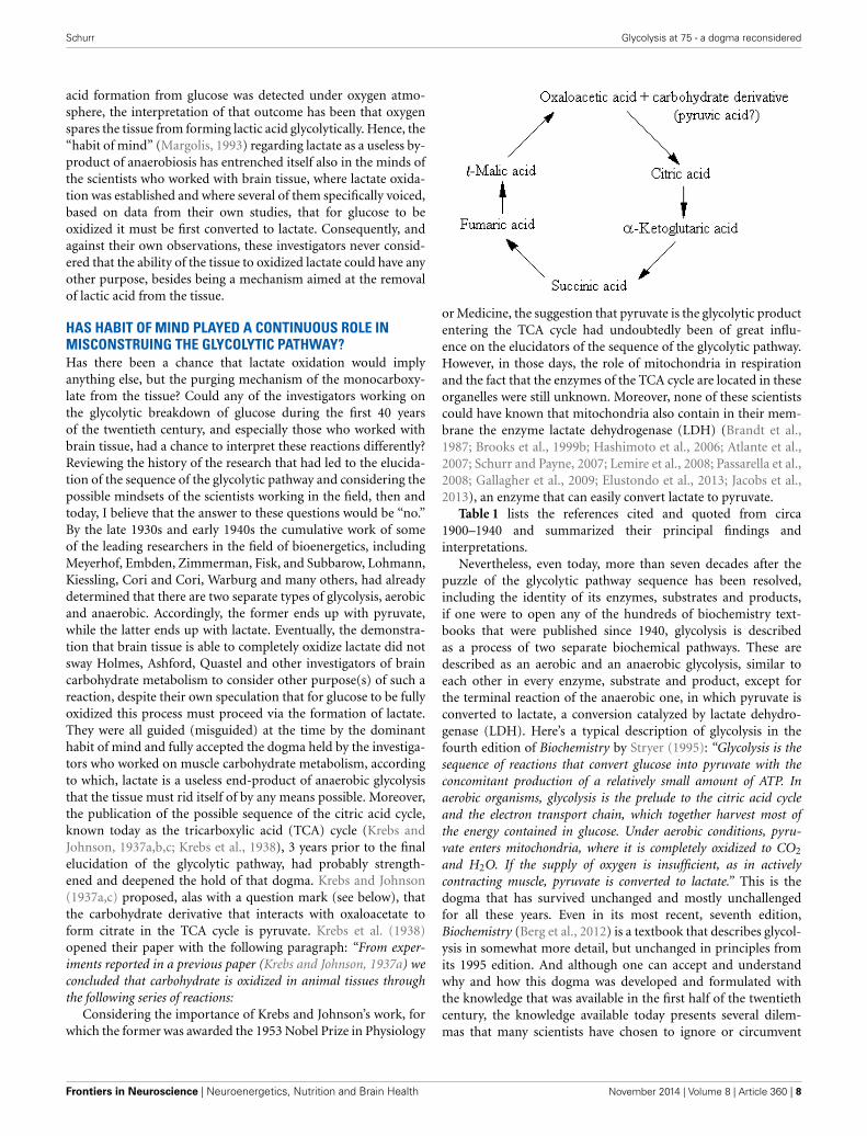
HAS HABIT OF MIND PLAYED A CONTINUOUS ROLE INMISCONSTRUING THE GLYCOLYTIC PATHWAY?Has there been a chance that lactate oxidation would implyanything else, but the purging mechanism of the monocarboxy-late from the tissue? Could any of the investigators working onthe glycolytic breakdown of glucose during the first 40 yearsof the twentieth century, and especially those who worked withbrain tissue, had a chance to interpret these reactions differently?Reviewing the history of the research that had led to the elucida-tion of the sequence of the glycolytic pathway and considering thepossible mindsets of the scientists working in the field, then andtoday, I believe that the answer to these questions would be “no.”By the late 1930s and early 1940s the cumulative work of someof the leading researchers in the field of bioenergetics, includingMeyerhof, Embden, Zimmerman, Fisk, and Subbarow, Lohmann,Kiessling, Cori and Cori, Warburg and many others, had alreadydetermined that there are two separate types of glycolysis, aerobicand anaerobic. Accordingly, the former ends up with pyruvate,while the latter ends up with lactate. Eventually, the demonstra-tion that brain tissue is able to completely oxidize lactate did notsway Holmes, Ashford, Quastel and other investigators of braincarbohydrate metabolism to consider other purpose(s) of such areaction, despite their own speculation that for glucose to be fullyoxidized this process must proceed via the formation of lactate.They were all guided (misguided) at the time by the dominanthabit of mind and fully accepted the dogma held by the investiga-tors who worked on muscle carbohydrate metabolism, accordingto which, lactate is a useless end-product of anaerobic glycolysisthat the tissue must rid itself of by any means possible. Moreover,the publication of the possible sequence of the citric acid cycle,known today as the tricarboxylic acid (TCA) cycle (Krebs andJohnson, 1937a,b,c; Krebs et al., 1938), 3 years prior to the finalelucidation of the glycolytic pathway, had probably strength-ened and deepened the hold of that dogma. Krebs and Johnson(1937a,c) proposed, alas with a question mark (see below), thatthe carbohydrate derivative that interacts with oxaloacetate toform citrate in the TCA cycle is pyruvate. Krebs et al. (1938)opened their paper with the following paragraph: “From exper-iments reported in a previous paper (Krebs and Johnson, 1937a) weconcluded that carbohydrate is oxidized in animal tissues throughthe following series of reactions:
Considering the importance of Krebs and Johnson’s work, forwhich the former was awarded the 1953 Nobel Prize in Physiology
or Medicine, the suggestion that pyruvate is the glycolytic productentering the TCA cycle had undoubtedly been of great influ-ence on the elucidators of the sequence of the glycolytic pathway.However, in those days, the role of mitochondria in respirationand the fact that the enzymes of the TCA cycle are located in theseorganelles were still unknown. Moreover, none of these scientistscould have known that mitochondria also contain in their mem-brane the enzyme lactate dehydrogenase (LDH) (Brandt et al.,1987; Brooks et al., 1999b; Hashimoto et al., 2006; Atlante et al.,2007; Schurr and Payne, 2007; Lemire et al., 2008; Passarella et al.,2008; Gallagher et al., 2009; Elustondo et al., 2013; Jacobs et al.,2013), an enzyme that can easily convert lactate to pyruvate.
Table 1 lists the references cited and quoted from circa1900–1940 and summarized their principal findings andinterpretations.
Nevertheless, even today, more than seven decades after thepuzzle of the glycolytic pathway sequence has been resolved,including the identity of its enzymes, substrates and products,if one were to open any of the hundreds of biochemistry text-books that were published since 1940, glycolysis is describedas a process of two separate biochemical pathways. These aredescribed as an aerobic and an anaerobic glycolysis, similar toeach other in every enzyme, substrate and product, except forthe terminal reaction of the anaerobic one, in which pyruvate isconverted to lactate, a conversion catalyzed by lactate dehydro-genase (LDH). Here’s a typical description of glycolysis in thefourth edition of Biochemistry by Stryer (1995): “Glycolysis is thesequence of reactions that convert glucose into pyruvate with theconcomitant production of a relatively small amount of ATP. Inaerobic organisms, glycolysis is the prelude to the citric acid cycleand the electron transport chain, which together harvest most ofthe energy contained in glucose. Under aerobic conditions, pyru-vate enters mitochondria, where it is completely oxidized to CO2
and H2O. If the supply of oxygen is insufficient, as in activelycontracting muscle, pyruvate is converted to lactate.” This is thedogma that has survived unchanged and mostly unchallengedfor all these years. Even in its most recent, seventh edition,Biochemistry (Berg et al., 2012) is a textbook that describes glycol-ysis in somewhat more detail, but unchanged in principles fromits 1995 edition. And although one can accept and understandwhy and how this dogma was developed and formulated withthe knowledge that was available in the first half of the twentiethcentury, the knowledge available today presents several dilem-mas that many scientists have chosen to ignore or circumvent
Frontiers in Neuroscience | Neuroenergetics, Nutrition and Brain Health November 2014 | Volume 8 | Article 360 | 8
Schurr Glycolysis at 75 - a dogma reconsidered
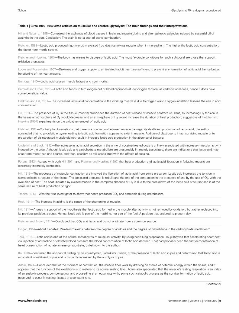
Table 1 | Circa 1900–1940 cited articles on muscular and cerebral glycolysis: The main findings and their interpretations.
Hill and Nabarro, 1895—Compared the exchange of blood gasses in brain and muscle during and after epileptic episodes induced by essential oil ofabsinthe in the dog. Conclusion: The brain is not a seat of active combustion.
Fletcher, 1898—Lactic acid produced rigor mortis in excised frog Gastrocnemius muscle when immersed in it. The higher the lactic acid concentration,the faster rigor mortis sets in.
Fletcher and Hopkins, 1907—The body has means to dispose of lactic acid. The most favorable conditions for such a disposal are those that supportoxidative processes.
Locke and Rosenheim, 1907—Dextrose and oxygen supply to an isolated rabbit heart are sufficient to prevent any formation of lactic acid, hence betterfunctioning of the heart muscle.
Burridge, 1910—Lactic acid causes muscle fatigue and rigor mortis.
Barcroft and Orbeli, 1910—Lactic acid tends to turn oxygen out of blood capillaries at low oxygen tension, as carbonic acid does, hence it does havesome beneficial value.
Feldman and Hill, 1911—The increased lactic acid concentration in the working muscle is due to oxygen want. Oxygen inhalation lessens the rise in acidconcentration.
Hill, 1911—The presence of O2 in the tissue (muscle) diminishes the duration of heat release of muscle contracture. Thus, by increasing O2 tension inthe tissue an atmosphere of O2 would decrease, and an atmosphere of H2 would increase the duration of heat production, suggestive of Fletcher andHopkins (1907) experiments on the oxidative removal of lactic acid.
Fletcher, 1911—Contrary to observations that there is a connection between muscle damage, its death and production of lactic acid, the authorconcluded that no glycolytic enzyme leading to lactic acid formation appears to exist in muscle. Addition of dextrose to intact surviving muscle or topreparation of disintegrated muscle did not result in increase lactic acid production in the absence of bacteria.
Underhill and Black, 1912—The increase in lactic acid secretion in the urine of cocaine-treated dogs is unlikely associated with increase muscular activityinduced by the drug. Although lactic acid and carbohydrate metabolism are presumably intimately associated, there are indications that lactic acid mayarise from more than one source, and thus, possibly be still associated with the effects of cocaine.
Peters, 1913—Agrees with both Hill (1911) and Fletcher and Hopkins (1907) that heat production and lactic acid liberation in fatiguing muscle areextremely intimately connected.
Hill, 1913—The processes of muscular contraction are involved the liberation of lactic acid from some precursor. Lactic acid increases the tension insome colloidal structure of the tissue. The lactic acid precursor is rebuilt and the end of the contraction in the presence of and by the use of O2, with theevolution of heat. The heat liberated by excited muscle in the complete absence of O2 is due to the breakdown of the lactic acid precursor and is of thesame nature of heat production of rigor.
Tashiro, 1913—Was the first investigator to show that nerve produced CO2 and ammonia during metabolism.
Roaf, 1914—The increase in acidity is the cause of the shortening of muscle.
Hill, 1914—Argues in support of the hypothesis that lactic acid formed in the muscle after activity is not removed by oxidation, but rather replaced intoits previous position, a sugar. Hence, lactic acid is part of the machine, not part of the fuel. A position that endured to present day.
Fletcher and Brown, 1914—Concluded that CO2 and lactic acid do not originate from a common source.
Ringer, 1914—About diabetes: Parallelism exists between the degree of acidosis and the degree of disturbance in the carbohydrate metabolism.
Tsuji, 1916—Lactic acid is one of the normal metabolites of muscular activity. By using heart-lung preparation, Tsuji showed that accelerating heart beatvia injection of adrenaline or elevated blood pressure the blood concentration of lactic acid declined. That had probably been the first demonstration ofheart consumption of lactate an energy substrate, unbeknown to the author.
Ito, 1916—confirmed the accidental finding by his countryman, Tatsukichi Irisawa, of the presence of lactic acid in pus and determined that lactic acid isa constant constituent of pus and is distinctly increased by the autolysis of pus.
Adam, 1921—Concluded that at the moment of contraction, the muscle fiber work by drawing on stores of potential energy within the tissue, and itappears that the function of the oxidations is to restore to its normal resting level. Adam also speculated that the muscle’s resting respiration is an indexof an anabolic process, compensating, and proceeding at an equal rate with, some such catabolic process as the survival formation of lactic acid,observed to occur in resting tissues at a constant rate.
(Continued)
www.frontiersin.org November 2014 | Volume 8 | Article 360 | 9
Schurr Glycolysis at 75 - a dogma reconsidered
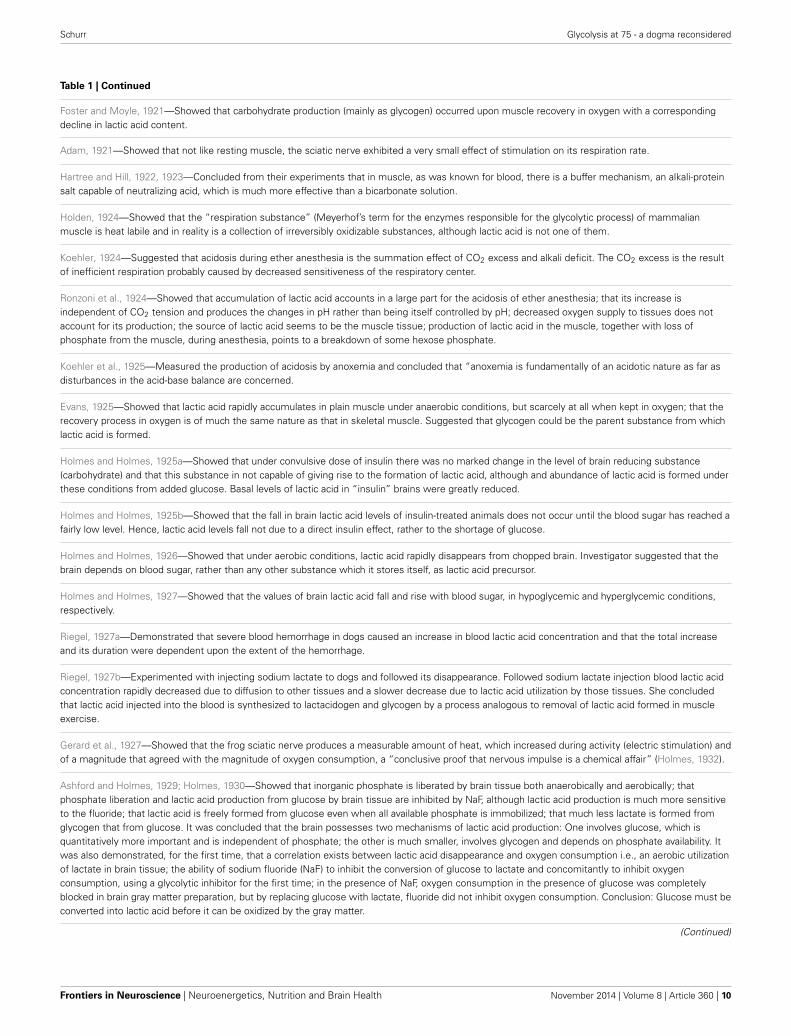
Table 1 | Continued
Foster and Moyle, 1921—Showed that carbohydrate production (mainly as glycogen) occurred upon muscle recovery in oxygen with a correspondingdecline in lactic acid content.
Adam, 1921—Showed that not like resting muscle, the sciatic nerve exhibited a very small effect of stimulation on its respiration rate.
Hartree and Hill, 1922, 1923—Concluded from their experiments that in muscle, as was known for blood, there is a buffer mechanism, an alkali-proteinsalt capable of neutralizing acid, which is much more effective than a bicarbonate solution.
Holden, 1924—Showed that the “respiration substance” (Meyerhof’s term for the enzymes responsible for the glycolytic process) of mammalianmuscle is heat labile and in reality is a collection of irreversibly oxidizable substances, although lactic acid is not one of them.
Koehler, 1924—Suggested that acidosis during ether anesthesia is the summation effect of CO2 excess and alkali deficit. The CO2 excess is the resultof inefficient respiration probably caused by decreased sensitiveness of the respiratory center.
Ronzoni et al., 1924—Showed that accumulation of lactic acid accounts in a large part for the acidosis of ether anesthesia; that its increase isindependent of CO2 tension and produces the changes in pH rather than being itself controlled by pH; decreased oxygen supply to tissues does notaccount for its production; the source of lactic acid seems to be the muscle tissue; production of lactic acid in the muscle, together with loss ofphosphate from the muscle, during anesthesia, points to a breakdown of some hexose phosphate.
Koehler et al., 1925—Measured the production of acidosis by anoxemia and concluded that “anoxemia is fundamentally of an acidotic nature as far asdisturbances in the acid-base balance are concerned.
Evans, 1925—Showed that lactic acid rapidly accumulates in plain muscle under anaerobic conditions, but scarcely at all when kept in oxygen; that therecovery process in oxygen is of much the same nature as that in skeletal muscle. Suggested that glycogen could be the parent substance from whichlactic acid is formed.
Holmes and Holmes, 1925a—Showed that under convulsive dose of insulin there was no marked change in the level of brain reducing substance(carbohydrate) and that this substance in not capable of giving rise to the formation of lactic acid, although and abundance of lactic acid is formed underthese conditions from added glucose. Basal levels of lactic acid in “insulin” brains were greatly reduced.
Holmes and Holmes, 1925b—Showed that the fall in brain lactic acid levels of insulin-treated animals does not occur until the blood sugar has reached afairly low level. Hence, lactic acid levels fall not due to a direct insulin effect, rather to the shortage of glucose.
Holmes and Holmes, 1926—Showed that under aerobic conditions, lactic acid rapidly disappears from chopped brain. Investigator suggested that thebrain depends on blood sugar, rather than any other substance which it stores itself, as lactic acid precursor.
Holmes and Holmes, 1927—Showed that the values of brain lactic acid fall and rise with blood sugar, in hypoglycemic and hyperglycemic conditions,respectively.
Riegel, 1927a—Demonstrated that severe blood hemorrhage in dogs caused an increase in blood lactic acid concentration and that the total increaseand its duration were dependent upon the extent of the hemorrhage.
Riegel, 1927b—Experimented with injecting sodium lactate to dogs and followed its disappearance. Followed sodium lactate injection blood lactic acidconcentration rapidly decreased due to diffusion to other tissues and a slower decrease due to lactic acid utilization by those tissues. She concludedthat lactic acid injected into the blood is synthesized to lactacidogen and glycogen by a process analogous to removal of lactic acid formed in muscleexercise.
Gerard et al., 1927—Showed that the frog sciatic nerve produces a measurable amount of heat, which increased during activity (electric stimulation) andof a magnitude that agreed with the magnitude of oxygen consumption, a “conclusive proof that nervous impulse is a chemical affair” (Holmes, 1932).
Ashford and Holmes, 1929; Holmes, 1930—Showed that inorganic phosphate is liberated by brain tissue both anaerobically and aerobically; thatphosphate liberation and lactic acid production from glucose by brain tissue are inhibited by NaF, although lactic acid production is much more sensitiveto the fluoride; that lactic acid is freely formed from glucose even when all available phosphate is immobilized; that much less lactate is formed fromglycogen that from glucose. It was concluded that the brain possesses two mechanisms of lactic acid production: One involves glucose, which isquantitatively more important and is independent of phosphate; the other is much smaller, involves glycogen and depends on phosphate availability. Itwas also demonstrated, for the first time, that a correlation exists between lactic acid disappearance and oxygen consumption i.e., an aerobic utilizationof lactate in brain tissue; the ability of sodium fluoride (NaF) to inhibit the conversion of glucose to lactate and concomitantly to inhibit oxygenconsumption, using a glycolytic inhibitor for the first time; in the presence of NaF, oxygen consumption in the presence of glucose was completelyblocked in brain gray matter preparation, but by replacing glucose with lactate, fluoride did not inhibit oxygen consumption. Conclusion: Glucose must beconverted into lactic acid before it can be oxidized by the gray matter.
(Continued)
Frontiers in Neuroscience | Neuroenergetics, Nutrition and Brain Health November 2014 | Volume 8 | Article 360 | 10
Schurr Glycolysis at 75 - a dogma reconsidered
Table 1 | Continued
Holmes and Ashford, 1930; Ashford and Holmes, 1931—Measured the “Meyerhof quotient,” the [total lactate disappearing]/[lactate oxidize], which inmuscle was determined to be ∼3, and found it in brain to be 1; found that the O2 uptake in oxygenated brain tissue shaken with lactate in the presenceof bicarbonate buffer in an O2/CO2 atmosphere is greater than in the presence of phosphate buffer and that such uptake increases with increasedoxygen tension in both cases; termed “Meyerhof quotient” the “respiratory quotient” and found its value, both of brain tissue alone and of tissueoxygenated with extra oxygen, to be close to unity, including in the case of brain from animals rendered hypoglycemic by insulin injection; concludedthat lactate oxidation is unlikely to spare the utilization of another substrate.
Quastel and Wheatley, 1932—Found that the rate of oxidation of an added substrate to brain tissue varies inversely with the size of the animal, ageneralization that does not apply to muscle; that glucose, lactate and pyruvate at equivalent concentrations are oxidized at the same rate by braintissue; that lactate is completely oxidized by brain tissue; that iodoacetic acid (IAA) inhibits glucose oxidation and stated the possibility that glucosenecessarily passes through lactic acid for its oxidation to take place; found that oxalate, unlike IAA and NaF, also inhibits the oxidation of lactate.
Holmes, 1933—Hinted at the possibility that lactate oxidation could support brain activity.
Dixon, 1935—Confirmed the fact that in oxygenated brain tissue lactic acid formation cannot be detected, but concluded that the purpose of thecomplete oxidation of lactate is simply to remove it from the tissue.
Krebs and Johnson, 1937a,b,c; Krebs et al., 1938—Suggested the carbohydrate derivative to enter the Krebs cycle (tricarboxylic acid cycle, TCA) ispyruvate, alas with a question mark.
due, most probably, to habit of mind (Margolis, 1993). If thereis any need for one to realize how strong an influence habitof mind can have, one needs only to recall how successful the,now defunct, lactic acidosis hypothesis of ischemic brain dam-age had been throughout the 1980s and 1990s (Kalimo et al.,1981; Rehncrona et al., 1981; Siesjö, 1981). Even four decadesafter the elucidation of the glycolytic pathway it was very easy topersuade a large contingency of scientists who studied possiblemechanisms of hypoxic and ischemic brain damage that the cul-prit behind such damage is no other than the “usual suspect” i.e.,lactate.
As has already been mentioned at the beginning of thismonograph, Brooks (1985) has demonstrated that lactate isthe glycolytic product and the oxidative substrate during sus-tained exercise. Later, Fox and Raichle (1986) have demonstrateda focal physiological uncoupling between cerebral blood flowand oxidative metabolism upon somatosensory stimulation inhumans, and Fox et al. (1988) showed that during focal physio-logic neural activity the consumption of glucose is non-oxidative.Simultaneously, Schurr et al. (1988) demonstrated the ability ofbrain tissue to maintain normal neuronal function with lactateas the sole oxidative energy substrate. With more publicationsadding support to the possible role of lactate in oxidative energymetabolism, both in muscle (Brooks, 1998, 2000, 2002a,b; Brookset al., 1999a,b) and especially in brain (Izumi et al., 1994; Pellerinand Magistretti, 1994, 2003; Larrabee, 1995, 1996; Tsacopoulosand Magistretti, 1996; Hu and Wilson, 1997a,b; Schurr et al.,1997a, 1999a,b; Schurr and Rigor, 1998; Magistretti and Pellerin,1999; Magistretti et al., 1999; Magistretti, 2000; Qu et al., 2000;Van Hall, 2000; Bliss and Sapolsky, 2001; Bouzier-Sore et al.,2003; Mangia et al., 2003; Smith et al., 2003; Dalsgaard et al.,2004; Kasischke et al., 2004; Schurr, 2006; Schurr and Payne,2007; Herrero-Mendez et al., 2009; Zielke et al., 2009; Schurr andGozal, 2011), a hot debate has ensued, focusing, unfortunately, onthe premise that lactate is somehow an alternative oxidative sub-strate to glucose in tissue energy metabolism (Chih et al., 2001;Dienel and Hertz, 2001, 2005; Chih and Roberts, 2003; Hertz,
2004; Hertz et al., 2007; Dienel, 2012a,b). Consequently, ratherthan viewing the oxidative utilization of lactate as an integral partof the oxidative energy metabolic pathway, which begins with glu-cose and glycolysis and ends with CO2, H2O and the mitochon-drial electron transport chain, many have portrayed lactate as acompetitor of glucose. Hence, several studies have aimed at show-ing that glucose is the obligatory energy substrate for maintenanceof various neuronal functions (Dienel and Cruz, 2004; Fillenz,2005; Bak et al., 2006; Cruz et al., 2007; Gandhi et al., 2009).However, this very role of glucose has never been questioned orchallenged by those who unraveled the oxidative utilization oflactate, either by muscle or brain tissue. After all, the principalsource of tissue lactate is glucose, a fact that has never been indispute. The utilization of lactate via its oxidation should havebeen understood simply as the most plausible and expected pro-gression of glucose breakdown via the glycolytic pathway wherelactate, not pyruvate, is the real first step in the mitochondrialTCA cycle. Nonetheless, a concerted effort has been mountedby many established investigators to minimize or marginal-ize lactate’s role in energy metabolism. The following quotefrom the chapter by Clarke and Sokoloff (1994) on Circulationand Energy Metabolism in the Brain in the fifth edition ofBasic Neurochemistry (1994) is most telling: “Lactate, pyruvate,fructose-1,6-biphosphate, acetate, β-hydroxybutyrate, and acetoac-etate can all be utilized by brain slices, homogenates, or cell-freefractions. . . but the substrate is not available to the brain becauseof inadequate blood levels or restricted transport through the BBB(blood brain barrier).” Clarke and Sokoloff, who were leadingscientists in the field of brain energy metabolism at the time,felt compelled to emphasize the limitations lactate faces as anenergy substrate specifically in response to the findings of Foxand Raichle (1986), Fox et al. (1988) and Schurr et al. (1988),as if the role of glucose in the process was somehow beingdiminished by lactate. Hence, Clarke and Sokoloff reemphasizethat “. . . the nervous system function in the intact animal dependson substrates supplied by the blood and no satisfactory, normal,endogenous substitute for glucose has been found. Glucose must
www.frontiersin.org November 2014 | Volume 8 | Article 360 | 11
Schurr Glycolysis at 75 - a dogma reconsidered
therefore be considered essential for the normal physiological behav-ior of the central nervous system.” Therefore, if one requires aproof that habit of mind is long-lived, one could simply followthe heated debate over the role of lactate in oxidative energymetabolism, as exemplified by the large number of con and propublications on the topic. At least in part, the strong rejection
of lactate as an “alternative” oxidative substrate to glucose inenergy metabolism, despite the fact that by now this monocar-boxylate is recognized as an oxidative substrate, has to do withthe old dogma of “lactate is a useless end-product of glycolysis.”Moreover, if lactate, as being offered here, is the real end prod-uct of glycolysis, both under aerobic and anaerobic conditions,
FIGURE 1 | A schematic representation of six different experimental
paradigms using rat hippocampal slices and electrophysiological
recording of CA1 evoked population spikes (neuronal function). Ineach experimental paradigm, slices were supplied either with 20 mMglucose (yellow bottle) or 20 mM 2-deoxy glucose (2DG, a glycolyticinhibitor) (orange bottle) and the gas mixture bubbled through theincubation chamber of the slices was either 95% O2/5% CO2 (normoxia,red bubbles) or 95% N2/5% CO2 (hypoxia, blue bubbles). At the end ofeach experimental paradigm, all slices in each compartment of the dualchamber were tested for the presence of neuronal function. Functional
slices are shown as percentage of the total number of slices present(green histograms on the right). Accordingly, by following the timelinefrom left to right, paradigm A is a protocol in which slices were incubatedunder normoxic conditions for 40 min, followed by 13-min hypoxia in thepresence of 2DG and then re-oxygenated for 30 min in the presence ofglucose. Under these conditions less than 20% of the slices recoveredtheir neuronal function at the end of the 80-min protocol. Similarly, eachof the remaining paradigms (B–F) describes its corresponding protocoland its outcome in terms of percentage of slices exhibiting neuronalfunction. ∗Significantly different from paradigm A (P < 0.05).
Frontiers in Neuroscience | Neuroenergetics, Nutrition and Brain Health November 2014 | Volume 8 | Article 360 | 12
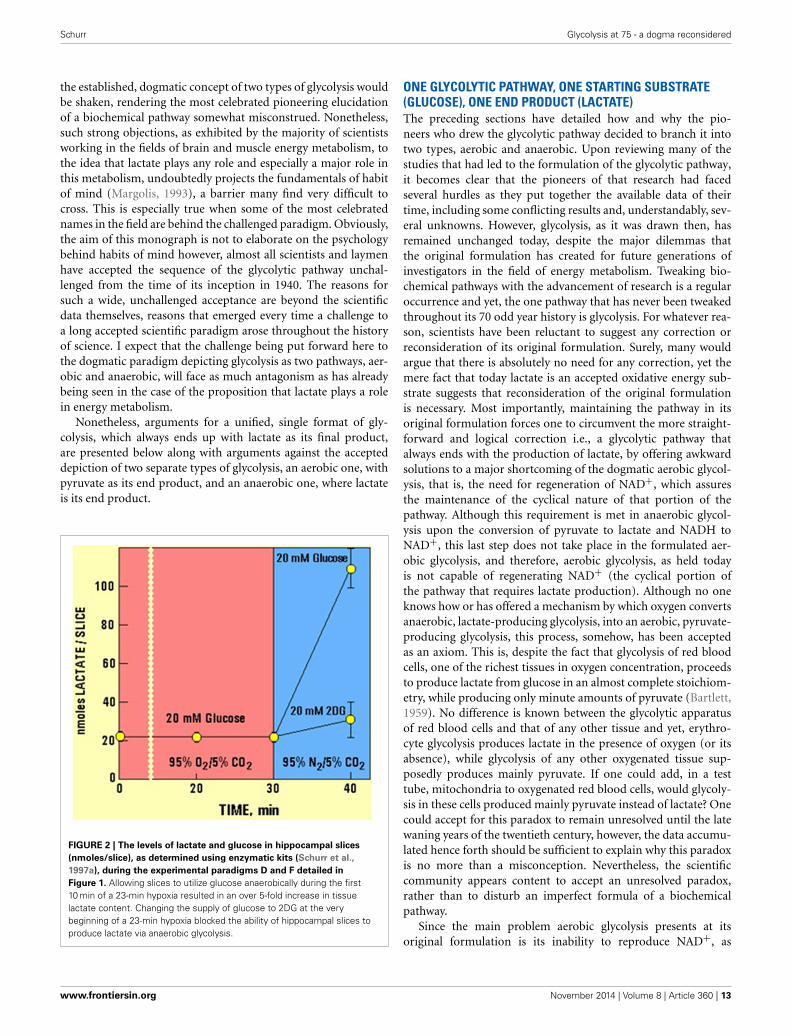
Schurr Glycolysis at 75 - a dogma reconsidered
the established, dogmatic concept of two types of glycolysis wouldbe shaken, rendering the most celebrated pioneering elucidationof a biochemical pathway somewhat misconstrued. Nonetheless,such strong objections, as exhibited by the majority of scientistsworking in the fields of brain and muscle energy metabolism, tothe idea that lactate plays any role and especially a major role inthis metabolism, undoubtedly projects the fundamentals of habitof mind (Margolis, 1993), a barrier many find very difficult tocross. This is especially true when some of the most celebratednames in the field are behind the challenged paradigm. Obviously,the aim of this monograph is not to elaborate on the psychologybehind habits of mind however, almost all scientists and laymenhave accepted the sequence of the glycolytic pathway unchal-lenged from the time of its inception in 1940. The reasons forsuch a wide, unchallenged acceptance are beyond the scientificdata themselves, reasons that emerged every time a challenge toa long accepted scientific paradigm arose throughout the historyof science. I expect that the challenge being put forward here tothe dogmatic paradigm depicting glycolysis as two pathways, aer-obic and anaerobic, will face as much antagonism as has alreadybeing seen in the case of the proposition that lactate plays a rolein energy metabolism.
Nonetheless, arguments for a unified, single format of gly-colysis, which always ends up with lactate as its final product,are presented below along with arguments against the accepteddepiction of two separate types of glycolysis, an aerobic one, withpyruvate as its end product, and an anaerobic one, where lactateis its end product.
FIGURE 2 | The levels of lactate and glucose in hippocampal slices
(nmoles/slice), as determined using enzymatic kits (Schurr et al.,
1997a), during the experimental paradigms D and F detailed in
Figure 1. Allowing slices to utilize glucose anaerobically during the first10 min of a 23-min hypoxia resulted in an over 5-fold increase in tissuelactate content. Changing the supply of glucose to 2DG at the verybeginning of a 23-min hypoxia blocked the ability of hippocampal slices toproduce lactate via anaerobic glycolysis.
ONE GLYCOLYTIC PATHWAY, ONE STARTING SUBSTRATE(GLUCOSE), ONE END PRODUCT (LACTATE)The preceding sections have detailed how and why the pio-neers who drew the glycolytic pathway decided to branch it intotwo types, aerobic and anaerobic. Upon reviewing many of thestudies that had led to the formulation of the glycolytic pathway,it becomes clear that the pioneers of that research had facedseveral hurdles as they put together the available data of theirtime, including some conflicting results and, understandably, sev-eral unknowns. However, glycolysis, as it was drawn then, hasremained unchanged today, despite the major dilemmas thatthe original formulation has created for future generations ofinvestigators in the field of energy metabolism. Tweaking bio-chemical pathways with the advancement of research is a regularoccurrence and yet, the one pathway that has never been tweakedthroughout its 70 odd year history is glycolysis. For whatever rea-son, scientists have been reluctant to suggest any correction orreconsideration of its original formulation. Surely, many wouldargue that there is absolutely no need for any correction, yet themere fact that today lactate is an accepted oxidative energy sub-strate suggests that reconsideration of the original formulationis necessary. Most importantly, maintaining the pathway in itsoriginal formulation forces one to circumvent the more straight-forward and logical correction i.e., a glycolytic pathway thatalways ends with the production of lactate, by offering awkwardsolutions to a major shortcoming of the dogmatic aerobic glycol-ysis, that is, the need for regeneration of NAD+, which assuresthe maintenance of the cyclical nature of that portion of thepathway. Although this requirement is met in anaerobic glycol-ysis upon the conversion of pyruvate to lactate and NADH toNAD+, this last step does not take place in the formulated aer-obic glycolysis, and therefore, aerobic glycolysis, as held todayis not capable of regenerating NAD+ (the cyclical portion ofthe pathway that requires lactate production). Although no oneknows how or has offered a mechanism by which oxygen convertsanaerobic, lactate-producing glycolysis, into an aerobic, pyruvate-producing glycolysis, this process, somehow, has been acceptedas an axiom. This is, despite the fact that glycolysis of red bloodcells, one of the richest tissues in oxygen concentration, proceedsto produce lactate from glucose in an almost complete stoichiom-etry, while producing only minute amounts of pyruvate (Bartlett,1959). No difference is known between the glycolytic apparatusof red blood cells and that of any other tissue and yet, erythro-cyte glycolysis produces lactate in the presence of oxygen (or itsabsence), while glycolysis of any other oxygenated tissue sup-posedly produces mainly pyruvate. If one could add, in a testtube, mitochondria to oxygenated red blood cells, would glycoly-sis in these cells produced mainly pyruvate instead of lactate? Onecould accept for this paradox to remain unresolved until the latewaning years of the twentieth century, however, the data accumu-lated hence forth should be sufficient to explain why this paradoxis no more than a misconception. Nevertheless, the scientificcommunity appears content to accept an unresolved paradox,rather than to disturb an imperfect formula of a biochemicalpathway.
Since the main problem aerobic glycolysis presents at itsoriginal formulation is its inability to reproduce NAD+, as
www.frontiersin.org November 2014 | Volume 8 | Article 360 | 13
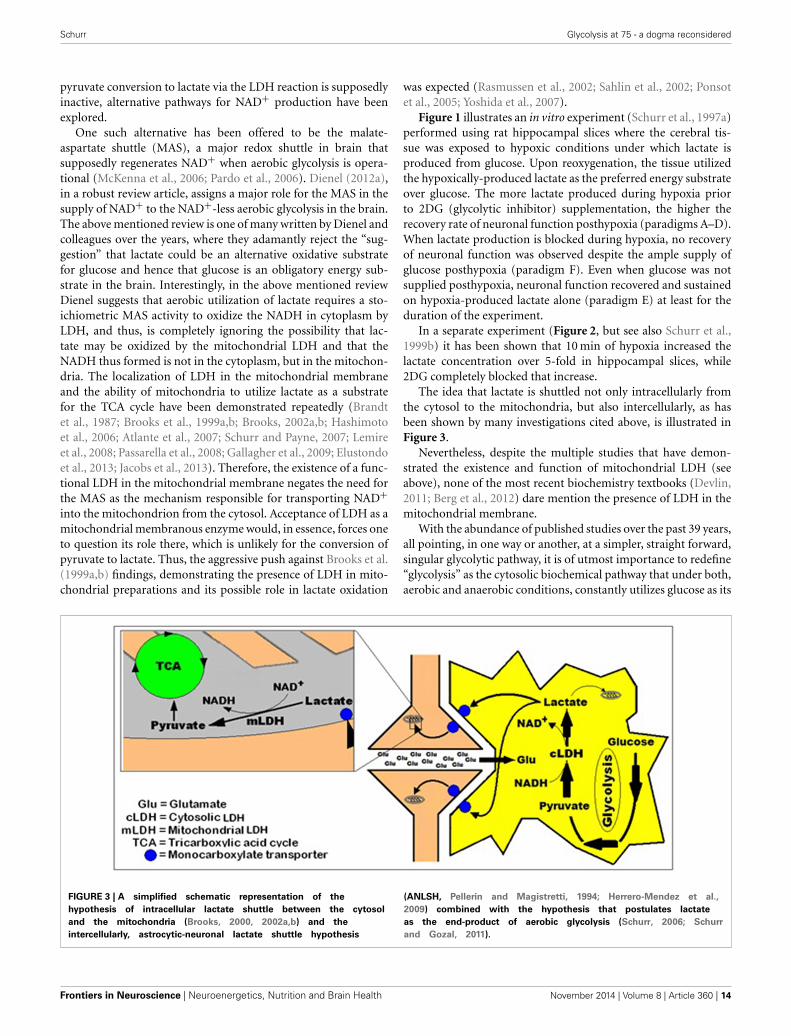
Schurr Glycolysis at 75 - a dogma reconsidered
pyruvate conversion to lactate via the LDH reaction is supposedlyinactive, alternative pathways for NAD+ production have beenexplored.
One such alternative has been offered to be the malate-aspartate shuttle (MAS), a major redox shuttle in brain thatsupposedly regenerates NAD+ when aerobic glycolysis is opera-tional (McKenna et al., 2006; Pardo et al., 2006). Dienel (2012a),in a robust review article, assigns a major role for the MAS in thesupply of NAD+ to the NAD+-less aerobic glycolysis in the brain.The above mentioned review is one of many written by Dienel andcolleagues over the years, where they adamantly reject the “sug-gestion” that lactate could be an alternative oxidative substratefor glucose and hence that glucose is an obligatory energy sub-strate in the brain. Interestingly, in the above mentioned reviewDienel suggests that aerobic utilization of lactate requires a sto-ichiometric MAS activity to oxidize the NADH in cytoplasm byLDH, and thus, is completely ignoring the possibility that lac-tate may be oxidized by the mitochondrial LDH and that theNADH thus formed is not in the cytoplasm, but in the mitochon-dria. The localization of LDH in the mitochondrial membraneand the ability of mitochondria to utilize lactate as a substratefor the TCA cycle have been demonstrated repeatedly (Brandtet al., 1987; Brooks et al., 1999a,b; Brooks, 2002a,b; Hashimotoet al., 2006; Atlante et al., 2007; Schurr and Payne, 2007; Lemireet al., 2008; Passarella et al., 2008; Gallagher et al., 2009; Elustondoet al., 2013; Jacobs et al., 2013). Therefore, the existence of a func-tional LDH in the mitochondrial membrane negates the need forthe MAS as the mechanism responsible for transporting NAD+into the mitochondrion from the cytosol. Acceptance of LDH as amitochondrial membranous enzyme would, in essence, forces oneto question its role there, which is unlikely for the conversion ofpyruvate to lactate. Thus, the aggressive push against Brooks et al.(1999a,b) findings, demonstrating the presence of LDH in mito-chondrial preparations and its possible role in lactate oxidation
was expected (Rasmussen et al., 2002; Sahlin et al., 2002; Ponsotet al., 2005; Yoshida et al., 2007).
Figure 1 illustrates an in vitro experiment (Schurr et al., 1997a)performed using rat hippocampal slices where the cerebral tis-sue was exposed to hypoxic conditions under which lactate isproduced from glucose. Upon reoxygenation, the tissue utilizedthe hypoxically-produced lactate as the preferred energy substrateover glucose. The more lactate produced during hypoxia priorto 2DG (glycolytic inhibitor) supplementation, the higher therecovery rate of neuronal function posthypoxia (paradigms A–D).When lactate production is blocked during hypoxia, no recoveryof neuronal function was observed despite the ample supply ofglucose posthypoxia (paradigm F). Even when glucose was notsupplied posthypoxia, neuronal function recovered and sustainedon hypoxia-produced lactate alone (paradigm E) at least for theduration of the experiment.
In a separate experiment (Figure 2, but see also Schurr et al.,1999b) it has been shown that 10 min of hypoxia increased thelactate concentration over 5-fold in hippocampal slices, while2DG completely blocked that increase.
The idea that lactate is shuttled not only intracellularly fromthe cytosol to the mitochondria, but also intercellularly, as hasbeen shown by many investigations cited above, is illustrated inFigure 3.
Nevertheless, despite the multiple studies that have demon-strated the existence and function of mitochondrial LDH (seeabove), none of the most recent biochemistry textbooks (Devlin,2011; Berg et al., 2012) dare mention the presence of LDH in themitochondrial membrane.
With the abundance of published studies over the past 39 years,all pointing, in one way or another, at a simpler, straight forward,singular glycolytic pathway, it is of utmost importance to redefine“glycolysis” as the cytosolic biochemical pathway that under both,aerobic and anaerobic conditions, constantly utilizes glucose as its
FIGURE 3 | A simplified schematic representation of the
hypothesis of intracellular lactate shuttle between the cytosol
and the mitochondria (Brooks, 2000, 2002a,b) and the
intercellularly, astrocytic-neuronal lactate shuttle hypothesis
(ANLSH, Pellerin and Magistretti, 1994; Herrero-Mendez et al.,
2009) combined with the hypothesis that postulates lactate
as the end-product of aerobic glycolysis (Schurr, 2006; Schurr
and Gozal, 2011).
Frontiers in Neuroscience | Neuroenergetics, Nutrition and Brain Health November 2014 | Volume 8 | Article 360 | 14
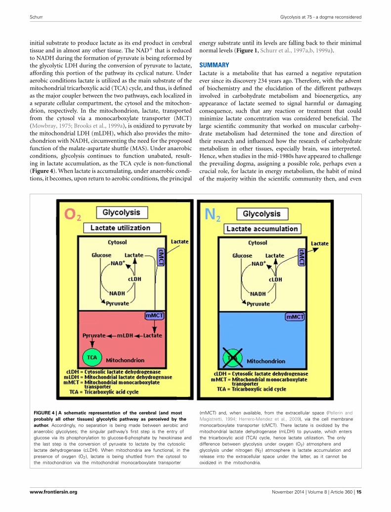
Schurr Glycolysis at 75 - a dogma reconsidered
initial substrate to produce lactate as its end product in cerebraltissue and in almost any other tissue. The NAD+ that is reducedto NADH during the formation of pyruvate is being reformed bythe glycolytic LDH during the conversion of pyruvate to lactate,affording this portion of the pathway its cyclical nature. Underaerobic conditions lactate is utilized as the main substrate of themitochondrial tricarboxylic acid (TCA) cycle, and thus, is definedas the major coupler between the two pathways, each localized ina separate cellular compartment, the cytosol and the mitochon-drion, respectively. In the mitochondrion, lactate, transportedfrom the cytosol via a monocarboxylate transporter (MCT)(Mowbray, 1975; Brooks et al., 1999a), is oxidized to pyruvate bythe mitochondrial LDH (mLDH), which also provides the mito-chondrion with NADH, circumventing the need for the proposedfunction of the malate-aspartate shuttle (MAS). Under anaerobicconditions, glycolysis continues to function unabated, result-ing in lactate accumulation, as the TCA cycle is non-functional(Figure 4). When lactate is accumulating, under anaerobic condi-tions, it becomes, upon return to aerobic conditions, the principal
energy substrate until its levels are falling back to their minimalnormal levels (Figure 1, Schurr et al., 1997a,b, 1999a).
SUMMARYLactate is a metabolite that has earned a negative reputationever since its discovery 234 years ago. Therefore, with the adventof biochemistry and the elucidation of the different pathwaysinvolved in carbohydrate metabolism and bioenergetics, anyappearance of lactate seemed to signal harmful or damagingconsequence, such that any reaction or treatment that couldminimize lactate concentration was considered beneficial. Thelarge scientific community that worked on muscular carbohy-drate metabolism had determined the tone and direction oftheir research and influenced how the research of carbohydratemetabolism in other tissues, especially brain, was interpreted.Hence, when studies in the mid-1980s have appeared to challengethe prevailing dogma, assigning a possible role, perhaps even acrucial role, for lactate in energy metabolism, the habit of mindof the majority within the scientific community then, and even
FIGURE 4 | A schematic representation of the cerebral (and most
probably all other tissues) glycolytic pathway as perceived by the
author. Accordingly, no separation is being made between aerobic andanaerobic glycolyses; the singular pathway’s first step is the entry ofglucose via its phosphorylation to glucose-6-phosphate by hexokinase andthe last step is the conversion of pyruvate to lactate by the cytosoliclactate dehydrogenase (cLDH). When mitochondria are functional, in thepresence of oxygen (O2), lactate is being shuttled from the cytosol tothe mitochondrion via the mitochondrial monocarboxylate transporter
(mMCT) and, when available, from the extracellular space (Pellerin andMagistretti, 1994; Herrero-Mendez et al., 2009), via the cell membranemonocarboxylate transporter (cMCT). There lactate is oxidized by themitochondrial lactate dehydrogenase (mLDH) to pyruvate, which entersthe tricarboxylic acid (TCA) cycle, hence lactate utilization. The onlydifference between glycolysis under oxygen (O2) atmosphere andglycolysis under nitrogen (N2) atmosphere is lactate accumulation andrelease into the extracellular space under the latter, as it cannot beoxidized in the mitochondria.
www.frontiersin.org November 2014 | Volume 8 | Article 360 | 15
Schurr Glycolysis at 75 - a dogma reconsidered
today, erected a barrier that prevents the acceptance of such role,despite the continuously growing number of publications in sup-port of it. This monograph attempted to review the publicationsof the first four or five decades of muscle and brain carbohy-drate metabolism in an effort to persuade its readers to open theirminds to the possibility that glycolysis, in cerebral and other tis-sues, is a singular pathway, in the presence or absence of oxygen,which begins with glucose as its substrate and terminates with theproduction of lactate as its main end product.
ACKNOWLEDGMENTSThe author greatly appreciates Prof. Roald Hoffmann, CornellUniversity, for his helpful comments and encouragement.
REFERENCESAdam, N. K. (1921). Note of the oxygen consumption of amphibian muscle and
nerve. Biochem. J. 15, 358–362.Ashford, C. A., and Holmes, E. G. (1929). Contributions to the study of brain
metabolism. V. Role of phosphates in lactic acid production. Biochem. J. 23,748–759.
Ashford, C. A., and Holmes, E. G. (1931). Further observations on the oxidation oflactic acid by brain tissue. Biochem. J. 25, 2028–2049.
Atlante, A., de Bari, L., Bobba, A., Marra, E., and Passarella, S. (2007). Transportand metabolism of l-lactate occur in mitochondria from cerebellar granulecells and are modified in cells undergoing low potassium dependent apoptosis.Biochim. Biophys. Acta 1767, 1285–1299. doi: 10.1016/j.bbabio.2007.08.003
Bak, L. K., Schousboe, A., Sonnewald, U., and Waagepetersen, H. S. (2006). Glucoseis necessary to maintain neurotransmitter homeostasis during synaptic activityin cultured glutamatergic neurons. J. Cereb. Blood Flow Metab. 26, 1285–1297.doi: 10.1038/sj.jcbfm.9600281
Barber, B. (1961). Resistance by scientists to scientific discovery. Science 134,596–602. doi: 10.1126/science.134.3479.596
Barcroft, J., and Orbeli, L. (1910). The influence of lactic acid upon the dissociationcurve of blood. J. Physiol. 41, 355–367.
Bartlett, G. R. (1959). Human red blood cell glycolytic intermediates. J. Biol. Chem.234, 449–458.
Berg, J. M., Tymoczko, J. L., Stryer, L., and Gatto, G. J. Jr. (2012). Biochemistry, 7thEdn. New York, NY: W. H. Freeman and Company.
Bliss, T. M., and Sapolsky, R. M. (2001). Interactions among glucose, lactate andadenosine regulate energy substrate utilization in hippocampal cultures. BrainRes. 899, 134–141. doi: 10.1016/S0006-8993(01)02218-1
Bouzier-Sore, A.-K., Voisin, P., Canioni, P., Magistretti, P. J., and Pellerin,L. (2003). Lactate is the preferential energy substrate over glucose forneurons in culture. J. Cereb. Blood Flow Metab. 23, 1298–1306. doi:10.1097/01.WCB.0000091761.61714.25
Brandt, R. B., Laux, J. E., Spainhour, S. E., and Kline, E. S. (1987). Lactate dehy-drogenase in rat mitochondria. Arch. Biochem. Biophys. 259, 412–422. doi:10.1016/0003-9861(87)90507-8
Brooks, G. A. (1985). “Lactate: glycolytic product and oxidative substrate duringsustained exercise in mammals – ‘the lactate shuttle”’ in Comparative Physiologyand Biochemistry – Currecnt Topics and Trends Vol A. Respiration-Metabolism-Circulation, ed R. Gilles (Berlin: Springer-Verlag), 208–218.
Brooks, G. A. (1998). Mammalian fuel utilization during sustained exercise. Comp.Biochem. Physiol. B Biochem. Mol. Biol. 120, 89–107. doi: 10.1016/S0305-0491(98)00025-X
Brooks, G. A. (2000). Intra- and extra-cellular lactate shuttles. Med. Sci. Sports.Exerc. 32, 790–799. doi: 10.1097/00005768-200004000-00011
Brooks, G. A. (2002a). Lactate shuttle – between but not within cells? J. Physiol. 541,333. doi: 10.1113/jphysiol.2002.023705
Brooks, G. A. (2002b). Lactate shuttles in nature. Biochem. Soc. Trans. 30, 258–264.doi: 10.1042/BST0300258
Brooks, G. A., Brown, M. A., Butz, C. E., Sicurello, J. P., and Dubouchaud, H.(1999a). Cardiac and skeletal muscle mitochondria have a monocarboxylatetransporter MCT1. J. Appl. Physiol. 87, 1713–1718.
Brooks, G. A., Dubouchaud, H., Brown, M., Sicurello, J. P., and Butz, C. E.(1999b). Role of mitochondrial lactate dehydrogenase and lactate oxidation in
the intracellular lactate shuttle. Proc. Natl. Acad. Sci. U.S.A. 96, 1129–1134. doi:10.1073/pnas.96.3.1129
Burridge, W. (1910). An inquiry into some chemical factors of fatigue. J. Physiol.41, 285–307.
Chih, C.-P., Lipton, P., and Roberts, E. L. Jr. (2001). Do active cerebral neu-rons really use lactate than glucose? Trends Neurosci. 24, 573–578. doi:10.1016/S0166-2236(00)01920-2
Chih, C.-P., and Roberts, E. L. Jr. (2003). Energy substrates for neurons during neu-ral activity: a critical review of the astrocyte-neuron lactate shuttle hypothesis.J. Cereb. Blood Flow Metab. 23, 1263–1281. doi: 10.1097/01.WCB.0000081369.51727.6F
Clarke, D. D., and Sokoloff, L. (1994). “Circulation and energy metabolism ofthe brain (Chap. 31),” in Basic neurochemistry, 5th Edn, eds G. J. Siegle, B.W. Agranoff, R. W. Albers, and P. B. Molinoff (New York, NY: Raven Press),645–680.
Cruz, N. F., Ball, K. K., and Dienel, G. A. (2007). Functional imaging of focal brainactivation in conscious rats: impact of [(14)C]glucose metabolite spreading andrelease. J. Neurosci. Res. 85, 3254–3266. doi: 10.1002/jnr.21193
Dalsgaard, M. K., Quistorff, B., Danielsen, E. R., Selmer, C., Vogelsang, T., andSecher, N. H. (2004). A reduced cerebral metabolic ratio in exercise reflectsmetabolism and not accumulation of lactate within the human brain. J. Physiol.554, 571–578. doi: 10.1113/jphysiol.2003.055053
Devlin, T. M. (2011). Textbook of Biochemistry with Clinical Correlations, 7th Edn.Hoboken, NJ: John Wiley & Sons, Inc.
Dienel, G. A. (2012a). Brain lactate metabolism: the discoveries and the con-troversies. J. Cereb. Blood Flow Metab. 32, 1107–1138. doi: 10.1038/jcbfm.2011.175
Dienel, G. A. (2012b). Fueling and imaging brain activation. ASN Neuro 4, e00093.doi: 10.1042/AN20120021
Dienel, G. A., and Cruz, N. F. (2004). Nutrition during brain activation:does cell-to-cell lactate shuttling contribute significantly to sweet and sourfood for thought? Neurochem. Int. 45, 321–351. doi: 10.1016/j.neuint.2003.10.011
Dienel, G. A., and Hertz, L. (2001). Glucose and lactate metabolism during brainactivation. J. Neurosci. Res. 66, 824–838. doi: 10.1002/jnr.10079
Dienel, G. A., and Hertz, L. (2005). Astrocytic contributions to bioenergetics ofcerebral ischemia. Glia 50, 362–388. doi: 10.1002/glia.20157
Dixon, K. C. (1935). Theoxidative disappearance of lactic acid from brain and thepasture effect reaction. Bichem. J. 29, 973–977.
Elustondo, P. A., White, A. E., Hughes, M. E., Brebner, K., Pavlov, E., and Kane,D. A. (2013). Physical and functional association of lactate dehydrogenase(LDH) with skeletal muscle mitochondria. J. Biol Chem. 288, 25309–25317. doi:10.1074/jbc.M113.476648
Evans, C. L. (1925). Studies on the physiology of plain muscle. IV. The lactate acidcontent of plain muscle under various conditions. Biochem. J. 19, 1115–1127.
Feldman, I., and Hill, L. (1911). The influence of oxygen inhalation on the lacticacid produced during hard work. J. Physiol. 42, 439–443.
Fillenz, M. (2005). The role of lactate in brain metabolism. Neurochem. Int. 47,413–417. doi: 10.1016/j.neuint.2005.05.011
Fletcher, W. M. (1898). The survival respiration of muscle. J. Physiol. 23, 10–99.Fletcher, W. M. (1911). On the alleged formation of lactic acid in muscle during
autolysis and post-survival periods. J. Physiol. 43, 286–312.Fletcher, W. M. (1913). Lactic acid formation, survival respiration and rigor mortis
in mammalian muscle. J. Physiol. 47, 361–380.Fletcher, W. M., and Brown, G. M. (1914). The carbon dioxide production and
hear rigor in muscle, and the theory of intra-molecular oxygen. J. Physiol. 48,177–204.
Fletcher, W. M., and Hopkins, F. G. (1907). Lactic acid in amphibian muscle.J. Physiol. 35, 247–309.
Foster, D. L., and Moyle, D. M. (1921). A contribution to the study of theinterconversion of carbohydrate and lactic acid in muscle. Biochem. J. 15,672–680.
Fox, P. T., and Raichle, M. E. (1986). Focal physiological uncoupling of cere-bral blood flow and oxidative metabolism during somatosensory stimula-tion in human subjects. Proc. Natl. Acad. Sci. U.S.A. 83, 1140–1144. doi:10.1073/pnas.83.4.1140
Fox, P. T., Raichle, M. E., Mintun, M. A., and Dence, C. (1988). Nonoxidative glu-cose consumption during focal physiologic neural activity. Science 241, 462–464.doi: 10.1126/science.3260686
Frontiers in Neuroscience | Neuroenergetics, Nutrition and Brain Health November 2014 | Volume 8 | Article 360 | 16
Schurr Glycolysis at 75 - a dogma reconsidered
Gallagher, C. N., Carpenter, K. L. H., Grice, P., Howe, D. J., Mason, A., Timofeev,I., et al. (2009). The human brain utilizes lactate via the tricarboxylic acid cycle:a 13C-labelled microdialysis and high-resolution nuclear magnetic resonancestudy. Brain 132, 2839–2849. doi: 10.1093/brain/awp202
Gandhi, G. K., Cruz, N. F., Ball, K. K., and Dienel, G. A. (2009). Astrocytesare poised for lactate trafficking and release from activated brain and forsupply of glucose to neurons. J. Neurochem. 111, 522–536. doi: 10.1111/j.1471-4159.2009.06333.x
Gerard, R. A., Hill, A. V., and Zotterman, Y. (1927). The effect of frequency ofstimulation on the heat production of nerve. J. Physiol. 63, 130–143.
Gladden, L. B. (2008). 200th Anniversary of lactate research in muscle. Exerc. SportSci. Rev. 36, 109–115. doi: 10.1097/JES.0b013e31817c0038
Hartree, W., and Hill, A. V. (1922). The recovery heat production in muscle.J. Physiol. 56, 367–381.
Hartree, W., and Hill, A. V. (1923). The anaerobic processs involved in muscularactivity. J. Physiol. 58, 127–137.
Hashimoto, T., Hussien, R., and Brooks, G. A. (2006). Colocalization of MCT1,CD147, and LDH in mitochondrial inner membrane of L6 muscle cells: evi-dence of a mitochondrial lactate oxidation complex. Am. J. Physiol. Endocrinol.Metab. 290, E1237–E1244. doi: 10.1152/ajpendo.00594.2005
Herrero-Mendez, A., Almeida, A., Fernandez, E., Maestre, C., Moncada, S., andBolanos, J. P. (2009). The bioenergetic and antioxidant status of neurons is con-trolled by continuous degradation of a key glycolytic enzyme by APC/C-Cdh1.Nature Cell Biol. 11, 747–752. doi: 10.1038/ncb1881
Hertz, L. (2004). The astrocyte-neuron lactate shuttle: a challenge of a challenge.J. Cereb Blood Flow Metab. 24, 1241–1248. doi: 10.1097/00004647-200411000-00008
Hertz, L., Peng, L., and Dienel, G. A. (2007). Energy metabolism in astrocytes:high rate of oxidative metabolism and spatiotemporal dependence on gly-colysis/glycogenolysis. J. Neurosci. Res. 27, 219–249. doi: 10.1038/sj.jcbfm.9600343
Hill, A. V. (1910). The heat produced in contracture and muscular tone. J. Physiol.40, 389–403.
Hill, A. V. (1911). The position occupied by the production of heat, in the chain ofprocesses constituting a muscular contraction. J. Physiol. 42, 1–43.
Hill, A. V. (1913). The energy degraded in the recovery processes of stimulatedmuscles. J. Physiol. 46, 28–80.
Hill, A. V. (1914). The oxidative removal of lactic acid. J. Physiol. 48(Suppl.), 10–11.Hill, L., and Nabarro, D. N. (1895). On the exchange of blood-gases in brain and
muscle during states of rest and activity. J. Physiol. 18, 218–229.Holden, H. F. (1924). Experiemnts on respiration and fermentation. Biochem. J. 18,
535–542.Holmes, B. E., and Holmes, E. G. (1925a). Contributions to the study of brain
metabolism. I. Carbohydrate metabolism. Preliminary paper. Biochem. J. 19,492–499.
Holmes, E. G. (1930). Oxidations in central and peripheral nervous tissue. Biochem.J. 24, 914–925.
Holmes, E. G. (1932). The metabolism of brain and nerve. Ann. Rev. Biochem. 1,487–506. doi: 10.1146/annurev.bi.01.070132.002415
Holmes, E. G. (1933). The relation between carbohydrate metabolism and thefunction of the grey matter of the central nervous system. Biochem. J. 27,523–536.
Holmes, E. G., and Ashford, C. A. (1930). Lactic acid oxidation in brain withreference to the “Meyerfof cycle.” Biochem. J. 24, 1119–1127.
Holmes, E. G., and Holmes, B. E. (1925b). Contributions to the study of brainmetabolism. II. Carbohydrate metabolism. Biochem. J. 19, 836–839.
Holmes, E. G., and Holmes, B. E. (1926). Contributions to the study of brainmetabolism. III. Carbohydrate metabolism relationship of glycogen and lacticacid. Biochem. J. 20, 1196–1203.
Holmes, E. G., and Holmes, B. E. (1927). Contributions to the study of brainmetabolism. IV. Carbohydrate metabolism of the brain tissue of depancreatisedcats. Biochem. J. 21, 412–418.
Hu, Y., and Wilson, G. S. (1997a). Rapid changes in local extracellular rat brainglucose observed with an in vivo glucose sensor. J. Neurochem. 68, 1745–1752.doi: 10.1046/j.1471-4159.1997.68041745.x
Hu, Y., and Wilson, G. S. (1997b). A temporary local energy pool coupled to neu-ronal activity: fluctuations of extracellular lactate levels in rat brain monitoredwith rapid-response enzyme-based sensor. J. Neurochem. 69, 1484–1490. doi:10.1046/j.1471-4159.1997.69041484.x
Ito, H. (1916). The formation of d-lactic acid by the autolysis of pus. J. Biol. Chem.26, 173–176.
Izumi, Y., Benz, A. M., Zorumski, C. F., and Olney, J. W. (1994). Effect of lactateand pyruvate on glucose utilization in rat hippocampal slices. Neuroreport 5,617–620. doi: 10.1097/00001756-199401000-00021
Jacobs, R. A., Meinild, A.-K., Nordsborg, N. B., and Lundby, C. (2013). Lactateoxidation in human skeletal muscle mitochondria. Endocrin. Metabol. 304,E686–E694. doi: 10.1152/ajpendo.00476.2012
Kalimo, H., Rehncrona, S., Soderfeldt, B., Olsson, Y., and Siejö, B. K. (1981). Brainlactic acidosis and ischemic cell-damage. 2. Histopathology. J. Cereb. Blood FlowMetab. 1, 313–327. doi: 10.1038/jcbfm.1981.35
Kasischke, K. A., Vishwasrao, H. D., Fisher, P. J., Zipfel, W. R., and Webb, W.W. (2004). Neural activity triggers neuronal oxidative metabolism followed byastrocytic glycolysis. Science 305, 99–103. doi: 10.1126/science.1096485
Koehler, A. E. (1924). The acidosis of operative anesthesia. J. Biol. Chem. 62,435–451.
Koehler, A. E., Brunquist, E. H., and Loevenhart, A. S. (1925). The production ofacidosis by anoxemia. J. Biol. Chem. 64, 313–323.
Krebs, H. A., and Johnson, W. A. (1937a). The role of citric acid in intermediarymetabolism in animal tissue. Enzymologia 4, 148–156.
Krebs, H. A., and Johnson, W. A. (1937b). Metabolism of ketonic acids in animaltissues. Biochem. J. 31, 64–660.
Krebs, H. A., and Johnson, W. A. (1937c). Acetopyruvic acid (αγdiketovaleric acid)as an intermediate metabolite in animal tissues. Bichem. J. 31, 772–779.
Krebs, H. A., Salvin, E., and Johnson, W. A. (1938). The formation of citric andα-ketoglutaric acids in the mammalian body. Bichem. J. 32, 113–117.
Kuhn, T. S. (1996). The Structure of Scientific Revolutions, 3rd Edn. London:The University of Chicago Press, Ltd. doi: 10.7208/chicago/9780226458106.001.0001
Larrabee, M. G. (1995). Lactate metabolism and its effects on glucose metabolismin the excised neural tissue. J. Neurochem. 64, 1734–1741. doi: 10.1046/j.1471-4159.1995.64041734.x
Larrabee, M. G. (1996). Partitioning of CO2 production between glucose and lac-tate in excised sympathetic ganglia, with implications for brain. J. Neurochem.67, 1726–1734. doi: 10.1046/j.1471-4159.1996.67041726.x
Lemire, J., Mailloux, R. J., and Appanna, V. D. (2008). Mitochondrial lactate dehy-drogenase is involved in oxidative-energy metabolism in human astrocytomacells (CCF-STTG1). PLoS ONE 3:e1550. doi: 10.1371/journal.pone.0001550
Locke, F. S., and Rosenheim, O. (1907). Contributions to the physiology of theisolated heart. The consumption of dextrose by mammalian cardiac muscle.J. Physiol. 36, 205–220.
Magistretti, P. J. (2000). Cellular bases of functional brain imaging: insights fromneuron-glia metabolic coupling. Brain Res. 886, 108–112. doi: 10.1016/S0006-8993(00)02945-0
Magistretti, P. J., and Pellerin, L. (1999). Cellular mechanisms of brain energymetabolism and their relevance to functional brain imaging. Philos. Tarns. R.Soc. Lond. B Biol. Sci. 354, 1155–1163. doi: 10.1098/rstb.1999.0471
Magistretti, P. J., Pellerin, L., Rothman, D. L., and Shulamn, R. G. (1999). Energyon demand. Science 283, 496–497. doi: 10.1126/science.283.5401.496
Magnus-Levy, A. (1911). John Hopkins Hospital Bulletin. Baltimore, MD: JohnHopkins Hospital Press.
Mangia, S., Garreffa, G., Bianciardi, M., Giove, F., Di Salle, F., and Maraviglia,B. (2003). The aerobic brain: lactate decrease at the onset of neural activity.Neuroscience 118, 7–10. doi: 10.1016/S0306-4522(02)00792-3
Margolis, H. (1993). Paradigms and Barriers: How Habits of Mind Govern ScientificBeliefs. London: The University of Chicago Press, Ltd.
Marriott, W. M. (1914). The blood in acidosis from the quantitative standpoint. JBiol. Chem. 18, 507–517.
McKenna, M. C., Waagepetersen, H. S., Schousboe, A., and Sonnewald,U. (2006). Neuronal and astrocytic shuttle mechanisms for cytosolic-mitochondrial transfer of reducing equivalents: current evidence and phar-macological tools. Biochem. Pharmacol. 71, 399–407. doi: 10.1016/j.bcp.2005.10.011
Mowbray, J. (1975). A mitochondrial monocarboxylate transporter in rat liver andheart and its possible function in cell control. Biochem. J. 148, 41–47.
Pardo, B., Contreras, L., Serrano, A., Ramos, M., Kobayashi, K., Iijima, M.,et al. (2006). Essential role of aralar in the transduction of small Ca2+signals to neuronal mitochondria. J. Biol. Chem. 281, 1039–1047. doi:10.1074/jbc.M507270200
www.frontiersin.org November 2014 | Volume 8 | Article 360 | 17
Schurr Glycolysis at 75 - a dogma reconsidered
Passarella, S., de Bari, L., Valenti, D., Pizzuto, R., Paventi, G., and Altane, A.(2008). Miochondria and l-lactate metabolism. FEBS Lett. 582, 3569–3576. doi:10.1016/j.febslet.2008.09.042
Pellerin, L., and Magistretti, P. J. (1994). Glutamate uptake into astrocytesstimulates aerobic glycolysis: a mechanism coupling neuronal activity toglucose utilization. Proc. Natl. Acad. Sci. U.S.A. 91, 10625–10629. doi:10.1073/pnas.91.22.10625
Pellerin, L., and Magistretti, P. J. (2003). Food for thought: challeng-ing the dogmas. J. Cereb. Blood Flow Metab. 23, 1282–1286. doi:10.1097/01.WCB.0000096064.12129.3D
Peters, R. A. (1913). The heat production of fatigue and its relation to theproduction of lactic acid in amphibian muscle. J. Physiol. 47, 243–271.
Ponsot, E., Zoll, J., N’Guessan, B., Ribera, F., Lampert, E., Richard, R., et al.(2005). Mitochondrial tissue specificity of substrates utilization in rat cardiacand skeletal muscles. J. Cell Physiol. 203, 479–486. doi: 10.1002/jcp.20245
Qu, H., Haberg, A., Haraldseth, O., Unsgard, G., and Sonnewald, U. (2000).(13)CMR spectroscopy study of lactate as substrate for rat brain. Dev. Neurosci.22, 429–436. doi: 10.1159/000017472
Quastel, J. H., and Wheatley, A. H. M. (1932). Oxidations by the brain. Biochem. J.26, 725–744.
Ransom, F. (1910). A contribution to the study of muscle-enzymes. J. Physiol. 40,1–16.
Rasmussen, H. N., van Hall, G., and Rasmussen, U. F. (2002). Lactate dehydro-genase is not a mitochondrial enzyme in human and mouse vastus lateralismuscle. J. Physiol. 541, 575–580. doi: 10.1113/jphysiol.2002.019216
Rehncrona, S., Rosen, I., and Seisö, B. K. (1981). Brain lactic acidosis and ischemiccell-damage. 1. Biochemistry and neurophysiology. J. Cereb. Blood Flow Metab.1, 297–311. doi: 10.1038/jcbfm.1981.34
Riegel, C. (1927a). Formation of lactic acid in the body after severe hemorrhage.J. Biol. Chem. 74, 123–134.
Riegel, C. (1927b). The rate of disappearance of sodium lactate injected intra-venously and its effect upon sugar and inorganic phosphate of the blood. J. Biol.Chem. 74, 135–148.
Ringer, A. I. (1914). Studies in diabetes. I. Theory of diabetes, with considerationof the probable mechanism of antiketogenesis and the cause of acidosis. J. BiolChem. 17, 107–119.
Roaf, H. E. (1914). The time relations of acid production in muscle duringcontraction. J. Physiol. 48, 380–391.
Ronzoni, E., Koechig, I., and Eaton, E. (1924). Ether Anesthesia. III. Role of lacticacid in the acidosis of ether anesthesia. J. Biol. Chem. 61, 465–492.
Sahlin, K., Fernstrom, M., and Tonkonogi, M. (2002). No evidence of an intra-cellular lactate shuttle in rat skeletal muscle. J. Physiol. 541, 569–574 doi:10.1113/jphysiol.2002.016683
Schurr, A. (2006). Lactate: the ultimate cerebral oxidative energy substrate? J. Cereb.Blood Flow Metab. 26, 142–152. doi: 10.1038/sj.jcbfm.9600174
Schurr, A., and Gozal, E. (2011). Aerobic production and utilization of lactate sat-isfy increased energy demands upon neuronal activation in hippocampal slicesand provide neuroprotection against oxidative stress. Front. Pharmacol. 2:96.doi: 10.3389/fphar.2011.00096
Schurr, A., Miller, J. J., Payne, R. S., and Rigor, B. M. (1999a). An increase in lactateoutput by brain tissue serves to meet the energy needs of glutamate-activatedneurons. J. Neurosci. 19, 34–39.
Schurr, A., and Payne, R. S. (2007). Lactate, not pyruvate, is neuronal aerobic gly-colysis end product: an in vitro electrophysiological study. Neuroscience 147,613–619. doi: 10.1016/j.neuroscience.2007.05.002
Schurr, A., Payne, R. S., Miller, J. J., and Rigor, B. M. (1997a). Brain lactate, not glu-cose, fuels the recovery of synaptic function from hypoxia upon reoxygenation:an in vitro study. Brain Res. 744, 105–111. doi: 10.1016/S0006-8993(96)01106-7
Schurr, A., Payne, R. S., Miller, J. J., and Rigor, B. M. (1997b). Brain lactate isan obligatory aerobic energy substrate for functional recovery after hypoxia:further in vitro validation. J. Neurochem. 69, 423–426. doi: 10.1046/j.1471-4159.1997.69010423.x
Schurr, A., Payne, R. S., Miller, J. J., and Rigor, B. M. (1999b). Study of cerebralenergy metabolism using the rat hippocampal slice preparation. Methods 18,117–126.
Schurr, A., and Rigor, B. M. (1998). Brain anaerobic lactate production: a suicidenote or a survival kit? Dev. Neurosci. 20, 348–357. doi: 10.1159/000017330
Schurr, A., West, C. A., and Rigor, B. M. (1988). Lactate-supported synaptic func-tion in the rat hippocampal slice preparation. Science 240, 1326–1328. doi:10.1126/science.3375817
Shulman, R. G., and Rothman, D. L. (2001). The “glycogen shunt” in exercisingmuscle: a role for glycogen in muscle energetic and fatigue. Proc. Natl. Acad. Sci.U.S.A. 98, 457–461. doi: 10.1073/pnas.98.2.457
Siesjö, B. K. (1981). Cell-damage in the brain – a speculative synthesis. J. Cereb.Blood Flow Metab. 1, 155–185. doi: 10.1038/jcbfm.1981.18
Smith, D., Pernet, A., Hallett, W. A., Bingham, E., Marsden, P. K., and Amiel, S. A.(2003). Lactate: a preferred fuel for human brain metabolism in vivo. J. Cereb.Blood Flow Metab. 23, 658–664. doi: 10.1097/01.WCB.0000063991.19746.11
Stryer, L. (1995). Biochemistry, 4th Edn, Chapter 19. New York, NY: W. H. Freemanand Company.
Tashiro, S. (1913). Carbon dioxide production from nerve fibres when resting andwhen stimulated; a contribution to the chemical basis of irritability. Am. J.Physiol. 32, 107–136.
Tsacopoulos, M., and Magistretti, P. J. (1996). Metabolic coupling between glia andneurons. J. Neurosci. 16, 877–885
Tsuji, K. (1916). Lactic acid metabolism in the isolated heart (heart lung prepara-tion). J. Physiol. 50, 312–321.
Underhill, F. P., and Black, C. L. (1912). The influence of cocaine upon metabolismwith special reference to the elimination of lactic acid. J. Biol. Chem. 11,235–252.
Van Hall, G. (2000). Lactate as a fuel for mitochondrial respiration. Acta Physiol.Scand. 168, 643–656. doi: 10.1046/j.1365-201x.2000.00716.x
Yoshida, Y., Holloway, G. P., Ljubicic, V., Hatta, H., Spriet, L. L., Hood, D. A., et al.(2007). Negligible direct lactate oxidation in subsarcolemmal and intermyofib-rillar mitochondria obtained from red and white rat skeletal muscle. J. Physiol.582, 1317–1335. doi: 10.1113/jphysiol.2007.135095
Zielke, H. R., Zielke, C. L., and Baab, P. J. (2009). Direct measurement of oxidativemetabolism in the living brain by microdialysis: a review. J. Neurochem. 109,24–29. doi: 10.1111/j.1471-4159.2009.05941.x
Conflict of Interest Statement: The author declares that the research was con-ducted in the absence of any commercial or financial relationships that could beconstrued as a potential conflict of interest.
Received: 11 September 2014; accepted: 21 October 2014; published online: 19November 2014.Citation: Schurr A (2014) Cerebral glycolysis: a century of persistent misunderstandingand misconception. Front. Neurosci. 8:360. doi: 10.3389/fnins.2014.00360This article was submitted to Neuroenergetics, Nutrition and Brain Health, a sectionof the journal Frontiers in Neuroscience.Copyright © 2014 Schurr. This is an open-access article distributed under the termsof the Creative Commons Attribution License (CC BY). The use, distribution or repro-duction in other forums is permitted, provided the original author(s) or licensor arecredited and that the original publication in this journal is cited, in accordance withaccepted academic practice. No use, distribution or reproduction is permitted whichdoes not comply with these terms.
Frontiers in Neuroscience | Neuroenergetics, Nutrition and Brain Health November 2014 | Volume 8 | Article 360 | 18