Cellular Plasticity in the Diabetic Myocardium ... - DTIC
99
AWARD NUMBER: W81XWH-16-1-0621 TITLE: Cellular Plasticity in the Diabetic Myocardium PRINCIPAL INVESTIGATOR: Nikolaos G Frangogiannis RECIPIENT: Albert Einstein College of Medicine Bronx, NY 10461 REPORT DATE: September 2017 TYPE OF REPORT: Annual PREPARED FOR: U.S. Army Medical Research and Materiel Command Fort Detrick, Maryland 21702-5012 DISTRIBUTION STATEMENT: Approved for Public Release; Distribution Unlimited The views, opinions and/or findings contained in this report are those of the author(s) and should not be construed as an official Department of the Army position, policy or decision unless so designated by other documentation.
-
Upload
khangminh22 -
Category
Documents
-
view
1 -
download
0
Transcript of Cellular Plasticity in the Diabetic Myocardium ... - DTIC

AWARD NUMBER: W81XWH-16-1-0621
TITLE: Cellular Plasticity in the Diabetic Myocardium
PRINCIPAL INVESTIGATOR: Nikolaos G Frangogiannis
RECIPIENT: Albert Einstein College of MedicineBronx, NY 10461
REPORT DATE: September 2017
TYPE OF REPORT: Annual
PREPARED FOR: U.S. Army Medical Research and Materiel Command Fort Detrick, Maryland 21702-5012
DISTRIBUTION STATEMENT: Approved for Public Release; Distribution Unlimited
The views, opinions and/or findings contained in this report are those of the author(s) and should not be construed as an official Department of the Army position, policy or decision unless so designated by other documentation.

REPORT DOCUMENTATION PAGE Form Approved OMB No. 0704-0188
Public reporting burden for this collection of information is estimated to average 1 hour per response, including the time for reviewing instructions, searching existing data sources, gathering and maintaining the data needed, and completing and reviewing this collection of information. Send comments regarding this burden estimate or any other aspect of this collection of information, including suggestions for reducing this burden to Department of Defense, Washington Headquarters Services, Directorate for Information Operations and Reports (0704-0188), 1215 Jefferson Davis Highway, Suite 1204, Arlington, VA 22202-4302. Respondents should be aware that notwithstanding any other provision of law, no person shall be subject to any penalty for failing to comply with a collection of information if it does not display a currently valid OMB control number. PLEASE DO NOT RETURN YOUR FORM TO THE ABOVE ADDRESS. 1. REPORT DATESeptember 17
2. REPORT TYPEAnnual
3. DATES COVERED1 Sep 2016 - 31 Aug 2017
4. TITLE AND SUBTITLE
Cellular Plasticity in the Diabetic Myocardium
5a. CONTRACT NUMBER
5b. GRANT NUMBER
W81XWH-16-1-0621
5c. PROGRAM ELEMENT NUMBER 6. AUTHOR(S)
Dr. Nikolaos Frangogiannis
5d. PROJECT NUMBER
5e. TASK NUMBER
E-Mail: [email protected] 5f. WORK UNIT NUMBER
7. PERFORMING ORGANIZATION NAME(S) AND ADDRESS(ES)
8. PERFORMING ORGANIZATION REPORTNUMBER
Albert Einstein College of Medicine 1300 Morris Park Avenue Bronx, NY 10461 9. SPONSORING / MONITORING AGENCY NAME(S) AND ADDRESS(ES) 10. SPONSOR/MONITOR’S ACRONYM(S)
U.S. Army Medical Research and Materiel Command Fort Detrick, Maryland 21702-5012 11. SPONSOR/MONITOR’S REPORT
NUMBER(S)
12. DISTRIBUTION / AVAILABILITY STATEMENT
Approved for Public Release; Distribution Unlimited
13. SUPPLEMENTARY NOTES
14. ABSTRACTHeart fibrosis and loss of blood vessels are prominent pathologic abnormalities in diabetics that lead to the development of heart failure. Moreover, reduced angiogenesis after a heart attack is responsible for defective myocardial repair in diabetic subjects. Although the negative impact of diabetes on the heart is widely appreciated, the cellular alterations and molecular signals involved in fibrosis and blood vessel loss in diabetes remain unknown. Applying genetic fate mapping tools, we have uncovered an unexpected plasticity and heterogeneity in reparative cells and identified common cellular links between angiogenesis and fibrosis. We investigate the role of these novel biological mechanisms in the pro-fibrotic and angiostatic effects of diabetes, focusing on the contribution of pericytes and endothelial cells in the cardiac tissue repair process.
15. SUBJECT TERMS Diabetes, cardiomyopathy, heart failure, fibrosis, angiogenesis, vascular rarefaction, pericytes,endothelial cells, endothelial-tomesenchymal transition, cellular plasticity, extracellular matrix, cell fate mapping, gene expression, signaling pathways 16. SECURITY CLASSIFICATION OF: 17. LIMITATION
OF ABSTRACT 18. NUMBEROF PAGES
19a. NAME OF RESPONSIBLE PERSON
a. REPORT
Unclassified
b. ABSTRACT
Unclassified
c. THIS PAGE
Unclassified Unclassified
19b. TELEPHONE NUMBER (include area code)
Standard Form 298 (Rev. 8-98) Prescribed by ANSI Std. Z39.18
99

Page
1. Introduction…………………………………………………………. 6
2. Keywords……………………………………………………………. 7
3. Accomplishments………..…………………………………………... 8
4. Impact…………………………...…………………………………… 16
5. Changes/Problems...….……………………………………………… 17
6. Products…………………………………….……….….……………. 17
7. Participants & Other Collaborating Organizations…………… 18
8. Special Reporting Requirements…………………………………… 21
9. Appendices…………………………………………………………… 22
Table of Contents

6
USAMRMC Proposal Number PR151029P1
Title: "Cellular Plasticity in the Diabetic Myocardium"
DoD Award Number W81XWH-16-1-0622
First Annual Report from 09/01/2016 to 08/31/2017
1. Introduction
Heart tissue fibrosis and loss of blood vessels are prominent pathologic
abnormalities in diabetics and lead to the development of heart failure. Moreover,
reduced angiogenesis after a heart attack is responsible for defective myocardial repair in
diabetic subjects. Although the negative impact of diabetes on heart function and repair
is widely appreciated, the cellular alterations and molecular signals involved in fibrosis
and blood vessel loss in diabetes remain unknown. Applying genetic fate mapping tools,
we have uncovered an unexpected plasticity and heterogeneity in reparative cells and
identified common cellular links between angiogenesis and fibrosis. We investigate the
role of these novel biological mechanisms in the pro-fibrotic and angiostatic effects of
diabetes, focusing on the contribution of pericytes and endothelial cells in the cardiac
tissue repair process.
2. Keywords
Diabetes, cardiomyopathy, heart failure, fibrosis, angiogenesis, vascular rarefaction,
pericytes, endothelial cells, endothelial-to-mesenchymal transition, cellular plasticity,
extracellular matrix, lineage tracing, cell fate mapping, gene expression, signaling
pathways

7
3. Accomplishments
Major Scientific Goals of the Project
The project has the following three major goals:
a) Determine the contribution of pericytes to the development of cardiac fibrosis
in diabetic mice (AECOM);
b) Evaluate the role of endothelial cells in the development of cardiac fibrosis in
diabetic mice (VUMC);
c) Identify molecular pathways promoting fibrosis and causing blood vessel loss
in diabetic hearts (AECOM & VUMC).
Scientific Accomplishments of the Project
a) Work performed at AECOM
We have characterized the db/db mouse as a model of cardiac fibrosis and
diastolic dysfunction that recapitulates characteristics of human heart failure with
preserved ejection fraction (HFpEF). Moreover, we have systematically studied gender-
specific responses in this model. Our experiments demonstrated that obese diabetic db/db
mice in a C57Bl6J background exhibit cardiac remodeling, associated with modest
ventricular dilation, accompanied by marked left ventricular hypertrophy, in the absence
of systolic dysfunction (Figure 1A-I). Elevated left ventricular end-diastolic pressure
(LVEDP) in db/db mice suggests significant diastolic dysfunction (Figure 1J-L).
Hypertrophic changes, chamber dilation and diastolic dysfunction are more prominent in
female animals. Thus, the db/db mouse model recapitulates features of HFpEF observed
in human patient populations and is particularly useful in understanding the pathogenesis
of cardiac dysfunction associated with metabolic disease.

8
Fig. 1: The db/db mouse recapitulates features of human Heart failure with preserved ejection fraction (HFpEF). A-C: db/db mice exhibit modest dilative remodeling at 6 months of age, evidenced by an increase in left ventricular end-diastolic volume (LVEDV). D-F. LV mass is markedly increased in db/db mice. Cardiac hypertrophy is accentuated in female db/db mice. G-I: Ejection fraction is preserved in db/db mice documenting absence of systolic dysfunction. J-L: Left ventricular end-diastolic pressure (LVEDP) is increased, predominantly in female db/db mice, suggesting diastolic dysfunction (n=6-38/group).

9
We have documented fibroblast activation in db/db mice. In order to examine the
mechanisms of fibroblast activation in diabetic mice, we have isolated fibroblasts from 4-
6 month old WT and db/db hearts. db/db fibroblasts had increased baseline levels of
collagen I and III transcription, but had blunted responses to TGF-β1 stimulation (Figure
2A-B). Fibroblast activation in db/db hearts is not associated with myofibroblast
conversion. In contrast to infarct fibroblasts, diabetic fibroblasts do not exhibit expression
of α-smooth muscle actin (α-SMA), periostin or fibroblast activation protein (FAP).
These findings suggest that diabetes stimulates an alternative activation pathway that is
not associated with myofibroblast conversion.
Fig 2: Activation of diabetic fibroblasts does not involve myofibroblast conversion. A-B: Cardiac fibroblasts harvested from db/db mice (at 4-6 months of age) exhibit increased baseline transcription of collagen I (A) and collagen III (B). However, the response of diabetic fibroblasts from 4-6 month old mice to TGF-β1 is blunted (n=6/group). C-H. Activation of fibroblasts in the diabetic heart does not involve myofibroblast conversion.
10
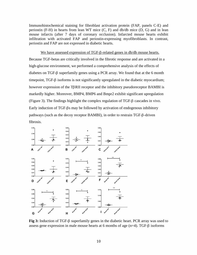
Immunohistochemical staining for fibroblast activation protein (FAP, panels C-E) and periostin (F-H) in hearts from lean WT mice (C, F) and db/db mice (D, G) and in lean mouse infarcts (after 7 days of coronary occlusion). Infarcted mouse hearts exhibit infiltration with activated FAP and periostin-expressing myofibroblasts. In contrast, periostin and FAP are not expressed in diabetic hearts.
We have assessed expression of TGF-β-related genes in db/db mouse hearts.
Because TGF-betas are critically involved in the fibrotic response and are activated in a
high-glucose environment, we performed a comprehensive analysis of the effects of
diabetes on TGF-β superfamily genes using a PCR array. We found that at the 6 month
timepoint, TGF-β isoforms is not significantly upregulated in the diabetic myocardium;
however expression of the TβRII receptor and the inhibitory pseudoreceptor BAMBI is
markedly higher. Moreover, BMP4, BMP6 and Bmpr2 exhibit significant upregulation
(Figure 3). The findings highlight the complex regulation of TGF-β cascades in vivo.
Early induction of TGF-βs may be followed by activation of endogenous inhibitory
pathways (such as the decoy receptor BAMBI), in order to restrain TGF-β-driven
fibrosis.
Fig 3: Induction of TGF-β superfamily genes in the diabetic heart. PCR array was used to assess gene expression in male mouse hearts at 6 months of age (n=4). TGF-β isoforms

11
were not significantly upregulated in db/db mice; however expression of the TβRII receptor and the inhibitory pseudoreceptor BAMBI were markedly elevated. There was also evidence for activation of the BMP cascade. BMP4, BMP6 and Bmpr2 expression levels were significantly higher in db/db hearts.
We are using lineage tracing strategies to test the hypothesis that cardiac pericytes
generate fibroblasts in the diabetic myocardium. We have crossed db/+ mice with NG2-
dsred pericyte reporter animals to generate diabetic pericyte reporter mice. db/+;
NG2dsred animals are bred with db/+ controls, in order to generate db/db;NG2dsred mice
(12.5% of the offspring). Unfortunately breeding of 2 NG2dsred animals appears to
significantly decrease litter size; thus generation of adequate numbers of diabetic pericyte
reporter mice will require expansion of our colonies. For lineage tracing, NG2-Cre mice
were crossed with R26RstopYFP (Rosa-YFP) mice in order to generate double transgenic
NG2-Cre-YFP mice to specifically label pericytes and their progeny. We currently have
12 breeding cages of db/+; NG2-Cre; Rosa-YFP to generate homozygote db/db mice that
are also positive for NG2-Cre Rosa-YFP. We have already generated 6 diabetic mice,
positive for NG-2Cre and Rosa-YFP that can be used for lineage tracing. The first
experimental material from these animals will be available in 2 months.
b) Work performed at VUMC
We are using Cre/Lox-based cell lineage tracing strategies to evaluate the
contribution of endothelial cells to the generation of extracellular matrix producing cells
in diabetic mouse hearts, thereby contributing to interstitial fibrosis and myocardial
dysfunction. To this end, we have crossed the Tie1-Cre mice to the R26RstopYFP (Rosa-
YFP) mice in order to generate double transgenic Tie-1-Cre-YFP mice to specifically
label endothelial cells and their progeny. These mice were then bred to each other and
genotyped in order to generate Tie-1-Cre-YFP mice that are homozygote for both the Cre
recombinase and R26RYFP loci. Double homozygote Tie-1-Cre-YFP mice were then
bred to db/+ heterozygotes to generate db/+ Tie1-Cre Rosa-YFP mice and non-diabetic
Tie1-Cre Rosa-YFP siblings as controls. We have obtained approximately 25 db/+
heterozygotes and 30 non-diabetic controls that are also heterozygote for both the Tie1-
Cre Rosa-YFP loci. We are currently breeding db/+ Tie-1-Cre Rosa-YFP mice to
generate homozygote db/db mice that are also positive for Tie-1-Cre Rosa-YFP. At

12
present, 5 breeding pairs are successfully breeding. Five newly added cages (total of 10
breeding pairs) are expected to expand the number of experimental animals with desired
genotypes to 10-15 mice, which will be sufficient to perform the endothelial cell lineage
tracing experiments.
c) Work performed collaboratively at VUMC and AECOM
To identify novel molecular pathways linked to pathological fibrosis and vascular
rarefaction in diabetics, we compared total heart gene expression profiles between female
control and diabetic mice at 6 months of age, where there is clear functional and
histological evidence of diabetic cardiomyopathy. In brief, hearts from 3 normal and 3
db/db mice were isolated at AECOM and shipped to VUMC where total RNA was
extracted using standard techniques. The 6 RNA samples were then sent to the
Vanderbilt sequencing core (VANTAGE). 5 of 6 samples (3 db/db and 2 normal
controls) passed the strict RNA quality control and processed for RNA sequencing. A
portion of the purified and validated RNA samples that passed quality controls was
shipped from VUMC back to AECOM for analysis using TGF-β superfamily gene PCR
arrays (please see section a above)
Paired end sequencing was performed at 75X depth with 45 million reads per
sample. Raw data (FASTQ files) were uploaded to Partek Flow for analysis and data
quality assessment. Sequences were aligned to the mm10 platform of the Mus musculus
genome using STAR, followed by total count normalization with minimum values set to
a minimum of 0.0001. Data were annotated using Ensemble Release 83 and quantified at
the gene level using the Partek E/M multimodel algorithm. Partek Genomics Suite 6.6
was used for principal components analysis and for hierarchical clustering with average
linkage and Euclidian distance of normalized values (RPM) generated from BAM files
(i.e., aligned sequences).
As shown in Figure 4, diabetic heart samples clustered separately from controls,
with 2,269 transcripts showing significant differences between the two genotypes. In

13
total, 310 genes showed higher and 1,959 genes showed lower expression levels in
diabetic hearts compared to normal controls.
Data were submitted to Gene Set Enrichment Analysis (GSEA) at the Broad
Institute and to DAVID (Database for Annotation, Visualization and Integrated
Discovery). Up and down-regulated processes were analyzed using DAVID and gene
ontology. GSEA provided signaling pathways derived from the RNA sequencing data
and associated genes.
Fig 4: Hierarchical clustering of 2,269 RNA transcript reads per kilobase per million mapped reads for genes with significant differential expression (fold difference >1.5, p value <0.05) between 2 control and 3 diabetic samples using Partek Genomics Suite with average linkage and Euclidean distance measures. Bright red, bright blue, and black indicate the highest, lowest, and median normalized reads, respectively. Vertical dendrograms represent the individual tissue samples (green = control, purple = diabetic).
The results showed that many upregulated transcripts represent genes expressed
during heart development (Figure 5), a finding that is consistent with previous reports
indicating activation of the fetal gene expression program in failing diabetic hearts.
Moreover, there is upregulation of extracellular matrix (ECM) proteins, indicative of
fibrosis. Among key signaling pathways, we found activation of Wnt signaling

14
components, suggesting a role of the pathway in defective angiogenesis and fibrosis in
diabetic hearts. In contrast, the majority of the downregulated pathways suggest
abnormal immune system response and deregulation of catabolic pathways (Figure 6).
Fig 5: Chart showing key up-regulated pathways in diabetic hearts. The number of genes in each pathway is indicated to the right.
Fig 6: Chart showing key down-regulated pathways in diabetic hearts. The number of genes in each pathway is indicated to the right.

15
Training opportunities and professional development
Linda Alex, PhD has joined the project. Dr Alex is a post-doctoral fellow who
completed her thesis work on the biology of cardiac fibroblasts. In our laboratory, Dr
Alex has characterized the cardiomyopathic process in diabetic mice (Figures 1-3), had
performed extensive in vitro work investigating the phenotype of diabetic fibroblasts and
has developed diabetic mouse lines for identification and lineage tracing of cardiac
pericytes. This project provides a unique training opportunity for Dr Alex, combining in
vitro and in vivo studies to dissect the cell biological basis of diabetes-associated cardiac
fibrosis.
How were the results disseminated to communities of interest?
A manuscript characterizing the fibrotic response in db/db mice is currently in
preparation and will be submitted for publication within the next few months. Findings
describing atrial remodeling in diabetic mice were recently published (Hanif et al.,
Cardiovasc. Pathol. 2017). Most of the findings presented in this report are preliminary;
these were not yet disseminated to the general scientific community and public, except
during informal presentations and discussions in internal work-in-progress meetings and
regular teleconferences between the partnering institutions. The concepts explored in this
proposal have significantly contributed to development of our paradigm on the
pathogenesis of cardiac fibrosis and are discussed in a series of invited review
manuscripts and editorials (please see “products”).
Plans for the next reporting period
During the next reporting period, we expect to perform a significant part of the
planned cell lineage tracing experiments. The time consuming breeding period to
achieve the desired triple genotypes is almost complete. Therefore, we expect to analyze
hearts from 2 and 6 months old mice using primarily histological techniques to assess the
extent of endothelial-to-mesenchymal transition contribution to vascular rarefaction and
fibrosis. Furthermore, the contribution of pericytes in fibrosis and the pathophysiology of
diabetic cardiomyopathy will be assessed using the corresponding Cre lines.

16
We will validate the primary RNAseq data using histological, molecular and
cellular techniques. These analyses will likely yield new insights in the development of
diabetic cardiomyopathy and may identify new targets to treat heart disease in diabetics.
We are particularly excited about the potential involvement of TGF-beta signaling
cascades in diabetes-associated fibrosis and the molecular links between the fundamental
metabolic perturbations in diabetes, obesity and metabolic dysfunction (hyperglycemia,
alterations in insulin signaling and adipokine expression, oxidative stress, generation of
advanced glycation end-products, etc) and the TGF-beta response.
Our preliminary data also show significant differences in gene activity in diabetic
hearts between female and male mice. Because women suffer more severe diabetic
cardiomyopathy than men, we plan to systematic compare gender-specific gene
expression profiles, expanding the RNAseq analysis to male hearts at comparable ages to
females. We will also perform independent lineage tracing studies in male and female
mice. We expect these original studies to provide novel information about gender-
specific deficits in diabetes.
4. Impact
HFpEF is a major cause of morbidity and mortality worldwide. There is currently
no effective treatment for patients with HFpEF. Although cardiac fibrosis has been
implicated in the pathogenesis of HFpEF, the cellular basis for fibrotic remodeling of the
ventricle is poorly understood. Metabolic diseases (such as obesity and diabetes) are
associated with an increased incidence of HFpEF; however, the pathophysiological
mechanisms responsible for this association remain unknown. Our experiments have
established a model of HFpEF due to metabolic disease that can be used to dissect
cellular mechanisms. This is of outstanding significance for pathophysiologic dissection
in vivo. Our planned experiments will use lineage tracing strategies, in vivo and in vitro
approaches to dissect the basis for activation of diabetic fibroblasts. The significance of
the studies extends beyond the cardiovascular field, as diabetes-associated tissue fibrosis
has an impact on other organs (such as the kidney and the liver).

17
5. Changes/Problems
No changes or problems to report
6. Products
Publications
1. NG Frangogiannis. The functional pluralism of fibroblasts in the infarcted
myocardium. Circ Res 2016; 119: 1049-1051. Acknowledgment of DoD grant support:
Yes.
2. AV Shinde, C Humeres, and NG Frangogiannis. The role of α-smooth muscle actin in
fibroblast-mediated matrix contraction and remodeling. BBA Mol Bas Dis 2017; 1863:
298-309. Acknowledgment of DoD grant support: Yes.
3. NG Frangogiannis. The extracellular matrix in myocardial injury, repair and
remodeling. J Clin Invest 2017; 127: 1600-1612. Acknowledgment of DoD grant support:
Yes.
4. W Hanif, L Alex, Y Su, AV Shinde, I Russo, N Li, and NG Frangogiannis. Left atrial
remodeling, hypertrophy and fibrosis in mouse models of heart failure. Cardiovasc
Pathol 2017;30-27-37. Acknowledgment of DoD grant support: Yes.
5. AV Shinde, M Dobaczewski, JJ De Haan, A Saxena, KK Lee, Y Xia, W Chen, Y Su,
W Hanif, IK Madahar, VM Paulino, G Melino and NG Frangogiannis. Tissue
transglutaminase induction in the pressure-overloaded myocardium regulates cardiac
remodeling. Cardiovasc Res 2017; 113:892-905. Acknowledgment of DoD grant support:
Yes.
6. NG Frangogiannis. Activation of the innate immune system in the pathogenesis of
acute heart failure. Eur Heart J Acute Cardiovasc Care 2017 Apr1 (epub ahead of print).
Acknowledgment of DoD grant support: Yes.

18
7. NG Frangogiannis. Fibroblasts and the extracellular matrix in right ventricular disease.
Cardiovasc Res 2017; 113: 12: 1453-1464. Acknowledgment of DoD grant support: Yes.
8. Shinde AV and NG Frangogiannis. Mechanisms of fibroblast activation in the
remodeling myocardium. Curr Pathobiol Rep 2017; 5:145-152. Acknowledgment of DoD
grant support: Yes.
5. PARTICIPANTS & OTHER COLLABORATING ORGANIZATIONS
What individuals have worked on the project? Provide the following information for: (1) PDs/PIs; and (2) each person who has worked at least one person month per year on the project during the reporting period, regardless of the source of compensation (a person month equals approximately 160 hours of effort). If information is unchanged from a previous submission, provide the name only and indicate “no change”.
Name Nikolaos Frangogiannis "no change"
Project Role PI Research Identifier e.g ORCHID ID):
Nearest person month worked 2 Contribution to Project Dr Frangogiannis developed the
proposed concepts, designed the experiments, and participated to the analysis.
Funding Support Name Ya Su "no change" Project Role Associate Research Identifier e.g ORCHID ID):
Nearest person month worked 6 Contribution to Project Dr Ya Su performs in vivo
echocardiographic experiments and contributes to the development of mouse lines for lineage tracing and to the in vitro studies.

19
Funding Support Name Linda Alex "no change" Project Role Research Associate
Research Identifier e.g ORCHID ID)
Nearest person month worked 12
Contribution to Project Dr Linda Alex is breeding the colonies of diabetic pericyte reporter mice and the animals for lineage tracing. She performs genotypic and characterization of all genetic tools. She performs in vitro experiments for stimulation of cardiac pericytes and fibroblasts.
Funding Support

20
Has there been a change in the active other support of the PD/PI(s) or senior/key personnel since the last reporting period? If there is nothing significant to report during this reporting period, state “Nothing to Report.” If the active support has changed for the PD/PI(s) or senior/key personnel, then describe what the change has been. Changes may occur, for example, if a previously active grant has closed and/or if a previously pending grant is now active. Annotate this information so it is clear what has changed from the previous submission. Submission of other support information is not necessary for pending changes or for changes in the level of effort for active support reported previously. The awarding agency may require prior written approval if a change in active other support significantly impacts the effort on the project that is the subject of the project report.
What other organizations were involved as partners? If there is nothing significant to report during this reporting period, state “Nothing to Report.” Describe partner organizations – academic institutions, other nonprofits, industrial or commercial firms, state or local governments, schools or school systems, or other organizations (foreign or domestic) – that were involved with the project. Partner organizations may have provided financial or in-kind support, supplied facilities or equipment, collaborated in the research, exchanged personnel, or otherwise contributed. Provide the following information for each partnership: Organization Name: Location of Organization: (if foreign location list country) Partner’s contribution to the project (identify one or more) • Financial support; • In-kind support (e.g., partner makes software, computers, equipment, etc.,
available to project staff); • Facilities (e.g., project staff use the partner’s facilities for project activities); • Collaboration (e.g., partner’s staff work with project staff on the project);
No changes to report.

21
• Personnel exchanges (e.g., project staff and/or partner’s staff use each other’s facilities, work at each other’s site); and
• Other. Organization Name Vanderbilt University
Location of Organization Vanderbilt Center for Stem Cell MRB IV - P425C Biology 2213 Garland Avenue Nashville, TN 37232-6300
Collaborating PI: Dr Antonis Hatzopoulos
Partner’s Contribution Dr. Antonis Hatzopoulos is an expert in the cell biology of the vascular cells and examines the involvement of endothelial cells in mediating diabetes-associated fibrosis
Financial Support
Facilities Collaboration Collaboration between the
PIs as outlined above
Personal exchanges Other
6. SPECIAL REPORTING REQUIREMENTS
COLLABORATIVE AWARDS: For collaborative awards, independent reports are required from BOTH the Initiating Principal Investigator (PI) and the Collaborating/Partnering PI. A duplicative report is acceptable; however, tasks shall be clearly marked with the responsible PI and research site. A report shall be submitted to https://ers.amedd.army.mil for each unique award. They must also submit a progress report. Dr Hatzopoulos (Partnering PI) will submit his own progress report. Both reports contain the same description of the accomplishments; contributions of each laboratory to the reported accomplishments are clearly indicated.

22
QUAD CHARTS: If applicable, the Quad Chart (available on https://www.usamraa.army.mil) should be updated and submitted with attachments. N/A
7. APPENDICES: Attach all appendices that contain information that supplements, clarifies or supports the text. Examples include original copies of journal articles, reprints of manuscripts and abstracts, a curriculum vitae, patent applications, study questionnaires, and surveys, etc.
Active other support of the PI (Nikolaos Frangogiannis)
ACTIVE: Title of the project: Chemokines in healing myocardial infarcts R01 HL076246 Funding agency: NIH/NHLBI Investigator relationship: Principal Investigator Dates of funding: 6/2005-4/2019. The project deals with cell-specific chemokine and cytokine actions in the infarcted myocardium. There is no overlap with the current DoD-funded project. Title of the project: Resolution of inflammation in healing myocardial infarcts R01 HL085440 Funding agency: NIH/NHLBI Investigator relationship: Principal Investigator Dates of funding: 12/2007-5/2021 This project deals with the role of intracellular signals that inhibit innate immune responses in the infarcted heart. There is no overlap with the current DoD-funded project. Title of the project: Fibroblast-Cardiomyocyte Interactions in the Pressure Overloaded Myocardium PR151134 Funding agency: Department of Defense office of the Congressionally Directed Medical Research Programs (CDMRP). Investigator relationship: Principal Investigator (Partnering PI award; co-PI: Dr. Richard Kitsis Albert Einstein College of Medicine) Dates of funding: 9/15/2016-9/14/2019.

23
Goals of the project: The project studies protective interactions between fibroblasts and cardiomyocytes in the pressure-overloaded heart. There is no overlap with the current DoD-funded project. PENDING: None OVERLAP: None

1049
Editorial
The mammalian heart contains a large population of in-terstitial fibroblast–like cells; in the adult mouse myo-
cardium, 10% to 30% of myocardial cells were identified as fibroblasts.1,2 These cells expand following injury and play an important role in cardiac repair3 but may also participate in the pathogenesis of adverse postinfarction remodeling.4 Traditional views consider cardiac fibroblasts as matrix-pro-ducing cells that simply serve to preserve the structural in-tegrity of the ventricle after acute myocardial infarction by replacing dead cardiomyocytes with scar tissue and contribute to cardiac fibrosis in pathophysiologic conditions associated with chronic pressure overload or metabolic dysfunction.5,6 However, this unidimensional view is not an accurate reflec-tion of fibroblast function. A growing body of in vitro findings and in vivo observations suggests that fibroblasts exhibit a re-markable functional pluralism. In addition to their established role in matrix synthesis and metabolism, fibroblasts are also capable of secreting a wide range of immunoregulatory, cyto-protective, and angiogenic mediators in response to microen-vironmental changes.
Article, see p 1116
In adult mammals, sudden death of myocardial cells over-whelms the negligible regenerative reserve of the myocardi-um; as a result, the infarcted heart heals through the formation of a collagen-based scar. Repair of the infarcted myocardium is dependent on timely activation and repression of an inflam-matory reaction that serves to clear the infarct from dead cells and matrix debris. Inflammation after myocardial infarction is activated through the release of danger-associated molecular patterns from dying cells and degraded matrix.7 These danger signals have been reported to activate all cell types involved in cardiac injury and repair. Cardiac fibroblasts respond to danger-associated molecular patterns and can produce large amounts of chemokines and cytokines that may play an im-portant role in the activation of the postinfarction inflamma-tory response.8 Moreover, it has been suggested that cardiac
fibroblasts may modulate prosurvival signaling cascades in ischemic cardiomyocytes, affecting their susceptibility to apoptosis or necrosis. Unfortunately, these intriguing con-cepts on the role of fibroblasts in myocardial disease are cur-rently supported almost exclusively by in vitro experiments and by associative evidence.7,9,10 Considering the wide range of cell types capable of responding to danger signals trigger-ing the inflammatory reaction after myocardial infarction, the relative significance of fibroblasts remains unclear. Dissection and documentation of the role of fibroblasts as cellular effec-tors of myocardial inflammation have been hampered by the challenges in the development of fibroblast-specific targeting approaches in vivo.11
In this issue of Circulation Research, Woodall et al12 pro-vide the first direct in vivo evidence supporting a crucial role for cardiac fibroblasts in regulating cardiomyocyte survival and in triggering the inflammatory response after myocardial infarction. The authors generated mice with fibroblast-specific loss of G protein–coupled receptor kinase 2 (GRK2), a ubiq-uitous member of the GRK family with a central role in signal transduction. In a model of reperfused myocardial infarction, fibroblast-specific GRK2 loss reduced the size of the infarct, decreasing secretion of proinflammatory cytokines, such as tumor necrosis factor-α, and attenuating cardiomyocyte apop-tosis. In vitro, GRK2 loss attenuated nuclear translocation of nuclear factor-κB and subsequent tumor necrosis factor-α synthesis in isolated fibroblasts. Moreover, conditioned media from fibroblasts lacking GRK2 potentiated Akt signaling in cardiomyocytes, suggesting the activation of a cytoprotective pathway. Although the study provides the first direct docu-mentation of a crucial role for cardiac fibroblasts in regulat-ing cardiomyocyte injury and inflammation in the early stages after myocardial ischemia, the molecular mechanisms respon-sible for the observed effects remain unclear.
Do Cardiac Fibroblasts in the Ischemic Myocardium Function as Inflammatory Cells?
After myocardial infarction, release of danger-associated mo-lecular patterns activates innate immune signaling pathways in several different cell types, triggering an intense inflamma-tory reaction. Endothelial cells, leukocytes, mast cells, and surviving cardiomyocytes have been suggested as likely cel-lular targets of danger-associated molecular patterns released by necrotic cells and may contribute to the activation of the postinfarction inflammatory response by secreting cytokines and chemokines.13–15 Cardiac fibroblasts are also capable of se-creting large amounts of proinflammatory mediators on stimu-lation with danger signals. Interleukin-1 is rapidly released in the infarcted myocardium and promotes a proinflammatory and matrix-degrading fibroblast phenotype, while suppressing
The Functional Pluralism of Fibroblasts in the Infarcted Myocardium
Nikolaos G. Frangogiannis
(Circ Res. 2016;119:1049-1051.DOI: 10.1161/CIRCRESAHA.116.309926.)
© 2016 American Heart Association, Inc.
Circulation Research is available at http://circres.ahajournals.org DOI: 10.1161/CIRCRESAHA.116.309926
The opinions expressed in this article are not necessarily those of the editors or of the American Heart Association.
From the Department of Medicine (Cardiology), The Wilf Family Cardiovascular Research Institute, Albert Einstein College of Medicine, Bronx, NY.
Correspondence to Nikolaos G. Frangogiannis, MD, Department of Medicine (Cardiology), The Wilf Family Cardiovascular Research Institute, Albert Einstein College of Medicine, 1300 Morris Park Ave Forchheimer G46B, Bronx, NY 10461. E-mail [email protected]
by guest on October 27, 2016
http://circres.ahajournals.org/D
ownloaded from

1050 Circulation Research October 28, 2016
α-smooth muscle actin synthesis and inhibiting myofibroblast conversion.9 Thus, interleukin-1 stimulation may delay pre-mature infiltration of the infarct with matrix-synthetic fibro-blasts, until the wound is cleared from dead cells and matrix debris. Although the findings of this study are consistent with an important role of cardiac fibroblasts in promoting inflam-mation after myocardial infarction, the protective effects of GRK2 loss may not be caused by direct anti-inflammatory ac-tions. Fibroblast-specific GRK2 loss decreased neutrophil in-filtration in vivo and reduced tumor necrosis factor-α release in vitro. However, the in vivo attenuation of the inflammatory response may represent an epiphenomenon reflecting the sig-nificant reduction in infarct size, in the absence of a primary role of GRK2 in regulation of inflammation. The notion that GRK2 may be directly involved in the activation of a proin-flammatory program is not supported by studies in immune cells. In vivo and in vitro investigations in T cells16 and in myeloid cells17 suggested that GRK2 not only does not stimu-late inflammatory gene synthesis but may also be involved in negative regulation of inflammation.
Fibroblasts May Regulate Cardiomyocyte Survival
A growing body of evidence suggests that in injured and re-modeling hearts, fibroblasts critically regulate cardiomyocyte responses. In the pressure-overloaded myocardium, activated fibroblasts transduce hypertrophic signals18 mediated, at least in part, through secretion of miRNA-enriched exosomes.19 In myocardial ischemia, administration of the secretome of neona-tal cardiac fibroblasts before reperfusion significantly reduced the size of the infarct.10 The current investigation suggests that endogenous fibroblast GRK2 signaling may extend ischemic injury, accentuating cardiomyocyte apoptosis. Several mecha-nisms may account for the proapoptotic effects of activated fi-broblasts during the early postischemic phase (Figure). First, fibroblasts may secrete soluble proapoptotic mediators, such as proinflammatory cytokines, thus promoting cardiomyocyte death. Second, fibroblasts may indirectly reduce cardiomyocyte survival by modulating the composition of the extracellular matrix through the secretion of proteases. Protease-mediated degradation of the pericellular extracellular matrix may deprive ischemic cardiomyocytes from essential prosurvival signals. Third, ischemic fibroblasts may secrete exosomes that activate proapoptotic pathways in cardiomyocytes. Final, activation of GRK2 in ischemic fibroblasts may inhibit a yet unidentified prosurvival mechanism that may involve fibroblast-derived se-cretion of soluble mediators or deposition of matricellular pro-teins. Unfortunately, this study did not systematically pursue the mechanisms responsible for these intriguing interactions between fibroblasts and cardiomyocytes.
The Role of Activated Fibroblasts During the Proliferative Phase of Infarct Healing: Beyond
Matrix SynthesisClearance of the infarcted heart from dead cells and matrix debris is associated with the activation of anti-inflammatory pathways, leading to the suppression and resolution of the inflammatory response.20 Although cardiac fibroblasts are
capable of producing large amounts of anti-inflammatory cytokines, such as interleukin-10 and transforming growth factor-β,21 whether they actively participate in negative regula-tion of the inflammatory response remains unknown. Growth factor-mediated conversion of fibroblasts into myofibroblasts is associated with the activation of a matrix-synthetic program and secretion of collagens. Deposition of structural matrix proteins is the best-documented function of myofibroblasts in healing infarcts.3 In addition to their role in scar formation, infarct myofibroblasts may also serve as an important source of growth factors and matricellular proteins, regulating the an-giogenic response after myocardial infarction.22 Whether the diverse functions of infarct fibroblasts in inflammation and repair reflect the activation of specific subpopulations remains unknown. Although several different developmental sources of cardiac fibroblasts have been identified in normal and in-jured hearts,2 the functional properties of these cells have not been systematically investigated. The inducible collagen1α2-Cre driver used in this study should target all cardiac fibro-blasts, thus precluding any conclusions on distinct effects of specific subsets.
Targeting the Cardiac Fibroblast in the Infarcted and Remodeling Myocardium
The consistent association between cardiac fibrosis and ad-verse outcome in a wide range of cardiac conditions has suggested that the fibroblast may be a promising therapeutic target in patients with myocardial infarction or heart failure. However, unlike primary fibrotic disorders in other systems (such as systemic sclerosis or idiopathic pulmonary fibro-sis), in the myocardium, fibrotic remodeling often reflects
Figure. Cardiac fibroblasts are not unidimensional matrix-secreting cells but exhibit remarkable functional pluralism. After myocardial infarction, release of danger-associated molecular patterns (DAMPs) by necrotic cardiomyocytes (CM) activates a proinflammatory phenotype in fibroblasts, inducing secretion of cytokines and chemokines and stimulating leukocyte (L) infiltration. Moreover, during the early postischemic period, fibroblasts may modulate survival pathways in cardiomyocytes, affecting their susceptibility to ischemic death. These effects may be mediated through the secretion of soluble proapoptotic or antiapoptotic mediators by fibroblasts, via release of exosomes containing miRNAs or through modulation of the extracellular matrix (ECM) by fibroblast-derived matrix metalloproteinases (MMPs). MMP-mediated degradation of the matrix may deprive fibroblasts from essential prosurvival signals. Fibroblast G protein–coupled receptor kinase 2 (GRK2) signaling may extend ischemic injury after myocardial infarction through proinflammatory actions or by activating a proapoptotic pathway in cardiomyocytes.
by guest on October 27, 2016
http://circres.ahajournals.org/D
ownloaded from

Frangogiannis Fibroblasts in Myocardial Infarction 1051
a reparative process that is activated in response to car-diomyocyte injury. In conditions associated with replace-ment fibrosis, such as myocardial infarction, targeting the reparative functions of fibroblasts may have catastrophic consequences. Implementation of therapeutic strategies tar-geting fibroblasts is further complicated by the wide range of modulatory functions of fibroblasts on cardiomyocyte hypertrophy and survival, on inflammatory activation, and on angiogenesis. In vivo dissection of the diverse actions of cardiac fibroblasts after injury is crucial to design therapeu-tic strategies that target detrimental actions, without inter-fering with protective effects. Moreover, identification and characterization of fibroblast subsets with distinct pheno-typic characteristics and functional profiles may explain the functional pluralism of fibroblasts in injured and remodel-ing tissues.
Sources of FundingDr Frangogiannis’s laboratory is supported by National Institutes of Health grants R01 HL76246 and R01 HL85440 and by grants from the Department of Defense Congressionally Directed Medical Research Programs (CDMRP).
DisclosuresNone.
References 1. Pinto AR, Ilinykh A, Ivey MJ, Kuwabara JT, D’Antoni ML, Debuque R,
Chandran A, Wang L, Arora K, Rosenthal NA, Tallquist MD. Revisiting cardiac cellular composition. Circ Res. 2016;118:400–409. doi: 10.1161/CIRCRESAHA.115.307778.
2. Ali SR, Ranjbarvaziri S, Talkhabi M, Zhao P, Subat A, Hojjat A, Kamran P, Müller AM, Volz KS, Tang Z, Red-Horse K, Ardehali R. Developmental heterogeneity of cardiac fibroblasts does not predict pathological proliferation and activation. Circ Res. 2014;115:625–635. doi: 10.1161/CIRCRESAHA.115.303794.
3. Shinde AV, Frangogiannis NG. Fibroblasts in myocardial infarction: a role in inflammation and repair. J Mol Cell Cardiol. 2014;70:74–82. doi: 10.1016/j.yjmcc.2013.11.015.
4. Kaur H, Takefuji M, Ngai CY, Carvalho J, Bayer J, Wietelmann A, Poetsch A, Hoelper S, Conway SJ, Möllmann H, Looso M, Troidl C, Offermanns S, Wettschureck N. Targeted ablation of perios-tin-expressing activated fibroblasts prevents adverse cardiac re-modeling in mice. Circ Res. 2016;118:1906–1917. doi: 10.1161/CIRCRESAHA.116.308643.
5. Kong P, Christia P, Frangogiannis NG. The pathogenesis of cardiac fibro-sis. Cell Mol Life Sci. 2014;71:549–574. doi: 10.1007/s00018-013-1349-6.
6. Travers JG, Kamal FA, Robbins J, Yutzey KE, Blaxall BC. Cardiac fi-brosis: the fibroblast awakens. Circ Res. 2016;118:1021–1040. doi: 10.1161/CIRCRESAHA.115.306565.
7. Zhang W, Lavine KJ, Epelman S, Evans SA, Weinheimer CJ, Barger PM, Mann DL. Necrotic myocardial cells release damage-associated molecular patterns that provoke fibroblast activation in vitro and trig-ger myocardial inflammation and fibrosis in vivo. J Am Heart Assoc. 2015;4:e001993. doi: 10.1161/JAHA.115.001993.
8. Smith RS, Smith TJ, Blieden TM, Phipps RP. Fibroblasts as sentinel cells. Synthesis of chemokines and regulation of inflammation. Am J Pathol. 1997;151:317–322.
9. Saxena A, Chen W, Su Y, Rai V, Uche OU, Li N, Frangogiannis NG. IL-1 induces proinflammatory leukocyte infiltration and regulates fibroblast phenotype in the infarcted myocardium. J Immunol. 2013;191:4838–4848. doi: 10.4049/jimmunol.1300725.
10. Abrial M, Da Silva CC, Pillot B, Augeul L, Ivanes F, Teixeira G, Cartier R, Angoulvant D, Ovize M, Ferrera R. Cardiac fibroblasts protect cardio-myocytes against lethal ischemia-reperfusion injury. J Mol Cell Cardiol. 2014;68:56–65. doi: 10.1016/j.yjmcc.2014.01.005.
11. Kong P, Christia P, Saxena A, Su Y, Frangogiannis NG. Lack of specific-ity of fibroblast-specific protein 1 in cardiac remodeling and fibrosis. Am J Physiol Heart Circ Physiol. 2013;305:H1363–H1372. doi: 10.1152/ajpheart.00395.2013.
12. Woodall MC, Woodall BP, Ghao E, Yuan A, Koch WJ. Cardiac fibroblast GRK2 deletion enhances contractility and remodeling following isch-emia/reperfusion injury. Circ Res. 2016:119:1116–1127. doi: 10.1161/CIRCRESAHA.116.309538.
13. Arslan F, Smeets MB, O’Neill LA, Keogh B, McGuirk P, Timmers L, Tersteeg C, Hoefer IE, Doevendans PA, Pasterkamp G, de Kleijn DP. Myocardial ischemia/reperfusion injury is mediated by leukocytic toll-like receptor-2 and reduced by systemic administration of a novel anti-toll-like receptor-2 antibody. Circulation. 2010;121:80–90. doi: 10.1161/CIRCULATIONAHA.109.880187.
14. Frangogiannis NG, Lindsey ML, Michael LH, Youker KA, Bressler RB, Mendoza LH, Spengler RN, Smith CW, Entman ML. Resident cardiac mast cells degranulate and release preformed TNF-alpha, initiating the cytokine cascade in experimental canine myocardial ischemia/reperfu-sion. Circulation. 1998;98:699–710.
15. Zhu M, Goetsch SC, Wang Z, Luo R, Hill JA, Schneider J, Morris SM Jr, Liu ZP. FoxO4 promotes early inflammatory response upon myocar-dial infarction via endothelial Arg1. Circ Res. 2015;117:967–977. doi: 10.1161/CIRCRESAHA.115.306919.
16. Vroon A, Heijnen CJ, Lombardi MS, Cobelens PM, Mayor F Jr, Caron MG, Kavelaars A. Reduced GRK2 level in T cells potentiates chemotax-is and signaling in response to CCL4. J Leukoc Biol. 2004;75:901–909. doi: 10.1189/jlb.0403136.
17. Patial S, Saini Y, Parvataneni S, Appledorn DM, Dorn GW II, Lapres JJ, Amalfitano A, Senagore P, Parameswaran N. Myeloid-specific GPCR ki-nase-2 negatively regulates NF-κB1p105-ERK pathway and limits endo-toxemic shock in mice. J Cell Physiol. 2011;226:627–637. doi: 10.1002/jcp.22384.
18. Takeda N, Manabe I, Uchino Y, Eguchi K, Matsumoto S, Nishimura S, Shindo T, Sano M, Otsu K, Snider P, Conway SJ, Nagai R. Cardiac fibro-blasts are essential for the adaptive response of the murine heart to pres-sure overload. J Clin Invest. 2010;120:254–265. doi: 10.1172/JCI40295.
19. Bang C, Batkai S, Dangwal S, et al. Cardiac fibroblast-derived microR-NA passenger strand-enriched exosomes mediate cardiomyocyte hyper-trophy. J Clin Invest. 2014;124:2136–2146. doi: 10.1172/JCI70577.
20. Prabhu SD, Frangogiannis NG. The biological basis for cardiac repair after myocardial infarction: from inflammation to fibrosis. Circ Res. 2016;119:91–112. doi: 10.1161/CIRCRESAHA.116.303577.
21. Driesen RB, Nagaraju CK, Abi-Char J, Coenen T, Lijnen PJ, Fagard RH, Sipido KR, Petrov VV. Reversible and irreversible differentiation of car-diac fibroblasts. Cardiovasc Res. 2014;101:411–422. doi: 10.1093/cvr/cvt338.
22. Dostal D, Glaser S, Baudino TA. Cardiac fibroblast physiology and pa-thology. Compr Physiol. 2015;5:887–909. doi: 10.1002/cphy.c140053.
Key Words: Editorials ■ fibroblasts ■ inflammation ■ mice ■ myocardial infarction ■ myocardium
by guest on October 27, 2016
http://circres.ahajournals.org/D
ownloaded from

Nikolaos G. FrangogiannisThe Functional Pluralism of Fibroblasts in the Infarcted Myocardium
Print ISSN: 0009-7330. Online ISSN: 1524-4571 Copyright © 2016 American Heart Association, Inc. All rights reserved.is published by the American Heart Association, 7272 Greenville Avenue, Dallas, TX 75231Circulation Research
doi: 10.1161/CIRCRESAHA.116.3099262016;119:1049-1051Circ Res.
http://circres.ahajournals.org/content/119/10/1049World Wide Web at:
The online version of this article, along with updated information and services, is located on the
http://circres.ahajournals.org//subscriptions/
is online at: Circulation Research Information about subscribing to Subscriptions:
http://www.lww.com/reprints Information about reprints can be found online at: Reprints:
document. Permissions and Rights Question and Answer about this process is available in the
located, click Request Permissions in the middle column of the Web page under Services. Further informationEditorial Office. Once the online version of the published article for which permission is being requested is
can be obtained via RightsLink, a service of the Copyright Clearance Center, not theCirculation Researchin Requests for permissions to reproduce figures, tables, or portions of articles originally publishedPermissions:
by guest on October 27, 2016
http://circres.ahajournals.org/D
ownloaded from

Biochimica et Biophysica Acta 1863 (2017) 298–309
Contents lists available at ScienceDirect
Biochimica et Biophysica Acta
j ourna l homepage: www.e lsev ie r .com/ locate /bbad is
The role of α-smooth muscle actin in fibroblast-mediated matrixcontraction and remodeling
Arti V. Shinde, Claudio Humeres, Nikolaos G. Frangogiannis ⁎The Wilf Family Cardiovascular Research Institute, Department of Medicine (Cardiology), Albert Einstein College of Medicine, Bronx, NY, United States
⁎ Corresponding author at: The Wilf Family CardiovasEinstein College of Medicine, 1300 Morris Park Avenue10461, United States.
E-mail address: [email protected]
http://dx.doi.org/10.1016/j.bbadis.2016.11.0060925-4439/© 2016 Elsevier B.V. All rights reserved.
a b s t r a c t
a r t i c l e i n f oArticle history:Received 21 June 2016Received in revised form 9 October 2016Accepted 2 November 2016Available online 4 November 2016
Cardiac myofibroblasts play an important role in myocardial remodeling. Although α-smooth muscle actin (α-SMA) expression is the hallmark ofmaturemyofibroblasts, its role in regulating fibroblast function remains poor-ly understood.We explore the effects of thematrix environment inmodulating cardiac fibroblast phenotype, andwe investigate the role of α-SMA in fibroblast function using loss- and gain-of-function approaches. In murinemyocardial infarction, infiltration of the infarct border zone with abundant α-SMA-positive myofibroblastswas associated with scar contraction. Isolated cardiac fibroblasts cultured in plates showed high α-SMA expres-sion localized in stress fibers, exhibited activation of focal adhesion kinase (FAK), and synthesized large amountsof extracellular matrix proteins. In contrast, when these cells were cultured in collagen lattices, they exhibitedmarked reduction of α-SMA expression, negligible FAK activation, attenuated collagen synthesis, and increasedtranscription of genes associatedwithmatrixmetabolism. TransformingGrowth Factor-β1-mediated contractionof fibroblast-populated collagen pads was associatedwith accentuatedα-SMA synthesis. In contrast, serum- andbasic Fibroblast Growth Factor-induced collagen pad contraction was associated with reduced α-SMA expres-sion. α-SMA siRNA knockdown attenuated contraction of collagen pads populated with serum-stimulatedcells. Surprisingly, α-SMA overexpression also reduced collagen pad contraction, suggesting that α-SMA is notsufficient to promote contraction of the matrix. Reduced contraction by α-SMA-overexpressing cells wasassociated with attenuated proliferative activity, in the absence of any effects on apoptosis. α-SMA may beimplicated in contraction and remodeling of the extracellular matrix, but is not sufficient to induce contraction.α-SMA expression may modulate cellular functions, beyond its effects on contractility.
© 2016 Elsevier B.V. All rights reserved.
Keywords:Myofibroblastα-Smooth muscle actinMyocardial infarctionExtracellular matrixTransforming growth factor-β
1. Introduction
In healing tissues, fibroblasts acquire a contractile phenotype,characterized by formation of microfilament bundles, and by de novoexpression of α-smooth muscle actin (α-SMA). These activated cells,termed “myofibroblasts” [1–7] participate in the reparative response,by secreting large amounts of extracellular matrix proteins [8,9] andmay be responsible for contraction of healing wounds [10]. Repair ofinjured tissues is dependent on timely activation and deactivation ofmyofibroblasts; prolonged or excessive myofibroblast activity mayresult in fibrosis and organ dysfunction [11–13].
The normal mammalian myocardium contains a large number offibroblast-like cells [14–16]. In the absence of injury, these interstitialcells remain quiescent; however, a wide range of injurious processes
cular Research Institute, AlbertForchheimer G46B, Bronx, NY
u (N.G. Frangogiannis).
can induce cardiac fibroblast activation. Conversion of cardiac fibroblastsinto activated α-SMA-positive myofibroblasts is consistently noted fol-lowing myocardial infarction, both in human patients [17] and in exper-imental models [18,19]. Because the adult mammalian heart hasnegligible regenerative capacity, activated myofibroblasts play a crucialrole in post-infarction cardiac repair, by secreting extracellular matrixproteins, thus protecting the heart from catastrophic rupture. Moreover,the capacity of infarct myofibroblasts to contract the scar may play animportant role in protecting the chamber from adverse remodeling[20]. On the other hand, excessive activation of fibroblasts in the infarct-ed heart may contribute to fibrosis, increase stiffness, and promote bothsystolic and diastolic dysfunction [21,12].
Our group and other investigators have used experimental animalmodels and in vitro approaches to study the role of fibroblasts in theinfarcted and remodelingmyocardium [22–29]. In vitro studies investi-gating myocardial fibrotic responses, have used either cardiac fibro-blasts cultured and stimulated in plates, or cells enmeshed in collagenlattices [25,30–32]. Assessment of fibroblast-mediated contraction incollagen pads provides a robust and pathophysiologically relevantmodel of fibroblast function. Although α-SMA expression is a hallmark

299A.V. Shinde et al. / Biochimica et Biophysica Acta 1863 (2017) 298–309
of the mature myofibroblast, its role in regulation of fibroblast behaviorand function remains poorly understood. Studies using fibroblast-populated collagen lattices suggested that α-SMA expression increasesfibroblast contractile activity [33], and plays a crucial role in focaladhesion maturation [34]. However, other investigations showed that,at a single cell level, fibroblasts and myofibroblasts were found toexert comparable contractile forces [35]. Because fibroblasts are highlydynamic cells [36], interpretation of the findings derived from differentin vitro models requires understanding of the distinct characteristics offibroblasts in eachmodel. Our study compares cardiac fibroblast pheno-type between the two models, and explores the role of α-SMA infibroblast-mediated matrix contraction.
We report that, in both reperfused and non-reperfused mouseinfarcts, scar contraction is associated with marked infiltration of theinfarct border zone with myofibroblasts. In vitro, culture of cardiacfibroblasts in collagen pads markedly suppressed α-SMA expressionand reduced extracellular matrix synthesis, while promoting synthesisof genes associated with matrix metabolism. Contraction of fibroblast-populated pads in response to serum, or specific growth factorswas not consistently associated with upregulation of α-SMA. siRNAknockdown and overexpression experiments demonstrated that α-SMA is involved in collagen pad contraction, but is not sufficient toinduce contraction of the matrix. Surprisingly α-SMA expressionmodulated proliferative activity in cardiac fibroblasts. Our findingshighlight the dynamic phenotype of fibroblasts under differentconditions and suggest that α-SMA may exert actions independent ofits effects on cell contraction.
2. Materials and methods
2.1. Mouse models of reperfused and non-reperfused myocardial infarction
Both male and female, 3–4 month old C57/BL6J mice underwentcoronary occlusion/reperfusion protocols as previously described[37]. Mice were anesthetized by isoflurane inhalation (isoflurane2–3% vol/vol). Non-reperfused myocardial infarction was inducedusing an open chest model of permanent left coronary artery ligation[38]. Reperfused infarction was induced using a well-characterizedclosed-chest model of coronary occlusion and reperfusion [39].Infarcted hearts (after 7 or 28 days of permanent coronary occlusionand after 1 h ischemia/7–28 days of reperfusion) were fixed informalin and embedded in paraffin.
2.2. Immunohistochemistry and histology
Infarcted hearts were sectioned systematically from based toapex in 250 μm partitions as previously described [39]. One sectionfrom each partition was stained for sirius red to identify the healingscar. Scar size was morphometrically assessed using ImagePro soft-ware by dividing the total scar area to the total area of the left ventri-cle (from all partitions). Myofibroblasts in infarcted hearts wereidentified, as spindle-shaped cells located outside the vascularmedia with α-SMA immunofluorescence using the anti-α-SMA anti-body (1A4, Santa Cruz Biotech) and an Alexa-Fluor 594-labeled sec-ondary antibody (Molecular Probes). Sections were counterstainedwith DAPI.
2.3. Isolation and stimulation of mouse cardiac fibroblasts
Cardiac fibroblasts were isolated from C57/BL6J animals using enzy-matic digestion as previously described [40,41] and were cultured inDMEM/F12 (GIBCO Invitrogen Corporation, Carlsbad, CA) with 10%Fetal Calf Serum (FCS). Cells were serum-starved at passage 2 for 16 hand subsequently stimulated with either 10 ng/ml or 50 ng/ml ofrecombinant TGF-β1 (R&D Systems, Minneapolis MN) for 4–24 h.Total RNA was isolated from the stimulated cells using GeneJET RNA
Purification Kit (ThermoFisher Scientific) and was reverse transcribedto cDNA using the iScript™ cDNA synthesis kit (Bio-Rad) following themanufacturer's guidelines. Quantitative PCR was performed usingSsoFast™ EvaGreen® Supermix (Bio-Rad) on the CFX384™ Real-TimePCR Detection System (Bio-Rad). Primerswere synthesized by Integrat-edDNATechnologies. The following geneswere assessed:α-SMA, type Icollagen, type III collagen, matrix metalloproteinase (MMP)2, MMP3,MMP8, tissue inhibitor of metalloproteinases (TIMP)1, fibronectin andGAPDH. Each sample was run in triplicate. For experiments withcollagen pads, RNA was isolated from the stimulated collagen padsusing GeneJET RNA Purification Kit and the RNA obtained was used forquantitative PCR as mentioned above.
2.4. Collagen pad contraction assay
Cardiac fibroblasts isolated from adult C57/BL6J mice were culturedto passage 2 and serum-starved overnight (16 h). Collagen matrixwas prepared on ice by diluting a stock solution of rat collagen I (3.0mg/ml) (GIBCO Invitrogen Corporation, Carlsbad, CA) with 2X MEMand distilled water for a final concentration of 1 mg/ml collagen. Cellsuspensions in 2X MEM were mixed with collagen solution to achievethe final 3∗105 cells/ml concentration. Subsequently, 500 μl of this sus-pension was aliquoted to a 24-well culture plate (BD Falcon, San Jose,CA) and allowed to polymerize at 37 °C for 30 min. Following polymer-ization, pads were released from wells, transferred to 6-well cultureplate (BD Falcon, San Jose, CA) and cultured in 0% FCS DMEM/F12 for24 h. After 24 h, the pictures of the plates were taken in Bio-RadChemiDoc Imager, and the area of each pad was measured usingImage Pro software. After incubation, the pads were fixed in formalinand processed in paraffin for subsequent histological analysis. Forgrowth factor stimulation experiments, the collagen pads weresuspended in either serum free DMEM/F12 or with 10% fetal bovineserum, or TGF-β1 (1–50 ng/ml), or bFGF (50 ng/ml) for 24 h.
2.5. α-SMA siRNA knockdown and overexpression experiments
For siRNA knockdown, mouse cardiac fibroblasts at passage 1were seeded at 80% confluence (10 cm dishes) in complete mediumandwere either transfected with 50 nMON-TARGET plus siRNA toα-SMA or transfected with a non-silencing control siRNA (Dharmacon)using Lipofectamine® 3000 Reagent (ThermoFisher Scientific). TheONTARGET modification is shown to dramatically decrease the off-target effects of the siRNA. In a pilot experiment we tested the effec-tiveness of 4 different siRNAs in reducing α-SMA protein levels inunstimulated HT cardiac fibroblasts. We found that although threeof the four siRNAs reduced the expression of α-SMA, only one ofthe duplexes (#4, J-061937-12) reduced α-SMA protein expressionby ~70%. We used this siRNA for all following in vitro experimentsto achieve targeted knockdown of α-SMA.
The cellswere returned to a 5% CO2 incubator and allowed to recoverfor 24 h. After 24 h, the cells were harvested using TrypLE ™ Expressreagent, counted and populated on collagen pads (3∗105 cells/ml con-centration). The pads were either suspended in serum free DMEM/F12or stimulated with media containing 10% serum or 10 ng/ml TGF-β for72 h after which the padswere Imaged using Bio-Rad ChemiDoc Imagerand contraction was assessed using Image J software. Cells were platedin parallel dishes to verify knockdown either by Western blots or byfluorescence microscopy.
For α-SMA overexpression experiments, mouse cardiac fibroblastsat passage 1 were seeded at 80% confluence (10 cm dishes) in completemedium and were either transfected with 2.5 ng of αSMA cDNA(Origene ™ Technologies) or transfected with a control entry vectorusing Lipofectamine®3000 Reagent (ThermoFisher Scientific). Thecells were then processed as above.

300 A.V. Shinde et al. / Biochimica et Biophysica Acta 1863 (2017) 298–309
2.6. Immunofluorescence
In order to assess focal adhesion kinase (FAK) activation, cardiac fi-broblasts (1∗105 cells per well) from wild type mice were seeded onfour-well glass culture slides (BD Falcon). Two days later, cells wereserum-starved for 16 h before adding different doses of TGF-β for65 min. The cells were fixed for 30 min with 4% paraformaldehydeand processed for immunofluorescence with antibodies recognizingphosphorylated FAK (Anti-phospho Y397 FAK, #ab39967, Abcam) andα-SMA (1A4, Santa Cruz Biotech), followed by Alexa Fluor 488– orAlexa Fluor 594–conjugated secondary antibodies respectively. Theslides were mounted using VECTASHIELD Antifade Mounting Mediumwith DAPI (#H-1200). Images were obtained with a Zeiss AxioImager.M2 microscope and Zeiss AxioVision software.
For the α-SMA knockdown, cardiac fibroblasts from wild type micewere transfected with 50 nM ONTARGETplus siRNA to α-SMA ortransfected with a non-silencing control siRNA (Dharmacon) and foroverexpression experiments, cells were transfected with 2.5 ng of α-SMA cDNA (Origene TM Technologies) or transfected with a controlentry vector using Lipofectamine®3000 Reagent. The cells werereturned to a 5% CO2 incubator, allowed to recover for 72 h, then fixedfor 30 min with 4% paraformaldehyde and processed for immunofluo-rescence with the anti-α-SMA antibody.
2.7. Assessment of cell density and size in collagen pads
Fibroblast-populated collagen pads were fixed in formalin and em-bedded in paraffin for histologic analysis. Histological sections fromfibroblast-populated pads were stained with sirius red, and counter-stained with hematoxylin, as previously described [25], to identify thefibroblasts and to quantitate their density and cell area. For assessmentof cell density 4 random lowpower (50×)fieldswere used for each pad;cell density was expressed as the number of fibroblast profiles per unitarea. For quantitation of cell area,we traced andmeasured the area of 30fibroblasts from each pad using AxioVision software (Zeiss).
2.8. Assessment of fibroblast proliferation in collagen pads
Histological sections from fibroblast-populated pads weredeparaffinized in xylene, rehydrated through graded alcohols, andpermeabilized using Triton-X (0.1%) at room temperature for 8 min,followed by several washes in PBS. Nonspecific antibody binding wasblocked by incubation for 1 h with 10% goat serum in PBS. Immunohis-tochemical staining was performed with the monoclonal Rat Anti-Mouse Ki-67 Antibody (Clone TEC-3, DAKO) using the Vectastain ABCkit (Vector laboratories). Sections were developed with the VectorBlue Alkaline Phosphatase Substrate (Vector laboratories) and counter-stained with the nuclear Fast Red (Sigma, N 8002) to identify nuclei.After two washes in distilled water, the pads were mounted using anaqueous mounting medium (Vecta Mount AQ; Vector laboratories).Quantitative analysis was performed by scanning 30 random fields at400× magnification. Ki-67 positive cells density was calculated as thenumber of Ki-67 positive cells per unit area.
2.9. Assessment of fibroblast apoptosis
Histological sections from fibroblast-populated pads weredeparaffinized, rehydrated, then permeabilized using Triton-X. Apopto-tic cells were detected using the fluorescent TUNEL cell death detectionkit (in situ cell death Detection Kit-TMR Red; Roche). Sections weremounted with mounting medium that contained DAPI, to counterstainnuclei (VECTASHIELD Mounting Medium with DAPI, Vector Laborato-ries). 4 random fields at 400× magnification were imaged from eachpad, and the number of positive and negative TUNEL+ cells wascounted. The density of apoptotic cells was expressed as the numberof TUNEL+ cells divided per unit area.
2.10. RNA extraction and qPCR
Isolated total RNA from isolated cardiac fibroblasts was reversetranscribed to cDNA using the iScript™ cDNA synthesis kit (Bio-Rad)following the manufacturer’s guidelines. Quantitative PCR wasperformed using SsoFast™ EvaGreen® Supermix (Bio-Rad) on theCFX384™ Real-Time PCR Detection System (Bio-Rad). Primers weresynthesized by Integrated DNA Technologies. The following sets ofprimers were used in the study: TIMP1 forward GCCTGAACACTGTCTACTT reverse TTGCTGCTGTCTGATAGTT; MMP2 forward TCCGCTGCATCCAGACTT, reverse GGTCCTGGCAATCCCTTTGTATA; MMP3 forwardATTTGGGTTTCTCTACTT, reverse GAAGAACTATAAGCATCAG; MMP8forward TTAGGATGAGCCATAAGT, reverse TTGCTTGGTCTCTTCTAT;collagen I forward GATACTTGAAGAATATGAAC, reverse AATGCTGAATCTAATGAA; collagen III forward TACTCATTCACCAGCATA, reverse GTATAGTCTTCAGGTCTCA; fibronectin forward AGACTTCTCTCCTCAATG, re-verse ACCAAACCATAAGAACTTT; GAPDH forward AACGACCCCTTCATTGACCT, reverse CACCAGTAGACTCCACGACA.
2.11. Protein extraction and western blotting
Mouse cardiac fibroblasts were transfected with either control non-targeting siRNA or α-SMA siRNA. 96 h after transfection, cells wererinsed and lysed on ice in standard radioimmunoprecipitation assay(RIPA) lysis buffer [50mMtris-HCl (pH 8), 150 mM NaCl, 1%Triton X-100, 0.2 mM EDTA, 0.1% SDS, 0.5% sodium deoxycholate, 2 mMphenylmethylsulfonyl fluoride, 10% protease inhibitor cocktail(Roche), 10% phosphatase inhibitor cocktail (Roche)], and proteinconcentrations were determined with the BCA Assay Reagent (Pierce).Lysates (30 μg–50 μg) were subjected to SDS–polyacrylamide gel elec-trophoresis and transferred onto Immuno-Blot polyvinylidenedifluoride membranes (Bio-Rad). The membranes were blocked [5%BSA in tris-buffered saline (TBS)–Tween], incubated with antibodies toα-SMA (1A4, Santa Cruz Biotech), or phosphorylated FAK (Anti-phospho Y397 FAK, #ab39967, Abcam), washed, and then incubatedwith horseradish peroxidase (HRP)–linked secondary antibody (1%nonfat milk in TBS-Tween for 1 h). Bound antibodies were detected byenhanced chemiluminescence with SuperSignal West Pico or Femto re-agents (Pierce). Signal intensity was measured with ChemiDoc™ MPSystem (Bio Rad) and analyzed by Image Lab 3.0 software (Bio Rad).Membranes were then stripped and reprobed with an antibody againstGAPDH (Santa Cruz Biotechnology) to verify equal loading.
2.12. Statistical analysis
Data are expressed as mean ± SEM. For comparisons of two groupsunpaired, 2-tailed Student's t-test using (when appropriate) Welch'scorrection for unequal variances was performed. The Mann-Whitneytest was used for comparisons between 2 groups that did not showGaussian distribution. For comparison of multiple groups 1-wayANOVA was performed, followed by t-test corrected for multiplecomparisons (Student-Newman-Keuls). The Kruskal-Wallis test,followed by Dunn's multiple comparison post-test was used when oneor more groups did not show Gaussian distribution.
3. Results
3.1. Infiltration of the healing infarct with α-SMA-expressingmyofibroblasts is associated with scar contraction
Repair of the adult mammalian heart is associated with replacementof dead cardiomyocytes with a collagen-based scar. We used quantita-tive morphometric analysis of sirius red-stained sections to studyremodeling of the scar in mouse models of reperfused and non-reperfused myocardial infarction. Reperfused myocardial infarctionheals through formation of a mid-myocardial scar after 7 days of

301A.V. Shinde et al. / Biochimica et Biophysica Acta 1863 (2017) 298–309
reperfusion, that spares subendocardial and subepicardial regions (Fig.1A). After 28 days of reperfusion, scar size is significantly decreased(Fig. 1B–C), reflecting scar contraction. Permanent coronary occlusionresults in transmural infarction of the same territory (Fig. 1D). Scarsize expressed as a percentage of the left ventricular area is markedlyreduced after 28 days of permanent coronary occlusion, reflectingcontraction and thinning of the scar and progressive hypertrophy ofthe non-infarcted segments (Fig. 1E–F). Immunofluorescent stainingfor α-SMA labels abundant myofibroblasts in the infarct border zoneafter 7 days of coronary occlusion, as spindle-shaped immunoreactivecells, located outside the vascular media (Fig. 1G). The number of α-SMA+ myofibroblasts is reduced after 28 days of coronary occlusion(Fig. 1H). In contrast, in remote myocardial segments, α-SMAimmunoreactivity is predominantly localized in vascular mural cells(Fig. 1I–J).
3.2. Fibroblasts populating free-floating collagen pads induce contractionupon stimulation with serum or TGF-β1
α-SMA+ myofibroblasts infiltrating the infarct may be responsiblefor contraction of healing myocardial scars. In order to investigatemediators responsible for fibroblast-mediated scar contraction, weused an in vitro model, in which isolated mouse cardiac fibroblasts areenmeshed in free-floating collagen pads. Stimulation with 10% seruminduced marked contraction of the fibroblast-populated pads (Supple-mental Fig. 1A, B). TGF-β1 stimulation (1 ng/ml) also induced pad
Fig. 1. In healing mouse infarcts, myofibroblast infiltration is associated with scar contraction.reperfused (A–C) and non-reperfused infarction protocols (D–F) (scalebar = 1 mm). Mousethe sirius red-stained area at each level. A. The representative image shows a midmyocardialand subepicardial region. B. After 1 h of ischemia and 28 days of reperfusion, there is marreduction of scar size after 28 days of reperfusion, reflecting contraction of the scar (*p b 0.05,to replacement of dead cardiomyocytes with scar (arrows) in the entire area at risk. E. MaQuantitative analysis showed a 50% reduction in scar size at the 28-day timepoint, reflectinmyofibroblasts in healing myocardial infarcts after 7 days of coronary ischemia (arrows). H. M(arrows). I–J. In contrast, in the remodeling non-infarcted myocardium, α-SMA immunorischemia; J, 28 days of ischemia) (scalebar = 50 μm).
contraction; higher concentrations of TGF-β1 (10–50 ng.ml) did notfurther increase pad contraction (Supplemental Fig. 1C–D).
3.3. When enmeshed into free-floating collagen pads, cardiac fibroblastsexhibit markedly reduced baseline synthesis of collagens and α-SMA anddecreased FAK activation
Next, we examined the effects of culture in the collagen pad oncardiacfibroblastmorphology and gene expression. Fibroblasts culturedin the high-tension (HT) environment of the plate exhibited a flat,spread out morphology. Immunofluorescent staining showed thatfibroblasts cultured in plates exhibited α-SMA incorporation incytoskeletal filaments (Fig. 2A). In contrast, fibroblasts cultured in thelow-tension (LT) environment of the collagen pad were elongated,had dendritic projections and had low levels ofα-SMA immunofluores-cencewith punctate cytoplasmic localization (Fig. 2B–C). Comparison ofgene expression showed that pad fibroblasts had a markedly lowerexpression of α-SMA, collagen I, and III, when compared with HT fibro-blasts. In contrast, fibronectin mRNA expression was comparablebetween LT and HT fibroblasts. LT fibroblasts exhibited markedlyaccentuated synthesis of genes associated with matrix metabolism,showing increased MMP2, MMP3, MMP8 and TIMP1 levels (Fig. 2).
Because FAK signaling is implicated in myofibroblasttransdifferentiation and activation [42,43], we compared FAK activi-ty between HT and LT cells. Western blotting for p-FAK and immuno-fluorescence demonstrated that HT cells had abundant expression of
A–F: Sirius red staining was performed to label the area of the infarct in mice undergoinghearts were sectioned at 250 μm partitions and the scar size was assessed by measuringscar (arrows) after 1 h of ischemia and 7 days of reperfusion, sparing the subendocardialked thinning of the infarcted area (arrows). C. Quantitative analysis shows a markedn = 15–23/group). D. Permanent coronary occlusion caused a transmural infarct, leadingrked thinning of the scar was noted after 28 days of permanent occlusion (arrows). F.g scar contraction (**p b 0.01, n = 14/group). G–J. α-SMA staining identifies abundantyofibroblast density is markedly reduced after 28 days of ischemia, as the scar matures
eactivity is predominantly localized in the arteriolar media (arrowheads - I, 7 days of

302 A.V. Shinde et al. / Biochimica et Biophysica Acta 1863 (2017) 298–309
p-FAK in the presence or absence of TGF-b1. In contrast, LT cellsshowed negligible FAK activity (Supplemental Fig. 2).
3.4. Distinct effects of TGF-β on collagen and α-SMA synthesis in HT and LTfibroblasts
Next, we examined the effects of TGF-β1 stimulation onα-SMA andcollagen mRNA synthesis in HT fibroblasts. TGF-β1 stimulation(10 ng/ml) induced a 1.7-fold upregulation of α-SMA levels after 4 hof stimulation and a 3-fold increase after 24 h of stimulation (Fig. 3A).Moreover, in HT cells, TGF-β1 induced a transient upregulation ofcollagen I and III mRNA synthesis after 4 h of stimulation (Fig. 3B, C).In low-tension cardiac fibroblasts both low (10 ng/ml) and high(50 ng/ml) concentrations of TGF-β induced a modest (1.5–2.0-fold)upregulation of α-SMA (Fig. 3D, G), but did not affect collagen I andcollagen III synthesis (Fig. 3E–F, H–I).
3.5. In LT fibroblasts, TGF-β1 stimulation promotes a matrix-preservingfibroblast phenotype
In LT fibroblasts, both low (10 ng/ml – Supplemental Fig. 3A–D) andhigh (50 ng/ml – Supplemental Fig. 3E–H) TGF-β1 concentrationsinduced a matrix-preserving program, markedly attenuating MMP2(Supplemental Fig. 3A, E), MMP3 (Supplemental Fig. 3B, F) and MMP8synthesis (Supplemental Fig. 3C, G), and upregulating TIMP1 expression(Supplemental Fig. 3D, H).
3.6. Serum-induced pad contraction is associated with reduced α-SMAmRNA synthesis
Stimulation with 10% serum markedly increased contraction offibroblast-populated collagen pads (Supplemental Fig. 1). Surprisingly,increased pad contraction upon serum stimulation was associatedwith reduced α-SMA expression (Supplemental Fig. 4A), suggestingthat serum-induced pad contraction was not due to enhancedmyofibroblast transdifferentiation. Moreover, serum stimulation didnot significantly affect collagen I and III synthesis (Supplemental Fig.4B–C), but reduced MMP2 mRNA expression (Supplemental Fig. 4D)and enhanced TIMP1 expression levels (Supplemental Fig. 4G). Serumhad no significant effects on transcription of the collagenases MMP3and MMP8 (Supplemental Fig. 4E, F).
3.7. Basic fibroblast growth factor (bFGF)-mediated pad contraction isassociated with attenuated α-SMA synthesis
Stimulation with bFGF (50 ng/ml) also significantly increasedcontraction in fibroblast-populated collagen pads (Fig. 4A, B). bFGFstimulation markedly reduced α-SMA synthesis in pad fibroblasts (Fig.4C), suggesting that enhanced myofibroblast transdifferentiation isnot responsible for bFGF-induced pad contraction. bFGF reducedmatrixprotein expression in padfibroblasts, significantly attenuating collagen Iand collagen III mRNA synthesis (Fig. 4D, E). Moreover, bFGF stimula-tion reduced MMP2 (Fig. 4F) and MMP8 expression (Fig. 4H) andincreased TIMP1 levels (Fig. 4I), changes consistent with activation ofa matrix-preserving fibroblast phenotype. bFGF had no effect onMMP3 mRNA expression (Fig. 4G).
3.8. α-SMA overexpression does not significantly affect contraction offibroblast-populated pads upon serum stimulation
α-SMA expression has been implicated in scar contraction bothin vivo, and in fibroblast-populated collagen pads. Our experimentsshowed that, although contraction of the healing infarct is associatedwith infiltration of the scar with α-SMA-expressing myofibroblasts(Fig. 1), pad fibroblasts express low levels of α-SMA, and accentuatedcontraction of pads populated with serum or bFGF-stimulated
fibroblasts cannot be attributed to increasedα-SMA levels (Supplemen-tal Fig. 4, Fig. 4). In order to directly investigate the potential role of α-SMA in mediating collagen pad contraction, we performed α-SMAoverexpression experiments. α-SMA-overexpressing cells, cultured inthe high-tension environment of the plate exhibited increased α-SMAexpression, incorporated into the cytoskeletal stress fibers (Fig. 5A–B).Surprisingly, α-SMA overexpression attenuated contraction in padspopulated by unstimulated cells, and did not affect contraction byserum-stimulated fibroblasts (Fig. 5C). Immunofluorescent stainingdemonstrated that α-SMA-overexpressing fibroblasts in collagen padsexhibited increased cytoplasmic and sub-membrane α-SMA staining,in the absence of localization in stress fibers (Fig. 5D, E).
3.9. In fibroblast-populated collagen pads, α-SMA overexpression attenu-ates the serum-mediated increase in fibroblast density
In order to examine the basis for the surprising attenuation of padcontraction uponα-SMA overexpression,we studied themorphometriccharacteristics of α-SMA-overexpressing fibroblasts in pads. Serumstimulation induced a marked increase in cell density; α-SMAoverexpression attenuated the serum-mediated increase in fibroblastdensity (Supplemental Fig. 5A, C). Quantitative morphometric analysisshowed that serum stimulation increased mean cell area; α-SMAoverexpression attenuated the serum-induced increase in cell size(Supplemental Fig. 5B, D).
3.10. α-SMA overexpression reduces fibroblast proliferative activity, butdoes not affect apoptosis
Reduced cell density in collagen pads populated with α-SMAoverexpressing fibroblasts may be due to attenuated proliferation orincreased cell death. In order to investigate the effects of α-SMAoverexpression on fibroblast proliferation, we identified proliferatingcells using staining for Ki-67. Sections from mouse bowel were usedas a positive control (Fig. 6A). Serum stimulation markedly increasedthe number of proliferating fibroblasts; α-SMA overexpressionattenuated serum-mediated fibroblast proliferation (Fig. 6B–C). Inorder to examine whether α-SMA overexpression affects fibroblastapoptosis we performed TUNEL staining (Fig. 6D). Serum stimulationreduced the number of apoptotic cells in both α-SMA overexpressingand control cells. α-SMA overexpression did not affect the number ofapoptotic cells (Fig. 6E–F).
3.11. α-SMA knockdown attenuates contraction in fibroblast-populatedcollagen pads upon serum stimulation
Next, we examined whether α-SMA knockdown affects contractionof fibroblast-populated collagen pads. siRNA knockdown markedlyattenuated expression of α-SMA in fibroblasts (Fig. 7A–C). α-SMAknockdown markedly reduced contraction of pads populated withserum-stimulated cells, suggesting that α-SMA is implicated in serum-induced pad contraction (Fig. 7D).
3.12. α-SMA knockdown increases cell density in serum-stimulatedcollagen pads
Attenuated serum-mediated contraction in pads populated with α-SMA KD cells occurred despite a significantly increased cell density(Supplemental Fig. 6A, C). α-SMA KD did not affect cell size inunstimulated and serum-stimulated cells (Supplemental Fig. 6B, D).
3.13. α-SMA knockdown accentuates serum-mediated fibroblastproliferation without affecting fibroblast apoptosis
Ki-67 staining showed that α-SMA knockdown increases baselineproliferative activity and accentuates serum-induced proliferation in

Fig. 2. Comparison of phenotypic characteristics between high-tension (HT) fibroblasts, cultured in plates, and low-tension (LT) fibroblasts after 24 h in collagen pads. A. HT and LT cellswere stained forα-SMA. Immunofluorescence shows that HTfibroblasts are large cellswith incorporation ofα-SMA in the cytoskeleton (scalebar=10 μm). In contrast, LT cells in collagenpads exhibit low level punctate α-SMA staining (arrows). qPCR analysis shows that HT fibroblasts exhibit markedly higher α-SMA (B), collagen I (C) and III (D) mRNA synthesis.Fibronectin synthesis was comparable between HT and LT cells (E). LT cells in collagen pads exhibited much higher expression of genes associated with matrix metabolism, such asMMP2 (F), MMP3 (G), MMP8 (H) and TIMP1 (I) (**p b 0.01, n = 6/group).
Fig. 3. Effects of TGF-β1 stimulation onα-SMA and collagen synthesis in high-tensionfibroblasts (cultured in plates) and in low-tension cardiacfibroblasts (stimulated in collagen pads). A.In high-tension cells, TGF-β1 (10 ng/ml) stimulation for 4–24 hmarkedly upregulatedα-SMAmRNA synthesis. B–C. TGF-β1 also induced a transient upregulation of collagen I (B) and III(C). D–F. In low tension cells, stimulation with low concentration TGF-β1 (10 ng/ml) modestly, but significantly increased α-SMA expression (D), without affecting collagen I (E) and III(F) synthesis. G–I. High concentration TGF-β1 (50ng/ml) also upregulatedα-SMAsynthesis (G) in low tension cellswithout affecting collagen I (H) and III (I) levels (*p b 0.05, **p b 0.01 vs.C, n = 6–12/group).
303A.V. Shinde et al. / Biochimica et Biophysica Acta 1863 (2017) 298–309

Fig. 4. Effects of bFGF on collagen pad contraction and fibroblast gene expression. A–B. bFGF enhanced contraction of fibroblast-populated collagen pads (scalebar = 5 mm). C. bFGFstimulation reduced α-SMA expression by cardiac fibroblasts. bFGF attenuated collagen I (D) and III (E) synthesis. F. bFGF reduced fibroblast MMP2 expression. G. bFGF did not affectMMP3 synthesis (G), but significantly decreased MMP8 expression by cardiac fibroblasts. I. bFGF accentuated TIMP1 expression (**p b 0.01, n = 8–14/group).
Fig. 5.α-SMA overexpression in cardiac fibroblasts attenuates baseline pad contraction and does not affect serum-stimulated contraction. A. Isolated high-tension fibroblastswere stainedfor α-SMA, phalloidin (to identify F-actin filaments) and DAPI. B.α-SMA overexpression markedly increased α-SMA expression and incorporation in cytoskeletal stress fibers. C.α-SMAoverexpression (OE) attenuated baseline pad contraction, without affecting serum (S)-induced contraction (**p b 0.01 vs. control-plasmid (Cp), n = 8–10/group; ^^p b 0.01 vs.unstimulated cells). D. In collagen pads, cardiac fibroblasts exhibit low levels of α-SMA immunoreactivity. E. α-SMA-overexpressing cells in collagen pads show high expression of α-SMA with cytoplasmic and sub-membrane localization (arrows) (scalebar = 10 μm).
304 A.V. Shinde et al. / Biochimica et Biophysica Acta 1863 (2017) 298–309

Fig. 6.α-SMA overexpression attenuates fibroblast proliferation, but does not affect apoptosis. A–C. Ki-67 immunohistochemistry was used to identify proliferating cells. A. Sections fromthe mouse bowel were used as a positive control, identifying proliferating epithelial cells in the base of intestinal crypts (arrows) (scalebar = 50 μm). B–C. Quantitative analysis of thedensity of Ki-67+ cells in pads showed that serum (S) increased the density of proliferating cells (C-arrows) (^p b 0.05, ^^p b 0.01 vs. control, *p b 0.05; n = 3/group). α-SMAoverexpression (OE) did not affect the baseline density of proliferating cells (when compared with control-plasmid (Cp) cells), but attenuated serum-mediated proliferative activity(B- scalebar = 20 μm). D–F. TUNEL staining was used to identify apoptotic cells (scalebar = 50 μm). D. DNAse-treated sections served as a positive control (arrows). Small numbers ofapoptotic cells were noted in control collagen pads (E, F - arrows). The density of apoptotic cells was reduced in serum-stimulated pads (^p b 0.05 vs. corresponding unstimulatedcells, n = 3/group); however, α-SMA overexpression did not affect fibroblast apoptosis.
305A.V. Shinde et al. / Biochimica et Biophysica Acta 1863 (2017) 298–309
cardiac fibroblasts (Fig. 8A–B). TUNEL staining showed that α-SMAknockdown does not affect fibroblast apoptosis in control and serum-stimulated fibroblasts (Fig. 8C–D).
4. Discussion
Our study demonstrates that cardiac fibroblasts exhibit dynamicphenotypic alterations in response to their microenvironment. Cultureof cardiac myofibroblasts in collagen pads causes disassembly of stressfibers and markedly reduces α-SMA and collagen synthesis, whileinducing expression of genes associated with matrix metabolism.Although α-SMA expression in infarct myofibroblasts has beenimplicated in scar contraction, in free-floating collagen pads, serum
and bFGF induce contraction, despite a reduction of α-SMA levels. Wealso report that α-SMA expression in fibroblasts is not sufficient toinduce contraction of free-floating collagen pads, but may modulateproliferative activity. Our findings illustrate the remarkable phenotypicplasticity of cardiac fibroblasts and suggest that α-SMA expression inmyofibroblasts may have effects beyond scar contraction.
4.1. The dynamic changes of the fibroblasts in healing infarcts
Following myocardial infarction, alarmins released by dyingcardiomyocytes trigger an intense inflammatory reaction that clearsthe wound from dead cells, while setting the stage for repair [44].Secretion of inflammatory mediators and growth factors induces

Fig. 7. α-SMA knockdown attenuates serum-mediated collagen pad contraction. siRNA knockdown experiments were performed to examine the role of α-SMA in contraction offibroblast-populated collagen pads. A–B. α-SMA staining showed that siRNA knockdown markedly reduced α-SMA content in high-tension cardiac fibroblasts (scalebar = 10 μm). C.Western blotting confirmed the reduction of α-SMA expression (**p b 0.01 vs., control siRNA, n = 3/group). D. α-SMA knockdown attenuated serum (S)-induced contraction ofcollagen pads (*p b 0.05 vs. Csi + S, n = 3–6/group).
306 A.V. Shinde et al. / Biochimica et Biophysica Acta 1863 (2017) 298–309
sequential phenotypic alterations in cardiac fibroblasts that contributeto the inflammatory and reparative response. During the early hoursafter infarction, cardiac fibroblasts may acquire a pro-inflammatoryand matrix-degrading phenotype, serving as an important source ofcytokines, chemokines and proteases, and mediating leukocyte infiltra-tion [41]. In the inflammatory environment of the infarct, Interleukin(IL)-1 induces MMP expression and reduces fibroblast α-SMA synthe-sis, delaying myofibroblast transdifferentiation until the wound iscleared from dead cells and matrix debris [41]. Clearance of apoptoticcells and matrix fragments by professional phagocytes is associatedwith release of anti-inflammatory mediators (such as TGF-β), andwith suppression of pro-inflammatory cytokine synthesis. In the infarctborder zone, release of bioactive TGF-β co-operates with specializedmatrix proteins, triggering conversion of fibroblasts intomyofibroblasts[40,45]. Activated myofibroblasts, the main matrix-producing cells inthe healing infarct [46], express large amounts of α-SMA (Fig. 1).Because of its abundant expression in infarct myofibroblasts and itsknown contractile properties, α-SMA has been suggested as a crucialmediator in contraction and remodeling of the healing scar [33].
4.2. Culture in a collagen lattice alters the phenotypic characteristics ofcardiac fibroblasts
Mechanical tension and changes in the composition of the surround-ing extracellular matrix play important roles in modulating the pheno-typic and functional characteristics of fibroblasts [47,48]. Our findingshighlight the pronounced effects of the matrix environment on fibro-blast phenotype. In culture plates, a large number of cardiac fibroblastsundergo myofibroblast transdifferentiation, forming stress fibersdecorated by α-SMA. In contrast, when cardiac fibroblasts are culturedin collagen pads, they acquire a round, or dendritic morphology andexpress low levels of α-SMA (b5% of the levels in high-tensionfibroblasts), exhibiting disassembly of the stress fibers (Fig. 2A–B).Activation of FAK signaling may be implicated in acquisition of amyofibroblast phenotype in cells cultured in plates [49] (SupplementalFig. 2). In contrast, in the low tension environment of the collagen padFAK activity is markedly suppressed.
Moreover, pad fibroblasts synthesize much lower amounts of colla-gen I and III, but exhibit high level synthesis of genes involved inmatrixmetabolism, such as MMPs and TIMPs (Fig. 2C–I). Thus, high tension fi-broblasts resemble activatedmyofibroblasts in healing infarcts,whereaspad fibroblasts have characteristics similar to the quiescent interstitialpopulations residing in normal myocardium. The two distinct modelsof cardiac fibroblast culture provide complementary informationregarding the properties of fibroblasts in homeostasis and disease.
4.3. Contraction of fibroblast-populated pads is not associated withincreased a-SMA expression
When populated with fibroblasts, collagen pads contractrecapitulating scar contraction following myocardial infarction. Colla-gen pad contraction is accentuated when the cells are stimulated withserum, or with the growth factors TGF-β1 and bFGF (SupplementalFig. 1, Fig. 4). Known associations between myofibroblast phenotypeand contractile activity suggest that increased expression of α-SMA,the predominant contractile protein expressed by myofibroblasts, maybe involved in pad contraction [33]. Surprisingly, our findings do notsupport this notion. Increased pad contraction is not consistently associ-ated with higher levels of α-SMA expression by fibroblasts. Although inTGF-β-stimulated cells, increased α-SMA levels were associated withaccentuated pad contraction (Supplemental Fig. 1, Fig. 3), serum andbFGF stimulation induced collagen contraction, despite a significantreduction in α-SMA synthesis (Supplemental Fig. 1, Supplemental Fig.4, Fig. 4).
4.4. Effects of a-SMA knockdown on contraction of fibroblast-populatedpads
In order to directly examine whether α-SMA expression mediatescontraction of fibroblast-populated collagen pads, we performedsiRNA knockdown experiments. α-SMA knockdown attenuated con-traction of serum-stimulated collagen pads (Fig. 7). The findingmay re-flect loss of early myofibroblast-induced contraction upon α-SMAdownmodulation. Before placement in the pads, cardiac myofibroblasts

Fig. 8. α-SMA knockdown increased fibroblast proliferation, but had no effects on apoptosis. A–B. Ki-67 staining was used to identify proliferating cells (scalebar = 20 μm). Serumstimulation (S) markedly increased cell proliferation (^^p b 0.01 vs. corresponding controls); α-SMA siRNA knockdown accentuated proliferative activity (*p b 0.05, **p b 0.01 vs.corresponding controls). C–D. TUNEL staining was used to assess apoptosis (scalebar = 50 μm). Serum stimulation decreased the density of apoptotic cells (^p b 0.05 vs. control);however α-SMA SiRNA knockdown did not affect fibroblast apoptosis (n = 3–6/group).
307A.V. Shinde et al. / Biochimica et Biophysica Acta 1863 (2017) 298–309
exhibit high levels of α-SMA expression that may be responsible forearly contraction of the pad, until levels are suppressed in the lowtension environment of the collagen lattice. Surprisingly,α-SMA knock-down accentuated proliferative activity, without affecting apoptosis(Fig. 8). Thus, reduced contraction upon α-SMA knockdown occurreddespite an increase in the number of cells populating the pad.
4.5. a-SMA overexpression is not sufficient to induce collagen padcontraction, but reduces proliferative activity
We reasoned that, although α-SMA may not be the only factormediating collagen contraction in fibroblast-populated pads, forcedoverexpression of α-SMA in fibroblasts may be sufficient to promotecontraction. Surprisingly, increased α-SMA expression in cardiacfibroblasts attenuated contraction of collagen pads populated withunstimulated cells, and had no effects on pads containing serum-stimulated fibroblasts (Fig. 5). The failure of α-SMA overexpression toinduce contractionmay be explained by the absence ofα-SMA incorpo-ration into stress fibers in cells populating the pads. In the low tensionenvironment of the collagen pad, fibroblasts do not exhibit stress fibers;overexpressing cells exhibit punctate cytoplasmic and sub-membraneα-SMA localization (Fig. 5D–E) that would not be expected to accentu-ate contractile force. Our findings suggest thatα-SMA overexpression is
not sufficient to increase fibroblast-mediated matrix contraction; thisprocess may require expression of other contractile proteins, oractivation of signaling pathways necessary for preservation of stressfibers and for incorporation of α-SMA into the stress fibers [50,51].Moreover, the observed inhibitory effects of α-SMA overexpression onpad contraction may reflect changes in proliferative activity of thecells. Reduced serum-induced fibroblast proliferation in α-SMA-overexpressing cells (Fig. 6) may be responsible, at least in part, forattenuated pad contraction.
4.6. a-SMA as a modulator of fibroblast phenotype
In addition to its known role as a regulator of contractile activity, α-SMA may exert additional actions, modulating pathways critical forcellular responses. Associative studies have suggested that high α-SMA expression may mark a fibroblast subtype with a higher capacityto remodel the extracellular matrix [52]. Whether this is due to directactions of α-SMA, or represents an epiphenomenon is unclear. Experi-ments investigating the effects of α-SMA expression on fibroblastmigration have produced conflicting results. Lungfibroblasts expressinglarge amounts ofα-SMA exhibited increasedmigratory activity towardsfibronectin [53]. However, other studies demonstrated that fibroblaststrains with increased α-SMA expression had reduced migratory

308 A.V. Shinde et al. / Biochimica et Biophysica Acta 1863 (2017) 298–309
capacity and that α-SMA knockdown increased cell motility [54]. α-SMA has been implicated in activation of mechanosensitive signalingpathways. Filamentous α-SMA activates Mitogen Activated ProteinKinase (MAPK) signaling, and cells overexpressing α-SMA exhibitaccentuated tensile force-induced activation of p38 [55]. In embryonicstem cells, α-SMA expression may regulate cell fate [56]. In mesenchy-mal stromal cells, α-SMA overexpression reduced adipogenic potentialby inducing Yes-associated protein (YAP)/transcriptional coactivatorwith PDZ-binding motif (TAZ) nuclear localization [57]. Becauseoveractive YAP/TAZ signaling is associated with a proliferativephenotype [58], activation of this pathway may not explain ourobserved effects of α-SMA overexpression on cardiac fibroblastproliferation. The potential significance of cytoplasmic α-SMA expres-sion in the absence of stress fiber localization remains unclear.
4.7. In vivo actions of α-SMA
Information on the role of α-SMA expression by activatedmyofibroblasts in vivo remains limited. In a model of cutaneous injury,mice with global loss of α-SMA had delayed contraction and immatureorganization of cutaneous wounds [59]. Whether these defects resultfrom impaired myofibroblast transdifferentiation, or reflect actions ofα-SMA in vascular cells andwound angiogenesis or vascularmaturationremains unknown. In vivo approaches specifically targeting α-SMAexpression in myofibroblasts are needed to dissect the potential roleof α-SMA expression in fibroblast activation and function.
Funding sources
Supported by NIH grants R01 HL76246 and R01 HL85440, grantsfrom the Department of Defense Congressionally-directed medicalresearch programs (CDMRP grants PR151134 and PR151029) (DrFrangogiannis), and by an American Heart Association (AHA) Founders'affiliate post-doctoral award (Dr Shinde) (Award #14POST20140028).
Disclosures
None.
Transparency document
The Transparency document associated with this article can befound, in the online version.
Appendix A. Supplementary data
Supplementary data to this article can be found online at http://dx.doi.org/10.1016/j.bbadis.2016.11.006.
References
[1] G. Gabbiani, G. Majno, Dupuytren's contracture: fibroblast contraction? An ultra-structural study, Am. J. Pathol. 66 (1972) 131–146.
[2] G. Gabbiani, M. Le Lous, A.J. Bailey, S. Bazin, A. Delaunay, Collagen andmyofibroblasts of granulation tissue. A chemical, ultrastructural and immunologicstudy, Virchows Arch. B Cell Pathol. 21 (1976) 133–145.
[3] G. Gabbiani, The myofibroblast in wound healing and fibrocontractive diseases, J.Pathol. 200 (2003) 500–503.
[4] B. Hinz, Formation and function of the myofibroblast during tissue repair, J. Invest.Dermatol. 127 (2007) 526–537.
[5] J. Davis, J.D. Molkentin, Myofibroblasts: trust your heart and let fate decide, J. Mol.Cell. Cardiol. 70 (2014) 9–18.
[6] J.J. Tomasek, G. Gabbiani, B. Hinz, C. Chaponnier, R.A. Brown, Myofibroblasts andmechano-regulation of connective tissue remodelling, Nat. Rev. Mol. Cell Biol. 3(2002) 349–363.
[7] B. Hinz, Myofibroblasts, Exp. Eye Res. 142 (2016) 56–70.[8] I.A. Darby, N. Zakuan, F. Billet, A. Desmouliere, The myofibroblast, a key cell in nor-
mal and pathological tissue repair, Cell. Mol. Life Sci. 73 (2016) 1145–1157.
[9] A. Deb, E. Ubil, Cardiac fibroblast in development and wound healing, J. Mol. Cell.Cardiol. 70 (2014) 47–55.
[10] F. Grinnell, Fibroblasts, myofibroblasts, and wound contraction, J. Cell Biol. 124(1994) 401–404.
[11] J.G. Travers, F.A. Kamal, J. Robbins, K.E. Yutzey, B.C. Blaxall, Cardiac fibrosis: the fibro-blast awakens, Circ. Res. 118 (2016) 1021–1040.
[12] P. Kong, P. Christia, N.G. Frangogiannis, The pathogenesis of cardiac fibrosis, Cell.Mol. Life Sci. 71 (2014) 549–574.
[13] N.A. Turner, K.E. Porter, Function and fate of myofibroblasts after myocardial infarc-tion, Fibrogenesis Tissue Repair 6 (2013) 5.
[14] C.A. Souders, S.L. Bowers, T.A. Baudino, Cardiac fibroblast: the renaissance cell, Circ.Res. 105 (2009) 1164–1176.
[15] R. Zak, Development and proliferative capacity of cardiac muscle cells, Circ. Res. 35(Suppl. II) (1974) 17–26.
[16] A.R. Pinto, A. Ilinykh, M.J. Ivey, J.T. Kuwabara, M.L. D'Antoni, R. Debuque, A.Chandran, L. Wang, K. Arora, N.A. Rosenthal, M.D. Tallquist, Revisiting cardiac cellu-lar composition, Circ. Res. 118 (2016) 400–409.
[17] I.E. Willems, M.G. Havenith, J.G. De Mey, M.J. Daemen, The alpha-smooth muscleactin-positive cells in healing human myocardial scars, Am. J. Pathol. 145 (1994)868–875.
[18] N.G. Frangogiannis, L.H. Michael, M.L. Entman, Myofibroblasts in reperfused myo-cardial infarcts express the embryonic form of smooth muscle myosin heavy chain(SMemb), Cardiovasc. Res. 48 (2000) 89–100.
[19] R. Vracko, D. Thorning, Contractile cells in rat myocardial scar tissue, Lab. Investig.65 (1991) 214–227.
[20] A.V. Shinde, N.G. Frangogiannis, Fibroblasts in myocardial infarction: a role in in-flammation and repair, J. Mol. Cell. Cardiol. 70C (2014) 74–82.
[21] N.G. Frangogiannis, The inflammatory response in myocardial injury, repair, and re-modelling, Nat. Rev. Cardiol. 11 (2014) 255–265.
[22] R.H. Cunnington, J.M. Northcott, S. Ghavami, K.L. Filomeno, F. Jahan, M.S. Kavosh, J.J.Davies, J.T. Wigle, I.M. Dixon, The Ski-Zeb2-Meox2 pathway provides a novel mech-anism for regulation of the cardiac myofibroblast phenotype, J. Cell Sci. 127 (2014)40–49.
[23] E.C. Flack, M.L. Lindsey, C.E. Squires, B.S. Kaplan, R.E. Stroud, L.L. Clark, P.G. Escobar,W.M. Yarbrough, F.G. Spinale, Alterations in cultured myocardial fibroblast functionfollowing the development of left ventricular failure, J. Mol. Cell. Cardiol. 40 (2006)474–483.
[24] C.E. Squires, G.P. Escobar, J.F. Payne, R.A. Leonardi, D.K. Goshorn, N.J. Sheats, I.M.Mains, J.T. Mingoia, E.C. Flack, M.L. Lindsey, Altered fibroblast function followingmyocardial infarction, J. Mol. Cell. Cardiol. 39 (2005) 699–707.
[25] M. Bujak, M. Dobaczewski, C. Gonzalez-Quesada, Y. Xia, T. Leucker, P. Zymek, V.Veeranna, A.M. Tager, A.D. Luster, N.G. Frangogiannis, Induction of the CXC chemo-kine interferon-gamma-inducible protein 10 regulates the reparative response fol-lowing myocardial infarction, Circ. Res. 105 (2009) 973–983.
[26] H. Lal, F. Ahmad, J. Zhou, J.E. Yu, R.J. Vagnozzi, Y. Guo, D. Yu, E.J. Tsai, J. Woodgett, E.Gao, T. Force, Cardiac fibroblast GSK-3beta regulates ventricular remodeling anddysfunction in ischemic heart, Circulation. (2014).
[27] M.R. Zeglinski, J.J. Davies, S. Ghavami, S.G. Rattan, A.J. Halayko, I.M. Dixon, Chronicexpression of ski induces apoptosis and represses autophagy in cardiacmyofibroblasts, Biochim. Biophys. Acta 1863 (2016) 1261–1268.
[28] R. Vivar, C. Humeres, C. Munoz, P. Boza, S. Bolivar, F. Tapia, S. Lavandero, M. Chiong,G. Diaz-Araya, FoxO1 mediates TGF-beta1-dependent cardiac myofibroblast differ-entiation, Biochim. Biophys. Acta 1863 (2016) 128–138.
[29] E. Ubil, J. Duan, I.C. Pillai, M. Rosa-Garrido, Y. Wu, F. Bargiacchi, Y. Lu, S. Stanbouly, J.Huang, M. Rojas, T.M. Vondriska, E. Stefani, A. Deb, Mesenchymal-endothelial tran-sition contributes to cardiac neovascularization, Nature 514 (2014) 585–590.
[30] P. Lijnen, V. Petrov, R. Fagard, Transforming growth factor-beta 1-mediated collagengel contraction by cardiac fibroblasts, J. Renin-Angiotensin-Aldosterone Syst. 4(2003) 113–118.
[31] J.M. Carthy, F.S. Garmaroudi, Z. Luo, B.M. McManus, Wnt3a induces myofibroblastdifferentiation by upregulating TGF-beta signaling through SMAD2 in a beta-catenin-dependent manner, PLoS One 6 (2011), e19809.
[32] O. Frunza, I. Russo, A. Saxena, A.V. Shinde, C. Humeres, W. Hanif, V. Rai, Y. Su, N.G.Frangogiannis, Myocardial Galectin-3 expression is associated with remodeling ofthe pressure-overloaded heart and may delay the hypertrophic response withoutaffecting survival, dysfunction, and cardiac fibrosis, Am. J. Pathol. 186 (2016)1114–1127.
[33] B. Hinz, G. Celetta, J.J. Tomasek, G. Gabbiani, C. Chaponnier, Alpha-smooth muscleactin expression upregulates fibroblast contractile activity, Mol. Biol. Cell 12(2001) 2730–2741.
[34] B. Hinz, V. Dugina, C. Ballestrem, B. Wehrle-Haller, C. Chaponnier, Alpha-smoothmuscle actin is crucial for focal adhesion maturation in myofibroblasts, Mol. Biol.Cell 14 (2003) 2508–2519.
[35] L.K. Wrobel, T.R. Fray, J.E. Molloy, J.J. Adams, M.P. Armitage, J.C. Sparrow, Contractil-ity of single human dermal myofibroblasts and fibroblasts, Cell Motil. Cytoskeleton52 (2002) 82–90.
[36] W. Chen, N.G. Frangogiannis, Fibroblasts in post-infarction inflammation and cardi-ac repair, Biochim. Biophys. Acta 1833 (2013) 945–953.
[37] W. Chen, A. Saxena, N. Li, J. Sun, A. Gupta, D.W. Lee, Q. Tian, M. Dobaczewski, N.G.Frangogiannis, Endogenous IRAK-M attenuates postinfarction remodeling througheffects on macrophages and fibroblasts, Arterioscler. Thromb. Vasc. Biol. 32 (2012)2598–2608.
[38] A. Saxena, A.V. Shinde, Z. Haque, Y.J. Wu,W. Chen, Y. Su, N.G. Frangogiannis, The roleof Interleukin Receptor Associated Kinase (IRAK)-M in regulation of myofibroblastphenotype in vitro, and in an experimental model of non-reperfused myocardial in-farction, J. Mol. Cell. Cardiol. 89 (2015) 223–231.

309A.V. Shinde et al. / Biochimica et Biophysica Acta 1863 (2017) 298–309
[39] P. Christia, M. Bujak, C. Gonzalez-Quesada, W. Chen, M. Dobaczewski, A. Reddy, N.G.Frangogiannis, Systematic characterization of myocardial inflammation, repair, andremodeling in a mouse model of reperfused myocardial infarction, J. Histochem.Cytochem. 61 (2013) 555–570.
[40] M. Dobaczewski, M. Bujak, N. Li, C. Gonzalez-Quesada, L.H. Mendoza, X.F. Wang, N.G.Frangogiannis, Smad3 signaling critically regulates fibroblast phenotype and func-tion in healing myocardial infarction, Circ. Res. 107 (2010) 418–428.
[41] A. Saxena, W. Chen, Y. Su, V. Rai, O.U. Uche, N. Li, N.G. Frangogiannis, IL-1 inducesproinflammatory leukocyte infiltration and regulates fibroblast phenotype in the in-farcted myocardium, J. Immunol. 191 (2013) 4838–4848.
[42] A. Leask, Focal adhesion kinase: a key mediator of transforming growth factor betasignaling in fibroblasts, Adv. Wound Care (New Rochelle) 2 (2013) 247–249.
[43] D. Lagares, O. Busnadiego, R.A. Garcia-Fernandez, M. Kapoor, S. Liu, D.E. Carter, D.Abraham, X. Shi-Wen, P. Carreira, B.A. Fontaine, B.S. Shea, A.M. Tager, A. Leask, S.Lamas, F. Rodriguez-Pascual, Inhibition of focal adhesion kinase prevents experi-mental lung fibrosis and myofibroblast formation, Arthritis Rheum. 64 (2012)1653–1664.
[44] J. Lugrin, R. Parapanov, N. Rosenblatt-Velin, S. Rignault-Clerc, F. Feihl, B. Waeber, O.Muller, C. Vergely, M. Zeller, A. Tardivel, P. Schneider, P. Pacher, L. Liaudet, Cuttingedge: IL-1alpha is a crucial danger signal triggering acute myocardial inflammationduring myocardial infarction, J. Immunol. 194 (2015) 499–503.
[45] F. Arslan, M.B. Smeets, P.W. Riem Vis, J.C. Karper, P.H. Quax, L.G. Bongartz, J.H. Peters,I.E. Hoefer, P.A. Doevendans, G. Pasterkamp, D.P. de Kleijn, Lack of fibronectin-EDApromotes survival and prevents adverse remodeling and heart function deteriora-tion after myocardial infarction, Circ. Res. 108 (2011) 582–592.
[46] J.P. Cleutjens, M.J. Verluyten, J.F. Smiths, M.J. Daemen, Collagen remodeling aftermyocardial infarction in the rat heart, Am. J. Pathol. 147 (1995) 325–338.
[47] M. Dobaczewski, J.J. de Haan, N.G. Frangogiannis, The extracellular matrixmodulatesfibroblast phenotype and function in the infarcted myocardium, J. Cardiovasc.Transl. Res. 5 (2012) 837–847.
[48] Y. Xia, M. Dobaczewski, C. Gonzalez-Quesada, W. Chen, A. Biernacka, N. Li, D.W. Lee,N.G. Frangogiannis, Endogenous thrombospondin 1 protects the pressure-overloaded myocardium by modulating fibroblast phenotype and matrix metabo-lism, Hypertension 58 (2011) 902–911.
[49] V.J. Thannickal, D.Y. Lee, E.S. White, Z. Cui, J.M. Larios, R. Chacon, J.C. Horowitz, R.M.Day, P.E. Thomas, Myofibroblast differentiation by transforming growth factor-
beta1 is dependent on cell adhesion and integrin signaling via focal adhesion kinase,J. Biol. Chem. 278 (2003) 12384–12389.
[50] S.D. Varney, C.B. Betts, R. Zheng, L. Wu, B. Hinz, J. Zhou, L. Van DeWater, Hic-5 is re-quired for myofibroblast differentiation by regulating mechanically dependentMRTF-A nuclear accumulation, J. Cell Sci. 129 (2016) 774–787.
[51] S. Anderson, L. DiCesare, I. Tan, T. Leung, N. SundarRaj, Rho-mediated assembly ofstress fibers is differentially regulated in corneal fibroblasts and myofibroblasts,Exp. Cell Res. 298 (2004) 574–583.
[52] P.D. Arora, C.A. McCulloch, Dependence of collagen remodelling on alpha-smoothmuscle actin expression by fibroblasts, J. Cell. Physiol. 159 (1994) 161–175.
[53] M. Kawamoto, T. Matsunami, R.F. Ertl, Y. Fukuda, M. Ogawa, J.R. Spurzem, N.Yamanaka, S.I. Rennard, Selective migration of alpha-smooth muscle actin-positive myofibroblasts toward fibronectin in the Boyden's blindwell chamber,Clin. Sci. (Lond.) 93 (1997) 355–362.
[54] L. Ronnov-Jessen, O.W. Petersen, A function for filamentous alpha-smooth muscleactin: retardation of motility in fibroblasts, J. Cell Biol. 134 (1996) 67–80.
[55] J. Wang, J. Fan, C. Laschinger, P.D. Arora, A. Kapus, A. Seth, C.A. McCulloch, Smoothmuscle actin determines mechanical force-induced p38 activation, J. Biol. Chem.280 (2005) 7273–7284.
[56] S. Clement, M. Stouffs, E. Bettiol, S. Kampf, K.H. Krause, C. Chaponnier, M. Jaconi, Ex-pression and function of alpha-smooth muscle actin during embryonic-stem-cell-derived cardiomyocyte differentiation, J. Cell Sci. 120 (2007) 229–238.
[57] N.P. Talele, J. Fradette, J.E. Davies, A. Kapus, B. Hinz, Expression of alpha-smoothmuscle actin determines the fate of mesenchymal stromal cells, Stem Cell Rep. 4(2015) 1016–1030.
[58] F. Liu, D. Lagares, K.M. Choi, L. Stopfer, A. Marinkovic, V. Vrbanac, C.K. Probst, S.E.Hiemer, T.H. Sisson, J.C. Horowitz, I.O. Rosas, L.E. Fredenburgh, C. Feghali-Bostwick,X. Varelas, A.M. Tager, D.J. Tschumperlin, Mechanosignaling through YAP and TAZdrives fibroblast activation and fibrosis, Am. J. Phys. Lung Cell. Mol. Phys. 308(2015) L344–L357.
[59] M.M. Ibrahim, L. Chen, J.E. Bond, M.A. Medina, L. Ren, G. Kokosis, A.M. Selim, H.Levinson, Myofibroblasts contribute to but are not necessary for wound contraction,Lab. Investig. 95 (2015) 1429–1438.
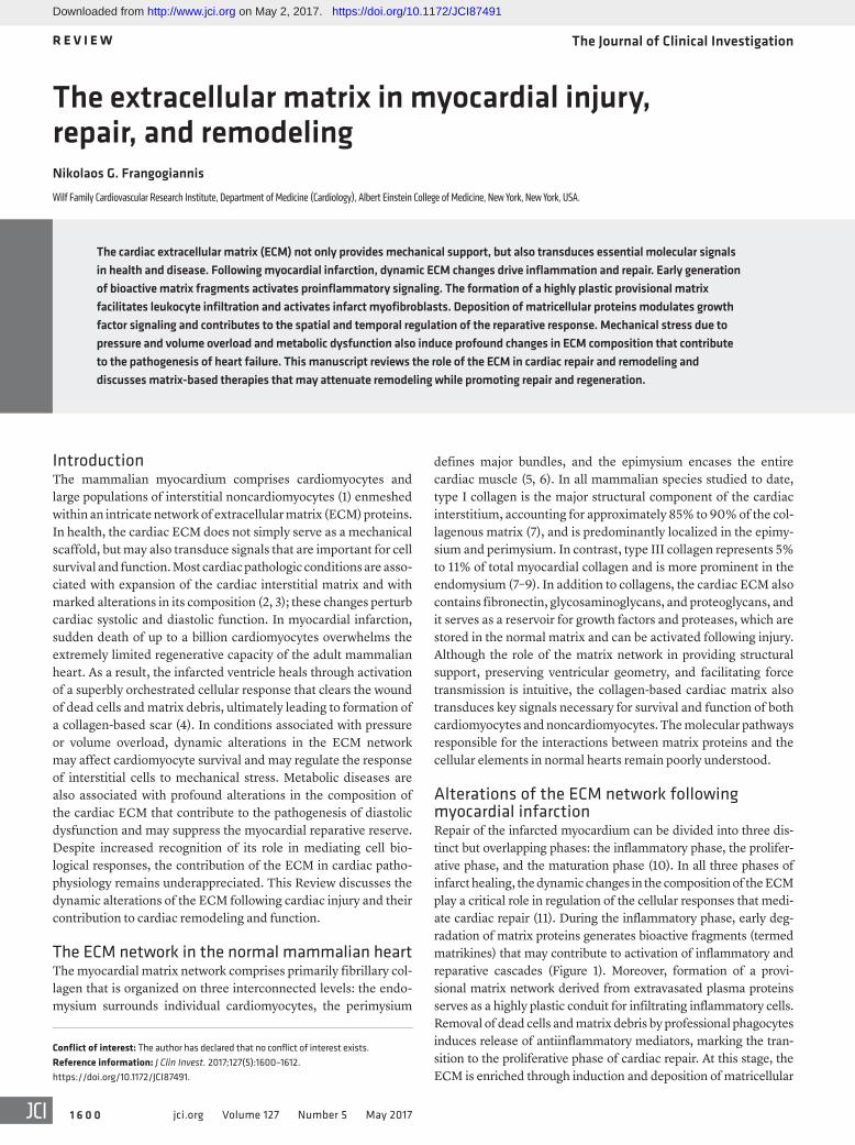
The Journal of Clinical Investigation R E V I E W
1 6 0 0 jci.org Volume 127 Number 5 May 2017
IntroductionThe mammalian myocardium comprises cardiomyocytes and large populations of interstitial noncardiomyocytes (1) enmeshed within an intricate network of extracellular matrix (ECM) proteins. In health, the cardiac ECM does not simply serve as a mechanical scaffold, but may also transduce signals that are important for cell survival and function. Most cardiac pathologic conditions are asso-ciated with expansion of the cardiac interstitial matrix and with marked alterations in its composition (2, 3); these changes perturb cardiac systolic and diastolic function. In myocardial infarction, sudden death of up to a billion cardiomyocytes overwhelms the extremely limited regenerative capacity of the adult mammalian heart. As a result, the infarcted ventricle heals through activation of a superbly orchestrated cellular response that clears the wound of dead cells and matrix debris, ultimately leading to formation of a collagen-based scar (4). In conditions associated with pressure or volume overload, dynamic alterations in the ECM network may affect cardiomyocyte survival and may regulate the response of interstitial cells to mechanical stress. Metabolic diseases are also associated with profound alterations in the composition of the cardiac ECM that contribute to the pathogenesis of diastolic dysfunction and may suppress the myocardial reparative reserve. Despite increased recognition of its role in mediating cell bio-logical responses, the contribution of the ECM in cardiac patho-physiology remains underappreciated. This Review discusses the dynamic alterations of the ECM following cardiac injury and their contribution to cardiac remodeling and function.
The ECM network in the normal mammalian heartThe myocardial matrix network comprises primarily fibrillary col-lagen that is organized on three interconnected levels: the endo-mysium surrounds individual cardiomyocytes, the perimysium
defines major bundles, and the epimysium encases the entire cardiac muscle (5, 6). In all mammalian species studied to date, type I collagen is the major structural component of the cardiac interstitium, accounting for approximately 85% to 90% of the col-lagenous matrix (7), and is predominantly localized in the epimy-sium and perimysium. In contrast, type III collagen represents 5% to 11% of total myocardial collagen and is more prominent in the endomysium (7–9). In addition to collagens, the cardiac ECM also contains fibronectin, glycosaminoglycans, and proteoglycans, and it serves as a reservoir for growth factors and proteases, which are stored in the normal matrix and can be activated following injury. Although the role of the matrix network in providing structural support, preserving ventricular geometry, and facilitating force transmission is intuitive, the collagen-based cardiac matrix also transduces key signals necessary for survival and function of both cardiomyocytes and noncardiomyocytes. The molecular pathways responsible for the interactions between matrix proteins and the cellular elements in normal hearts remain poorly understood.
Alterations of the ECM network following myocardial infarctionRepair of the infarcted myocardium can be divided into three dis-tinct but overlapping phases: the inflammatory phase, the prolifer-ative phase, and the maturation phase (10). In all three phases of infarct healing, the dynamic changes in the composition of the ECM play a critical role in regulation of the cellular responses that medi-ate cardiac repair (11). During the inflammatory phase, early deg-radation of matrix proteins generates bioactive fragments (termed matrikines) that may contribute to activation of inflammatory and reparative cascades (Figure 1). Moreover, formation of a provi-sional matrix network derived from extravasated plasma proteins serves as a highly plastic conduit for infiltrating inflammatory cells. Removal of dead cells and matrix debris by professional phagocytes induces release of antiinflammatory mediators, marking the tran-sition to the proliferative phase of cardiac repair. At this stage, the ECM is enriched through induction and deposition of matricellular
The cardiac extracellular matrix (ECM) not only provides mechanical support, but also transduces essential molecular signals in health and disease. Following myocardial infarction, dynamic ECM changes drive inflammation and repair. Early generation of bioactive matrix fragments activates proinflammatory signaling. The formation of a highly plastic provisional matrix facilitates leukocyte infiltration and activates infarct myofibroblasts. Deposition of matricellular proteins modulates growth factor signaling and contributes to the spatial and temporal regulation of the reparative response. Mechanical stress due to pressure and volume overload and metabolic dysfunction also induce profound changes in ECM composition that contribute to the pathogenesis of heart failure. This manuscript reviews the role of the ECM in cardiac repair and remodeling and discusses matrix-based therapies that may attenuate remodeling while promoting repair and regeneration.
The extracellular matrix in myocardial injury, repair, and remodelingNikolaos G. Frangogiannis
Wilf Family Cardiovascular Research Institute, Department of Medicine (Cardiology), Albert Einstein College of Medicine, New York, New York, USA.
Conflict of interest: The author has declared that no conflict of interest exists.Reference information: J Clin Invest. 2017;127(5):1600–1612. https://doi.org/10.1172/JCI87491.
Downloaded from http://www.jci.org on May 2, 2017. https://doi.org/10.1172/JCI87491

The Journal of Clinical Investigation R E V I E W
1 6 0 1jci.org Volume 127 Number 5 May 2017
ECM during the inflammatory phase of cardiac repairActivation of matrix metalloproteinases. Myocardial ischemia caus-es rapid activation of latent matrix metalloproteinases (MMPs) and subsequent generation of matrix fragments (14, 15). MMP activation is detected in the cardiac interstitium as early as ten minutes after coronary occlusion (16), before any evidence of irre-versible cardiomyocyte injury, and may be driven by ischemia-
proteins, defined as extracellular macromolecules that do not serve a primary structural role, but modulate cellular phenotype, activate proteases and growth factors, and transduce signaling cascades (12, 13). Activated myofibroblasts are the dominant cells during the pro-liferative phase of cardiac repair and deposit large amounts of struc-tural ECM proteins. Finally, during the maturation phase, crosslink-ing of the ECM, fibroblast quiescence, and vascular maturation lead to formation of a stable collagen-based scar.
Figure 1. The ECM during the inflammatory phase of cardiac repair. (A) Cardiomyocyte necrosis is associated with induction and activation of proteases in the infarcted region. Activated proteases cause fragmentation of the native ECM, resulting in release of matrikines, bioactive peptides that activate an inflammatory macrophage phenotype and may also modulate responses of fibroblasts and vascular endothelial cells. The effects of MMPs in the ischemic and infarcted myocardium are not limited to the ECM. MMPs may modulate inflammatory and reparative responses by processing cytokines and chemok-ines. They may also inhibit chemokine actions by degrading glycosaminoglycan-binding sites and mediate dysfunction by targeting intracellular proteins. Increased vessel permeability in the infarcted region results in extravasation of plasma proteins, such as fibrinogen and fibronectin. Accumulation of these proteins in the infarcted region forms a provisional matrix that serves as a scaffold for infiltrating leukocytes. Fibrin and fibronectin modulate the phenotype of immune and reparative cells through integrin-mediated actions. (B) Fibrinogen/fibrin staining using a peroxidase-based technique (black) illustrates the formation of the provisional fibrin-based matrix network (indicated by arrows) in the infarcted canine myocardium (one hour ischemia followed by seven days reperfusion). Counterstained with eosin. Scale bar: 50 μm. Reproduced with permission from the FASEB Journal (155).
Downloaded from http://www.jci.org on May 2, 2017. https://doi.org/10.1172/JCI87491

The Journal of Clinical Investigation R E V I E W
1 6 0 2 jci.org Volume 127 Number 5 May 2017
dynamic fibrin-based matrix network (39, 49). The role of the pro-visional matrix in regulating cardiac repair remains poorly under-stood; current concepts are predominantly based on extrapolation of findings from studies that investigate reparative responses in other systems. In addition to its hemostatic role, the provisional matrix may serve as a scaffold for migrating inflammatory cells and support proliferating endothelial cells and fibroblasts (50). Components of the provisional matrix interact with migrating cells through cell surface integrins (51) and may also transduce signaling cascades that modulate immune cell phenotype and gene expression (52). In vitro, fibrinogen stimulates macrophage- derived chemokine secretion through TLR4 activation (53).
In a mouse model of reperfused myocardial infarction, fibrin-mediated interactions contributed to early injury by accen-tuating the inflammatory response (54). Treatment with a nat-urally occurring peptide that competes with the fibrin fragment N-terminal disulfide knot-II (an analog of the fibrin E1 fragment) for binding to vascular endothelial cadherin reduced infarct size, attenuating leukocyte infiltration in the ischemic myocardium in both rodent and large animal models (54, 55). Unfortunately, the effects of the peptide in a small clinical trial were much less impressive. Peptide administration in patients with ST-elevation myocardial infarction (STEMI) did not affect the size of the infarct assessed through magnetic resonance imaging and did not reduce serum troponin I levels (56).
The provisional matrix may also play an important role in the transition to the proliferative phase of cardiac repair by serving as a reservoir for cytokines and growth factors. The heparin-binding domain of fibrin binds to a wide range of growth factors, includ-ing members of the PDGF, FGF, VEGF, and TGF families (57), that may activate reparative fibroblasts and vascular cells.
ECM during the proliferative phase of infarct healingDynamic changes in the composition of the ECM may contribute to the reparative cellular responses during the proliferative phase of cardiac repair (Figure 2). Clearance of matrix fragments by phagocytes may activate antiinflammatory signals, suppressing recruitment of proinflammatory leukocytes. Lysis of the plas-ma-derived provisional matrix is followed by organization of a cell-derived matrix network, comprising cellular fibronectin, hyal-uronan, proteoglycans, and a wide range of matricellular macro-molecules (39) that transduce growth factor signals to reparative cells (58). The dynamic alterations of the ECM during the prolifer-ative phase provide essential signals for conversion of fibroblasts into myofibroblasts and may activate angiogenic pathways nec-essary for neovessel formation, thus supplying the metabolically active wound with oxygen and nutrients (11).
Fibrin network clearance and cell-derived provisional matrix for-mation. In healing wounds, the plasma-derived ECM is cleared through extracellular proteolysis by fibrinolytic enzymes (59) and through endocytosis by CCR2+ macrophages (60). In the healing infarct, clearance of the fibrin-based provisional matrix by the plasminogen/plasmin system is an important part of the reparative response. Mice lacking plasminogen exhibited markedly atten-uated recruitment of inflammatory leukocytes in the infarct and impaired granulation tissue formation (61). Lysis of the fibrin-based
mediated ROS generation (17). Cardiomyocyte necrosis accentu-ates the matrix-degrading response. Large amounts of MMPs are synthesized de novo by ischemic cardiomyocytes, fibroblasts, endo-thelial cells, and inflammatory leukocytes that infiltrate the infarct (18). Both collagenases (e.g., MMP1) and gelatinases (MMP2 and MMP9) are upregulated in the infarcted myocardium (19).
It should be emphasized that the actions of MMPs are not lim-ited to effects on the ECM. MMPs regulate inflammatory responses through proteolytic processing of cytokines, chemokines, and growth factors (20–24) or by degrading glycosaminoglycan binding sites, thus interfering with a molecular step that is critical for chemokine immobilization on the endothelial cell surface and subsequent inter-action with leukocytes (25). MMPs also have intracellular targets, degrading cardiomyocyte proteins, such as myosin, α-actinin, and titin (26–28). The relative significance of the matrix-independent actions of MMPs in the pathogenesis of ischemic dysfunction and postinfarction remodeling remains unknown.
Generation of matrikines. In injured and remodeling tissues, protease-mediated fragmentation of matrix proteins results in generation of matrikines (29–32). Elastin fragments and colla-gen-derived peptides are the best-studied matrikines and have been implicated in activation of immune cells and fibroblasts (29, 33). The collagen-derived tripeptide proline-glycine-proline (PGP) and its acetylated form have been demonstrated as acting as neutrophil chemoattractants in models of pulmonary inflam-mation, signaling through activation of the chemokine receptor CXCR2 (34). PGP generation requires activation of a multistep cascade that involves MMP8, MMP9, and prolyl endopeptidase (35). Proteolytic processing of laminins by MMP2 and MMP14 has also been demonstrated as yielding fragments with potent neutro-phil chemoattractant properties (36).
Although the rapid activation of MMPs in the infarct is asso-ciated with matrix fragmentation, the role of these fragments as bioactive proinflammatory matrikines has not been documented. In experimental models, release of type I collagen fragments in the serum has been documented within 30 minutes after coronary occlusion (37). Fragmentation of components of the basement membrane, such as collagen IV, and of noncollagenous matrix constituents has also been demonstrated in the infarcted myocar-dium (38–41). Low–molecular weight hyaluronan fragments exert potent proinflammatory actions in the infarcted region; impaired clearance of these fragments has been shown to prolong and accentuate proinflammatory signaling in leukocytes and vascu-lar cells (42, 43). Matrix fragments may also modulate fibroblast and vascular cell phenotype (44). Endostatin, a 20-kDa fragment of collagen XVIII, exerts potent angiostatic actions (45) and stim-ulates fibroblast proliferation (46). MMP9-mediated cleavage of collagen IV also generates fragments with angiostatic properties, such as tumstatin (47). The role of endogenous matrix fragments in regulation of fibrogenic and angiogenic responses following myocardial infarction remains poorly understood.
The plasma-derived provisional matrix regulates the inflammato-ry response. Degradation of the original cardiac matrix network in the infarcted myocardium is accompanied by formation of a provi-sional matrix (39). Rapid induction of VEGF in the infarcted heart increases vascular permeability (48), leading to extravasation of plasma fibrinogen and fibronectin and generating a complex and
Downloaded from http://www.jci.org on May 2, 2017. https://doi.org/10.1172/JCI87491

The Journal of Clinical Investigation R E V I E W
1 6 0 3jci.org Volume 127 Number 5 May 2017
been implicated in myofibroblast conver-sion. In vitro, pericellular hyaluronan was required to maintain a myofibroblast phe-notype in TGF-β–stimulated cells (67). In vivo, loss of CD44, the main receptor for haluronan, impaired collagen synthesis in infarct fibroblasts (40). Versican loss in dermal fibroblasts attenuated myofibro-blast conversion (68). Although versican is upregulated in healing myocardial infarcts (69), its in vivo role in transdifferentiation of injury-site cardiac myofibroblasts has not been directly documented.
Induction of matricellular proteins. During the proliferative phase of infarct healing, the cardiac ECM is enriched through the deposition of a wide range of structurally diverse matricellular proteins that do not serve a direct structural role, but act contextually by regulating cytokine and growth factor responses and modulating cell phenotype and function (70, 71). The family includes the thrombospondins TSP-1, -2, and -4, tenascin-C and -X, secreted protein, acidic and cysteine-rich (SPARC), osteopontin, periostin, and members of the CCN family (72). Several other pro-teins (including members of the galectin and syndecan family, fibulins, osteoglycin, and other small leucine-rich proteoglycans) have been found to exert matricellular actions as part of their broader spectrum of functional properties. Most members of the matricellular family show low expression in the normal myocardium, but are markedly upregulated following myocardial injury. In healing infarctions, angiotensin and growth
factors stimulate de novo synthesis and secretion of matricellular proteins in fibroblasts or immune or vascular cells (39, 73, 74). Once secreted into the interstitium, matricellular macromolecules bind to the structural matrix and transduce signaling cascades through ligation of cell surface receptors or contribute to activa-tion of cytokines, growth factors, and proteases in the pericellular space. An overview of the pattern of regulation, function, targets, and mechanisms of action of the matricellular proteins discussed below is given in Table 1.
Several prototypical members of the matricellular family are markedly upregulated following myocardial infarction and pro-tect the myocardium from adverse remodeling (75–78). Localized induction of TSP-1 in the infarct border zone may form a barrier that prevents expansion of inflammation in viable myocardial areas through local activation of antiinflammatory signals (75). Osteopon-tin may protect from left ventricular dilation by promoting matrix deposition in the infarcted region (79). SPARC has been shown to protect from cardiac rupture and postinfarction heart failure by con-tributing to collagen maturation through activation of growth factor signaling (80). Periostin serves as a crucial regulator of fibroblast
matrix is followed by secretion of cellular fibronectin by fibroblasts and macrophages (62, 63) and by deposition of hyaluronan and versican, forming a network of cell-derived provisional matrix that is later enriched with a wide range of matricellular macromolecules (12). During the proliferative phase of healing, the highly dynamic matrix becomes a regulatory center that transduces essential sig-nals to activate reparative fibroblasts and vascular cells.
Components of the provisional matrix regulate fibroblast activa-tion. Repair of the infarcted heart is dependent on recruiting and activating resident fibroblast populations (64) to acquire a myo-fibroblast phenotype that expresses contractile proteins such as α-smooth muscle actin (α-SMA) and secretes large amounts of col-lagens. Myofibroblast transdifferentiation requires cooperation between soluble growth factors, such as TGF-β1, and components of the provisional matrix. In vitro, the ED-A domain splice variant of fibronectin is critical for TGF-β1–mediated α-SMA upregulation (65). In vivo, ED-A fibronectin loss attenuates myofibroblast trans-differentiation in healing myocardial infarction (66). The specific interactions between the ED-A segment and the TGF-β–signaling cascade remain unknown. Hyaluronan and versican have also
Figure 2. The role of the cell-derived provisional matrix in cardiac repair. (A) During the proliferative phase of cardiac repair, fibroblasts and macrophages contribute to the formation of a cell-derived provisional matrix, enriched with a wide range of matricellular macromolecules that do not serve a primary structural role, but modulate cellular phenotype and function. Specialized matrix proteins (such as ED-A domain fibronectin) and matricellular proteins, such as TSPs, tenascin-C (TNC), osteo-pontin (OPN), SPARC, periostin, osteoglycin, and members of the CCN family, bind to the matrix and modulate growth factor and protease activity. Specific matricellular proteins have been reported as regulating inflammation, participating in fibrogenic and angiogenic responses, modulating cardio-myocyte survival, and contributing to assembly of the structural matrix. (B) Matricellular proteins may be critical in spatial and temporal regulation of growth factor signaling. Immunohistochemical staining using a peroxidase-based technique (black) shows the strikingly selective localization of the prototypical matricellular protein TSP1 (arrows), a critical activator of TGF-β, in the border zone of a healing canine myocardial infarction (one hour ischemia followed by seven days reperfusion). Spa-tially and temporally restricted induction of matricellular proteins regulates growth factor signaling, preventing expansion of profibrotic responses beyond the infarcted area, despite possible diffusion of the soluble mediators in viable segments. Counterstained with eosin. Reproduced with permission from Circulation (75). Scale bar: 50 μm. GAGs, glycosaminoglycosans.
Downloaded from http://www.jci.org on May 2, 2017. https://doi.org/10.1172/JCI87491

The Journal of Clinical Investigation R E V I E W
1 6 0 4 jci.org Volume 127 Number 5 May 2017
environmental cues, interstitial cells exhibit phenotypic changes (84–86), leading to transitions from inflammatory to reparative phenotypes (87). Through their transient induction and incorpora-tion into the matrix, matricellular proteins may play a critical role in regulating cell differentiation, contributing to the cellular plasticity observed in healing tissues through direct actions and via modula-tion of growth factor–mediated pathways.
ECM in the maturation phase of infarct healingScar maturation is associated with ECM crosslinking in the infarct zone and with a marked reduction in myofibroblast density (88). Descriptive studies have suggested that apoptosis may be involved in elimination of granulation tissue cells from the infarct (89, 90); however, the mechanisms responsible for cell-specific activation of a proapoptotic program in the late phase of infarct healing have not been investigated. Acquisition of a quiescent phenotype may precede apoptosis of infarct myofibroblasts. Formation of a mature crosslinked matrix and clearance of matricellular proteins may play an important role in regulating fibroblast deactivation in the healing infarct (91–93). It is tempting to hypothesize that endogenous mech-anisms that restrain matricellular actions protect the infarcted heart from progressive fibrosis, depriving fibroblasts of the prolonged actions of growth factor–mediated signals. Vascular cells may also
recruitment and activity (81) and prevents cardiac rupture (81, 82), promoting formation of an organized collagen-based scar.
Although each matricellular protein has a unique function-al profile, several unifying themes have emerged regarding their actions in the infarcted heart. First, the strikingly selective local-ization of prototypical members of the family in the infarct border zone and their regulatory effects on the activity of soluble medi-ators highlight their role in spatial regulation of cellular respons-es. Secreted growth factors and cytokines can diffuse beyond the infarct zone; the requirement for matricellular proteins in activating growth factor–mediated responses may serve to localize inflamma-tory and fibrotic responses within areas of injury. Second, transient expression of matricellular proteins during the proliferative phase of healing ensures temporal regulation of growth factor respons-es. Clearance of matricellular proteins from the infarcted area may serve as an important STOP signal, preventing uncontrolled fibrosis following injury. In vitro studies have demonstrated that alternatively activated macrophages internalize SPARC through the scavenger receptor stabilin 1 (83). However, the potential role of immune cell subsets in endocytosis and degradation of matri-cellular proteins in vivo has not been tested. Third, repair of the infarcted myocardium is dependent on a highly dynamic microen-vironment that promotes cellular plasticity. In response to micro-
Table 1. Matricellular proteins in myocardial infarction
Matricellular protein
Pattern of regulation Function Possible cellular targets Possible mechanisms of action References
TSP-1 Transient upregulation and deposition in the infarct border zone during the proliferative phase of healing
Protects the infarcted heart from adverse remodeling, limiting expansion of inflammation and extension of fibrosis
Fibroblasts, macrophages, vascular endothelial cells, T cells, cardiomyocytes
Activates TGF-β Inhibits MMPs Exerts angiostatic actions Exerts antiinflammatory actions Modulates nitric oxide signaling
12
Tenascin-C Transient upregulation in infarcted area, border zone, and remodeling myocardium
May contribute to myocardial regeneration in fish and amphibians Promotes fibrosis and adverse remodeling in mammals
Fibroblasts, cardiomyocytes, macrophages, vascular cells
Exerts proinflammatory actions Triggers fibrogenic actions though accentuation of growth factor signaling Promotes angiogenic effects
145, 156, 157
SPARC Abundant, but transient upregulation in infarcted myocardium, predominantly localized in macrophages and myofibroblasts
Protects from cardiac rupture May promote systolic dysfunction during the early postischemic phase
Fibroblasts, macrophages, vascular cells
Regulates matrix assembly and metabolism Accentuates growth factor signaling, promoting fibroblast activation and angiogenesis
80, 158
OPN Markedly and transiently upregulated following infarction, primarily localized in macrophages
Protects from adverse remodeling Fibroblasts, macrophages, T cells, vascular cells, cardiomyocytes
Regulates matrix assembly Activates growth factor signaling, promoting fibroblast activation and angiogenesis
79
Periostin Marked, but transient upregulation in activated myofibroblasts in the infarct, border zone, and remodeling area
Protects from cardiac rupture, while promoting late fibrosis in remodeling segments
Fibroblasts Regulates matrix assembly Activates a fibrogenic program
81, 82
CCN2 Marked, but transient upregulation in infarcted hearts through activation of TGF-β and angiotensin II
Cardiomyocyte-specific CCN2 overexpression studies suggested that CCN2 reduces infarct size following ischemia/reperfusion and attenuates adverse remodeling in nonreperfused infarction
Cardiomyocytes, fibroblasts, macrophages
Activates prosurvival pathways in cardiomyocytes Attenuates inflammation
159, 160
Osteoglycin Marked upregulation in infarcted myocardium, associated with collagen deposition
Protects from cardiac rupture and prevents adverse remodeling
Fibroblasts Regulates maturation of collagen fibers 161
Downloaded from http://www.jci.org on May 2, 2017. https://doi.org/10.1172/JCI87491

The Journal of Clinical Investigation R E V I E W
1 6 0 5jci.org Volume 127 Number 5 May 2017
respond to the mature ECM environment. During scar maturation, infarct microvessels acquire a coat of mural cells through activation of PDGFR-β signaling (88, 94); uncoated vessels regress. ECM pro-teins have been implicated in modulation of endothelial-pericyte interactions in vitro and in vivo (95, 96), but whether such effects play a role in maturation of infarct microvessels remains unknown.
It should be emphasized that in the presence of a large infarction with significant hemodynamic consequences, via-ble noninfarcted myocardium also exhibits slowly progres-sive interstitial fibrosis related to the pathophysiologic effects of pressure and volume loads. Inflammation and fibrosis are suppressed in the healing infarct, leading to formation of a mature collagen-based scar, but in the viable noninfarcted zone, increased wall stress may locally activate macrophages and fibroblasts, triggering chronic progressive expansion of the cardiac interstitial matrix (97, 98).
ECM in cardiac pressure and volume overloadPressure and volume overload are critically implicated in the patho-genesis of heart failure in a wide variety of cardiac conditions. Left ventricular pressure overload is the critical pathophysiologic com-panion of the cardiomyopathy induced by systemic hypertension and aortic stenosis. On the other hand, valvular regurgitant lesions (such as aortic and mitral insufficiency) cause cardiac remodeling and heart failure by subjecting the heart to volume overload. More-over, both pressure and volume loads contribute to the pathophys-
iology of postinfarction remodeling. Pressure and volume overload have distinct effects on the cardiac ECM that may account for their conse-quences on ventricular geometry and function.
ECM in the pressure-overloaded myocardi-um. In both experimental models and in human patients, cardiac pressure overload causes early hypertrophic remodeling and diastolic dys-function, followed by decompensation, cham-ber dilation, and development of systolic heart failure. Pressure overload is associated with profound and dynamic changes in the com-position of the ECM; these changes regulate geometry and function, not only by affecting the mechanical properties of the ventricle, but also by modulating cellular responses. In ani-mal models, activation of cardiac fibroblasts is one of the earliest effects of pressure overload in the myocardium, ultimately leading to depo-sition of collagenous matrix and expansion of the interstitium (99–101). In human patients, hypertensive heart disease is associated with development of interstitial and periarteriolar fibrosis even in the absence of significant cor-onary atherosclerosis (102).
Fibrotic changes that develop in the pressure-overloaded myocardium may involve activation of cardiomyocytes, fibroblasts, and immune and vascular cells. Mechani-cal stress due to pressure overload activates the renin-angiotensin-aldosterone system
(RAAS), triggering inflammatory signaling and leading to down-stream stimulation of TGF-β cascades. Neurohumoral mediators, cytokines, and growth factors directly stimulate a fibrogenic pro-gram, triggering myofibroblast conversion and stimulating syn-thesis of large amounts of structural matrix proteins (103–106). Stress-induced fibroblast activation in the pressure-overloaded myocardium may also be indirect, at least in part, requiring stim-ulation of fibrogenic cascades in cardiomyocytes and immune cells. Mechanical stretch has been suggested as triggering puriner-gic signaling in cardiomyocytes, leading to release of fibrogenic growth factors (107). Moreover, T lymphocytes and macrophages have been implicated in activation of resident cardiac fibroblasts following pressure overload (108, 109).
Even in the absence of cardiomyocyte necrosis, mechanically activated myofibroblasts and immune cells secrete specialized matrix proteins (such as fibronectin) and enrich the interstitial matrix with a wide range of matricellular macromolecules (Figure 3 and refs. 110, 111). Fibronectin deposition in the pressure-overloaded heart may be involved in myofibroblast transdifferentiation and has been impli-cated as an important mediator in cardiomyocyte hypertrophy (112), possibly through activation of growth factor signaling. Deposition of matricellular proteins in the pressure-overloaded heart generates a highly dynamic matrix microenvironment and modulates fibrogenic and hypertrophic responses. Table 2 provides an overview of the reg-ulation, functional role, and mechanisms of action of the matricellu-lar proteins in the pressure-overloaded myocardium.
Figure 3. Matricellular proteins regulate cellular responses in the pressure-overloaded myocar-dium. In the pressure-overloaded heart, mechanical stress activates neurohumoral pathways and induces synthesis and release of matricellular macromolecules, including TSP1, -2, and -4, tenascin-C, OPN, SPARC, and periostin. Matricellular proteins have been implicated in regulation of matrix assembly, in transduction of mechanosensitive signaling, and in the pathogenesis of fibrosis and cardiac hypertrophy, and may also modulate survival of cardiomyocytes under condi-tions of stress. The effects of the matricellular proteins are exerted through direct activation of cell surface receptors or through modulation of growth factor– and protease-mediated responses.
Downloaded from http://www.jci.org on May 2, 2017. https://doi.org/10.1172/JCI87491

The Journal of Clinical Investigation R E V I E W
1 6 0 6 jci.org Volume 127 Number 5 May 2017
ECM in cardiac volume overload. Volume overload is associat-ed with a distinct profile of changes in the composition of the ECM that ultimately lead to chamber dilation and contribute to systolic dysfunction. In contrast to the marked accentuation in collagen deposition triggered by a pressure load, volume overload is asso-ciated with a reduction in interstitial collagen content due to increased MMP expression (113–115) and augmented autophag-ic degradation of procollagen in cardiac fibroblasts (116). Phar-macologic inhibition studies suggested that MMP activation is directly implicated in dilative remodeling of the volume-over-loaded ventricle (117), but the mechanisms responsible for the distinct cell biological changes and matrix alterations in volume overload remain poorly understood. Bradykinin receptor signal-ing has been implicated in matrix loss associated with volume overload (118), but the links between specific mechanical stim-uli and myocardial cell activation are understudied. The limited data on the matricellular protein profile in volume-overload-ed hearts are not accompanied by systematic investigations of the potential role of these proteins. Although marked increas-es in synthesis of fibronectin and periostin have been report-ed in experimental volume overload (114, 119), deposition of these fibrogenic matricellular proteins was not associated with increased secretion of structural matrix proteins.
The cardiac ECM in metabolic diseaseDiabetics exhibit a high incidence of heart failure with preserved ejection fraction, associated with expansion of the cardiac ECM network (120). Fibrotic changes in diabetic hearts are, at least in part, independent of coronary artery disease or hypertension (121), reflecting direct effects of metabolic dysregulation on the ECM. Diabetes, obesity, and metabolic dysfunction are associated with activation of cardiac fibroblasts (122) and are accompanied by deposition of matricellular macromolecules (123) and progres-sive accumulation of fibrillary collagens in the cardiac interstitium (124, 125). The molecular basis for activation of the so-called dia-betic fibroblast remains unknown. The role of hyperglycemia in mediating fibrogenic activation remains poorly defined. Whether stimulation of a fibrogenic program in diabetic fibroblasts requires diabetes-associated activation of fibrogenic signaling in cardiomyo-cytes, vascular, or immune cells is unclear. Expansion of the cardiac interstitium in diabetes involves activation of several distinct but overlapping pathways, including neurohumoral mediators (such as the RAAS), ROS, inflammatory cytokines and growth factors (such as TGF-β), adipokines, and the advanced glycation endproduct (AGE)/receptor for AGE (RAGE) axis. Moreover, hyperglycemia induces matricellular protein synthesis in several cell types (126). Diabetes-associated induction of matricellular proteins, such as
Table 2. Matricellular proteins in the pressure-overloaded myocardium
Matricellular protein
Pattern of regulation in models of pressure overload
Function Possible mechanisms of action References
TSP-1 Transient upregulation and deposition in interstitial and perivascular areas in models of TAC and systemic hypertension and in a combined model of hyperglycemia/abdominal aortic constriction
Activates fibroblasts in the pressure-overloaded heart, promoting a matrix-synthetic phenotype
Activates TGF-β Inhibits MMP activity
111, 162–164
TSP-2 High expression in myocardium of renin-overexpressing hypertensive rats that developed heart failure High expression in human patients with cardiac hypertrophy
Protects the angiotensin II–stimulated myocardium from rupture Protects the aging heart from dilation
Inhibition of MMP synthesis and activity Activation of prosurvival pathways in cardiomyocytes
165, 166
TSP-4 Rapid upregulation in the myocardium in response to angiotensin II treatment or TAC
Protects the pressure-overloaded heart from functional decompensation, systolic dysfunction, and death Downmodulates the fibrotic response in the pressure-overloaded heart
Suppresses expression of ECM proteins in fibroblasts and endothelial cells Activates mechanosensitive Erk and Akt signaling in cardiomyocytes, increasing contractility following pressure overload Has been suggested as also acting intracellularly, augmenting ER function and protecting from injury-related ER stress
167–171
Tenascin-C Deposition in the cardiac interstitium following pressure overload Upregulation in cardiomyocytes subjected to mechanical strain
Accelerates cardiac fibrosis in response to angiotensin II infusion
May act in part through activation of integrin-mediated cytokine secretion in macrophages
99, 172–174
SPARC Upregulation in models of TAC Mediates fibrosis and promotes diastolic dysfunction in models of TAC
Contributes to postsynthetic procollagen processing
175, 176
OPN Upregulation in models of TAC and angiotensin II infusion Increased expression in human hypertensive heart disease
Mediates fibrosis in models of TAC, angiotensin, and aldosterone infusion Prohypertrophic effects are less consistently observed in various models
Promotes fibroblast proliferation Activates fibroblasts through an miR-21–mediated pathway 177–183
Periostin Upregulated in activated fibroblasts and deposited in the cardiac interstitium in models of TAC
Promotes hypertrophy and fibrosis Activates cardiac fibroblasts, promoting their migration and transdifferentiation Regulates collagen fibrillogenesis
82, 184
TAC, transverse aortic constriction
Downloaded from http://www.jci.org on May 2, 2017. https://doi.org/10.1172/JCI87491

The Journal of Clinical Investigation R E V I E W
1 6 0 7jci.org Volume 127 Number 5 May 2017
of matricellular macromolecules to modulate growth factor and cytokine signaling, thus localizing therapeutic effects in the area of interest. Unfortunately, the daunting complexity of the biol-ogy of matricellular proteins hampers therapeutic implementa-tion. Matricellular proteins have multiple functional domains and act contextually depending on microenvironmental fac-tors, such as the local composition of the matrix and the profile of cytokine and growth factor expression. Distinct actions of matricellular protein fragments further complicate therapeu-tic implementation. Identification of the functional domains responsible for specific protective or detrimental matricellular actions is needed in order to design peptide-based strategies that simulate these effects (12, 135).
Targeting the ECM to promote cardiac regeneration. In con-trast with fish and amphibians, adult mammals have very limited capacity for myocardial regeneration (136), which is overwhelmed by the massive sudden loss of cardiomyocytes following infarction. Over the last 15 years, a wide range of cell therapy approaches has been tested in attempts to regenerate the injured human myocardium, with little success (137–139). Although beneficial actions have been reported with several different types of cell therapy, in many cases, protection of the infarcted myocardium was attributed to paracrine effects and to the modulation of inflammatory and fibrogenic signals rather than to activation of a regenerative program.
Several lines of evidence suggest that modulation of the ECM may be a crucial component of a regenerative response. First, in vitro, the composition of the ECM has a profound effect in regu-lation of cell cycle entry in cardiomyocyte progenitors and in neo-natal cardiac myocytes (140, 141). Compliant matrices contain-ing elastin promote dedifferentiation, proliferation, and clonal expansion of rat and mouse neonatal cardiomyocytes (140). Sec-ond, in fish and amphibians, myocardial regenerative responses seem to be dependent on deposition of a specialized ECM (142). A recent study reported that intramyocardial administration of ECM from healing zebrafish hearts may stimulate regeneration in mouse infarcts (143). Loss-of-function approaches suggested that fibronectin deposition may be critical for regeneration of the zebrafish heart (144). In newts, cardiac regeneration following injury was preceded by formation of a matrix network compris-ing tenascin-C, fibronectin, and hyaluronan. This specialized matrix network may serve as a path for progenitor cells as well as play an active role in activation of a regenerative program (145). Considering that, much like in fish and amphibians, myocardi-al infarction in mammals also induces a marked upregulation of tenascin-C and fibronectin without stimulating remuscular-ization, it is unlikely that matrix-dependent actions are suffi-cient to activate a regenerative program. Myocardial regenera-tion likely requires not only a “regenerative” matrix profile, but also local secretion of a yet-undefined combination of cytokines and growth factors, and activation of pathways that enhance plasticity of progenitor cell populations (146). Third, in sever-al cell therapy studies in mammalian models of cardiac injury, application of matrix-based patches containing progenitor cells showed enhanced effectiveness (147, 148). It has been suggested that application of matrix patches that recapitulate the regener-ative environment of the embryonic myocardium may increase
TSP-1, may drive the fate of cardiac interstitial cells toward a fibro-blast phenotype promoting fibrotic remodeling.
Therapeutic ECM targeting in injured and remodeling myocardiumMatrix-based strategies to preserve structure, geometry, and function. Because of its importance in preserving structural integrity and function of the heart (127) and its crucial involvement in regula-tion of cellular responses in cardiac injury, repair, and remodeling (12), the cardiac ECM provides unique opportunities for therapeu-tic interventions in patients with myocardial infarction and heart failure. Several therapeutic approaches with established benefi-cial effects in patients with myocardial infarction and heart fail-ure, such as ACE inhibition, angiotensin type 1 receptor blockade, β-adrenergic receptor antagonism, and mechanical unloading, may exert some of their protective actions through modulation of ECM deposition and metabolism (128).
Following myocardial infarction, catastrophic mechanical complications, such as cardiac rupture (129), are associated with accentuated matrix degradation or perturbed deposition of new structural matrix (130). These can be treated through application of a patch containing ECM proteins to restore the structural integ-rity of the ventricle (131). In the remodeling heart, approaches to tightly regulating matrix deposition and crosslinking may pro-tect from adverse remodeling and development of heart failure. A large body of experimental evidence suggests that excessive accumulation of crosslinked structural matrix proteins increases myocardial stiffness and promotes diastolic dysfunction; in con-trast, overactive matrix-degrading pathways may promote dila-tive remodeling, causing systolic dysfunction. In patients with myocardial infarction, therapeutic interventions directly target-ing ECM metabolism through MMP inhibition have produced mixed results. Administration of a selective oral MMP inhibitor in patients with STEMI and reduced ejection fraction showed no significant protection from adverse remodeling (132), despite significant antiremodeling effects in animal models. In contrast, early nonselective MMP inhibition with doxycycline attenuated progression to dilative remodeling in patients with STEMI and left ventricular dysfunction (133). The conflicting findings may reflect the pathophysiological heterogeneity of human myocar-dial infarction; successful implementation of matrix modulation strategies may require development of biomarkers or imaging approaches to identify patients with specific alterations in matrix metabolism (134). Moreover, interpretation of the effects of therapies targeting MMPs are complicated by their wide range of actions beyond matrix metabolism involving processing and modulation of bioactive cytokines and growth factors.
Matrix-based interventions to modulate cellular cardiac repair responses. Specialized matrix proteins and matricellular macromolecules critically modulate the cellular responses in the infarcted and remodeling heart and may hold the key for development of new effective strategies to optimize repair and to reduce adverse remodeling following cardiac injury. Ani-mal model studies have suggested that several members of the matricellular family protect the infarcted heart from adverse remodeling (12). Therapeutic approaches based on matricellu-lar proteins are particularly attractive because of the capacity
Downloaded from http://www.jci.org on May 2, 2017. https://doi.org/10.1172/JCI87491

The Journal of Clinical Investigation R E V I E W
1 6 0 8 jci.org Volume 127 Number 5 May 2017
electrical activity. Despite these problems, such matrix-driven strategies may represent the first step toward the generation of functional human cardiac grafts.
ConclusionsAll myocardial cells are enmeshed within a dynamic network of ECM that not only serves a structural role and facilitates mechanical force transmission, but also regulates cell phenotype and function. Fol-lowing cardiac injury, changes in composition of the cardiac ECM critically regulate inflammatory, reparative, fibrotic, and regenera-tive responses. Over the last two decades, understanding of the biol-ogy of the cardiac matrix has expanded well beyond the early con-cepts focusing on the role of matrix proteins in cardiac mechanics. We now view the matrix as a dynamic entity that responds to injury by undergoing dramatic transformations. Release of bioactive frag-ments and incorporation of matricellular proteins expands the func-tional repertoire of the matrix and drives repair by transducing key signals to many different cell types. Expansion of our knowledge on the structure, proteomic profile, and functional properties of matrix constituents will enrich our understanding of the pathobiology of heart disease, suggesting new therapeutic opportunities.
AcknowledgmentsNGF’s laboratory is supported by NIH grants R01 HL76246 and R01 HL85440 and by grants PR151134 and PR151029 from the Depart-ment of Defense Congressionally Directed Medical Research Pro-grams (CDMRP).
Address correspondence to: Nikolaos G. Frangogiannis, The Wilf Family Cardiovascular Research Institute, Albert Einstein College of Medicine, 1300 Morris Park Avenue, Forchheimer G46B, Bronx, New York 10461, USA. Phone: 718.430.3546; E-mail: nikolaos. [email protected].
remuscularization following injury (149). Whether such effects may be due to direct actions of specific matrix proteins on car-diac progenitors or reflect matrix-dependent activation of other cell types (such as immune or vascular cells) remains unknown. Fourth, some experimental studies in mammals have suggest-ed that matricellular macromolecules may induce proliferation of differentiated cardiomyocytes. Kühn and coworkers showed that recombinant periostin administered as an epicardial Gel-foam patch following infarction improved cardiac function and attenuated cardiac remodeling, triggering cell cycle reentry in differentiated cardiomyocytes in an integrin-dependent manner (150). Unfortunately, genetic manipulation of periostin expres-sion in mice did not support the proposed proliferative actions of periostin on cardiomyocytes (151). Whether modulation of the matricellular protein profile following myocardial injury could facilitate activation of a regenerative program in adult infarcted hearts remains unknown.
A growing body of experimental work attempts to exploit technological advances in perfusion-decellularization strate-gies (152) to generate scaffolds comprising human cardiac ECM that may serve as tools for manufacture of cardiac grafts (153, 154). The acellular matrix obtained through decellularization could subsequently be used for recellularization with cardio-myocytes derived from induced pluripotent stem cells (iPSCs) in order to generate myocardial-like structures. Early studies have generated tissue constructs with definitive sarcomeric structure that exhibit contractile function and electrical con-duction (153). Clearly, the current technology has significant limitations, and major advances are needed in order to pursue clinical translation. Complete reendothelialization is necessary to prevent activation of the coagulation system, generation of functional valvular structures is critical for ventricular func-tion, and a conduction system is needed for transmission of
1. Pinto AR, et al. Revisiting cardiac cellular compo-sition. Circ Res. 2016;118(3):400–409.
2. Kong P, Christia P, Frangogiannis NG. The pathogenesis of cardiac fibrosis. Cell Mol Life Sci. 2014;71(4):549–574.
3. Berk BC, Fujiwara K, Lehoux S. ECM remodel-ing in hypertensive heart disease. J Clin Invest. 2007;117(3):568–575.
4. Prabhu SD, Frangogiannis NG. The biological basis for cardiac repair after myocardial infarc-tion: from inflammation to fibrosis. Circ Res. 2016;119(1):91–112.
5. Medugorac I, Jacob R. Characterisation of left ventricular collagen in the rat. Cardiovasc Res. 1983;17(1):15–21.
6. Leonard BL, Smaill BH, LeGrice IJ. Structural remodeling and mechanical function in heart failure. Microsc Microanal. 2012;18(1):50–67.
7. Bashey RI, Martinez-Hernandez A, Jimenez SA. Isolation, characterization, and localization of cardiac collagen type VI. Associations with other extracellular matrix components. Circ Res. 1992;70(5):1006–1017.
8. Jugdutt BI. Ventricular remodeling after infarction and the extracellular collagen matrix: when is enough enough? Circulation.
2003;108(11):1395–1403. 9. Weber KT. Cardiac interstitium in health and
disease: the fibrillar collagen network. J Am Coll Cardiol. 1989;13(7):1637–1652.
10. Frangogiannis NG. Pathophysiology of myocardi-al infarction. Compr Physiol. 2015;5(4):1841–1875.
11. Dobaczewski M, Gonzalez-Quesada C, Frangogi-annis NG. The extracellular matrix as a modula-tor of the inflammatory and reparative response following myocardial infarction. J Mol Cell Cardi-ol. 2010;48(3):504–511.
12. Frangogiannis NG. Matricellular proteins in cardiac adaptation and disease. Physiol Rev. 2012;92(2):635–688.
13. Murphy-Ullrich JE, Sage EH. Revisiting the matricellular concept. Matrix Biol. 2014;37:1–14.
14. Cannon RO, et al. Early degradation of collagen after acute myocardial infarction in the rat. Am J Cardiol. 1983;52(3):390–395.
15. Whittaker P, Boughner DR, Kloner RA. Role of collagen in acute myocardial infarct expansion. Circulation. 1991;84(5):2123–2134.
16. Etoh T, et al. Myocardial and interstitial matrix metalloproteinase activity after acute myocardial infarction in pigs. Am J Physiol Heart Circ Physiol. 2001;281(3):H987–H994.
17. Wang W, Sawicki G, Schulz R. Peroxynitrite- induced myocardial injury is mediated through matrix metalloproteinase-2. Cardiovasc Res. 2002;53(1):165–174.
18. Alfonso-Jaume MA, et al. Cardiac ischemia- reperfusion injury induces matrix metallopro-teinase-2 expression through the AP-1 compo-nents FosB and JunB. Am J Physiol Heart Circ Physiol. 2006;291(4):H1838–H1846.
19. Danielsen CC, Wiggers H, Andersen HR. Increased amounts of collagenase and gelatinase in porcine myocardium following ischemia and reperfusion. J Mol Cell Cardiol. 1998;30(7):1431–1442.
20. Gearing AJ, et al. Matrix metalloproteinases and processing of pro-TNF-α. J Leukoc Biol. 1995;57(5):774–777.
21. Van Lint P, Libert C. Chemokine and cytokine processing by matrix metalloproteinases and its effect on leukocyte migration and inflammation. J Leukoc Biol. 2007;82(6):1375–1381.
22. Feng G, Hao D, Chai J. Processing of CXCL12 impedes the recruitment of endothelial pro-genitor cells in diabetic wound healing. FEBS J. 2014;281(22):5054–5062.
23. Peng H, Wu Y, Duan Z, Ciborowski P, Zheng JC. Proteolytic processing of SDF-1α by matrix
Downloaded from http://www.jci.org on May 2, 2017. https://doi.org/10.1172/JCI87491

The Journal of Clinical Investigation R E V I E W
1 6 0 9jci.org Volume 127 Number 5 May 2017
metalloproteinase-2 impairs CXCR4 signaling and reduces neural progenitor cell migration. Protein Cell. 2012;3(11):875–882.
24. Denney H, Clench MR, Woodroofe MN. Cleav-age of chemokines CCL2 and CXCL10 by matrix metalloproteinases-2 and -9: implications for chemotaxis. Biochem Biophys Res Commun. 2009;382(2):341–347.
25. Cox JH, Dean RA, Roberts CR, Overall CM. Matrix metalloproteinase processing of CXCL11/I-TAC results in loss of chemoattractant activity and altered glycosaminoglycan binding. J Biol Chem. 2008;283(28):19389–19399.
26. Cauwe B, Opdenakker G. Intracellular substrate cleavage: a novel dimension in the biochemistry, biology and pathology of matrix metalloproteinas-es. Crit Rev Biochem Mol Biol. 2010;45(5):351–423.
27. Schulz R. Intracellular targets of matrix metal-loproteinase-2 in cardiac disease: rationale and therapeutic approaches. Annu Rev Pharmacol Toxicol. 2007;47:211–242.
28. Ali MA, Cho WJ, Hudson B, Kassiri Z, Gran-zier H, Schulz R. Titin is a target of matrix metalloproteinase-2: implications in myocar-dial ischemia/reperfusion injury. Circulation. 2010;122(20):2039–2047.
29. Wells JM, Gaggar A, Blalock JE. MMP generated matrikines. Matrix Biol. 2015;44–46:122–129.
30. Gaggar A, Weathington N. Bioactive extracellular matrix fragments in lung health and disease. J Clin Invest. 2016;126(9):3176–3184.
31. Akthar S, et al. Matrikines are key regulators in modulating the amplitude of lung inflammation in acute pulmonary infection. Nat Commun. 2015;6:8423.
32. Adair-Kirk TL, Senior RM. Fragments of extracel-lular matrix as mediators of inflammation. Int J Biochem Cell Biol. 2008;40(6–7):1101–1110.
33. Senior RM, Griffin GL, Mecham RP. Chemotactic activity of elastin-derived peptides. J Clin Invest. 1980;66(4):859–862.
34. Weathington NM, et al. A novel peptide CXCR ligand derived from extracellular matrix deg-radation during airway inflammation. Nat Med. 2006;12(3):317–323.
35. Gaggar A, et al. A novel proteolytic cascade gen-erates an extracellular matrix-derived chemoat-tractant in chronic neutrophilic inflammation. J Immunol. 2008;180(8):5662–5669.
36. Mydel P, et al. Neutrophil elastase cleaves laminin- 332 (laminin-5) generating peptides that are chemotactic for neutrophils. J Biol Chem. 2008;283(15):9513–9522.
37. Villarreal F, Omens J, Dillmann W, Risteli J, Nguyen J, Covell J. Early degradation and serum appearance of type I collagen fragments after myocardial infarction. J Mol Cell Cardiol. 2004;36(4):597–601.
38. Lauten A, Gerhard-Garcia A, Suhr F, Fischer JH, Figulla HR, Bloch W. Impact of ischemia-reper-fusion on extracellular matrix processing and structure of the basement membrane of the heart. PLoS One. 2014;9(3):e92833.
39. Dobaczewski M, Bujak M, Zymek P, Ren G, Ent-man ML, Frangogiannis NG. Extracellular matrix remodeling in canine and mouse myocardial infarcts. Cell Tissue Res. 2006;324(3):475–488.
40. Huebener P, et al. CD44 is critically involved
in infarct healing by regulating the inflam-matory and fibrotic response. J Immunol. 2008;180(4):2625–2633.
41. Trial J, et al. Fibronectin fragments modulate monocyte VLA-5 expression and monocyte migration. J Clin Invest. 1999;104(4):419–430.
42. Taylor KR, Trowbridge JM, Rudisill JA, Termeer CC, Simon JC, Gallo RL. Hyaluronan fragments stimulate endothelial recognition of injury through TLR4. J Biol Chem. 2004;279(17):17079–17084.
43. Teder P, et al. Resolution of lung inflammation by CD44. Science. 2002;296(5565):155–158.
44. Lindsey ML, et al. A novel collagen matric-ryptin reduces left ventricular dilation post-myocardial infarction by promoting scar formation and angiogenesis. J Am Coll Cardiol. 2015;66(12):1364–1374.
45. O’Reilly MS, et al. Endostatin: an endogenous inhibitor of angiogenesis and tumor growth. Cell. 1997;88(2):277–285.
46. Okada M, Oba Y, Yamawaki H. Endostatin stim-ulates proliferation and migration of adult rat cardiac fibroblasts through PI3K/Akt pathway. Eur J Pharmacol. 2015;750:20–26.
47. Hamano Y, et al. Physiological levels of tum-statin, a fragment of collagen IV α3 chain, are generated by MMP-9 proteolysis and suppress angiogenesis via αVβ3 integrin. Cancer Cell. 2003;3(6):589–601.
48. Andersson L, et al. Rip2 modifies VEGF-induced signalling and vascular permeability in myocardial ischaemia. Cardiovasc Res. 2015;107(4):478–486.
49. Brown LF, et al. Expression of vascular permea-bility factor (vascular endothelial growth factor) by epidermal keratinocytes during wound heal-ing. J Exp Med. 1992;176(5):1375–1379.
50. Clark RA. Overview and general considerations of wound repair. In: Clark RA, Henson PM, eds. The Molecular and Cellular Biology of Wound Repair. New York, New York, USA: Plenum Press; 1988:3–33.
51. Flick MJ, et al. Leukocyte engagement of fibrin (ogen) via the integrin receptor αMβ2/Mac-1 is critical for host inflammatory response in vivo. J Clin Invest. 2004;113(11):1596–1606.
52. Corbett SA, Schwarzbauer JE. Fibronectin-fibrin cross-linking: a regulator of cell behavior. Trends Cardiovasc Med. 1998;8(8):357–362.
53. Smiley ST, King JA, Hancock WW. Fibrinogen stimulates macrophage chemokine secre-tion through toll-like receptor 4. J Immunol. 2001;167(5):2887–2894.
54. Petzelbauer P, et al. The fibrin-derived pep-tide Bbeta15-42 protects the myocardium against ischemia-reperfusion injury. Nat Med. 2005;11(3):298–304.
55. Roesner JP, et al. The fibrin-derived peptide Bbeta15-42 is cardioprotective in a pig model of myocardial ischemia-reperfusion injury. Crit Care Med. 2007;35(7):1730–1735.
56. Atar D, et al. Effect of intravenous FX06 as an adjunct to primary percutaneous coronary intervention for acute ST-segment elevation myocardial infarction results of the F.I.R.E. (Efficacy of FX06 in the Prevention of Myocar-dial Reperfusion Injury) trial. J Am Coll Cardiol. 2009;53(8):720–729.
57. Martino MM, Briquez PS, Ranga A, Lutolf MP, Hubbell JA. Heparin-binding domain of
fibrin(ogen) binds growth factors and pro-motes tissue repair when incorporated within a synthetic matrix. Proc Natl Acad Sci U S A. 2013;110(12):4563–4568.
58. Rienks M, Papageorgiou AP, Frangogiannis NG, Heymans S. Myocardial extracellular matrix: an ever-changing and diverse entity. Circ Res. 2014;114(5):872–888.
59. Schäfer BM, Maier K, Eickhoff U, Todd RF, Kram-er MD. Plasminogen activation in healing human wounds. Am J Pathol. 1994;144(6):1269–1280.
60. Motley MP, et al. A CCR2 macrophage endocytic pathway mediates extravascular fibrin clearance in vivo. Blood. 2016;127(9):1085–1096.
61. Creemers E, et al. Disruption of the plasmin-ogen gene in mice abolishes wound healing after myocardial infarction. Am J Pathol. 2000;156(6):1865–1873.
62. Brown LF, Dubin D, Lavigne L, Logan B, Dvorak HF, Van de Water L. Macrophages and fibroblasts express embryonic fibronectins during cutaneous wound healing. Am J Pathol. 1993;142(3):793–801.
63. Ulrich MM, et al. Increased expression of fibronectin isoforms after myocardial infarction in rats. J Mol Cell Cardiol. 1997;29(9):2533–2543.
64. Kanisicak O, et al. Genetic lineage tracing defines myofibroblast origin and function in the injured heart. Nat Commun. 2016;7:12260.
65. Serini G, et al. The fibronectin domain ED-A is crucial for myofibroblastic phenotype induction by transforming growth factor-beta1. J Cell Biol. 1998;142(3):873–881.
66. Arslan F, et al. Lack of fibronectin-EDA promotes survival and prevents adverse remodeling and heart function deterioration after myocardial infarction. Circ Res. 2011;108(5):582–592.
67. Webber J, Meran S, Steadman R, Phillips A. Hyaluronan orchestrates transforming growth factor-β1-dependent maintenance of myofibroblast phenotype. J Biol Chem. 2009;284(14):9083–9092.
68. Hattori N, et al. Pericellular versican regulates the fibroblast-myofibroblast transition: a role for ADAMTS5 protease-mediated proteolysis. J Biol Chem. 2011;286(39):34298–34310.
69. Toeda K, et al. Versican is induced in infiltrating monocytes in myocardial infarction. Mol Cell Bio-chem. 2005;280(1-2):47–56.
70. Bornstein P. Matricellular proteins: an overview. J Cell Commun Signal. 2009;3(3–4):163–165.
71. Murphy-Ullrich JE. The de-adhesive activi-ty of matricellular proteins: is intermediate cell adhesion an adaptive state? J Clin Invest. 2001;107(7):785–790.
72. Bornstein P, Sage EH. Matricellular proteins: extracellular modulators of cell function. Curr Opin Cell Biol. 2002;14(5):608–616.
73. Mackie EJ, et al. Expression of tenascin by vascu-lar smooth muscle cells. Alterations in hyperten-sive rats and stimulation by angiotensin II. Am J Pathol. 1992;141(2):377–388.
74. Hashimoto S, Suzuki T, Dong HY, Yamazaki N, Matsushima K. Serial analysis of gene expression in human monocytes and macrophages. Blood. 1999;94(3):837–844.
75. Frangogiannis NG, et al. The critical role of endogenous Thrombospondin (TSP)-1 in pre-venting expansion of healing myocardial infarcts.
Downloaded from http://www.jci.org on May 2, 2017. https://doi.org/10.1172/JCI87491

The Journal of Clinical Investigation R E V I E W
1 6 1 0 jci.org Volume 127 Number 5 May 2017
Circulation. 2005;111(22):2935–2942. 76. Willems IE, Arends JW, Daemen MJ. Tenascin
and fibronectin expression in healing human myocardial scars. J Pathol. 1996;179(3):321–325.
77. Murry CE, Giachelli CM, Schwartz SM, Vracko R. Macrophages express osteopontin during repair of myocardial necrosis. Am J Pathol. 1994;145(6):1450–1462.
78. Dewald O, et al. Of mice and dogs: species- specific differences in the inflammatory response following myocardial infarction. Am J Pathol. 2004;164(2):665–677.
79. Trueblood NA, et al. Exaggerated left ventricular dilation and reduced collagen deposition after myocardial infarction in mice lacking osteopon-tin. Circ Res. 2001;88(10):1080–1087.
80. Schellings MW, et al. Absence of SPARC results in increased cardiac rupture and dysfunction after acute myocardial infarction. J Exp Med. 2009;206(1):113–123.
81. Shimazaki M, et al. Periostin is essential for cardi-ac healing after acute myocardial infarction. J Exp Med. 2008;205(2):295–303.
82. Oka T, et al. Genetic manipulation of periostin expression reveals a role in cardiac hyper-trophy and ventricular remodeling. Circ Res. 2007;101(3):313–321.
83. Kzhyshkowska J, et al. Novel function of alterna-tively activated macrophages: stabilin-1- mediated clearance of SPARC. J Immunol. 2006;176(10):5825–5832.
84. Aisagbonhi O, Rai M, Ryzhov S, Atria N, Feokti-stov I, Hatzopoulos AK. Experimental myocar-dial infarction triggers canonical Wnt signaling and endothelial-to-mesenchymal transition. Dis Model Mech. 2011;4(4):469–483.
85. Ubil E, et al. Mesenchymal-endothelial transi-tion contributes to cardiac neovascularization. Nature. 2014;514(7524):585–590.
86. Zeisberg EM, et al. Endothelial-to-mesenchymal transition contributes to cardiac fibrosis. Nat Med. 2007;13(8):952–961.
87. Saxena A, et al. IL-1 induces proinflammatory leukocyte infiltration and regulates fibroblast phenotype in the infarcted myocardium. J Immu-nol. 2013;191(9):4838–4848.
88. Ren G, Michael LH, Entman ML, Frangogiannis NG. Morphological characteristics of the micro-vasculature in healing myocardial infarcts. J Histochem Cytochem. 2002;50(1):71–79.
89. Zhao W, Lu L, Chen SS, Sun Y. Temporal and spatial characteristics of apoptosis in the infarct-ed rat heart. Biochem Biophys Res Commun. 2004;325(2):605–611.
90. Takemura G, et al. Role of apoptosis in the dis-appearance of infiltrated and proliferated inter-stitial cells after myocardial infarction. Circ Res. 1998;82(11):1130–1138.
91. Tomasek JJ, Gabbiani G, Hinz B, Chaponnier C, Brown RA. Myofibroblasts and mechano- regulation of connective tissue remodelling. Nat Rev Mol Cell Biol. 2002;3(5):349–363.
92. Hinz B. Formation and function of the myofi-broblast during tissue repair. J Invest Dermatol. 2007;127(3):526–537.
93. Bhattacharyya S, et al. Tenascin-C drives persistence of organ fibrosis. Nat Commun. 2016;7:11703.
94. Zymek P, et al. The role of platelet-derived growth factor signaling in healing myocardial infarcts. J Am Coll Cardiol. 2006;48(11):2315–2323.
95. Davis GE, Norden PR, Bowers SL. Molecular con-trol of capillary morphogenesis and maturation by recognition and remodeling of the extracel-lular matrix: functional roles of endothelial cells and pericytes in health and disease. Connect Tissue Res. 2015;56(5):392–402.
96. Rivera LB, Brekken RA. SPARC promotes pericyte recruitment via inhibition of endog-lin-dependent TGF-β1 activity. J Cell Biol. 2011;193(7):1305–1319.
97. Sager HB, et al. Proliferation and recruit-ment contribute to myocardial macrophage expansion in chronic heart failure. Circ Res. 2016;119(7):853–864.
98. Chen B, Frangogiannis NG. Macrophages in the remodeling failing heart. Circ Res. 2016;119(7):776–778.
99. Xia Y, Lee K, Li N, Corbett D, Mendoza L, Fran-gogiannis NG. Characterization of the inflam-matory and fibrotic response in a mouse model of cardiac pressure overload. Histochem Cell Biol. 2009;131(4):471–481.
100. Abrahams C, Janicki JS, Weber KT. Myocardial hypertrophy in Macaca fascicularis. Structural remodeling of the collagen matrix. Lab Invest. 1987;56(6):676–683.
101. Herum KM, et al. Syndecan-4 is a key deter-minant of collagen cross-linking and passive myocardial stiffness in the pressure-overloaded heart. Cardiovasc Res. 2015;106(2):217–226.
102. Schwartzkopff B, Mundhenke M, Strauer BE. Remodelling of intramyocardial arterioles and extracellular matrix in patients with arterial hypertension and impaired coronary reserve. Eur Heart J. 1995;16(suppl I):82–86.
103. Moore-Morris T, et al. Resident fibroblast lineag-es mediate pressure overload-induced cardiac fibrosis. J Clin Invest. 2014;124(7):2921–2934.
104. Ali SR, et al. Developmental heterogeneity of cardiac fibroblasts does not predict patho-logical proliferation and activation. Circ Res. 2014;115(7):625–635.
105. Kuster GM, et al. Mineralocorticoid receptor inhibition ameliorates the transition to myocar-dial failure and decreases oxidative stress and inflammation in mice with chronic pressure over-load. Circulation. 2005;111(4):420–427.
106. Grimm D, et al. Regulation of extracellular matrix proteins in pressure-overload cardiac hypertro-phy: effects of angiotensin converting enzyme inhibition. J Hypertens. 1998;16(9):1345–1355.
107. Nishida M, et al. P2Y6 receptor-Galpha12/13 signalling in cardiomyocytes triggers pressure overload-induced cardiac fibrosis. EMBO J. 2008;27(23):3104–3115.
108. Yu Q, Horak K, Larson DF. Role of T lym-phocytes in hypertension-induced cardiac extracellular matrix remodeling. Hypertension. 2006;48(1):98–104.
109. Laroumanie F, et al. CD4+ T cells promote the transition from hypertrophy to heart failure during chronic pressure overload. Circulation. 2014;129(21):2111–2124.
110. Samuel JL, et al. Accumulation of fetal fibronec-tin mRNAs during the development of rat cardiac
hypertrophy induced by pressure overload. J Clin Invest. 1991;88(5):1737–1746.
111. Xia Y, et al. Endogenous thrombospondin 1 protects the pressure-overloaded myocardium by modulating fibroblast phenotype and matrix metabolism. Hypertension. 2011;58(5):902–911.
112. Konstandin MH, et al. Fibronectin contrib-utes to pathological cardiac hypertrophy but not physiological growth. Basic Res Cardiol. 2013;108(5):375.
113. Ulasova E, et al. Loss of interstitial collagen causes structural and functional alterations of cardiomyocyte subsarcolemmal mitochondria in acute volume overload. J Mol Cell Cardiol. 2011;50(1):147–156.
114. Chen YW, et al. Dynamic molecular and histo-pathological changes in the extracellular matrix and inflammation in the transition to heart failure in isolated volume overload. Am J Physiol Heart Circ Physiol. 2011;300(6):H2251–H2260.
115. Hutchinson KR, Stewart JA Jr,Lucchesi PA. Extra-cellular matrix remodeling during the progres-sion of volume overload-induced heart failure. J Mol Cell Cardiol. 2010;48(3):564–569.
116. Fu L, Wei CC, Powell PC, Bradley WE, Collawn JF, Dell’Italia LJ. Volume overload induces auto-phagic degradation of procollagen in cardiac fibro-blasts. J Mol Cell Cardiol. 2015;89(Pt B):241–250.
117. Chancey AL, Brower GL, Peterson JT, Janicki JS. Effects of matrix metalloproteinase inhibition on ventricular remodeling due to volume overload. Circulation. 2002;105(16):1983–1988.
118. Ryan TD, et al. Left ventricular eccentric remod-eling and matrix loss are mediated by bradyki-nin and precede cardiomyocyte elongation in rats with volume overload. J Am Coll Cardiol. 2007;49(7):811–821.
119. Borer JS, et al. Myocardial fibrosis in chronic aortic regurgitation: molecular and cellular responses to volume overload. Circulation. 2002;105(15):1837–1842.
120. Russo I, Frangogiannis NG. Diabetes-associated cardiac fibrosis: Cellular effectors, molecular mechanisms and therapeutic opportunities. J Mol Cell Cardiol. 2016;90:84–93.
121. Regan TJ, et al. Evidence for cardiomyopathy in familial diabetes mellitus. J Clin Invest. 1977;60(4):884–899.
122. Hutchinson KR, Lord CK, West TA, Stewart JA. Cardiac fibroblast-dependent extracellular matrix accumulation is associated with dia-stolic stiffness in type 2 diabetes. PLoS One. 2013;8(8):e72080.
123. Gonzalez-Quesada C, et al. Thrombospondin-1 induction in the diabetic myocardium stabilizes the cardiac matrix in addition to promoting vas-cular rarefaction through angiopoietin-2 upregu-lation. Circ Res. 2013;113(12):1331–1344.
124. Fischer VW, Barner HB, Larose LS. Pathomor-phologic aspects of muscular tissue in diabetes mellitus. Hum Pathol. 1984;15(12):1127–1136.
125. van Hoeven KH, Factor SM. A comparison of the pathological spectrum of hypertensive, diabetic, and hypertensive-diabetic heart disease. Circula-tion. 1990;82(3):848–855.
126. Raman P, Krukovets I, Marinic TE, Bornstein P, Stenina OI. Glycosylation mediates up-regulation of a potent antiangiogenic and proathero-
Downloaded from http://www.jci.org on May 2, 2017. https://doi.org/10.1172/JCI87491

The Journal of Clinical Investigation R E V I E W
1 6 1 1jci.org Volume 127 Number 5 May 2017
genic protein, thrombospondin-1, by glucose in vascular smooth muscle cells. J Biol Chem. 2007;282(8):5704–5714.
127. Clarke SA, Richardson WJ, Holmes JW. Mod-ifying the mechanics of healing infarcts: Is better the enemy of good? J Mol Cell Cardiol. 2016;93:115–124.
128. Silvestre JS, et al. Activation of cardiac aldoste-rone production in rat myocardial infarction: effect of angiotensin II receptor blockade and role in cardiac fibrosis. Circulation. 1999;99(20):2694–2701.
129. Honda S, et al. Trends in the clinical and patho-logical characteristics of cardiac rupture in patients with acute myocardial infarction over 35 years. J Am Heart Assoc. 2014;3(5):e000984.
130. Hayashidani S, et al. Targeted deletion of MMP-2 attenuates early LV rupture and late remodeling after experimental myocardial infarction. Am J Physiol Heart Circ Physiol. 2003;285(3):H1229–H1235.
131. Holubec T, Caliskan E, Bettex D, Maisano F. Repair of post-infarction left ventricular free wall rupture using an extracellular matrix patch. Eur J Cardiothorac Surg. 2015;48(5):800–803.
132. Hudson MP, et al. Effects of selective matrix metalloproteinase inhibitor (PG-116800) to prevent ventricular remodeling after myocardial infarction: results of the PREMIER (Prevention of Myocardial Infarction Early Remodeling) trial. J Am Coll Cardiol. 2006;48(1):15–20.
133. Cerisano G, et al. Early short-term doxycycline therapy in patients with acute myocardial infarction and left ventricular dysfunction to prevent the ominous progression to adverse remodelling: the TIPTOP trial. Eur Heart J. 2014;35(3):184–191.
134. Frangogiannis NG. The inflammatory response in myocardial injury, repair, and remodelling. Nat Rev Cardiol. 2014;11(5):255–265.
135. Sawyer AJ, Kyriakides TR. Matricellular proteins in drug delivery: Therapeutic targets, active agents, and therapeutic localization. Adv Drug Deliv Rev. 2016;97:56–68.
136. Bergman O, et al. Evidence for cardio-myocyte renewal in humans. Science. 2009;324(5923):98–102.
137. Laflamme MA, Murray CE. Heart regeneration. Nature. 2011;473(7347):326–335.
138. Matsa E, Sallum K, Wu JC. Cardiac stem cell biol-ogy: glimpse of the past, present, and future. Circ Res. 2014;114(1):21–27.
139. Karantalis V, Hare JM. Use of mesenchymal stem cells for therapy of cardiac disease. Circ Res. 2015;116(8):1413–1430.
140. Yahalom-Ronen Y, Rajchman D, Sarig R, Geiger B, Tzahor E. Reduced matrix rigidity promotes neonatal cardiomyocyte dedifferentiation, proliferation and clonal expansion. Elife. 2015;4:e07455.
141. Qiu Y, et al. A role for matrix stiffness in the regulation of cardiac side population cell function. Am J Physiol Heart Circ Physiol. 2015;308(9):H990–H997.
142. Chablais F, Veit J, Rainer G, Jaźwińska A. The zebrafish heart regenerates after cryoinjury- induced myocardial infarction. BMC Dev Biol. 2011;11:21.
143. Chen WC, et al. Decellularized zebrafish cardiac extracellular matrix induces mammalian heart regeneration. Sci Adv. 2016;2(11):e1600844.
144. Wang J, Karra R, Dickson AL, Poss KD. Fibronec-tin is deposited by injury-activated epicardial cells and is necessary for zebrafish heart regener-ation. Dev Biol. 2013;382(2):427–435.
145. Mercer SE, Odelberg SJ, Simon HG. A dynamic spatiotemporal extracellular matrix facilitates epicardial-mediated vertebrate heart regenera-tion. Dev Biol. 2013;382(2):457–469.
146. Uygur A, Lee RT. Mechanisms of cardiac regener-ation. Dev Cell. 2016;36(4):362–374.
147. Gaetani R, et al. Epicardial application of cardiac progenitor cells in a 3D-printed gelatin/hyaluronic acid patch preserves cardiac function after myo-cardial infarction. Biomaterials. 2015;61:339–348.
148. Jang J, et al. 3D printed complex tissue construct using stem cell-laden decellularized extracellular matrix bioinks for cardiac repair. Biomaterials. 2017;112:264–274.
149. Serpooshan V, et al. Use of bio-mimetic three- dimensional technology in therapeutics for heart disease. Bioengineered. 2014;5(3):193–197.
150. Kühn B, et al. Periostin induces proliferation of differentiated cardiomyocytes and promotes car-diac repair. Nat Med. 2007;13(8):962–969.
151. Lorts A, Schwanekamp JA, Elrod JW, Sargent MA, Molkentin JD. Genetic manipulation of periostin expression in the heart does not affect myocyte content, cell cycle activity, or cardiac repair. Circ Res. 2009;104(1):e1–7.
152. Ott HC, et al. Perfusion-decellularized matrix: using nature’s platform to engineer a bioartificial heart. Nat Med. 2008;14(2):213–221.
153. Guyette JP, et al. Bioengineering human myo-cardium on native extracellular matrix. Circ Res. 2016;118(1):56–72.
154. Sánchez PL, et al. Acellular human heart matrix: A critical step toward whole heart grafts. Bioma-terials. 2015;61:279–289.
155. Frangogiannis NG, Mendoza LH, Lewallen M, Michael LH, Smith SW, Entman ML. Induction and suppression of interferon-inducible pro-tein-10 (IP-10) following experimental canine myocardial infarction may regulate angiogene-sis. FASEB J. 2001;15(8):1428–30.
156. Chablais F, Jazwinska A. The regenerative capac-ity of the zebrafish heart is dependent on TGFβ signaling. Development. 2012;139(11):1921–1930.
157. Tamaoki M, et al. Tenascin-C regulates recruitment of myofibroblasts during tissue repair after myocardial injury. Am J Pathol. 2005;167(1):71–80.
158. McCurdy SM, et al. SPARC mediates early extra-cellular matrix remodeling following myocar-dial infarction. Am J Physiol Heart Circ Physiol. 2011;301(2):H497–H505.
159. Ahmed MS, et al. Mechanisms of novel cardio-protective functions of CCN2/CTGF in myocar-dial ischemia-reperfusion injury. Am J Physiol Heart Circ Physiol. 2011;300(4):H1291–H1302.
160. Gravning J, et al. Myocardial connective tissue growth factor (CCN2/CTGF) attenuates left ven-tricular remodeling after myocardial infarction. PLoS One. 2012;7(12):e52120.
161. Van Aelst LN, et al. Osteoglycin prevents cardiac dilatation and dysfunction after myocardial
infarction through infarct collagen strengthen-ing. Circ Res. 2015;116(3):425–436.
162. Zhou Y, Poczatek MH, Berecek KH, Murphy- Ullrich JE. Thrombospondin 1 mediates angio-tensin II induction of TGF-β activation by cardiac and renal cells under both high and low glucose conditions. Biochem Biophys Res Commun. 2006;339(2):633–641.
163. Muñoz-Pacheco P, et al. Eplerenone enhanc-es cardioprotective effects of standard heart failure therapy through matricellular proteins in hypertensive heart failure. J Hypertens. 2013;31(11):2309–2319.
164. Belmadani S, et al. A thrombospondin-1 antag-onist of transforming growth factor-β activation blocks cardiomyopathy in rats with diabetes and elevated angiotensin II. Am J Pathol. 2007;171(3):777–789.
165. Schroen B, et al. Thrombospondin-2 is essen-tial for myocardial matrix integrity: increased expression identifies failure-prone cardiac hyper-trophy. Circ Res. 2004;95(5):515–522.
166. Swinnen M, et al. Absence of thrombospondin-2 causes age-related dilated cardiomyopathy. Cir-culation. 2009;120(16):1585–1597.
167. Frolova EG, et al. Thrombospondin-4 regulates fibrosis and remodeling of the myocardium in response to pressure overload. FASEB J. 2012;26(6):2363–2373.
168. Mustonen E, et al. Thrombospondin-4 expression is rapidly upregulated by cardi-ac overload. Biochem Biophys Res Commun. 2008;373(2):186–191.
169. Lynch JM, et al. A thrombospondin-dependent pathway for a protective ER stress response. Cell. 2012;149(6):1257–1268.
170. Palao T, Rippe C, van Veen H, VanBavel E, Swärd K, Bakker EN. Thrombospondin-4 knockout in hypertension protects small- artery endothelial function but induces aortic aneurysms. Am J Physiol Heart Circ Physiol. 2016;310(11):H1486–H1493.
171. Cingolani OH, et al. Thrombospondin-4 is required for stretch-mediated contractility augmentation in cardiac muscle. Circ Res. 2011;109(12):1410–1414.
172. Yamamoto K, Dang QN, Kennedy SP, Osath-anondh R, Kelly RA, Lee RT. Induction of tenascin-C in cardiac myocytes by mechanical deformation. Role of reactive oxygen species. J Biol Chem. 1999;274(31):21840–21846.
173. Hessel M, Steendijk P, den Adel B, Schutte C, van der Laarse A. Pressure overload-induced right ventricular failure is associated with re-expression of myocardial tenascin-C and elevated plasma tenascin-C levels. Cell Physiol Biochem. 2009;24 (3–4):201–210.
174. Shimojo N, et al. Tenascin-C may accelerate car-diac fibrosis by activating macrophages via the integrin αVβ3/nuclear factor-κB/interleukin-6 axis. Hypertension. 2015;66(4):757–766.
175. Bradshaw AD, et al. Pressure overload-induced alterations in fibrillar collagen content and myocardial diastolic function: role of secreted protein acidic and rich in cysteine (SPARC) in post-synthetic procollagen processing. Circula-tion. 2009;119(2):269–280.
176. Harris BS, Zhang Y, Card L, Rivera LB, Brek-
Downloaded from http://www.jci.org on May 2, 2017. https://doi.org/10.1172/JCI87491

The Journal of Clinical Investigation R E V I E W
1 6 1 2 jci.org Volume 127 Number 5 May 2017
ken RA, Bradshaw AD. SPARC regulates collagen interaction with cardiac fibroblast cell surfaces. Am J Physiol Heart Circ Physiol. 2011;301(3):H841–H847.
177. Matsui Y, et al. Role of osteopontin in cardiac fibrosis and remodeling in angiotensin II- induced cardiac hypertrophy. Hypertension. 2004;43(6):1195–1201.
178. Xie Z, Singh M, Singh K. Osteopontin modu-lates myocardial hypertrophy in response to chronic pressure overload in mice. Hypertension.
2004;44(6):826–831. 179. Singh K, et al. Myocardial osteopontin expression
coincides with the development of heart failure. Hypertension. 1999;33(2):663–670.
180. Collins AR, et al. Osteopontin modulates angio-tensin II-induced fibrosis in the intact murine heart. J Am Coll Cardiol. 2004;43(9):1698–1705.
181. Lorenzen JM, et al. Osteopontin is indispensible for AP1-mediated angiotensin II-related miR-21 transcription during cardiac fibrosis. Eur Heart J. 2015;36(32):2184–2196.
182. López B, et al. Osteopontin-mediated myocardial fibrosis in heart failure: a role for lysyl oxidase? Cardiovasc Res. 2013;99(1):111–120.
183. Sam F, et al. Mice lacking osteopontin exhibit increased left ventricular dilation and reduced fibrosis after aldosterone infusion. Am J Hyper-tens. 2004;17(2):188–193.
184. Wang D, et al. Effects of pressure overload on extracellular matrix expression in the heart of the atrial natriuretic peptide-null mouse. Hyperten-sion. 2003;42(1):88–95.
Downloaded from http://www.jci.org on May 2, 2017. https://doi.org/10.1172/JCI87491

Cardiovascular Pathology 30 (2017) 27–37
Contents lists available at ScienceDirect
Cardiovascular Pathology
Original Article
Left atrial remodeling, hypertrophy, and fibrosis in mouse models of
heart failureWaqas Hanif, Linda Alex, Ya Su, Arti V Shinde, Ilaria Russo, Na Li, Nikolaos G Frangogiannis ⁎The Wilf Family Cardiovascular Research Institute, Department of Medicine, Division of Cardiology, Albert Einstein College of Medicine, Bronx, NY
a b s t r a c ta r t i c l e i n f o
Sources of funding: Supported by grants from the NaHL76246 and R01 HL85440 to N.G.F.), the DepartmenPR151029 to N.G.F.), and the American Heart Association
Disclosures: none.Conflicts of interest: none.
⁎ Corresponding author at: Division of Cardiology, TheWstitute, 1300 Morris Park Avenue Forchheimer G46B, AlbeBronx, NY 10461. Tel.: +1 718 430 3546; fax: +1 718 43
E-mail address: [email protected]
http://dx.doi.org/10.1016/j.carpath.2017.06.0031054-8807/© 2017 Elsevier Inc. All rights reserved.
Article history:Received 16 February 2017Received in revised form 15 June 2017Accepted 16 June 2017Available online xxxx
Keywords:Myocardial infarctionHeart failureAtrial remodelingFibrosisHypertrophyDiabetes
Left ventricular dysfunction increases left atrial pressures and causes atrial remodeling. In human subjects, in-creased left atrial size is a powerful predictor of mortality and adverse events in a broad range of cardiac patho-logic conditions. Moreover, structural remodeling of the atrium plays an important role in the pathogenesis ofatrial tachyarrhythmias. Despite the potential value of the atrium in assessment of functional endpoints in myo-cardial disease, atrial pathologic alterations in mouse models of left ventricular disease have not been systemat-ically investigated. Our study describes the geometric, morphologic, and structural changes in experimentalmouse models of cardiac pressure overload (induced through transverse aortic constriction), myocardial infarc-tion, and diabetes. Morphometric and histological analysis showed that pressure overload was associated withleft atrial dilation, increased left atrial mass, loss of myofibrillar content in a subset of atrial cardiomyocytes, atrialcardiomyocyte hypertrophy, and atrial fibrosis. In mice undergoing nonreperfused myocardial infarction proto-cols,marked left ventricular systolic dysfunctionwas associatedwith left atrial enlargement, atrial cardiomyocytehypertrophy, and atrial fibrosis. Both infarcted animals and pressure overloaded mice exhibited attenuation andperturbed localization of atrial connexin-43 immunoreactivity, suggesting gap junctional remodeling. In the ab-sence of injury, obese diabetic db/db mice had diastolic dysfunction associated with atrial dilation, atrial cardio-myocyte hypertrophy, and mild atrial fibrosis. Considering the challenges in assessment of clinically relevantfunctional endpoints in mouse models of heart disease, study of atrial geometry and morphology may serve asan important new tool for evaluation of ventricular function.
tional Institutes of Health (R01t of Defense (PR151134 andFounders' Affiliate (to A.V.S.).
ilf Cardiovascular Research In-rt Einstein College of Medicine,0 8989.u (N.G. Frangogiannis).
© 2017 Elsevier Inc. All rights reserved.
1. Introduction
Perturbations in left ventricular function have profound effects onthe geometry and structural properties of the left atrium. Both systolicand diastolic heart failures result in elevation of left atrial pressures,leading to atrial dilation and hypertrophy. In human subjects, adverseleft atrial remodeling predicts mortality and adverse outcome in awide range of cardiac pathologic conditions. In a cross-sectional sampleof a population N45 years of age, increased atrial volume was a markerof severity and duration of diastolic dysfunction [1]. In patients surviv-ing acute myocardial infarction, left atrial volume was found to be apowerful predictor of mortality [2] and provided incremental
prognostic information over clinical and echocardiographic data [3].Left atrial volume independently predicted adverse outcome in patientswith hypertrophic cardiomyopathy [4], ischemic cardiomyopathy [5],and chronic heart failure [6]. The prognostic value of atrial size is notlimited to patientswith establishedmyocardial disease but has been ex-tended to individuals free of cardiovascular conditions. In subjects withtype 2 diabetes butwithout any known cardiovascular disease, left atrialvolume was found to be an independent predictor of morbidity andmortality [7]. In a population of middle-aged and elderly individualsfree of clinical cardiovascular disease, left atrial diameter was an inde-pendent predictor of incident first cardiovascular events [8]. Thus, inhuman patients, assessment of left atrial size and geometry is a valuabletool in evaluation of ventricular function and provides important prog-nostic information.
In addition to its prognostic significance, remodeling of the left atri-um plays an important direct role in the pathogenesis of atrial tachyar-rhythmias [9]. Atrial fibrosis disrupts electrical conduction andpredisposes to initiation and maintenance of atrial fibrillation [10–12].Moreover, structural and geometric atrial remodeling generates athrombogenic substrate that predisposes to ischemic stroke and sys-temic embolic events [13].

28 W. Hanif et al. / Cardiovascular Pathology 30 (2017) 27–37
Mouse models of myocardial disease have been extensively used tostudy the pathophysiology of myocardial infarction and heart failure[14–20]. Although sophisticated imaging modalities, histopathologicstudies, and molecular approaches are used to study remodeling ofthe left ventricle, atrial pathology is often ignored. Few studies havedocumented atrial dilation in experimental mouse models of pressureoverload [21] and permanent coronary ligation [22]; however, system-atic characterization of atrial remodeling in experimental mousemodels of heart disease has not been performed. The current study pro-vides the first analysis of atrial remodeling, hypertrophy, and fibrosis inmousemodels of postinfarctive cardiac remodeling, pressure-overload-induced cardiomyopathy, and diabetic heart disease.
2. Materials and methods
2.1. Animal protocols
Animal experimentswere conducted in accordancewith theNation-al Institutes of Health Guide for the Care and Use of Laboratory Animalsand were approved by the Albert Einstein College of Medicine Institu-tional Review Board.
2.2. Mouse model of cardiac pressure overload induced through transverseaortic constriction (TAC)
Male and female 4-month-oldwild-type C57BL/6Jmice (20.0–30.0 gbody weight) were anesthetized with inhaled isoflurane andunderwent TAC protocols for 7 days (n=11) or 28 days (n=8). Aorticbanding was achieved by creating a constriction between the right in-nominate and left carotid arteries, as previously described [23–26].The degree of pressure overload was assessed by measuring the right-to-left carotid artery flow velocity ratio after constricting the transverseaorta. Mice had echocardiographic analysis at baseline and after 7 and28 days of TAC. At the end of the experiment, the animals weresacrificed, and the hearts were excised, fixed in zinc-formalin, and em-bedded in paraffin for histological studies. Comparisons with a group ofcontrol mice (n=7) was performed.
2.3. Mouse model of nonreperfused myocardial infarction
Male and female mice 3–4months of age (20.0–30.0 g body weight)were anesthetized using inhaled isoflurane and underwent permanentproximal left coronary artery occlusion protocols. Mortality was 55%,observed during the first 6 days, primarily due to cardiac rupture. Sur-vivingmice were followed for 7 days (n=6) or 28 days (n=10), as pre-viously described [27]. Briefly, anesthetized mice underwentthoracotomy, and an 8.0 polypropylene suture with a U-shaped taperedneedle was passed under the left anterior descending (LAD) coronaryartery. Occlusion of the LAD was accomplished by ligating LAD untilan ST-elevation appears on the electrocardiogram. Mice enrolled inthe 28-day permanent occlusion group underwent echocardiographyat baseline and prior to sacrifice. At the end of the experiment, the ani-mals were sacrificed, and the hearts were excised, fixed in zinc-formalin, and embedded in paraffin for histological studies. Findingswere compared to a group of control mice (n=7).
2.4. Mouse model of diabetes-associated heart disease
Leprdb/+ mice on a C57BL/6J background (db/+) were purchasedfrom Jackson Laboratories (Bar Harbor, ME, USA). Heterozygous db/+mice were crossed in order to obtain db/db mice and correspondinglean controls [18,28]. Genotyping was performed through establishedpolymerase chain reaction protocols. At 6 months of age, db/db mice(n=9)and corresponding control leanmice (n=7)underwent echocar-diography and then were sacrificed, and the hearts were excised, fixedin zinc-formalin, and embedded in paraffin for histological studies.
2.5. Echocardiography
Short-axis M-mode and long-axis B-mode echocardiography wasperformed in anesthetized mice using the Vevo 2100 system(VisualSonics, Toronto, ON, Canada). The following parameters weremeasured as indicators of function and ventricular remodeling: left ven-tricular end-diastolic volume (LVEDV), ejection fraction, end-diastolicleft ventricular anterior wall thickness (LVAWTd), end-diastolic leftventricular posterior wall thickness (LVPWTd), and left ventricularmass (LV mass). Lean and diabetic mice underwent Doppler echocardi-ography and tissue Doppler imaging at 6 months of age to assess dia-stolic function using the Vevo 2100 system (VisualSonics). TransmitralLV inflow velocities were measured from apical four-chamber view bypulsed-wave Doppler. Peak early E (E-wave) and late A (A-wave) fillingvelocities and E/A ratio were measured according to the guidelines ofthe American Society of Echocardiography [29]. Tissue Doppler imagingof the mitral annulus was obtained from the apical four-chamber view.A 1.0-mm sample volume was placed sequentially at the medial mitralannulus. Analysis was performed for assessment of early (E’) diastolicvelocity, and the E/E’ ratio was calculated. Views and data wereexported for offline calculation using Vevo 2100 quantification software(Vevo 2100 v.1.6.0). The offline analysis was performed by a sonogra-pher blinded to study groups.
2.6. Histological analysis and quantitative morphometry
Formalin-fixed, paraffin-embedded heartswere sectioned from baseto apex at 250-μmintervals as previously described [30]. Ten serial 5-μmsections were obtained at each interval, corresponding to an additional50-μm segment. The first section from each interval was stained withhematoxylin/eosin (H&E). Each section was scanned at ×10 magnifica-tion, the left atriumwas identified, and the left atrial wall area (LAWA),left atrial chamber area (LACA), and short and long axis of the atriumwere measured using Axiovision LE 4.8 software (Zeiss). Assessmentof left atrial volume and mass was performed by calculating the sumof the volumes of all 300-μm partitions. Because histological processingresults in shrinkage of the processed tissue [31], the values werecorrected by a linear factor of 1.3. The following formulas were used,adapted from our published work describing morphometric strategiesused for assessment of ventricular remodeling following reperfusedmurine myocardial infarction [30]:
LA volume ¼ LACA1þ LACA2þ :………þ LACAnð Þ�h;where h ¼ 0:3 mm;LA wall volume ¼ LAWA1þ LAWA2þ…þ LAWAnð Þ�h h ¼ 0:3mmð Þ;and LA mass ¼ LA wall volume�1:065mg=μl specific gravity of myocardiumð Þ:
2.7. Wheat germ agglutinin (WGA) lectin histochemistry and quantitativeassessment of cardiomyocyte size, interstitial fibrosis, and interstitialcellularity
In order to assess atrial cardiomyocyte hypertrophy in mousemodels of heart disease, cardiomyocytes were outlined using WGAlectin histochemistry using a WGA Alexa Fluor 594 Conjugate (LifeTechnologies) (dilution 1:100). Sections were counterstained withDAPI. Cardiomyocyte size was quantitatively assessed using AxiovisionLE 4.8 software (Zeiss). Fifty cardiomyocytes cut at cross section wereassessed from each mouse. Mean cardiomyocyte area was expressedin μm2. Interstitial fibrosis was assessed through quantitation of theWGA-stained interstitial area using Image Pro Plus software. Quantita-tive assessment of the WGA-stained area has been validated as a reli-able technique for evaluation of cardiac fibrosis [32]. Quantitativeassessment of interstitial cellularity in the left atrium was performedby counting the number of DAPI-positive interstitial nuclei per area ofmyocardium. Cell density was expressed as cells/mm2. Six fields

29W. Hanif et al. / Cardiovascular Pathology 30 (2017) 27–37
scanned at 400× magnification from two different stained sectionswere used for analysis of each mouse.
2.8. Sirius red staining
Light microscopy and polarized light microscopy were used to labelcollagen fibers in the atrium, as previously described [19,33]. Briefly,paraffin sections (5 μ thick) were stained using picrosirius red. Circularlypolarized images were obtained using Axio Imager M2 for polarized lightmicroscopy (Zeiss). When the collagen fibers are stained with picrosiriusred and viewed with polarized light, depending on the thickness of thecollagen fibers, the hues range from green to yellow to orange to red.
2.9. Immunohistochemistry
In order to study connexin-43 localization in remodeling atria, histo-logical sections from four mice per group were stainedimmunohistochemically with rabbit anti-connexin-43/GJA1 polyclonalantibody (Abcam, ab11370, dilution 1:1000). Staining was performedusing a peroxidase-based technique with the Vectastain ELITE kit (VectorLabs). Following antigen retrieval with citrate buffer, sections were treat-ed with 3% hydrogen peroxide to inhibit endogenous peroxidase activityand blocked with 10% goat serum to block nonspecific protein binding.Peroxidase activity was detected using diaminobenzidine with nickel.Negative controls were performed with incubation with rabbit IgG atthe same concentration.
2.10. Statistical analysis
Statistical comparisons between more than two groups was per-formed using analysis of variance followed by t test corrected for multi-ple comparisons (Sidak's test). Comparisons between two groups wereperformed using t test. Non-Gaussian distributions were comparedusing the Mann–Whitney test. Data were expressed as mean ± S.E.M.Statistical significance was set at .05.
3. Results
3.1. Cardiac pressure overload causes left atrial hypertrophy and dilation
TAC protocols induced comparable increases in R:L carotid velocityratio in the 7- (7.5±0.94) and 28-day group (9.0±1.04), suggestingsimilar pressure loads. The effects of pressure overload protocols onleft ventricular geometry and function are shown in Table 1. After 7days of TAC, mice exhibited early concentric left ventricular hypertro-phy, followed by modest dilation and systolic dysfunction after 28days. Pressure overload induced through TAC caused left atrial dilationand hypertrophy (Fig. 1A), evidenced by marked and progressive in-creases in left atrial volume and left atrial mass after 7–28 days
Table 1Left ventricular remodeling in the TAC model of cardiac pressure overload
Pre 7 days (n=11) Pre 28 days (n=8)
LVEDD (mm) 3.63±0.07 3.13±0.18⁎ 3.01±0.26 3.86±0.17⁎
Ejection fraction (%) 61.32±3.77 59.08±7.34 78.54±3.87 49.89±4.91⁎⁎
LVEDV (μl) 55.75±2.64 40.64±5.39⁎ 37.87±7.01 65.62±7.29⁎
AWTd (mm) 0.90±0.07 1.37±0.10⁎⁎ 0.85±0.09 1.21±0.11⁎⁎
PWTd (mm) 0.70±0.03 0.96±0.05⁎⁎ 0.85±0.07 0.98±0.08LV mass (mg) 97.10±8.30 135.50±13.71⁎ 84.56±17.45 161.20±19.01⁎
Two distinct groups of animals underwent 7-day (n=11) and 28-day (n=8) TACprotocols.⁎ Pb.05.⁎⁎ Pb.01 vs. corresponding baseline.
(Fig. 1B–C). TAC reduced sphericity of the atrium, causing a marked in-crease in the long-axis dimension accompanied by a less impressive in-crease in the short axis (Fig. 1D and E).
3.2. Alterations of atrial geometry following nonreperfused myocardialinfarction
Permanent left coronary artery occlusion caused transmural leftventricular myocardial infarction in all mice studied, accompanied bysevere systolic dysfunction and dilative ventricular remodeling(Table 2). Postinfarctive LV remodeling was associated with markedleft atrial hypertrophy and dilation (Fig. 2A). Left atrial volume andleft atrial mass markedly increased in infarcted animals 7–28 daysafter coronary occlusion (Fig. 2B–C). Infarction was associated with amarked increase in long-axis dimensions (Fig. 2D); in contrast, the in-crease in short axis did not reach statistical significance (Fig. 2E), sug-gesting eccentric dilation of the atrium.
3.3. Diabetic hearts exhibit atrial remodeling
At 6 months of age, db/db mice exhibited marked obesity (bodyweight: db/db mice 59.7±6.42 g vs. WTmice 22.8±0.8 g, Pb.01). Echo-cardiographic analysis showed that db/db mice had a significant in-crease in LVPWTd and a trend toward an increase in LV mass(Table 3). Doppler echocardiography showed that db/db mice had sig-nificantly higher E/E’ ratio; this finding is consistent with worse diastol-ic function (Table 3). db/db mice developed atrial dilation andhypertrophy evidenced by markedly increased LA volume and LAmass (Fig. 3A–C). db/db mice had significantly increased long-axis, butnot short-axis, dimensions, suggesting eccentric remodeling of the atri-um (Fig. 3D–E).
3.4. Atrial cardiomyocyte hypertrophy and interstitial fibrosis in mousemodels of left ventricular remodeling
Histologic analysis showed progressive loss of contractile elementsin atrial cardiomyocytes of pressure-overloaded or infarcted animalsbut not in obese diabetic mice (Figs. 4-6). Evidence of left atrial cardio-myocyte hypertrophy was noted in all three models of left ventricularremodeling. In the model of cardiac pressure overload, the atrial myo-cardium showed loss of contractile elements in a subset ofcardiomyocytes after 7–28 days of TAC (Fig. 4A–D). Sirius red stainingvisualized with light (Fig. 4E–G) and polarized (Fig. 4H–J) microscopyshowed significant atrial interstitial fibrosis after 7–28 days of TAC.Quantitative analysis showed that left atrial cardiomyocytes exhibiteda marked and progressive increase in size after 7–28 days of TAC(Fig. 4K–N).
Following myocardial infarction, some atrial cardiomyocytes exhib-ited marked loss of contractile elements and significant myocytolysis(Fig. 5A–D). Sirius red staining visualized with light and polarized mi-croscopy showed significant fibrosis in the atrial myocardium 7–28days after myocardial infarction (Fig. 5E–J). Quantitative analysisshowed that atrial cardiomyocyte area was markedly increased after 7days and remained increased after 28 days of coronary occlusion(Fig. 5K–N). In 6-month-old obese diabetic db/db mice, atrial cardio-myocyte morphology was relatively preserved (Fig. 6A–C). Sirius redstaining showed that db/db mice exhibit mild atrial fibrosis. Atrial car-diomyocyte size was significantly increased in db/db mice when com-pared with age-matched lean animals (Fig. 6H–L).
Remodeling hearts also exhibited significant atrial fibrosis, evi-denced by a marked expansion of the atrial interstitium, identifiedthrough sirius red andWGA lectin histochemical staining. Cardiac pres-sure overload was associated with a marked fivefold expansion of theatrial interstitium after 7–28 days of TAC (Fig. 4O). Nonreperfusedmyo-cardial infarctionwas associatedwith a sixfold increase in atrial intersti-tial area after 7–28 days of permanent coronary occlusion (Fig. 5O). db/

Fig. 1. Cardiac pressure overload induces left atrial remodeling. Following TAC, adult mice developed marked concentric left ventricular hypertrophy, followed by late depression of leftventricular systolic function and chamber dilation (Table 1). (A) Hearts were sectioned from base to apex; left atrial volumes and mass were assessed using quantitative morphometricanalysis. TAC was associated with a marked increase in left atrial (LA) size. Representative images show H&E-stained sections from a control mouse and from animals sacrificed after 7and 28 days of TAC. Quantitative analysis showed that pressure overload induced a marked increase in LA volume (B) and LA mass (C). TAC was associated with a marked increase inthe long-axis dimension (D) of the LA and a much less impressive increase in the short-axis dimension (E), suggesting eccentric remodeling (*Pb.05, **Pb.01 vs. control, n=7–11/group). Additional symbols: LV, left ventricle; RV, right ventricle (scalebar=1 mm).
30 W. Hanif et al. / Cardiovascular Pathology 30 (2017) 27–37
db mice had a twofold increase in atrial WGA-stained area, reflectingsignificant widening of the atrial interstitium in obese diabetic animals(Fig. 6M).
Atrial fibrosis was associated with increased interstitial cellular con-tent in all threemodels of left ventricular disease. Cardiac pressure over-load was associated with a fourfold increase in interstitial cellularity
Table 2Left ventricular remodeling in the mouse model of myocardial infarction
Pre 28 days (n=9)
LVEDD (mm) 3.77±0.10 6.70±0.28⁎⁎
Ejection fraction (%) 61.32±3.96 9.81±2.35⁎⁎
LVEDV (μl) 62.31±4.25 245.20±23.92⁎⁎
AWTd (mm) 0.74±0.04 0.36±0.05⁎⁎
PWTd (mm) 0.59±0.02 0.35±0.05⁎⁎
LV mass (mg) 85.54±7.52 121.10±21.30
⁎⁎ Pb.01 vs. pre.
after 7–28 days of TAC (Fig. 4P). Following nonreperfused myocardialinfarction, atrial interstitial density exhibited a threefold increase after7 and 28 days of coronary occlusion (Fig. 5P). db/db hearts exhibited a50% increase in interstitial cellularity when compared with leanmouse hearts (Fig. 6N).
3.5. Localization of connexin-43 in remodeling atria
Because changes in connexin expression have been linked with atri-al arrhythmias, we examined connexin-43 localization in remodelingatria. In normal left atrium, connexin-43 was localized in intercalateddiscs (Fig. 7A). Following myocardial infarction, atrial connexin-43 im-munoreactivity was markedly reduced and exhibited heterogeneousdistribution and lateralization (Fig. 7B–C). Pressure-overloaded heartsalso exhibited attenuated atrial connexin-43 expression, accompaniedby lateralization (Fig. 7D–E). In contrast, db/dbmice did not have signif-icant changes in atrial connexin-43 expression (Fig. 7F).

Fig. 2. Left atrial remodeling following nonreperfused myocardial infarction. Nonreperfused myocardial infarction is associated with marked left ventricular dilation and systolicdysfunction (Table 2). (A) Infarcted mouse hearts exhibit marked increases in LA size. Morphometric analysis showed that LA volume (B) and LA mass (C) are markedly increased7–28 days after myocardial infarction. Atrial remodeling is characterized by marked increase in the long-axis dimension (D) but no significant effect on the short-axis dimension (E),suggesting eccentric remodeling (*Pb.05, **Pb.01 vs. control, n=6–10/group) (scalebar=1 mm).
31W. Hanif et al. / Cardiovascular Pathology 30 (2017) 27–37
4. Discussion
In humanpatients, assessment of atrial size and geometry serves as awindow to cardiac hemodynamics and diastolic function. In contrast toDoppler indices of diastolic function, which are sensitive to changes in
Table 3Left ventricular remodeling in diabetic hearts
Lean (n=6) db/db (n=8)
BW (g) 22.81±0.81 59.66±6.42TL (mm) 18.66±0.76 20.25±0.91LVEDD (mm) 3.55±0.21 3.85±0.20Ejection fraction (%) 67.28±2.71 65.41±1.86LVEDV (μl) 55.98±4.57 73.68±6.73 (P=.07)AWTd (mm) 0.91±0.01 0.91±0.04PWTd (mm) 0.73±0.04 0.98±0.06⁎⁎
LV mass (mg) 109.38±13.95 137.27±10.16 (P=.12)E:A 1.5±0.15 1.54±0.18E:E’ 18.6±1.12 37.19±3.72⁎⁎
⁎⁎ Pb.01 vs. lean.
preload and afterload [34], left atrial sizemay be amore stable indicatorthat reflects the duration and severity of diastolic dysfunction [35]. Al-though left atrial volume has been extensively used as a relevant func-tional endpoint in assessment of diastolic function in large animalmodels of cardiac injury [36], changes in atrial geometry and structureare seldom studied in mouse models of left ventricular disease. This isdue in part to technical challenges in evaluation of the left atrium inmice and in part to the absence of evidence characterizing the structuraland geometric changes of the left atrium followingmurine cardiac inju-ry. Our study provides a systematic descriptive analysis of atrial remod-eling in three clinically relevant mouse models of left ventriculardisease.
4.1. Atrial cardiomyopathy in mouse models of left ventricular disease
In the clinical context, the term atrial cardiomyopathy is used to de-scribe awide range of structural, functional, and electrophysiological al-terations of the atria, capable of producing clinicalmanifestations [37]. Arecent histopathological classification defined four classes of atrial

Fig. 3. Left atrial remodeling in diabeticmice. At 6months of age, db/dbmice exhibited a trend toward increased LVmass and evidence of diastolic dysfunction (Table 3). (A) These changeswere associatedwith significantly increased LA size.Morphometric analysis showed that LA volume (B) and LAmass (C)were significantly higher in db/dbmicewhen comparedwith leancontrols. There was a significant increase in the long-axis LA dimension (D) without a significant difference in the LA short-axis dimension (E), suggesting eccentric atrial remodeling(scalebar=1 mm).
32 W. Hanif et al. / Cardiovascular Pathology 30 (2017) 27–37

Fig. 4. Left ventricular pressure overload induces progressive loss of contractile elements in atrial cardiomyocytes and causes atrial cardiomyocyte hypertrophy and interstitial fibrosis. (A–D)H&E staining shows representative left atrial sections from a control mouse (A) and from pressure-overloaded animals after 7 (B) and 28 days (C–D) of TAC (scalebar=40 μm). Pressure-overloaded animals exhibited extensive loss of the myofibrillar component in a subset of atrial cardiomyocytes (arrows). (E–J) Sirius red staining visualized with light (E–G) and polarizedmicroscopy (H–J) demonstrated significant atrial interstitial fibrosis (arrows) after 7–28 days of TAC. Thicker fibers exhibit orange/red birefringence when visualized with polarizedlight (J, arrowheads). Scalebar=75 μm. (K–M)WGA lectin histochemistry was used to outline cardiomyocytes and to quantitatively assess the atrial interstitial space. DAPI fluorescentstaining was used to assess interstitial cellularity. Representative images show WGA/DAPI fluorescence in left atrial sections from control hearts (K) and from pressure-overloadedanimals after 7 (L) and 28 (M) days of TAC (scalebar=25 μm). (N) Quantitative analysis showed that atrial cardiomyocyte size significantly increased after 7–28 days of TAC (**Pb.01vs. control; ^^Pb.01 vs. 7 days, n=6/group). (O) The WGA-stained area in the left atrium was significantly increased after 7–28 days of TAC (**Pb.01 vs. control, n=6/group),suggesting significant atrial interstitial fibrosis. (P) Left atrial interstitial cellularity was significantly increased after 7–28 days of TAC (**Pb.01 vs. control, n=6/group).
33W. Hanif et al. / Cardiovascular Pathology 30 (2017) 27–37

Fig. 5.Myocardial infarction is associated with atrial cardiomyocyte hypertrophy and interstitial fibrosis. (A–D) Representative H&E-stained atrial sections from control (A) and infarctedmouse hearts after 7 (B) and 28 days (C–D) of permanent coronary occlusion (scalebar=40 μm). Infarcted mice showed fibrosis (arrowheads). A significant number of cardiomyocytesexhibited loss of contractile material (arrows). (E–J) Sirius-red-stained sections visualized with light (E–G) and polarized (H–J) microscopy show fibrotic remodeling of the atriumfollowing myocardial infarction (arrows, scalebar=60 μm). Thicker collagen fibers exhibit orange/red birefringence under polarized light (arrows). (K–M) WGA/DAPI fluorescence inrepresentative atrial sections from control animals (K) and from infarcted animals after 7 (L) and 28 days of permanent coronary occlusion (M). Scalebar=25 μm. Quantitative analysisshowed that left atrial cardiomyocyte size (N), the WGA-stained atrial interstitial area (O), and atrial interstitial cell density (P) were markedly increased 7–28 days after coronaryocclusion (**Pb.01 vs. control, ^^Pb.01 vs. 7-days, n=6/group).
34 W. Hanif et al. / Cardiovascular Pathology 30 (2017) 27–37

Fig. 6.Diabetic hearts exhibit atrial cardiomyocyte hypertrophy and expansion of the atrial interstitium. (A–C) Representative H&E-stained left atrial sections show that atria fromdb/dbmicehave hypertrophic changes andmild fibrosis (arrows) at 6 months of age (scalebar=40 μm). (D–G) Sirius-red-stained sections visualized with light (D–E) and polarized (F–G) microscopyshow mild fibrotic remodeling of the atrium in db/db mice (arrows, scalebar=75 μm). Thicker collagen fibers exhibit orange/red birefringence under polarized light (G, arrow). (H–K)Representative images showWGA/DAPI fluorescence in left atrial sections from lean control mice (H, I) and db/db animals (J, K). Scalebar=30 μm. db/db animals exhibited increased leftatrial cardiomyocyte size (L), an increase in WGA-stained interstitial area (M), and higher interstitial cell density (N) when compared to lean control animals (*Pb.05, **Pb.01 vs control,n=6/group).
35W. Hanif et al. / Cardiovascular Pathology 30 (2017) 27–37
cardiomyopathies on the basis of the predominant underlying patholo-gy: class I shows primarily cardiomyocyte changes, class II exhibits prin-cipally fibrotic remodeling, class III shows a combination ofcardiomyocyte and fibrotic alterations, and class IV is characterized bydeposition of noncollagenous material or inflammatory infiltration inthe presence or absence of cardiomyocyte alterations [37]. Our findingsin three different mouse models of left ventricular disease suggest thepresence of an atrial cardiomyopathy with class III characteristics.Mice undergoing TAC protocols and animals with nonreperfused myo-cardial infarction exhibited loss of contractile material in a subset ofcardiomyocytes associated with progressive hypertrophy and fibrosis(Figs. 4 and 5). Obese diabetic mice had less severe atrial pathology,exhibiting mild atrial cardiomyocyte hypertrophy and atrial fibrosis(Fig. 6). The morphological and histopathological changes in themouse models of left ventricular disease are summarized in Table 4.
4.2. Atrial remodeling and fibrosis in response to cardiac pressure overload
Using quantitative morphometry, we demonstrated that pressureoverload causes marked increases in left atrial volume and left atrialmass, and reduced sphericity of the chamber, as early as 7 days after
TAC (Fig. 1). These changes were associated with evidence of atrial fi-brosis and atrial cardiomyocyte hypertrophy (Fig. 4). Published studieshave documented dilative atrial remodeling in mouse models of pres-sure overload. Using echocardiographic analysis, Zile and coworkers[38] showed a late increase in left atrial dimensions 4 weeks after TAC.Late detection of atrial remodeling may reflect the challenges in echo-cardiographic visualization of themouse atria and the eccentric patternof remodeling. Moreover, in male mice undergoing TAC protocols, DeJong and colleagues noted a progressive atrial hypertrophy accompa-nied by increases in atrial expression of extracellular matrix proteinssuch as collagen I, III, and fibronectin [21]. The authors did not docu-ment histological evidence of atrial fibrosis; however, the limited sensi-tivity of the trichrome stain used to assess the atrial interstitial matrix[22] may explain these negative findings.
4.3. Atrial remodeling following myocardial infarction
Our findings documented eccentric left atrial dilation, hypertrophy,and left atrial fibrosis inmicewith large nonreperfused left coronary ar-tery territory infarcts (Figs. 2 and 5). The findings are consistent withsimilar observations by Colazzo and coworkers [22] that demonstrated

Fig. 7. Connexin-43 localization in remodeling atria. (A) Immunohistochemical staining shows that, in normal mouse left atrium, connexin-43 is localized in intercalated discs (arrows).(B–C) Representative left atrial sections from infarcted mice after 7 (B) and 28 days (C) of coronary occlusion showmarked reduction of connexin-43 immunoreactivity associated withloss of the normal pattern and lateralization (arrows). (D–E) Attenuated connexin-43 expression and lateralization (arrows)were also noted in pressure-overloaded hearts after 7 (D) and28 days (E) of TAC. (F) In contrast, db/db mice did not have significant changes in connexin-43 immunoreactivity. Representative sections show findings from four different animals foreach group. Scalebar=25 μm.
36 W. Hanif et al. / Cardiovascular Pathology 30 (2017) 27–37
echocardiographic evidence of increased left atrial dimensions 4 weeksafter coronary occlusion. Although increased filling pressures due to theextensive left ventricular dysfunction likely account for the alterationsin atrial geometry and architecture observed in infarcted animals, a sub-set of mice exhibited small areas of left atrial infarction that may con-tribute to atrial remodeling.
4.4. Atrial remodeling in diabetes, obesity, and metabolic dysfunction
Obese and diabetic subjects exhibit increases in left atrial volumeseven in the absence of hypertension or overt cardiovascular disease[39]; these changes are accompanied by an increased incidence of atrialtachyarrhythmias [40]. Our findings show that mice with obesity anddiabetes due to genetic central leptin resistance have significant atrialdilation and increased left atrial mass associated with atrial cardiomyo-cyte hypertrophy, fibrosis, and a 50% increase in interstitial cellularity(Fig. 6). Consistent with our findings, published studies have demon-strated significant atrial fibrosis in rats with type1 or type 2 diabetes[41,42]. In contrast, in mouse models of diabetes, atrial fibrosis has notbeen consistently documented. In adult male mice, a high-fat diet for2 months increased the vulnerability to atrial arrhythmias withoutcausing atrial fibrosis [43].
Atrial remodeling in obese diabetic subjectsmay reflect elevations inleft ventricular filling pressures due to diastolic dysfunction but mayalso result from direct effects of metabolic dysregulation on atrial
Table 4Overview of the geometric and histopathological alterations of the left atrium in models of left
Mouse model Geometric changes Cardiomyocyte alt
Nonreperfused myocardialinfarction
1. Severe, progressive, eccentric leftatrial dilation.2. Markedly increased left atrial mass.
1. Myofibrillar loss2. Progressive and3. Connexin-43 lo
TAC 1. Severe, progressive eccentric leftatrial dilation.2. Markedly increased left atrial mass.
1. Myofibrillar loss2. Progressive and3. Connexin-43 lo
db/db mouse 1. Left atrial dilation.2. Increased left atrial mass.
1. Atrial cardiomy2. No significant d3. Minimal change
cardiomyocytes and fibroblasts [44]. Diabetes-associated generation ofreactive oxygen species, activation of advanced glycation end-products (AGE)–receptor for AGE signaling, and stimulation of angio-tensin II or growth factor pathways may activate atrial fibroblasts, trig-gering left atrial fibrosis [45]. In patients with coronary disease, diabeteswas associated with an activated atrial fibroblast phenotype, showing atwofold increase in collagen transcription [46].
4.5. Connexin remodeling: a potential link between structural alterationsand atrial tachyarrhythmias
Atrial remodeling is often associated with atrial tachyarrhythmias;however, the molecular links between structural changes and arrhyth-mia generation remain poorly understood. Gap junctions are specializedintercellular connections that enable propagation of the electrical im-pulse and consist of four transmembrane proteins, members of theconnexin family [47]. In both experimentalmodels and human patients,atrial fibrillation is associatedwith alterations in the levels of expressionand in localization of the connexins [48,49]. However, the patterns ofthese changes are often inconsistent in various models [47]. Our find-ings demonstrate a marked attenuation in connexin-43 immunoreac-tivity in the left atrium of mice undergoing pressure overload ormyocardial infarction protocols (Fig. 7). Reduced expression ofconnexin-43 in remodeling atria was associated with “lateralization,”ectopic expression of connexin-43 along the major axis of the
ventricular disease
erations Interstitial changes
in a subset of cardiomyocytes.severe atrial cardiomyocyte hypertrophy.ss and lateralization.
1. Severe atrial fibrosis.2. Marked infiltration withinterstitial cells
in a subset of cardiomyocytes.severe atrial cardiomyocyte hypertrophy.ss and lateralization.
1. Severe atrial fibrosis.2. Marked infiltration withinterstitial cells
ocyte hypertrophy.egenerative changes in atrial cardiomyocytes.s in connexin-43 localization.
1. Mild atrial fibrosis.2. Increased interstitial cell content.

37W. Hanif et al. / Cardiovascular Pathology 30 (2017) 27–37
cardiomyocytes, instead of the typical localization in intercalated discs.Whether gap junctional remodeling may trigger atrial fibrillation inmouse models of left ventricular disease remains unknown.
4.6. Conclusions
Much like in human patients, mouse models of left ventricular dis-ease exhibit left atrial remodeling. Considering the prognostic value ofatrial size parameters in patients with cardiovascular disease, assess-ment of left atrial mass, volume, and geometric remodeling may repre-sent an important tool for functional evaluation, in particular in studiesinvestigating the chronic consequences of diastolic dysfunction. More-over, dissection of the cell biological mechanisms responsible for atrialfibrosismay contribute to our understanding of the pathogenesis of atri-al tachyarrhythmias.
References
[1] Pritchett AM, Mahoney DW, Jacobsen SJ, Rodeheffer RJ, Karon BL, Redfield MM. Di-astolic dysfunction and left atrial volume: a population-based study. J Am CollCardiol 2005;45(1):87–92.
[2] Moller JE, Hillis GS, Oh JK, Seward JB, Reeder GS,Wright RS, et al. Left atrial volume: apowerful predictor of survival after acute myocardial infarction. Circulation 2003;107(17):2207–12.
[3] Beinart R, Boyko V, Schwammenthal E, Kuperstein R, Sagie A, HodH, et al. Long-termprognostic significance of left atrial volume in acute myocardial infarction. J Am CollCardiol 2004;44(2):327–34.
[4] Yang WI, Shim CY, Kim YJ, Kim SA, Rhee SJ, Choi EY, et al. Left atrial volume index: apredictor of adverse outcome in patients with hypertrophic cardiomyopathy. J AmSoc Echocardiogr 2009;22(12):1338–43.
[5] Sabharwal N, Cemin R, Rajan K, HickmanM, Lahiri A, Senior R. Usefulness of left atri-al volume as a predictor of mortality in patients with ischemic cardiomyopathy. AmJ Cardiol 2004;94(6):760–3.
[6] Rossi A, Cicoira M, Bonapace S, Golia G, Zanolla L, Franceschini L, et al. Left atrial vol-ume provides independent and incremental information compared with exercisetolerance parameters in patients with heart failure and left ventricular systolic dys-function. Heart 2007;93(11):1420–5.
[7] Poulsen MK, Dahl JS, Henriksen JE, Hey TM, Hoilund-Carlsen PF, Beck-Nielsen H,et al. Left atrial volume index: relation to long-term clinical outcome in type 2 dia-betes. J Am Coll Cardiol 2013;62(25):2416–21.
[8] Kizer JR, Bella JN, Palmieri V, Liu JE, Best LG, Lee ET, et al. Left atrial diameter as anindependent predictor of first clinical cardiovascular events in middle-aged and el-derly adults: the Strong Heart Study (SHS). Am Heart J 2006;151(2):412–8.
[9] Corradi D. Atrial fibrillation from the pathologist's perspective. Cardiovasc Pathol2014;23(2):71–84.
[10] Thomas L, AbhayaratnaWP. Left atrial reverse remodeling: mechanisms, evaluation,and clinical significance. JACC Cardiovasc Imaging 2017;10(1):65–77.
[11] Tanaka K, Zlochiver S, Vikstrom KL, Yamazaki M, Moreno J, Klos M, et al. Spatial dis-tribution of fibrosis governs fibrillation wave dynamics in the posterior left atriumduring heart failure. Circ Res 2007;101(8):839–47.
[12] Nattel S. How does fibrosis promote atrial fibrillation persistence: in silico findings,clinical observations, and experimental data. Cardiovasc Res 2016;110(3):295–7.
[13] Hirsh BJ, Copeland-Halperin RS, Halperin JL. Fibrotic atrial cardiomyopathy, atrial fi-brillation, and thromboembolism: mechanistic links and clinical inferences. J AmColl Cardiol 2015;65(20):2239–51.
[14] Frangogiannis NG. Pathophysiology of myocardial infarction. Compr Physiol 2015;5(4):1841–75.
[15] Rai V, Sharma P, Agrawal S, Agrawal DK. Relevance ofmousemodels of cardiac fibro-sis and hypertrophy in cardiac research. Mol Cell Biochem 2017;424(1–2):123–45.
[16] Kong P, Christia P, Frangogiannis NG. The pathogenesis of cardiac fibrosis. Cell MolLife Sci 2014;71(4):549–74.
[17] Kemp CD, Conte JV. The pathophysiology of heart failure. Cardiovasc Pathol 2012;21(5):365–71.
[18] Gonzalez-Quesada C, Cavalera M, Biernacka A, Kong P, Lee DW, Saxena A, et al.Thrombospondin-1 induction in the diabetic myocardium stabilizes the cardiac ma-trix in addition to promoting vascular rarefaction through angiopoietin-2 upregula-tion. Circ Res 2013;113(12):1331–44.
[19] Shinde AV, Dobaczewski M, de Haan JJ, Saxena A, Lee KK, Xia Y, et al. Tissuetransglutaminase induction in the pressure-overloaded myocardium regulates ma-trix remodeling. Cardiovasc Res 2017. http://dx.doi.org/10.1093/cvr/cvx053 [epubahead of print].
[20] Frangogiannis NG. The extracellular matrix in myocardial injury, repair, and remod-eling. J Clin Invest 2017;127(5):1600–12.
[21] De Jong AM, Van Gelder IC, Vreeswijk-Baudoin I, Cannon MV, Van Gilst WH, MaassAH. Atrial remodeling is directly related to end-diastolic left ventricular pressure in amouse model of ventricular pressure overload. PLoS One 2013;8(9):e72651.
[22] Colazzo F, Castiglioni L, Sironi L, Fontana L, Nobili E, Franzosi M, et al. Murine left atri-um and left atrial appendage structure and function: echocardiographic and mor-phologic evaluation. PLoS One 2015;10(4):e0125541.
[23] Rockman HA, Ross RS, Harris AN, Knowlton KU, Steinhelper ME, Field LJ, et al. Seg-regation of atrial-specific and inducible expression of an atrial natriuretic factortransgene in an in vivo murine model of cardiac hypertrophy. Proc Natl Acad Sci US A 1991;88(18):8277–81.
[24] Xia Y, Lee K, Li N, Corbett D, Mendoza L, Frangogiannis NG. Characterization of theinflammatory and fibrotic response in a mouse model of cardiac pressure overload.Histochem Cell Biol 2009;131(4):471–81.
[25] Frunza O, Russo I, Saxena A, Shinde AV, Humeres C, Hanif W, et al. Myocardialgalectin-3 expression is associated with remodeling of the pressure-overloadedheart and may delay the hypertrophic response without affecting survival, dysfunc-tion, and cardiac fibrosis. Am J Pathol 2016;186(5):1114–27.
[26] Xia Y, Dobaczewski M, Gonzalez-Quesada C, ChenW, Biernacka A, Li N, et al. Endoge-nous thrombospondin 1 protects the pressure-overloadedmyocardium bymodulatingfibroblast phenotype and matrix metabolism. Hypertension 2011;58(5):902–11.
[27] Saxena A, Shinde AV, Haque Z, Wu YJ, ChenW, Su Y, et al. The role of interleukin re-ceptor associated kinase (IRAK)-M in regulation of myofibroblast phenotypein vitro, and in an experimental model of non-reperfused myocardial infarction. JMol Cell Cardiol 2015;89(Pt B):223–31.
[28] Biernacka A, Cavalera M, Wang J, Russo I, Shinde A, Kong P, et al. Smad3 signalingpromotes fibrosis while preserving cardiac and aortic geometry in obese diabeticmice. Circ Heart Fail 2015;8(4):788–98.
[29] Nagueh SF, Appleton CP, Gillebert TC, Marino PN, Oh JK, Smiseth OA, et al. Recom-mendations for the evaluation of left ventricular diastolic function by echocardiogra-phy. J Am Soc Echocardiogr 2009;22(2):107–33.
[30] Christia P, Bujak M, Gonzalez-Quesada C, Chen W, Dobaczewski M, Reddy A, et al.Systematic characterization of myocardial inflammation, repair, and remodeling ina mouse model of reperfused myocardial infarction. J Histochem Cytochem 2013;61(8):555–70.
[31] Kerns MJ, Darst MA, Olsen TG, Fenster M, Hall P, Grevey S. Shrinkage of cutaneousspecimens: formalin or other factors involved? J Cutan Pathol 2008;35(12):1093–6.
[32] Emde B, Heinen A, Godecke A, Bottermann K. Wheat germ agglutinin staining as asuitable method for detection and quantification of fibrosis in cardiac tissue aftermyocardial infarction. Eur J Histochem 2014;58(4):2448.
[33] Whittaker P, Kloner RA, Boughner DR, Pickering JG. Quantitative assessment of myo-cardial collagen with picrosirius red staining and circularly polarized light. Basic ResCardiol 1994;89(5):397–410.
[34] Hurrell DG, Nishimura RA, Ilstrup DM, Appleton CP. Utility of preload alteration inassessment of left ventricular filling pressure by Doppler echocardiography: a simul-taneous catheterization and Doppler echocardiographic study. J Am Coll Cardiol1997;30(2):459–67.
[35] Simek CL, Feldman MD, Haber HL, Wu CC, Jayaweera AR, Kaul S. Relationship be-tween left ventricular wall thickness and left atrial size: comparison with othermeasures of diastolic function. J Am Soc Echocardiogr 1995;8(1):37–47.
[36] Ishikawa K, Aguero J, Oh JG, Hammoudi N, Fish LA, Leonardson L, et al. Increasedstiffness is the major early abnormality in a pig model of severe aortic stenosisand predisposes to congestive heart failure in the absence of systolic dysfunction. JAm Heart Assoc 2015;4(5). http://dx.doi.org/10.1161/JAHA.115.001925.
[37] Goette A, Kalman JM, Aguinaga L, Akar J, Cabrera JA, Chen SA, et al. EHRA/HRS/APHRS/SOLAECE expert consensus on atrial cardiomyopathies: definition, charac-terization, and clinical implication. Europace 2016;18(10):1455–90.
[38] Zile MR, Baicu CF, Stroud RE, Van Laer A, Arroyo J, Mukherjee R, et al. Pressureoverload-dependent membrane type 1-matrix metalloproteinase induction: rela-tionship to LV remodeling and fibrosis. Am J Physiol Heart Circ Physiol 2012;302(7):H1429–37.
[39] Atas H, Kepez A, Atas DB, Kanar BG, Dervisova R, Kivrak T, et al. Effects of diabetesmellitus on left atrial volume and functions in normotensive patients without symp-tomatic cardiovascular disease. J Diabetes Complications 2014;28(6):858–62.
[40] Goudis CA, Korantzopoulos P, Ntalas IV, Kallergis EM, Ketikoglou DG. Obesity andatrial fibrillation: a comprehensive review of the pathophysiological mechanismsand links. J Cardiol 2015;66(5):361–9.
[41] Saito S, Teshima Y, Fukui A, Kondo H, Nishio S, Nakagawa M, et al. Glucose fluctua-tions increase the incidence of atrial fibrillation in diabetic rats. Cardiovasc Res 2014;104(1):5–14.
[42] Linz D, Hohl M, Dhein S, Ruf S, Reil JC, Kabiri M, et al. Cathepsin a mediates suscep-tibility to atrial tachyarrhythmia and impairment of atrial emptying function inZucker diabetic fatty rats. Cardiovasc Res 2016;110(3):371–80.
[43] Takahashi K, Sasano T, Sugiyama K, Kurokawa J, Tamura N, Soejima Y, et al. High-fatdiet increases vulnerability to atrial arrhythmia by conduction disturbance via miR-27b. J Mol Cell Cardiol 2016;90:38–46.
[44] Cavalera M, Wang J, Frangogiannis NG. Obesity, metabolic dysfunction, and cardiacfibrosis: pathophysiological pathways, molecular mechanisms, and therapeutic op-portunities. Transl Res 2014;164(4):323–35.
[45] Russo I, Frangogiannis NG. Diabetes-associated cardiac fibrosis: cellular effectors, mo-lecular mechanisms and therapeutic opportunities. J Mol Cell Cardiol 2016;90:84–93.
[46] Sedgwick B, Riches K, Bageghni SA, O'Regan DJ, Porter KE, Turner NA. Investigatinginherent functional differences between human cardiac fibroblasts cultured fromnondiabetic and type 2 diabetic donors. Cardiovasc Pathol 2014;23(4):204–10.
[47] Corradi D, Callegari S, Maestri R, Benussi S, Alfieri O. Structural remodeling in atrialfibrillation. Nat Clin Pract Cardiovasc Med 2008;5(12):782–96.
[48] Polontchouk L, Haefliger JA, Ebelt B, Schaefer T, Stuhlmann D, Mehlhorn U, et al. Ef-fects of chronic atrial fibrillation on gap junction distribution in human and rat atria.J Am Coll Cardiol 2001;38(3):883–91.
[49] Kostin S, Klein G, Szalay Z, Hein S, Bauer EP, Schaper J. Structural correlate of atrialfibrillation in human patients. Cardiovasc Res 2002;54(2):361–79.

..
..
..
..
..
..
..
..
..
..
..
..
..
..
..
.
Tissue transglutaminase induction in the
pressure-overloaded myocardium regulates
matrix remodelling
Arti V. Shinde1, Marcin Dobaczewski1, Judith J. de Haan1, Amit Saxena1, Kang-Kon Lee1,
Ying Xia2, Wei Chen1, Ya Su1, Waqas Hanif1, Inderpreet Kaur Madahar1,
Victor M. Paulino1, Gerry Melino3, and Nikolaos G. Frangogiannis1,2*
1Department of Medicine (Cardiology), Albert Einstein College of Medicine, The Wilf Family Cardiovascular Research Institute, Bronx, NY 10021, USA; 2Department of Medicine, BaylorCollege of Medicine, Houston, TX 77030, USA; and 3Biochemistry IDI-IRCCS Laboratory, Department of Experimental Medicine and Surgery, University of Rome Tor Vergata, 00133 Rome, Italy
Received 2 August 2016; revised 2 February 2017; editorial decision 3 March 2017; accepted 16 March 2017
Time for primary review: 37 days
Aims Tissue transglutaminase (tTG) is induced in injured and remodelling tissues, and modulates cellular phenotype, whilecontributing to matrix cross-linking. Our study tested the hypothesis that tTG may be expressed in the pressure-overloaded myocardium, and may regulate cardiac function, myocardial fibrosis and chamber remodelling.
....................................................................................................................................................................................................Methodsand results
In order to test the hypothesis, wild-type and tTG null mice were subjected to pressure overload induced throughtransverse aortic constriction. Moreover, we used isolated cardiac fibroblasts and macrophages to dissect themechanisms of tTG-mediated actions. tTG expression was upregulated in the pressure-overloaded mouse heartand was localized in cardiomyocytes, interstitial cells, and in the extracellular matrix. In contrast, expression oftransglutaminases 1, 3, 4, 5, 6, 7 and FXIII was not induced in the remodelling myocardium. In vitro, transforminggrowth factor (TGF)-b1 stimulated tTG synthesis in cardiac fibroblasts and in macrophages through distinct signal-ling pathways. tTG null mice had increased mortality and enhanced ventricular dilation following pressure overload,but were protected from diastolic dysfunction. tTG loss was associated with a hypercellular cardiac interstitium,reduced collagen cross-linking, and with accentuated matrix metalloproteinase (MMP)2 activity in the pressure-overloaded myocardium. In vitro, tTG did not modulate TGF-b-mediated responses in cardiac fibroblasts; however,tTG loss was associated with accentuated proliferative activity. Moreover, when bound to the matrix, recombinanttTG induced synthesis of tissue inhibitor of metalloproteinases (TIMP)-1 through transamidase-independent actions.
....................................................................................................................................................................................................Conclusions Following pressure overload, endogenous tTG mediates matrix cross-linking, while protecting the remodelling myo-
cardium from dilation by exerting matrix-preserving actions.� � � � � � � � � � � � � � � � � � � � � � � � � � � � � � � � � � � � � � � � � � � � � � � � � � � � � � � � � � � � � � � � � � � � � � � � � � � � � � � � � � � � � � � � � � � � � � � � � � � � � � � � � � � � � � � � � � � � � � � � � � � � � � � � � � � � � � � � � � � � � � � � � � � � � � � � � � � � � � � � � � � � � � � � � � � � � � � � � � � � � � � � � � � � � � � � � � � � � � � � � � � � � � � � � � � �
Keywords Transglutaminase • Cardiac fibrosis • Collagen cross-linking • Fibroblast • Matrix metalloproteinase
1. Introduction
The extracellular matrix plays a critical role in regulating cardiac geometryand function, not only by providing structural support and by determiningthe mechanical properties of the ventricle, but also by modulating cellularphenotype and behaviour.1–4 In the normal heart, the cardiac matrixshields interstitial cells from mechanical stress preventing their activation;matrix components may also transduce important homeostatic signals incardiomyocytes, promoting their survival and regulating their function. Inthe pressure-overloaded myocardium, the cardiac extracellular matrix
undergoes profound changes that critically regulate phenotype andfunction of both cardiomyocytes and interstitial cells. As the ventricle re-models, newly-synthesized matricellular macromolecules are incorpo-rated into the matrix, serving as molecular bridges that transduce, ormodulate growth factor- and cytokine-mediated signals, conferring plasti-city to the cardiac interstitium.5–7 Because all myocardial cells are en-meshed in the matrix network, matricellular actions drive the dynamiccellular changes that occur in the remodelling pressure-overloaded heart.
Tissue transglutaminase (tTG, transglutaminase 2/TG2), a member ofthe transglutaminase family, is a ubiquitously expressed 80 kDa protein
* Corresponding author. Tel: þ718 430 3546; fax: þ718 430 8989, E-mail: [email protected]
Published on behalf of the European Society of Cardiology. All rights reserved. VC The Author 2017. For permissions, please email: [email protected].
Cardiovascular Research (2017) 0, 1–14doi:10.1093/cvr/cvx053

..
..
..
..
..
..
..
..
..
..
..
..
..
..
..
..
..
..
..
..
..
..
..
..
..
..
..
..
..
..
..
..
..
..
..
..
..
..
..
..
..
..
..
..
..
..
..
..
..
..
..
..
..
..
..
..
..
..
..
..
..
..
..
..
..
..
..
..
..
..
..
..
..
..
..
..
..
..
..
..
..
..
..
..
..
..
..
..
..
.that exerts a wide range of effects on the extracellular matrix and on cel-lular elements.8,9 tTG may be secreted into the extracellular space,where it may influence cell behaviour through both enzymatic and non-enzymatic actions. As a typical transglutaminase, tTG catalyses proteindeamidation, transamidation and cross-linking;10 some of its enzymaticsubstrates have been identified in extracellular compartments, on thecell surface, and in the matrix.8,11 tTG-mediated cross-linking of matrixproteins such as fibrinogen, fibronectin, collagen and laminin–nidogenbasement membrane complexes12,13 may contribute to the generationof a mechanically stable, stiff and protease-resistant matrix network.More recently, non-enzymatic actions of tTG have been documented,including adapter/scaffolding functions that may directly regulate cell ad-hesion, migration and differentiation.8,9,14 Because activation of tTG inthe extracellular environment appears to be transient due to its oxida-tion,15 the non-enzymatic functions of the molecule may be particularlyimportant in vivo. tTG is a stress-inducible gene in most mammalian tis-sues.16 In failing hearts, tTG is one of the most upregulated proteins, ex-hibiting marked induction in experimental models of cardiac volume andpressure overload.17,18 Although transgenic cardiac-specific overexpres-sion of tTG in mice causes mild hypertrophy and diffuse interstitial fibro-sis,19 the role of endogenous tTG in cardiac remodelling has not beenstudied. We hypothesized that tTG induction in the pressure-overloaded myocardium may play an important role in fibrosis, dysfunc-tion and geometric remodelling of the ventricle. We demonstrate forthe first time that tTG exerts potent matrix-preserving effects on thepressure-overloaded myocardium, promoting fibrosis, matrix cross-linking, and diastolic dysfunction, while protecting from dilative re-modelling and systolic dysfunction. Our in vivo and in vitro findings suggestthat in addition to its actions on matrix cross-linking, tTG inhibits fibro-blast proliferation and promotes a matrix-preserving phenotype in car-diac fibroblasts. When bound to the extracellular matrix, tTG stimulatesTIMP1 synthesis through non-enzymatic effects, acting as a matricellularprotein.
2. Methods (detailed description ofthe methodology is provided in theonline supplement)
2.1 Animal protocolsAnimal experiments were conducted in accordance with the NationalInstitutes of Health Guide for the Care and Use of Laboratory Animalsand were approved by the Baylor College of Medicine and AlbertEinstein College of Medicine institutional review committees. Male andfemale, 2- to 4-month-old wild-type (WT) and tTG knockout (KO)20
mice in a C57BL/6J background were genotyped using established proto-cols. Animals were anesthetized with inhaled isoflurane (4% for induc-tion, 2% for maintenance). Aortic banding was achieved by creating aconstriction between the right innominate and left carotid arteries, aspreviously described.21 The degree of pressure overload was assessedby measuring right-to-left carotid artery flow velocity ratio after con-stricting the transverse aorta. For analgesia, buprenorphine (0.05–0.2 mg/kg s.c.) was administered at the time of surgery and q12h there-after for 2 days. Intraoperatively, heart rate, respiratory rate and electro-cardiogram were monitored and the depth of anaesthesia was assessedusing the toe pinch method. At the end of the experiment, euthanasiawas performed using 2% inhaled isoflurane followed by cervical disloca-tion. Early euthanasia was performed following criteria indicating
suffering of the animal: weight loss > 20%, vocalization, dehiscent wound,hypothermia, signs of heart failure (cyanosis, dyspnoea, and tachypnea),lack of movement, hunched back, lack of food or water ingestion.
2.2 EchocardiographyEchocardiography was performed prior to instrumentation and beforethe end of each experiment using the Vevo 2100 system (VisualSonics,Toronto ON).
2.3 Pressure:volume loop analysisLeft ventricular pressure–volume analysis was performed using progres-sive isovolumic Langendorff retrograde perfusion of isolated murinehearts, as previously described.22,23
2.4 Immunohistochemistry, dualfluorescence and quantitative histologyFormalin-fixed, paraffin-embedded tissue samples were used for immu-nohistochemical staining. Collagen was labelled using picrosirius red.Quantitative assessment of myofibroblast and macrophage density wasperformed by counting the number of cells/myocardial area.Cardiomyocytes were outlined using wheat germ agglutinin (WGA)histochemistry.
2.5 Assessment of apoptosis using TUNELstaining and WGA lectin fluorescenceApoptotic cardiomyocytes and interstitial cells were identified usingfluorescent In situ Cell Death Detection Kit (Roche) and WGA staining.
2.6 RNA extraction and qPCRRNA extraction and quantitative PCR were performed using establishedprotocols.
2.7 Hydroxyproline biochemical assayTo assess cross-linking of collagen in pressure overloaded hearts weadopted established methods.24
2.8 ZymographyMMP activity in the pressure overloaded myocardium was examined bygelatin zymography.23
2.9 Flow cytometrySuspensions of interstitial cells were prepared from WT and tTG KOhearts after 7 days of TAC. Cell suspensions were analysed with a CellLab Quanta SC flow cytometer (Beckman Coulter).
2.10 Cardiac fibroblast isolation andstimulationCardiac fibroblasts were isolated from WT, Smad3 KO and tTG KO ani-mals as previously described.25
2.11 Western blottingProtein was isolated from stimulated cells and western blotting was per-formed using standard protocols.
2.12 Studies on cardiac fibroblastspopulating collagen padsCardiac fibroblasts isolated from WT and tTG KO animals were cul-tured to passage 2 and serum-starved overnight (16 h), then cultured in
2 A.V. Shinde et al.

..
..
..
..
..
..
..
..
..
..
..
..
..
..
..
..
..
..
..
..
..
..
..
..
..
..
..
..
..
..
..
..
..
..
..
..
..
..
..
..
..
..
..
..
..
..
..
..
..
..
..
..
..
..
..
..
..
..
..
..
..
..
..
..
..
..
..
..
..
..
..
..
..
..
..
..
..
..
..
..
..
..
..
..
..
..
..
..
..
.collagen pads as previously described.26,27 After incubation, the padswere used for RNA extraction and subsequent analyses. For experi-ments examining the effects of tTG on fibroblast phenotype, enzymati-cally active recombinant mouse tTG, or inactive tTG with a pointmutation in the catalytic site (Zedira GmbH) was incorporated into col-lagen pads at a concentration of 50 mg/mL.
2.13 Macrophage isolation and stimulationCD11bþmacrophages were isolated from the mouse spleen usingimmunomagnetic sorting.
2.14 Statistical analysisFor comparisons between more than two groups, parametric or non-parametric one-way ANOVA was used followed by t-test corrected formultiple comparisons. Comparisons between two groups were per-formed using unpaired t-test, or the Mann–Whitney test. Mortality wascompared using the log rank test. Data were expressed as mean ± SEM.Statistical significance was set at 0.05.
3. Results
3.1 tTG is upregulated in the pressure-overloaded myocardium and is deposited inthe extracellular matrixqPCR analysis showed that tTG expression is markedly upregulated in thepressure-overloaded myocardium after 7 days of transverse aortic con-striction (TAC) (Figure 1A). In contrast, myocardial expression of othertransglutaminases (TG1, TG3, TG4, TG5, TG6, TG7 and factor XIIIA) wasnot upregulated following pressure overload (see Supplementary materialonline, Figure S1). We confirmed the specificity of the anti-tTG antibodyusing tissues from WT and tTG KO mice (see Supplementary materialonline, Figure S2). Immunohistochemical staining demonstrated that after7–28 days of TAC, tTG was expressed in cardiomyocytes and interstitialcells in the remodelling myocardium, and was deposited in the fibroticextracellular matrix (Figure 1B–I). Sections from tTG KO mice wereused as negative controls, showing minimal tTG immunofluorescence(Figure 1D). Dual immunofluorescence localized intracellular tTG incardiomyocytes (Figure 1H and I), a-SMAþmyofibroblasts (Figure 1J) andMac2þmacrophages (Figure 1K).
3.2 Transforming growth factor (TGF)-binduces tTG synthesis in cardiac fibroblastsin a Smad3-dependent mannerTGF-b1 is induced and activated in the remodelling pressure-overloadedmyocardium and may exert potent fibrogenic actions, inducing myofi-broblast transdifferentiation and expression of matrix proteins.28
Because cardiac myofibroblasts were identified as a major cellular sourceof tTG in the remodelling myocardium, we examined whether TGF-bstimulation upregulates tTG synthesis in isolated cardiac fibroblasts.TGF-b1 (25 ng/mL) induced a 2.0-fold increase in tTG mRNA expres-sion by cardiac fibroblasts after 4 h of stimulation (Figure 1L). The effectsof TGF-b were abrogated in Smad3 null cardiac fibroblasts, suggestingthat TGF-b-induced tTG upregulation is Smad-dependent (Figure 1L).Although tTG was the TG showing the highest level of expression in car-diac fibroblasts, TG4 was also expressed. Expression levels of TG1, TG3,TG5, TG6, TG7 and FXIIIA were very low and were not upregulatedupon stimulation with TGF-b1 (see Supplementary material online,
Figure S3A–H). Dual staining combining pan-cadherin fluorescence tolabel the cytoplasmic membrane and tTG staining also showed tTG ex-pression in cardiac fibroblasts and documented release into the extracel-lular matrix upon stimulation. Stimulation with serum, or TGF-b1increased the intensity of tTG immunofluorescence and induced releaseof immunoreactive tTG into the surrounding matrix (see Supplementarymaterial online, Figure S4). Cells from tTG KO hearts were used as anegative control and showed negligible immunofluorescence (seeSupplementary material online, Figure S4).
3.3 TGF-b1 induces tTG upregulation inmouse macrophages through a Smad-independent pathwayBecause tTG was localized in a subset of macrophages in the pressure-overloaded heart (Figure 1K), we examined whether cytokine stimulationupregulates tTG expression in mouse macrophages. TGF-b1, but notIL-1b stimulation induced tTG synthesis is mouse splenic macrophagesharvested from WT or Smad3 KO animals (Figure 1M), suggesting thatTGF-b1-induced tTG upregulation is mediated through a Smad3-independent pathway. Macrophages harvested from the pressure over-loaded heart after 7 days of TAC showed significantly higher levels oftTG expression in comparison to mouse splenic macrophages (Figure1M). tTG was the highest expressed TG in splenic macrophages (seeSupplementary material online, Figure S3I). Macrophages also expressedlower levels of TG4 and FXIIIA; other TGs were not detected (seeSupplementary material online, Figure S3J and K). TGF-b1 and IL-1bstimulation did not significantly modulate macrophage TG4 and FXIII ex-pression (see Supplementary material online, Figure S3J and K).
3.4 tTG loss does not significantly affectbaseline cardiac morphology and functionAlthough both WT and tTG KO mice were normoglycemic, tTG KOshad lower fasting blood glucose levels (see Supplementary material on-line, Figure S5A). Systolic and diastolic blood pressure was comparablebetween groups (see Supplementary material online, Figure S5B). tTGabsence had no effects on baseline cardiac morphology and did not affectcardiac geometry and systolic function. Mitral inflow Doppler and tissueDoppler imaging showed no significant effects of tTG loss on baselinediastolic function (see Supplementary material online, Figure S5).
3.5 tTG null mice exhibit increasedmortality following pressure overloadtTG-/- (n = 129) and WT mice (n = 128) underwent TAC protocols.Carotid flow ratio after TAC was comparable between groups (WT,7.14þ 0.23; KO, 7.16þ 0.3; P = NS) indicating comparable pressureload. Sham WT and KO mice had comparable carotid flow ratios beforeand after the procedure (WT sham: pre, 0.96þ 0.05 and post,1.05þ 0.30; KO sham: pre, 1.2þ 0.26 and post, 1.08þ 0.04, P = NS).When compared with WT animals, tTG KO mice had significantlyincreased mortality following pressure overload (P < 0.01, Figure 2A).There were no deaths in sham animals (WT, n = 31; KO, n = 39).
3.6 tTG absence is associated withaccentuated dilative remodelling of thepressure-overloaded heartAfter 28 days of pressure overload, tTG null mice exhibited marked ven-tricular dilation, evidenced by markedly higher LVEDD, LVESD, LVEDV
Tissue transglutaminase in cardiac remodelling 3

Figure 1 tTG regulation and localization in the pressure-overloaded myocardium. (A) qPCR analysis demonstrated marked tTG mRNA upregulation inthe pressure-overloaded myocardium after 7 days of TAC (*P < 0.05 vs. corresponding sham; P < 0.05 vs. 3-day sham; n = 5–12/group). (B–G)Immunohistochemical staining demonstrated tTG localization in cardiomyocytes, interstitial matrix, and interstitial cells in the pressure-overloaded heart after7–28 days of TAC. The specificity of the anti-tTG antibody was validated using tissues from WT and tTG KO animals (see Supplementary material online,Figure S2). In sham WT myocardium, tTG was expressed in cardiomyocytes and vascular cells (arrows). In the pressure-overloaded myocardium, tTG was highlyexpressed in a subset of cardiomyocytes (white arrows), was localized in interstitial cells (arrowheads), and was deposited in the perivascular extracellular matrix(black arrows) after 7–28 days of TAC. Sections from pressure-overloaded tTG KO (D) hearts showed minimal immunoreactivity (despite evidence of extensivefibrotic remodelling—arrows) and served as negative controls. Significant heterogeneity in cardiomyocyte tTG expression was noted in the pressure overloadedmyocardium with some cells (white arrows, F) exhibiting very high levels of immunoreactivity. (H and I) Dual immunofluorescence for tTG and WGA localizedtTG (green) in cardiomyocytes (arrowheads) and in the WGA-positive interstitial matrix (arrows—orange). (J) tTG immunoreactivity was also noted in a subsetof infiltrating spindle-shaped myofibroblasts (arrows—orange), identified with a-SMA staining (red). (K) A subset of interstitial macrophages (orange—arrows),stained with Mac2 (red) also expressed tTG (green). Please note that cardiomyocytes adjacent to areas of fibrosis are intensely positive for tTG (arrowheads).(L) TGF-b stimulation upregulated tTG in cardiac fibroblasts through Smad-dependent signalling. In cardiac fibroblasts harvested from WT hearts, TGF-b1 stimu-lation (25 ng/ml for 4 h) induced tTG mRNA upregulation (*P < 0.05 vs. control, n = 6–9). In contrast, no significant tTG upregulation was noted in TGF-b1-stimulated Smad3-/ - (S3KO) fibroblasts (P = NS, n = 6–9). (M) In isolated splenic macrophages, TGF-b1, but not IL-1b induced tTG in both WT and Smad3 KOcells. Macrophages harvested from the pressure-overloaded (PO) myocardium after 7 days of TAC had higher tTG expression levels than control splenicmacrophages (*P < 0.01, **P < 0.01 vs. corresponding controls; P < 0.01 vs. WT C, n = 9–15/group). Scale bar = 40lm.
4 A.V. Shinde et al.

Figure 2 tTG loss increases mortality and dilative ventricular remodelling, but protects from the development of diastolic dysfunction following pressureoverload. (A) When compared with WT animals, tTG KO mice had increased mortality following pressure overload (P < 0.01; WT, n = 129; tTG KO, n = 128,log-rank test). (B–E) tTG KO mice had increased dilative remodelling after 28 days of TAC, exhibiting significantly higher LVEDD (B), LVESD (C), LVEDV (D)and LVESV (E) in comparison to WT animals (**P < 0.01 vs. corresponding WT, n = 13–18/group, one-way ANOVA followed by Sidak’s test). (F) tTG KOmice had a trend towards reduced ejection fraction after 28 days of TAC. (G) Both WT and tTG KO mice exhibited cardiac hypertrophy following pressureoverload (P < 0.05,ˆP < 0.01 vs. corresponding baseline values, Kruskal–Wallis test). tTG KO mice had trends towards increased anterior wall thickness after7–28 days of TAC. (H and I) Both heart weight and heart weight:body weight ratio was increased in both WT and tTG KO animals following pressure overload(**P < 0.01 vs. corresponding sham, Kruskal–Wallis test). KO mice had significantly increased heart weight and a trend towards an increase in HW:BW after28 days of TAC. (J) Lung weight:body weight ratio was also significantly increased in both WT and tTG KO mice after 7–28 days of TAC indicating the develop-ment of heart failure (*P < 0.05, **P < 0.01 vs. sham, n = 12–28/group, Kruskal–Wallis test). However, no significant differences in lung weight were noted be-tween WT and tTG KO groups. (K and L) tTG loss attenuated diastolic dysfunction in the pressure overloaded myocardium. (K) Pressure:Volume analysis inisolated perfused hearts showed that tTG KO mice had a rightward shift of the curve. (L) Following TAC, chamber stiffness was significantly higher in WT ani-mals when compared with tTG KOs (*P < 0.05, n = 6–8/group, ANOVA followed by Sidak’s test).
Tissue transglutaminase in cardiac remodelling 5

..
..
..
..
..
..
..
..
..
..
..
..
..
..
..
..
..
..
..
..
..
..
..
..
..
..
..
..
..
..
..
..
..
..
..
..
..
..
..
..
..
..
..
..
..
..
..
..
..
..
..
..
..
..
..
..
..
..
..
..
..
..
..
..
..
..
..
..
..
..
..
..
..
..
..
..
..
..
..
..
..
..
..
..
..and LVESV in comparison to WT animals (Figure 2B–E). Accentuateddilative remodelling in pressure-overloaded tTG KO mice was not asso-ciated with a significant deterioration in systolic function (Figure 2F). BothWT and tTG KO mice exhibited marked hypertrophic changes followingpressure overload, showing increases in LVAWTd, in heart weight, andin the heart weight: body weight ratio after 7 and 28 days of TAC (Figure2G–I). tTG null mice showed a trend towards increased hypertrophic re-modelling after 7 and 28 days of TAC (Figure 2G–I). Lung weight:bodyweight ratio after TAC was comparable between groups (2J).Pressure:volume loop analysis in pressure-overloaded hearts using aLangendorff system showed that after 28 days of TAC, tTG KO heartswere more compliant than corresponding WT hearts, suggested by sig-nificantly lower chamber stiffness (Figure 2K and L). Thus, tTG lossenhanced chamber dilation and systolic dysfunction, but increased ven-tricular compliance and attenuated diastolic dysfunction following pres-sure overload.
3.7 tTG loss does not affect cardiomyocyteapoptosis following pressure overloadNext, we examined whether accentuated dilation in pressure-overloaded tTG null hearts was due to increased cardiomyocyteapoptosis. We identified rare apoptotic cells in the pressure-overloadedmyocardium. Dual fluorescence combining WGA histochemistry tooutline cardiomyocytes and TUNEL showed that the majority ofapoptotic cells were interstitial cells. After 3, 7 and 28 days of TAC, tTGnull and WT mice had comparable numbers of apoptotic cardiomyo-cytes and non-cardiomyocytes (see Supplementary material online,Figure S6).
3.8 Effects of tTG loss on collagendeposition in the pressure-overloadedmyocardiumAlterations in composition of the interstitial matrix critically regulategeometric remodelling of the ventricle. Chamber dilation is often associ-ated with accentuated matrix degradation, whereas deposition of cross-linked collagen in the cardiac interstitium causes diastolic dysfunction.Accordingly, we examined whether the geometric and functionalchanges in pressure-overloaded tTG null hearts are associated with al-terations in collagen deposition and remodelling.
We used light microscopy, polarized microscopy and biochemicalanalysis to assess collagen deposition and structure in the remodellingmyocardium (Figure 3A–I). Quantitative analysis of total collagen stainingshowed no significant effects of tTG loss on the collagen-stained areaafter 7–28 days of TAC (Figure 3B and C). Assessment of green andorange-red collagen fibres (reflecting thinner and thicker collagen fibres,respectively) using polarized light showed no significant differences be-tween WT and tTG KO mice (Figure 3D and E). Biochemical analysisshowed that in WT mice, pressure overloaded hearts had markedlyhigher levels of total collagen, insoluble collagen, and an increased ratioof insoluble:soluble and insoluble:total collagen (Figure 3F–I). In contrast,in pressure-overloaded tTG KO hearts, no statistically significant in-crease in collagen and insoluble collagen levels was noted in comparisonto sham hearts.
3.9 tTG absence is associated withincreased MMP2 activity and accentuatedexpression of cross-linking enzymes in thepressure-overloaded myocardiumZymography demonstrated that tTG null hearts had increased levels ofactive MMP2 after 28 days of TAC, when compared with WT hearts(Figure 4A and B). Accentuated MMP activity in tTG null hearts was notdue to significantly higher MMP2 mRNA levels (Figure 4C). tTG loss wasassociated with a significant accentuation in expression of the matrixcross-linking enzymes lysyl oxidase-like (LOXL)1, LOXL2 and LOXL4.(**P < 0.01 vs. corresponding WT). In contrast, LOX and LOXL3 expres-sion was comparable between WT and tTG KO animals (Figure 4D–H).
3.10 Pressure-overloaded tTG null heartshave a higher number of non-cardiomyocytesthan WT hearts, exhibiting increased macro-phage and myofibroblast densityHematoxylin/eosin staining demonstrated that tTG KO hearts had ahighly cellular interstitium after 7–28 days of pressure overload (seeSupplementary material online, Figure S7). We compared the cellularcomposition of the interstitium between pressure-overloaded WT andtTG null hearts using two independent techniques: flow cytometry andimmunohistochemistry. Flow cytometric analysis of non-cardiomyocytesharvested from pressure-overloaded hearts (Figure 5A–F) demonstratedthat after 7 days of TAC, tTG KO mice had higher cellular content (Figure5C), increased number of CD45þ hematopoietic cells (Figure 5D), morea-smooth muscle actin (SMA)þ/collagen Iþmyofibroblasts (Figure 5E),but comparable numbers of CD31þ endothelial cells (Figure 5F). Flowcytometric findings were supported by immunohistochemical studies:tTG KO hearts had higher numbers of Mac2þmacrophages and a-SMAþmyofibroblasts after 7 and 28 days of TAC (Figure 6). Taken to-gether, the quantitative analysis of collagen content (Figure 3) and the flowcytometric, histological and immunohistochemical study of the cellular in-filtrate (Figures 5 and 6; see Supplementary material online, Figure S7) sup-ported the notion that tTG loss is associated with a hypercellularinterstitium that contains lower amounts of collagen, findings consistentwith a matrix-degrading environment.
3.11 Accentuated MMP activity in tTG nullhearts is not due to changes in macrophageor fibroblast MMP synthesisRecently, tTG has been proposed as a specific marker for M2 macro-phages that is conserved between species;29 however, its role in modula-tion of macrophage phenotype remains unknown. In order to examinewhether the effects of tTG on cardiac remodelling are mediatedthrough alterations in macrophage phenotype we harvested myocardialCD11bþmyeloid cells from WT and tTG KO mice undergoingTAC protocols. CD11bþmacrophages isolated from tTG null animalsexhibited lower levels of MMP2 and MMP9 expression and comparableMMP3 and TIMP1 expression (see Supplementary material online,Figure S8). Moreover, fibroblasts isolated from tTG KO hearts alsoexpressed lower MMP2 mRNA levels than WT cells (seeSupplementary material online, Figure S9). The findings suggested thatthe increased MMP2 activity observed in pressure-overloaded tTG null
6 A.V. Shinde et al.

Figure 3 Effects of tTG loss on collagen deposition in the pressure-overloaded myocardium. (A) Light microscopy (l) of sirius red-stained sections andpolarized light microscopy (p) were used to study the distribution and birefringence of collagen fibres in sham and pressure-overloaded myocardium(arrows). (B and C) Quantitative analysis showed that in both WT and tTG KO animals, pressure overloaded hearts had significantly increased collagen-stained area (P < 0.05,ˆP < 0.01 vs. corresponding sham, Kruskal–Wallis test, n = 6–14/group) after 7–28 days of TAC. No significant differences were notedin total collagen content (B and C) and in the amount of thinner green (D) and thicker orange-red (E) collagen fibres between WT and tTG KO groups. (F–I)A hydroxyproline biochemical assay showed that after 28 days of TAC, WT animals exhibited a significant increase in total myocardial collagen content (F),insoluble collagen (G), insoluble:total collagen (H) and insoluble:soluble collagen (I) when compared with sham mice (*P < 0.05 vs. sham, n = 6–9/group,Kruskal–Wallis test). In comparison to corresponding sham animals, tTG KO mice had no significant increase in total collagen content after 28 days of pres-sure overload and no significant increase in the fraction of insoluble collagen. The findings suggest that tTG loss may impair collagen cross-linking in the pres-sure-overloaded myocardium.
Tissue transglutaminase in cardiac remodelling 7

Figure 4 tTG loss increases myocardial MMP2 activity and is associated with accentuated upregulation of LOXL1, LOXL2 and LOXL4 in the pressure-overloaded myocardium. (A and B) Zymography demonstrated that after 28 days of pressure overload tTG KO animals had increased myocardial MMP2 ac-tivity when compared with WT animals (*P < 0.05 vs. WT, n = 6–7/group, unpaired t-test). (C) Increased MMP2 activity in tTG null hearts was not due toincreased MMP2 transcription. After 28 days of TAC, MMP2 mRNA levels were comparable between WT and tTG KO hearts (**P < 0.01 vs. correspondingsham, Kruskal–Wallis test, n = 6–12/group). (D–H) tTG absence had significant effects on expression of matrix cross-linking genes in the pressure over-loaded myocardium (P < 0.05,ˆP < 0.01 vs. corresponding shams). In comparison to corresponding WT animals, tTG KO mice had significantly increasedLOXL1, LOXL2 and LOXL4 mRNA levels after 3–28 days of TAC (*P < 0.05, **P < 0.01, n = 6–17/group, parametric ANOVA followed by Sidak’s test orKruskal–Wallis test).
8 A.V. Shinde et al.

..
..
..
..
..
..
..
..
..
..
..
..
..
..hearts was not due to modulation of macrophage or fibroblast MMP2mRNA synthesis.
3.12 tTG loss does not affect TGF-b-mediated activation of Smad2/3 signallingtTG has been implicated in TGF-b activation.30,31 Accordingly, we askedwhether tTG loss affects TGF-b-induced Smad2/3 activation. Western blot-ting experiments demonstrated that WT and tTG KO cardiac fibroblastshad comparable activation of Smad2 signalling (evidenced through a marked
increase in Smad2 phosphorylation), upon stimulation with TGF-b1 for5–30 min. Moreover, in fibroblasts cultured in collagen pads, tTG KO cellsand WT cells had comparable p-Smad2 immunoreactivity after 24 h ofstimulation with TGF-b1 (see Supplementary material online, Figure S10).
3.13 tTG loss is associated with increasedfibroblast proliferative activityUsing an in vitro proliferation assay, we found that tTG KO fibroblastshad markedly increased proliferative activity upon stimulation with
Figure 5 tTG loss is associated with increased infiltration of the remodelling myocardium with non-cardiomyocytes. (A and B) Representative flowcytometry identifying CD45þ hematopoietic cells (A) and a-SMA/collagen-expressing myofibroblasts (B) in the remodelling myocardium after 7 days ofTAC. (C–F) Quantitative analysis of flow cytometry data showed that tTG KO mice had increased numbers of interstitial cells (C), CD45þ hematopoieticcells (D), a-SMAþ/collagenþmyofibroblasts (E), but comparable numbers of CD31þ endothelial cells (F) after 7 days of pressure overload (**P < 0.01 vs.WT, n = 3/group, unpaired t-test).
Tissue transglutaminase in cardiac remodelling 9

Figure 6 Immunohistochemical staining demonstrates that tTG KO animals have increased macrophage density and enhanced myofibroblast infiltrationafter 7–28 days of TAC. (A–F) Mac2 immunohistochemistry (black) was used to identify macrophages in the remodelling myocardium of WT (A–C) andtTG KO animals (D–F) (arrows). WT (A) and tTG KO (D) sham hearts exhibited small populations of Mac2þmacrophages. Intense infiltration with macro-phages was noted in perivascular and interstitial areas after 7–28 days of TAC. tTG KO mice (E and F) exhibited more intense infiltration than WT animals(B and C). It should be noted that in the pressure-overloaded myocardium, a subset of cardiomyocytes exhibits Mac2 immunoreactivity41 (arrowhead). Forquantitative analysis, only cells with interstitial localization are identified as macrophages (arrows). (G–L) Immunohistochemical staining for a-SMA was usedto identify myofibroblasts in the pressure overloaded myocardium of WT (G–I) and tTG KO mice (J–L) as immunoreactive cells located outside the vascularmedia. In sham WT (G) and tTG KO (J) animals a-SMA immunoreactivity was exclusively localized in vascular smooth muscle cells (arrowheads). After7–28 days of TAC, both WT (H and I) and tTG KO (K and L) mice exhibited infiltration with a-SMAþ interstitial myofibroblasts (arrows). Myofibroblast infil-tration was more intense in tTG KO animals (K and L). Quantitative analysis showed that macrophage density (M) and myofibroblast density (N) was mark-edly higher in tTG KO animals after 7–28 days of pressure overload (**P < 0.01 vs. corresponding WT, n = 7–9/group, Kruskal–Wallis test) (Scalebar = 50lm; counterstained with eosin).
10 A.V. Shinde et al.

..
..
..
..
..
..
..
..
..
..
..
..
..
..
..
..
..
..
..
..
..
..
..
..
..
..
..
..
..
..
..
..
..
..
..
..
..
..
..
..
..
..
..
..
..
..
..
..
..
..
..
..
..
..
..
..
..
..
..
..
..
..
..
..
..
..
..
..
..
..
..
..
..
..
..
..
..
..
..
..
..
..
..
..
..
..
..
..
..
.serum (Figure 7A). Experiments in fibroblasts cultured in collagen pads(Figure 7B) provided further support to the role of intracellular tTG ininhibition of proliferative activity. Collagen pads populated with tTGKO cells had higher fibroblast density upon stimulation with serum(Figure 7C).
3.14 Matrix-bound tTG enhances TIMP-1expression by cardiac fibroblasts through anon-enzymatic pathwayIn order to test the hypothesis that extracellular tTG may modulate fibro-blast phenotype, we examined the effects of matrix-bound recombinanttTG (rtTG) on gene expression by cardiac fibroblasts populating collagenpads. When incorporated into the matrix, tTG induced TIMP1 mRNAexpression in both WT and tTG KO cardiac fibroblasts (Figure 7D),suggesting the role of extracellular tTG actions in modulating fibro-blast profile. Matrix-bound tTG did not significantly modulate MMP2mRNA expression in WT, or in tTG KO fibroblasts (Figure 7D).Immunofluorescent staining confirmed that rtTG increases TIMP1 immu-noreactivity in fibroblasts populating collagen pads (Figure 7E). Antibodyneutralization experiments demonstrated that the effects of rtTG wereindependent of b1 integrin activation (Figure 7F). Inactive Cys277Ser-mutant recombinant tTG (itTG) with a point mutation in the catalyticcentre also increased fibroblast TIMP-1 synthesis, documenting that thiseffect is transamidase-independent (Figure 7G).
4. Discussion
We document for the first time a critical role for endogenous tTG in car-diac remodelling. We demonstrate that tTG is markedly upregulated inthe pressure-overloaded myocardium. tTG loss has profound effects onthe ventricular response to a pressure load, attenuating diastolic dysfunc-tion, while increasing mortality and accentuating chamber dilation. Theeffects of tTG absence are not due to actions on cardiomyocyte survival,but reflect activation of a proliferative phenotype in cardiac fibroblastsand modulation of matrix metabolism resulting in overactive matrix-degrading pathways.
Most cell types are capable of producing tTG; vascular cells, fibro-blasts and M2 macrophages have been identified as important sources oftTG in vitro and in vivo.29,32,33 Neonatal rat cardiomyocytes constitutivelyexpress tTG; its expression is upregulated upon stimulation with endo-thelin-1,34 or reactive oxygen species.35 Published evidence suggests thattTG is markedly upregulated in experimental rat models of heart failuredue to pressure or volume overload;17,18 its expression is associatedwith transition to heart failure. Our experiments show persistent tTGupregulation in the remodelling mouse myocardium after 7–28 days ofpressure overload and suggest that tTG is localized in cardiomyocytes,interstitial cells and in the remodelling extracellular matrix (Figure 1).
Oxidative stress, growth factors, and cytokines are activated in the re-modelling heart,28 and are capable of inducing tTG transcription in vari-ous cell types.8 TGF-b, a prominent mediator in cardiac remodelling andfibrosis36 has been shown to exert cell-specific actions on tTG expres-sion, increasing its transcription in fibroblasts and many other cell types,but downregulating tTG synthesis in epithelial cells.8 Our experimentsdemonstrate that TGF-b1 induces tTG upregulation in isolated cardiacfibroblasts through activation of Smad3 signalling (Figure 1L). In macro-phages, TGF-b1, but not the pro-inflammatory cytokine IL-1b stimulatedtTG synthesis; however the effects of TGF-b were independent ofSmad3 (Figure 1M).
In order to explore the role of tTG in cardiac remodelling, we studiedthe effects of tTG gene disruption on the pressure-overloaded myocar-dium. Baseline blood pressure, left ventricular geometry, systolic and dia-stolic function were not affected by tTG loss. However, followingpressure overload tTG null animals exhibited significantly increased mor-tality when compared with WT controls, associated with accentuatedchamber dilation (Figure 2B–E). Both WT and tTG KO animals hadmarked increases in heart and lung weight following pressure overload,suggesting comparable severity of heart failure, despite increased dilativeremodelling of tTG null hearts. This may reflect the attenuated diastolicdysfunction observed in tTG KO animals (Figure 2K and L); the adverseeffects of tTG loss on cardiac geometry may be counterbalanced bybeneficial actions that reduce chamber stiffness.
The effects of tTG on function and remodelling of the pressure-overloaded heart may reflect a wide range of actions on many cell typesand effects on the cardiac extracellular matrix. Because tTG has beenimplicated in the regulation of cellular apoptosis,37,38 we examinedwhether worse remodelling in tTG KO hearts is due to loss of pro-survival signalling and subsequent apoptotic death of cardiomyocytes.TUNEL staining demonstrated that apoptotic cardiomyocytes were rarein the pressure-overloaded heart, and their number was not affected bythe absence of tTG (see Supplementary material online, Figure S6).However, despite comparable loss of cardiomyocytes, tTG KO mice ex-hibited significant alterations in the cardiac interstitium following pres-sure overload. We used three different methods to examine the effectsof tTG loss on collagen deposition in the remodelling myocardium. Bothlight microscopy and quantitative analysis of birefringence with polarizedlight microscopy showed no significant differences in collagen depositionbetween WT and tTG KO animals (Figure 3). Using a biochemical assaywe found that pressure overload induced a significant increase in totaland insoluble collagen in the remodelling myocardium. In contrast toWT animals, tTG null mice did not exhibit an increase in insoluble colla-gen content following pressure overload (Figure 3G–H), presumably re-flecting the loss of the matrix cross-linking effects of tTG. Moreover,zymography showed that tTG absence significantly enhanced MMP2 acti-vation in the remodelling myocardium (Figure 4A and B). Both flowcytometry and immunohistochemistry demonstrated that tTG KO micehad markedly increased numbers of interstitial myofibroblasts in the re-modelling myocardium (Figures 5 and 6). tTG absence also increased ex-pression of LOXL1, LOXL2 and LOXL4 in the pressure-overloadedmyocardium. Recently published evidence suggested that LOXL2 stimu-lates cardiac fibroblasts promoting myofibroblast transdifferentiationand migratory activity.39 Accentuated expression of lysyl oxidases in theabsence of tTG may be responsible, at least in part, for the observed ex-pansion of the myofibroblast population in the interstitium of pressure-overloaded tTG KO hearts.
Increased cellularity, impaired matrix cross-linking and enhancedMMP activity may explain the geometric and functional consequences oftTG loss on the remodelling heart. The abundance of interstitial cells,increased matrix-degrading activity, and reduced deposition of cross-linked collagen may on one hand improve compliance and attenuate dia-stolic dysfunction, while promoting dilative ventricular remodelling.
What is the cell biological basis for the effects of tTG on the re-modelling myocardium? Effects of tTG on fibroblast phenotype and func-tion have been previously reported. tTG may affect fibroblast functionby modulating growth factor-stimulated signalling cascades. Experimentsin human fibroblasts have demonstrated that cell surface tTG facilitatesplatelet-derived growth factor signalling.31 Moreover, a role for tTG inactivation and induction of the TGF-b response has been suggested.30,40
Tissue transglutaminase in cardiac remodelling 11

Figure 7 Actions of matrix-bound tTG on cardiac fibroblasts. (A–C) Loss of intracellular tTG increases fibroblast proliferative activity. Using a BrdU prolif-eration assay, we found that tTG KO cardiac fibroblasts had significantly higher proliferative activity than WT cells at baseline and after stimulation withserum (S) (**P < 0.01 vs. corresponding WT, n = 30, one-way ANOVA followed by Sidak’s test). (B and C) The effects of tTG in suppression of fibroblastproliferation were supported by experiments in fibroblasts populating collagen pads. Fibroblasts were identified using sirius red staining (B, scalebar = 50lm). Incubation with serum, TGF-b, or bFGF increased cell density in pads populated with WT or tTG KO cells (*P < 0.05, **P < 0.01 vs. corres-ponding control, Kruskal–Wallis test). Upon serum stimulation, pads populated with tTG KO cells had markedly higher density than pads stimulated withWT cells (P < 0.01, n = 5, Kruskal–Wallis test). (D) Extracellular matrix-bound rtTG induces TIMP-1 synthesis by cardiac fibroblasts through both enzymaticand non-enzymatic actions. To examine the effects of extracellular rtTG on cardiac fibroblasts, we performed 24-h tTG stimulation experiments in fibro-blasts populating collagen pads. rtTG did not affect MMP2 synthesis in WT or tTG KO cells. rtTG markedly induced TIMP-1 mRNA expression in both WTand tTG KO cardiac fibroblasts (P < 0.05,ˆP < 0.01 vs. control, n = 10–12/group, unpaired t-test). (E) Immunofluorescent staining showed that serum, TGF-b1, or rtTG stimulation accentuated TIMP1 immunoreactivity in fibroblasts populating collagen pads. (F) Incubation with an antibody to b1 integrin(b1ITGAb) demonstrated that the effects of rtTG were independent of b1 integrin signalling. (G) Stimulation with inactive tTG with a point mutation in thecatalytic site (itTG) also induced TIMP1 in cardiac fibroblasts, suggesting effects independent of transamidase activity (**P < 0.01 vs. control, one-wayANOVA followed by Sidak’s test).
12 A.V. Shinde et al.

..
..
..
..
..
..
..
..
..
..
..
..
..
..
..
..
..
..
..
..
..
..
..
..
..
..
..
..
..
..
..
..
..
..
..
..
..
..
..
..
..
..
..
..
..
..
..
..
..
..
..
..
..
..
..
..
..
..
..
..
..
..
..
..
..
..
..
..
..
..
..
..
..
..
..
..
..
..
..
..
..
..
..
..
..
..
..
..
..
.Our in vitro experiments demonstrated that tTG does not play a signifi-cant role in regulation of TGF-b responses in cardiac fibroblasts. WTand tTG KO cells had comparable Smad2 phosphorylation upon stimula-tion with TGF-b1 (see Supplementary material online, Figure S10).However, tTG null cells exhibited increased proliferative activity at base-line, and upon stimulation with serum (Figure 7). Accentuated prolifer-ation may explain the abundance of interstitial fibroblasts in pressureoverloaded tTG KO hearts.
In order to examine the effects of tTG in modulating behaviour offibroblasts in their matrix environment, we used an in vitro model of car-diac fibroblasts cultured in collagen pads enriched with tTG. We foundthat matrix-bound recombinant tTG exerts potent matrix-preserving ac-tions on cardiac fibroblasts. When incorporated into the collagenousmatrix, tTG induced TIMP-1 synthesis in both WT and tTG KO cells(Figure 7). The effect of tTG on TIMP1 expression by fibroblasts is notdue to enzymatic actions; mutant tTG that lacks transglutaminase activitydue to a point mutation in the active catalytic centre had similar effectson cardiac fibroblasts populating collagen pads (Figure 7G).
Our observations illustrate the critical role of matrix metabolism incardiac remodelling. In the pressure-overloaded heart, activation ofmatrix-preserving signals results in fibrosis and diastolic dysfunction.From a theoretical perspective, inhibition of selected matrix-preservingmechanisms (such as tTG) seems an attractive therapeutic approach toreduce fibrosis and to protect from diastolic heart failure. However, thisapproach has significant risks. Matrix-preserving signals may exert pro-tective actions by providing much needed mechanical support to thepressure-overloaded ventricle. Thus, overzealous inhibition of pathwaysinvolved in preservation of the matrix (such as the complete loss of tTGin mutant mice) may perturb the matrix balance leading to chamber dila-tion and systolic dysfunction.
Supplementary material
Supplementary material is available at Cardiovascular Research online.
Conflict of interest: none declared.
FundingSupported by grants from the National Institutes of Health (R01 HL76246and R01 HL85440 to N.G.F.), the Department of Defense (PR151134 andPR151029 to N.G.F.) and the American Heart Association Founders’ affiliate(to A.V.S.).
References1. Frangogiannis NG. Matricellular proteins in cardiac adaptation and disease. Physiol Rev
2012;92:635–688.2. Skrbic B, Engebretsen KV, Strand ME, Lunde IG, Herum KM, Marstein HS, Sjaastad I,
Lunde PK, Carlson CR, Christensen G, Bjornstad JL Tonnessen T. Lack of collagenVIII reduces fibrosis and promotes early mortality and cardiac dilatation in pressureoverload in mice. Cardiovasc Res 2015;106:32–42.
3. Gonzalez-Santamaria J, Villalba M, Busnadiego O, Lopez-Olaneta MM, Sandoval P,Snabel J, Lopez-Cabrera M, Erler JT, Hanemaaijer R, Lara-Pezzi E Rodriguez-PascualF. Matrix cross-linking lysyl oxidases are induced in response to myocardial infarctionand promote cardiac dysfunction. Cardiovasc Res 2016;109:67–78.
4. Takawale A, Sakamuri SS Kassiri Z. Extracellular matrix communication and turnoverin cardiac physiology and pathology. Compr Physiol 2015;5:687–719.
5. Goldsmith EC, Bradshaw AD Spinale FG. Cellular mechanisms of tissue fibrosis. 2.Contributory pathways leading to myocardial fibrosis: moving beyond collagen ex-pression. Am J Physiol Cell Physiol 2013;304:C393–C402.
6. Kong P, Christia P Frangogiannis NG. The pathogenesis of cardiac fibrosis. Cell MolLife Sci 2014;71:549–574.
7. Schellings MW, Pinto YM Heymans S. Matricellular proteins in the heart: possiblerole during stress and remodeling. Cardiovasc Res 2004;64:24–31.
8. Nurminskaya MV Belkin AM. Cellular functions of tissue transglutaminase. Int Rev CellMol Biol 2012;294:1–97.
9. Wang Z Griffin M. TG2, a novel extracellular protein with multiple functions. AminoAcids 2012;42:939–949.
10. Iismaa SE, Mearns BM, Lorand L Graham RM. Transglutaminases and disease: lessonsfrom genetically engineered mouse models and inherited disorders. Physiol Rev2009;89:991–1023.
11. Facchiano A Facchiano F. Transglutaminases and their substrates in biology andhuman diseases: 50 years of growing. Amino Acids 2009;36:599–614.
12. Barsigian C, Fellin FM, Jain A Martinez J. Dissociation of fibrinogen and fibronectinbinding from transglutaminase-mediated cross-linking at the hepatocyte surface. J BiolChem 1988;263:14015–14022.
13. Aeschlimann D Paulsson M. Cross-linking of laminin-nidogen complexes by tissuetransglutaminase. A novel mechanism for basement membrane stabilization. J BiolChem 1991;266:15308–15317.
14. Belkin AM. Extracellular TG2: emerging functions and regulation. FEBS J 2011;278:4704–4716.
15. Stamnaes J, Pinkas DM, Fleckenstein B, Khosla C Sollid LM. Redox regulation oftransglutaminase 2 activity. J Biol Chem 2010;285:25402–25409.
16. Oh K, Park HB, Byoun OJ, Shin DM, Jeong EM, Kim YW, Kim YS, Melino G, Kim IGLee DS. Epithelial transglutaminase 2 is needed for T cell interleukin-17 productionand subsequent pulmonary inflammation and fibrosis in bleomycin-treated mice. JExp Med 2011;208:1707–1719.
17. Iwai N, Shimoike H Kinoshita M. Genes up-regulated in hypertrophied ventricle.Biochem Biophys Res Commun 1995;209:527–534.
18. Petrak J, Pospisilova J, Sedinova M, Jedelsky P, Lorkova L, Vit O, Kolar M, Strnad H,Benes J, Sedmera D, Cervenka L Melenovsky V. Proteomic and transcriptomic ana-lysis of heart failure due to volume overload in a rat aorto-caval fistula model pro-vides support for new potential therapeutic targets – monoamine oxidase A andtransglutaminase 2. Proteome Sci 2011;9:69.
19. Small K, Feng JF, Lorenz J, Donnelly ET, Yu A, Im MJ, Dorn GW, 2nd Liggett SB.Cardiac specific overexpression of transglutaminase II (G(h)) results in a uniquehypertrophy phenotype independent of phospholipase C activation. J Biol Chem1999;274:21291–21296.
20. De Laurenzi V Melino G. Gene disruption of tissue transglutaminase. Mol Cell Biol2001;21:148–155.
21. Xia Y, Dobaczewski M, Gonzalez-Quesada C, Chen W, Biernacka A, Li N, Lee DWFrangogiannis NG. Endogenous thrombospondin 1 protects the pressure-overloadedmyocardium by modulating fibroblast phenotype and matrix metabolism. Hypertension2011;58:902–911.
22. Hilfiker-Kleiner D, Hilfiker A, Fuchs M, Kaminski K, Schaefer A, Schieffer B, HillmerA, Schmiedl A, Ding Z, Podewski E, Podewski E, Poli V, Schneider MD, Schulz R,Park JK, Wollert KC Drexler H. Signal transducer and activator of transcription 3 isrequired for myocardial capillary growth, control of interstitial matrix deposition,and heart protection from ischemic injury. Circ Res 2004;95:187–195.
23. Biernacka A, Cavalera M, Wang J, Russo I, Shinde A, Kong P, Gonzalez-Quesada C,Rai V, Dobaczewski M, Lee DW, Wang XF Frangogiannis NG. Smad3 signaling pro-motes fibrosis while preserving cardiac and aortic geometry in obese diabetic mice.Circ Heart Fail 2015;8:788–798.
24. Mukherjee D Sen S. Collagen phenotypes during development and regression of myo-cardial hypertrophy in spontaneously hypertensive rats. Circ Res 1990;67:1474–1480.
25. Dobaczewski M, Bujak M, Li N, Gonzalez-Quesada C, Mendoza LH, Wang XFFrangogiannis NG. Smad3 signaling critically regulates fibroblast phenotype and func-tion in healing myocardial infarction. Circ Res 2010;107:418–428.
26. Saxena A, Bujak M, Frunza O, Dobaczewski M, Gonzalez-Quesada C, Lu B, Gerard CFrangogiannis NG. CXCR3-independent actions of the CXC chemokine CXCL10 inthe infarcted myocardium and in isolated cardiac fibroblasts are mediated throughproteoglycans. Cardiovasc Res 2014;103:217–227.
27. Shinde AV, Humeres C Frangogiannis NG. The role of alpha-smooth muscle actin infibroblast-mediated matrix contraction and remodeling. Biochim Biophys Acta 2017;1863:298–309.
28. Xia Y, Lee K, Li N, Corbett D, Mendoza L Frangogiannis NG. Characterization of theinflammatory and fibrotic response in a mouse model of cardiac pressure overload.Histochem Cell Biol 2009;131:471–481.
29. Martinez FO, Helming L, Milde R, Varin A, Melgert BN, Draijer C, Thomas B, FabbriM, Crawshaw A, Ho LP, Ten Hacken NH, Cobos Jimenez V, Kootstra NA, Hamann J,Greaves DR, Locati M, Mantovani A Gordon S. Genetic programs expressed in rest-ing and IL-4 alternatively activated mouse and human macrophages: similarities anddifferences. Blood 2013;121:e57–e69.
30. Nunes I, Shapiro RL Rifkin DB. Characterization of latent TGF-beta activation bymurine peritoneal macrophages. J Immunol 1995;155:1450–1459.
31. Zemskov EA, Loukinova E, Mikhailenko I, Coleman RA, Strickland DK Belkin AM.Regulation of platelet-derived growth factor receptor function by integrin-associatedcell surface transglutaminase. J Biol Chem 2009;284:16693–16703.
32. Haroon ZA, Hettasch JM, Lai TS, Dewhirst MW Greenberg CS. Tissue transglutami-nase is expressed, active, and directly involved in rat dermal wound healing andangiogenesis. FASEB J 1999;13:1787–1795.
Tissue transglutaminase in cardiac remodelling 13

..
..
..
..
..
..
..
..
..
..
..
..
..
..
..
..
..
.33. Jung SA, Lee HK, Yoon JS, Kim SJ, Kim CY, Song H, Hwang KC, Lee JB Lee JH.
Upregulation of TGF-beta-induced tissue transglutaminase expression by PI3K-Aktpathway activation in human subconjunctival fibroblasts. Invest Ophthalmol Vis Sci2007;48:1952–1958.
34. Li X, Wei XL, Meng LL, Chi MG, Yan JQ, Ma XY, Jia YS, Liang L, Yan HT Zheng JQ.Involvement of tissue transglutaminase in endothelin 1-induced hypertrophy in cul-tured neonatal rat cardiomyocytes. Hypertension 2009;54:839–844.
35. Song H, Kim BK, Chang W, Lim S, Song BW, Cha MJ, Jang Y Hwang KC. Tissue trans-glutaminase 2 promotes apoptosis of rat neonatal cardiomyocytes under oxidativestress. J Recept Signal Transduct Res 2011;31:66–74.
36. Dobaczewski M, Chen W Frangogiannis NG. Transforming growth factor (TGF)-betasignaling in cardiac remodeling. J Mol Cell Cardiol 2011;51:600–606.
37. Fesus L Szondy Z. Transglutaminase 2 in the balance of cell death and survival. FEBSLett 2005;579:3297–3302.
38. Verderio EA, Telci D, Okoye A, Melino G Griffin M. A novel RGD-independent celadhesion pathway mediated by fibronectin-bound tissue transglutaminase rescuescells from anoikis. J Biol Chem 2003;278:42604–42614.
39. Yang J, Savvatis K, Kang JS, Fan P, Zhong H, Schwartz K, Barry V, Mikels-Vigdal A,Karpinski S, Kornyeyev D, Adamkewicz J, Feng X, Zhou Q, Shang C, Kumar P, PhanD, Kasner M, Lopez B, Diez J, Wright KC, Kovacs RL, Chen PS, Quertermous T,Smith V, Yao L, Tschope C Chang CP. Targeting LOXL2 for cardiac interstitial fibro-sis and heart failure treatment. Nat Commun 2016;7:13710.
40. Telci D, Collighan RJ, Basaga H Griffin M. Increased TG2 expression can result in inductionof transforming growth factor beta1, causing increased synthesis and deposition of matrixproteins, which can be regulated by nitric oxide. J Biol Chem 2009;284:29547–29558.
41. Frunza O, Russo I, Saxena A, Shinde AV, Humeres C, Hanif W, Rai V, Su YFrangogiannis NG. Myocardial galectin-3 expression is associated with remodeling ofthe pressure-overloaded heart and may delay the hypertrophic response without af-fecting survival, dysfunction, and cardiac fibrosis. Am J Pathol 2016;186:1114–1127.
14 A.V. Shinde et al.

https://doi.org/10.1177/2048872617707456
European Heart Journal: Acute Cardiovascular Care 1 –4© The European Society of Cardiology 2017Reprints and permissions: sagepub.co.uk/journalsPermissions.navDOI: 10.1177/2048872617707456journals.sagepub.com/home/acc
EUROPEANSOCIETY OFCARDIOLOGY ®
Acute heart failure syndromes account for more than 950,000 annual emergency department visits in the United States1 and represent a major healthcare burden in western societies.2,3 Virtually all patients with chronic heart failure will experience at some point an acute exacerbation that requires admission to the hospital.4 Despite therapeutic advances, acute heart failure remains a life-threatening condition with a mortality rate in most registries ranging from 4% to 7%. The rate of rehospitalisation is also very high; almost 30% of the patients are readmitted within 3 months following discharge.5 Our limited success in pre-venting recurrent hospitalisations in patients with heart failure reflects, at least in part, our superficial understand-ing of the pathophysiological basis for the acute exacerba-tions. Traditional clinical experience teaches that acute heart failure exacerbations may be triggered by a wide range of precipitating factors, both cardiovascular (such as ischaemia, arrhythmic events, hypertensive crises, etc.) and non-cardiovascular (including infections, anaemia, renal or pulmonary disease and non-compliance with med-ical treatment or dietary restrictions). Although this per-spective remains extremely useful in managing patients with acute heart failure, there is a need to deepen our understanding of the underlying pathophysiological mech-anisms, by identifying specific cellular events and molecu-lar signals that may mediate the functional deterioration. Understanding of the mechanistic underpinnings of acute heart failure exacerbations is crucial in order to develop new therapeutic strategies.
Prolonged, excessive or dysregulated activation of the immune system has been implicated in the pathogenesis and progression of chronic heart failure.6 The innate immune system has evolved to protect multicellular organ-isms from pathogens and to initiate a reparative response to ‘danger signals’ following tissue injury. Because the evolu-tionary pressures that led to the development of innate immune responses were related to protection from patho-genic organisms or traumatic injury, responses to sterile myocardial injury may be excessive for the delicate require-ments of the myocardium.7 Thus, in the heart, immune responses triggered by injurious stimuli may perturb the
relation between structure and function, leading to cata-strophic consequences such as cardiomyocyte death, inflammation and fibrosis. Both experimental studies in animal models and clinical investigations have suggested an important role for innate immune pathways in chronic heart failure. Activation of Toll-like receptors, the central effectors of the innate immune response has been docu-mented in injured and failing hearts and has been suggested to play an important role in cardiac remodelling.8 Another vital component of the innate immune response, the com-plement system, is composed of more than 30 proteins organised into proteolytic cascades, which are activated in response to the recognition of pathogen-associated molecu-lar patterns, antigen–antibody complexes or danger signals released by injured cells.9 Activation of the complement cascade has been documented in patients with chronic heart failure10,11 and has been suggested to participate in disease progression. Although experimental evidence suggests that activation of inflammatory and immune responses may contribute to the pathogenesis of chronic heart failure, whether innate immunity mediates acute heart failure exac-erbations remains unknown.
In the current issue of the journal, Trendelenburg and co-workers12 examined the possible link between the activation of innate immunity and acute decompensated heart failure by measuring levels of complement activation products in the peripheral blood of patients admitted with an acute exac-erbation of heart failure. The authors found that, when com-pared with healthy individuals, patients with acute heart failure exhibited increased plasma levels of the complement activation proteins C4d, C3a and sC5b-9. Complement pro-tein levels were significantly higher in individuals with an infectious cause triggering the exacerbation, in comparison
Activation of the innate immune system in the pathogenesis of acute heart failure
Nikolaos G Frangogiannis
The Wilf Family Cardiovascular Research Institute, Albert Einstein College of Medicine, USA
Corresponding author:Nikolaos G Frangogiannis, The Wilf Family Cardiovascular Research Institute, Albert Einstein College of Medicine, 1300 Morris Park Avenue Forchheimer G46B Bronx, NY 10461, USA. Email: [email protected]
707456 ACC0010.1177/2048872617707456European Heart Journal: Acute Cardiovascular CareFrangogianniseditorial2017
Editorial

2 European Heart Journal: Acute Cardiovascular Care 00(0)
to individuals with other precipitating factors. Complement activation did not predict mortality within 2 years after the acute event. The study has several major limitations. First, the control group consisted of healthy individuals and was not adjusted for age or gender or the presence of comorbid conditions that could affect complement levels. Thus, the observed differences in complement activation between acute heart failure patients and control individuals may be due to the presence of comorbidities, or may simply reflect the presence of chronic heart failure. An adjusted control group of individuals with chronic heart failure is needed, in order to test whether acute exacerbations activate the com-plement cascade. Second, complement protein levels in the peripheral blood may not directly reflect myocardial locali-sation but may be due to activation of the cascade in other tissues. Third, the activation of innate immune signalling and downstream secretion of pro-inflammatory mediators was not systematically studied.
Despite these limitations, the study supports a very attractive concept, suggesting that in some patients with heart failure, activation of innate immune pathways may accompany (and possibly even trigger) an acute exacerba-tion. Moreover, the findings raise several intriguing ques-tions: Which cellular events are responsible for sudden activation of the innate immune system in patients with chronic heart failure? Does innate immune activation play a causative role, or does it simply represent an epiphenom-enon reflecting the systemic consequences of decompen-sated acute heart failure? If causative links between innate immune activation and acute heart failure do exist, which specific immune-mediated events cause cardiac dysfunc-tion? Does evidence of immune activation identify a subset of patients who may benefit from a brief course of targeted anti-inflammatory intervention?
Possible mechanisms of complement activation in patients with heart failure: links with known precipitating factors
Elevated complement activation proteins in the blood of patients with an acute heart failure exacerbation may reflect myocardial production and activation of complement compo-nents in response to injury, or the release of complement pro-teins due to a systemic condition (such as infection, or systemic inflammatory disease) (Figure 1). Patients with infection as a precipitating factor exhibited the highest levels of complement activation proteins, supporting the potential role of innate immune signalling in this subpopulation. In the myocardium, a wide range of stimuli, including ischaemia and pressure overload may trigger the local synthesis of com-plement components by cardiomyocytes, vascular endothe-lial cells or macrophages. Local activation of the complement cascade and deposition of complement components has been documented in the experimental models of myocardial
ischaemia.13 Activation of neurohumoral pathways in response to pressure overload may also activate the comple-ment cascade. C5a levels were found to be increased in hypertensive individuals. Moreover, in a mouse model of angiotensin infusion complement activation mediated inflam-matory activation.14 Thus, in patients with a hypertensive cri-sis, the stimulation of neurohumoral signalling may activate the complement cascade in the myocardium contributing to decompensation. Microvascular endothelial cells may be an important site of complement activation in the failing heart. Cytokine stimulation of endothelial cells induces comple-ment activation that is not only bound to the cells, but is also deposited in the subendothelial extracellular matrix.15
The cellular consequences of complement activation on the failing heart
In host defence, complement actions are mediated through three broad effector limbs: (a) assembly of the membrane
Figure 1. The schematic illustration proposes a cell biological paradigm for the potential involvement of complement activation in acute heart failure. In patients with chronic heart failure precipitating factors such as infection, ischaemia or pressure overload may trigger activation of the complement cascade, contributing to functional decompensation. Activated complement components may stimulate a pro-inflammatory programme in cardiac microvascular endothelial cells (EC), thus promoting inflammatory monocyte (Mo) recruitment, may induce cytokine expression in macrophages (Ma), and may trigger degranulation and cytokine production in cardiac mast cells (MC). C5a stimulation of cardiomyocytes (CM) may suppress contractile function and stimulate the synthesis of pro-inflammatory cytokines. Activation of a pro-inflammatory environment in the myocardium may overwhelm the limited functional reserve of patients with chronic heart failure causing an acute exacerbation. Considering the pathophysiological heterogeneity of acute heart failure, elevated circulating complement activation proteins may identify a patient subpopulation with immune-mediated decompensation who may benefit from brief and targeted anti-inflammatory interventions (such as interleukin (IL)-1 blockade). TNF: tumour necrosis factor.

Frangogiannis 3
attack complex and subsequent lysis of the targeted patho-gen; (b) opsonisation of the target, leading to phagocyto-sis by macrophages and neutrophils; and (c) activation of pro-inflammatory cascades. In patients with heart failure, complement activation may play an important role in trig-gering systolic dysfunction by stimulating the secretion of pro-inflammatory cytokines that suppress contractile function, such as tumour necrosis factor (TNF)-α and interleukin (IL)-1β. Complement may stimulate several myocardial cell types, including cardiomyocytes, micro-vascular endothelial cells, cardiac macrophages and mast cells (Figure 1).16 In macrophages, C5a is known to induce pro-inflammatory cytokine synthesis.17 In mast cells, C3a and C5a may trigger degranulation and promote the release of chemokines and TNF-α.18,19 Endothelial cells respond to C5a by increasing the expression of adhesion molecules, chemokines, cytokines and chemokine recep-tors;20 these effects may promote local inflammation. In cardiomyocytes, complement-mediated signalling may stimulate cytokine expression and cause contractile dys-function.21 Due to their limited functional reserve, patients with chronic heart failure may be particularly susceptible to modest elevations in activated complement compo-nents and may respond with significant suppression of contractile function.
The clinical implications of innate immune activation in patients with acute heart failure
In chronic heart failure, elevations in complement proteins identified a subpopulation with a higher incidence of adverse events.22 In the current study, complement proteins did not predict death within 2 years, failing to support a role for complement activation as a prognostic biomarker in patients with decompensated heart failure. However, assessment of complement activation may serve as a valuable tool for pathophysiological stratification of heart failure patients, by distinguishing acute episodes of inflammation-driven decompensation, or by identifying patient subpopulations susceptible to recurrent immune-mediated heart failure exacerbations. Such insights may have important therapeu-tic implications, identifying patients who may benefit from a brief course of targeted anti-inflammatory therapy. A recent study demonstrated that IL-1 blockade with anakinra reduced C-reactive protein levels in patients with acute decompensated heart failure,23 suggesting effective inhibi-tion of inflammatory signalling. Whether brief inhibition of the innate immune response is beneficial in subsets of patients with acute heart failure has not been tested.
Effective treatment of complex cardiovascular condi-tions, such as heart failure, is hampered by their pathophys-iological heterogeneity.24 The expanding use of relevant biomarkers and sophisticated imaging strategies will allow us to stratify patients on the basis of underlying
pathophysiological perturbations, providing the basis for mechanism-driven therapy.
Conflict of interest
The author declares that there is no conflict of interest.
Funding
The author’s laboratory is supported by grants from the National Institutes of Health (R01 HL76246 and R01 HL85440), and the US Department of Defense (PR151134 and PR151029).
References
1. Storrow AB, Jenkins CA, Self WH, et al. The burden of acute heart failure on U.S. emergency departments. JACC Heart Fail 2014; 2: 269–277.
2. Abdo AS. Hospital management of acute decompensated heart failure. Am J Med Sci 2017; 353: 265–274.
3. Miro O, Peacock FW, McMurray JJ, et al. Acute Heart Failure Study Group of the ESCACCA. European Society of Cardiology – Acute Cardiovascular Care Association position paper on safe discharge of acute heart failure patients from the emergency department. Eur Heart J Acute Cardiovasc Care. Epub ahead of print 21 February 2016. DOI: 10.1177/2048872616633853.
4. Weintraub NL, Collins SP, Pang PS, et al. American Heart Association Council on Clinical Cardiology, Council on Cardiopulmonary CCP Resuscitation. Acute heart failure syndromes: emergency department presentation, treat-ment, and disposition: current approaches and future aims: a scientific statement from the American Heart Association. Circulation 2010; 122: 1975–1996.
5. Farmakis D, Parissis J, Lekakis J, et al. Acute heart failure: epidemiology, risk factors, and prevention. Rev Esp Cardiol (Engl Ed) 2015; 68: 245–248.
6. Mann DL. The emerging role of innate immunity in the heart and vascular system: for whom the cell tolls. Circ Res 2011; 108: 1133–1145.
7. Frangogiannis NG. Regulation of the inflammatory response in cardiac repair. Circ Res 2012; 110: 159–173.
8. Frantz S, Kobzik L, Kim YD, et al. Toll4 (TLR4) expres-sion in cardiac myocytes in normal and failing myocardium. J Clin Invest 1999; 104: 271–280.
9. Dunkelberger JR and Song WC. Complement and its role in innate and adaptive immune responses. Cell Res 2010; 20: 34–50.
10. Aukrust P, Gullestad L, Lappegard KT, et al. Complement activation in patients with congestive heart failure: effect of high-dose intravenous immunoglobulin treatment. Circulation 2001; 104: 1494–1500.
11. Shahini N, Michelsen AE, Nilsson PH, et al. The alternative complement pathway is dysregulated in patients with chronic heart failure. Sci Rep 2017; 7: 42532.
12. Trendelenburg M, Stallone F, Pershyna K, et al. Complement activation products in acute heart failure: potential role in pathophysiology, responses to treatment and impacts on long term survival. Eur Heart J Acute Cardiovasc Care. Epub ahead of print 1 February 2017. DOI: 10.1177/2048872617694674.

4 European Heart Journal: Acute Cardiovascular Care 00(0)
13. Yasojima K, Kilgore KS, Washington RA, et al. Complement gene expression by rabbit heart: upregulation by ischemia and reperfusion. Circ Res 1998; 82: 1224–1230.
14. Zhang C, Li Y, Wang C, et al. Complement 5a receptor medi-ates angiotensin II-induced cardiac inflammation and remod-eling. Arterioscler Thromb Vasc Biol 2014; 34: 1240–1248.
15. Hindmarsh EJ and Marks RM. Complement activation occurs on subendothelial extracellular matrix in vitro and is initiated by retraction or removal of overlying endothelial cells. J Immunol 1998; 160: 6128–6136.
16. Frangogiannis NG, Lindsey ML, Michael LH, et al. Resident cardiac mast cells degranulate and release preformed TNF-alpha, initiating the cytokine cascade in experimental canine myocardial ischemia/reperfusion. Circulation 1998; 98: 699–710.
17. Cavaillon JM, Fitting C and Haeffner-Cavaillon N. Recombinant C5a enhances interleukin 1 and tumor necrosis factor release by lipopolysaccharide-stimulated monocytes and macrophages. Eur J Immunol 1990; 20: 253–257.
18. Pundir P, MacDonald CA and Kulka M. The novel recep-tor C5aR2 is required for C5a-mediated human mast cell
adhesion, migration, and proinflammatory mediator pro-duction. J Immunol 2015; 195: 2774–2787.
19. Gaudenzio N, Sibilano R, Marichal T, et al. Different acti-vation signals induce distinct mast cell degranulation strate-gies. J Clin Invest 2016; 126: 3981–3998.
20. Albrecht EA, Chinnaiyan AM, Varambally S, et al. C5a-induced gene expression in human umbilical vein endothelial cells. Am J Pathol 2004; 164: 849–859.
21. Kalbitz M, Fattahi F, Herron TJ, et al. Complement desta-bilizes cardiomyocyte function in vivo after polymicrobial sepsis and in vitro. J Immunol 2016; 197: 2353–2361.
22. Clark DJ, Cleman MW, Pfau SE, et al. Serum complement activation in congestive heart failure. Am Heart J 2001; 141: 684–690.
23. Van Tassell BW, Abouzaki NA, Oddi Erdle C, et al. Interleukin-1 blockade in acute decompensated heart fail-ure: a randomized, double-blinded, placebo-controlled pilot study. J Cardiovasc Pharmacol 2016; 67: 544–551.
24. Frangogiannis NG. The inflammatory response in myocar-dial injury, repair, and remodelling. Nat Rev Cardiol 2014; 11: 255–265.
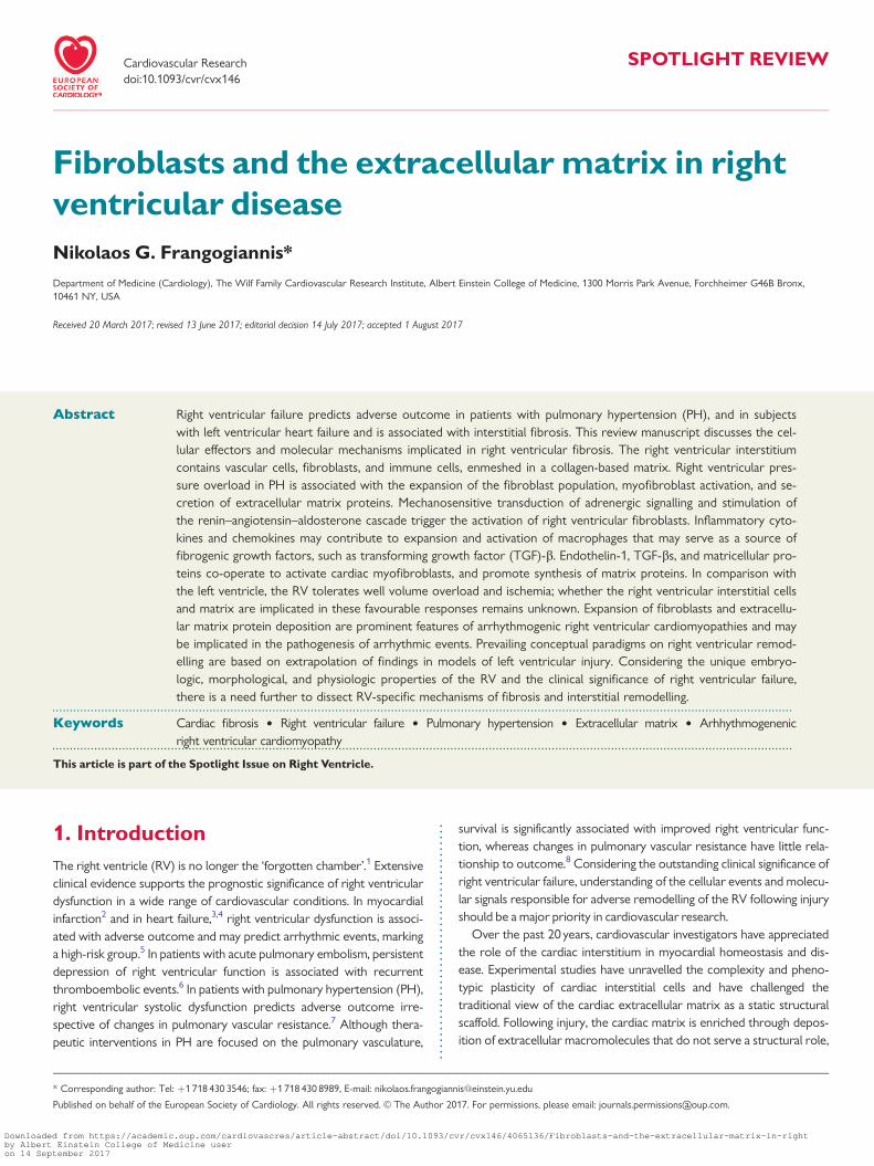
..
..
..
..
..
..
..
..
..
..
..
..
..
..
..
..
..
..
..
..
.
Fibroblasts and the extracellular matrix in right
ventricular disease
Nikolaos G. Frangogiannis*
Department of Medicine (Cardiology), The Wilf Family Cardiovascular Research Institute, Albert Einstein College of Medicine, 1300 Morris Park Avenue, Forchheimer G46B Bronx,10461 NY, USA
Received 20 March 2017; revised 13 June 2017; editorial decision 14 July 2017; accepted 1 August 2017
Abstract Right ventricular failure predicts adverse outcome in patients with pulmonary hypertension (PH), and in subjectswith left ventricular heart failure and is associated with interstitial fibrosis. This review manuscript discusses the cel-lular effectors and molecular mechanisms implicated in right ventricular fibrosis. The right ventricular interstitiumcontains vascular cells, fibroblasts, and immune cells, enmeshed in a collagen-based matrix. Right ventricular pres-sure overload in PH is associated with the expansion of the fibroblast population, myofibroblast activation, and se-cretion of extracellular matrix proteins. Mechanosensitive transduction of adrenergic signalling and stimulation ofthe renin–angiotensin–aldosterone cascade trigger the activation of right ventricular fibroblasts. Inflammatory cyto-kines and chemokines may contribute to expansion and activation of macrophages that may serve as a source offibrogenic growth factors, such as transforming growth factor (TGF)-b. Endothelin-1, TGF-bs, and matricellular pro-teins co-operate to activate cardiac myofibroblasts, and promote synthesis of matrix proteins. In comparison withthe left ventricle, the RV tolerates well volume overload and ischemia; whether the right ventricular interstitial cellsand matrix are implicated in these favourable responses remains unknown. Expansion of fibroblasts and extracellu-lar matrix protein deposition are prominent features of arrhythmogenic right ventricular cardiomyopathies and maybe implicated in the pathogenesis of arrhythmic events. Prevailing conceptual paradigms on right ventricular remod-elling are based on extrapolation of findings in models of left ventricular injury. Considering the unique embryo-logic, morphological, and physiologic properties of the RV and the clinical significance of right ventricular failure,there is a need further to dissect RV-specific mechanisms of fibrosis and interstitial remodelling.
� � � � � � � � � � � � � � � � � � � � � � � � � � � � � � � � � � � � � � � � � � � � � � � � � � � � � � � � � � � � � � � � � � � � � � � � � � � � � � � � � � � � � � � � � � � � � � � � � � � � � � � � � � � � � � � � � � � � � � � � � � � � � � � � � � � � � � � � � � � � � � � � � � � � � � � � � � � � � � � � � � � � � � � � � � � � � � � � � � � � � � � � � � � � � � � � � � � � � � � � � � � � � � � � � � � �
Keywords Cardiac fibrosis • Right ventricular failure • Pulmonary hypertension • Extracellular matrix • Arhhythmogenenicright ventricular cardiomyopathy
� � � � � � � � � � � � � � � � � � � � � � � � � � � � � � � � � � � � � � � � � � � � � � � � � � � � � � � � � � � � � � � � � � � � � � � � � � � � � � � � � � � � � � � � � � � � � � � � � � � � � � � � � � � � � � � � � � � � � � � � � � � � � � � � � � � � � � � � � � � � � � � � � � � � � � � � � � � � � � � � � � � � � � � � � � � � � � � � � � � � � � � � � � � � � � � � � � � � � � � � � � � � � � � � � � � �
This article is part of the Spotlight Issue on Right Ventricle.
1. Introduction
The right ventricle (RV) is no longer the ‘forgotten chamber’.1 Extensiveclinical evidence supports the prognostic significance of right ventriculardysfunction in a wide range of cardiovascular conditions. In myocardialinfarction2 and in heart failure,3,4 right ventricular dysfunction is associ-ated with adverse outcome and may predict arrhythmic events, markinga high-risk group.5 In patients with acute pulmonary embolism, persistentdepression of right ventricular function is associated with recurrentthromboembolic events.6 In patients with pulmonary hypertension (PH),right ventricular systolic dysfunction predicts adverse outcome irre-spective of changes in pulmonary vascular resistance.7 Although thera-peutic interventions in PH are focused on the pulmonary vasculature,
survival is significantly associated with improved right ventricular func-tion, whereas changes in pulmonary vascular resistance have little rela-tionship to outcome.8 Considering the outstanding clinical significance ofright ventricular failure, understanding of the cellular events and molecu-lar signals responsible for adverse remodelling of the RV following injuryshould be a major priority in cardiovascular research.
Over the past 20 years, cardiovascular investigators have appreciatedthe role of the cardiac interstitium in myocardial homeostasis and dis-ease. Experimental studies have unravelled the complexity and pheno-typic plasticity of cardiac interstitial cells and have challenged thetraditional view of the cardiac extracellular matrix as a static structuralscaffold. Following injury, the cardiac matrix is enriched through depos-ition of extracellular macromolecules that do not serve a structural role,
* Corresponding author: Tel: þ1 718 430 3546; fax: þ1 718 430 8989, E-mail: [email protected]
Published on behalf of the European Society of Cardiology. All rights reserved. VC The Author 2017. For permissions, please email: [email protected].
Cardiovascular Research SPOTLIGHT REVIEWdoi:10.1093/cvr/cvx146
Downloaded from https://academic.oup.com/cardiovascres/article-abstract/doi/10.1093/cvr/cvx146/4065136/Fibroblasts-and-the-extracellular-matrix-in-rightby Albert Einstein College of Medicine useron 14 September 2017

..
..
..
..
..
..
..
..
..
..
..
..
..
..
..
..
..
..
..
..
..
..
..
..
..
..
..
..
..
..
..
..
..
..
..
..
..
..
..
..
..
..
..
..
..
..but modulate signalling cascades by regulating growth factor activity andby transducing signals through binding to cell surface receptors. Thesemacromolecules termed ‘matricellular proteins’,9,10 drive the cellular re-sponses that mediate repair, remodelling, and fibrosis of the injured myo-cardium.11 Although our knowledge on the role of the cardiacinterstitium in myocardial repair, remodelling, and fibrosis is derived pri-marily from studies investigating left ventricular injury, a growing body ofevidence suggests a crucial involvement of interstitial changes and matrixremodelling in the pathogenesis and progression of right ventricular dis-ease. The current manuscript discusses the cellular effectors and mo-lecular signals implicated in interstitial remodelling of the RV in a widerange of pathophysiological conditions. Although right ventricular andleft ventricular interstitial cells may have similar baseline phenotypic pro-files and functional properties, the distinct morphologic, physiologic, andpathophysiological characteristics of the RV create a unique environ-ment, leading to different responses under conditions of stress.Unfortunately, in many cases, prevailing paradigms on the mechanisms ofright ventricular fibrosis and on the role of the extracellular matrix in re-modelling of the RV are based on extrapolation of experimental obser-vations in models of left ventricular disease.
2. The interstitium in the healthyright ventricle
The adult mammalian heart is composed of cardiomyocytes, a rich micro-vascular network, and a highly cellular collagen-based interstitium (Figure 1)that contains abundant fibroblast-like interstitial cells and smaller popula-tions of macrophages, mast cells and dendritic cells.12–15 Interstitial cells playan important role in maintaining the cardiac extracellular matrix and may
also transduce signals that regulate cardiomyocyte survival and function.Systematic studies comparing the cellular composition and extracellular ma-trix content of the right and left ventricle have not been performed.
While the left ventricle is derived exclusively from the first heart field,the RV derives in part from the second heart field, which is characterizedby increased proliferative activity and differentiation delay.16 Moreover,the low resistance of the pulmonary vascular territory reduces energydemands on the right ventricular myocardium, leading to thinner walls, alower muscle mass and increased compliance.17 To what extent the dis-tinct embryologic origin and the lower pressure load affect the cellularcomposition and extracellular matrix content of the RV in the absenceof injury remains unknown. At birth, right and left ventricles have com-parable collagen content; neonatal remodelling of the heart leads to re-gression in right ventricular myocyte size, resulting in a relative increaseof collagen content in the adult right ventricle.18,19 In normal adult rats, ahydroxyproline assay demonstrated a lower collagen content in the leftventricular free wall, when compared with the right ventricle and theinterventricular septum.20 On the other hand, experiments comparingfibroblasts from different sites suggested no significant differences intranscriptomic profiles between right ventricular and left ventricular car-diac fibroblasts.21,22 All forms of myocardial injury activate the cardiacinterstitium leading to expansion of interstitial cell populations and dy-namic alterations in the composition of the extracellular matrix.
3. The right ventricular interstitiumin heart disease
The RV tolerates a volume load much better than a pressure load and ismore sensitive to changes in afterload than the left ventricle.23
Figure 1 The right ventricular interstitium. (A) The adult mammalian myocardium is comprised of cardiomyocytes, abundant microvessels (EC, endothelialcells) and a highly cellular interstitium that contains large numbers of fibroblasts (F), pericytes (P) and smaller populations of macrophages (Ma) and other im-mune cells, enmeshed into a collagen-based matrix. Although systematic comparison of the cellular composition and matrix content between the right and theleft ventricular myocardium has not been performed, the interstitium of the RV and the left ventricle have similar morphological characteristics. (B and C)Representative images show staining with picrosirius red of sections from the right ventricular free wall (B, RV) and the left ventricular free wall (C, LV) in adultmouse hearts, in order to identify the collagen network. The epimysial collagen surrounds the organ (arrow); each cardiomyocyte is associated with a thin rimof endomysial collagen (arrowhead). (D, E) Dual fluorescence for wheat germ agglutinin outlines the cardiomyocytes in the mouse right ventricle (D, RV) and inthe left ventricular free wall (E, LV). Experimental studies in adult rats have suggested that due to the lower baseline pressure load and the smaller size of rightventricular cardiomyocytes, the RV exhibits a higher relative collagen content, when compared with the left ventricle and the interventricular septum.20 (F)Immunohistochemical staining for the macrophage-specific antibody Mac2 identified a resident macrophage population in the right ventricular wall (arrows).
2 N.G. Frangogiannis
Downloaded from https://academic.oup.com/cardiovascres/article-abstract/doi/10.1093/cvr/cvx146/4065136/Fibroblasts-and-the-extracellular-matrix-in-rightby Albert Einstein College of Medicine useron 14 September 2017

..
..
..
..
..
..
..
..
..
..
..
..
..
..
..
..
..
..
..
..
..
..
..
..
..
..
..
..
..
..
..
..
..
..
..
..
..
..
..
..
..
..
..
..
..
..
..
..
..
..
..
..
..
..
..
..
..
..
..
..
..
..
..
..
..
..
..
..
..
..
..
..
..
..
..
..
..
..
..
..
..
..
..
..
..
..
..
..
..
.Moreover, due to a more favourable supply and demand ratio, the RV isless susceptible to ischemic injury. Thus, in human patients, the mostcommon pathophysiological cause of right ventricular failure is pressureoverload due to pulmonary hypertension. The impact of pressure over-load on the RV is dependent not only on the magnitude of the pressureincrease but also on the underlying aetiology, and on the rate of progres-sion of the changes in pulmonary pressures. A sudden increase in pul-monary pressures following massive acute pulmonary embolism causes amarked elevation in right ventricular end-diastolic volume, as the cham-ber attempts to compensate in order to maintain stroke volume.Marked acute increases in pulmonary pressures overwhelm the com-pensatory response, leading to rapid development of right ventricularfailure. In contrast, chronic progressive elevations of pulmonary pres-sures are better tolerated by the RV and lead to extensive remodelling,involving both the cardiomyocytes and the right ventricular interstitium.
3.1 The extracellular matrix in acute rightventricular pressure overloadDescriptive studies in experimental models of acute pulmonary embol-ism demonstrated rapid infiltration of the right ventricular outflow tract(RVOT) with neutrophils and monocytes, accompanied by local activa-tion of matrix metalloproteinases (MMPs),24 followed by stimulation of apro-fibrotic program.25 Early matrix degradation may contribute to thepathogenesis of acute right ventricular failure, depriving the cardiomyo-cytes from important pro-survival signals, or abrogating matrix-dependent pathways that preserve cardiomyocyte function. In threestudies performed by the same group in rat, canine and ovine models ofacute pulmonary embolism pre-treatment with doxycycline, a broadnon-selective MMP inhibitor attenuated right ventricular dilation26–28
and decreased cardiomyocyte injury.27 Considering the broad effects ofdoxycycline on inflammatory signalling and oxidative stress,29 whetherthe protective actions reflect attenuated matrix degradation remainsunknown.
3.2 The extracellular matrix in chronicpressure overloadChronic right ventricular pressure overload increases wall stress andpromotes a hypertrophic response. Right ventricular mass is significantlyincreased in PH patients30 and right ventricular hypertrophy is consist-ently observed in experimental models of PH. However, in human pa-tients, the likelihood of progression to right ventricular failure cannot beexplained by differences in right ventricular mass or in pressure load.Despite similar elevations in right ventricular pressures and comparableright ventricular hypertrophy, some patients remain stable for decades,while others rapidly decompensate developing right ventricular fail-ure.8,31 The aetiology of PH plays a major role in determining the out-come. For example, right ventricular failure is highly prevalent in patientswith scleroderma-induced PH.32 In contrast, patients with right ventricu-lar pressure overload caused through purely mechanical causes (such aspulmonic stenosis) exhibit prolonged concentric hypertrophy withoutsignificant contractile depression.33,31 Animal models recapitulate thisheterogeneity, as development of right ventricular dysfunction is de-pendent not only on the severity of pressure overload but also on theunderlying pathophysiological basis.34 In models of PH due to administra-tion of endothelial toxins (such as monocrotaline), right ventricular fail-ure is more prominent than in models of mechanical pressure overloadinduced through pulmonary artery banding.35 Distinct responses ofinterstitial cells and the extracellular matrix may explain the
heterogeneity of the functional responses of the RV to the various path-ophysiological conditions causing PH.
3.2.1 Fibrosis in the pressure-overloaded right ventricularmyocardiumA growing body of evidence suggests that right ventricular fibrosis, theexpansion of the interstitium through deposition of collagenous matrix,may play an important role in the pathogenesis of right ventricular dys-function and failure following pressure overload. Magnetic resonanceimaging (MRI) suggests that many patients with PH exhibit evidence ofright ventricular fibrosis.36 Histological studies have demonstratedincreased collagen deposition in the RV from patients with end-stage PHundergoing heart–lung transplantation.30 In experimental models of rightventricular pressure overload, fibrotic remodelling of the RV is oftenassociated with decompensation and evidence of severe systolic dys-function.37 Ex vivo studies suggested that the contribution of fibrosis tothe increased right ventricular stiffness increases with worsening severityof dysfunction.38 Increased collagen deposition in the RV following pres-sure overload is due to new synthesis of collagen protein,39 and is associ-ated with a disproportionate increase in collagen I secretion,38 a subtypeexhibiting higher tensile strength and reduced elasticity when comparedwith type III collagen. Perturbation of the collagen I:III ratio may be re-sponsible, at least in part, for the increase in right ventricular stiffness fol-lowing pressure overload.
In the model of pulmonary artery banding, late increases in the levelsof insoluble collagen are noted and are associated with right ventriculardiastolic dysfunction.40 Accumulation of insoluble collagen may reflectcross-linking of the right ventricular interstitial matrix; however, the po-tential involvement of specific cross-linking enzymes, such as lysyl-oxidases and tissue transglutaminase has not been investigated.
3.2.2 The cellular effectors implicated in right ventricularfibrosis and remodellingOur current understanding of the cell biological events responsible for fi-brotic remodelling of the pressure-overloaded RV is based predomin-antly on descriptive experiments and on extrapolation of findings fromstudies investigating left ventricular fibrosis. Thus, the prevailing paradigm(Figure 2) assumes similar cellular properties and mechanism of activationfor right and left ventricular myocardial cells, and has not been systemat-ically validated by direct experimentation.
3.2.2.1 Fibroblasts. Cardiac fibroblasts are the main effector cells incardiac fibrosis.41 Extensive evidence suggests that fibrosis of thepressure-overloaded RV is associated with activation and expansion offibroblasts, which acquire a myofibroblast phenotype and secrete largeamounts of extracellular matrix proteins. In a rabbit model of pulmonaryartery constriction, early infiltration of the myocardium with mononu-clear cells was noted 2 days after banding and was associated with evi-dence of cardiomyocyte injury. After 7 days of banding, resolution of theinflammatory infiltrate was accompanied by recruitment of activatedmyofibroblasts, expressing a-smooth muscle actin. After 14 days, theright ventricular interstitium contained significant amounts of fibrillar col-lagen.42 Activated myofibroblasts in the pressure-overloaded RV synthe-size large amounts of collagen and secrete matricellular proteins.40 Thetime course of these cellular alterations suggests that fibrogenic activa-tion may be dependent on early induction of inflammation in thepressure-overloaded RV, either through direct stimulation of mechano-sensitive pro-inflammatory signalling pathways, or secondary to cardio-myocyte necrosis. Direct stimulation of resident cardiac fibroblasts
Interstitial remodelling of the right ventricle 3
Downloaded from https://academic.oup.com/cardiovascres/article-abstract/doi/10.1093/cvr/cvx146/4065136/Fibroblasts-and-the-extracellular-matrix-in-rightby Albert Einstein College of Medicine useron 14 September 2017

..
..
..
..
..
..
..
..
..
..
..
..
..
..
..
..
..
..
..
..
..
..
..
..
..
..
..
..
..
..
..
..
..
..
..
..
..
..
..
..
..
..
..
..
..
..
..
..
..
..
..
..
..
..
..
..
..
..
..
..
..
..
..
..
..
..
..
..
..
..
..
..
..
..
..
..
..
..
..
..
..
..
..
..
..
..
..
..
..
.
through activation of neurohumoral or growth factor-mediated path-ways may also contribute to the fibrotic response.
The cellular origin of fibroblasts in the remodelling RV has not beensystematically investigated. Recent lineage tracing experiments demon-strated that in experimental models of left ventricular pressure overload,resident cardiac fibroblast populations are the main source of activatedmyofibroblasts.43,44 Endothelial to mesenchymal transdifferentiation andcirculating fibroblast progenitors may also contribute smaller, but poten-tially important, fibroblast subsets.45 The distinct embryologic, morpho-logic and functional characteristics of the RV, and the unique features ofthe right ventricular response to injury may have an impact on the rela-tive contributions of various cell types in right ventricular fibrosis.
3.2.2.2 Macrophages and lymphocytes. The adult mouse RV con-tains a sparse population of macrophages.15 Right ventricular pressureoverload is associated with significant expansion of the macrophagepopulation in the right ventricular wall.46 Moreover, patients with PH ex-hibit significantly increased macrophage density in the RV.47 In models ofleft ventricular pressure overload, cardiac macrophages have been impli-cated in the activation of a fibrogenic program.48 Expansion of themacrophage population in the pressure-overloaded myocardium may betriggered through chemokine-dependent mechanisms.49 Moreover, neu-rohumoral mediators may activate macrophages towards a pro-fibroticphenotype characterized by secretion of growth factors and cytokines.Unfortunately, experimental evidence supporting this plausible conceptis lacking, as studies investigating the contribution of macrophages inright ventricular fibrosis have not been performed.
A growing body of evidence implicates lymphocytes in the pathogen-esis of myocardial fibrosis in experimental models of left ventricularpressure overload.50 In a model of transverse aortic constriction,CD4þT cells-mediated fibrosis, decompensation, and transition toheart failure.51 Although systemic aldosterone infusion leads to recruit-ment of lymphocytes in the RV,52 the potential involvement of lympho-cyte subpopulations in right ventricular fibrosis has not beeninvestigated.
3.2.2.3 Mast cells. Mast cells have been implicated in the pathogenesisof left ventricular remodelling and fibrosis.53 Mast cells accumulate in theinjured myocardium and degranulate, releasing fibrogenic cytokines andgrowth factors, and secreting mast cell-specific mediators withfibroblast-activating properties, such as histamine, tryptase, and chy-mase.54–56 In rodent models of pulmonary artery banding, significant in-creases in mast cell density were observed in the right ventricularmyocardium.57,58 Whether mast cell-derived mediators critically regu-late the fibrotic response in the RV has not been investigated.
3.2.2.4 Cardiomyocytes. Focal cardiomyocyte necrosis has been docu-mented in experimental models of right ventricular pressure overload42
and can trigger fibrosis by activating pro-inflammatory signalling. Underconditions of stress, cardiomyocytes are capable of stimulating fibrosiseven in the absence of significant necrosis. Mechanical stretch inducesexpression of renin, angiotensinogen, and ACE in cardiomyocytes59;these effects may trigger an angiotensin-driven fibrogenic program fol-lowing pressure overload. Moreover, stimulated cardiomyocytes may bean important source of transforming growth factor (TGF)-b,60 a crucialfibrogenic mediator that promotes myofibroblast transdifferentiationand induces extracellular matrix protein synthesis.61 The potential con-tribution of pressure-overloaded right ventricular cardiomyocytes in fi-brotic remodelling of the RV has not been investigated.
3.2.2.5 Vascular cells. In the pressure-overloaded myocardium, vascu-lar endothelial cells may be an important source of fibrogenic mediators,such as endothelin-1. Right ventricular capillary rarefaction is a consistentcharacteristic of maladaptive right ventricular hypertrophy in both animalmodels of pressure overload and in human patients with PH.8,62,63
Reduced perfusion of the right ventricular wall may exacerbate local is-chemia, thus promoting a fibrogenic response. Although capillary loss inthe RV may reflect transdifferentiation of vascular endothelial cells orpericytes to fibroblasts, evidence suggesting an important role for vascu-lar cells as a cellular source for activated fibroblasts infiltrating the pres-sure overloaded RV is lacking.
3.2.3. Molecular signals mediating right ventricularfibrosisDevelopment of fibrosis in the pressure-overloaded RV involves activa-tion of mechanosensitive signalling pathways that release neurohumoralmediators, stimulate inflammatory responses, and trigger growth factor-mediated cascades (Table 1 and Figure 3).
3.2.3.1 Neurohumoral pathwaysSympathetic stimulation and activation of the renin–angiotensin–aldos-terone system (RAAS) are prominent features of PH and are implicatedin the pathogenesis of right ventricular dysfunction.64 In experimentalmodels of PH, the administration of the non-selective b-blocker carvedi-lol,65 or the b1 adrenergic receptor (AR) antagonist bisoprolol66 attenu-ated right ventricular remodelling and decreased fibrosis of the rightventricular wall without affecting pulmonary pressures. Although
Figure 2 Cell biological effectors of fibrosis in the pressure-over-loaded RV. Based on insights from lineage tracing experiments in mod-els of left ventricular pressure overload, activated myofibroblasts arepredominantly derived from resident populations. Endothelial cells,pericytes, epicardial epithelial cells, and circulating fibroblast progeni-tors may also play contributory roles. Vascular cells, stressed cardio-myocytes, resident, and recruited macrophages may play an importantrole in activation of resident fibroblast population in the pressure-over-loaded myocardium.
4 N.G. Frangogiannis
Downloaded from https://academic.oup.com/cardiovascres/article-abstract/doi/10.1093/cvr/cvx146/4065136/Fibroblasts-and-the-extracellular-matrix-in-rightby Albert Einstein College of Medicine useron 14 September 2017

..
..
..
..
..
..
..
..
..
..
..
..
..
..
..
..
.adrenergic stimulation is known to activate cardiac fibroblasts in vitro,67
whether reduced fibrosis following AR blockade in vivo is due to directactions on the fibroblasts, or represents an epiphenomenon, reflectingeffects on cardiomyocytes or vascular cells is unknown. Moreover, therelative significance of attenuated fibrosis and modulation of the matrixenvironment in mediating the protective actions of b-AR blockade onright ventricular function has not been elucidated.
Local activation of angiotensin II and aldosterone signalling may alsobe implicated in the pathogenesis of right ventricular fibrosis followingpressure overload. In vitro studies have demonstrated that mechanicalstress induces components of the RAAS in cardiomyocytes.59,68.
In models of PH, significantly increased expression of angiotensin con-verting enzyme was noted in the RV and was localized in fibrotic areas.69
Evidence supporting an important role for the RAAS in mediating rightventricular fibrosis following pressure overload is predominantly derivedfrom pharmacologic inhibition experiments. In a rabbit model of pul-monary artery banding and in a model of monocrotaline-induced pul-monary hypertension, angiotensin type 1 (AT1) receptor blockadeattenuated right ventricular fibrosis.70,71 In contrast, experiments in a ratmodel of pulmonary artery constriction, combined treatment with anAT1 inhibitor and an aldosterone antagonist had no effects on right ven-tricular dysfunction, did not significantly affect fibrosis, and did not
..............................................................................................................................................................................................................................
Table 1 Molecular signals implicated in right ventricular fibrosis following pressure overload
Pathway Likely mechanisms of action Functional effects References
Adrenergic
stimulation
b-AR signalling stimulates fibroblast proliferation67,145
and may modulate fibroblast function146
AR signalling may contribute to adverse right ventricular
remodelling and may promote progression of right-
sided heart failure
65,66
Renin–angiotensin–
aldosterone sys-
tem (RAAS)
Angiotensin II-mediated AT-1 activation promotes fibro-
blast activation, and stimulates extracellular matrix
protein synthesis147
Aldosterone-mediated mineralocorticoid receptor sig-
nalling activates fibroblasts148 and may promote a
fibrogenic macrophage phenotype149
Activation of the RAAS may promote inflammation,
thus triggering a fibrotic response52.
Pharmacologic inhibition experiments have suggested an
important role for the RAAS in right ventricular dys-
function following pressure overload in some70,71 but
not all studies72
70–72
Endothelin-1 Endothelin-1 stimulates proliferation and matrix synthe-
sis in cardiac fibroblasts77
Endothelin-1 may promote a pro-fibrotic phenotype in
vascular cells150
Pharmacological inhibition experiments suggest that
endothelin receptor signalling promotes right ven-
tricular remodelling and fibrosis in models of pulmon-
ary hypertension.78–80 In addition to its role in the
pulmonary vasculature, endothelin-1 may also medi-
ate fibrosis of the RV
78–80
Oxidative stress Mechanosensitive activation of Rho kinase may trigger
ROS generation in fibroblasts, thus promoting colla-
gen synthesis
Accentuated oxidative stress in the pressure-over-
loaded RV may be due to defective antioxidant
responses
Pharmacologic interventions using antioxidants suggest
that ROS generation contributes to right ventricular
remodelling fibrosis and dysfunction following pres-
sure overload
81–83
Inflammatory cyto-
kines and
chemokines
Neurohumoral pathways and oxidative stress may be
responsible for induction of inflammatory cytokines
and chemokines in the pressure-overloaded RV.
Cytokines may modulate fibroblast activity and may
promote a fibrogenic phenotype in macrophages
Although descriptive studies show induction of inflam-
matory signals in the pressure overloaded RV, studies
examining the role of cytokines and chemokines in
the pathogenesis of right ventricular fibrosis have not
been performed
85
TGF-b TGF-bs are critical regulators of fibroblast function, but
may also mediate fibrosis by promoting fibrogenic ac-
tions on other cell types (such as immune and vascu-
lar cells)
Although descriptive studies suggest TGF-b1 induction
in the remodelling RV, the role of the TGF-b cascade
in right ventricular fibrosis has not been systematically
studied.
Studies in both angio-obliterative and surgical models of
right ventricular pressure overload have suggested an
important role for the accessory TGF-b receptor
endoglin in right ventricular fibrosis and dysfunction97
70,91,97
Matricellular proteins Matricellular proteins (thrombospondins, tenascin-C,
periostin, osteopontin, SPARC, CCNs) modulate
growth factor signalling and protease activity and have
profound effects on phenotype and function of fibro-
blasts, inflammatory cells and vascular cells
Upregulation of osteopontin, tenascin-C, SPARC and
CCN2 in the RV has been demonstrated in models of
pulmonary hypertension. However, their role in right
ventricular remodelling and fibrosis has not been
investigated
40,70,102,103
Interstitial remodelling of the right ventricle 5
Downloaded from https://academic.oup.com/cardiovascres/article-abstract/doi/10.1093/cvr/cvx146/4065136/Fibroblasts-and-the-extracellular-matrix-in-rightby Albert Einstein College of Medicine useron 14 September 2017

..
..
..
..
..
..
..
..
..
..
..
..
..
..
..
..
..
..
..
..
..
..
..
..
..
..
..
..
..
..
..
..
..
..
..
..
..
..
..
..
..
..
..
..
..
..
..
..
..
..
..
..
..
..
..
..
..
..
..
..
..
..
..
..
..
..
..
..
..
..
..
..
..
..
..
..
..
..
..
..
..
..
..
..
..
..
..
..
..
.
attenuate capillary rarefaction.72 Currently, there is no evidence to sup-port the use of RAAS inhibition strategies in patients with right ventricu-lar dysfunction due to PH.73
3.2.3.2 Endothelin-1Endothelin-1, a powerful vasoconstrictor with potent fibrogenic proper-ties, is critically implicated in the pathogenesis of PH.74 Endothelin recep-tor blockade is a safe and effective therapeutic approach for PHpatients.75 A growing body of evidence suggests that, in addition to its ac-tions on the pulmonary vasculature, endothelin-1 may also mediate rightventricular dysfunction and fibrosis. The expression of endothelin andthe endothelin type A receptor are significantly upregulated in the RV inexperimental models of right ventricular pressure overload and in pa-tients with PH.76 In the RV, activation of the endothelin-1 axis may pro-mote fibroblast proliferation and stimulate synthesis of extracellularmatrix proteins.77 Endothelin receptor blockade attenuates right ven-tricular fibrosis in mechanical, angio-obliterative, and hypoxia-induced
models of pulmonary hypertension.78–80 Whether these actions contrib-ute to the beneficial effects of endothelin inhibition in patients with PHremains unknown.
3.2.3.3 Oxidative stressGeneration of reactive oxygen species (ROS) plays an important role infibroblast activation and in deposition of extracellular matrix proteins inthe remodelling myocardium. In vivo studies in a model of pulmonary ar-tery constriction demonstrated that right ventricular pressure overloadcauses a marked increase in oxidative stress in the right ventricular myo-cardium, and suggested that, when compared to the left ventricle, theRV may exhibit an accentuated oxidative response to a pressure load.81
Mechanosensitive activation of Rho kinase may play an important role inROS generation following right ventricular pressure overload.81 It hasbeen suggested that the exaggerated oxidative response of the RV fol-lowing stress may be due to defective antioxidant defenses. In a model ofNO deficiency induced by L-NAME feeding, the RV exhibited early cardi-omyopathic changes, and dilation; in contrast, the left ventricle had onlysubtle hypertrophic alterations. The selective involvement of the RV inthis model was associated with increased oxidative stress and failure toactivate antioxidant responses.82 Treatment with antioxidants attenu-ated adverse remodelling and dysfunction of the RV, reducing right ven-tricular fibrosis in the rat monocrotaline model of pulmonaryhypertension.83
3.2.3.4 Inflammatory signalsPro-inflammatory cytokines and chemokines play an important role in fi-brotic remodelling in many tissues, including the myocardium, boththrough direct stimulation of fibroblasts and through induction of fibro-genic signals in other cell types (such as macrophages and vascularcells).84 Our understanding of the role of inflammatory mediators in rightventricular fibrosis is based on descriptive evidence and on known ef-fects of these signals in left ventricular disease. In a mouse model of pul-monary artery constriction, marked upregulation of several members ofthe chemokine family was noted in the pressure-overloaded RV, includ-ing the CC chemokines CCL2 and CCL5, the CXC chemokinesCXCL16 and CXCL10, and CX3CL1.85 Neurohumoral mediators, gen-eration of ROS, and direct activation of mechanosensitive pro-inflammatory signals in cardiomyocytes and macrophages may explainthe induction of inflammatory genes in the pressure-overloaded RV. Invitro studies suggested that CXCL16, CX3CL1, and CCL5 may increaseexpression of the small leucine-rich glycoproteins decorin, biglycan,lumican, and fibromodulin, important activators of the fibrotic re-sponse.85 CCL2 may also be implicated in the fibrotic myocardial re-sponse by recruiting mononuclear cells that may activate cardiacfibroblasts.86 On the other hand, induction of the anti-fibrotic chemokineCXCL1087,88 may serve as an endogenous inhibitory signal that restrainsthe fibrotic response.
3.2.3.5 The TGF-b superfamilySeveral members of the TGF-b superfamily have been implicated inregulation of tissue fibrosis and extracellular matrix remodelling. TGF-b1is critically involved in activation of a pro-fibrotic program, by inducingsynthesis of extracellular matrix proteins and by promoting myofibro-blast transdifferentiation.61 On the other hand, members of the bonemorphogenetic protein (BMP) subfamily, such as BMP-2 and BMP-7, mayattenuate fibrosis by inhibiting TGF-b actions.45,89,90. The role of TGF-bsignalling pathways in mediating right ventricular dysfunction and fibrosis
Figure 3 Molecular signals implicated in fibroblast activation in thepressure-overloaded RV. Right ventricular pressure overload trans-duces mechanosensitive signalling in cardiomyocytes (CM), fibroblasts(F), vascular endothelial cells (EC), and macrophages (Ma), stimulatingneurohumoral pathways and activating reactive oxygen species (ROS)generation. Activation of the RAAS pathway, adrenergic signalling, andoxidative stress stimulate fibroblast expansion and triggers synthesis ofextracellular matrix proteins. Angiotensin II and aldosterone may alsoinduce an inflammatory and fibrogenic program in endothelial cells,stimulating expression of endothelin-1 (ET-1), and synthesis of chemo-kines, and triggering recruitment of monocytes (Mo). Activated macro-phages secrete transforming growth factor (TGF)-b and other growthfactors in the cardiac interstitium. Enrichment of the extracellular ma-trix though the deposition of matricellular proteins regulates activationof growth factor signalling. TGF-b exerts potent matrix-preservingactions by promoting myofibroblast transdifferentiation, by stimulatingsynthesis of matrix proteins, and by inducing expression ofantiproteases.
6 N.G. Frangogiannis
Downloaded from https://academic.oup.com/cardiovascres/article-abstract/doi/10.1093/cvr/cvx146/4065136/Fibroblasts-and-the-extracellular-matrix-in-rightby Albert Einstein College of Medicine useron 14 September 2017

..
..
..
..
..
..
..
..
..
..
..
..
..
..
..
..
..
..
..
..
..
..
..
..
..
..
..
..
..
..
..
..
..
..
..
..
..
..
..
..
..
..
..
..
..
..
..
..
..
..
..
..
..
..
..
..
..
..
..
..
..
..
..
..
..
..
..
..
..
..
..
..
..
..
..
..
..
..
..
..
..
..
..
..
..
..
..
..
..
.following pressure overload is poorly understood. TGF-b1 upregulationhas been demonstrated in the RV in models of pulmonary artery con-striction and in pulmonary hypertension secondary to left sided heartfailure91; however, findings on downstream activation of TGF-b signallingcascades are conflicting. In an experimental model of pulmonary arterybanding, increased levels of phosphorylated Smad2 (p-Smad2) in theright ventricular myocardium suggested activation of TGF-b signalling.70
However, in patients with end-stage pulmonary hypertension, p-Smad2immunoreactivity in the RV was reduced, when compared with con-trols.92 The contradictory findings may reflect the dynamic regulation ofthe TGF-b cascade in vivo. Early activation of the pathway in the pres-sure-overloaded myocardium may be followed by induction of inhibitorysignals that restrain TGF-b/Smad signalling.
Dissection of the in vivo actions of TGF-b family members is hamperedby the wide range of their context-dependent effects that target manydifferent cell types. In models of reparative and interstitial left ventricularfibrosis, both Smad-dependent and Smad-independent actions of TGF-bsignalling have been suggested to play important roles in regulation offibroblast responses93–96 and in deposition of extracellular matrix pro-teins. Experiments investigating the in vivo role of TGF-b signalling cas-cades in fibrosis and remodelling of the RV have not been performed.Experimental studies in both angio-obliterative and surgical models ofright ventricular pressure overload suggested that endoglin, an accessoryreceptor for the TGF-b family, may mediate fibrosis and dysfunction ofthe RV.97
3.2.3.6 The matricellular proteinsMatricellular proteins are extracellular macromolecules that do notserve a primary structural role, but are secreted following injury andmodulate growth factor and cytokine responses. In the remodellingmyocardium, the extracellular matrix is enriched through the depositionof matricellular proteins. Over the last 10–15 years, experimental studieshave suggested that several members of the matricellular family (includ-ing the thrombospondins, tenascins, osteopontin, SPARC, periostin, andmembers of the CCN family) are markedly induced in the injured andpressure overloaded left ventricle and play a critical role in regulating in-flammatory, fibrotic, and angiogenic responses.11,98–101 The potential in-volvement of members of the matricellular proteins in remodelling ofthe pressure-overloaded RV is suggested almost exclusively by descrip-tive studies. Right ventricular upregulation of osteopontin, tenascin-C,SPARC, and CCN2 has been documented in experimental models ofPH.40,70,102,103 Induction of matricellular proteins in the pressure-overloaded RV may be mediated through the activation of neurohu-moral and inflammatory pathways and may involve activation of TGF-b.Investigations examining the role of matricellular actions in regulatingright ventricular fibrosis, matrix remodelling, and dysfunction have notbeen performed.
3.3. The extracellular matrix in rightventricular volume overloadPure right ventricular volume overload is generally well tolerated byhuman patients and experimental animals. Many patients with tetralogyof Fallot develop pulmonary insufficiency that typically has minimal con-sequences over several decades, but can be the source of late complica-tions including right ventricular dysfunction due to chronic volumeoverload.104 In both large animal and rodent models of pulmonary insuf-ficiency, right ventricular volume overload resulted in dilation of the RV,accompanied by diastolic dysfunction, without affecting systolic
function.105,106 In the mouse model, right ventricular volume overloadwas associated with development of subendocardial fibrosis in the rightventricular wall. Dynamic changes in the expression of matrix re-modelling genes were observed: early downregulation of some mem-bers of the TGF-b superfamily (such as growth differentiation factor(GDF)-5 and GDF-7) was followed by late induction of cytokines andchemokines, TGF-b1 upregulation and increased transcription of matri-cellular proteins (including thromsbospondin-1 and periostin).105 Thefunctional significance of these alterations remains unknown.
3.4. Right ventricular infarctionAlthough the cellular events and molecular pathways involved in fibroticrepair of the infracted left ventricle have been extensively character-ized,107 much less is known about the response of the RV to infractive in-jury. In the clinic, right ventricular infarction is quite common; 40–50% ofpatients with acute inferior myocardial infarction exhibit right ventricularinvolvement.108 Experimental animal models and clinical experience sug-gest that although early functional depression of the RV following infarc-tion may have adverse consequences, the right ventricular myocardiumis resistant to infarction and recovers even after prolonged coronaryocclusion.109 In human patients with early right ventricular ischemicdysfunction, the function of the RV returns to normal within3–12 months.110 In the dog, persistent occlusion of the right coronary ar-tery does not cause infarction.111 The more favourable balance betweenoxygen supply and demand explains the reduced vulnerability of the RVto ischemic injury. The extensive collateralization of the RV providesmore reliable blood supply under ischemic conditions, while the lowermyocardial mass and the reduced afterload account for the decreaseddemand, in comparison to the left ventricular myocardium. Moreover,the RV exhibits higher capabilities to extract oxygen under conditions ofstress, due to its low oxygen contraction levels at rest. Whether differ-ences in the matrix composition of the cardiac interstitium, or distinctfunctional properties of right ventricular fibroblasts and immune cellscontribute to the unique response of the RV to ischemia is unknown.
3.5. The right ventricular extracellularmatrix in arrhythmogeniccardiomyopathiesMyocardial fibrosis perturbs propagation of the electrical impulse andhas been implicated in arrhythmia generation. Evidence of right ventricu-lar interstitial or perivascular fibrosis was found in 56% of patients withventricular arrhythmias in the absence of clinical structural heart dis-ease.112 Interstitial fibrosis in the RVOT has been detected in patientswith sudden cardiac death related to Brugada syndrome.113 In patientswith hypertrophic cardiomyopathy, right ventricular fibrosis predictedarrhythmic events.114 Individuals with arrhythmogenic right ventricularcardiomyopathy (ARVC) exhibit expansion of right ventricular fibroadi-pocytes, leading to fibrosis and fatty infiltration.115–118
The mechanisms responsible for the marked alterations in myocardialcellular composition in patients with ARVC and other arrhythmogeniccardiomyopathies remain unknown. Genetic studies have identified mu-tations in genes encoding desmosome proteins as the underlying causein many patients with ARVC. Mutations in PKP2, which encodesplakophilin-2, in desmoplakin, and desmoglein-2 have been identified inmany patients with ARVC.119–121 In mice, loss of desmoplakin in a subsetof interstitial cells with fibroadipocyte characteristics promotes adipo-cyte differentiation through a Wnt-dependent mechanism.122,123 Thecellular link between these mutations and right ventricular fibrosis is less
Interstitial remodelling of the right ventricle 7
Downloaded from https://academic.oup.com/cardiovascres/article-abstract/doi/10.1093/cvr/cvx146/4065136/Fibroblasts-and-the-extracellular-matrix-in-rightby Albert Einstein College of Medicine useron 14 September 2017

..
..
..
..
..
..
..
..
..
..
..
..
..
..
..
..
..
..
..
..
..
..
..
..
..
..
..
..
..
..
..
..
..
..
..
..
..
..
..
..
..
..
..
..
..
..
..
..
..
..
..
..
..
..
..
..
..
..
..
..
..
..
..
..
..
..
..
..
..
..
..
..
..
..
..
..
..
..
..
..
..
..
..
..
..
..
..
..
..
.clear. Fibrosis may reflect cardiomyocyte injury, or primary activation ofinterstitial cell populations and transition to a fibroblast phenotype.124
Recent cell biological experiments suggested that plakophilin-2 loss pro-motes TGF-b1 expression and may activate a fibrogenic program in car-diomyocytes.125 The significance of these actions in the pathogenesis offibrosis in ARVC remains unclear.
3.6. Right ventricular fibrosis in response tocancer chemotherapyPatients treated with anthracyclines as cancer chemotherapy exhibit ahigh incidence of cardiac complications. Acute anthracycline cardiotoxic-ity is rare in human patients and is associated with dose-dependent cardi-omyocyte injury and secondary inflammation.126 Chronic cardiotoxicityon the other hand, may occur within 1 year after completion of treat-ment (early-onset chronic cardiomyopathy), or may affect cancer sur-vivors many years after therapy (late-onset chronic cardiomyopathy).127
A growing body of evidence suggests that interstitial fibrosis may be animportant component of the cardiac pathology in cancer survivorsexposed to anthracyclines. Magnetic resonance imaging showedenhanced chronic fibrotic changes in anthracycline-treated patients.128
Moreover, late development of systolic heart failure, arrhythmias, andsudden death in patients treated with anthracyclines was associated withhistological evidence of myocardial fibrosis.129,130 In animal models ofdoxorubicin toxicity, a subacute increase in myocardial fibrosis wasstrongly linked with late mortality.131 Data on the extent of right ven-tricular fibrotic remodelling following anthracycline administration areconflicting. In a mouse model, doxorubicin administration significantlyincreased collagen deposition in both the left and the right ventricle.132
In contrast, in rabbits treated with daunorubicin fibrotic, changes werereported to be much less severe in the right ventricle.133 Species-specificeffects, differences in the dose of the anthracycline, and in the timing ofassessment of fibrosis may explain the conflicting findings. The cellularbasis of fibrosis in response to anthracycline treatment has not beensystematically studied. Fibrotic changes may represent an epiphenom-enon, reflecting activation of a reparative program in response toanthracycline-induced cardiomyocyte death. Direct effects of anthracy-clines on fibroblasts should also be considered. A recent study suggestedthat doxorubicin-induced generation of reactive oxygen species may ac-tivate ataxia telangiectasia mutated (ATM) kinase in cardiac fibroblastspromoting a fibrotic response.134
3.7. The profibrotic effects of cigarettesmokeA recently published study suggested that cigarette smoke exposuremay increase right ventricular collagen content in mice, suppressing rightventricular function, in the absence of any effects on the left ventricleand on the pulmonary vasculature.135 The profibrotic effects of cigarettesmoke were attributed to nicotine-mediated activation of a7 nicotineacetylcholine receptor signalling in cardiac fibroblasts. The basis for theselective involvement of the RV is unknown. Direct profibrotic effects ofcigarette smoke may explain the presence of right ventricular dysfunc-tion in some patients with chronic obstructive pulmonary disease, des-pite the absence of pulmonary hypertension.136
3.8. Right ventricular fibrosis in response toleft ventricular diseaseAlthough the systemic and the pulmonary circulation are arranged in ser-ies, there are several layers of functional integration between the left and
the right ventricle. The common pericardium that encircles both cham-bers, the continuity between the muscle fibres of the left and right ven-tricular-free wall, the shared interventricular septum, and perfusion bythe coronary arterial system generate a physiologic interdependence be-tween the right and the left ventricle. In left ventricular disease, additionalmechanisms contribute to structural remodelling and dysfunction of theRV. In an experimental model of left ventricular myocardial infarction, amarked upregulation in right ventricular collagen type I and III mRNAsynthesis was reported, peaking 7–14 days after coronary occlusion.Collagen deposition was followed by significantly increased right ven-tricular stiffness, 56 days after infarction.137 Moreover, increased rightventricular stiffness is noted in a model of left ventricular pressure over-load,138 and in a model of renovascular hypertension, interstitial fibrosisis noted in both the pressure-overloaded left ventricle and in the RV.19
Structural remodelling of the extracellular matrix network of the RV inresponse to left ventricular disease is, at least in part, secondary to theincreased right ventricular pressures caused by left ventricular failure,but may also reflect systemic activation of neurohumoral pathways thataffect both ventricles.19
3.9. The right ventricular interstitium insenescent and diabetic heartsAging, obesity, and diabetes are associated with interstitial and perivascu-lar cardiac fibrosis that may contribute to diastolic dysfunction.139–141
Some studies have reported that in senescent mice, the fibrotic responsemay be more diffuse in the RV than in the left ventricle, suggesting thatthese changes may explain the aging-associated increase in the incidenceof arrhythmias.142,143 Moreover, in a rat model of type 1 diabetes, signifi-cant RV fibrosis was noted in the absence of hypertrophy or cardiomyo-cyte apoptosis.144 Whether these observations are relevant in senescentor diabetic human subjects remains unknown.
4. Does the right ventricularinterstitium exhibit uniquepathophysiological responses?
Although the importance of right ventricular dysfunction in patients withcardiac and pulmonary disease is widely appreciated, understanding ofthe pathophysiological underpinnings of right ventricular failure remainslimited. This is due, at least in part, to the notion that considering the in-tense research interest on the cell biological responses to left ventricularinjury, relatively little additional information can be derived by investigat-ing interactions between the same cell types in the RV. In the absence ofinjury, right and left ventricular fibroblasts appear to have similar charac-teristics; the matrix networks of the left and the right ventricle sharecommon structural properties. However, the right ventricular predilec-tion of certain genetic conditions associated with interstitial remodelling,such as ARVC, suggests unique characteristics of the interstitial cellspopulating the right ventricular wall. The cellular composition of the RVhas not been systematically studied and the molecular links between gen-etic alterations and right ventricular interstitial remodelling remainunknown.
Moreover, various diseases impose unique pathophysiological chal-lenges to the right ventricular interstitium. In conditions associated withright ventricular pressure overload, the dramatic pressure increaseswould be expected to transduce potent activating signals in interstitialcells populating the RV. Under hypoxic conditions, the favourable
8 N.G. Frangogiannis
Downloaded from https://academic.oup.com/cardiovascres/article-abstract/doi/10.1093/cvr/cvx146/4065136/Fibroblasts-and-the-extracellular-matrix-in-rightby Albert Einstein College of Medicine useron 14 September 2017

..
..
..
..
..
..
..
..
..
..
..
..
..
..
..
..
..
..
..
..
..
..
..
..
..
..
..
..
..
..
..
..
..
..
..
..
..
..
..
..
..
..
..
..
..
..
..
..
..
..
..
..
..
..
..
..
..
..
..
..
..
..
..
..
..
..
..
..
..
..
..
..
..
..
..
..
..
..
..
..
..
..
..
..
..
..
..
..
..
.oxygen supply and demand relations in the RV may result in distinctinterstitial cell responses. To what extent the relative resistance of theRV to infarctive injury is dependent on unique properties of the rightventricular interstitium remains unknown. Thus, understanding the path-obiology of right ventricular interstitial cells may provide unique insightsinto strategies to improve adverse remodelling in left ventriculardiseases.
5. Conclusions
A growing body of evidence suggests that phenotypic and functional al-terations of interstitial cell populations and changes in the compositionof the extracellular matrix network may play an important role in thepathogenesis of right ventricular dysfunction in a wide range of patho-logic conditions. Our current understanding of the cell biological mech-anisms and molecular signals involved in right ventricular fibrosis is basedto a large extent on extrapolation from studies investigating left ventricu-lar remodelling. Although the two ventricles have similarities in cellularcomposition and share several layers of functional integration, they alsoexhibit significant differences in embryologic origin, physiologic proper-ties, and pathophysiological responses. Studies dissecting RV-specific cel-lular responses to injury are needed in order to understand the basis forright ventricular remodelling and dysfunction. Insights into the mechan-isms of right ventricular fibrosis are necessary in order to develop noveltherapeutic strategies to prevent right ventricular dysfunction in patientswith pulmonary hypertension, and to inhibit arrhythmia generation in pa-tients with right ventricular cardiomyopathies.
FundingDr Frangogiannis’ Laboratory is supported by grants from the NationalInstitutes of Health (R01 HL76246 and R01 HL85440), and theDepartment of Defense (PR151134 and PR151029).
Conflict of interest. none declared.
References1. Rigolin VH, Robiolio PA, Wilson JS, Harrison JK, Bashore TM. The forgotten cham-
ber: the importance of the right ventricle. Cathet Cardiovasc Diagn 1995;35:18–28.2. Mehta SR, Eikelboom JW, Natarajan MK, Diaz R, Yi C, Gibbons RJ, Yusuf S. Impact
of right ventricular involvement on mortality and morbidity in patients with inferiormyocardial infarction. J Am Coll Cardiol 2001;37:37–43.
3. de Groote P, Millaire A, Foucher-Hossein C, Nugue O, Marchandise X, Ducloux G,Lablanche JM. Right ventricular ejection fraction is an independent predictor of sur-vival in patients with moderate heart failure. J Am Coll Cardiol 1998;32:948–954.
4. Burke MA, Katz DH, Beussink L, Selvaraj S, Gupta DK, Fox J, Chakrabarti S, SauerAJ, Rich JD, Freed BH, Shah SJ. Prognostic importance of pathophysiologic markersin patients with heart failure and preserved ejection fraction. Circ Heart Fail 2014;7:288–299.
5. Mikami Y, Jolly U, Heydari B, Peng M, Almehmadi F, Zahrani M, Bokhari M, Stirrat J,Lydell CP, Howarth AG, Yee R, White JA. Right ventricular ejection fraction Is in-cremental to left ventricular ejection fraction for the prediction of future arrhyth-mic events in patients with systolic dysfunction. Circ Arrhythm Electrophysiol 2017;10:e004067.
6. Grifoni S, Vanni S, Magazzini S, Olivotto I, Conti A, Zanobetti M, Polidori G, PieralliF, Peiman N, Becattini C, Agnelli G. Association of persistent right ventricular dys-function at hospital discharge after acute pulmonary embolism with recurrentthromboembolic events. Arch Intern Med 2006;166:2151–2156.
7. van de Veerdonk MC, Kind T, Marcus JT, Mauritz GJ, Heymans MW, Bogaard HJ,Boonstra A, Marques KM, Westerhof N, Vonk-Noordegraaf A. Progressive rightventricular dysfunction in patients with pulmonary arterial hypertension respondingto therapy. J Am Coll Cardiol 2011;58:2511–2519.
8. Ryan JJ, Archer SL. The right ventricle in pulmonary arterial hypertension: disordersof metabolism, angiogenesis and adrenergic signaling in right ventricular failure. CircRes 2014;115:176–188.
9. Bornstein P. Matricellular proteins: an overview. J Cell Commun Signal 2009;3:163–165.
10. Sage EH, Bornstein P. Extracellular proteins that modulate cell–matrix interactions.SPARC, tenascin, and thrombospondin. J Biol Chem 1991;266:14831–14834.
11. Frangogiannis NG. Matricellular proteins in cardiac adaptation and disease. PhysiolRev 2012;92:635–688.
12. Pinto AR, Ilinykh A, Ivey MJ, Kuwabara JT, D’antoni ML, Debuque R, Chandran A,Wang L, Arora K, Rosenthal NA, Tallquist MD. Revisiting cardiac cellular compos-ition. Circ Res 2016;118:400–409.
13. Frangogiannis NG, Burns AR, Michael LH, Entman ML. Histochemical and morpho-logical characteristics of canine cardiac mast cells. Histochem J 1999;31:221–229.
14. Nag AC. Study of non-muscle cells of the adult mammalian heart: a fine structuralanalysis and distribution. Cytobios 1980;28:41–61.
15. Mylonas KJ, Jenkins SJ, Castellan RF, Ruckerl D, McGregor K, Phythian-Adams AT,Hewitson JP, Campbell SM, MacDonald AS, Allen JE, Gray GA. The adult murineheart has a sparse, phagocytically active macrophage population that expandsthrough monocyte recruitment and adopts an 0M20 phenotype in response to Th2immunologic challenge. Immunobiology 2015;220:924–933.
16. Buckingham M, Meilhac S, Zaffran S. Building the mammalian heart from two sour-ces of myocardial cells. Nat Rev Genet 2005;6:826–835.
17. Laks MM, Garner D, Swan HJ. Volumes and compliances measured simultaneouslyin the right and left ventricles of the dog. Circ Res 1967;20:565–569.
18. Weber KT, Brilla CG. Pathological hypertrophy and cardiac interstitium. Fibrosisand renin-angiotensin-aldosterone system. Circulation 1991;83:1849–1865.
19. Brilla CG, Pick R, Tan LB, Janicki JS, Weber KT. Remodeling of the rat right and leftventricles in experimental hypertension. Circ Res 1990;67:1355–1364.
20. Medugorac I. Collagen content in different areas of normal and hypertrophied ratmyocardium. Cardiovasc Res 1980;14:551–554.
21. Furtado MB, Costa MW, Pranoto EA, Salimova E, Pinto AR, Lam NT, Park A, SniderP, Chandran A, Harvey RP, Boyd R, Conway SJ, Pearson J, Kaye DM, Rosenthal NA.Cardiogenic genes expressed in cardiac fibroblasts contribute to heart developmentand repair. Circ Res 2014;114:1422–1434.
22. Furtado MB, Nim HT, Boyd SE, Rosenthal NA. View from the heart: cardiac fibro-blasts in development, scarring and regeneration. Development 2016;143:387–397.
23. Dandel M, Hetzer R. Echocardiographic assessment of the right ventricle: impact ofthe distinctly load dependency of its size, geometry and performance. Int J Cardiol2016;221:1132–1142.
24. Watts JA, Gellar MA, Obraztsova M, Kline JA, Zagorski J. Role of inflammation inright ventricular damage and repair following experimental pulmonary embolism inrats. Int J Exp Pathol 2008;89:389–399.
25. Zagorski J, Obraztsova M, Gellar MA, Kline JA, Watts JA. Transcriptional changes inright ventricular tissues are enriched in the outflow tract compared with the apexduring chronic pulmonary embolism in rats. Physiol Genomics 2009;39:61–71.
26. Cau SB, Barato RC, Celes MR, Muniz JJ, Rossi MA, Tanus-Santos JE. Doxycyclineprevents acute pulmonary embolism-induced mortality and right ventricular de-formation in rats. Cardiovasc Drugs Ther 2013;27:259–267.
27. Neto-Neves EM, Dias-Junior CA, Rizzi E, Castro MM, Sonego F, Gerlach RF, Tanus-Santos JE. Metalloproteinase inhibition protects against cardiomyocyte injury duringexperimental acute pulmonary thromboembolism. Crit Care Med 2011;39:349–356.
28. Neto-Neves EM, Sousa-Santos O, Ferraz KC, Rizzi E, Ceron CS, Romano MM, GaliLG, Maciel BC, Schulz R, Gerlach RF, Tanus-Santos JE. Matrix metalloproteinase in-hibition attenuates right ventricular dysfunction and improves responses to dobut-amine during acute pulmonary thromboembolism. J Cell Mol Med 2013;17:1588–1597.
29. Griffin MO, Ceballos G, Villarreal FJ. Tetracycline compounds with non-antimicrobial organ protective properties: possible mechanisms of action. PharmacolRes 2011;63:102–107.
30. Rain S, Handoko ML, Trip P, Gan CT, Westerhof N, Stienen GJ, Paulus WJ,Ottenheijm CA, Marcus JT, Dorfmuller P, Guignabert C, Humbert M, Macdonald P,Dos Remedios C, Postmus PE, Saripalli C, Hidalgo CG, Granzier HL, Vonk-Noordegraaf A, van der Velden J, de Man FS. Right ventricular diastolic impairmentin patients with pulmonary arterial hypertension. Circulation 2013;128:2016–2025.2011–2010.
31. Jurcut R, Giusca S, Ticulescu R, Popa E, Amzulescu MS, Ghiorghiu I, Coman IM,Popescu BA, Voigt JU, Ginghina C. Different patterns of adaptation of the right ven-tricle to pressure overload: a comparison between pulmonary hypertension andpulmonary stenosis. J Am Soc Echocardiogr 2011;24:1109–1117.
32. Kawut SM, Taichman DB, Archer-Chicko CL, Palevsky HI, Kimmel SE.Hemodynamics and survival in patients with pulmonary arterial hypertensionrelated to systemic sclerosis. Chest 2003;123:344–350.
33. De Meester P, Buys R, Van De Bruaene A, Gabriels C, Voigt JU, Vanhees L,Herijgers P, Troost E, Budts W. Functional and haemodynamic assessment of mild-to-moderate pulmonary valve stenosis at rest and during exercise. Heart 2014;100:1354–1359.
34. Guihaire J, Bogaard HJ, Flecher E, Noly PE, Mercier O, Haddad F, Fadel E.Experimental models of right heart failure: a window for translational research inpulmonary hypertension. Semin Respir Crit Care Med 2013;34:689–699.
35. Sutendra G, Dromparis P, Paulin R, Zervopoulos S, Haromy A, Nagendran J,Michelakis ED. A metabolic remodeling in right ventricular hypertrophy is
Interstitial remodelling of the right ventricle 9
Downloaded from https://academic.oup.com/cardiovascres/article-abstract/doi/10.1093/cvr/cvx146/4065136/Fibroblasts-and-the-extracellular-matrix-in-rightby Albert Einstein College of Medicine useron 14 September 2017

..
..
..
..
..
..
..
..
..
..
..
..
..
..
..
..
..
..
..
..
..
..
..
..
..
..
..
..
..
..
..
..
..
..
..
..
..
..
..
..
..
..
..
..
..
..
..
..
..
..
..
..
..
..
..
..
..
..
..
..
..
..
..
..
..
..
..
..
..
..
..
..
..
..
..
..
..
..
..
..
..
..
..
..
..
..
..
..
..
.associated with decreased angiogenesis and a transition from a compensated to adecompensated state in pulmonary hypertension. J Mol Med 2013;91:1315–1327.
36. Ozawa K, Funabashi N, Takaoka H, Tanabe N, Tatsumi K, Kobayashi Y. Detectionof right ventricular myocardial fibrosis using quantitative CT attenuation of the rightventricular myocardium in the late phase on 320 slice CT in subjects with pulmon-ary hypertension. Int J Cardiol 2017;228:165–168.
37. Egemnazarov B, Schmidt A, Crnkovic S, Sydykov A, Nagy BM, Kovacs G,Weissmann N, Olschewski H, Olschewski A, Kwapiszewska G, Marsh LM. Pressureoverload creates right ventricular diastolic dysfunction in a mouse model: assess-ment by echocardiography. J Am Soc Echocardiogr 2015;28:828–843.
38. Rain S, Andersen S, Najafi A, Gammelgaard Schultz J, da Silva Goncalves Bos D,Handoko ML, Bogaard HJ, Vonk-Noordegraaf A, Andersen A, van der Velden J,Ottenheijm CA, de Man FS. Right ventricular myocardial stiffness in experimentalpulmonary arterial hypertension: relative contribution of fibrosis and myofibril stiff-ness. Circ Heart Fail 2016;9:e002636.
39. Bonnin CM, Sparrow MP, Taylor RR. Collagen synthesis and content in right ven-tricular hypertrophy in the dog. Am J Physiol 1981;241:H708–H713.
40. Baicu CF, Li J, Zhang Y, Kasiganesan H, Cooper G, Zile MR, Bradshaw AD. Timecourse of right ventricular pressure-overload induced myocardial fibrosis: relation-ship to changes in fibroblast postsynthetic procollagen processing. Am J Physiol HeartCirc Physiol 2012;303:H1128–H1134.
41. Kong P, Christia P, Frangogiannis NG. The pathogenesis of cardiac fibrosis. Cell MolLife Sci 2014;71:549–574.
42. Leslie KO, Taatjes DJ, Schwarz J, vonTurkovich M, Low RB. Cardiac myofibroblastsexpress alpha smooth muscle actin during right ventricular pressure overload in therabbit. Am J Pathol 1991;139:207–216.
43. Moore-Morris T, Guimaraes-Camboa N, Banerjee I, Zambon AC, Kisseleva T,Velayoudon A, Stallcup WB, Gu Y, Dalton ND, Cedenilla M, Gomez-Amaro R,Zhou B, Brenner DA, Peterson KL, Chen J, Evans SM. Resident fibroblast lineagesmediate pressure overload-induced cardiac fibrosis. J Clin Invest 2014;124:2921–2934.
44. Ali SR, Ranjbarvaziri S, Talkhabi M, Zhao P, Subat A, Hojjat A, Kamran P, MullerAM, Volz KS, Tang Z, Red-Horse K, Ardehali R. Developmental heterogeneity ofcardiac fibroblasts does not predict pathological proliferation and activation. CircRes 2014;115:625–635.
45. Zeisberg EM, Tarnavski O, Zeisberg M, Dorfman AL, McMullen JR, Gustafsson E,Chandraker A, Yuan X, Pu WT, Roberts AB, Neilson EG, Sayegh MH, Izumo S,Kalluri R. Endothelial-to-mesenchymal transition contributes to cardiac fibrosis. NatMed 2007;13:952–961.
46. Dewachter C, Belhaj A, Rondelet B, Vercruyssen M, Schraufnagel DP, RemmelinkM, Brimioulle S, Kerbaul F, Naeije R, Dewachter L. Myocardial inflammation in ex-perimental acute right ventricular failure: effects of prostacyclin therapy. J HeartLung Transplant 2015;34:1334–1345.
47. Overbeek MJ, Mouchaers KTB, Niessen HM, Hadi AM, Kupreishvili K, Boonstra A,Voskuyl AE, Belien JAM, Smit EF, Dijkmans BC, Vonk-Noordegraaf A, Grunberg K.Characteristics of interstitial fibrosis and inflammatory cell infiltration in right ven-tricles of systemic sclerosis-associated pulmonary arterial hypertension. Int JRheumatol 2010;2010:1.
48. Usher MG, Duan SZ, Ivaschenko CY, Frieler RA, Berger S, Schutz G, Lumeng CN,Mortensen RM. Myeloid mineralocorticoid receptor controls macrophage polariza-tion and cardiovascular hypertrophy and remodeling in mice. J Clin Invest 2010;120:3350–3364.
49. Shioi T, Matsumori A, Kihara Y, Inoko M, Ono K, Iwanaga Y, Yamada T, Iwasaki A,Matsushima K, Sasayama S. Increased expression of interleukin-1 beta and mono-cyte chemotactic and activating factor/monocyte chemoattractant protein-1 in thehypertrophied and failing heart with pressure overload. Circ Res 1997;81:664–671.
50. Nevers T, Salvador AM, Grodecki-Pena A, Knapp A, Velazquez F, Aronovitz M,Kapur NK, Karas RH, Blanton RM, Alcaide P. Left ventricular T-cell recruitmentcontributes to the pathogenesis of heart failure. Circ Heart Fail 2015;8:776–787.
51. Laroumanie F, Douin-Echinard V, Pozzo J, Lairez O, Tortosa F, Vinel C, Delage C,Calise D, Dutaur M, Parini A, Pizzinat N. CD4þ T cells promote the transitionfrom hypertrophy to heart failure during chronic pressure overload. Circulation2014;129:2111–2124.
52. Sun Y, Zhang J, Lu L, Chen SS, Quinn MT, Weber KT. Aldosterone-induced inflam-mation in the rat heart: role of oxidative stress. Am J Pathol 2002;161:1773–1781.
53. Levick SP, Melendez GC, Plante E, McLarty JL, Brower GL, Janicki JS. Cardiac mastcells: the centrepiece in adverse myocardial remodelling. Cardiovasc Res 2011;89:12–19.
54. Frangogiannis NG, Lindsey ML, Michael LH, Youker KA, Bressler RB, Mendoza LH,Spengler RN, Smith CW, Entman ML. Resident cardiac mast cells degranulate andrelease preformed TNF-alpha, initiating the cytokine cascade in experimental caninemyocardial ischemia/reperfusion. Circulation 1998;98:699–710.
55. Frangogiannis NG, Perrard JL, Mendoza LH, Burns AR, Lindsey ML, Ballantyne CM,Michael LH, Smith CW, Entman ML. Stem cell factor induction is associated withmast cell accumulation after canine myocardial ischemia and reperfusion. Circulation1998;98:687–698.
56. Somasundaram P, Ren G, Nagar H, Kraemer D, Mendoza L, Michael LH, CaugheyGH, Entman ML, Frangogiannis NG. Mast cell tryptase may modulate endothelialcell phenotype in healing myocardial infarcts. J Pathol 2005;205:102–111.
57. Olivetti G, Lagrasta C, Ricci R, Sonnenblick EH, Capasso JM, Anversa P. Long-termpressure-induced cardiac hypertrophy: capillary and mast cell proliferation. Am JPhysiol 1989;257:H1766–H1772.
58. Luitel H, Sydykov A, Schymura Y, Mamazhakypov A, Janssen W, Pradhan K,Wietelmann A, Kosanovic D, Dahal BK, Weissmann N, Seeger W, Grimminger F,Ghofrani HA, Schermuly RT. Pressure overload leads to an increased accumulationand activity of mast cells in the right ventricle. PHY2 2017;5:e13146.
59. Malhotra R, Sadoshima J, Brosius FC 3rd, Izumo S. Mechanical stretch and angioten-sin II differentially upregulate the renin–angiotensin system in cardiac myocytesin vitro. Circ Res 1999;85:137–146.
60. Takahashi N, Calderone A, Izzo NJ Jr, Maki TM, Marsh JD, Colucci WS.Hypertrophic stimuli induce transforming growth factor-beta 1 expression in ratventricular myocytes. J Clin Invest 1994;94:1470–1476.
61. Biernacka A, Dobaczewski M, Frangogiannis NG. TGF-beta signaling in fibrosis.Growth Factors 2011;29:196–202.
62. Nergui S, Fukumoto Y, Do EZ, Nakajima S, Shimizu T, Ikeda S, Elias-Al-Mamun M,Shimokawa H. Role of endothelial nitric oxide synthase and collagen metabolism inright ventricular remodeling due to pulmonary hypertension. Circ J 2014;78:1465–1474.
63. Drake JI, Bogaard HJ, Mizuno S, Clifton B, Xie B, Gao Y, Dumur CI, Fawcett P,Voelkel NF, Natarajan R. Molecular signature of a right heart failure program inchronic severe pulmonary hypertension. Am J Respir Cell Mol Biol 2011;45:1239–1247.
64. Maron BA, Leopold JA. Emerging concepts in the molecular basis of pulmonary ar-terial hypertension: Part II: neurohormonal signaling contributes to the pulmonaryvascular and right ventricular pathophenotype of pulmonary arterial hypertension.Circulation 2015;131:2079–2091.
65. Bogaard HJ, Natarajan R, Mizuno S, Abbate A, Chang PJ, Chau VQ, Hoke NN,Kraskauskas D, Kasper M, Salloum FN, Voelkel NF. Adrenergic receptor blockadereverses right heart remodeling and dysfunction in pulmonary hypertensive rats. AmJ Respir Crit Care Med 2010;182:652–660.
66. de Man FS, Handoko ML, van Ballegoij JJ, Schalij I, Bogaards SJ, Postmus PE, van derVelden J, Westerhof N, Paulus WJ, Vonk-Noordegraaf A. Bisoprolol delays progres-sion towards right heart failure in experimental pulmonary hypertension. Circ HeartFail 2012;5:97–105.
67. Kim J, Eckhart AD, Eguchi S, Koch WJ. Beta-adrenergic receptor-mediated DNAsynthesis in cardiac fibroblasts is dependent on transactivation of the epidermalgrowth factor receptor and subsequent activation of extracellular signal-regulatedkinases. J Biol Chem 2002;277:32116–32123.
68. Zou Y, Akazawa H, Qin Y, Sano M, Takano H, Minamino T, Makita N, Iwanaga K,Zhu W, Kudoh S, Toko H, Tamura K, Kihara M, Nagai T, Fukamizu A, Umemura S,Iiri T, Fujita T, Komuro I. Mechanical stress activates angiotensin II type 1 receptorwithout the involvement of angiotensin II. Nat Cell Biol 2004;6:499–506.
69. Morrell NW, Danilov SM, Satyan KB, Morris KG, Stenmark KR. Right ventricularangiotensin converting enzyme activity and expression is increased during hypoxicpulmonary hypertension. Cardiovasc Res 1997;34:393–403.
70. Friedberg MK, Cho MY, Li J, Assad RS, Sun M, Rohailla S, Honjo O, Apitz C, RedingtonAN. Adverse biventricular remodeling in isolated right ventricular hypertension is medi-ated by increased transforming growth factor-beta1 signaling and is abrogated by angio-tensin receptor blockade. Am J Respir Cell Mol Biol 2013;49:1019–1028.
71. Okada M, Harada T, Kikuzuki R, Yamawaki H, Hara Y. Effects of telmisartan on rightventricular remodeling induced by monocrotaline in rats. J Pharmacol Sci 2009;111:193–200.
72. Borgdorff MA, Bartelds B, Dickinson MG, Steendijk P, Berger RM. A cornerstone ofheart failure treatment is not effective in experimental right ventricular failure. Int JCardiol 2013;169:183–189.
73. Ameri P, Bertero E, Meliota G, Cheli M, Canepa M, Brunelli C, Balbi M.Neurohormonal activation and pharmacological inhibition in pulmonary arterialhypertension and related right ventricular failure. Heart Fail Rev 2016;21:539–547.
74. Giaid A, Yanagisawa M, Langleben D, Michel RP, Levy R, Shennib H, Kimura S,Masaki T, Duguid WP, Stewart DJ. Expression of endothelin-1 in the lungs of pa-tients with pulmonary hypertension. N Engl J Med 1993;328:1732–1739.
75. Rubin LJ, Badesch DB, Barst RJ, Galie N, Black CM, Keogh A, Pulido T, Frost A,Roux S, Leconte I, Landzberg M, Simonneau G. Bosentan therapy for pulmonary ar-terial hypertension. N Engl J Med 2002;346:896–903.
76. Nagendran J, Sutendra G, Paterson I, Champion HC, Webster L, Chiu B, HaromyA, Rebeyka IM, Ross DB, Michelakis ED. Endothelin axis is upregulated in humanand rat right ventricular hypertrophy. Circ Res 2013;112:347–354.
77. Piacentini L, Gray M, Honbo NY, Chentoufi J, Bergman M, Karliner JS. Endothelin-1stimulates cardiac fibroblast proliferation through activation of protein kinase C.J Mol Cell Cardiol 2000;32:565–576.
78. Mouchaers KT, Schalij I, Versteilen AM, Hadi AM, van Nieuw Amerongen GP, vanHinsbergh VW, Postmus PE, van der Laarse WJ, Vonk-Noordegraaf A. Endothelinreceptor blockade combined with phosphodiesterase-5 inhibition increases rightventricular mitochondrial capacity in pulmonary arterial hypertension. Am J PhysiolHeart Circ Physiol 2009;297:H200–H207.
79. Choudhary G, Troncales F, Martin D, Harrington EO, Klinger JR. Bosentan attenu-ates right ventricular hypertrophy and fibrosis in normobaric hypoxia model of pul-monary hypertension. J Heart Lung Transplant 2011;30:827–833.
10 N.G. Frangogiannis
Downloaded from https://academic.oup.com/cardiovascres/article-abstract/doi/10.1093/cvr/cvx146/4065136/Fibroblasts-and-the-extracellular-matrix-in-rightby Albert Einstein College of Medicine useron 14 September 2017

..
..
..
..
..
..
..
..
..
..
..
..
..
..
..
..
..
..
..
..
..
..
..
..
..
..
..
..
..
..
..
..
..
..
..
..
..
..
..
..
..
..
..
..
..
..
..
..
..
..
..
..
..
..
..
..
..
..
..
..
..
..
..
..
..
..
..
..
..
..
..
..
..
..
..
..
..
..
..
..
..
..
..
..
..
..
..
..
..
.80. Nielsen EA, Sun M, Honjo O, Hjortdal VE, Redington AN, Friedberg MK, Gonzalez
GE. Dual endothelin receptor blockade abrogates right ventricular remodeling andbiventricular fibrosis in isolated elevated right ventricular afterload. PLoS One 2016;11:e0146767.
81. Ikeda S, Satoh K, Kikuchi N, Miyata S, Suzuki K, Omura J, Shimizu T, Kobayashi K,Kobayashi K, Fukumoto Y, Sakata Y, Shimokawa H. Crucial role of rho-kinase inpressure overload-induced right ventricular hypertrophy and dysfunction in mice.Arterioscler Thromb Vasc Biol 2014;34:1260–1271.
82. Schreckenberg R, Rebelo M, Deten A, Weber M, Rohrbach S, Pipicz M, Csonka C,Ferdinandy P, Schulz R, Schluter KD. Specific mechanisms underlying right heart fail-ure: the missing upregulation of superoxide dismutase-2 and its decisive role in anti-oxidative defense. Antioxid Redox Signal 2015;23:1220–1232.
83. Redout EM, van der Toorn A, Zuidwijk MJ, van de Kolk CW, van Echteld CJ,Musters RJ, van Hardeveld C, Paulus WJ, Simonides WS. Antioxidant treatment at-tenuates pulmonary arterial hypertension-induced heart failure. Am J Physiol HeartCirc Physiol 2010;298:H1038–H1047.
84. Dobaczewski M, Frangogiannis NG. Chemokines and cardiac fibrosis. Front Biosci(Schol Ed) 2009;1:391–405.
85. Waehre A, Vistnes M, Sjaastad I, Nygard S, Husberg C, Lunde IG, Aukrust P,Yndestad A, Vinge LE, Behmen D, Neukamm C, Brun H, Thaulow E, Christensen G.Chemokines regulate small leucine-rich proteoglycans in the extracellular matrix ofthe pressure-overloaded right ventricle. J Appl Physiol (1985) 2012;112:1372–1382.
86. Frangogiannis NG, Dewald O, Xia Y, Ren G, Haudek S, Leucker T, Kraemer D,Taffet G, Rollins BJ, Entman ML. Critical role of monocyte chemoattractant protein-1/CC chemokine ligand 2 in the pathogenesis of ischemic cardiomyopathy.Circulation 2007;115:584–592.
87. Bujak M, Dobaczewski M, Gonzalez-Quesada C, Xia Y, Leucker T, Zymek P,Veeranna V, Tager AM, Luster AD, Frangogiannis NG. Induction of the CXC che-mokine interferon-gamma-inducible protein 10 regulates the reparative responsefollowing myocardial infarction. Circ Res 2009;105:973–983.
88. Saxena A, Bujak M, Frunza O, Dobaczewski M, Gonzalez-Quesada C, Lu B, GerardC, Frangogiannis NG. CXCR3-independent actions of the CXC chemokineCXCL10 in the infarcted myocardium and in isolated cardiac fibroblasts are medi-ated through proteoglycans. Cardiovasc Res 2014;103:217–227.
89. Wang S, Sun A, Li L, Zhao G, Jia J, Wang K, Ge J, Zou Y. Up-regulation of BMP-2antagonizes TGF-beta1/ROCK-enhanced cardiac fibrotic signalling through activa-tion of Smurf1/Smad6 complex. J Cell Mol Med 2012;16:2301–2310.
90. Merino D, Villar AV, Garcia R, Tramullas M, Ruiz L, Ribas C, Cabezudo S, Nistal JF,Hurle MA. BMP-7 attenuates left ventricular remodelling under pressure overloadand facilitates reverse remodelling and functional recovery. Cardiovasc Res 2016;110:331–345.
91. Kapur NK, Paruchuri V, Aronovitz MJ, Qiao X, Mackey EE, Daly GH, Ughreja K,Levine J, Blanton R, Hill NS, Karas RH. Biventricular remodeling in murine modelsof right ventricular pressure overload. PLoS One 2013;8:e70802.
92. van der Bruggen CE, Happe CM, Dorfmuller P, Trip P, Spruijt OA, Rol N,Hoevenaars FP, Houweling AC, Girerd B, Marcus JT, Mercier O, Humbert M,Handoko ML, van der Velden J, Vonk Noordegraaf A, Bogaard HJ, Goumans MJ, deMan FS. Bone morphogenetic protein receptor type 2 mutation in pulmonary arter-ial hypertension: a view on the right ventricle. Circulation 2016;133:1747–1760.
93. Bujak M, Ren G, Kweon HJ, Dobaczewski M, Reddy A, Taffet G, Wang XF,Frangogiannis NG. Essential role of Smad3 in infarct healing and in the pathogenesisof cardiac remodeling. Circulation 2007;116:2127–2138.
94. Dobaczewski M, Bujak M, Li N, Gonzalez-Quesada C, Mendoza LH, Wang XF,Frangogiannis NG. Smad3 signaling critically regulates fibroblast phenotype andfunction in healing myocardial infarction. Circ Res 2010;107:418–428.
95. Koitabashi N, Danner T, Zaiman AL, Pinto YM, Rowell J, Mankowski J, Zhang D,Nakamura T, Takimoto E, Kass DA. Pivotal role of cardiomyocyte TGF-beta signal-ing in the murine pathological response to sustained pressure overload. J Clin Invest2011;121:2301–2312.
96. Biernacka A, Cavalera M, Wang J, Russo I, Shinde A, Kong P, Gonzalez-Quesada C,Rai V, Dobaczewski M, Lee DW, Wang XF, Frangogiannis NG. Smad3 signaling pro-motes fibrosis while preserving cardiac and aortic geometry in obese diabetic mice.Circ Heart Fail 2015;8:788–798.
97. Kapur NK, Qiao X, Paruchuri V, Mackey EE, Daly GH, Ughreja K, Morine KJ, LevineJ, Aronovitz MJ, Hill NS, Jaffe IZ, Letarte M, Karas RH. Reducing endoglin activitylimits calcineurin and TRPC-6 expression and improves survival in a mouse modelof right ventricular pressure overload. J Am Heart Assoc 2014;3:e000965.
98. Xia Y, Dobaczewski M, Gonzalez-Quesada C, Li N, Tian Q, Mendoza L,Frangogiannis NG. Endogenous TSP-1 modulates the hypertrophic and fibrotic re-sponse in the pressure-overloaded heart. Circulation 2010;122:A16231. abstract).
99. Schellings MW, Vanhoutte D, Swinnen M, Cleutjens JP, Debets J, van Leeuwen RE,D’hooge J, Van de Werf F, Carmeliet P, Pinto YM, Sage EH, Heymans S. Absence ofSPARC results in increased cardiac rupture and dysfunction after acute myocardialinfarction. J Exp Med 2009;206:113–123.
100. Bradshaw AD, Baicu CF, Rentz TJ, Van Laer AO, Boggs J, Lacy JM, Zile MR.Pressure overload-induced alterations in fibrillar collagen content and myocardialdiastolic function: role of secreted protein acidic and rich in cysteine (SPARC) inpost-synthetic procollagen processing. Circulation 2009;119:269–280.
101. Frangogiannis NG. The extracellular matrix in myocardial injury, repair, and re-modeling. J Clin Invest 2017;127:1600–1612.
102. Nadadur RD, Umar S, Wong G, Eghbali M, Iorga A, Matori H, Partow-Navid R,Eghbali M. Reverse right ventricular structural and extracellular matrix remodelingby estrogen in severe pulmonary hypertension. J Appl Physiol (1985) 2012;113:149–158.
103. Hessel M, Steendijk P, den Adel B, Schutte C, van der Laarse A. Pressure overload-induced right ventricular failure is associated with re-expression of myocardialtenascin-C and elevated plasma tenascin-C levels. Cell Physiol Biochem 2009;24:201–210.
104. Gregg D, Foster E. Pulmonary insufficiency is the nexus of late complications in tet-ralogy of Fallot. Curr Cardiol Rep 2007;9:315–322.
105. Reddy S, Zhao M, Hu DQ, Fajardo G, Katznelson E, Punn R, Spin JM, Chan FP,Bernstein D. Physiologic and molecular characterization of a murine model of rightventricular volume overload. Am J Physiol Heart Circ Physiol 2013;304:H1314–H1327.
106. Agger P, Hyldebrandt JA, Nielsen EA, Hjortdal V, Smerup M. A novel porcine modelfor right ventricular dilatation by external suture plication of the pulmonary valveleaflets – practical and reproducible. Interact CardioVasc Thoracic Surg 2010;10:962–966.
107. Frangogiannis NG. Pathophysiology of myocardial infarction. Compr Physiol 2015;5:1841–1875.
108. Goldstein JA. Pathophysiology and management of right heart ischemia. J Am CollCardiol 2002;40:841–853.
109. Goldstein JA. Acute right ventricular infarction: insights for the interventional era.Curr Probl Cardiol 2012;37:533–557.
110. Yasuda T, Okada RD, Leinbach RC, Gold HK, Phillips H, McKusick KA, Glover DK,Boucher CA, Strauss HW. Serial evaluation of right ventricular dysfunction associ-ated with acute inferior myocardial infarction. Am Heart J 1990;119:816–822.
111. Laster SB, Shelton TJ, Barzilai B, Goldstein JA. Determinants of the recovery of rightventricular performance following experimental chronic right coronary artery oc-clusion. Circulation 1993;88:696–708.
112. d’Amati G, Factor SM. Endomyocardial biopsy findings in patients with ventriculararrhythmias of unknown origin. Cardiovasc Pathol 1996;5:139–144.
113. Nademanee K, Raju H, de Noronha SV, Papadakis M, Robinson L, Rothery S, MakitaN, Kowase S, Boonmee N, Vitayakritsirikul V, Ratanarapee S, Sharma S, van derWal AC, Christiansen M, Tan HL, Wilde AA, Nogami A, Sheppard MN, Veerakul G,Behr ER. Fibrosis, Connexin-43, and conduction abnormalities in the Brugada syn-drome. J Am Coll Cardiol 2015;66:1976–1986.
114. Wada Y, Aiba T, Matsuyama T-A, Nakajima I, Ishibashi K, Miyamoto K, Yamada Y,Okamura H, Noda T, Satomi K, Morita Y, Kanzaki H, Kusano K, Anzai T, KamakuraS, Ishibashi-Ueda H, Shimizu W, Horie M, Yasuda S, Ogawa H. Clinical and patho-logical impact of tissue fibrosis on lethal arrhythmic events in hypertrophic cardio-myopathy patients with impaired systolic function. Circ J 2015;79:1733–1741.
115. Fornes P, Ratel S, Lecomte D. Pathology of arrhythmogenic right ventricular cardio-myopathy/dysplasia–an autopsy study of 20 forensic cases. J Forensic Sci 1998;43:777–783.
116. Burke AP, Farb A, Tashko G, Virmani R. Arrhythmogenic right ventricular cardiomy-opathy and fatty replacement of the right ventricular myocardium: are they differentdiseases? Circulation 1998;97:1571–1580.
117. Hamilton RM. Arrhythmogenic right ventricular cardiomyopathy. 2009;32:S44–S51.118. Tandri H, Saranathan M, Rodriguez ER, Martinez C, Bomma C, Nasir K, Rosen B,
Lima JA, Calkins H, Bluemke DA. Noninvasive detection of myocardial fibrosis inarrhythmogenic right ventricular cardiomyopathy using delayed-enhancement mag-netic resonance imaging. J Am Coll Cardiol 2005;45:98–103.
119. Yang Z, Bowles NE, Scherer SE, Taylor MD, Kearney DL, Ge S, Nadvoretskiy VV,DeFreitas G, Carabello B, Brandon LI, Godsel LM, Green KJ, Saffitz JE, Li H, DanieliGA, Calkins H, Marcus F, Towbin JA. Desmosomal dysfunction due to mutations indesmoplakin causes arrhythmogenic right ventricular dysplasia/cardiomyopathy. CircRes 2006;99:646–655.
120. Gerull B, Heuser A, Wichter T, Paul M, Basson CT, McDermott DA, Lerman BB,Markowitz SM, Ellinor PT, MacRae CA, Peters S, Grossmann KS, Drenckhahn J,Michely B, Sasse-Klaassen S, Birchmeier W, Dietz R, Breithardt G, Schulze-Bahr E,Thierfelder L. Mutations in the desmosomal protein plakophilin-2 are common inarrhythmogenic right ventricular cardiomyopathy. Nat Genet 2004;36:1162–1164.
121. Pilichou K, Nava A, Basso C, Beffagna G, Bauce B, Lorenzon A, Frigo G, Vettori A,Valente M, Towbin J, Thiene G, Danieli GA, Rampazzo A. Mutations in desmoglein-2 gene are associated with arrhythmogenic right ventricular cardiomyopathy.Circulation 2006;113:1171–1179.
122. Lombardi R, Chen SN, Ruggiero A, Gurha P, Czernuszewicz GZ, Willerson JT,Marian AJ. Cardiac fibro-adipocyte progenitors express desmosome proteins andpreferentially differentiate to adipocytes upon deletion of the Desmoplakin gene.Circ Res 2016;119:41–54.
123. Lombardi R, Dong J, Rodriguez G, Bell A, Leung TK, Schwartz RJ, Willerson JT,Brugada R, Marian AJ. Genetic fate mapping identifies second heart field progenitorcells as a source of adipocytes in arrhythmogenic right ventricular cardiomyopathy.Circ Res 2009;104:1076–1084.
124. Garcia-Gras E, Lombardi R, Giocondo MJ, Willerson JT, Schneider MD, Khoury DS,Marian AJ. Suppression of canonical Wnt/beta-catenin signaling by nuclear
Interstitial remodelling of the right ventricle 11
Downloaded from https://academic.oup.com/cardiovascres/article-abstract/doi/10.1093/cvr/cvx146/4065136/Fibroblasts-and-the-extracellular-matrix-in-rightby Albert Einstein College of Medicine useron 14 September 2017

..
..
..
..
..
..
..
..
..
..
..
..
..
..
..
..
..
..
..
..
..
..
..
..
..
..
..
..
..
..
..
..
..
..
..
..
..
..
..
..
..
..
..
..
..
..
..
..
..
.plakoglobin recapitulates phenotype of arrhythmogenic right ventricular cardiomy-opathy. J Clin Invest 2006;116:2012–2021.
125. Dubash AD, Kam CY, Aguado BA, Patel DM, Delmar M, Shea LD, Green KJ.Plakophilin-2 loss promotes TGF-beta1/p38 MAPK-dependent fibrotic gene expres-sion in cardiomyocytes. J Cell Biol 2016;212:425–438.
126. Bristow MR, Mason JW, Billingham ME, Daniels JR. Doxorubicin cardiomyopathy:evaluation by phonocardiography, endomyocardial biopsy, and cardiac catheteriza-tion. Ann Intern Med 1978;88:168–175.
127. Volkova M, Russell R 3rd. Anthracycline cardiotoxicity: prevalence, pathogenesisand treatment. Curr Cardiol Rev 2011;7:214–220.
128. Neilan TG, Coelho-Filho OR, Shah RV, Feng JH, Pena-Herrera D, Mandry D,Pierre-Mongeon F, Heydari B, Francis SA, Moslehi J, Kwong RY, Jerosch-Herold M.Myocardial extracellular volume by cardiac magnetic resonance imaging inpatients treated with anthracycline-based chemotherapy. Am J Cardiol 2013;111:717–722.
129. Steinherz LJ, Steinherz PG, Tan CT, Heller G, Murphy ML. Cardiac toxicity 4 to20 years after completing anthracycline therapy. JAMA 1991;266:1672–1677.
130. Steinherz LJ, Steinherz PG, Tan C. Cardiac failure and dysrhythmias 6–19 years afteranthracycline therapy: a series of 15 patients. Med Pediatr Oncol 1995;24:352–361.
131. Farhad H, Staziaki PV, Addison D, Coelho-Filho OR, Shah RV, Mitchell RN,Szilveszter B, Abbasi SA, Kwong RY, Scherrer-Crosbie M, Hoffmann U, Jerosch-Herold M, Neilan TG. Characterization of the changes in cardiac structure andfunction in mice treated with anthracyclines using serial cardiac magnetic resonanceimaging. Circ Cardiovasc Imaging 2016;9:e003584.
132. Milano G, Raucci A, Scopece A, Daniele R, Guerrini U, Sironi L, Cardinale D,Capogrossi MC, Pompilio G. Doxorubicin and trastuzumab regimen induces biven-tricular failure in mice. J Am Soc Echocardiogr 2014;27:568–579.
133. Lencova-Popelova O, Jirkovsky E, Mazurova Y, Lenco J, Adamcova M, Simunek T,Gersl V, Sterba M. Molecular remodeling of left and right ventricular myocardium inchronic anthracycline cardiotoxicity and post-treatment follow up. PLoS One 2014;9:e96055.
134. Zhan H, Aizawa K, Sun J, Tomida S, Otsu K, Conway SJ, McKinnon PJ, Manabe I,Komuro I, Miyagawa K, Nagai R, Suzuki T. Ataxia telangiectasia mutated in cardiacfibroblasts regulates doxorubicin-induced cardiotoxicity. Cardiovasc Res 2016;110:85–95.
135. Vang A, Clements RT, Chichger H, Kue N, Allawzi A, O’connell K, Jeong E-M,Dudley SC, Sakhatskyy P, Lu Q, Zhang P, Rounds S, Choudhary G. Effect of alpha7nicotinic acetylcholine receptor activation on cardiac fibroblasts: a mechanismunderlying RV fibrosis associated with cigarette smoke exposure. Am J Physiol LungCell Mol Physiol 2017;312:L748–L759.
136. Hilde JM, Skjorten I, Grotta OJ, Hansteen V, Melsom MN, Hisdal J, Humerfelt S,Steine K. Right ventricular dysfunction and remodeling in chronic obstructive pul-monary disease without pulmonary hypertension. J Am Coll Cardiol 2013;62:1103–1111.
137. Dixon IM, Ju H, Jassal DS, Peterson DJ. Effect of ramipril and losartan on collagenexpression in right and left heart after myocardial infarction. Mol Cell Biochem 1996;165:31–45.
138. Goncalves N, Gomes-Ferreira C, Moura C, Roncon-Albuquerque R, Leite-MoreiraAF, Falc~ao-Pires I. Worse cardiac remodeling in response to pressure overload intype 2 diabetes mellitus. Int J Cardiol 2016;217:195–204.
139. Biernacka A, Frangogiannis NG. Aging and cardiac fibrosis. Aging Dis 2011;2:158–173.
140. Russo I, Frangogiannis NG. Diabetes-associated cardiac fibrosis: cellular effectors,molecular mechanisms and therapeutic opportunities. J Mol Cell Cardiol 2016;90:84–93.
141. Cavalera M, Wang J, Frangogiannis NG. Obesity, metabolic dysfunction, and cardiacfibrosis: pathophysiological pathways, molecular mechanisms, and therapeuticopportunities. Transl Res 2014;164:323–335.
142. Spadaccio C, Rainer A, Mozetic P, Trombetta M, Dion RA, Barbato R, Nappi F,Chello M. The role of extracellular matrix in age-related conduction disorders: aforgotten player? J Geriatr Cardiol 2015;12:76–82.
143. Stein M, Noorman M, van Veen TA, Herold E, Engelen MA, Boulaksil M, AntoonsG, Jansen JA, van Oosterhout MF, Hauer RN, de Bakker JM, van Rijen HV.Dominant arrhythmia vulnerability of the right ventricle in senescent mice. HeartRhythm 2008;5:438–448.
144. Hao PP, Yang JM, Zhang MX, Zhang K, Chen YG, Zhang C, Zhang Y. Angiotensin-(1-7) treatment mitigates right ventricular fibrosis as a distinctive feature of diabeticcardiomyopathy. Am J Physiol Heart Circ Physiol 2015;308:H1007–H1019.
145. Turner NA, Porter KE, Smith WH, White HL, Ball SG, Balmforth AJ. Chronicbeta2-adrenergic receptor stimulation increases proliferation of human cardiacfibroblasts via an autocrine mechanism. Cardiovasc Res 2003;57:784–792.
146. Sun M, Yu H, Zhang Y, Li Z, Gao W. MicroRNA-214 mediates isoproterenol-induced proliferation and collagen synthesis in cardiac fibroblasts. Sci Rep 2016;5:18351.
147. Hafizi S, Wharton J, Chester AH, Yacoub MH. Profibrotic effects of endothelin-1via the ETA receptor in cultured human cardiac fibroblasts. Cell Physiol Biochem2004;14:285–292.
148. Brilla CG, Zhou G, Matsubara L, Weber KT. Collagen metabolism in cultured adultrat cardiac fibroblasts: response to angiotensin II and aldosterone. J Mol Cell Cardiol1994;26:809–820.
149. Rickard AJ, Morgan J, Tesch G, Funder JW, Fuller PJ, Young MJ. Deletion of min-eralocorticoid receptors from macrophages protects against deoxycorticosterone/salt-induced cardiac fibrosis and increased blood pressure. Hypertension 2009;54:537–543.
150. Widyantoro B, Emoto N, Nakayama K, Anggrahini DW, Adiarto S, Iwasa N, Yagi K,Miyagawa K, Rikitake Y, Suzuki T, Kisanuki YY, Yanagisawa M, Hirata K. Endothelialcell-derived endothelin-1 promotes cardiac fibrosis in diabetic hearts through stimu-lation of endothelial-to-mesenchymal transition. Circulation 2010;121:2407–2418.
12 N.G. Frangogiannis
Downloaded from https://academic.oup.com/cardiovascres/article-abstract/doi/10.1093/cvr/cvx146/4065136/Fibroblasts-and-the-extracellular-matrix-in-rightby Albert Einstein College of Medicine useron 14 September 2017

ACTIVATED MYOFIBROBLASTS AND FIBROSIS IN VARIOUS ORGANS (T KISSELEVA AND Y LIU, SECTION EDITORS)
Mechanisms of Fibroblast Activationin the Remodeling Myocardium
Arti V. Shinde1 & Nikolaos G. Frangogiannis1,2
Published online: 22 April 2017# Springer Science+Business Media New York 2017
AbstractPurpose of Review Activated fibroblasts are critically impli-cated in repair and remodeling of the injured heart. This man-uscript discusses recent progress in the cell biology of fibro-blasts in the infarcted and remodeling myocardium, highlight-ing advances in understanding the origin, function, and mech-anisms of activation of these cells.Recent Findings Following myocardial injury, fibroblasts un-dergo activation and myofibroblast transdifferentiation.Recently published studies have suggested that most activatedmyofibroblasts in the infarcted and pressure-overloaded heartsare derived from resident fibroblast populations. In the healinginfarct, fibroblasts undergo dynamic phenotypic alterations inresponse to changes in the cytokine milieu and in the compo-sition of the extracellular matrix. Fibroblasts do not simplyserve as matrix-producing cells, but may also regulate inflam-mation, modulate cardiomyocyte survival and function, medi-ate angiogenesis, and contribute to phagocytosis of dead cells.Summary In the injured myocardium, fibroblasts are derivedpredominantly from resident populations and serve a widerange of functions.
Keywords Fibroblast . Myofibroblast . Myocardialinfarction . Cardiac remodeling . Cytokine
Introduction
Heart failure is a major cause of morbidity and mortality inwestern societies [1]. Despite extensive research in the field,prognosis for patients with heart failure remains poor,reflecting our limited understanding of the pathophysiologyof the disease and the challenges in development and imple-mentation of new therapeutic strategies [2]. Cardiac fibrosis isone of the major pathophysiologic underpinnings of heart fail-ure [3]. Expansion of the cardiac interstitium and deposition ofextracellular matrix proteins are consistently noted in experi-mental models of heart failure and in human patients withcardiomyopathic conditions, regardless of etiology. In the in-jured heart, fibrosis is an important part of the reparative re-sponse. Cardiomyocytes have very limited regenerative ca-pacity; as a result, sudden loss of significant amounts of car-diac muscle in myocardial infarction activates a fibrotic re-sponse that preserves the structural integrity of the heart,preventing catastrophic events, such as cardiac rupture. Thereparative function of cardiac fibrosis is dependent on timelyactivation and suppression of signals that mediate matrix de-position. Excessive or prolonged fibrogenic activation follow-ing myocardial injury increases chamber stiffness causing di-astolic dysfunction. Moreover, perturbations of the myocardi-al architecture in fibrotic hearts can also trigger systolic dys-function [4].
Fibroblasts are the central cellular effectors of fibrosis.Following myocardial injury, fibroblasts undergo dramaticphenotypic changes in response to microenvironmental alter-ations in the cytokine milieu and in the composition of theextracellular matrix [5–8]. Traditional concepts paint a
This article is part of the Topical Collection on Activated Myofibroblastsand Fibrosis in Various Organs
* Nikolaos G. [email protected]
1 The Wilf Family Cardiovascular Research Institute, Department ofMedicine, Division of Cardiology, Albert Einstein College ofMedicine, Bronx, NY, USA
2 Division of Cardiology, the Wilf Cardiovascular Research Institute,Albert Einstein College of Medicine, 1300 Morris Park AvenueForchheimer G46B, Bronx, NY 10461, USA
Curr Pathobiol Rep (2017) 5:145–152DOI 10.1007/s40139-017-0132-z

unidimensional picture of cardiac fibroblasts, as the main cel-lular source of extracellular matrix proteins in the injuredmyocardium [9]. However, a growing body of evidence sug-gests that fibroblasts are functionally and phenotypically het-erogeneous, andmay play diverse roles in cardiac homeostasisand disease [10••, 11–13]. The current review manuscript dis-cusses recent advances in our understanding of the biology offibroblasts in cardiac remodeling.Wewill focus on the cellularorigin and function of activated fibroblasts in infarcted andremodeling hearts, and we will discuss key molecular signalsimplicated in fibroblast activation.
Fibroblasts in Normal Myocardium
Extensive experimental evidence suggests that the adult mam-malian heart contains a large population of interstitial cells;many of these cells exhibit fibroblast-like characteristics.Early reports using transmission electron microscopy sug-gested that fibroblasts may be the most abundant myocardialcells [14]. More recent studies using combinations of markersfor cell labeling suggested that in adult mouse hearts, less than20% of non-cardiomyocytes can be identified as fibroblasts[15•]. The relative abundance of myocardial fibroblasts re-ported in different studies varies depending on the species,gender, and age of experimental subjects and on the markersused for cell identification. The absence of specific markers isa major limitation for definitive identification of fibroblastpopulations in both normal and injured hearts.
The role of fibroblasts in cardiac homeostasis remainspoorly understood. In vitro studies have suggested that em-bryonic cardiac fibroblasts stimulate cardiomyocyte prolifera-tion, whereas adult cells promote hypertrophy [16]. In theabsence of injury, resident cardiac fibroblasts may serve tomaintain the cardiac extracellular matrix network. Becauseof their abundance and their close interactions withcardiomyocytes and vascular cells, fibroblasts may also playan important role in regulating baseline cardiac function.However, in vivo experiments testing this intriguing hypoth-esis have not been performed.
Fibroblasts in the Infarcted Myocardium
The adult mammalian heart has limited regenerative capacity;as a result, sudden death of a large number of cardiomyocytesfollowing infarction triggers a reparative response, forming acollagen-based scar that preserves the structural integrity ofthe ventricle [17]. Cardiac repair following myocardial infarc-tion can be divided in three distinct but overlapping phases:the inflammatory, proliferative, and maturation phase [18]. Inresponse to the dramatic changes in the cytokine milieu and tothe alterations in composition of the surrounding extracellularmatrix following infarction, cardiac fibroblasts exhibit
dynamic phenotypic changes during the three phases of car-diac repair [5, 19, 20].
The Fibroblasts during the Inflammatory Phase of InfarctHealing
In the infarcted myocardium, necrosis of cardiomyocytes ac-tivates innate immune signaling pathways triggering an in-tense inflammatory reaction [21, 22], associated with markedupregulation of pro-inflammatory cytokines and chemokines[23]. Upon stimulation with interleukin (IL)-1 or tumor necro-sis factor (TNF)-α, cardiac fibroblasts are capable of secretinglarge amounts of pro-inflammatory mediators and proteases[13, 24, 25]. Considering their relative abundance and theirstrategic location in close proximity to vessels andcardiomyocytes, fibroblasts may be important cellular effec-tors of the post-infarction inflammatory response. Althoughin vivo experiments testing this hypothesis have not been per-formed, a growing body of evidence suggests that residentcardiac fibroblasts may promote early post-ischemic dysfunc-tion, at least in part, through activation of a pro-inflammatoryprogram [11, 12].
The Fibroblasts during the Proliferative Phase:Myofibroblast Transdifferentiation
Activation of endogenous pathways that inhibit innate im-mune signaling and suppress pro-inflammatory activation[26] marks the transition from the inflammatory to the prolif-erative phase of infarct healing. As the neutrophil infiltrate iscleared by macrophages, fibroblasts expand and undergomyofibroblast transdifferentiation [13], expressing contractileproteins, such as α-smooth muscle actin (αSMA) (Fig. 1 a-d),and secreting large amounts of extracellular matrix proteins[27, 28]. Activated fibroblasts play a critical role in preserva-tion of the structural integrity of the infarcted ventricle [10••];however, excessive or prolonged activation of fibroblast pop-ulations may reduce ventricular compliance, promote adverseremodeling, and precipitate heart failure [29]. In addition totheir established role in matrix synthesis, injury-associatedmyofibroblasts (or specific subsets of these cells) may servea wide range of additional roles. In the infarcted myocardium,activated fibroblasts have been implicated in phagocytosis ofdead cells [30]. Moreover, activated fibroblasts may modulatecardiomyocyte survival, hypertrophy, and function under con-ditions of stress [31••]. Recent evidence has suggested thatfollowing injury, myocardial fibroblasts exhibit remarkablephenotypic plasticity and may generate endothelial cells con-tributing to neovascularization [32•].
Although the heart contains abundant resident cardiac fi-broblasts that can respond to activating signals, several otherpotential cellular sources have been proposed to explain theexpanding myofibroblast population in the infarcted and
146 Curr Pathobiol Rep (2017) 5:145–152

remodeling myocardium. Endothelial cells, hematopoietic fi-broblast progenitors, pericytes and vascular smooth musclecells, and epicardial epithelial cells have been proposed asimportant contributors to myocardial fibrotic responses(Fig. 1e) [33]. Over the last 10 years, several investigativegroups have combined bone marrow transplantation experi-ments, parabiosis and lineage tracing strategies to investigatethe cellular origin of fibroblasts in the infarcted and remodel-ing myocardium [10••, 34–38]. Interpretation of the findingsis inherently challenging because of the functional and phe-notypic heterogeneity of fibroblast populations and the lack ofspecific molecular markers to identify fibroblasts [39].
Moreover, it should be emphasized that the relative contribu-tions of various cell types may depend on the type of myocar-dial injury. In pathophysiologic conditions associated withextensive cardiomyocyte necrosis (such as myocardial infarc-tion), intense upregulation of chemokines may drive recruit-ment of non-resident populations that may significantly con-tribute to the activated fibroblast populations. Table 1 providesan overview of recently published investigations examiningthe cellular origin of activated fibroblasts in infarcted andpressure-overloaded hearts. Although earlier studies have sug-gested important contributions of endothelial cells [34, 35]and hematopoietic progenitors [36], recent investigations
Fig. 1 Myofibroblasts in theinfarcted and remodelingmyocardium. αSMAimmunofluorescence identifiesabundant myofibroblasts(arrows) in infarcted mousehearts (after 7 days of coronaryocclusion) (a, b) and in pressure-overloaded hearts after 7 days oftransverse aortic constriction (c,d). e Myofibroblasts in injuredhearts may originate from avariety of sources, includingepicardial epithelial cells,endothelium (through EndMT),vascular pericytes, bone marrow-derived precursors, and residentcardiac fibroblasts. Recentlypublished studies in mousemodels using lineage tracingapproaches suggest that residentcardiac fibroblast populationsmay be the most important sourceof activated myofibroblasts ininfarcted and pressure-overloadedhearts
Curr Pathobiol Rep (2017) 5:145–152 147

Table 1 Overview of the studies identifying the cellular sources of activated fibroblasts in the infarcted and remodeling myocardium
Reference Cellular source(s) of activatedfibroblasts
Species Model of cardiacremodeling
Strategies used to identify thecellular origins of fibroblasts
Markers used forfibroblast labeling
Kanisicak et al.[10••]
Activated fibroblasts in infarcted andremodeling hearts are derived fromTcf21+ tissue-resident fibroblasts.Endothelial cells, myeloid cells andsmooth muscle cells do not significantlycontribute to the activated fibroblastpopulation
Mouse Myocardial infarction.Pressure overload
induced throughTransverse aorticconstriction (TAC)
Lineage tracing using PeriostinMCM,Tcf21MCM (to label residentfibroblasts), LysM-Cre (to labelmyeloid cells), Cdh5Cre (forendothelial cells) and Myh11CreERT2
(for smooth muscle cells)
Vimentin, PDGFRα,αSMA
Ruiz-Villalba et al.[40]
Epicardial-derived resident mesenchymalcells(Wt1Cre-eYFP+/CD31−/CD90+/αSM-A low/−) (major contribution) and bonemarrow-derived cells(Lin/cKit+/Sca1+/Flk2+/CD34) (minorcontribution), mobilized in response tochemotactic stromal cell–derived factor(SDF)-1α gradient
Mouse Myocardial infarctionPressure overload
induced throughangiotensininfusion
Permanent genetic tracing ofepicardium-derived cell and bonemarrow-derived blood cell lineagesusing Wt1/IRES/GFP-Cre (Wt1Cre)mice crossed with Rosa26R-eYFPactivating permanent reporterenhanced yellow fluorescent protein(eYFP+) expression in the Wt1+ celllineage (Wt1Cre-YFP+)
Collagen I, FSP1, DDR2,CD90, αSMA
Aisaghboni et al.[34]
A significant proportion of activatedfibroblasts (35–40%) is derived fromendothelial cells
Mouse Myocardial infarction Cell lineage tracing using TOPGALreporter transgenic mouse line, whichcarries the lacZ gene under the controlof three tandem β-catenin-responsiveconsensus TCF/LEF-binding motifsupstream of a minimal fos promoterand double transgenic line carryingthe inducible Cre recombinase underthe endothelial-specific enhancer ofthe stem cell leukemia (SCL) geneand the R26RstoplacZ locus
αSMA, snail, FSP1,vimentin, and collagenI
Zhou et al. [41] Epicardium-derived cells differentiate intofibroblasts in the infarcted myocardium
Mouse Myocardial infarction Genetic lineage tracing strategy usingtamoxifen induced Cre allele,Wt1CreERT2/+, withepicardium-restricted cardiac activity,crossed with Rosa26mTmG/+reporter line, which switches frommRFP to mGFP expression followingCre catalyzed recombination
FSP1, procollagen I,collagen III,fibronectin, α-SMA
Van Amerongenet al. [42]
Bonemarrow-derived cells contribute to themyofibroblast population in the infarctedmyocardium (approximately 24% ofmyofibroblasts are bonemarrow-derived)
Mouse Myocardial infarction Myocardial infarction induced inC57BL/6 mice reconstituted withbone marrow transgenic for EGFP, asa reporter molecule, or with bonemarrow cells that express two reportergenes (luciferase andβ-galactosidase) under the control ofthe promoter and enhancer elementsof the collagen I (α2 chain) gene
α-SMA+ cells withspindle shapedmorphology
Fujita et al. [43] Blood-derived cells contributed to themyofibroblast population
Mouse Myocardial infarction Whole bone marrow or singlehematopoietic cell transplantationfrom GFP transgenic mice
CD45low/− elongatedcells expressingvimentin and αSMA
Mollmann et al.[36]
A large population of infarct fibroblasts isderived frombonemarrow cells (57%onday 7 after infarction, 32% on day 21)
Mouse Myocardial infarction Bone marrow transplantation fromenhanced green fluorescent protein(eGFP)-transgenic mice
Vimentin,αSMA, SMemb
Yano et al. [44] Circulating bone marrow cells do notcontribute to the myofibroblastpopulation
Rat Myocardial infarction Bone marrow transplantation from greenfluorescent protein (GFP) +transgenic mice into nude rats
Vimentin, αSMA
Ali et al. [37] The majority of cardiac fibroblasts in thepressure-overloaded myocardium arederived from epicardial populations, aminority from endothelial cells, and asmall fraction from Pax3-expressingcells
Mouse Pressure overloadthrough TAC
Fate-mapping models using Pax3Cre/+,Tie2Cre/+, Wt1CreERT2/+,Myh11cre/+-GFP, Vav1Cre/+,Tbx18Cre transgenic mice, Myh6-GFPand R26RmT/mG mice, bone marrowtransplantation and parabiosis, global-and fibroblast-specific gene expressionanalysis
Collagen I, DDR2,PDGFRα, vimentin,
αSMA, CD90exclusion criteria forhematopoietic cells,macrophages andendothelial cells
Moore-Morriset al. [38]
Activated fibroblasts in thepressure-overloaded myocardium arederived from two resident fibroblastpopulations, and not from hematopoieticcells, endothelial cells or epithelial cells
Mouse Pressure overloadthrough TAC
Genetic lineage tracing using transgenicGFP reporter mouse line driven by acollagen1a1 enhancer crossed withWt1-Cre, Tie2-Cre, Vav-Cre,VE-cadherin-CreERT2, Tbx18-Cre,Wt1-CreERT2, Nfatc1-Cre, andRosa-tdT-Cre
Vimentin, PDGFRα,Thy1, DDR2
148 Curr Pathobiol Rep (2017) 5:145–152

combining lineage tracing approaches with several distinctCre drivers suggested that subpopulations of resident cardiacfibroblasts are the main source for activated myofibroblasts ininfarcted and remodeling hearts [10••, 37••, 38••].
Signals Mediating Myofibroblast Activationin the Remodeling Myocardium
Myofibroblast activation in the infarcted and remodelingmyocardium requires the cooperation of growth factors andspecialized matrix proteins, which signal through cell surfacereceptors to activate transcription of extracellular matrix pro-teins. Macrophages, mast cells and lymphocytes infiltratingthe remodeling heart play an important role in fibroblast acti-vation by secreting a wide range of bioactive mediators, in-cluding cytokines (such as transforming growth factor(TGF)-β and IL-10) and matricellular proteins [46–50].Stimulated cardiomyocytes and vascular cells in the area ofinjury may also activate molecular cascades that modulatefibroblast behavior [51].
Activation of the renin-angiotensin-aldosterone sys-tem signaling plays an important role in fibroblast pro-liferation and activation in the infarcted and remodelingmyocardium. Experimental studies have demonstratedthat angiotensin type 1 receptor (AT1) and aldosteronesignaling activate fibroblasts in healing myocardial in-farcts [52, 53]. Clinical studies in human patients withacute infarction support this concept demonstrating thatadministration of an aldosterone antagonist reduces thelevels of circulating markers of collagen synthesis [54].Moreover, in patients with hypertensive heart disease,AT1 blockade significantly reduced indicators associatedwith myocardial fibrosis [55].
The pleiotropic mediator TGF-β also plays a crucialrole in activation of fibroblasts in the remodeling myo-cardium. TGF-β isoforms are markedly upregulated inthe infarcted and remodeling myocardium and are se-creted by macrophages, fibroblasts, platelets, vascularcells, and cardiomyocytes in a latent form [56, 57].Activation of TGF-β in the cardiac interstitium requiresprotease actions and an interaction with the matricellularprotein thrombospondin-1 [58, 59]. Following activa-tion, the TGF-β dimer binds to a heterodimeric complexof TGF-β receptors I and II activating canonical signal-ing cascades that involve the intracellular effectorsSmad2 and Smad3, and triggering Smad-independentpathways. Smad3 signaling appears to play an importantrole in fibroblast-mediated matrix synthesis and αSMAexpression [60, 61]. Although effects of Smad-independent pathways have been documented in hyper-trophic remodeling and dysfunction of cardiomyocytesin the pressure-overloaded myocardium [62], thein vivo role of non-Smad signaling in cardiac fibroblastfunction has not been documented.
Recent studies have revealed that profibrotic mediators,such as angiotensin II or TGF-β act by activating the transientreceptor potential (TRP) channel-calcineurin axis. In fibro-blasts, TRPC6 is induced through TGF-β-mediated Smad-independent signaling and is implicated in cardiacmyofibroblast transdifferentiation by activating acalcineurin-nuclear factor of activated T cells (NFAT) cascade[63, 64••]. Experiments in atrial fibroblasts suggested thatTRPM7 is implicated in TGF-β-induced calcium signalingand in myofibroblast transdifferentiation [65] TRPV4 is alsoinvolved in cardiac myofibroblast activation by integratingsignals from secreted growth factors (such as TGF-β) andmechanosensitive stimuli [66].
Table 1 (continued)
Reference Cellular source(s) of activatedfibroblasts
Species Model of cardiacremodeling
Strategies used to identify thecellular origins of fibroblasts
Markers used forfibroblast labeling
Zeisberg et al. [35] Activated fibroblasts in thepressure-overloaded myocardium arederived from endothelial cells throughendothelial-mesenchymal transition(EndMT) (27–35% of all fibroblasts)either FSP1+ or αSMA+), and frombone marrow-derived cells (13.4% ofFSP1+ cells and 21.1% of α-SMA+cells)
Mouse Pressure overloadthrough TAC
Lineage tracing usingTie1Cre;R26RstoplacZ mice, inwhich cells of endothelial origin areirrevocably marked by lacZexpression, and FSP1-GFP transgenicmice, in which green fluorescentprotein (GFP) is expressed under thecontrol of the promoter offibroblast-specific protein 1 (FSP1),bone marrow transplantation of WTmice with Tie1Cre;R26RstoplacZbone marrow
FSP-1, αSMA, DDR2,type I collagen α1
Krammann et al.[45•]
Gli-1+ pericytes contribute to themyofibroblast population in theremodeling pressure-overloadedmyocardium (approximately 60% ofactivated fibroblasts are derived fromGli1+ cells)
Mouse Pressure overloadinduced throughangiotensininfusion orascending aorticconstriction
Lineage tracing using Gli1CreERT2 mice Collagen I, PDGFRα,αSMA
Curr Pathobiol Rep (2017) 5:145–152 149

Fibroblast Deactivation, Quiescence, and Apoptosisin the Infarcted and Remodeling Myocardium
As the healing scar matures, myofibroblasts becomequiescent, reducing synthesis of extracellular matrix pro-teins. Many myofibroblasts in the infarct border zonemay undergo apoptosis. Despite their potential impor-tance in protecting the infarcted and remodeling myo-cardium from overactive fibrosis and dysfunction, theinhibitory signals responsible for myofibroblast deactiva-tion in the healing scar are poorly understood. Our ex-perimental work has suggested that at all stages of re-pair, fibroblasts are exposed to inhibitory mediators,such as the CXC chemokine interferon-γ-inducible pro-tein (IP)-10/CXCL10 that may serve to prevent exces-sive fibrosis [67, 68]. However, the role of specificendogenous inhibitory pathways in negative regulationof TGF-β and angiotensin-mediated responses followinginfarction has not been investigated.
Conclusions and Future Directions
Cardiac fibroblasts play a crucial role in repair of theinfarcted myocardium, but are also implicated in thepathogenesis of adverse remodeling and heart failurefollowing cardiac injury. Despite the recent expansionof our knowledge on the cellular origins of fibroblastsin infarcted and remodeling hearts, our understanding ofthe molecular signals implicated in fibroblast activationfollowing myocardial injury remains limited. Future re-search needs to focus on in vivo experiments to identifyfunctionally distinct fibroblast subsets in injured andremodeling hearts, and on studies dissecting the molec-ular pathways mediating specific fibroblast responses.Moreover, study of endogenous inhibitory signals thatinhibit fibroblast activity is crucial in order to designnovel strategies protecting from adverse remodelingand heart failure.
Compliance with Ethical Standards
Conflict of Interest Arti Shinde and Nikolaos Frangogiannis declarethat they have no conflicts of interest.
Human and Animal Rights and Informed Consent This article doesnot contain any studies with human or animal subjects performed by anyof the authors.
Sources of Funding Supported by grants from the National Institutesof Health (R01 HL76246 and R01 HL85440 to N.G.F.), the Departmentof Defense (PR151134 and PR151029 to N.G.F.) and a post-doctoralaward by the American Heart Association Founders’ affiliate (to A.V.S.).
References
Papers of particular interest, published recently, have beenhighlighted as:• Of importance•• Of major importance
1. ChristiansenMN,Kober L,Weeke P, Vasan RS, Jeppesen JL, SmithJG et al (2017) Age-specific trends in incidence, mortality andcomorbidities of heart failure in Denmark 1995-2012. Circulation.doi:10.1161/CIRCULATIONAHA.116.025941
2. Chen J, Hsieh AF, Dharmarajan K, Masoudi FA, KrumholzHM (2013) National trends in heart failure hospitalizationafter acute myocardial infarction for Medicare beneficiaries:1998-2010. Circulation 128(24):2577–2584. doi:10.1161/CIRCULATIONAHA.113.003668
3. Kong P, Christia P, Frangogiannis NG (2014) The pathogenesis ofcardiac fibrosis. Cell Mol Life Sci 71(4):549–574
4. Berk BC, Fujiwara K, Lehoux S (2007) ECM remodeling in hyper-tensive heart disease. J Clin Invest 117(3):568–575
5. Shinde AV, Frangogiannis NG (2014) Fibroblasts in myocardialinfarction: a role in inflammation and repair. J Mol Cell Cardiol70C:74–82
6. Souders CA, Bowers SL, Baudino TA (2009) Cardiac fibroblast:the renaissance cell. Circ Res 105(12):1164–1176
7. Turner NA, Porter KE (2013) Function and fate of myofibroblastsafter myocardial infarction. Fibrogenesis Tissue Repair 6(1):5. doi:10.1186/1755-1536-6-5
8. Dobaczewski M, de Haan JJ, Frangogiannis NG (2012) The extra-cellular matrix modulates fibroblast phenotype and function in theinfarcted myocardium. J Cardiovasc Transl Res 5(6):837–847
9. Cleutjens JP, Verluyten MJ, Smiths JF, Daemen MJ (1995)Collagen remodeling after myocardial infarction in the rat heart.Am J Pathol 147(2):325–338
10.•• Kanisicak O, Khalil H, Ivey MJ, Karch J, Maliken BD, Correll RNet al (2016) Genetic lineage tracing defines myofibroblast originand function in the injured heart. Nat Commun 7:12260. doi:10.1038/ncomms12260 This study used a wide range of lineagetracing experiments to investigate the cellular origin ofactivated myofibroblasts in healing myocardial infarction.
11. Frangogiannis NG (2016) The functional pluralism of fibroblasts inthe infarcted myocardium. Circ Res 119(10):1049–1051. doi:10.1161/CIRCRESAHA.116.309926
12. Woodall MC, Woodall BP, Gao E, Yuan A, Koch WJ (2016)Cardiac fibroblast GRK2 deletion enhances contractility and re-modeling following ischemia/reperfusion injury. Circ Res119(10):1116–1127. doi:10.1161/CIRCRESAHA.116.309538
13. Saxena A, Chen W, Su Y, Rai V, Uche OU, Li N et al (2013) IL-1induces Proinflammatory leukocyte infiltration and regulates fibro-blast phenotype in the infarcted myocardium. J Immunol 191(9):4838–4848
14. Nag AC (1980) Study of non-muscle cells of the adult mammalianheart: a fine structural analysis and distribution. Cytobios 28(109):41–61
15.• Pinto AR, Ilinykh A, Ivey MJ, Kuwabara JT, D’Antoni ML,Debuque R et al (2016) Revisiting cardiac cellular composition.Circ Res 118(3):400–409. doi:10.1161/CIRCRESAHA.115.307778 A robust and systematic characterization of thecellular composition of the adult mouse heart.
16. Ieda M, Tsuchihashi T, Ivey KN, Ross RS, Hong TT, Shaw RMet al (2009) Cardiac fibroblasts regulate myocardial proliferationthrough beta1 integrin signaling. Dev Cell 16(2):233–244
17. Frangogiannis NG (2015) Pathophysiology of myocardial infarc-tion. Compr Physiol 5(4):1841–1875. doi:10.1002/cphy.c150006
150 Curr Pathobiol Rep (2017) 5:145–152

18. Frangogiannis NG (2012) Regulation of the inflammatory responsein cardiac repair. Circ Res 110(1):159–173
19. Chen W, Frangogiannis NG (2013) Fibroblasts in post-infarctioninflammation and cardiac repair. Biochim Biophys Acta 1833(4):945–953
20. Frangogiannis NG (2016) Fibroblast-extracellular matrix interac-tions in tissue fibrosis. Curr Pathobiol Rep 4(1):11–18. doi:10.1007/s40139-016-0099-1
21. Prabhu SD, Frangogiannis NG (2016) The biological basis for car-diac repair after myocardial infarction: from inflammation to fibro-sis. Circ Res 119(1):91–112. doi:10.1161/CIRCRESAHA.116.303577
22. Epelman S, Liu PP, Mann DL (2015) Role of innate and adaptiveimmunemechanisms in cardiac injury and repair. Nat Rev Immunol15(2):117–129. doi:10.1038/nri3800
23. Dewald O, Zymek P, Winkelmann K, Koerting A, Ren G, Abou-Khamis T et al (2005) CCL2/monocyte chemoattractant protein-1regulates inflammatory responses critical to healing myocardial in-farcts. Circ Res 96(8):881–889
24. van Nieuwenhoven FA, Hemmings KE, Porter KE, Turner NA(2013) Combined effects of interleukin-1alpha and transforminggrowth factor-beta1 on modulation of human cardiac fibroblastfunction. Matrix Biol 32(7–8):399–406. doi:10.1016/j.matbio.2013.03.008
25. Zymek P, Nah DY, Bujak M, Ren G, Koerting A, Leucker T et al(2007) Interleukin-10 is not a critical regulator of infarct healingand left ventricular remodeling. Cardiovasc Res 74(2):313–322
26. Chen W, Saxena A, Li N, Sun J, Gupta A, Lee DW et al (2012)Endogenous IRAK-M attenuates postinfarction remodelingthrough effects on macrophages and fibroblasts. ArteriosclerThromb Vasc Biol 32(11):2598–2608
27. Shinde AV, Humeres C, Frangogiannis NG (2017) The role ofalpha-smooth muscle actin in fibroblast-mediated matrix contrac-tion and remodeling. Biochim Biophys Acta 1863(1):298–309. doi:10.1016/j.bbadis.2016.11.006
28. Frangogiannis NG, Michael LH, Entman ML (2000)Myofibroblasts in reperfused myocardial infarcts express the em-bryonic form of smooth muscle myosin heavy chain (SMemb).Cardiovasc Res 48(1):89–100
29. Kaur H, Takefuji M, Ngai CY, Carvalho J, Bayer J, Wietelmann Aet al (2016) Targeted ablation of Periostin-expressing activated fi-broblasts prevents adverse cardiac remodeling in mice. Circ Res118(12):1906–1917. doi:10.1161/CIRCRESAHA.116.308643
30. Nakaya M, Watari K, Tajima M, Nakaya T, Matsuda S, Ohara Het al (2017) Cardiac myofibroblast engulfment of dead cells facili-tates recovery after myocardial infarction. J Clin Invest 127(1):383–401. doi:10.1172/JCI83822
31.•• Bang C, Batkai S, Dangwal S, Gupta SK, Foinquinos A, HolzmannA et al (2014) Cardiac fibroblast-derived microRNA passengerstrand-enriched exosomes mediate cardiomyocyte hypertrophy. JClin Invest 124(5):2136–2146. doi:10.1172/JCI70577 The studydocuments effects of fibroblasts in regulation of cardiomyocytehypertrophy, mediated through secretion of miRNA-richexosomes.
32.• Ubil E, Duan J, Pillai IC, Rosa-GarridoM,WuY, Bargiacchi F et al(2014) Mesenchymal-endothelial transition contributes to cardiacneovascularization. Nature 514(7524):585–590. doi:10.1038/nature13839 This study suggests that fibroblasts exhibitremarkable plasticity and may generate endothelial cellscontributing to neovessel formation.
33. Krenning G, Zeisberg EM, Kalluri R (2010) The origin of fibro-blasts and mechanism of cardiac fibrosis. J Cell Physiol 225(3):631–637. doi:10.1002/jcp.22322
34. Aisagbonhi O, Rai M, Ryzhov S, Atria N, Feoktistov I,Hatzopoulos AK (2011) Experimental myocardial infarction trig-gers canonical Wnt signaling and endothelial-to-mesenchymal
transition. Dis Model Mech 4(4):469–483. doi:10.1242/dmm.006510
35. Zeisberg EM, Tarnavski O, Zeisberg M, Dorfman AL, McMullenJR, Gustafsson E et al (2007) Endothelial-to-mesenchymal transi-tion contributes to cardiac fibrosis. Nat Med 13(8):952–961
36. Mollmann H, Nef HM, Kostin S, von Kalle C, Pilz I, WeberM et al(2006) Bone marrow-derived cells contribute to infarct remodel-ling. Cardiovasc Res 71(4):661–671
37.•• Ali SR, Ranjbarvaziri S, Talkhabi M, Zhao P, Subat A, Hojjat Aet al (2014) Developmental heterogeneity of cardiac fibroblastsdoes not predict pathological proliferation and activation. CircRes 115(7):625–635. doi:10.1161/CIRCRESAHA.115.303794 Asystematic and well-documented investigation on the cellularorigins of activated fibroblasts in remodeling pressure-overloaded hearts.
38.•• Moore-Morris T, Guimaraes-Camboa N, Banerjee I, Zambon AC,Kisseleva T, Velayoudon A et al (2014) Resident fibroblast lineagesmediate pressure overload-induced cardiac fibrosis. J Clin Invest124(7):2921–2934. doi:10.1172/JCI74783 This study identifiedresident fibroblast populations as the main cellular source ofactivated fibroblasts in the remodeling pressure-overloadedmyocardium.
39. Kong P, Christia P, Saxena A, Su Y, Frangogiannis NG (2013) Lackof specificity of fibroblast-specific protein 1 in cardiac remodelingand fibrosis. Am J Physiol Heart Circ Physiol 305(9):H1363–H1372
40. Ruiz-Villalba A, SimonAM, Pogontke C, CastilloMI, Abizanda G,Pelacho B et al (2015) Interacting resident epicardium-derived fi-broblasts and recruited bone marrow cells form myocardial infarc-tion scar. J Am Coll Cardiol 65(19):2057–2066. doi:10.1016/j.jacc.2015.03.520
41. Zhou B, Honor LB, He H, Ma Q, Oh JH, Butterfield C et al (2011)Adult mouse epicardium modulates myocardial injury by secretingparacrine factors. J Clin Invest 121(5):1894–1904. doi:10.1172/JCI45529
42. van Amerongen MJ, Bou-Gharios G, Popa E, van Ark J, PetersenAH, van Dam GM et al (2008) Bone marrow-derivedmyofibroblasts contribute functionally to scar formation after myo-cardial infarction. J Pathol 214(3):377–386
43. Fujita J, Mori M, Kawada H, Ieda Y, Tsuma M, Matsuzaki Y et al(2007) Administration of granulocyte colony-stimulating factor af-ter myocardial infarction enhances the recruitment of hematopoieticstem cell-derived myofibroblasts and contributes to cardiac repair.Stem Cells 25(11):2750–2759. doi:10.1634/stemcells.2007-0275
44. Yano T, Miura T, Ikeda Y, Matsuda E, Saito K, Miki T et al (2005)Intracardiac fibroblasts, but not bone marrow derived cells, are theorigin of myofibroblasts in myocardial infarct repair. CardiovascPathol 14(5):241–246
45.• Kramann R, Schneider RK, DiRocco DP, Machado F, Fleig S,Bondzie PA et al (2015) Perivascular Gli1+ progenitors are keycontributors to injury-induced organ fibrosis. Cell Stem Cell16(1):51–66. doi:10.1016/j.stem.2014.11.004 This studydocuments the contribution of Gli1+ pericytes as a source ofactivated myofibroblasts in the injured myocardium and inother fibrotic tissues.
46. Carlson S, Helterline D, Asbe L, Dupras S, Minami E, Farris S et al(2016) Cardiac macrophages adopt profibrotic/M2 phenotype ininfarcted hearts: role of urokinase plasminogen activator. J MolCell Cardiol. doi:10.1016/j.yjmcc.2016.05.016
47. Nevers T, Salvador AM, Grodecki-Pena A, Knapp A, Velazquez F,Aronovitz M et al (2015) Left ventricular T-cell recruitment con-tributes to the pathogenesis of heart failure. Circ Heart Fail 8(4):776–787. doi:10.1161/CIRCHEARTFAILURE.115.002225
48. Saxena A, Dobaczewski M, Rai V, Haque Z, Chen W, Li N et al(2014) Regulatory T cells are recruited in the infarcted mouse myo-cardium and may modulate fibroblast phenotype and function. Am
Curr Pathobiol Rep (2017) 5:145–152 151

J Physiol Heart Circ Physiol 307(8):H1233–H1242. doi:10.1152/ajpheart.00328.2014
49. ShiraishiM, Shintani Y, Shintani Y, Ishida H, Saba R, Yamaguchi Aet al (2016) Alternatively activated macrophages determine repairof the infarcted adult murine heart. J Clin Invest 126(6):2151–2166.doi:10.1172/JCI85782
50. Frangogiannis NG (2012) Matricellular proteins in cardiac adapta-tion and disease. Physiol Rev 92(2):635–688
51. Sassi Y, Ahles A, Truong DJ, Baqi Y, Lee SY, Husse B et al (2014)Cardiac myocyte-secreted cAMP exerts paracrine action via aden-osine receptor activation. J Clin Invest 124(12):5385–5397. doi:10.1172/JCI74349
52. Ju H, Zhao S, Jassal DS, Dixon IM (1997) Effect of AT1 receptorblockade on cardiac collagen remodeling after myocardial infarc-tion. Cardiovasc Res 35(2):223–232
53. van den Borne SW, Isobe S, Zandbergen HR, Li P, Petrov A, WongND et al (2009) Molecular imaging for efficacy of pharmacologicintervention in myocardial remodeling. JACC Cardiovasc Imaging2(2):187–198. doi:10.1016/j.jcmg.2008.11.011
54. Hayashi M, Tsutamoto T, Wada A, Tsutsui T, Ishii C, Ohno K et al(2003) Immediate administration of mineralocorticoid receptor an-tagonist spironolactone prevents post-infarct left ventricular remod-eling associated with suppression of a marker of myocardial colla-gen synthesis in patients with first anterior acute myocardial infarc-tion. Circulation 107(20):2559–2565
55. Ciulla MM, Paliotti R, Esposito A, Diez J, Lopez B, Dahlof B et al(2004) Different effects of antihypertensive therapies based onlosartan or atenolol on ultrasound and biochemical markers of myo-cardial fibrosis: results of a randomized trial. Circulation 110(5):552–557. doi:10.1161/01.CIR.0000137118.47943.5C
56. Dobaczewski M, ChenW, Frangogiannis NG (2011) Transforminggrowth factor (TGF)-beta signaling in cardiac remodeling. J MolCell Cardiol 51(4):600–606
57. Dewald O, Ren G, Duerr GD, ZoerleinM, KlemmC,Gersch C et al(2004) Of mice and dogs: species-specific differences in the inflam-matory response following myocardial infarction. Am J Pathol164(2):665–677
58. Xia Y, Dobaczewski M, Gonzalez-Quesada C, Chen W, BiernackaA, Li N et al (2011) Endogenous thrombospondin 1 protects thepressure-overloaded myocardium by modulating fibroblast pheno-type and matrix metabolism. Hypertension 58(5):902–911
59. Frangogiannis NG, Ren G, Dewald O, Zymek P, Haudek S,Koerting A et al (2005) The critical role of endogenous
thrombospondin (TSP)-1 in preventing expansion of healing myo-cardial infarcts. Circulation 111(22):2935–2942
60. Bujak M, Ren G, Kweon HJ, Dobaczewski M, Reddy A, Taffet Get al (2007) Essential role of Smad3 in infarct healing and in thepathogenesis of cardiac remodeling. Circulation 116:2127–2138
61. Dobaczewski M, Bujak M, Li N, Gonzalez-Quesada C, MendozaLH, Wang XF et al (2010) Smad3 signaling critically regulatesfibroblast phenotype and function in healing myocardial infarction.Circ Res 107(3):418–428
62. Koitabashi N, Danner T, Zaiman AL, Pinto YM, Rowell J,Mankowski J et al (2011) Pivotal role of cardiomyocyte TGF-beta signaling in the murine pathological response to sustainedpressure overload. J Clin Invest 121(6):2301–2312
63. Nishida M, Onohara N, Sato Y, Suda R, Ogushi M, Tanabe S et al(2007) Galpha12/13-mediated up-regulation of TRPC6 negativelyregulates endothelin-1-induced cardiac myofibroblast formationand collagen synthesis through nuclear factor of activated T cellsactivation. J Biol Chem 282(32):23117–23128. doi:10.1074/jbc.M611780200
64.•• Davis J, Burr AR, Davis GF, Birnbaumer L,Molkentin JD (2012)ATRPC6-dependent pathway for myofibroblast transdifferentiationand wound healing in vivo. Dev Cell 23(4):705–715 This studyprovides the first in vivo demonstration of an important role forTRPC6 in myofibroblast activation in healing wounds.
65. Du J, Xie J, Zhang Z, Tsujikawa H, Fusco D, Silverman D et al(2010) TRPM7-mediated Ca2+ signals confer fibrogenesis in hu-man atrial fibrillation. Circ Res 106(5):992–1003. doi:10.1161/CIRCRESAHA.109.206771
66. Adapala RK, Thoppil RJ, Luther DJ, Paruchuri S, MeszarosJG, Chilian WM et al (2013) TRPV4 channels mediate car-diac fibroblast differentiation by integrating mechanical andsoluble signals. J Mol Cell Cardiol 54:45–52. doi:10.1016/j.yjmcc.2012.10.016
67. Saxena A, Bujak M, Frunza O, Dobaczewski M, Gonzalez-Quesada C, Lu B et al (2014) CXCR3-independent actions of theCXC chemokine CXCL10 in the infarcted myocardium and inisolated cardiac fibroblasts are mediated through proteoglycans.Cardiovasc Res 103(2):217–227. doi:10.1093/cvr/cvu138
68. BujakM, DobaczewskiM, Gonzalez-Quesada C, Xia Y, Leucker T,Zymek P et al (2009) Induction of the CXC chemokine interferon-gamma-inducible protein 10 regulates the reparative response fol-lowing myocardial infarction. Circ Res 105(10):973–983
152 Curr Pathobiol Rep (2017) 5:145–152