MECHANISM AND KINETICS OF TRANSFORMATION OF ALUMINA INCLUSIONS BY CALCIUM TREATMENT

Cellular Localization and Developmentof Neuronal Intranuclear Inclusions inStriatal and Cortical Neurons in R6/2
Transgenic Mice
CHRISTOPHER A. MEADE,1* YUN-PING DENG,1 FRANCESCA R. FUSCO,2
NOBEL DEL MAR,1 STEVEN HERSCH,3 DAN GOLDOWITZ,1AND ANTON REINER1
1Department of Anatomy and Neurobiology, University of Tennessee—Memphis, TheHealth Science Center, Memphis, Tennessee 38163
2Fondazione Santa Lucia—IRCCS, Laboratory of Experimental Neurorehabilitation, BasalGaglia Unit, 00179 Rome, Italy
3Department of Neurology, Massachusetts General Hospital,Charlestown, Massachusetts 02129-4404
ABSTRACTThe cellular localization and development of neuronal intranuclear inclusions (NIIs) in
cortex and striatum of R6/2 HD transgenic mice were studied to ascertain the relationship ofNIIs to symptom formation in these mice and gain clues regarding the possible relationshipof NII formation to neuropathology in Huntington’s disease (HD). All NIIs observed in R6/2mice were ubiquitinated, and no evidence was observed for a contribution to them fromwild-type huntingtin; they were first observed in cortex and striatum at 3.5 weeks of age. Incortex, NIIs increased rapidly in size and prevalence after their appearance. Generally,cortical projection neurons developed NIIs more rapidly than cortical interneurons contain-ing calbindin or parvalbumin. Few cortical somatostatinergic interneurons, however, formedNIIs. In striatum, calbindinergic projection neurons and parvalbuminergic interneuronsrapidly formed NIIs, but they formed more gradually in cholinergic interneurons, and fewsomatostatinergic interneurons developed NIIs. Striatal NIIs tended to be smaller than thosein cortex. The early accumulation of NIIs in cortex and striatum in R6/2 mice is consistentwith the early appearance of motor and learning abnormalities in these mice, and theeventual pervasiveness of NIIs at ages at which severe abnormalities are evident is consis-tent with their contribution to a neuronal dysfunction underlying the abnormalities. Thatcortex develops larger NIIs than striatum, however, is inconsistent with the preferential lossof striatal neurons in HD but is consistent with recent evidence of early morphologicalabnormalities in cortical neurons in HD. That calbindinergic and parvalbuminergic striatalneurons develop large NIIs is consistent with a contribution of nuclear aggregate formationto their high degree of vulnerability in HD. J. Comp. Neurol. 449:241–269, 2002.© 2002 Wiley-Liss, Inc.
Indexing terms: immunohistochemistry; Huntington’s disease; ubiquitin; animal disease models;
light microscopy; confocal microscopy
Huntington’s disease (HD) is a dominant hereditaryneurodegenerative disease characterized by progressivemotor and cognitive decline (Bruyn, 1986; Albin andTagle, 1995). The major sites of neuronal loss in HD arethe striatum and cortex (Vonsattel et al., 1985; de laMonte et al., 1988; Hedreen et al., 1991; Storey et al.,1992). Within striatum, projection neurons and parvalbu-minergic interneurons are severely affected (Reiner et al.,1988; Albin et al., 1990a,b, 1992; Kiyama et al., 1990;Harrington and Kowall, 1991; Ferrer et al., 1994; Richfieldet al., 1995), whereas cholinergic interneurons and inter-
neurons cocontaining somatostatin (SS), neuropeptide Y(NPY), and/or neuronal nitric oxide synthase (nNOS) sur-
*Correspondence to: Dr. Christopher Meade, Department of Anatomyand Neurobiology, University of Tennessee—Memphis, 855 Monroe Ave.,Memphis, TN 38163. E-mail: [email protected]
Received 3 October 2001; Revised 18 April 2002; Accepted 18 April 2002DOI 10.1002/cne.10295Published online the week of June 10, 2002 in Wiley InterScience (www.
interscience.wiley.com).
THE JOURNAL OF COMPARATIVE NEUROLOGY 449:241–269 (2002)
© 2002 WILEY-LISS, INC.

vive well (Ferrante et al., 1985, 1986, 1987). Within cor-tex, selective loss of pyramidal cells in layers III, V, and VIhas been reported late in the disease, especially in layerVI (Tellez-Nagel and Wisniewski, 1973; Roos, 1986; Cud-kowicz and Kowall, 1990; Hedreen et al., 1991; Sotrel etal., 1993; MacDonald et al., 1997).
The disease is caused by an unstable and expandedCAG repeat in the gene that encodes the protein hunting-tin (Ht; The Huntington’s Disease Collaborative ResearchGroup, 1993). Both Ht protein and mRNA are widespreadin bodily tissues and the central nervous system, in whichthe protein is normally localized to the cytoplasm (Li et al.,1993; DiFiglia et al., 1995; Sharp et al., 1995; Bhide et al.,1996; Gourfinkel-An et al., 1997). The function of normalHt is still poorly understood, although it is thought to beinvolved in vesicle trafficking (DiFiglia et al., 1995). Thepolyglutamine expansion in mutant Ht appears to confer a“gain of function” on the protein, as evidenced by theautosomal dominant inheritance of HD, the unaltered ex-pression of the mutant gene (Persichetti et al., 1995;Bates, 1996), and studies demonstrating that a partial orcomplete knockout of the Ht gene does not result in HD-like symptoms (Wexler et al., 1987; Myers et al., 1989;Ambrose et al., 1994; Duyao et al., 1995; Nasir et al., 1995;Zeitlin et al., 1995; Gusella and MacDonald, 1996; Per-sichetti et al., 1996). Recent studies have shown thatmutated Ht aggregates within neuronal nuclei and cyto-plasm in HD victims and in in vitro cellular models of HD,raising the possibility that this may be a key pathogenicevent in the gain-of-function process (DiFiglia et al., 1997;Li and Li, 1998; Martindale et al., 1998; Gutekunst et al.,1999; Maat-Schieman et al., 1999). The mechanisms un-derlying aggregate formation, however, are uncertain.Some authors have suggested that the process is mediatedby transglutaminase-catalyzed cross-linking of the poly-glutamine sequence (Kahlem et al., 1996), whereas othershave suggested that polar zipper hydrogen bonding of thepolyglutamine tract is sufficient (Perutz et al., 1994). Typ-ically, the aggregates become ubiquitinated, but it hasbeen uncertain whether this is a necessary early step inaggregate growth or a late consequence of aggregate for-mation (Davies et al., 1997; DiFiglia et al., 1997). Otherproteins may also be sequestered in aggregates, especiallywithin nuclei, including normal full-length Ht and otherproteins containing polyglutamine tracts (Huang et al.,1998; Sisodia, 1998; Narain et al., 1999; Preisinger et al.,1999; Ferrigno and Silver, 2000; Wheeler et al., 2000). Thestudies reporting evidence for localization of wild-type Htto nuclear aggregates used in vitro methods or indirect invivo evidence. Thus, direct evidence for the localization ofwild-type Ht to nuclear aggregates of the mutant proteinin in vivo models is scant.
Whether aggregates underlie clinical symptoms andneuronal death in HD also remains unresolved (Davies etal., 1997; DiFiglia et al., 1997; Kim and Tanzi, 1998;Saudou et al., 1998; Sisodia, 1998). Aggregates couldcause symptoms by leading to cellular dysfunction culmi-nating in death of those neurons forming aggregates.Events found to be associated with aggregate formationthat could impair cellular function include the sequester-ing of critical proteins and transcription factors, with thelatter causing an impairment of the transcriptional ma-chinery of the cell. Either, in the end, could activate anapoptotic cascade as the final mediator of cell death (Di-Figlia et al., 1995, 1997; Davies et al., 1997, 1998; Paulsonet al., 1997; Ross, 1997; Cha et al., 1998; Hackam et al.,
1998b; Huang et al., 1998; Kosinski et al., 1999; Maat-Schieman et al., 1999; Perutz, 1999; Preisinger et al.,1999). One approach for assessing the role of the aggre-gates in neuronal dysfunction and death in HD involvesdetermining whether the neurons containing aggregatesare those that die in HD and are those whose malfunctioncould account for the symptoms of HD. Although aggre-gates have been found in cortical and striatal neurons inHD (DiFiglia et al., 1997; Gutekunst et al., 1999; Maat-Schieman et al., 1999), little information is availableabout the cellular identity of the neurons containing ag-gregates, and little information is available on whetherthey form at a time when symptoms begin to appear(Kuemmerle et al., 1999).
Transgenic mouse models of HD showing prominentaggregate formation in cortex and striatum, such as theR6/2 mouse, can help in addressing these issues. TheR6/2 mouse bears a transgene that carries a 1.9 kbfragment of a mutant human HD gene consisting ofexon 1 with about 150 CAG repeats and the upstreampromoter region (Mangiarini et al., 1996). Neuronsthroughout the brain in these mice rapidly form ubiqui-tinated aggregates containing the transgene proteinproduct (Davies et al., 1997; Kosinski et al., 1999). Theaggregates prominently form in nuclei (and these arecommonly called neuronal intranuclear inclusions, orNIIs) rather than in the neuronal cytoplasm in thistransgenic mouse, apparently because the N-terminalfragment of mutant Ht tends to undergo translocationto the nucleus (DiFiglia et al., 1997; Huang et al., 1998;Thomas et al., 1998; Dorsman et al., 1999; Wheeler etal., 2000). The R6/2 mice begin to show motor abnor-malities, such as tremors, seizures, and impaired gait,by 9 –11 weeks, and these abnormalities rapidly worsen,culminating in death, typically by 12–15 weeks (Mang-iarini et al., 1996). The rapid NII formation and symp-tom progression facilitate the use of these mice to assessthe relationship between aggregate formation in cortexand striatum and the development of symptoms. Addi-tionally, the distinct nature of NIIs and the relativeease with which they can be detected makes it possibleto characterize the rate at which specific types of corti-cal and striatal neurons form NIIs.
The present study sought to characterize the formationof NIIs within specific neuron types in cortex and striatumto provide possible insight into the role of NIIs in symptomformation and cellular pathology in HD. We additionallysought to address whether the propensity for NII forma-tion is linked to the levels of Ht production for individualneuron types, whether ubiquitination is a late or earlyevent in NII formation, and whether NIIs also sequesterwild-type Ht (DiFiglia et al., 1997; Huang et al., 1998;Thomas et al., 1998; Dorsman et al., 1999; Wheeler et al.,2000). Note that, although the truncated nature of thetransgene protein in the R6/2 mice may favor NII overcytoplasmic aggregate formation, we believed our findingswould be of general relevance in determining the role ofaggregate formation in HD pathophysiology and patho-genesis. Our results show that, although NII formation inR6/2 mice does not directly explain the greater vulnera-bility of striatum than cortex in HD, the vulnerable neu-rons in striatum rapidly form prominent NIIs, whereasthe resistant neurons do not.
242 C.A. MEADE ET AL.
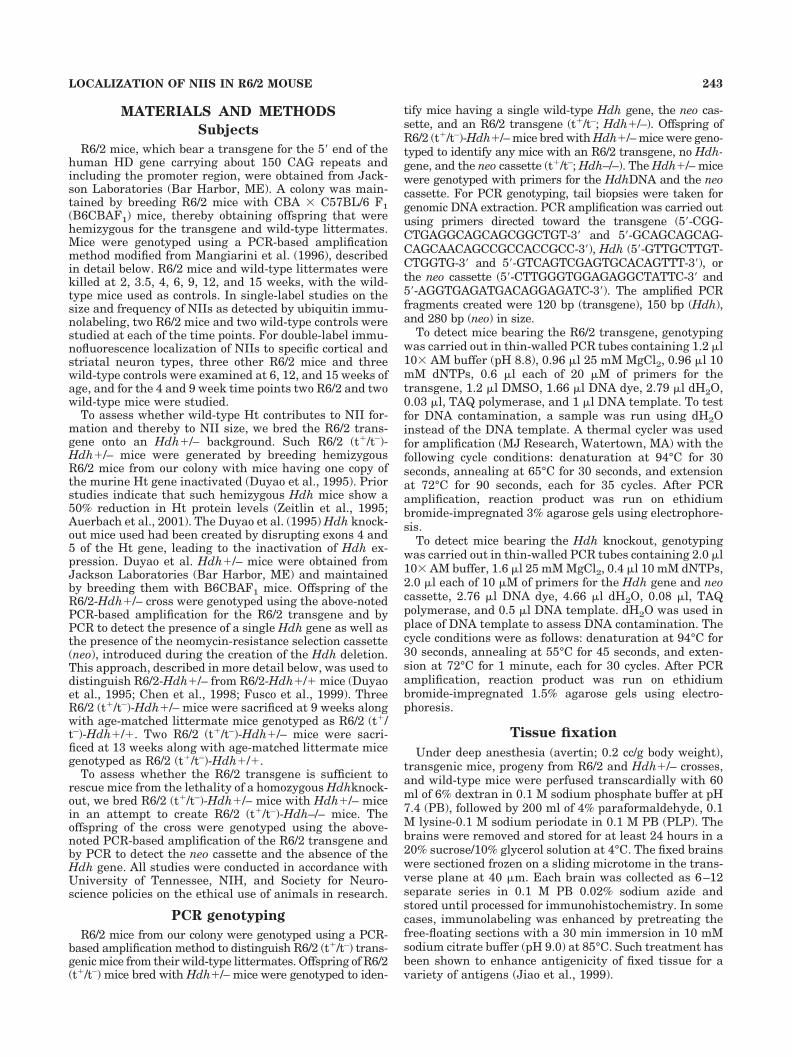
MATERIALS AND METHODS
Subjects
R6/2 mice, which bear a transgene for the 5� end of thehuman HD gene carrying about 150 CAG repeats andincluding the promoter region, were obtained from Jack-son Laboratories (Bar Harbor, ME). A colony was main-tained by breeding R6/2 mice with CBA � C57BL/6 F1(B6CBAF1) mice, thereby obtaining offspring that werehemizygous for the transgene and wild-type littermates.Mice were genotyped using a PCR-based amplificationmethod modified from Mangiarini et al. (1996), describedin detail below. R6/2 mice and wild-type littermates werekilled at 2, 3.5, 4, 6, 9, 12, and 15 weeks, with the wild-type mice used as controls. In single-label studies on thesize and frequency of NIIs as detected by ubiquitin immu-nolabeling, two R6/2 mice and two wild-type controls werestudied at each of the time points. For double-label immu-nofluorescence localization of NIIs to specific cortical andstriatal neuron types, three other R6/2 mice and threewild-type controls were examined at 6, 12, and 15 weeks ofage, and for the 4 and 9 week time points two R6/2 and twowild-type mice were studied.
To assess whether wild-type Ht contributes to NII for-mation and thereby to NII size, we bred the R6/2 trans-gene onto an Hdh�/– background. Such R6/2 (t�/t–)-Hdh�/– mice were generated by breeding hemizygousR6/2 mice from our colony with mice having one copy ofthe murine Ht gene inactivated (Duyao et al., 1995). Priorstudies indicate that such hemizygous Hdh mice show a50% reduction in Ht protein levels (Zeitlin et al., 1995;Auerbach et al., 2001). The Duyao et al. (1995) Hdh knock-out mice used had been created by disrupting exons 4 and5 of the Ht gene, leading to the inactivation of Hdh ex-pression. Duyao et al. Hdh�/– mice were obtained fromJackson Laboratories (Bar Harbor, ME) and maintainedby breeding them with B6CBAF1 mice. Offspring of theR6/2-Hdh�/– cross were genotyped using the above-notedPCR-based amplification for the R6/2 transgene and byPCR to detect the presence of a single Hdh gene as well asthe presence of the neomycin-resistance selection cassette(neo), introduced during the creation of the Hdh deletion.This approach, described in more detail below, was used todistinguish R6/2-Hdh�/– from R6/2-Hdh�/� mice (Duyaoet al., 1995; Chen et al., 1998; Fusco et al., 1999). ThreeR6/2 (t�/t–)-Hdh�/– mice were sacrificed at 9 weeks alongwith age-matched littermate mice genotyped as R6/2 (t�/t–)-Hdh�/�. Two R6/2 (t�/t–)-Hdh�/– mice were sacri-ficed at 13 weeks along with age-matched littermate micegenotyped as R6/2 (t�/t–)-Hdh�/�.
To assess whether the R6/2 transgene is sufficient torescue mice from the lethality of a homozygous Hdhknock-out, we bred R6/2 (t�/t–)-Hdh�/– mice with Hdh�/– micein an attempt to create R6/2 (t�/t–)-Hdh–/– mice. Theoffspring of the cross were genotyped using the above-noted PCR-based amplification of the R6/2 transgene andby PCR to detect the neo cassette and the absence of theHdh gene. All studies were conducted in accordance withUniversity of Tennessee, NIH, and Society for Neuro-science policies on the ethical use of animals in research.
PCR genotyping
R6/2 mice from our colony were genotyped using a PCR-based amplification method to distinguish R6/2 (t�/t–) trans-genic mice from their wild-type littermates. Offspring of R6/2(t�/t–) mice bred with Hdh�/– mice were genotyped to iden-
tify mice having a single wild-type Hdh gene, the neo cas-sette, and an R6/2 transgene (t�/t–; Hdh�/–). Offspring ofR6/2 (t�/t–)-Hdh�/– mice bred with Hdh�/– mice were geno-typed to identify any mice with an R6/2 transgene, no Hdh-gene, and the neo cassette (t�/t–; Hdh–/–). The Hdh�/– micewere genotyped with primers for the HdhDNA and the neocassette. For PCR genotyping, tail biopsies were taken forgenomic DNA extraction. PCR amplification was carried outusing primers directed toward the transgene (5�-CGG-CTGAGGCAGCAGCGGCTGT-3� and 5�-GCAGCAGCAG-CAGCAACAGCCGCCACCGCC-3�), Hdh (5�-GTTGCTTGT-CTGGTG-3� and 5�-GTCAGTCGAGTGCACAGTTT-3�), orthe neo cassette (5�-CTTGGGTGGAGAGGCTATTC-3� and5�-AGGTGAGATGACAGGAGATC-3�). The amplified PCRfragments created were 120 bp (transgene), 150 bp (Hdh),and 280 bp (neo) in size.
To detect mice bearing the R6/2 transgene, genotypingwas carried out in thin-walled PCR tubes containing 1.2 �l10� AM buffer (pH 8.8), 0.96 �l 25 mM MgCl2, 0.96 �l 10mM dNTPs, 0.6 �l each of 20 �M of primers for thetransgene, 1.2 �l DMSO, 1.66 �l DNA dye, 2.79 �l dH2O,0.03 �l, TAQ polymerase, and 1 �l DNA template. To testfor DNA contamination, a sample was run using dH2Oinstead of the DNA template. A thermal cycler was usedfor amplification (MJ Research, Watertown, MA) with thefollowing cycle conditions: denaturation at 94°C for 30seconds, annealing at 65°C for 30 seconds, and extensionat 72°C for 90 seconds, each for 35 cycles. After PCRamplification, reaction product was run on ethidiumbromide-impregnated 3% agarose gels using electrophore-sis.
To detect mice bearing the Hdh knockout, genotypingwas carried out in thin-walled PCR tubes containing 2.0 �l10� AM buffer, 1.6 �l 25 mM MgCl2, 0.4 �l 10 mM dNTPs,2.0 �l each of 10 �M of primers for the Hdh gene and neocassette, 2.76 �l DNA dye, 4.66 �l dH2O, 0.08 �l, TAQpolymerase, and 0.5 �l DNA template. dH2O was used inplace of DNA template to assess DNA contamination. Thecycle conditions were as follows: denaturation at 94°C for30 seconds, annealing at 55°C for 45 seconds, and exten-sion at 72°C for 1 minute, each for 30 cycles. After PCRamplification, reaction product was run on ethidiumbromide-impregnated 1.5% agarose gels using electro-phoresis.
Tissue fixation
Under deep anesthesia (avertin; 0.2 cc/g body weight),transgenic mice, progeny from R6/2 and Hdh�/– crosses,and wild-type mice were perfused transcardially with 60ml of 6% dextran in 0.1 M sodium phosphate buffer at pH7.4 (PB), followed by 200 ml of 4% paraformaldehyde, 0.1M lysine-0.1 M sodium periodate in 0.1 M PB (PLP). Thebrains were removed and stored for at least 24 hours in a20% sucrose/10% glycerol solution at 4°C. The fixed brainswere sectioned frozen on a sliding microtome in the trans-verse plane at 40 �m. Each brain was collected as 6–12separate series in 0.1 M PB 0.02% sodium azide andstored until processed for immunohistochemistry. In somecases, immunolabeling was enhanced by pretreating thefree-floating sections with a 30 min immersion in 10 mMsodium citrate buffer (pH 9.0) at 85°C. Such treatment hasbeen shown to enhance antigenicity of fixed tissue for avariety of antigens (Jiao et al., 1999).
243LOCALIZATION OF NIIS IN R6/2 MOUSE

Immunohistochemical single labeling
Immunohistochemical single labeling for ubiquitin, tovisualize NIIs in R6/2 mice, was carried out using aperoxidase-antiperoxidase (PAP) procedure described pre-viously (Anderson and Reiner, 1990; Figueredo-Cardenaset al., 1994), whereas immunohistochemical single label-ing to localize Ht employed the tyramide signal amplifi-cation (TSA) method using the TSA Indirect kit (NEN LifeScience Products Inc., Boston, MA; Bobrow et al., 1989,1991; Adams et al., 1992; Berghorn et al., 1994; Fusco etal., 1999). To detect ubiquitin (Chemicon, Temecula, CA),a monoclonal antibody was used, the specificity and effi-cacy of which have been shown previously (Askanas et al.,1992; Spillantini et al., 1998). Single labeling for ubiquitinwas used in the wild-type mice as a control to rule out thepresence of ubiquitinated NIIs in normal animals. A set offour mouse monoclonal antibodies (Mab2166, Mab2168,Mab2170, and Mab2172; Chemicon Inc.) against humanHt was used. The antibodies were generated against dif-ferent fragments of human Ht, as follows: 1) Mab2166,amino acids (aa) 181–812; 2) Mab2168, aa 2416–2541; 3)Mab2170, aa 1247–1646; and 4) Mab2172, aa 2683–2979.The specificity of these antibodies against Ht (both rodentand human) was demonstrated previously (Bessert et al.,1995; Trottier et al., 1995; Fusco et al., 1999). Primaryantibody omission controls were used to confirm furtherthe specificity of our immunohistochemical labeling. Arabbit polyclonal antibody (EM48) against human Ht(IT15) was also used. This antibody was generated againstaa 1–256 of IT15, possessing a polyglutamine tract con-taining only two glutamines. The specificity of this anti-body has been shown previously (Li and Li, 1998). Pri-mary antiserum omission controls were used to confirmfurther the specificity of our immunohistochemical label-ing with EM48. A series of sections adjacent to ubiquitin-labeled sections were stained with cresyl violet for use indetermining percentage of neurons containing NIIs.
To carry out conventional single-label immunohisto-chemistry, sections were incubated for 72 hours at 4°C inprimary antiserum diluted 1:200–1:5,000 with 0.3% Tri-ton X-100/0.02% sodium azide/0.1 M PB (PBX). Sectionswere then rinsed and incubated in donkey anti-mouse IgGor anti-rabbit IgG diluted 1:50 in PBX, followed by incu-bation in the appropriate mouse or rabbit PAP complexdiluted 1:100 in PBX, with each incubation at room tem-perature for 1 hour. The sections were rinsed betweensecondary and PAP incubations in three 5 minute washesof PB. Subsequently to the PAP incubation, the sectionswere rinsed with three to six 10 minute washes in 0.1 MPB, and a peroxidase reaction using diaminobenzidinetetrahydrochloride (DAB) was carried out. Sections wereincubated in 5 ml of 0.05 M imidazole/0.05 M cacodylatebuffer (pH 7.2) containing 5 mg DAB for 10 minutes andthen incubated for an additional 10 minutes after adding20 �l of 3% hydrogen peroxide, with continuous agitationthroughout. Sections were then washed in distilled water,placed in 0.1 M PB, mounted onto gelatin-coated slides,dried, dehydrated, and coverslipped with Permount.
For the TSA method, after incubation in primary anti-serum, sections were sequentially incubated in biotinyl-ated anti-mouse IgG diluted 1:50–1:200 in 0.1 M Tris-HCl(pH 7.5)/0.15 M NaCl/0.5% blocking reagent (TNB) buffer,followed by incubation in streptavidin-conjugated horse-radish peroxidase (SA-HRP) diluted 1:100 in TNB, each atroom temperature for 1 hour. The sections were then
incubated in biotinylated tyramide diluted 1:50 in ampli-fication diluent at room temperature, for 3–10 minutes.The tissue labeled with biotinylated tyramide was incu-bated for 1 hour at room temperature in SA-HRP diluted1:100 in TNB. Between incubations, sections were rinsedthree times for 5 minutes in 0.1 M Tris-HCl (pH 7.5)/0.15M NaCl/0.05% Tween 20 (TNT) buffer. The peroxidaselabeling was visualized with DAB as described above forPAP/DAB labeling, and the labeled sections weremounted, dehydrated, coverslipped, and examined.
Immunofluorescence double labeling
Immunohistochemical double labeling was carried outusing simultaneous immunofluorescence as described pre-viously (Anderson and Reiner, 1990; Figueredo-Cardenaset al., 1994) or the TSA method using the TSA Direct kit(NEN Life Science Products Inc.) combined with conven-tional immunofluorescence (Bobrow et al., 1989, 1991;Adams et al., 1992; Berghorn et al., 1994; Fusco et al.,1999).
As part of the studies on normal Ht localization inmouse, we double labeled wild-type mouse brain sectionsfor Ht by the TSA method and for markers characteristicof specific striatal neuron types by conventional immuno-fluorescence. These studies were carried out to determinethe localization of Ht in the different neuronal subsets ofthe striatum, for the purpose of assessing the relationshipbetween Ht production and NII formation in specific neu-ron types. One of the above-noted mouse monoclonal an-tibodies against Ht (MAB 2170; Chemicon Inc.) was usedtogether with an antiserum or antibody against eachof the following striatal neuron type-specific markers:1) choline acetyltransferase (ChAT) for cholinergic striatalinterneurons; 2) parvalbumin (PARV) for the large�-aminobutyric acid (GABA)-ergic/parvalbuminergic stri-atal interneurons (Cowan et al., 1990; Kita et al., 1990); 3)SS for the striatal interneurons cocontaining SS, NPY,nNOS (Figueredo-Cardenas et al., 1997); or 4) calbindin(CALB) for striatal matrix projection neurons (Gerfen andYoung, 1988; Fusco et al., 1999).
In the immunohistochemical double-label studies ofR6/2 mouse, sections of forebrain were processed usingsimultaneous immunofluorescence for ubiquitin and a cor-tical or striatal neuron type-specific marker. The neurontype-specific markers were: 1) ChAT for cholinergic stria-tal interneurons; 2) PARV for the large GABAergic/parvalbuminergic cortical and striatal interneurons(Cowan et al., 1990; Kita et al., 1990); 3) SS for the corticaland striatal interneurons cocontaining SS, NPY, andnNOS (Figueredo-Cardenas et al., 1997); or 4) CALB forcortical calbindinergic interneurons and striatal matrixprojection neurons (Gerfen and Young, 1988; Figueredo-Cardenas et al., 1998). Sections from the brains of R6/2mice in each age group studied were also doubly labeledusing conventional immunofluorescence methods withmouse antiubiquitin and EM48 to determine the degree towhich NIIs containing mutant Ht were ubiquitinated.
To carry out the immunohistochemical double labeling,sections were incubated for 72 hours at 4°C in a cocktailcontaining two primary antibodies diluted with PBX. Thefirst antibody was either mouse antiubiquitin (1:5,000) ormouse antihuntingtin (Mab 2170; 1:200). The second waseither EM48 (1:500) or an antiserum against one of thestriatal neuron type-specific markers. The sources anddilutions of the latter were: 1) goat anti-ChAT (ChemiconInc.), 1:500; 2) rabbit anti-PARV (Sigma Chemical Co, St.
244 C.A. MEADE ET AL.

Louis, MO), 1:1,000; 3) rabbit anti-SS (Incstar Corp., Still-water, MN), 1:500; and 4) rabbit anti-CALB (Swant, Bell-inzona, Switzerland), 1:200. The specificity and efficacy ofthese antisera have been described previously (Fusco etal., 1999). Note that, whereas the anti-CALB reportedlydoes have 10% cross-reactivity with calretinin (manufac-turer’s product information), this small degree of cross-reactivity is unlikely to yield significant labeling of calr-etinergic striatal neurons. Moreover, any cross-reactivelabeling of calretinergic striatal neurons is unlikely tocontribute substantially to counts of labeled calbindiner-gic neurons, because calretininergic neurons include�0.5% of all striatal neurons (Figueredo-Cardenas et al.,1996a). After three 5 minute rinses in 0.1 M PB, thesections were incubated in fluorescein isothiocyanate(FITC)-conjugated donkey anti-mouse IgG (for visualiza-tion of the mouse antiubiquitin or the mouse antihunting-tin) and either tetramethylrhodamine isothiocyanate(TRITC)-conjugated donkey anti-rabbit IgG (for EM48,CALB, PARV, and SS) or TRITC-conjugated donkey anti-goat IgG (for ChAT), both diluted 1:50 in PBX.
For the double labeling in which the TSA method wascombined with immunofluorescence, after incubation inthe primary antibody cocktail, sections were incubated inbiotinylated anti-mouse IgG diluted 1:50–1:200 in TNB.Subsequently, sections were incubated in SA-HRP diluted1:50–1:200 in TNB, at room temperature for 1 hour, fol-lowed by an incubation in tetramethylrhodamine tyra-mide diluted 1:50–1:200 with amplification diluent, atroom temperature for 3–10 minutes. The tissue was thenincubated in a secondary antiserum specific for the neurontype-specific antiserum used for that tissue, conjugated toFITC at a 1:50 dilution. After immunolabeling, tissue wasrinsed in PB, mounted on gelatin-coated slides, and cov-erslipped with 9:1 glycerin:0.05 M carbonate buffer or 9:1glycerol:PBS containing p-phenylenediamine.
Microscopic analysis
Single-label studies of NII frequency and size devel-
opment. Sections singly labeled for ubiquitin from twoR6/2 mice and two wild-type controls were analyzed at 2,3.5, 4, 6, 9, 12, and 15 weeks of age. Images of thesesections were captured and/or analyzed using a 40� ob-jective on an Olympus BH-2 microscope, a Newvicon videocamera, and a Mac IIci computer, as were images of thesame fields in adjacent cresyl violet-stained sections.These pairs of images were used to determine NII fre-quency and size for the cortex and striatum.
For determining NII frequency in cortex, layers II/III,IV, V, and VI of primary somatosensory and primarymotor cortex in two sections stained with cresyl violet andtwo sections immunostained for ubiquitin per animal wereanalyzed. For each of these two cortical regions in eachsection, three to eight images of adjacent fields of eachlayer were captured of the cresyl violet-stained material,and the number of neurons within the was image counted.Typically, 200–400 neurons were counted per layer in agiven image. For determining NII frequency in striatum,three sections stained with cresyl violet and three immu-nostained for ubiquitin (one through rostral striatum, onethrough midstriatum, and one through caudal striatum)were analyzed per animal. For each of these levels, twoimages at the center of the striatum of the cresyl violet-stained material were captured, and the number of neu-rons within the image was counted, with 400–500 neuronsbeing detected in a typical image. The number of NIIs was
then counted in the images of the corresponding corticaland striatal fields in the adjacent sections singly labeledfor ubiquitin. NII frequency was determined by dividingthe number of neurons with NIIs by the total number ofneurons within the same field in the adjacent cresyl violet-stained sections. Counts of NIIs and neurons were cor-rected for double counting of neurons at the section sur-face according to the Abercombie method (Guillery andHerrup, 1997). For this correction, neuron size was deter-mined for 20 neurons per layer per cortical area or perstriatal level per animal from images captured using a40� objective, and NII size was determined as describedbelow. Note that NII frequency seemed to be maximal by12 weeks, so NII frequency was not determined for thesingle-labeled material for the 15 week time point.
NII size (i.e., diameter) was measured for cortex andstriatum in the same ubiquitin-immunolabeled cases aswere used to determine NII frequency as well as for 15week-old R6/2 mice. For cortex, NII size was measured inneurons of layers II/III, IV, V, and VI of primary somato-sensory, primary motor, primary visual, and cingulatecortex at these ages. Three sections were analyzed percortical region per animal, and, for each region in a givensection, two fields were captured for determining NII size.For striatum, NII size was measured for central, dorsome-dial, ventromedial, dorsolateral, and ventrolateral sectorsat the level of the anterior commissure. Three striatalsections were examined per animal, and, for each section,one field per topographic region was captured for deter-mining NII size. To determine NII diameter from thecaptured images of the ubiquitin single-labeled sections,the public-domain software NIH Image, developed at theU.S. National Institutes of Health (available at http://rsb.info.hih.gov/nih-image), was used to threshold NIIs inthe images. Artifacts in the images were removed prior tothresholding, and the size of the thresholded NIIs wasassessed using the Particle Measurement function of NIHImage. NII diameter was similarly measured in R6/2 (t�/t–)-Hdh�/– mice and an equal number of littermate R6/2(t�/t–)-Hdh�/� mice at 9 weeks (N � 3) and at 13 weeks(N � 2) of age.
Double-label studies of NII frequency and size devel-
opment. The development of NII frequency and size inspecific types of cortical and striatal neurons was assessedin the tissue that had been doubly labeled using simulta-neous immunofluorescence procedures for ubiquitin and amarker of a cortical or striatal neuron type. Sections fromthree R6/2 mice at 6, 12 and 15 weeks of age were ana-lyzed, and, for 4 and 9 weeks, two R6/2 and two wild-typemice were analyzed. For these analyses, the labeling wasvisualized using a Bio-Rad MRC-1000 confocal laser scan-ning microscope (CLSM). For cortex, the percentage ofPARV�, SS�, and CALB� interneurons possessing an NIIin primary somatosensory cortex was determined fromtwo sections per animal using live CLSM images, yieldinga sample of about 50 neurons of each type for each agegroup. The nucleus of each neuron was examined through-out its entire depth to ascertain whether an NII waspresent. For striatum, the percentage of ChAT�, PARV�,and SS� interneurons possessing an NII was determinedusing live CLSM to examine each of three sections peranimal, for a sample size of about 50 of each interneurontype per animal. The nucleus of each neuron was againexamined throughout its entire depth to ascertainwhether an NII was present. For CALB� striatal projec-tion neurons, the analysis was carried out on archived
245LOCALIZATION OF NIIS IN R6/2 MOUSE

CLSM images, because the large number of CALB� neu-rons in striatum made it difficult to analyze live CLSMimages. A sample of about 100 neurons per hemisphere foreach of three sections was analyzed to determine the per-centage of CALB� striatal neurons in R6/2 mice with NIIs.CALB� perikarya that did not appear to include the nu-cleus in the plane of capture were excluded from thecounts. NII size was also measured for each specific stri-atal neuron type for each animal studied, using archivedCLSM images for each cell type. For the ChAT�, PARV�,and SS� striatal interneurons, the size of the NII in about20 NII� neurons per type was analyzed per animal,whereas, for the CALB� type, about 80 NII� neurons weremeasured per animal.
Double-label studies of NII frequency and size devel-
opment. The extent of colocalization of ubiquitin andmutant Ht in NIIs was assessed in sections doubly labeledby simultaneous immunofluorescence with antiubiquitinand EM48. One animal each at 3.5, 6, 9, 12, and 15 weeksof age was doubly labeled and examined.
Double-label studies of Ht localization in striatum.
The extent of localization of Ht (using Ab2170) in thedifferent striatal neuron types was determined from thetissue doubly labeled by TSA plus conventional immuno-labeling. Sections from two 12-week-old wild-type micewere labeled for Ht and each striatal-neuron type-specificmarker, and the frequency of localization of Ht in eachtype was assessed. Archived CLSM images of striatumfrom both hemispheres in each of three sections for eachanimal were used for analysis.
RESULTS
Localization of Ht in cortex and striatum inwild-type mice
As reported previously for rats (Fusco et al., 1999),cortical neurons in mice were rich in Ht protein (Fig. 1). Inparticular, pyramidal neurons of layer V were notably richin Ht (Fig. 1A). In striatum, most of the medium-sized,presumptive projection neurons, which make up about95% of striatal neurons, were labeled moderately for Ht.Among specific striatal neuron types, immunofluorescencedouble-labeling studies revealed that 78% of the cholin-ergic interneurons contained Ht, and these tended to besomewhat richer in Ht than medium-sized striatal neu-rons (Fig. 1E,F). We found that, among other striatalinterneurons, 25% of the parvalbuminergic neurons and1% of the somatostatinergic neurons contained Ht in nor-mal mice. Double-label studies in which calbindin wasused to identify striatal projection neurons of the matrixcompartment revealed that about 70% of these calbindin-ergic neurons labeled for Ht (Fig. 1C,D). The abundance ofHt-labeled neurons in the calbindin-poor patches was sim-ilar to that in the calbindin-rich matrix compartment,suggesting that Ht is equally prevalent in striatal patchand matrix compartment neurons.
Ubiquitinated NIIs in the cortexof R6/2 mice
No ubiquitinated inclusions were observed in neuronalnuclei of telencephalic cortex in any of the wild-type lit-termates of the R6/2 mice at any age examined. Similarly,no such neuronal intranuclear inclusions (NIIs) were ob-served in neurons in any layer of cortex at 2 weeks of agein R6/2 mice. Ubiqitinated NIIs were, however, evident in
a minority of neurons in all layers of R6/2 cortex by 3.5weeks of age (Figs. 2–4). For example, in layers II/III, IV,and VI of primary somatosensory (S1) and primary motor(M1) cortex, about 25–50% of the neurons possessed adistinct NII by 3.5 weeks. By contrast, only about 10% ofneurons in layer V of either cortical region possessed anNII at 3.5 weeks. The NIIs in all cortical regions examined(S1, M1, primary visual, and cingulate) were 1.0–1.2 �min diameter at 3.5 weeks of age in all layers (Fig. 5). By 4weeks of age, a dramatic increase in the number of neu-rons containing an NII was evident (Fig. 2). For example,at this age, about 45% of the neurons in layers II and IIIof both primary somatosensory and primary motor cortex,75% in layer IV of S1 (M1 lacks layer IV), 20% in layer Vof both, and 45% in layer VI of both possessed an NII. TheNIIs were 1.0–1.4 �m in diameter by 4 weeks (Fig. 5).
At 6 weeks, a further increase in the frequency and sizeof NIIs in cortex was evident. For example, by this age,about 90% of neurons in layers II and III of S1 and M1possessed an NII, as did 100% in layer IV and 80–85% inlayer VI. Neurons of layer V of S1, however, appeared todevelop NIIs more slowly than layer V of M1, in that about60% of the former and 85% of the latter possessed an NIIat 6 weeks. At this age, NII size was greater in somato-sensory cortex (1.7–2.0 �m) than in the other corticalregions examined (1.25–1.45 �m; Fig. 5). By 9 weeks ofage, about 95–100% of the neurons in layers II/III, IV, andVI of primary somatosensory and motor cortex possessedan NII, whereas only about 80% of the neurons in layer Vpossessed one (Fig. 2). Between 6 and 9 weeks, NII diam-eter increased by about 1 �m (Fig. 5). In all areas of cortexstudied, layer IV neurons had the smallest NIIs (2.0–2.2�m in diameter), and layer V had the largest (2.4–2.7 �min diameter). The abundance of NIIs in cortical neuronsacross layers at 12 weeks was similar to what it hadbeen at 9 weeks, and even layer V reached 90% (Fig. 2).The size of NIIs at 12 weeks was similar to that at 9weeks, but by 15 weeks NIIs in layer V neurons wereclearly larger than in any other layer, nearly 3.0 �m indiameter (Fig. 5).
Essentially all pyramidal neurons of layers II–III andV–VI and all granule cells of layer IV had developed NIIsby 9 weeks. Double labeling for ubiquitin and neuronaltype-specific markers was used to characterize NII devel-opment in specific types of cortical interneurons (Fig. 2).These studies revealed that some types of cortical inter-neurons did form NIIs but developed them more slowlyand to a lesser extent than did pyramidal and granule cellcortical neurons. For example, only 1–8% of cortical so-matostatinergic (SS) neurons possessed an NII from 6 to15 weeks of age in the R6/2 mice. Similarly, only about20% of calbindinergic (CALB) cortical interneurons pos-sessed an NII at 6 weeks (Fig. 6), and at 6 weeks andthereafter only 66% of them possessed one. In parvalbu-minergic (PARV) interneurons, by contrast, the eventualfrequency of NIIs was reminiscent of that in the corticalpyramidal and granule neurons, but they formed some-what more slowly in the PARV interneurons (Fig. 6).
Ubiquitinated NIIs in the striatumof R6/2 mice
We used immunohistochemical single labeling for ubiq-uitin to characterize the age-related changes in the fre-quency and size of NIIs in the striatum of R6/2 mice (Fig.7). We found no regional differences in this regard, so nosuch distinctions are made in the data presented. Ubiqui-
246 C.A. MEADE ET AL.

Fig. 1. Immunolabeling of somatosensory cortex for Ht (A,B) andimmunofluorescence double labeling, using CLSM, of striatum for Htand either calbindin or ChAT (C–F). Layer V cortical neurons labelintensely for Ht (A,B), whereas moderate levels (C) are found in
calbindinergic striatal neurons (D). Ht (E) is also found to labelstriatal CHAT neurons intensely (F). Arrows indicate cells that con-tain either CALB and Ht (C,D) or CHAT and Ht (E,F). Scale bars � 50�m in A,E, 10 �m in B (applies to B–F).

tinated inclusions were absent from striatal neuronal nu-clei in the wild-type littermates of the R6/2 mice at all agesexamined. As in cortex, NIIs were also not observed inR6/2 mice that were 2 weeks old. NIIs were first observedin R6/2 striatum at 3.5 weeks, at which time about 50% ofthe neurons already contained an NII (Fig. 2). Neither thefrequency nor the size of NIIs differed across striatal ar-eas, and the mean diameter across areas was 1.05 �m(Fig. 5). By 6 weeks, NII frequency was increased to about85%, and at 9 weeks it was about 99%. Although corticalNIIs grew significantly in size between 6 and 9 weeks,striatal NIIs were similar in diameter at 9 weeks (1.7 �m)to those observed at 6 weeks. At 12 weeks, the NII fre-quency was similar to that at 9 weeks, although NII sizehad grown to nearly 2.0 �m. Because NII frequency forstriatum was similar at 9 and 12 weeks, their neuronal
frequency in striatum was not determined for the 15 weektime point. NII size at 15 weeks was similar to that at 12weeks.
As is true of cortex, the striatum consists of a number ofneurochemically distinct projection neuron and interneu-ron types (Anderson and Reiner, 1990; Kawaguchi et al.,1990). We used immunohistochemical double labeling toidentify these neuron types and determine the frequencywith which they contained NIIs in 4–15-week-old R6/2mice (Figs. 2, 5). Striatal SS� interneurons, which aremedium-sized, were nearly completely devoid of NIIs inthe R6/2 striatum up to 6 weeks of age, with only about 2%of the neurons at this time possessing an NII, and meanNII size being 0.6 �m (Fig. 8). NIIs were still infrequent inSS� neurons at 9–12 weeks, with only about 1–2% ob-served to contain an NII, and with these still small in
Fig. 2. A–E: Frequency (%) of NII development in cortex andstriatum. NII frequency in cortex (A–C) and striatum (D–E) of R6/2mice 2–15 weeks of age. In primary somatosensory (A) and primarymotor (B) cortices, NII frequency was determined for the layers ofcortex using tissue labeled for ubiqutin and adjacent sections stainedwith cresyl violet. An asterisk indicates frequencies that were over
100% after being corrected using Ambecrombie’s correction. The fre-quency of NIIs in different cell type-specific cortical interneurons wasdetermined (C) using tissue immunolabeled for ubiquitin and mark-ers of these neuron types. In the striatum, (D) the frequency of NIIswas determined as for cortex. E: The frequency of NIIs within differ-ent cell type-specific markers was also determined.
248 C.A. MEADE ET AL.

diameter (0.9–1.0 �m). At 6 weeks, 1% of the striatalcholinergic (ChAT�) interneurons in R6/2 mouse con-tained an ubiquitinated NII (Fig. 8), and these were char-acteristically small (0.5 �m). The frequency of NIIs incholinergic interneurons was still low at 9 weeks (6.4%)and NIIs were still small. By 12–15 weeks, however, 70–75% of the striatal cholinergic interneurons contained anNII, which tended to be intermediate in size (1.6–1.8 �m).In the case of PARV� interneurons, only 1% were ob-served to contain an NII at 4 weeks, and NII diameter was1.2 �m (Fig. 9). By 6 weeks of age, 74% of the PARV�
striatal interneurons possessed an NII, which was of in-termediate size (1.4 �m). The frequency of NIIs in PARV�
interneurons was similar at 9 weeks, but NII size hadincreased dramatically (2.1 �m). By 12–15 weeks, a largeNII (2.2–2.3 �m) was observed in nearly every striatalPARV� interneuron.
CALB was used as a marker for projection neurons inthe matrix compartment of the striatum (Johnston et al.,
1990; Figs. 2, 5, 9). In 4-week-old R6/2 mice, only 21% ofthe CALB� projection neuron perikarya were seen to pos-sess an NII, and all were relatively small (0.9 �m). By 6weeks, there was a dramatic increase to 70% of theCALB� projection neurons possessing an NII, typically ofclearly larger diameter than at 4 weeks (1.4 �m). Thefrequency of NIIs in CALB� striatal projection neuronswas slightly increased at 9 weeks, to about 75%, and theseNIIs were larger still (1.8 �m). At 12 weeks 90% of thestriatal projection neurons labeled for CALB contained anNII, and at 15 weeks essentially all (98%) CALB neuronscontained an NII. NII size over the 9–15 week periodincreased to slightly over 2.0 �m
Are NIIs ubiquitinated at all agesin R6/2 mice?
We used an N-terminus-specific antiserum (EM48) as away of detecting the presence of the N-terminal fragmentof Ht in ubiquitin-containing NIIs in R6/2 mice. We as-
Fig. 3. Images of layers I–IV of somatosensory cortex, labeled for ubiquitin, in 4-week-old (A),6-week-old (B), 9-week-old (C), and 12-week-old (D) R6/2 mice. Although NIIs are found in neurons of alllayers at 4 weeks of age, there is a large increase in their frequency between 4 and 6 weeks of age. TheseNIIs continue to develop in size from 6 weeks through 12 weeks of age. Scale bar � 100 �m.
249LOCALIZATION OF NIIS IN R6/2 MOUSE

sumed that the predominant contributor to theN-terminal Ht fragment found in the NIIs would be thetransgene protein product. Our goal was to determinewhether NIIs that presumptively contain the mutant Htfragment are ubiquitinated at all ages at which they areobserved in R6/2 mice. In brief, we found that from 3.5weeks to 15 weeks, NIIs labeled for ubiquitin in cortex orstriatum were also labeled for Ht with EM48 (Fig. 10).Additionally, the diameters of NIIs detected with antiu-biquitin coincided with the diameters detected withEM48.
Does normal Ht contributeto NII formation?
We used two approaches to address this issue. In thefirst approach, we sought to determine whether NII for-mation is retarded in R6/2 mice possessing only one copyof normal Ht (t�/t––Hdh�/–). We created such mice as
described in Materials and Methods and found that theyshowed a normal R6/2 life span of 12–15 weeks and asimilar behavioral phenotype. We compared cortical andstriatal NII size in R6/2 mice containing only one Hdhallele to R6/2 littermates containing both Hdh alleles (Fig.11). Comparisons were made in mice at 9 weeks of age and13 weeks of age (Table 1). At 9 weeks of age, R6/2(�/–)-Hdh�/– mice had cortical NIIs whose mean diameter forall layers of motor and somatosensory cortex was 2.32 �mand striatal NIIs with a mean diameter of 1.95 �m. TheNII diameters in the 9-week-old R6/2(�/–)-Hdh�/� mice(i.e., littermate R6/2 mice with normal Ht levels) wereindistinguishable from this, with cortical NIIs for all lay-ers of motor and somatosensory cortex having a meandiameter of 2.25 �m and striatal NIIs having a meandiameter of 2.01 �m. There was also no evident differencebetween the Ht-normal R6/2 mice and the Ht-deficientR6/2 mice in NII size at 13 weeks. The 13-week-old R6/
Fig. 4. Similarly to Figure 2, 4 (A), 6 (B), 9 (C), and 12 (D)-week-old R6/2 mice labeled for ubiquitinare shown depicting layers V and VI of somatosensory cortex. The frequency of NIIs in layer V is abouthalf that in layer VI at 4 weeks of age. By 6 weeks of age, layer V still has fewer neurons containing NIIs,however, by 9 weeks, virtually all neurons contain NIIs. Scale bar � 100 �m.
250 C.A. MEADE ET AL.

2(�/–)-Hdh�/– mice had cortical NIIs whose mean diam-eter for all layers of motor and somatosensory cortex was2.46 �m and striatal NIIs that were 2.1 �m in diameter.Similarly, the 13-week-old R6/2(�/–)-Hdh�/� mice hadcortical NIIs whose mean diameter for all layers of motorand somatosensory cortex was 2.38 �m and striatal NIIsthat were 1.97 �m in diameter. The NIIs size was notsignificantly different between the Ht-normal R6/2 miceand the Ht-deficient R6/2 mice for either cortex or stria-tum by �2.
In our second approach for determining whether normalHt was present in NIIs in R6/2 mice, we immunohisto-chemically singly labeled 12-week-old Ht-normal R6/2mouse brain sections using the set of monoclonal antibod-ies against mid- and C-terminus portions of Ht (i.e.,Ab2166, Ab2168, Ab2170, Ab2172). We detected no label-
ing of NIIs in this tissue. Additionally, we doubly labeledtissue with one of these antibodies (Mab2170 against aa1247–1646) and with antiubiquitin in an attempt to detectfull-length Ht in ubiquitin-labeled NIIs (Fig. 12). Al-though there was some occasional evidence for nuclear Htlabeling with Mab2170, this labeling was never colocal-ized with ubiquitin in NIIs.
Note that, in the course of our breeding to create R6/2mice with one copy of Hdh, we bred R6/2 (t�/t–)-Hdh�/–mice with Hdh�/– mice in an attempt to create R6/2(t�/t–)-Hdh–/– mice. Our goal was to determine whetherthe R6/2 transgene is sufficient to rescue mice from thelethality of a homozygous Hdh knockout (Duyao et al.,1995). If so, then we should have created some mice withthe R6/2 transgene and no normal copies of Ht (t�/t–;Hdh–/–). Among 26 mice born in a Hdh�/– � Hdh�/–
Fig. 5. A–F: Development of NII size (�m) in cortex and striatum.Mean NII size in cortex (A–D) and striatum (E,F) of R6/2 mice 2–15weeks of age. In primary somatosensory (A), primary visual (B),primary motor (C) and cingulate (D) cortices, mean NII size wasdetermined for the layers using tissue labeled for ubiquitin. In the
striatum (E), the mean NII size was also determined using tissuelabeled for ubiquitin. Different areas of striatum were measured andfound to be similar, so only the mean of the striatal areas is reported.NII size was also determined for several specific neuron types in thestriatum.
251LOCALIZATION OF NIIS IN R6/2 MOUSE

Fig. 6. Immunofluorescent, double-labeled, CLSM images depict-ing layers IV-V of cortex in R6/2 mice at 4 (A,B), 6 (C,D) and 12 (E,F)weeks of age labeled for PARV (red; A,C,E) or CALB (red; B,D,F) andin all cases additionally labeled for ubiquitin (green). NIIs proliferate
rapidly in cortex, but few are found in PARV� or CALB� neurons until9 weeks of age. Arrows indicate labeled cells containing NIIs. Scalebar � 50 �m.

mating, 15 were Hdh�/– and 11 were Hdh�/�. Genotyp-ing revealed that no Hdh–/– mice were born. Insofar asstatistical considerations (P 0.01; �2 analysis) suggestthat some such mice should have been created, the failureto detect any among the mice born suggests that the R6/2transgene fragment does not possess sufficient functionalcapacity to rescue Hdh–/– mice from the embryonic lethal-ity of a complete Ht knockout.
DISCUSSION
We used the R6/2 mouse to provide clues to the possiblerelationship between NII formation and the cortical andstriatal cellular pathology found in HD as well as thebehavioral symptoms of HD. We examined the cellularlocalization of NIIs, including their development and theirpercentage localization to specific populations of corticaland striatal neurons. We found that, although distinctNIIs form within neuron types known to be vulnerable inHD, the regional pattern of NII formation did not explain
the greater neuron loss in striatum than in cortex that isobserved in HD. Additionally, we found that ubiquitina-tion is an early event in NII formation in R6/2 mice, thatneurons rich in Ht did not necessarily form prominentNIIs, and that there was no evidence from R6/2 mice thatwild-type Ht becomes sequestered in NIIs. The implica-tions of our various findings are discussed in more detailbelow.
Ht distribution in wild-type mice
Our study of the localization of normal Ht in the wild-type mice confirms the ubiquitous distribution of Ht intelencephalon described previously (DiFiglia et al., 1995;Gutekunst et al., 1995; Bhide et al., 1996; Fusco et al.,1999). Our single-label study indicates that, in mice, largelayer V pyramidal neurons are enriched in Ht, whereasmedium-sized striatal neurons are only moderately rich inHt. These findings are in agreement with previous obser-vations in rats (Fusco et al., 1999). However, becausemutant Ht does not appear to differ significantly in its
Fig. 7. Images of striatum from an R6/2 mouse singly labeled for ubiquitin at 4 (A), 6 (B), 9 (C), and12 (D) weeks of age. At 4 and 6 weeks, there are relatively few NIIs in the striatum. By 9 weeks, NIIsare clearly larger and more abundant than observed earlier. Further size increase is evident at 12 weeks.Scale bar � 100 �m.
253LOCALIZATION OF NIIS IN R6/2 MOUSE

regional or cellular expression from that of normal Ht(Landwehrmeyer et al., 1995; Trottier et al., 1995; Bhideet al., 1996; Gourfinkel-An et al., 1997; Sapp et al., 1997),these findings alone do not explain why the striatumrather than layer V of cortex is the major site of HDneuronal loss (Sharp and Ross, 1996; Fusco et al., 1999).
Among the striatal neuronal types, cholinergic neuronswere most enriched in Ht in wild-type mice. These dataare in agreement with our previous findings for rat brain(Fusco et al., 1999) but differ from the results of authorswho have reported that striatal cholinergic interneuronsare devoid of Ht (Ferrante et al., 1997; Kosinski et al.,1997). A possible explanation for the conflicting findingson cholinergic neurons and the localization of Ht has beendiscussed in our previous paper (Fusco et al., 1999) andmight be related to differences in the antisera used. In the
present study, the distribution of Ht among the striatalprojection neurons was similar to that previously observedin rat (Kosinski et al., 1997; Fusco et al., 1999). Ourobservations in mice, based on CALB immunolabeling,that not all projection neurons (which are the main targetof HD) are enriched in Ht further raises a question aboutthe relationship between the cellular Ht content and thesusceptibility to degeneration in HD. If the distribution ofmutant Ht is similar to that of normal Ht (as has beenreported to be the case; Landwehrmeyer et al., 1995) andmutant Ht is directly responsible for cell death, then wewould expect high levels of normal Ht in striatal projec-tion neurons. In addition, relatively few PARV striatalinterneurons in mice, which are also affected in HD (Har-rington and Kowall, 1991; Ferrer et al., 1994), contain Ht.Thus, the cellular localization of wild-type Ht in PARV
Fig. 8. CLSM images of 6-week-old (A,B) and 12-week-old (C,D)R6/2 mouse striatum labeled for ubiquitin (green) and SS (red; A,C) orubiquitin (green) and CHAT (red; B,D). Few SS� neurons developNIIs even 12 weeks of age, whereas numerous CHAT neurons develop
relatively small NIIs by this age. Arrows indicate labeled cells con-taining NIIs; arrowheads indicate unlabeled cells containing NIIs.Scale bar � 50 �m.
254 C.A. MEADE ET AL.

Fig. 9. CLSM images of R6/2 mouse striatum at 4 (A,B), 6 (C,D) and 12 (E,F) weeks old labeled forubiquitin (green) and PARV (red; A,C,E) or ubiquitin (green) and CALB (red; B,D,F). NIIs develop morerapidly and are larger in these neuron types than in the SS and CHAT neurons. Arrows indicate labeledcells containing NIIs; arrowheads indicate unlabeled cells containing NIIs. Scale bar � 50 �m.

striatal interneurons is inconsistent with their high vul-nerability in HD as well.
Development of NIIs in R6/2 miceNIIs in R6/2 miceNIIs in R6/2 miceNIIs in R6/2
miceNIIs in R6/2 mice
The time course of the development of NIIs in specificneuronal cell types of cortex and striatum was studiedhere using cell type-specific markers. In the case of cortex,the laminar pattern of NII formation was also assessed.The implications of our findings are discussed below.
Cortex. When the R6/2 mouse model was initially gen-erated and studied, it was found to have a reduced corticalvolume, but no gross abnormalities in cytoarchitecture ornumber of pyramidal neurons (Mangiarini et al., 1996).Subsequently, Ht immunoreactivity was identified in thecytoplasm as well as in N-terminal Ht fragments in NIIsin the R6/2 mouse (Davies et al., 1997). In the initialstudies (Table 2), mutant Ht was detected in NIIs at 3.5weeks of age using an N-terminus specific antibody thatdetects the mutant Ht fragment generated from the trans-gene (Ab1), whereas ubiquitin immunolabeling of the NIIswas not detected until 5–6 weeks of age (Davies et al.,1997). Recently, Morton et al. (2000) reported NIIs at 3weeks of age in the R6/2 cortex and striatum. Our findingsfor the first appearance of NIIs as detected by N-terminalHt immunolabeling are similar to those in these otherstudies (Table 2), but our immunohistochemical double-labeling studies showed that all NIIs labeled for Ht by 3.5weeks of age were ubiquitinated by that time. Our earlierdetection of ubiquitinated NIIs than reported in initialstudies of R6/2 mouse may be due to our use of antigenretrieval techniques that enhanced ubiquitin detection(Jiao et al., 1999).
In addition, our study explored the development of NIIswithin cortical layers and specific-neuron types. We foundin our single-label study that NIIs first appear at 3.5weeks of age in all cortical areas, seem to increase theirfrequency at a similar rate in all cortical areas, and showa large size increase from 6 to 9 weeks (Figs. 2, 5). NIIs,however, initially grow in size somewhat more rapidly insomatosensory cortex than in visual, motor, and cingulatecortices. For example, average NII diameter across layersof somatosensory cortex is about 1.7–2.0 �m at 6 weeks ofage. By contrast, average NII diameter across the layers ofvisual, motor, and cingulate cortices does not reach thissize until about 8–9 weeks of age. By 12–15 weeks, NIIsize appears similar across cortical areas for a given layer.It is of interest that NIIs in neurons of layer V typicallyseem to increase more slowly in frequency over the first 6weeks of age than do those in the other pyramidal cell-richlayers (i.e., layers II/III and VI). Nonetheless, by 15 weeksof age, the NIIs observed in layer V neurons, which pre-dominantly include pyramidal neurons, are as common asand larger than those in the neurons of the other layerswithin each cortical area. Similar findings for the onset of
Fig. 10. CLSM image of R6/2 mouse striatum immunofluores-cently doubly labeled for ubiquitin (green) and EM75 (red). The com-posite photomicrograph is shown (A) as well as the separate imagesfor ubiquitin (B) and EM75 (C). There is a complete overlap in thelabeling of the two Antibodies (yellow), indicating that there areno NIIs that are not ubiquitinated in the R6/2 mouse. Scale bar� 100 �m.
256 C.A. MEADE ET AL.

NIIs in cortex and their size and frequency developmentwere recently reported by Morton et al. (2000). The find-ings of our single-label studies on NII formation in cortexin R6/2 mice largely pertain to pyramidal neurons of cor-tex, because these are the predominant neuron type incortex. In our double-label studies, we examined the pat-
terns of NII development within several specific interneu-ron types of cortex. We found that the development of NIIsize and frequency for PARV interneurons was very sim-ilar to that for cortical pyramidal neurons, with about100% of PARV neurons possessing 2.0-�m-diameter NIIsby 12 weeks of age. CALB interneurons also develop NIIs
TABLE 1. NII Size (�m) in Striatum and Cortex of R6/2-Hdh KO Mice1
Genotype Age (weeks) Str
Cortex (M1) Cortex (S1)
II/III IV V VI II/III IV V VI Mean
R62(t�/t)-Hdh�/ 9 1.95 2.33 N/A 2.60 2.23 2.35 2.00 2.60 2.13 2.32R62(t�/t)-Hdh�/� 9 2.01 2.26 N/A 2.44 2.11 2.28 1.88 2.55 2.20 2.25R62(t�/t)-Hdh�/ 13 2.10 2.58 N/A 2.63 2.29 2.72 1.94 2.74 2.29 2.46R62(t�/t)-Hdh�/� 13 1.97 2.77 N/A 2.66 2.04 2.60 1.78 2.60 2.23 2.38
1Comparison of NII size in mice at 9 and 13 weeks of age that bear the R6/2 transgene and a hemizygous Hdh knockout with age-matched mice that bear the R6/2 transgene withoutany Hdh knockout. NII size was determined in striatum and different layers of primary motor (M1) and somatosensory (S1) cortices using tissue labeled for ubiquitin. There areno significant differences at either age between groups, demonstrating that wild-type Ht does not significantly contribute to NII formation.
Fig. 11. Images of somatosensory cortex (A) and striatum (C) from a 13-week-old R6/2 mouse labeledfor ubiquitin and from the cortex (B) and striatum (D) of a R6/2�/ Hdh�/ mouse immunoreacted forubiquitin. Note that the size of NIIs between mice in either brain area is not significantly different,indicating that wild-type Ht does not contribute significantly to the formation of NIIs. Scale bar � 50 �m.
257LOCALIZATION OF NIIS IN R6/2 MOUSE

that are 2.0 �m in diameter by 12 weeks, but only about70% of these interneurons develop NIIs. Finally, exceed-ingly few SS neurons develop NIIs in cortex (about 2%).The presence of interneuron types, such as those contain-ing SS, that are poor in NIIs may explain why we detectedfewer than 100% of cortical neurons to possess an NII inour single-label studies.
Striatum. As is true for cortex, the striatum in R6/2mouse is reduced in size compared with that of wild-typelittermates but shows no cell loss or abnormalities incytoarchitecture (DiFiglia et al., 1997). NIIs were initiallyreported to be first observed by 4.5 weeks of age in R6/2striatum (Table 2), using AB1 against the N-terminus ofHt, with ubiquitination of NIIs becoming immunohisto-chemically evident only at 5–6 weeks (Davies et al., 1997).By contrast, we found that NIIs could be detected in stri-atum as early as 3.5 weeks of age, with both anti-Ht(EM48) and antiubiquitin. Thereafter, NIIs develop rela-tively rapidly in striatum, with about 95–99% of all stri-atal neurons possessing 2-�m-diameter NIIs by 9–12weeks of age. Again, the difference between our findingsand prior findings on the onset of NII ubiquitination maybe due to our use of antigen retrieval techniques. Therecent study of Morton et al. (2000) again reports resultsconsistent with our own on the onset and pattern of NIIformation in striatum (Table 2), with the exceptions thatwe did not observe any inhomogeneities in the develop-ment of NIIs in striatum and we observed higher NIIfrequencies at all ages examined.
The frequency of NIIs in different types of striatal neu-rons in 12-week-old R6/2 mice has been reported recentlyby Kosinski et al. (1999). Their findings indicating a largepercentage of CALB striatal projection neurons (89%) andPARV striatal interneurons (86%) containing a ubiquiti-nated NII by 12 weeks are consistent with our own (Table2), although we additionally found essentially all striatalneurons of both types to possess an NII by 15 weeks.Kosinski et al. (1999) report a low frequency of NIIs in thetypes of striatal interneurons containing NOS, ChAT, orcalretinin (Table 2), the former of which also cocontainsSS and/or NPY (Beal et al., 1988; Figueredo-Cardenas etal., 1996b). The finding by Kosinski et al. (1997) of NOS-containing neurons is consistent with our finding for thesame neuron type visualized with anti-SS. We found, incontrast to the Kosinski et al. (1997) findings, that 70% ofChAT-containing interneurons contain an NII in 12-week-old R6/2 mice. The typically small size of these NIIs aswell as the use of an antiserum against ubiquitin differentfrom that used by Kosinski et al. (1997) could explain thegreater prevalence of NIIs in cholinergic neurons that weobserved. The recent findings of Morton et al. (2000) alsoraise the possibility that some of the inclusions we ob-served in cholinergic neurons may have been cytoplasmic.Although we did not examine the frequency with whichcalretinergic interneurons possess NIIs in R6/2 striatumin the present study, the paucity of NIIs in this cell typeand in the SS cell type, which each make up about 1% of
Fig. 12. CLSM image of striatum from a 12-week-old R6/2 mouselabeled for ubiquitin (green) and Ht (red). The composite image (A) aswell as the individual images for Ht (B) and ubiquitin (C) indicatethat, although immunolabeling for full-length Ht with mAb 2170 caninfrequently be found in the nucleus, it is not found in NIIs. Scalebar � 50 �m.
258 C.A. MEADE ET AL.

all striatal neurons (Kawaguchi et al., 1990; Figueredo-Cardenas et al., 1996a,b), may explain why our single-labeling studies did not show all striatal neurons to pos-sess an NII at 12–15 weeks.
Finally, it is of interest that the somatostatinergic in-terneurons of cortex and striatum closely resemble oneanother in the infrequency with which they contain NIIsby 12–15 weeks. Similarly, the PARV interneurons ofcortex resemble those in striatum in the prevalence ofNIIs in this cell type by 12–15 weeks. The similarities ofeach of these two neurochemically defined cortical inter-neuron types to their striatal counterpart may be a reflec-tion of their common origin from the medial ganglioniceminence (MGE) during development (Anderson et al.,1997a; Lavdas et al., 1999; Wichterle et al., 1999). Bycontrast, the CALB interneurons of cortex, which alsoderive from the MGE, appear to be a cell type differentfrom the calbindinergic type of striatum, which arisesfrom the lateral ganglionic eminence (Anderson et al.,1997b). The different origins and natures of these twoCALB cell types may explain their slight differences inNII frequency.
Implications for NII formation. In summary, ourfindings and those of Morton et al. (2000) demonstratethat NII formation differs among telencephalic areas inthe R6/2 mouse. For example, cortical pyramidal neuronsform NIIs that by 9 weeks and thereafter are 20–25%larger in diameter than those in striatal neurons. Amongcortical areas, NIIs appear slightly sooner and reach theirmaximal abundance in somatosensory cortex earlier thanin the other cortices examined. Additionally, no simplepattern is consistently evident for which neurons formNIIs and which do not. For example, while cortical andstriatal projection neurons both form NIIs, layer IV corti-cal granule cells and some cortical and striatal interneu-ron types do as well (Fig. 2). Even among neurons that doform NIIs, there are differences in the rate at which theyform. For example, NII size and frequency increase grad-ually in striatal projection neurons, whereas striatal cho-linergic interneurons show an abrupt increase in NII fre-quency between 9 and 12 weeks, and PARV neurons showan abrupt increase between 4 and 6 weeks. Note that,whereas in some cases neuron size seems to be correlated
with NII size (e.g., cortical pyramidal neurons, striatalprojection neurons, and cortical granule cells), examplessuch as striatal cholinergic interneurons show that largeneurons do not invariably make large NIIs. The next sec-tion considers the possible basis of differences among celltypes in NII formation.
Mechanisms of NII formation
To understand better the mechanism by which NIIsform, we examined the time course of their ubiquitination,the contribution of wild-type Ht to NIIs, and the relation-ship of the amount of wild-type Ht normally found in aneuron of a given type to the size of the NII formed in aneuron of that type. These issues are discussed in thefollowing sections.
Are NIIs ubiquitinated from the outset? We foundthat all NIIs in cortex and striatum of 3.5-week-old R6/2mice that are detectable with an antiserum against themutant N-terminal fragment of Ht (EM48) are alreadyubiquitinated. By contrast, NIIs could not be detectedwith either EM48 or antiubiquitin at 2 weeks of age inR6/2 mice. This indicates that ubiquitination is an earlyevent in NII formation, possibly occurring prior to trans-port of the mutant Ht protein fragment into the nucleus(Lunkes et al., 1998). This is supported by evidence fromin vitro studies showing that blocking ubiquitinationblocks NII formation (Saudou et al., 1998). In HD brain,however, several authors have reported that not all NIIsare ubiquitinated (DiFiglia et al., 1997; Gutekunst et al.,1999). For example, Gutekunst et al. (1999) reported that,in grade 4 HD, only 75% of both nuclear and neuropilaggregates, as revealed with an N-terminal antibodyagainst Ht (EM48), were ubiquitinated (Gutekunst et al.,1999). DiFiglia et al. (1997) also reported that fewer NIIslabel in HD cortex for ubiquitin than label for the HtN-terminus. Although these various immunohistochemi-cal studies raise the possibility that ubiquitination in hu-man HD occurs after the aggregation of the mutantpolyglutamine-containing protein, the failure to detectubiquitin in all aggregates in these studies could stemfrom the same sensitivity disparity that may have ledprior authors of studies with R6/2 mice to conclude thatNII formation precedes ubiquitination. Recent studies of
TABLE 2. Comparisons of R6/2 Studies1
Davies et al. 1997 Morton et al. 2000 Kosinski et al. 1999 This study
Firstappear(weeks)
Percentage at12 weeks
Firstappear(weeks)
Percentage at12 weeks
Firstappear(weeks)
Percentage at12 weeks
Firstappear(weeks)
Percentage at12 weeks
CortexOverall 3.5 N/D 3 �80 N/D N/D 3.5 95Layers II/III N/D N/D 3 N/D N/D N/D 4 97Layer IV N/D N/D 4 N/D N/D N/D 3.5 100Layer V N/D N/D 3 N/D N/D N/D 3.5 92Layer VI N/D N/D 4 N/D N/D N/D 4 88CALB N/D N/D N/D N/D N/D N/D 4 58PARV N/D N/D N/D N/D N/D N/D 4 94SS N/D N/D N/D N/D N/D N/D 6 1
StriatumOverall 4.5 �100 3 86 N/D N/D 3.5 94CALB N/D N/D N/D N/D N/D 89 4 90PARV N/D N/D N/D N/D N/D 86 4 98CHAT No NIIs ever 0 N/D N/D N/D 22 6 72SS/NOS No NIIs ever 0 N/D N/D N/D 8 6 2Calretinin N/D N/D N/D N/D N/D 9 N/D N/D
1Comparison of NII first appearance and abundance at 12 weeks of age found by different research groups. NIIs were detected using antibodies against ubiquitin or Ht in cortexand striatum. Comparison of the changes in various striatal and cortical neurochemical traits and various behavioral traits with NII development across the life span of the R6/2mouse. The increased frequency of NIIs in striatal projection neurons parallels the decline in various neurochemical markers of these neurons. The increased frequency of NIIsin cortex and striatum parallels the behavioral decline.
259LOCALIZATION OF NIIS IN R6/2 MOUSE

SCA1 transgenic mice suggest the possibility that ubiqui-tinated mutant polyglutamine-bearing proteins aggregatebecause of the inability of the ubiquitin-proteosome path-way to degrade them (Cummings et al., 1999). Consis-tently with this notion, aggregates of mutant protein intransgenic animal models pof olyglutamine disease alsocontain molecular chaperones and proteosome compo-nents (Cummings et al., 1998).
Does normal HT contribute to NII formation? Wild-type Ht is a 350 kDa protein that, because of its large size,appears to be unable to diffuse unaided across the nuclearmembrane (Zasloff, 1983; Goldfarb et al., 1986; Newmeyeret al., 1986; Dingwall and Laskey, 1991; Ross, 1997). Thisinterpretation is consistent with the observation thatwild-type Ht is largely localized to the cytoplasm (DiFigliaet al., 1995; Gutekunst et al., 1995; Trottier et al., 1995;Bhide et al., 1996; Gourfinkel-An et al., 1997; Sapp et al.,1997; Hackam et al., 1998a; Martindale et al., 1998; Fuscoet al., 1999). Although wild-type Ht has an N-terminalstretch of residues similar to those that act as a nuclearlocalization signal in other proteins (Sisodia, 1998), thepaucity of wild-type Ht in the nucleus of normal humansand animals suggests that this part of the molecule isinactive in the wild-type protein. A conformational changeinduced by an increase in polyglutamine length could,however, expose the nuclear localization signal and be thebasis of the translocation of the mutant protein to thenucleus observed in HD brains, as could be truncation ofthe mutant protein. Polyglutamine-containing proteinssuch as wild-type Ht are known to interact with and betransported into the nucleus by proteins with an expandedpolyglutamine tract (Hoogeveen et al., 1993; Huang et al.,1998; Lunkes and Mandel, 1998; Dorsman et al., 1999;Kazantsev et al., 1999; Preisinger et al., 1999; Wheeler etal., 2000). The mutation in the HD gene could then be abasis by which the mutant protein drags the wild-typeprotein to the nucleus and sequesters it. Consistently withthis possibility, several in vitro studies have indicatedthat mutant Ht can recruit wild-type Ht into insolubleaggregates, and one in vivo study has provided indirectevidence for this process (Huang et al., 1998; Martindaleet al., 1998; Kazantsev et al., 1999; Preisinger et al., 1999;Wheeler et al., 2000). In addition, in the R6/2 mouse, thereis a decrease of wild-type Ht immunoreactivity in thecortex and striatum at 9 weeks of age (Ona et al., 1999),which might stem from sequestration of wild-type proteininto aggregates. Our comparison of NII sizes between R6/2mice and R6/2 Hdh�/– mice, however, revealed no signif-icant differences, suggesting that normal, full-length Ht, ifit is translocated to the nucleus by mutant Ht, does notcontribute greatly to the size of NIIs. Additionally, ourimmunolabeling of R6/2 tissue with antibodies directedtoward the C-terminus of Ht did not reveal any wild-typeHt colocalized in NIIs labeled for ubiquitin. Our data areconsistent with the findings of others, who have not seendirect evidence for the presence of wild-type Ht withinnuclear aggregates in the brains of human HD victims ortransgenic HD mice (DiFiglia et al., 1997; Dorsman et al.,1999; Hodgson et al., 1999; Sieradzan et al., 1999). Thebasis of the disparities among studies on the localizationof wild-type Ht in nuclear aggregates is not entirely cer-tain. It may be that misfolding of wild-type Ht presentwithin aggregates renders the molecule inaccessible toantibodies directed against its mid- and C-terminal parts.
Relation of Ht dose to formation. In the R6/2 mousebrain, wild-type Ht and mutant Ht appear to be formed in
similar amounts by neurons, as would be expected insofaras the transgene is driven by the Ht promoter (Mangiariniet al., 1996). At a simple level, therefore, it might beexpected that neurons that make more mutant Ht wouldmake NIIs sooner and/or larger than neurons that makeless mutant Ht. Our results indicate that, although thetwo seem correlated for some brain regions and/or neurontypes, the correlation does not hold true for all neurontypes examined here. For example, the formation of largerNIIs in cortex than in striatum is consistent with thegreater enrichment of cortex in wild-type Ht (Landwehrm-eyer et al., 1995; Trottier et al., 1995; Bhide et al., 1996;Gourfinkel-An et al., 1997; Sapp et al., 1997; Fusco et al.,1999). The localization of Ht in layer V pyramidal neuronsis, however, only in part consistent with their pattern ofNII formation. Wild-type Ht is found in high levels inlayer V cortical pyramidal neurons in mice and otherspecies (Landwehrmeyer et al., 1995; Trottier et al., 1995;Bhide et al., 1996; Gourfinkel-An et al., 1997; Sapp et al.,1997; Fusco et al., 1999). Nonetheless, these neurons, atleast in some parts of cortex (e.g., somatosensory cortex),appear to form NIIs more slowly than do neurons in theother layers. By 12–15 weeks of age, however, the NIIs inpyramidal neurons of layer V are larger than in neurons inany other layer in cortex. Although it is unclear why layerV pyramidal neurons develop NIIs relatively slowly, thelarge size of the NIIs that layer V pyramidal neurons doeventually form is consistent with their enrichment inwild-type Ht.
In the striatum, there is no consistent correlation be-tween the enrichment of given neuron types in wild-typeHt and the rapidity with which they formed NIIs or thesize of those NIIs. For example, in normal mice and rats,only 60–70% of all calbindinergic striatal projection neu-rons produce immunohistochemically detectable Ht, andthe levels that are produced are only moderate (Fusco etal., 1999). Nonetheless, in R6/2 mouse striatum, relativelylarge NIIs develop by 12 weeks of age in nearly all CALBprojection neurons. This is in contrast to cholinergic stri-atal interneurons, essentially all of which produce highlevels of wild-type Ht in normal mice. Whereas manycholinergic neurons do form NIIs in transgenic R6/2 miceby 12 weeks of age, these NIIs are smaller than thoseformed in the CALB striatal neurons. Similarly, relativelyfew PARV striatal interneurons were observed to containwild-type Ht in R6/2 mice, but nearly all PARV� striatalneurons in these mice form large NIIs. SS striatal inter-neurons, however, show a consistency between wild-typeHt production and NII formation in R6/2 mice, in that theyproduce little wild-type Ht and only a few form an NII(which is invariably small). The overall results for corticaland striatal neuron types in R6/2 mice suggest that celltype-specific processing or transport of the mutant proteinfragment affects the formation of NIIs.
Relationship between NIIs and functionalabnormalities in HD transgenic mice
Our results and those of others show that NIIs developrapidly throughout the brain in R6/2 mice. By 12 weeks,the vast majority of neurons not only in cortex and stria-tum but also throughout brain possesses NIIs (Davies etal., 1997). By 12 weeks, these animals begin to show theseizures, wasting, hypoactivity, and motor abnormalitiesthat result in their death sometime between 12 and 16weeks (Mangiarini et al., 1996). The relationship of NIIformation to the occurrence of these symptoms is of inter-
260 C.A. MEADE ET AL.

est for understanding the basis of these symptoms and forunderstanding the possible role NIIs play in HD symp-toms and pathogenesis. In the following sections, we dis-cuss the relationship between NII formation in cortex andstriatum and neurochemical pathology in the R6/2 mousebrain and the progressive behavioral abnormalities ob-served in R6/2 mice (Fig. 13).
The R6/2 mouse and transmitter changes. Despitethe prevalence of NIIs in R6/2 mouse brain, neuronaldeath is reportedly rare and largely limited to the cingu-late cortex (Mangiarini et al., 1996; Davies et al., 1997;Turmaine et al., 2000). Nonetheless, several recent stud-ies have shown that many cortical and striatal neurons inR6/2 mouse appear to be undergoing degenerativechanges, as judged from their morphology (Turmaine etal., 2000). Consistently with the possibility that corticaland striatal neurons are undergoing early stages of degen-eration, a variety of neurochemical defects has been ob-served in cortical and striatal neurons in R6/2 mouse. Forexample, reductions in a variety of striatal neuron neuro-transmitters, receptors, and signaling molecules havebeen reported for R6/2 mice, with some of the reductionsoccurring as early as 4–8 weeks, as summarized in Figure13 (Cha et al., 1998, 1999; Bibb et al., 2000; Denovan-Wright and Robertson, 2000; Luthi-Carter et al., 2000;Menalled et al., 2000). Inasmuch as NII frequency instriatum increases from about 50% to 90% during the 4–8week age range, the early neurochemical changes in stri-atal neurons could stem from the early development ofNIIs in these neurons. It is of interest that some of theearliest changes in striatal projection neurons occur inmarkers preferentially localized to enkephalinergic neu-rons (e.g., enkephalin and the A2a receptor). This cell typeis preferentially affected early in HD (Reiner et al., 1988;Albin et al., 1990a,b, 1992; Richfield et al., 1995). It wouldbe of interest to know whether the striatal neurons pos-sessing NIIs at 4–6 weeks include a disproportionatenumber of enkephalinergic neurons. Similarly, variousneurochemical markers found in cortical neurons are alsodecreased at 12 weeks in R6/2 mice, including AMPA-typeglutamate receptors, kainate-type glutamate receptors,muscarinic cholinergic receptors, and group II-typemetabotropic glutamate receptors (mGluR; Cha et al.,1998, 1999; Denovan-Wright and Robertson, 2000; Luthi-Carter et al., 2000), by which time 95–100% of corticalneurons possess a prominent NII.
Although these various neurochemical changes in corti-cal and striatal neurons are likely to reflect an abnormal-ity induced in these neurons by the R6/2 transgene proteinproduct, the mechanism by which the abnormalities areproduced is uncertain. One possibility, based on the ob-servation that mitochondrial complex IV and aconitaseare reduced in cortex and/or striatum in R6/2 mouse(Tabrizi et al., 2000), is that the neurochemical abnormal-ities stem from a defect in energy metabolism caused bythe mutant Ht fragment. The observation, however, that awide variety of indicators of neuronal function is normalin R6/2 cortex and striatum (including group I mGluRs,N-methyl-D-aspartate-type glutamate receptors, GABAa,and GABAb receptors; Cha et al., 1998, 1999) is inconsis-tent with the type of global defect that might be expectedfrom a pervasive failure in energy metabolism. The workof Cha et al. (1998, 1999) has raised the possibility thatthe mutant Ht fragment in R6/2 mice may directly injurecortical and striatal neurons by impairing the transcrip-tion of some key neuronal proteins. The mutant Ht frag-
ment may also cause cortical neurons to injure striatalneurons in R6/2 mice, insofar as the reduction in group IImGluR on cortical terminals in striatum that has beenreported could act to increase glutamate release to excito-toxic levels. Additionally, Morton and Edwardson (2001)have shown that the synaptic vesicle-associated proteincomplexin II is reduced in R6/2 mice, which may furtherdisturb the function of corticostriatal terminals.
The R6/2 mouse and behavioral changes. The agesat which NIIs become large and highly prevalent in telen-cephalon coincides well with the age at which the majorsymptoms observed in R6/2 mice (Mangiarini et al., 1996;Carter et al., 1999) become manifest (see Fig. 13). Theseimpairments become overt by 6 weeks, by which time NIIfrequency in cortex and striatum approaches 90–100%,and the NII size reaches about 1.5 �m. By 12 weeks, atwhich time nearly all cortical and striatal neurons pos-sessed NIIs of 2 �m or larger, and NIIs are found through-out the brain and cytoplasmic aggregates are common aswell (Davies et al., 1997), resting tremors, significantweight loss, epileptic seizures, and irregular gait are allevident (Carter et al., 1999). These various severe abnor-malities are followed by death, typically sometime be-tween 12 and 15 weeks (Mangiarini et al., 1996). Althoughthe motor and neurological testing reveals an associationbetween extensive NII formation and these severe abnor-malities, behavioral testing has revealed that even theearly stages of NII formation (4–6 weeks) in these miceare associated with significant defects in spatial learningtasks, such as the Morris water maze and a T-maze (Lioneet al., 1999). The spatial learning defects have been at-tributed, at least in part, to defects in long-term potenti-ation (LTP) and long-term depression (LTD) in the hip-pocampus associated with the early appearance of NIIs inthis structure (Murphy et al., 2000).
Comparisions with other HD mouse models. Vari-ous mouse models of HD, in addition to R6/2 and R6/1mice (Davies et al., 1997), have been generated that fur-ther understanding of the mechanisms of NII formationand provide some insight into the relationship betweenNII formation on one hand and brain pathology and be-havioral abnormality on the other (Reddy et al., 1998;Hodgson et al., 1999; Schilling et al., 1999; Shelbourne etal., 1999; Wheeler et al., 2000; Laforet et al., 2001; Lin etal., 2001). These data for the various mouse models aresummarized in Table 3. The data suggest, among otherconclusions, that transgenic mice expressing anN-terminal fragment of mutant Ht (Mangiarini et al.,1996; Schilling et al., 1999; Laforet et al., 2001) are morelikely to develop NIIs than transgenic mice expressingfull-length protein with a polyglutamine tract of similarlength (Hodgson et al., 1999). For example, although ubi-quitinated NIIs are widespread in the brain of mice ex-pressing an N-terminal fragment of Ht, with a line of miceexpressing full-length mutant Ht (Hodgson et al., 1999),nuclear accumulation of mutant protein was reported tobe largely restricted to striatum and cortex and was notedas consisting of diffuse nuclear immunolabeling for theN-terminal fragment of the mutant protein. Similarly,Reddy et al. (1998, 1999) have reported that, in anotherline of mice expressing full-length mutant human Ht, NIIsare widespread but infrequent throughout the brain. No-tably, 1% of striatal neurons were reported to possess anNII, whereas some were additionally observed to possessdiffuse nuclear labeling. The mice expressing a full-lengthmutant Ht transgene also reveal that degenerative
261LOCALIZATION OF NIIS IN R6/2 MOUSE

changes in striatal neurons and behavioral abnormalitiesdo not require prior NII formation. Mice expressing full-length mutant Ht have also been created (Table 3) byexpanding the CAG repeat length of the endogenous mu-rine Ht gene (Hdh; Shelbourne et al., 1999; Wheeler et al.,2000; Lin et al., 2001). In these mice, too, NII formation isa late or rare event and is favored in mice with longer CAGrepeat expansions. The relationship of aggregate forma-tion to neuropathology and symptom formation for theknock-in mice, however, requires further study.
Implications for NII formation and brain abnormal-
ities in HD mice. The findings with the various trans-genic HD mouse models noted above, as well as findingsfrom cultured cells transfected with varying lengths ofCAG repeats (Li et al., 2000), indicate that, the longer theCAG repeat, the greater the number of neurons that con-tain an NII. Additionally, consistent with this finding is
the observation that NIIs are more prevalent in juvenile-onset HD brains than with adult onset, because juvenile-onset HD is associated with a high CAG repeat length(DiFiglia et al., 1997). These observations imply a criticalrole for the polyglutamine tract of mutant Ht in the ag-gregation process. Additionally, HD transgene fragmentscoding for the N-terminus yield more NII� neurons thando full-length HD transgenes or knock-in of CAG repeatsinto endogenous Hdh. It is thought that full-length Htfavors NII formation less because of the necessity for itstruncation prior to nuclear transport and aggregation(Wellington and Hayden, 1997; Petersen et al., 1999). Thefindings with full-length transgenic or knock-in HD mousemodels also suggest that diffuse accumulation of mutantHt in the nucleus may precede NII formation and may beadequate for the occurrence of neuronal and behavioralabnormalities. Additionally, it seems clear that neurode-
Fig. 13. Comparison of R6/2 traits and NII formation. Comparisonof the changes in various striatal and cortical neurochemical traitsand various behavioral traits with NII development across the lifespan of the R6/2 mouse. The increased frequency of NIIs in striatal
projection neurons parallels the decline in various neurochemicalmarkers of these neurons. The increased frequency of NIIs in cortexand striatum parallels the behavioral decline.
262 C.A. MEADE ET AL.

TA
BL
E3.
Com
pari
son
ofH
DM
ouse
Mod
els1
Res
earc
hgr
oup
(ref
eren
ce)
Gen
em
anip
ula
tion
Pro
mot
erC
AG
repe
ats
Lif
esp
anN
IIs
NII
sfi
rst
appe
arN
IIs
ubq
ult
inat
edC
ell
loss
Cla
spin
gon
set
(wee
ks)
On
set
ofro
taro
dim
pair
men
t(w
eeks
)O
ther
sym
ptom
s
Bat
es(M
angi
ann
iet
al.,
1996
)E
xon
1tr
ansg
ene
Hu
man
Hdh
150
12–1
5w
eeks
Th
rou
ghou
tbr
ain
3.5
wee
ksY
esC
orte
x,st
riat
um
,an
dce
rebe
llu
m
85
Tre
mor
s,ga
it
Hay
den
(Hod
gson
etal
.,19
99)
Fu
ll-l
engt
hY
AC
tran
sgen
e
Hu
man
Hdh
72�
1ye
arS
tria
tum
12m
onth
sY
esS
tria
tum
12N
/DH
yper
acti
ve
Ros
s/B
orch
elt
(Sch
illi
ng
etal
.,19
99)
N-t
erm
inal
Ht
(171
bp)
tran
sgen
e
Mou
sepr
ion
825–
6m
onth
sor
8–11
mon
ths
Str
iatu
m,
cort
ex,
hip
poca
mpu
s,am
ygda
la,
and
cere
bell
um
Las
tm
onth
ofli
feY
esn
one
2012
Tre
mor
s,ga
it
Aro
nin
(Laf
oret
etal
.,20
01)
1/3
N-t
erm
inal
Ht
Rat
neu
ron
-spe
cifi
cen
olas
e46
and
100
�18
mon
ths
Cor
tex
and
stri
atu
m3
mon
ths
Yes
Str
iatu
man
dco
rtex
�12
�12
Gai
tan
dh
ypoa
ctiv
ity
Tag
ie(R
eddy
etal
.,19
98,
1999
)F
ull
-len
gth
hu
nti
ngt
intr
ansg
ene
CM
V89
2–3
year
sS
pars
ely
scat
tere
dth
rou
ghou
tbr
ain
Not
know
nY
esS
tria
tum
,co
rtex
,h
ippo
cam
pus
and
thal
amu
s
16N
otkn
own
Hyp
erac
tive
Mac
Don
ad/J
oyn
er(W
hee
ler
etal
.,20
00)
CA
Gre
peat
knoc
k-in
Mou
seH
dh92
and
111
2–3
year
sC
AL
B�
stri
atal
neu
ron
s,co
rtex
,ol
fact
ory
tube
rcle
9–11 m
onth
sY
esN
one
Non
eN
one
Non
ere
cord
ed
Mye
rs(S
hel
bou
rne
etal
.,19
99)
CA
Gre
peat
knoc
k-in
Mou
seH
dh80
2–3
year
sN
one
Nev
erN
otkn
own
Non
eN
one
Non
eA
ggre
ssio
n
Det
loff
(Lin
etal
.,20
01)
CA
Gre
peat
knoc
k-in
Mou
seH
dh70
–78
2–3
year
sS
tria
tum
,n
,ac
cum
ben
s,co
rtic
alla
yers
III
and
IV
40w
eeks
Yes
Non
e40
40H
ypoa
ctiv
ity
1 Com
pari
son
ofdi
ffer
ent
mou
sem
odel
sof
HD
,not
ing
the
len
gth
ofC
AG
repe
ats,
life
span
,NII
abu
nda
nce
,cel
llos
s,an
dbe
hav
iora
lim
pair
men
ts.T
her
eis
anin
vers
eco
rrel
atio
nbe
twee
nC
AG
repe
atle
ngt
han
dli
fesp
an,a
nd
knoc
k-in
mod
els
hav
ea
lon
ger
life
span
than
tran
sgen
icm
odel
s.H
owev
er,
no
cell
loss
has
been
repo
rted
inth
ekn
ock-
inm
odel
s,an
dth
ese
mic
eh
ave
few
ersy
mpt
oms.
2 Tw
odi
ffer
ent
lin
es.
263LOCALIZATION OF NIIS IN R6/2 MOUSE

generative changes in striatum in these various HD mod-els can occur in the absence of distinct NII formation butmay require at least diffuse nuclear accumulation of mu-tant Ht.
Implications for HD pathology
In considering the implications of the findings in thepresent study on the regional and cellular patterns of NIIformation in the R6/2 HD transgenic mouse for HD patho-genesis, three issues should be considered. First is theevidence that the cells types affected in R6/2 mouse re-semble those affected in HD. Second, what is the relation-ship of the cellular pattern of NII formation in the R6/2mouse to the identity of the cell types affected in the R6/2mouse itself? Third, what is the relationship between NIIformation in R6/2 mouse to aggregate formation in humanHD? These issues are each considered below.
Resenblance between R6/2 mice and HD in their pa-
thology. The situation in the R6/2 mouse differs fromhuman HD in that overt striatal and cortical neuron lossat the time of animal demise is minimal (Davies et al.,1997). Nonetheless, ongoing degenerative changes havebeen observed that might lead to neuron loss given moretime (Turmaine et al., 2000). Neurochemical abnormali-ties in R6/2 striatum and cortex (Fig. 13) suggest thepossibility that the types of neurons undergoing thesechanges might include those that are known to be highlyvulnerable in human HD. For example, striatal projectionneurons are quite vulnerable in HD (Reiner et al., 1988;Reynolds and Pearson, 1990; Richfield et al., 1995), andreductions in such general striatal projection neuronmarkers as DARPP32 (Bibb et al., 2000), CB1 cannabinoidreceptors, AMPA-type glutamate receptors, kainate-typeglutamate receptors, and muscarinic cholinergic receptorshave been observed in R6/2 mice by 12 weeks of age (Chaet al., 1998, 1999; Denovan-Wright and Robertson, 2000).Additionally, R6/2 mice may, in fact, show a preferentialabnormality of the type of striatal neuron projecting to theexternal segment of globus pallidus, which is also highlyvulnerable in HD (Reiner et al., 1988; Albin et al., 1990a,b,1992; Richfield et al., 1995), in that markers of this celltype such as enkephalin and the A2a adenosine receptor(Cha et al., 1998, 1999; Menalled et al., 2000) are prefer-entially reduced in R6/2 mice. By contrast, substance P(which is found in the other major striatal projection neu-ron types) is not reduced in R6/2 mice (Cha et al., 1998,1999; Menalled et al., 2000), and the type of substanceP-containing striatal neuron projecting to the internalpallidal segment is the least vulnerable in HD (Reiner etal., 1988; Albin et al., 1990a,b, 1992; Sapp et al., 1995).There is also evidence for cortical abnormalities in R6/2mice; various neurochemical markers are reduced (Cha etal., 1998, 1999), and neurons show degenerative changes(Turmaine et al., 2000). Seemingly sublethal pathologiesare evident in cortical neurons and in the axons of corticalneurons early in HD as well (Sapp et al., 1999, 2001),followed by eventual neuron loss (Vonsattel and DiFiglia,1998). Thus, changes that parallel somewhat those in HDcortex and striatum are observed in the R6/2 mouse butare without significant cell death in this case.
NII formation and cellular abnormalities in R6/2
mice. If NIIs directly injure neurons that contain them,as has been proposed (DiFiglia et al., 1997; Li and Li,1998; Martindale et al., 1998; Gutekunst et al., 1999;Maat-Schieman et al., 1999), then they would be expectedto be more prominent in the brain regions or neuronal
types that show the above-noted neurochemical abnormal-ities. In R6/2 mice, cortical and striatal neurons developNIIs at comparable rates, but the NIIs in cortical neuronstend to be larger, especially those in layer V neurons.There is, however, no clear evidence that cortex is moreaffected than the striatum in R6/2 mouse in terms ofneurochemical alterations of the type noted above (Cha etal., 1998, 1999). Histological studies do show, however,that degenerating and apoptotic neurons are more com-mon in cortex than striatum in R6/2 mice (Davies et al.,1997; Turmaine et al., 2000). The studies noted aboveindicate that, among striatal neurons, the enkephalin-ergic projection neurons show reductions in preproen-kephalin (PPE) mRNA by 6–8 weeks, whereas SP-containing striatal projection neurons show no significantreduction in preprotachykinin (PPT) message. From thepattern of NII formation alone, it is unclear why thisshould be the case; at 12 weeks essentially all striatalprojection neurons possess 2-�m-diameter NIIs. It may bethe case, however, that enkephalinergic striatal neuronsform NIIs by 4 weeks (putatively accounting for most ofthe neurons possessing NIIs by this time), whereas SP�
neurons do not form them until 6–8 weeks. Even with thispossibility, it would be difficult to explain why ENK�
neurons show reduced PPE mRNA within weeks of NIIformation but SP� neurons do not show comparable re-duction in PPT after the same time period. Thus, althoughcortical pathology may be linked to cortical NII formationin R6/2 mouse, it is uncertain that HD-like cell type-specific striatal abnormalities in R6/2 mouse can be attrib-uted to differential striatal NII formation.
NII formation and cellular pathology in HD. If theregional and cellular pattern of NII formation in R6/2mouse provides an indication of which regions and celltypes preferentially form aggregates in HD, then the re-gional and cellular pattern of NII formation in R6/2 mousecan be used to assess the possible contribution of aggre-gate formation to HD pathogenesis at the regional andcellular levels. In R6/2 mice, cortical and striatal neuronsdevelop NIIs at comparable rates, but the NIIs in corticalneurons tend to be larger, especially those in layer Vneurons. This result is consistent with the finding thatNIIs develop more extensively in cortex than striatum inHD victims as well (Davies et al., 1997; DiFiglia et al.,1997; Becher et al., 1998; Gourfinkel-An et al., 1998;Maat-Schieman et al., 1999). Both outcomes, however, failto explain the greater neuron loss in striatum than incortex in HD victims, at least in terms of the regional locusof aggregate formation. In addition, the widespread na-ture of NIIs in all cortical layers and areas in R6/2 mouseand in HD brain (DiFiglia et al., 1997; Maat-Schieman etal., 1999) does not explain why HD cortical neuron loss ismost prominent in layer VI (Hedreen et al., 1991). NIIsare also formed in areas of R6/2 brain not affected by thehuman disease (Reddy et al., 1998). It may be the casethat for cortex, at least, the sites of NII formation predictthe functional disturbances although not necessarily theneurodegeneration. This would be consistent with thefinding that one of the early signs of HD pathology is thedevelopment of dystrophies in corticofugal axons (Sapp etal., 1999). For the R6/2 mouse, it is clear that NII forma-tion is linked to behavioral and functional abnormalities(Fig. 13), although not necessarily with neuronal loss (Li-one et al., 1999; Murphy et al., 2000). It is also importantto note that, whereas nearly all cortical neurons possessNIIs in 12-week-old R6/2 mouse, even in juvenile HD
264 C.A. MEADE ET AL.

brain (whose greater CAG repeat length presumably fa-vors aggregate formation), the fraction of cortical neuronspossessing NIIs is much lower than in R6/2 mice (DiFigliaet al., 1997; Maat-Schieman et al., 1999; Sieradzan et al.,1999). Thus, it is not established that NIIs are contribu-tors to cortical abnormalities or pathology in HD.
Within striatum, CALB projection neurons and PARVinterneurons degenerate in HD (Reiner et al., 1988; Albinet al., 1990a,b, 1992; Kiyama et al., 1990; Harrington andKowall, 1991; Ferrer et al., 1994; Richfield et al., 1995),whereas striatal interneurons containing ChAT or SS sur-vive well (Ferrante et al., 1985, 1986, 1987). Our findingsthat exceedingly few striatal SS neurons contain an NII,but a large percentage of striatal CALB projection neu-rons and PARV interneurons contain an NII, are consis-tent with the possibility that NIIs contribute to the differ-ential vulnerability of these neuron types in HD. The factthat ChAT interneurons possess relatively small NIIsmight be considered consistent with their low vulnerabil-ity in HD, although the high percentage of ChAT neuronspossessing an NII is not. In considering the implications ofthe cellular pattern of NII formation in R6/2 mouse for thetypes of neurons in striatum affected by HD, however, it isimportant also to consider whether the cellular pattern ofNII formation in R6/2 mouse resembles that in HD stria-tum. Although only limited data are available on thisissue, several studies have noted that NIIs are relativelyinfrequent in HD striatum (no greater than 10%), irre-spective of grade or type of HD (DiFiglia et al., 1997;Gutekunst et al., 1999; Maat-Schieman et al., 1999; Sappet al., 1999; Gomez-Tortosa et al., 2001). Given the perva-siveness of striatal projection neuron loss in HD, thesefindings are inconsistent with a prominent role of NIIs instriatal neuron death in HD. One study that specificallyexamined the localization of aggregates in striatal neurontypes showed that the majority of somatostatinergic neu-rons in HD striatum possessed mutant Ht-containing cy-toplasmic aggregates in their perikarya and, more rarely,an NII (Kuemmerle et al., 1999). By contrast, under 4% ofcalbindinergic striatal neurons possessed either an NII orcytoplasmic aggregates. Thus, although the cellular local-ization of NIIs in striatum in R6/2 mouse is supportive ofa role for aggregate formation in the vulnerability of theseneurons in HD, the data from HD itself are not. Thus,available data do not support the view that NIIs play arole in directly killing cortical or striatal neurons in HD.One issue that we did not address in our study is theregional and cellular pattern of cytoplasmic aggregateformation. Available data are not consistent with a role ofneuronal aggregates in directly killing neurons in HD, butit may be that cytoplasmic aggregates do have a directinjurious effect (Morton et al., 2000; Laforet et al., 2001).
Do NIIs directly kill cells?
An expansion of the number of CAG repeats is respon-sible for a group of neurodegenerative diseases that in-clude HD, dentatorubropalidoluysian atrophy, spinobul-bar muscular atrophy, and spinocerebellar ataxias (Ross,1997; Davies et al., 1998; Holmberg et al., 1998; Kim andTanzi, 1998; Klement et al., 1998; Sisodia, 1998). In eachof these diseases, there is a direct relationship betweenthe length of the CAG repeat and the severity of thedisease (Hackam et al., 1998a). In all of these diseases,except for some spinocerebellar ataxias, NIIs are formed(Paulson et al., 1997; Vonsattel and DiFiglia, 1998; Lin etal., 1999). NIIs are present in HD, but, as summarized
above, the current R6/2 data and data from HD itself donot support the notion that they are directly responsiblefor the selective neuron death that occurs in the cortex andstriatum. Additionally, Saudou et al. (1998) have usedcultured cells transfected with mutant Ht to show that celldeath induced by the mutation is not correlated with NIIformation. Therefore, the formation of NIIs may not causethe neuronal cell death mediated by mutated Ht. In rec-ognition of evidence against the role of NIIs in directneuronal injury, some authors have suggested that NIIformation may be a means by which cells sequester mal-formed proteins, such as mutant Ht, as part of an effort toprotect themselves against the directly deleterious effectsof the free mutant protein (Saudou et al., 1998; Preisingeret al., 1999). It may be that the proclivity for NII formationin cortex over striatum or among specific striatal cell typesshown in our study of R6/2 mouse reveals the proclivity ofspecific regions or cell types to accumulate the toxic frag-ment of mutant Ht in human HD. This toxic fragmentalone may not be sufficient to kill neurons directly; other-wise, cortical vulnerability would be higher than striatalin HD. The mutant protein, however, may perturb corticalfunction in such a way that it has a long-term adverseexcitotoxic effect on the striatum, perhaps by perturbingglutamate release from corticostriatal terminals by im-pairing transcription of proteins involved in presynapticregulation of transmitter release (Cha et al., 1998, 1999;Fusco et al., 1999).
ACKNOWLEDGMENTS
We thank Lydia Hu and Jian Ping Xie for their excellenttechnical assistance. This research was supported by aCure HD Contract from the Hereditary Disease Founda-tion (D.G., A.R.), and NIH grants NS19620 (A.R.) andNS28721 (A.R.). EM48 was provided through a Cure HDContract from the Hereditary Disease Foundation (S.H.).
LITERATURE CITED
Adams SM, Helps NR, Sharp MG, Brammar WJ, Walker RA, Varley JM.1992. Isolation and characterization of a novel gene with differentialexpression in benign and malignant human breast tumours. Hum MolGenet 1:91–96.
Albin RL, Tagle DA. 1995. Genetics and molecular biology of Huntington’sdisease. Trends Neurosci 18:11–14.
Albin RL, Reiner A, Anderson KD, Penney JB, Young AB. 1990a. Striataland nigral neuron subpopulations in rigid Huntington’s disease: impli-cations for the functional anatomy of chorea and rigidity-akinesia. AnnNeurol 27:357–365.
Albin RL, Young AB, Penney JB, Handelin B, Balfour R, Anderson KD,Markel DS, Tourtellotte WW, Reiner A. 1990b. Abnormalities of stri-atal projection neurons and N-methyl-D-aspartate receptors in pre-symptomatic Huntington’s disease. N Engl J Med 322:1293–1298.
Albin RL, Reiner A, Anderson KD, Dure LS, Handelin B, Balfour R,Whetsell WO Jr, Penney JB, Young AB. 1992. Preferential loss ofstriatoexternal pallidal projection neurons in presymptomatic Hun-tington’s disease. Ann Neurol 31:425–430.
Ambrose CM, Duyao MP, Barnes G, Bates GP, Lin CS, Srinidhi J, Baxen-dale S, Hummerich H, Lehrach H, Altherr M, Wasmuth J, Buckler A,Church D, Housman D, Berks M, Micklem G, Durbin R, Dodge A, ReadA, Gusella J, MacDonald ME. 1994. Structure and expression of theHuntington’s disease gene: evidence against simple inactivation due toan expanded CAG repeat. Somat Cell Mol Genet 20:27–38.
Anderson KD, Reiner A. 1990. Extensive cooccurrence of substance P anddynorphin in striatal projection neurons: an evolutionarily conservedfeature of basal ganglia organization. J Comp Neurol 295:339–369.
Anderson SA, Eisenstat DD, Shi L, Rubenstein JL. 1997a. Interneuronmigration from basal forebrain to neocortex: dependence on Dlx genes.Science 278:474–476.
265LOCALIZATION OF NIIS IN R6/2 MOUSE

Anderson SA, Qiu M, Bulfone A, Eisenstat DD, Meneses J, Pedersen R,Rubenstein JL. 1997b. Mutations of the homeobox genes Dlx-1 andDlx-2 disrupt the striatal subventricular zone and differentiation oflate born striatal neurons. Neuron 19:27–37.
Askanas V, Serdaroglu P, Engel WK, Alvarez RB. 1992. Immunocytochem-ical localization of ubiquitin in inclusion body myositis allows its light-microscopic distinction from polymyositis. Neurology 42:460–461.
Auerbach W, Hurlbert MS, Hilditch-Maguire P, Wadghiri YZ, Wheeler VC,Cohen SI, Joyner AL, MacDonald ME, Turnbull DH. 2001. The HDmutation causes progressive lethal neurological disease in mice ex-pressing reduced levels of huntingtin. Hum Mol Genet 10:2515–2523.
Bates G. 1996. Expanded glutamines and neurodegeneration—a gain ofinsight. Bioessays 18:175–178.
Beal MF, Mazurek MF, Ellison DW, Swartz KJ, McGarvey U, Bird ED,Martin JB. 1988. Somatostatin and neuropeptide Y concentrations inpathologically graded cases of Huntington’s disease. Ann Neurol 23:562–569.
Becher MW, Kotzuk JA, Sharp AH, Davies SW, Bates GP, Price DL, RossCA. 1998. Intranuclear neuronal inclusions in Huntington’s diseaseand dentatorubral and pallidoluysian atrophy: correlation between thedensity of inclusions and IT15 CAG triplet repeat length. Neurobiol Dis4:387–397.
Berghorn KA, Bonnett JH, Hoffman GE. 1994. cFos immunoreactivity isenhanced with biotin amplification. J Histochem Cytochem 42:1635–1642.
Bessert DA, Gutridge KL, Dunbar JC, Carlock LR. 1995. The identificationof a functional nuclear localization signal in the Huntington diseaseprotein. Brain Res Mol Brain Res 33:165–173.
Bhide PG, Day M, Sapp E, Schwarz C, Sheth A, Kim J, Young AB, PenneyJ, Golden J, Aronin N, DiFiglia M. 1996. Expression of normal andmutant huntingtin in the developing brain. J Neurosci 16:5523–5535.
Bibb JA, Yan Z, Svenningsson P, Snyder GL, Pieribone VA, Horiuchi A,Nairn AC, Messer A, Greengard P. 2000. Severe deficiencies in dopa-mine signaling in presymptomatic Huntington’s disease mice. ProcNatl Acad Sci U S A 97:6809–6814.
Bobrow MN, Harris TD, Shaughnessy KJ, Litt GJ. 1989. Catalyzed re-porter deposition, a novel method of signal amplification. Application toimmunoassays. J Immunol Methods 125:279–285.
Bobrow MN, Shaughnessy KJ, Litt GJ. 1991. Catalyzed reporter deposi-tion, a novel method of signal amplification. II. Application to mem-brane immunoassays. J Immunol Methods 137:103–112.
Bruyn GW. 1986. Huntington’s chorea. Historical, clinical and laboratorysynopsis. In: Vinken PJ, Bruyn GW, editors. Handbook of clinicalneurology. Amsterdam: Elsevier. p 298–378.
Carter RJ, Lione LA, Humby T, Mangiarini L, Mahal A, Bates GP, DunnettSB, Morton AJ. 1999. Characterization of progressive motor deficits inmice transgenic for the human Huntington’s disease mutation. J Neu-rosci 19:3248–3257.
Cha JH, Kosinski CM, Kerner JA, Alsdorf SA, Mangiarini L, Davies SW,Penney JB, Bates GP, Young AB. 1998. Altered brain neurotransmitterreceptors in transgenic mice expressing a portion of an abnormal hu-man huntington disease gene. Proc Natl Acad Sci U S A 95:6480–6485.
Cha JH, Frey AS, Alsdorf SA, Kerner JA, Kosinski CM, Mangiarini L,Penney JB Jr, Davies SW, Bates GP, Young AB. 1999. Altered neuro-transmitter receptor expression in transgenic mouse models of Hun-tington’s disease. Phil Trans R Soc London [Biol] 354:981–989.
Chen Q, Veenman L, Knopp K, Yan Z, Medina L, Song WJ, Surmeier DJ,Reiner A. 1998. Evidence for the preferential localization of glutamatereceptor-1 subunits of AMPA receptors to the dendritic spines of me-dium spiny neurons in rat striatum. Neuroscience 83:749–761.
Cowan RL, Wilson CJ, Emson PC, Heizmann CW. 1990. Parvalbumin-containing GABAergic interneurons in the rat neostriatum. J CompNeurol 302:197–205.
Cudkowicz M, Kowall NW. 1990. Degeneration of pyramidal projectionneurons in Huntington’s disease cortex. Ann Neurol 27:200–204.
Cummings CJ, Mancini MA, Antalffy B, DeFranco DB, Orr HT, Zoghbi HY.1998. Chaperone suppression of aggregation and altered subcellularproteasome localization imply protein misfolding in SCA1. Nat Genet19:148–154.
Cummings CJ, Reinstein E, Sun Y, Antalffy B, Jiang Y, Ciechanover A, OrrHT, Beaudet AL, Zoghbi HY. 1999. Mutation of the E6-AP ubiquitinligase reduces nuclear inclusion frequency while acceleratingpolyglutamine-induced pathology in SCA1 mice. Neuron 24:879–892.
Davies SW, Turmaine M, Cozens BA, DiFiglia M, Sharp AH, Ross CA,Scherzinger E, Wanker EE, Mangiarini L, Bates GP. 1997. Formationof neuronal intranuclear inclusions underlies the neurological dysfunc-tion in mice transgenic for the HD mutation. Cell 90:537–548.
Davies SW, Beardsall K, Turmaine M, DiFiglia M, Aronin N, Bates GP.1998. Are neuronal intranuclear inclusions the common neuropathol-ogy of triplet-repeat disorders with polyglutamine-repeat expansions?Lancet 351:131–133.
de la Monte SM, Vonsattel JP, Richardson EP Jr. 1988. Morphometricdemonstration of atrophic changes in the cerebral cortex, white matter,and neostriatum in Huntington’s disease. J Neuropathol Exp Neurol47:516–525.
Denovan-Wright EM, Robertson HA. 2000. Cannabinoid receptor messen-ger RNA levels decrease in a subset of neurons of the lateral striatum,cortex and hippocampus of transgenic Huntington’s disease mice. Neu-roscience 98:705–713.
DiFiglia M, Sapp E, Chase K, Schwarz C, Meloni A, Young C, Martin E,Vonsattel JP, Carraway R, Reeves SA, Boyce FM, Aronin N. 1995.Huntingtin is a cytoplasmic protein associated with vesicles in humanand rat brain neurons. Neuron 14:1075–1081.
DiFiglia M, Sapp E, Chase KO, Davies SW, Bates GP, Vonsattel JP, AroninN. 1997. Aggregation of huntingtin in neuronal intranuclear inclusionsand dystrophic neurites in brain. Science 277:1990–1993.
Dingwall C, Laskey RA. 1991. Nuclear targeting sequences—a consensus?Trends Biochem Sci 16:478–481.
Dorsman JC, Smoor MA, Maat-Schieman ML, Bout M, Siesling S, vanDuinen SG, Verschuuren JJ, den Dunnen JT, Roos RA, van OmmenGJ. 1999. Analysis of the subcellular localization of huntingtin with aset of rabbit polyclonal antibodies in cultured mammalian cells ofneuronal origin: comparison with the distribution of huntingtin inHuntington’s disease autopsy brain. Phil Trans R Soc London [Biol]354:1061–1067.
Duyao MP, Auerbach AB, Ryan A, Persichetti F, Barnes GT, McNeil SM,Ge P, Vonsattel JP, Gusella JF, Joyner AL, MacDonald M. 1995.Inactivation of the mouse Huntington’s disease gene homolog Hdh.Science 269:407–410.
Ferrante RJ, Kowall NW, Beal MF, Richardson EP Jr, Bird ED, Martin JB.1985. Selective sparing of a class of striatal neurons in Huntington’sdisease. Science 230:561–563.
Ferrante RJ, Kowall NW, Richardson EP Jr, Bird ED, Martin JB. 1986.Topography of enkephalin, substance P and acetylcholinesterase stain-ing in Huntington’s disease striatum. Neurosci Lett 71:283–288.
Ferrante RJ, Beal MF, Kowall NW, Richardson EP Jr, Martin JB. 1987.Sparing of acetylcholinesterase-containing striatal neurons in Hun-tington’s disease. Brain Res 411:162–166.
Ferrante RJ, Gutekunst CA, Persichetti F, McNeil SM, Kowall NW,Gusella JF, MacDonald ME, Beal MF, Hersch SM. 1997. Heteroge-neous topographic and cellular distribution of huntingtin expression inthe normal human neostriatum. J Neurosci 17:3052–3063.
Ferrer I, Kulisevsky J, Gonzalez G, Escartin A, Chivite A, Casas R. 1994.Parvalbumin-immunoreactive neurons in the cerebral cortex and stri-atum in Huntington’s disease. Neurodegeneration 3:169–173.
Ferrigno P, Silver PA. 2000. Polyglutamine expansions: proteolysis, chap-erones, and the dangers of promiscuity. Neuron 26:9–12.
Figueredo-Cardenas G, Anderson KD, Chen Q, Veenman CL, Reiner A.1994. Relative survival of striatal projection neurons and interneuronsafter intrastriatal injection of quinolinic acid in rats. Exp Neurol 129:37–56.
Figueredo-Cardenas G, Medina L, Reiner A. 1996a. Calretinin is largelylocalized to a unique population of striatal interneurons in rats. BrainRes 709:145–150.
Figueredo-Cardenas G, Morello M, Sancesario G, Bernardi G, Reiner A.1996b. Colocalization of somatostatin, neuropeptide Y, neuronal nitricoxide synthase and NADPH-diaphorase in striatal interneurons inrats. Brain Res 735:317–324.
Figueredo-Cardenas G, Chen Q, Reiner A. 1997. Age-dependent differencesin survival of striatal somatostatin-NPY-NADPH-diaphorase-containing interneurons versus striatal projection neurons after intra-striatal injection of quinolinic acid in rats. Exp Neurol 146:444–457.
Figueredo-Cardenas G, Harris CL, Anderson KD, Reiner A. 1998. Relativeresistance of striatal neurons containing calbindin or parvalbumin toquinolinic acid-mediated excitotoxicity compared to other striatal neu-ron types. Exp Neurol 149:356–372.
Fusco FR, Chen Q, Lamoreaux WJ, Figueredo-Cardenas G, Jiao Y, Coff-man JA, Surmeier DJ, Honig MG, Carlock LR, Reiner A. 1999. Cellularlocalization of huntingtin in striatal and cortical neurons in rats: lackof correlation with neuronal vulnerability in Huntington’s disease.J Neurosci 19:1189–1202.
Gerfen CR, Young WS. 1988. Distribution of striatonigral and striatopal-lidal peptidergic neurons in both patch and matrix compartments: an
266 C.A. MEADE ET AL.

in situ hybridization histochemistry and fluorescent retrograde tracingstudy. Brain Res 460:161–167.
Goldfarb DS, Gariepy J, Schoolnik G, Kornberg RD. 1986. Synthetic pep-tides as nuclear localization signals. Nature 322:641–644.
Gomez-Tortosa E, MacDonald ME, Friend JC, Taylor SA, Weiler LJ,Cupples LA, Srinidhi J, Gusella JF, Bird ED, Vonsattel JP, Myers RH.2001. Quantitative neuropathological changes in presymptomatic Hun-tington’s disease. Ann Neurol 49:29–34.
Gourfinkel-An I, Cancel G, Trottier Y, Devys D, Tora L, Lutz Y, Imbert G,Saudou F, Stevanin G, Agid Y, Brice A, Mandel JL, Hirsch EC. 1997.Differential distribution of the normal and mutated forms of hunting-tin in the human brain. Ann Neurol 42:712–719.
Gourfinkel-An I, Cancel G, Duyckaerts C, Faucheux B, Hauw JJ, TrottierY, Brice A, Agid Y, Hirsch EC. 1998. Neuronal distribution of intranu-clear inclusions in Huntington’s disease with adult onset. Neuroreport9:1823–1826.
Guillery RW, Herrup K. 1997. Quantification without pontification: choos-ing a method for counting objects in sectioned tissues [comment].J Comp Neurol 386:2–7.
Gusella JF, MacDonald ME. 1996. Trinucleotide instability: a repeatingtheme in human inherited disorders. Annu Rev Med 47:201–209.
Gutekunst CA, Levey AI, Heilman CJ, Whaley WL, Yi H, Nash NR, ReesHD, Madden JJ, Hersch SM. 1995. Identification and localization ofhuntingtin in brain and human lymphoblastoid cell lines with anti-fusion protein antibodies. Proc Natl Acad Sci U S A 92:8710–8714.
Gutekunst CA, Li SH, Yi H, Mulroy JS, Kuemmerle S, Jones R, Rye D,Ferrante RJ, Hersch SM, Li XJ. 1999. Nuclear and neuropil aggregatesin Huntington’s disease: relationship to neuropathology. J Neurosci19:2522–2534.
Hackam AS, Singaraja R, Wellington CL, Metzler M, McCutcheon K,Zhang T, Kalchman M, Hayden MR. 1998a. The influence of huntingtinprotein size on nuclear localization and cellular toxicity. J Cell Biol141:1097–1105.
Hackam AS, Wellington CL, Hayden MR. 1998b. The fatal attraction ofpolyglutamine-containing proteins. Clin Genet 53:233–242.
Harrington K, Kowall N. 1991. Parvalbumin immunoreactive neuronsresist degeneration in Huntington’s disease striatum. J NeuropatholExpl Neurol 50:309.
Hedreen JC, Peyser CE, Folstein SE, Ross CA. 1991. Neuronal loss inlayers V and VI of cerebral cortex in Huntington’s disease. NeurosciLett 133:257–261.
Hodgson JG, Agopyan N, Gutekunst C-A, Leavit BR, LePiane F, SingarajaR, Smith DJ, Bissada N, McCutcheon K, Nasir J, Jamot L, Li X-J,Stevens ME, Rosemond E, Roder JC, Phillips AG, Rubin EM, HerschSM, Hayden MR. 1999. A YAC mouse model for Huntingon’s diseasewith full-length mutant huntingtin, cytoplasmic toxicity, and selectivestriatal neurodegeneration. Neuron 23:181–192.
Holmberg M, Duyckaerts C, Durr A, Cancel G, Gourfinkel-An I, Damier P,Faucheux B, Trottier Y, Hirsch EC, Agid Y, Brice A. 1998. Spinocere-bellar ataxia type 7 (SCA7): a neurodegenerative disorder with neuro-nal intranuclear inclusions. Hum Mol Genet 7:913–918.
Hoogeveen AT, Willemsen R, Meyer N, de Rooij KE, Roos RA, van OmmenGJ, Galjaard H. 1993. Characterization and localization of the Hun-tington disease gene product. Hum Mol Genet 2:2069–2073.
Huang CC, Faber PW, Persichetti F, Mittal V, Vonsattel JP, MacDonaldME, Gusella JF. 1998. Amyloid formation by mutant huntingtin:threshold, progressivity and recruitment of normal polyglutamine pro-teins. Somat Cell Mol Genet 24:217–233.
Jiao Y, Sun Z, Lee T, Fusco FR, Kimble TD, Meade CA, Cuthbertson S,Reiner A. 1999. A simple and sensitive antigen retrieval method forfree-floating and slide-mounted tissue sections [in process citation].J Neurosci Methods 93:149–162.
Johnston JG, Gerfen CR, Haber SN, van der Kooy D. 1990. Mechanisms ofstriatal pattern formation: conservation of mammalian compartmen-talization. Brain Res Dev Brain Res 57:93–102.
Kahlem P, Terre C, Green H, Djian P. 1996. Peptides containing glutaminerepeats as substrates for transglutaminase-catalyzed cross-linking: rel-evance to diseases of the nervous system. Proc Natl Acad Sci U S A93:14580–14585.
Kawaguchi Y, Wilson CJ, Emson PC. 1990. Projection subtypes of ratneostriatal matrix cells revealed by intracellular injection of biocytin.J Neurosci 10:3421–3438.
Kazantsev A, Preisinger E, Dranovsky A, Goldgaber D, Housman D. 1999.Insoluble detergent-resistant aggregates form between pathologicaland nonpathological lengths of polyglutamine in mammalian cells.Proc Natl Acad Sci U S A 96:11404–11409.
Kim TW, Tanzi RE. 1998. Neuronal intranuclear inclusions in polyglu-tamine diseases: nuclear weapons or nuclear fallout? Neuron 21:657–659.
Kita H, Kosaka T, Heizmann CW. 1990. Parvalbumin-immunoreactiveneurons in the rat neostriatum: a light and electron microscopic study.Brain Res 536:1–15.
Kiyama H, Seto-Ohshima A, Emson PC. 1990. Calbindin D28K as amarker for the degeneration of the striatonigral pathway in Hunting-ton’s disease. Brain Res 525:209–214.
Klement IA, Skinner PJ, Kaytor MD, Yi H, Hersch SM, Clark HB, ZoghbiHY, Orr HT. 1998. Ataxin-1 nuclear localization and aggregation: rolein polyglutamine-induced disease in SCA1 transgenic mice. Cell 95:41–53.
Kosinski CM, Cha JH, Young AB, Mangiarini L, Bates G, Schiefer J,Schwarz M. 1999. Intranuclear inclusions in subtypes of striatal neu-rons in Huntington’s disease transgenic mice. Neuroreport 10:3891–3896.
Kosinski CM, Cha JH, Young AB, Persichetti F, MacDonald M, Gusella JF,Penney JB Jr, Standaert DG. 1997. Huntingtin immunoreactivity inthe rat neostriatum: differential accumulation in projection and inter-neurons. Exp Neurol 144:239–247.
Kuemmerle S, Gutekunst CA, Klein AM, Li XJ, Li SH, Beal MF, HerschSM, Ferrante RJ. 1999. Huntington aggregates may not predict neu-ronal death in Huntington’s disease. Ann Neurol 46:842–849.
Laforet GA, Sapp E, Chase K, McIntyre C, Boyce FM, Campbell M, Cadi-gan BA, Warzecki L, Tagle DA, Reddy PH, Cepeda C, Calvert CR, JokelES, Klapstein GJ, Ariano MA, Levine MS, DiFiglia M, Aronin N. 2001.Changes in cortical and striatal neurons predict behavioral and elec-trophysiological abnormalities in a transgenic murine model of Hun-tington’s disease. J Neurosci 21:9112–9123.
Landwehrmeyer GB, McNeil SM, Dure LSt, Ge P, Aizawa H, Huang Q,Ambrose CM, Duyao MP, Bird ED, Bonilla E, de Young M, Avila-Gonzales AJ, Wexler NS, Difiglia M, Gusella JF, MacDonald ME,Renney JB, Young AB, Vonsattel J-P. 1995. Huntington’s disease gene:regional and cellular expression in brain of normal and affected indi-viduals. Ann Neurol 37:218–230.
Lavdas AA, Grigoriou M, Pachnis V, Parnavelas JG. 1999. The medialganglionic eminence gives rise to a population of early neurons in thedeveloping cerebral cortex. J Neurosci 19:7881–7888.
Li H, Li SH, Johnston H, Shelbourne PF, Li XJ. 2000. Amino-terminalfragments of mutant huntingtin show selective accumulation in stria-tal neurons and synaptic toxicity. Nat Genet 25:385–389.
Li SH, Li XJ. 1998. Aggregation of N-terminal huntingtin is dependent onthe length of its glutamine repeats. Hum Mol Genet 7:777–782.
Li SH, Schilling G, Young WSd, Li XJ, Margolis RL, Stine OC, WagsterMV, Abbott MH, Franz ML, Ranen NG, Folstein SE, Hedreen JC, RossCA. 1993. Huntington’s disease gene (IT15) is widely expressed inhuman and rat tissues. Neuron 11:985–993.
Lin CH, Tallaksen-Greene S, Chien WM, Cearley JA, Jackson WS, CrouseAB, Ren S, Li XJ, Albin RL, Detloff PJ. 2001. Neurological abnormal-ities in a knock-in mouse model of Huntington’s disease. Hum MolGenet 10:137–144.
Lin X, Cummings CJ, Zoghbi HY. 1999. Expanding our understanding ofpolyglutamine diseases through mouse models. Neuron 24:499–502.
Lione LA, Carter RJ, Hunt MJ, Bates GP, Morton AJ, Dunnett SB. 1999.Selective discrimination learning impairments in mice expressing thehuman Huntington’s disease mutation. J Neurosci 19:10428–10437.
Lunkes A, Mandel JL. 1998. A cellular model that recapitulates majorpathogenic steps of Huntington’s disease. Hum Mol Genet 7:1355–1361.
Lunkes A, Trottier Y, Mandel JL. 1998. Pathological mechanisms in Hun-tington’s disease and other polyglutamine expansion diseases. EssaysBiochem 33:149–163.
Luthi-Carter R, Strand A, Peters NL, Solano SM, Hollingsworth ZR, Me-non AS, Frey AS, Spektor BS, Penney EB, Schilling G, Ross CA,Borchelt DR, Tapscott SJ, Young AB, Cha JH, Olson JM. 2000. De-creased expression of striatal signaling genes in a mouse model ofHuntington’s disease [in process citation]. Hum Mol Genet 9:1259–1271.
Maat-Schieman ML, Dorsman JC, Smoor MA, Siesling S, Van Duinen SG,Verschuuren JJ, den Dunnen JT, Van Ommen GJ, Roos RA. 1999.Distribution of inclusions in neuronal nuclei and dystrophic neurites inHuntington disease brain. J Neuropathol Exp Neurol 58:129–137.
Macdonald V, Halliday GM, Trent RJ, McCusker EA. 1997. Significant lossof pyramidal neurons in the angular gyrus of patients with Hunting-ton’s disease. Neuropathol Appl Neurobiol 23:492–495.
Mangiarini L, Sathasivam K, Seller M, Cozens B, Harper A, Hetherington
267LOCALIZATION OF NIIS IN R6/2 MOUSE

C, Lawton M, Trottier Y, Lehrach H, Davies SW, Bates GP. 1996. Exon1 of the HD gene with an expanded CAG repeat is sufficient to cause aprogressive neurological phenotype in transgenic mice. Cell 87:493–506.
Martindale D, Hackam A, Wieczorek A, Ellerby L, Wellington C, McCutch-eon K, Singaraja R, Kazemi-Esfarjani P, Devon R, Kim SU, BredesenDE, Tufaro F, Hayden MR. 1998. Length of huntingtin and its poly-glutamine tract influences localization and frequency of intracellularaggregates. Nat Genet 18:150–154.
Menalled L, Zanjani H, MacKenzie L, Koppel A, Carpenter E, Zeitlin S,Chesselet MF. 2000. Decrease in striatal enkephalin mRNA in mousemodels of Huntington’s disease. Exp Neurol 162:328–342.
Morton AJ, Edwardson JM. 2001. Progressive depletion of complexin II ina transgenic mouse model of Huntington’s disease. J Neurochem 76:166–172.
Morton AJ, Lagan MA, Skepper JN, Dunnett SB. 2000. Progressive for-mation of inclusions in the striatum and hippocampus of mice trans-genic for the human Huntington’s disease mutation. J Neurocytol29:679–702.
Murphy KP, Carter RJ, Lione LA, Mangiarini L, Mahal A, Bates GP,Dunnett SB, Morton AJ. 2000. Abnormal synaptic plasticity and im-paired spatial cognition in mice transgenic for exon 1 of the humanHuntington’s disease mutation. J Neurosci 20:5115–5123.
Myers RH, Leavitt J, Farrer LA, Jagadeesh J, McFarlane H, MastromauroCA, Mark RJ, Gusella JF. 1989. Homozygote for Huntington disease.Am J Hum Genet 45:615–618.
Narain Y, Wyttenbach A, Rankin J, Furlong RA, Rubinsztein DC. 1999. Amolecular investigation of true dominance in Huntington’s disease.J Med Genet 36:739–746.
Nasir J, Floresco SB, O’Kusky JR, Diewert VM, Richman JM, Zeisler J,Borowski A, Marth JD, Phillips AG, Hayden MR. 1995. Targeted dis-ruption of the Huntington’s disease gene results in embryonic lethalityand behavioral and morphological changes in heterozygotes. Cell 81:811–823.
Newmeyer DD, Lucocq JM, Burglin TR, De Robertis EM. 1986. Assemblyin vitro of nuclei active in nuclear protein transport: ATP is required fornucleoplasmin accumulation. EMBO J 5:501–510.
Ona VO, Li M, Vonsattel JP, Andrews LJ, Khan SQ, Chung WM, Frey AS,Menon AS, Li XJ, Stieg PE, Yuan J, Penney JB, Young AB, Cha JH,Friedlander RM. 1999. Inhibition of caspase-1 slows disease progres-sion in a mouse model of Huntington’s disease [see comments]. Nature399:263–267.
Paulson HL, Perez MK, Trottier Y, Trojanowski JQ, Subramony SH, DasSS, Vig P, Mandel JL, Fischbeck KH, Pittman RN. 1997. Intranuclearinclusions of expanded polyglutamine protein in spinocerebellar ataxiatype 3. Neuron 19:333–344.
Persichetti F, Ambrose CM, Ge P, McNeil SM, Srinidhi J, Anderson MA,Jenkins B, Barnes GT, Duyao MP, Kanaley L, Wexler NS, Myers RH,Bird ED, Vonsattel J-P, MacDonald ME, Gusella JF. 1995. Normal andexpanded Huntington’s disease gene alleles produce distinguishableproteins due to translation across the CAG repeat. Mol Med 1:374–383.
Persichetti F, Carlee L, Faber PW, McNeil SM, Ambrose CM, Srinidhi J,Anderson M, Barnes GT, Gusella JF, MacDonald ME. 1996. Differen-tial expression of normal and mutant Huntington’s disease gene al-leles. Neurobiol Dis 3:183–190.
Perutz MF. 1999. Glutamine repeats and neurodegenerative diseases:molecular aspects. Trends Biochem Sci 24:58–63.
Perutz MF, Johnson T, Suzuki M, Finch JT. 1994. Glutamine repeats aspolar zippers: their possible role in inherited neurodegenerative dis-eases. Proc Natl Acad Sci U S A 91:5355–5358.
Petersen A, Mani K, Brundin P. 1999. Recent advances on the pathogen-esis of Huntington’s disease. Exp Neurol 157:1–18.
Preisinger E, Jordan BM, Kazantsev A, Housman D. 1999. Evidence for arecruitment and sequestration mechanism in Huntington’s disease.Phil Trans R Soc London [Biol] 354:1029–1034.
Reddy PH, Williams M, Charles V, Garrett L, Pike-Buchanan L, WhetsellWO Jr, Miller G, Tagle DA. 1998. Behavioural abnormalities andselective neuronal loss in HD transgenic mice expressing mutatedfull-length HD cDNA. Nat Genet 20:198–202.
Reddy PH, Williams M, Tagle DA. 1999. Recent advances in understandingthe pathogenesis of Huntington’s disease [in process citation]. TrendsNeurosci 22:248–255.
Reiner A, Albin RL, Anderson KD, D’Amato CJ, Penney JB, Young AB.1988. Differential loss of striatal projection neurons in Huntingtondisease. Proc Natl Acad Sci U S A 85:5733–5737.
Reynolds GP, Pearson SJ. 1990. Brain GABA levels in asymptomaticHuntington’s disease. N Engl J Med 323:682.
Richfield EK, Maguire-Zeiss KA, Vonkeman HE, Voorn P. 1995. Preferen-tial loss of preproenkephalin versus preprotachykinin neurons from thestriatum of Huntington’s disease patients. Ann Neurol 38:852–861.
Roos RAC. 1986. Neuropathology of Huntington’s chorea. In: Vinken PJ,Bruyn GW, Klawans HL, editors. Handbook of clinical neurology. Am-sterdam: Elsevier. p 315–326.
Ross CA. 1997. Intranuclear neuronal inclusions: a common pathogenicmechanism for glutamine-repeat neurodegenerative diseases? Neuron19:1147–1150.
Sapp E, Ge P, Aizawa H, Bird E, Penney J, Young AB, Vonsattel JP,DiFiglia M. 1995. Evidence for a preferential loss of enkephalin immu-noreactivity in the external globus pallidus in low grade Huntington’sdisease using high resolution image analysis. Neuroscience 64:397–404.
Sapp E, Schwarz C, Chase K, Bhide PG, Young AB, Penney J, VonsattelJP, Aronin N, DiFiglia M. 1997. Huntingtin localization in brains ofnormal and Huntington’s disease patients. Ann Neurol 42:604–612.
Sapp E, Penney J, Young A, Aronin N, Vonsattel JP, DiFiglia M. 1999.Axonal transport of N-terminal huntingtin suggests early pathology ofcorticostriatal projections in Huntington disease. J Neuropathol ExpNeurol 58:165–173.
Sapp E, Kegel KB, Aronin N, Hashikawa T, Uchiyama Y, Tohyama K,Bhide PG, Vonsattel JP, DiFiglia M. 2001. Early and progressiveaccumulation of reactive microglia in the Huntington disease brain.J Neuropathol Exp Neurol 60:161–172.
Saudou F, Finkbeiner S, Devys D, Greenberg ME. 1998. Huntingtin acts inthe nucleus to induce apoptosis but death does not correlate with theformation of intranuclear inclusions. Cell 95:55–66.
Schilling G, Becher MW, Sharp AH, Jinnah HA, Duan K, Kotzuk JA, SluntHH, Ratovitski T, Cooper JK, Jenkins NA, Copeland NG, Price DL,Ross CA, Borchelt DR. 1999. Intranuclear inclusions and neuritic ag-gregates in transgenic mice expressing a mutant N-terminal fragmentof huntingtin [published erratum appears in Hum Mol Genet 8:943,1999]. Hum Mol Genet 8:397–407.
Sharp AH, Ross CA. 1996. Neurobiology of Huntington’s disease. NeurobiolDis 3:3–15.
Sharp AH, Loev SJ, Schilling G, Li SH, Li XJ, Bao J, Wagster MV, KotzukJA, Steiner JP, Lo A, Hedreen J, Sisodia S, Synder SH, Dawson TM,Ryugo DOK, Ross CA. 1995. Widespread expression of Huntington’sdisease gene (IT15) protein product. Neuron 14:1065–74.
Shelbourne PF, Killeen N, Hevner RF, Johnston HM, Tecott L, LewandoskiM, Ennis M, Ramirez L, Li Z, Iannicola C, Littman DR, Myers RM.1999. A Huntington’s disease CAG expansion at the murine Hdh locusis unstable and associated with behavioural abnormalities in mice.Hum Mol Genet 8:763–774.
Sieradzan KA, Mechan AO, Jones L, Wanker EE, Nukina N, Mann DM.1999. Huntington’s disease intranuclear inclusions contain truncated,ubiquitinated huntingtin protein. Exp Neurol 156:92–99.
Sisodia SS. 1998. Nuclear inclusions in glutamine repeat disorders: arethey pernicious, coincidental, or beneficial? [comment]. Cell 95:1–4.
Sotrel A, Williams RS, Kaufmann WE, Myers RH. 1993. Evidence forneuronal degeneration and dendritic plasticity in cortical pyramidalneurons of Huntington’s disease: a quantitative Golgi study. Neurology43:2088–2096.
Spillantini MG, Crowther RA, Jakes R, Hasegawa M, Goedert M. 1998.Alpha-synuclein in filamentous inclusions of Lewy bodies from Parkin-son’s disease and dementia with lewy bodies. Proc Natl Acad Sci U S A95:6469–6473.
Storey E, Kowall NW, Finn SF, Mazurek MF, Beal MF. 1992. The corticallesion of Huntington’s disease: further neurochemical characterization,and reproduction of some of the histological and neurochemical fea-tures by N-methyl-D-aspartate lesions of rat cortex. Ann Neurol 32:526–534.
Tabrizi SJ, Workman J, Hart PE, Mangiarini L, Mahal A, Bates G, CooperJM, Schapira AH. 2000. Mitochondrial dysfunction and free radicaldamage in the Huntington R6/2 transgenic mouse [in process citation].Ann Neurol 47:80–86.
Tellez-Nagel I, Wisniewski HM. 1973. Ultrastructure of neurofibrillarytangles in Steele-Richardson-Olszewski syndrome. Arch Neurol 29:324–327.
The Huntington’s Disease Collaborative Research Group. 1993. A novelgene containing a trinucleotide repeat that is expanded and unstableon Huntington’s disease chromosomes. Cell 72:971–983.
Thomas P, Wilkinson F, thi Man N, Harper PS, Neal JW, Morris GE, JonesAL. 1998. Full length huntingtin is not detected in intranuclear inclu-sions in Huntington’s disease brain [in process citation]. Biochem SocTrans 26:S243.
268 C.A. MEADE ET AL.

Trottier Y, Devys D, Imbert G, Saudou F, An I, Lutz Y, Weber C, Agid Y,Hirsch EC, Mandel JL. 1995. Cellular localization of the Huntington’sdisease protein and discrimination of the normal and mutated form.Nat Genet 10:104–110.
Turmaine M, Raza A, Mahal A, Mangiarini L, Bates GP, Davies SW. 2000.Nonapoptotic neurodegeneration in a transgenic mouse model of Hun-tington’s disease. Proc Natl Acad Sci U S A 97:8093–8097.
Vonsattel JP, DiFiglia M. 1998. Huntington disease. J Neuropathol ExpNeurol 57:369–384.
Vonsattel JP, Myers RH, Stevens TJ, Ferrante RJ, Bird ED, RichardsonEP Jr. 1985. Neuropathological classification of Huntington’s disease.J Neuropathol Exp Neurol 44:559–577.
Wellington CL, Hayden MR. 1997. Of molecular interactions, mice andmechanisms: new insights into Huntington’s disease. Curr Opin Neu-rol 10:291–298.
Wexler NS, Young AB, Tanzi RE, Travers H, Starosta-Rubinstein S, Pen-ney JB, Snodgrass SR, Shoulson I, Gomez F, Ramos Arroyo MA, Pen-
chaszadeh GK, Moreno H, Gibbons K, Farniarz A, Hobbs W, AndersonMA, Bonilla E, Conneally PM, Gusella JF. 1987. Homozygotes forHuntington’s disease. Nature 326:194–197.
Wheeler VC, White JK, Gutekunst CA, Vrbanac V, Weaver M, Li XJ, LiSH, Yi H, Vonsattel JP, Gusella JF, Hersch S, Auerbach W, Joyner AL,MacDonald ME. 2000. Long glutamine tracts cause nuclear localizationof a novel form of huntingtin in medium spiny striatal neurons inHdhQ92 and HdhQ111 knock-in mice. Hum Mol Genet 9:503–513.
Wichterle H, Garcia-Verdugo JM, Herrera DG, Alvarez-Buylla A. 1999.Young neurons from medial ganglionic eminence disperse in adult andembryonic brain. Nat Neurosci 2:461–466.
Zasloff M. 1983. tRNA transport from the nucleus in a eukaryotic cell:carrier-mediated translocation process. Proc Natl Acad Sci U S A80:6436–6440.
Zeitlin S, Liu JP, Chapman DL, Papaioannou VE, Efstratiadis A. 1995.Increased apoptosis and early embryonic lethality in mice nullizygousfor the Huntington’s disease gene homologue. Nat Genet 11:155–163.
269LOCALIZATION OF NIIS IN R6/2 MOUSE
Copyright © 2022 FDOKUMEN