Cell mechanics: principles, practices, and prospects - Wiley ...
18
Overview Cell mechanics: principles, practices, and prospects Emad Moeendarbary 1,2∗,† and Andrew R. Harris 3∗,† Cells generate and sustain mechanical forces within their environment as part of their normal physiology. They are active materials that can detect mechanical stim- ulation by the activation of mechanosensitive signaling pathways, and respond to physical cues through cytoskeletal re-organization and force generation. Genetic mutations and pathogens that disrupt the cytoskeletal architecture can result in changes to cell mechanical properties such as elasticity, adhesiveness, and viscos- ity. On the other hand, perturbations to the mechanical environment can affect cell behavior. These transformations are often a hallmark and symptom of a variety of pathologies. Consequently, there are now a myriad of experimental techniques and theoretical models adapted from soft matter physics and mechanical engineering to characterize cell mechanical properties. Interdisciplinary research combining modern molecular biology with advanced cell mechanical characterization tech- niques now paves the way for furthering our fundamental understanding of cell mechanics and its role in development, physiology, and disease. We describe a generalized outline for measuring cell mechanical properties including loading protocols, tools, and data interpretation. We summarize recent advances in the field and explain how cell biomechanics research can be adopted by physicists, engi- neers, biologists, and clinicians alike. © 2014 The Authors. WIREs Systems Biology and Medicine published by Wiley Periodicals, Inc. How to cite this article: WIREs Syst Biol Med 2014, 6:371–388. doi: 10.1002/wsbm.1275 CELL MECHANICS 2 1st century biomechanics research has entered an exciting era of investigation; where the mechanical behaviors of cells and tissues can be both a direct consequence, and a regulating factor of biological function and cellular architecture. 1,2 The underlying goal of current cell biomechanics ∗ Correspondence to: [email protected]; [email protected] 1 Department of Physiology, Development and Neuroscience, Uni- versity of Cambridge, Cambridge, UK 2 Hughes Hall, University of Cambridge, Cambridge, UK 3 Department of Bioengineering, University of California Berkeley, Berkeley, CA, USA The copyright line in this article was changed on 10 October 2014 after online publication. † These authors contributed equally. Conflict of interest: The authors have declared no conflicts of interest for this article. research is to combine theoretical, experimental, and computational approaches to construct a real- istic description of cell mechanical behaviors that can be used to provide new perspectives on the role of mechanics in disease. 3,4 In pursuit of this, biotechnological experimental methods are becoming increasingly diverse and the interpretation of results complex. Furthermore, achieving this goal requires a complement of both physical and biological research methods, which can prove daunting for non-experts in the field. Aiming to facilitate the understanding of the field to non-experts, we overview the principles, practices, and prospects of cell mechanics research. We summarize the choice of experimental tool, load- ing protocols, quantification, and examination of mechanical measurement results, and how these can be interpreted to perceive the underlying biological mechanisms of cellular force generation and physical Volume 6, September/October 2014 371 © 2014 The Authors. WIREs Systems Biology and Medicine published by Wiley Periodicals, Inc. This is an open access article under the terms of the Creative Commons Attribution-NonCommercial-NoDerivs License, which permits use and distribution in any medium, provided the original work is properly cited, the use is non-commercial and no modifications or adaptations are made.
-
Upload
khangminh22 -
Category
Documents
-
view
3 -
download
0
Transcript of Cell mechanics: principles, practices, and prospects - Wiley ...

Overview
Cell mechanics: principles,practices, and prospectsEmad Moeendarbary1,2∗,† and Andrew R. Harris3∗,†
Cells generate and sustain mechanical forces within their environment as part oftheir normal physiology. They are active materials that can detect mechanical stim-ulation by the activation of mechanosensitive signaling pathways, and respond tophysical cues through cytoskeletal re-organization and force generation. Geneticmutations and pathogens that disrupt the cytoskeletal architecture can result inchanges to cell mechanical properties such as elasticity, adhesiveness, and viscos-ity. On the other hand, perturbations to the mechanical environment can affect cellbehavior. These transformations are often a hallmark and symptom of a variety ofpathologies. Consequently, there are now a myriad of experimental techniques andtheoretical models adapted from soft matter physics and mechanical engineeringto characterize cell mechanical properties. Interdisciplinary research combiningmodern molecular biology with advanced cell mechanical characterization tech-niques now paves the way for furthering our fundamental understanding of cellmechanics and its role in development, physiology, and disease. We describe ageneralized outline for measuring cell mechanical properties including loadingprotocols, tools, and data interpretation. We summarize recent advances in the fieldand explain how cell biomechanics research can be adopted by physicists, engi-neers, biologists, and clinicians alike. © 2014 The Authors. WIREs Systems Biology and Medicinepublished by Wiley Periodicals, Inc.
How to cite this article:WIREs Syst Biol Med 2014, 6:371–388. doi: 10.1002/wsbm.1275
CELL MECHANICS
21st century biomechanics research has enteredan exciting era of investigation; where the
mechanical behaviors of cells and tissues can beboth a direct consequence, and a regulating factorof biological function and cellular architecture.1,2
The underlying goal of current cell biomechanics
∗Correspondence to: [email protected]; [email protected] of Physiology, Development and Neuroscience, Uni-versity of Cambridge, Cambridge, UK2Hughes Hall, University of Cambridge, Cambridge, UK3Department of Bioengineering, University of California Berkeley,Berkeley, CA, USAThe copyright line in this article was changed on 10 October 2014after online publication.†These authors contributed equally.Conflict of interest: The authors have declared no conflicts of interestfor this article.
research is to combine theoretical, experimental,and computational approaches to construct a real-istic description of cell mechanical behaviors thatcan be used to provide new perspectives on therole of mechanics in disease.3,4 In pursuit of this,biotechnological experimental methods are becomingincreasingly diverse and the interpretation of resultscomplex. Furthermore, achieving this goal requires acomplement of both physical and biological researchmethods, which can prove daunting for non-expertsin the field. Aiming to facilitate the understanding ofthe field to non-experts, we overview the principles,practices, and prospects of cell mechanics research.We summarize the choice of experimental tool, load-ing protocols, quantification, and examination ofmechanical measurement results, and how these canbe interpreted to perceive the underlying biologicalmechanisms of cellular force generation and physical
Volume 6, September/October 2014 371© 2014 The Authors. WIREs Systems Biology and Medicine published by Wiley Periodicals, Inc.This is an open access article under the terms of the Creative Commons Attribution-NonCommercial-NoDerivs License, which permits use and distribution inany medium, provided the original work is properly cited, the use is non-commercial and no modifications or adaptations are made.
美国细胞修复系统医学中心 www.cytothesis.us
Sticky Note
「全球化3.0医学」的「3.0居家照护」 www.cytothesis.us

Overview wires.wiley.com/sysbio
behaviors. We summarize mechanical tools such asatomic force microscopy (AFM) and optical tweezerswhich are commercially available mechanical testingsystems, and provide an overview of the most recentapplications of these tools,5,6,46,83 including rheolog-ical measurements.7,8 We also place an emphasis ontools that do not require large amounts of specializedequipment such as particle tracking microrheology9
(PTM) and traction force microscopy (TFM),10 whichcan be easily adopted by laboratories that are newto the field. In the following sections we outline theinterpretation of typical cell mechanical measure-ments using theories such as linear viscoelastic andpower law models,11–15 soft glassy rheology,16,17
purified gel models18,19 and poroelasticity.20,21
Forces in PhysiologyA basic requirement of every organism is that itcan sustain, detect, and interact with physical forceswithin its environment. This requirement is soimportant to life and survival that it has become acornerstone of biological design. The skeleton pro-vides structural support to sustain the force of gravity.Skin provides a protective barrier that is maintainedupon the application of external stretch and hindersthe invasion of bacteria and microbes that wouldcause infection. Even the simplest of physiologicalfunctions, such as respiration and circulation, requirethe generation of forces to breathe in air and topump blood around the body. These are but a fewfundamental examples of how generating, sustain-ing, and detecting physical forces forms an integralpart of everyday life. Biomechanics research in pastdecades has largely focussed on understanding andquantifying these behaviors at the organism andorgan levels. Early research includes compressiontesting of bone, to quantify the levels of forces it canwithstand before breaking and the amount of force amuscle can generate to lift a defined load.22 However,until the last decade the underlying mechanisms offorce detection, load bearing, and force generationat the cellular level had remained largely elusive.With the development of new experimental methodsin both cell culture and surface sciences, the roleof physical interactions in development, physiology,and disease are beginning to be uncovered. In fact,sustaining, detecting, and generating physical forcesat single cell level is a crucial intermediate betweenmolecular mechanosensitivity, tissue and organphysiology.
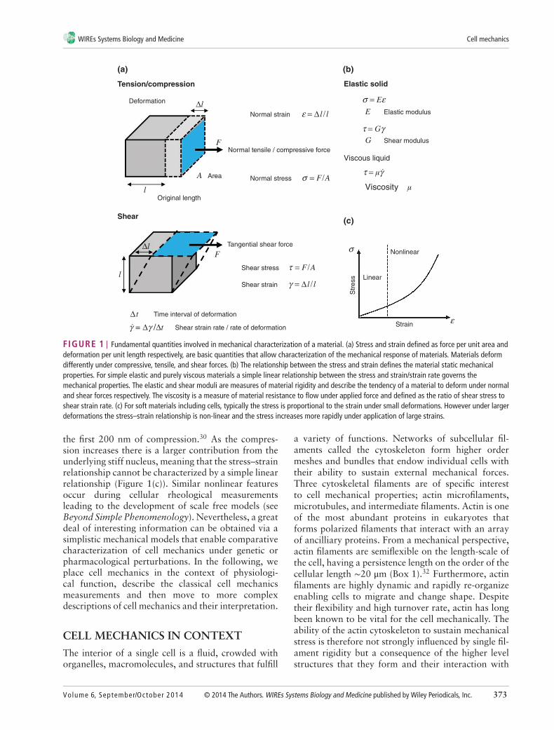
Mechanical PropertiesHow a material responds to mechanical stimuli isdefined by a group of characteristics referred to
broadly as its ‘mechanical properties’ (Figure 1).In general, these terms describe how a materialdeforms in response to an applied stress, and how thisdeformation evolves over time. The scaling betweenstress and strain of a solid material is a constantcalled the Young’s modulus (often referred to as thematerial’s elasticity with a unit of pascals), which is afundamental property of solids as it determines theirability to sustain their shape under mechanical stress(Figure 1(a)). In contrast to elastic solids, fluids flowunder the application of stress and are unable to storeelastic energy. The rate at which a fluid flows undera defined load is quantified by its viscosity (given inthe unit pascal-seconds) (Figure 1(b)). However, manymaterials exhibit both elastic and viscous propertiesand are referred to as viscoelastic. A viscoelastic mate-rial undergoing deformation simultaneously storesand dissipates mechanical energy and thus mechanicalstress relaxes and deformation increases over time.
Under physiologically relevant timescales cellsare intrinsically viscoelastic, as they display a combi-nation of both elastic and time-dependent responsesto deformation. With an emphasis on the interplaybetween stress, strain, and the rate of flow, rheologicalmeasurements are used to investigate how cells flowrather than deform purely elastically in response to anapplied force. At this point it is important to note thatthe simple mechanical terms elasticity and viscositycan be used as comparative quantities in cell mechan-ics. Many of the early cell mechanical measurementshave shown links between local increases in cellu-lar elasticity and subcellular structures such as stressfibres23–25 and changes to cellular elastic and viscousproperties under different treatments.26–28 However,the applicability of these terms in a strict engineeringsense is non-trivial and a range of complex rheologicalbehaviors for cellular systems have been observed (seesection, Universal Cell Behaviors and Beyond Sim-ple Phenomenology). Typical values for the cell elasticmodulus range from a few hundred pascals to tens ofkilopascals (Figure 2(c)), and the cellular viscosity isin the order of a few hundred pascal-seconds.
Even if thinking of a cell as an inert material, oneof the main difficulties in cell mechanics is to under-stand the structural origins of the measured cellularmechanical properties. Indeed, cells are complex het-erogeneous media containing a range of proteins, fil-aments, subcellular structures and organelles that canhave different contributions to cell elasticity and vis-cosity. One particular example of this is the role of thenucleus in defining whole cell elasticity. The nucleusis known to be stiffer than the cytoplasmic portionof the cell.29–31 During cell compression experiments,the cell membrane and cortex is deformed within
372 © 2014 The Authors. WIREs Systems Biology and Medicine published by Wiley Periodicals, Inc. Volume 6, September/October 2014
WIREs Systems Biology and Medicine Cell mechanics
Tension/compression
Deformation
Shear
Normal strain
(a)
Normal tensile / compressive force
Tangential shear force
Shear stress
Shear strain
Area
Original length
Normal stress
Shear strain rate / rate of deformation
Time interval of deformation
Elastic solid
Viscous liquid
Viscosity
Elastic modulus
Shear modulus
(b)
Linear
Nonlinear
Strain
Str
ess
(c)
FIGURE 1 | Fundamental quantities involved in mechanical characterization of a material. (a) Stress and strain defined as force per unit area anddeformation per unit length respectively, are basic quantities that allow characterization of the mechanical response of materials. Materials deformdifferently under compressive, tensile, and shear forces. (b) The relationship between the stress and strain defines the material static mechanicalproperties. For simple elastic and purely viscous materials a simple linear relationship between the stress and strain/strain rate governs themechanical properties. The elastic and shear moduli are measures of material rigidity and describe the tendency of a material to deform under normaland shear forces respectively. The viscosity is a measure of material resistance to flow under applied force and defined as the ratio of shear stress toshear strain rate. (c) For soft materials including cells, typically the stress is proportional to the strain under small deformations. However under largerdeformations the stress–strain relationship is non-linear and the stress increases more rapidly under application of large strains.
the first 200 nm of compression.30 As the compres-sion increases there is a larger contribution from theunderlying stiff nucleus, meaning that the stress–strainrelationship cannot be characterized by a simple linearrelationship (Figure 1(c)). Similar nonlinear featuresoccur during cellular rheological measurementsleading to the development of scale free models (seeBeyond Simple Phenomenology). Nevertheless, a greatdeal of interesting information can be obtained via asimplistic mechanical models that enable comparativecharacterization of cell mechanics under genetic orpharmacological perturbations. In the following, weplace cell mechanics in the context of physiologi-cal function, describe the classical cell mechanicsmeasurements and then move to more complexdescriptions of cell mechanics and their interpretation.
CELL MECHANICS IN CONTEXT
The interior of a single cell is a fluid, crowded withorganelles, macromolecules, and structures that fulfill
a variety of functions. Networks of subcellular fil-aments called the cytoskeleton form higher ordermeshes and bundles that endow individual cells withtheir ability to sustain external mechanical forces.Three cytoskeletal filaments are of specific interestto cell mechanical properties; actin microfilaments,microtubules, and intermediate filaments. Actin is oneof the most abundant proteins in eukaryotes thatforms polarized filaments that interact with an arrayof ancilliary proteins. From a mechanical perspective,actin filaments are semiflexible on the length-scale ofthe cell, having a persistence length on the order of thecellular length ∼20 μm (Box 1).32 Furthermore, actinfilaments are highly dynamic and rapidly re-organizeenabling cells to migrate and change shape. Despitetheir flexibility and high turnover rate, actin has longbeen known to be vital for the cell mechanically. Theability of the actin cytoskeleton to sustain mechanicalstress is therefore not strongly influenced by single fil-ament rigidity but a consequence of the higher levelstructures that they form and their interaction with
Volume 6, September/October 2014 © 2014 The Authors. WIREs Systems Biology and Medicine published by Wiley Periodicals, Inc. 373

Overview wires.wiley.com/sysbio
Lengthscale (m)
Timescale (s)
Elastic modulus (Pa)
cm
day
GPa MPa KPa
min sec ms nsμshour
mm
10–1 10–3 10–4 10–5 10–6
105 104 103 102 101 10–1 10–2 10–6 10–12
1011 1010 109108 107 106 105 104 103 102 101
10–9
Organ
(a)
(b)
(c)
Development Cell division Cell migration
Transcription
Steel Bone Tendon Ruber Muscle
Articular cartilage Eukaryotic cells
Polymers(i.e. polystyrene)
DiffusionActin Polymerisation
Active transport
Protein dynamics:Conformational changesInteractionsChemical reactions
Tissue Cellular aggregate Cell Subcellular structures:Nucleus, Organelles,MacromoleculesCytoskeleton
nmμm
FIGURE 2 | Main parameters involved in choosing the mechanical measurement tool. The choice of experimental tool requires consideration of(a) the lengthscale, (b) the timescale of the measurement and (c) the level of forces (or elasticity of the sample). A reasonable estimate of these threefactors indicates which characterization tool is the most appropriate technique for mechanical study of a particular sample.
crosslinkers and polymerizing factors. For example,the actin cytoskeleton forms a 200-nm-thick meshbelow the apical plasma membrane that endowsthe cell with its mechanical integrity, basal fibersanchor the cell to the extracellular matrix (ECM),and linear bundles coordinate and sustain forcesalong intercellular junctions. Cells are active mate-rials that carry an intrinsic pre-stress generated bymyosin motors. As the myosin proteins crosslinkand process along filaments they generate an inter-nal stress between anti-parallel neighboring filaments.This pre-stress is particularly apparent in contractileactin structures such as stress fibers and intercellular
junctions that rapidly relax following dissection withlaser nano-scissors (Figure 5(b)).34
Chemical signaling between cells span a rangeof lengthscales; from hormones that travel betweenorgans in the blood stream; to paracrine signalingbetween local groups of cells; small molecule signal-ing between contacting neighbors through gap junc-tions; and intracellular signaling cascades. A wealth ofrecent research has shown that cells are able to sensemechanical signals and forces in their environment.36
The mechanical properties of the culture substratedetermine cell differentiation and fate.37,38 Cells tune
374 © 2014 The Authors. WIREs Systems Biology and Medicine published by Wiley Periodicals, Inc. Volume 6, September/October 2014

WIREs Systems Biology and Medicine Cell mechanics
BOX 1
PERSISTENCE AND ENTANGLEMENTLENGTHS
For a single filament, the persistence length orthe length of thermal flexibility lp is the length-scale over which thermal bending fluctuationsbecome appreciable and can change the direc-tion of the filament, lp = 𝜅/kBT where 𝜅 is thebending modulus of a single filament and kBTthe thermal energy. For network of filaments theentanglement length l is the confining length-scale over which the motion of each filament isrestricted via topological constraints from neigh-boring filaments.
their mechanical properties to match that of theirsubstrate,37,38 and migrate toward particular mechani-cal conditions (known as durotaxis). Complex sensorymachineries located on different cellular sites, suchfocal adhesion complexes39 and more recently focaladherens junctions,40 have been shown to make uppart of the molecular machinery involved in sensingmechanical stimulation.
The importance of sustaining, generating, andsensing mechanical forces at the cellular level isbrought largely into context when examining diseasesthat target the cytoskeleton. Genetic disorders that dis-rupt the actin cytoskeleton or the binding of actinto the membrane of red blood cells, lead to abnor-mal cell shape and compromised function in diseasessuch as malaria41 and sickle cell anemia. One recentclinical example suggests that changes in cell rheol-ogy can have consequences for the health of patients.Some patients with a low neutrophil count exhibit aconstitutively active mutation in the Wiskott Aldrichsyndrome protein (CA-WASp) that results in increasedactin polymerization through unregulated activationof the Arp2/3 complex. The overabundance of cyto-plasmic F-actin increases cellular apparent viscosityresulting in kinetic defects in mitosis.3 Genetic muta-tions to the intermediate filament cytoskeleton areoften linked to diseases such as Epidermolysis BullosaSimplex which have symptoms that include increasedtissue fragility.42 A hallmark of cancer is changes in thecell stiffness often resulting from perturbed cytoskele-ton. The alterations in cancer cells stiffness could havesignificant effects in their ability to squeeze throughthe surrounding tissue, invade and metastasize.43,44
One of the most exciting avenues of cellmechanics research is to make links between cel-lular level mechanosensitivity, force generation,mechanical properties and the underlying molecular
mechanisms.45 Several studies have now been ableto monitor mechanical changes with the activity ofmolecular signaling pathways. Stretching the moleculetalin using magnetic tweezers and AFM revealed bind-ing domains for vinculin and its recruitment to focaladhesions.46 AFM has been used to monitor themechanical properties of cells and how they evolvefollowing the activation of the angiotensin-1 recep-tor which induced an actin dependent contractionwithin the cell.47 The influence of G-protein-coupled-receptors on cell morphology and contractility wasstudied using AFM to monitor variations in cellheight.48
MECHANICAL MEASUREMENTSIN PRACTICE
In comparison to typical materials such as metals,plastics and glass, cells are small soft objects. Theability to measure the mechanical properties of thecell on spatially accurate length scales has only arisenwith the development of new technologies from thesurface sciences including piezoelectric ceramics thatcan change shape in nanometer level increments,and microfabrication of micron-size cantilevers andmechanical parts. This has lead to the development ofa vast number of cell mechanical measurement tech-niques, making choosing the appropriate tool puz-zling. Taken broadly, mechanical measurement tech-niques can be put into two categories: those thatcan be used to apply controlled deformations andforces on part of, or on the entire cell (such as mag-netic bead cytometry,14,16,49 optical tweezers,50–52 cellstretchers,53–56 flow rheometry54,57,58 and AFM59),and those that monitor the ability of a cell to generateforces and deform its environment (such as TFM60,61
and micropillar arrays62). Local cell mechanical prop-erties can also be extracted by tracking the motionof endogenous cellular structures, such as the move-ment of actin filaments, microtubules, mitochondria,or embedded particles of various sizes that are excitedthermally or driven with an external force (PTM63–65).In this section we outline the basic logic for choosingthe appropriate experimental tool and what factorsshould influence this decision.
Choosing Forces, Lengthscales, Timescales,and Experimental PracticalitiesThe choice of experimental tool depends on three keyconsiderations: the compliance and the lengthscale ofthe cellular material under investigation, the timescaleat which mechanical properties are investigated, and
Volume 6, September/October 2014 © 2014 The Authors. WIREs Systems Biology and Medicine published by Wiley Periodicals, Inc. 375

Overview wires.wiley.com/sysbio
the required environmental and experimental condi-tions (Figure 2).
First, eukaryotic cells are soft biological mate-rials with an elastic modulus that varies from a fewhundred pascals to tens of kilo-pascals and a size onthe order of tens of microns (Figure 2(a,b)). Becauseof the cell size and low elastic modulus, the mechan-ical measurement technique needs to be capable ofapplying and monitoring deformations and forces inthe range of micro to nano meters and nano topico newtons respectively. Typical measurement tech-niques that are capable of applying these deforma-tions and forces at a high spatial accuracy includeatomic force microscopy.59,66 To measure local cellu-lar mechanical properties the size of the measurementprobe must be much smaller than the cell length asin PTM. As the size of the cellular sample changes,for example to the multicellular aggregate and embryolevel, then the experimental tool must be changed toapply larger forces over a greater area. For examplemechanical shivering of multicellular aggregates hasbeen observed using micropipette aspiration of entireaggregates rather than the more traditional single cellaspiration experiments.67 As an alternative, AFM witha stiffer cantilever can be used to apply higher forces,and a larger bead can be attached to the end of the can-tilever to increase the size of the probe.68,69 Tools tostudy cellular force generation can be chosen with sim-ilar considerations to the lengthscales of the specimenand the degree of cellular force generation. Cells cantypically generate forces on the order of nano-newtonsmeaning that the culture environment should have anappropriately tuned elasticity (e.g., kilo-pascal gels)for observable deformations (see case studies).
Second, the experimental tool can be chosenaccording to the frequency (timescale) of applica-tion and measurement of forces or deformations(Figure 2(c)). Simple conventional materials exhibitvery simple frequency-dependent responses: for nor-mal solids the response (dynamic modulus) is inde-pendent of excitation frequency and for liquids it islinearly correlated with excitation frequency throughthe viscosity of the liquid. However, materials, and inparticular cells with more complex hierarchical struc-tures and a high degree of heterogeneity, can exhibita variety of complex time-dependent responses thatdepend on strength, frequency, and spatial applicationof deformations and forces (Figure 3). Tools such asAFM, magnetic and optical tweezers can be used withclosed loop feedback systems to apply stress relax-ation, creep, and oscillatory loading protocols8,70–72
(Figure 3) and capture the time-dependent mechanicalresponse of the cell over a wide range of timescales.
On the other hand PTM techniques inherently cap-ture the frequency dependent response. Here it worthemphasizing that further to cell structural complex-ity and passive processes, active biochemical processes(such as continuous turnover of cytoskeletal fibres,association/dissociation of crosslinkers and activity ofmolecular motors) also govern the cell rheology andtherefore the timescale of active processes should alsobe considered when picking the mechanical tool andthe loading protocol.
Third, a variety of experimental and environ-mental conditions can determine the choice of experi-mental tool. To date many mechanical measurementsare performed in ambient conditions, far from thoseof the true physiological environment. Some tech-niques that use optical traps can cause an increasein cell temperature.73 Long time course experimentsin particular should be combined with temperaturecontrol; one strategy includes incorporating an envi-ronmentally controlled chamber.17,74 Another impor-tant concern is the bio-compatibility of materials usedin mechanical measurements. For instance, one com-mon material is PDMS, a bio-compatible elastomerused in microfabricated cell microfluidic devices, thatis often used as a soft membrane for cell cultureonto stretchable substrates and in the fabrication ofmicro-pillar arrays.54,62 Designing alternative biolog-ically and mechanically compatible synthetic hydro-gels is indeed an important avenue of research inorder to provide improved alternatives for polyacry-lamide hydrogels in TFM experiments. Another goalof mechanical measurements is to observe changesin the localization of different proteins and proteinactivation during mechanical loading. Inverted fluo-rescence microscopes can be easily integrated withmechanical testing techniques6,56 to image proteinexpression and localization.
In the next section, due to their wide range ofapplicability, commercial availability and ease of oper-ation for novices, we discuss four mechanical mea-surement techniques in detail and summarize recentfindings in our case studies. For in depth discus-sion of other techniques we refer readers to a recentreview.75
CASE STUDIES: MECHANICALMEASUREMENT TECHNIQUES
Atomic Force Microscopy (AFM)The atomic force microscope is a high resolu-tion surface characterization technique, that hasbecome rapidly adopted for imaging and mechanicalcharacterization of a range of biological samples59
376 © 2014 The Authors. WIREs Systems Biology and Medicine published by Wiley Periodicals, Inc. Volume 6, September/October 2014

WIREs Systems Biology and Medicine Cell mechanics
Stress relaxation
Oscillatory rheology
Oscillatory Stress response
= 0 sin (t + )
= G´() sin(t) + G´´ () cos (t)
G´
G´2 G´´2
G´´
Oscillatory Strain excitation
0
0
t0 t0Time
Time
Pure elastic materials:
Pure viscous materials:
Time
Storage modulus
Loss modulus
Phase lag
Dynamic modulus:
0
0
Str
ain
Str
ain
or S
tres
s
Str
ain
Str
ess
Str
ess
Creep
Recorded response
Imposed excitation
(a)
(b)
= 0 sin(t)
= 0 G´ = G G´´ = 0 |G*| = G
= π/2 G´ = 0 G´´ = |G*| =
|G*|Angular frequency
= +
FIGURE 3 | Common loading conditions for measuring time-dependent mechanical properties. (a) Typical rheological characterizationincorporates measuring the temporal evolution of strain under application of a constant stress (creep) or the temporal evolution of stress underapplication of a constant strain (stress-relaxation). (b) Oscillatory techniques are another method of characterizing the viscoelastic properties ofmaterials where normally a sinusoidal strain is applied and the cyclic stress response is monitored. In this approach the timescale of the test isdefined by the frequency of oscillation 𝜔. By observing material response at a range of frequencies the relative contribution of elastic (indicated bystorage modulus G′) and viscous (indicated by loss modulus G′′) responses can be characterized at different timescales. The dynamic modulus G* isthe indicator of overall viscoelastic behavior. Oscillatory tests can reveal a set of material viscoelastic responses over specific span of frequencies(timescales).
(Figure 4(a)). AFM measurements utilize a micron-sized tip connected to a micro-fabricated cantileverbeam to deform and interact with the sample. It iscapable of probing surface topography and inter-action forces with subnano-meter and pico-newtonresolution. One of the most widespread uses of AFMin cell mechanics is AFM force spectroscopy to mea-sure cellular elasticity and rheology. To extract the cellelasticity, the tip of AFM cantilever is pressed againstthe cell while the force and the imposed cellular defor-mation are monitored. Considering the tip geometryand using an appropriate contact model, the elasticityof the cell can be computed from the measured forceversus indentation data.6 The success of cellular forcespectroscopy measurements is in part due to the easeof the measurements, good measurement throughputand commercial systems that are readily available.Furthermore, because the levels of force and defor-mation can be very accurately measured over time,AFM has been applied for a variety of rheologicalmeasurements. Using a feedback loop (incorporated
into most commercial systems) levels of strain andstress can be controlled over time, following indenta-tion of the cell via AFM cantilever. Stress-relaxationand creep experiments78,79 can be readily applied andoscillatory tests71 can also be conducted to measuretime-dependent cellular mechanical properties.
A recent close examination of AFM indenta-tion and stress-relaxation tests on cells revealed thatcells behave according to the theory of ‘poroelastic-ity’ (see section Beyond Simple Phenomenology) whenmechanically stimulated in a way similar to that expe-rienced in organs within the body.8 Of particular inter-est in cell biology and medicine is the capability ofAFM to monitor changes in cell elasticity under differ-ent pharmacological and genetic perturbations.3,80 Forexample, using the AFM indentation tests the elasticityof the vascular smooth muscle cells (VSMCs) isolatedfrom thoracic aorta of old and young monkeys wasmeasured.81 Increases in VSMCs elasticity with agesuggested that cellular rheology has a significant con-tribution to aging-associated vascular stiffness and
Volume 6, September/October 2014 © 2014 The Authors. WIREs Systems Biology and Medicine published by Wiley Periodicals, Inc. 377

Overview wires.wiley.com/sysbio
Atomic force microscopy (AFM) I
II
AFM bead
10 μ
m
5 μm
20 μm
20 μm
3
2
1
Cell
II
I
II
I
I
II III
5
240
200
160
120
80
40
00 3 6 9
F0
Fm
12 15
Distance (μm)
For
ce (
pN)
4
3
2
1
0
0 0.2 0.4Indentation (μm)
For
ce (
nN)
0.6 0.8
Photodiode detector
Optical tweezers
Laser beam
Objective
Traction force microscopy (TFM)
Deformation vectors
Objective
Tracer particles embedded in a gel
Trapped bead
Tether force
TetherPiezoelectric ceramicLaser light path
Cantilever tip
Particle tracking microrheology (PTM)
Tracer beads
Objective
Trajectory
100 nm Time (s)
MS
D (
μm
2 )
10-1
10-2
10-3
10-4
10-2 10-1 100 1011
1 1
Nucleus
Nucleus
NucleusNucleus
(a) (b)
(d)(c)
FIGURE 4 | Four cell mechanical measurement techniques. (a) AFM: A laser beam is reflected off the back of the cantilever and collected byphotodiodes. Interactions between the tip and the sample change the bending of the cantilever and consequently the reflection path of the laserbeam which is precisely measured by the photodiodes. The bending of the cantilever is converted to force using its spring constant. A piezo-electricceramic in a feedback loop is used to move the cantilever up and down to adjust bending of the cantilever and the applied force. (a-I) A confocalmicroscopy image shows the HeLa cell profile as the cell (green) is indented by a spherical bead (blue) attached to AFM cantilever. (a-II) A typicalAFM force-indentation curve on cell. This curve can be fitted with an indentation model to estimate the cell elasticity. (Reprinted with permission fromRef 8. Copyright 2013 Nature Publishing Group.) (b) Optical tweezers: A small particle is stably trapped by a highly focused laser beam. The positionof the optically trapped particle can be controlled by the movement of trap and small forces can be estimated from the changes in the displacementof the particle from the center of trap. (b-I, II) A tether extraction experiment involves pulling of an optically trapped bead attached to a cellmembrane away from the cell. (b-II) The force-distance curve of tether extraction experiments on microglial cell. (Reprinted with permission from Ref76. Copyright 2013 Public Library of Science.) (c) PTM: The micron or submicron beads disperse within the cytoplasm following injection into live cells.Using a high magnification objective the random spontaneous motions of the beads are captured with high spatial and temporal resolution. (c-I) 100nm fluorescent beads injected into the cytoplasm of 3T3 fibroblasts. (c-II, III) The recorded time-dependent trajectories (c-II) of the beads are used tocalculate their mean squared displacements (c-III) by which the nature of intracellular diffusion and microscopic viscoelastic properties of cellularenvironment can be studied. (Reprinted with permission from Ref 77. Copyright 2009 Public Library of Science.) (d) TFM: The cell is cultured onto (orwithin) a bead-embedded polymeric gel. Cellular contractions deform the gel and for a known gel elastic modulus the cellular traction forces can becalculated from the bead displacements. (d-I, II) Deformation vectors and traction stress field of fibroblast cultured on polyacrylamide gel werecalculated by monitoring the displacement of fluorescent beads embedded in the gel. (Reprinted with permission from Ref 60. Copyright 2001 CellPress.)
disease. Another recent use of AFM involves placinga tipless cantilever on a cell entering mitosis and mon-itoring the temporal changes of cell rounding forcesduring mitosis.82 Employing a perfusion system toapply drug and osmotic perturbations while measur-ing mitotic forces via AFM, it has been found that bothactomyosin contractility and transmembrane ion gra-dient determine the levels of mitotic forces.82
Optical TweezersThe use of optical methods to image cells has beenwell established for decades. More recently, with thegrowing interest in cell mechanics there have beenseveral methods developed that employ light trappingto manipulate part of a cell83–85 or stretch the wholecell.43,86 These techniques rely on the concept that as
light enters a medium of a different refractive index thelight path changes. Conservation of momentum meansthat there is a restoring force created by the lightpassing through the material that resists higher levelsof refraction. Typically this means that a cell or a beadcan be trapped/ deformed optically and manipulatedwith a collimated light source. Because of the highsensitivity of these optical techniques (pico-newtonresolution) and high spatio-temporal accuracy theyare well suited to sub cellular measurements suchas the pulling of membrane tethers.76,83–85, In theseexperiments a small particle attached to the cellmembrane is pulled away from the cell by opticaltweezers and a tube-like structure, referred to as amembrane tether is created. Early measurements usingthis approach revealed the presence of a large amount
378 © 2014 The Authors. WIREs Systems Biology and Medicine published by Wiley Periodicals, Inc. Volume 6, September/October 2014

WIREs Systems Biology and Medicine Cell mechanics
of excess membrane at the cell surface that flows intothe tether as it is pulled.83 This technique has beenalso applied to study the adhesion strength of theplasma membrane to the actin cortex and the roleof membrane proteins such as PIP2 in regulating thisadhesion energy.84,85 More recently elastic propertiesof the membrane of a variety of central nervous systemcells have been reported using optical tweezers toperform tether extraction experiments.76
Although optical tweezers provide a valuabletool for high precision measurements of small forces,there is an inherent limit on the amount of forcethat can be applied using these methods. In particularincreasing the laser power to increase the optical forcesinduces local heating of the cell that might damagethe cell structure and influence its mechanical proper-ties. To increase the amount of optical forces, leavingminimal photodamage, another type of optical manip-ulation technique involves coupling a laser light toanother optical fiber that enables trapping and stretch-ing of the whole cell.86 By combining this opticalstretcher technique with a microfluidic platform, highthroughput mechanical characterization of diseasedand healthy cells in suspension has been reported.43
Particle Tracking Microrheology (PTM)One shortcoming of most cell rheological techniques,including AFM, is that they require external interven-tions and measure the combined response of subcel-lular structures (such as cell membrane, cytoplasm,and the nucleus) by application of external forcesdirectly on the cell surface (Figure 4(a)). However,in PTM (Figure 4(c)), localized mechanical measure-ments inside the cytoplasm can be achieved by track-ing thermally driven motion of embedded tracer parti-cles, without a need for a direct contact of between thecell and an external probe.65 Furthermore, using PTMit is possible to study mechanics of cells embedded ina 3D matrix64 (a more physiologically relevant con-dition) while it is very difficult to probe cells in threedimensions by other mechanical techniques .
Microrheological analyzes of movement of par-ticles in cell indicate existence of non-Brownian fluc-tuations. Indeed the movement of particles within thecell is perturbed by both elastic and viscous mechani-cal resistances.63 Interaction of trapped particles insidethe cell with elastic cellular network could be one mainsource of non-Brownian fluctuations (Box 2). Otherpossible contributions could include the influence ofenergy-consuming active processes and macromolec-ular crowding on tracer particles inside the cyto-plasm. Generally PTM experiments on cells revealeda dominant elastic response at short timescales anda more viscous behavior over longer time periods.
BOX 2
BROWNIAN MOTION AND DIFFUSION
Microscopic mechanical properties of soft mate-rial including cells can be measured by trackingthe movement of embedded tracer particles.In a purely viscous environment, particles moverandomly due to thermal fluctuations and theirmean squared displacement (MSD, < r2 >) growslinearly in time t following ⟨r2⟩=DTt, where DTis the particle translational diffusion coefficient.This behavior is the characteristic of normaldiffusion or Brownian motion. During normaldiffusion the thermal motion of a particle withsize a is slowed down by only a linear viscousdrag from surrounding environment. Thereforeby monitoring random motion of particles insidea purely viscous medium the MSD and subse-quently particle diffusion constant DT can becalculated to derive the viscosity of the mediumusing Stokes–Einstein relationship 𝜇= kBT /DTawhere kBT is the thermal energy. By contrast, ina purely elastic environment such as dense andcross-linked network of polymers, the embed-ded particles are stuck within the network andthermal agitations are not strong enough toinduce significant random motion. In generalthe MSD of particles embedded in a complex vis-coelastic environment such as cell follows a morecomplex time-dependence relationship than thedescribed linear Brownian or weak fluctuatingbehaviors. In this case the MSD still grows con-tinually with time due to thermal fluctuationsbut as the normal diffusion relationship is notapplicable anymore the particle non-Brownianfluctuations can be characterized by asemi-empirical anomalous diffusion equation< r2 >∼ t𝛼 where 𝛼 can be any number except 1.
However, the magnitude and timescale of these vis-coelastic responses may vary for various cell types,under different pharmacological treatments and phys-iological conditions.77,87,88 For instance, in an in vivostudy PTM was carried out by microinjection ofnanoparticles into zygotes to extract viscoelastic prop-erties of the Caenorhabditis elegans embryos.89 Unlikedifferentiated cells, the cytoplasm of these embryosexhibits a highly viscous behavior over wide rangeof timescales. Another application of PTM discoveredsignificant stiffening of intracellular environment ofmesenchymal stem cells in response to the applica-tion of different growth factors such as TGF-𝛽1.88
Other recent studies employed PTM to investigateeffects of mechanical perturbation (such as flow shear
Volume 6, September/October 2014 © 2014 The Authors. WIREs Systems Biology and Medicine published by Wiley Periodicals, Inc. 379

Overview wires.wiley.com/sysbio
stress87,90,91 and stretching35) and also environmentalchanges (such as temperature and pH92) on spatiotem-poral cell viscoelastic properties.
Traction Force Microscopy (TFM)Force sensing techniques are another type oftool that have advanced our understanding ofmechanotransduction36 and improved quantita-tive modeling of cellular interactions with the ECM.These techniques measure traction forces generatedby the cell on the surrounding environment usingdifferent detection mechanisms such as micropillararrays62 or embedded beads in soft gels (Figure 4(d)).Traction forces drive cell spreading and migrationduring commonly occurring cell processes such asmorphogenesis, wound healing, and tumor metasta-sis. Applications of high resolution time-lapse TFM inliving neuronal growth cones enabled nanoscale prob-ing of the complex dynamics of traction forces exertedby growth cone93 and its filopodia.94 These recentstudies, had a major impact in unraveling the roleof mechanical cues in neuronal development. Theseresults showed that focal adhesions exhibit eitherstable or oscillating force transmission to the ECMvia adhesion sites and ECM stiffness modulates thedynamics of focal adhesions. Using TFM other recentworks also studied the oscillation of forces withinfocal adhesions and the impact of ECM complianceon force fluctuations and directed cell migration.95,96
Most studies measure the traction forces of cells cul-tured on planar substrates but combining TFM withlaser scanning confocal microscopy allows probing ofthe cellular traction forces in three dimensions.61,97
UNIVERSAL CELLMECHANICAL BEHAVIORS
Having selected an experimental approach and aloading protocol it is important to know what typeof behavior could be expected for cell rheology andwhat this means for the underlying microstructureand cellular function. Cell mechanical studies overthe years have revealed a rich phenomenologicallandscape of rheological behaviors that are dependentupon the cell type, probe geometry, loading protocol,and loading frequency. Although assessing cellularelasticity and viscosity yield some useful informationfor comparative characterization between differ-ent treatments, the mechanical behavior of the cellis inherently much more complicated. Due to theheterogeneity and complexity of the cell and thecytoskeleton, submicrometer-scale measurementscan lead to considerably different evaluations ofmechanical properties compared to bulk (several
micrometer-scale) measurements. However recentrheological measurements on eukaryotic cells agreeon the presence of four cellular phenomenologicalbehaviors that are universal.98 (1) Cell rheology isscale free: plots of the frequency response of manycell types (obtained via different methods) on log–logscale display the same shape and follow a weakpower law spanning several decades of frequency33
(Figure 5(a)). (2) Cells are prestressed: mechanicalstresses generated continuously by the internal activ-ity of actomyosin or applied externally on the cellare counterbalanced by the tensional/compressionalstate of the cytoskeleton99 (Figure 5(b)). (3) TheStokes–Einstein relation breaks down and diffusionis anomalous13: The spontaneous motions of endoge-nous particles or embedded/attached beads present inthe cell do not follow the Stokes–Einstein relationship(see Box 2, Figure 5(c)). (4) Stiffness and viscous dissi-pation are altered by stretch35: Application of stretchsignificantly perturbs the rheological properties of thecell, and depending on the experimental condition,the cell can exhibit different behaviors such as stressstiffening, fluidization and rejuvenation.
BEYOND SIMPLE PHENOMENOLOGY
One of the main ongoing challenges in the field ofcell mechanics is to find a unified framework underwhich the measured phenomenological behaviors canbe interpreted to obtain realistic information aboutthe dynamics of the microstructure of the cell, orvice versa to estimate the bulk rheological propertiesfrom the observed microstructural interactions. Sev-eral top-down (linear viscoelastic, tensegrity, powerlaw, and soft glassy rheology) and bottom-up (net-works of polymers, poroelasticity) theoretical models(Figure 6) have been successfully applied to explainthe observed phenomenological mechanical behav-iors. However there is still no unifying mechanistictheory to explain the physical mechanisms that gov-ern the universal cellular behaviors and encompassessuch a rich phenomenological landscape. As willbe described briefly in the following sections, linearviscoelastic, immobilized colloids/soft glasses, and thenetwork of cytosketal filaments are the most widelyused models that have been proposed to study cellmechanics and we provide a brief introduction to theseconcepts and frameworks.
Linear Viscoelastic and Power Law ModelsThe majority of the work to date utilizes a viscoelas-tic description of cells that considers the cell as asingle phase homogenous continuum material.11,12 As
380 © 2014 The Authors. WIREs Systems Biology and Medicine published by Wiley Periodicals, Inc. Volume 6, September/October 2014

WIREs Systems Biology and Medicine Cell mechanics
Experimental data
10–1
0 s 1 s 5 s
100 101
3/4
III
III
𝛽2
𝛽1
102
Frequency (rad/s)
Nor
mal
ized
|G∗ (
ω)|
103 104 105
104
2 5 s
20 s35 s
995 s
1
tw
Nucleus
103
102
101
100
10–1 100 101
Time (s)
Mea
n sq
uare
dis
plac
emen
t (nm
2 )
(a)
(b)
(c)
FIGURE 5 | Cell universal behaviors. (a) The frequency response of cells measured with several mechanical measurement techniques (such asAFM, PTM, and magnetic twisting cytometry) collapses into two master curves after rescaling the experimental data from different rheologicalmeasurements (see Ref 33 for details). The frequency dependent dynamic modulus follow two distinct regimes in these master curves that can befitted by a power law function: |G * (𝜔)| :∼𝜔𝛽 , where 𝜔 is frequency and 𝛽 is power law exponent. At high frequencies both curves show 𝛽 = 3/4 butat low frequencies they exhibit lower range of power-law exponents, 𝛽1 ∼ 0.25 and 𝛽2 ∼ 0.15 (see Ref 33 for further details). (b) Spontaneousretraction of a single actin stress fiber upon severing with a laser nanoscissor shows existence of prestress in the cytosketal bundles. Scale bar= 2μm. (Reprinted with permission from Ref 34. Copyright 2006 Cell Press.) (c) Anomalous diffusion and response of the cell to stretch. (c-I, II)Spontaneous movements of beads attached firmly to the cell show intermittent dynamics. Scale bars= 10 μm. (Reprinted with permission from Ref13. Copyright 2005 Nature Publishing Group.) (c-III) The mean square displacement (MSD) of a bead anchored to the cytoskeleton exhibits anomalousdiffusion dynamics < r2 >∼ t𝛼 (subdiffusive 𝛼 < 1 at short time intervals and superdiffusive 𝛼 > 1 at longer time intervals). Red curve indicates theMSD of the bead for a non-stretched cell and other curves show the MSD of the bead in response to a global stretch measured at different waitingtimes (t𝜔) after stretch cessation. (Reprinted with permission from Ref 35. Copyright 2007 Nature Publishing Group.)
we have described previously (see section Mechani-cal Properties), this assumption allows characteriza-tion of the cell time-dependent mechanical responsesby introducing a finite number of elastic and viscouselements (springs and dashpots) coupled in series orparallel leading to exponential decay functions witha finite number of relaxation times. The combinationof several springs and dashpots create a system that,for example, exponentially relaxes to a new size underthe application of a constant stress (Figure 6(a)). Insuch models, the resultant exponential functions canhave a single relaxation time if there is only one dash-pot element or a distribution of relaxation times ifthere are several. Examples of such models are theMaxwell and the Standard Linear Solid (also knownas the Zener) models that has been widely used to
describe the time-dependent behaviors of biomateri-als including living cells.12,78 Beyond this, power lawmodels also provide good fits to the experimentalrelaxation data51 but unlike spring-dashpot models,they do not have any characteristic relaxation time andcannot be easily described using mechanical analogs(Figure 6(b)). Power law structural damping modelshave been applied very commonly in recent microrhe-ological experiments to describe the measured vis-coelastic response of the cells over a broader range oftimescales.13,14
The main advantage of these empirical models isthat they can be used as a diagnostic tool in cell biol-ogy. The mechanical properties of the cell in healthyand diseased states, under physiological changes andalso with different genetic perturbations and drugtreatments, can be characterized using these models.
Volume 6, September/October 2014 © 2014 The Authors. WIREs Systems Biology and Medicine published by Wiley Periodicals, Inc. 381

Overview wires.wiley.com/sysbio
(a) (b)
(c)
(d)
Spring and dashpot models
Network of filaments
Myosin motors
Filaments
Crosslinkers
Power law models
Soft glassy rheology
Ene
rgy
leve
l
Viscous dashpot
σ = ε0
Elastic springs
K2100
I
300 nm
–200 nm
Z
II
σ ~ε0 K0 t–β
10–1
10–2
10–3
10–3 10–2 10–1 100 101 102 103
Time
Nor
mal
ized
str
ess
K1
K2 K1
K1exp t+
η
η
FIGURE 6 | Models of cell rheology. (a) Spring and dashpot representation of Standard Linear Solid viscoelastic model and the functional form ofits stress relaxation in response to sudden constant strain. (b) The power law type of relaxation in log–log scale is a line with the slope 𝛽 which is thepower law exponent. For purely elastic and viscos materials the power law exponents are 𝛽 = 0 and 𝛽 = 1 respectively. When 0<𝛽 < 1 combinationof elastic and viscous mechanisms contribute to relaxation response. (c) Schematic representation of soft glassy rheology: A rheological model thatexplains glassy and weak power-law behavior of soft disordered materials such as foams and colloids. (d) Dynamic network of cytoskeletal filamentssuch as actin network determine the cell rheology. (d-I) Image of actin filaments in a COS-7 cell taken by dual stochastic optical reconstructionmicroscopy (STORM) (Reprinted with permission from Ref 100. Copyright 2012 Nature Publishing Group). The height is color coded with respect tothe scaling shown in the color scale bar. Scale bars= 2 μm. (d-II) In reconstituted gel models of cytoskeletal filaments, interaction of binding proteinswith different properties (such as type, organization, and concentration) with filaments significantly influence the network rheology.
Despite their widespread use and offering good char-acteristic fits to the measured mechanical responses,the major limitation of these models is that they arenot mechanistic, fail to relate the measured rheolog-ical properties to structural or biological parameterswithin the cell, and thus cannot predict changes in rhe-ology due to microstructural changes.
Cell as a Soft Glassy MaterialA large body of more recent research has foundthat some of the cellular rheological behaviors areempirically similar to the rheology of soft materialssuch as foams, emulsions, pastes, and slurries. Fol-lowing some experimental observations,13,14,17,35,71 it
was proposed that cells could be considered as softglassy materials.101 As a conceptual model, soft glassyrheology (SGR) explains how macroscopic rheologi-cal responses are linked to localized structural rear-rangements originating from structural disorder andmetastability (Figure 6(c)). The SGR system con-sists of crowded particles that are trapped in energylandscapes arising from their interactions with sur-rounding neighbors. In such a system, thermal energyis not sufficient to drive structural rearrangementand, as a consequence out of equilibrium trappingoccurs. Over time, remodeling/rearrangement (micro-reconfiguration) happens when particles escape theenergy barriers of their neighbors and jump from onemetastable state to another reaching a more stable
382 © 2014 The Authors. WIREs Systems Biology and Medicine published by Wiley Periodicals, Inc. Volume 6, September/October 2014
WIREs Systems Biology and Medicine Cell mechanics
state with relaxation rates slower than any exponen-tial process.13 In such a system, injecting agitationalenergy, sourced from non-thermal origins (such asmechanical shear or ATP-dependent conformationalchanges of proteins35), liberates particles from theenergy cages in which they are trapped and facilitatesstructural rearrangements causing the material to flow.
Indeed, measuring the fluctuations of particleswithin the cytoplasm revealed that in many casesthey exhibit a larger random amplitude of fluctua-tions than expected and a greater degree of direc-tionality than could be developed solely from thermalfluctuations.102 These fluctuations deviate from theStokes–Einstein relation and can be empirically char-acterized by anomalous diffusion processes (Box 2)that are similar to particle fluctuations in a soft glassymaterial. The deviation from the Stokes–Einsteinrelationship has been attributed to reaction forcesfrom active cytosketal processes103 and crowding ofa large number of macromolecules inside the cyto-plasm, such as mobile intracellular globular proteinsand other fixed obstacles like cytoskeletal filamentsand organelles, that reduce the available solventvolume and provide barriers to particle Brownianmotion.104–106 On the basis of these observations, itwas proposed that the high concentration of differ-ent proteins in the cytoplasm can lead to liquid crys-tal and colloidal behaviors that can be interpreted interms of the SGR model.101 Crowded colloidal suspen-sions or soft glasses exhibit anomalous diffusion and aweak power-law rheology corresponding to a contin-uous spectrum of relaxation times. Similar to this, thedynamic modulus of cells, except at high frequencies,scales with frequency as a weak power law valid overa wide spectrum of timescales.14,71 Despite its abilityto describe the cell rheology over a wide spectrum oftimescale, the semi-empirical SGR model cannot yetexplain many of cell mechanical features such as thecell rheological responses at high frequencies.
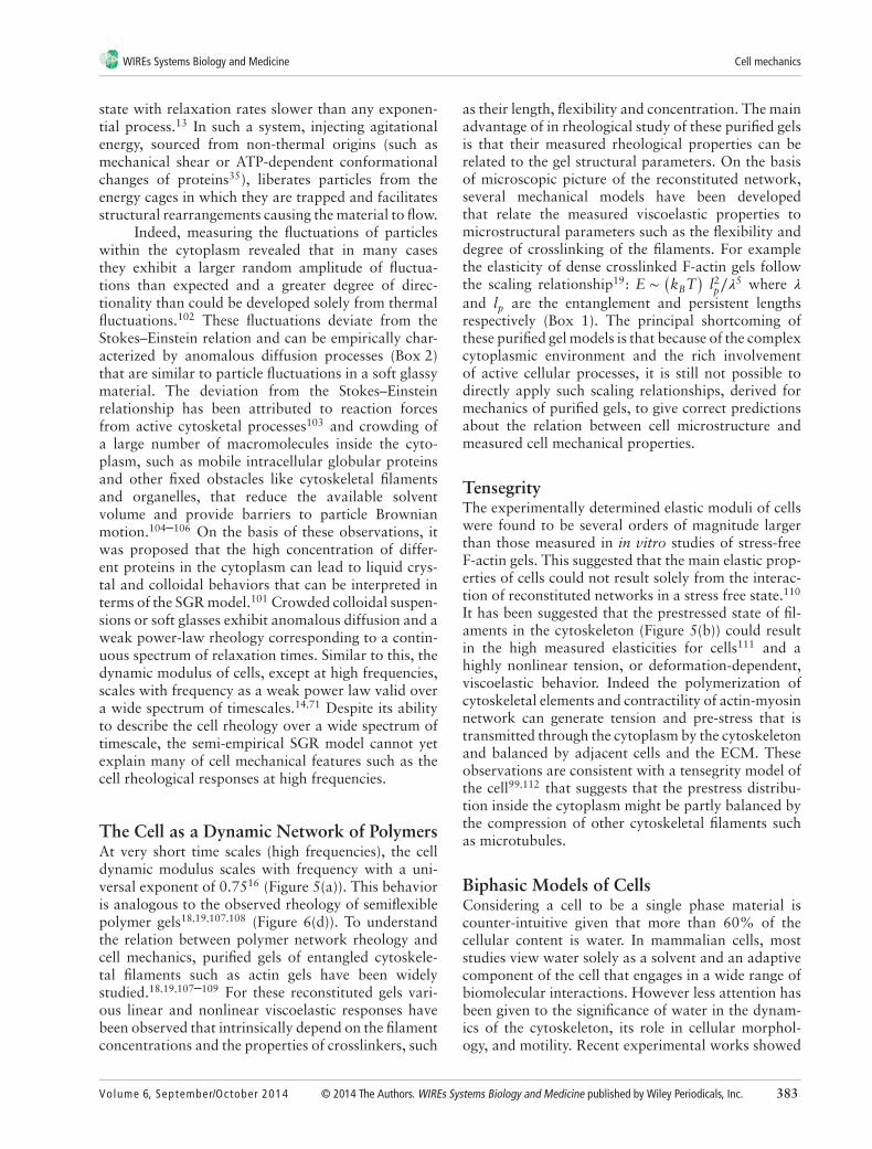
The Cell as a Dynamic Network of PolymersAt very short time scales (high frequencies), the celldynamic modulus scales with frequency with a uni-versal exponent of 0.7516 (Figure 5(a)). This behavioris analogous to the observed rheology of semiflexiblepolymer gels18,19,107,108 (Figure 6(d)). To understandthe relation between polymer network rheology andcell mechanics, purified gels of entangled cytoskele-tal filaments such as actin gels have been widelystudied.18,19,107–109 For these reconstituted gels vari-ous linear and nonlinear viscoelastic responses havebeen observed that intrinsically depend on the filamentconcentrations and the properties of crosslinkers, such
as their length, flexibility and concentration. The mainadvantage of in rheological study of these purified gelsis that their measured rheological properties can berelated to the gel structural parameters. On the basisof microscopic picture of the reconstituted network,several mechanical models have been developedthat relate the measured viscoelastic properties tomicrostructural parameters such as the flexibility anddegree of crosslinking of the filaments. For examplethe elasticity of dense crosslinked F-actin gels followthe scaling relationship19: E ∼
(kBT
)l2p∕𝜆
5 where 𝜆
and lp are the entanglement and persistent lengthsrespectively (Box 1). The principal shortcoming ofthese purified gel models is that because of the complexcytoplasmic environment and the rich involvementof active cellular processes, it is still not possible todirectly apply such scaling relationships, derived formechanics of purified gels, to give correct predictionsabout the relation between cell microstructure andmeasured cell mechanical properties.
TensegrityThe experimentally determined elastic moduli of cellswere found to be several orders of magnitude largerthan those measured in in vitro studies of stress-freeF-actin gels. This suggested that the main elastic prop-erties of cells could not result solely from the interac-tion of reconstituted networks in a stress free state.110
It has been suggested that the prestressed state of fil-aments in the cytoskeleton (Figure 5(b)) could resultin the high measured elasticities for cells111 and ahighly nonlinear tension, or deformation-dependent,viscoelastic behavior. Indeed the polymerization ofcytoskeletal elements and contractility of actin-myosinnetwork can generate tension and pre-stress that istransmitted through the cytoplasm by the cytoskeletonand balanced by adjacent cells and the ECM. Theseobservations are consistent with a tensegrity model ofthe cell99,112 that suggests that the prestress distribu-tion inside the cytoplasm might be partly balanced bythe compression of other cytoskeletal filaments suchas microtubules.
Biphasic Models of CellsConsidering a cell to be a single phase material iscounter-intuitive given that more than 60% of thecellular content is water. In mammalian cells, moststudies view water solely as a solvent and an adaptivecomponent of the cell that engages in a wide range ofbiomolecular interactions. However less attention hasbeen given to the significance of water in the dynam-ics of the cytoskeleton, its role in cellular morphol-ogy, and motility. Recent experimental works showed
Volume 6, September/October 2014 © 2014 The Authors. WIREs Systems Biology and Medicine published by Wiley Periodicals, Inc. 383

Overview wires.wiley.com/sysbio
the presence of transient pressure gradients insidecells and suggested that these could be explained bythe biphasic nature of cytoplasm.20,21,113 As a con-sequence, a fluid-filled sponge model of cells wasproposed based on poroelasticity (or biphasic the-ory), in which the cytoplasm is biphasic consistingof a porous elastic solid meshwork (cytoskeleton,organelles, macromolecules) bathed in an interstitialfluid (cytosol).20,21 In this framework, the viscoelas-tic properties of the cell are a manifestation of thetime-scale needed for redistribution of intracellularfluids in response to applied mechanical stresses andthe response of the cell to force application depends ona single experimental parameter: the poroelastic dif-fusion constant Dp, with larger poroelastic diffusionconstants corresponding to more rapid stress relax-ations. For this poroelastic picture of the cell, a mini-mal scaling law was proposed Dp ∼E𝜉2/𝜇 that relatesthe diffusion constant to the drained elastic modu-lus of the solid matrix E, the pore size of the solidmatrix 𝜉, and the viscosity of the cytosol 𝜇.8 There-fore, contrary to viscoelastic models, the dynamics ofcellular deformation in response to stress derived fromporoelasticity can be described using measurable cellu-lar parameters, allowing changes of rheology with E,𝜉, and 𝜇 to be predicted which makes this frameworkparticularly appealing conceptually.8
CONCLUSIONCell mechanics research has great potential to providenew perspectives on pathologies and classic biologicalresearch questions. To facilitate wider use of mechan-ical experimental tools and cell rheological character-ization we have outlined a simple set of considera-tions for non-experts in the field looking to attemptmechanical measurements. The choice of experimen-tal tool depends on the lengthscale of the sample andthe level of force that is needed to deform the sam-ple. Many techniques are now spatially accurate to
the subcellular level and sensitive enough to measurepico to nano newton levels of force and can be scaledappropriately by altering the size and stiffness of themeasurement probe. There are also a variety of envi-ronmental and experimental conditions that need tobe considered, such as temperature control and theinterface with other measurement techniques, such asoptical microscopy. Upon choosing the mechanicalmeasurement tool that will be spatially accurate, canapply the correct forces and comply with physiologi-cal condition, there are also a variety of mechanicalloading protocols that can be employed. Consider-ing the cell active features, the loading condition andthe timescale of mechanical measurement could havea direct relevance in probing the underlying activecellular processes. Typical loading protocols involvestep changes in stress or strain while monitoring theensuing relaxation response. Other loading protocolsinclude application of oscillatory mechanical excita-tions that provide significant insight about time depen-dent mechanical properties.
While their ability to describe the complexcell rheological behavior is extremely limited, lin-ear viscoelastic characterization of cellular mechanicalresponses in terms of spring-dashpot models lead toestimation of stiffnesses and viscosities that are usefulfor evaluating cell mechanics under different biologi-cal and chemical perturbations. Beyond simple linearmechanical descriptions are scale free models that bet-ter explain some of the commonly observed univer-sal cell mechanical behaviors. These can be appliedwith great effect to capture some of the mechanicalresponses of the cell under different loading condi-tions and at a wide range of timescales. Other recon-stituted gel and biphasic models provide a mechanisticinsight about cell rheological measurements. However,a unifying theory that describes all of the complexitiesof cell mechanical behavior remains an exciting andactive area of research.
ACKNOWLEDGMENT
This work was supported by University of Cambridge Herchel Smith Postdoctoral Fellowship Fund to EMoeendarbary. AR Harris was an EMBO long term postdoctoral research fellow (1075–2013).
REFERENCES1. Maloney JM, Nikova D, Lautenschläger F, Clarke E,
Langer R, Guck J, Van Vliet KJ. Mesenchymal stem cellmechanics from the attached to the suspended state.Biophys J 2010, 99:2479–2487.
2. Sun Y, Villa-Diaz LG, Lam RH, Chen W, Krebs-bach PH, Fu J. Mechanics regulates fate decisionsof human embryonic stem cells. PLoS One 2012, 7:e37178.
384 © 2014 The Authors. WIREs Systems Biology and Medicine published by Wiley Periodicals, Inc. Volume 6, September/October 2014

WIREs Systems Biology and Medicine Cell mechanics
3. Moulding DA, Moeendarbary E, Valon L, Record J,Charras GT, Thrasher AJ. Excess F-actin mechani-cally impedes mitosis leading to cytokinesis failurein X-linked neutropenia by exceeding AuroraB kinase error correction capacity. Blood 2012,120:3803–3811.
4. Park Y, Best CA, Badizadegan K, Dasari RR, FeldMS, Kuriabova T, Henle ML, Levine AJ, PopescuG. Measurement of red blood cell mechanics duringmorphological changes. Proc Natl Acad Sci USA2010, 107:6731–6736.
5. Stewart MP, Toyoda Y, Hyman AA, Müller DJ.Tracking mechanics and volume of globular cellswith atomic force microscopy using a constant-heightclamp. Nat Protoc 2012, 7:143–154.
6. Harris AR, Charras GT. Experimental validation ofatomic force microscopy-based cell elasticity measure-ments. Nanotechnology 2011, 22:345102.
7. Crow A, Webster KD, Hohlfeld E, Ng WP, Geissler P,Fletcher DA. Contractile equilibration of single cellsto step changes in extracellular stiffness. Biophys J2012, 102:443–451.
8. Moeendarbary E, Valon L, Fritzsche M, Harris AR,Moulding DA, Thrasher AJ, Stride E, Mahadevan L,Charras GT. The cytoplasm of living cells behaves asa poroelastic material. Nat Mater 2013, 12:253–261.
9. Ruthardt N, Lamb DC, Bräuchle C. Single-particletracking as a quantitative microscopy-based approachto unravel cell entry mechanisms of viruses andpharmaceutical nanoparticles. Mol Ther 2011,19:1199–1211.
10. Legant WR, Choi CK, Miller JS, Shao L, Gao L,Betzig E, Chen CS. Multidimensional traction forcemicroscopy reveals out-of-plane rotational momentsabout focal adhesions. Proc Natl Acad Sci USA 2013,110:881–886.
11. Bausch AR, Möller W, Sackmann E. Measurementof local viscoelasticity and forces in living cells bymagnetic tweezers. Biophys J 1999, 76:573–579.
12. Darling EM, Zauscher S, Guilak F. Viscoelastic prop-erties of zonal articular chondrocytes measured byatomic force microscopy. Osteoarthritis Cartilage2006, 14:571–579.
13. Bursac P, Lenormand G, Fabry B, Oliver M, WeitzDA, Viasnoff V, Butler JP, Fredberg JJ. Cytoskeletalremodelling and slow dynamics in the living cell. NatMater 2005, 4:557–561.
14. Fabry B, Maksym GN, Butler JP, Glogauer M, NavajasD, Fredberg JJ. Scaling the microrheology of livingcells. Phys Rev Lett 2001, 87:148102.
15. Lau AW, Hoffman BD, Davies A, Crocker JC, Luben-sky TC. Microrheology, stress fluctuations, andactive behavior of living cells. Phys Rev Lett 2003,91:198101.
16. Deng L, Trepat X, Butler JP, Millet E, Morgan KG,Weitz DA, Fredberg JJ. Fast and slow dynamics of thecytoskeleton. Nat Mater 2006, 5:636–640.
17. Angelini TE, Hannezo E, Trepat X, Marquez M, Fred-berg JJ, Weitz DA. Glass-like dynamics of collec-tive cell migration. Proc Natl Acad Sci USA 2011,108:4714–4719.
18. Gardel M, Valentine M, Crocker JC, Bausch A, WeitzDA. Microrheology of entangled F-actin solutions.Phys Rev Lett 2003, 91:158302.
19. Gardel ML, Shin JH, MacKintosh FC, Mahade-van L, Matsudaira P, Weitz DA. Elastic behaviorof cross-linked and bundled actin networks. Science2004, 304:1301–1305.
20. Charras GT, Mitchison TJ, Mahadevan L. Animal cellhydraulics. J Cell Sci 2009, 122:3233–3241.
21. Charras GT, Yarrow JC, Horton MA, MahadevanL, Mitchison TJ. Non-equilibration of hydrostaticpressure in blebbing cells. Nature 2005, 435:365–369.
22. Fung Y. Biomechanics: mechanical properties of livingtissues. New York: Springer; 1993.
23. Satcher RL Jr, Dewey CF Jr. Theoretical estimates ofmechanical properties of the endothelial cell cytoskele-ton. Biophys J 1996, 71:109–118.
24. Wang N, Naruse K, Stamenovic D, Fredberg JJ,Mijailovich SM, Tolic-Nørrelykke IM, Polte T, Man-nix R, Ingber DE. Mechanical behavior in living cellsconsistent with the tensegrity model. Proc Natl AcadSci USA 2001, 98:7765–7770.
25. Haga H, Sasaki S, Kawabata K, Ito E, Ushiki T,Sambongi T. Elasticity mapping of living fibrob-lasts by AFM and immunofluorescence observationof the cytoskeleton. Ultramicroscopy 2000, 82:253–258.
26. Rotsch C, Radmacher M. Drug-induced changes ofcytoskeletal structure and mechanics in fibroblasts:an atomic force microscopy study. Biophys J 2000,78:520–535.
27. Yamada S, Wirtz D, Kuo SC. Mechanics of living cellsmeasured by laser tracking microrheology. Biophys J2000, 78:1736–1747.
28. Wakatsuki T, Schwab B, Thompson NC, ElsonEL. Effects of cytochalasin D and latrunculin Bon mechanical properties of cells. J Cell Sci 2001,114:1025–1036.
29. Guilak F, Tedrow JR, Burgkart R. Viscoelastic proper-ties of the cell nucleus. Biochem Biophys Res Commun2000, 269:781–786.
30. Caille N, Thoumine O, Tardy Y, Meister J-J. Contri-bution of the nucleus to the mechanical properties ofendothelial cells. J Biomech 2002, 35:177–187.
31. Vaziri A, Mofrad MRK. Mechanics and deformationof the nucleus in micropipette aspiration experiment.J Biomech 2007, 40:2053–2062.
Volume 6, September/October 2014 © 2014 The Authors. WIREs Systems Biology and Medicine published by Wiley Periodicals, Inc. 385

Overview wires.wiley.com/sysbio
32. Gittes F, Mickey B, Nettleton J, Howard J. Flexuralrigidity of microtubules and actin filaments measuredfrom thermal fluctuations in shape. J Cell Biol 1993,120:923–934.
33. Hoffman BD, Crocker JC. Cell mechanics: dissectingthe physical responses of cells to force. Annu RevBiomed Eng 2009, 11:259–288.
34. Kumar S, Maxwell IZ, Heisterkamp A, Polte TR, LeleTP, Salanga M, Mazur E, Ingber DE. Viscoelasticretraction of single living stress fibers and its impact oncell shape, cytoskeletal organization, and extracellularmatrix mechanics. Biophys J 2006, 90:3762–3773.
35. Trepat X, Deng L, An SS, Navajas D, TschumperlinDJ, Gerthoffer WT, Butler JP, Fredberg JJ. Universalphysical responses to stretch in the living cell. Nature2007, 447:592–595.
36. Kolahi KS, Mofrad MR. Mechanotransduction: amajor regulator of homeostasis and development.WIREs Syst Biol Med 2010, 2:625–639.
37. Engler AJ, Sen S, Sweeney HL, Discher DE. Matrixelasticity directs stem cell lineage specification. Cell2006, 126:677–689.
38. Discher DE, Janmey P, Wang Y-l. Tissue cells feeland respond to the stiffness of their substrate. Science2005, 310:1139–1143.
39. Goffin JM, Pittet P, Csucs G, Lussi JW, MeisterJ-J, Hinz B. Focal adhesion size controls tension-dependent recruitment of 𝛼-smooth muscle actin tostress fibers. J Cell Biol 2006, 172:259–268.
40. Huveneers S, Oldenburg J, Spanjaard E, van der KrogtG, Grigoriev I, Akhmanova A, Rehmann H, de RooijJ. Vinculin associates with endothelial VE-cadherinjunctions to control force-dependent remodeling.J Cell Biol 2012, 196:641–652.
41. Glenister FK, Coppel RL, Cowman AF, MohandasN, Cooke BM. Contribution of parasite proteins toaltered mechanical properties of malaria-infected redblood cells. Blood 2002, 99:1060–1063.
42. Coulombe PA, Kerns ML, Fuchs E. Epidermolysisbullosa simplex: a paradigm for disorders of tissuefragility. J Clin Invest 2009, 119:1784.
43. Guck J, Schinkinger S, Lincoln B, Wottawah F, EbertS, Romeyke M, Lenz D, Erickson HM, Ananthakr-ishnan R, Mitchell D. Optical deformability as aninherent cell marker for testing malignant transfor-mation and metastatic competence. Biophys J 2005,88:3689–3698.
44. Lekka M, Laidler P, Gil D, Lekki J, StachuraZ, Hrynkiewicz A. Elasticity of normal andcancerous human bladder cells studied by scan-ning force microscopy. Eur Biophys J 1999, 28:312–316.
45. Ingber DE. Cellular mechanotransduction: putting allthe pieces together again. FASEB J 2006, 20:811–827.
46. del Rio A, Perez-Jimenez R, Liu R, Roca-Cusachs P,Fernandez JM, Sheetz MP. Stretching single talin rod
molecules activates vinculin binding. Science 2009,323:638–641.
47. Cuerrier CM, Benoit M, Guillemette G, GrandboisM. Real-time monitoring of angiotensin II-inducedcontractile response and cytoskeleton remodeling inindividual cells by atomic force microscopy. PflügersArch 2009, 457:1361–1372.
48. Simard E, Kovacs JJ, Miller WE, Kim J, GrandboisM, Lefkowitz RJ. 𝛽-arrestin regulation of myosin lightchain phosphorylation promotes AT1aR-mediatedcell contraction and migration. PLoS One 2013,8:e80532.
49. Wang N, Butler JP, Ingber DE. Mechanotransductionacross the cell surface and through the cytoskeleton.Science 1993, 260:1124–1127.
50. Schmidt CE, Horwitz AF, Lauffenburger DA, SheetzMP. Integrin-cytoskeletal interactions in migratingfibroblasts are dynamic, asymmetric, and regulated.J Cell Biol 1993, 123:977–991.
51. Balland M, Desprat N, Icard D, Féréol S, AsnaciosA, Browaeys J, Hénon S, Gallet F. Power laws inmicrorheology experiments on living cells: compara-tive analysis and modeling. Phys Rev E Stat NonlinSoft Matter Phys 2006, 74:021911.
52. Henon S, Lenormand G, Richert A, Gallet F. A newdetermination of the shear modulus of the humanerythrocyte membrane using optical tweezers. BiophysJ 1999, 76:1145–1151.
53. Krishnan R, Park CY, Lin Y-C, Mead J, JaspersRT, Trepat X, Lenormand G, Tambe D, SmolenskyAV, Knoll AH. Reinforcement versus fluidization incytoskeletal mechanoresponsiveness. PLoS One 2009,4:e5486.
54. Steward RL, Cheng C-M, Jonathan DY, Bellin RM,LeDuc PR. Mechanical stretch and shear flow inducedreorganization and recruitment of fibronectin infibroblasts. Sci Rep 2011, 1.
55. Katsumi A, Milanini J, Kiosses WB, del Pozo MA,Kaunas R, Chien S, Hahn KM, Schwartz MA. Effectsof cell tension on the small GTPase Rac. J Cell Biol2002, 158:153–164.
56. Harris AR, Bellis J, Khalilgharibi N, Wyatt T, BaumB, Kabla AJ, Charras GT. Generating suspended cellmonolayers for mechanobiological studies. Nat Protoc2013, 8:2516–2530.
57. Lu H, Koo LY, Wang WM, Lauffenburger DA, GriffithLG, Jensen KF. Microfluidic shear devices for quan-titative analysis of cell adhesion. Anal Chem 2004,76:5257–5264.
58. Gossett DR, Henry T, Lee SA, Ying Y, Lindgren AG,Yang OO, Rao J, Clark AT, Di Carlo D. Hydrody-namic stretching of single cells for large populationmechanical phenotyping. Proc Natl Acad Sci USA2012, 109:7630–7635.
386 © 2014 The Authors. WIREs Systems Biology and Medicine published by Wiley Periodicals, Inc. Volume 6, September/October 2014

WIREs Systems Biology and Medicine Cell mechanics
59. Müller DJ, Dufrene YF. Atomic force microscopy as amultifunctional molecular toolbox in nanobiotechnol-ogy. Nat Nanotechnol 2008, 3:261–269.
60. Munevar S, Wang Y-l, Dembo M. Traction forcemicroscopy of migrating normal and H-ras trans-formed 3T3 fibroblasts. Biophys J 2001, 80:1744–1757.
61. Legant WR, Miller JS, Blakely BL, Cohen DM, GeninGM, Chen CS. Measurement of mechanical tractionsexerted by cells in three-dimensional matrices. NatMethods 2010, 7:969–971.
62. Fu J, Wang Y-K, Yang MT, Desai RA, Yu X, Liu Z,Chen CS. Mechanical regulation of cell function withgeometrically modulated elastomeric substrates. NatMethods 2010, 7:733–736.
63. Tseng Y, Kole TP, Wirtz D. Micromechanical mappingof live cells by multiple-particle-tracking microrheol-ogy. Biophys J 2002, 83:3162–3176.
64. Baker EL, Bonnecaze RT, Zaman MH. Extracellu-lar matrix stiffness and architecture govern intra-cellular rheology in cancer. Biophys J 2009, 97:1013–1021.
65. Wirtz D. Particle-tracking microrheology of livingcells: principles and applications. Annu Rev Biophys2009, 38:301–326.
66. Rettler E, Hoeppener S, Sigusch BW, Schubert US.Mapping the mechanical properties of biomaterials ondifferent length scales: depth-sensing indentation andAFM based nanoindentation. J Mater Chem B 2013,1:2789–2806.
67. Guevorkian K, Gonzalez-Rodriguez D, Carlier C,Dufour S, Brochard-Wyart F. Mechanosensitive shiver-ing of model tissues under controlled aspiration. ProcNatl Acad Sci USA 2011, 108:13387–13392.
68. Marturano JE, Arena JD, Schiller ZA, GeorgakoudiI, Kuo CK. Characterization of mechanical and bio-chemical properties of developing embryonic tendon.Proc Natl Acad Sci USA 2013, 110:6370–6375.
69. Harris AR, Daeden A, Charras GT. Formation ofadherens junctions leads to the emergence of a tissue-level tension in epithelial monolayers. J Cell Sci 2014,127:2507–2517.
70. Kollmannsberger P, Fabry B. High-force magnetictweezers with force feedback for biological applica-tions. Rev Sci Instrum 2007, 78:114301.
71. Alcaraz J, Buscemi L, Grabulosa M, Trepat X, FabryB, Farré R, Navajas D. Microrheology of human lungepithelial cells measured by atomic force microscopy.Biophys J 2003, 84:2071–2079.
72. Desprat N, Richert A, Simeon J, Asnacios A. Creepfunction of a single living cell. Biophys J 2005,88:2224–2233.
73. Chan C, Whyte G, Boyde L, Salbreux G, GuckJ. Impact of heating on passive and active biome-chanics of suspended cells. Interface Focus 2014, 4:20130069.
74. Serra-Picamal X, Conte V, Vincent R, Anon E, TambeDT, Bazellieres E, Butler JP, Fredberg JJ, Trepat X.Mechanical waves during tissue expansion. Nat Phys2012, 8:628–634.
75. Rodriguez ML, McGarry PJ, Sniadecki NJ. Reviewon cell mechanics: experimental and modelingapproaches. Appl Mech Rev 2013, 65:060801.
76. Pontes B, Ayala Y, Fonseca ACC, Romão LF, AmaralRF, Salgado LT, Lima FR, Farina M, Viana NB,Moura-Neto V. Membrane elastic properties and cellfunction. PLoS One 2013, 8:e67708.
77. Hale CM, Sun SX, Wirtz D. Resolving the role ofactoymyosin contractility in cell microrheology. PLoSOne 2009, 4:e7054.
78. Moreno-Flores S, Benitez R, Toca-Herrera JL. Stressrelaxation and creep on living cells with the atomicforce microscope: a means to calculate elastic moduliand viscosities of cell components. Nanotechnology2010, 21:445101.
79. Darling EM, Zauscher S, Block JA, Guilak F. Athin-layer model for viscoelastic, stress-relaxationtesting of cells using atomic force microscopy: docell properties reflect metastatic potential? Biophys J2007, 92:1784–1791.
80. Kunda P, Rodrigues NT, Moeendarbary E, Liu T,Ivetic A, Charras G, Baum B. PP1-mediated moesindephosphorylation couples polar relaxation to mitoticexit. Curr Biol 2012, 22:231–236.
81. Qiu H, Zhu Y, Sun Z, Trzeciakowski JP, Gansner M,Depre C, Resuello RR, Natividad FF, Hunter WC,Genin GM. Short communication: vascular smoothmuscle cell stiffness as a mechanism for increased aor-tic stiffness with aging. Circ Res 2010, 107:615–619.
82. Stewart MP, Helenius J, Toyoda Y, Ramanathan SP,Muller DJ, Hyman AA. Hydrostatic pressure and theactomyosin cortex drive mitotic cell rounding. Nature2011, 469:226–230.
83. Hochmuth F, Shao J-Y, Dai J, Sheetz MP. Deformationand flow of membrane into tethers extracted fromneuronal growth cones. Biophys J 1996, 70:358–369.
84. Raucher D, Sheetz MP. Characteristics of a mem-brane reservoir buffering membrane tension. Biophys J1999, 77:1992–2002.
85. Raucher D, Stauffer T, Chen W, Shen K, Guo S,York JD, Sheetz MP, Meyer T. Phosphatidylinositol 4,5-bisphosphate functions as a second messenger thatregulates cytoskeleton–plasma membrane adhesion.Cell 2000, 100:221–228.
86. Guck J, Ananthakrishnan R, Mahmood H, Moon TJ,Cunningham CC, Käs J. The optical stretcher: a novellaser tool to micromanipulate cells. Biophys J 2001,81:767–784.
87. Lee JS, Panorchan P, Hale CM, Khatau SB, Kole TP,Tseng Y, Wirtz D. Ballistic intracellular nanorheologyreveals ROCK-hard cytoplasmic stiffening response tofluid flow. J Cell Sci 2006, 119:1760–1768.
Volume 6, September/October 2014 © 2014 The Authors. WIREs Systems Biology and Medicine published by Wiley Periodicals, Inc. 387

Overview wires.wiley.com/sysbio
88. Ghosh D, Lili L, McGrail DJ, Matyunina LV, McDon-ald JF, Dawson MR. Integral role of platelet-derivedgrowth factor in mediating transforming growthfactor-𝛽1-dependent mesenchymal stem cell stiffening.Stem Cells Dev 2013, 23:245–261.
89. Daniels BR, Masi BC, Wirtz D. Probing single-cellmicromechanics in vivo: the microrheology ofC. elegans developing embryos. Biophys J 2006,90:4712–4719.
90. Dangaria JH, Butler PJ. Macrorheology and adap-tive microrheology of endothelial cells subjected tofluid shear stress. Am J Physiol Cell Physiol 2007,293:C1568.
91. Del Álamo JC, Norwich GN, Li Y-sJ, Lasheras JC,Chien S. Anisotropic rheology and directional mechan-otransduction in vascular endothelial cells. Proc NatlAcad Sci USA 2008, 105:15411–15416.
92. Picard C, Donald A. The impact of environmentalchanges upon the microrheological response of adher-ent cells. Eur Phys J E Soft Matter 2009, 30:127–134.
93. Koch D, Rosoff WJ, Jiang J, Geller HM, UrbachJS. Strength in the periphery: growth cone biome-chanics and substrate rigidity response in peripheraland central nervous system neurons. Biophys J 2012,102:452–460.
94. Chan CE, Odde DJ. Traction dynamics of filopodia oncompliant substrates. Science 2008, 322:1687–1691.
95. Plotnikov SV, Pasapera AM, Sabass B, WatermanCM. Force fluctuations within focal adhesions mediateECM-rigidity sensing to guide directed cell migration.Cell 2012, 151:1513–1527.
96. Kraning-Rush CM, Califano JP, Reinhart-King CA.Cellular traction stresses increase with increasingmetastatic potential. PLoS One 2012, 7:e32572.
97. Franck C, Maskarinec SA, Tirrell DA, Ravichan-dran G. Three-dimensional traction force microscopy:a new tool for quantifying cell-matrix interactions.PLoS One 2011, 6:e17833.
98. Trepat X, Lenormand G, Fredberg JJ. Universality incell mechanics. Soft Matter 2008, 4:1750–1759.
99. Ingber DE, Tensegrity I. Cell structure and hierarchicalsystems biology. J Cell Sci 2003, 116:1157.
100. Xu K, Babcock HP, Zhuang X. Dual-objectiveSTORM reveals three-dimensional filament organi-zation in the actin cytoskeleton. Nat Methods 2012,9:185–188.
101. Sollich P. Rheological constitutive equation for amodel of soft glassy materials. Phys Rev E Stat NonlinSoft Matter Phys 1998, 58:738.
102. Dix JA, Verkman AS. Crowding effects on diffu-sion in solutions and cells. Annu Rev Biophys 2008,37:247–263.
103. Hoffman BD, Massiera G, Van Citters KM, CrockerJC. The consensus mechanics of cultured mam-malian cells. Proc Natl Acad Sci USA 2006,103:10259–10264.
104. Wong IY, Gardel ML, Reichman DR, Weeks ER,Valentine MT, Bausch AR, Weitz DA. Anomalousdiffusion probes microstructure dynamics of entangledF-actin networks. Phys Rev Lett 2004, 92:178101.
105. Novak IL, Kraikivski P, Slepchenko BM. Diffusion incytoplasm: effects of excluded volume due to internalmembranes and cytoskeletal structures. Biophys J2009, 97:758–767.
106. Guigas G, Kalla C, Weiss M. The degree of macro-molecular crowding in the cytoplasm and nucleoplasmof mammalian cells is conserved. FEBS Lett 2007,581:5094–5098.
107. Gittes F, Schnurr B, Olmsted PD, MacKintosh FC,Schmidt CF. Microscopic viscoelasticity: shear moduliof soft materials determined from thermal fluctuations.Phys Rev Lett 1997, 79:3286–3289.
108. Morse DC. Viscoelasticity of concentrated isotropicsolutions of semiflexible polymers. 2. Linear response.Macromolecules 1998, 31:7044–7067.
109. Tharmann R, Claessens M, Bausch AR. Viscoelasticityof isotropically cross-linked actin networks. Phys RevLett 2007, 98:88103.
110. Gardel M, Nakamura F, Hartwig J, Crocker J, StosselT, Weitz D. Prestressed F-actin networks cross-linkedby hinged filamins replicate mechanical properties ofcells. Proc Natl Acad Sci USA 2006, 103:1762–1767.
111. Gardel ML, Nakamura F, Hartwig J, Crocker JC,Stossel TP, Weitz DA. Stress-dependent elasticity ofcomposite actin networks as a model for cell behavior.Phys Rev Lett 2006, 96:88102.
112. Ingber DE. Cellular tensegrity: defining new rules ofbiological design that govern the cytoskeleton. J CellSci 1993, 104:613–627.
113. Dembo M, Harlow F. Cell motion, contractile net-works, and the physics of interpenetrating reactiveflow. Biophys J 1986, 50:109–121.
FURTHER READING/RESOURCESKollmannsberger P, Fabry B. Linear and nonlinear rheology of living cells. Ann Rev Mater Res 2011, 41:75–97.
Lim C, Zhou E, Quek S. Mechanical models for living cells—a review. J Biomech 2006, 39:195–216.
388 © 2014 The Authors. WIREs Systems Biology and Medicine published by Wiley Periodicals, Inc. Volume 6, September/October 2014