
Catalog Products & Services - Agilent
276
Catalog Products & Services 2016
-
Upload
khangminh22 -
Category
Documents
-
view
2 -
download
0
Transcript of Catalog Products & Services - Agilent

Catalog Products & Services2016

How to contact Dako
To place an order: Tel: 800 235 5763, Option 1 Fax: 800 566 3256 e-mail: [email protected] Phone lines are open Monday through Friday between 6 am and 5 pm PST Voice mail is available 24 hours a day. Calls will be returned during normal business hours. Fax line is open 24 hours a day. Orders will be processed and confirmed during normal business hours. Hardcopy orders required for order $10k or greater on non-credit card payment orders.
For technical assistance: Tel: 800 235 5763, Option 2 Fax: 805 566 3746 e-mail: [email protected] Call Monday through Friday between 7 am and 8 pm EST (Voice mail 24 hours a day) Fax line open 24 hours a day
OEM Sales Department: 877 636 3256 Call Monday through Friday between 8 am and 5 pm PST (Voice mail 24 hours a day)
To request literature: e-mail: [email protected]
To request an MSDS: 800 400 3256, extension: 5535 e-mail: [email protected] (Voice mail 24 hours a day)
General information: Tel: 805 566 6655 800 235 5743 Fax: 805 566 6688 e-mail: [email protected] Call Monday through Friday between 8 am and 5 pm PST (Voice mail 24 hours a day) Fax line open 24 hours a day
To contact us by mail: Dako North America, Inc. 6392 Via Real Carpinteria, CA 93013, USA
To visit the Dako Web site: www.dako.com
Ready When You Are.Online Shopping at www.Dako.com*
� Convenient 24/7/365 ordering on your schedule � Exceptional Customer Service Team � Log in and register today
*Accessible in North America Dako Never Sleeps.
eBusiness 2012 Catalog Banner Ad.indd 1 1/16/2012 9:40:22 AM
ColorGraphics General StatementThis catalog was printed by an environmentally sustainable printer with Chain-of Custody certification under the Forest Stewardship Council (FSC) and the Sustainable Forestry Initiative (SFI) with provisional status in the Sustainable Green Partnership (SGP) pending full certification. Environmental improvement and conservation were integrated into the printing process by the use of recycling processes and environmentally sustainable or eco-certified materials having low VOC levels and/or an aqueous or high vegetable-based content.
For your convenience, write your Dako customer account number(s) below:

Dear Valued Customer,
Agilent is committed to the fight against cancer. Leveraging our tremendous strength within pathology, genomics and companion diagnostics enables us to serve you, our pathology customers, with a full breadth of workflow solutions for routine diagnostics. At the same time, we are in a unique position to accelerate the adoption of new groundbreaking technologies from a research into a clinical setting. These new solutions further address critical issues in bringing robust and timely diagnosis to patients.
We want to be your dedicated partner that can offer a broad product portfolio of products and the promise of exciting new technologies, with an ever-increasing ability to provide you with trusted answers that positively affect patient diagnosis and ultimately patient treatment. We are committed to meet your lab’s needs, both today and tomorrow.
It is this ability to drive innovation and implement game-changing technologies into a diagnostic setting which makes us truly unique. And as we continually develop new and compliant solutions, collaborating with key pathology labs, our pharma partners and leading academic institutions from around the world, we will work together with you to continue to develop technologies which will advance the diagnosis and treatment of cancer.
In this year’s catalog, we are pleased to present several new assays, including the first FDA-approved tests for the PD-L1 marker. These products are the most recent example of our leadership in the diagnostics space, as the first company providing FDA-approved tests for PD-L1. In addition, we are proud to introduce 15 new FLEX Ready-to-Use antibodies for Dako Omnis as well as new SureFISH* probes are introduced.
We hope you enjoy reading and using the new catalog. We are here for you and your laboratory, and will continue to do our best to be first choice as a laboratory partner in clinical research and diagnostics, so that together we can provide patients with trusted answers.
Sincerely,
Christian SauberVice President and General Manager Pathology Division
* SureFISH probes are manufactured by Agilent Technologies, Inc.
USCatPathology.book Page 1 Monday, March 7, 2016 2:21 PM
2
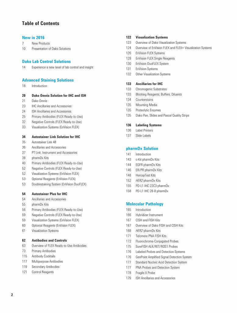
Table of Contents
New in 20167 New Products10 Presentation of Dako Solutions
Dako Lab Control Solutions14 Experience a new level of lab control and insight
Advanced Staining Solutions 18 Introduction
20 Dako Omnis Solution for IHC and ISH21 Dako Omnis 23 IHC Ancillaries and Accessories 24 ISH Ancillaries and Accessories 25 Primary Antibodies (FLEX Ready-to-Use) 32 Negative Controls (FLEX Ready-to-Use) 33 Visualization Systems (EnVision FLEX)
34 Autostainer Link Solution for IHC 35 Autostainer Link 4836 Ancillaries and Accessories 37 PT Link, Instrument and Accessories 38 pharmDx Kits 40 Primary Antibodies (FLEX Ready-to-Use) 52 Negative Controls (FLEX Ready-to-Use) 52 Visualization Systems (EnVision FLEX) 53 Optional Reagents (EnVision FLEX) 53 Doublestaining System (EnVision DuoFLEX)
54 Autostainer Plus for IHC 54 Ancillaries and Accessories 55 pharmDx Kits 56 Primary Antibodies (FLEX Ready-to-Use) 59 Negative Controls (FLEX Ready-to-Use) 59 Visualization Systems (EnVision FLEX) 60 Optional Reagents (EnVision FLEX) 61 Visualization Systems
62 Antibodies and Controls 63 Overview of FLEX Ready-to-Use Antibodies73 Primary Antibodies115 Antibody Cocktails 117 Multipurpose Antibodies 119 Secondary Antibodies 121 Control Reagents
122 Visualization Systems 123 Overview of Dako Visualization Systems124 Overview of EnVision FLEX and FLEX+ Visualization Systems126 EnVision FLEX Systems 128 EnVision FLEX Single Reagents 130 EnVision DuoFLEX System 131 EnVision Systems 132 Other Visualization Systems
133 Ancillaries for IHC 133 Chromogenic Substrates 133 Blocking Reagents, Buffers, Diluents 134 Counterstains 135 Mounting Media 135 Proteolytic Enzymes 135 Dako Pen, Slides and Pascal Quality Strips
136 Labeling Systems 136 Label Printers137 Slide Labels
pharmDx Solution141 Introduction143 c-Kit pharmDx Kits 144 EGFR pharmDx Kits 146 ER/PR pharmDx Kits 148 HercepTest Kits 152 HER2 pharmDx Kits 155 PD-L1 IHC 22C3 pharmDx 158 PD-L1 IHC 28-8 pharmDx
Molecular Pathology165 Introduction166 Hybridizer Instrument 167 CISH and FISH Kits 167 Overview of Dako FISH and CISH Kits168 HER2 pharmDx Kits 171 Telomere PNA FISH Kits 172 Fluorochrome-Conjugated Probes 175 SureFISH ALK/RET/ROS1 Probes176 Labeled Probes and Detection Systems176 GenPoint Amplified Signal Detection System 177 Standard Nucleic Acid Detection System 177 PNA Probes and Detection System 178 Fragile X Probe 179 ISH Ancillaries and Accessories
USCatPathology.book Page 2 Wednesday, March 9, 2016 10:34 PM

3
Table of Contents (continued)
H&E Solution182 Introduction183 Dako CoverStainer 184 Coverslipper for Glass Slides 184 Ancillaries and Accessories
Special Stains Solution188 Introduction189 Artisan Link Pro Special Staining System 191 Artisan Accessories 192 Artisan Link and Link Pro Special Stains 194 Artisan Special Stains
Dako Academy200 Introduction201 Training206 Literature207 e-Learning208 Events
Service and Support210 Introduction211 Deployment Services212 Instrument Services213 Application and Technical Support214 Instrument Service Agreements
General Product Information216 Monoclonal Antibodies216 Polyclonal Antibodies217 Biotinylated Antibodies217 Alkaline Phosphatase-Conjugated Antibodies218 Peroxidase-Conjugated Antibodies219 Fluorescein-Conjugated Antibodies for Tissue Staining
Indexes223 Alphabetical Index241 Synonym List244 Antibody Clone Index246 Product Code System247 Product Code Index
Distribution267 Dako Sales Offices and Distributors
Flow Cytometry and Specific ProteinsReagent Partnership Division provides Dako’s clinical diagnostic products within the area of flow cytometry and specific proteins. The Division focuses on two business areas: Retail sales of IVD-approved products within the areas of flow cytometry and specific proteins, including a
broad range of assays for turbidimetry OEM bulk sales and assay development of antibody solutions and kits with special expertise in assay
development and validation for turbidimetric platforms To acquire a product catalog for Flow Cytometry and/or Specific Proteins, please contact [email protected] or visit our homepage www.dako.com/index/products.htm.
00005 TOC_montage US.fm Page 3 Tuesday, March 8, 2016 9:10 AM

USCatPathology.book Page 4 Monday, March 7, 2016 2:21 PM

New in 2016New Products 7Presentation of Dako Solutions 10
USCatPathology.book Page 5 Monday, March 7, 2016 2:21 PM

USCatPathology.book Page 6 Monday, March 7, 2016 2:21 PM

7
New Products
pharmDx Solution
PD-L1 IHC 22C3 pharmDx (Code SK006)For in vitro diagnostic use. PD-L1 IHC 22C3 pharmDx is a qualitative immunohistochemical assay using Monoclonal Mouse Anti-PD-L1, Clone 22C3 intended for use in the detection of PD-L1 protein in formalin-fixed, paraffin-embedded (FFPE) non-small cell lung cancer (NSCLC) tissue using EnVision FLEX visualization system on Autostainer Link 48. PD-L1 protein expression is determined by using Tumor Proportion Score (TPS), which is the percentage of viable tumor cells showing partial or complete membrane staining. The specimen should be considered PD-L1 positive if TPS ≥ 50% of the viable tumor cells exhibit membrane staining at any intensity.
PD-L1 IHC 22C3 pharmDx is indicated as an aid in identifying NSCLC patients for treatment with KEYTRUDA® (pembrolizumab).
PD-L1 IHC 28-8 pharmDx (Code SK005) For in vitro diagnostic use. PD-L1 IHC 28-8 pharmDx is a qualitative immunohistochemical assay using Monoclonal Rabbit Anti-PD-L1, Clone 28-8 intended for use in the detection of PD-L1 protein in formalin-fixed paraffin-embedded (FFPE) non-squamous non-small cell lung cancer (NSCLC) and melanoma tissues using EnVision FLEX visualization system on Autostainer Link 48. PD-L1 protein expression is defined as the percentage of tumor cells exhibiting positive membrane staining at any intensity.
PD-L1 expression as detected by PD-L1 IHC 28-8 pharmDx in non-squamous NSCLC may be associated with enhanced survival from OPDIVO® (nivolumab).
Positive PD-L1 status as determined by PD-L1 IHC 28-8 pharmDx in melanoma is correlated with the magnitude of the treatment effect on progression-free survival from OPDIVO®.
USCatPathology.book Page 7 Monday, March 7, 2016 2:21 PM

8
New Products (continued)
Advanced Staining Solutions
Dako Omnis Solution for IHC and ISH
Autostainer Link Solution for IHC
FLEX Ready-to-Use Antibodies for Dako OmnisPage Code Product Package Size26 73 GA500 Rb a Hu Alpha-1-Fetoprotein, Ready-to-Use (Dako Omnis) 60 tests, 12 mL26 75 GA702 Mo a Hu Beta-Catenin, Clone β-Catenin-1, Ready-to-Use (Dako Omnis) 60 tests, 12 mL26 76 GA515 Rb a Hu Calcitonin, Ready-to-Use (Dako Omnis) 60 tests, 12 mL26 76 GA054 Mo a Hu Caldesmon, Clone h-CD, Ready-to-Use (Dako Omnis) 60 tests, 12 mL27 79 GA623 Mo a Hu CD8, Clone C8/114B, Ready-to-Use (Dako Omnis) 60 tests, 12 mL27 81 GA781 Mo a Hu CD23, Clone DAK-CD23, Ready-to-Use (Dako Omnis) 60 tests, 12 mL28 86 GA508 Rb a Hu Chorionic Gonadotropin, Ready-to-Use (Dako Omnis) 60 tests, 12 mL28 87 GA083 Rb a Hu Cyclin D1, Clone EP12, Ready-to-Use (Dako Omnis) 60 tests, 12 mL29 92 GA659 Rb a Hu ERG, Clone EP111, Ready-to-Use (Dako Omnis) 60 tests, 12 mL30 96 GA510 Rb a Hu IgA, Ready-to-Use (Dako Omnis) 60 tests, 12 mL30 100 GA074 Mo a Hu Mammaglobin, Clone 304-1A5, Ready-to-Use (Dako Omnis) 60 tests, 12 mL31 104 GA607 Mo a Hu Neurofilament Protein, Clone 2F11, Ready-to-Use (Dako Omnis) 60 tests, 12 mL31 109 GA075 Mo a Hu Renal Cell Carcinoma Marker, Clone SPM314, Ready-to-Use (Dako Omnis) 60 tests, 12 mL
PT Link Page Code Product Package Size37 PT200 PT Link Instrument 1 unit
PT Link AccessoriesPage Code Product Package Size37 PT202 Replacement Tank for PT200 1 unit37 PT203 Spare Tank Cover for PT200 1 unit
00031 IntroSection NewProducts US.fm Page 8 Friday, March 11, 2016 9:29 AM
9
New Products (continued)
Molecular Pathology
H&E Solution
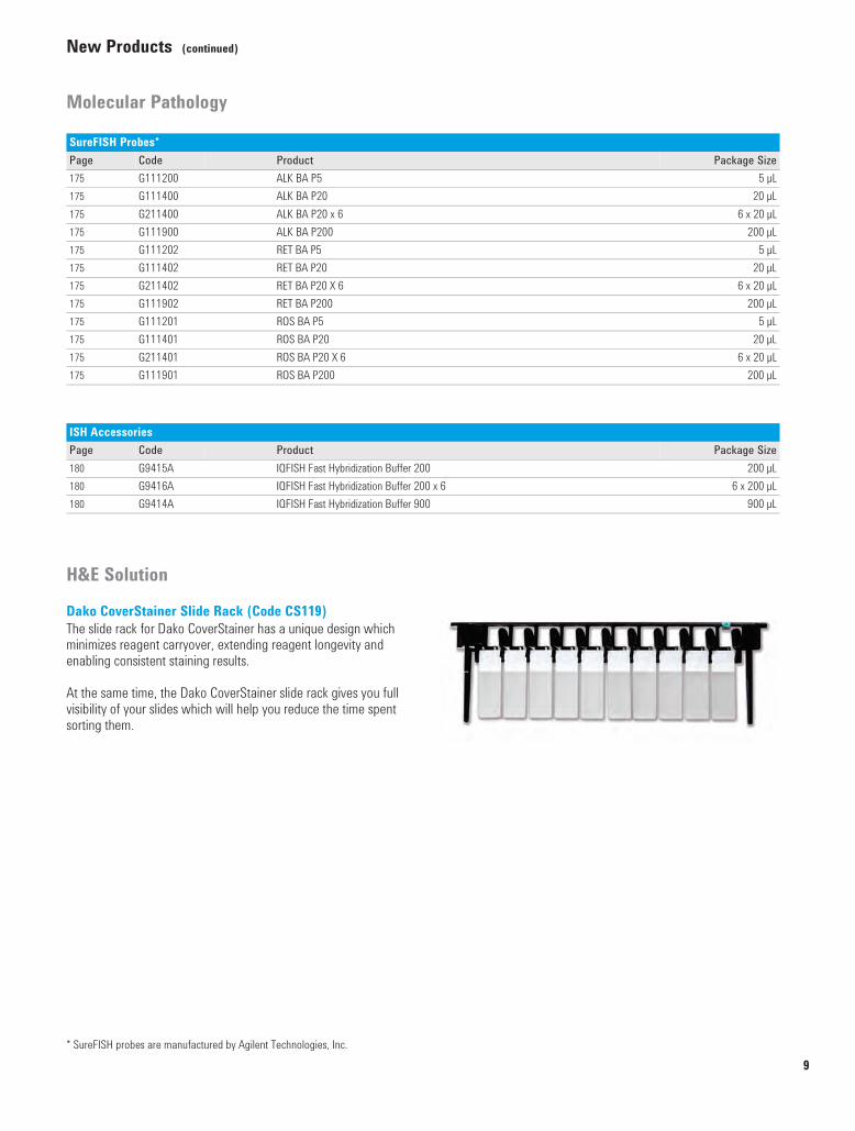
Dako CoverStainer Slide Rack (Code CS119)The slide rack for Dako CoverStainer has a unique design which minimizes reagent carryover, extending reagent longevity and enabling consistent staining results.
At the same time, the Dako CoverStainer slide rack gives you full visibility of your slides which will help you reduce the time spent sorting them.
* SureFISH probes are manufactured by Agilent Technologies, Inc.
SureFISH Probes*Page Code Product Package Size175 G111200 ALK BA P5 5 µL175 G111400 ALK BA P20 20 µL175 G211400 ALK BA P20 x 6 6 x 20 µL175 G111900 ALK BA P200 200 µL175 G111202 RET BA P5 5 µL175 G111402 RET BA P20 20 µL175 G211402 RET BA P20 X 6 6 x 20 µL175 G111902 RET BA P200 200 µL175 G111201 ROS BA P5 5 µL175 G111401 ROS BA P20 20 µL175 G211401 ROS BA P20 X 6 6 x 20 µL175 G111901 ROS BA P200 200 µL
ISH AccessoriesPage Code Product Package Size180 G9415A IQFISH Fast Hybridization Buffer 200 200 µL180 G9416A IQFISH Fast Hybridization Buffer 200 x 6 6 x 200 µL180 G9414A IQFISH Fast Hybridization Buffer 900 900 µL
USCatPathology.book Page 9 Monday, March 7, 2016 2:21 PM

Instrument connectivity
Get the full picture with Dako Solutions
DakoLink Omnis workstation
DakoLinkserver
Test request in LIS
Dako Omnis
IHC
FISH
CISH
Molecular Pathology
10
USCatPathology.book Page 10 Monday, March 7, 2016 2:21 PM

Autostainer Link
Supported by excellent service and support for your laboratory
Dako CoverStainer
Artisan Link ProDakoLink
workstation
Special Stains
IHC
pharmDx
Companion Diagnostics
H&E
11
USCatPathology.book Page 11 Monday, March 7, 2016 2:21 PM

USCatPathology.book Page 12 Monday, March 7, 2016 2:21 PM

Dako Lab Control SolutionsExperience a new level of lab control and insight 14
USCatPathology.book Page 13 Monday, March 7, 2016 2:21 PM

True Positive ID
Slide Review
Case Assembly
Accessioning
Grossing
Processing
Embedding
Sectioning
H&E and Special Stains
IHC and ISH
14
USCatPathology.book Page 14 Monday, March 7, 2016 2:21 PM

Dako Lab Control Solutions
Experience a new level of lab control and insight
The Dako Lab Control solutions consist of staining management, sample tracking and connectivity software that is both flexible and scalable to meet the needs of each individual lab. Either as separate modules or combined, DakoLink and True Positive ID enable your lab to: Minimize errors to improve patient safety Improve efficiency by reducing hands-on time Provide a full electronic audit trail to support quality and regulatory needs
The DakoLink and the DakoLink Omnis staining management software connect all Dako staining instruments and allow you to share information across functions, create customized reports based on information captured and easily manage all instruments, slides, reagents and protocols. DakoLink True Positive ID (TPID) adds sample creation and tracking capabilities, from accessioning to archiving. By registering every action for all case parts throughout all of the lab processes, TPID increases patient safety by reducing the risk of transcription errors and misplaced samples. Dako connectivity for total lab control DakoLink and TPID can integrate with your Laboratory Information System (LIS) and even connect between multiple locations, providing access from your lab to anywhere on your network. DakoLink has the ability to read LIS barcodes or create its own unique 2D barcode, ensuring every slide is uniquely identified.With flexible connectivity capabilities, unique identification, work lists and reports, TPID and DakoLink work together to give you total control of your lab. DakoLink
g
15
USCatPathology.book Page 15 Monday, March 7, 2016 2:21 PM

USCatPathology.book Page 16 Monday, March 7, 2016 2:21 PM

Advanced Staining SolutionsIntroduction 18Dako Omnis Solution for IHC and ISH 20Autostainer Link Solution for IHC 34Autostainer Plus for IHC 54Antibodies and Controls 62Antibody Cocktails 115Multipurpose Antibodies 117Secondary Antibodies 119Control Reagents 121Visualization Systems 122Ancillaries for IHC 133Labeling Systems 136
USCatPathology.book Page 17 Wednesday, March 9, 2016 10:37 PM

18
www.
dako
.com
Adva
nced
Sta
inin
g So
lutio
nsIn
trodu
ctio
nIntroduction to theAdvanced Staining Solutions
We listened. We responded.Our commitment to advancing pathology begins with something very simple – listening. By listening carefully to pathologists and lab personnel around the world, we learned that there is growing pressure to: Manage increasing slide volumes with limited personnel and financial
resources Process slides faster, to minimize time to diagnosis Cope with fluctuations in workload without sacrificing turnaround time Improve quality control of processes and secure consistency in quality Increase the traceability of patient samples to enable accreditation Find and retain well-trained, qualified staff
With almost 50 years of dialogue with our customers, We have helped drive scientific advancement and certainty in cancer diagnostics. We remain committed to delivering novel solutions and innovative technologies which support you to meet the challenges of today and tomorrow.
Dako Omnis. Developed by the lab for the lab. Developed together with pathologists, lab managers and lab technicians from around the world, with the needs of the pathology lab very much in focus. Dako Omnis builds on our reputation for delivering quality reagents and staining solutions that bring certainty to cancer diagnostics.
Dako Omnis provides: A true automated, walk-away solution High throughput and overnight capacity Same-day IHC and ISH provides complete patient case management Unparalleled onboard capacity of temperature-controlled reagents Increased productivity with limited setup and minimal maintenance
time
Dako Omnis delivers what pathologists, lab managers and technicians are asking for in terms of time, choice and better patient care.
One supplier. Two choices. With the addition of Dako Omnis, we can now deliver a unique and flexible combination of comprehensive advanced staining solutions. These solutions can be used independently or together, to help you meet the individual needs of your lab, without compromising on quality and consistency results.
Speak to your local representative to assess which solution addresses the needs of your lab now and in the future.
Dako Omnis is a generation ahead in IHC and ISH. Parallel or batch loading, the choice is yours. With a high throughput and full automation, this is a true walk-away solution. A controlled onboard environment facilitates unattended overnight processing of patient cases.
Autostainer Link 48 is a compact, bench-top, open system that delivers the flexibility required in a research and clinical environment. Adaptable to your individual setup, and helps to maximize productivity by the decoupled pre-treatment and the ability to run either large batches of up to 48 IHC slides, or mini batches.
USCatPathology.book Page 18 Monday, March 7, 2016 6:49 PM
19
IntroductionAdvanced Staining Solutions
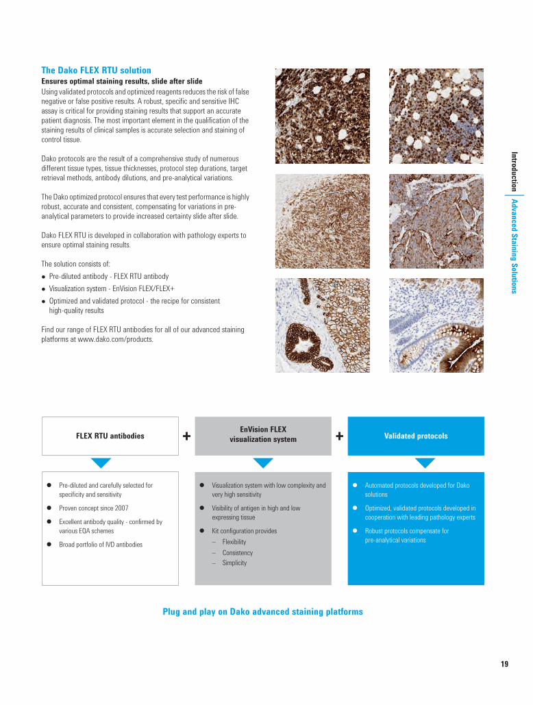
The Dako FLEX RTU solution Ensures optimal staining results, slide after slideUsing validated protocols and optimized reagents reduces the risk of false negative or false positive results. A robust, specific and sensitive IHC assay is critical for providing staining results that support an accurate patient diagnosis. The most important element in the qualification of the staining results of clinical samples is accurate selection and staining of control tissue.
Dako protocols are the result of a comprehensive study of numerous different tissue types, tissue thicknesses, protocol step durations, target retrieval methods, antibody dilutions, and pre-analytical variations.
The Dako optimized protocol ensures that every test performance is highly robust, accurate and consistent, compensating for variations in pre-analytical parameters to provide increased certainty slide after slide.
Dako FLEX RTU is developed in collaboration with pathology experts to ensure optimal staining results.
The solution consists of: Pre-diluted antibody - FLEX RTU antibody Visualization system - EnVision FLEX/FLEX+ Optimized and validated protocol - the recipe for consistent
high-quality results
Find our range of FLEX RTU antibodies for all of our advanced staining platforms at www.dako.com/products.
Plug and play on Dako advanced staining platforms
FLEX RTU antibodies
� Pre-diluted and carefully selected for specificity and sensitivity
� Proven concept since 2007
� Excellent antibody quality - confirmed by various EQA schemes
� Broad portfolio of IVD antibodies
EnVision FLEX visualization system
� Visualization system with low complexity and very high sensitivity
� Visibility of antigen in high and low expressing tissue
� Kit configuration provides – Flexibility – Consistency – Simplicity
Validated protocols
� Automated protocols developed for Dako solutions
� Optimized, validated protocols developed in cooperation with leading pathology experts
� Robust protocols compensate for pre-analytical variations
USCatPathology.book Page 19 Monday, March 7, 2016 6:49 PM

20
www.
dako
.com
Adva
nced
Sta
inin
g So
lutio
nsDa
ko O
mni
s Sol
utio
n fo
r IHC
and
ISH
Dako Omnis Solution for IHC and ISH
Dako Omnis meets the challenges of the modern pathology lab. It accommodates an increasing number of diverse, advanced staining methods in an increasingly unpredictable working day. Dako Omnis achieves this by automating any advanced staining method using a simple interface with little hands-on time. Lab staff can deliver consistent IHC and ISH results with minimal training.
Continuous sample loading allows prioritized patient cases to stream seamlessly into an ongoing workflow. With turnaround times of less than four hours for FISH slides, they are ready within the same time frame as IHC slides. Dako Omnis delivers consistent results in IHC and ISH, regardless of operator experience. The system logs operator actions and built-in controls reduce possible human errors.
Dako Omnis gives more time Process 165 IHC slides in a typical workday, including setting up
overnight runs Handle the workload with fewer instruments thanks to an unparalleled
capacity Enable faster diagnosis of whole patient cases with same-day IHC and
ISH results Minimize hands-on time with automation designed for the clinical
laboratory Free up lab techs for other tasks thanks to accurate run-time
information
Dako Omnis allows greater choice Choose continuous loading to match patient cases, or load in batches
to utilize full through-put capacity Absorb peaks in workload by processing up to 60 slides in unattended
overnight runs Eliminate operator waiting time by loading slides and/or reagents
anytime, also during runs, while keeping an optimal through-put because runs continue uninterrupted during the loading
Ensure transparency by enabling staff to monitor the slide flow from their workstations
Dako Omnis enables better patient care Get results with ease and greater certainty thanks to the FLEX Ready-
to-Use reagents and optimized protocols Increase lab quality and staffing options because Dako Omnis
minimizes the risk of human error Facilitate lab accreditation and improve patient safety by automatic
tracking and reporting Apply patient case workflow while optimizing capacity utilization
thanks to the 5-slide racks Achieve consistent staining conditions with temperature-controlled
and humidity-controlled staining chambers alongside temperature-controlled reagent positions
USCatPathology.book Page 20 Monday, March 7, 2016 6:49 PM

21
Dako Omnis Solution for IHC and ISH
Advanced Staining SolutionsDako Omnis
IHC and ISH automated on the same platform, coupled with fully optimized and validated protocols, enables a fast turnaround time of patient cases. It supports your lab to deliver consistent quality and optimal results day after day and slide after slide for increased certainty.
Dako Omnis provides: Automated IHC and ISH, from deparaffinization to counterstaining Parallel or batch processing Flexible loading, virtually zero waiting time to add slides or reagents Up to 60 slides processed simultaneously Capacity for 60 temperature-controlled reagents on board Limited setup and little maintenance High throughput, including possibility for overnight run Full traceability of patient cases through onboard and workstation
software Intuitive user interface and individual user log in LAN seats that display information where needed, including
information from the LIS Dynamic Gap staining technology that helps ensure consistent, high-
quality staining results with very low variation between slides, instruments and days.
IHC and ISH automated on the same platformDako Omnis meets head-on the challenges in the modern pathology lab to accommodate an increasing number of diverse, advanced staining methods in an increasingly unpredictable working day. Dako Omnis achieves this by automating any advanced staining method using the simplest of user interfaces with little hands-on time, so new users can start producing results in minutes.
Compensate for increasing fluctuations in workflowProcess 60 IHC slides completely unattended overnight (or 45 IHC plus 15 ISH). You decide if the slides should be ready as soon as possible or at the start of the next working day.
Batch or continuous flow, the choice is yours. Continuous sample loading allows prioritized patient cases to stream seamlessly into an ongoing workflow. Full flexibility for the unpredictable lab environment.
Monitor and control your staining workflowMonitor the progress of your run at a glance. Clear visual alerts notify you when user interactions are necessary. Dako Omnis connects to your LIS system. Share, monitor and track slides wherever you are.
Manage increasing slide volumes with limited resourcesLoad your IHC or ISH slides when convenient and the system informs you which reagents are needed. Once slides are loaded, you are free to perform other tasks. Just load and walk away. With minimal hands-on time, daily setup takes just 15 minutes and little daily maintenance is required. Less time for preparation, faster processing.
Dako OmnisI GI100 Advanced staining system 1 unit
USCatPathology.book Page 21 Monday, March 7, 2016 6:49 PM

22
Dako Omnis (continued)
www.dako.com
Adva
nced
Sta
inin
g So
lutio
nsDa
ko O
mni
s Sol
utio
n fo
r IHC
and
ISH
Hardware Specifications
FeaturesProcesses Fully automated and simultaneous IHC and/or ISH Deparaffinization, staining and counterstaining with parallel
processing
Operation Continuous or batch workflow 5-slot racks to optimize capacity utilization and patient-case
management Reagents and slides can be loaded anytime, also during runs Easy-to-use software interface and ready-to-use reagents Built-in controls to reduce possible human errors
Staining conditions Temperature controlled onboard reagent storage Dynamic Gap staining technology Temperature and humidity controlled processing environment FLEX RTU reagents and protocols for optimal staining results
Connectivity and control LAN seats for setup and monitoring from anywhere Full integration with Laboratory Information Systems 1D and 2D barcodes Data logging, reporting and access rights for traceability and
accreditation
Learn more about Dako Omnis by visiting www.dako.com/omnis
Turn-around time IHC: 2 hours 30 minutesISH: 3 hours 40 minutes
Throughput 165 slides can be loaded in a typical workday (including preparation for an overnight run)Slide capacity 60 slides for IHC or ISH (up to 15 ISH slides)Reagent capacity 60 reagent vialsBulk fluid capacity 8 x 3.5 L bottle and 4 x 7 L bottleWaste capacity 4 x 7 L bottle (non-hazardous)
1 x 7 L bottle (low-hazardous, below limit values)Dimensions 57.1" W x 31.2" D x 69.5" H (145 cm W x 79.3 cm D x 176.5 cm H) Weight 1,323 lbs (600 kg) fully loadedVoltage 120/220-240 VACPower consumption 1200 W
00110 IHC Solutions US_1.fm Page 22 Wednesday, March 16, 2016 8:05 PM

23
Dako Omnis Solution for IHC and ISH
Advanced Staining SolutionsIHC Ancillaries and Accessories (Dako Omnis)
ClearifyTM
GC810 3.8 LClearifyTM is used onboard Dako Omnis to remove paraffin from tissue sections for both IHC and ISH staining in a two-phase dewaxing procedure.
DAB+ Substrate Chromogen System (Dako Omnis)I GV825 Onboard mixing 150 testsEnVision FLEX DAB+ Substrate Chromogen System (Dako Omnis) is intended for use in immunohistochemistry together with Dako Omnis. The working solution is prepared onboard by the Dako Omnis instrument. It is a high sensitivity DAB system suitable for use in combination with the EnVision FLEX visualization system (Codes GV800/GV823). Upon oxidation, DAB forms a brown end-product at the site of the target antigen. The reagent is intended for use on formalin-fixed, paraffin-embedded tissue sections.
Hematoxylin (Dako Omnis)I GC808 Ready-to-use 8 x 22.5 mL, 600 testsIntended for use in immunohistochemistry together with Dako Omnis. The reagent is recommended for counterstaining on formalin-fixed, paraffin-embedded tissue sections providing a clear blue, nuclear staining.
IHC Microscope Slides, FLEXI K8020 Coated glass slides 5 x 100 slidesCoated microscope slides for adhesion of formalin-fixed, paraffin-embedded tissue sections for use in immunohistochemistry with Dako EnVision FLEX visualization systems. FLEX IHC Microscope Slides are compatible with, but not limited to, the following Dako instruments: Dako Omnis, Autostainer Link, Dako Autostainer/Autostainer Plus and PT Link.
Mixing Strip, for Dako OmnisGC107 10-well mixing strip 25 strips
Dako Omnis Mixing Strip is intended for mixing of the chromogen working solution during staining onboard Dako Omnis. Dako Omnis Mixing Strip has ten wells designed to hold chromogen for five slides with minimal dead volume. Wells are covered with a lid to limit spill of reagent during disposal of strips. Dako Omnis Mixing Strip can stand unsupported on a table. Arrows indicate correct insertion on Dako Omnis. Dako Omnis Mixing Strip is single use only and used strips are classified as hazardous waste due to chromogen residuals.
Reagent Vial, Small/Large, for Dako OmnisGC201 Small vial 25 x 2 mLGC202 Large vial 25 x 30 mL
Reagent vials designed to allow the use of a user-defined reagent on Dako Omnis. Each single-use bottle is labeled with positive identification technology. User-fillable reagent vial may be filled to a maximum fill volume of approximately 2 mL/30 mL, respectively. The vial closure contains a septum to reduce evaporation of reagent during onboard use in Dako Omnis reagent storage.
Slide Rack, for Dako OmnisGC101 Slide racks holding 5 slides each 6 racks
Dako Omnis Slide Rack is designed for use on Dako Omnis. The Slide Rack holds the slides with samples to be processed on Dako Omnis.Each Slide Rack can carry up to five slides. Each slide is placed in a positioning groove and fixated by a spring. Dako Omnis is validated with FLEX IHC Microscope Slides and Superfrost Plus Slides. Dako does not recommend the use of other slide types. Dako Omnis Slide Rack is classified as non-hazardous waste, and Slide Rack parts comply with incineration or parts may be dismantled for recycling.
Slide Rack Color Clips, for Dako OmnisGC104 Blue 25 clipsGC105 Green 25 clipsGC106 Gray 25 clipsGC103 Red 25 clips
The colored clips are attached to the slide rack for visual identification of individual racks. Each Dako Omnis Slide Rack can hold two Dako Omnis Slide Rack Color Clips and the colors available are: blue, green, gray and red. Dako Omnis Slide Racks are supplied with black color clips as default.
Sulfuric Acid, 0.3 M, for Dako OmnisGC203 10 x 22.5 mL
Sulfuric Acid, 0.3 M is a generic cleaning agent used to remove residue (primarily protein) from various surfaces. It is used on Dako Omnis to automatically clean the Liquid Handling Tip after pipetting of reagents with high protein content, specifically primary antibodies.
Wash Buffer (20x) (Dako Omnis)I GC807 Concentrate 20 x 175 mL, 1700 testsWash Buffer 20x (Dako Omnis) is intended for use in immunohistochemistry. The product is used as wash buffer for immunohistochemical staining procedures onboard Dako Omnis.
USCatPathology.book Page 23 Monday, March 7, 2016 6:49 PM

24
www.
dako
.com
Adva
nced
Sta
inin
g So
lutio
nsDa
ko O
mni
s Sol
utio
n fo
r IHC
and
ISH
ISH Ancillaries and Accessories (Dako Omnis)
Fluorescence Mounting Medium (Dako Omnis)I GM304 Ready-to-use 20 tests, 0.8 mLFluorescence Mounting Medium (Dako Omnis) is intended for mounting of formalin-fixed, paraffin-embedded (FFPE) tissue sections after FISH staining performed onboard the Dako Omnis instrument. The mounting medium also contains 500 µg/L DAPI for improved nuclei staining.
ISH Cleaning Solution (Dako Omnis) I GC207 Ready-to-use 100 tests, 10 mLISH Cleaning Solution (Dako Omnis) is an accessory to the Dako Omnis instrument. It is used for cleaning the pipette tip between dispenses of in situ hybridization probes. Washing with ISH Cleaning Solution dissolves ISH probe, allowing remaining probe to be effectively washed away with water. The product is provided in a ready-to-use vial for the Dako Omnis instrument.
ISH Ethanol Solution, 96% (Dako Omnis)I GM300 Ready-to-use 20 tests, 14 mLISH Ethanol Solution, 96% (Dako Omnis) is intended for use in automated in situ hybridization assays together with the Dako Omnis instrument on formalin-fixed, paraffin-embedded (FFPE) tissue sections. The solution is used in the wash step after target retrieval. The product is provided in a ready-to-use vial for the Dako Omnis instrument.
ISH Lid, for Dako OmnisGC102 5 lids
Dako Omnis ISH Lid is intended for use in FISH procedures. Each Dako Omnis ISH Lid holds five slides and has five built-in Cover Glasses and one Humidity Pad. The Cover Glasses serve to distribute probe buffer across the staining area and to reduce buffer evaporation. The Humidity Pad with deionized water added serves to increase the humidity inside Dako Omnis ISH Lid to further reduce evaporation. Dako Omnis ISH Lid also provides insulation to maintain proper denaturation temperature.Dako Omnis ISH Lid is single use only and is classified as non-hazardous waste.
ISH Pepsin (Dako Omnis)I GM302 Ready-to-use 20 tests, 7 mLISH Pepsin (Dako Omnis) is intended for use in automated in situ hybridization assays together with the Dako Omnis instrument on formalin-fixed, paraffin-embedded (FFPE) tissue sections. The solution is used in the digestion step. The product is provided in a ready-to-use vial for the Dako Omnis instrument.
ISH Pre-Treatment Solution (20x) (Dako Omnis)I GM301 Concentrate 175 mL, 20x concentratedISH Pre-Treatment Solution (20x) (Dako Omnis) is intended for use in automated in situ hybridization assays together with the Dako Omnis instrument on formalin-fixed, paraffin-embedded (FFPE) tissue sections. The solution is used in the pre-treatment step. An inert green color is added to the buffer for easy identification and user friendliness. The volume is tailored for dilution in one Dako Omnis bulk bottle.
ISH Stringent Wash Buffer (20x) (Dako Omnis)I GM303 Concentrate 175 mL, 20x concentratedISH Stringent Wash Buffer (20x) (Dako Omnis) is intended for use in automated in situ hybridization assays together with the Dako Omnis instrument on formalin-fixed, paraffin-embedded (FFPE) tissue sections. The solution is used in the post-hybridization step. An inert yellow color is added to the buffer for easy identification and user friendliness. The volume is tailored for dilution in one Dako Omnis bulk bottle.
Mixing Device, for Dako OmnisGC116 1 unit
Dako Omnis Mixing Device is an accessory to the Dako Omnis instrument. It is designed specifically to support the fluorescence in situ hybridization (FISH) and the chromogenic in situ hybridization (CISH) procedures. The Dako IQISH buffer is extremely viscous, and during storage the reagent phase separates. Hence the Dako IQISH reagents require a particular preparatory processing to thaw and unify the content.Some Dako ISH reagents are therefore provided in dedicated ISH reagent vials containing a mixing ball, and the Dako Omnis Mixing Device is designed to fit together with these ISH reagent vials.Dako Omnis Mixing Device contains a magnet that enables the mixing ball to move up and down (110 cycles) inside the vial after 40 minutes thawing of the ISH reagent; thus ensuring a homogenous probe mix prior to application on the Dako Omnis instrument.
Vial with Mixing Ball, 2 mL, for Dako OmnisGC206 25 vials 2 mL
Dako Omnis Vial with Mixing Ball, 2 mL has been designed as an accessory for Dako Omnis and Dako Omnis Mixing Device and is intended for use in ISH procedures using user-provided FISH probes diluted in ethylene carbonate-based hybridization buffer (IQFISH). Dako Omnis Vial with Mixing Ball, 2 mL includes a mixing ball that is used by Dako Omnis Mixing Device to mix the IQFISH hybridization buffer with the user-provided probe. Each package contains 25 vials, 25 caps and 25 mixing balls.
USCatPathology.book Page 24 Monday, March 7, 2016 6:49 PM

25
Dako Omnis Solution for IHC and ISH
Advanced Staining SolutionsPrimary Antibodies (FLEX Ready-to-Use) (Dako Omnis)
For Dako Omnis, we offer a dedicated series of high-quality, pre-diluted, ready-to-use (RTU) primary antibodies.
FLEX Ready-to-Use antibodies are pre-diluted primary antibodies specifically developed for automated use while maintaining the high-quality staining performance for which Dako antibodies is known.
Each FLEX RTU antibody is accompanied by a validated protocol that is optimized to absorb variations related to pre-analytical factors. This enables a reliable staining performance in various tissue types containing both high and low-expression structures. The antibody specificity and protocol have both been evaluated and approved by external pathology experts.
Key Features Optimized staining performance of both high and low-expression
structures Dako Omnis and the dynamic gap staining technology provide
consistent and uniform staining with excellent morphology Crisp and clear staining with no background Optimal laboratory efficiency with RTU antibodies on Dako Omnis
The GA-Series FLEX Ready-to-Use Primary Antibodies listed in this section are packaged in Dako Omnis vials for use on Dako Omnis instruments, and can be used only with the EnVision FLEX system for Dako Omnis.
Dako Omnis
Antibodieswith high
specificity andsensitivity
Excellentstainingresults
OptimizedFLEX
protocols
USCatPathology.book Page 25 Monday, March 7, 2016 6:49 PM

26
Primary Antibodies (FLEX Ready-to-Use) (Dako Omnis) (continued)
www.
dako
.com
Adva
nced
Sta
inin
g So
lutio
nsDa
ko O
mni
s Sol
utio
n fo
r IHC
and
ISH
Polyclonal Rabbit Anti-HumanAlpha-1-FetoproteinI GA500 NEW 60 tests, 12 mL
Monoclonal Rabbit Anti-HumanAMACRI GA060 Clone 13H4 60 tests, 12 mL
Monoclonal Mouse Anti-HumanB-Cell-Specific Activator ProteinI GA650 Clone DAK-Pax5 60 tests, 12 mL
Monoclonal Mouse Anti-HumanBCL6 ProteinI GA625 Clone PG-B6p 60 tests, 12 mL
Monoclonal Mouse Anti-HumanBeta-CateninI GA702 Clone b-Catenin-1 NEW 60 tests, 12 mL
Monoclonal Mouse Anti-HumanCA 125I GA701 Clone M11 60 tests, 12 mL
Polyclonal Rabbit Anti-HumanCalcitoninI GA515 NEW 60 tests, 12 mL
Monoclonal Mouse Anti-HumanCaldesmonI GA054 Clone h-CD NEW 60 tests, 12 mL
Monoclonal Mouse Anti-HumanCarcinoembryonic Antigen (CEA)I GA622 Clone II-7 60 tests, 12 mL
Polyclonal Rabbit Anti-HumanCarcinoembryonic Antigen (CEA)I GA526 60 tests, 12 mL
A1AT-deficient liver (FFPE) stained with FLEX Anti-Alpha-1-Antitrypsin, Code GA505.
Prostate adenocarcinoma (FFPE) stained with FLEX Anti-AMACR, Code GA060.
Lymph node (FFPE) stained with FLEX Anti-BSAP, Code GA650.
Follicular lymphoma (FFPE) stained with FLEX Anti-BCL6, Code GA625.
Colon adenocarcinoma (FFPE) stained with FLEX Anti-Beta-Catenin, Code GA702.
Ovarian carcinoma (FFPE) stained with FLEX Anti-CA 125, Code GA701.
Thyroid medullary carcinoma (FFPE) stained with FLEX Anti-Calcitonin, Code GA515.
Leiomyosarcoma (FFPE) stained with FLEX Anti-Caldesmon, Code GA054.
Colon adenocarcinoma (FFPE) stained with FLEX Anti-CEA, Code GA622.
USCatPathology.book Page 26 Monday, March 7, 2016 6:49 PM

Primary Antibodies (FLEX Ready-to-Use) (Dako Omnis) (continued)
27
Dako Omnis Solution for IHC and ISH
Advanced Staining Solutions
Monoclonal Mouse Anti-HumanCD2I GA651 Clone AB75 60 tests, 12 mL
Polyclonal Rabbit Anti-HumanCD3I GA503 60 tests, 12 mL
Monoclonal Mouse Anti-HumanCD7I GA643 Clone CBC.37 60 tests, 12 mL
Monoclonal Mouse Anti-HumanCD8I GA623 Clone C8/144B NEW 60 tests, 12 mL
Monoclonal Mouse Anti-HumanCD10I GA648 Clone 56C6 60 tests, 12 mL
Monoclonal Mouse Anti-HumanCD15I GA062 Clone Carb-3 60 tests, 12 mL
Monoclonal Mouse Anti-HumanCD20cyI GA604 Clone L26 60 tests, 12 mL
Monoclonal Mouse Anti-HumanCD23I GA781 Clone DAK-CD23 NEW 60 tests, 12 mL
Monoclonal Mouse Anti-HumanCD31, Endothelial CellI GA610 Clone JC70A 60 tests, 12 mL
Monoclonal Mouse Anti-HumanCD34 Class III GA632 Clone QBEnd 10 60 tests, 12 mL
Precursor T-lymphoblastic lymphoma (FFPE) stained with FLEX Anti-CD2, Code GA651.
T-cell lymphoma (FFPE) stained with FLEX Anti-CD3, Code GA503.
Lymphoma (FFPE) stained with FLEX Anti-CD7 Code GA643.
Angioimmunoblastic T-cell lymphoma (FFPE) stained with FLEX Anti-CD8, Code GA623.
Lymphoma (FFPE) stained with FLEX Anti-CD10, Code GA648.
Hodgkin's Lymphoma (FFPE) stained with FLEX Anti-CD15, Code GA062.
B-cell chronic lymphocytic leukemia/small lymphocytic lymphoma (FFPE) stained with FLEX Anti-CD20cy, Code GA604.
Chronic lymphocytic leukemia/small lymphocytic lymphoma (FFPE) stained with FLEX Anti-CD23, Code GA781.
Angiosarcoma (FFPE) stained with FLEX Anti-CD31, Code GA610.
Angiosarcoma (FFPE) stained with FLEX Anti-CD34, Code GA632.
USCatPathology.book Page 27 Monday, March 7, 2016 6:49 PM

28
Primary Antibodies (FLEX Ready-to-Use) (Dako Omnis) (continued)
www.
dako
.com
Adva
nced
Sta
inin
g So
lutio
nsDa
ko O
mni
s Sol
utio
n fo
r IHC
and
ISH
Monoclonal Mouse Anti-HumanCD43I GA636 Clone DF-T1 60 tests, 12 mL
Monoclonal Mouse Anti-HumanCD45, Leucocyte Common AntigenI GA751 Clones 2B11 + PD7/26 60 tests, 12 mL
Monoclonal Mouse Anti-HumanCD68I GA609 Clone KP1 60 tests, 12 mL
Monoclonal Mouse Anti-HumanCD68I GA613 Clone PG-M1 60 tests, 12 mL
Monoclonal Mouse Anti-HumanCD79aI GA621 Clone JCB117 60 tests, 12 mL
Monoclonal Mouse Anti-HumanCD138I GA642 Clone MI15 60 tests, 12 mL
Monoclonal Mouse Anti-HumanCD246, ALK ProteinI GA641 Clone ALK1 60 tests, 12 mL
Monoclonal Mouse Anti-HumanCDX2I GA080 Clone DAK-CDX2 60 tests, 12 mL
Polyclonal Rabbit Anti-HumanChorionic Gonadotropin (hCG)I GA508 NEW 60 tests, 12 mL
Monoclonal Rabbit Anti-HumanCyclin D1I GA083 Clone EP12 NEW 60 tests, 12 mL
Tonsil (FFPE) stained with FLEX Anti-CD43, Code GA636.
Tonsil (FFPE) stained with FLEX Anti-CD45, Code GA751.
Tonsil (FFPE) stained with FLEX Anti-CD68, Code GA609.
Tonsil (FFPE) stained with FLEX Anti-CD68, Code GA613.
Plasmacytoma (FFPE) stained with FLEX Anti-CD79a, Code GA621.
High grade myeloma (FFPE) stained with FLEX Anti-CD138, Code GA642.
Anaplastic large cell lymphoma (FFPE) stained with FLEX Anti-CD246, ALK Protein, Code GA641.
Colon adenocarcinoma (FFPE) stained with FLEX Anti-CDX2, Code GA080.
Placenta (FFPE) stained with FLEX Anti-Human Chorionic Gonadotropin, Code GA508.
Mantle cell lymphoma (FFPE) stained with FLEX Anti-Cyclin D1, Code GA083.
USCatPathology.book Page 28 Monday, March 7, 2016 6:49 PM

Primary Antibodies (FLEX Ready-to-Use) (Dako Omnis) (continued)
29
Dako Omnis Solution for IHC and ISH
Advanced Staining Solutions
Monoclonal Mouse Anti-HumanCytokeratinI GA053 Clone AE1/AE3 60 tests, 12 mL
Monoclonal Mouse Anti-HumanCytokeratin 5/6I GA780 Clone D5/16 B4 60 tests, 12 mL
Monoclonal Mouse Anti-HumanCytokeratin 7I GA619 Clone OV-TL 12/30 60 tests, 12 mL
Monoclonal Mouse Anti-HumanCytokeratin 18I GA618 Clone DC 10 60 tests, 12 mL
Monoclonal Mouse Anti-HumanCytokeratin 19I GA615 Clone RCK108 60 tests, 12 mL
Monoclonal Mouse Anti-HumanCytokeratin 20I GA777 Clone Ks20.8 60 tests, 12 mL
Monoclonal Mouse Anti-HumanCytokeratin, High Molecular WeightI GA051 Clone 34bE12 60 tests, 12 mL
Monoclonal Mouse Anti-HumanE-CadherinI GA059 Clone NCH-38 60 tests, 12 mL
Monoclonal Mouse Anti-HumanEpithelial AntigenI GA637 Clone Ber-EP4 60 tests, 12 mL
Monoclonal Rabbit Anti-HumanERG (Ets-Related Gene)I GA659 Clone EP111 NEW 60 tests, 12 mL
Adenocarcinoma (FFPE) stained with FLEX Anti-Cytokeratin, Code GA053.
Ductal carcinoma (FFPE) stained with FLEX Anti-Cytokeratin 7, Code GA619.
Renal clear cell carcinoma stained with FLEX Anti-Cytokeratin 18, Code GA618.
Thyroid papillary carcinoma (FFPE) stained with FLEX Anti-Cytokeratin 19, Code GA615.
Merkel cell carcinoma (FFPE) stained with FLEX Anti-Cytokeratin 20, Code GA777.
Prostate (FFPE) stained with FLEX Anti-Cytokeratin HMW, Code GA051.
Poorly differentiated ductal carcinoma (FFPE) stained with FLEX Anti-E-Cadherin, Code GA059.
Adenocarcinoma (FFPE) stained with FLEX Anti-Epithelial Antigen, Code GA637.
Prostate carcinoma (FFPE) stained with FLEX Anti-ERG, Code GA659.
USCatPathology.book Page 29 Monday, March 7, 2016 6:49 PM

30
Primary Antibodies (FLEX Ready-to-Use) (Dako Omnis) (continued)
www.dako.com
Adva
nced
Sta
inin
g So
lutio
nsDa
ko O
mni
s Sol
utio
n fo
r IHC
and
ISH
Polyclonal Rabbit Anti-HumanGastrinI GA519 60 tests, 12 mL
Polyclonal Rabbit Anti-Glial Fibrillary Acidic ProteinI GA524 60 tests, 12 mL
Monoclonal Mouse Anti-HumanGross Cystic Disease Fluid Protein-15I GA077 Clone 23A3 60 tests, 12 mL
Monoclonal Mouse Anti-HumanHepatocyteI GA624 Clone OCH1E5 60 tests, 12 mL
Polyclonal Rabbit Anti-HumanIgAI GA510 NEW 60 tests, 12 mL
Polyclonal Rabbit Anti-HumanKappa Light ChainsI GA506 60 tests, 12 mL
Monoclonal Mouse Anti-HumanKi-67 AntigenI GA626 Clone MIB-1 60 tests, 12 mL
Polyclonal Rabbit Anti-HumanLambda Light ChainsI GA507 60 tests, 12 mL
Monoclonal Mouse Anti-HumanMammaglobinI GA074 Clone 304-1A5 NEW 60 tests, 12 mL
Gastrin-producing tumor (FFPE) stained with FLEX Anti-Gastrin, Code GA519.
Glioblastoma (FFPE) stained with FLEX Anti-GFAP, Code GA524.
Breast hyperplasia (FFPE) stained with FLEX Anti-GCDFP-15, Code GA077.
Hepatocellular carcinoma (FFPE) stained with FLEX Anti-Hepatocyte, Code GA624.
Multiple myeloma (FFPE, bone marrow) stained with FLEX Anti-IgA, Code GA510.
Tonsil (FFPE) stained with FLEX Anti-Kappa light chains, Code GA506.
High grade lymphoma (FFPE) stained with FLEX Anti-Ki-67, Code GA626.
Tonsil (FFPE) stained with FLEX Anti-Lambda Light Chains, Code GA507.
Invasive ductal carcinoma (FFPE) stained with FLEX Anti-Mammaglobin, Code GA074.
00110 IHC Solutions US_1.fm Page 30 Friday, March 11, 2016 9:35 AM

Primary Antibodies (FLEX Ready-to-Use) (Dako Omnis) (continued)
31
Dako Omnis Solution for IHC and ISH
Advanced Staining Solutions
Monoclonal Mouse Anti-HumanMelanosomeI GA052 Clone HMB-45 60 tests, 12 mL
Monoclonal Mouse Anti-HumanMUM1 ProteinI GA644 Clone MUM1p 60 tests, 12 mL
Polyclonal Rabbit Anti-HumanMyeloperoxidaseI GA511 60 tests, 12 mL
Monoclonal Mouse Anti-HumanNeurofilament ProteinI GA607 Clone 2F11 NEW 60 tests, 12 mL
Monoclonal Mouse Anti-HumanNucleophosminI GA652 Clone 376 60 tests, 12 mL
Monoclonal Mouse Anti-Humanp53 ProteinI GA616 Clone DO-7 60 tests, 12 mL
Polyclonal Rabbit Anti-HumanProstate-Specific Antigen (PSA)I GA514 60 tests, 12 mL
Monoclonal Mouse Anti-HumanRenal Cell Carcinoma MarkerI GA075 Clone SPM314 NEW 60 tests, 12 mL
Polyclonal Rabbit Anti-S100I GA504 60 tests, 12 mL
Polyclonal Rabbit Anti-HumanThyroglobulinI GA509 60 tests, 12 mL
Melanoma (FFPE) stained with FLEX Anti-Melanosome, Code GA052.
Diffuse large B-cell lymphoma (FFPE) stained with FLEX Anti-MUM1, Code GA644.
Acute myeloid leukemia (FFPE) stained with FLEX Anti-Myeloperoxidase, Code GA511.
Merkel cell tumor (FFPE) stained with FLEX Anti-Neurofilament Protein, Code GA607.
Acute myeloid leukemia (AML) (FFPE) stained with FLEX Anti-Nucleophosmin, Code GA652.
Invasive transitional cell carcinoma (FFPE) stained with FLEX Anti-p53, Code GA616.
Prostate adenocarcinoma (FFPE) stained with FLEX Anti-Prostate Specific Antigen, Code GA514.
Renal clear cell carcinoma (FFPE) stained with FLEX Anti-Renal Cell Carcinoma Marker, Code GA075.
Breast carcinoma (FFPE) stained with FLEX Anti-S100, Code GA504.
Thyroid follicular carcinoma (FFPE) stained with FLEX Anti-Thyroglobulin, Code GA509.
USCatPathology.book Page 31 Monday, March 7, 2016 6:49 PM
32
Primary Antibodies (FLEX Ready-to-Use) (Dako Omnis) (continued)
www.
dako
.com
Adva
nced
Sta
inin
g So
lutio
nsDa
ko O
mni
s Sol
utio
n fo
r IHC
and
ISH
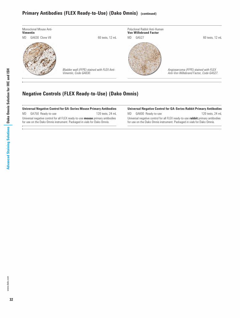
Monoclonal Mouse Anti-VimentinI GA630 Clone V9 60 tests, 12 mL
Polyclonal Rabbit Anti-HumanVon Willebrand FactorI GA527 60 tests, 12 mL
Negative Controls (FLEX Ready-to-Use) (Dako Omnis)
Universal Negative Control for GA-Series Mouse Primary AntibodiesI GA750 Ready-to-use 120 tests, 24 mLUniversal negative control for all FLEX ready-to-use mouse primary antibodies for use on the Dako Omnis instrument. Packaged in vials for Dako Omnis.
Universal Negative Control for GA-Series Rabbit Primary AntibodiesI GA600 Ready-to-use 120 tests, 24 mLUniversal negative control for all FLEX ready-to-use rabbit primary antibodies for use on the Dako Omnis instrument. Packaged in vials for Dako Omnis.
Bladder wall (FFPE) stained with FLEX Anti-Vimentin, Code GA630.
Angiosarcoma (FFPE) stained with FLEX Anti-Von Willebrand Factor, Code GA527.
USCatPathology.book Page 32 Monday, March 7, 2016 6:49 PM

33
Dako Omnis Solution for IHC and ISH
Advanced Staining SolutionsVisualization Systems (EnVision FLEX) (Dako Omnis)
EnVision FLEX Visualization Systems for Dako OmnisEnVision FLEX, the well-known Dako visualization system, has been configured into a dedicated system for Dako Omnis. The highly sensitive polymer-based EnVision FLEX system builds upon simple intelligent chemistry that allows for distinct clear staining. The Dynamic Gap staining technology utilized onboard Dako Omnis, the high-quality primary antibodies and the EnVision FLEX system all come together to provide a robust system that produces stains with excellent morphology and diagnostic certainty.
The streamlined kits and optional reagents for Dako Omnis are packaged for your convenience and are easy to order, making the system flexible, versatile and functional.
EnVision FLEX, High pH (Dako Omnis)I GV800 HRP. Rabbit/Mouse. High pH 600 testsEnVision FLEX, High pH is a high-sensitivity visualization system intended for use in immunohistochemistry together with Dako Omnis. The dual link system detects primary mouse and rabbit antibodies and the reaction is visualized by DAB+ Chromogen. The convenience kit includes Peroxidase-Blocking Reagent, EnVision/HRP, DAB+ Chromogen, Substrate Buffer and Target Retrieval Solution, High pH (50x Tris/EDTA buffer, pH 9). EnVision FLEX convenience kits are compatible with all optional EnVision FLEX and FLEX+ reagents for Dako Omnis.
EnVision FLEX Mini Kit, High pH (Dako Omnis)I GV823 HRP. Rabbit/Mouse. High pH 150 testsEnVision FLEX Mini Kit, High pH is a high-sensitivity visualization system intended for use in immunohistochemistry together with Dako Omnis. The dual link system detects primary mouse and rabbit antibodies and the reaction is visualized by DAB+ Chromogen. The convenience kit includes Peroxidase-Blocking Reagent, EnVision/HRP, DAB+ Chromogen, Substrate Buffer and Target Retrieval Solution, High pH (50x Tris/EDTA buffer, pH 9). EnVision FLEX convenience kits are compatible with all optional EnVision FLEX and FLEX+ reagents for Dako Omnis.
Mouse LINKER (Dako Omnis)I GV821 Ready-to-use 75 tests, 22.5 mLEnVision FLEX+ Mouse LINKER is an optional EnVision FLEX+ reagent and may be used with EnVision FLEX convenience kits (GV800 and GV823) for Dako Omnis to amplify the signal of primary mouse antibodies.
Rabbit LINKER (Dako Omnis)I GV809 Ready-to-use 75 tests, 22.5 mLEnVision FLEX+ Rabbit LINKER is an optional EnVision FLEX+ reagent and may be used with EnVision FLEX convenience kits (GV800 and GV823) for Dako Omnis to amplify the signal of primary rabbit antibodies.
Target Retrieval Solution, High pH (Dako Omnis)I GV804 Concentrate 3 x 68 mL, 225 testsEnVision FLEX Target Retrieval Solution, High pH (Dako Omnis) is an optional EnVision FLEX reagent containing 50x concentrated Tris/EDTA, pH 9 and is compatible with EnVision FLEX convenience kits for Dako Omnis. The volume is optimized for dilution in Dako Omnis bulk bottles.
Target Retrieval Solution, Low pH (Dako Omnis)I GV805 Concentrate 3 x 68 mL, 225 testsEnVision FLEX Target Retrieval Solution, Low pH (Dako Omnis) is an optional EnVision FLEX reagent containing 50x concentrated citrate buffer, pH 6.1 and is compatible with EnVision FLEX convenience kits for Dako Omnis. The volume is optimized for dilution in Dako Omnis bulk bottles.
EnVision FLEX SystemsFLEX
High pHFLEX
Low pHFLEX+
High pHFLEX+
Low pHCode
Code
GV800or
GV823
GV800 + GV805or
GV823 + GV805
GV800 + GV821 (Mouse LINKER)or
GV800 + GV809 (Rabbit LINKER)
GV800 + GV805 + GV821 (Mouse LINKER)or
GV800 + GV805 + GV809 (Rabbit LINKER)
00110 IHC Solutions US_1.fm Page 33 Monday, March 7, 2016 7:46 PM

34
www.
dako
.com
Adva
nced
Sta
inin
g So
lutio
nsAu
tost
aine
r Lin
k So
lutio
n fo
r IHC
Autostainer Link Solution for IHC
Automated Link Platforms is the line of instruments with whichpathology laboratories will experience an outstanding level of integration that provides high productivity and efficient workflow.
The Autostainer Link 48 staining instrument with the latest release of DakoLink software enables improved productivity in a pathology laboratory by staining 48 slides in less than three hours. When processing slides in parallel, using only one Autostainer Link 48 and one PT Link pre-treatment module, up to 144 slides can be processed in a regular working day, including setting up an overnight run.
With PT Link, pathology laboratories can further maximize productivity by reducing the number of operations needed in the specimen preparation processes of deparaffinization, rehydration and target retrieval. The fact that pre-treatment and staining are decoupled gives high flexibilitly and productivity.
The revolutionary DakoLink software and connectivity options will improve workflow and productivity even further by, among other things, completely eliminating re-labeling steps and repetitive test request entries.
Autostainer Link 48 Process 48 slides in less than three hours Organize your working day to the minute with precise run-time
estimation Achieve high quality, when staining slides with FLEX RTU primary
antibodies and EnVision FLEX/FLEX+ visualization optimized for Autostainer Link 48
PT Link Maximize productivity by processing slides in parallel Run deparaffinization, target retrieval and dehydration in one step with
the 3-in-1 buffer Have confidence in your pre-treatment process, as it is controlled every
second Possibility to track via DakoLink software
DakoLink Software Enables a fully integrated pathology solution with Dako
instrumentation for Advanced Staining and Histostaining Significant tracking improvements by implementing slide pre-
treatment Full laboratory connectivity by controlling all slides and slide IDs from
one workstation Reporting made easy Improved laboratory efficiency
USCatPathology.book Page 34 Monday, March 7, 2016 6:59 PM

35
Autostainer Link Solution for IHCAdvanced Staining Solutions
Autostainer Link 48
Reliability and innovation come together in Autostainer Link 48. Our trusted immunohistochemistry stainer is united with revolutionary software and connectivity options, delivering an outstanding level of integration that provides high productivity and efficient workflow.
Autostainer Link 48 ensures optimal staining results and offers a high slide and reagent capacity. Save space and centralize slide programming by connecting up to three instruments and three PT Links to one computer.The DakoLink software has optimized run-time estimation.
Get high quality staining results - on time Process 48 slides in less than three hours. This makes it possible to
finalize 96 slides during a regular working day with only one Autostainer Link 48 and one PT Link
Gain up to 45 minutes of your run time compared to our previously fastest Autostainer – Autostainer Plus
Get the most out of your laboratory time by processing slides in parallel using PT Link and the fastest ever Autostainer Link 48
Have the freedom to set up your own standards and a possibility to control these
Confidence secured Consistent high-quality staining is ensured by validated staining
protocols optimized with Dako reagents – FLEX ready-to-use primary antibodies and EnVision FLEX/FLEX+ visualization systems
Get necessary quality control documentation with DakoLink consolidated reporting. Any kind of customized report is just a few mouse clicks away
Autostainer Link 48I AS480 Slide-processing instrument 1 unit
The Autostainer Link solution
Autostainer Link 48
DakoLink software
FLEX Ready-to-Use reagents
PT Link
USCatPathology.book Page 35 Monday, March 7, 2016 6:59 PM

36
Autostainer Link 48 (continued)
www.
dako
.com
Adva
nced
Sta
inin
g So
lutio
nsAu
tost
aine
r Lin
k So
lutio
n fo
r IHC
Hardware Specifications
Ancillaries and Accessories (Autostainer Link)
Hematoxylin (Link)I SK308 Ready-to-use 45 mLThis product is optimized for use on Autostainer Link Instruments. This histological staining reagent is suitable for visualization of nuclei in tissue sections and cell preparations. This product does not contain alcohol and is suitable for use with all chromogens commonly used in immunohistochemistry applications.
IHC Microscope Slides, FLEXI K8020 Coated glass slides 5 x 100 slidesCoated microscope slides for adhesion of formalin-fixed, paraffin-embedded tissue sections for use in immunohistochemistry with Dako EnVision FLEX visualization systems. FLEX IHC Microscope Slides are compatible with, but not limited to, the following Dako instruments: Dako Omnis, Autostainer Link, Dako Autostainer/Autostainer Plus and PT Link.
Instrument Cleaning Kit (Link)I SK301 Ready-to-use 18 runsThe cleaning kit provides enough solution for 18 cleaning procedures for Autostainer Link 48. The easy-to-follow instructions for use can be found in Autostainer Link 48 Basic User Guide.
Reagent Bottles, User-Fillable, for Autostainer Link InstrumentsI SK200 25 bottles 5 mLI SK201 25 bottles 12 mLI SK202 25 bottles 25 mLI SK203 25 bottles 50 mLReagent bottles designed to allow the use of a user-defined reagent on Autostainer Link instruments. Each single-use bottle is labeled with positive identification technology.
Dimensions 35'' W x 26'' D x 27'' H (0.89 m W x 0.66 m D x 0.68 m H)Weight 147 lbs (66.7 kg)Electrical specifications 120 V: 110/120 V (+/- 10%), 60 Hz (+/- 2 Hz)
220 V: 220/240 V (+/- 10%), 50 Hz (+/- 2 Hz)Current requirements 3 A at 220 V; 6 A at 110 VNormal operating temperature 18-26 °C (64-79 °F)Total slide capacity 48 slides (US and international sizes)Reagent capacity 42 reagentsBulk fluid capacity 2 x 10 L; 10 000 slides (at 200 µL dispense volume)Waste capacity 2 x 10 L; 10 000 slides (at 200 µL dispense volume)Software requirements Windows XP SP3, Windows 7 (32 bit) or higher
USCatPathology.book Page 36 Monday, March 7, 2016 6:59 PM

37
Autostainer Link Solution for IHCAdvanced Staining Solutions
PT Link, Instrument and Accessories
PT Link, Pre-Treatment Module for Tissue SpecimensI PT200 NEW 1 unitPT Link allows the entire pre-treatment process of deparaffinization, rehydration and epitope retrieval to be combined into a well-documented, 3-in-1 specimen preparation procedure.With PT Link, pathology laboratories can maximize productivity by reducing the number of operations needed in the pre-treatment process, while saving time by using the same slide rack from pre-treatment all the way through the immunohistochemical staining. Quality control reports from the pre-treatment process can be printed directly from the user-friendly software, while additional confidence in the procedures come from features such as no-boil option and low-fluid warning at 5 mm below the frosted label area of a slide. Options such as delayed start and preheat mode provide the flexibility that is required to make pre-treatment work in parallel with other processes
DakoLink Software Enables a fully integrated pathology solution with Dako instrumentation for
Advanced staining and Histostaining Significant tracking improvements by implementing slide pre-treatment Full laboratory connectivity by maintaining all slides and IDs from one
workstation Reporting made easy Improved laboratory efficiency
Hardware Specifications
PT Link Rinse StationI PT109 1 container and lidThis container is for the working solution of Dako Wash Buffer (10x), Code S3006, used for the rinse step in the 3-in-1 pre-treatment procedure for deparaffinization, rehydration and epitope retrieval. The container should be used in conjunction with PT Link, Code PT100/PT101/PT200. The container holds two Autostainer slide racks.
Tank for PT LinkPT102 Replacement tank for PT100/PT101 1 unitPT202 Replacement tank for PT200 NEW 1 unit
Tank Cover for PT LinkPT103 Spare tank cover for PT100/PT101 1 unitPT203 Spare tank cover for PT200 NEW 1 unit
Pre-treatment tanks 2
Total slide capacity 48 (each tank holds 24 slides in two Autostainer slide racks)
Dimensions 29.0 cm W x 64.7 cm D x 32.0 cm H (11.4" W x 25.5" D x 12.6" H)
Weight 23 kg (51 lbs)
Electrical specifications 100-120 V, 50 Hz/60 Hz; 220-240 V, 50 Hz/60 Hz
Normal operating temperature 15-30 °C (59-86 °F)
Temperature range for target retrieval mode 65-102 °C (149-216 °F)
Temperature range for preheat mode 30-85 °C (86-185 °F)
USCatPathology.book Page 37 Monday, March 7, 2016 6:59 PM

38
www.
dako
.com
Adva
nced
Sta
inin
g So
lutio
nsAu
tost
aine
r Lin
k So
lutio
n fo
r IHC
pharmDx Kits (Autostainer Link)
ER/PR pharmDx Kit for Automated Link PlatformsI SK310 50 testsER/PR pharmDx Kit is a semi-quantitative immunohistochemical kit system to identify estrogen receptor (ER) a protein and progesterone receptor (PR) protein expression in normal and neoplastic tissues. The assay specifically detects the ER a protein as well as the PR protein located in the cell nuclei of ER and PR-expressing cells, respectively. ER/PR pharmDx Kit is indicated as an aid in identifying patients eligible for treatment with anti-hormonal or aromatase inhibitor therapies as well as an aid in the prognosis and management of breast cancer.The kit utilizes a simple two-step staining procedure and is suitable for formalin-fixed, paraffin-embedded specimens.The kit provides all the reagents needed to run the ER/PR tests, including control slides to validate each run, and detailed instructions. A scoring guideline is included to facilitate interpretation.
HercepTest for Automated Link PlatformsI SK001 50 testsHercepTest is a semi-quantitative immunohistochemical assay for determination of HER2 protein (c-erbB-2 oncoprotein) overexpression in breast cancer tissues routinely processed for histological evaluation and formalin-fixed, paraffin-embedded cancer tissue from patients with metastatic gastric or gastroesophageal junction adenocarcinoma. HercepTest specifically demonstrates overexpression of HER2 protein. HercepTest is indicated as an aid in the assessment of breast and gastric cancer patients for whom Herceptin® (trastuzumab) treatment is being considered and for breast cancer patients for whom PERJETATM (pertuzumab) or KADCYLATM (ado-trastuzumab emtansine) treatment is being considered (see Herceptin®, PERJETATM, and KADCYLATM package inserts).The kit includes reagents required for the immunohistochemical staining (except wash buffer), control slides representing different expression levels of HER2 protein, and detailed instructions. SK001 has been tailored especially for use on Autostainer Link instruments.HercepTestTM, Herceptin®, PERJETATM, and KADCYLATM are trademarks owned by Genentech, Inc. and/or F. Hoffmann-La Roche Ltd.; HercepTestTM is subject to an exclusive trademark license to Dako Denmark A/S.
Estrogen receptor (FFPE) stained with ER/PR pharmDx Kit.
Progesterone receptor (FFPE) stained with ER/PR pharmDx Kit.
Gastric adenocarcinoma (FFPE) stained with HercepTest, 3+ staining.
Breast carcinoma (FFPE) stained with HercepTest, 3+ staining.
USCatPathology.book Page 38 Monday, March 7, 2016 6:59 PM

pharmDx Kits (Autostainer Link) (continued)
39
Autostainer Link Solution for IHCAdvanced Staining Solutions
PD-L1 IHC 22C3 pharmDx for Autostainer Link 48I SK006 For Autostainer Link 48 NEW 50 testsPD-L1 IHC 22C3 pharmDx is a qualitative immunohistochemical assay using Monoclonal Mouse Anti-PD-L1, Clone 22C3 intended for use in the detection of PD-L1 protein in formalin-fixed, paraffin-embedded (FFPE) non-small cell lung cancer (NSCLC) tissue using EnVision FLEX visualization system on Autostainer Link 48. PD-L1 protein expression is determined by using Tumor Proportion Score (TPS), which is the percentage of viable tumor cells showing partial or complete membrane staining. The specimen should be considered PD-L1 positive if TPS i 50% of the viable tumor cells exhibit membrane staining at any intensity. PD-L1 IHC 22C3 pharmDx is indicated as an aid in identifying NSCLC patients for treatment with KEYTRUDA® (pembrolizumab).
PD-L1 IHC 22C3 pharmDx kitThe kit includes reagents required for the immunohistochemical staining (except wash buffer), control slides representing different expression levels of PD-L1 protein, and detailed instructions. The kit has been tailored especially for use on Autostainer Link 48 instruments. The materials provided are sufficient for 50 tests (50 slides incubated with monoclonal mouse antibody to PD-L1 and 50 slides incubated with the corresponding negative control reagent, 100 slides in total).Reference:1. Garon EB, Rizvi NA, Hui R, Leighl N, Balmanoukian AS, Eder JP, et al.
Pembrolizumab for the treatment of non-small cell lung cancer. New Eng J Med 2015;372:2018-28.
PD-L1 IHC 28-8 pharmDx for Autostainer Link 48I SK005 For Autostainer Link 48 NEW 50 testsPD-L1 IHC 28-8 pharmDx is a qualitative immunohistochemical assay using Monoclonal Rabbit Anti-PD-L1, Clone 28-8 intended for use in the detection of PD-L1 protein in formalin-fixed paraffin-embedded (FFPE) non-squamous non- small cell lung cancer (NSCLC) and melanoma tissues using EnVision FLEX visualization system on Autostainer Link 48. PD-L1 protein expression is defined as the percentage of tumor cells exhibiting positive membrane staining at any intensity.
Non-squamous NSCLCPD-L1 expression as detected by PD-L1 IHC 28-8 pharmDx in non-squamous NSCLC may be associated with enhanced survival from OPDIVO® (nivolumab).
MelanomaPositive PD-L1 status as determined by PD-L1 IHC 28-8 pharmDx in melanoma is correlated with the magnitude of the treatment effect on progression-free survival from OPDIVO®.
PD-L1 IHC 28-8 pharmDx kitThe kit includes reagents required for the immunohistochemical staining (except wash buffer), control slides representing different expression levels of PD-L1 protein, and detailed instructions. The kit has been tailored especially for use on Autostainer Link 48 instruments. The materials provided are sufficient for 50 tests (50 slides incubated with monoclonal rabbit antibody to PD-L1 and 50 slides incubated with the corresponding Negative Control Reagent, 100 slides in total). PD-L1 IHC 28-8 pharmDx is subject to an exclusive trademark license to Dako Denmark A/S. OPDIVO® is a trademark owned by Bristol-Myers Squibb.Reference:1. Phillips T, Simmons P, Inzunza HD, Cogswell J, Novotny J Jr, Taylor C, et al.
Development of an automated PD-L1 immunohistochemistry (IHC) assay for non-small cell lung cancer. Appl Immunohistochem Mol Morphol 2015. Aug 25. [Epub ahead of print]
Go to page 139 to read about all our pharmDx products.
Non-small cell lung carcinoma (FFPE) with PD-L1 High Expression stained with PD-L1 IHC 22C3 pharmDx, Code SK006. PD-L1 expression ≥ 50%
Non-squamous NSCLC (FFPE) stained with PD-L1 IHC 28-8 pharmDx, Code SK005. PD-L1 expression ≥ 10%.
Melanoma (FFPE) stained with PD-L1 IHC 28-8 pharmDx, Code SK005. PD-L1 expression ≥ 1%.
USCatPathology.book Page 39 Monday, March 7, 2016 6:59 PM

40
www.
dako
.com
Adva
nced
Sta
inin
g So
lutio
nsAu
tost
aine
r Lin
k So
lutio
n fo
r IHC
Primary Antibodies (FLEX Ready-to-Use) (Autostainer Link)
FLEX Ready-to-Use (RTU) antibodies are pre-diluted primary antibodies specifically developed for automated use while maintaining the high-quality staining performance for which Dako antibodies is known. Each FLEX RTU antibody has been developed with focus on delivering a consistent, high-quality staining performance with just one flexible staining protocol. The staining performance of all antibodies has been defined, tested and approved through collaboration with leading international pathologists.
For each FLEX RTU antibody, one protocol is recommended to obtain optimal staining results. The quality of the stainings has been reviewed by a group of expert pathologists. In our Atlas of Stains guide book, we present staining images of high and low-expression structures as well as of recommended control tissues.
FLEX RTU Antibodies Dako FLEX RTU antibody selection together with the easy-to-use Dako EnVision FLEX/FLEX+ Visualization Systems (1) provides: Efficient epitope retrieval High-quality antibodies/clones Optimal antibody dilution Optimal visualization system Unique reference document: Dako Atlas of Stains (2)
The IR-Series FLEX Ready-to-Use Primary Antibodies listed in this section are packaged in Universal Reagent Vials for use on Autostainer Link instruments, and can only be used with EnVision FLEX and EnVision FLEX+ Visualization Systems.
High-Quality AntibodiesEmpirical data from the quality assurance organization, NordiQC, published on their Web site (3), shows that applying high-quality antibodies/clones brings staining results to a higher level. Clone quality, combined with a high degree of protocol standardization, delivers lower error rates and higher staining quality.
NordiQC Pass Rate Overview (3). In a sample of top antibodies, Dako FLEX RTU antibodies deliver high pass rate.
References:1. Skaland I, Nordhus M, Gudlaugsson E, Klos J, Kjellevold KH, Janssen EA, et
al. Evaluation of 5 different labeled polymer immunohistochemical detection systems. Appl Immunohistochem Mol Morphol 2010;18:90-6.
2. Atlas of Stains - 4th edition, Dako Order No. 00230.3. Test results from www.nordiqc.org/Assessments.htm.
Antibody Name Clone Optimal/Good No. SamplesAMACR 13H4 100 % 5BCL2 Oncoprotein 124 100 % 14B-Cell-Specific Activator Protein DAK-Pax5 95 % 21CD10 56C6 98 % 47CD15 CARB-3 96 % 49CD31 JC70A 97 % 34CD45, Leucocyte Common Antigen 2B11 + PD 7/26 100 % 31Cytokeratin 18 DC10 100 % 15Cytokeratin 20 Ks20.8 100 % 25Ki-67 MIB-1 97 % 38MutL Protein Homolog 1 ES05 92 % 27Podoplanin D2-40 100 % 15Progesterone Receptor Pgr 636 96 % 78
USCatPathology.book Page 40 Monday, March 7, 2016 6:59 PM

Primary Antibodies (FLEX Ready-to-Use) (Autostainer Link) (continued)
41
Autostainer Link Solution for IHCAdvanced Staining Solutions
Monoclonal Mouse Anti-HumanActin (Muscle)I IR700 Clone HHF35 60 tests, 12 mL
Monoclonal Mouse Anti-HumanActin (Smooth Muscle)I IR611 Clone 1A4 60 tests, 12 mL
Polyclonal Rabbit Anti-HumanAlpha-1-FetoproteinI IR500 60 tests, 12 mL
Monoclonal Rabbit Anti-HumanAMACRI IR060 Clone 13H4 60 tests, 12 mL
Monoclonal Mouse Anti-HumanB-Cell-Specific Activator ProteinI IR650 Clone DAK-Pax5 60 tests, 12 mL
Monoclonal Mouse Anti-HumanBCL2 OncoproteinI IR614 Clone 124 60 tests, 12 mL
Monoclonal Mouse Anti-HumanBCL6 ProteinI IR625 Clone PG-B6p 60 tests, 12 mL
Monoclonal Mouse Anti-HumanBeta-CateninI IR702 Clone b-Catenin-1 60 tests, 12 mL
Monoclonal Mouse Anti-HumanCA 125I IR701 Clone M11 60 tests, 12 mL
Polyclonal Rabbit Anti-HumanCalcitoninI IR515 60 tests, 12 mL
Rhabdomyosarcoma (FFPE) stained with FLEX Anti-Actin (Muscle), Code IR700/IS700.
Uterine leiomyoma (FFPE) stained with FLEX Anti-Actin (Smooth Muscle), Code IR611/IS611.
Embryonal carcinoma (FFPE) stained with FLEX Anti-Alpha-1-Fetoprotein, Code IR500/IS500.
Prostate adenocarcinoma (FFPE) stained with FLEX Anti-AMACR, Code IR060/IS060.
B-cell chronic lymphatic leukemia (FFPE) stained with FLEX Anti-BSAP, Code IR650/IS650.
Follicular lymphoma (FFPE) stained with FLEX Anti-BCL2 Oncoprotein, Code IR614/IS614.
Follicular lymphoma (FFPE) stained with FLEX Anti-BCL6 Protein, Code IR625/IS625.
Colon adenoma (FFPE) stained with FLEX Anti-Beta-Catenin, Code IR702/IS702.
Mesothelioma (FFPE) stained with FLEX Anti-CA 125, Code IR701/IS701.
Thyroid medullary carcinoma (FFPE) stained with FLEX Anti-Calcitonin, Code IR515/IS515.
USCatPathology.book Page 41 Monday, March 7, 2016 6:59 PM

42
Primary Antibodies (FLEX Ready-to-Use) (Autostainer Link) (continued)
www.
dako
.com
Adva
nced
Sta
inin
g So
lutio
nsAu
tost
aine
r Lin
k So
lutio
n fo
r IHC
Monoclonal Mouse Anti-HumanCaldesmonI IR054 Clone h-CD 60 tests, 12 mL
Monoclonal Mouse Anti-HumanCalretininI IR627 Clone DAK-Calret 1 60 tests, 12 mL
Monoclonal Mouse Anti-HumanCarcinoembryonic Antigen (CEA)I IR622 Clone II-7 60 tests, 12 mL
Polyclonal Rabbit Anti-HumanCarcinoembryonic Antigen (CEA)I IR526 60 tests, 12 mL
Monoclonal Mouse Anti-HumanCD1aI IR069 Clone 010 60 tests, 12 mL
Monoclonal Mouse Anti-HumanCD2I IR651 Clone AB75 60 tests, 12 mL
Polyclonal Rabbit Anti-HumanCD3I IR503 60 tests, 12 mL
Monoclonal Mouse Anti-HumanCD4I IR649 Clone 4B12 60 tests, 12 mL
Monoclonal Mouse Anti-HumanCD5I IR082 Clone 4C7 60 tests, 12 mL
Monoclonal Mouse Anti-HumanCD7I IR643 Clone CBC.37 60 tests, 12 mL
Leiomyosarcoma (FFPE) stained with FLEX Anti-Caldesmon, Code IR054/IS054.
Granulosa cell tumor (FFPE) stained with FLEX Anti-Calretinin, Code IR627/IS627.
Medullary carcinoma (FFPE) stained with FLEX Anti-CEA, Code IR622/IS622.
Secondary adenocarcinoma (FFPE) stained with FLEX Anti-CEA, Code IR526/IS526.
Thymoma (FFPE) stained with FLEX Anti-CD1a, Code IR069/IS069.
Precursor T-lymphoblastic lymphoma (FFPE) stained with FLEX Anti-CD2, Code IR651/IS651.
Precursor T-lymphoblastic lymphoma (FFPE) stained with FLEX Anti-CD3, Code IR503/IS503.
Anaplastic large cell lymphoma (FFPE) stained wit FLEX Anti-CD4, Code IR649/IS649.
Mantle cell lymphoma (FFPE) stained with FLEX Anti-CD5, Code IR082/IS082.
Peripheral T-cell lymphoma (FFPE) stained with FLEX Anti-CD7, Code IR643/IS643.
USCatPathology.book Page 42 Monday, March 7, 2016 6:59 PM

Primary Antibodies (FLEX Ready-to-Use) (Autostainer Link) (continued)
43
Autostainer Link Solution for IHCAdvanced Staining Solutions
Monoclonal Mouse Anti-HumanCD8I IR623 Clone C8/144B 60 tests, 12 mL
Monoclonal Mouse Anti-HumanCD10I IR648 Clone 56C6 60 tests, 12 mL
Monoclonal Mouse Anti-HumanCD15I IR062 Clone Carb-3 60 tests, 12 mL
Monoclonal Mouse Anti-HumanCD19I IR656 Clone LE-CD19 60 tests, 12 mL
Monoclonal Mouse Anti-HumanCD20cyI IR604 Clone L26 60 tests, 12 mL
Monoclonal Mouse Anti-HumanCD21I IR608 Clone 1F8 60 tests, 12 mL
Monoclonal Mouse Anti-HumanCD23I IR781 Clone DAK-CD23 60 tests, 12 mL
Monoclonal Mouse Anti-HumanCD30I IR602 Clone Ber-H2 60 tests, 12 mL
Monoclonal Mouse Anti-HumanCD31, Endothelial CellI IR610 Clone JC70A 60 tests, 12 mL
Monoclonal Mouse Anti-HumanCD34 Class III IR632 Clone QBEnd 10 60 tests, 12 mL
Peripheral T-cell lymphoma (FFPE) stained with FLEX Anti-CD8, Code IR623/IS623.
Precursor B-lymphoblastic lymphoma (FFPE) stained with FLEX Anti-CD10, Code IR648/IS648.
Lung adenocarcinoma (FFPE) stained with FLEX Anti-CD15, Code IR062/IS062.
B-cell chronic lymphatic leukemia (FFPE) stained with FLEX Anti-CD19, Code IR656/IS656.
Mantle cell lymphoma (FFPE) stained with FLEX Anti-CD20cy, Code IR604/IS604.
Follicular lymphoma (FFPE) stained with FLEX Anti-CD21, Code IR608/IS608.
B-cell chronic lymphocytic leukemia/small lymphocytic lymphoma (FFPE) stained with FLEX Anti-CD23, Clone DAK-CD23, Code IR781/IS781.
Anaplastic large cell lymphoma (FFPE) stained with FLEX Anti-CD30, Code IR602/IS602.
Kaposi sarcoma (FFPE) stained with FLEX Anti-CD31, Code IR610/IS610.
Angiosarcoma (FFPE) stained with FLEX Anti-CD34 Class II, Code IR632/IS632.
USCatPathology.book Page 43 Monday, March 7, 2016 6:59 PM

44
Primary Antibodies (FLEX Ready-to-Use) (Autostainer Link) (continued)
www.
dako
.com
Adva
nced
Sta
inin
g So
lutio
nsAu
tost
aine
r Lin
k So
lutio
n fo
r IHC
Monoclonal Mouse Anti-HumanCD43I IR636 Clone DF-T1 60 tests, 12 mL
Monoclonal Mouse Anti-HumanCD45, Leucocyte Common AntigenI IR751 Clones 2B11 + PD7/26 60 tests, 12 mL
Monoclonal Mouse Anti-HumanCD56I IR628 Clone 123C3 60 tests, 12 mL
Monoclonal Mouse Anti-HumanCD57I IR647 Clone TB01 60 tests, 12 mL
Monoclonal Mouse Anti-HumanCD68I IR609 Clone KP1 60 tests, 12 mL
Monoclonal Mouse Anti-HumanCD68I IR613 Clone PG-M1 60 tests, 12 mL
Monoclonal Mouse Anti-HumanCD79aI IR621 Clone JCB117 60 tests, 12 mL
Monoclonal Mouse Anti-HumanCD99, MIC2 Gene Products, Ewing's Sarcoma MarkerI IR057 Clone 12E7 60 tests, 12 mL
Monoclonal Mouse Anti-HumanCD138I IR642 Clone MI15 60 tests, 12 mL
Monoclonal Mouse Anti-HumanCD246, ALK ProteinI IR641 Clone ALK1 60 tests, 12 mL
Peripheral B-cell lymphoma (FFPE) stained with FLEX Anti-CD43, Code IR636/IS636.
B-cell chronic lymphatic leukemia (FFPE) stained with FLEX Anti-CD45, Code IR751/IS751.
Small cell carcinoma (FFPE) stained with FLEX Anti-CD56, Code IR628/IS628.
Hodgkin lymphoma (LP subtype) (FFPE) stained with FLEX Anti-CD57, Code IR647/IS647.
Acute myeloid leukemia (FAB Type M1) (FFPE) stained with FLEX Anti-CD68, Code IR609/IS609.
Acute myeloid leukemia (FAB Type M1) (FFPE) stained with FLEX Anti-CD68, Code IR613/IS613.
Plasmacytoma (FFPE) stained with FLEX Anti-CD79a, Code IR621/IS621.
Granulosa cell tumor (FFPE) stained with FLEX Anti-CD99, Code IR057/IS057.
Myeloma (FFPE) stained with FLEX Anti-CD138, Code IR642/IS642.
Anaplastic large cell lymphoma (FFPE) stained with FLEX Anti-CD246, ALK Protein, Code IR641/IS641.
USCatPathology.book Page 44 Monday, March 7, 2016 6:59 PM

Primary Antibodies (FLEX Ready-to-Use) (Autostainer Link) (continued)
45
Autostainer Link Solution for IHCAdvanced Staining Solutions
Monoclonal Mouse Anti-HumanCDX2I IR080 Clone DAK-CDX2 60 tests, 12 mL
Polyclonal Rabbit Anti-HumanChorionic Gonadotropin (hCG)I IR508 60 tests, 12 mL
Monoclonal Rabbit Anti-HumanCyclin D1I IR083 Clone EP12 60 tests, 12 mLR
Monoclonal Mouse Anti-HumanCytokeratinI IR053 Clone AE1/AE3 60 tests, 12 mL
Monoclonal Mouse Anti-HumanCytokeratin 5/6I IR780 Clone D5/16 B4 60 tests, 12 mL
Monoclonal Mouse Anti-HumanCytokeratin 7I IR619 Clone OV-TL 12/30 60 tests, 12 mL
Monoclonal Rabbit Anti-HumanCytokeratin 8/18I IR094 Clone EP17/EP30 60 tests, 12 mL
Monoclonal Mouse Anti-Cytokeratin 17I IR620 Clone E3 60 tests, 12 mL
Monoclonal Mouse Anti-HumanCytokeratin 18I IR618 Clone DC 10 60 tests, 12 mL
Monoclonal Mouse Anti-HumanCytokeratin 19I IR615 Clone RCK108 60 tests, 12 mL
Carcinoids (FFPE) stained with FLEX Anti-CDX2, Code IR080/IS080.
Seminoma (FFPE) stained with FLEX Anti-hCG, Code IR508/IS508.
Mantle cell lymphoma (FFPE) stained with FLEX Anti-Cyclin D1, Code IR083/IS083.
Merkel cell tumor (FFPE) stained with FLEX Anti-Cytokeratin, Code IR053/IS053.
Prostate hyperplasia and carcinoma (FFPE) stained with FLEX Anti-Cytokeratin 5/6, Code IR780/IS780.
Breast lobular (FFPE) stained with FLEX Anti-Cytokeratin 7, Code IR619/IS619.
Hepatocellular carcinoma (FFPE) stained with FLEX Anti-Cytokeratin 8/18, Code IR094.
Pancreatic adenocarcinoma (FFPE) stained with FLEX Anti-Cytokeratin 17, Code IR620/IS620.
Merkel cell carcinoma (FFPE) stained with FLEX Anti-Cytokeratin 18, Code IR618/IS618.
Thyroid papillary carcinoma (FFPE) stained with FLEX Anti-Cytokeratin 19, Code IR615/IS615.
USCatPathology.book Page 45 Monday, March 7, 2016 6:59 PM

46
Primary Antibodies (FLEX Ready-to-Use) (Autostainer Link) (continued)
www.
dako
.com
Adva
nced
Sta
inin
g So
lutio
nsAu
tost
aine
r Lin
k So
lutio
n fo
r IHC
Monoclonal Mouse Anti-HumanCytokeratin 20I IR777 Clone Ks20.8 60 tests, 12 mL
Monoclonal Mouse Anti-HumanCytokeratin, High Molecular WeightI IR051 Clone 34bE12 60 tests, 12 mL
Monoclonal Mouse Anti-HumanDesminI IR606 Clone D33 60 tests, 12 mL
Monoclonal Mouse Anti-HumanE-CadherinI IR059 Clone NCH-38 60 tests, 12 mL
Monoclonal Mouse Anti-HumanEpithelial AntigenI IR637 Clone Ber-EP4 60 tests, 12 mL
Monoclonal Mouse Anti-HumanEpithelial Membrane Antigen (EMA)I IR629 Clone E29 60 tests, 12 mL
Monoclonal Mouse Anti-HumanERCC1I IR091 Clone 4F9 60 tests, 12 mL
Monoclonal Rabbit Anti-HumanERG (Ets-Related Gene)I IR659 Clone EP111 60 tests, 12 mL
Monoclonal Rabbit Anti-HumanEstrogen Receptor aI IR084 Clone EP1 60 tests, 12 mL
Polyclonal Rabbit Anti-HumanGastrinI IR519 60 tests, 12 mL
Merkel cell carcinoma (FFPE) stained with FLEX Anti-Cytokeratin 20, Code IR777/IS777.
Prostate adenocarcinoma (FFPE) stained with FLEX Anti-Cytokeratin, High Molecular Weight, Code IR051/IS051.
Uterine leiomyoma (FFPE) stained with FLEX Anti-Desmin, Code IR606/IS606.
Breast ductal carcinoma (FFPE) stained with FLEX Anti-E-Cadherin, Code IR059/IS059.
Lung adenocarcinoma (FFPE) stained with FLEX Anti-Epithelial Antigen, Code IR637/IS637.
Mesothelioma (FFPE) stained with FLEX Anti-EMA, Code IR629/IS629.
Breast carcinoma (FFPE) stained with FLEX Anti-ERCC1, Code IR091.
Prostate adenocarcinoma (FFPE) stained with FLEX Anti-ERG, Code IR659.
Breast tissue (FFPE) stained with FLEX Anti-Estrogen Receptor a, Clone EP1.
Gastrin-producing tumor spread to the liver (FFPE) stained with FLEX Anti-Gastrin, Code IR519/IS519.
USCatPathology.book Page 46 Monday, March 7, 2016 6:59 PM

Primary Antibodies (FLEX Ready-to-Use) (Autostainer Link) (continued)
47
Autostainer Link Solution for IHCAdvanced Staining Solutions
Polyclonal Rabbit Anti-Glial Fibrillary Acidic ProteinI IR524 60 tests, 12 mL
Monoclonal Mouse Anti-HumanGross Cystic Disease Fluid Protein-15I IR077 Clone 23A3 60 tests, 12 mL
Monoclonal Mouse Anti-HumanHepatocyteI IR624 Clone OCH1E5 60 tests, 12 mL
Polyclonal Rabbit Anti-HumanIgAI IR510 60 tests, 12 mL
Polyclonal Rabbit Anti-HumanIgDI IR517 60 tests, 12 mL
Polyclonal Rabbit Anti-HumanIgGI IR512 60 tests, 12 mL
Polyclonal Rabbit Anti-HumanIgMI IR513 60 tests, 12 mL
Monoclonal Mouse Anti-HumanInhibin aI IR058 Clone R1 60 tests, 12 mL
Polyclonal Guinea Pig Anti-InsulinI IR002 60 tests, 12 mL
Polyclonal Rabbit Anti-HumanKappa Light ChainsI IR506 60 tests, 12 mL
Glioblastoma (FFPE) stained with FLEX Anti-GFAP, Code IR524/IS524.
Breast hyperplasia (FFPE) stained with FLEX Anti-GCDFP-15, Code IR077/IS077.
Hepatocellular carcinoma (FFPE) stained with FLEX Anti-Hepatocyte, Code IR624/IS624.
Tonsil (FFPE) stained with FLEX Anti-IgA, Code IR510/IS510.
Splenic marginal zone lymphoma (FFPE) stained with FLEX Anti-IgD, Code IR517/IS517.
Plasmacytoma IgG subtype (FFPE) stained with FLEX Anti-IgG, Code IR512/IS512.
Chronic lymphocytic leukemia IgM subtype (FFPE) stained with FLEX Anti-IgM, Code IR513/IS513.
Granulosa cell tumor (FFPE) stained with FLEX Anti-Inhibin a, Code IR058/IS058.
Pancreas (FFPE) stained with FLEX Anti-Insulin, Code IR002/IS002.
Plasmacytoma (FFPE) stained with FLEX Anti-Kappa Light Chains, Code IR506/IS506.
USCatPathology.book Page 47 Monday, March 7, 2016 6:59 PM

48
Primary Antibodies (FLEX Ready-to-Use) (Autostainer Link) (continued)
www.
dako
.com
Adva
nced
Sta
inin
g So
lutio
nsAu
tost
aine
r Lin
k So
lutio
n fo
r IHC
Monoclonal Mouse Anti-HumanKi-67 AntigenI IR626 Clone MIB-1 60 tests, 12 mL
Polyclonal Rabbit Anti-HumanLambda Light ChainsI IR507 60 tests, 12 mL
Monoclonal Mouse Anti-HumanMammaglobinI IR074 Clone 304-1A5 60 tests, 12 mL
Monoclonal Mouse Anti-HumanMast Cell TryptaseI IR640 Clone AA1 60 tests, 12 mL
Monoclonal Mouse Anti-HumanMelan-AI IR633 Clone A103 60 tests, 12 mL
Monoclonal Mouse Anti-HumanMelanosomeI IR052 Clone HMB-45 60 tests, 12 mL
Monoclonal Mouse Anti-HumanMUC2I IR658 Clone CCP58 60 tests, 12 mL
Monoclonal Mouse Anti-HumanMUC5ACI IR661 Clone CLH2 60 tests, 12 mL
Monoclonal Mouse Anti-HumanMUM1 ProteinI IR644 Clone MUM1p 60 tests, 12 mL
Monoclonal Mouse Anti-HumanMutL Protein Homolog 1I IR079 Clone ES05 60 tests, 12 mL
Burkitt lymphoma (FFPE) stained with FLEX Anti-Ki-67 Antigen, Code IR626/IS626.
Plasmacytoma (FFPE) stained with FLEX Anti-Lambda Light Chains, Code IR507/IS507.
Breast hyperplasia (FFPE) stained with FLEX Anti-Mammaglobin, Code IR074/IS074.
Mastocytosis in the subcutis (FFPE) stained with FLEX Anti-Mast Cell Tryptase, Code IR640/ IS640.
Granulosa cell tumor (FFPE) stained with FLEX Anti-Melan-A, Code IR633/IS633.
Metastatic melanoma (FFPE) stained with FLEX Anti-Melanosome, Code IR052/IS052.
Barrett's esophagus (FPPE) stained with FLEX Anti-MUC2, Code IR658.
Pancreas adenocarcinoma (FFPE) stained with FLEX Anti-MUC5AC, Clone CLH2, Code IR661.
Classic Hodgkin's lymphoma, (FFPE) stained with FLEX Anti-MUM1 Protein, Code IR644/IS644.
Colon adenocarcinoma (FFPE) stained with FLEX Anti-MutL Protein Homolog 1, Code IR079/IS079.
USCatPathology.book Page 48 Monday, March 7, 2016 6:59 PM

Primary Antibodies (FLEX Ready-to-Use) (Autostainer Link) (continued)
49
Autostainer Link Solution for IHCAdvanced Staining Solutions
Monoclonal Mouse Anti-HumanMutS Protein Homolog 2I IR085 Clone FE11 60 tests, 12 mL
Monoclonal Rabbit Anti-HumanMutS Protein Homolog 6I IR086 Clone EP49 60 tests, 12 mL
Polyclonal Rabbit Anti-HumanMyeloperoxidaseI IR511 60 tests, 12 mL
Monoclonal Mouse Anti-MyogeninI IR067 Clone F5D 60 tests, 12 mL
Monoclonal Mouse Anti-HumanNeurofilament ProteinI IR607 Clone 2F11 60 tests, 12 mL
Monoclonal Mouse Anti-HumanNeuron-Specific Enolase (NSE)I IR612 Clone BBS/NC/VI-H14 60 tests, 12 mL
Monoclonal Mouse Anti-HumanNucleophosminI IR652 Clone 376 60 tests, 12 mL
Monoclonal Mouse Anti-HumanOctamer-Binding Transcription Factor 3/4I IR092 Clone N1NK 60 tests, 12 mL
Monoclonal Mouse Anti-Humanp53 ProteinI IR616 Clone DO-7 60 tests, 12 mL
Monoclonal Mouse Anti-HumanPlacental Alkaline PhosphataseI IR779 Clone 8A9 60 tests, 12 mL
Colon adenocarcinoma (FFPE) with loss of MSH2 protein stained with FLEX Anti-MSH2, Code IR085.
Colon adenocarcinoma (FFPE) with loss of MSH6 protein stained with FLEX Anti-MSH6, Code IR086.
Acute myeloid leukemia (FFPE) stained with FLEX Anti-Myeloperoxidase, Code IR511/IS511.
Wilms' tumor (FFPE) stained with FLEX Anti-Myogenin, Code IR067/IS067.
Merkel cell carcinoma (FFPE) stained with FLEX Anti-Neurofilament Protein, Code IR607/IS607.
Carcinoid tumor (FFPE) stained with FLEX Anti-NSE, Code IR612/IS612.
Acute myeloid leukemia (FFPE) stained with FLEX Anti-Nucleophosmin, Code IR652.
Seminoma (FFPE) stained with FLEX Anti-OCT3/4, Code IR092.
Transitional cell carcinoma (FFPE) stained with FLEX Anti-p53 Protein, Code IR616/IS616.
Seminoma (FFPE) stained with FLEX Anti-Placental Alkaline Phosphatase, Code IR779/IS779.
USCatPathology.book Page 49 Monday, March 7, 2016 7:14 PM

50
Primary Antibodies (FLEX Ready-to-Use) (Autostainer Link) (continued)
www.
dako
.com
Adva
nced
Sta
inin
g So
lutio
nsAu
tost
aine
r Lin
k So
lutio
n fo
r IHC
Monoclonal Mouse Anti-HumanPodoplaninI IR072 Clone D2-40 60 tests, 12 mL
Monoclonal Rabbit Anti-HumanPostmeiotic Segregation Increased 2I IR087 Clone EP51 60 tests, 12 mL
Monoclonal Mouse Anti-HumanProgesterone ReceptorI IR068 Clone PgR 636 60 tests, 12 mL
Polyclonal Rabbit Anti-HumanProstate-Specific Antigen (PSA)I IR514 60 tests, 12 mL
Monoclonal Mouse Anti-HumanProstate-Specific Membrane AntigenI IR089 Clone 3E6 60 tests, 12 mL
Monoclonal Mouse Anti-HumanProsteinI IR088 Clone 10E3 60 tests, 12 mL
Monoclonal Mouse Anti-HumanRenal Cell Carcinoma MarkerI IR075 Clone SPM314 60 tests, 12 mL
Polyclonal Rabbit Anti-S100I IR504 60 tests, 12 mL
Monoclonal Mouse Anti-HumanSmooth Muscle Myosin Heavy ChainI IR066 Clone SMMS-1 60 tests, 12 mL
Monoclonal Mouse Anti-HumanSynaptophysinI IR660 Clone DAK-SYNAP 60 tests, 12 mL
Mesothelioma (FFPE) stained with FLEX Anti-Podoplanin, Code IR072/IS072.
Colon adenocarcinoma (FFPE) with loss of PMS2 protein stained with FLEX Anti-PMS2, Code IR087.
Breast ductal carcinoma (FFPE) stained with FLEX Anti-Progesterone Receptor, Code IR068.
Prostatic adenocarcinoma (FFPE) stained with FLEX Anti-PSA, Code IR514/IS514.
Prostate adenocarcinoma (FFPE) stained with FLEX Anti-PSMA, Code IR089.
Prostate adenocarcinoma (FPPE) stained with FLEX Anti-Prostein, Code IR088.
Renal cell carcinoma (FFPE) stained with FLEX Anti-Renal Cell Carcinoma Marker, Code IR075/IS075.
Malignant melanoma (FFPE) stained with FLEX Anti-S100, Code IR504/IS504.
Breast hyperplasia (FFPE) stained with FLEX Anti-Myosin Heavy Chain (Smooth Muscle), Code IR066/IS066.
Small intestine carcinoid (FFPE) stained with FLEX Anti-Synaptophysin, Code IR660.
USCatPathology.book Page 50 Monday, March 7, 2016 7:14 PM

Primary Antibodies (FLEX Ready-to-Use) (Autostainer Link) (continued)
51
Autostainer Link Solution for IHCAdvanced Staining Solutions
Monoclonal Rabbit Anti-HumanTerminal Deoxynucleotidyl Transferase (TdT)I IR093 Clone EP266 60 tests, 12 mL
Polyclonal Rabbit Anti-HumanThyroglobulinI IR509 60 tests, 12 mL
Monoclonal Mouse Anti-Thyroid Transcription Factor (TTF-1)I IR056 Clone 8G7G3/1 60 tests, 12 mL
Monoclonal Muose Anti-HumanTyrosinaseI IR061 Clone T311 60 tests, 12 mL
Monoclonal Mouse Anti-VillinI IR076 Clone 1D2 C3 60 tests, 12 mL
Monoclonal Mouse Anti-VimentinI IR630 Clone V9 60 tests, 12 mL
Polyclonal Rabbit Anti-HumanVon Willebrand FactorI IR527 60 tests, 12 mL
Monoclonal Mouse Anti-HumanWilms' Tumor 1 (WT1) ProteinI IR055 Clone 6F-H2 60 tests, 12 mL
Monoclonal Mouse Anti-HumanZAP-70I IR653 Clone 2F3.2 60 tests, 12 mL
Thymoma (FFPE) stained with FLEX Anti-TdT, Code IR093.
Thyroid follicular carcinoma (FFPE) stained with FLEX Anti-Thyroglobulin, Code IR509/IS509.
Thyroid follicular carcinoma (FFPE) stained with FLEX Anti-TTF-1, Code IR056/IS056.
Melanoma (FFPE) stained with FLEX Anti-Tyrosinase, Code IR061/IS061.
Renal cell carcinoma (FFPE) stained with FLEX Anti-Villin, Code IR076/IS076.
B-cell chronic lymphocytic leukemia (FFPE) stained with FLEX Anti-Vimentin, Code IR630/ IS630.
Acute myeloid leukemia (FAB Type M7) (FFPE) stained with FLEX Anti-Von Willebrand Factor, Code IR527/IS527.
Serous ovarian adenocarcinoma (FFPE) stained with FLEX Anti-Wilms' Tumor 1 (WT1) Protein, Code IR055/IS055.
B-cell chronic lymphocytic leukemia (FFPE) stained with FLEX Anti-ZAP-70, Code IR653.
USCatPathology.book Page 51 Monday, March 7, 2016 7:14 PM

52
www.
dako
.com
Adva
nced
Sta
inin
g So
lutio
nsAu
tost
aine
r Lin
k So
lutio
n fo
r IHC
Negative Controls (FLEX Ready-to-Use) (Autostainer Link)
Negative Control for IR-Series Mouse Primary AntibodiesI IR750 Ready-to-use 120 tests, 24 mLUniversal negative control for all FLEX ready-to-use mouse primary antibodies for use on Automated Link instruments. Packaged in Universal Vial.
Negative Control for IR-Series Rabbit Primary AntibodiesI IR600 Ready-to-use 120 tests, 24 mLUniversal negative control for all FLEX ready-to-use rabbit primary antibodies for use on Automatedr Link instruments. Packaged in Universal Vial.
Visualization Systems (EnVision FLEX) (Autostainer Link)
EnVision FLEX and FLEX+ visualization systems are one of the building blocks in the FLEX IHC solution. EnVision FLEX and FLEX+ kits have been validated and packaged to ensure the reagents are working optimally together. Furthermore, the EnVision FLEX and FLEX+ protocols are available in the software for Autostainer Link 48, and this concept of having only one protocol for each system adds another level of
standardization. When combined with the FLEX RTU antibody series - which are optimized to one of the EnVision FLEX or FLEX+ protocols - an even further level of standardization is achieved. The result is robust and high-quality staining of the tissues. Customer usage has confirmed that EnVision FLEX and FLEX+ easily can be the preferred visualization system, e.g. Skaland et al.* have produced the below results in their laboratory.
5 Skaland I, Nordhus M, Gudlaugsson E, Klos J, Kjellevold KH, Janssen EA, et al. Evaluation of 5 different labeled polymer immunohistochemical detection systems. Appl Immunohistochem Mol Morphol 2010;18:90-6.
EnVision FLEX, High pH (Link)I K8000 HRP. Rabbit/Mouse. High pH 400-600 testsEnVision FLEX, High pH is a high-sensitivity visualization system intended for use in immunohistochemistry together with Autostainer Link Instruments. The dual link system detects primary mouse and rabbit antibodies and the reaction is visualized by DAB+ Chromogen. The convenience kit includes Peroxidase-Blocking Reagent, EnVision/HRP, DAB+ Chromogen, Substrate Buffer, Target Retrieval Solution, High pH (50x Tris/EDTA buffer, pH 9), and Wash Buffer (20x). EnVision FLEX convenience kits are compatible with all optional EnVision FLEX and FLEX+ reagents for Autostainer Link Instruments.
EnVision FLEX Mini Kit, High pH (Link)I K8023 HRP. Rabbit/Mouse. High pH 125-190 testsEnVision FLEX Mini Kit, High pH is a high-sensitivity visualization system intended for use in immunohistochemistry together with Autostainer Link Instruments. The dual link system detects primary mouse and rabbit antibodies and the reaction is visualized by DAB+ Chromogen. The convenience kit includes Peroxidase-Blocking Reagent, EnVision/HRP, DAB+ Chromogen, Substrate Buffer, Target Retrieval Solution, High pH (50x Tris/EDTA buffer, pH 9), and Wash Buffer (20x). EnVision FLEX convenience kits are compatible with all optional EnVision FLEX and FLEX+ reagents for Autostainer Link Instruments.
EnVision FLEX+, Mouse, High pH (Link)I K8002 HRP. Mouse. High pH 400-600 testsEnVision FLEX+, Mouse, High pH is a very-high-sensitivity visualization system intended for use in immunohistochemistry together with Autostainer Link Instruments. The EnVision FLEX+ Mouse (LINKER) amplifies the signal of primary mouse antibodies and the reaction is visualized by DAB+ Chromogen. In addition to the EnVision FLEX+ Mouse (LINKER) the convenience kit includes Peroxidase-Blocking Reagent, EnVision/HRP, DAB+ Chromogen, Substrate Buffer, Target Retrieval Solution, High pH (50x Tris/EDTA buffer, pH 9), and Wash Buffer (20x). The EnVision FLEX+ Rabbit (LINKER), Code K8009, is an optional EnVision FLEX reagent that may be used with EnVision FLEX and FLEX+ convenience kits to amplify the signal of primary rabbit antibodies. EnVision FLEX+ convenience kits are compatible with all optional EnVision FLEX and FLEX+ reagents for Autostainer Link Instruments.
Sensitivity Ranking No. False Negative Background Staining TAT (minutes) Overall ConclusionEnVision FLEX+ 1 No No 259 Best choiceEnVision FLEX 2 No No 224 OK choice EnVision 2 No No 224 OK choice
Competitor Product 1 2 (mouse)3 (rabbit) Sometimes Yes 342 Not the best choice
Competitor Product 2 4 Sometimes Yes 308 Not the best choice
USCatPathology.book Page 52 Monday, March 7, 2016 7:14 PM
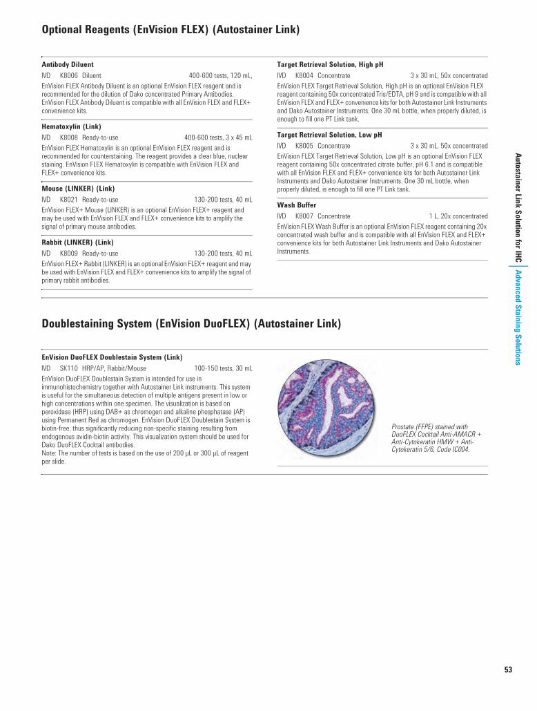
53
Autostainer Link Solution for IHCAdvanced Staining Solutions
Optional Reagents (EnVision FLEX) (Autostainer Link)
Antibody DiluentI K8006 Diluent 400-600 tests, 120 mL,EnVision FLEX Antibody Diluent is an optional EnVision FLEX reagent and is recommended for the dilution of Dako concentrated Primary Antibodies. EnVision FLEX Antibody Diluent is compatible with all EnVision FLEX and FLEX+ convenience kits.
Hematoxylin (Link)I K8008 Ready-to-use 400-600 tests, 3 x 45 mLEnVision FLEX Hematoxylin is an optional EnVision FLEX reagent and is recommended for counterstaining. The reagent provides a clear blue, nuclear staining. EnVision FLEX Hematoxylin is compatible with EnVision FLEX and FLEX+ convenience kits.
Mouse (LINKER) (Link)I K8021 Ready-to-use 130-200 tests, 40 mLEnVision FLEX+ Mouse (LINKER) is an optional EnVision FLEX+ reagent and may be used with EnVision FLEX and FLEX+ convenience kits to amplify the signal of primary mouse antibodies.
Rabbit (LINKER) (Link)I K8009 Ready-to-use 130-200 tests, 40 mLEnVision FLEX+ Rabbit (LINKER) is an optional EnVision FLEX+ reagent and may be used with EnVision FLEX and FLEX+ convenience kits to amplify the signal of primary rabbit antibodies.
Target Retrieval Solution, High pHI K8004 Concentrate 3 x 30 mL, 50x concentratedEnVision FLEX Target Retrieval Solution, High pH is an optional EnVision FLEX reagent containing 50x concentrated Tris/EDTA, pH 9 and is compatible with all EnVision FLEX and FLEX+ convenience kits for both Autostainer Link Instruments and Dako Autostainer Instruments. One 30 mL bottle, when properly diluted, is enough to fill one PT Link tank.
Target Retrieval Solution, Low pHI K8005 Concentrate 3 x 30 mL, 50x concentratedEnVision FLEX Target Retrieval Solution, Low pH is an optional EnVision FLEX reagent containing 50x concentrated citrate buffer, pH 6.1 and is compatible with all EnVision FLEX and FLEX+ convenience kits for both Autostainer Link Instruments and Dako Autostainer Instruments. One 30 mL bottle, when properly diluted, is enough to fill one PT Link tank.
Wash BufferI K8007 Concentrate 1 L, 20x concentratedEnVision FLEX Wash Buffer is an optional EnVision FLEX reagent containing 20x concentrated wash buffer and is compatible with all EnVision FLEX and FLEX+ convenience kits for both Autostainer Link Instruments and Dako Autostainer Instruments.
Doublestaining System (EnVision DuoFLEX) (Autostainer Link)
EnVision DuoFLEX Doublestain System (Link)I SK110 HRP/AP, Rabbit/Mouse 100-150 tests, 30 mLEnVision DuoFLEX Doublestain System is intended for use in immunohistochemistry together with Autostainer Link instruments. This system is useful for the simultaneous detection of multiple antigens present in low or high concentrations within one specimen. The visualization is based on peroxidase (HRP) using DAB+ as chromogen and alkaline phosphatase (AP) using Permanent Red as chromogen. EnVision DuoFLEX Doublestain System is biotin-free, thus significantly reducing non-specific staining resulting from endogenous avidin-biotin activity. This visualization system should be used for Dako DuoFLEX Cocktail antibodies.Note: The number of tests is based on the use of 200 µL or 300 µL of reagent per slide.
Prostate (FFPE) stained with DuoFLEX Cocktail Anti-AMACR + Anti-Cytokeratin HMW + Anti-Cytokeratin 5/6, Code IC004.
USCatPathology.book Page 53 Monday, March 7, 2016 7:14 PM

54
www.
dako
.com
Adva
nced
Sta
inin
g So
lutio
nsAu
tost
aine
r Plu
s for
IHC
Autostainer Plus for IHC
Dako Autostainer Plus Staining System allows for staining procedures and processing of specimens to be performed automatically. The instrument is suited for immunostaining of tissue sections, cytospins and cell smears.
The system has four elements: Autostainer Plus Instrument
The instrument is no longer available, but is still being supported
Autostainer Plus SoftwareThe flexible and open software allows for creating and saving staining protocols. It does not provide means of connectivity to Laboratory Information System or Local Area Network
ReagentsA dedicated line of FLEX ready-to-use reagents gives high quality staining results
Slide Labeling SystemSlide labels can be printed from the Autostainer Plus software
Ancillaries and Accessories (Dako Autostainer)
Accessory UtensilsS3424 Dako Autostainer Reagent Racks, One White/One
Blue 2 racksS3425 Dako Autostainer Reagent Vials 100 vialsS3704 Dako Autostainer Slide Racks 4 racks
AEC+ Substrate-ChromogenI K3461 Ready-to-use 150 tests, 15 mLI K3469 Ready-to-use 1100 tests, 110 mLAEC+ Substrate-Chromogen is especially useful in applications requiring high sensitivity. It is suitable for use in peroxidase-based immunohistochemical staining methods. AEC (3-amino-9-ethylcarbazole) forms a red end-product at the site of the target antigen. AEC must be used together with aqueous mounting fluids.
Clear-It Cleaning Reagent for Dako AutostainerI SL002 3.8 LThis reagent is suitable for cleaning the Dako Autostainer sink and reservoir after performing histological stains.
DAB+, LiquidI K3468 10 x 11 mL"Liquid DAB+ is a high sensitivity substrate-chromogen system for use in peroxidase-based immunohistochemical staining methods. DAB (diaminobenzidine) forms a very stable, brown end-product at the site of the target antigen. DAB may be used together with mounting fluids containing organic solvents.
DAB-Away®, Cleaning AgentI S1967 DAB chromogen removal system 50 tests, 250 mLFor cleaning of glassware, parts and surfaces that have come in contact with 3,3'-diaminobenzidine (DAB). Is recommended for routine cleaning of the Dako Autostainer probe and surfaces. Contains materials sufficient for 250 mL of cleaning reagent working solution and 250 mL of decolorizer.
HematoxylinI S3301 500 mLDeveloped for use with the Dako Autostainer.
IHC Microscope Slides, FLEXI K8020 Coated glass slides 5 x 100 slidesCoated microscope slides for adhesion of formalin-fixed, paraffin-embedded tissue sections for use in immunohistochemistry with Dako EnVision FLEX visualization systems. FLEX IHC Microscope Slides are compatible with, but not limited to, the following Dako instruments: Dako Omnis, Autostainer Link, Dako Autostainer/Autostainer Plus and PT Link.
Peroxidase and Alkaline Phosphatase Blocking Reagent(Dual Endogenous Enzyme-Blocking Reagent)I S2003 10 x 11 mL"Suppresses endogenous alkaline phosphatase and peroxidase in cell preparations, frozen tissue sections, and formalin-fixed, paraffin-embedded tissue sections.
Proteinase KI S3020 Ready-to-use 10 x 11 mL"Proteinase K is intended for proteolytic epitope retrieval in formalin-fixed, paraffin-embedded tissues prior to immunohistochemical procedures.
Proteolytic EnzymeI S3007 Ready-to-use 10 x 11 mL"Proteolytic Enzyme, Ready-to-Use, is intended for the proteolytic digestion of formalin-fixed, paraffin-embedded tissues, cell blocks or cell specimens prior to immunohistochemical (IHC) or in situ hybridization (ISH) procedures. Proteolytic digestion of formalin-fixed tissues improves accessibility of antibodies and DNA probes to target sites within tissues. In IHC, proteolytic digestion exposes certain epitopes which have been masked during fixation. In ISH procedures, accessibility of DNA sequences is enhanced allowing better probe penetration and hybridization.
Wash Buffer 10xI S3006 Concentrate 1 L, 10x concentratedTris-buffered saline solution containing 0.05% Tween 20, pH 7.6. Well-suited for use in manual and automated immunohistochemical staining protocols.
" Packaged in vials for use with Dako Autostainer instruments
USCatPathology.book Page 54 Monday, March 7, 2016 7:14 PM

55
Autostainer Plus for IHCAdvanced Staining Solutions
pharmDx Kits (Dako Autostainer)
c-Kit pharmDx for Dako AutostainerI K1907 35 testsc-Kit pharmDx is a qualitative immunohistochemical kit system for the identification of c-kit (CD117) protein expression in normal and neoplastic tissues. c-Kit pharmDx is indicated as an aid in the differential diagnosis of gastrointestinal stromal tumors (GIST). Accurate assessment of c-kit protein expression is now a critical factor in the diagnosis of GIST and is becoming increasingly important in influencing decisions regarding clinical management, including the use of Gleevec®/Glivec® (imatinib mesylate) for the treatment of patients with confirmed GIST.c-Kit pharmDx utilizes a simple two-step staining procedure and is suitable for formalin-fixed, paraffin-embedded specimens. The kit includes ready-to-use primary antibody, negative control reagent, cell line control slides and detailed instructions. The kit has been tailored especially for use on the Dako Autostainer.
EGFR pharmDx Kit for Dako AutostainerI K1494 50 testsEGFR pharmDx Kit is a qualitative immunohistochemical kit system that includes all reagents necessary to identify expression of epidermal growth factor receptor (EGFR) protein on the surface of normal and neoplastic cells. EGFR protein is also called HER1 protein. Through the use of standard methods and reagents, EGFR pharmDx Kit will provide reproducible results from laboratory to laboratory. EGFR is indicated as an aid in identifying colorectal cancer patients eligible for treatment with Erbitux® (cetuximab) or VectibixTM (panitumumab).The kit utilizes a simple two-step staining procedure and is suitable for formalin-fixed, paraffin-embedded specimens. Results can be available within 1 day, giving clinicians EGFR expression levels in a quick and reliable manner. The kit has been designed for use on the Dako Autostainer.
ER/PR pharmDx Kit for the Dako AutostainerI K4071 50 testsER/PR pharmDx Kit is a semi-quantitative immunohistochemical kit system to identify estrogen receptor (ER) a protein and progesterone receptor (PR) protein expression in normal and neoplastic tissues. The assay specifically detects the ER a protein as well as the PR protein located in the cell nuclei of ER and PR-expressing cells, respectively. ER/PR pharmDx Kit is indicated as an aid in identifying patients eligible for treatment with anti-hormonal or aromatase inhibitor therapies as well as an aid in the prognosis and management of breast cancer.The kit utilizes a simple two-step staining procedure and is suitable for formalin-fixed, paraffin-embedded specimens. The kit provides all the reagents needed to run the ER/PR tests, including control slides to validate each run, and detailed instructions. A scoring guideline is included to facilitate interpretation.
HercepTest for Dako AutostainerI K5207 50 testsHercepTest is a semi-quantitative immunohistochemical assay for determination of HER2 protein (c-erbB-2 oncoprotein) overexpression in breast cancer tissues routinely processed for histological evaluation and formalin-fixed, paraffin-embedded cancer tissue from patients with metastatic gastric or gastroesophageal junction adenocarcinoma. HercepTest specifically demonstrates overexpression of HER2 protein. HercepTest is indicated as an aid in the assessment of breast and gastric cancer patients for whom Herceptin® (trastuzumab) treatment is being considered and for breast cancer patients for whom PERJETATM (pertuzumab) or KADCYLATM (ado-trastuzumab emtansine) treatment is being considered (see Herceptin®, PERJETATM, and KADCYLATM package inserts). K5207 has been tailored especially for use on Dako Autostainer.HercepTestTM, Herceptin®, PERJETATM, and KADCYLATM are trademarks owned by Genentech, Inc. and/or F. Hoffmann-La Roche Ltd.; HercepTestTM is subject to an exclusive trademark license to Dako Denmark A/S.
Go to page 139 to read about all our pharmDx products.
USCatPathology.book Page 55 Monday, March 7, 2016 7:14 PM

56
www.
dako
.com
Adva
nced
Sta
inin
g So
lutio
nsAu
tost
aine
r Plu
s for
IHC
Primary Antibodies (FLEX Ready-to-Use) (Dako Autostainer)
The IS-Series FLEX Ready-to-Use Primary Antibodies listed in this section are packaged in Dako Autostainer Reagent Vials for convenience, ease of operation, and time savings.IS-Series Antibodies can only be used with EnVision FLEX and EnVision FLEX+ Visualization Systems.
The package insert included with each Dako antibody gives a detailed product description.
Monoclonal Mouse Anti-HumanActin (Muscle)I IS700 Clone HHF35 30 tests, 6 mL
Monoclonal Mouse Anti-HumanActin (Smooth Muscle)I IS611 Clone 1A4 30 tests, 6 mL
Polyclonal Rabbit Anti-HumanAlpha-1-FetoproteinI IS500 30 tests, 6 mL
Monoclonal Rabbit Anti-HumanAMACRI IS060 Clone 13H4 30 tests, 6 mL
Monoclonal Mouse Anti-HumanB-Cell-Specific Activator ProteinI IS650 Clone DAK-Pax5 30 tests, 6 mL
Monoclonal Mouse Anti-HumanBCL2 OncoproteinI IS614 Clone 124 30 tests, 6 mL
Monoclonal Mouse Anti-HumanBCL6 ProteinI IS625 Clone PG-B6p 30 tests, 6 mL
Monoclonal Mouse Anti-HumanBeta-CateninI IS702 Clone b-Catenin-1 30 tests, 6 mL
Monoclonal Mouse Anti-HumanCA 125I IS701 Clone M11 30 tests, 6 mL
Polyclonal Rabbit Anti-HumanCalcitoninI IS515 30 tests, 6 mL
Monoclonal Mouse Anti-HumanCaldesmonI IS054 Clone h-CD 30 tests, 6 mL
Monoclonal Mouse Anti-HumanCalretininI IS627 Clone DAK-Calret 1 30 tests, 6 mL
Monoclonal Mouse Anti-HumanCarcinoembryonic Antigen (CEA)I IS622 Clone II-7 30 tests, 6 mL
Polyclonal Rabbit Anti-HumanCarcinoembryonic Antigen (CEA)I IS526 30 tests, 6 mL
Monoclonal Mouse Anti-HumanCD1aI IS069 Clone 010 30 tests, 6 mL
Monoclonal Mouse Anti-HumanCD2I IS651 Clone AB75 30 tests, 6 mL
Polyclonal Rabbit Anti-HumanCD3I IS503 30 tests, 6 mL
Monoclonal Mouse Anti-HumanCD4I IS649 Clone 4B12 30 tests, 6 mL
Monoclonal Mouse Anti-HumanCD5I IS082 Clone 4C7 30 tests, 6 mL
Monoclonal Mouse Anti-HumanCD7I IS643 Clone CBC.37 30 tests, 6 mL
Monoclonal Mouse Anti-HumanCD8I IS623 Clone C8/144B 30 tests, 6 mL
Monoclonal Mouse Anti-HumanCD10I IS648 Clone 56C6 30 tests, 6 mL
Monoclonal Mouse Anti-HumanCD15I IS062 Clone Carb-3 30 tests, 6 mL
Monoclonal Mouse Anti-HumanCD19I IS656 Clone LE-CD19 30 tests, 6 mL
Monoclonal Mouse Anti-HumanCD20cyI IS604 Clone L26 30 tests, 6 mL
Monoclonal Mouse Anti-HumanCD21I IS608 Clone 1F8 30 tests, 6 mL
Monoclonal Mouse Anti-HumanCD23I IS781 Clone DAK-CD23 30 tests, 6 mL
Monoclonal Mouse Anti-HumanCD30I IS602 Clone Ber-H2 30 tests, 6 mL
Monoclonal Mouse Anti-HumanCD31, Endothelial CellI IS610 Clone JC70A 30 tests, 6 mL
Monoclonal Mouse Anti-HumanCD34 Class III IS632 Clone QBEnd 10 30 tests, 6 mL
USCatPathology.book Page 56 Monday, March 7, 2016 7:14 PM

Primary Antibodies (FLEX Ready-to-Use) (Dako Autostainer) (continued)
57
Autostainer Plus for IHCAdvanced Staining Solutions
Monoclonal Mouse Anti-HumanCD43I IS636 Clone DF-T1 30 tests, 6 mL
Monoclonal Mouse Anti-HumanCD45, Leucocyte Common AntigenI IS751 Clones 2B11 + PD7/26 30 tests, 6 mL
Monoclonal Mouse Anti-HumanCD56I IS628 Clone 123C3 30 tests, 6 mL
Monoclonal Mouse Anti-HumanCD57I IS647 Clone TB01 30 tests, 6 mL
Monoclonal Mouse Anti-HumanCD68I IS609 Clone KP1 30 tests, 6 mL
Monoclonal Mouse Anti-HumanCD68I IS613 Clone PG-M1 30 tests, 6 mL
Monoclonal Mouse Anti-HumanCD79aI IS621 Clone JCB117 30 tests, 6 mL
Monoclonal Mouse Anti-HumanCD99, MIC2 Gene Products, Ewing's Sarcoma MarkerI IS057 Clone 12E7 30 tests, 6 mL
Monoclonal Mouse Anti-HumanCD138I IS642 Clone MI15 30 tests, 6 mL
Monoclonal Mouse Anti-HumanCD246, ALK ProteinI IS641 Clone ALK1 30 tests, 6 mL
Monoclonal Mouse Anti-HumanCDX2I IS080 Clone DAK-CDX2 30 tests, 6 mL
Polyclonal Rabbit Anti-HumanChorionic Gonadotropin (hCG)I IS508 30 tests, 6 mL
Monoclonal Rabbit Anti-HumanCyclin D1I IS083 Clone EP12 30 tests, 6 mL
Monoclonal Mouse Anti-HumanCytokeratinI IS053 Clone AE1/AE3 30 tests, 6 mL
Monoclonal Mouse Anti-HumanCytokeratin 5/6I IS780 Clone D5/16 B4 30 tests, 6 mL
Monoclonal Mouse Anti-HumanCytokeratin 7I IS619 Clone OV-TL 12/30 30 tests, 6 mL
Monoclonal Mouse Anti-Cytokeratin 17I IS620 Clone E3 30 tests, 6 mL
Monoclonal Mouse Anti-HumanCytokeratin 18I IS618 Clone DC 10 30 tests, 6 mL
Monoclonal Mouse Anti-HumanCytokeratin 19I IS615 Clone RCK108 30 tests, 6 mL
Monoclonal Mouse Anti-HumanCytokeratin 20I IS777 Clone Ks20.8 30 tests, 6 mL
Monoclonal Mouse Anti-HumanCytokeratin, High Molecular WeightI IS051 Clone 34ßE12 30 tests, 6 mL
Monoclonal Mouse Anti-HumanDesminI IS606 Clone D33 30 tests, 6 mL
Monoclonal Mouse Anti-HumanE-CadherinI IS059 Clone NCH-38 30 tests, 6 mL
Monoclonal Mouse Anti-HumanEpithelial AntigenI IS637 Clone Ber-EP4 30 tests, 6 mL
Monoclonal Mouse Anti-HumanEpithelial Membrane Antigen (EMA)I IS629 Clone E29 30 tests, 6 mL
Polyclonal Rabbit Anti-HumanGastrinI IS519 30 tests, 6 mL
Polyclonal Rabbit Anti-Glial Fibrillary Acidic ProteinI IS524 30 tests, 6 mL
Monoclonal Mouse Anti-HumanGross Cystic Disease Fluid Protein-15I IS077 Clone 23A3 30 tests, 6 mL
Monoclonal Mouse Anti-HumanHepatocyteI IS624 Clone OCH1E5 30 tests, 6 mL
Polyclonal Rabbit Anti-HumanIgAI IS510 30 tests, 6 mL
Polyclonal Rabbit Anti-HumanIgDI IS517 30 tests, 6 mL
Polyclonal Rabbit Anti-HumanIgGI IS512 30 tests, 6 mL
Polyclonal Rabbit Anti-HumanIgMI IS513 30 tests, 6 mL
Monoclonal Mouse Anti-HumanInhibin aI IS058 Clone R1 30 tests, 6 mL
USCatPathology.book Page 57 Monday, March 7, 2016 7:14 PM

58
Primary Antibodies (FLEX Ready-to-Use) (Dako Autostainer) (continued)
www.
dako
.com
Adva
nced
Sta
inin
g So
lutio
nsAu
tost
aine
r Plu
s for
IHC
Polyclonal Guinea Pig Anti-InsulinI IS002 30 tests, 6 mL
Polyclonal Rabbit Anti-HumanKappa Light ChainsI IS506 30 tests, 6 mL
Monoclonal Mouse Anti-HumanKi-67 AntigenI IS626 Clone MIB-1 30 tests, 6 mL
Polyclonal Rabbit Anti-HumanLambda Light ChainsI IS507 30 tests, 6 mL
Monoclonal Mouse Anti-HumanMammaglobinI IS074 Clone 304-1A5 30 tests, 6 mL
Monoclonal Mouse Anti-HumanMast Cell TryptaseI IS640 Clone AA1 30 tests, 6 mL
Monoclonal Mouse Anti-HumanMelan-AI IS633 Clone A103 30 tests, 6 mL
Monoclonal Mouse Anti-HumanMelanosomeI IS052 Clone HMB-45 30 tests, 6 mL
Monoclonal Mouse Anti-HumanMUM1 ProteinI IS644 Clone MUM1p 30 tests, 6 mL
Monoclonal Mouse Anti-HumanMutL Protein Homolog 1I IS079 Clone ES05 30 tests, 6 mL
Polyclonal Rabbit Anti-HumanMyeloperoxidaseI IS511 30 tests, 6 mL
Monoclonal Mouse Anti-MyogeninI IS067 Clone F5D 30 tests, 6 mL
Monoclonal Mouse Anti-HumanNeurofilament ProteinI IS607 Clone 2F11 30 tests, 6 mL
Monoclonal Mouse Anti-HumanNeuron-Specific Enolase (NSE)I IS612 Clone BBS/NC/VI-H14 30 tests, 6 mL
Monoclonal Mouse Anti-Humanp53 ProteinI IS616 Clone DO-7 30 tests, 6 mL
Monoclonal Mouse Anti-HumanPlacental Alkaline PhosphataseI IS779 Clone 8A9 30 tests, 6 mL
Monoclonal Mouse Anti-HumanPodoplaninI IS072 Clone D2-40 30 tests, 6 mL
Polyclonal Rabbit Anti-HumanProstate-Specific Antigen (PSA)I IS514 30 tests, 6 mL
Monoclonal Mouse Anti-HumanRenal Cell Carcinoma MarkerI IS075 Clone SPM314 30 tests, 6 mL
Polyclonal Rabbit Anti-S100I IS504 30 tests, 6 mL
Monoclonal Mouse Anti-HumanSmooth Muscle Myosin Heavy ChainI IS066 Clone SMMS-1 30 tests, 6 mL
Polyclonal Rabbit Anti-HumanThyroglobulinI IS509 30 tests, 6 mL
Monoclonal Mouse Anti-Thyroid Transcription Factor (TTF-1)I IS056 Clone 8G7G3/1 30 tests, 6 mL
Monoclonal Mouse Anti-HumanTyrosinaseI IS061 Clone T311 30 tests, 6 mL
Monoclonal Mouse Anti-VillinI IS076 Clone 1D2 C3 30 tests, 6 mL
Monoclonal Mouse Anti-VimentinI IS630 Clone V9 30 tests, 6 mL
Polyclonal Rabbit Anti-HumanVon Willebrand FactorI IS527 30 tests, 6 mL
Monoclonal Mouse Anti-HumanWilms' Tumor 1 (WT1) ProteinI IS055 Clone 6F-H2 30 tests, 6 mL
USCatPathology.book Page 58 Monday, March 7, 2016 7:14 PM

59
Autostainer Plus for IHCAdvanced Staining Solutions
Negative Controls (FLEX Ready-to-Use) (Dako Autostainer)
Negative Control for IS-Series Mouse Primary AntibodiesI IS750 Ready-to-use 60 tests, 12 mLUniversal negative control for all FLEX ready-to-use mouse primary antibodies for use on Dako Autostainer/Autostainer Plus instruments. Packaged in Dako Autostainer Vial.
Negative Control for IS-Series Rabbit Primary AntibodiesI IS600 Ready-to-use 60 tests, 12 mLUniversal negative control for all FLEX ready-to-use rabbit primary antibodies for use on Dako Autostainer/Autostainer Plus instruments. Packaged in Autostainer Reagent Vial.
Visualization Systems (EnVision FLEX) (Dako Autostainer)
EnVision FLEX, High pH(Dako Autostainer/Autostainer Plus)I K8010 HRP. Rabbit/Mouse. High pH 400-600 testsEnVision FLEX, High pH is a high-sensitivity visualization system intended for use in immunohistochemistry together with Dako Autostainer Instruments. The dual link system detects primary mouse and rabbit antibodies and the reaction is visualized by DAB+ Chromogen. The convenience kit includes Peroxidase-Blocking Reagent, EnVision/HRP, DAB+ Chromogen, Substrate Buffer, Target Retrieval Solution, High pH (50x Tris/EDTA buffer, pH 9), and Wash Buffer (20x). EnVision FLEX convenience kits are compatible with all optional EnVision FLEX and FLEX+ reagents for Dako Autostainer Instruments.
EnVision FLEX Mini Kit, High pH(Dako Autostainer/Autostainer Plus)I K8024 HRP. Rabbit/Mouse. High pH 125-190 testsEnVision FLEX Mini Kit, High pH is a high-sensitivity visualization system intended for use in immunohistochemistry together with Dako Autostainer Instruments. The dual link system detects primary mouse and rabbit antibodies and the reaction is visualized by DAB+ Chromogen. The convenience kit includes Peroxidase-Blocking Reagent, EnVision/HRP, DAB+ Chromogen, Substrate Buffer, Target Retrieval Solution, High pH (50x Tris/EDTA buffer, pH 9), and Wash Buffer (20x). EnVision FLEX convenience kits are compatible with all optional EnVision FLEX and FLEX+ reagents for Dako Autostainer Instruments.
EnVision FLEX+, Mouse, High pH(Dako Autostainer/Autostainer Plus)I K8012 HRP. Mouse. High pH 400-600 testsEnVision FLEX+, Mouse, High pH is a very-high-sensitivity visualization system intended for use in immunohistochemistry together with Dako Autostainer Instruments. The EnVision FLEX+ Mouse (LINKER) amplifies the signal of primary mouse antibodies and the reaction is visualized by DAB+ Chromogen. In addition to the EnVision FLEX+ Mouse (LINKER) the convenience kit includes Peroxidase-Blocking Reagent, EnVision/HRP, DAB+ Chromogen, Substrate Buffer, Target Retrieval Solution, High pH (50x Tris/EDTA buffer, pH 9), and Wash Buffer (20x). The EnVision FLEX+ Rabbit (LINKER), Code K8019, is an optional EnVision FLEX reagent that may be used with EnVision FLEX and FLEX+ convenience kits to amplify the signal of primary rabbit antibodies. EnVision FLEX+ convenience kits are compatible with all optional EnVision FLEX and FLEX+ reagents for Dako Autostainer Instruments.
USCatPathology.book Page 59 Monday, March 7, 2016 7:14 PM

60
www.
dako
.com
Adva
nced
Sta
inin
g So
lutio
nsAu
tost
aine
r Plu
s for
IHC
Optional Reagents (EnVision FLEX) (Dako Autostainer)
Antibody DiluentI K8006 Diluent 400-600 tests, 120 mL,EnVision FLEX Antibody Diluent is an optional EnVision FLEX reagent and is recommended for the dilution of Dako concentrated Primary Antibodies. EnVision FLEX Antibody Diluent is compatible with all EnVision FLEX and FLEX+ convenience kits.
Hematoxylin (Dako Autostainer/Autostainer Plus)I K8018 Ready-to-use 400-600 tests, 10 x 13 mLEnVision FLEX Hematoxylin is an optional EnVision FLEX reagent and is recommended for counterstaining. The reagent provides a clear blue, nuclear staining. EnVision FLEX Hematoxylin is compatible with EnVision FLEX and FLEX+ convenience kits.
Mouse (LINKER) (Dako Autostainer/Autostainer Plus)I K8022 Ready-to-use 120-190 tests, 3 x 13 mLEnVision FLEX+ Mouse (LINKER) is an optional EnVision FLEX+ reagent and may be used with EnVision FLEX and FLEX+ convenience kits to amplify the signal of primary mouse antibodies.
Rabbit (LINKER) (Dako Autostainer/Autostainer Plus)I K8019 Ready-to-use 120-190 tests, 3 x 13 mLEnVision FLEX+ Rabbit (LINKER) is an optional EnVision FLEX+ reagent and may be used with EnVision FLEX and FLEX+ convenience kits to amplify the signal of primary rabbit antibodies.
Target Retrieval Solution, High pHI K8004 Concentrate 3 x 30 mL, 50x concentratedEnVision FLEX Target Retrieval Solution, High pH is an optional EnVision FLEX reagent containing 50x concentrated Tris/EDTA, pH 9 and is compatible with all EnVision FLEX and FLEX+ convenience kits for both Autostainer Link Instruments and Dako Autostainer Instruments. One 30 mL bottle, when properly diluted, is enough to fill one PT Link tank.
Target Retrieval Solution, Low pHI K8005 Concentrate 3 x 30 mL, 50x concentratedEnVision FLEX Target Retrieval Solution, Low pH is an optional EnVision FLEX reagent containing 50x concentrated citrate buffer, pH 6.1 and is compatible with all EnVision FLEX and FLEX+ convenience kits for both Autostainer Link Instruments and Dako Autostainer Instruments. One 30 mL bottle, when properly diluted, is enough to fill one PT Link tank.
Wash BufferI K8007 Concentrate 1 L, 20x concentratedEnVision FLEX Wash Buffer is an optional EnVision FLEX reagent containing 20x concentrated wash buffer and is compatible with all EnVision FLEX and FLEX+ convenience kits for both Autostainer Link Instruments and Dako Autostainer Instruments.
USCatPathology.book Page 60 Monday, March 7, 2016 7:14 PM

61
Autostainer Plus for IHCAdvanced Staining Solutions
Visualization Systems (Dako Autostainer)
ADVANCEI K4069 HRP. Rabbit/Mouse 55 testsI K4068 HRP. Rabbit/Mouse 550 testsThis ready-to-use, peroxidase-based ADVANCE kit is compatible with suitably diluted rabbit and mouse primary antibodies. The ADVANCE kit is a super-sensitive, non-biotin based, immunohistochemical visualization system that is useful for the detection of antigens in low concentrations, for short incubation time or for higher dilution of primary antibodies. ADVANCE is 5 or more times more sensitive than EnVision+ and with approximately the same sensitivity as CSA II.Note: The number of tests for this kit is based on the use of 200 µL of reagent per slide.
EnVision+ Dual Link KitI K4065 HRP. Rabbit/Mouse (DAB+) 150 testsThis ready-to-use, peroxidase-based EnVision+ Dual Link Kit is compatible with suitably diluted rabbit and mouse primary antibodies. In addition to the ready-to-use EnVision+ Dual Link reagent, the kit includes a blocking reagent for endogenous peroxidase and a high-sensitivity diaminobenzidine (DAB+) chromogenic substrate system.
EnVision+ Dual Link, Single ReagentI K4061 HRP. Rabbit/Mouse 10 x 11 mLThis ready-to-use, peroxidase-conjugated EnVision+ Dual Link reagent is for use when a detection system more sensitive than the LSAB2 kit is needed. It is compatible with suitably diluted rabbit and mouse primary antibodies. The reagent is provided in Dako Autostainer Reagent Vials.
EnVision G|2 Doublestain System, Rabbit/Mouse(DAB+/Permanent Red)I K5361 150 testsEnVision G|2 Doublestain System is a high-sensitivity peroxidase and alkaline-phosphatase-based 2nd generation visualization kit. The kit is intended for use in immunohistochemistry for the simultaneous detection of two different antigens within the same specimen, and is compatible with suitably diluted rabbit and mouse primary antibodies. The kit may be used on formalin-fixed, paraffin-embedded tissue sections and fixed cell smears. In addition to the ready-to-use EnVision G|2 reagents packaged in Dako Autostainer Reagent Vials, the kit includes both DAB+ and Permanent Red chromogenic substrate systems.Note: The number of tests for this kit is based on the use of 200 µL of reagent per slide.
EnVision G|2 System/AP, Rabbit/Mouse (Permanent Red)I K5355 50 tests/500 testsEnVision G|2 System/AP is a high-sensitivity alkaline-phosphatase-based 2nd generation visualization kit. The kit is intended for use in immunohistochemistry, and it is compatible with suitably diluted rabbit and mouse primary antibodies. The kit may be used on formalin-fixed, paraffin-embedded tissue sections, frozen sections and fixed cell smears. In addition to the ready-to-use EnVision G|2 reagents packaged in Dako Autostainer Reagent Vials, the kit includes a Permanent Red chromogenic substrate system. The kit may be used in manual procedures or with the Dako Autostainer instruments.Note: The number of tests for this kit is based on the use of 200 µL of reagent per slide.
EnVision+ KitsI K4005 HRP. Mouse (AEC+) 1100 testsI K4007 HRP. Mouse (DAB+) 1100 testsI K4009 HRP. Rabbit (AEC+) 1100 testsI K4011 HRP. Rabbit (DAB+) 1100 testsThese ready-to-use, peroxidase-based EnVision+ kits are compatible with suitably diluted mouse or rabbit primary antibodies, respectively. In addition to the ready-to-use EnVision+ reagent, the kits include a blocking reagent for endogenous peroxidase, and a high-sensitivity 3-amino-9-ethylcarbazole (AEC+) chromogenic substrate system.
Universal LSAB2 Kit/HRP, Rabbit/MouseI K0675 10 x 11 mL Link + 10 x 11 mL Streptavidin/HRPThis peroxidase-based visualization kit is for use with both rabbit and mouse primary antibodies. The biotinylated link antibody in the kit is produced in goat. Reagents are ready-to-use and are provided in Dako Autostainer Reagent Vials.
USCatPathology.book Page 61 Monday, March 7, 2016 7:14 PM

62
www.
dako
.com
Adva
nced
Sta
inin
g So
lutio
nsAn
tibod
ies a
nd C
ontro
lsAntibodies and Controls
Primary AntibodiesPrimary monoclonal and polyclonal antibodies, primary antibody conjugates, and prediluted ready-to-use antibody systems are presented here. These products are intended for use on tissue sections and/or cell smears.
Products originating from the same primary antibody have been grouped together. For each group, suitable tissue fixation and pre-treatment of sections are indicated.
All Dako antibody reagents for immunohistochemistry are in liquid form and contain an antimicrobial agent. A detailed product description, a guideline for dilution, and a recommended staining procedure are given in the package insert included with each vial.
Antibody FormsDako primary antibodies for immunohistochemistry are provided undiluted as well as in ready-to-use forms adapted for different staining procedures and detection kits as described below.
Concentrated antibody reagentsThe polyclonal antibody reagents are provided as whole serum, Ig fractions, F(ab')2 fragments or affinity-isolated while the monoclonal antibodies are provided as tissue culture supernatants or purified from culture supernatants. In a few cases, monoclonal antibodies have been purified from ascitic fluid.
Ready-to-use, for EnVision FLEX(GA, IR and IS-Series)GA-series is designed for Dako Omnis, IR-series is designed for Automated Link Platforms, and the IS-series is designed for Dako Autostainer and Autostainer Plus. For optimal performance these prediluted primary antibodies can only be used with EnVisionFLEX and EnVisionFLEX+ visualization systems. FLEX Ready-to-Use Universal Negative Controls are available separately for monoclonal mouse (Code GA750, IR750 and IS750) and polyclonal rabbit (Code GA600, IR600 and IS600) primary antibodies.
Ready-to-Use, for EnVision DuoFLEX(IC-Series)These ready-to-use antibody cocktails are designed for optimal performance using EnVision DuoFLEX Doublestain System, and will provide a two-color staining reaction on the same tissue section. The IC-series is designed for Automated Link Platforms.
Antibody ApplicationsAntigens may resist fixation to a variable degree. Therefore, suitable tissue fixation and, when necessary, antigen retrieval method are listed for each antibody and explained below. Frozen: The appropriately diluted and concentrated antibody is useful
for labelling sections of frozen tissue fixed in acetone for 10 minutes. It also works well on cell smears fixed in a 19:19:1 mixture of acetone:methanol:36% fomalin for 90 seconds, or in acetone for 10 minutes.
Formalin: The antibody is suitable for labelling formalin-fixed, paraffin-embedded (FFPE) tissue sections. The duration of the formalin fixation should, generally, not exceed 24-48 hours.
Enzyme: Optimal staining results require adequate treatment of deparaffinized, formalin-fixed tissue sections with a proteolytic enzyme before incubation with the antibody. Detailed procedures for the use of Dako proteolytic enzymes are included with the products.
HIER: Optimal staining results require heat-induced epitope retrieval (HIER) of deparaffinized, formalin-fixed tissue sections, for example in a microwave oven, a pressure cooker or a water bath, before incubation with the antibody. During heat treatment the sections must be immersed in a suitable buffer. A detailed procedure is available from Dako.
Enzyme/HIER: Optimal staining results on deparaffinized, formalin-fixed tissue sections require either treatment with a proteolytic enzyme or heating in a suitable buffer before incubation with the antibody.
Enzyme + HIER: Optimal staining results on deparaffinized, formalin-fixed tissue sections require treatment with a proteolytic enzyme as well as heating in a suitable buffer before incubation with the antibody.
StainsAll immunohistochemical stains are from formalin-fixed, paraffin-embedded (FFPE) tissue sections unless otherwise specified. For more images and high-resolution zoom tool, please visit our Products section on www.dako.com.
Autostainer Link 48 InstallersDakoLink users can download Installers, which will update specific DakoLink database protocols. Go to the www.dako.com/installer and locate the antibody code number/protocol in question, click the link to download the Installer to a portable device, e.g. a USB flash drive, and transfer the file to the DakoLink Server computer. Read the full installation guide before beginning the update process.
Adjunctive Diagnostic InformationIHC class I devices provide pathologists with adjunctive diagnostic information that may be incorporated into the pathologist's report, but that is not ordinarily reported to the clinician as an independent finding.
Results aid in the classification of neoplasms. Differential classification is aided by the results from a panel of antibodies. The clinical interpretation of any staining or its absence should be complemented
by morphological studies using proper controls and should be evaluated within the context of the patient's clinical history and other diagnostic tests by a qualified pathologist. Primary antibodies are
intended to be used after the primary diagnosis of tumor has been made by conventional histopathology using non-immunologic histochemical stains, such as hematoxylin and eosin.
USCatPathology.book Page 62 Monday, March 7, 2016 7:14 PM

63
Antibodies and ControlsAdvanced Staining Solutions
Overview of FLEX Ready-to-Use Antibodies
FLEX Ready-to-Use (RTU) antibodies are pre-diluted primary antibodies specifically developed for automated use while maintaining the high-quality staining performance that Dako’s antibodies are known for. Each FLEX RTU antibody has been developed with focus on delivering a consistent, high-quality staining performance with just one flexible staining protocol. The staining performance of all antibodies has been defined, tested and approved through collaboration with leading international pathologists.
For each FLEX RTU antibody, one protocol is recommended to obtain optimal staining results. The quality of the stainings has been reviewed by a group of expert pathologists. In our Atlas of Stains guide book, we present staining images of high and low-expression structures as well as of recommended control tissues.
In the following, the FLEX RTU antibodies are presented in tables according to organ or tissue type. This is done to ease and enhance the selection of antibodies for relevant markers to examine the expression pattern in the different tissue or organ types.
FLEX RTU Antibodies Deliver a high-quality staining performance reviewed and accepted by
leading pathologists Provide a basis for consistency Improve overall laboratory efficiency Increase productivity
Content1. Breast Tissue2. Endocrine System3. Gastrointestinal Tract4. Kidney and Urinary Tract5. Liver, Biliary System and Exocrine Pancreas6. Lymphatic Tissue and Bone Marrow7. Mesothelial Surfaces8. Nervous System9. Prostate10. Reproductive System11. Respiratory System12. Skeletal Muscles13. Skin14. Soft Tissue and Bones 15. Undifferentiated Tumors
For product descriptions, staining images, package inserts and contact information for local Dako representatives, please visit: www.dako.com.
Terms and Conditions for Use of Target-Specific FLEX Ready-to-Use TablesThe Target-Specific FLEX Ready-to-Use tables are by no means intended as a replacement of the professional judgment of a certified pathologist. The contents of the tables should not be regarded as indicative of a standard procedure for diagnosis or treatment. The products mentioned in the Target-Specific FLEX Ready-to-Use tables should be used by qualified personnel only. The user of such products should refer to the Package Inserts accompanying the selected Dako product. Dako and its partners cannot be held responsible for the use of products in any other way than described in the Package Inserts, or for conclusions drawn based on results obtained with our products.
Dako does not claim or make warranties that the information provided in the Target-Specific FLEX Ready-to-Use tables are valid everywhere, since there are differences in the acceptance of the relevance of various markers between countries and pathologists and in the standard operating procedures used.
FLEX Ready-to-Use Atlas of Stains - 4th Edition
I N T E R P R E TAT I O N
A Better Path for Cancer Diagnostics
USCatPathology.book Page 63 Monday, March 7, 2016 7:14 PM

64
Overview of FLEX Ready-to-Use Antibodies (continued)
www.
dako
.com
Adva
nced
Sta
inin
g So
lutio
nsAn
tibod
ies a
nd C
ontro
ls
FLEX RTU Antibodies | Breast Tissue
FLEX RTU Antibodies | Endocrine System
Page Anti- Clone74 BCL2 Oncoprotein 12476 Caldesmon h-CD81 CD31, Endothelial Cell JC70A82 CD34 Class II QBEnd 1088 Cytokeratin 8/18 EP17/EP3090 E-Cadherin NCH-3892 Epithelial Membrane Antigen E2993 Estrogen Receptor α EP195 Gross Cystic Disease Fluid Protein-15 23A398 Ki-67 Antigen MIB-1
100 Mammaglobin 304-1A573 Muscle Actin HHF35
104 Myosin Heavy Chain, Smooth Muscle SMMS-1106 p53 Protein DO-7108 Progesterone Receptor PgR 636110 S100 Polyclonal
73 Smooth Muscle Actin 1A4
Page Anti- CloneGeneral neuroendocrine markers
83 CD56 123C3
104 Neuron-Specific Enolase BBS/NC/VI-H14
110 S100 Polyclonal110 Synaptophysin DAK-SYNAP
Alimentary tract and pancreas86 CDX2 DAK-CDX294 Gastrin Polyclonal97 Insulin Polyclonal
Gonades86 Chorionic Gonadotropin Polyclonal97 Inhibin α R1
100 Melan-A A103Pituitary glands
86 Chorionic Gonadotropin PolyclonalThyroid and parathyroid glands
76 Calcitonin Polyclonal111 Thyroglobulin Polyclonal112 Thyroid Transcription Factor, TTF-1 8G7G3/1
Breast ductal carcinoma stained with Anti-Gross Cystic Disease Fluid Protein-15, Code IR077/IS077.
Breast ductal carcinoma stained with Anti-Mammaglobin, Code IR074/IS074.
Gastrointestinal carcinoma stained with Anti-CDX2, Code IR080/IS080.
Insulinoma stained with Anti-Insulin, Code IR002/IS002.
Granulosa cell tumor stained with Anti-Melan A, Code IR633/IS633.
Lung small cell carcinoma stained with Anti-Thyroid Transcription Factor, Code IR056/IS056.
USCatPathology.book Page 64 Monday, March 7, 2016 7:14 PM

Overview of FLEX Ready-to-Use Antibodies (continued)
65
Antibodies and ControlsAdvanced Staining Solutions
FLEX RTU Antibodies | Gastrointestinal Tract
Page Anti- Clone75 Beta-Catenin β-Catenin-177 Calretinin DAK-Calret 1
82 CD45, Leucocyte Common Antigen 2B11 + PD7/26
86 CDX2 DAK-CDX290 Desmin D3390 E-Cadherin NCH-38
101 MUC2 CCP58102 MUC5AC CLH2102 MutL Protein Homolog 1 ES05102 MutS Protein Homolog 2 FE11103 MutS Protein Homolog 6 EP49104 Neurofilament Protein 2F11
104 Neuron-Specific Enolase BBS/NC/VI-H14
106 p53 Protein DO-7107 Podoplanin D2-40107 Postmeiotic Segregation Increased 2 EP51110 S100 Polyclonal
73 Smooth Muscle Actin 1A4113 Villin 1D2 C3113 Vimentin V9
Colon adenocarcinoma stained with Anti-CDX2, Code IR080/IS080.
Rhabdomyosarcoma stained with Anti-Desmin, Code IR606/IS606.
Colon adenocarcinoma (FFPE) metastatic to the ovary stained with FLEX Anti-MUC2, Code IR658.
Epitheloid mesothelioma stained with Anti-Podoplanin, Code IR072/IS072.
Colon adenocarcinoma (FFPE) with loss of PMS2 protein stained with FLEX Anti-PMS2, Code IR087.
USCatPathology.book Page 65 Monday, March 7, 2016 8:08 PM

66
Overview of FLEX Ready-to-Use Antibodies (continued)
www.
dako
.com
Adva
nced
Sta
inin
g So
lutio
nsAn
tibod
ies a
nd C
ontro
ls
FLEX RTU Antibodies | Kidney and Urinary Tract
FLEX RTU Antibodies | Liver, Biliary System and Exocrine Pancreas
Page Anti- Clone79 CD10 56C692 Epithelial Membrane Antigen E2998 Ki-67 Antigen MIB-1
100 Melan-A A103106 p53 Protein DO-7109 Renal Cell Carcinoma Marker SPM314110 S100 Polyclonal
116 S100 + Tyrosinase + Melan-A Polyclonal + T311 + A103
73 Smooth Muscle Actin 1A4113 Vimentin V9
Page Anti- Clone73 Alpha-1-Fetoprotein Polyclonal76 CA 125 M1179 CD10 56C681 CD31, Endothelial Cell JC/70A82 CD34 Class II QBEnd 1086 CDX2 DAK-CDX292 Epithelial Membrane Antigen E2995 Hepatocyte OCH1E598 Ki-67 Antigen MIB-1
106 p53 Protein DO-7108 Progesterone Receptor PgR 636
73 Smooth Muscle Actin 1A4
Transitional carcinoma of the bladder stained with Anti-p53 Protein, Code IR616/IS616.
Renal cell carcinoma stained with Anti-Renal Cell Carcinoma Marker, Code IR075/IS075.
Hemangiosarcoma stained with Anti-CD31, Endothelial Cell, Code IR610/IS610.
Angiosarcoma stained with Anti-CD34, Class II, Code IR632/IS632.
Hepatocellular carcinoma stained with Anti-Hepatocyte, Code IR624/IS624.
USCatPathology.book Page 66 Monday, March 7, 2016 8:08 PM

Overview of FLEX Ready-to-Use Antibodies (continued)
67
Antibodies and ControlsAdvanced Staining Solutions
FLEX RTU Antibodies | Lymphatic Tissue and Bone Marrow
Page Anti- Clone74 B-Cell-Specific Activator Protein DAK-Pax-574 BCL2 Oncoprotein 12475 BCL6 Protein PG-B6p77 CD1a 01078 CD2 AB7578 CD3 Polyclonal
115 CD3 + CD20cy Polyclonal + L26
78 CD4 4B1278 CD5 4C779 CD7 CBC.3779 CD8 C8/144B79 CD10 56C680 CD15 Carb-380 CD19 LE-CD1980 CD20cy L2680 CD21 1F881 CD23 DAK-CD2381 CD30 Ber-H281 CD31, Endothelial Cell JC70A82 CD34 Class II QBEnd 1082 CD43 DF-T1
82 CD45, Leucocyte Common Antigen 2B11 + PD7/26
83 CD56 123C383 CD57 TB0184 CD68 KP184 CD68 PG-M184 CD79α JCB11785 CD138 MI1586 CD246, ALK Protein ALK187 Cyclin D1 EP1292 Epithelial Membrane Antigen E2996 IgA Polyclonal96 IgD Polyclonal96 IgG Polyclonal97 IgM Polyclonal98 Kappa Light Chains Polyclonal98 Ki-67 Antigen MIB-198 Lambda Light Chains Polyclonal
102 MUM1 Protein MUM1p103 Myeloperoxidase Polyclonal105 Nucleophosmin 376110 S100 Polyclonal111 Terminal Deoxynucleotidyl Transferase EP266114 Von Willebrand Factor Polyclonal114 ZAP-70 2F3.2
Precursor T-cell lymphoblastic lymphoma/leukemia stained with Anti-CD2, Code IR651/IS651.
Anaplastic large cell lymphoma stained with Anti-CD4, Code IR649/IS649.
Precursor B-cell lymphoblastic lymphoma/leukemia stained with Anti-CD10, Code IR648/IS648.
Mantle cell lymphoma stained with Anti-Cyclin D1, Code IR083/IS083.
USCatPathology.book Page 67 Monday, March 7, 2016 8:08 PM

68
Overview of FLEX Ready-to-Use Antibodies (continued)
www.
dako
.com
Adva
nced
Sta
inin
g So
lutio
nsAn
tibod
ies a
nd C
ontro
ls
FLEX RTU Antibodies | Mesothelial Surfaces
FLEX RTU Antibodies | Nervous System
Page Anti- Clone76 CA 125 M1177 Calretinin DAK-Calret 177 Carcinoembryonic Antigen II-777 Carcinoembryonic Antigen Polyclonal90 Desmin D3391 Epithelial Antigen Ber-EP492 Epithelial Membrane Antigen E29
107 Podoplanin D2-4073 Smooth Muscle Actin 1A4
113 Vimentin V9114 Wilms’ Tumor 1 Protein 6F-H2
Uterine leiomyoma stained with Anti-Desmin, Code IR606/IS606.
Mesothelioma stained with Anti-Epithelial Antigen, Code IR637/IS637.
Mesothelioma stained with Anti-Podoplanin, Code IR072/IS072.
Mesothelioma stained with Anti-Wilms’ Tumor 1 Protein, Code IR055/IS055.
Page Anti- Clone
82 CD45, Leucocyte Common Antigen 2B11 + PD7/26
87 Cytokeratin AE1/AE392 Epithelial Membrane Antigen E2994 Glial Fibrillary Acidic Protein 6F2
104 Neurofilament Protein 2F11110 S100 Polyclonal110 Synaptophysin DAK-SYNAP
Astrocytoma stained with Anti-Glial Fibrillary Acidic Protein, Code IR524/IS524.
Merkel cell carcinoma stained with Anti-Neurofilament Protein, Code IR607/IS607.
Schwannoma stained with Anti-S100, Code IR504/IS504.
USCatPathology.book Page 68 Monday, March 7, 2016 8:08 PM

Overview of FLEX Ready-to-Use Antibodies (continued)
69
Antibodies and ControlsAdvanced Staining Solutions
FLEX RTU Antibodies | Prostate
FLEX RTU Antibodies | Reproductive System
Page Anti- Clone74 AMACR 13H4
115 AMACR + Cytokeratin HMW + Cytokeratin 5/6
13H4 + 34βE12 + D5/16 B4
87 Cytokeratin 5/6 D5/16 B492 ERG EP11198 Ki-67 Antigen MIB-1
106 p53 Protein DO-7108 Prostate-Specific Antigen Polyclonal109 Prostate-Specific Membrane Antigen 3E6109 Prostein 10E3
Prostate hyperplasia and prostate carcinoma stained with Anti-Cytokeratin 5/6, Code IR780/IS780.
Prostate adenocarcinoma (FFPE) stained with FLEX Anti-ERG, Code IR659.
Prostate adenocarcinoma (FPPE) stained with FLEX Anti-Prostein, Code IR088.
Page Anti- Clone73 Alpha-1-Fetoprotein Polyclonal76 CA 125 M1177 Calretinin DAK-Calret 177 Carcinoembryonic Antigen II-777 Carcinoembryonic Antigen Polyclonal86 CDX2 DAK-CDX292 Epithelial Membrane Antigen E2992 ERCC1 4F997 Inhibin α R198 Ki-67 Antigen MIB-1
100 Melan-A A103101 Melanosome HMB-45105 Octamer-Binding Transcription Factor 3/4 N1NK106 p53 Protein DO-7107 Placental Alkaline Phosphatase 8A9108 Progesterone Receptor PgR 636110 S100 Polyclonal
73 Smooth Muscle Actin 1A4110 Synaptophysin DAK-SYNAP110 Vimentin V9114 Wilms’ Tumor 1 Protein 6F-H2
Yolk sac tumor stained with Anti-Alpha-1-Fetoprotein, Code IR500/IS500.
Serous ovarian carcinoma stained with Anti-p53 Protein, Code IR616/IS616.
Intratubular germ cell tumor stained with Anti-Placental Alkaline Phosphatase, Code IR779/IS779.
Serous ovarian adenocarcinoma stained with Anti-Wilms’ Tumor 1 Protein, Code IR055/IS055.
USCatPathology.book Page 69 Monday, March 7, 2016 8:08 PM

70
Overview of FLEX Ready-to-Use Antibodies (continued)
www.
dako
.com
Adva
nced
Sta
inin
g So
lutio
nsAn
tibod
ies a
nd C
ontro
ls
FLEX RTU Antibodies | Respiratory System
FLEX RTU Antibodies | Skeletal Muscles
Page Anti- Clone77 Calretinin DAK-Calret 177 Carcinoembryonic Antigen II-777 Carcinoembryonic Antigen Polyclonal83 CD56 123C391 Epithelial Antigen Ber-EP492 Epithelial Membrane Antigen E2992 ERCC1 4F998 Ki-67 Antigen MIB-1
106 p53 Protein DO-7107 Podoplanin D2-40110 S100 Polyclonal110 Synaptophysin DAK-SYNAP112 Thyroid Transcription Factor, TTF-1 8G7G3/1113 Vimentin V9
Small cell carcinoma of the lung stained with Anti-CD56, Code IR628/IS628.
Small cell lung cancer (FFPE) stained with FLEX Anti-Synaptophysin, Code IR660.
Lung adenocarcinoma stained with Anti-Epithelial Antigen, Code IR637/IS637.
Page Anti- Clone76 Caldesmon h-CD90 Desmin D3373 Muscle Actin HHF35
104 Myogenin F5D104 Myosin Heavy Chain, Smooth Muscle SMMS-1
73 Smooth Muscle Actin 1A4
Leiomyosarcoma stained with Anti-Caldesmon, Code IR054/IS054.
Leiomyosarcoma stained with Anti-Desmin, Code IR606/IS606.
USCatPathology.book Page 70 Monday, March 7, 2016 8:08 PM

Overview of FLEX Ready-to-Use Antibodies (continued)
71
Antibodies and ControlsAdvanced Staining Solutions
FLEX RTU Antibodies | Skin
FLEX RTU Antibodies | Soft Tissue and Bones
Page Anti- Clone
82 CD45, Leucocyte Common Antigen 2B11 + PD7/26
90 E-Cadherin NCH-3892 Epithelial Membrane Antigen E29
100 Melan-A A103101 Melanosome HMB-45110 S100 Polyclonal
116 S100 + Tyrosinase Melan-A Polyclonal + T311 + A103
73 Smooth Muscle Actin 1A4110 Synaptophysin DAK-SYNAP112 Tyrosinase T311
Page Anti- Clone81 CD31, Endothelial Cell JC70A82 CD34 Class II QBEnd 1084 CD68 KP184 CD68 PG-M1
84 CD99, MIC2 Gene Products, Ewing’s Sarcoma Marker 12E7
90 Desmin D3392 Epithelial Membrane Antigen E2998 Ki-67 Antigen MIB-1
100 Melan-A A103101 Melanosome HMB-45
73 Muscle Actin HHF35104 Myogenin F5D104 Myosin Heavy Chain, Smooth Muscle SMMS-1106 p53 Protein DO-7107 Podoplanin D2-40110 S100 Polyclonal
73 Smooth Muscle Actin 1A4110 Synaptophysin DAK-SYNAP113 Vimentin V9114 Von Willebrand Factor Polyclonal114 Wilms’ Tumor 1 Protein 6F-H2
Melanoma stained with Anti-Melan-A, Code IR633/IR633.
Melanoma stained with Anti-Melanosome, Code IR052/IS052.
Malignant melanoma stained with Anti-Tyrosinase, Code IR061/IS061.
Ewing’s sarcoma stained with Anti-CD99, Code IR057/IS057.
Rhabdomyosarcoma (FFPE) stained with FLEX Anti-Myogenin, Code IR067/IS067.
USCatPathology.book Page 71 Monday, March 7, 2016 8:08 PM

72
Overview of FLEX Ready-to-Use Antibodies (continued)
www.
dako
.com
Adva
nced
Sta
inin
g So
lutio
nsAn
tibod
ies a
nd C
ontro
ls
FLEX RTU Antibodies | Undifferentiated Tumors
Page Anti- Clone
82 CD45, Leucocyte Common Antigen 2B11 + PD7/26
87 Cytokeratin AE1/AE3110 S100 Polyclonal113 Vimentin V9
Acute myeloid leukemia stained with Anti-CD45, Code IR751/IS751.
Merkel cell tumor stained with Anti-Cytokeratin, Code IR053/IS053.
Schmannoma stained with Anti-S100, Code IR504/IS504.
B-cell chronic lympocytic lymphoma stained with Anti-Vimentin, Code IR630/IS630.
USCatPathology.book Page 72 Monday, March 7, 2016 8:08 PM

73
Antibodies and ControlsAdvanced Staining Solutions
Primary Antibodies
Monoclonal Mouse Anti-HumanActin (Muscle)Clone: HHF35Isotype: IgG1, kappa Frozen Formalin HIER I M0635 Culture supernatant 1 mLI IR700 RTU5, FLEX 60 tests, 12 mL2I IS700 RTU5, FLEX 30 tests, 6 mL"Labels myocardial, skeletal and smooth muscle cells as well as myoepithelial cells. It also reacts with 'myofibroblasts' in the stroma of certain tumors. The antibody is a useful aid for classification of rhabdomyosarcomas, leiomyomas and leiomyosarcomas, and many carcinomas.
Monoclonal Mouse Anti-Actin (Sarcomeric)Clone: Alpha-Sr-1Isotype: IgM, kappa Frozen Formalin HIER I M0874 Culture supernatant 1 mLReacts with human sarcomeric actin expressed in striated and cardiac muscle cells. Results aid in the classification of neoplasms derived from these types of cells. Sarcomeric actin from rabbit has been used for immunization.
Monoclonal Mouse Anti-HumanActin (Smooth Muscle)Clone: 1A4Isotype: IgG2a, kappa Frozen Formalin HIER I M0851 Culture supernatant 0.2 mL/1 mLI IR611 RTU5, FLEX 60 tests, 12 mL2I IS611 RTU5, FLEX 30 tests, 6 mL"This antibody labels smooth muscle cells, myofibroblasts and myoepithelial cells. It is a useful aid for classification of leiomyomas, leiomyosarcomas (1), and pleomorphic adenomas (2).References:1. Rizeq MN, van de Rijn M, Hendrickson MR, Rouse RV. A comparative
immunohistochemical study of uterine smooth muscle neoplasms with emphasis on the epithelioid variant. Hum Pathol 1994;25:671-7.
2. Brennan PA, Umar T, Zaki GA, Langdon JD, Spedding A, Buckley J, et al. Are myoepithelial cells responsible for the widespread expression of inducible nitric oxide synthase in pleomorphic adenoma? An immunohistochemical study. J Oral Pathol Med 2000;29:279-83.
Monoclonal Mouse Anti-Adrenocorticotropin (ACTH)Clone: 02A3Isotype: IgG1, kappa Frozen Formalin I M3501 Culture supernatant 1 mLIs specific for the C-terminal sequence of ACTH (ACTH 24-39) and was found not to cross-react with bLPH by radioimmunoassay. The antibody labels corticotrophs in the adenohypophysis. It may be a useful aid for classification of pituitary adenomas and for classification of primary and metastatic tumors of the pituitary.
Monoclonal Rabbit Anti-HumanAkt-pS473, Phosphorylation Site SpecificClone: 14-5S M3628 Culture supernatant 1 mLThis monoclonal rabbit antibody labels activated Akt protein that is phosphorylated at serine residue 473. Akt, also known as protein kinase B (PKB) or Rac-a, is a serine/threonine protein kinase that functions as an important regulator of various cell processes including apoptosis, proliferation, differentiation and metabolism. Akt is a critical downstream effector of PI3-kinase (PI-3K), which mediates signal transduction initiated by a variety of stimuli including hormones, growth factors and cytokines. PI-3K activates Akt through a second messenger, which results in phosphorylation of Akt at threonine 308 and at serine 473 by upstream protein kinases. Activated Akt phosphorylates a number of protein substrates including BAD, caspase-9, forkhead transcription factors, GSK-3-a-b, CREB and mTOR/FRAP.
Polyclonal Rabbit Anti-HumanAlbumin Formalin I F0117 FITC. Ig fraction 2 mL
ALK ProteinSee: CD246, ALK Protein
Polyclonal Rabbit Anti-HumanAlpha-1-Fetoprotein Formalin HIER I A0008 Ig fraction 0.2 mLI GA500 RTU5, FLEX NEW 60 tests, 12 mL8I IR500 RTU5, FLEX 60 tests, 12 mL2I IS500 RTU5, FLEX 30 tests, 6 mL"Alpha-1-fetoprotein (AFP) is a 70 kDa glycoprotein, synthesized by the cells of the embryonic yolk sac, fetal liver and fetal intestinal tract. Expression of AFP has been demonstrated in many hepatocellular carcinomas and in gonadal and extragonadal germ cells tumors, including yolk sac tumors. The antibody is a useful aid for classification of neoplastic liver diseases, yolk sac tumors and mixed germ cell tumors.
Leiomyosarcoma (FFPE) stained with FLEX Anti-Actin (Muscle), Code IR700/IS700.
Leiomyosarcoma (FFPE) stained with FLEX Anti-Actin (Smooth Muscle), Code IR611/IS611.
Dako FLEX RTU Antibodies for Liver/Biliary/Pancreas TestingSee our panel of FLEX antibodies at page 66
Embryonal carcinoma (FFPE) stained with FLEX Anti-Alpha-1-Fetoprotein, Code GA500, on Dako Omnis.
1 Packaged in vials for use with Dako Omnis2 Packaged in vials for use with Autostainer Link instruments" Packaged in vials for use with Dako Autostainer instruments5 Ready-to-use antibody. To be used with EnVision FLEX or EnVision FLEX+ visualization systems on formalin-fixed, paraffin-embedded tissue sections
USCatPathology.book Page 73 Monday, March 7, 2016 8:08 PM

74
Primary Antibodies (continued)
www.
dako
.com
Adva
nced
Sta
inin
g So
lutio
nsAn
tibod
ies a
nd C
ontro
ls
Monoclonal Rabbit Anti-HumanAMACRClone: 13H4 Frozen Formalin HIER I M3616 Culture supernatant 0.2 mL/1 mLI GA060 RTU5, FLEX 60 tests, 12 mL8I IR060 RTU5, FLEX 60 tests, 12 mL2I IS060 RTU5, FLEX 30 tests, 6 mL"Recognizes a 382-amino-acid protein, alpha-methylacyl-CoA racemase (AMACR), that was identified by cDNA library subtraction in conjunction with high throughput microarray screening of prostate adenocarcinomas. AMACR, also known as P504S, is an enzyme that is involved in bile acid biosynthesis and b-oxidation of branched-chain fatty acids. Results aid in the classification of premalignant high-grade prostatic intraepithelial neoplasia (HGPIN) and prostate adenocarcinoma (1). AMACR is present at low or undetectable levels in glandular epithelial cells of normal and benign hyperplastic prostates.Reference:1. Luo J, Zha S, Gage WR, Dunn TA, Hicks JL, Bennett CJ, et al. Alpha-
methylacyl-CoA racemase: a new molecular marker for prostate cancer. Cancer Res 2002;62:220-6.
Monoclonal Mouse Anti-HumanAndrogen ReceptorClone: AR441Isotype: IgG1, kappa Frozen Formalin HIER I M3562 Culture supernatant 1 mLLabels the nuclei of cells known to contain the androgen receptor. In Western blot, the antibody identifies a 110 and 112 kDa doublet in extracts of the metastatic prostate cancer cell line, LNCap, and in extracts of cells transfected with the gene for androgen receptor.
Monoclonal Mouse Anti-HumanB-Cell-Specific Activator ProteinClone: DAK-Pax5Isotype: IgG1, kappa Frozen Formalin HIER I M7307 Culture supernatant 1 mLI GA650 RTU5, FLEX 60 tests, 12 mL8I IR650 RTU5, FLEX 60 tests, 12 mL2I IS650 RTU5, FLEX 30 tests, 6 mL"B-cell-specific activator protein, BSAP, also known as Pax-5, is a transcription factor expressed in B cells. Antibodies to BSAP may be useful for the identification of pro, pre, and mature B cells and for classification of lymphomas and subclassification of classic Hodgkin's lymphoma and anaplastic large cell lymphoma of the T and null-cell type.
Monoclonal Mouse Anti-HumanBCL2 OncoproteinClone: 124Isotype: IgG1, kappa Frozen Formalin HIER I M0887 Culture supernatant 0.2 mL/1 mLI IR614 RTU5, FLEX 60 tests, 12 mL2I IS614 RTU5, FLEX 30 tests, 6 mL"Reacts with the BCL2 oncoprotein encoded by a gene involved in the t(14;18) chromosomal translocation. The BCL2 oncoprotein plays a central role in apoptosis. The antibody may be a useful aid for classification of follicular lymphomas and various diffuse lymphoproliferative diseases (1).Reference:1. Pezzella F, Tse AGD, Cordell JL, Pulford KAF, Gatter KC, Mason DY.
Expression of the bcl-2 oncogene protein is not specific for the 14;18 chromosomal translocation. Am J Pathol 1990;137:225-32.
Prostate adenocarcinoma (FFPE) stained with FLEX Anti-AMACR, Code GA060, on Dako Omnis.
Benign prostatic hyperplasia (FFPE) stained with Anti-Androgen Receptor, Code M3562.
Lymph node (FFPE) stained with FLEX Anti-BSAP, Code GA650, on Dako Omnis.
Follicular lymphoma (FFPE) stained with FLEX Anti-BCL2 Oncoprotein, Code IR614/IS614.
1 Packaged in vials for use with Dako Omnis2 Packaged in vials for use with Autostainer Link instruments" Packaged in vials for use with Dako Autostainer instruments5 Ready-to-use antibody. To be used with EnVision FLEX or EnVision FLEX+ visualization systems on formalin-fixed, paraffin-embedded tissue sections
USCatPathology.book Page 74 Monday, March 7, 2016 8:08 PM

Primary Antibodies (continued)
75
Antibodies and ControlsAdvanced Staining Solutions
Monoclonal Mouse Anti-HumanBCL6 ProteinClone: PG-B6pIsotype: IgG1, kappa Frozen Formalin HIER I M7211 Culture supernatant 0.2 mL/1 mLI GA625 RTU5, FLEX 60 tests, 12 mL8I IR625 RTU5, FLEX 60 tests, 12 mL2I IS625 RTU5, FLEX 30 tests, 6 mL"The BCL6 gene encodes a 706 amino acid nuclear protein of the Kruppel-type zinc finger protein. It is rearranged in about 30% of diffuse large B-cell lymphomas, and is expressed predominantly in normal germinal centre B cells and related lymphomas . The antibody is a useful aid for classification of follicular lymphomas, diffuse large B-cell lymphomas, Burkitt's lymphomas, and nodular, lymphocyte-predominance Hodgkin's lymphoma. The BCL6 antibody, together with BCL2 antibody, is also a useful aid in classification of mantle cell lymphomas, and nodular, lymphocyte-predominance Hodgkin's lymphoma. BCL6 protein is not expressed in B-CLL, hairy cell leukemia, mantle cell and marginal-zone derived lymphomas (1, 2).References:1. Flenghi L, Bigerna B, Fizzotti M, Venturi S, Pasqualucci L, Pileri S, et al.
Monoclonal antibodies PG-B6a and PG-B6p recognize, respectively, a highly conserved and a formol-resistant epitope on the human BCL-6 protein amino-terminal region. Am J Pathol 1996;148:1543-55.
2. Falini B, Bigerna B, Pasqualucci L, Fizzotti M, Martelli MF, Pileri S, et al. Distinctive expression pattern of the BCL-6 protein in nodular lymphocyte predominance Hodgkin's disease. Blood 1996;87:465-71.
Monoclonal Mouse Anti-HumanBCL10 ProteinClone: 151Isotype: IgG1, kappa Formalin HIER I M7260 Culture supernatant 0.2 mLBCL10 is an apoptotic regulatory molecule identified through its direct involvement in t(1;14) of mucosa-associated lymphoid tissue (MALT) lymphoma. The antibody labels subpopulations of normal B and T cells and is a useful aid for subclassification of MALT lymphomas (1).Reference:1. Ye H, Dogan A, Karran L, Willis TG, Chen L, Wlodarska I, et al. BCL10
expression in normal and neoplastic lymphoid tissue. Nuclear localization in MALT lymphoma. Am J Pathol 2000;157:1147-54.
Monoclonal Mouse Anti-HumanBeta-AmyloidClone: 6F/3DIsotype: IgG1, kappa Formalin HIER I M0872 Culture supernatant 1 mLLabels deposits of beta-amyloid in brain tissue. Formalin-fixed, paraffin-embedded tissue sections must be treated with formic acid prior to the immunohistochemical staining.
Monoclonal Mouse Anti-HumanBeta-CateninClone: b-Catenin-1Isotype: IgG1, kappa Frozen Formalin HIER I M3539 Culture supernatant 1 mLI GA702 RTU5, FLEX NEW 60 tests, 12 mL8I IR702 RTU5, FLEX 60 tests, 12 mL2I IS702 RTU5, FLEX 30 tests, 6 mL"Beta-catenin is an 88 kDa multifunctional protein playing an essential role in cell-cell adhesion by binding to the transmembrane protein, cadherin. Beta-catenin is also involved in the regulation of gene expression as a mediator of the Wnt signaling pathway. The expression and intracellular localization of beta-catenin is altered in many types of cancers.
Monoclonal Mouse Anti-BromodeoxyuridineClone: Bu20aIsotype: IgG1, kappa Frozen Formalin HIER U M0744 Culture supernatant 1 mLBinds to cells which have incorporated bromodeoxyuridine into their DNA during the S-phase of the cell cycle.
Polyclonal Rabbit Anti-HumanC1q Complement Frozen U F0254 FITC. Ig fraction 2 mL
Polyclonal Rabbit Anti-HumanC3c Complement Formalin I F0201 FITC. Ig fraction 2 mLThe antigen used for immunization is C3c. Thus the antibody reacts both with C3c as well as with the C3c part of native C3 and C3b. There is no reaction with C3d and C3a.
Polyclonal Rabbit Anti-HumanC4c ComplementI F0169 FITC. Ig fraction 2 mL
Follicular lymphoma (FFPE) stained with FLEX Anti-BCL6, Code GA625, on Dako Omnis.
Colon adenocarcinoma (FFPE) stained with FLEX Anti-Beta-Catenin, Code GA702, on Dako Omnis.
1 Packaged in vials for use with Dako Omnis2 Packaged in vials for use with Autostainer Link instruments" Packaged in vials for use with Dako Autostainer instruments5 Ready-to-use antibody. To be used with EnVision FLEX or EnVision FLEX+ visualization systems on formalin-fixed, paraffin-embedded tissue sections
USCatPathology.book Page 75 Monday, March 7, 2016 8:08 PM

76
Primary Antibodies (continued)
www.
dako
.com
Adva
nced
Sta
inin
g So
lutio
nsAn
tibod
ies a
nd C
ontro
ls
Monoclonal Mouse Anti-HumanCA 19-9Clone: 1116-NS-19-9Isotype: IgG1, kappa Formalin I M3517 Ascites 1 mLReacts with sialylated Lea-active pentasaccharide (sialylated lacto-N-fucopentaose II) which is enzymatically synthesized by sialylation of type 1 carbohydrate chains. The CA 19-9 antigen has been immunohistochemically demonstrated in ductal epithelium of the breast, kidney, salivary glands and sweat glands. The antibody reacts with epithelium of the lung and the colon, pancreatic acini and ducts, biliary epithelium in the liver and ductal epithelium of the prostate. Results aid in the classification of gastrointestinal carcinomas, including adenocarcinomas of the stomach, intestine, and pancreas.
Monoclonal Mouse Anti-HumanCA 125Clone: M11Isotype: IgG1, kappa Formalin HIER I M3520 Ascites 1 mLI GA701 RTU5, FLEX 60 tests, 12 mL8I IR701 RTU5, FLEX 60 tests, 12 mL2I IS701 RTU5, FLEX 30 tests, 6 mL"Recognizes a mucin-like glycoprotein larger than 200 kDa, expressing the CA 125 epitope. The antibody is a useful aid for classification of a variety of tumors, such as some adenocarcinomas of the colon, breast carcinomas, malignant mesothelioma, uterine adenomatoid tumor, lung bronchoalveolar carcinoma, and ovarian endometrioid and serous carcinomas. Results may also aid in the classification of adenocarcinomas (1).Reference:1. Neal S. Goldstein, MD. Immunophenotypic characterization of 225 prostate
adenocarcinomas with intermediate or high Gleason scores. Am J Clin Pathol 2002;117:471-7.
Polyclonal Rabbit Anti-HumanCalcitonin Formalin HIER I A0576 Ig fraction 1 mLI GA515 RTU5, FLEX NEW 60 tests, 12 mL8I IR515 RTU5, FLEX 60 tests, 12 mL2I IS515 RTU5, FLEX 30 tests, 6 mL"Calcitonin is a 32 amino acid peptide hormone, produced in the parafollicular C cells of the thyroid. Calcitonin acts through its receptors, causing osteoclast-mediated bone resorption and calcium excretion by the kidney. The antibody is useful for the identification of calcitonin-producing C cells and is a useful aid for classification of medullary thyroid carcinoma.
Monoclonal Mouse Anti-HumanCaldesmonClone: h-CDIsotype: IgG1, kappa Frozen Formalin Enzyme + HIER I M3557 Culture supernatant 1 mLI GA054 RTU5, FLEX NEW 60 tests, 12 mL8I IR054 RTU5, FLEX 60 tests, 12 mL2I IS054 RTU5, FLEX 30 tests, 6 mL"Caldesmon is a smooth muscle-specific protein involved in the regulation of smooth muscle contraction. The antibody recognizes the high molecular mass variant of caldesmon (h-caldesmon) and does not react with the non-muscle variant.
Ovarian carcinoma (FFPE) stained with FLEX Anti-CA 125, Code GA701, on Dako Omnis.
Thyroid medullary carcinoma (FFPE) stained with FLEX Anti-Calcitonin, Code GA515, on Dako Omnis.
Leiomyosarcoma (FFPE) stained with FLEX Anti-Caldesmon, Code GA054, on Dako Omnis.
1 Packaged in vials for use with Dako Omnis2 Packaged in vials for use with Autostainer Link instruments" Packaged in vials for use with Dako Autostainer instruments5 Ready-to-use antibody. To be used with EnVision FLEX or EnVision FLEX+ visualization systems on formalin-fixed, paraffin-embedded tissue sections
USCatPathology.book Page 76 Monday, March 7, 2016 8:08 PM

Primary Antibodies (continued)
77
Antibodies and ControlsAdvanced Staining Solutions
Monoclonal Mouse Anti-HumanCalponinClone: CALPIsotype: IgG1, kappa Frozen Formalin Enzyme + HIER I M3556 Culture supernatant 1 mLCalponin is a developmentally regulated protein thought to play a role in the regulation of the thin filament-associated system of smooth muscle contraction. On Western blots, the antibody reacts with a 34 kDa protein (calponin) found in tissue extracts from smooth muscle, but not in fibroblast extracts.
Monoclonal Mouse Anti-HumanCalretininClone: DAK-Calret 1Isotype: IgG1, kappa Formalin Enzyme/HIER I M7245 Culture supernatant 0.2 mL/1 mLI IR627 RTU5, FLEX 60 tests, 12 mL2I IS627 RTU5, FLEX 30 tests, 6 mL"Calretinin is a 32 kDa member of the superfamily of calcium-binding proteins. It is abundantly expressed in central and peripheral neural tissues, particularly in the retina and in the neurons of the sensory pathways, and calretinin may play an important role in the survival of nerve cells during disturbances in calcium homeostasis. Calretinin is also expressed by mesothelial cells, and the antibody is a useful aid for classification of malignant mesotheliomas of the epithelial type.
Monoclonal Mouse Anti-HumanCarcinoembryonic Antigen (CEA)Clone: II-7Isotype: IgG1, kappa Frozen Formalin HIER I M7072 Culture supernatant 0.2 mL/1 mLI GA622 RTU5, FLEX 60 tests, 12 mL8I IR622 RTU5, FLEX 60 tests, 12 mL2I IS622 RTU5, FLEX 30 tests, 6 mL"Monoclonal antibodies to CEA have been classified into five essentially non-interacting epitope groups, designated Gold 1 to 5. Dako Anti-CEA, clone II-7, belongs to epitope group Gold 1 and shows a high degree of CEA specificity (1). The antibody is a useful aid for classification of adenocarcinomas, notably in the gastrointestinal tract, including colonic and pancreatic carcinomas. Results also
aid in the classification of secretory meningiomas (2) and medullary carcinoma of the thyroid (3).References:1. Nap M, Hammarström ML, Börmer O, Hammarström S, Wagener C, Handt
S, et al. Specificity and affinity of monoclonal antibodies against carcinoembryonic antigen. Cancer Res 1992;52:2329-39.
2. Probst-Cousin S, Villagran-Lillo R, Lahl R, Bergmann M, Schmid KW, Gullotta F. Secretory meningioma. Clinical, histologic, and immunohistochemical findings in 31 cases. Cancer 1997;79:2003-15.
3. Uribe M, Fenoglio-Preisler CM, Grimes M, Feind C. Medullary carcinoma of the thyroid gland. Am J Surg Pathol 1985;9:577-94.
Polyclonal Rabbit Anti-HumanCarcinoembryonic Antigen (CEA) Formalin Enzyme I GA526 RTU5, FLEX 60 tests, 12 mL8I IR526 RTU5, FLEX 60 tests, 12 mL2I IS526 RTU5, FLEX 30 tests, 6 mL"The antibody has been absorbed with blood group antigens A and B, and insolubilized normal human plasma. The antibody shows a strong reaction with CEA and CEA-like proteins, such as CEACAM1 (biliary glycoprotein, BGP1) and CEACAM6 (non-specific cross-reacting antigen, NCA).Reference:1. Sheahan K, O'Brien MJ, Burke B, Dervan PA, O'Keane JC, Gottlieb LS, et al.
Differential reactivities of carcinoembryonic antigen (CEA) and CEA-related monoclonal and polyclonal antibodies in common epithelial malignancies. Am J Clin Pathol 1990;94:157-64.
Monoclonal Mouse Anti-HumanCD1aClone: 010Isotype: IgG1, kappa Frozen Formalin HIER I M3571 Culture supernatant 1 mLI IR069 RTU5, FLEX 60 tests, 12 mL2I IS069 RTU5, FLEX 30 tests, 6 mL"CD1a, a member of the CD1 antigen family, is a non-polymorphic MHC class I-related cell surface glycoprotein expressed in association with ß2-microglobulin. Langerhans' cells, interdigitating dendritic cells and medullary thymocytes in thymic medulla are labeled by anti-CD1a. This antibody is a useful aid for classification of thymomas and malignancies of T-cell precursors.
Fibroadenoma (FFPE) stained with Anti-Calponin, Code M3556.
Mesothelioma (FFPE) stained with FLEX Anti-Calretinin, Code IR627/IS627.
Colon adenocarcinoma (FFPE) stained with FLEX Anti-CEA, Code GA622, on Dako Omnis.
Thymoma (FFPE) stained with FLEX Anti-CD1a, Code IR069/IS069.
1 Packaged in vials for use with Dako Omnis2 Packaged in vials for use with Autostainer Link instruments" Packaged in vials for use with Dako Autostainer instruments5 Ready-to-use antibody. To be used with EnVision FLEX or EnVision FLEX+ visualization systems on formalin-fixed, paraffin-embedded tissue sections
USCatPathology.book Page 77 Monday, March 7, 2016 8:08 PM

78
Primary Antibodies (continued)
www.
dako
.com
Adva
nced
Sta
inin
g So
lutio
nsAn
tibod
ies a
nd C
ontro
ls
Monoclonal Mouse Anti-HumanCD2Clone: AB75Isotype: IgG1, kappa Formalin HIER I M7309 Culture supernatant 1 mLI GA651 RTU5, FLEX 60 tests, 12 mL8I IR651 RTU5, FLEX 60 tests, 12 mL2I IS651 RTU5, FLEX 30 tests, 6 mL"CD2 is a transmembrane glycoprotein that is considered a pan-T-cell antigen expressed on the majority of thymocytes and virtually all peripheral T lymphocytes. The antibody may be a useful aid for classification of peripheral T-cell lymphoma, anaplastic large cell lymphoma and precursor T-cell lymphoma.
Monoclonal Mouse Anti-HumanCD3Clone: F7.2.38Isotype: IgG1, kappa Frozen Formalin HIER I M7254 Culture supernatant 0.2 mL/1 mLCD3 is expressed by peripheral T cells, thymocytes, and activated natural killer cells. The antibody is a useful aid for classification of T-cell neoplasms. The antibody recognizes an epitope on the intracytoplasmic portion of the e-chain of CD3. The performance of the F7.2.38 antibody is comparable to Dako Polyclonal Rabbit Anti-Human CD3, Code A0452 (1).Reference:1. Alibaud L, Llobera R, AI Saati T, March M, Delsol G, Rubin B. A new
monoclonal anti-CD3e antibody reactive on paraffin sections. J Histochem Cytochem 2000;48:1609-16.
Polyclonal Rabbit Anti-HumanCD3 Frozen Formalin HIER I A0452 Affinity isolated 0.2 mL/1 mLI GA503 RTU5, FLEX 60 tests, 12 mL8I IR503 RTU5, FLEX 60 tests, 12 mL2I IS503 RTU5, FLEX 30 tests, 6 mL"Synthetic peptide from the intracellular part of the e-chain of human CD3 was coupled to bovine thyroglobulin and used for immunization. The antibody is a pan-T cell marker for identification of T cells. It is well-suited for labeling reactive T cells in tissue with lymphoid infiltrates, and for classification of T-cell neoplasms. The antibody shows a stronger labeling intensity than corresponding
monoclonal antibodies to CD3, and should, generally, be preferred on formalin-fixed, paraffin-embedded tissue sections.
Monoclonal Mouse Anti-HumanCD4Clone: 4B12Isotype: IgG1, kappa Frozen Formalin HIER I M7310 Culture supernatant 0.2 mL/1 mLI IR649 RTU5, FLEX 60 tests, 12 mL2I IS649 RTU5, FLEX 30 tests, 6 mL"CD4 is a transmembrane glycoprotein, expressed on normal thymocytes, T-helper cells, majority of mature peripheral T cells, and a subset of suppressor or cytotoxic T cells. CD4 is not found on immature thymocytes. The antibody is a useful aid for classification of anaplastic large cell lymphomas, unspecified peripheral T-cell lymphomas and mycosis fungoides.
Monoclonal Mouse Anti-HumanCD5Clone: 4C7Isotype: IgG1, kappa Formalin HIER I M3641 Culture supernatant 1 mLI IR082 RTU5, FLEX 60 tests, 12 mL2I IS082 RTU5, FLEX 30 tests, 6 mL"Reacts with CD5 expressed on B and T cells and may be a useful aid for theclassification of B and T-cell malignancies. This includes B-cell chronic lymphoid leukemia (B-CLL), B-cell small lymphocytic lymphoma (B-SLL), mantle cell lymphoma (MCL) and T-cell lymphoma and leukemia.
Precursor T-lymphoblastic lymphoma (FFPE) stained with FLEX Anti-CD2, Code GA651, on Dako Omnis.
Tonsil (FFPE) stained with Anti-CD3, Code M7254.
T-cell lymphoma (FFPE) stained with FLEX Anti-CD3, Code GA503, on Dako Omnis.
Peripheral T-cell lymphoma (FFPE) stained with FLEX Anti-CD4, Code IR649/IS649.
Mantle cell lymphoma (FFPE) stained with FLEX Anti-CD5, Code IR082/IS082.
1 Packaged in vials for use with Dako Omnis2 Packaged in vials for use with Autostainer Link instruments" Packaged in vials for use with Dako Autostainer instruments5 Ready-to-use antibody. To be used with EnVision FLEX or EnVision FLEX+ visualization systems on formalin-fixed, paraffin-embedded tissue sections
USCatPathology.book Page 78 Monday, March 7, 2016 8:08 PM

Primary Antibodies (continued)
79
Antibodies and ControlsAdvanced Staining Solutions
Monoclonal Mouse Anti-HumanCD7Clone: CBC.37Isotype: IgG2b, kappa Frozen Formalin HIER I M7255 Culture supernatant 1 mLI GA643 RTU5, FLEX 60 tests, 12 mL8I IR643 RTU5, FLEX 60 tests, 12 mL2I IS643 RTU5, FLEX 30 tests, 6 mL"CD7 is expressed by the majority of peripheral blood T cells, NK cells, and all thymocytes. It is one of the earliest surface antigens on T and NK-cell lineages. The antibody is a useful aid for classification of T-cell malignancies (1).Reference:1. Al Saati T, Alibaud L, Lamant L, Boyes J, March M, Delsol G. A new
monoclonal anti-CD7 antibody reactive on paraffin sections. Appl Immunohistochem Mol Morphol 2001;9:289-96.
Monoclonal Mouse Anti-HumanCD8Clone: C8/144BIsotype: IgG1, kappa Frozen Formalin HIER I M7103 Culture supernatant 1 mLI GA623 RTU5, FLEX NEW 60 tests, 12 mL8I IR623 RTU5, FLEX 60 tests, 12 mL2I IS623 RTU5, FLEX 30 tests, 6 mL"CD8 is a 68 kDa transmembrane glycoprotein expressed as a heterodimer by a majority of thymocytes, and by class I major histocompatibility complex restricted, mature, suppressor/cytotoxic T cells. The antibody is a useful aid for classification of cytotoxic/suppressor T-cell lymphomas.
Monoclonal Mouse Anti-HumanCD10Clone: 56C6Isotype: IgG1 Formalin HIER I M7308 Culture supernatant 0.2 mL/1 mLI GA648 RTU5, FLEX 60 tests, 12 mL8I IR648 RTU5, FLEX 60 tests, 12 mL2I IS648 RTU5, FLEX 30 tests, 6 mL"CD10 is a cell surface metallopeptidase, expressed on early lymphoid progenitor cells and on a small subset of immature B cells in bone marrow, but is lost as the cells reach maturation. CD10 is, however, re-expressed on proliferating B cells and mature neutrophils. Various non-lymphoid cells, including bile canaliculi and renal glomerular and tubular epithelial cells are also CD10-positive. The antibody may be a useful aid for classification of Burkitt's lymphoma, follicular lymphoma except grade III, precursor B-cell acute lymphoblastic leukemia, and clear cell renal cell carcinoma. Futhermore, CD10 antibodies may also be a useful aid in the subclassification of the mature T-cell neoplasia subtype and angioimmunoblastic T-cell lymphoma. Anti-CD10, Clone 56C6, is well-suited for use on formalin-fixed tissue sections.Reference:1. Attygalle A, Al-Jehani R, Diss TC, Munson P, Liu H, Du M-Q, et al.
Neoplastic T cells in angioimmunoblastic T-cell lymphoma express CD10. Blood 2002;99:627-33.
Monoclonal Mouse Anti-HumanCD14Clone: TÜK4Isotype: IgG2a, kappa Frozen I M0825 Culture supernatant 1 mLCD14 is a 55 kDa protein which functions as a receptor for the complex of lipopolysaccharide (LPS) and LPS-binding protein (LBP). CD14 is primarily expressed on monocytes and macrophages. The antibody is a useful aid for classification of neoplastic cells of the monocytic cell lineage.Reference:1. Wright SD, Ramos RA, Tobias PS, Ulevitch RJ, Mathison JC. CD14, a
receptor for complexes of lipopolysaccharide (LPS) and LPS binding protein. Science 1990;249:1431-3.
Lymphoma (FFPE) stained with FLEX Anti-CD7, Code GA643, on Dako Omnis.
Angioimmunoblastic T-cell lymphoma (FFPE) stained with FLEX Anti-CD8, Code GA623, on Dako Omnis.
Dako FLEX RTU Antibodies for Kidney/Urinary Tract TestingSee our panel of FLEX antibodies at page 66
Lymphoma (FFPE) stained with FLEX Anti-CD10, Code GA648, on Dako Omnis.
1 Packaged in vials for use with Dako Omnis2 Packaged in vials for use with Autostainer Link instruments" Packaged in vials for use with Dako Autostainer instruments5 Ready-to-use antibody. To be used with EnVision FLEX or EnVision FLEX+ visualization systems on formalin-fixed, paraffin-embedded tissue sections
USCatPathology.book Page 79 Monday, March 7, 2016 8:08 PM

80
Primary Antibodies (continued)
www.
dako
.com
Adva
nced
Sta
inin
g So
lutio
nsAn
tibod
ies a
nd C
ontro
ls
Monoclonal Mouse Anti-HumanCD15Clone: Carb-3Isotype: IgM Frozen Formalin HIER I M3631 Culture supernatant 0.2 mL/1 mLI GA062 RTU5, FLEX 60 tests, 12 mL8I IR062 RTU5, FLEX 60 tests, 12 mL2I IS062 RTU5, FLEX 30 tests, 6 mL"Reacts with a carbohydrate antigen, termed Lewis X (Lex), X hapten or CD15 antigen, expressed on Reed-Sternberg cells and various other cell types including myeloid cells and epithelial cells. Results aid in the classification of acute myeloid leukemia and chronic myelogenous leukemia, as well as carcinomas derived from various organs (1). The antibody is of value in the identification of Reed-Sternberg cells for classification of Hodgkin's lymphoma.Reference:1. Arber DA, Weiss LM. CD15: a review. Applied Immunohistochem
1993;1:17-30.
Monoclonal Mouse Anti-HumanCD19Clone: LE-CD19Isotype: IgG1, kappa Frozen Formalin HIER I M7296 Culture supernatant 0.2 mLI IR656 RTU5, FLEX 60 tests, 12 mL2I IS656 RTU5, FLEX 30 tests, 6 mL"CD19 is the broadest lineage-specific surface marker for B cells. CD19 is present on the surface of virtually all B lymphocytes, including early B-progenitor cells, but it is lost upon terminal differentiation to plasma cells. CD19 is also expressed on follicular dendritic cells (1). Results aid in the classification of B-lineage leukemias and lymphomas.Reference:1. Sato S, Tedder TF. BC3. CD19 workshop panel report. In: Kishimoto T,
Kikutani H, von dem Borne AEG, Goyert SM, Mason DY, Miyasaka M, et al., editors. Leucocyte typing VI. White cell differentiation antigens. Proceedings of the 6th International Workshop and Conference; 1996 Nov 10-14; Kobe, Japan. New York, London: Garland Publishing Inc.; 1997. p. 133-5.
Monoclonal Mouse Anti-HumanCD20cyClone: L26Isotype: IgG2a, kappa Frozen Formalin HIER I M0755 Culture supernatant 0.2 mL/1 mLI GA604 RTU5, FLEX 60 tests, 12 mL8I IR604 RTU5, FLEX 60 tests, 12 mL2I IS604 RTU5, FLEX 30 tests, 6 mL"CD20 is a transmembrane, non-glycosylated protein expressed on B-cell precursors and mature B cells, but is lost following differentiation into plasma cells. In resting B cells, CD20 appears in a 33 kDa non-phosphorylated form. After mitogen stimulation, CD20 becomes heavily phosphorylated (35-37 kDa isoforms), and it is a dominant phosphoprotein in activated B cells. The antibody reacts with an intracytoplasmic epitope localized on the CD20 antigen and labels cells of the B-cell lineage. It is a useful aid for classification of neoplasms of B-cell derivation.
Monoclonal Mouse Anti-HumanCD21Clone: 1F8Isotype: IgG1, kappa Frozen Formalin HIER I M0784 Culture supernatant 1 mLI IR608 RTU5, FLEX 60 tests, 12 mL2I IS608 RTU5, FLEX 30 tests, 6 mL"CD21 is a transmembrane glycoprotein belonging to a family of complement regulatory proteins. It is expressed by follicular dendritic cells (FDC) and mature B cells, as well as by several types of epithelial cells. The antibody is a useful aid for classification of malignant lymphomas. Furthermore, the antibody may be useful for the subclassification of mature T-cell lymphoma of the angioimmunoblastic subtype.References:1. Bagdi E, Krenacs L, Krenacs T, Miller K, Isaacson PG. Follicular dendritic
cells in reactive and neoplastic lymphoid tissues: a reevaluation of staining patterns of CD21, CD23, and CD35 antibodies in paraffin sections after wet heat-induced epitope retrieval. Appl Immunohistochem Mol Morphol 2001;9:117-24.
2. Troxell ML, Schwartz EJ, van de Rijn M, Ross DT, Warnke RA, Higgins JP, et al. Follicular dendritic cell immunohistochemical markers in angioimmunoblastic T-cell lymphoma. Appl Immunohistochem Mol Morphol 2005;13:297-303.
Hodgkin's Lymphoma (FFPE) stained with FLEX Anti-CD15, Code GA062, on Dako Omnis.
Precursor B-cell lymphoblastic leukemia/lymphoma (FFPE) stained with FLEX Anti-CD19, Code IR656/IS656.
Dako FLEX RTU Antibodies for Lymphatic Tissue TestingSee our panel of FLEX antibodies at page 67
B-cell chronic lymphocytic leukemia/small lymphocytic lymphoma (FFPE) stained with FLEX Anti-CD20cy, Code GA604, on Dako Omnis.
1 Packaged in vials for use with Dako Omnis2 Packaged in vials for use with Autostainer Link instruments" Packaged in vials for use with Dako Autostainer instruments5 Ready-to-use antibody. To be used with EnVision FLEX or EnVision FLEX+ visualization systems on formalin-fixed, paraffin-embedded tissue sections
USCatPathology.book Page 80 Monday, March 7, 2016 8:08 PM

Primary Antibodies (continued)
81
Antibodies and ControlsAdvanced Staining Solutions
Monoclonal Mouse Anti-HumanCD23Clone: DAK-CD23Isotype: IgG1, kappa Frozen Formalin HIER I M7312 Culture supernatant 1 mLI GA781 RTU5, FLEX NEW 60 tests, 12 mL8I IR781 RTU5, FLEX 60 tests, 12 mL2I IS781 RTU5, FLEX 30 tests, 6 mL"CD23 is primarily expressed on B cells and monocytes, including a strong expression on Epstein-Barr Virus-transformed B lymphoblasts. Anti-CD23 is a useful aid for classification of CD23-positive B-cell chronic lymphocytic leukemia/small lymphocytic lymphoma (1).Reference:1. Rossi S, Laurino L, Furlanetto A, Chinellato S, Orvieto E, Canal F, et al. Rabbit
monoclonal antibodies: a comparative study between a novel category of immunoreagents and the corresponding mouse monoclonal antibodies. Am J Clin Pathol 2005;124:295-302.
Monoclonal Mouse Anti-HumanCD30Clone: Ber-H2Isotype: IgG1, kappa Frozen Formalin HIER I M0751 Culture supernatant 0.2 mL/1 mLI IR602 RTU5, FLEX 60 tests, 12 mL2I IS602 RTU5, FLEX 30 tests, 6 mL"CD30 is a transmembrane cytokine receptor belonging to the tumor necrosis factor (TNF) receptor superfamily. Mature CD30 has a molecular mass of 120 kDa. The intracellular part of CD30 possesses kinase activity, indicating a role in differentation and/or proliferation. CD30 expression is found on Hodgkin's and Reed-Sternberg cells, and on activated B and T lymphocytes. CD30 is also expressed by embryonal carcinoma cells. This antibody is a useful aid for the classification of anaplastic large cell lymphoma.
Monoclonal Mouse Anti-HumanCD31, Endothelial CellClone: JC70AIsotype: IgG1, kappa Frozen Formalin HIER I M0823 Culture supernatant 0.2 mL/1 mLI GA610 RTU5, FLEX 60 tests, 12 mL8I IR610 RTU5, FLEX 60 tests, 12 mL2I IS610 RTU5, FLEX 30 tests, 6 mL"Reacts with a 130 kDa glycoprotein, also designated platelet endothelial cell adhesion molecule-1 (PECAM-1). The antibody strongly labels endothelial cells and is a useful aid for classification of neoplasms arising from endothelial cells.Reference:1. Parums DV, Cordell JL, Micklem K, Heryet AR, Gatter KC, Mason DY. JC70:
a monoclonal antibody that detects vascular endothelium associated antigen on routinely processed tissue sections. J Clin Pathol 1990;43:572-7.
Follicular lymphoma (FFPE) stained with FLEX Anti-CD21, Code IR608/IS608.
Chronic lymphocytic leukemia/small lymphocytic lymphoma (FFPE) stained with FLEX Anti-CD23, Code GA781, on Dako Omnis.
Hodgkin's lymphoma (FFPE) stained with FLEX Anti-CD30, Code IR602/IS602.
Angiosarcoma (FFPE) stained with FLEX Anti-CD31, Code GA610, on Dako Omnis.
1 Packaged in vials for use with Dako Omnis2 Packaged in vials for use with Autostainer Link instruments" Packaged in vials for use with Dako Autostainer instruments5 Ready-to-use antibody. To be used with EnVision FLEX or EnVision FLEX+ visualization systems on formalin-fixed, paraffin-embedded tissue sections
USCatPathology.book Page 81 Monday, March 7, 2016 8:11 PM

82
Primary Antibodies (continued)
www.
dako
.com
Adva
nced
Sta
inin
g So
lutio
nsAn
tibod
ies a
nd C
ontro
ls
Monoclonal Mouse Anti-HumanCD34 Class IIClone: QBEnd 10Isotype: IgG1, kappa Frozen Formalin Enzyme/HIER I M7165 Culture supernatant 0.2 mL/1 mLI GA632 RTU5, FLEX 60 tests, 12 mL8I IR632 RTU5, FLEX 60 tests, 12 mL2I IS632 RTU5, FLEX 30 tests, 6 mL"CD34 is a single-chain transmembrane protein of approximately 116 kDa, expressed on immature hematopoietic stem/progenitor cells, capillary endothelial cells, embryonic fibroblasts and rare glial cells in nervous tissue. CD34 is a stage-specific, rather than a lineage-specific, leucocyte differentiation antigen. The antibody is a useful aid for classificaction of vascular and lymphatic tumors and for the subclassification of leukemias.
Monoclonal Mouse Anti-HumanCD35Clone: Ber-MAC-DRCIsotype: IgG1, kappa Frozen Formalin HIER I M0846 Culture supernatant 1 mLReacts with a formalin-resistant epitope of the receptor (CR1) for the C3b fragment of human complement C3. The antibody is well-suited for the demonstration of follicular dendritic cells.Reference:1. Yamakawa M, Imay Y. Complement activation in the follicular light zone of
human lymphoid tissues. Immunology 1992;76:378-84.
Monoclonal Mouse Anti-HumanCD43Clone: DF-T1Isotype: IgG1, kappa Frozen Formalin HIER I M0786 Culture supernatant 1 mLI GA636 RTU5, FLEX 60 tests, 12 mL8I IR636 RTU5, FLEX 60 tests, 12 mL2I IS636 RTU5, FLEX 30 tests, 6 mL"CD43 is an integral membrane protein typically expressed at high levels on all leucocytes, except most resting B lymphocytes. Antibodies to CD43 may be a useful aid for the classification of low-grade B-cell lymphomas and myeloid disorders.
Monoclonal Mouse Anti-HumanCD44, Phagocytic Glycoprotein-1Clone: DF1485Isotype: IgG1, kappa Frozen Formalin HIER I M7082 Culture supernatant 1 mLCD44 is an adhesion molecule, which binds hyaluronic acid and participates in a number of cell-cell interactions, including lymphocyte homing. CD44 is expressed on approximately 90% of lymphocytes, monocytes, granulocytes, and, in lower amounts on thymocytes, fibroblasts, and erythrocytes. Platelets lack CD44. In non-hematopoietic tissues, CD44 is widely distributed.Reference:1. Horny HP, Menke DM, Kaiserling E. Neoplastic human tissue mast cells
express the adhesion molecule CD44/HCAM. Virchows Arch 1996;429:91-4.
Monoclonal Mouse Anti-HumanCD45, Leucocyte Common AntigenClone: 2B11 + PD7/26Isotype: IgG1, kappa + IgG1, kappa Frozen Formalin HIER I M0701 Culture supernatant 0.2 mL/1 mLI GA751 RTU5, FLEX 60 tests, 12 mL8I IR751 RTU5, FLEX 60 tests, 12 mL2I IS751 RTU5, FLEX 30 tests, 6 mL"CD45 is a transmembrane glycoprotein expressed on most nucleated cells of hematopoetic origin. On human leucocytes, five different isoforms of CD45 have been identified, named ABC, AB, BC, B and 0. Clone 2B11 reacts with all known isotypes of the CD45 family and clone PD7/26 has been clustered as anti-CD45RB. The antibody is a useful aid for classification of lymphoid neoplasms.
Angiosarcoma (FFPE) stained with FLEX Anti-CD34,Code GA632, on Dako Omnis.
Follicular dendritic cell sarcoma (FFPE) stained with Anti-CD35, Code M0846.
Tonsil (FFPE) stained with FLEX Anti-CD43, Code GA636, on Dako Omnis.
Tonsil (FFPE) stained with FLEX Anti-CD45, Code GA751, on Dako Omnis.
1 Packaged in vials for use with Dako Omnis2 Packaged in vials for use with Autostainer Link instruments" Packaged in vials for use with Dako Autostainer instruments5 Ready-to-use antibody. To be used with EnVision FLEX or EnVision FLEX+ visualization systems on formalin-fixed, paraffin-embedded tissue sections
00110 IHC Solutions US_1.fm Page 82 Monday, March 7, 2016 8:24 PM
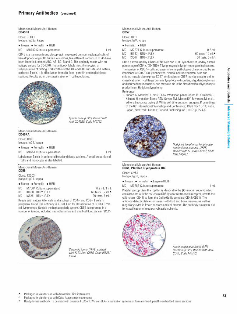
Primary Antibodies (continued)
83
Antibodies and ControlsAdvanced Staining Solutions
Monoclonal Mouse Anti-HumanCD45R0Clone: UCHL1Isotype: IgG2a, kappa Frozen Formalin HIER I M0742 Culture supernatant 1 mLCD45 is a transmembrane glycoprotein expressed on most nucleated cells of hematopoetic origin. On human leucocytes, five different isoforms of CD45 have been identified, named ABC, AB, BC, B and 0. This antibody reacts with an epitope unique for CD45R0. The antibody labels most thymocytes, a subpopulation of resting T cells within both CD4 and CD8 subsets, and mature, activated T cells. It is effective on formalin-fixed, paraffin-embedded tissue sections. Results aid in the classification of T-cell neoplasms.
Monoclonal Mouse Anti-HumanCD45RAClone: 4KB5Isotype: IgG1, kappa Frozen Formalin HIER I M0754 Culture supernatant 1 mLLabels most B cells in peripheral blood and tissue sections. A small proportion of T cells and monocytes is also labeled.
Monoclonal Mouse Anti-HumanCD56Clone: 123C3Isotype: IgG1, kappa Frozen Formalin HIER I M7304 Culture supernatant 0.2 mL/1 mLI IR628 RTU5, FLEX 60 tests, 12 mL2I IS628 RTU5, FLEX 30 tests, 6 mL"Reacts with natural killer cells and a subset of CD4+ and CD8+ T cells in peripheral blood. The antibody is a useful aid for classification of CD56+ T/NK-cell lymphomas. Outside the hematopoietic system, CD56 is expressed in a number of tumors, including neuroblastomas and small cell lung cancer (SCLC).
Monoclonal Mouse Anti-HumanCD57Clone: TB01Isotype: IgM, kappa Formalin HIER I M7271 Culture supernatant 0.2 mLI IR647 RTU5, FLEX 60 tests, 12 mL2I IS647 RTU5, FLEX 30 tests, 6 mL"CD57 is expressed by subsets of NK cells and CD8+ lymphocytes, and by a small percentage of CD4+/CD45R0+ T lymphocytes in lymph node germinal centres. The number of CD57+ cells increases in some pathologies characterized by an imbalance of CD4/CD8 lymphocytes. Normal neuroectodermal cells and striated muscle also express CD57. Antibodies to CD57 may be a useful aid for classification of T-cell large granular lymphocyte disorders, oligodendrogliomas and neuroendocrine tumors, and may also aid in the classification of lymphocyte predominant Hodgkin's lymphoma.Reference:1. Funaro A, Malavasi F. NK5. CD57 Workshop panel report. In: Kishimoto T,
Kikutani H, von dem Borne AEG, Goyert SM, Mason DY, Miyasaka M, et al., editors. Leucocyte typing VI. White cell differentiation antigens. Proceedings of the 6th International Workshop and Conference; 1996 Nov 10-14; Kobe, Japan. New York, London: Garland Publishing Inc.; 1997. p. 274-6.
Monoclonal Mouse Anti-HumanCD61, Platelet Glycoprotein IIIaClone: Y2/51Isotype: IgG1, kappa Frozen Formalin Enzyme/HIER I M0753 Culture supernatant 1 mLPlatelet glycoprotein IIIa (GpIIIa) is identical to the b3-integrin subunit, which can associate with the aV-chain (CD51) to form vitronectin receptor, or with the aIIb-chain (CD41) to form the GpIIb/GpIIIa complex (CD41/CD61). The antibody detects platelets in smears of blood and bone marrow, as well as megakaryocytes in frozen sections and cell smears. The antibody is a useful aid for classification of megakaryoblastic leukemia.
Lymph node (FFPE) stained with Anti-CD45R0, Code M0742.
Carcinoid tumor (FFPE) stained with FLEX Anti-CD56, Code IR628/IS628.
Hodgkin's lymphoma, lymphocyte predominant subtype, (FFPE) stained with FLEX Anti-CD57, Code IR647/IS647.
Acute megakaryoblastic (M7) leukemia (FFPE) stained with Anti-CD61, Code M0753.
2 Packaged in vials for use with Autostainer Link instruments" Packaged in vials for use with Dako Autostainer instruments5 Ready-to-use antibody. To be used with EnVision FLEX or EnVision FLEX+ visualization systems on formalin-fixed, paraffin-embedded tissue sections
USCatPathology.book Page 83 Monday, March 7, 2016 8:11 PM

84
Primary Antibodies (continued)
www.
dako
.com
Adva
nced
Sta
inin
g So
lutio
nsAn
tibod
ies a
nd C
ontro
ls
Monoclonal Mouse Anti-HumanCD68Clone: EBM11Isotype: IgG1, kappa Frozen I M0718 Culture supernatant 1 mLLabels human monocytes and macrophages and can be used for identifying a population of cells as being of mononuclear phagocyte origin and for demonstrating the macrophage origin of giant cells.
Monoclonal Mouse Anti-HumanCD68Clone: KP1Isotype: IgG1, kappa Formalin HIER I M0814 Culture supernatant 1 mLI GA609 RTU5, FLEX 60 tests, 12 mL8I IR609 RTU5, FLEX 60 tests, 12 mL2I IS609 RTU5, FLEX 30 tests, 6 mL"Labels human monocytes, macrophages and myeloid cells. It is of value for demonstrating reactive macrophages in a wide variety of normal and pathological specimens and for the identification of myeloid and histiocytic cells. Results aid in the classification of neoplasms of myeloid and macrophage/monocyte origin.
Monoclonal Mouse Anti-HumanCD68Clone: PG-M1Isotype: IgG3, kappa Frozen Formalin (Enzyme)/HIER I M0876 Culture supernatant 0.2 mL/1 mLI GA613 RTU5, FLEX 60 tests, 12 mL8I IR613 RTU5, FLEX 60 tests, 12 mL2I IS613 RTU5, FLEX 30 tests, 6 mL"Labels human monocytes and macrophages, but not myeloid cells. The antibody is of value for demonstration of monocytes and macrophages in normal and pathological specimens. Results aid in the classification of acute myeloid leukemia (AML), and histiocytic sarcoma.
Monoclonal Mouse Anti-HumanCD79aClone: JCB117Isotype: IgG1, kappa Frozen Formalin HIER I M7050 Culture supernatant 0.2 mL/1 mLI GA621 RTU5, FLEX 60 tests, 12 mL8I IR621 RTU5, FLEX 60 tests, 12 mL2I IS621 RTU5, FLEX 30 tests, 6 mL"CD79a is encoded by the mb-1 gene and was previously called Iga. The antibody recognizes an extracellular epitope expressed on the CD79a molecule. Results aid in the classification of B-cell neoplasms in routine biopsy material. In addition to the expression in B cells, CD79a has been found to be co-expressed with CD3 in 10% of cases of T-lymphoblastic leukemia/lymphoma. Antibodies to CD79a may also be a useful aid for classification of Hodgkin's disease.References:1. Mason DY, Cordell JL, Brown MH, Borst J, Jones M, Pulford K, et al.
CD79a: a novel marker for B-cell neoplasms in routinely processed tissue samples. Blood 1995;86:1453-9.
2. Pilozzi E, Pulford K, Jones M, Muller-Hermelink HK, Falini B, Ralfkiaer E, et al. Co-expression of CD79a (JCB117) and CD3 by lymphoblastic lymphoma. J Pathol 1998;186:140-3.
3. Chu PG, Arber DA. CD79: a review. Appl Immunohistochem Mol Morphol 2001;9:97-106
Monoclonal Mouse Anti-HumanCD99, MIC2 Gene Products, Ewing's Sarcoma MarkerClone: 12E7Isotype: IgG1, kappa Frozen Formalin I M3601 Culture supernatant 1 mLI IR057 RTU5, FLEX 60 tests, 12 mL2I IS057 RTU5, FLEX 30 tests, 6 mL"The MIC2 gene products are also called p30/32mic2. They are expressed on the cell membrane of some lymphocytes (bone marrow, lymph nodes and spleen), cortical thymocytes, granulosa cells of the ovary, most Langerhans' islet cells, CNS ependymal cells, Sertoli's cells of the testis and, in a few cases, endothelial cells of single blood vessels. Results aid in the classification of Ewing's sarcoma and primitive peripheral neuroectoderm tumors.
Tonsil (FFPE) stained with FLEX Anti-CD68, Code GA609, on Dako Omnis.
Tonsil (FFPE) stained with FLEX Anti-CD68, Code GA613, on Dako Omnis.
Plasmacytoma (FFPE) stained with FLEX Anti-CD79a Code GA621, on Dako Omnis.
Ewing's sarcoma (FFPE) stained with FLEX Anti-CD99, Code IR057/IS057.
1 Packaged in vials for use with Dako Omnis2 Packaged in vials for use with Autostainer Link instruments" Packaged in vials for use with Dako Autostainer instruments5 Ready-to-use antibody. To be used with EnVision FLEX or EnVision FLEX+ visualization systems on formalin-fixed, paraffin-embedded tissue sections
USCatPathology.book Page 84 Monday, March 7, 2016 8:11 PM

Primary Antibodies (continued)
85
Antibodies and ControlsAdvanced Staining Solutions
Monoclonal Mouse Anti-HumanCD105, EndoglinClone: SN6hIsotype: IgG1, kappa Frozen Formalin Enzyme I M3527 Culture supernatant 1 mLEndoglin is a type I transmembrane protein which is highly expressed on human vascular endothelial cells. A large variety of tissues express endoglin.
Polyclonal Rabbit Anti-HumanCD117, c-kit Frozen Formalin HIER I A4502 Affinity isolated 0.2 mLThe antibody labels the transmembrane tyrosine kinase receptor CD117/c-kit, located in hematopoietic stem cells, melanocytes, mast cells, Cajal cells, germ cells, basal cells of skin, and mammary ductal epithelia. Antibodies to CD117 may be useful for the classification of several cancers expressing c-kit, including gastrointestinal stromal tumors (GISTs), mast cell diseases, acute myeloid leukemia (AML), small cell lung carcinoma (SCLC), and Ewing's sarcoma (1-4).References:1. Tsuura Y, Hiraki H, Watanabe K, Igarashi S, Shimamura K, Fukuda T, et al.
Preferential localization of c-kit product in tissue mast cells, basal cells of skin, epithelial cells of breast, small cell lung carcinoma and seminoma/dysgerminoma in human: immunohistochemical study on formalin-fixed, paraffin-embedded tissues. Virchows Arch 1994;424:135-41.
2. van Oosterom AT, Judson I, Verweij J, Stroobants S, di Paola ED, Dimitrijevic S, et al. Safety and efficacy of imatinib (STI571) in metastatic gastrointestinal stromal tumours: a phase I study. Lancet 2001;358:1421-3.
3. Hornick JL, Fletcher CDM. Immunohistochemical staining for KIT (CD117) in soft tissue sarcomas is very limited in distribution. Am J Clin Pathol 2002;117:188-93
4. Smithey BE, Pappo AS, Hill DA. C-kit expression in pediatric solid tumors: a comparative immunohistochemical study. Am J Surg Pathol 2002;26:486-92.
Monoclonal Mouse Anti-HumanCD138Clone: MI15Isotype: IgG1, kappa Frozen Formalin HIER I M7228 Culture supernatant 1 mLI GA642 RTU5, FLEX 60 tests, 12 mL8I IR642 RTU5, FLEX 60 tests, 12 mL2I IS642 RTU5, FLEX 30 tests, 6 mL"CD138, syndecan-1, is a transmembrane proteoglycan with a main cellular expression in stratified and simple epithelia. Within the hemopoietic system, CD138 is mainly confined to late stages of B-cell differentiation. CD138 expression is reduced during malignant transformation of various epithelia, and CD138 is rapidly shed by myeloma cells entering into apoptosis. This antibody is a useful aid for classification of multiple myeloma. Anti-CD138 may also be useful for the subclassification of diffuse large B-cell lymphomas.
CD141See: Thrombomodulin
Monoclonal Mouse Anti-HumanCD235a, Glycophorin AClone: JC159Isotype: IgG1, kappa Frozen Formalin HIER I M0819 Culture supernatant 1 mLReacts with normal erythroid cells at essentially all stages of differentiation from erythroblasts to mature erythrocytes. The antibody is a useful aid for classification of erythroleukemia.
CD236RSee: Glycophorin C
Gastrointestinal tumor (FFPE) stained with Anti-CD117, c-kit.
High grade myeloma (FFPE) stained with FLEX Anti-CD138, Code GA642, on Dako Omnis.
Bone marrow normal erythropoiesis (FFPE) stained with Anti-CD235a, Glycophorin A, Code M0819.
1 Packaged in vials for use with Dako Omnis2 Packaged in vials for use with Autostainer Link instruments" Packaged in vials for use with Dako Autostainer instruments5 Ready-to-use antibody. To be used with EnVision FLEX or EnVision FLEX+ visualization systems on formalin-fixed, paraffin-embedded tissue sections
00110 IHC Solutions US_1.fm Page 85 Monday, March 14, 2016 11:31 PM

86
Primary Antibodies (continued)
www.
dako
.com
Adva
nced
Sta
inin
g So
lutio
nsAn
tibod
ies a
nd C
ontro
ls
Monoclonal Mouse Anti-HumanCD246, ALK ProteinClone: ALK1Isotype: IgG3, kappa Frozen Formalin HIER I M7195 Culture supernatant 0.2 mL/1 mLI GA641 RTU5, FLEX 60 tests, 12 mL8I IR641 RTU5, FLEX 60 tests, 12 mL2I IS641 RTU5, FLEX 30 tests, 6 mL"Recognizes a formalin-resistant epitope in both the 80 kDa NPM-ALK chimeric and the 200 kDa normal human ALK proteins (1). Normal ALK protein expression is restricted to the central nervous system. The hybrid gene, NPM-ALK, created by the t(2;5)(p23;q35) chromosomal translocation encodes part of the nucleolar phosphoprotein, nucleophosmin (NPM), joined to the cytoplasmic portion of the anaplastic lymphoma kinase (ALK) receptor tyrosine kinase (2). This antibody is a useful aid for classification of anaplastic large cell lymphoma (ALCL).References:1. Pulford K, Lamant L, Morris SW, Butler LH, Wood KM, Stroud D, et al.
Detection of anaplastic lymphoma kinase (ALK) and nucleolar protein nucleophosmin (NPM)-ALK proteins in normal and neoplastic cells with the monoclonal antibody ALK1. Blood 1997;89:1394-404.
2. Morris SW, Kirstein MN, Valentine MB, Dittmer KG, Shapiro DN, Saltman DL, et al. Fusion of a kinase gene, ALK, to a nucleolar protein gene, NPM, in non-Hodgkin's lymphoma (published erratum appears in Science 1995;267:316-7). Science 1994;263:1281-4.
Monoclonal Mouse Anti-HumanCDX2Clone: DAK-CDX2Isotype: IgG1, kappa Frozen Formalin HIER I M3636 Culture supernatant 0.2 mL/1 mLI GA080 RTU5, FLEX 60 tests, 12 mL8I IR080 RTU5, FLEX 60 tests, 12 mL2I IS080 RTU5, FLEX 30 tests, 6 mL"Cdx2, a human gene homologous to the Drosophila caudal-type homeobox gene, encodes a transcription factor, which is involved in proliferation and differentiation of intestinal epithelial cells. The CDX2 protein is widely expressed in intestinal epithelium from the duodenum to the rectum. Scattered expression has been reported in pancreatic ductules, while no expression has been observed in other normal tissues tested. Results aid in the classification of both primary and metastatic tumors of the gastrointestinal tract, including carcinoids.
Polyclonal Rabbit Anti-Humanc-erbB-2 OncoproteinS A0485 Affinity isolated 0.2 mLA synthetic peptide, coupled to keyhole limpet hemocyanin, has been used for immunization. The peptide represents part of the predicted 185 kDa oncoprotein encoded by ERBB2, also termed HER2 or NEU. The antibody recognizes an epitope on the cytoplasmic part of the cell membrane-bound c-erbB-2 oncoprotein.
Polyclonal Rabbit Anti-HumanChorionic Gonadotropin (hCG) Formalin HIER I A0231 Ig fraction 2 mLI GA508 RTU5, FLEX NEW 60 tests, 12 mL8I IR508 RTU5, FLEX 60 tests, 12 mL2I IS508 RTU5, FLEX 30 tests, 6 mL"The isolated b-chain of hCG is used for immunization. The antibody cross-reacts with human luteinizing hormone (LH). For immunohistochemical use, the cross-reaction with LH will not cause misinterpretation, and the antibody is well-suited for identification of hCG in trophoblastic elements and is a useful aid for classification of germ cell tumors.
Monoclonal Mouse Anti-HumanChromogranin AClone: DAK-A3Isotype: IgG2b, kappa Frozen Formalin HIER I M0869 Culture supernatant 0.2 mL/1 mLLabels cells of neuroendocrine origin. Anti-Chromogranin A, clone DAK-A3, reacts with an epitope on the C-terminal half of the chromogranin A molecule. Results aid in the classification of neuroendocrine-derived tumors.
c-kitSee: CD117, c-kit
Anaplastic large cell lymphoma (FFPE) stained with FLEX Anti-CD246, ALK Protein, Code GA641, on Dako Omnis.
Colon adenocarcinoma (FFPE) stained with FLEX Anti-CDX2, Code GA080, on Dako Omnis.
Placenta (FFPE) stained with FLEX Anti-Human Chorionic Gonadotropin, Code GA508, on Dako Omnis.
1 Packaged in vials for use with Dako Omnis2 Packaged in vials for use with Autostainer Link instruments" Packaged in vials for use with Dako Autostainer instruments5 Ready-to-use antibody. To be used with EnVision FLEX or EnVision FLEX+ visualization systems on formalin-fixed, paraffin-embedded tissue sections
USCatPathology.book Page 86 Monday, March 7, 2016 8:11 PM

Primary Antibodies (continued)
87
Antibodies and ControlsAdvanced Staining Solutions
Monoclonal Mouse Anti-HumanCollagen IVClone: CIV 22Isotype: IgG1, kappa Frozen Formalin HIER I M0785 Culture supernatant 1 mLIs directed against collagen IV, a major constituent of the basement membrane. The antibody is important in demonstrating the loss of basement membrane. The antibody is a useful aid for classification of invasive carcinomas.
Monoclonal Mouse Anti-HumanCOX-2Clone: CX-294Isotype: IgG2a, kappa Frozen Formalin HIER I M3617 Culture supernatant 1 mLThe cyclooxygenase (COX) enzymes are critical in the biosynthesis of prostaglandins from arachidonic acid. COX-2 is a 70 kDa enzyme that is responsible for prostaglandin synthesis at the site of inflammation and is readily induced in response to cell activation by cytokines, growth factors and tumor promoters. Results aid in the classification of a variety of malignancies, including colorectal adenocarcinoma, a subset of breast adenocarcinomas and adjacent ductal carcinoma in situ, lung adenocarcinoma, esophageal squamous cell carcinoma and squamous adenocarcinoma, malignant melanoma, and subsets of ovarian carcinoma and prostate carcinoma.
Monoclonal Rabbit Anti-HumanCyclin D1Clone: EP12 Formalin HIER I M3642 Affinity isolated 1 mLI GA083 RTU5, FLEX NEW 60 tests, 12 mL8I IR083 RTU5, FLEX 60 tests, 12 mL2I IS083 RTU5, FLEX 30 tests, 6 mL "This monoclonal rabbit antibody reacts with cyclin D1, a 36 kDa protein encoded by the CCND1 (bcl-1) gene located on chromosome 11q13. Cyclin D1 is part of the cell cycle regulation and oncogenic transformation in mammalian cells. The cyclin-dependent kinases, CDK4 and CDK6, are associated with and activated by cyclin D1 thereby promoting G1 phase progression by retinoblastoma protein phosphorylating along with related proteins. Antibodies to cyclin D1 are a useful aid for classification of mantle cell lymphomas in the context of lymphoid tumors (1-2).References:1. Donnellan R, Chetty R. Cyclin D1 and human neoplasia. J Clin Pathol: Mol
Pathol 1998;51:1-7.2. Falini B, Martelli MP, Tiacci E, Ascani S, Thiede C, Pileri SA.
Immunohistochemical surrogates for genetic alterations of CCND1, PML, ALK, and NPM1 genes in lymphomas and acute myeloid leukemia. Best Practice & Research Clin Haematol 2010:23:417-31.
Monoclonal Mouse Anti-HumanCytokeratinClone: AE1/AE3Isotype: IgG1, kappa Frozen Formalin (Enzyme)/HIER I M3515 Ascites 0.2 mL/1 mLI GA053 RTU5, FLEX 60 tests, 12 mL8I IR053 RTU5, FLEX 60 tests, 12 mL2I IS053 RTU5, FLEX 30 tests, 6 mL"Reacts with the 65-67, 64, 59, 58, 56.5, 56, 54, 52, 50, 48 and 40 kDa cytokeratins. The antibody labels keratinized and corneal epidermis, stratified squamous epithelia of internal organs, stratified epithelia, hyperproliferative keratinocytes, and simple epithelia. The antibody is useful aid for classification of tumors of epithelial origin as well as undifferentiated carcinomas.
Monoclonal Mouse Anti-HumanCytokeratinClone: MNF116Isotype: IgG1, kappa Frozen Formalin Enzyme I M0821 Culture supernatant 1 mLReacts with cytokeratins 5, 6, 8, 17 and may also react with 19. The antibody shows an especially broad pattern of reactivity with human epithelial tissue from simple glandular to stratified squamous epithelium and can be used in the classification of neoplastic cells of epithelial origin. References:1. Prieto VG, Lugo J, McNutt NS. Intermediate- and low-molecular-weight
keratin detection with the monoclonal antibody MNF116. An immunohistochemical study on 232 paraffin-embedded cutaneous lesions. J Cutan Pathol 1996;23:234-41.
2. Richter T, Nährig J, Komminoth P, Kowolik J, Werner M. Protocol for ultrarapid immunostaining of frozen sections. J Clin Pathol 1999;52:461-3.
Mantle cell lymphoma (FFPE) stained with FLEX Anti-Cyclin D1, Code GA083, on Dako Omnis.
Dako FLEX RTU Antibodies for Undifferentiated Tumor TestingSee our panel of FLEX antibodies at page 72
Adenocarcinoma (FFPE) stained with FLEX Anti-Cytokeratin, Code GA053, on Dako Omnis.
Skin (FFPE) stained with Anti-Cytokeratin, Code M0821.
1 Packaged in vials for use with Dako Omnis2 Packaged in vials for use with Autostainer Link instruments" Packaged in vials for use with Dako Autostainer instruments5 Ready-to-use antibody. To be used with EnVision FLEX or EnVision FLEX+ visualization systems on formalin-fixed, paraffin-embedded tissue sections
USCatPathology.book Page 87 Monday, March 7, 2016 8:11 PM

88
Primary Antibodies (continued)
www.
dako
.com
Adva
nced
Sta
inin
g So
lutio
nsAn
tibod
ies a
nd C
ontro
ls
Monoclonal Mouse Anti-HumanCytokeratin 5/6Clone: D5/16 B4Isotype: IgG1, kappa Frozen Formalin HIER I M7237 Ascites Ig fraction 0.2 mL/1 mLI GA780 RTU5, FLEX 60 tests, 12 mL8I IR780 RTU5, FLEX 60 tests, 12 mL2I IS780 RTU5, FLEX 30 tests, 6 mL"Reacts strongly with cytokeratins 5 and 6 and weakly with cytokeratin 4. The antibody does not cross-react with cytokeratins 1, 7, 8, 10, 13, 14, 18 and 19. It labels mesothelioma, and epithelial basal cells in prostate and tonsil. No reactivity with other mesodermally derived tissues, such as muscle and connective tissues, has been observed. The antibody is a useful aid for classification of epithelioid mesotheliomas.
Monoclonal Mouse Anti-HumanCytokeratin 7Clone: OV-TL 12/30Isotype: IgG1, kappa Frozen Formalin Enzyme/HIER I M7018 Culture supernatant 0.2 mL/1 mLI GA619 RTU5, FLEX 60 tests, 12 mL8I IR619 RTU5, FLEX 60 tests, 12 mL2I IS619 RTU5, FLEX 30 tests, 6 mL"Reacts with the 54 kDa protein corresponding to cytokeratin 7. The antibody labels several types of normal and neoplastic epithelia, including many ductal and glandular epithelia. The antibody is a useful aid for classification of adenocarcinomas of the lung, breast and endometrium, thyroid gland and ovary, as well as chromophobe renal cell carcinomas. Results may also aid in the classification of prostate carcinomas where CK7 is rarely expressed.Reference:1. Chu P, Wu W, Weiss LM. Cytokeratin 7 and Cytokeratin 20 expression in
epithelial neoplasms: a survey of 435 cases. Mod Pathol 2000;13:962-72.
Monoclonal Rabbit Anti-HumanCytokeratin 8/18Clone: EP17/EP30 Formalin HIER I M3652 Culture supernatant 1 mLI IR094 RTU5 60 tests, 12 mL2Cytokeratins 8 and 18 (CK8/18) make up one of the low molecular weight cytokeratins (LMW-CK). CK8/18 is expressed in simple, non-stratified epithelia, basal and superficial cells of transitional epithelium, the luminal/secretory cells of complex epithelia, mesothelium, and may be present in some types of mesenchymal cells. Multiple cytokeratins family members may be expressed in a given cell and are characteristic of the cell type and differentiation state. Nearly all carcinomas of epithelial origin and mesotheliomas express CK8/18, and CK8/18 expression patterns aid in the classification of tumors of unknown origin and poorly differentiated carcinomas. Antibodies to CK8/18 may be useful for classification of tumors of epithelial origin (1).This product is a cocktail of two monoclonal rabbit antibodies.Reference:1. Moll R, Divo M, Langbein L. The human keratins: biology and pathology.
Histochem Cell Biol 2008;129:705-33.
Monoclonal Mouse Anti-HumanCytokeratin 10Clone: DE-K10Isotype: IgG1, kappa Frozen Formalin HIER I M7002 Culture supernatant 1 mLReacts with the 56.5 kDa protein corresponding to cytokeratin 10. The antibody labels cytokeratin 10 present in keratinizing and non-keratinizing stratified epithelia and in the more differentiated areas of some squamous cell carcinomas.
Monoclonal Mouse Anti-HumanCytokeratin 10/13Clone: DE-K13Isotype: IgG2a, kappa Frozen Formalin HIER I M7003 Culture supernatant 1 mLReacts on formalin-fixed, paraffin-embedded tissue sections with the 53 kDa protein corresponding to cytokeratin 13. This cytokeratin is present in several non-cornified, stratified squamous epithelia, for example tongue mucosa, and tracheal and anal canal epithelium. On frozen sections the antibody, in addition, reacts with the 56.5 kDa protein corresponding to cytokeratin 10.
Squamous cell carcinoma of the lung (FFPE) stained with FLEX Anti-Cytokeratin 5/6, Code IR780/IS780.
Ductal carcinoma (FFPE) stained with FLEX Anti-Cytokeratin 7, Code GA619, on Dako Omnis.
Hepatocellular carcinoma (FFPE) stained with FLEX Anti-Cytokeratin 8/18, Code IR094.
1 Packaged in vials for use with Dako Omnis2 Packaged in vials for use with Autostainer Link instruments" Packaged in vials for use with Dako Autostainer instruments5 Ready-to-use antibody. To be used with EnVision FLEX or EnVision FLEX+ visualization systems on formalin-fixed, paraffin-embedded tissue sections
00110 IHC Solutions US_1.fm Page 88 Monday, March 14, 2016 11:36 PM

Primary Antibodies (continued)
89
Antibodies and ControlsAdvanced Staining Solutions
Monoclonal Mouse Anti-Cytokeratin 17Clone: E3Isotype: IgG2b, kappa Frozen Formalin HIER I M7046 Culture supernatant 1 mLI IR620 RTU5, FLEX 60 tests, 12 mL2I IS620 RTU5, FLEX 30 tests, 6 mL"Reacts with the 46 kDa human protein corresponding to cytokeratin 17. The antibody labels the basal layer of complex epithelia, i.e. the basal layer of pseudostratified epithelium in the larynx, trachea and bronchi. Results aid in the classification of squamous cell carcinomas of the lung, cervix and oral cavity.
Monoclonal Mouse Anti-HumanCytokeratin 18Clone: DC 10Isotype: IgG1, kappa Frozen Formalin Enzyme/HIER I M7010 Culture supernatant 0.2 mLI GA618 RTU5, FLEX 60 tests, 12 mL8I IR618 RTU5, FLEX 60 tests, 12 mL2I IS618 RTU5, FLEX 30 tests, 6 mL"Reacts with the 45 kDa protein corresponding to cytokeratin 18. The antibody labels a large number of simple epithelia, including many ductal and glandular epithelia. The antibody is a useful aid for classification of tumors of epithelial origin. In vascular tumors, the antibody may be a useful aid in the classification of epithelioid hemangioendotheliomas.Reference:1. Lauerova L, Kovarik J, Bartek J, Rejthar A, Vojtesek B. Novel monoclonal
antibodies defining epitope of human cytokeratin 18 molecule. Hybridoma 1988;7:495-504.
Monoclonal Mouse Anti-HumanCytokeratin 19Clone: RCK108Isotype: IgG1, kappa Frozen Formalin Enzyme/HIER I M0888 Culture supernatant 1 mLI GA615 RTU5, FLEX 60 tests, 12 mL8I IR615 RTU5, FLEX 60 tests, 12 mL2I IS615 RTU5, FLEX 30 tests, 6 mL"Reacts with the 40 kDa protein corresponding to cytokeratin 19. The antibody labels many types of simple and non-keratinizing epithelia, including ductal and glandular epithelia. The antibody is a useful aid for classification of tumors of epithelial origin.Reference:1. Dalal P, Shousha S. Keratin 19 in paraffin sections of medullary carcinoma
and other benign and malignant breast lesions. Mod Pathol 1995;8:413-6.
Monoclonal Mouse Anti-HumanCytokeratin 20Clone: Ks20.8Isotype: IgG2a, kappa Formalin Enzyme/HIER I M7019 Purified 0.2 mL/1 mLI GA777 RTU5, FLEX 60 tests, 12 mL8I IR777 RTU5, FLEX 60 tests, 12 mL2I IS777 RTU5, FLEX 30 tests, 6 mL"Reacts with the 46 kDa protein corresponding to cytokeratin 20. The antibody is a useful aid for classification of adenocarcinomas of the colon, mucinous ovarian tumors, transitional-cell and Merkel-cell carcinomas, adenocarcinomas of the stomach, bile system and pancreas. Results also aid in the classification of most squamous cell carcinomas and most adenocarcinomas from other sites (breast, endometrium, lung, prostate), non-mucinous tumors of the ovary, and small-cell lung carcinomas as these may be negative.References:1. Moll R, Löwe A, Laufer J, Franke WW. Cytokeratin 20 in human carcinomas.
Am J Pathol 1992;140:427-47.2. Harnden P, Mahmood N, Southgate J. Expression of cytokeratin 20
redefines urothelial papillomas of the bladder. Lancet 1999;353:974-7.3. Chu P, Wu W, Weiss LM. Cytokeratin 7 and cytokeratin 20 expression in
epithelial neoplasms: a survey of 435 cases. Mod Pathol 2000;13:962-72.
Renal clear cell carcinoma stained with FLEX Anti-Cytokeratin 18, Code GA618, on Dako Omnis.
Thyroid papillary carcinoma (FFPE) stained with FLEX Anti-Cytokeratin 19, Code GA615, on Dako Omnis.
Merkel cell carcinoma (FFPE) stained with FLEX Anti-Cytokeratin 20, Code GA777, on Dako Omnis.
1 Packaged in vials for use with Dako Omnis2 Packaged in vials for use with Autostainer Link instruments" Packaged in vials for use with Dako Autostainer instruments5 Ready-to-use antibody. To be used with EnVision FLEX or EnVision FLEX+ visualization systems on formalin-fixed, paraffin-embedded tissue sections
USCatPathology.book Page 89 Monday, March 7, 2016 8:11 PM

90
Primary Antibodies (continued)
www.
dako
.com
Adva
nced
Sta
inin
g So
lutio
nsAn
tibod
ies a
nd C
ontro
ls
Monoclonal Mouse Anti-HumanCytokeratin, High Molecular WeightClone: 34bE12Isotype: IgG1, kappa Frozen Formalin Enzyme + HIER I M0630 Culture supernatant 0.2 mL/1 mLI GA051 RTU5, FLEX 60 tests, 12 mL8I IR051 RTU5, FLEX 60 tests, 12 mL2I IS051 RTU5, FLEX 30 tests, 6 mL"Reacts with the 68 kDa, 58 kDa, 56.5 kDa and 50 kDa proteins corresponding to cytokeratins 1, 5, 10 and 14. The antibody labels squamous, ductal and complex epithelia. Results aid in the classification of prostatic adenocarcinoma and in the classification of neoplastic tissue as carcinoma of epithelial origin.
Polyclonal Rabbit Anti-Cytokeratin, Wide Spectrum Screening Formalin Enzyme I Z0622 Ig fraction 1 mLCow keratin has been used as immunogen. This antibody is particularly well-suited for the staining of a broad spectrum of human keratins and is a useful aid for the classification of neoplasm of epithelial origin.
Monoclonal Mouse Anti-CytomegalovirusClone: CCH2 + DDG9Isotype: IgG2a, kappa + IgG1, kappaS M0854 Culture supernatant 1 mLReacts with CMV immediate early antigen and early antigen. The antibody shows no cross-reaction with other herpesviruses or with adenovirus.
D2-40See: Podoplanin
Dendritic Reticulum CellSee: CD35
Monoclonal Mouse Anti-HumanDesminClone: D33Isotype: IgG1, kappa Frozen Formalin HIER I M0760 Culture supernatant 0.2 mL/1 mLI IR606 RTU5, FLEX 60 tests, 12 mL2I IS606 RTU5, FLEX 30 tests, 6 mL"Labels smooth and striated muscle cells. The antibody works on paraffin-embedded material without pretreament with proteolytic enzymes. Anti-Desmin, clone D33, is a useful aid for classification of tumors of uncertain origin.
Monoclonal Mouse Anti-HumanE-CadherinClone: NCH-38Isotype: IgG1, kappa Frozen Formalin HIER I M3612 Culture supernatant 0.2 mL/1 mLI GA059 RTU5, FLEX 60 tests, 12 mL8I IR059 RTU5, FLEX 60 tests, 12 mL2I IS059 RTU5, FLEX 30 tests, 6 mL"E (epithelial)-cadherin is a 120 kDa transmembrane cell adhesion molecule. It has a significant function in intercellular adhesion of epithelial cells, the establishment of epithelial polarization, glandular differentiation and stratification. Down-regulation of E-cadherin expression has been observed in a number of carcinomas and is usually associated with advanced stage and progression. The antibody is a useful aid for classification of ductal breast carcinomas.
Prostate (FFPE) stained with FLEX Anti-Cytokeratin HMW, Code GA051, on Dako Omnis.
Squamous cell carcinoma (FFPE) stained with Anti-Cytokeratin, Wide Spectrum Screening.
Dako FLEX RTU Antibodies for Skeletal Muscle TestingSee our panel of FLEX antibodies at page 70
Rhabdomyosarcoma (FFPE) stained with FLEX Anti-Desmin, Code IR606/IS606.
Poorly differentiated ductal carcinoma (FFPE) stained with FLEX Anti-E-Cadherin, Code GA059, on Dako Omnis.
1 Packaged in vials for use with Dako Omnis2 Packaged in vials for use with Autostainer Link instruments" Packaged in vials for use with Dako Autostainer instruments5 Ready-to-use antibody. To be used with EnVision FLEX or EnVision FLEX+ visualization systems on formalin-fixed, paraffin-embedded tissue sections
USCatPathology.book Page 90 Monday, March 7, 2016 8:11 PM

Primary Antibodies (continued)
91
Antibodies and ControlsAdvanced Staining Solutions
Monoclonal Mouse Anti-HumanEGFRClone: H11Isotype: IgG1, kappa Frozen Formalin Enzyme I M3563 Culture supernatant 1 mLLabels the majority of simple and squamous epithelia. Results aid in the classification of neoplasms exhibiting EGFR expression, for example in a proportion of cases of breast, bladder, pancreatic, stomach and thyroid tumors.
Monoclonal Mouse Anti-HumanEGFR, Wild-TypeClone: DAK-H1-WTIsotype: IgG1 Formalin HIER U M7298 Culture supernatant 0.2 mLReacts with the N-terminal part of the extracellular domain of human epidermal growth factor receptor (EGFR). EGFR is a 170 kDa transmembrane receptor that is expressed by a variety of normal cells including fibroblasts and a wide range of epithelia. This antibody does not label the 2-7 truncated EGFR variant (EGFR-vIII).Reference:1. Jorissen RN, Walker F, Pouliot N, Garrett TP, Ward CW, Burgess AW.
Epidermal growth factor receptor: mechanisms of activation and signaling [review]. Exp Cell Res 2003;284:31-53.
Monoclonal Mouse Anti-HumanEGFR-pY1197Phosphorylation Site SpecificClone: DAK-H1-1197Isotype: IgG2a Formalin HIER U M7299 Culture supernatant 0.2 mLReacts with epidermal growth factor receptor (EGFR) phosphorylated at tyrosine residue 1197 (pY1197). Binding of epidermal growth factor to the extracellular domain of EGFR results in receptor dimerization and autophosphorylation on tyrosine residues, pY1197 being one of the major autophosphorylation sites (1, 2).References:1. Chattopadhyay A, Vecchi M, Ji Q, Mernaugh R, Carpenter G. The role of
individual SH2 domains in mediating association of phospholipase C-gamma1 with the activated EGF receptor. J Biol Chem 1999;274:26091-7.
2. Lombardo CR, Consler TG, Kassel DB. In vitro phosphorylation of the epidermal growth factor receptor autophosphorylation domain by c-src: identification of phosphorylation sites and c-src SH2 domain binding sites. Biochem 1995;34:16456-66.
EndoglinSee: CD105, Endoglin
Endothelial CellSee: CD31, Endothelial Cell
Monoclonal Mouse Anti-EnterovirusClone: 5-D8/1Isotype: IgG2a, kappaS M7064 Culture supernatant 1 mLReacts with an epitope on the VP1 peptide which is highly conserved within the enterovirus group. The antibody, originally generated using coxsackie virus B5 as immunogen, reacts with most of the enterovirus strains of the coxsackie, echo and poliovirus groups. No reaction is seen with human rotavirus, yellow fever virus, measles virus, rhinovirus A1 and adenovirus 18.
Epidermal Growth Factor ReceptorSee: EGFR
Monoclonal Mouse Anti-HumanEpithelial AntigenClone: Ber-EP4Isotype: IgG1, kappa Frozen Formalin HIER I M0804 Culture supernatant 0.2 mL/1 mLI GA637 RTU5, FLEX 60 tests, 12 mL8I IR637 RTU5, FLEX 60 tests, 12 mL2I IS637 RTU5, FLEX 30 tests, 6 mL"Epithelial antigen is a transmembrane glycoprotein functioning as a cellular adhesion molecule. This epithelium-specific antigen is broadly distributed in epithelial cells, and displays a highly conserved expression in carcinomas. The antibody is a useful aid for classification of adenocarcinoma. Anti-Epithelial Antigen may also aid in classification of micrometastases in lymph nodes of esophageal carcinoma as well as in classification of basal and squamous cell carcinomas of the skin.
Squamous cell carcinoma (FFPE) stained with Anti-EGFR.
Dako FLEX RTU Antibodies for Mesothelial Surface TestingSee our panel of FLEX antibodies at page 68
Adenocarcinoma (FFPE) stained with FLEX Anti-Epithelial Antigen, Code GA637, on Dako Omnis.
1 Packaged in vials for use with Dako Omnis2 Packaged in vials for use with Autostainer Link instruments" Packaged in vials for use with Dako Autostainer instruments5 Ready-to-use antibody. To be used with EnVision FLEX or EnVision FLEX+ visualization systems on formalin-fixed, paraffin-embedded tissue sections
USCatPathology.book Page 91 Monday, March 7, 2016 8:11 PM
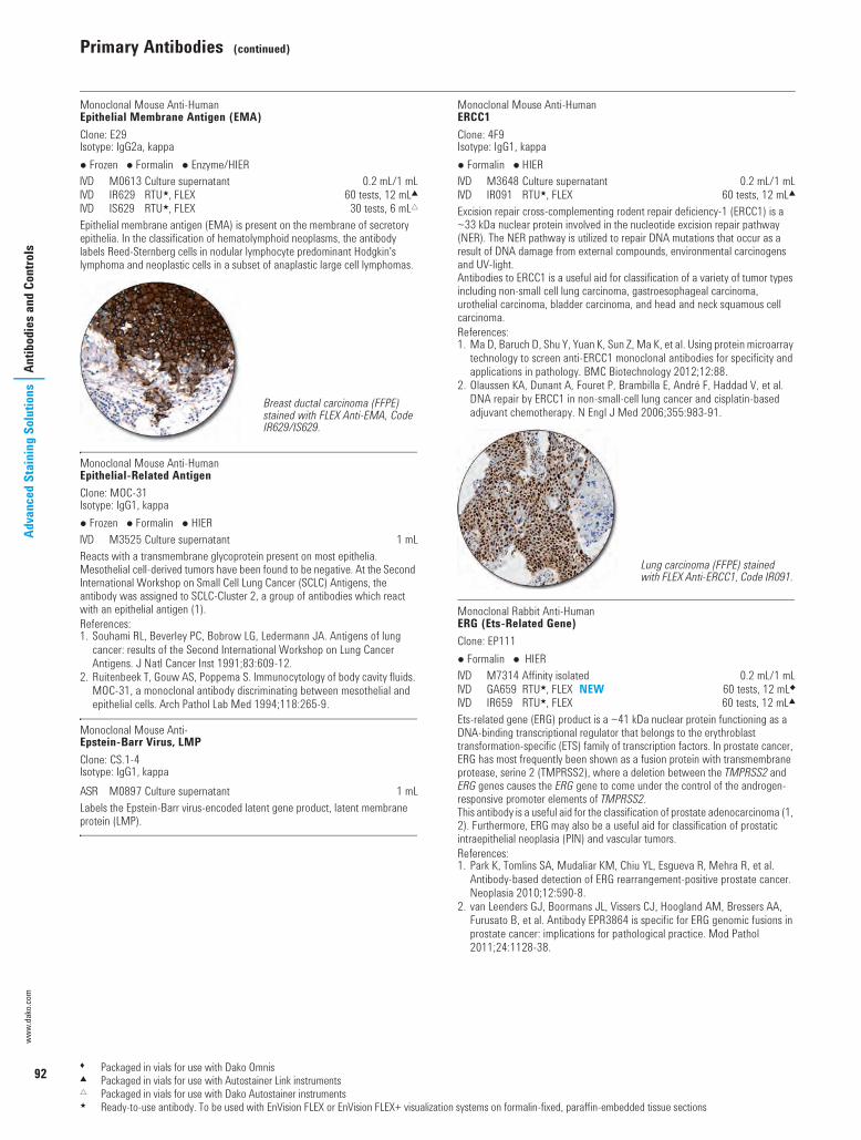
92
Primary Antibodies (continued)
www.
dako
.com
Adva
nced
Sta
inin
g So
lutio
nsAn
tibod
ies a
nd C
ontro
ls
Monoclonal Mouse Anti-HumanEpithelial Membrane Antigen (EMA)Clone: E29Isotype: IgG2a, kappa Frozen Formalin Enzyme/HIER I M0613 Culture supernatant 0.2 mL/1 mLI IR629 RTU5, FLEX 60 tests, 12 mL2I IS629 RTU5, FLEX 30 tests, 6 mL"Epithelial membrane antigen (EMA) is present on the membrane of secretory epithelia. In the classification of hematolymphoid neoplasms, the antibody labels Reed-Sternberg cells in nodular lymphocyte predominant Hodgkin's lymphoma and neoplastic cells in a subset of anaplastic large cell lymphomas.
Monoclonal Mouse Anti-HumanEpithelial-Related AntigenClone: MOC-31Isotype: IgG1, kappa Frozen Formalin HIER I M3525 Culture supernatant 1 mLReacts with a transmembrane glycoprotein present on most epithelia. Mesothelial cell-derived tumors have been found to be negative. At the Second International Workshop on Small Cell Lung Cancer (SCLC) Antigens, the antibody was assigned to SCLC-Cluster 2, a group of antibodies which react with an epithelial antigen (1).References:1. Souhami RL, Beverley PC, Bobrow LG, Ledermann JA. Antigens of lung
cancer: results of the Second International Workshop on Lung Cancer Antigens. J Natl Cancer Inst 1991;83:609-12.
2. Ruitenbeek T, Gouw AS, Poppema S. Immunocytology of body cavity fluids. MOC-31, a monoclonal antibody discriminating between mesothelial and epithelial cells. Arch Pathol Lab Med 1994;118:265-9.
Monoclonal Mouse Anti-Epstein-Barr Virus, LMPClone: CS.1-4Isotype: IgG1, kappaS M0897 Culture supernatant 1 mLLabels the Epstein-Barr virus-encoded latent gene product, latent membrane protein (LMP).
Monoclonal Mouse Anti-HumanERCC1Clone: 4F9Isotype: IgG1, kappa Formalin HIER I M3648 Culture supernatant 0.2 mL/1 mLI IR091 RTU5, FLEX 60 tests, 12 mL2Excision repair cross-complementing rodent repair deficiency-1 (ERCC1) is a ~33 kDa nuclear protein involved in the nucleotide excision repair pathway (NER). The NER pathway is utilized to repair DNA mutations that occur as a result of DNA damage from external compounds, environmental carcinogens and UV-light.Antibodies to ERCC1 is a useful aid for classification of a variety of tumor types including non-small cell lung carcinoma, gastroesophageal carcinoma, urothelial carcinoma, bladder carcinoma, and head and neck squamous cell carcinoma.References:1. Ma D, Baruch D, Shu Y, Yuan K, Sun Z, Ma K, et al. Using protein microarray
technology to screen anti-ERCC1 monoclonal antibodies for specificity and applications in pathology. BMC Biotechnology 2012;12:88.
2. Olaussen KA, Dunant A, Fouret P, Brambilla E, André F, Haddad V, et al. DNA repair by ERCC1 in non-small-cell lung cancer and cisplatin-based adjuvant chemotherapy. N Engl J Med 2006;355:983-91.
Monoclonal Rabbit Anti-HumanERG (Ets-Related Gene)Clone: EP111 Formalin HIER I M7314 Affinity isolated 0.2 mL/1 mLI GA659 RTU5, FLEX NEW 60 tests, 12 mL8I IR659 RTU5, FLEX 60 tests, 12 mL2Ets-related gene (ERG) product is a ~41 kDa nuclear protein functioning as a DNA-binding transcriptional regulator that belongs to the erythroblast transformation-specific (ETS) family of transcription factors. In prostate cancer, ERG has most frequently been shown as a fusion protein with transmembrane protease, serine 2 (TMPRSS2), where a deletion between the TMPRSS2 and ERG genes causes the ERG gene to come under the control of the androgen-responsive promoter elements of TMPRSS2.This antibody is a useful aid for the classification of prostate adenocarcinoma (1, 2). Furthermore, ERG may also be a useful aid for classification of prostatic intraepithelial neoplasia (PIN) and vascular tumors.References:1. Park K, Tomlins SA, Mudaliar KM, Chiu YL, Esgueva R, Mehra R, et al.
Antibody-based detection of ERG rearrangement-positive prostate cancer. Neoplasia 2010;12:590-8.
2. van Leenders GJ, Boormans JL, Vissers CJ, Hoogland AM, Bressers AA, Furusato B, et al. Antibody EPR3864 is specific for ERG genomic fusions in prostate cancer: implications for pathological practice. Mod Pathol 2011;24:1128-38.
Breast ductal carcinoma (FFPE) stained with FLEX Anti-EMA, Code IR629/IS629.
Lung carcinoma (FFPE) stained with FLEX Anti-ERCC1, Code IR091.
1 Packaged in vials for use with Dako Omnis2 Packaged in vials for use with Autostainer Link instruments" Packaged in vials for use with Dako Autostainer instruments5 Ready-to-use antibody. To be used with EnVision FLEX or EnVision FLEX+ visualization systems on formalin-fixed, paraffin-embedded tissue sections
USCatPathology.book Page 92 Monday, March 7, 2016 8:11 PM

Primary Antibodies (continued)
93
Antibodies and ControlsAdvanced Staining Solutions
Monoclonal Rabbit Anti-HumanEstrogen Receptor aClone: EP1 Formalin HIER I IR084 RTU5, FLEX 60 tests, 12 mL2This monoclonal rabbit antibody reacts with human estrogen receptor a (ER a). Estrogens have been found to be preferentially concentrated in the estrogen target organs of animals and in human breast cancers, and it is well documented that the mitogenic effects of estrogen are mediated by ER. Historical studies have shown that ER status is correlated with untreated outcome and with response to anti-hormonal therapy, e.g. tamoxifen (1). The antibody can used in the semi-quantitative detection of human estrogen receptor in tissue sections of human breast cancer by immunohistochemistry for assessment of estrogen receptor status.in human breast carcinomas.Reference:1. Elledge RM, Fuqua SAW. Chapter 31: Estrogen and Progesterone
Receptors. In: Diseases of the Breast. Harris JR, et al. eds. Philadelphia: Lippincott Williams & Wilkins 2000:471-85.
Monoclonal Mouse Anti-HumanEstrogen Receptor b1Clone: PPG5/10Isotype: IgG2a Formalin HIER I M7292 Culture supernatant7 1 mLThe antibody labels human wild type estrogen receptorβ (ERβ1) protein and is a useful tool for the characterization of the ERβ1 status in human breast (1, 2) and prostate carcinomas (3). Results from a number of studies suggest a loss of ERβ expression or a decreased expression in many cancers, including breast, ovary and colon, compared with the expression in the corresponding normal tissues, and alteration in the ERa/ERβ ratio is proposed to govern tumor development (4). In prostatic neoplasia, the expression of ERβ appears complex, thus ERβ, as detected with anti-ERERβ1, clone PPG5/10, has been reported to be partially lost in high-grade prostatic intraepithelial neoplasia (HGPIN) and in androgen-insensitive stages of prostatic adenocarcinoma, while it is retained in untreated primary and metastatic prostatic adenocarcinoma (3).
References:1. Skliris GP, Parkes AT, Limer JL, Burdall SE, Carder PJ, Speirs V. Evaluation
of seven oestrogen receptor β antibodies for immunohistochemistry, western blotting, and flow cytometry in human breast tissue. J Pathol 2002;197:155-62.
2. Saunders PTK, Millar MR, Williams K, Macpherson S, Bayne C, O'Sullivan C, et al. Expression of oestrogen receptor beta (ERβ1) protein in human breast cancer biopsies. Br J Cancer 2002;86:250-6.
3. Fixemer T, Remberger K, Bonkhoff H. Differential expression of the estrogen receptor beta (ERβ) in human prostate tissue, premalignant changes, and in primary, metastatic, and recurrent prostatic adenocarcinoma. Prostate 2003;54:79-87.
4. Bardin A, Boulle N, Lazennec G, Vignon F, Pujol P. Loss of EBβ expression as a common step in estrogen-dependent tumor progression (review). Endocr Relat Cancer 2004;11:537-51.
Ewing's Sarcoma Marker, MIC2 Gene ProductSee: CD99, MIC2 Gene Product, Ewing's Sarcoma Marker
Factor VIII-Related AntigenSee: Von Willebrand Factor
Monoclonal Mouse Anti-HumanFascinClone: 55K-2Isotype: IgG1 Frozen Formalin HIER U M3567 Culture supernatant 1 mLFascin, encoded by the human homologue for the sn gene, HSN, has been localized to microspikes and stress-fibers of cultured cells, where it is thought to be involved in the formation of microfilament bundles. Fascin is expressed in the cytoplasm of Reed-Sternberg cells, as well as in certain dendritic cells.
Polyclonal Rabbit Anti-HumanFibrinogen Frozen Formalin I F0111 FITC. Ig fraction 2 mLF0111 reacts with fibrinogen, fibrin and the fibrinogen fragments D and E.
Monoclonal Mouse Anti-HumanFollicle-Stimulating Hormone (FSH)Clone: C10Isotype: IgG1, kappa Formalin Enzyme I M3504 Culture supernatant 1 mLReacts with the β-subunit of FSH. By double monoclonal EIA, no detectable cross-reactivity was found against LH, TSH, βhCG, prolactin, hGH and hCG. The antibody labels gonadotrophic cells of the pituitary. Results aid in the classification of pituitary adenomas.
Dako FLEX RTU Antibodies for Breast Tissue TestingSee our panel FLEX antibodies at page 64
Prostate carcinoma (FFPE) stained with FLEX Anti-ERG, Code GA659, on Dako Omnis.
Breast carcinoma (FFPE) stained with FLEX Anti-Estrogen Receptor a, Clone EP1.
6 Product to be discontinued on 31 December 20162 Packaged in vials for use with Autostainer Link instruments5 Ready-to-use antibody. To be used with EnVision FLEX or EnVision FLEX+ visualization systems on formalin-fixed, paraffin-embedded tissue sections
USCatPathology.book Page 93 Monday, March 7, 2016 8:11 PM

94
Primary Antibodies (continued)
www.
dako
.com
Adva
nced
Sta
inin
g So
lutio
nsAn
tibod
ies a
nd C
ontro
ls
Monoclonal Mouse Anti-HumanFollicular Dendritic CellClone: CNA.42Isotype: IgM Formalin HIER I M7157 Culture supernatant 1 mLRecognizes a non-lineage restricted 120 kDa antigen expressed on follicular dendritic cells. This antibody is a useful aid for classification of nodular, lymphocyte-predominant Hodgkin's lymphoma, follicular dendritic reticulum cell sarcoma and EBV-positive inflammatory pseudotumors of FDC origin. This antibody is a good supplement for the demonstration of follicular dendritic cells (1, 2).References:1. Delsol G, Meggetto F, Brousset P, Cohen-Knafo E, Al Saati T, Rochaix P, et
al. Relation of follicular dendritic reticulum cells to Reed-Sternberg cells of Hodgkin's disease with emphasis on the expression of CD21 antigen. Am J Pathol 1993;142:1729-38.
2. Raymond I, Al Saati T, Tkaczuk J, Chittal S, Delsol G. CNA.42, a new monoclonal antibody directed against a fixative-resistant antigen of follicular dendritic reticulum cells. Am J Pathol 1997;151:1577-85.
Polyclonal Rabbit Anti-HumanGastrin Formalin I A0568 Ig fraction 1 mLI GA519 RTU5, FLEX 60 tests, 12 mL8I IR519 RTU5, FLEX 60 tests, 12 mL2I IS519 RTU5, FLEX 30 tests, 6 mL"Gastrin is a peptide hormone that is important in the regulation of gastric acid secretion and mucosal cell proliferation. This antibody labels cells producing gastrin or structural gastrin analogues.
Monoclonal Mouse Anti-HumanGlial Fibrillary Acidic Protein (GFAP)Clone: 6F2Isotype: IgG1, kappa Frozen Formalin HIER I M0761 Culture supernatant 1 mLLabels astrocytes and some CNS ependymal cells. The antibody is a useful aid for classification of tumors of uncertain origin.
Polyclonal Rabbit Anti-Glial Fibrillary Acidic Protein (GFAP) Frozen Formalin Enzyme/(HIER) I Z0334 Ig fraction 0.2 mL/1 mLI GA524 RTU5, FLEX 60 tests, 12 mL8I IR524 RTU5, FLEX 60 tests, 12 mL2I IS524 RTU5, FLEX 30 tests, 6 mL"Glial fibrillary acidic protein (GFAP) is a 50 kDa intracytoplasmic filamentous protein that constitutes a portion of the cytoskeleton in astrocytes. With increasing astrocyte malignancy, there is a progressive loss of GFAP production. This antibody is a useful aid for the classification of astrocytoma and glioblastoma.Reference:1. Eng LF, Ghirnikar RS, Lee YL. Glial fibrillary acidic protein: GFAP-thirty-one
years (1969-2000). Neurochem Res 2000;25:1439-51.
Glycophorin ASee: CD235a, Glycophorin A
Monoclonal Mouse Anti-HumanGlycophorin CClone: Ret40fIsotype: IgG1, kappa Frozen Formalin Enzyme/HIER I M0820 Culture supernatant 1 mLReacts with all red cells and their precursors. Results aid in the classification of neoplasms derived from the erythroid lineage.
Monoclonal Mouse Anti-HumanGranzyme BClone: GrB-7Isotype: IgG2a, kappa Formalin HIER I M7235 Purified 1 mLRecognizes granzyme B, a 29 kDa serine protease with chymotrypsin-like substrate specificity. Cytotoxic T lymphocytes (CTL) and natural killer (NK) cells are the major acteurs in the elimination of neoplastic and virally infected cells. The antibody is a useful aid for classification of T/NK-cell lymphomas with cytotoxic phenotypes.
Tonsil (FFPE) stained with Anti-Follicular Dendritic Cell, Code M7157.
Gastrin-producing tumor (FFPE) stained with FLEX Anti-Gastrin, Code GA519, on Dako Omnis.
Glioblastoma (FFPE) stained with FLEX Anti-GFAP, Code GA524, on Dako Omnis.
NK-lymphoma (FFPE) stained with Anti-Granzyme B.
1 Packaged in vials for use with Dako Omnis2 Packaged in vials for use with Autostainer Link instruments" Packaged in vials for use with Dako Autostainer instruments5 Ready-to-use antibody. To be used with EnVision FLEX or EnVision FLEX+ visualization systems on formalin-fixed, paraffin-embedded tissue sections
USCatPathology.book Page 94 Monday, March 7, 2016 8:11 PM

Primary Antibodies (continued)
95
Antibodies and ControlsAdvanced Staining Solutions
Monoclonal Mouse Anti-HumanGross Cystic Disease Fluid Protein-15Clone: 23A3Isotype: IgG2a, kappa Frozen Formalin HIER I M3638 Culture supernatant 1 mLI GA077 RTU5, FLEX 60 tests, 12 mL8I IR077 RTU5, FLEX 60 tests, 12 mL2I IS077 RTU5, FLEX 30 tests, 6 mL"Gross cystic disease fluid protein-15 (GCDFP-15) is a 15 kDa monomeric secretory glycoprotein encoded by the prolactin-inducible protein (PIP) gene. GCDFP-15 is a marker of apocrine differentiation, and is expressed in breast cystic fluid as well as in apocrine, lacrimal, ceruminous, Moll's and eccrine glands. Antibodies to GCDFP-15 are a useful aid for classification of breast carcinoma and metastatic tumors of breast origin.
Polyclonal Rabbit Anti-HumanGrowth Hormone (hGH) Frozen Formalin I A0570 Whole serum 1 mLReacts with growth hormone-producing cells in the pituitary and is a useful aid for classification of pituitary adenomas.
Polyclonal Rabbit Anti-Helicobacter PyloriS B0471 Ig fraction 0.2 mL/1 mLThe antibody labels H. pylori on the surface and in the cytoplasm of epithelial cells in stomach biopsies.
Polyclonal Rabbit Anti-Hepatitis B Virus Core Antigen (HBcAg)S B0586 Whole serum 1 mLLabels nuclei and occasionally the cytoplasm of virus-infected liver cells.
Monoclonal Mouse Anti-HumanHepatocyteClone: OCH1E5Isotype: IgG1, kappa Frozen Formalin HIER I M7158 Culture supernatant 1 mLI GA624 RTU5, FLEX 60 tests, 12 mL8I IR624 RTU5, FLEX 60 tests, 12 mL2I IS624 RTU5, FLEX 30 tests, 6 mL"The antigen recognized by this antibody is present in normal human hepatocytes and is conserved in a majority of hepatocellular carcinomas (1).The antibody is a useful aid for classification of hepatocellular carcinomas. The antibody does not label other tumors, except for some rare tumors of gastrointestinal origin.Reference:1. Minervini MI, Demetris AJ, Lee RG, Carr BI, Madariaga J, Nalesnik MA.
Utilization of hepatocyte-specific antibody in the immunocytochemical evaluation of liver tumors. Mod Pathol 1997;10:686-92.
Monoclonal Mouse Anti-HumanHER3Clone: DAK-H3-ICIsotype: IgG2a, kappa Frozen Formalin HIER U M7297 Culture supernatant 0.2 mLReacts with the intracellular domain of HER3. HER3 is 1 of 4 related members of the human epidermal growth factor receptor (HER) family. It is expressed in a wide variety of normal human tissues including the cells of the gastrointestinal, reproductive, respiratory and urinary tracts as well as the skin, endocrine and nervous system.
Polyclonal Rabbit Anti-Herpes Simplex Virus Type 1 Frozen Formalin I B0114 Ig fraction 2 mLThe antigen used for immunization has been prepared by sonication and extraction of herpes simplex virus type 1-infected rabbit cornea cells. In the antigen all the virus proteins are present. The antibody reacts with type-specific, i.e. HSV-1, as well as with type-common, i.e. also with HSV-2 antigens.
Polyclonal Rabbit Anti-Herpes Simplex Virus Type 2 Frozen Formalin I B0116 Ig fraction 2 mLThe antigen used for immunization has been prepared by sonication and extraction of herpes simplex virus type 2-infected rabbit cornea cells. In the antigen all the virus proteins are present. The antibody reacts with type-specific as well as with type-common antigens.
HIVSee: Human Immunodeficiency Virus (HIV), p24
Monoclonal Mouse Anti-HumanHLA-ABC AntigenClone: W6/32Isotype: IgG2a, kappa Frozen I M0736 Culture supernatant 1 mLIs directed against a monomorphic epitope on the 45 kDa polypeptide products of the HLA-A, B and C loci. These antigens belong to class I of the mammalian major histocompatibility complex (MHC), in humans known as human leucocyte-associated antigens (HLA). The antibody is not intended for use in tissue typing.
Breast hyperplasia (FFPE) stained with FLEX Anti-GCDFP-15, Code GA077, on Dako Omnis.
Hepatocellular carcinoma (FFPE) stained with FLEX Anti-Hepatocyte, GA624, on Dako Omnis.
1 Packaged in vials for use with Dako Omnis2 Packaged in vials for use with Autostainer Link instruments" Packaged in vials for use with Dako Autostainer instruments5 Ready-to-use antibody. To be used with EnVision FLEX or EnVision FLEX+ visualization systems on formalin-fixed, paraffin-embedded tissue sections
USCatPathology.book Page 95 Monday, March 7, 2016 8:11 PM

96
Primary Antibodies (continued)
www.
dako
.com
Adva
nced
Sta
inin
g So
lutio
nsAn
tibod
ies a
nd C
ontro
ls
Monoclonal Mouse Anti-HumanHLA-DP, DQ, DR AntigenClone: CR3/43Isotype: IgG1, kappa Frozen Formalin I M0775 Culture supernatant 1 mLReacts with the alpha and beta-chains of all products of the DP, DQ and DR subregions. These antigens belong to the histocompatibility (HLA) complex class II, or MHC class II. The antibody principally labels B cells, interdigitating reticulum cells, Langerhans' cells and many macrophages. In peripheral blood it labels B cells, most monocytes and activated T cells, but is unreactive with normal T cells and polymorphs. It is excellent on frozen as well as formalin-fixed, paraffin-embedded tissue sections. The antibody is not intended for use in tissue typing.
Monoclonal Mouse Anti-HumanHLA-DR Antigen, Alpha-ChainClone: TAL.1B5Isotype: IgG1, kappa Frozen Formalin HIER U M0746 Culture supernatant 1 mLReacts with the alpha-chain of monomorphic HLA-class II DR antigen and is valuable for analysing variations in class II expression. The antibody is not intended for use in tissue typing.
Monoclonal Mouse Anti-Human Immunodeficiency Virus (HIV), p24Clone: Kal-1Isotype: IgG1, kappaS M0857 Culture supernatant 1 mLReacts with the 24 kDa inner capsid protein of HIV.
Polyclonal Rabbit Anti-HumanIgA, Specific for Alpha-Chains Frozen Formalin Enzyme/HIER I A0262 Ig fraction 1 mLI F0204 FITC. Ig fraction 2 mLI GA510 RTU5, FLEX NEW 60 tests, 12 mL8I IR510 RTU5, FLEX 60 tests, 12 mL2I IS510 RTU5, FLEX 30 tests, 6 mL"The antigen used for immunization is serum IgA. The very high specificity has been ascertained in immunohistochemistry as well as in indirect ELISA and immunoblotting. Additionally, the specificity has been tested by crossed immunoelectrophoresis using 12.5 µL antibody per square cm gel area against 2 µL human plasma.The F(ab')2 fragment antibody is particularly useful for labeling unfixed blood cells containing active Fc receptors.
Polyclonal Rabbit Anti-HumanIgA, Specific for Alpha-ChainsS F0316 FITC. F(ab')2 1 mL
Polyclonal Rabbit Anti-HumanIgA, IgG, IgM, Kappa, Lambda Frozen Formalin Enzyme I F0200 FITC. Ig fraction 2 mLI P0212 HRP. Ig fraction 2 mLVery well-suited for the demonstration of human antibodies, no matter what the immunoglobulin class may be.
Polyclonal Rabbit Anti-HumanIgD Formalin HIER I IR517 RTU5, FLEX 60 tests, 12 mL2I IS517 RTU5, FLEX 30 tests, 6 mL"The antibody reacts with delta-chains of human IgD. The antibody is a useful aid for classification of splenic marginal zone lymphoma, mantle cell lymphoma, B-cell lymphocytic lymphoma, and rare subsets of multiple myeloma.
Polyclonal Rabbit Anti-HumanIgG, Specific for Gamma-Chains Frozen Formalin Enzyme/HIER I A0423 Ig fraction 1 mLI D0336 AP. Affinity isolated 1 mLI F0202 FITC. Ig fraction 2 mLI F0315 FITC. F(ab')2 1 mLI P0214 HRP. Ig fraction 2 mLI IR512 RTU5, FLEX 60 tests, 12 mL2I IS512 RTU5, FLEX 30 tests, 6 mL"The very high specificity and good performance of the antibody have been ascertained in immunohistochemistry as well as in indirect ELISA and immunoblotting. Additionally, the specificity has been tested by crossed immunoelectrophoresis.This antibody is useful for the identification of plasma cells and related lymphoid cells containing IgG, and it is a useful aid for classification of B-cell neoplasia.
Multiple myeloma (FFPE, bone marrow) stained with FLEX Anti-IgA, Code GA510, on Dako Omnis.
Mantle cell lymphoma (FFPE) stained with FLEX Anti-IgD, Code IR517/IS517.
Plasmacytoma IgG subtype (FFPE) stained with FLEX Anti-IgG, Code IR512/IS512.
1 Packaged in vials for use with Dako Omnis2 Packaged in vials for use with Autostainer Link instruments" Packaged in vials for use with Dako Autostainer instruments5 Ready-to-use antibody. To be used with EnVision FLEX or EnVision FLEX+ visualization systems on formalin-fixed, paraffin-embedded tissue sections
USCatPathology.book Page 96 Monday, March 7, 2016 8:11 PM
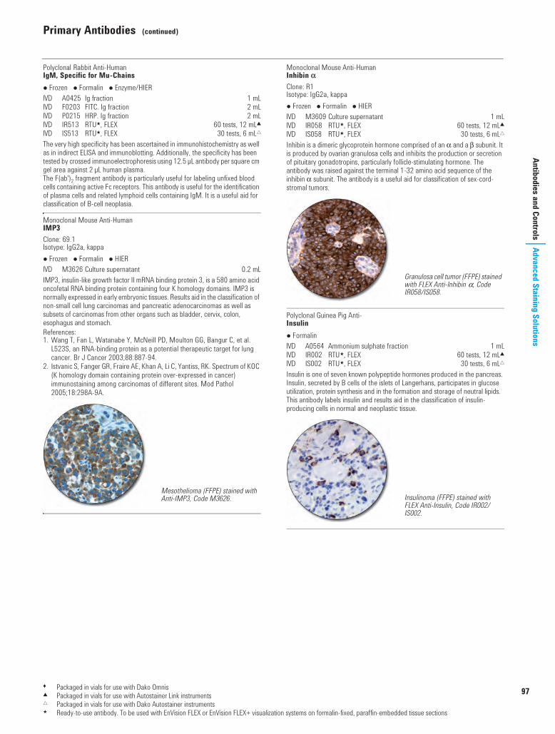
Primary Antibodies (continued)
97
Antibodies and ControlsAdvanced Staining Solutions
Polyclonal Rabbit Anti-HumanIgM, Specific for Mu-Chains Frozen Formalin Enzyme/HIER I A0425 Ig fraction 1 mLI F0203 FITC. Ig fraction 2 mLI P0215 HRP. Ig fraction 2 mLI IR513 RTU5, FLEX 60 tests, 12 mL2I IS513 RTU5, FLEX 30 tests, 6 mL"The very high specificity has been ascertained in immunohistochemistry as well as in indirect ELISA and immunoblotting. Additionally, the specificity has been tested by crossed immunoelectrophoresis using 12.5 µL antibody per square cm gel area against 2 µL human plasma. The F(ab')2 fragment antibody is particularly useful for labeling unfixed blood cells containing active Fc receptors. This antibody is useful for the identification of plasma cells and related lymphoid cells containing IgM. It is a useful aid for classification of B-cell neoplasia.
Monoclonal Mouse Anti-HumanIMP3Clone: 69.1Isotype: IgG2a, kappa Frozen Formalin HIER I M3626 Culture supernatant 0.2 mLIMP3, insulin-like growth factor II mRNA binding protein 3, is a 580 amino acid oncofetal RNA binding protein containing four K homology domains. IMP3 is normally expressed in early embryonic tissues. Results aid in the classification of non-small cell lung carcinomas and pancreatic adenocarcinomas as well as subsets of carcinomas from other organs such as bladder, cervix, colon, esophagus and stomach. References:1. Wang T, Fan L, Watanabe Y, McNeill PD, Moulton GG, Bangur C, et al.
L523S, an RNA-binding protein as a potential therapeutic target for lung cancer. Br J Cancer 2003;88:887-94.
2. Istvanic S, Fanger GR, Fraire AE, Khan A, Li C, Yantiss, RK. Spectrum of KOC (K homology domain containing protein over-expressed in cancer) immunostaining among carcinomas of different sites. Mod Pathol 2005;18:298A-9A.
Monoclonal Mouse Anti-HumanInhibin aClone: R1Isotype: IgG2a, kappa Frozen Formalin HIER I M3609 Culture supernatant 1 mLI IR058 RTU5, FLEX 60 tests, 12 mL2I IS058 RTU5, FLEX 30 tests, 6 mL"Inhibin is a dimeric glycoprotein hormone comprised of an a and a b subunit. It is produced by ovarian granulosa cells and inhibits the production or secretion of pituitary gonadotropins, particularly follicle-stimulating hormone. The antibody was raised against the terminal 1-32 amino acid sequence of the inhibin a subunit. The antibody is a useful aid for classification of sex-cord-stromal tumors.
Polyclonal Guinea Pig Anti-Insulin Formalin I A0564 Ammonium sulphate fraction 1 mLI IR002 RTU5, FLEX 60 tests, 12 mL2I IS002 RTU5, FLEX 30 tests, 6 mL"Insulin is one of seven known polypeptide hormones produced in the pancreas. Insulin, secreted by B cells of the islets of Langerhans, participates in glucose utilization, protein synthesis and in the formation and storage of neutral lipids. This antibody labels insulin and results aid in the classification of insulin-producing cells in normal and neoplastic tissue.
Mesothelioma (FFPE) stained with Anti-IMP3, Code M3626.
Granulosa cell tumor (FFPE) stained with FLEX Anti-Inhibin a, Code IR058/IS058.
Insulinoma (FFPE) stained with FLEX Anti-Insulin, Code IR002/IS002.
1 Packaged in vials for use with Dako Omnis2 Packaged in vials for use with Autostainer Link instruments" Packaged in vials for use with Dako Autostainer instruments5 Ready-to-use antibody. To be used with EnVision FLEX or EnVision FLEX+ visualization systems on formalin-fixed, paraffin-embedded tissue sections
00110 IHC Solutions US_1.fm Page 97 Friday, March 11, 2016 9:37 AM
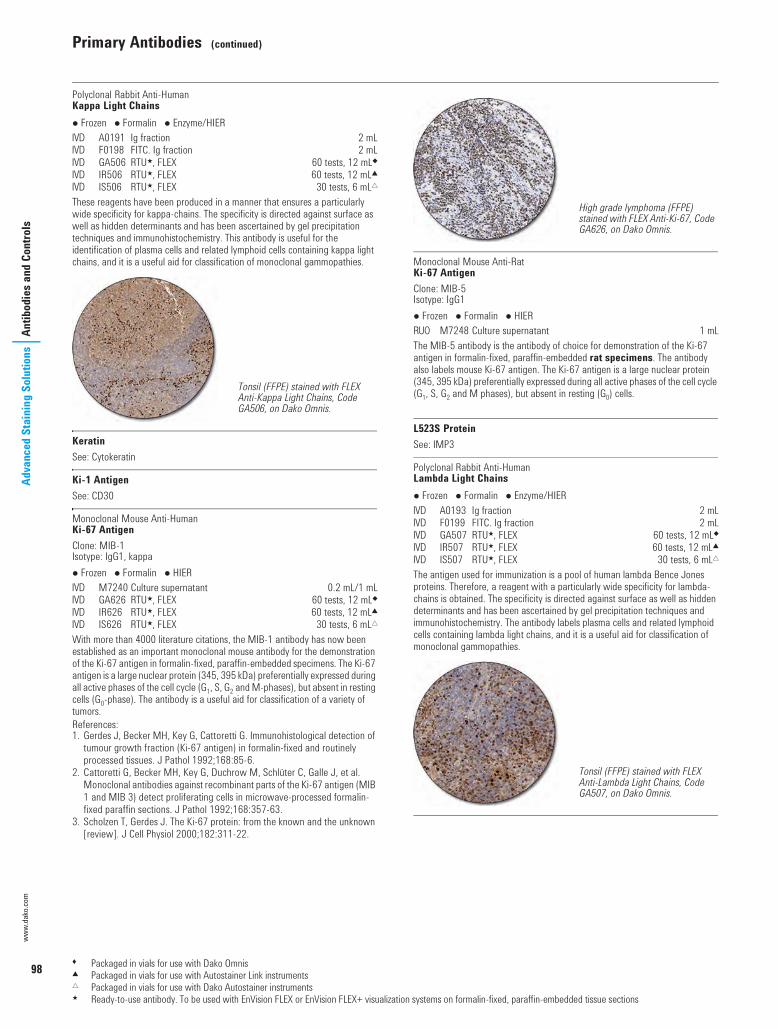
98
Primary Antibodies (continued)
www.
dako
.com
Adva
nced
Sta
inin
g So
lutio
nsAn
tibod
ies a
nd C
ontro
ls
Polyclonal Rabbit Anti-HumanKappa Light Chains Frozen Formalin Enzyme/HIER I A0191 Ig fraction 2 mLI F0198 FITC. Ig fraction 2 mLI GA506 RTU5, FLEX 60 tests, 12 mL8I IR506 RTU5, FLEX 60 tests, 12 mL2I IS506 RTU5, FLEX 30 tests, 6 mL"These reagents have been produced in a manner that ensures a particularly wide specificity for kappa-chains. The specificity is directed against surface as well as hidden determinants and has been ascertained by gel precipitation techniques and immunohistochemistry. This antibody is useful for the identification of plasma cells and related lymphoid cells containing kappa light chains, and it is a useful aid for classification of monoclonal gammopathies.
KeratinSee: Cytokeratin
Ki-1 AntigenSee: CD30
Monoclonal Mouse Anti-HumanKi-67 AntigenClone: MIB-1Isotype: IgG1, kappa Frozen Formalin HIER I M7240 Culture supernatant 0.2 mL/1 mLI GA626 RTU5, FLEX 60 tests, 12 mL8I IR626 RTU5, FLEX 60 tests, 12 mL2I IS626 RTU5, FLEX 30 tests, 6 mL"With more than 4000 literature citations, the MIB-1 antibody has now been established as an important monoclonal mouse antibody for the demonstration of the Ki-67 antigen in formalin-fixed, paraffin-embedded specimens. The Ki-67 antigen is a large nuclear protein (345, 395 kDa) preferentially expressed during all active phases of the cell cycle (G1, S, G2 and M-phases), but absent in resting cells (G0-phase). The antibody is a useful aid for classification of a variety of tumors.References:1. Gerdes J, Becker MH, Key G, Cattoretti G. Immunohistological detection of
tumour growth fraction (Ki-67 antigen) in formalin-fixed and routinely processed tissues. J Pathol 1992;168:85-6.
2. Cattoretti G, Becker MH, Key G, Duchrow M, Schlüter C, Galle J, et al. Monoclonal antibodies against recombinant parts of the Ki-67 antigen (MIB 1 and MIB 3) detect proliferating cells in microwave-processed formalin-fixed paraffin sections. J Pathol 1992;168:357-63.
3. Scholzen T, Gerdes J. The Ki-67 protein: from the known and the unknown [review]. J Cell Physiol 2000;182:311-22.
Monoclonal Mouse Anti-RatKi-67 AntigenClone: MIB-5Isotype: IgG1 Frozen Formalin HIER U M7248 Culture supernatant 1 mLThe MIB-5 antibody is the antibody of choice for demonstration of the Ki-67 antigen in formalin-fixed, paraffin-embedded rat specimens. The antibody also labels mouse Ki-67 antigen. The Ki-67 antigen is a large nuclear protein (345, 395 kDa) preferentially expressed during all active phases of the cell cycle (G1, S, G2 and M phases), but absent in resting (G0) cells.
L523S ProteinSee: IMP3
Polyclonal Rabbit Anti-HumanLambda Light Chains Frozen Formalin Enzyme/HIER I A0193 Ig fraction 2 mLI F0199 FITC. Ig fraction 2 mLI GA507 RTU5, FLEX 60 tests, 12 mL8I IR507 RTU5, FLEX 60 tests, 12 mL2I IS507 RTU5, FLEX 30 tests, 6 mL"The antigen used for immunization is a pool of human lambda Bence Jones proteins. Therefore, a reagent with a particularly wide specificity for lambda-chains is obtained. The specificity is directed against surface as well as hidden determinants and has been ascertained by gel precipitation techniques and immunohistochemistry. The antibody labels plasma cells and related lymphoid cells containing lambda light chains, and it is a useful aid for classification of monoclonal gammopathies.
Tonsil (FFPE) stained with FLEX Anti-Kappa Light Chains, Code GA506, on Dako Omnis.
High grade lymphoma (FFPE) stained with FLEX Anti-Ki-67, Code GA626, on Dako Omnis.
Tonsil (FFPE) stained with FLEX Anti-Lambda Light Chains, Code GA507, on Dako Omnis.
1 Packaged in vials for use with Dako Omnis2 Packaged in vials for use with Autostainer Link instruments" Packaged in vials for use with Dako Autostainer instruments5 Ready-to-use antibody. To be used with EnVision FLEX or EnVision FLEX+ visualization systems on formalin-fixed, paraffin-embedded tissue sections
USCatPathology.book Page 98 Monday, March 7, 2016 8:13 PM

Primary Antibodies (continued)
99
Antibodies and ControlsAdvanced Staining Solutions
Monoclonal Mouse Anti-HumanLamininClone: 4C7Isotype: IgG2a, kappa Frozen Formalin Enzyme I M0638 Ascites 1 mLReacts with a 380 kDa laminin alpha5-chain. Fibroblasts, epithelial, endothelial and smooth muscle cells secrete laminin. In normal tissues, laminin is invariably present in all basal membranes surrounding muscle, nerve, fat and decidua cells and separating epithelial and endothelial cells from connective tissues.
Polyclonal Rabbit Anti-Laminin Frozen Formalin Enzyme I Z0097 Ig fraction 1 mLLaminin, isolated from a rat yolk sac tumor cell line, has been used for immunization. The antibody reacts strongly with human laminin, and labels basement membranes surrounding normal vessels, glands, muscles, nerves, fat and decidua cells, separating these from abutting connective tissues. Loss of basement membrane integrity may be observed in neoplastic invasion and around metastatic lesions.
Monoclonal Mouse Anti-HumanLaminin-5, Gamma-2 ChainClone: 4G1Isotype: IgG1, kappa Frozen Formalin HIER I M7262 Culture supernatant 1 mLThe expression of laminin-5 is restricted to epithelial tissues, where the protein is part of the epithelial anchoring systems and cell locomotion. Of the 15 laminins presently known, only laminin-5 contains the g2 chain. This antibody is specific for the g2 chain. Results aid in the classification of invading epithelial cancer cells in various types of squamous cell carcinomas, colon adenocarcinomas and lung adenocarcinomas.References:1. Skyldberg B, Salo S, Eriksson E, Aspenblad U, Moberger B, Tryggvason K, et
al. Laminin-5 as a marker of invasiveness in cervical lesions. J Natl Cancer Inst 1999;91:1882-7.
2. Määttä M, Soini Y, Pääkkö P, Salo S, Tryggvason K, Autio-Harmainen H. Expression of the laminin g2 chain in different histological types of lung carcinoma. A study by immunohistochemistry and in situ hybridisation. J Pathol 1999;188:361-8.
Monoclonal Mouse Anti-HumanLAT ProteinClone: LAT-1Isotype: IgG1, kappa Frozen Formalin HIER I M7279 Culture supernatant 0.2 mLLAT (linker for activation of T cells) protein appears early in the T-cell development, at the thymocyte stage, and before expression of terminal deoxynucleotidyl transferase (TdT) in embryos. It is expressed by natural killer cells and T cells without restriction to any T-cell subpopulation. Megakaryocytes
and mast cells also express LAT protein, whereas other myeloid cells, monocytic derived cells and B cells do not express LAT protein.
Leucocyte Common AntigenSee: CD45, Leucocyte Common Antigen
Monoclonal Mouse Anti-HumanLeukaemia, Hairy CellClone: DBA.44Isotype: IgM, kappa Frozen Formalin (Enzyme)/HIER I M0880 Culture supernatant 1 mLResults aid in the classification of hairy cell leukemia.
Monoclonal Mouse Anti-HumanLuteinizing Hormone (LH)Clone: C93Isotype: IgG1, kappa Formalin I M3502 Culture supernatant 1 mLReacts with the b-chain of luteinizing hormone. By radioimmunoassay, this antibody was determined to be 100% reactive with hLH, 1.5% with hCG, 0.44% with hFSH and less than 1.0% with hTSH. The antibody labels gonadotrophic cells of the pituitary. Results aid in the classification of pituitary adenomas.
Polyclonal Rabbit Anti-HumanLysozyme EC 3.2.1.17 (Muramidase) Formalin Enzyme I A0099 Ig fraction 2 mLA0099 has been used for classification of histiocytic neoplasias and myeloid leukemias (1, 2). Lysozyme isolated from urine of patients with monocytic leukemia has been used for immunization.References:1. Meister P, Huhn D, Nathrath W. Malignant histiocytosis.
Immunohistochemical characterization on paraffin-embedded tissue. Virchows Arch A Path Anat Histol 1980;385:233-46.
2. Krugliak L, Meyer PR, Taylor CR. The distribution of lysozyme, alpha-1-antitrypsin, and alpha-1-antichymotrypsin in normal hematopoietic cells and in myeloid leukaemias. Am J Hematol 1986;21:99-109.
Liver (FFPE) stained with Anti-Laminin, Code Z0097.
Colon carcinoma (FFPE) stained with Anti-LAT Protein, Code M7279.
Bone marrow interstitial infiltration (FFPE) stained with Anti-Hairy Cell Leukaemia, Code M0880.
USCatPathology.book Page 99 Monday, March 7, 2016 8:13 PM

100
Primary Antibodies (continued)
www.
dako
.com
Adva
nced
Sta
inin
g So
lutio
nsAn
tibod
ies a
nd C
ontro
ls
Monoclonal Mouse Anti-RabbitMacrophageClone: RAM11Isotype: IgG1, kappa Frozen Formalin U M0633 Culture supernatant 1 mLLabels rabbit macrophages and may be used in studies of the cellular components of atherosclerotic lesions in rabbits.
Monoclonal Mouse Anti-HumanMammaglobinClone: 304-1A5Isotype: IgG1, kappa Frozen Formalin HIER I M3625 Culture supernatant 0.2 mLI GA074 RTU5, FLEX NEW 60 tests, 12 mL8I IR074 RTU5, FLEX 60 tests, 12 mL2I IS074 RTU5, FLEX 30 tests, 6 mL"Mammaglobin, a 93-amino acid glycoprotein, is encoded by a gene first identified in a study directed at the isolation of novel human breast cancer-associated genes (1). Mammaglobin expression is mostly, although not exclusively, confined to breast tissue, and anti-mammoglobin is a useful aid for the classification of carcinomas of breast origin.Reference:1. Watson MA, Dintzis S, Darrow CM, Voss LE, DiPersio J, Jensen R, et al.
Mammaglobin expression in primary, metastatic, and occult breast cancer. Cancer Res 1999;59:3028-31.
Monoclonal Mouse Anti-HumanMast Cell TryptaseClone: AA1Isotype: IgG1, kappa Frozen Formalin HIER I M7052 Culture supernatant 0.2 mLI IR640 RTU5, FLEX 60 tests, 12 mL2I IS640 RTU5, FLEX 30 tests, 6 mL"Human mast cell tryptases comprise a family of trypsin-like neutral serine proteases that are predominantly expressed in mast cells. These cells play an active role in such diverse diseases as atherosclerosis, asthma, arthritis, bile duct fibrosis, malignancy and pulmonary fibrosis. This antibody is a useful aid for classification of mast cell leukemia.
Monoclonal Mouse Anti-HumanMCM3 ProteinClone: 101Isotype: IgG1, kappa Formalin HIER I M7263 Culture supernatant 0.2 mLMCM3 protein is expressed in proliferating cells but disappears more slowly after initiation of cell differentiation than the Ki-67 antigen. This observation, correlated with the fact that MCM3 protein is not expressed when a marker of terminal differentiation, such as p27, is expressed, indicates that antibodies against MCM3 protein also label cells that have ceased to proliferate, but are not terminally differentiated (1).Reference:1. Endl E, Kausch I, Baack M, Knippers R, Gerdes J, Scholzen T. The expression
of Ki-67, MCM3, and p27 defines distinct subsets of proliferating, resting, and differentiated cells. J Pathol 2001;195:457-62.
Monoclonal Mouse Anti-HumanMelan-AClone: A103Isotype: IgG1, kappa Frozen Formalin HIER I M7196 Culture supernatant 0.2 mL/1 mLI IR633 RTU5, FLEX 60 tests, 12 mL2I IS633 RTU5, FLEX 30 tests, 6 mL"Melan-A, isolated as a melanoma-specific antigen, is a transmembrane protein, which is expressed in skin, retina and the majority of cultured melanocytes as well as in melanomas and angiomyolipomas. The antibody is a useful aid for classification of melanomas and adrenocortical carcinomas. The antibody is also a useful aid for classification of angiomyolipomas.References:1. Fetsch PA, Cormier J, Hijazi YM. Immunocytochemical detection of MART-
1 in fresh and paraffin-embedded malignant melanomas. J Immunother 1997;20:60-4.
2. Kawakami Y, Eliyahu S, Delgado CH, Robbins PF, Sakaguchi K, Apella E, et al. Identification of a human melanoma antigen recognized by tumor-infiltrating lymphocytes associated with in vivo tumor rejection. Proc Natl Acad Sci USA 1994;91:6458-62.
Invasive ductal carcinoma (FFPE) stained with FLEX Anti-Mammaglobin, Code GA074, on Dako Omnis.
Mastocytosis in the subcutis (FFPE) stained with FLEX Anti-Mast Cell Tryptase, Code IR640/IS640.
Dako FLEX RTU Antibodies for Skin TestingSee our panel of FLEX antibodies at page 71
Melanoma (FFPE) stained with FLEX Anti-Melan-A, Code IR633/IS633.
1 Packaged in vials for use with Dako Omnis2 Packaged in vials for use with Autostainer Link instruments" Packaged in vials for use with Dako Autostainer instruments5 Ready-to-use antibody. To be used with EnVision FLEX or EnVision FLEX+ visualization systems on formalin-fixed, paraffin-embedded tissue sections
USCatPathology.book Page 100 Monday, March 7, 2016 8:13 PM

Primary Antibodies (continued)
101
Antibodies and ControlsAdvanced Staining Solutions
Monoclonal Mouse Anti-HumanMelanosomeClone: HMB-45Isotype: IgG1, kappa Frozen Formalin Enzyme/HIER I M0634 Culture supernatant 0.2 mL/1 mLI GA052 RTU5, FLEX 60 tests, 12 mL8I IR052 RTU5, FLEX 60 tests, 12 mL2I IS052 RTU5, FLEX 30 tests, 6 mL"Labels an intracytoplasmic antigen in the majority of melanocytes with immature melanosome formation in normal skin and nevus. The antibody also reacts with junctional and blue nevus cells. Results aid in the classification of melanomas and melanocytic lesions.
Monoclonal Mouse Anti-HumanMesothelial CellClone: HBME-1Isotype: IgM Frozen Formalin I M3505 Culture supernatant 1 mLReacts with an antigen present in the membrane of mesothelial cells. No reactivity has been observed on kidney, liver, placenta, skin and thyroid. The antibody is a useful aid for classification of epithelial mesotheliomas and adenocarcinomas of various origin.Reference:1. Miettinen M, Kovatich AJ. HMBE-1 a monoclonal antibody useful in the
differential diagnosis of mesothelioma, adenocarcinoma, and soft-tissue and bone tumors. Appl Immunohistochem 1995;3:115-22.
Monoclonal Mouse Anti-MetallothioneinClone: E9Isotype: IgG1, kappa Frozen Formalin I M0639 Ascites 1 mLReacts with a conserved epitope shared by human metallothionein isoforms 1 and 2. Metallothioneins are low molecular weight, heavy metal-binding proteins. The expression of metallothioneins is induced by heavy metals, but also by other factors such as stress, glucocorticoids and lymphokines.
MIB-1See: Ki-67 Antigen
MIC2 Gene Products, Ewing's Sarcoma MarkerSee: CD99, MIC2 Gene Products, Ewing's Sarcoma Marker
Monoclonal Mouse Anti-HumanMITFClone: D5Isotype: IgG1, kappa Frozen Formalin HIER I M3621 Culture supernatant 0.2 mLMITF (microphthalmia transcription factor) is a basic helix-loop-helix-leucine-zipper (bHLH-Zip) transcription factor that regulates the development and survival of melanocytes and retinal pigment epithelium, and also is involved in transcription of pigmentation enzyme genes such as tyrosinase TRP1 and TRP2. MITF has been shown to be phosphorylated by MAP kinase in response to c-kit activation, resulting in upregulation of MITF transcriptional activity. Multiple isoforms of MITF exist, including MITF-A, MITF-B, MITF-C, MITF-H, and MITF-M, which differ in the amino-terminal domain and in their expression patterns. The MITF-M isoform is restricted to the melanocyte cell lineage. Anti-MITF, D5, recognizes a nuclear protein and is a useful aid for classification of primary and metastatic epithelioid malignant melanomas.
Mouse Ki-67 AntigenSee: Ki-67 Antigen
Monoclonal Mouse Anti-HumanMUC2Clone: CCP58Isotype: IgG1, kappa Formalin HIER I M7313 Culture supernatant 0.2 mL/1 mLI IR658 RTU5, FLEX 60 tests, 12 mL2Mucin 2 glycoprotein (MUC2) is a 520 kDa glycoprotein belonging to the superfamily of mucins. Mucins are high molecular weight glycoproteins produced by epithelial cells and can be divided into two families; secretory mucins and membrane bound mucins. MUC2 is a mucus-forming secreted mucin found in the cytoplasm of goblet cells in small intestine, colon, bronchus and salivary glands. MUC2 expression has been noted in primary gastrointestinal tract tumors of colonic, gastric and esophageal origin (1). MUC2 expression has also been found in goblet cells in colorectal carcinoma metastatic to the ovary, but not in primary mucinous ovarian adenocarcinoma.This antibody is a useful aid for classification of mucinous adenocarcinoma of gastrointestinal origin (1, 2) and intestinal metaplasia in Barrett's esophagus. Anti-MUC2 may also be a useful aid for classification of colon adenocarcinoma metastatic to the ovary.References:1. Lau SK, Weiss LM, Chu PG. Differential expression of MUC1, MUC2 and
MUC5AC in carcinomas of various sites. Am J Clin Pathol 2004;122:61-9.2. Lee HS, Lee HK, Kim HS, Yang HK, Kim YI, Kim WH. MUC1, MUC2,
MUC5AC, and MUC6 expression in gastric carcinomas. Cancer 2001;92:1427-34.
Melanoma (FFPE) stained with FLEX Anti-Melanosome, Code GA052, on Dako Omnis.
Colon adenocarcinoma (FFPE) metastatic to the ovary stained with FLEX Anti-MUC2, Code IR658.
1 Packaged in vials for use with Dako Omnis2 Packaged in vials for use with Autostainer Link instruments" Packaged in vials for use with Dako Autostainer instruments5 Ready-to-use antibody. To be used with EnVision FLEX or EnVision FLEX+ visualization systems on formalin-fixed, paraffin-embedded tissue sections
USCatPathology.book Page 101 Monday, March 7, 2016 8:13 PM

102
Primary Antibodies (continued)
www.
dako
.com
Adva
nced
Sta
inin
g So
lutio
nsAn
tibod
ies a
nd C
ontro
ls
Monoclonal Mouse Anti-HumanMUC5ACClone: CLH2Isotype: IgG1, kappa Formalin HIER I M7316 Culture supernatant 0.2 mL/1 mLI IR661 RTU5, FLEX 60 tests, 12 mL2Mucin 5AC glycoprotein (MUC5AC) is a 641 kDa glycoprotein belonging to the superfamily of mucins. Mucins are high molecular weight glycoproteins produced by epithelial cells and can be divided into two families; secretory mucins and membrane bound mucins. MUC5AC is a mucus-forming secreted mucin that is found in normal gastric and tracheo-bronchial mucosa, but absent from normal colon. MUC5AC expression is present in primary ovarian mucinous cancer but usually absent in colorectal adenocarcinoma, thus showing an expression pattern opposite to MUC2.Anti-MUC5AC is a useful aid for classification of primary mucinous ovarian tumors. MUC5AC antibodies are also a useful aid for classification of intestinal metaplasia as well as in the classification of pancreatic carcinoma.
Monoclonal Mouse Anti-HumanMUM1 ProteinClone: MUM1pIsotype: IgG1, kappa Frozen Formalin HIER I M7259 Culture supernatant 0.2 mL/1 mLI GA644 RTU5, FLEX 60 tests, 12 mL8I IR644 RTU5, FLEX 60 tests, 12 mL2I IS644 RTU5, FLEX 30 tests, 6 mL"Labels MUM1 protein in a subset of light zone germinal centre (GC) B cells (probably centrocytes and their progeny), plasma cells and activated T cells. Antibodies to MUM1 protein are a useful aid for classification of a wide spectrum of hematolymphoid neoplasms derived from these cells. Of non-hematolymphoid neoplasms only a proportion of melanomas are labeled (1, 2). Antibodies to MUM1 protein are a useful aid for subclassification of B-cell lymphomas (1-3).References:1. Falini B, Fizzotti M, Pucciarini A, Bigerna B, Marafioti T, Gambacorta M, et
al. A monoclonal antibody (MUM1p) detects expression of the MUM1/IRF4 protein in a subset of germinal center B cells, plasma cells, and activated T cells. Blood 2000;95;2084-92.
2. Natkunam Y, Warnke RA, Montgomery K, Falini B, van de Rijn M. Analysis of MUM1/IRF4 protein expression using tissue microarrays and immunohistochemistry. Mod Pathol 2001;14:686-94.
3. Gaidano G, Carbone A. MUM1: a step toward the understanding of lymphoma histogenesis. Leukemia 2000;14:563-6.
Monoclonal Mouse Anti-HumanMutL Protein Homolog 1Clone: ES05Isotype: IgG1 Formalin HIER I M3640 Culture supernatant 0.2 mL/1 mLI IR079 RTU5, FLEX 60 tests, 12 mL2I IS079 RTU5, FLEX 30 tests, 6 mL"Mismatch repair gene hMLH1 is a ubiquitous gene encoding the mismatch repair protein (MMR) known as MutL protein homolog 1 (MLH1). MLH1 is utilized by normal proliferating cells to repair point mutations that may occur during DNA replication. Antibodies to MLH1 are a useful aid for classification of colorectal cancer.
Monoclonal Mouse Anti-HumanMutS Protein Homolog 2Clone: FE11Isotype: IgG1, kappa Formalin HIER I M3639 Ascites, protein A purified 0.2 mL/1 mLI IR085 RTU5, FLEX 60 tests, 12 mL2MutS protein homolog 2 (MSH2) is part of the mismatch repair (MMR) pathway which is utilized by normal proliferating cells to repair mutations that may occur during DNA replication. Antibodies to MSH2 are a useful aid for classification of tumors of the gastrointestinal tract, including associated extracolonic cancers. Reference:1. Peltomäki P. Role of DNA mismatch repair defects in the pathogenesis of
human cancer. J Clin Oncol 2003;21:1174-9.
Mucinous ovarian carcinoma (FFPE) stained with FLEX Anti-MUC5AC, Code IR661.
Diffuse large B-cell lymphoma (FFPE) stained with FLEX Anti-MUM1, Code GA644, on Dako Omnis.
Colon adenocarcinoma (FFPE) stained with FLEX Anti-MutL Protein Homolog 1, Code IR079/IS079.
Colon adenocarcinoma (FFPE) with loss of MSH2 protein stained with FLEX Anti-MSH2, Code IR085.
1 Packaged in vials for use with Dako Omnis2 Packaged in vials for use with Autostainer Link instruments" Packaged in vials for use with Dako Autostainer instruments5 Ready-to-use antibody. To be used with EnVision FLEX or EnVision FLEX+ visualization systems on formalin-fixed, paraffin-embedded tissue sections
USCatPathology.book Page 102 Monday, March 7, 2016 8:13 PM

Primary Antibodies (continued)
103
Antibodies and ControlsAdvanced Staining Solutions
Monoclonal Rabbit Anti-HumanMutS Protein Homolog 6Clone: EP49 Formalin HIER I M3646 Affinity-isolated 0.2 mL/1 mLI IR086 RTU5, FLEX 60 tests, 12 mL2MutS protein homolog 6 (MSH6) is part of the mismatch repair (MMR) pathway which is utilized by normal proliferating cells to repair mutations that may occur during DNA replication. Antibodies to MSH6 are a useful aid for classification of tumors of the gastrointestinal tract, including associated extracolonic cancersReference:1. Peltomäki P. Role of DNA mismatch repair defects in the pathogenesis of
human cancer. J Clin Oncol 2003;21:1174-9.
Polyclonal Rabbit Anti-HumanMyelin Basic Protein Formalin I A0623 Ig fraction 1 mLLabels myelin membranes of oligodendrocytes and Schwann cells.
Monoclonal Mouse Anti-HumanMyeloid/Histiocyte AntigenClone: MAC 387Isotype: IgG1, kappa Formalin (Enzyme) I M0747 Culture supernatant 1 mLReacts with a human cytoplasmic antigen (L1-antigen or calprotectin) which contains two different subunits (L1H and L1L). The protein is a member of the S100 family, and the subunits are in this context termed S100A8 and S100A9. It is expressed in granulocytes, blood monocytes, tissue histiocytes, squamous mucosal epithelia, and reactive epidermis. The antibody is a useful aid for classification of malignant lymphomas and lymphoid neoplasms of histiocytic origin.References:1. Fagerhol MK. Nomenclature for proteins: is calprotectin a proper name for
the elusive myelomonocytic protein? J Clin Pathol: Mol Pathol 1996;49:M74-9.
2. Schäfer BW, Heizmann CW. The S100 family of EF-hand calciumbinding proteins: functions and pathology. Trends Biochem Sci 1996;21:134-40.
Polyclonal Rabbit Anti-HumanMyeloperoxidase Formalin Enzyme/HIER I A0398 Ig fraction 0.2 mLI GA511 RTU5, FLEX 60 tests, 12 mL8I IR511 RTU5, FLEX 60 tests, 12 mL2I IS511 RTU5, FLEX 30 tests, 6 mL"The antigen has been isolated from primary granulocytes (1). The antibody reacts with neutrophil granulocytes and monocytes in blood and with precursors of granulocytes in the bone marrow. The antibody is useful as an aid for classification of neoplastic tissue, i.e. myeloblasts and immature myeloid cells of acute myelogenous leukemia, progranulocytic leukemia, monomyelocytic leukemia, erythroleukemia and myeloblastoma (2).References:1. Matheson NR, Wong PS, Travis J. Isolation and properties of human
neutrophil myeloperoxidase. Biochem 1981;20:325-30.2. Pinkus GS, Pinkus JI. Myeloperoxidase: a specific marker for myeloid cells
in paraffin sections. Mod Pathol 1991;4:733-41.
Monoclonal Mouse Anti-MyoD1Clone: 5.8AIsotype: IgG1, kappa Frozen Formalin HIER I M3512 Culture supernatant 1 mLThe MyoD1 protein is a 45 kDa nuclear phosphoprotein which induces myogenesis through transcriptional activation of muscle-specific genes. Nuclear expression of MyoD1 is restricted to skeletal muscle tissue and has been demonstrated to be a sensitive marker of myogenic differentiation. The antibody strongly labels the nuclei of myoblasts in developing skeletal muscle tissue, whereas the majority of adult skeletal muscle is negative. Results aid in the classification of rhabdomyosarcomas of various histological subtypes.Reference:1. Dias P, Parham DM, Shapiro DN, Tapscott SJ, Houghton PJ. Monoclonal
antibodies to the myogenic regulatory protein MyoD1: epitope mapping and diagnostic utility. Cancer Res 1992;52:6431-9.
Colon adenocarcinoma (FFPE) with loss of MSH6 protein stained with FLEX Anti-MSH6, Code IR086.
Acute myeloid leukemia (FFPE) stained with FLEX Anti-Myeloperoxidase, Code GA511, on Dako Omnis.
Rhabdomyosarcoma (FFPE) stained with Anti-MyoD1, Code M3512.
1 Packaged in vials for use with Dako Omnis2 Packaged in vials for use with Autostainer Link instruments" Packaged in vials for use with Dako Autostainer instruments5 Ready-to-use antibody. To be used with EnVision FLEX or EnVision FLEX+ visualization systems on formalin-fixed, paraffin-embedded tissue sections
USCatPathology.book Page 103 Monday, March 7, 2016 8:13 PM

104
Primary Antibodies (continued)
www.
dako
.com
Adva
nced
Sta
inin
g So
lutio
nsAn
tibod
ies a
nd C
ontro
ls
Monoclonal Mouse Anti-MyogeninClone: F5DIsotype: IgG1, kappa Frozen Formalin HIER I M3559 Ig fraction of culture supernatant 1 mLI IR067 RTU5, FLEX 60 tests, 12 mL2I IS067 RTU5, FLEX 30 tests, 6 mL"Myogenin belongs to a family of regulatory proteins essential for muscle development. Expression of myogenin is restricted to cells of skeletal muscle origin, and appears to be inversely related to the degree of cellular differentiation. The antibody recognizes an epitope located in the amino acid region 138-158 of the myogenin protein. The antibody is a useful aid for classification of rhabdomyosarcomas and Wilms' tumors. No reactivity with Ewing's sarcoma/peripheral primitive neuroectodermal tumor, neuroblastoma, or adult skeletal muscle has been observed. Myogenin peptide from rat has been used for immunization.
Monoclonal Mouse Anti-HumanMyosin Heavy Chain (Smooth Muscle)Clone: SMMS-1Isotype: IgG1, kappa Frozen Formalin Enzyme + HIER I M3558 Culture supernatant 1 mLI IR066 RTU5, FLEX 60 tests, 12 mL2I IS066 RTU5, FLEX 30 tests, 6 mL"Reacts with smooth muscle cells and myoepithelial cells, but not with myofibroblasts. The antibody is a useful aid for classification of breast tumors.
Monoclonal Mouse Anti-HumanN-CadherinClone: 6G11Isotype: IgG1, kappa Frozen Formalin HIER I M3613 Culture supernatant 1 mLN-cadherin is a 140 kDa protein belonging to a family of transmembrane molecules that mediate calcium-dependent intercellular adhesion. Cadherins are involved in controlling morphogenetic movements during development and regulate cell surface adhesion through homotypic adhesion with the same cadherin species. Expression of N-cadherin has been reported on a variety of normal tissues including neuronal, endothelial and muscle cells, and a subpopulation of early hematopoietic progenitor cells. Results aid in the classification of malignant noncarcinomatous neoplasms including mesotheliomas, chordomas, synovial sarcomas, malignant melanomas, epithelioid sarcomas, epithelioid angiosarcomas, clear cell sarcomas as well as serous and endometrioid tumors of the ovary have been demonstrated to be N-cadherin positive, whereas mucinous tumors are negative. Other N-cadherin-positive neoplasms include renal cell carcinomas and some variant breast tumors, including medullary breast carcinomas and sarcomatoid metaplastic breast carcinomas.
Monoclonal Mouse Anti-HumanNeurofilament ProteinClone: 2F11Isotype: IgG1, kappa Frozen Formalin HIER I M0762 Culture supernatant 0.2 mLI GA607 RTU5, FLEX NEW 60 tests, 12 mL8I IR607 RTU5, FLEX 60 tests, 12 mL2I IS607 RTU5, FLEX 30 tests, 6 mL"Neurofilaments belong to the family of intermediate filaments and are structural elements of the neuronal cytoskeleton in an interconnection with actin microfilaments, microtubules and other intermediate filaments. This antibody labels neurons (axons) of the central and peripheral nervous system, and is a useful aid for classification of tumors with neuronal differentiation.
Monoclonal Mouse Anti-HumanNeuron-Specific Enolase (NSE)Clone: BBS/NC/VI-H14Isotype: IgG1, kappa Formalin HIER I M0873 Culture supernatant 0.2 mL/1 mLI IR612 RTU5, FLEX 60 tests, 12 mL2I IS612 RTU5, FLEX 30 tests, 6 mL"Neuron-specific enolase is one of three main groups of enolases, the other two being non-neuronal enolase and muscle-specific enolase. The antibody labels cells of neuronal and neuroendocrine origin. Although neuron-specific enolase is not an exclusive neuronal marker, it may be used for the identification of peripheral nerves. Results aid in the classification of neural and neuroendocrine tumors, such as neuroblastomas, retinoblastomas, desmoplastic malignant melanoma, and small-cell lung cancer.
Rhabdomyosarcoma (FFPE) stained with FLEX Anti-Myogenin, Code IR067/IS067.
Breast hyperplasia (FFPE) stained with FLEX Anti-Myosin Heavy Chain (Smooth Muscle), Code IR066/IS066.
Merkel cell tumor (FFPE) stained with FLEX Anti-Neurofilament Protein, Code GA607, on Dako Omnis.
1 Packaged in vials for use with Dako Omnis2 Packaged in vials for use with Autostainer Link instruments" Packaged in vials for use with Dako Autostainer instruments5 Ready-to-use antibody. To be used with EnVision FLEX or EnVision FLEX+ visualization systems on formalin-fixed, paraffin-embedded tissue sections
USCatPathology.book Page 104 Monday, March 7, 2016 8:13 PM

Primary Antibodies (continued)
105
Antibodies and ControlsAdvanced Staining Solutions
Monoclonal Mouse Anti-HumanNeutrophil ElastaseClone: NP57Isotype: IgG1, kappa Frozen Formalin I M0752 Culture supernatant 1 mLLabels neutrophil precursors strongly. A minor population of monocytes is also labeled, but with a lower intensity. The antibody is a useful aid for classification of acute myeloid leukemia and extramedullary myeloid cell tumor (1).Note: The neutrophil elastase epitope corresponding to this antibody is destroyed by heat-induced epitope retrieval methods.Reference:1. Pulford KAF, Erber WN, Crick JA, Olsson I, Micklem KJ, Gatter KC, et al. Use
of monoclonal antibody against human neutrophil elastase in normal and leukaemic myeloid cells. J Clin Pathol 1988;41:853-60.
Monoclonal Mouse Anti-HumanNucleophosminClone: 376Isotype: IgG1, lambda Formalin HIER I M7305 Culture supernatant 1 mLI GA652 RTU5, FLEX 60 tests, 12 mL8I IR652 RTU5, FLEX 60 tests, 12 mL2Reacts with nucleophosmin (NPM) and its mutated counterpart NPMc. NPM is predominantly localized in the nucleus of cells in most tissues. However, NPMc is aberrantly accumulated in the cytoplasm of leukemic blasts in a large subgroup of acute myeloid leukemia (AML) cases with a normal karyotype (1-2). The antibody may be a useful aid for classification of acute myeloid leukemia (3).References:1. Falini B, Mecucci C, Tiacci E, Alcaly M, Rosatin R, Pasqualucci L, et al.
Cytoplasmic nucleophosmin in acute myologenous leukemia with a normal karyotype. New Engl J Med 2005;352:254-66.
2. Falini B, Bolli N, Shan J, Martelli PM, Liso A, Pucciarini A, et al. Both carboxy-terminus NES motif and mutated tryptophan(s) are crucial for aberrant nuclear export of nucleophosmin leukemic mutants in NPMc+ AML. Blood 2006;107:4514-23.
3. Pasqualucci L, Liso A, Martelli MP, Bolli N, Pacini R, Tabarrinni A, et al. Mutated nucleophosmin detects clonal multilineage involvement in acute myeloid leukemia: Impact on WHO classification. Blood 2006: Epub. ahead of print.
Monoclonal Mouse Anti-HumanOctamer-Binding Transcription Factor 3/4Clone: N1NKIsotype: IgG1, kappa Formalin HIER I M3649 Culture supernatant 0.2 mL/1 mLI IR092 RTU5, FLEX 60 tests, 12 mL2Octamer-binding transcription factor 3/4 (OCT3/4) is expressed in early embryonic cells and germ cells and is central to the gene regulatory network responsible for self-renewal, pluripotency, and lineage commitment in embryonic stem cells and induced pluripotent stem cells (1).Antibodies to OCT3/4 may be a useful aid for classification of specific subtypes of germ cell tumors including seminoma, embryonal carcinoma and intratubular germ cell neoplasia of unclassified type (IGCNU) (2, 3).References:1. Bhartiya D, Kasiviswanathan S, Unni SK, Pethe P, Dhabalia J, Patwardhan
S, et al. Newer insights into premeiotic development of germ cells in adult human testis using Oct-4 as a stem cell marker. J Histochem Cytochem 2010;58:1093-1106.
2. Cheng L. Establishing a germ cell origin for metastatic tumors using OCT4 immunohistochemistry. Cancer 2004;101:2006-10.
3. Jones TD, Ulbright TM, Eble JN, Cheng L. OCT4: a sensitive and specific biomarker for intratubular germ cell neoplasia of the testis. Clin Cancer Res 2004;10:8544-7.
Schwannoma (FFPE) stained with FLEX Anti-Neuron-Specific Enolase, Code IR612/IS612.
Acute myeloid leukemia (AML) (FFPE) stained with FLEX Anti-Nucleophosmin, Code GA652, on Dako Omnis.
Embryonal carcinoma (FFPE) stained with FLEX Anti-OCT3/4, Code IR092.
1 Packaged in vials for use with Dako Omnis2 Packaged in vials for use with Autostainer Link instruments5 Ready-to-use antibody. To be used with EnVision FLEX or EnVision FLEX+ visualization systems on formalin-fixed, paraffin-embedded tissue sections
USCatPathology.book Page 105 Monday, March 7, 2016 8:13 PM

106
Primary Antibodies (continued)
www.
dako
.com
Adva
nced
Sta
inin
g So
lutio
nsAn
tibod
ies a
nd C
ontro
ls
Monoclonal Mouse Anti-Humanp21WAF1/Cip1
Clone: SX118Isotype: IgG1, kappa Frozen Formalin HIER I M7202 Culture supernatant 0.2 mLThe protein p21WAF1/Cip1 inhibits the activity of several cyclin/cyclin-dependent kinase complexes and blocks cell-cycle progression (1). In tumor cells that have lost the p53 protein, or contain an altered form of p53, p21WAF1/Cip1 levels are dramatically reduced or totally absent (2). Because p21WAF1/Cip1 appears to mediate several of the growth-regulatory functions of p53, its expression would be predicted to reflect the functional status of p53 more precisely than p53 accumulation. References:1. Xiong Y, Hannon GJ, Zhang H, Casso D, Kobayashi R, Beach D. p21 is a
universal inhibitor of cyclin kinases. Nature 1993;366:701-4.2. El-Deiry WS, Tokino T, Velculescu VE, Levy DB, Parsons R, Trent JM, et al.
WAF1, a potential mediator of p53 tumor suppression. Cell 1993;75:817-25.
Monoclonal Mouse Anti-Humanp27Kip1
Clone: SX53G8Isotype: IgG1, kappa Formalin HIER I M7203 Culture supernatant 1 mLp27Kip1, a cyclin-dependent kinase (cdk) inhibitor, regulates progression from G1 into S phase of the cell cycle by binding and inhibiting cyclin/cdks (1, 2). p27Kip1 exhibits structural and functional similarities with p21WAF1/Cip1 (1). References:1. Toyoshima H, Hunter T. p27, a novel inhibitor of G1 cyclin-cdk protein kinase
activity, is related to p21. Cell 1994;78:67-74.2. Polyak K, Lee M-H, Erdjument-Bromage H, Koff A, Roberts JM, Tempst P,
et al. Cloning of p27Kip1, a cyclin-dependent kinase inhibitor and a potential mediator of extracellular antimitogenic signals. Cell 1994;78:59-66.
Monoclonal Mouse Anti-Humanp53 ProteinClone: DO-7Isotype: IgG2b, kappa Frozen Formalin HIER I M7001 Culture supernatant 0.2 mL/1 mLI GA616 RTU5, FLEX 60 tests, 12 mL8I IR616 RTU5, FLEX 60 tests, 12 mL2I IS616 RTU5, FLEX 30 tests, 6 mL"Reacts with the wild type and mutant type of the p53 protein. The antibody is a useful aid for classification of tumors of all cell lineages.
Monoclonal Rabbit Anti-Humanp53 ProteinClone: 318-6-11 Frozen Formalin HIER I M3629 Culture supernatant 0.2 mL/1 mLThis monoclonal rabbit antibody is useful for the identification of p53 protein. The p53 tumor suppressor gene is activated by DNA damage, abnormal growth signals, and other intrinsic and extrinsic stresses. In normal cells, the expression level of p53 protein is generally below the detection level of immunohisto-chemical methods. Mutations of the p53 gene are among the most common molecular changes identified in human cancers. These mutations can result in accumulation and overexpression of mutant p53 protein. Results aid in the classification of a range of human tumor types from organs such as bladder, colorectum, esophagus, lung, head and neck, ovary, pancreas, prostate, skin, stomach, and many others.References:1. Harris SL, Levine AJ. The p53 pathway: positive and negative feedback
loops [review]. Oncogene 2005;24:2899-908.2. Steele RJ, Lane DP. P53 in cancer: a paradigm for modern management of
cancer [review]. Surgeon 2005;3:197-05.
P501SSee: Prostein
P504SSee: AMACR
Monoclonal Mouse Anti-HumanPapillomavirus (HPV)Clone: K1H8Isotype: IgG1, kappaS M3528 Culture supernatant 1 mLAlkaline-disrupted HPV, type 1, has been used for the immunization. The antibody reacts with a non-conformational, internal, linear epitope of a major capsid protein of HPV-1. This epitope is broadly expressed among the different HPV subtypes 6, 11, 16, 18, 31, 33, 42, 51, 52, 56 and 58.
Tonsil (FFPE) stained with Anti-p27Kip1, Code M7203.
Invasive transitional cell carcinoma (FFPE) stained with FLEX Anti-p53, Code GA616, on Dako Omnis.
Squamous cell carcinoma (FFPE) stained with Anti-p53 Protein, Code M3629.
1 Packaged in vials for use with Dako Omnis2 Packaged in vials for use with Autostainer Link instruments" Packaged in vials for use with Dako Autostainer instruments5 Ready-to-use antibody. To be used with EnVision FLEX or EnVision FLEX+ visualization systems on formalin-fixed, paraffin-embedded tissue sections
USCatPathology.book Page 106 Monday, March 7, 2016 8:13 PM
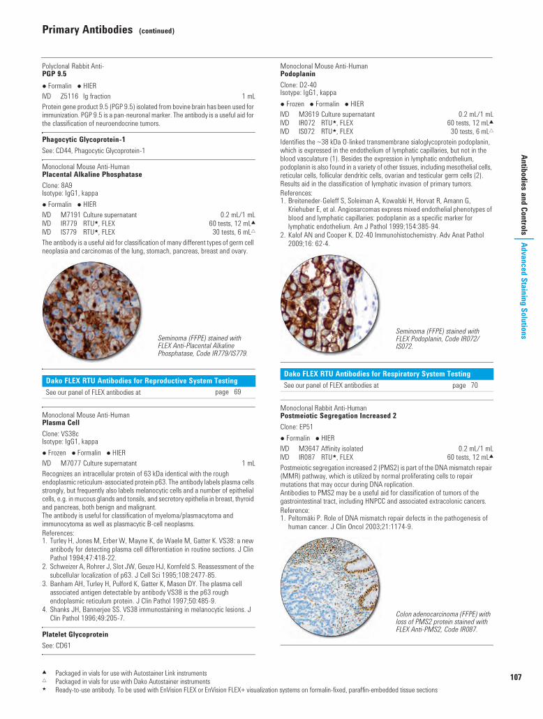
Primary Antibodies (continued)
107
Antibodies and ControlsAdvanced Staining Solutions
Polyclonal Rabbit Anti-PGP 9.5 Formalin HIER I Z5116 Ig fraction 1 mLProtein gene product 9.5 (PGP 9.5) isolated from bovine brain has been used for immunization. PGP 9.5 is a pan-neuronal marker. The antibody is a useful aid for the classification of neuroendocrine tumors.
Phagocytic Glycoprotein-1See: CD44, Phagocytic Glycoprotein-1
Monoclonal Mouse Anti-HumanPlacental Alkaline PhosphataseClone: 8A9Isotype: IgG1, kappa Formalin HIER I M7191 Culture supernatant 0.2 mL/1 mLI IR779 RTU5, FLEX 60 tests, 12 mL2I IS779 RTU5, FLEX 30 tests, 6 mL"The antibody is a useful aid for classification of many different types of germ cell neoplasia and carcinomas of the lung, stomach, pancreas, breast and ovary.
Monoclonal Mouse Anti-HumanPlasma CellClone: VS38cIsotype: IgG1, kappa Frozen Formalin HIER I M7077 Culture supernatant 1 mLRecognizes an intracellular protein of 63 kDa identical with the rough endoplasmic reticulum-associated protein p63. The antibody labels plasma cells strongly, but frequently also labels melanocytic cells and a number of epithelial cells, e.g. in mucous glands and tonsils, and secretory epithelia in breast, thyroid and pancreas, both benign and malignant.The antibody is useful for classification of myeloma/plasmacytoma and immunocytoma as well as plasmacytic B-cell neoplasms.References:1. Turley H, Jones M, Erber W, Mayne K, de Waele M, Gatter K. VS38: a new
antibody for detecting plasma cell differentiation in routine sections. J Clin Pathol 1994;47:418-22.
2. Schweizer A, Rohrer J, Slot JW, Geuze HJ, Kornfeld S. Reassessment of the subcellular localization of p63. J Cell Sci 1995;108:2477-85.
3. Banham AH, Turley H, Pulford K, Gatter K, Mason DY. The plasma cell associated antigen detectable by antibody VS38 is the p63 rough endoplasmic reticulum protein. J Clin Pathol 1997;50:485-9.
4. Shanks JH, Bannerjee SS. VS38 immunostaining in melanocytic lesions. J Clin Pathol 1996;49:205-7.
Platelet GlycoproteinSee: CD61
Monoclonal Mouse Anti-HumanPodoplaninClone: D2-40Isotype: IgG1, kappa Frozen Formalin HIER I M3619 Culture supernatant 0.2 mL/1 mLI IR072 RTU5, FLEX 60 tests, 12 mL2I IS072 RTU5, FLEX 30 tests, 6 mL"Identifies the ~38 kDa O-linked transmembrane sialoglycoprotein podoplanin, which is expressed in the endothelium of lymphatic capillaries, but not in the blood vasculature (1). Besides the expression in lymphatic endothelium, podoplanin is also found in a variety of other tissues, including mesothelial cells, reticular cells, follicular dendritic cells, ovarian and testicular germ cells (2). Results aid in the classification of lymphatic invasion of primary tumors.References:1. Breiteneder-Geleff S, Soleiman A, Kowalski H, Horvat R, Amann G,
Kriehuber E, et al. Angiosarcomas express mixed endothelial phenotypes of blood and lymphatic capillaries: podoplanin as a specific marker for lymphatic endothelium. Am J Pathol 1999;154:385-94.
2. Kalof AN and Cooper K. D2-40 Immunohistochemistry. Adv Anat Pathol 2009;16: 62-4.
Monoclonal Rabbit Anti-HumanPostmeiotic Segregation Increased 2Clone: EP51 Formalin HIER I M3647 Affinity isolated 0.2 mL/1 mLI IR087 RTU5, FLEX 60 tests, 12 mL2Postmeiotic segregation increased 2 (PMS2) is part of the DNA mismatch repair (MMR) pathway, which is utilized by normal proliferating cells to repair mutations that may occur during DNA replication. Antibodies to PMS2 may be a useful aid for classification of tumors of the gastrointestinal tract, including HNPCC and associated extracolonic cancers. Reference:1. Peltomäki P. Role of DNA mismatch repair defects in the pathogenesis of
human cancer. J Clin Oncol 2003;21:1174-9.
Dako FLEX RTU Antibodies for Reproductive System TestingSee our panel of FLEX antibodies at page 69
Seminoma (FFPE) stained with FLEX Anti-Placental Alkaline Phosphatase, Code IR779/IS779.
Dako FLEX RTU Antibodies for Respiratory System TestingSee our panel of FLEX antibodies at page 70
Seminoma (FFPE) stained with FLEX Podoplanin, Code IR072/IS072.
Colon adenocarcinoma (FFPE) with loss of PMS2 protein stained with FLEX Anti-PMS2, Code IR087.
2 Packaged in vials for use with Autostainer Link instruments" Packaged in vials for use with Dako Autostainer instruments5 Ready-to-use antibody. To be used with EnVision FLEX or EnVision FLEX+ visualization systems on formalin-fixed, paraffin-embedded tissue sections
USCatPathology.book Page 107 Monday, March 7, 2016 8:13 PM
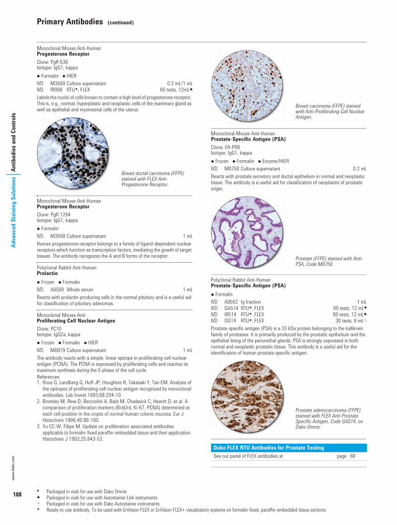
108
Primary Antibodies (continued)
www.
dako
.com
Adva
nced
Sta
inin
g So
lutio
nsAn
tibod
ies a
nd C
ontro
ls
Monoclonal Mouse Anti-HumanProgesterone ReceptorClone: PgR 636Isotype: IgG1, kappa Formalin HIER I M3569 Culture supernatant 0.2 mL/1 mLI IR068 RTU5, FLEX 60 tests, 12mL2Labels the nuclei of cells known to contain a high level of progesterone receptor. This is, e.g., normal, hyperplastic and neoplastic cells of the mammary gland as well as epithelial and myometrial cells of the uterus.
Monoclonal Mouse Anti-HumanProgesterone ReceptorClone: PgR 1294Isotype: IgG1, kappa Formalin I M3568 Culture supernatant 1 mLHuman progesterone receptor belongs to a family of ligand-dependent nuclear receptors which function as transcription factors, mediating the gowth of target tissues. The antibody recognizes the A and B forms of the receptor.
Polyclonal Rabbit Anti-HumanProlactin Frozen Formalin I A0569 Whole serum 1 mLReacts with prolactin-producing cells in the normal pituitary and is a useful aid for classification of pituitary adenomas.
Monoclonal Mouse Anti-Proliferating Cell Nuclear AntigenClone: PC10Isotype: IgG2a, kappa Frozen Formalin HIER I M0879 Culture supernatant 1 mLThe antibody reacts with a simple, linear epitope in proliferating cell nuclear antigen (PCNA). The PCNA is expressed by proliferating cells and reaches its maximum synthesis during the S-phase of the cell cycle.References:1. Roos G, Landberg G, Huff JP, Houghten R, Takasaki Y, Tan EM. Analysis of
the epitopes of proliferating cell nuclear antigen recognized by monoclonal antibodies. Lab Invest 1993;68:204-10.
2. Bromley M, Rew D, Becciolini A, Balzi M, Chadwick C, Hewitt D, et al. A comparison of proliferation markers (BrdUrd, Ki-67, PCNA) determined at each cell position in the crypts of normal human colonic mucosa. Eur J Histochem 1996;40:89-100.
3. Yu CC-W, Filipe M. Update on proliferation-associated antibodies applicable to formalin-fixed paraffin-embedded tissue and their application. Histochem J 1993;25:843-53.
Monoclonal Mouse Anti-HumanProstate-Specific Antigen (PSA)Clone: ER-PR8Isotype: IgG1, kappa Frozen Formalin Enzyme/HIER I M0750 Culture supernatant 0.2 mLReacts with prostate secretory and ductal epithelium in normal and neoplastic tissue. The antibody is a useful aid for classification of neoplasms of prostatic origin.
Polyclonal Rabbit Anti-HumanProstate-Specific Antigen (PSA) Formalin I A0562 Ig fraction 1 mLI GA514 RTU5, FLEX 60 tests, 12 mL8I IR514 RTU5, FLEX 60 tests, 12 mL2I IS514 RTU5, FLEX 30 tests, 6 mL"Prostate-specific antigen (PSA) is a 33 kDa protein belonging to the kallikrein family of proteases. It is primarily produced by the prostatic epithelium and the epithelial lining of the periurethal glands. PSA is strongly expressed in both normal and neoplastic prostatic tissue. This antibody is a useful aid for the identification of human prostate-specific antigen.
Breast ductal carcinoma (FFPE) stained with FLEX Anti-Progesterone Receptor.
Dako FLEX RTU Antibodies for Prostate TestingSee our panel of FLEX antibodies at page 69
Breast carcinoma (FFPE) stained with Anti-Proliferating Cell Nuclear Antigen.
Prostate (FFPE) stained with Anti-PSA, Code M0750.
Prostate adenocarcinoma (FFPE) stained with FLEX Anti-Prostate Specific Antigen, Code GA514, on Dako Omnis.
1 Packaged in vials for use with Dako Omnis2 Packaged in vials for use with Autostainer Link instruments" Packaged in vials for use with Dako Autostainer instruments5 Ready-to-use antibody. To be used with EnVision FLEX or EnVision FLEX+ visualization systems on formalin-fixed, paraffin-embedded tissue sections
USCatPathology.book Page 108 Monday, March 7, 2016 8:13 PM

Primary Antibodies (continued)
109
Antibodies and ControlsAdvanced Staining Solutions
Monoclonal Mouse Anti-HumanProstate-Specific Membrane Antigen (PSMA)Clone: 3E6Isotype: IgG1, kappa Formalin HIER I M3620 Culture supernatant 0.2 mL/1 mLI IR089 RTU5, FLEX 60 tests, 12 mL2Prostate-specific membrane antigen (PSMA) is expressed in normal and malignant prostatic epithelium and in a subset of non-prostatic tissues. In prostate cancer, PSMA expression has been shown to correlate with disease progression, with highest levels expressed in hormone-refractory and metastatic disease. The cellular localization of PSMA is cytoplasmic and/or membranous.This antibody labels PSMA-expressing cells in normal and neoplastic tissues and is a useful aid for classification of prostate adenocarcinomas.Reference:1. Mannweiler S, Amersdorfer P, Trajanoski S, Terrett J, King D, Mehes G.
Heterogeneity of prostate-specific membrane antigen (PSMA) expression in prostate carcinoma with distant metastasis. Pathol Oncol Res 2009;15:167-72.
Monoclonal Mouse Anti-HumanProstatic Acid PhosphataseClone: PASE/4LJIsotype: IgG1, kappa Frozen Formalin HIER I M0792 Culture supernatant 1 mLReacts with the glandular epithelium of prostate. Results aid in the classification of prostate carcinoma. Occasionally, carcinoid tumors may be labeled.
Monoclonal Mouse Anti-HumanProsteinClone: 10E3Isotype: IgG2a, kappa Formalin HIER I M3615 Culture supernatant 0.2 mL/1 mLI IR088 RTU5, FLEX 60 tests, 12 mL2Prostein is a 553-amino acid protein, also known as P501S. Prostein protein is a type IIIa plasma membrane protein which has been shown to be exclusively expressed in cells of normal and malignant prostate by Northern blot, cDNA microarray, real-time PCR and immunohistochemistry. Prostein is localized to the Golgi complex in the cytoplasm of cells and is expressed by both benign and neoplastic prostate tissue, whereas it has not been detected in any other normal or malignant tissue examined.This antibody is a useful aid for classification of prostate adenocarcinomas.
Monoclonal Mouse Anti-HumanPTENClone: 6H2.1Isotype: IgG2a, kappa Frozen Formalin HIER I M3627 Culture supernatant 0.2 mLThe PTEN protein is a lipid phosphatase with tumor-suppressing abilities. Results aid in the classification of a variety of malignancies, including breast (1), prostate (2) and endometrial cancer. References:1. Perren A, Weng LP, Boag AH, Ziebold U, Thakore K, Dahia PL, et al.
Immunohistochemical evidence of loss of PTEN expression in primary ductal adenocarcinomas of the breast. Am J Pathol 1999;155:1253-60.
2. McMenamin ME, Soung P, Perera S, Kaplan I, Loda M, Sellers WR. Loss of PTEN expression in paraffin-embedded primary prostate cancer correlates with high Gleason score and advanced stage. Cancer Res 1999;59:4291-6.
Rat Ki-67 AntigenSee: Ki-67 Antigen
Monoclonal Mouse Anti-HumanRenal Cell Carcinoma MarkerClone: SPM314Isotype: IgG2b, kappa Frozen Formalin HIER I M3632 Culture supernatant 1 mLI GA075 RTU5, FLEX NEW 60 tests, 12 mL8I IR075 RTU5, FLEX 60 tests, 12 mL2I IS075 RTU5, FLEX 30 tests, 6 mL"Reacts with renal cell carcinoma marker (gp200), a surface membrane glycoprotein. The antigen is expressed on the brush border of proximal renal tubules and on the luminal surface of Bowman's capsule, as well as in parathyroid parenchymal cells and colloid of thyroid follicles. The antibody is a useful aid for classification of primary and metastatic renal cell carcinomas.
Prostatic intraepithelial neoplasia (PIN) (FFPE) stained with FLEX Anti-PSMA, Code IR089.
Prostatic intraepithelial neoplasia (PIN) (FPPE) stained with FLEX Anti-Prostein, Code IR088.
Renal clear cell carcinoma (FFPE) stained with FLEX Anti-Renal Cell Carcinoma Marker, Code GA075, on Dako Omnis.
1 Packaged in vials for use with Dako Omnis2 Packaged in vials for use with Autostainer Link instruments" Packaged in vials for use with Dako Autostainer instruments5 Ready-to-use antibody. To be used with EnVision FLEX or EnVision FLEX+ visualization systems on formalin-fixed, paraffin-embedded tissue sections
USCatPathology.book Page 109 Monday, March 7, 2016 8:13 PM

110
Primary Antibodies (continued)
www.
dako
.com
Adva
nced
Sta
inin
g So
lutio
nsAn
tibod
ies a
nd C
ontro
ls
Monoclonal Mouse Anti-HumanRibosomal Protein S6-pS240Phosphorylation Site SpecificClone: DAK-S6-240Isotype: IgG1 Formalin HIER U M7300 Culture supernatant 0.2 mLReacts with human ribosomal protein S6 phosphorylated at serine residue 240 (pS240). Phosphorylation of ribosomal protein S6 correlates with an increase in translation of mRNAs that encode for proteins involved in cell cycle progression and proteins controlling mammalian cell growth and proliferation.
Polyclonal Rabbit Anti-S100 Formalin HIER I Z0311 Ig fraction 0.2 mL/1 mLI GA504 RTU5, FLEX 60 tests, 12 mL8I IR504 RTU5, FLEX 60 tests, 12 mL2I IS504 RTU5, FLEX 30 tests, 6 mL"Reacts strongly with human S100B, and weakly or very weakly with S100A1 and S100A6, respectively. S100 from ox brain has been used for the immunization. Z0311 labels glial cells in the brain and ependymal cells. Moreover, Schwann's cells of the peripheral nervous system are positive. Results aid in the classification of tumors in central and peripheral nervous system, such as schwannomas, ependydomas as well as in different grades of astrogliomas, also including glioblastomas. A large proportion of cells in human tumors originating from different salivary glands are labeled by anti-S100. The antibody is also a useful aid for classification of malignant melanocytic tumors of the skin and metastases of human malignant melanomas.
Polyclonal Rabbit Anti-HumanS100A4 Frozen Formalin HIER U A5114 Ig fraction 1 mLRecombinant human S100A4 has been used for immunization.
Monoclonal Mouse Anti-HumanSerotoninClone: 5HT-H209Isotype: IgG1, kappa Frozen Formalin I M0758 Culture supernatant 1 mLReacts with serotonin in a broad range of normal, hyperplastic and neoplastic tissues. Serotonin is also called 5-hydroxytryptamine. The antibody is a useful aid for classification of primary and metastatic carcinoid tumors expressing serotonin.
Polyclonal Rabbit Anti-HumanSomatostatin Frozen Formalin I A0566 Whole serum 1 mLSomatostatin is one of seven known polypeptide hormones produced in the pancreas. It functions as an inhibitory hormone of the neuroendocrine system and is secreted by D cells of the islets of Langerhans, the fundus and antrum of the stomach and in the upper small intestine. The antibody labels somatostatin-containing cells and is a useful aid for classification of pancreatic tumors and islet cell hyperplasia.
Monoclonal Mouse Anti-HumanSurvivinClone: 12C4Isotype: IgG2a, kappa Formalin HIER I M3624 Culture supernatant 0.2 mLSurvivin is a member of the inhibitor of apoptosis (IAP) gene family which counteracts apoptosis by inhibiting the activity of initiator and effector caspases. Survivin associates with the microtubules in the mitotic spindle and antagonizes mitochondrial-dependent apoptosis. In addition to cell death regulation, survivin has also been shown to be important in cell division and its expression is controlled at the transcriptional level in a cell cycle dependent manner. Results aid in the classification of a wide variety of neoplasms including tumors of the lung, breast, colon, stomach, esophagus, pancreas, liver, uterus, ovaries, Hodgkin’s disease, non-Hodgkin’s lymphoma, leukemias, neuroblastoma, phaeochromocytoma, soft-tissue sarcoma, gliomas and melanomas. The cellular localization of survivin is nuclear and/or cytoplasmic.
Monoclonal Mouse Anti-HumanSynaptophysinClone: DAK-SYNAPIsotype: IgG1, kappa Formalin HIER I M7315 Culture supernatant 0.2 mL/1 mLI IR660 RTU5, FLEX 60 tests, 12 mL2Synaptophysin is a 38 kDa membrane glycoprotein expressed in neuroendocrine cells and neurons involved in transmission via synaptic vehicles. Results aid in the classification of neuroendocrine neoplasms, such as neuroendocrine lung tumors (e.g. carcinoid, atypic carcinoid, SCLC, LCNEC and non-small cell lung cancer), neuroendocrine tumors of the gastroentero-pancreatic tract, e.g. neuroendocrine tumors (NETs), neuroendocrine carcinoma (NECs) and other epithelial neuroendocrine neoplasms such as pituitary adenomas, medullary thyroid carcinoma and parathyroid adenomas.Clone DAK-SYNAP is raised against a recombinant immunogen corresponding to the C-terminal cytoplasmic domain of human synaptophysin.References:1. Wiedenmann B, Franke WW. Identification and localization of
synaptophysin, an integral membrane glycoprotein of Mr 38,000 characteristic of presynaptic vesicles. Cell 1985;40:1017-28.
2. Kwon SE, Chapman ER. Synaptophysin regulates the kinetics of synaptic vesicle endocytosis in central neurons. Neuron 2011:70:847-85.
Breast carcinoma (FFPE) stained with FLEX Anti-S100, Code GA504, on Dako Omnis.
1 Packaged in vials for use with Dako Omnis2 Packaged in vials for use with Autostainer Link instruments" Packaged in vials for use with Dako Autostainer instruments5 Ready-to-use antibody. To be used with EnVision FLEX or EnVision FLEX+ visualization systems on formalin-fixed, paraffin-embedded tissue sections
USCatPathology.book Page 110 Monday, March 7, 2016 8:13 PM
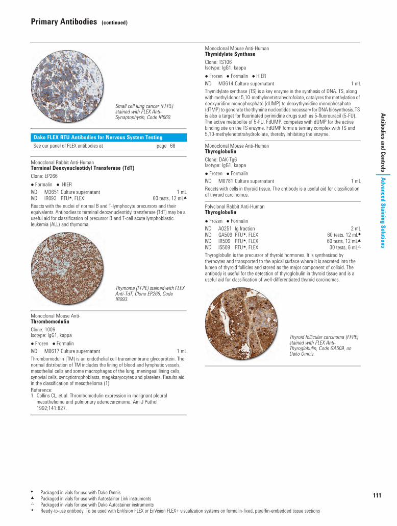
Primary Antibodies (continued)
111
Antibodies and ControlsAdvanced Staining Solutions
Monoclonal Rabbit Anti-HumanTerminal Deoxynucleotidyl Transferase (TdT)Clone: EP266 Formalin HIER I M3651 Culture supernatant 1 mLI IR093 RTU5, FLEX 60 tests, 12 mL2Reacts with the nuclei of normal B and T-lymphocyte precursors and their equivalents. Antibodies to terminal deoxynucleotidyl transferase (TdT) may be a useful aid for classification of precursor B and T-cell acute lymphoblastic leukemia (ALL) and thymoma.
Monoclonal Mouse Anti-ThrombomodulinClone: 1009Isotype: IgG1, kappa Frozen Formalin I M0617 Culture supernatant 1 mLThrombomodulin (TM) is an endothelial cell transmembrane glycoprotein. The normal distribution of TM includes the lining of blood and lymphatic vessels, mesothelial cells and some macrophages of the lung, meningeal lining cells, synovial cells, syncytiotrophoblasts, megakaryocytes and platelets. Results aid in the classification of mesothelioma (1).Reference:1. Collins CL, et al. Thrombomodulin expression in malignant pleural
mesothelioma and pulmonary adenocarcinoma. Am J Pathol 1992;141:827.
Monoclonal Mouse Anti-HumanThymidylate SynthaseClone: TS106Isotype: IgG1, kappa Frozen Formalin HIER I M3614 Culture supernatant 1 mLThymidylate synthase (TS) is a key enzyme in the synthesis of DNA. TS, along with methyl donor 5,10-methylenetetrahydrofolate, catalyzes the methylation of deoxyuridine monophosphate (dUMP) to deoxythymidine monophosphate (dTMP) to generate the thymine nucleotides necessary for DNA biosynthesis. TS is also a target for fluorinated pyrimidine drugs such as 5-fluorouracil (5-FU). The active metabolite of 5-FU, FdUMP, competes with dUMP for the active binding site on the TS enzyme. FdUMP forms a ternary complex with TS and 5,10-methylenetetrahydrofolate, thereby inhibiting the enzyme.
Monoclonal Mouse Anti-HumanThyroglobulinClone: DAK-Tg6Isotype: IgG1, kappa Frozen Formalin I M0781 Culture supernatant 1 mLReacts with cells in thyroid tissue. The antibody is a useful aid for classification of thyroid carcinomas.
Polyclonal Rabbit Anti-HumanThyroglobulin Frozen Formalin I A0251 Ig fraction 2 mLI GA509 RTU5, FLEX 60 tests, 12 mL8I IR509 RTU5, FLEX 60 tests, 12 mL2I IS509 RTU5, FLEX 30 tests, 6 mL"Thyroglobulin is the precursor of thyroid hormones. It is synthesized by thyrocytes and transported to the apical surface where it is secreted into the lumen of thyroid follicles and stored as the major component of colloid. The antibody is useful for the detection of thyroglobulin in thyroid tissue and is a useful aid for classification of well-differentiated thyroid carcinomas.
Dako FLEX RTU Antibodies for Nervous System TestingSee our panel of FLEX antibodies at page 68
Small cell lung cancer (FFPE) stained with FLEX Anti-Synaptophysin, Code IR660.
Thymoma (FFPE) stained with FLEX Anti-TdT, Clone EP266, Code IR093.
Thyroid follicular carcinoma (FFPE) stained with FLEX Anti-Thyroglobulin, Code GA509, on Dako Omnis.
1 Packaged in vials for use with Dako Omnis2 Packaged in vials for use with Autostainer Link instruments" Packaged in vials for use with Dako Autostainer instruments5 Ready-to-use antibody. To be used with EnVision FLEX or EnVision FLEX+ visualization systems on formalin-fixed, paraffin-embedded tissue sections
USCatPathology.book Page 111 Monday, March 7, 2016 8:13 PM

112
Primary Antibodies (continued)
www.
dako
.com
Adva
nced
Sta
inin
g So
lutio
nsAn
tibod
ies a
nd C
ontro
ls
Monoclonal Mouse Anti-HumanThyroid Peroxidase (TPO)Clone: MoAb47Isotype: IgG1, kappa Frozen Formalin HIER I M7257 Culture supernatant 0.2 mLThyroid peroxidase (TPO) is present as a dimer on the apical surface of thyroid folliular cells, and it is the primary enzyme involved in thyroid hormone synthesis. A reduction of TPO-positive cells has been found in malignant nodular thyroid tissue, and the antibody is a useful aid for classification of solitary cold thyroid nodules in fine needle aspiration cytology samples (1).Reference:1. Christensen L, Blichert-Toft M, Brandt M, Lange M, Sneppen SB, Ravnsbæk
J. Thyroperoxidase TPO immunostaining of the solitary cold thyroid nodule. Clin Endocrinol 2000;53:161-9.
Monoclonal Mouse Anti-HumanThyroid-Stimulating Hormone (TSH)Clone: 0042Isotype: IgG1, kappa Formalin I M3503 Culture supernatant 1 mLReacts with the β-subunit of thyroid-stimulating hormone. By immunoradiometric assay, no detectable cross-reactivity was found against hGH, prolactin, hCG or bovine TSH. The antibody labels thyrotrophic cells of the pituitary. Results aid in the classification of pituitary adenomas.
Monoclonal Mouse Anti-Thyroid Transcription Factor (TTF-1)Clone: 8G7G3/1Isotype: IgG1, kappa Frozen Formalin HIER I M3575 Culture supernatant 0.2 mL/1 mLI IR056 RTU5, FLEX 60 tests, 12 mL2I IS056 RTU5, FLEX 30 tests, 6 mL"Identifies the 40 kDa TTF-1 band in nuclear extracts or whole cell lysates of TTF-1-positive cell lines of rat, mouse and man. TTF-1 is selectively expressed in lung and thyroid, and the antibody may aid in the classification of tumors of the lung and thyroid.
Monoclonal Mouse Anti-HumanTissue Inhibitor of Metalloproteinases 1Clone: VT7Isotype: IgG1, kappa Formalin HIER I M7293 Culture supernatant 0.2 mLReacts with tissue inhibitor of metalloproteinases 1 (TIMP-1). TIMP-1 is expressed in a proportion of stromal myofibroblast-like cells adjacent to invading cancer cells of colon adenocarcinomas. Results aid in the classification of adenocarcinomas and colon adenomas (1). TIMP-1 is also expressed in neuroendocrine cells (2). TIMP-1 plays a pivotal role in extracellular matrix remodeling, and may affect malignant cell transformation, cell proliferation and apoptosis (3).References:1. Sørensen IV, Winther H, Foged NT, Fenger C, Brünner N. Tissue inhibitor of
metalloproteinases 1 (TIMP-1) as an immunohistochemical marker for colorectal cancer. Eur J Cancer Supplements 2005;3:196.
2. Sørensen IV, Fenger C, Winther H, Foged NT, Lademann U, Brünner N, et al. Characterization of anti-TIMP-1 monoclonal antibodies for immunohistochemical localization in formalin fixed paraffin embedded tissue. J Histochem Cytochem 2006;In press.
3. Fassina G, Ferrari N, Brigati C, Benelli R, Santi L, Noonan DM, et al. Tissue inhibitors of metalloproteases: regulation and biological activities. Clin Exp Metastasis 2000;18:111-20.
Monoclonal Mouse Anti-HumanTopoisomerase IIaClone: Ki-S1Isotype: IgG2a, kappa Frozen Formalin HIER I M7186 Purified 1 mLThe topoisomerase II enzymes control DNA topology by cleaving and rejoining DNA strands and passing other DNA strands through the transient gaps. The topoisomerase IIa isoform is a 170 kDa nuclear protein that is only expressed in proliferating cells.
Monoclonal Mouse Anti-HumanTyrosinaseClone: T311Isotype: IgG2a, kappa Formalin HIER I M3623 Culture supernatant 0.2 mLI IR061 RTU5, FLEX 60 tests, 12 mL2I IS061 RTU5, FLEX 30 tests, 6 mL"Tyrosinase is a copper-glycoenzyme involved in the production of melanin pigments, including both eumelanin and pheomelanin. As a marker of melanocytic lineage, tyrosinase is localized to melanocytes which can be found on the dermal/epidermal junction of normal skin. It is not detected in other normal cells. Results aid in the classification of primary and metastatic malignant melanomas. The cellular localization of tyrosinase is cytoplasmic and/or perinuclear.
Thyroid adenoma (benign) (FFPE) stained with Anti-TPO, Code M7257.
Lung smal cell carcinoma (FFPE) stained with FLEX Anti-TTF-1, Code IR056/IS056.
Colon adenocarcinoma (FFPE) stained with Anti-TIMP-1, Code M7293.
2 Packaged in vials for use with Autostainer Link instruments" Packaged in vials for use with Dako Autostainer instruments5 Ready-to-use antibody. To be used with EnVision FLEX or EnVision FLEX+ visualization systems on formalin-fixed, paraffin-embedded tissue sections
USCatPathology.book Page 112 Monday, March 7, 2016 8:13 PM

Primary Antibodies (continued)
113
Antibodies and ControlsAdvanced Staining Solutions
Monoclonal Mouse Anti-HumanuPARClone: R4Isotype: IgG1, kappa Frozen Formalin HIER I M7294 Culture supernatant 0.2 mLReacts with urokinase-type plasminogen activator receptor (uPAR), also designated CD87. uPAR is part of the plasminogen activator system, which is involved in both early phases of carcinogenesis as well as cancer invasion and metastasis (1, 2). In normal tissue, the antibody labels a subpopulation of macrophages and neutrophils in tonsil and breast tissue as well as in the lamina propria of the intestine. The antibody labels a subpopulation of tumor-associated stromal macrophages and is a useful aid for classification of invasive colon and breast carcinomas as well neoplastic glands of colon adenocarcinomas. References:1. Duffy MJ. The urokinase plasminogen activator system: role in malignancy
[review]. Curr Pharm Des 2004;10:39-49.2. Andreasen PA, Egelund R, Petersen HH. The plasminogen activation system
in tumor growth, invasion, and metastasis [review]. Cell Mol Life Sci 2000;57:25-40.
Monoclonal Mouse Anti-HumanVascular Endothelial Growth Factor (VEGF)Clone: VG1Isotype: IgG1, kappa Formalin HIER U M7273 Culture supernatant 0.2 mLVascular endothelial growth factor (VEGF) is a key regulator of physiological angiogenesis during embryogenesis, skeletal growth and reproductive functions. Of the six different isoforms of VEGF, the antibody labels the VEGF-121, VEGF-165, and VEGF-189 isoforms.
Monoclonal Mouse Anti-VillinClone: 1D2 C3Isotype: IgG1, kappa Frozen Formalin HIER I M3637 Culture supernatant 1 mLI IR076 RTU5, FLEX 60 tests, 12 mL2I IS076 RTU5, FLEX 30 tests, 6 mL"Villin is a 95 kDa calcium-regulated, actin-binding protein that plays a role iin regulating actin filament assembly. It is a major constituent in the microvilli, which compose the brush border of epithelial cells forming absorptive surfaces of the intestinal and renal proximal tubular epithelia. In normal human tissue, villin is expressed by a limited number of simple epithelia of the gastrointestinal and urogenital tract. Antibodies to villin are a useful aid for classification of primary and metastatic colorectal carcinomas.
Monoclonal Mouse Anti-VimentinClone: V9Isotype: IgG1, kappa Frozen Formalin HIER I M0725 Culture supernatant 0.2 mL/1 mLI GA630 RTU5, FLEX 60 tests, 12 mL8I IR630 RTU5, FLEX 60 tests, 12 mL2I IS630 RTU5, FLEX 30 tests, 6 mL"Vimentin is a 57 kDa intermediate filament protein which forms part of the cytoskeleton of vertebrate cells and is characteristically found in cells of mesenchymal origin. The coexpression of intermediate filaments, particularly vimentin and cytokeratin, has been demonstrated in a variety of normal cells/tissues and in neoplastic lesions, necessitating the use of antibodies against other types of intermediate filaments. The antibody is a useful aid for classification of neoplastic tissues of mesenchymal origin.
Melanoma (FFPE) stained with FLEX Anti-Tyrosinase, Code IR061/IS061.
Colon carcinoma (FFPE) stained with Anti-uPAR, Code M7294.
Dako FLEX RTU Antibodies for Gastrointestinal Tract TestingSee our panel of FLEX antibodies at page 65
Colon adenocarcinoma (FFPE) stained with FLEX Anti-Villin, Code IS076/IS076.
Bladder wall (FFPE) stained with FLEX Anti-Vimentin, Code GA630, on Dako Omnis.
1 Packaged in vials for use with Dako Omnis2 Packaged in vials for use with Autostainer Link instruments" Packaged in vials for use with Dako Autostainer instruments5 Ready-to-use antibody. To be used with EnVision FLEX or EnVision FLEX+ visualization systems on formalin-fixed, paraffin-embedded tissue sections
USCatPathology.book Page 113 Wednesday, March 9, 2016 10:36 PM

114
Primary Antibodies (continued)
www.
dako
.com
Adva
nced
Sta
inin
g So
lutio
nsAn
tibod
ies a
nd C
ontro
ls
Monoclonal Mouse Anti-VimentinClone: Vim 3B4Isotype: IgG2a, kappa Frozen Formalin Enzyme/HIER I M7020 Purified 1 mLReacts strongly with human vimentin and labels cells of mesenchymal origin. This antibody is particularly well-suited for use on formalin-fixed, paraffin-embedded tissue sections. The antibody is a useful aid for classification of tumors of mesenchymal origin.
Monoclonal Mouse Anti-HumanVon Willebrand FactorClone: F8/86Isotype: IgG1, kappa Frozen Formalin HIER I M0616 Culture supernatant 1 mLReacts with von Willebrand factor present in endothelial cells and in the cytoplasm of megakaryocytes. The antibody is a useful aid for classification of tumors derived from megakaryocytes. The former designation for von Willebrand factor was Factor VIII-related antigen.
Polyclonal Rabbit Anti-HumanVon Willebrand Factor Formalin Enzyme/HIER I A0082 Ig fraction 0.2 mL/2 mLI GA527 RTU5, FLEX 60 tests, 12 mL8I IR527 RTU5, FLEX 60 tests, 12 mL2I IS527 RTU5, FLEX 30 tests, 6 mL"Expression of the von Willebrand factor gene is tissue specific and confined to endothelial cells and megakaryocytes. VWF is present in plasma, in the Weibel-Pallade bodies of endothelial cells, in the alpha-granule in megakaryocytes and platelets derived from them, as well as in the subendothelial matrix of the vessel wall. Results aid in the classification of acute myeloid leukemia FAB type M7, angiosarcoma and epithelioid hemangioendothelioma.References:1. Weidner N, Semple JP, Welch WR, Folkman J. Tumor angiogenesis and
metastasis - correlation in invasive breast carcinoma. N Engl J Med 1991;324:1-8.
2. Makhlouf HR, Ihsak KD, Goodman ZD. Epithelioid hemangioendothelioma of the liver: a clinicopathologic study of 137 cases. Cancer 1999;85:562-82.
Monoclonal Mouse Anti-HumanWilms' Tumor 1 (WT1) ProteinClone: 6F-H2Isotype: IgG1, kappa Frozen Formalin Enzyme I M3561 Culture supernatant 1 mLI IR055 RTU5, FLEX 60 tests, 12 mL2I IS055 RTU5, FLEX 30 tests, 6 mL"WT1 is a gene involved in the induction of Wilms' tumor, a pediatric renal malignancy. Wilms' tumor 1 protein regulates transcription of other genes and can function both as a transcriptional activator and repressor. The antibody reacts with all isoforms of the full-length WT1 and also identifies WT1 lacking exon 2-encoded amino acids. Results aid in the classification of Wilms' tumors, malignant mesotheliomas and acute leukemias.
Monoclonal Mouse Anti-HumanZAP-70**Clone: 2F3.2Isotype: IgG2a, kappa Frozen Formalin HIER I M7303 Culture supernatant 1 mLI IR653 RTU5, FLEX 60 tests, 12 mL2Reacts with ZAP-70 expressed in T cells, natural killer cells, pro/pre B cells but not in normal mature B cells. The antibody is a useful aid for classification of a subset of chronic lymphocytic leukemias (CLL). In CLL, ZAP-70 expression is closely associated with an unmutated configuration of the immunoglobulin heavy-chain variable region (IgVH) genes (1).** This product is for in vitro diagnostic use only. The product embodies
technology described in US Patent 7,329,502 and pending Canadian Patent Application No. 2,413,475.
Reference:1. Carreras J, Villamor N, Colomo L, Moreno C, Cajal S, Crespo M, et al.
Immunohistochemical analysis of ZAP70 expression in B-cell lymphoid neoplasms. J Pathol 2005;205;507-13.
Tonsil (FFPE) stained with Anti-Vimentin, Code M7020.
Angiosarcoma (FFPE) stained with FLEX Anti-Von Willebrand Factor, Code GA527, on Dako Omnis.
Dako FLEX RTU Antibodies for Soft Tissue/Bones TestingSee our panel of FLEX antibodies at page 71
Mesothelioma (FFPE) stained with FLEX Anti-Wilms' Tumor 1 (WT1) Protein, Code IR055/IS055.
B-cell chronic lymphocytic leukemia/small lymphocytic lymphoma (FFPE) stained with FLEX Anti-ZAP-70, Code IR653.
1 Packaged in vials for use with Dako Omnis2 Packaged in vials for use with Autostainer Link instruments" Packaged in vials for use with Dako Autostainer instruments5 Ready-to-use antibody. To be used with EnVision FLEX or EnVision FLEX+ visualization systems on formalin-fixed, paraffin-embedded tissue sections
USCatPathology.book Page 114 Monday, March 7, 2016 8:14 PM
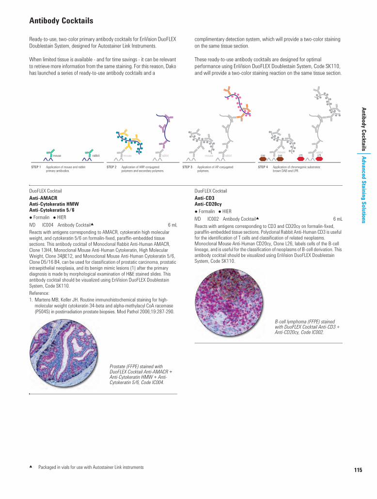
115
Antibody CocktailsAdvanced Staining Solutions
Antibody Cocktails
Ready-to-use, two-color primary antibody cocktails for EnVision DuoFLEX Doublestain System, designed for Autostainer Link Instruments.
When limited tissue is available - and for time savings - it can be relevant to retrieve more information from the same staining. For this reason, Dako has launched a series of ready-to-use antibody cocktails and a
complimentary detection system, which will provide a two-color staining on the same tissue section.
These ready-to-use antibody cocktails are designed for optimal performance using EnVision DuoFLEX Doublestain System, Code SK110, and will provide a two-color staining reaction on the same tissue section.
DuoFLEX Cocktail Anti-AMACRAnti-Cytokeratin HMWAnti-Cytokeratin 5/6 Formalin HIERI IC004 Antibody Cocktail2 6 mLReacts with antigens corresponding to AMACR, cytokeratin high molecular weight, and cytokeratin 5/6 on formalin-fixed, paraffin-embedded tissue sections. This antibody cocktail of Monoclonal Rabbit Anti-Human AMACR, Clone 13H4, Monoclonal Mouse Anti-Human Cytokeratin, High Molecular Weight, Clone 34bE12, and Monoclonal Mouse Anti-Human Cytokeratin 5/6, Clone D5/16 B4, can be used for classification of prostatic carcinoma, prostatic intraepithelial neoplasia, and its benign mimic lesions (1) after the primary diagnosis is made by morphological examination of H&E stained slides. This antibody cocktail should be visualized using EnVision DuoFLEX Doublestain System, Code SK110.Reference:1. Martens MB, Keller JH. Routine immunohistochemical staining for high-
molecular weight cytokeratin 34-beta and alpha-methylacyl CoA racemase (P504S) in postirradiation prostate biopsies. Mod Pathol 2006;19:287-290.
DuoFLEX Cocktail Anti-CD3Anti-CD20cy Formalin HIERI IC002 Antibody Cocktail2 6 mLReacts with antigens corresponding to CD3 and CD20cy on formalin-fixed, paraffin-embedded tissue sections. Polyclonal Rabbit Anti-Human CD3 is useful for the identification of T cells and classification of related neoplasms. Monoclonal Mouse Anti-Human CD20cy, Clone L26, labels cells of the B-cell lineage, and is useful for the classification of neoplasms of B-cell derivation. This antibody cocktail should be visualized using EnVision DuoFLEX Doublestain System, Code SK110.
rabbitmouseLPR LPRDAB DABrabbitmouserabbitmouserabbitmouse
STEP 1 Application of mouse and rabbit primary antibodies.
STEP 2 Application of HRP-conjugated polymers and secondary polymers.
STEP 3 Application of AP-conjugated polymers.
STEP 4 Application of chromogenic substrates: brown DAB and LPR.
Prostate (FFPE) stained with DuoFLEX Cocktail Anti-AMACR + Anti-Cytokeratin HMW + Anti-Cytokeratin 5/6, Code IC004.
B-cell lymphoma (FFPE) stained with DuoFLEX Cocktail Anti-CD3 + Anti-CD20cy, Code IC002.
2 Packaged in vials for use with Autostainer Link instruments
USCatPathology.book Page 115 Wednesday, March 9, 2016 10:38 PM

116
www.
dako
.com
Adva
nced
Sta
inin
g So
lutio
nsAn
tibod
y Coc
ktai
lsAntibody Cocktails (continued)
DuoFLEX Cocktail Anti-S100Anti-TyrosinaseAnti-Melan-A Formalin HIER I IC001 Antibody Cocktail2 6 mLReacts with antigens corresponding to S100, tyrosinase, and melan-A on formalin-fixed, paraffin-embedded tissue sections. Polyclonal Rabbit Anti-S100 is useful for the classification of S100-positive neoplasms, such as malignant melanoma and Langerhans' histiocytosis. Monoclonal Mouse Anti-Human Tyrosinase, Clone T311, is useful for the classification of melanocytic lesions and melanoma. Monoclonal Mouse Anti-Human Melan-A, Clone A103, is useful for the classification of melanomas and is also applicable for angiomyolipomas. This antibody cocktail should be visualized using EnVision DuoFLEX Doublestain System, Code SK110.
Melanoma (FFPE) stained with DuoFLEX Cocktail Anti-S100 + Anti-Tyrosinase + Anti-Melan-A, Code IC001.
2 Packaged in vials for use with Autostainer Link instruments
USCatPathology.book Page 116 Monday, March 7, 2016 8:14 PM

117
Multipurpose Antibodies
Advanced Staining SolutionsMultipurpose Antibodies
Owing to their high specificity and precipitating ability, a number of primary antibodies, notably polyclonal antibodies, are well-suited for a
variety of applications. This section presents these antibodies, and lists the techniques for which they have been tested and proven useful.
Polyclonal Rabbit Anti-HumanAlpha-1-Fetoprotein Precipitation ELISA IHC I A0008 Ig fraction 0.2 mLA0008 can be used for detection of alpha-1-fetoprotein in human sera by enzyme-immunoassays (1). A0008 is also a valuable reagent for immunohistochemistry (2).References:1. MacDonal DJ, Kelly AM. The rapid quantitation of serum alpha-fetoprotein by
two-site micro enzyme immunoassay. Clin Chim Acta 1978;87:367-72.2. Jacobsen GK, Jacobsen M, Clausen PP. Distribution of tumor-associated
antigens in the various histiologic components of germ cell tumors of the testis. Am J Surg Pathol 1981;5:257-66.
Polyclonal Rabbit Anti-Escherichia Coli Precipitation Blot I B0357 Ig fraction 2 mLAn aqueous extract of a sonicate of E. coli (non-transformed strain K12 C600) has been used for immunization. B0357 reacts with at least 80 different E. coli antigens in crossed immunoelectrophoresis, and also reveals a multitude of E. coli antigens in immunoblotting from SDS polyacrylamide gel electrophoresis. Broad reactivity with E. coli proteins (strains C600, HB101 and chi 1776 have been tested) makes B0357 particularly well-suited for monitoring the purification of proteins made in E. coli by genetic engineering.
Polyclonal Rabbit Anti-HumanFibrinogen Precipitation IHC I F0111 FITC. Ig fraction 2 mLF0111 reacts with fibrinogen, fibrin and the fibrinogen fragments D and E.
Polyclonal Rabbit Anti-Glial Fibrillary Acidic Protein (GFAP) Precipitation IHC I Z0334 Ig fraction 0.2 mL/1 mLGFAP isolated from bovine spinal cord has been used for immunization. Z0334 reacts strongly with human GFAP and with GFAP in many animal species tested: cat, cow, dog, mouse, rat and sheep. In the central nervous system Z0334 labels astrocytes and some groups of ependymal cells. In the peripheral nervous system Schwann's cells, satellite cells and enteric glial cells are labeled. Negative labeling is found in skin, connective tissue, lymphatic tissue, muscle, gastrointestinal tract, including liver and pancreas, kidney, ureter and bladder.Z0334 is useful particularly for distinguishing cells of astrocytic origin in the central nervous system. The antibody can be used on frozen and formalin-fixed, paraffin-embedded tissue sections.
Polyclonal Rabbit Anti-HumanIgA, Specific for Alpha-Chains Blot ELISA IHC I A0262 Ig fraction 1 mLI F0204 FITC. Ig fraction 2 mLFor use in methods demanding a very high specificity. The specificity and performance of the antibody have been ascertained in immunohistochemistry and ELISA. Additionally, the specificity has been tested by crossed immunoelectrophoresis using 12.5 microlitre antibody per square cm gel area against 2 microlitre human plasma. The antigen used for immunization is serum IgA.
Polyclonal Rabbit Anti-HumanIgA, IgG, IgM, Kappa, Lambda ELISA IHC I F0200 FITC. Ig fraction 2 mLI P0212 HRP. Ig fraction 2 mLF0200 is very well-suited for the flourescent treponemal antibody (FTA) test and for the demonstration of anti-nuclear antibodies (ANA) as well as other human autoantibodies, no matter what the immunoglobulin class may be. P0212 has found a wide application in ELISA for the detection of human antibodies.
Polyclonal Rabbit Anti-HumanIgG, Specific for Gamma-Chains Blot ELISA IHC I A0423 Ig fraction 1 mLI D0336 AP. Affinity isolated 1 mLI F0202 FITC. Ig fraction 2 mLI F0315 FITC. F(ab')2 1 mLI P0214 HRP. Ig fraction 2 mLFor use in methods demanding a very high specificity. The specificity and performance of the antibody have been ascertained in immunohistochemistry and ELISA. Additionally, the specificity has been tested by crossed immunoelectrophoresis using 12.5 microlitre antibody per square cm gel area against 2 microlitre human plasma.The F(ab')2 fragment antibody is particularly useful for labeling unfixed blood cells containing active Fc receptors, and for other applications where the Fc part of the antibody molecule could disturb.Please note that F(ab')2 fragment antibodies are not suited for techniques dependant on aggregation or precipitation of antigen-antibody complexes.
Polyclonal Rabbit Anti-HumanIgM, Specific for Mu-Chains Blot ELISA IHC I A0425 Ig fraction 1 mLI F0203 FITC. Ig fraction 2 mLI P0215 HRP. Ig fraction 2 mLFor use in methods demanding a very high specificity. The specificity and performance of the antibody have been ascertained in immunohistochemistry and ELISA. Additionally, the specificity has been tested by crossed immunoelectrophoresis using 12.5 microlitre antibody per square cm gel area against 2 microlitre human plasma.
Polyclonal Rabbit Anti-HumanKappa Light Chains Precipitation Blot ELISA IHC I A0191 Ig fraction 2 mLI F0198 FITC. Ig fraction 2 mLThese reagents have been produced in a manner that ensures a particularly wide specificity for kappa-chains. The specificity is directed against surface as well as hidden determinants and has been ascertained by gel precipitation techniques and immunohistochemistry.A0191 is excellent for the typing of free and bound monoclonal kappa-chains by immunoelectrophoresis and immunofixation.
USCatPathology.book Page 117 Monday, March 7, 2016 8:14 PM

118
www.
dako
.com
Adva
nced
Sta
inin
g So
lutio
nsM
ultip
urpo
se A
ntib
odie
sMultipurpose Antibodies (continued)
Polyclonal Rabbit Anti-HumanLambda Light Chains Precipitation Blot ELISA IHC I A0193 Ig fraction 2 mLI F0199 FITC. Ig fraction 2 mLThe antigen used for immunization is a pool of human lambda Bence Jones proteins. Therefore, a reagent with a particularly wide specificity for lambda-chains is obtained. The specificity is directed against surface as well as hidden determinants and has been ascertained by gel precipitation techniques and immunohistochemistry. A0193 is excellent for the typing of free and bound monoclonal lambda-chains by immunoelectrophoresis and immunofixation.
Polyclonal Rabbit Anti-HumanLysozyme EC 3.2.1.17 Precipitation Blot IHC I A0099 Ig fraction 2 mLA0099 is a valuable tool for identification of histiocytic neoplasias and myeloid leukemias (1, 2). Lysozyme in undiluted serum and urine can be quantitated by rocket immunoelectrophoresis (3). The buffer used for dilution of the standards should contain 1% bovine albumin, as lysozyme is unstable in protein-poor solutions. Please see reference 4 for the use of A0099 in immunoblotting.
References:1. Meister P, Huhn D, Nathrath W. Malignant histiocytosis. Immunocytochemical
characterization on paraffin-embedded tissue. Virchows Arch A Path Anat Histol 1980;385:233-46.
2. Krugliak L, Meyer PR, Taylor CR. The distribution of lysozyme, alpha-1-antitrypsin, and alpha-1-antichymotrypsin in normal hematopoietic cells and in myeloid leukaemias. Am J Hematol 1986;21:99-109.
3. Johansson BG, Malmquist J. Quantitative immunochemical determination of lysozyme (muramidase) in serum and urine. Scand J Clin Lab Invest 1971;27:255-61.
4. Mörsky P. Detection of lysozyme and alpha-2-macroglobulin-lysozyme complexes by immunoblotting. Clin Chim Acta 1988;178:327-36.
Polyclonal Rabbit Anti-HumanThyroglobulin Precipitation IHC I A0251 Ig fraction 2 mLThe antigen used for the production of A0251 has beeen isolated from normal human thyroids. The antibody has been made specific for thyroglobulin by absorption with insolubilized human serum.
USCatPathology.book Page 118 Monday, March 7, 2016 8:14 PM

119
Secondary AntibodiesAdvanced Staining Solutions
Secondary Antibodies
Secondary antibodies (antibodies to animal immunoglobulins) are utilized either for the direct detection of animal immunoglobulins or for amplification of the reaction between a primary antibody and the target antigen. Secondary antibodies play an important role in immunohistochemical procedures, immunoblotting, ELISAs and a number of other immunological methods. All Dako secondary antibodies and their conjugates are carefully tested to ensure good performance and low lot-to-lot variation. Several Dako secondary antibodies are presented in different grades regarding antiserum purification (immunoglobulin fraction of antiserum, affinity-isolated antibody, F(ab')2 fragment antibody) and also regarding the absorptions which have been performed to remove interspecies cross-reactions.
Further InformationA package insert with detailed product description and guidelines for dilutions accompanies all Dako secondary antibodies.
Polyclonal Rabbit Anti-Goat ImmunoglobulinsI F0250 FITC. Ig fraction 2 mLI P0160 HRP. Ig fraction 2 mLThe antibody has not been absorbed to remove cross-reactions with immunoglobulins from other species, therefore it shows a particularly strong reaction with its corresponding antigen. F0250 and P0160 are very good for indirect immunohistochemical techniques. Additionally, P0160 works well in ELISA and immunoblotting.
Polyclonal Rabbit Anti-Goat ImmunoglobulinsI E0466 Biotin. Affinity isolated 1 mLI P0449 HRP. Affinity isolated 1 mLCross-reaction with human immunoglobulins has been removed by solid-phase absorption. The antibody may cross-react with immunoglobulins from other species. Due to a particularly strong cross-reaction with sheep immunoglobulins, E0466 serves as an excellent link antibody with sheep primary antibodies.E0466 and P0449 are well-suited for immunohistochemistry, ELISA and immunoblotting.
Polyclonal Rabbit Anti-Guinea Pig ImmunoglobulinsI P0141 HRP. Ig fraction 2 mLThe antibody has not been absorbed to remove cross-reactions with immunoglobulins from other species, therefore it shows a particularly strong reaction with its corresponding antigen. P0141 is well-suited for immunohistochemistry, ELISA and immunoblotting.
Polyclonal Goat Anti-Mouse ImmunoglobulinsI Z0420 Affinity isolated 1 mLI D0486 AP. Affinity isolated 2 mLI E0433 Biotin. Affinity isolated 1 mLI P0447 HRP. Affinity isolated 1 mLCross-reaction with human immunoglobulins and fetal calf serum has been removed by solid-phase absorption. The antibody may cross-react with immunoglobulins from other species. However, it should be noted that the cross-reaction with rabbit immunoglobulins is very low. Therefore, the reagents are particularly well-suited for double labeling techniques when the two primary antibodies are from mouse and rabbit, respectively.Main applications are immunohistochemistry, ELISA and immunoblotting.
Polyclonal Rabbit Anti-Mouse Immunoglobulins
F0232 FITC. Ig fraction 2 mLI P0161 HRP. Ig fraction 2 mLThe antibody has not been absorbed to remove cross-reactions with immunoglobulins from other species, therefore it shows a particularly strong reaction with its corresponding antigen. Cross-reaction with human and cow immunoglobulins is pronounced.Main applications are as follows: Immunohistochemistry: F0232 and P0161. ELISA and immunoblotting: P0161.When cross-reaction with human and rat immunoglobulins and fetal calf serum disturbs, we recommend the use of one of Dako's preabsorbed antibodies to mouse immunoglobulins.
Polyclonal Rabbit Anti-Mouse ImmunoglobulinsI Z0259 Ig fraction 2 mLI D0314 AP. Affinity isolated 2 mLI E0354 Biotin. Affinity isolated 1 mLI E0413 Biotin. Affinity-isolated F(ab')2 1 mLI F0261 FITC. Ig fraction 2 mLI P0260 HRP. Ig fraction 2 mLCross-reaction with human immunoglobulins and fetal calf serum has been removed by solid-phase absorption. The reagents may cross-react with immunoglobulins from other species. Main applications are as follows: Immunohistochemistry: Z0259, D0314, E0354, E0413, F0261 and P0260. Please note that E0413 should be preferred to E0354 for the staining of blood cells and for other applications where the Fc part of the antibody could disturb. ELISA and immunoblotting: Z0259, D0314, E0354, E0413 and P0260. P0260 is particularly well-suited for the demonstration of monoclonal antibodies in ELISA.
Polyclonal Goat Anti-Rabbit ImmunoglobulinsI D0487 AP. Affinity isolated 1 mL
E0432 Biotin. Affinity isolated 1 mLI P0448 HRP. Affinity isolated 1 mLCross-reaction with human immunoglobulins and fetal calf serum has been removed by solid-phase absorption. The antibody may cross-react with immunoglobulins from other species. All reagents are well-suited for immunohistochemistry, ELISA and immunoblotting.
USCatPathology.book Page 119 Monday, March 7, 2016 8:14 PM

120
www.
dako
.com
Adva
nced
Sta
inin
g So
lutio
nsSe
cond
ary A
ntib
odie
sSecondary Antibodies (continued)
Monoclonal Mouse Anti-Rabbit ImmunoglobulinsClone: MR12/53Isotype: IgG1, kappa
I M0737 Culture supernatant 1 mLIs intended as a second stage reagent to be used when labeling tissue or cell samples with rabbit primary antibodies.
Polyclonal Swine Anti-Rabbit ImmunoglobulinsI Z0196 Ig fraction 2 mLI D0306 AP. Affinity isolated 1 mLI E0353 Biotin. Affinity isolated 1 mLI E0431 Biotin. Affinity-isolated F(ab')2 1 mLI F0205 FITC. Affinity isolated 2 mLI P0217 HRP. Ig fraction 2 mLI P0399 HRP. Affinity isolated 1 mLCross-reaction with human immunoglobulins has been removed by solid-phase absorption. The antibody may cross-react with immunoglobulins from other species.All reagents are well-suited for immunohistochemical techniques although E0431 should be preferred to E0353 for staining of blood cells and for other applications, where the Fc part of the antibody could disturb. Z0196 is also widely used as secondary antibody in radioimmunoassays. In addition, Z0196, D0306, E0353, E0431, P0217 and P0399 are valuable reagents for ELISA and immunoblotting.
Polyclonal Rabbit Anti-Rat Immunoglobulins
P0450 HRP. Ig fraction 1 mLCross-reaction with human immunoglobulins and fetal calf serum has been removed by solid-phase absorption. The antibody may cross-react with immunoglobulins from other species. P0450 is well-suited for immunohistochemistry, ELISA and immunoblotting.
Polyclonal Rabbit Anti-Sheep ImmunoglobulinsI P0163 HRP. Ig fraction 2 mLThe antibody has not been absorbed to remove cross-reactions with immunoglobulins from other species, therefore it shows a particularly strong reaction with its corresponding antigen. P0163 is well-suited for immunohistochemistry, ELISA and immunoblotting.
USCatPathology.book Page 120 Monday, March 7, 2016 8:14 PM

121
Control ReagentsAdvanced Staining Solutions
Control Reagents
The normal animal sera and animal immunoglobulins listed are well-suited as qualitative negative controls for Dako antibodies. The products are in liquid form and contain an antimicrobial agent.
Negative controls should always be diluted to match the concentration of the corresponding antibody. Negative controls for EnVision FLEX ready-to-use antibodies are also listed here.
Goat Serum (Normal)U X0907 Whole serum 10 mL
Mouse IgG1I X0931 Culture supernatant 1 mLX0931 is a cell culture supernatant containing monoclonal mouse IgG1 antibody to Aspergillus niger glucose oxidase, an enzyme which is neither present nor inducible in mammalian tissues. X0931 is well-suited as a negative control in all techniques utilizing monoclonal mouse antibodies of isotype IgG1.
Mouse IgG2aI X0943 Culture supernatant 1 mLX0943 is a cell culture supernatant containing monoclonal mouse IgG2a antibody to Aspergillus niger glucose oxidase, an enzyme which is neither present nor inducible in mammalian tissues. X0943 is well-suited as a negative control in all techniques utilizing monoclonal mouse antibodies of isotype IgG2a.
Mouse IgG2bI X0944 Culture supernatant 1 mLX0944 is a cell culture supernatant containing monoclonal mouse IgG2b antibody to Aspergillus niger glucose oxidase, an enzyme which is neither present nor inducible in mammalian tissues. X0944 is well-suited as a negative control in all techniques utilizing monoclonal mouse antibodies of isotype IgG2b.
Mouse IgMI X0942 Culture supernatant 1 mLX0942 is a cell culture supernatant containing monoclonal mouse IgM antibody to Aspergillus niger glucose oxidase, an enzyme which is neither present nor inducible in mammalian tissues. X0942 is well-suited as a negative control in all techniques utilizing monoclonal mouse antibodies of isotype IgM.
Rabbit Immunoglobulin Fraction (Normal)U X0903 Ig fraction 2 mL/10 mLThis product has been prepared from sera of non-immunized rabbits. The immunoglobulin fraction has been isolated in the same way as the immunoglobulin fraction of Dako rabbit antibodies. The protein concentration of X0903 is 20 g/L.
Rabbit Immunoglobulin Fraction (Solid-Phase Absorbed)I X0936 Ig fraction 2 mLThis product has been prepared from sera of non-immunized rabbits. The immunoglobulin fraction has been isolated in the same way as the immunoglobulin fraction of Dako rabbit antibodies. In addition, the product has been passed through a column containing immobilized human plasma proteins. This reduces the non-specific background and makes X0936 particularly well-suited as a negative control for Dako solid-phase absorbed rabbit antibodies. Especially when the primary antibody is used for immunohistochemistry at a relatively high concentration, above 0.1 g/L, X0936 should be preferred to Dako Rabbit Immunoglobulin Fraction (Normal), Code X0903. The protein concentration of X0936 is 15 g/L.
Rabbit Serum (Normal)U X0902 Whole serum 10 mL
Swine Serum (Normal)U X0901 Whole serum 10 mLEspecially for use in immunohistological techniques. Normal swine serum diluted 1:20 might reduce non-specific adsorption of antibodies to tissue, e.g. in the PAP technique.
Universal Negative Control for GA-Series Mouse Primary AntibodiesI GA750 Ready-to-use 120 tests, 24 mLUniversal negative control for all FLEX ready-to-use mouse primary antibodies for use on the Dako Omnis instrument. Packaged in vials for Dako Omnis.
Universal Negative Control for GA-Series Rabbit Primary AntibodiesI GA600 Ready-to-use 120 tests, 24 mLUniversal negative control for all FLEX ready-to-use rabbit primary antibodies for use on the Dako Omnis instrument. Packaged in vials for Dako Omnis.
Universal Negative Control for IR-Series Mouse Primary AntibodiesI IR750 Ready-to-use 120 tests, 24 mLUniversal negative control for all FLEX ready-to-use mouse primary antibodies for use on Automated Link Platforms. Packaged in Universal Vial.
Universal Negative Control for IR-Series Rabbit Primary AntibodiesI IR600 Ready-to-use 120 tests, 24 mLUniversal negative control for all FLEX ready-to-use rabbit primary antibodies for use on Automated Link Platforms. Packaged in Universal Vial.
Universal Negative Control for IS-Series Mouse Primary AntibodiesI IS750 Ready-to-use 60 tests, 12 mLUniversal negative control for all FLEX ready-to-use mouse primary antibodies for use on Dako Autostainer Instruments. Packaged in Dako Autostainer Vial.
Universal Negative Control for IS-Series Rabbit Primary AntibodiesI IS600 Ready-to-use 60 tests, 12 mLUniversal negative control to all FLEX ready-to-use rabbit primary antibodies for use on Dako Autostainer Instruments. Packaged in Dako Autostainer Vial.
USCatPathology.book Page 121 Monday, March 7, 2016 8:14 PM

122
www.
dako
.com
Adva
nced
Sta
inin
g So
lutio
nsVi
sual
izatio
n Sy
stem
sVisualization Systems
antibody
DABsubstrate
LPRsubstrate
tissueantigen
polymer linker
Legend for Schematics
primaryantibody
antibody
HRPenzyme
APenzyme
secondaryantibody
USCatPathology.book Page 122 Monday, March 7, 2016 8:14 PM

123
Visualization Systems
Advanced Staining SolutionsOverview of Dako Visualization Systems
Complexity Versus Sensitivity of Dako Visualization Systems
Comparative Features of Dako Visualization Systems
CSA
EnVision G|2
EnVision+EnVision FLEX
EnVision G|2Doublestain
5
4
3
2
1
1 2 3 4 5 6
Sensitivity
EnVisionDuoFLEX
DoublestainCSA II
ADVANCEEnVision FLEX+
Num
ber o
f Ste
ps
ARK
LSAB2
Product Name No. of Steps Primary Antibody Primary Antibody Enzyme Label TechnologyEnVision FLEX 2 Concentrate or FLEX RTU Mouse + Rabbit HRP Dextran (Biotin-Free)EnVision FLEX+ 3 Concentrate or FLEX RTU Mouse + Rabbit HRP Dextran (Biotin-Free)EnVision+ 2 Concentrate Mouse and/or Rabbit HRP Dextran (Biotin-Free)EnVision DuoFLEX Doublestain 4 Cocktail or Concentrate Mouse + Rabbit AP and HRP Dextran (Biotin-Free)EnVision G|2 3 Concentrate or RTU Mouse + Rabbit AP Dextran (Biotin-Free)EnVision G|2 Doublestain 5 Concentrate or RTU Mouse + Rabbit AP and HRP Dextran (Biotin-Free)ADVANCE 3 Concentrate or RTU Mouse + Rabbit HRP Dextran (Biotin-Free)ARK 2 Concentrate Mouse HRP Labeled Streptavidin-BiotinCSA 5 Concentrate Mouse or Rabbit HRP TyramideCSA II 4 Concentrate Mouse or Rabbit HRP Tyramide (Biotin-Free)LSAB 2 3 Concentrate or RTU Mouse + Rabbit HRP Labeled Streptavidin-Biotin
Chronic lymphocytic leukemia/small lymphocytic lymphoma (FFPE) stained with
FLEX Anti-CD23, Code GA781, on Dako Omnis.
Our FLEX Ready-to-Use antibodies are optimized for the FLEX/FLEX+ visualization systems. Simply pick one of the convenience kits and add any of the optional reagents and you have a
powerful IHC visualization solution that is ready to take on the diagnostic and workflow challenges in today’s pathology laboratory.
USCatPathology.book Page 123 Monday, March 7, 2016 8:14 PM

124
www.
dako
.com
Adva
nced
Sta
inin
g So
lutio
nsVi
sual
izatio
n Sy
stem
sOverview of EnVision FLEX and FLEX+ Visualization Systems
Achieve highly reliable and reproducible results without sacrificing quality or flexibility. EnVision FLEX and FLEX+ Visualization Systems give you high-
quality reagents, packaged in kit configurations that are easy to choose and use.
Dako Omnis
Autostainer Link 48
Dako Autostainer/Autostainer Plus
Single Reagents for EnVision FLEX Systems
EnVision FLEX SystemsFLEX
High pHFLEX
Low pHFLEX+ High pH
FLEX+ Low pH
Code
GV800or
GV823
GV800 + GV805 (Low pH TRS)or
GV823 + GV805 (Low pH TRS)
GV800 + GV821 (Mouse LINKER)or
GV800 + GV809 (Rabbit LINKER)
GV800 + GV805 + GV821or
GV800 + GV805 + GV809
EnVision FLEX SystemsFLEX
High pHFLEX
Low pHFLEX+ High pH
FLEX+ Low pH
Code
K8000or
K8023
K8000 + K8005 (Low pH TRS) or
K8023 + K8005 (Low pH TRS)
K8002 (incl. Mouse LINKER)or
K8002 + K8009 (Rabbit LINKER)
K8002 + K8005or
K8002 + K8005 + K8009
EnVision FLEX SystemsFLEX
High pHFLEX
Low pHFLEX+ High pH
FLEX+ Low pH
Code
K8010or
K8024
K8010 + K8005or
K8024 + K8005
K8012 (incl. Mouse LINKER)or
K8012 + K8009 (Rabbit LINKER)
K8012 (incl. Mouse LINKER) + K8005or
K8012 + K8005 + K8009 (Rabbit LINKER)
EnVision FLEX SystemsDako Omnis Autostainer Link 48 Dako Autostainer/Autostainer Plus
Prod
ucts
Mouse LINKER(Code GV821)
Mouse LINKER(Code K8021)
Mouse LINKER(Code K8022)
Rabbit LINKER(Code GV809)
Rabbit LINKER(Code K8009)
Rabbit LINKER(Code K8019)
Hematoxylin(Code GC808)
Hematoxylin(Code K8008)
Hematoxylin(Code K8018)
Target Retrieval Solution,High pH, 50x (Code GV804)
Target Retrieval Solution,High pH, 50x (Code K8004)
Target Retrieval Solution,High pH, 50x (Code K8004)
Target Retrieval Solution,Low pH, 50x (Code GV805)
Target Retrieval Solution,Low pH, 50x (Code K8005)
Target Retrieval Solution,Low pH, 50x (Code K8005)
Wash Buffer, 20x(Code GC807)
Wash Buffer, 20x(Code K8007)
Wash Buffer, 20x(Code K8007)
Antibody Diluent(Code K8006)
Antibody Diluent(Code K8006)
Antibody Diluent(Code K8006)
DAB+ Substrate ChromogenSystem (Code GV825)
USCatPathology.book Page 124 Monday, March 7, 2016 8:14 PM

125
Visualization Systems
Advanced Staining SolutionsWorkflow Diagram EnVision FLEX Systems Other Reagents
Possibilities Sensitivity options for both high sensitivity staining with EnVision FLEX
and very-high sensitivity staining with EnVision FLEX+ Kit components and reagent options create a complete and high
quality IHC Solution built upon the proven performance of Dako’sEnVision visualization technology platform
One code number orders the majority of reagents needed to complete a visualization run which ensures that reagents are optimized for usetogether
Benefits Offers an integrated solution Consistent and documented pre-treatment process Reliable, quality reagents with protocols created to optimize laboratory
efficiency Link software data management capabilities combined with
predetermined number of tests for all FLEX kits and options freespersonnel from reagent inventory tracking and eliminates excessreagent waste
HematoxylinDAB
Enhancer
Deparaffinization
Rehydration
Target Retrieval
High pH
Deparaffinization
Rehydration
Target Retrieval
Low pH
FLEX+
Mouse
(LINKER)
Concentrated
Antibody
Peroxidase
Blocking
Reagent*
FLEX
HRP
DAB+
Substrate
DAB+
Chromogen
FLEX
Ready-to-Use
Antibody
Pre-treatment Antibody Labeling Visualization Counterstain and Mounting
FLEX+
Rabbit
(LINKER)Antibody
Diluent
Mounting
Medium
Wash Buffer
* Onboard Dako Omnis, the blocking step is performed after primary antibody incubation.
EnVision FLEX with Low pH, 1:25 Ab dilution. EnVision FLEX with High pH, 1:25 Ab dilution. Notice the increased staining intensity using high pH pre-treatment.
EnVision FLEX and FLEX+. High and Very-High-Sensitivity Stains: Anti-TTF-1, Clone 8G7G3/1 (Code M3575) labeling lung adenocarcinoma.
EnVision FLEX with High pH, 1:75 Ab dilution.
EnVision FLEX. High pH and Low pH Stains: Anti-Cytokeratin 5/6, Clone D5/16 B4 (Code M7237) labeling basal cells in prostate gland.
EnVision FLEX+ with High pH, 1:75 Ab dilution. Notice the increased staining intensity using EnVision FLEX+.
USCatPathology.book Page 125 Wednesday, March 9, 2016 10:41 PM

126
ww
w.d
ako.c
om
Adva
nced
Sta
inin
g So
lutio
nsVi
sual
izatio
n Sy
stem
sEnVision FLEX Systems
EnVision FLEX and FLEX+ Visualization Systems gives you high-quality reagents packaged in easy-to-choose-and-use kit configurations. Simply pick one of the Convenience Kits, add any of the optional reagents, and you have an IHC visualization solution that satisfies the modern pathology laboratory's complex diagnostic needs.
EnVision FLEX Systems are simple, two-step visualization systems of high sensitivity. The FLEX+ kits have even greater sensitivity. They are all based on a unique enzyme-conjugated polymer backbone, which, in addition,
also carries secondary antibody molecules. Endogenous biotin will not affect EnVision FLEX staining results.
Formalin-fixed, paraffin-embedded tissue sections are suitable for use with EnVision FLEX systems. The kits are packaged either for use on Dako Omnis, Autostainer Link or Dako Autostainer instruments.
EnVision FLEX System
EnVision FLEX+ System
Read more about all the EnVision FLEX and FLEX+ kit configurations and optional reagents on the following pages
Antigen
PrimaryAntibody
mouse or rabbit
mouse or rabbit
mouse or rabbit
Antigen
LabeledPolymer
DAB DAB
STEP 1 Application of primary antibody. STEP 2 Application of EnVision FLEX/HRP. STEP 3 Application of DAB+.
Antigen
PrimaryAntibody
Linker
LabeledPolymer
LabeledPolymer
DABDAB DABDABAntigen
mouse or rabbit
mouse or rabbit
mouse or rabbit
mouse or rabbit
STEP 1 Application of primary antibody. STEP 3 Application of EnVision FLEX/HRP. STEP 4 Application of DAB+.STEP 2 Application of linker.
EnVision FLEX+, Mouse, High pH (K8002), 1:75 Ab dilution, Protocol #7, 10 min Ab/HRP incubation. More intense staining with shorter incubation time.
Anti-Cytokeratin 20, clone Ks20.8 (Code M7019) applied on colon adenocarcinoma.
EnVision FLEX, High pH (K8000), 1:75 Ab dilution, Protocol #2, 20 min Ab/HRP incubation. Anti-Cytokeratin 20,
clone Ks20.8 (Code M7019) applied on colon adenocarcinoma.
USCatPathology.book Page 126 Wednesday, March 9, 2016 10:41 PM

EnVision FLEX Systems (continued)
127
Visualization Systems
Advanced Staining Solutions
Dako Omnis
EnVision FLEX, High pH(Dako Omnis)I GV800 HRP. Rabbit/Mouse. High pH 600 testsEnVision FLEX, High pH is a high-sensitivity visualization system intended for use in immunohistochemistry together with Dako Omnis. The dual link system detects primary mouse and rabbit antibodies and the reaction is visualized by DAB+ Chromogen. The convenience kit includes Peroxidase-Blocking Reagent, EnVision/HRP, DAB+ Chromogen, Substrate Buffer and Target Retrieval Solution, High pH (50x Tris/EDTA buffer, pH 9). EnVision FLEX convenience kits are compatible with all optional EnVision FLEX and FLEX+ reagents for Dako Omnis.
EnVision FLEX Mini Kit, High pH(Dako Omnis)I GV823 HRP. Rabbit/Mouse. High pH 150 testsEnVision FLEX Mini Kit, High pH is a high-sensitivity visualization system intended for use in immunohistochemistry together with Dako Omnis. The dual link system detects primary mouse and rabbit antibodies and the reaction is visualized by DAB+ Chromogen. The convenience kit includes Peroxidase-Blocking Reagent, EnVision/HRP, DAB+ Chromogen, Substrate Buffer and Target Retrieval Solution, High pH (50x Tris/EDTA buffer, pH 9). EnVision FLEX convenience kits are compatible with all optional EnVision FLEX and FLEX+ reagents for Dako Omnis.
Autostainer Link 48
EnVision FLEX, High pH(Link)I K8000 HRP. Rabbit/Mouse. High pH 400-600 testsEnVision FLEX, High pH is a high-sensitivity visualization system intended for use in immunohistochemistry together with Autostainer Link Instruments. The dual link system detects primary mouse and rabbit antibodies and the reaction is visualized by DAB+ Chromogen. The convenience kit includes Peroxidase-Blocking Reagent, EnVision/HRP, DAB+ Chromogen, Substrate Buffer, Target Retrieval Solution, High pH (50x Tris/EDTA buffer, pH 9), and Wash Buffer (20x). EnVision FLEX convenience kits are compatible with all optional EnVision FLEX and FLEX+ reagents for Autostainer Link Instruments.
EnVision FLEX Mini Kit, High pH(Link)I K8023 HRP. Rabbit/Mouse. High pH 125-190 testsEnVision FLEX Mini Kit, High pH is a high-sensitivity visualization system intended for use in immunohistochemistry together with Autostainer Link Instruments. The dual link system detects primary mouse and rabbit antibodies and the reaction is visualized by DAB+ Chromogen. The convenience kit includes Peroxidase-Blocking Reagent, EnVision/HRP, DAB+ Chromogen,
Substrate Buffer, Target Retrieval Solution, High pH (50x Tris/EDTA buffer, pH 9), and Wash Buffer (20x). EnVision FLEX convenience kits are compatible with all optional EnVision FLEX and FLEX+ reagents for Autostainer Link Instruments.
EnVision FLEX+, Mouse, High pH(Link)I K8002 HRP. Mouse. High pH 400-600 testsEnVision FLEX+, Mouse, High pH is a very-high-sensitivity visualization system intended for use in immunohistochemistry together with Autostainer Link Instruments. The EnVision FLEX+ Mouse (LINKER) amplifies the signal of primary mouse antibodies and the reaction is visualized by DAB+ Chromogen. In addition to the EnVision FLEX+ Mouse (LINKER) the convenience kit includes Peroxidase-Blocking Reagent, EnVision/HRP, DAB+ Chromogen, Substrate Buffer, Target Retrieval Solution, High pH (50x Tris/EDTA buffer, pH 9), and Wash Buffer (20x). The EnVision FLEX+ Rabbit (LINKER), Code K8009, is an optional EnVision FLEX reagent that may be used with EnVision FLEX and FLEX+ convenience kits to amplify the signal of primary rabbit antibodies. EnVision FLEX+ convenience kits are compatible with all optional EnVision FLEX and FLEX+ reagents for Autostainer Link Instruments.
Dako Autostainer/Autostainer Plus
EnVision FLEX, High pH(Dako Autostainer/Autostainer Plus)I K8010 HRP. Rabbit/Mouse. High pH 400-600 testsEnVision FLEX, High pH is a high-sensitivity visualization system intended for use in immunohistochemistry together with Dako Autostainer Instruments. The dual link system detects primary mouse and rabbit antibodies and the reaction is visualized by DAB+ Chromogen. The convenience kit includes Peroxidase-Blocking Reagent, EnVision/HRP, DAB+ Chromogen, Substrate Buffer, Target Retrieval Solution, High pH (50x Tris/EDTA buffer, pH 9), and Wash Buffer (20x). EnVision FLEX convenience kits are compatible with all optional EnVision FLEX and FLEX+ reagents for Dako Autostainer Instruments.
EnVision FLEX Mini Kit, High pH(Dako Autostainer/Autostainer Plus)I K8024 HRP. Rabbit/Mouse. High pH 125-190 testsEnVision FLEX Mini Kit, High pH is a high-sensitivity visualization system intended for use in immunohistochemistry together with Dako Autostainer Instruments. The dual link system detects primary mouse and rabbit antibodies and the reaction is visualized by DAB+ Chromogen. The convenience kit includes Peroxidase-Blocking Reagent, EnVision/HRP, DAB+ Chromogen,
Substrate Buffer, Target Retrieval Solution, High pH (50x Tris/EDTA buffer, pH 9), and Wash Buffer (20x). EnVision FLEX convenience kits are compatible with all optional EnVision FLEX and FLEX+ reagents for Dako Autostainer Instruments.
EnVision FLEX+, Mouse, High pH(Dako Autostainer/Autostainer Plus)I K8012 HRP. Mouse. High pH 400-600 testsEnVision FLEX+, Mouse, High pH is a very-high-sensitivity visualization system intended for use in immunohistochemistry together with Dako Autostainer Instruments. The EnVision FLEX+ Mouse (LINKER) amplifies the signal of primary mouse antibodies and the reaction is visualized by DAB+ Chromogen. In addition to the EnVision FLEX+ Mouse (LINKER) the convenience kit includes Peroxidase-Blocking Reagent, EnVision/HRP, DAB+ Chromogen, Substrate Buffer, Target Retrieval Solution, High pH (50x Tris/EDTA buffer, pH 9), and Wash Buffer (20x). The EnVision FLEX+ Rabbit (LINKER), Code K8019, is an optional EnVision FLEX reagent that may be used with EnVision FLEX and FLEX+ convenience kits to amplify the signal of primary rabbit antibodies. EnVision FLEX+ convenience kits are compatible with all optional EnVision FLEX and FLEX+ reagents for Dako Autostainer Instruments.
USCatPathology.book Page 127 Monday, March 7, 2016 8:14 PM

128
www.
dako
.com
Adva
nced
Sta
inin
g So
lutio
nsVi
sual
izatio
n Sy
stem
sEnVision FLEX Single Reagents
The flexibility and versatility of EnVision FLEX systems make it easy to tailor a range of solutions to meet the specific needs for the pathology
laboratories. The Convenience Kits can be supplemented with our EnVision FLEX Single Reagents.
Dako Omnis
DAB+ Substrate Chromogen System (Dako Omnis)I GV825 Onboard mixing 150 testsEnVision FLEX DAB+ Substrate Chromogen System (Dako Omnis) is intended for use in immunohistochemistry together with Dako Omnis. The working solution is prepared onboard by the Dako Omnis instrument. It is a high sensitivity DAB system suitable for use in combination with the EnVision FLEX visualization system (Codes GV800/GV823). Upon oxidation, DAB forms a brown end-product at the site of the target antigen. The reagent is intended for use on formalin-fixed, paraffin-embedded tissue sections.
Hematoxylin (Dako Omnis)I GC808 Ready-to-use 8 x 22.5 mL, 600 testsIntended for use in immunohistochemistry together with Dako Omnis. The reagent is recommended for counterstaining on formalin-fixed, paraffin-embedded tissue sections providing a clear blue, nuclear staining.
Mouse LINKER(Dako Omnis)I GV821 Ready-to-use 75 tests, 22.5 mLEnVision FLEX+ Mouse LINKER is an optional EnVision FLEX+ reagent and may be used with EnVision FLEX convenience kits (GV800 and GV823) for Dako Omnis to amplify the signal of primary mouse antibodies.
Rabbit LINKER(Dako Omnis)I GV809 Ready-to-use 75 tests, 22.5 mLEnVision FLEX+ Rabbit LINKER is an optional EnVision FLEX+ reagent and may be used with EnVision FLEX convenience kits (GV800 and GV823) for Dako Omnis to amplify the signal of primary rabbit antibodies.
Target Retrieval Solution, High pH(Dako Omnis)I GV804 Concentrate 3 x 68 mL, 225 testsEnVision FLEX Target Retrieval Solution, High pH (Dako Omnis) is an optional EnVision FLEX reagent containing 50x concentrated Tris/EDTA, pH 9 and is compatible with EnVision FLEX convenience kits for Dako Omnis. The volume is optimized for dilution in Dako Omnis bulk bottles.
Target Retrieval Solution, Low pH(Dako Omnis)I GV805 Concentrate 3 x 68 mL, 225 testsEnVision FLEX Target Retrieval Solution, Low pH (Dako Omnis) is an optional EnVision FLEX reagent containing 50x concentrated citrate buffer, pH 6.1 and is compatible with EnVision FLEX convenience kits for Dako Omnis. The volume is optimized for dilution in Dako Omnis bulk bottles.
Wash Buffer (20x) (Dako Omnis)I GC807 Concentrate 20 x 175 mL, 1700 testsWash Buffer 20x (Dako Omnis) is intended for use in immunohistochemistry. The product is used as wash buffer for immunohistochemical staining procedures onboard Dako Omnis.
Dako Omnis Mouse LINKER Rabbit LINKER Hematoxylin Target Retrieval Solution, High pH Target Retrieval Solution, Low pH DAB+ Substrate Chromogen System Antibody Diluent Wash Buffer
Autostainer Link Mouse LINKER Rabbit LINKER Hematoxylin Target Retrieval Solution, High pH Target Retrieval Solution, Low pH Antibody Diluent Wash Buffer
Dako Autostainer/Autostainer Plus Mouse LINKER Rabbit LINKER Hematoxylin Target Retrieval Solution, High pH Target Retrieval Solution, Low pH Antibody Diluent Wash Buffer
USCatPathology.book Page 128 Monday, March 7, 2016 8:14 PM

EnVision FLEX Single Reagents (continued)
129
Visualization Systems
Advanced Staining Solutions
Autostainer Link 48
Hematoxylin(Link)I K8008 Ready-to-use 400-600 tests, 3 x 45 mLEnVision FLEX Hematoxylin is an optional EnVision FLEX reagent and is recommended for counterstaining. The reagent provides a clear blue, nuclear staining. EnVision FLEX Hematoxylin is compatible with EnVision FLEX and FLEX+ convenience kits.
Mouse (LINKER)(Link)I K8021 Ready-to-use 130-200 tests, 40 mLEnVision FLEX+ Mouse (LINKER) is an optional EnVision FLEX+ reagent and may be used with EnVision FLEX and FLEX+ convenience kits to amplify the signal of primary mouse antibodies.
Rabbit (LINKER)(Link)I K8009 Ready-to-use 130-200 tests, 40 mLEnVision FLEX+ Rabbit (LINKER) is an optional EnVision FLEX+ reagent and may be used with EnVision FLEX and FLEX+ convenience kits to amplify the signal of primary rabbit antibodies.
Target Retrieval Solution, High pHI K8004 Concentrate 3 x 30 mLEnVision FLEX Target Retrieval Solution, High pH is an optional EnVision FLEX reagent containing 50x concentrated Tris/EDTA, pH 9 and is compatible with all EnVision FLEX and FLEX+ convenience kits for both Autostainer Link Instruments and Dako Autostainer Instruments. One 30 mL bottle, when properly diluted, is enough to fill one PT Link tank.
Target Retrieval Solution, Low pHI K8005 Concentrate 3 x 30 mLEnVision FLEX Target Retrieval Solution, Low pH is an optional EnVision FLEX reagent containing 50x concentrated citrate buffer, pH 6.1 and is compatible with all EnVision FLEX and FLEX+ convenience kits for both Autostainer Link Instruments and Dako Autostainer Instruments. One 30 mL bottle, when properly diluted, is enough to fill one PT Link tank.
Wash BufferI K8007 Concentrate 1 LEnVision FLEX Wash Buffer is an optional EnVision FLEX reagent containing 20x concentrated wash buffer and is compatible with all EnVision FLEX and FLEX+ convenience kits for both Autostainer Link Instruments and Dako Autostainer Instruments.
Dako Autostainer/Autostainer Plus
Hematoxylin(Dako Autostainer/Autostainer Plus)I K8018 Ready-to-use 400-600 tests, 10 x 13 mLEnVision FLEX Hematoxylin is an optional EnVision FLEX reagent and is recommended for counterstaining. The reagent provides a clear blue, nuclear staining. EnVision FLEX Hematoxylin is compatible with EnVision FLEX and FLEX+ convenience kits.
Mouse (LINKER)(Dako Autostainer/Autostainer Plus)I K8022 Ready-to-use 120-190 tests, 3 x 13 mLEnVision FLEX+ Mouse (LINKER) is an optional EnVision FLEX+ reagent and may be used with EnVision FLEX and FLEX+ convenience kits to amplify the signal of primary mouse antibodies.
Rabbit (LINKER)(Dako Autostainer/Autostainer Plus)I K8019 Ready-to-use 120-190 tests, 3 x 13 mLEnVision FLEX+ Rabbit (LINKER) is an optional EnVision FLEX+ reagent and may be used with EnVision FLEX and FLEX+ convenience kits to amplify the signal of primary rabbit antibodies.
Target Retrieval Solution, High pHI K8004 Concentrate 3 x 30 mLEnVision FLEX Target Retrieval Solution, High pH is an optional EnVision FLEX reagent containing 50x concentrated Tris/EDTA, pH 9 and is compatible with all EnVision FLEX and FLEX+ convenience kits for both Autostainer Link Instruments and Dako Autostainer Instruments. One 30 mL bottle, when properly diluted, is enough to fill one PT Link tank.
Target Retrieval Solution, Low pHI K8005 Concentrate 3 x 30 mLEnVision FLEX Target Retrieval Solution, Low pH is an optional EnVision FLEX reagent containing 50x concentrated citrate buffer, pH 6.1 and is compatible with all EnVision FLEX and FLEX+ convenience kits for both Autostainer Link Instruments and Dako Autostainer Instruments. One 30 mL bottle, when properly diluted, is enough to fill one PT Link tank.
Wash BufferI K8007 Concentrate 1 LEnVision FLEX Wash Buffer is an optional EnVision FLEX reagent containing 20x concentrated wash buffer and is compatible with all EnVision FLEX and FLEX+ convenience kits for both Autostainer Link Instruments and Dako Autostainer Instruments.
All Platforms
Antibody DiluentI K8006 Diluent 400-600 tests, 120 mLEnVision FLEX Antibody Diluent is an optional EnVision FLEX reagent and is recommended for the dilution of Dako concentrated Primary Antibodies. EnVision FLEX Antibody Diluent is compatible with all EnVision FLEX and FLEX+ convenience kits for both Dako Omnis, Autostainer Link Instruments and Dako Autostainer Instruments.
IHC Microscope Slides, FLEXI K8020 Coated glass slides 5 x 100 slidesCoated microscope slides for adhesion of formalin-fixed, paraffin-embedded tissue sections for use in immunohistochemistry with Dako EnVision FLEX visualization systems. FLEX IHC Microscope Slides are compatible with, but not limited to, the following Dako instruments: Dako Omnis, Autostainer Link, Dako Autostainer/Autostainer Plus and PT Link.
USCatPathology.book Page 129 Monday, March 7, 2016 8:40 PM

130
www.
dako
.com
Adva
nced
Sta
inin
g So
lutio
nsVi
sual
izatio
n Sy
stem
sEnVision DuoFLEX System
EnVision DuoFLEX Doublestain System is a two-color detection system in an easy-to-choose-and-use kit configuration, based on the EnVision polymer technology known to provide high-quality staining results. The system will enable staining of two or more markers on a single slide using HRP and AP reactions. The system has been developed for ready-to-use DuoFLEX Antibody Cocktails, but may also be used with customers' own antibody cocktails or individual antibodies that are sequentially incubated on a single slide. The final staining result will be brown using DAB for mouse primary antibodies and red using LPR (liquid permanent red) for rabbit primary antibodies.
Staining for two or more targets in one tissue section will not only provide a two-color staining result using just one procedure thereby reducing time, but also give a more complex and informative staining result of the antigen expression in the particular tissue section. For further time savings it can be used with Dako DuoFLEX Antibody Cocktails.
EnVision DuoFLEX Doublestain System is a simple, four-step visualization system of high sensitivity. The kit is ready-to-use and suitable for use on formalin-fixed, paraffin-embedded tissue sections. The kit is packaged for use on Autostainer Link Instruments.
EnVision DuoFLEX Doublestain SystemI SK110 HRP/AP, Rabbit/Mouse2 100-150 tests, 30 mLEnVision DuoFLEX Doublestain System is intended for use in immunohisto-chemistry together with Autostainer Link instruments. This system is useful for the simultaneous detection of multiple antigens present in low or high concentrations within one specimen. The visualization is based on peroxidase (HRP) using DAB+ as chromogen and alkaline phosphatase (AP) using
Permanent Red as chromogen. EnVision DuoFLEX Doublestain System is biotin-free, thus significantly reducing non-specific staining resulting from endogenous avidin-biotin activity. This visualization system should be used for Dako DuoFLEX Cocktail antibodies.Note: The number of tests is based on the use of 200 µL or 300 µL of reagent per slide.
rabbitmouseLPR LPRDAB DABrabbitmouserabbitmouserabbitmouse
STEP 1 Application of mouse and rabbit primary antibodies.
STEP 2 Application of HRP-conjugated polymers and secondary polymers.
STEP 3 Application of AP-conjugated polymers.
STEP 4 Application of chromogenic substrates: brown DAB and LPR.
2 Packaged in vials for use with Autostainer Link instruments
USCatPathology.book Page 130 Wednesday, March 9, 2016 10:42 PM

131
Visualization Systems
Advanced Staining SolutionsEnVision Systems
Dako EnVision Systems are simple, two-step visualization systems of very high sensitivity. They are based on a unique enzyme-conjugated polymer backbone, which, in addition, also carries secondary antibody molecules. Endogenous biotin will not affect EnVision staining results.
The EnVision+ System has a particularly high sensitivity, and a recommended 30-minute incubation time with primary antibody and EnVision+ Reagent, respectively.
The EnVision G|2 Systems are 2nd generation visualization kits. Routinely fixed paraffin sections, smears, frozen sections, imprints and cytocentrifuge preparations are suitable for use with EnVision Systems. The number of tests that can be performed with the individual product is based on the use of 100 µL of reagent per slide.
EnVision DuoFLEX Doublestain System is useful for the simultaneous detection of multiple antigens present in low or high concentrations within one specimen.
EnVision G|2 Doublestain SystemRabbit/Mouse (DAB+/Permanent Red)I K5361 150 testsEnVision G|2 Doublestain System is a high-sensitivity peroxidase and alkaline-phosphatase-based 2nd generation visualization kit. The kit is intended for use in immunohistochemistry for the simultaneous detection of two different antigens within the same specimen, and is compatible with suitably diluted rabbit and mouse primary antibodies. The kit may be used on formalin-fixed, paraffin-embedded tissue sections and fixed cell smears. In addition to the ready-to-use EnVision G|2 reagents packaged in Dako Autostainer Reagent Vials, the kit includes both DAB+ and Permanent Red chromogenic substrate systems.Note: The number of tests for this kit is based on the use of 200 µL of reagent per slide.
EnVision G|2 System/APRabbit/Mouse (Permanent Red)I K5355 50 tests/500 testsEnVision G|2 System/AP is a high-sensitivity alkaline-phosphatase-based 2nd generation visualization kit. The kit is intended for use in immunohistochemistry, and it is compatible with suitably diluted rabbit and mouse primary antibodies. The kit may be used on formalin-fixed, paraffin-embedded tissue sections, frozen sections and fixed cell smears. In addition to the ready-to-use EnVision G|2 reagents packaged in Dako Autostainer Reagent Vials, the kit includes a Permanent Red chromogenic substrate system. The kit may be used in manual procedures or with the Dako Autostainer instruments.Note: The number of tests for this kit is based on the use of 200 µL of reagent per slide.
EnVision+ KitsI K4004 HRP. Mouse (AEC+) 150 testsI K4005 HRP. Mouse (AEC+) 1100 testsI K4006 HRP. Mouse (DAB+) 150 testsI K4007 HRP. Mouse (DAB+) 1100 testsI K4008 HRP. Rabbit (AEC+) 150 testsI K4009 HRP. Rabbit (AEC+) 1100 testsI K4010 HRP. Rabbit (DAB+) 150 testsI K4011 HRP. Rabbit (DAB+) 1100 testsThese ready-to-use, peroxidase-based EnVision+ kits are compatible with suitably diluted mouse or rabbit primary antibodies, respectively. In addition to the ready-to-use EnVision+ reagent, the kits include a blocking reagent for endogenous peroxidase, and a high sensitivity 3-amino-9-ethylcarbazole (AEC+) chromogenic substrate system. The kits are provided with detailed instructions.
EnVision+ Dual Link KitI K4065 HRP. Rabbit/Mouse (DAB+) 150 testsThis ready-to-use, peroxidase-based EnVision+ Dual Link Kit is compatible with suitably diluted rabbit and mouse primary antibodies. In addition to the ready-to-use EnVision+ Dual Link reagent, the kit includes a blocking reagent for endogenous peroxidase and a high-sensitivity diaminobenzidine (DAB+) chromogenic substrate system. The kit is provided with detailed instructions.
EnVision+ Dual Link, Single ReagentsI K4063 HRP. Rabbit/Mouse 150 tests, 15 mLI K4061 HRP. Rabbit/Mouse 1100 tests, 10 x 11 mLThese ready-to-use, peroxidase-conjugated EnVision+ Dual Link reagents are compatible with suitably diluted rabbit and mouse primary antibodies. A working procedure is included with the reagents.
EnVision+ Single ReagentsI K4000 HRP. Mouse 150 tests, 15 mLI K4001 HRP. Mouse 1100 tests, 110 mLI K4002 HRP. Rabbit 150 tests, 15 mLI K4003 HRP. Rabbit 1100 tests, 110 mLThese ready-to-use, peroxidase-conjugated EnVision+ reagents are compatible with suitably diluted mouse or rabbit primary antibodies, respectively. A working procedure is included with the reagents.
STEP 1 Application of primary antibody. Incubate 10 or 30 min.
STEP 2 Application of EnVision. Incubate 10 or 30 min.
USCatPathology.book Page 131 Wednesday, March 9, 2016 10:42 PM

132
www.
dako
.com
Adva
nced
Sta
inin
g So
lutio
nsVi
sual
izatio
n Sy
stem
sOther Visualization Systems
In this section you will find all Dako visualization systems that are not part of the EnVision series. These visualization systems are ADVANCE, ARK, CSA, CSA II and LSAB2.
ADVANCEI K4069 HRP. Rabbit/Mouse 55 tests, 11 mLI K4068 HRP. Rabbit/Mouse 550 tests, 110 mLThis ready-to-use, peroxidase-based ADVANCE kit is compatible with suitably diluted rabbit and mouse primary antibodies. The ADVANCE kit is a super-sensitive, non-biotin based, immunohistochemical visualization system that is useful for the detection of antigens in low concentrations, for short incubation time or for higher dilution of primary antibodies. ADVANCE is 5 or more times more sensitive than EnVision+ and with approximately the same sensitivity as CSA II.Note: The number of tests for this kit is based on the use of 200 µL of reagent per slide.
ARK (Animal Research Kit) PeroxidaseU K3954 150 testsFor use with mouse primary antibodies.Included in the kit is peroxidase-blocking reagent, biotinylated F(ab') anti-mouse Ig, normal mouse serum, peroxidase-conjugated streptavidin, buffered substrate solution, and liquid DAB+ chromogen (3,3'-diaminobenzidine solution). The number of tests is based on the use of 100 µL of reagent per slide. The kit is provided with detailed instructions.The ARKulator is recommended for automatic calculation of reagent volumes required for preparation of primary antibodies. Download ARKulator for free from our Web site.
CSA, Catalyzed Signal Amplification SystemI K1500 150 testsFor use with monoclonal mouse primary antibodies.The kit contains a blocking solution for endogenous peroxidase, protein block for reduction of background staining, and chromogenic substrate for peroxidase (DAB). The primary antibody and negative control are sufficient for 70 tests. The other reagents of the kit are for 150 tests. The number of tests is based on the use of 100 µL of reagent per slide. The kit is provided with detailed instructions.Reference:1. Erber WN, Willis JI, Hoffman GJ. An enhanced immunocytochemical method
for staining bone marrow trephine sections. J Clin Pathol 1997;50:389-93.
CSA II, Biotin-Free Catalyzed Amplification SystemI K1497 150 tests, 15 mLFor use with monoclonal mouse primary antibodies.The CSA II kit contains a blocking solution for endogenous peroxidase, protein block for reduction of background staining, and chromogenic substrate for peroxidase (DAB). The reagents of the kit are for 150 tests. The number of tests is based on the use of 100 µL of reagent per slide. The kit is provided with detailed instructions.
CSA II Rabbit LinkI K1501 150 tests, 15 mLCSA II Rabbit Link is for use with polyclonal rabbit primary antibodies and the ultra-sensitive, biotin-free CSA II System, Code K1497. The CSA II System is intended for difficult, low-affinity primary antibodies of mouse origin and can be adapted for use with rabbit primary antibodies by replacing the peroxidase-conjugated secondary antibody in Code K1497 with the CSA II Rabbit Link, Code K1501. CSA II Rabbit Link is ready-to-use, peroxidase-conjugated goat anti-rabbit immunoglobulins. The number of tests is based on the use of 100 µL of reagent per test.
LSAB2 Kits, UniversalI K0675 HRP. Rabbit/Mouse 1100 testsU K0609 HRP. Rabbit/Mouse. For use on rat tissue 150 testsThese 2nd generation visualization kits are for use with both rabbit and mouse primary antibodies. The biotinylated link antibody in the kits is produced in goat. No blocking step for reducing background staining caused by protein-protein interaction is required, the enzyme-conjugated streptavidin is provided in prediluted form.It is a prerequisite for omission of the blocking step that the primary antibody is diluted in a buffer containing 1% bovine serum albumin. In K0609, the biotinylated link antibody shows no cross-reaction with rat immunoglobulins. This kit is therefore well-suited for use on rat tissue.Note: The kits, K0609 and K675, do not contain chromogenic substrate.
StreptavidinI P0397 HRP 1 mLThe conjugate is optimized for use in immunohistochemical procedures, but it is also well-suited for other techniques.The streptavidin used for conjugation has an inherent low non-specific binding. The streptavidin conjugate is particularly useful in techniques where binding to lectins is undesirable because, in contrast to avidin from chicken's egg, streptavidin does not contain carbohydrate moieties.
USCatPathology.book Page 132 Monday, March 7, 2016 8:40 PM

133
Ancillaries for IHCAdvanced Staining Solutions
Ancillaries for IHC
We offer a range of other products in our Immunohistochemistry section. These products cover chromogenic substrates, blockingreagents, buffers and diluents, counterstains, mounting media, proteolytic enzymes, and Dako Pen, slides and Pascal strips.
This section lists reagents that are not used for a specific instrument. For reagents specifically developed for an instrument, please go to the Ancillaries and Accessories section for the instrument.
Chromogenic Substrates
AEC Substrate-ChromogenI K3464 Ready-to-use 1100 tests, 110 mLAEC Substrate-Chromogen is suitable for use in peroxidase-based immunohistochemical and in situ hybridization staining methods. AEC (3-amino-9-ethylcarbazole) forms a red end-product at the site of the target antigen or nucleic acid. AEC must be used together with aqueous mounting fluids.
AEC+ Substrate-ChromogenI K3461 Ready-to-use 150 tests, 15 mLI K3469 Ready-to-use 1100 tests, 110 mLAEC+ Substrate-Chromogen is especially useful in applications requiring high sensitivity. It is suitable for use in peroxidase-based immunohistochemical and in situ hybridization staining methods. AEC (3-amino-9-ethylcarbazole) forms a red end-product at the site of the target antigen or nucleic acid. AEC has to be used together with aqueous mounting fluids.
BCIP/NBT Substrate SystemI K0598 150 testsIntended for both immunohistochemical and in situ hybridization staining procedures. Alkaline phosphatase develops an intensely dark blue-purple, insoluble reaction product when exposed to BCIP (5-bromo-4-chloro-3-indolyl phosphate), and NBT (nitro blue tetrazolium). The number of tests is based on the use of 100 µL of reagent per slide.
DAB EnhancerI S1961 30 mL/110 mLDAB Enhancer is a ready-to-use enhancement reagent for DAB chromogens. It can be used as an auxiliary automated step on the Dako Autostainer instruments for richer, chocolate-brown DAB results.
DAB+, LiquidI K3467 150 tests, 15 mLI K3468 1100 tests, 110 mLLiquid DAB+ is a high-sensitivity substrate-chromogen system for use in peroxidase-based immunohistochemical and in situ hybridization staining methods. DAB (diaminobenzidine) forms a very stable, brown end-product at the site of the target antigen or nucleic acid. DAB may be used together with mounting fluids containing organic solvents.
Fuchsin+ Substrate-ChromogenI K0625 300 tests, 30 mL/1100 tests, 110 mLIntended for both immunohistochemical and in situ hybridization staining procedures when alkaline phosphatase is the enzyme label. The Fuchsin+ Substrate-Chromogen is especially useful in applications requiring high sensitivity and can be used with Dako LSAB+/AP or similar visualization systems. Fuchsin+ forms a red/magenta-colored, semi-permanent reaction product at the site of the target antigen or nucleic acid.
New Fuchsin Substrate SystemK0698 1750 tests
Intended for immunohistochemical staining procedures when alkaline phosphatase is the enzyme label. The New Fuchsin Substrate System is especially useful on tissue sections. The number of tests is based on the use of 100 µL of reagent per slide.
Permanent RedK0695 10 tablets, 30 mL
Intended for both immunohistochemical and in situ hybridization staining procedures when alkaline phosphatase is the enzyme label. Permanent Red is a Fast Red chromogen that yields a vibrant, red-colored, permanent reaction product at the site of the target antigen or nucleic acid. It can be dehydrated in alcohol, xylene or xylene substitutes and permanently mounted with an organic mounting medium while maintaining a vibrant staining result.
Permanent Red Substrate-Chromogen, LiquidI K0640 2-component system 300 tests, 30 mL/1100 tests, 110 mLLiquid Permanent Red (LPR) Substrate-Chromogen offers ease of use and is intended for use in immunohistochemical and in situ hybridization staining methods where alkaline phosphatase is the enzyme label. LPR forms a permanent red reaction product at the site of the target antigen or nucleic acid, which can be visualized with standard optical light microscopy or fluorescence microscopy using Texas Red or Rhodamine filters (1). Coverslip with permanent or aqueous mounting media. The number of tests is based on the use of 100 µL of reagent per slide.Reference:1. Speel EJ, Schutte B, Wiegant J, Raemaekers FC, Hopman AH. A novel
fluorescence detection method for in situ hybridization, based on the alkaline phosphatase-fast red reaction. J Histochem Cytochem 1992;40:1299-308.
Blocking Reagents, Buffers, Diluents
Antibody DiluentI S0809 Ready-to-use diluent 50 mL/125 mLAntibody Diluent is intended for the preparation of primary and secondary antibody dilutions as well as negative control reagents for use in immunohistochemical staining procedures.
Antibody Diluent, Background ReducingI S3022 Ready-to-use diluent 50 mL/125 mLThis product is for dilution of antibodies which tend to give high, non-specific background staining in immunohistochemical procedures. S3022 diminishes background staining while maintaining adequate, but occasionally reduced, specific staining.
USCatPathology.book Page 133 Monday, March 7, 2016 8:40 PM

134
Blocking Reagents, Buffers, Diluents (continued)
www.
dako
.com
Adva
nced
Sta
inin
g So
lutio
nsAn
cilla
ries f
or IH
C
Biotin-Blocking SystemI X0590 Ready-to-use reagents 15 mL + 15 mLThis product inhibits non-specific staining due to endogenous biotin in immunohistochemical procedures employing avidin-biotin based visualization systems. Package size: 15 mL avidin solution and 15 mL biotin solution.
Levamisole SolutionI X3021 AP-inhibitor 15 mLLevamisole reduces endogenous alkaline phosphatase activity in frozen sections and cell smears. Add 1 drop of X3021 to 1 mL of the chromogenic substrate used for alkaline phosphatase staining. Note that placental and intestinal alkaline phosphatases are not inhibited by levamisole.
Peroxidase and Alkaline Phosphatase Blocking Reagent(Dual Endogenous Enzyme-Blocking Reagent)I S2003 10 x 11 mL"Suppresses endogenous alkaline phosphatase and peroxidase in cell preparations, frozen tissue sections, and formalin-fixed, paraffin-embedded tissue sections.
Phosphate-Buffered Saline, pH 7.0I S3024 6 x 1 LThe buffer is supplied as 6 packages. Each makes 1 L of 0.02 mol/L sodium phosphate buffer, 0.15 mol/L NaCl, pH 7.0.
Protein Block, Serum-FreeI X0909 110 mLFor blocking non-specific background staining in immunohistochemical procedures. Is compatible with all primary and secondary antibodies regardless of species.
Target Retrieval SolutionI S1699 Concentrate 500 mL, 10x concentratedI S1700 Ready-to-use solution 500 mLThis product is a modified citrate buffer, pH 6.1. Using S1700 and S1699 for heat-induced target retrieval significantly improves the staining results obtained with many previously unreactive antigens/antibodies in formalin-fixed, paraffin-embedded tissue sections compared to using a standard 0.01 mol/L citrate buffer, pH 6.0. S1700 and S1699 are also useful for in situ hybridization procedures.
Target Retrieval Solution, Citrate pH 6I S2369 Concentrate 500 mL, 10x concentratedThis product is a citrate buffer, pH 6, intended for heat-induced target retrieval prior to immunohistochemical staining procedures. It is well-suited for use on formalin-fixed, paraffin-embedded tissue sections mounted on glass slides, and for use on cytological specimens.
Target Retrieval Solution, pH 9I S2367 Concentrate 500 mL, 10x concentratedI S2368 Ready-to-use solution 500 mLThis product is a Tris/EDTA buffer, pH 9, intended for heat-induced target retrieval prior to immunohistochemical staining procedures. It is well-suited for use on formalin-fixed, paraffin-embedded tissue sections mounted on glass slides. Compared with 0.01 mol/L citrate buffer, pH 6, the use of Target Retrieval Solution, pH 9, significantly improves staining results for many antigens, and it is especially useful in combination with the Dako EnVision visualization systems. Target Retrieval Solution, pH 9, is as effective as pH 9.9 solutions for the majority of antigens, and it preserves the morphology better.
Target Retrieval Solution, pH 9 (10x), (3-in-1)I S2375 Concentrate 500 mL, 10x concentratedThis reagent is designed for optimal performance when used together with Dako PT Link, for 3-in-1 procedure for deparaffinization, rehydration, and heat-induced epitope-retrieval (HIER) of formalin-fixed, paraffin-embedded tissue sections prior to staining on Dako Autostainer, Autostainer Plus, Autostainer Link Instruments or manual staining. This reagent can also be used as target retrieval solution (HIER) after conventional deparaffinization of the tissue sections.
Tris-Buffered NaCl Solution with Tween 20, pH 7.6I S3306 Diluent and wash buffer 500 mL, 10x concentratedMakes 5 L of working buffer, 0.05 mol/L Tris/HCl, 0.30 mol/L NaCl, 0.1% Tween 20, pH 7.6. Contains a preservative. The relatively high salt concentration makes this buffer particularly well-suited for immunohistochemical staining methods that require very thorough washing and for in situ hybridization procedures.
Tris-Buffered Saline, pH 7.6I S3001 Diluent and wash buffer 6 x 1 LI S1968 Diluent and wash buffer 2 x 5 LSupplied as packets of buffer salts for making 6 x 1 L or 2 x 5 L of 0.05 mol/L Tris/HCl, 0.15 mol/L NaCl, pH 7.6.
Tween 20I S1966 100 mL
Wash Buffer 10xI S3006 Concentrate 1 L, 10x concentratedMakes 10 L of working buffer, 0.05 mol/L Tris/HCl, 0.15 mol/L NaCl, 0.05% Tween 20, pH 7.6. Contains a preservative. Well-suited as a wash buffer for immunohistochemical staining methods using manual procedures and Dako automated platforms.
Counterstains
HematoxylinI S3302 500 mL
Hematoxylin, Mayer'sI S3309 Ready-to-use aqueous solution 500 mLHematoxylin is well-suited as nuclear counterstain for chromogens such as AEC, DAB, Fast Red, Fuchsin and Liquid Permanent Red.
Methyl GreenI S1962 Ready-to-use 500 mLMethyl Green enhances nuclear staining in tissue sections and cell preparations when used together with chromogens such as DAB or Fuchsin. A detailed procedure ensuring optimal results is supplied with the product.
" Packaged in vials for use with Dako Autostainer instruments
USCatPathology.book Page 134 Monday, March 7, 2016 8:40 PM

135
Ancillaries for IHCAdvanced Staining Solutions
Mounting Media
Faramount Mounting Medium, AqueousI S3025 Mounting medium 15 mLThis mounting medium is specifically formulated for mounting tissue specimens, cell smears, and cytospins which have been stained with immunohistochemical methods for viewing by light microscopy. It is ideal for use with chromogens, such as AEC, that are alcohol-soluble or incompatible with organic solvents. Faramount dries completely when slides are cover-slipped forming a coating that facilitates handling and storage.
Fluorescence Mounting MediumI S3023 Mounting medium 15 mLUsage of this mounting medium will help reduce fading of immunofluorescence during microscopy.
Glycergel Mounting Medium, AqueousI C0563 Mounting medium 15 mLGlycergel is an aqueous, histologic mounting medium. Glycergel is suitable whenever a permanent, watersoluble mounting medium is desired. Provided in dropper bottle.
Ultramount Permanent Mounting Medium, AqueousI S1964 Mounting medium 15 mLThis mounting medium does not require cover-slipping. It is specially formulated for permanent mounting of tissue specimens, cell smears and cytospins which have been stained with histochemical and immunohistochemical methods for viewing by light microscopy. It is ideal for use with chromogens such as AEC and Fast Red that are alcohol-soluble or incompatible with organic solvents. Ultramount dries completely over the specimen forming a clear, solid coating.
Proteolytic Enzymes
PepsinI S3002 Proteolytic enzyme 6 x 250 to 500 mLPepsin is used for the proteolytic digestion of paraffin-embedded, formalin-fixed tissues prior to in situ hybridization procedures, or prior to staining of certain antigens by immunohistochemical methods. The pepsin in each packet is sufficient for preparing 250 mL of pepsin solution for in situ hybridization, or 500 mL of pepsin solution for immunohistochemistry. When used in a staining bath, this volume is sufficient for treating 25-50 slides.
Proteinase KI S3004 2 mL concentrate 2mLI S3020 Ready-to-use 150 tests, 15 mL/1100 tests, 110 mLProteinase K is intended for proteolytic digestion of formalin-fixed, paraffin-embedded tissues prior to immunohistochemical or in situ hybridization procedures.
Proteolytic EnzymeI S3007 Ready-to-use 10 x 11 mL"Proteolytic Enzyme, Ready-to-Use, is intended for the proteolytic digestion of formalin-fixed, paraffin-embedded tissues, cell blocks or cell specimens prior to immunohistochemical (IHC) or in situ hybridization (ISH) procedures. Proteolytic digestion of formalin-fixed tissues improves accessibility of antibodies and DNA probes to target sites within tissues. In IHC, proteolytic digestion exposes certain epitopes which have been masked during fixation. In ISH procedures, accessibility of DNA sequences is enhanced allowing better probe penetration and hybridization.
Dako Pen, Slides and Pascal Quality Strips
Dako PenI S2002 Delimiting pen 1 unitUsing the Dako Pen, a water repelling 'magic circle' can be drawn around tissue sections. This circle provides a barrier to liquids such as antibody solutions or chromogenic substrates applied to the sections, thus helping to obtain more uniform immunohistochemical staining results and making it possible to reduce the amount of reagents.
IHC Microscope Slides, FLEXI K8020 Coated glass slides 5 x 100 slidesCoated microscope slides for adhesion of formalin-fixed, paraffin-embedded tissue sections for use in immunohistochemistry with Dako EnVision FLEX visualization systems. FLEX IHC Microscope Slides are compatible with, but not limited to, the following Dako instruments: Dako Omnis, Autostainer Link, Dako Autostainer/Autostainer Plus and PT Link.
Pascal Quality StripsI S2801 100 stripsPascal Quality Strips are heat and pressure-sensitive strips that allow the user to monitor both pressure and heat inside the Pascal pressure chamber. The strip must register the proper color (charcoal black) to ensure that optimum heat and pressure are achieved. The strip can be dated and used for quality assurance purposes.
Silanized SlidesI S3003 100 slidesThe use of slides coated with organosilane in combination with baking for at least 30 minutes at 55-60 °C ensures optimal adhesion of tissue sections.
" Packaged in vials for use with Dako Autostainer instruments
USCatPathology.book Page 135 Monday, March 7, 2016 8:40 PM

136
www.
dako
.com
Adva
nced
Sta
inin
g So
lutio
nsLa
belin
g Sy
stem
sLabeling Systems
Label Printers
Label printers provide a simple and efficient way to permanently identify slides. We offer two label printers: one label printer (Code DL412) which works with Dako instruments connected through DakoLink software as well as Dako CoverStainer, and one (Code S2700) which is specific for Dako Autostainer Plus.
Universal Label Printer (Link) prints text and barcodes on Large Flap Slide Labels, Code S3417 Square Flap Slide Labels, Code S3380 Small Flap Slide Labels, Code DL213
Label Printer (Autostainer Plus) prints text and barcodes on Large Flap Slide Labels, Code S3417 Square Flap Slide Labels, Code S3380
Universal Label Printer (Link)DL412 Label printer 1 unit
The printer works with all Dako instruments connected through DakoLink software as well as Dako CoverStainer. This direct thermal printer is engineered to print large, square and small flap labels from Large Flap Slide Label Kit, Code S3417, Square Flap Label Roll, Code S3380, and Small Flap Slide Label Kit, Code DL213.
Components Label printer USB communication cable and powercord Printed product information
System Specifications Model: Zebra GX430t Dimensions: 19 cm W x 26 cm D x 19 cm H (7.6'' W x 10.2'' D x 7.5'' H) Weight: 2.1 kg (4.6 lbs) Electrical: 100-240 VAC , 50-60 Hz Ribbon size: 11 cm W x 91 m L (4.3'' W x 298.5' L)
Label Printer (Dako Autostainer Plus)S2700 Label printer 1 unit
The printer works only with Dako Autostainer Plus instruments. This direct thermal printer is engineered to print both large and square flap labels from Large Flap Slide Label Kit, Code S3417 and Square Flap Slide Label Roll, Code S3380.
Components Label printer USB communication cable and powercord Printed product information
System Specifications Model: Zebra GX420t Dimensions: 19 cm W x 26 cm D x 19 cm H (7.6'' W x 10.2'' D x 7.5'' H) Weight: 2.1 kg (4.6 lbs) Electrical: 100-240 VAC, 50-60 Hz Ribbon size: 11 cm W x 91 m L (4.3'' W x 298.5' L)
USCatPathology.book Page 136 Monday, March 7, 2016 8:40 PM

137
Labeling Systems
Advanced Staining SolutionsSlide Labels
The slide label is an innovative new slide identification solution that provides a means for permanent identification of slides using text and barcodes. The labels are highly resistant to heat (up to 130 °C) and chemicals used in the slide staining and preparation process, including
xylene, acetic acid, ethanol, ammonia, and common laboratory stains. A chemical resistant flap protects the labels. Slide Labels are printed using a direct thermal process using Label Printers.
Labels are resistant to the following procedures, chemicals and stains
Slide Label Kit, Large FlapS3417 Large flap slide label kit 3000 labels
The label kit consists of 6 label rolls, each containing 500 individual labels, 1 ribbon roll for printing of 3000 labels, 1 cleaning pen for printer maintenance, and 1 cleaning kit of 23 mL isopropyl alcohol and 25 cotton swabs.
Specifications Label size: 24 mm W x 22 mm H (0.950'' W x 0.875'' H) 1 label across Tear-off perforation between rows of labels 500 labels per roll, 6 rolls per kit 3000 labels per kit Extra roll with Large Flap Labels, Code S3386 Cleaning pen for printer maintenance is included in the kit
Slide Labels, Large FlapS3386 Large flap labels 500 labels
Slide Label Kit, Small FlapDL213 Small flap slide label kit 1500 labels
The slide label is an innovative slide identification solution that provides a means for permanent identification of slides using text and barcodes. The labels are highly resistant to heat (up to 130 °C) and chemicals used in the slide staining and preparation process, including xylene, acetic acid, ethanol, ammonia, and common laboratory stains. Slide Labels are printed using a direct thermal process using Universal Label Printer, Code DL412.
Specifications Label size: 0.875" W x 0.75" H (22 mm W x 19 mm H) 1 label across Tear-off perforation between rows of labels 500 labels per roll, 3 rolls per kit 1500 labels per kit Extra roll with 500 labels, Code S3393 1 ink ribbon 1 cleaning pen for printer maintenance is included in the kit
Slide Labels, Small FlapS3393 Small flap labels 500 labels
Slide Labels, Square FlapS3380 Square flap labels 500 labels
Specifications Label size: 0.875'' W x 0.875'' H (22 mm W x 22 mm H) 1 label across Tear-off perforation between rows of labels 500 labels per roll
Deparaffinization of tissue sections Hematoxylin stain (organic and aqueous) PAS stainHIER in aqueous solutions Methyl Green stain Trichrome stainHydrogen peroxide Iodine stain Wright stainGlacial acetic acid Papanicolaou stain (Pap stain) Silver stainBleach Gram stain Giemsa stain
Iron stain
USCatPathology.book Page 137 Monday, March 7, 2016 8:40 PM

USCatPathology.book Page 138 Monday, March 7, 2016 8:40 PM

pharmDx SolutionIntroduction 141c-Kit pharmDx Kits 143EGFR pharmDx Kits 144ER/PR pharmDx Kits 146HercepTest Kits 148HER2 pharmDx Kits 152PD-L1 IHC 22C3 pharmDx 155PD-L1 IHC 28-8 pharmDx 158
USCatPathology.book Page 139 Wednesday, March 9, 2016 10:43 PM

USCatPathology.book Page 140 Monday, March 7, 2016 8:40 PM

141
Introductionpharm
Dx SolutionIntroduction to thepharmDx Solution
Quality results for precise interpretation Dako pharmDx Solution is a portfolio of all-in-one pharmDx kits that lead to optimal diagnostic results with accuracy and quality.
pharmDx is all about personalized medicineDako pharmDx kits make a difference to the assessment of patient treatment. The validated kits have the right specificity and sensitivity. Correct results the first time lead to fewer reruns and will help select the right therapy for each patient - every time. We help improve patient care by delivering fast results with great confidence and reducing time from biopsy to diagnosis.
Dako pharmDx Solution provides you with: All-in-one kits that include reagents, control cell line slides and
protocols Established methodology Detailed interpretation manuals Comprehensive educational programs Expert technical support
USCatPathology.book Page 141 Wednesday, March 9, 2016 10:44 PM

142
www.
dako
.com
phar
mDx
Sol
utio
nIn
trodu
ctio
n
Legend for Schematics
tissueantigen
otherproteins
primaryantibody
secondaryantibody
HRPenzyme
dextranbackbone
DAB
calibratedpressurecooker
dual endogenousenzyme block/
peroxidase block
proteinase K
USCatPathology.book Page 142 Monday, March 7, 2016 8:40 PM

143
c-Kit pharmDx Kits
pharmDx Solution
c-Kit pharmDx Kits
c-kit, otherwise known as CD117 and stem cell factor receptor, is a 145 kDa type III transmembrane receptor tyrosine kinase encoded by the c-Kit proto-oncogene. Studies suggest that the c-Kit gene product is closely related to the process of malignant transformation, and to the pathogenesis of some specific types of human solid tumors (1-3).
The c-Kit pharmDx assay is a qualitative immunohistochemical (IHC) kit system used for the identification of c-kit (CD117) protein/CD117 antigen (c-kit protein) expression in normal and neoplastic formalin-fixed, paraffin-embedded tissues for histological evaluation. The c-Kit pharmDx rabbit polyclonal antibodies specifically detect the c-kit protein in CD117 antigen-expressing cells.
The c-Kit pharmDx is indicated as an aid in the differential diagnosis of gastrointestinal stromal tumors (GIST). After diagnosis of GIST, results from c-Kit pharmDx may be used as an aid in identifying those patients eligible for treatment with Gleevec®/Glivec® (imatinib mesylate).
Results from hematoxylin and eosin (H&E) stains and a panel of antibodies can aid in the differential diagnosis of GIST. Interpretation must be made by a qualified pathologist, within the context of a patient’s clinical history, proper controls, and other diagnostic tests.
Note: This test is not intended as the sole basis for making the diagnosis of GIST and is not intended as the sole basis for selecting Gleevec®/Glivec® therapy. The outcome of c-kit negative GIST patients treated with Gleevec®/Glivec® has not been established. A negative result would not necessarily exclude the diagnosis of GIST, nor should it preclude treatment with Gleevec®/Glivec® (1-3).
All subjects in the Novartis Gleevec®/Glivec® clinical trials were selected using the anti-c-kit protein primary antibody used in the c-Kit pharmDx kit under an investigational Novartis Clinical trial protocol (NCTP). The primary anti-c-Kit rabbit polyclonal antibody reagent used in the NCTP was purchased from Dako. The c-Kit pharmDx primary polyclonal antibody reagent underwent the same method of production, purification and quality control as did the NCTP polyclonal anti-c-Kit reagent.c-Kit pharmDx utilizes a simple two-step staining procedure and is suitable for formalin-fixed, paraffin-embedded specimens. The kit includes ready-to-use primary antibody, negative control reagent, cell line control slides and detailed instructions.
References:1. de Silva CM, Reid R. Gastrointestinal stromal tumors (GIST): C-kit mutations,
CD117 expression, differential diagnosis and targeted cancer therapy with Imatinib. Pathol Oncol Res 2003;9:13-9.
2. van Oosterom AT, Judson I, Verweij J, Stroobants S, Donato di Paola E, Dimitrijevic S, et al. Safety and efficacy of imatinib (STI571) in metastatic gastrointestinal stromal tumours: a phase I study. Lancet 2001;358:1421-3.
3. Miettinen M, Lasota J. Gastrointestinal stromal tumors - definition, clinical, histological, immunohistochemical, and molecular genetic features and differential diagnosis. Virchows Arch 2001;438:1-12.
4. Heinrich M, Corless C, Duensing A, McGreevey L, Chen CJ, Joseph N, et al. PDGFRA activating mutations in gastrointestinal stromal tumors. Science 2003;229:708-10.
5. Taniguchi M, Nishida T, Hirota S, Isozaki K, Ito T, Nomura T, et al. Effect of c-KIT mutation on prognosis of gastrointestinal stromal tumors. Cancer Res 1999;59:4297-300.
6. Medeiros F, Codess CL, Duensing A, Homick JL, Oliveira AM, Heinrich MC, et al. KIT-negative gastrointestinal stromal tumors: proof of concept and therapeutic implications. Am J Surg Pathol 2004;7:889-94.
Facts about c-Kit pharmDx Kits FDA approved Utilizes the same antibody employed in the original Gleevec® clinical
trial Proven test sensitivity and specificity lessens the burden of extensive
validation by laboratory staff Clinically relevant protocol and interpretation guidelines based on
comparison studies between the clinical trial assay and c-Kit pharmDx including retested specimens from the original clinical trials
The c-Kit pharmDx kits were developed and validated for use with the following Dako accessory reagents.Materials required, but not supplied:
Wash Buffer 10x, Code S3006 Dual Endogenous Enzyme Block, Code S2003 Target Retrieval Solution, Code S1699 or S1700 EnVision+/HRP, Rabbit, Code K4002 or K4003 Liquid DAB+, Code K3467 or K3468 Hematoxylin for Dako Autostainer, Code S3301 or S3302
Gastrointestinal stromal tumor (FFPE), stained with c-Kit pharmDx, Code K1906 or K1907; 3+ staining.
c-Kit pharmDx for Manual UseI K1906 25 tests
c-Kit pharmDx for Dako AutostainerI K1907 35 tests
USCatPathology.book Page 143 Monday, March 7, 2016 8:40 PM

144
www.
dako
.com
phar
mDx
Sol
utio
nEG
FR p
harm
Dx K
itsEGFR pharmDx Kits
Epidermal growth factor receptor (EGFR) is a transmembrane receptor encoded by the human HER1 gene. EGFR is a member of the EGF/erbB receptor family of related growth factor receptors, which include HER2/erbB2 or neu, HER3/erbB3, and HER4/erbB4. The EGFR protein is expressed by a variety of normal cells and is thought to play an important
role in the regulation of cell division and tumor growth. EGFR overexpression has been demonstrated in a variety of neoplasms.
EGFR pharmDx Kit is indicated as an aid in identifying colorectal cancer patients eligible for treatment with Erbitux® (cetuximab) or VectibixTM (panitumumab).
Erbitux® (cetuximab) is an IgG1 monoclonal antibody that exclusively targets the epidermal growth factor receptor. Erbitux® in combination with irinotecan is indicated for the treatment of patients with EGFR-expressing metastatic colorectal cancer after failure of irinotecan, including cytotoxic therapy.
VectibixTM (panitumumab) is a recombinant, human IgG2 monoclonal antibody that binds specifically to the human epidermal growth factor receptor. VectibixTM is indicated for the treatment of EGFR-expressing, metastatic colorectal carcinoma with disease progression on or following fluoropyrimidine-, oxaliplatin-, and irinotecan-containing chemotherapy regimens.
The system is based on the consecutive application of:1. Primary antibody against EGFR2. Peroxidase-labeled polymer3. Chromogenic substrateAll reagents, control cell line slides, protocols and scoring guidelines are provided.
EGFR pharmDx Kits offer:
Complete set of optimized reagents Reproducible IHC assay for identifying EGFR protein expression Performance control slides containing sections of formalin-fixed,
paraffin-embedded cell lines that represent positive and negative levels of EGFR protein expression
EGFR pharmDx Procedure
Control Cell Line Staining for Assay Validation
EGFR pharmDx Kit for Manual UseI K1492 35 tests
EGFR pharmDx Kit for Dako AutostainerI K1494 50 tests
STEP 1 Application of proteinase K.Incubate 5 min.
STEP 2 Application of peroxidaseblock. Incubate 5 min.
STEP 3 Application of primaryantibody. Incubate 30 min.
Manual procedureSTEP 5 Application of chromogenic substrate. Incubate 10 min.
STEP 4 Application of HRP-labeled polymer. Incubate 30 min.
0 control cell line CAMA-1. No staining of membrane is observed. 10x. 2+ control cell line HT-29. Moderate membrane staining is observed. 10x.
USCatPathology.book Page 144 Monday, March 7, 2016 8:40 PM

145
EGFR pharmDx Kits
pharmDx Solution
EGFR pharmDx Kits (continued)
Facts about EGFR pharmDx Kit FDA approved The assay specifically detects the EGFR (HER1) protein located on the
cell membrane of EGFR-expressing cells
Also available from Dako by request:EGFR pharmDx Interpretation Manual, Order No. 08052
EGFR pharmDx Kits include: Proteinase K Peroxidase Block Monoclonal Mouse Antibody Mouse IgG1 Negative Control Reagent Peroxidase-Labeled Polymer Liquid DAB+ Chromogen DAB Substrate Buffer Wash Buffer 10x Control Slides
Examples of tissues stained with EGFR pharmDx Kit
Recommended counterstain
Hematoxylin for Dako Autostainer and Autostainer Plus, 500 mL,Code S3301
Colorectal cancer, no membrane staining, 0 staining intensity. Colorectal cancer, membrane staining, 1+ staining intensity.
EGFR Positive EGFR Positive
Colorectal cancer, membrane staining, 2+ staining intensity. Colorectal cancer, membrane staining, 3+ staining intensity.
EGFR Negative EGFR Positive
USCatPathology.book Page 145 Monday, March 7, 2016 8:42 PM

146
www.
dako
.com
phar
mDx
Sol
utio
nER
/PR
phar
mDx
Kits
ER/PR pharmDx Kits
Levels of steroid hormone receptors (estrogen and progesterone receptors) can aid in predicting which women are likely to benefit from hormone treatment. Treatment guidelines recommend measurement of steroid hormone receptors status in the diagnosis, prognosis, and treatment planning for women with breast cancer.
ER/PR pharmDx Kit is indicated as an aid in identifying patients eligible for treatment with anti-hormonal or aromatase inhibitor therapies as well as an aid in the prognosis and management of breast cancer.
ER/PR pharmDx Kit provides reliable results for ER and PR expression levels. FDA cleared Highly specific ER antibody cocktail and PR
antibody with demonstrated sensitivity and specificity (anti-ER, clones 1D5 and ER-2-123; anti-PR, clone PgR 1294)
Optimized protocol with validated scoring system for the determination of ER/PR status applicable in the management of breast cancer patients (1-5)
Concordance demonstrated between ER/PR pharmDx
and an established method with positive/negative cut-off IHC score calibrated using samples with known biochemical and clinical response data
Verified cut-off IHC score for ER/PR pharmDx assay Proven test sensitivity and specificity lessening the
burden of extensive validation by laboratory staff
ER/PR pharmDx Kits offer: Complete set of optimized reagents Ready-to-use antibodies and negative control reagent
to ensure consistent sensitivity Control slides to validate each run
Also available from Dako by request:ER/PR pharmDx Interpretation Manual, Order No. 28252
ER/PR pharmDx Kit Control Slides
ER/PR pharmDx Kits For Automated Link PlatformsI SK310 For Automated Link Platforms 50 tests
ER/PR pharmDx Kit for Dako AutostainerI K4071 For Dako Autostainer 50 tests
Estrogen receptor staining, 20x (PS4) + (IS2) = TS6.
Progesterone receptor staining, 20x (PS3) + (IS1) = TS4.
HT-29; negative cell line control stained with ER/PR pharmDx Kit, 40x.
CAMA-1; positive cell line control stained with ER/PR pharmDx Kit, 40x.
CAMA-1; positive cell line control stained with ER/PR pharmDx Kit, 40x.
Negative Estrogen Receptor Progesterone Receptor
USCatPathology.book Page 146 Monday, March 7, 2016 8:42 PM

147
ER/PR pharmDx Kits
pharmDx Solution
ER/PR pharmDx Kits (continued)
ER/PR pharmDx Scoring SystemER/PR pharmDx Kit labels cell nuclei when using anti-ER and anti-PR. The immunostaining pattern in breast carcinoma is normally heterogeneous. Scoring is based on examination of all tumor cells on the slide.
A proportion score (PS) = estimated proportion of tumor cells with positive nuclear staining
An intensity score (IS) = estimated average staining intensity of all positive tumor cells
A total score (TS) = sum of PS and IS (0 or 2-8) A positive result for both ER and PR is defined as TS i 3, which was
validated in numerous large clinical studies (1-4)Allred Scoring Guideline Scoring guidelines modified and used with the permission of D.C. Allred, M.D.
ER/PR pharmDx Procedure
ER/PR pharmDx Kits include: Epitope Retrieval Solution Peroxidase-Blocking Reagent Mouse Anti-Human ER Cocktail Mouse Anti-Human PR Negative Control Reagent Visualization Reagent DAB+ Substrate Buffer DAB+ Chromogen Wash Buffer (10x) Control Slides User-Fillable Bottles (only included in Code SK310)
Recommended counterstains Hematoxylin for Automated Link Platforms, 45 mL, Code SK308 Hematoxylin for Dako Autostainer and Autostainer Plus, 500 mL,
Code S3301
References:1. Elledge RM, Green S, Pugh R, Allred DC, Clark GM, Hill J, et al. Estrogen
receptor (ER) and progesterone receptor (PgR), by ligand-binding assay compared with ER, PgR and pS2, by immunohistochemistry in predicting response to tamoxifen in metastatic breast cancer: a Southwest Oncology Group Study. Int J Cancer 2000;89:111-7.
2. Allred DC, Harvey JM, Berardo M, Clark GM. Prognostic and predictive factors in breast cancer by immunohistochemical analysis. Mod Pathol 1998;11:155-68.
3. Harvey JM, Clark GM, Hilsenbeck SG, Osborne CKO, Allred DC: Immunohistochemistry is superior to ligand binding assay for evaluating estrogen receptor status in a study of 1,982 breast cancer patients. J Clin Oncol 1999;17:1474-81.
4. Mohsin SK,, Weiss H, Havighurst T, Clark GC, Bernardo M, Roanh LD, et al. Progesterone receptor by immunohistochemistry and clinical outcome in breast cancer: a validation study. Mod Pathol 2004;17:1545-54.
5. Phillips T, Murray G, Wakamiya K, Askaa J, Huang D, Welcher R, et al. Development of standard estrogen and progesterone receptor immunohistochemical assays for selection of patients for antihormonal therapy. Appl Immunohistochem Mol Morphol 2007;15:325-31.
STEP 5 Application ofchromogenic substrate. Incubate 10 min.
STEP 4 Application of visualization reagent. Incubate 30 min.
STEP 3 Application of primaryantibody. Incubate 30 min.
STEP 2 Application of peroxidase block. Incubate 5 min.
STEP 1 Epitope retrieval inpressure cooker. Incubate 5 min. at 125 °C.
USCatPathology.book Page 147 Monday, March 7, 2016 8:42 PM

148
www.
dako
.com
phar
mDx
Sol
utio
nHe
rcep
Test
Kits
HercepTest Kits
HercepTest is a semi-quantitative immunohistochemical assay for determination of HER2 protein (c-erbB-2 oncoprotein) overexpression in breast cancer tissues routinely processed for histological evaluation and formalin-fixed, paraffin-embedded cancer tissue from patients with metastatic gastric or gastroesophageal junction adenocarcinoma. HercepTest specifically demonstrates overexpression of HER2 protein. HercepTest is indicated as an aid in the assessment of breast and gastric cancer patients for whom Herceptin® (trastuzumab) treatment is being considered and for breast cancer patients for whom PERJETATM
(pertuzumab) or KADCYLATM (ado-trastuzumab emtansine) treatment is being considered (see Herceptin®, PERJETATM and KADCYLATM package inserts).
HercepTestTM, Herceptin®, PERJETATM and KADCYLATM are trademarks owned by Genentech, Inc. and/or F. Hoffmann-La Roche Ltd.; HercepTestTM is subject to an exclusive trademark license to Dako Denmark A/S.
The system is based on the consecutive application of:1. Primary antibody against HER22. Visualization reagent3. Chromogenic substrateAll reagents, control cell line slides and detailed instructions are provided.
HercepTest Kits are FDA approved and include:
Epitope Retrieval Solution Peroxidase Block Polyclonal Rabbit Anti-Human HER2 Protein Negative Control Reagent Peroxidase-labeled polymer Liquid DAB+ Chromogen DAB Substrate Buffer Wash Buffer 10x (not included in Code SK001) Control Slides User-Fillable Bottles (only included in Code SK001)
Also available from Dako by request:Breast cancerGuidelines for Scoring HercepTest - Breast Order No. 38602HercepTest Interpretation Manual - Breast Order No. 29036Gastric cancerGuidelines for Scoring HercepTest - Gastric Order No. 38662HercepTest Interpretation Manual - Gastric Order No. 29028
HercepTest for Automated Link PlatformsI SK001 50 tests
HercepTestI K5204 35 tests
HercepTest for Dako AutostainerI K5207 50 test
Breast carcinoma (FFPE) stained with HercepTest, Code K5204, 3+ staining.
Gastric adenocarcinoma (FFPE) stained with HercepTest, Code K5204, 3+ staining.
USCatPathology.book Page 148 Monday, March 7, 2016 8:42 PM

149
HercepTest Kitspharm
Dx SolutionHercepTest Kits (continued)
HercepTest Procedure
Control Cell Lines for Staining Procedure Validation
Recommended counterstains
Hematoxylin for Automated Link Platforms, 45 mL,Code SK308
Hematoxylin for Dako Autostainer and Autostainer Plus, 500 mL,Code S3301
STEP 4 Application of chromogenic substrate. Incubate 10 min.
STEP 3 Application of HRP-labeled polymer. Incubate 30 min.
STEP 2 Application of primary antibody. Incubate 30 min.
STEP 1 Application of peroxidase block. Incubate 5 min.
3+ control cell line SK-BR-3. A strong staining of the entire membrane is observed. 20x.
1+ control cell line MDA-175. A faint perceptible staining of the membrane is observed. The cells exhibit incomplete membrane staining. 20x.
0 control cell line MDA-231. No staining of the membrane is observed. 20x.
USCatPathology.book Page 149 Monday, March 7, 2016 8:42 PM

150
www.
dako
.com
phar
mDx
Sol
utio
nHe
rcep
Test
Kits
HercepTest Kits (continued)
Guidelines for HercepTest Interpretation - Breast Cancer
Only specimens from patients with invasive breast carcinoma should be scored. In cases with carcinoma in situ and invasive carcinoma in the same specimen, only the invasive component should be scored.
* Weakly positive cases (2+): May be considered equivocal and reflexed to ISH testing.
Score to report
HER2 protein overexpression assessment Staining pattern
0 Negative No staining is observed, or faint membrane staining present in less than 10% of the tumor cells.
1+ Negative A faint/barely perceptible membrane staining is detected in more than 10% of the tumor cells. The cells exhibit incomplete membrane staining.
2+ Weakly positive* A weak to moderate complete membrane staining is observed in more than 10% of the tumor cells.3+ Strongly Positive A strong complete membrane staining is observed in more than 10% of the tumor cells.
Score: 1+ (40x)Score: 0 (40x)
Score: 2+ (40x) Score: 3+ (40x)
USCatPathology.book Page 150 Monday, March 7, 2016 8:42 PM

151
HercepTest Kitspharm
Dx SolutionHercepTest Kits (continued)
Guidelines for HercepTest Interpretation - Gastric CancerOnly specimens from patients with metastatic gastric or gastroesophageal junction adenocarcinoma should be scored. In cases with intestinal metaplasia and gastric adenocarcinoma in the same specimen, only the gastric (adenocarcinoma) component should be scored. HercepTest is interpreted as negative for HER2 protein overexpression (0 and 1+ staining intensity), equivocal (2+ staining intensity), and positive (3+ staining intensity).
Guidelines for surgical specimens:
Guidelines for biopsy specimens:
Guidelines based on Hofmann M, Stoss O, Shi D, Büttner R, van de Vijver M, Kim W, et al. Assessment of a HER2 scoring system for gastric cancer: results from a validation study. Histopath 2008; 52:797–805.
Score to report
HER2 protein overexpression assessment Staining pattern
0 Negative No reactivity or membranous reactivity in < 10% of tumor cells
1+ Negative Faint/barely perceptible membranous reactivity in � 10% of tumor cells; cells are reactive only in part of their membrane
2+ Equivocal Weak to moderate complete, basolateral or lateral membranous reactivity in � 10% of tumor cells3+ Positive Strong complete, basolateral or lateral membranous reactivity in � 10% of tumor cells
Score to report
HER2 protein overexpression assessment Staining pattern
0 Negative No reactivity or no membranous reactivity in any (or < 5 clustered) tumor cells
1+ Negative Tumor cell cluster (� 5 cells) with a faint/barely perceptible membranous reactivity irrespective of percentage of tumor cells stained
2+ Equivocal Tumor cell cluster (� 5 cells) with a weak to moderate complete, basolateral or lateral membranous reactivity irrespective of percentage of tumor cells stained
3+ Positive Tumor cell cluster (� 5 cells) with a strong complete, basolateral or lateral membranous reactivity irrespective of percentage of tumor cells stained
Score: 1+ (40x)Score: 0 (40x)
Score: 2+ (40x) Score: 3+ (40x)
USCatPathology.book Page 151 Monday, March 7, 2016 8:42 PM

152
www.dako.com
phar
mDx
Sol
utio
nHER2
pha
rmDx
Kits
HER2 pharmDx Kits
The human HER2 gene with the generic name ERBB2 (also known as NEU) encodes the HER2 protein or p185HER2. The HER2 protein is a membrane receptor tyrosine kinase with homology to the epidermal growth factor receptor (EGFR). The HER2 gene is a normal component present in two copies in all normal diploid cells.
Breast CancerIn a fraction of patients (20-25%) with breast cancer, the HER2 gene is amplified as a part of the process of malignant transformation and tumor progression. HER2 gene amplification leads to overexpression of the HER2 protein on the surface of breast cancer cells.
HER2 gene amplification and receptor prevalence correlates with poor breast cancer prognosis, including relapse-free and overall survival.
The recombinant, humanized monoclonal antibody trastuzumab (Herceptin®) was rationally developed to specifically target HER2-positive breast cancers. Demonstration of high HER2 overexpression or gene amplification is essential for treatment with Herceptin®. Clinical studies have shown that patients whose tumors have high HER2 receptor overexpression and/or amplification benefit most from Herceptin® (1).
Pertuzumab (PERJETATM) is a recombinant humanized monoclonal antibody that binds to sub-domain II of the extracellular part of the HER2 protein thereby blocking its ability to form heterodimers with other members of the HER family, including HER1 (EGFR), HER3 and HER4. PERJETA™ has shown to be effective and safe in the treatment of breast cancer patients with HER2 protein overexpression. During clinical studies of pertuzumab, HER2 overexpression was demonstrated directly by IHC or indirectly evidenced through correlation of HER2 gene amplification to protein overexpression as demonstrated by FISH. However, in the randomized trial, data were available for a limited number of patients (8/808) for whom the FISH results were positive but the IHC results were negative (0, 1+) (2,3).
Ado-trastuzumab emtansine (KADCYLATM) is a novel antibody-drug conjugate specifically designed for the treatment of HER2-positive cancer. It is composed of the potent cytotoxic agent DM1 (a thiol-containing maytansinoid anti-microtubule agent) conjugated to trastuzumab via a linker molecule. Ado-trastuzumab emtansine binds to HER2 with an affinity similar to that of trastuzumab; such binding is required for its anti-tumor activity. It is hypothesized that after binding to HER2, ado-trastuzumab emtansine undergoes receptor-mediated internalization, followed by intracellular release of DM1 and subsequent cytotoxicity (4). A number of clinical studies have shown that ado-trastuzumab emtansine is effective and safe in treatment of HER2-positive breast cancer patients (5-8).
Gastric CancerA number of studies have analysed HER2 overexpression in gastric cancer; one has reported 24% of patients with gastroesophageal adenocarcinomas as showing overexpression of HER2 (9).
Pre-clinical data has showed that trastuzumab has significant antitumor activity in gastric cancer (10). This finding has prompted the investigation of the potential clinical benefit of trastuzumab in this type of cancer. The results from the phase III trial (ToGA), where both HercepTest and HER2 FISH pharmDx were used as initial screening tests, showed the added benefit of combining Herceptin® with standard chemotherapy (11).
Precaution for gastric cancer: HER2 protein overexpression and HER2 gene amplification are not as correlated in gastric cancer as with breast cancer; therefore a single method should not be used to determine HER2 status.
KitsHercepTest was the first FDA-approved diagnostic kit designed to quickly and accurately assess HER2 protein status.
HER2 CISH pharmDx Kit is an FDA-approved HER2 diagnostic kit combining the genetic information from FISH with the interpretation advantages of CISH.
HER2 IQFISH pharmDx is the latest FDA-approved Dako HER2 diagnostic kit indicated as an aid in the assessment of HER2 gene status.
References:1. Bilous M, Dowsett M, Hanna W, Isola J, Lebeau A, Moreno A, et al.
Current perspectives on HER2 testing: a review of national testing guidelines. Mol Pathol 2003;16:173-82.
2. Baselga J, Gelmon KA, Verma S, et al. Phase II trial of pertuzumab and trastuzumab in patients with human epidermal growth factor receptor 2-positive metastatic breast cancer that progressed during prior trastuzumab therapy. J Clin Oncol 2010;28:1138-44.
3. Baselga J, Cortés J, Kim SB, Im SA, Hegg R, Im YH, et al. Pertuzumab plus trastuzumab plus docetaxel for metastatic breast cancer. N Engl J Med 2012;366:109-19.
4. Lewis Phillips GD, Li G, Dugger DL, et al. Targeting HER2-positive breast cancer with trastuzumab-DM1, an antibody-cytotoxic drug conjugate. Cancer Res 2008;68:9280-90.
5. Krop IE, Beeram M, Modi S, et al. Phase I study of trastuzumab-DM1, an HER2 antibody-drug conjugate, given every 3 weeks to patients with HER2-positive metastatic breast cancer. J Clin Oncol 2010;28:2698-704.
6. Burris HA 3rd, Rugo HS, Vukelja SJ, et al. Phase II study of the antibody drug conjugate trastuzumab-DM1 for the treatment of human epidermal growth factor receptor 2 (HER2)-positive breast cancer after prior HER2-directed therapy. J Clin Oncol 2011;29:398-405.
7. Krop IE, Lorusso P, Miller KD, et al. A phase II study of trastuzumab emtansine in patients with human epidermal growth factor receptor 2-positive metastatic breast cancer who were previously treated with trastuzumab, lapatinib, an anthracycline, a taxane, and capecitabine. J Clin Oncol 2012;30:3234-41.
8. Verma S, Miles D, Gianni L, Krop IE, Welslau M, Baslega J, et al,. Trastuzumab emtansine for HER2-positive advanced breast cancer. New Engl J Med 2012;367:1783-91.
9. Tanner M, Hollmén M, Junttila TT, Kapanen AI, Tommola S, Soini Y, et al. Amplification of HER-2 in gastric carcinoma: association with Topoisomerase IIa gene amplification, intestinal type, poor prognosis and sensitivity to trastuzumab. Ann Oncol 2005;16:273-8.
10. Fujimoto-Ouchy K, Sekiguchi F, Yasuno H, Moriya Y, Mori K, Tanaka Y. Antitumor activity of trastuzumab in combination with chemotherapy in human gastric cancer xenograft models. Cancer Chemother Pharmacol 2007;59:795-805.
11. Van Cutsem E, Kang Y, Chung H, et al. Efficacy results from the ToGA trial: a phase III study of trastuzumab added to standard chemotherapy (CT) in first-line human epidermal growth factor receptor 2 (HER2)-positive advanced gastric cancer (GC). J Clin Oncol 2009;18S:Abstract LBA4509.
00130 pharmDX Solutions US.fm Page 152 Tuesday, March 15, 2016 12:50 AM

153
HER2 pharmDx Kits
pharmDx Solution
HER2 pharmDx Kits (continued)
IQFISH – One day turnaround time for FISHWhen it comes to cancer diagnosis, time is everything. The time required to run tests can restrict the laboratory’s workflow and delay results from being handed over to the pathologist. Time is one of the key parameters
of successful laboratory operations. Of course, laboratory professionals do everything they can to speed up operations, but they always reach a point where there is nothing more they can do. But now they can.
HER2 IQFISH pharmDx is a direct fluorescence in situ hybridization (FISH) assay based on Dako’s new fast IQISH hybridization buffer chemistry. The IQISH hybridization buffer is non-toxic and allows genomic DNA probe hybridization to be performed in just 60-120 minutes. The short hybridization time results in a turnaround time less than 4 hours for a complete FISH staining from deparaffinization to mounting.
HER2 IQFISH pharmDx is designed to quantitatively determine HER2 gene amplification in formalin-fixed, paraffin-embedded (FFPE) breast cancer tissue specimens and FFPE specimens from patients with adenocarcinoma of the stomach, including gastro-esophageal junction. HER2 IQFISH pharmDx with the indication adenocarcinoma of the stomach, including the gastroesophageal junction, is not available in all markets. Gene amplification is determined from the ratio between the number of signals from the hybridization of the HER2 gene probe (red signals) and the number of signals from the hybridization of the CEN-17 reference chromosome 17 probe (green signals).
HER2 IQFISH pharmDx is indicated as an aid in the assessment of breast and gastric cancer patients for whom Herceptin® treatment is being considered and for breast cancer patients for whom PERJETATM or KADCYLATM treatment is being considered (see Herceptin®, PERJETATM, and KADCYLATM package inserts). For breast cancer patient, results from the HER2 IQFISH pharmDx are intended for use as an adjunct to the information currently used for estimating prognosis in stage II, node-positive breast cancer patients.
HER2 IQFISH pharmDx is a complete system providing all reagents required to perform 20 FISH assays. This includes pre-treatment reagents, HER2 and CEN-17 reference chromosome 17 probe mix in IQISH hybridization buffer, buffers and mounting medium. A standard validated procedure and validated interpretation guidelines are also provided.
HER2 IQFISH pharmDx, Code K5731, is CE marked and FDA approved.
HercepTestTM, Herceptin®, PERJETATM and KADCYLATM are trademarks owned by Genentech, Inc. and/or F. Hoffmann-La Roche Ltd.; HercepTestTM is subject to an exclusive trademark license to Dako Denmark A/S.
FeaturesTurnaround time Less than 4 hours total protocol Run FISH simultaneously with IHC Solve urgent cases fast Easy planning improves workflow
Non-toxic solution Safer work environment No need for hybridization in fume hoods Flexible planning
Excellent quality Crisp and clear dual fluorescent signals Robust and easy protocol Accurate answer, first time Accurate answer, every time
Easy protocol Easy protocol improves workflow Optional batch pepsin treatment for high throughput
HER2 IQFISH pharmDxI K5731 20 tests
Breast carcinoma (FFPE) stained with HER2 IQFISH pharmDx, Code K5731. Tumor cells show HER2 gene amplification (HER2/CEN-17 ratio i 2).
Gastric cancer (FFPE) stained with HER2 IQFISH pharmDx, Code K5731. Tumor cells show HER2 gene amplification (HER2/CEN-17 ratio i 2).
USCatPathology.book Page 153 Wednesday, March 9, 2016 10:45 PM
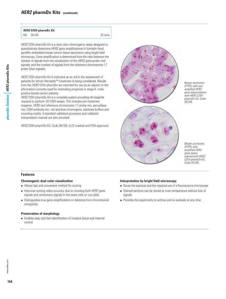
154
www.
dako
.com
phar
mDx
Sol
utio
nHER2
pha
rmDx
Kits
HER2 pharmDx Kits (continued)
HER2 CISH pharmDx Kit is a dual color chromogenic assay designed to quantitatively determine HER2 gene amplifications in formalin-fixed, paraffin-embedded breast cancer tissue specimens using bright field microscopy. Gene amplification is determined from the ratio between the number of signals from the visualization of the HER2 gene probe (red signals) and the number of signals from the reference chromosome 17 probe (blue signals).
HER2 CISH pharmDx Kit is indicated as an aid in the assessment of patients for whom HerceptinTM treatment is being considered. Results from the HER2 CISH pharmDx are intended for use as an adjunct to the information currently used for estimating prognosis in stage II, node-positive breast cancer patients.HER2 CISH pharmDx Kit is a complete system providing all reagents required to perform 20 CISH assays. This includes pre-treatment reagents, HER2 and reference chromosome 17 probe mix, peroxidase mix, CISH antibody mix, red and blue chromogens, substrate buffers and mounting media. A standard validated procedure and validated interpretation manual are also provided.
HER2 CISH pharmDx Kit, Code SK109, is CE marked and FDA approved.
FeaturesChromogenic dual color visualization Allows fast and convenient method for scoring Improves scoring ratios accuracy due to counting both HER2 gene
signals and centromere signals in the same cells on one slide Distinguishes true gene amplifications or deletions from chromosomal
aneuploidy
Preservation of morphology Enables easy and fast identification of invasive tissue and internal
control
Interpretation by bright field microscopy Saves the expense and the required use of a fluorescence microscope Stained sections can be stored at room temperature without loss of
signals Provides the opportunity to archive and re-evaluate at any time
HER2 CISH pharmDx KitI SK109 20 tests
Breast carcinoma (FFPE) with non-amplified HER2 gene status stained with HER2 CISH pharmDx Kit, Code SK109.
Breast carcinoma (FFPE) with amplified HER2 gene status stained with HER2 CISH pharmDx Kit, Code SK109.
USCatPathology.book Page 154 Monday, March 7, 2016 8:42 PM

155
PD-L1 IHC 22C3 pharmDx
pharmDx Solution
PD-L1 IHC 22C3 pharmDx
PD-L1 Testing - Advancing Treatment Options
The immune system and role of PD-L1 protein Cytotoxic T cells work to detect and eliminate infected cells and tumor
cells from the body To limit damage to surrounding tissue, normal cells differentiate
themselves by expressing a protein signal called PD-L1 (programmed death ligand 1). This signal is similar to a warning sign and is designed to avoid elimination of normal cells.
T cells detect the PD-L1 signal through a receptor called PD-1 (programmed death receptor 1)
Tumor involvement and role of PD-1-targeted therapy Some tumors can also express the PD-L1 signal to mimic normal cells
and escape elimination Anti-PD-1 therapy works by blocking the PD-1 receptor that detects
the PD-L1 signal in tumor cells Identifying PD-L1 expression on tumor cells is important in identifying
the signal needed to assess which patients will respond better to anti-PD-1 therapy
PD-L1 IHC 22C3 pharmDx is a qualitative immunohistochemical assay using Monoclonal Mouse Anti-PD-L1, Clone 22C3 intended for use in the detection of PD-L1 protein in formalin-fixed, paraffin-embedded (FFPE) non-small cell lung cancer (NSCLC) tissue using EnVision FLEX visualization system on Autostainer Link 48. PD-L1 protein expression is determined by using Tumor Proportion Score (TPS), which is the percentage of viable tumor cells showing partial or complete membrane staining. The specimen should be considered PD-L1 positive if TPS i 50% of the viable tumor cells exhibit membrane staining at any intensity.
PD-L1 IHC 22C3 pharmDx is indicated as an aid in identifying NSCLC patients for treatment with KEYTRUDA® (pembrolizumab).
The kit includes reagents required for the immunohistochemical staining (except wash buffer), control slides representing different expression levels of PD-L1 protein, and detailed instructions. The kit has been tailored especially for use on Autostainer Link 48 instruments. The
materials provided are sufficient for 50 tests (50 slides incubated with monoclonal mouse antibody to PD-L1 and 50 slides incubated with the corresponding negative control reagent, 100 slides in total).
Reference:1. Garon EB, Rizvi NA, Hui R, Leighl N, Balmanoukian AS, Eder JP, et al.
Pembrolizumab for the treatment of non-small cell lung cancer. New Eng J Med 2015;372:2018-28.
Non-small cell lung carcinoma (FFPE) with PD-L1 High Expression stained with PD-L1 IHC 22C3 pharmDx, Code SK006.
The system is based on the consecutive application of:1. Primary antibody against PD-L12. Visualization reagent3. Chromogenic substrate
All reagents, control cell line slides and detailed instructions are provided.
PD-L1 IHC 22C3 pharmDx kit is FDA approved and includes: Peroxidase-Blocking Reagent Monoclonal Mouse Anti-PD-L1, Clone 22C3 Negative Control Reagent Mouse LINKER
Visualization Reagent-HRP DAB+ Substrate Buffer DAB+ Chromogen DAB Enhancer EnVision FLEX Target Retrieval Solution, Low pH (50x) PD-L1 IHC 22C3 pharmDx Control Slides
Reagents required but not included in kit EnVision FLEX Wash Buffer (20x) K8007 EnVision FLEX Hematoxylin K8008
PD-1
Inactive cytotoxic T-cell
PD-L1 exxprpresessing cell
PD-L1
TuTuTumomomorrr cececellllll
Inactive cytotoxic T-cell
PD-1
PD-L1
Tumor cellActive cytotoxic T-cell
Anti-PD-1 therapy
Blocking the PD-1/PD-L1 interaction helps to enable active T cells and tumor cell death and elimination.
Inactivation of T cells reduces tumor cell death and elimination. Inactivation of T cells limits damage to normal tissue.
PD-L1 IHC 22C3 pharmDx for Autostainer Link 48I SK006 For Autostainer Link 48 NEW 50 tests
USCatPathology.book Page 155 Monday, March 7, 2016 8:42 PM

156
www.dako.com
phar
mDx
Sol
utio
nPD
-L1
IHC
22C3
pha
rmDx
PD-L1 IHC 22C3 pharmDx (continued)
PD-L1 IHC 22C3 Procedure Graphical representation of assay steps
Control cell linesControl slides are provided to help validate the staining runs which enhance the confidence in results. They are not to be used as an interpretation reference.
Positive Control Cell Pellet
Acceptable staining of positive pellet. At least 70% of the cells contain cell membrane staining of at least 2+ average staining intensity. Any background staining is less than 1+ staining intensity. 20x magnification.
Negative Control Cell Pellet
Acceptable staining of negative pellet. The majority of cells should demonstrate no staining. The presence of 10 or less cells with distinct cell membrane staining is acceptable. Any background staining is less than 1+ staining intensity. 20x magnification.
Antigen
PrimaryAntibody
Linker
LabeledPolymer
LabeledPolymer
Antigen
STEP 4 Application of DAB+ Substrate
Chromogen Solution.STEP 5 Application of DAB Enhancer.STEP 1 Application of
Primary Antibody.STEP 2 Application of Linker. STEP 3 Application of
Visualization Reagent.
DABDAB DABDAB DABDAB DABDAB
00130 pharmDX Solutions US.fm Page 156 Tuesday, March 15, 2016 6:56 PM

157
PD-L1 IHC 22C3 pharmDx
pharmDx Solution
PD-L1 IHC 22C3 pharmDx (continued)
Tumor Proportion ScorePD-L1 protein expression is determined by using Tumor Proportion Score (TPS), which is the percentage of viable tumor cells showing partial or complete membrane staining. NSCLC patient specimens should be considered positive for PD-L1 expression if TPS ≥ 50% of the of viable tumor cells exhibit membrane staining at any intensity (i.e. ≥ 1+). These patients may be considered for treatment with KEYTRUDA® (pembrolizumab).
PD-L1 staining definition PD-L1 staining is any perceptible (≥ 1+) partial or complete cell membrane staining of viable tumor cells.
Interpretation of PD-L1 expression Patient specimens tested with PD-L1 IHC 22C3 pharmDx are interpreted as having No Expression, Low Expression, or High Expression of PD-L1. The percentage of viable tumor cells showing partial or complete cell membrane staining, the tumor proportion score, determines the PD-L1 expression status of the specimen. See Dako Interpretation Manual for complete interpretation of PD-L1 IHC 22C3 pharmDx staining results, Order No. 29111.
No Expression (TPS < 1%). Low Expression (TPS ≥ 1–49%).
High Expression (TPS ≥ 50%).
Make the right choiceMake the right test choice for assessing KEYTRUDA® (pembrolizumab) eligibility Use the same test employed in the KEYTRUDA® clinical studies Incorporate PD-L1 testing early Utilize our extensive education and training resources to incorporate
PD-L1 testing in your laboratory Clinically validated assay and scoring guidelines for KEYTRUDA®
eligibility Includes all reagents necessary to perform a complete staining run Includes positive and negative control cell line slides to validate
staining run
USCatPathology.book Page 157 Monday, March 7, 2016 8:42 PM

158
www.
dako
.com
phar
mDx
Sol
utio
nPD
-L1
IHC
28-8
pha
rmDx
PD-L1 IHC 28-8 pharmDx
The role of CTLA-4 and PD-1/PD-L1 pathways in cancer
Normal cells limit damage to healthy tissue
Figure 1. Inactivation of T cells limits damage to healthy tissue.
CTLA-4 therapy augments T cell activation and proliferation
Figure 3. Blocking CTLA-4 contributes to an increase in anti-tumor immune response.
Tumor cells escape detection
Figure 2. Inactivation of T cells reduces tumor cell killing.
PD-1 therapies harness the immune response to fight tumors
Figure 4. Blocking PD-1 enables cytotoxic T cells to actively remove tumor cells.
PD-L1 IHC 28-8 pharmDx is a qualitative immunohistochemical assay using Monoclonal Rabbit Anti-PD-L1, Clone 28-8 intended for use in the detection of PD-L1 protein in formalin-fixed paraffin-embedded (FFPE) non-squamous non-small cell lung cancer (NSCLC) and melanoma tissues using EnVision FLEX visualization system on Autostainer Link 48. PD-L1 protein expression is defined as the percentage of tumor cells exhibiting positive membrane staining at any intensity.
Non-squamous NSCLCPD-L1 expression as detected by PD-L1 IHC 28-8 pharmDx in non-squamous NSCLC may be associated with enhanced survival from OPDIVO® (nivolumab).
MelanomaPositive PD-L1 status as determined by PD-L1 IHC 28-8 pharmDx in melanoma is correlated with the magnitude of the treatment effect on progression-free survival from OPDIVO®.
PD-L1 IHC 28-8 pharmDx kitThe kit includes reagents required for the immunohistochemical staining (except wash buffer), control slides representing different expression levels of PD-L1 protein, and detailed instructions. The kit has been tailored especially for use on Autostainer Link 48 instruments. The materials provided are sufficient for 50 tests (50 slides incubated with monoclonal rabbit antibody to PD-L1 and 50 slides incubated with the corresponding Negative Control Reagent, 100 slides in total).
PD-L1 IHC 28-8 pharmDx is subject to an exclusive trademark license to Dako Denmark A/S. OPDIVO® is a trademark owned by Bristol-Myers Squibb.
Reference:1. Phillips T, Simmons P, Inzunza HD, Cogswell J, Novotny J Jr, Taylor C, et al.
Development of an automated PD-L1 immunohistochemistry (IHC) assay for non-small cell lung cancer. Appl Immunohistochem Mol Morphol 2015;23:541-9.
Inactive cytotoxic T cell
Antigen Presenting Cell (APC)
CD80/CD86PD-1
PD-L1 CTLA-4
PD-L1 expressing cell
Anti-CTLA-4 Therapy
Active cytotoxic T cell
Inactive cytotoxic T cellTumor cell
PD-L1
PD-1
Active cytotoxic T cellTumor cell
Anti-PD-1 Therapy
PD-L1 IHC 28-8 pharmDx for Autostainer Link 48I SK005 For Autostainer Link 48 NEW 50 tests
USCatPathology.book Page 158 Monday, March 7, 2016 8:42 PM

159
PD-L1 IHC 28-8 pharmDx
pharmDx Solution
PD-L1 IHC 28-8 pharmDx (continued)
Non-squamous NSCLC (FFPE) stained with PD-L1 IHC 28-8 pharmDx, Code SK005. PD-L1 expression ≥ 10%.
Melanoma (FFPE) stained with PD-L1 IHC 28-8 pharmDx, Code SK005. PD-L1 expression ≥ 1%.
Kit componentsDako PD-L1 IHC 28-8 pharmDx is a complete kit with reagents sufficient for 50 tests (50 slides incubated with primary antibody to PD-L1 and 50 slides incubated with the corresponding negative control reagent) and 15 Control Slides for use on Autostainer Link 48.
EnVision FLEX Target Retrieval Solution, Low pH (50x) Peroxidase-Blocking Reagent Monoclonal Rabbit Anti-PD-L1, Clone 28-8 Negative Control Reagent Anti-Rabbit LINKER Visualization Reagent-HRP DAB+ Substrate Buffer DAB+ Chromogen DAB Enhancer Control Slides
Reagents required but not included in kit EnVision FLEX Wash Buffer (20x) K8007 EnVision FLEX Hematoxylin K8008
PD-L1 IHC 28-8 pharmDx ProcedureGraphical representation of assay steps
Antigen
PrimaryAntibody
Linker
LabeledPolymer
LabeledPolymer
Antigen
STEP 4 Application of DAB+ Substrate
Chromogen Solution.STEP 5 Application of DAB Enhancer.STEP 1 Application of
Primary Antibody.STEP 2 Application of Linker. STEP 3 Application of
Visualization Reagent.
DABDAB DABDAB DABDAB DABDAB
USCatPathology.book Page 159 Monday, March 7, 2016 8:42 PM

160
www.
dako
.com
phar
mDx
Sol
utio
nPD
-L1
IHC
28-8
pha
rmDx
PD-L1 IHC 28-8 pharmDx (continued)
Control cell linesControl slides are provided to help validate the staining runs which enhance the confidence in results. They are not to be used as an interpretation reference.
Positive Control Cell Pellet
Acceptable staining of positive pellet. At least 80% of the cells contain plasma membrane staining of at least 2+ average staining intensity. Any background staining is of less than 1+ staining intensity.
Negative Control Cell Pellet
Acceptable staining of negative pellet. No plasma membrane staining . Any background staining is of less than 1+ staining intensity. Staining of a few cells in the negative pellet may occasionally be observed. The presence of 10 or less cells with distinct plasma membrane staining, or cytoplasmic staining with ≥ 1 + intensity within the boundaries of the cell pellet are acceptable.
Interpretation of Test Results in non-squamous NSCLCTo assess the PD-L1 expression level in patient slides stained with PD-L1 IHC 28-8 pharmDx, pathologists should determine the percentage of viable tumor cells exhibiting partial or complete linear circumferential plasma membrane staining at any staining intensity.
See Dako Interpretation Manual for complete interpretation of PD-L1 IHC 28-8 pharmDx staining results, Order No. 29211.
PD-L1 expression < 1%. PD-L1 expression ≥ 1%.
PD-L1 expression ≥ 5%. PD-L1 expression ≥ 10%.
USCatPathology.book Page 160 Monday, March 7, 2016 8:42 PM

161
PD-L1 IHC 28-8 pharmDx
pharmDx Solution
PD-L1 IHC 28-8 pharmDx (continued)
Interpretations of MelanomaPositive PD-L1 status is correlated with the magnitude of the treatment effect on progression-free survival from OPDIVO®. Specimen is considered positive if ≥ 1% of melanoma cells exhibit linear circumferential complete or partial membrane staining of PD-L1. Specimen is considered negative if < 1% of melanoma cells exhibiting linear circumferential complete or partial membrane staining of PD-L1.
See Dako Interpretation Manual Melanoma for complete interpretation of PD-L1 IHC 28-8 pharmDx staining results, Order No. 29120.
< 1% of the viable tumor cells exhibit complete circumferential or partial linear plasma membrane staining at any intensity. PD-L1 expression < 1%: NEGATIVE.
≥ 1% of the viable tumor cells exhibit complete circumferential or partial linear plasma membrane staining at any intensity. PD-L1 expression ≥ 1%: POSITIVE.
Key benefits of PD-L1 IHC 28-8 pharmDx The only FDA-approved test for PD-L1 expression associated with
enhanced survival with OPDIVO® for non-squamous NSCLC The first and only FDA approved PD-L1 test for use in the assessment
of the magnitude of treatment effect on progression-free survival in melanoma patients from OPDIVO®
Experience the easy integration of PD-L1 in your IHC workflow Integrate PD-L1 IHC 28-8 pharmDx without changing staining lab
workflow Ready-to-use reagents and cell line controls optimized for Autostainer
Link 48 Pre-programmed, validated protocol
USCatPathology.book Page 161 Monday, March 7, 2016 10:11 PM

USCatPathology.book Page 162 Monday, March 7, 2016 10:11 PM

Molecular PathologyIntroduction 165Hybridizer Instrument 166CISH and FISH Kits 167Fluorochrome-Conjugated Probes 172Labeled Probes and Detection Systems 176Blotting Detection System 178ISH Ancillaries and Accessories 179
USCatPathology.book Page 163 Monday, March 7, 2016 10:11 PM

164
www.
dako
.com
Mol
ecul
ar P
atho
logy
Intro
duct
ion
Molecular Pathology
Biotinylated probe
FITC label
DAB
Biotinyl tyramide
Peroxidase-conjugated anti-FITC
Alkaline phosphatase-conjugated streptavidin
Peroxidase (HRP)
Biotin
Blue chromogen signal
Alkaline phosphatase-conjugated anti-Texas Red
H2O2/DAB mixture
Target DNA
Streptavidin
Texas Red label Red chromogen signal
USCatPathology.book Page 164 Monday, March 7, 2016 10:11 PM

165
IntroductionM
olecular PathologyIntroduction toMolecular Pathology
A Complete FISH Run in Just 4 HoursThe recently introduced Dako technology* completely changed the rules of in situ hybridization. The IQFISH pharmDx kit build on a strong history of Dako assays. And now, for the first time ever, laboratory professionals can run DNA-based hybridization assays in a timeframe comparable to protein-based immunohistochemistry assays.
The IQISH hybridization buffer is non-toxic and allows genomic DNA probe hybridization to be performed outside a fume hood in just 60-120 minutes. The short hybridization time results in a turnaround time of about 4 hours for a complete FISH staining from deparaffinization to mounting.
* Patents pending
SureFISH* - The Next Generation FISH TechnologyFluorescence in situ hybridization (FISH) is a laboratoy procedure in which fluorescently labeled DNA fragments (the FISH probe) hybridize to complementary DNA sequences in the cell's nucleus. The resulting fluorescent signals are visualized using a microscope and indicate the presence and localization of one or more targeted DNA sequences.
Oligonucleotide-Based FISH ProbesThe unique SureFISH DNA FISH probes are designed in silico and chemically synthesized using the company's high-fidelity, oligonucleotide library synthesis (OLS) technology. This eliminates the limitations of FISH probes manufactured with bacterial artificial chromosome (BAC) technology.
Brightness: New oligo design algorithm and labeling chemistry enable brighter probes compared to competing BAC FISH probes
Background: Oligo FISH probes target regions that are repeat free, enabling low background and minimal cross hybridization
Balance: Oligo FISH technology provides total flexibility on size of targeted region and number of oligos, enabling optimal signal balance between child probes
Co-localized: Unique micro-gap design leads to tight co-localization of child probes, enabling quick and accurate analysis
* SureFISH probes are manufactured by Agilent Technologies, Inc.
HER2 IQFISH pharmDxThe new IQ buffer from Dako completely changes the rules of in situ hybridization.
Improved overall staining quality :6 6 Easy identification of cancer cells6 Crisp and clear signals
Instant Quality FISH. The name says it all.
Accurate quantification
USCatPathology.book Page 165 Monday, March 7, 2016 10:11 PM

166
www.
dako
.com
Mol
ecul
ar P
atho
logy
Hybr
idize
r Ins
trum
ent
Hybridizer Instrument
Hybridizer is a hands-free co-denaturation and hybridization instrument designed for slide-based fluorescence (FISH) and chromogenic (CISH) in situ hybridization. The system reduces the manual steps, and improves the efficiency, throughput and precision compared to manually performed conventional ISH procedures. Hybridizer has a 12-slide capacity and heats and cools the slides through two temperature ranges with short ramp times. The system is easy to program for a wide range of protocols and is optimized for Dako DNA and PNA probes for FISH and CISH.
Hybridizer Humidity Control Strips are specially designed for the Hybridizer instrument, and ensure a controlled high humidity during the in situ hybridization procedure. The strips consist of a special hydrophilic polymer fibre material with high surface area and the ability to quickly adsorb and desorb moisture. The strips should be replaced frequently, every 1-2 weeks, as the performance deteriorates over time and with use.
Hardware Specifications
HybridizerI S2450 110-120 volt5 1 unitI S2451 200-240 volt55 1 unit
Hybridizer Humidity Control StripsI S2452 For the Hybridizer instrument 20 strips
Slide capacity Up to 12 slides per runProgram capacity Up to 40 programs (names and numbers)Program types Denaturation and hybridization
Hybridization onlyFixed temperature
Humidity control system Hydrophobic polymer fiber systemTemperature uniformity +/- 1 °C or better Denaturation temperature 50-99 °C, 0-30 minutes. Hybridization temperature Room temperature, 30-70 °C, 0-99 hoursFixed temperature Room temperature, 30-99 °C, 0-99 hoursRamp time 37-95 °C in less than 1 minuteCooling time 95-45 °C in less than 6 minutesDimensions 9" W x 6" D x 5" H (22.8 cm W x 40.6 cm D x 12.7 cm H)Weight 18 lbs (8 kg) Ambient operating temperature 15-40 °CPre-set voltage 100-120 V or 220-240 VFrequency 50-60 Hz
Humidity control strips stabilize internal moisture.
Hinged and sealable cover to retain moisture and prevent drying.
Programmable with up to 40 protocols by name.
12-slide capacity with easy-to-use single-handed loading and unloading feature.
5 Not available in the EU55 Not available in the United States
USCatPathology.book Page 166 Monday, March 7, 2016 10:11 PM

167
CISH and FISH KitsM
olecular PathologyCISH and FISH Kits
In situ hybridization techniques are used to localize specific nucleic acid sequences within the DNA in cells in tissues or cytological preparations, on chromosomes, or in whole mounts. Development of non-radioactive probes and detection systems has made the ISH technology available to a wide variety of routine and research applications. We have developed kits for both FISH and CISH applications.
The IQFISH technology* reduces the hybridization time from 14-20 hours to 1-2 hours. The IQISH hybridization buffer is non-toxic and allows genomic DNA probe hybridization to be performed in just 60-120 minutes. The short hybridization time results in a turnaround time of about 4 hours for a complete FISH staining from deparaffinization to mounting.
* Patents pending
Overview of Dako FISH and CISH Kits
FISH Kit CISH Kit
Accessory Kits
HER2 IQFISH pharmDx (20 tests), Code K5731Pre-treatment Solution (20x)Pepsin, ready-to-usePepsin Diluent (10x)HER2/CEN-17 Probe Mix in IQISH bufferStringent Wash Buffer (20x) Wash Buffer (20x)Fluorescence Mounting Medium, containing DAPICoverslip Sealant
HER2 CISH pharmDx Kit (20 tests), Code SK109Pre-treatment Solution (20x)Pepsin, ready-to-useHER2/CEN-17 Probe MixStringent Wash Buffer (20x) Peroxidase BlockCISH Antibody MixRed and Blue ChromogensRed and Blue Substrate BuffersWash Buffer (20x) and Wash Buffer (10x)CISH Mounting mediumCoverslip Sealant
Histology FISH Accessory Kits (20 tests), Code K5799Pre-treatment Solution (20x)Pepsin, ready-to-usePepsin Diluent (10x)Stringent Wash Buffer (20x)Wash Buffer (20x)Fluorescence Mounting Medium, containing DAPICoverslip Sealant
Cytology FISH Accessory Kits (20 tests), Code K5499Stringent Wash Buffer (20x)Wash Buffer (20x)Fluorescence Mounting Medium, containing DAPICoverslip Sealant
USCatPathology.book Page 167 Monday, March 7, 2016 10:11 PM
168
www.dako.com
Mol
ecul
ar P
atho
logy
CISH
and
FIS
H Ki
tsHER2 pharmDx Kits
The human HER2 gene with the generic name ERBB2 (also known as NEU) encodes the HER2 protein or p185HER2. The HER2 protein is a membrane receptor tyrosine kinase with homology to the epidermal growth factor receptor (EGFR). The HER2 gene is a normal component present in two copies in all normal diploid cells.
Breast CancerIn a fraction of patients (20-25%) with breast cancer, the HER2 gene is amplified as a part of the process of malignant transformation and tumor progression.
HER2 gene amplification and receptor prevalence correlates with poor breast cancer prognosis, including relapse-free and overall survival.
The recombinant, humanized monoclonal antibody trastuzumab (Herceptin®) was rationally developed to specifically target HER2-positive breast cancers. Demonstration of high HER2 overexpression or gene amplification is essential for treatment with trastuzumab (Herceptin®). Clinical studies have shown that patients whose tumors have high HER2 receptor overexpression and/or amplification benefit most from Herceptin® (1).
Pertuzumab (PERJETA™) is a recombinant humanized monoclonal antibody that binds to sub-domain II of the extracellular part of the HER2 protein thereby blocking its ability to form heterodimers with other members of the HER family, including HER1 (EGFR), HER3 and HER4. PERJETA™ has shown to be effective and safe in the treatment of breast cancer patients with HER2 protein overexpression. During clinical studies of pertuzumab, HER2 overexpression was demonstrated directly by IHC or indirectly evidenced through correlation of HER2 gene amplification to protein overexpression as demonstrated by FISH. However, in the randomized trial, data were available for a limited number of patients (8/808) for whom the FISH results were positive but the IHC results were negative (0, 1+) (2,3).
Ado-trastuzumab emtansine (KADCYLA™) is a novel antibody-drug conjugate specifically designed for the treatment of HER2-positive cancer. It is composed of the potent cytotoxic agent DM1 (a thiol-containing maytansinoid anti-microtubule agent) conjugated to trastuzumab via a linker molecule. Ado-trastuzumab emtansine binds to HER2 with an affinity similar to that of trastuzumab; such binding is required for its anti-tumor activity. It is hypothesized that after binding to HER2, ado-trastuzumab emtansine undergoes receptor-mediated internalization, followed by intracellular release of DM1 and subsequent cytotoxicity (4). A number of clinical studies have shown that ado-trastuzumab emtansine is effective and safe in treatment of HER2-positive breast cancer patients (5-8).
Gastric CancerA number of studies have analysed HER2 overexpression in gastric cancer; one has reported 24% of patients with gastroesophageal adenocarcinomas as showing overexpression of HER2 (9).
Pre-clinical data has showed that trastuzumab has significant antitumor activity in gastric cancer (10). This finding has prompted the investigation of the potential clinical benefit of trastuzumab in this type of cancer. The results from the phase III trial (ToGA), where both HercepTest and HER2 FISH pharmDx were used as initial screening tests, showed the added benefit of combining Herceptin® with standard chemotherapy (11).
Precaution for gastric cancer: HER2 protein overexpression and HER2 gene amplification are not as correlated in gastric cancer as with breast cancer; therefore a single method should not be used to determine HER2 status.
KitsHER2 IQFISH pharmDx is an FDA-approved Dako HER2 diagnostic kit indicated as an aid in the assessment of HER2 gene status.
HER2 CISH pharmDx Kit is an FDA-approved HER2 diagnostic kit combining the genetic information from FISH with the interpretation advantages of CISH.
References:1. Bilous M, Dowsett M, Hanna W, Isola J, Lebeau A, Moreno A, et al.
Current perspectives on HER2 testing: a review of national testing guidelines. Mol Pathol 2003;16:173-82.
2. Baselga J, Gelmon KA, Verma S, et al. Phase II trial of pertuzumab and trastuzumab in patients with human epidermal growth factor receptor 2-positive metastatic breast cancer that progressed during prior trastuzumab therapy. J Clin Oncol 2010;28:1138-44.
3. Baselga J, Cortés J, Kim SB, Im SA, Hegg R, Im YH, et al. Pertuzumab plus trastuzumab plus docetaxel for metastatic breast cancer. N Engl J Med 2012;366:109-19.
4. Lewis Phillips GD, Li G, Dugger DL, et al. Targeting HER2-positive breast cancer with trastuzumab-DM1, an antibody-cytotoxic drug conjugate. Cancer Res 2008;68:9280-90.
5. Krop IE, Beeram M, Modi S, et al. Phase I study of trastuzumab-DM1, an HER2 antibody-drug conjugate, given every 3 weeks to patients with HER2-positive metastatic breast cancer. J Clin Oncol 2010;28:2698-704.
6. Burris HA 3rd, Rugo HS, Vukelja SJ, et al. Phase II study of the antibody drug conjugate trastuzumab-DM1 for the treatment of human epidermal growth factor receptor 2 (HER2)-positive breast cancer after prior HER2-directed therapy. J Clin Oncol 2011;29:398-405.
7. Krop IE, Lorusso P, Miller KD, et al. A phase II study of trastuzumab emtansine in patients with human epidermal growth factor receptor 2-positive metastatic breast cancer who were previously treated with trastuzumab, lapatinib, an anthracycline, a taxane, and capecitabine. J Clin Oncol 2012;30:3234-41.
8. Verma S, Miles D, Gianni L, Krop IE, Welslau M, Baslega J, et al,. Trastuzumab emtansine for HER2-positive advanced breast cancer. New Engl J Med 2012;367:1783-91.
9. Tanner M, Hollmén M, Junttila TT, Kapanen AI, Tommola S, Soini Y, et al. Amplification of HER-2 in gastric carcinoma: association with Topoisomerase IIa gene amplification, intestinal type, poor prognosis and sensitivity to trastuzumab. Ann Oncol 2005;16:273-8.
10. Fujimoto-Ouchy K, Sekiguchi F, Yasuno H, Moriya Y, Mori K, Tanaka Y. Antitumor activity of trastuzumab in combination with chemotherapy in human gastric cancer xenograft models. Cancer Chemother Pharmacol 2007;59:795-805.
11. Van Cutsem E, Kang Y, Chung H, et al. Efficacy results from the ToGA trial: a phase III study of trastuzumab added to standard chemotherapy (CT) in first-line human epidermal growth factor receptor 2 (HER2)-positive advanced gastric cancer (GC). J Clin Oncol 2009;18S:Abstract LBA4509.
USCatPathology.book Page 168 Wednesday, March 9, 2016 10:46 PM

169
CISH and FISH KitsM
olecular PathologyHER2 pharmDx Kits (continued)
IQFISH – One day turnaround time for FISHWhen it comes to cancer diagnosis, time is everything. The time required to run tests can restrict the laboratory’s workflow and delay results from being handed over to the pathologist. Time is one of the key parameters
of successful laboratory operations. Of course, laboratory professionals do everything they can to speed up operations, but they always reach a point where there is nothing more they can do. But now they can.
HER2 IQFISH pharmDx is a direct fluorescence in situ hybridization (FISH) assay based on Dako's new fast IQISH hybridization buffer chemistry. The IQISH hybridization buffer is non-toxic and allows genomic DNA probe hybridization to be performed in just 60-120 minutes. The short hybridization time results in a turnaround time less than 4 hours for a complete FISH staining from deparaffinization to mounting.
HER2 IQFISH pharmDx is designed to quantitatively determine HER2 gene amplification in formalin-fixed, paraffin-embedded (FFPE) breast cancer tissue specimens and FFPE specimens from patients with metastatic gastric or gastroesophageal junction adenocarcinoma. Gene amplification is determined from the ratio between the number of signals from the hybridization of the HER2 gene probe (red signals) and the number of signals from the hybridization of the CEN-17 reference chromosome 17 probe (green signals).
HER2 IQFISH pharmDx is indicated as an aid in the assessment of breast and gastric cancer patients for whom Herceptin® treatment is being considered and for breast cancer patients for whom PERJETATM or KADCYLATM treatment is being considered (see Herceptin®, PERJETATM, and KADCYLATM package inserts). For breast cancer patient, results from the HER2 IQFISH pharmDx are intended for use as an adjunct to the information currently used for estimating prognosis in stage II, node-positive breast cancer patients.
HER2 IQFISH pharmDx is a complete system providing all reagents required to perform 20 assays. A validated procedure and validated interpretation guidelines are also provided.
HER2 IQFISH pharmDx, Code K5731, is CE marked and FDA approved.
FeaturesTurnaround time Less than 4 hours total protocol Run FISH simultaneous with IHC Solve urgent cases fast Easy planning improves workflow
Non-toxic solution Safer work environment Hybridization outside a fume hood Flexible planning
Excellent quality Crisp and clear dual fluorescent signals Robust and easy protocol Correct answer, first time Correct answer, every time
Easy protocol Easy protocol improves workflow Optional batch pepsin treatment for high throughput
HER2 IQFISH pharmDxI K5731 20 tests
Breast carcinoma (FFPE) stained with HER2 IQFISH pharmDx, Code K5731. Tumor cells show HER2 gene amplification (HER2/CEN-17 ratio i 2).
Gastric cancer (FFPE) stained with HER2 IQFISH pharmDx, Code K5731. Tumor cells show HER2 gene amplification (HER2/CEN-17 ratio i 2).
USCatPathology.book Page 169 Monday, March 7, 2016 10:11 PM

170
www.
dako
.com
Mol
ecul
ar P
atho
logy
CISH
and
FIS
H Ki
tsHER2 pharmDx Kits (continued)
HER2 CISH pharmDx Kit is a dual color chromogenic assay designed to quantitatively determine HER2 gene amplifications in formalin-fixed, paraffin-embedded breast cancer tissue specimens using bright field microscopy. Gene amplification is determined from the ratio between the number of signals from the visualization of the HER2 gene probe (red signals) and the number of signals from the reference chromosome 17 probe (blue signals).
HER2 CISH pharmDx Kit is indicated as an aid in the assessment of patients for whom Herceptin treatment is being considered. Results from the HER2 CISH pharmDx are intended for use as an adjunct to the information currently used for estimating prognosis in stage II, node-positive breast cancer patients.
HER2 CISH pharmDx Kit is a complete system providing all reagents required to perform 20 CISH assays. This includes pre-treatment reagents, HER2 and reference chromosome 17 probe mix, peroxidase mix, CISH antibody mix, red and blue chromogens, substrate buffers and mounting media. A standard validated procedure and validated interpretation manual are also provided.
HER2 CISH pharmDx Kit, Code SK109, is FDA approved.
FeaturesChromogenic dual color visualization Allows fast and convenient method for scoring Improves scoring ratios accuracy due to counting both HER2
gene signals and centromere signals in the same cells on one slide
Distinguishes true gene amplifications or deletions from chromosomal aneuploidy
Preservation of morphology Enables easy and fast identification of invasive tissue and internal
control
Interpretation by bright field microscopy Saves the expense and the required use of a fluorescence microscope Stained sections can be stored at room temperature without loss of
signals Provides the opportunity to archive and re-evaluate at any time
HER2 CISH pharmDx KitI SK109 20 tests
Breast carcinoma (FFPE) with non-amplified HER2 gene status stained with HER2 CISH pharmDx Kit, Code SK109.
Breast carcinoma (FFPE) with amplified HER2 gene status stained with HER2 CISH pharmDx Kit, Code SK109.
USCatPathology.book Page 170 Monday, March 7, 2016 10:11 PM
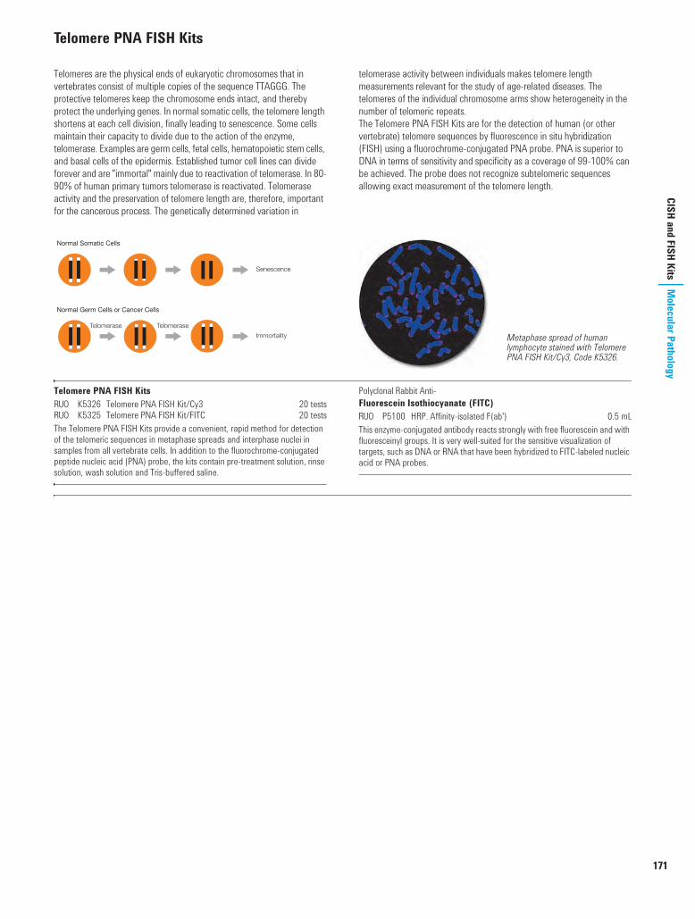
171
CISH and FISH KitsM
olecular PathologyTelomere PNA FISH Kits
Telomeres are the physical ends of eukaryotic chromosomes that in vertebrates consist of multiple copies of the sequence TTAGGG. The protective telomeres keep the chromosome ends intact, and thereby protect the underlying genes. In normal somatic cells, the telomere length shortens at each cell division, finally leading to senescence. Some cells maintain their capacity to divide due to the action of the enzyme, telomerase. Examples are germ cells, fetal cells, hematopoietic stem cells, and basal cells of the epidermis. Established tumor cell lines can divide forever and are "immortal" mainly due to reactivation of telomerase. In 80-90% of human primary tumors telomerase is reactivated. Telomerase activity and the preservation of telomere length are, therefore, important for the cancerous process. The genetically determined variation in
telomerase activity between individuals makes telomere length measurements relevant for the study of age-related diseases. The telomeres of the individual chromosome arms show heterogeneity in the number of telomeric repeats.The Telomere PNA FISH Kits are for the detection of human (or other vertebrate) telomere sequences by fluorescence in situ hybridization (FISH) using a fluorochrome-conjugated PNA probe. PNA is superior to DNA in terms of sensitivity and specificity as a coverage of 99-100% can be achieved. The probe does not recognize subtelomeric sequences allowing exact measurement of the telomere length.
Telomere PNA FISH KitsU K5326 Telomere PNA FISH Kit/Cy3 20 testsU K5325 Telomere PNA FISH Kit/FITC 20 testsThe Telomere PNA FISH Kits provide a convenient, rapid method for detection of the telomeric sequences in metaphase spreads and interphase nuclei in samples from all vertebrate cells. In addition to the fluorochrome-conjugated peptide nucleic acid (PNA) probe, the kits contain pre-treatment solution, rinse solution, wash solution and Tris-buffered saline.
Polyclonal Rabbit Anti-Fluorescein Isothiocyanate (FITC)U P5100 HRP. Affinity-isolated F(ab') 0.5 mLThis enzyme-conjugated antibody reacts strongly with free fluorescein and with fluoresceinyl groups. It is very well-suited for the sensitive visualization of targets, such as DNA or RNA that have been hybridized to FITC-labeled nucleic acid or PNA probes.
Normal Somatic Cells
Normal Germ Cells or Cancer Cells
Senescence
Immortality
Telomerase Telomerase
Metaphase spread of human lymphocyte stained with Telomere PNA FISH Kit/Cy3, Code K5326.
USCatPathology.book Page 171 Monday, March 7, 2016 10:11 PM

172
www.
dako
.com
Mol
ecul
ar P
atho
logy
Fluo
roch
rom
e-Co
njug
ated
Pro
bes
Fluorochrome-Conjugated Probes
ALK DNA Probe, Fluorochrome-ConjugatedS Y5417 0.2 mLThe human ALK gene consists of 29 exons spanning a region of ~728 kb on chromosome 2 band p23. Y5417 is a probe based on a combination of DNA and PNA technology, and contains a two-part fluorochrome-conjugated DNA probe and unlabeled PNA blocking probes. The fluorochrome-conjugated DNA probe
is a Texas Red-labeled DNA probe (ALK-Downstream) covering 289 kb telomeric to the ALK breakpoint cluster region and a fluorescein-labeled DNA probe (ALK-Upstream) covering 557 kb centromeric to the ALK breakpoint cluster region.
BCL2 DNA Probe, Fluorochrome-ConjugatedS Y5407 0.2 mLThe human BCL2 gene consists of 3 exons spanning a region of ~195 kb on chromosome 18 band q21. Y5407 is a probe based on a combination of DNA and PNA technology, and contains a two-part fluorochrome-conjugated DNA probe and unlabeled PNA blocking probes. The fluorochrome-conjugated DNA
probe is a Texas Red-labeled DNA probe (BCL2-Upstream) covering 375 kb telomeric to the BCL2 major/minor breakpoint cluster regions and a fluorescein-labeled DNA probe (BCL2-Downstream) covering 641 kb centromeric to the BCL2 major/minor breakpoint cluster regions.
BCL6 DNA Probe, Fluorochrome-ConjugatedS Y5408 0.2 mLThe human BCL6 gene consists of 10 exons spanning a region of ~24 kb on chromosome 3 band q27. Y5408 is a probe based on a combination of DNA and PNA technology, and contains a two-part fluorochrome-conjugated DNA probe and unlabeled PNA blocking probes. The fluorochrome-conjugated DNA probe
is a Texas Red-labeled DNA probe (BCL6-Upstream) covering 352 kb telomeric to the BCL6 breakpoint cluster region, and a fluorescein-labeled DNA probe (BCL6-Downstream) covering 573 kb centromeric to the BCL6 breakpoint cluster region.
2p23
1234567891011121329-14
Breakpoint
ALK-Upstream
557 kb
ALK-Downstream
289 kb
VCR breakpoint region Major/Minor breakpoint regions
BCL2-Upstream BCL2-Downstream
641 kb375 kb
18q21
1 2 3
3q27
123459-610
Breakpoint
BCL6-Upstream
352 kb
BCL6-Downstream
573 kb
USCatPathology.book Page 172 Monday, March 7, 2016 10:11 PM

173
Fluorochrome-Conjugated Probes
Molecular Pathology
Fluorochrome-Conjugated Probes (continued)
CCND1 DNA Probe, Fluorochrome-ConjugatedS Y5414 0.2 mLThe human CCND1 gene consists of 5 exons spanning a region of ~13 kb on chromosome 11 band q13. Y5414 is a probe based on a combination of DNA and PNA technology, and contains a two-part fluorochrome-conjugated DNA probe and unlabeled PNA blocking probes. The fluorochrome-conjugated DNA
probe is a Texas Red-labeled DNA probe (CCND1-Upstream) covering 163 kb centromeric to the CCND1 breakpoint cluster region and a fluorescein-labeled DNA probe (CCND1-Downstream) covering 644 kb telomeric to the CCND1 breakpoint cluster region.
IGH DNA Probe, Fluorochrome-ConjugatedS Y5406 0.2 mLThe IGH locus at chromosome 14 consists of 170-176 genes. These genes are divided into 123-129 variable (IGHV) genes, 27 diversity (IGHD) gene segments, 9 joining (IGHJ) gene segments, and 11 constant (IGHC) genes. The IGH locus spans a region of 1.25 Mb on chromosome 14 band q32. Y5406 is a probe based on a combination of DNA and PNA technology, and contains a two-part
fluorochrome-conjugated DNA probe and unlabeled PNA blocking probes. The fluorochrome-conjugated DNA probe is a Texas Red-labeled DNA probe (IGH-Downstream) covering 460 kb centromeric to the IGH breakpoint cluster region and a fluorescein-labeled DNA probe (IGH-Upstream) covering 612 kb telomeric to the IGH breakpoint cluster region.
MALT1 DNA Probe, Fluorochrome-ConjugatedS Y5409 0.2 mLThe human MALT1 gene consists of 17 exons spanning a region of ~80 kb on chromosome 18 band q21. Y5409 is a probe based on a combination of DNA and PNA technology, and contains a two-part fluorochrome-conjugated DNA probe and unlabeled PNA blocking probes. The fluorochrome-conjugated DNA
probe is a Texas Red-labeled DNA probe (MALT1-Upstream) covering 316 kb centromeric to the MALT1 breakpoint cluster region and a fluorescein-labeled DNA probe (MALT1-Downstream) covering 508 kb telomeric to the MALT1 breakpoint cluster region.
Breakpoint
CCND1-Downstream
644 kb
CCND1-Upstream
163 kb
11q13
1-5
IGH-Upstream IGH-Downstream
Breakpoint
IGHV IGHD-J IGHC
460 kb612 kb
14q32
18q21
123410-517-11
Breakpoint
MALT1-Upstream
316 kb
MALT1-Downstream
508 kb
USCatPathology.book Page 173 Monday, March 7, 2016 10:11 PM

174
www.
dako
.com
Mol
ecul
ar P
atho
logy
Fluo
roch
rom
e-Co
njug
ated
Pro
bes
Fluorochrome-Conjugated Probes (continued)
MYC DNA Probe, Fluorochrome-ConjugatedS Y5410 0.2 mLThe human MYC gene consists of 3 exons spanning a region of ~5 kb on chromosome 8 band q24. Y5410 is a probe based on a combination of DNA and PNA technology, and contains a two-part fluorochrome-conjugated DNA probe and unlabeled PNA blocking probes. The fluorochrome-conjugated DNA probe
is a Texas Red-labeled DNA probe (MYC-Downstream) covering 418 kb telomeric to the MYC breakpoint cluster region and a fluorescein-labeled DNA probe (MYC-Upstream) covering 652 kb centromeric to the MYC breakpoint cluster region.
Breakpoint5’ Breakpoint 3’ Breakpoint
MYC-Upstream MYC-Downstream
418 kb652 kb
8q24
1-3
USCatPathology.book Page 174 Monday, March 7, 2016 10:11 PM
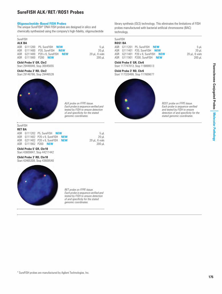
175
Fluorochrome-Conjugated Probes
Molecular Pathology
SureFISH ALK/RET/ROS1 Probes
Oligonucleotide-Based FISH ProbesThe unique SureFISH* DNA FISH probes are designed in silico and chemically synthesized using the company's high-fidelity, oligonucleotide
library synthesis (OLS) technology. This eliminates the limitations of FISH probes manufactured with bacterial artificial chromosome (BAC) technology.
SureFISH ALK BAS G111200 P5, SureFISH NEW 5 µLS G111400 P20, SureFISH NEW 20 µLS G211400 P20 x 6, SureFISH NEW 20 µL, 6 vialsS G111900 P200 NEW 200 µLChild Probe 5' GR, Chr2Start 29446949, Stop 30045655Child Probe 3' RD, Chr2Start 29146786, Stop 29446528
SureFISH RET BAS G111202 P5, SureFISH NEW 5 µLS G111402 P20 x 6, SureFISH NEW 20 µLS G211402 P20 x 6, SureFISH NEW 20 µL, 6 vialsS G111902 P200 NEW 200 µLChild Probe 5' GR, Chr10Start 43609847, Stop 44211442Child Probe 3' RD, Chr10Start 43465309, Stop 43609540
SureFISH ROS1 BAS G111201 P5, SureFISH NEW 5 µLS G111401 P20, SureFISH NEW 20 µLS G211401 P20 x 6, SureFISH NEW 20 µL, 6 vialsS G111901 P200, SureFISH NEW 200 µLChild Probe 5' GR, Chr6Start 117747013, Stop 118899513Child Probe 3' RD, Chr6Start 117320499, Stop 117609677
* SureFISH probes are manufactured by Agilent Technologies, Inc.
ALK probe on FFPE tissue.Each probe is sequence verified and tested by FISH to ensure detection of and specificity for the stated genomic coordinates.
RET probe on FFPE tissue.Each probe is sequence verified and tested by FISH to ensure detection of and specificity for the stated genomic coordinates.
ROS1 probe on FFPE tissue.Each probe is sequence-verified and tested by FISH to ensure detection of and specificity for the stated genomic coordinates.
USCatPathology.book Page 175 Wednesday, March 9, 2016 10:47 PM

176
www.
dako
.com
Mol
ecul
ar P
atho
logy
Labe
led
Prob
es a
nd D
etec
tion
Syst
ems
Labeled Probes and Detection Systems
GenPoint Amplified Signal Detection System
Dako GenPoint system is a powerful, non-radioactive in situ hybridization signal amplification system for biotinylated probes. Based on a patented catalyzed signal amplification (CSA) methodology, it offers in situ PCR sensitivity with none of the associated complexities of costly instrumentation or contamination concerns. Dako GenPoint system can be used with routinely processed tissue or cell preparations making it an extremely versatile tool for in situ hybridization. Its signal amplification allows the user to visualize gene targets which are normally not detectable by traditional in situ hybridization procedures; for instance, the Dako GenPoint system positively labels single copies of HPV16 in SiHa cells. CSA is based on the catalyzed deposition of biotinyl tyramide. When followed by HRP-conjugated streptavidin, a high amplification of the signal occurs at the hybridization site.
Further InformationThe method is based on the consecutive application of:1. Biotinylated probe2. Peroxidase-conjugated streptavidin3. Biotinyl tyramide4. Peroxidase-conjugated streptavidin5. Chromogenic substrate (diaminobenzidine (DAB))
GenPoint, Catalyzed Signal Amplification System*I K0620 For in situ hybridization 65 testsThe GenPointTM system locates target sites hybridized with biotinylated nucleic acid probes with high sensitivity and resolution. The kit includes stringent wash concentrate, peroxidase-conjugated streptavidin, biotinyl tyramide, and diaminobenzidine (DAB) chromogenic substrate.
* Dako GenPoint embodies technology developed by and licensed from NEN Life Science Products, Inc. (U.S. Patent 5,196,306).Limited Use LicenceThis product is distributed and sold to the End-User pursuant to a license from NEN Life Science Products, Inc. for use by the End-User in manual or automated processing on Dako instrumentation only of glass microscope slides or other support material (other than microarrays and bio-chips) containing cell or tissue specimens for examining those specimens under a microscope (including automated image capture and analysis systems) for the purpose of detecting target nucleic acids or proteins. Purchase does not include or carry any right to resell or transfer this product either as a stand alone product or as a component of another product. Any use of this product other than the licensed use without the express written authorization of NEN Life Science Products, Inc. is strictly prohibited.
Human condyloma stained with a HPV Type 11, DNA probe.
STEP 1 Hybridization of biotinylatedprobe to target nucleic acid.
STEP 2 Application of peroxidase-conjugated streptadivin.
STEP 4 Deposition of biotinyl tyramideis followed by application ofperoxidase-conjugatedstreptadivin.
STEP 5 Application of chromogenicsubstrate (DAB).
STEP 3 Application of biotinyl tyramide.
USCatPathology.book Page 176 Monday, March 7, 2016 10:11 PM

177
Labeled Probes and Detection Systems
Molecular Pathology
Standard Nucleic Acid Detection System
In Situ Hybridization Detection System, Code K0601, provides a rapid, simple, ready-to-use detection of biotinylated DNA or RNA probes.
In Situ Hybridization Detection SystemI K0601 For biotinylated nucleic acid probes 50 testsComponents are: Stringent wash concentrate, ready-to-use alkaline phosphatase-conjugated streptavidin, ready-to-use BCIP/NBT chromogen solution and detailed instructions. Results may be obtained within two hours. The number of tests is based on the use of 150 µL of reagent per slide.
PNA Probes and Detection System
Hybridization techniques are used to localize and identify specific nucleic acid sequences. Initially used as a research tool, the development of non-radioactive probe labeling and detection systems has made this technology available to a wide variety of routine applications. Traditionally either cloned probes or synthesized oligonucleotide probes have been used for hybridization. We have successfully introduced the peptide nucleic acid (PNA) probe, a nucleotide analogue capable of binding to DNA/RNA in a sequence-specific manner obeying the Watson-Crick base pairing rules.
In PNA, the sugar phosphate backbone of DNA/RNA has been replaced by a synthetic peptide backbone keeping the distances between bases exactly the same as in DNA/RNA. The increase in affinity is evident from the increase in the Tm of 1-1.5 °C per base pair. Further, the PNAs are very stable molecules. Experiments have shown virtually no degradation by DNases, RNases, proteinases or peptidases. The PNA probes are labeled with fluorescein and detected using the sensitive PNA ISH Detection Kit, Code K5201.
Epstein-Barr Virus (EBER) PNA Probe/FluoresceinU Y5200 40 tests, 1 mLEpstein-Barr Virus (EBER) PNA Probe/Fluorescein detects the 2 nuclear RNA transcripts EBER1 and EBER2 encoded by Epstein-Barr virus. The product contains a mixture of 4 fluorescein-labeled PNA probes complementary to parts of the 2 nuclear EBER RNAs.
PNA ISH Detection KitI K5201 40 testsPNA ISH Detection Kit for fluorescein-labeled peptide nucleic acid (PNA) probes contains all reagents necessary for performing an in situ hybridization, except for the specific probe. The kit contains proteinase K, negative and positive control probes, stringent wash solution, alkaline phosphatase-conjugated F(ab') fragment of rabbit anti-FITC, chromogenic substrate combined with an inhibitor of endogenous alkaline phosphatase (BCIP/NBT/levamisole), and Tris-buffered saline. A detailed working procedure is included. The number of tests is based on the use of 150 µL of reagent per slide.
STEP 1 Hybridization ofbiotinylated probe totarget nucleic acid.
STEP 2 Application of alkaline phosphatase-conjugated streptavidin.
Nasopharyngeal carcinoma stained with EBV (EBER) PNA Probe/FITC, Code Y5200, and PNA ISH Detection Kit, Code K5201.
USCatPathology.book Page 177 Monday, March 7, 2016 10:17 PM

178
www.
dako
.com
Mol
ecul
ar P
atho
logy
Labe
led
Prob
es a
nd D
etec
tion
Syst
ems
Fragile X Probe
Fragile X Probe, Fluorescein-LabeledS Y1442 5 blots x 150 cm2
Blotting Detection System
Chemiluminescent System for Nucleic Acid BlottingChemiluminescent System for Nucleic Acid Blotting is a complete kit for the detection of Southern or Northern blot hybridizations using fluorescein-labeled probes. Hybridized probes are detected by incubation with an alkaline phosphatase-conjugated antibody fragment directed against fluorescein. The immobilized alkaline phosphatase activity is used to hydrolyze a chemiluminescent dioxetane-phosphate substrate, resulting in sustained light emission at the site of the hybridized probe. The light emitted from the blot is captured by overlaying the blot with photographic film, or by imaging using a sensitive CCD camera system.
Special features of the Dako Chemiluminescent System: Non-radioactive reagents (health and disposal issues eliminated) Exposure time reduced to minutes as opposed to days using the 32P-
method Complete with all necessary detection reagents in either liquid
concentrates or pre-measured powder packets for convenience Uses standard film and processing equipment
Detection System
Chemiluminescent System for Nucleic Acid BlottingK0626 20 blots x 150 cm2
Included in the kit are: Blocking solution, wash buffer, alkaline phosphatase-conjugated anti-FITC, chemiluminescent substrate, and detailed instructions.
Ancillaries
Hybridization BufferS1801 250 mL
SSC Stringent Wash for Oligo (short) ProbesS1803 5 pk
Fluorescein-labeled IGHJ6 probe and chemilumine-scence. Percentages represent the fraction of tumor cell DNA mixed with normal mononuclear cell DNA.
Exposure time: minutes
32P-labeled IGHJ6 probe and autoradiography. Percentages represent the fraction of tumor cell DNA mixed with normal mononuclear cell DNA.
Exposure time: days
USCatPathology.book Page 178 Monday, March 7, 2016 10:17 PM

179
Labeled Probes and Detection Systems
Molecular Pathology
ISH Ancillaries and Accessories
Dako Omnis Fluorescence Mounting Medium (Dako Omnis)I GM304 Ready-to-use 20 tests, 0.8 mLFluorescence Mounting Medium (Dako Omnis) is intended for mounting of formalin-fixed, paraffin-embedded (FFPE) tissue sections after FISH staining performed onboard the Dako Omnis instrument. The mounting medium also contains 500 µg/L DAPI for improved nuclei staining.
ISH Cleaning Solution (Dako Omnis)I GC207 Ready-to-use 100 tests, 10 mLISH Cleaning Solution (Dako Omnis) is an accessory to the Dako Omnis instrument. It is used for cleaning the pipette tip between dispenses of in situ hybridization probes. Washing with ISH Cleaning Solution dissolves ISH probe, allowing remaining probe to be effectively washed away with water. The product is provided in a ready-to-use vial for the Dako Omnis instrument.
ISH Ethanol Solution, 96% (Dako Omnis)I GM300 Ready-to-use 20 tests, 14 mLISH Ethanol Solution, 96% (Dako Omnis) is intended for use in automated in situ hybridization assays together with the Dako Omnis instrument on formalin-fixed, paraffin-embedded (FFPE) tissue sections. The solution is used in the wash step after target retrieval. The product is provided in a ready-to-use vial for the Dako Omnis instrument.
ISH Lid, for Dako OmnisGC102 5 lids
Dako Omnis ISH Lid is intended for use in FISH procedures. Each Dako Omnis ISH Lid holds five slides and has five built-in Cover Glasses and one Humidity Pad. The Cover Glasses serve to distribute probe buffer across the staining area and to reduce buffer evaporation. The Humidity Pad with deionized water added serves to increase the humidity inside Dako Omnis ISH Lid to further reduce evaporation. Dako Omnis ISH Lid also provides insulation to maintain proper denaturation temperature.Dako Omnis ISH Lid is single use only and is classified as non-hazardous waste.
ISH Pepsin (Dako Omnis)I GM302 Ready-to-use 20 tests, 7 mLISH Pepsin (Dako Omnis) is intended for use in automated in situ hybridization assays together with the Dako Omnis instrument on formalin-fixed, paraffin-embedded (FFPE) tissue sections. The solution is used in the digestion step. The product is provided in a ready-to-use vial for the Dako Omnis instrument.
ISH Pre-Treatment Solution (20x) (Dako Omnis)I GM301 Concentrate 175 mL, 20x concentratedISH Pre-Treatment Solution (20x) (Dako Omnis) is intended for use in automated in situ hybridization assays together with the Dako Omnis instrument on formalin-fixed, paraffin-embedded (FFPE) tissue sections. The solution is used in the pre-treatment step. An inert green color is added to the buffer for easy identification and user friendliness. The volume is tailored for dilution in one Dako Omnis bulk bottle.
ISH Stringent Wash Buffer (20x) (Dako Omnis)I GM303 Concentrate 175 mL, 20x concentratedISH Stringent Wash Buffer (20x) (Dako Omnis) is intended for use in automated in situ hybridization assays together with the Dako Omnis instrument on formalin-fixed, paraffin-embedded (FFPE) tissue sections. The solution is used in the post-hybridization step. An inert yellow color is added to the buffer for easy identification and user friendliness. The volume is tailored for dilution in one Dako Omnis bulk bottle.
Mixing Device, for Dako OmnisGC116 1 unit
Dako Omnis Mixing Device is an accessory to the Dako Omnis instrument. It is designed specifically to support the fluorescence in situ hybridization (FISH) and the chromogenic in situ hybridization (CISH) procedures. The Dako IQISH buffer is extremely viscous, and during storage the reagent phase separates. Hence the Dako IQISH reagents require a particular preparatory processing to thaw and unify the content.Some Dako ISH reagents are therefore provided in dedicated ISH reagent vials containing a mixing ball, and the Dako Omnis Mixing Device is designed to fit together with these ISH reagent vials.Dako Omnis Mixing Device contains a magnet that enables the mixing ball to move up and down (110 cycles) inside the vial after 40 minutes thawing of the ISH reagent; thus ensuring a homogenous probe mix prior to application on the Dako Omnis instrument.
Vial with Mixing Ball, 2 mL, for Dako OmnisGC206 25 vials 2 mL
Dako Omnis Vial with Mixing Ball, 2 mL has been designed as an accessory for Dako Omnis and Dako Omnis Mixing Device and is intended for use in ISH procedures using user-provided FISH probes diluted in ethylene carbonate-based hybridization buffer (IQFISH). Dako Omnis Vial with Mixing Ball, 2 mL includes a mixing ball that is used by Dako Omnis Mixing Device to mix the IQFISH hybridization buffer with the user-provided probe. Each package contains 25 vials, 25 caps and 25 mixing balls.
00135 Molecular Pathology US.fm Page 179 Tuesday, March 15, 2016 10:30 PM

180
www.
dako
.com
Mol
ecul
ar P
atho
logy
Labe
led
Prob
es a
nd D
etec
tion
Syst
ems
ISH Ancillaries and Accessories (continued)
Manual Kits Cytology FISH Accessory KitI K5499 20 testsCytology FISH Accessory Kit is designed for optimal performance with Dako FISH DNA probes on cytological specimens. Instructions for the simple FISH procedure are provided with the kit. The Cytology FISH Accessory Kit contains all key reagents, except for the probe, necessary to perform 20 FISH assays, i.e. stringent wash buffer, wash buffer, antifade mounting medium with fluorescent nuclei counterstain, and coverslip sealant. No protease treatment is required. The procedure may be performed manually or using the Dako Hybridizer, Codes S2450/S2451.
Histology FISH Accessory KitI K5799 20 testsHistology FISH Accessory Kit is designed for optimal performance with Dako FISH probes on formalin-fixed, paraffin-embedded tissue sections. Instructions for the simple FISH procedure are provided with the kit. The Histology FISH Accessory Kit contains all key reagents, except for the probe, necessary to perform 20 FISH assays in up to 10 independent staining runs, i.e. pre-treatment solution, pepsin (ready-to-use), stringent wash buffer, wash buffer, antifade mounting medium with fluorescent nuclei counterstain, and coverslip sealant. The procedure may be performed manually or using Dako Hybridizer, Codes S2450/S2451.
Manual Reagents IQFISH Fast Hybridization Buffer*
G9415A Hybridization Buffer 200 NEW 200 µLG9416A Hybridization Buffer 200 x 6 NEW 200 µL x 6G9414A Hybridization Buffer 900 NEW 900 µL
* Manufactured by Agilent Technologies, Inc.
00135 Molecular Pathology US.fm Page 180 Tuesday, March 15, 2016 8:30 PM

H&E SolutionIntroduction 182Dako CoverStainer 183Coverslipper for Glass Slides 184Ancillaries and Accessories 184
USCatPathology.book Page 181 Monday, March 7, 2016 10:17 PM

182
www.
dako
.com
H&E
Solu
tion
Intro
duct
ion
Introduction to theH&E Solution
Dako CoverStainer for H&EOur state-of-the-art H&E solution fully automates every step of the hematoxylin and eosin staining process from baking to drying. Providing the operational flexibility to meet laboratories' special needs, it delivers consistent, high-quality results while maximizing productivity and optimizing resource utilization.
Dako H&E solution combines the advanced Dako CoverStainer with Dako ready-to-use reagents, a Dako validated, optimized protocol, and features Dako Reagent Management System (DakoRMS) - an intelligent, automated reagent handling system that ensures consistent, high-quality staining.
Excellent QualityExcellent quality staining is ensured when combining Dako CoverStainer's comprehensive capabilities with our high-quality, ready-to-use reagents.
Great ConsistencyDako CoverStainer consistently stains up to 3,000 slides with the same reagents to achieve accurate results. DakoRMS features reagent circulation which minimizes precipitation while specially designed racks reduce carry-over.
High ProductivityDako CoverStainer automates the H&E process completely from microtome to microscope with one of the fastest fully automated H&E solutions on the market. Dako CoverStainer offers a turn-around time of as little as 46 minutes and the ability to process up to 240 slides per hour. The productivity is increased with the ability to do overnight runs.
Optimized Workflow Dako CoverStainer is designed with lean processes in mind. It is simple to operate and requires minimal user intervention, reducing hands-on time and freeing up staff to complete other tasks. With continuous batch loading and unloading, Dako CoverStainer will even out your workload, optimizing your routine staining process.
Outstanding FlexibilityOptimize your flexibility and control over the end result when using Dako CoverStainer to fully automate your process. The laboratory's individual needs are realized with the ability to customize staining protocols. Get the individualized results that pathologists desire by running multiple protocols simultaneously.
DakoLink Software Dako CoverStainer is integrated in the DakoLink software. This one-way connection from Dako CoverStainer to DakoLink enables you to benefit from the quality system offered, including features such as easy report generation, labeling system and slide tracking.
Make use of the advantages that DakoLink software offers!
USCatPathology.book Page 182 Monday, March 7, 2016 10:17 PM

183
Dako CoverStainerH&E Solution
Dako CoverStainer
Dako CoverStainer is a fully automated H&E working station that covers: Baking p Dewaxing p Staining p Dehydration p Coverslipping p Drying. Featuring fully automated and flexible operation, Dako CoverStainer provides the H&E solution laboratories can count on for accurate results, streamlined workflow, and enhanced productivity.
Full automation reduces errors, manual handling and turnaround time System mobility utilizes laboratory space more efficiently Continuous loading and unloading of slides improves throughput Integrated baking and heating capacity adds convenience and
efficiency Wide range of customizable protocols provides maximum system
flexibility Elimination of separate workstations creates seamless staining to
coverslipping Intuitive software facilitates rapid start-up and operation
Dako CoverStainer automates every step of the H&E process from baking, dewaxing and staining through to the dehydrated, coverslipped and dried slide that is ready for examination by the pathologist. Dako Reagent Management System (DakoRMS), which is an integral part of Dako CoverStainer, automates the reagent handling process, and secures consistent high staining quality as well as better safety for laboratory personnel when handling reagents.Dako CoverStainer is part of the Dako H&E solution which also consists of ready-to-use reagents, an optimized, validated H&E protocol consumables as well as DakoLink software Integration. This combination gives laboratories consistently high staining quality while at the same time reducing hands-on and turnaround time.
Hardware Specifications
Dako CoverStainerI CS100 H&E instrument 1 unit
Dimensions 65.1” W x 26.5” D x 50.1” H (165.5 cm W x 67.2 cm D x 128.0 cm H)Electrical specifications 100-120 V: ~6 A, 50/60 Hz
220-240 V: ~3 A, 50/60 HzNormal operating temperature 18-26 °C (64-79 °F), relative humidity: 25-60%Network/LIS connection LAN: preferably with DHCP serverCable Minimum CAT-5 patch cable, maximum length: 3 m (10 ft)Total slide capacity 120 slides per run/240 slides per hour, continuous loadingReagent capacity 36 reagent stations, 18 dip tanks with 2 compartments
H&E, Colon - Slide No. 1
These H&E slides illustrate the staining quality and consistency of Dako CoverStainer. This slide is the first stained with fresh Dako H&E reagents.
H&E, Colon - Slide No. 3000
This slide is the 3000th slide stained with the very same reagents. It demonstrates quality and consistency for up to 3000 slides (or 5 days usage)
with the same H&E reagents.
USCatPathology.book Page 183 Monday, March 7, 2016 10:17 PM

184
www.
dako
.com
H&E
Solu
tion
Cove
rslip
per f
or G
lass
Slid
esCoverslipper for Glass Slides
The compact Dako Coverslipper is a conveniently small and fast instrument which provides the right combination of efficiency and design simplicity to help improve laboratory productivity. For both small and large laboratories requiring high reliability and consistency in slide output, the easy-to-use Dako Coverslipper is an excellent solution.
High throughput by combining efficiency and design simplicity High efficiency via automated versatility and accelerated slide
processing High performance with consistent uptime through trusted Dako
reliability
Coverslipper automates and simplifies the tedious task of adding cover glass/coverslips on microscope slides. The instrument can handle up to 600 slides per hour making it one of the fastest on the market. Quality is not offset by the speed of the instrument and the unit consistently provides high quality coverslipping. The flexibility of Coverslipper allows it to handle various cover glass/coverslip sizes. In addition to the flexibility of Coverslipper it is easy and straightforward to operate and cleaning and maintenance is simple to do. It is small enough to fit into fume cabinets, easy to move around and accepts a variety of commercial mounting media.
Ancillaries and Accessories
AncillariesWe offer high-quality cover glasses in different sizes. They range from 24 mm x 40-60 mm.
AccessoriesDako H&E Accessories are ready-to-use reagents that provide excellent staining quality and reproducibility using Dako CoverStainer. All reagents are fully compatible with Dako CoverStainer and can be inserted directly in the instrument thereby minimizing hands-on time. Mounting Medium and Cover Glass reduce the risk of air bubbles thereby providing crisp and clear Hematoxylin & Eosin stained tissue sections.
Dako CoverStainer Slide RackThe slide rack for DakoCoverStainer has a unique design which minimizes reagent carryover, extending reagent longevity and enabling consistent staining results.
At the same time, the Dako CoverStainer slide rack gives you full visibilityof your slides which will help you reduce the time spent sorting them.
Coverslipper, DakoCR100 Coverslipper 1 unit, 120/240 volts
f
USCatPathology.book Page 184 Monday, March 7, 2016 10:17 PM

Ancillaries and Accessories (continued)
185
Coverslipper for Glass SlidesH&E Solution
Bluing Buffer, DakoI CS702 Ready-to-use Up to 3000 tests, 1 LDako Bluing Buffer ensures the proper alkalinity in primary staining resulting in a detailed, crisp and clear nuclear staining. Dako Bluing Buffer is ready-to-use and suitable for use with Dako CoverStainer.
Cover Glass, DakoI CS704 24 x 50 mm 5 x 200 pcsDako Cover Glass can be used both for automated and manual coverslipping. These high-quality cover glasses are manufactured with consistent and uniform thickness and size for reliable coverslipping results. Dako Cover Glass are suitable for use with Dako CoverStainer.
Cover GlassCR124 24 mm x 40 mm 5 x 200 pcsCR122 24 mm x 55 mm 5 x 200 pcsCR121 24 mm x 60 mm 5 x 200 pcs
All sizes of Cover Glass are suitable for use with the compact Dako Coverslipper or manual coverslipping.
Eosin, DakoI CS701 Ready-to-use Up to 3000 tests, 1 LDako Eosin is used for primary staining as a counterstain to Dako Hematoxylin. The reagent stains cytoplasm structures of certain cells (e.g.muscle), collagen and red blood cells in various shades of pink to orange. The result is a well-defined, stained slide. Dako Eosin is ready-to-use and suitable for use with Dako CoverStainer.
Hematoxylin, DakoI CS700 Ready-to-use Up to 3000 tests, 1 LDako Hematoxylin is a histological staining reagent which is suitable for visualization of nuclei in tissue sections and cell preparations. The reagent does
not contain alcohol and may be used for primary staining. The hematoxylin staining result is a well delineated crisp color in cell nuclei. Dako Hematoxylin is ready-to-use and suitable for use with Dako CoverStainer.
Mounting MediumI CS703 Ready-to-use 473 mLDako Mounting Medium is a low viscosity, fast drying medium that is designed for use with an automated glass coverslipper. It is a permanent mounting medium and is compatible with xylene (aromatic) and xylene-free (aliphatic) clearing agents. Dako Mounting Medium is ready-to-use and suitable for use with Dako CoverStainer.
Mounting Medium, Toluene-FreeI CS705 Ready-to-use 500 mLDakoToluene-Free Mounting Medium is a low viscosity, fast drying medium that is designed for use with an automated glass coverslipper. It is a permanent mounting medium and is compatible with xylene (aromatic) and xylene-free (aliphatic) clearing agents. Dako Toluene-Free Mounting Medium is ready-to-use and suitable for use with Dako CoverStainer.
Slide Rack, Dako CoverStainerCS119 Slide racks, each holding 10 glass slides NEW 10 racks
Dako CoverStainer Slide Rack is designed for use on Dako CoverStainer. The slide rack holds the slides with samples to be processed on Dako CoverStainer. Each slide rack can carry up to ten slides and each slide is placed in positioning grooves and fixated by a spring. Dako CoverStainer is validated with slides having non-beveled edges (25-26 mm width x 76 mm length). Dako does not recommend the use of other slide types.
Universal Label Printer (Link)DL412 Label printer 1 unit
The printer works with all Dako instruments connected through DakoLink software, including Dako CoverStainer.
H&E, Liver - Slide No. 1
These H&E slides illustrate the staining quality and consistency of Dako CoverStainer. This slide is the first stained with fresh Dako H&E reagents.
H&E, Liver - Slide No. 3000
This slide is the 3000th slide stained with the very same reagents. It demonstrates quality and consistency for up to 3000 slides (or 5 days usage)
with the same H&E reagents.
USCatPathology.book Page 185 Wednesday, March 9, 2016 10:49 PM

USCatPathology.book Page 186 Monday, March 7, 2016 10:17 PM
Special Stains SolutionIntroduction 188Artisan Link Pro Special Staining System 189Artisan Accessories 191Artisan Link and Link Pro Special Stains 192Artisan Special Stains 194
USCatPathology.book Page 187 Wednesday, March 9, 2016 10:50 PM

188
www.
dako
.com
Spec
ial S
tain
s Sol
utio
nIn
trodu
ctio
nIntroduction to theSpecial Stains Solution
Artisan Link Pro delivers excellent quality special stains using Artisan Reagents Dako Special Stains Solution is a combination of the integrated Artisan Link Pro Special Staining System and high-quality special stains kits and accessories. The Artisan Link Pro unit consists of a slide processor, a touch screen computer system with DakoLink software and a report and slide label printer. Artisan Reagents are special stains kits and accessory reagents packaged in patented cartridges that provide precision dispensing of reagents for optimal staining quality.
Simplify laboratory processes and improve productivityReady-to-use reagents and waste separation to four containers not only simplify laboratory processes but are also crucial factors to comply with newer and stricter safety and risk management requirements. Aimed to improve the productivity in your laboratory, the Artisan Link Pro Special Staining System also incorporates barcode reading technology for reagents and slides.
Special Stains Solution connected to the whole pathology laboratoryUp to three Artisan Link Pro Special Staining Systems can be connected to one touch screen computer. It operates either in a single or in a networked configuration system using a LAN system (DakoLink software) or a Laboratory Information System (LIS). Each workstation has full access to a central database that contains all data and historical slide information for the entire installation. The software can manage and monitor all DakoLink-connected instruments on the network.
Artisan Clearing Solution automates the process of slide drying and dewaxingArtisan Clearing Solution allows for loading of slides directly from the microtome, thus automating the process of slide drying and dewaxing onboard the staining system for all protocols.
USCatPathology.book Page 188 Monday, March 7, 2016 10:17 PM

189
Artisan Link Pro Special Staining SystemSpecial Stains Solution
Artisan Link Pro Special Staining System
This unit consists of a slide processor, a touch screen computer system with DakoLink software and a report and slide label printer. Up to three Artisan Link Pro Special Staining Systems can be connected to one touch screen computer. It operates either in single or in a networked configuration system using a LAN system (DakoLink software) or a Laboratory Information System (LIS); providing 24/7 access to the stainer processing status. Artisan Link Pro has a touch screen interface, which minimizes system footprint and simplifies operation.
It delivers excellent quality special stains using Artisan Reagents, which are packaged in patented cartridges that provide precision dispensing of reagents for optimal staining quality.
Artisan Link Pro InstrumentThis unit consists of a slide processor, a touch screen computersystem with DakoLink software and a report and slide labelprinter
Artisan ReagentsArtisan reagents include special stains and accessoryreagents. Dako offers special stains kits specifically designed for Artisan Special Staining Systems. Artisan Clearing Solution isused for automated, onboard drying and dewaxing on ArtisanLink and Artisan Link Pro.
The Artisan Link Pro instrument uses the DakoLink software, which you can customize to meet your specific needs and requirements. Each workstation has full access to a central database that contains all data and historical slide information for the entire system. Each installation of DakoLink software can manage and monitor all staining instruments on the network.
A DakoLink server computer can do anything a workstation computer can do, except start a run. Laboratory Information System (LIS) located anywhere in the laboratory can be connected to Artisan Link Pro.
48-slide capacity with individual slide heating up to 65 ºC gives optimal staining for both heat-sensitive and heat-tolerant special stains. This allows for shortened incubation times resulting in increased throughput.
The reagent labels include a color strip which indicates the required storage conditions.
Four separate waste bottles mean simple, neat and inexpensive waste removal for increased cost savings and risk management compliance. Four bottles separate organic solvents, acids and toxic chemicals from aqueous waste.
Preview and print reports from the central database that contains all data and historical slide information. Reagent activities can be reported to track and manage your inventories.
Artisan Clearing Solution allows for automating the slide drying and dewax process on-board the staining system.
Touch screen interface minimizes footprint and simplifies operation.
Artisan Link Pro Special Staining SystemI AR310 Slide-processing instrument 1 unit
Artisan Link Pro Special Staining System delivers excellent quality stains in its broad menu.
Aimed to improve the productivity and reduce the possibility of error in the laboratory, Artisan Link Pro incorporates automated 2D matrix reading technology to the slides and reagents.
Ready-to-use reagents and waste separation to four containers not only simplify the laboratory workflow but are also crucial factors to comply with newer and stricter safety and risk management requirements.
USCatPathology.book Page 189 Monday, March 7, 2016 10:17 PM

190
www.
dako
.com
Spec
ial S
tain
s Sol
utio
nAr
tisan
Lin
k Pr
o Sp
ecia
l Sta
inin
g Sy
stem
Artisan Link Pro Special Staining System (continued)
Hardware Specifications
Installation Requirements An area of approximately 55” W x 36” D x 36” H (140 cm W x 90 cm D x 90 cm H) will be required for the instrument The specified area includes necessary room behind and on top of instrument for proper exhaust from the fans. To ensure adequate airflow behind the
instrument, the space required for the DC input plug and the waste trap is sufficient. Dedicated power outlet preferred (or shared outlet with a provided surge protector)
Artisan Link ProComponent Description
Dimensions 32" W x 26" D x 20.5" H (80 cm W x 67 cm D x 52 cm H)Weight 150 lbs (68 kg)Total slide capacity 48 glass slides (25 mm and 26 mm)Total reagent capacity 50 reagent packsPack size 50 tests pack: 200 dispense strokes
100 tests pack: 400 dispense strokesBulk fluid capacity Six 1 L bottles (optional: 1 off-board 4 L bottle)Reagent waste capacity Two 2 L bottles, two 4 L bottles Connections and cables Two Ethernet cables for Artisan Link Pro connection network
AC line cordElectrical requirements 120/220-240 VAC
50/60 Hz800 W
Power supply T 6.30 A. 250 V Schurter FST0034.3125 time lag fuse
Artisan Link Pro WorkstationComponent Description
Printer (reports) Model HP DeskJet or equivalent Printer (labels) Universal Label Printer (Link)Client/Server PC with operating system
All-in-one, medical grade touch screen computer (workstation), 2.2 GHz Intel® Core™ 2 or equivalent
Monitor and keyboard 17" flat panel, touch screenPollution degree 2 Installation category II
USCatPathology.book Page 190 Monday, March 7, 2016 10:17 PM

191
Artisan AccessoriesSpecial Stains Solution
Artisan Accessories
Alpha-Amylase, ArtisanI AR171 50 tests/100 tests
Clearing Solution, ArtisanI AR309 Ready-to-use 5 x 100 testsArtisan Clearing Solution is an environmentally safe, non-hazardous solution that allows deparaffinization and rehydration of tissue sections on Artisan Link and Artisan Link Pro Staining Systems. It removes paraffin from tissue sections as a pre-staining step and is compatible with all Artisan staining kits.Artisan Clearing Solution allows for automated, on-board drying, deparaffinization and rehydration of mounted tissue sections coming directly from the microtome.
Maintenance Kit, ArtisanAR314 Ready-to-use 33 tests
Artisan Maintenance Kit is used for maintenance of Artisan Link and Artisan Link Pro Staining Systems. This product is recommended for routine cleaning of the Artisan Link and Artisan Link Pro instruments, according to the recommended cleaning schedule. It has been optimally prepared and requires no mixing or diluting.
Reagent Holder, 14 Pack, ArtisanAR409 1 unit
Slide Label Kit, Small FlapDL213 Slide label kit 1500 labels
Universal Label Printer (Link)DL412 Label printer 1 unit
Wash Solution, ArtisanI AR102 Concentrated 4 x 200 mL, 50x concentrated
USCatPathology.book Page 191 Monday, March 7, 2016 10:17 PM
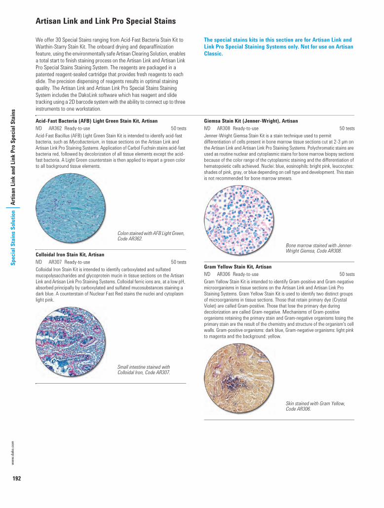
192
www.
dako
.com
Spec
ial S
tain
s Sol
utio
nAr
tisan
Lin
k an
d Li
nk P
ro S
peci
al S
tain
sArtisan Link and Link Pro Special Stains
We offer 30 Special Stains ranging from Acid-Fast Bacteria Stain Kit to Warthin-Starry Stain Kit. The onboard drying and deparaffinization feature, using the environmentally safe Artisan Clearing Solution, enables a total start to finish staining process on the Artisan Link and Artisan Link Pro Special Stains Staining System. The reagents are packaged in a patented reagent-sealed cartridge that provides fresh reagents to each slide. The precision dispensing of reagents results in optimal staining quality. The Artisan Link and Artisan Link Pro Special Stains Staining System includes the DakoLink software which has reagent and slide tracking using a 2D barcode system with the ability to connect up to three instruments to one workstation.
The special stains kits in this section are for Artisan Link and Link Pro Special Staining Systems only. Not for use on Artisan Classic.
Acid-Fast Bacteria (AFB) Light Green Stain Kit, ArtisanI AR362 Ready-to-use 50 testsAcid-Fast Bacillus (AFB) Light Green Stain Kit is intended to identify acid-fast bacteria, such as Mycobacterium, in tissue sections on the Artisan Link and Artisan Link Pro Staining Systems. Application of Carbol Fuchsin stains acid-fast bacteria red, followed by decolorization of all tissue elements except the acid-fast bacteria. A Light Green counterstain is then applied to impart a green color to all background tissue elements.
Colloidal Iron Stain Kit, ArtisanI AR307 Ready-to-use 50 testsColloidal Iron Stain Kit is intended to identify carboxylated and sulfated mucopolysaccharides and glycoprotein mucin in tissue sections on the Artisan Link and Artisan Link Pro Staining Systems. Colloidal ferric ions are, at a low pH, absorbed principally by carboxylated and sulfated mucosubstances staining a dark blue. A counterstain of Nuclear Fast Red stains the nuclei and cytoplasm light pink.
Giemsa Stain Kit (Jenner-Wright), ArtisanI AR308 Ready-to-use 50 testsJenner-Wright Giemsa Stain Kit is a stain technique used to permit differentiation of cells present in bone marrow tissue sections cut at 2-3 µm on the Artisan Link and Artisan Link Pro Staining Systems. Polychromatic stains are used as routine nuclear and cytoplasmic stains for bone marrow biopsy sections because of the color range of the cytoplasmic staining and the differentiation of hematopoietic cells achieved. Nuclei: blue, eosinophils: bright pink, leucocytes: shades of pink, gray, or blue depending on cell type and development. This stain is not recommended for bone marrow smears.
Gram Yellow Stain Kit, ArtisanI AR306 Ready-to-use 50 testsGram Yellow Stain Kit is intended to identify Gram-positive and Gram-negative microorganisms in tissue sections on the Artisan Link and Artisan Link Pro Staining Systems. Gram Yellow Stain Kit is used to identify two distinct groups of microorganisms in tissue sections. Those that retain primary dye (Crystal Violet) are called Gram-positive. Those that lose the primary dye during decolorization are called Gram-negative. Mechanisms of Gram-positive organisms retaining the primary stain and Gram-negative organisms losing the primary stain are the result of the chemistry and structure of the organism's cell walls. Gram-positive organisms: dark blue, Gram-negative organisms: light pink to magenta and the background: yellow.
Colon stained with AFB Light Green, Code AR362.
Small intestine stained with Colloidal Iron, Code AR307.
Bone marrow stained with Jenner-Wright Giemsa, Code AR308.
Skin stained with Gram Yellow, Code AR306.
USCatPathology.book Page 192 Monday, March 7, 2016 10:17 PM

193
Artisan Link and Link Pro Special StainsSpecial Stains Solution
Artisan Link and Link Pro Special Stains (continued)
Grocott's Methenamine Silver (GMS) Eosin Stain Kit, ArtisanI AR376 Ready-to-use 50 testsGrocott's Methenamine Silver (GMS) Eosin Stain Kit is intended to identify fungal organisms and Pneumocystis jiroveci (formerly known as carinii) in tissue sections on the Artisan Link and Artisan Link Pro Staining Systems. Fungi and Pneumocystis jiroveci are stained black while other tissue elements are stained pink. This stain is not recommended for cytology specimens.
Jones' Basement Membrane (PAS-M) H&E Stain Kit, ArtisanI AR480 Ready-to-use 100 testsJones' Basement Membrane H&E Stain Kit is used to identify basement membranes, specifically glomerular and tubular basement membranes in renal tissue sections cut at 2 µm on the Artisan Link and Artisan Link Pro Staining Systems. The Bowman's capsule: black, inner basement membrane: black to gray, nuclei: blue, collagen, cytoplasm and other tissue elements: pink.
Jones' Basement Membrane (PAS-M) Light Green Stain Kit, ArtisanI AR380 Ready-to-use 50 testsJones' Basement Membrane Light Green Stain Kit is used to identify basement membranes, specifically glomerular and tubular basement membranes in renal tissue sections cut at 2 µm on the Artisan Link and Artisan Link Pro Staining Systems. The Bowman's capsule: black, inner basement membrane: black to gray, other tissue elements: light green.
Orcein Stain Kit, ArtisanI AR313 Ready-to-use 50 testsOrcein Stain Kit is a stain technique used to identify viral inclusion bodies of hepatitis B surface antigen (HBsAG) and copper-associated proteins in tissue sections on the Artisan Link and Artisan Link Pro Staining Systems. Virus particles inside host cells are called viral inclusion bodies. A hepatitis B virus lies on the surface of the virus particle. HBsAG can be detected by using the Orcein staining method. The antigen appears as fine granules either diffusely spread throughout the cytoplasm or concentrated in the cytoplasm peripheral to the sinusoid space. Copper-associated proteins when in excessive pathologic amounts, such as Wilson's disease and some forms of cirrhosis, can be detected by using the Orcein staining method. HBsAG/elastin fibers: dark reddish brown, copper-associated protein: dark red-purple and background: pale pink-pink.
Lung stained with GMS Eosin, Code AR376.
Kidney stained with PAS-M H&E, Code AR480.
Kidney stained with PAS-M Light Green, Code AR380.
Liver stained with Orcein for copper-associated protein, Code AR313.
USCatPathology.book Page 193 Monday, March 7, 2016 10:27 PM

194
www.
dako
.com
Spec
ial S
tain
s Sol
utio
nAr
tisan
Spe
cial
Sta
ins
Artisan Special Stains
We offer 30 Special Stains ranging from Acid-Fast Bacteria Stain Kit to Warthin-Starry Stain Kit. The onboard drying and deparaffinization feature, using the environmentally safe Artisan Clearing Solution, enables a total start to finish staining process on the Artisan Link and Artisan Link Pro Special Stains Staining System. The reagents are packaged in a patented reagent-sealed cartridge that provides fresh reagents to each slide. The precision dispensing of reagents results in optimal staining quality. The Artisan Link and Artisan Link Pro Special Stains Staining System includes the DakoLink software which has reagent and slide tracking using a 2D barcode system with the ability to connect up to three instruments to one workstation.
The Special Stains kits in this section are suitable for all versions of Artisan, i.e. Artisan Link Pro, Artisan Link and Artisan Classic.
Acid-Fast Bacteria (AFB) Stain Kit, ArtisanI AR162 Ready-to-use 50 tests/100 testsAcid-Fast Bacillus (AFB) Stain Kit is intended to identify acid-fast bacteria, such as Mycobacterium, in tissue sections on the Artisan Link and Artisan Link Pro Staining Systems. Application of Carbol Fuchsin stains acid-fast bacteria red, followed by decolorization of all tissue elements except the acid-fast bacteria. A Methylene Blue counterstain is then applied to impart a blue color to all background tissue elements.
Alcian Blue pH 2.5 Stain Kit, ArtisanI AR160 Ready-to-use 50 tests/100 testsAlcian Blue pH 2.5 Stain Kit is intended to identify weakly sulfated mucins in tissue sections on the Artisan Link and Artisan Link Pro Staining Systems. At pH 2.5, acidic mucopolysaccharides stains blue. A Nuclear Fast Red counterstain will stain the nuclei red to pink and all other tissue elements a pale pink.
Alcian Blue/PAS Stain Kit, ArtisanI AR169 Ready-to-use 50 tests/100 testsAlcian Blue/PAS Stain Kit is intended to identify acidic and neutral mucins in tissue sections on the Artisan Link and Artisan Link Pro Staining Systems. This procedure clearly separates the acidic and neutral mucins by color and can be used to distinguish all mucins in tissue sections. Alcian Blue pH 2.5 stains the acid mucin blue while the Schiff's reagent stains the neutral mucins pink to red. Mixtures of the two mucins will appear purple due to the positive reactions with both Alcian Blue and Schiff's reagent. This stain can be used with the digestive enzyme, Artisan Alpha-Amylase (Code AR171), for the demonstration of glycogen in tissue.
Alcian Blue/PAS/Hematoxylin Stain Kit, ArtisanI AR178 Ready-to-use 50 tests/100 testsAlcian Blue/PAS/Hematoxylin Stain Kit is intended to identify acidic and neutral mucins in tissue sections on the Artisan Link and Artisan Link Pro Staining Systems. This procedure clearly separates the acidic and neutral mucins by color and can be used to distinguish all mucins in tissue sections. Alcian Blue pH 2.5 stains the acid mucin blue while the Schiff's reagent stains the neutral mucins pink to red. Mixtures of the two mucins will appear purple due to the positive reactions with both Alcian Blue and Schiff's reagent. A hematoxylin counterstain is then applied to impart a blue/black color to the nuclei.
Colon stained with AFB, Code AR162.
Small intestine stained with Alcian Blue pH 2.5, Code AR160.
Small intestine stained with Alcian Blue/PAS, Code AR169.
Large intestine stained with Alcian Blue/PAS/Hematoxylin, Code AR178.
USCatPathology.book Page 194 Monday, March 7, 2016 10:27 PM

195
Artisan Special StainsSpecial Stains Solution
Artisan Special Stains (continued)
Congo Red Stain Kit, ArtisanI AR161 Ready-to-use 50 tests/100 testsCongo Red Stain Kit is used to identify amyloid in tissue sections on Artisan Link and Artisan Link Pro Staining Systems. Amyloid is an abnormal protein product that can be found in various pathologic conditions. This stain demonstrates amyloid in pink to dark pink with light (bright field) microscopy or the characteristic apple green bi-refringence with polarized light. Mayers Hematoxylin is used as a counterstain. This stain has been optimized with 8 µm thick tissue sections
Elastic Stain Kit, ArtisanI AR163 Ready-to-use 50 tests/100 testsElastic Stain Kit is used to identify elastin fibers in tissue sections on the Artisan Link and Artisan Link Pro Staining Systems. This stain utilizes Alcoholic Hematoxylin, Ferric Chloride and Lugol's Iodine solutions to stain elastin fibers. Van Gieson's solution is added to differentiate collagen from elastin. Elastin fibers are stained black while remaining tissue elements are stained as follows: nuclei: blue/black, collagen: red, and other tissue elements: yellow.
Giemsa Stain Kit, ArtisanI AR164 Ready-to-use 50 testsGiemsa Stain Kit is typically used to identify hematopoietic cells (i.e. mast cells, basophils, polymorphoncuclear leucocytes, etc.) in tissue sections on the Artisan Link and Artisan Link Pro Staining Systems. In tissue specimens it will stain mast cells, which may be useful in the identification of mast cell tumors. Although Giemsa stains are frequently used to study bone marrow specimens as an aid in assessment of blood disorders, this Giemsa Stain Kit is not designed for this application. The tissue elements are stained as follows: Mast cell granules: purple, basophils: purple, eosinophils: bright pink and lymphocytes: blue. This stain is not recommended for cytology specimens or bone marrow smears.
Gomori's Blue Trichrome Stain Kit, ArtisanI AR167 Ready-to-use 50 testsGomori's Blue Trichrome Stain Kit is used to identify collagen fibers in liver and kidney tissue sections on the Artisan Link and Artisan Link Pro Staining Systems. Gomori's Blue Trichrome Stain is often used to demonstrate increased collagen deposition that is associated with replacement of functional tissue by scar tissue. This stain is useful in the asessment of cirrhosis of the liver in which thickened collagen replaces normal tissue causing liver dysfunction. In Gomori's one-step procedure, the collagen and nuclei are stained blue, cytoplasm, erythrocytes and fibrin are stained pink to red.
Gomori's Green Trichrome Stain Kit, ArtisanI AR166 Ready-to-use 50 testsGomori's Green Trichrome Stain Kit is used to identify collagen fibers in liver and kidney tissue sections on the Artisan Link and Artisan Link Pro Staining Systems. Gomori's Blue Trichrome Stain is often used to demonstrate increased collagen deposition that is associated with replacement of functional tissue by scar tissue. This stain is useful in the assessment of cirrhosis of the liver in which thickened collagen replaces normal tissue causing liver dysfunction. In Gomori's one-step procedure, the collagen and nuclei are stained green, cytoplasm, erythrocytes and fibrin are stained pink to red.
Amyloid stained with Congo Red, Code AR161.
Skin stained with Elastic Stain, Code AR163.
Spleen stained with Giemsa, Code AR164.
Liver stained with Gomori's Blue, Code AR167.
Intestine stained with Gomori's Green, Code AR166.
USCatPathology.book Page 195 Monday, March 7, 2016 10:27 PM

196
www.
dako
.com
Spec
ial S
tain
s Sol
utio
nAr
tisan
Spe
cial
Sta
ins
Artisan Special Stains (continued)
Gram Stain Kit, ArtisanI AR175 Ready-to-use 50 tests/100 testsGram Stain Kit is used to identify two distinct groups of microorganisms in tissue sections on Artisan Link and Artisan Link Pro Staining Systems. Those that retain primary dye (Crystal Violet) are called Gram-positive. Those that lose the primary dye during decolorization are called Gram-negative. Mechanisms of Gram-positive organisms retaining the primary stain and Gram-negative organisms losing the primary stain are the result of the chemistry and structure of the organism's cell walls. The tissue elements are stained as follow: Gram-positive organisms: blue, Gram-negative organisms: red and background: varying shades of blue/green.
Grocott's Methenamine Silver (GMS) Stain Kit, ArtisanI AR176 Ready-to-use 50 tests/100 testsGrocott's Methenamine Silver (GMS) Stain Kit is intended to identify fungal organisms and Pneumocystis jiroveci (formerly known as carinii) in tissue sections on the Artisan Link and Artisan Link Pro Staining Systems. Fungi and Pneumocystis jiroveci are stained black while other tissue elements are stained green. This stain is not recommended for cytology specimens.
Iron Stain Kit, ArtisanI AR158 Ready-to-use 50 tests/100 testsIron Stain Kit is used to identify iron pigment in tissue sections on the Artisan Link and Artisan Link Pro Staining Systems. Iron is an important component of the human body, especially as a vital constituent of oxygen carrying hemoglobin. Iron is stored in bone marrow, and a loss of iron stores is indicative of anemia. An excess of iron deposited in organs such as liver, spleen, and bone marrow may be a result of hemachromatosis. Ferric deposits: blue, nuclei: red, and the background tissue elements are stained pink. This stain is not recommended for bone marrow smears.
Jones' Basement Membrane (PAS-M) Stain Kit, ArtisanI AR180 Ready-to-use 50 tests/100 testsJones' Basement Membrane Stain Kit is used to identify basement membranes, specifically glomerular and tubular basement membranes in renal tissue sections cut at 2 µm on the Artisan Link and Artisan Link Pro Staining Systems. The Bowman's capsule: black, inner basement membrane: black to gray, nuclei: red, collagen: rose and cytoplasm and other tissue elements: pink.
Masson's Trichrome Stain Kit, ArtisanI AR173 Ready-to-use 50 tests/100 testsMasson's Trichrome Stain Kit is used to identify muscle, collagen fibers, fibrin and erythrocytes in tissue sections on the Artisan Link and Artisan Link Pro Staining Systems. Masson's Trichrome is often used to demonstrate increased collagen deposition that is associated with replacement of functional tissue by scar tissue. This is useful in the assessment of sclerosis of the liver in which thickened collagen replaces normal tissue causing liver dysfunction. Muscle: red, collagen: blue, fibrin: pink, erythrocytes: red and nuclei: blue/black.
Mucicarmine Stain Kit, ArtisanI AR168 Ready-to-use 50 tests/100 testsMucicarmine Stain Kit is used to identify epithelial mucins in tissue sections on the Artisan Link and Artisan Link Pro Staining Systems. These mucins are a family of polysaccharides covalently linked to proteins in epithelial cells. The mucins: pink, nuclei: black and other tissue elements: yellow.
Skin stained with Gram, Code AR175.
Lung stained with GMS, Code AR176.
Liver stained with Iron, Code AR158.
Kidney stained with PAS-M, Code AR180.
Liver stained with Masson's Trichrome, Code AR173.
Small intestine stained with Mucicarmine, Code AR168.
USCatPathology.book Page 196 Monday, March 7, 2016 10:27 PM

197
Artisan Special StainsSpecial Stains Solution
Artisan Special Stains (continued)
PAS-Green Stain Kit, ArtisanI AR172 Ready-to-use 50 tests/100 testsPAS-Green Stain Kit is used to identify fungi in tissue sections on the Artisan Link and Artisan Link Pro Staining Systems. The most commonly used application is for demonstrating fungi in skin infections. Unlike GMS, the PAS-Green technique will not stain all fungi and yeast. However, fungi typically found in dermatologic specimens are commonly identified using PAS-Green. Positive fungi: magenta and the remaining tissue elements: blue/green.
Periodic Acid-Schiff (PAS) Stain Kit, ArtisanI AR165 Ready-to-use 50 tests/100 testsPAS Stain Kit is used to identify glycogen in tissue sections on the Artisan Link and Artisan Link Pro Staining Systems. PAS-positive entities and structures are also numerous in tissue sections. The most common application is for demonstrating glycogen in the liver. Duplicate sections are stained with or without a pre-treatment, such as the Artisan Alpha-Amylase (Code AR171), which is a glycogen-digesting enzyme. Comparing PAS signal intensity in a digested tissue sample with one that has not been digested will give an indication of the amount of glycogen present. A loss of glycogen may be indicative of a metabolic disorder or damage to the liver. Positive glycogen: magenta, nuclei: blue and the background: pink.
Reticulin/No Counterstain Stain Kit, ArtisanI AR182 Ready-to-use 100 testsReticulin/No Counterstain Stain Kit is used to identify a primitive form of connective tissue, called reticulin, in tissue sections on the Artisan Link and Artisan Link Pro Staining Systems. An Ammoniacal Silver Nitrate solution is applied to stain the reticulin fibers in tissue. The silver is then reduced and toned to produce a black coloration of the fibers, which are visible by light microscopy. A counterstain may be applied off line.
Reticulin/Nuclear Fast Red Stain Kit, ArtisanI AR179 Ready-to-use 50 tests/100 testsReticulin/Nuclear Fast Stain Kit is used to identify a primitive form of connective tissue, called reticulin, in tissue sections on the Artisan Link and Artisan Link Pro Staining Systems. An Ammoniacal Silver Nitrate solution is applied to stain the reticulin fibers in tissue. The silver is then reduced and toned to produce a black coloration of the fibers, which are visible by light microscopy. Other tissue elements will be stained pink.
Warthin-Starry Stain Kit, ArtisanI AR181 Ready-to-use 50 tests/100 testsWarthin-Starry Stain Kit is used to identify Helicobacter pylori, spirochetes and other microorganisms in tissue sections on the Artisan Link and Artisan Link Pro Staining Systems. H. pylori and spirochetes are stained black while the background is stained golden yellow.
Skin stained with PAS-Green, Code AR172.
Liver stained with PAS, Code AR165.
Liver stained with Reticulin/No Counterstain, Code AR182.
Liver stained with Reticulin/NFR, Code AR179.
Helicobacter stained with Warthin-Starry, Code AR181.
USCatPathology.book Page 197 Monday, March 7, 2016 10:27 PM

USCatPathology.book Page 198 Monday, March 7, 2016 10:27 PM

Dako AcademyIntroduction 200Training 201Literature 206e-Learning 207Events 208
USCatPathology.book Page 199 Wednesday, March 9, 2016 10:51 PM

200
www.
dako
.com
Dako
Aca
dem
yIn
trodu
ctio
nIntroduction to theDako Academy
Within the Dako Academy, our customers will find everything they need to obtain a comprehensive understanding for the successful use of Dako products. We also provide ample opportunities for proficiency development in a number of specialty areas, utilizing the latest technology in respect to reagents, kits, detection, technique and instrumentation.
The Dako Academy offers an extensive customer support program ranging from on-site operator training sessions, to renowned educational manuals. We host scientific seminars and facilitate the sharing of knowledge, expertise and experience amongst anatomical pathology labs.
The scope of Dako Academy covers: Training Literature e-Learning Events
We look forward to seeing you in one of our training facilities or courses.
Providing the right knowledge and skills for improved patient diagnostics
USCatPathology.book Page 200 Monday, March 7, 2016 10:27 PM

201
TrainingDako Academ
yTraining
The Dako Academy offers a broad selection of training sessions in order to develop and increase your laboratory staff’s confidence and competence. Theoretical education is combined with instruction and hands-on sessions employing Dako reagents and instruments.
Lectures and laboratory sessions are divided and tailor-made for the participants, enhancing the skills of both beginners and experts through small classes and individual attention. For information about course fees and course schedules, please contact your Dako Key Account Manager.
Course CreditsBuilding on our extensive experience of providing world-class training, Dako has put together a wide variety of courses within pathology that build on each other in order to reach advanced competency levels. Dako course certificates are appreciated by many pathology departments, to document their competence development work.
For courses conducted in our United States facilities, all administered course contact hours are recorded with the American Society of Clinical Laboratory Scientists (ASCLS) and Professional Acknowledgement for Continuing Education (P.A.C.E.®).
LocationsDako Academy has three major training centers providing state-of-the-art facilities for advanced customer training, one center in Europe, one in the United States and one in Shanghai. For other locations or on-site training, please contact your Dako Key Account Manager.
Please note that some training courses may not be available on all locations.
Dako Academy, Europe Location: Dako Denmark A/S
Produktionsvej 42DK-2600 Glostrup, Denmark
E-mail: [email protected]
Dako Training Academy Center, US Location: Agilent Center of Excellence
5301 Stevens Creek BlvdSanta Clara, CA 95051-7201, USA
E-mail: [email protected]
Dako Academy, ChinaLocation: Dako Diagnostics (Shanghai) Co., Ltd.
17F, Citic PlazaNo.1350 North Sichuan Rd.Shanghai, China
E-mail: [email protected]
USCatPathology.book Page 201 Monday, March 7, 2016 10:27 PM

202
www.
dako
.com
Dako
Aca
dem
yTr
aini
ngTraining (continued)
CoursesWith a focus on Dako solution areas, the courses conducted on a regular basis in our training centers include:
Dako Omnis System and Immunohistochemistry Autostainer Link 48 System and Immunohistochemistry Artisan Link Pro Special Staining System and Special Stains Dako CoverStainer (H&E)
You will find further training opportunities under the heading “Training On-Demand”.
Dako Omnis System and Immunohistochemistry (IHC)
Course description: Covers the theory and practice of performing immunohistochemical stainings utilizing the Dako Omnis solution. The training consists of a system overview, daily use and advanced training on instrument and workstation software. On completion of this course, trainees will have the expertise to work independently with the system.
Course covers:
A comprehensive training on the Dako Omnis hardware and software applications
Laboratory sessions utilizing Dako Omnis system Quality Control and troubleshooting sessions A review of various RTU antibodies and detection chemistry
Length: Four days
Autostainer Link 48 System and Immunohistochemistry (IHC)
Course description: Covers the theory and practice of performing immunohistochemical stainings using Autostainer Link 48 system. The lecture component incorporates a review of frequently used IHC stains and their diagnostic implications. Laboratory sessions utilize automated platforms to demonstrate immunostaining performance, quality control, troubleshooting, open system architecture, data transfer and instrument management.
Course covers:
A comprehensive training on the Autostainer Link hardware and client software applications
Laboratory sessions utilizing the Autostainer Link 48 system A review of various antibodies and detection chemistries, along with
quality control and effective troubleshooting methods
Length: Three days
USCatPathology.book Page 202 Monday, March 7, 2016 10:27 PM

203
TrainingDako Academ
yTraining (continued)
Artisan Link Pro Special Staining System and Special Stains
Course description: Covers the theory and practice of performing histochemical stains on the Artisan Link Special Staining System.
Course covers:
Lecture session, including a review of frequently used special stains and their diagnostic application, as well as a comprehensive overview of the Artisan hardware and software applications
Laboratory sessions utilizing Artisan Link Pro for performing a variety of special stains
Slide reviews and troubleshooting session
Length: Two days
Dako CoverStainer (H&E)
Course description: Includes the theory and practice of performing Hematoxylin and Eosin (H&E) staining on the Dako CoverStainer Reagent Management System (RMS). Lectures incorporate workflow, staining performance, optimization, troubleshooting, maintenance, and hands-on laboratory sessions.
Course covers:
A comprehensive overview of the Dako CoverStainer hardware and software applications
Laboratory sessions using Dako CoverStainer Effective troubleshooting methods A review of basic procedures and recommended laboratory practice
associated with high-quality H&E staining
Length: Two days
USCatPathology.book Page 203 Monday, March 7, 2016 10:27 PM

204
www.
dako
.com
Dako
Aca
dem
yTr
aini
ngTraining On-Demand
Adding to our regular course portfolio, Dako Academy offers a wide range of on-demand courses in order to meet specific needs. If you
do not find what you are looking for, please contact your Dako Key Account Manager.
Immunohistochemistry
Course description: This popular course covers the basic theory and practice of immunohistochemistry.
Who should attend: Basic and intermediate level trained technicians
Course covers:
Lecture component focuses on principles of pre-treatment, antibody and detection use and basic histology as it relates to quality immunostaining
Laboratory sessions cover manual and automated immunostaining, troubleshooting and quality control
Regulatory guidelines issued by the College of American Pathologists (CAP) are also covered for US participants
Length: Three days
pharmDx Introduction Workshop
Course description: Targeted therapy has created the need for reliable and reproducible assays to aid in the assessment of eligible patients. The intent of personalized medicine requires tests that consistently provide relevant, accurate results. The pharmDx family of kits was developed and validated to fulfill this need. As pharmDx results will depend on strict adherence to protocols, workshop participants will be taken step-by-step through the entire assay procedure. Participants will also be introduced to approved scoring methods for achieving results with minimal intra and inter-laboratory variability.
Who should attend: Laboratory technicians and pathologists
Course covers:
One pharmDx assay is covered per workshop Workshops will focus on either interpretation or technical components
Length: One day
Interpretation Workshop
Course description: Skilled pathologists facilitate this workshop in a forum setting, utilizing both virtual images and microscope slides. Participants are encouraged to bring additional cases from their respective laboratories.
Who should attend: Clinical pathologists
Course covers:
Detailed instruction in the practical use of assays, interpretative parameters and interactive assessment of provided sample cases
Length: One day
USCatPathology.book Page 204 Monday, March 7, 2016 10:27 PM

205
TrainingDako Academ
yTraining On-Demand (continued)
Technical Workshop
Course description: Includes a lecture on the biology of the assay target and description of test components. Adherence to protocols is strongly emphasized throughout the workshop. Microscope slides are used to demonstrate appropriate results and potential staining artifacts.
Who should attend: Immunotechnicians
Course covers: Pharmacodiagnostics overview Review of kit components and protocols Hands-on laboratory sessions utilizing the Autostainer Link 48,
Autostainer Plus or manual staining Troubleshooting
Length: One day (immunohistochemistry) Two days (in situ hybridization)
FISH/ISH
Course description: This course presents basic in situ hybridization training.
Who should attend: Technicians performing or expecting to perform in situ hybridization techniques
Course covers: Lecture component covers molecular pathology theory, denaturation,
hybridization and detection The laboratory sessions include manual as well as automated
hybridization, and methods for detection of hybridized probes
Length: Three days
USCatPathology.book Page 205 Monday, March 7, 2016 10:27 PM

206
www.
dako
.com
Dako
Aca
dem
yLi
tera
ture
Literature
Dako offers educational materials and guidebooks to ensure your success in using our products. You can download it from our Web site or contact your Dako Key Account Manager for further information.
Education Guides
Dako Education Guides are well-known for providing valuable insights into the theoretical basis and methodology for different disciplines, including interpretation of staining results.
Immunohistochemical Staining Methods, Sixth Edition (2013) The Illustrated Guide to Bone Marrow Diagnosis, Second Edition
(2009) Demasking of Antigens, Second Edition (2008) Special Stains and H&E, Second Edition (2010)
Interpretation Manuals and Guidelines
Based on experience and guidelines from renowned experts, we have gathered interpretation advice and guidelines to help achieve reliable and reproducible results. Utilization of the guidelines will ensure that your laboratory achieves quality results.
HercepTest - Breast Cancer or Gastric Cancer EGFR pharmDx c-Kit pharmDx ER/PR pharmDx HER2 IQFISH pharmDx FLEX RTU Atlas of Stains, Fourth Edition Atlas of Special Stains PD-L1 IHC 22C3 pharmDx PD-L1 IHC 28-8 pharmDx
Procedures
In order to minimize the possibilities for error, a quick and easy solution is to use our step-by-step procedures. The use of these procedures has proven efficient for many laboratories.
Immunohistochemical staining General ELISA Procedure Immunovisualization of protein blots HER2 IQFISH pharmDx FISH procedures for cytology and histology samples CISH procedures for manual use, Autostainer, Autostainer Plus and
Autostainer Link 48
IHC Guidebook Sixth Edition
EDUCATIONAL
Immunohistochemical Staining Methods
USCatPathology.book Page 206 Monday, March 7, 2016 10:27 PM

207
e-LearningDako Academ
yLiterature (continued)
Product Manuals
To facilitate work tasks and procedures for laboratory personnel when interacting with our instruments, we provide a complete series of easy to use manuals for all our instruments. Logically structured and available in nine different languages, the three types of Dako manuals will quickly provide the information for which you are looking.
User Guides Quick Start Guides Handbooks
White Papers, Reviews, Studies and Webinar
In close collaboration with experts in the field, we publish white papers, reviews and studies on various pathology topics and related technologies. These publications are available online at www.dako.com.
Prostate Pathology Review IQFISH White Paper Dynamic Gap Staining Standards for IHC Controls, webinar
e-Learning
We offer multiple training opportunities to our customers, one of those opportunities includes easy-to-use, interactive e-Learning programs. Dako e-Learning programs are designed to quickly provide laboratory
technicians, pathologists and scientists worldwide with accurate information on how to achieve reliable results using Dako products.
HercepTest e-Learning
This HercepTest e-Learning program is developed to supply laboratory technicians and pathologists with accurate and fastknowledge of how to achieve reliable staining results and accurate interpretation with HercepTest.
The e-Learning program includes: A general introduction to HER2 testing and biology Description of HercepTest kits Overview of the complete manual and automated laboratory
procedures Interpretation guidelines Interpreting artifacts Troubleshooting Tests for personal training as a HercepTest user
CISH e-Learning
This e-Learning program is developed to supply laboratory technicians and pathologists with accurate and fast knowledge of how to achieve reliable staining and accurate interpretation with HER2 CISH pharmDx Kit.
The e-Learning program includes: General introduction to the CISH technology Product presentation Photo presentation of the complete laboratory procedure Interpretation guide Q&A Troubleshooting section Test for personal evaluation for procedure and interpretation Test for Web-based certification for the procedure and interpretation
USCatPathology.book Page 207 Monday, March 7, 2016 10:27 PM

208
www.
dako
.com
Dako
Aca
dem
yEv
ents
e-Learning (continued)
PD-L1 IHC 22C3 pharmdDx Pathology Training Program
The Dako PD-L1 IHC 22C3 pharmDx pathology training program uses in-depth content, engaging activities, and comprehensive cases to help you confidently:
The e-Learning program includes: Understand the core principles of PD-L1 pathology Learn the process for evaluating stained images for PD-L1 expression Recognize confounding considerations that affect PD-L1 scoring Score and interpret images for PD-L1 expression across a variety of
patient samples
Events
We support a wide number of events in the pathology community to facilitate knowledge sharing and feedback opportunities for our customers. We are also renowned for supporting and arranging
opportunities for scientists and laboratory personnel to meet and discuss hands-on findings and research results.
User Group Meetings
Having lasting partnerships and listening to our customers have always been priorities for us. Through our field personnel we strive to provide our customers with opportunities to share knowledge with each other and with us.
Our user group meetings allow customers to provide direct feedback and suggestions on further improvement of Dako solutions and customer support. These meetings are crucial in our efforts to develop products that meet customer-specific requirements.
We’ve been listening!
USCatPathology.book Page 208 Monday, March 7, 2016 10:27 PM

Service and SupportIntroduction 210Deployment Services 211Instrument Services 212Application and Technical Support 213Instrument Service Agreements 214
USCatPathology.book Page 209 Tuesday, March 8, 2016 12:14 AM

210
www.
dako
.com
Serv
ice
and
Supp
ort
Intro
duct
ion
Introduction toService and Support
Service and Support has a dedicated team of highly skilled and experienced service professionals ready to work with you to improve your laboratory workflow. We are committed to ensure that our customers feel confident in using our products by providing state-of-the-art service and support.
The Service and Support offerings include: Deployment Services Instrument Services Application and Technical Support Instrument Service Agreements
Deployment Services ensures that Dako systems are installed correctly and perfectly integrated into the facility’s network, Instrument Services see to that instruments run smoothly and in the event of any problems, resolves it promptly. Application and Technical Support provide on-site training, optimize software performance and ensure fast troubleshooting.
As a manufacturer and supplier of instruments, software and reagents, Dako provides its Field Personnel with the latest tools and all necessary training in order to get the most out of our products. Using Dako Service and Support, you as a customer can feel confident that your laboratory always performs at its best.
Instrument Service Agreements facilitate the budget process by providing pre-packaged service solutions in different price levels.
When you need us, we will be there
USCatPathology.book Page 210 Tuesday, March 8, 2016 12:14 AM
211
Deployment Services
Service and SupportDeployment Services
Our deployment team has a long history of service excellence. Software installation and implementation of advanced systems all over the world has provided us with an experience level that few in the industry can match. We have an extensive practice of complex Local Area Network (LAN) and Laboratory Information System (LIS) integrations, and we have a thorough knowledge base regarding possible IT security concerns and how to solve them.
Deployment Services include: Presite Inspection Installation Qualification Operational Qualification Connectivity
Presite Inspection
To prepare for a quick and smooth installation, the Presite Inspection service provides invaluable assistance. Potential surprises are avoided if the installation site has been inspected. Everything that needs to be in place for a trouble-free installation is investigated.
This includes physical location and support, water and electrical supply, waste water dispense, ventilation and light. The Presite Inspection often render in recommendations that will further improve ease of use and productivity of the instrument.
Installation Qualification
When an instrument is installed the Installation Qualification (IQ) process makes sure that this is done according to Dako specifications. The IQ verifies that all parts shipped were received and that software updates are compatible with the configuration of the instrument.
By performing an IQ you are certain that the instrument performs correctly and comply with regulatory standards. An IQ check is performed in connection with the installation of a new instrument and is also recommended if a new functionality is added, e.g. through a software upgrade, and whenever the instrument is relocated.
Operational Qualification
An Operational Qualification (OQ) process ensures that the instrument has the necessary accuracy in order to meet performance and quality standards. This also ensures that the instrument meet specific performance criteria by using a controlled test environment. An OQ check is performed in connection
with the installation of a new instrument and is also recommended if a new functionality is added, e.g. through a software upgrade, and whenever the instrument is relocated. An OQ check can also be performed with regular time intervals according to your Service Contract to guarantee optimal instrument performance.
Connectivity
The middleware solution – DakoLink – enables Dako instruments to connect to other laboratory instruments and to the laboratory and hospital information systems. Integration into hospital LIS and LAN in order to exchange information for test cases, requires a thorough understanding of the complexity and a long experience of solving
similar communication issues. Dako has long standing relationships with all LIS vendors on the market and Dako Deployment Services ensures that software integration is handled quickly and professionally.
USCatPathology.book Page 211 Tuesday, March 8, 2016 12:14 AM

212
www.
dako
.com
Serv
ice
and
Supp
ort
Inst
rum
ent S
ervi
ces
Instrument Services
Dako Instrument Services provides fast and professional support when hands-on assistance is required by our customers.
A global network of field personnel, all of which are regularly trained on the latest products and service techniques, ensure that we can resolve any issue you may have in the fastest way possible.
Dako is widely recognized for our high standards for how to deliver service and support. This is a reputation that we intend to keep regardless of whether we provide support remotely, over the phone or through hands-on assistance.
Instrument Services include: Planned Maintenance Corrective Maintenance Software Upgrades
Planned Maintenance
Planned Maintenance (PM) covers your instrument's routine maintenance needs and is scheduled based on usage and time. Dako PMs are of course planned so that they do not interfere with laboratory
activities, and at the same time you can be sure that your instrument is always running at peak performance.
Corrective Maintenance
Corrective Maintenance offers you the security that your laboratory requires in the event of any unforeseen problems. Our Service Engineers have excellent troubleshooting expertise that includes both fluidics and
electronics combined with strong IT skills. This, combined with short response times, keeps instrument downtime to a minimum.
Software Upgrades
The key enabler for managing Dako products in the laboratory is the DakoLink Path software. It links instruments and connects them to the laboratory information system while managing the performance of the individual instruments. On a regular basis, new functionality is added and
new reagents are released which require upgrades to the software. These software upgrades are carried out as PMs in order not to interfere with laboratory activities.
USCatPathology.book Page 212 Tuesday, March 8, 2016 12:14 AM

213
Application and Technical SupportService and Support
Application and Technical Support
We have been a leader within antibody-based cancer diagnostics for almost 50 years and our support to our customers when it comes to staining performance and optimizing reagent protocols is still recognized as the best in the market. We have a highly committed support organization that always provides you with professional and yet personal service.
With an extensive geographic presence, our local staff is always within easy reach. No matter where you are in the world, your dedicated Application Specialist or Technical Support Representative is just a phone call or e-mail away.
Application and Technical Support include: Protocol Optimization Protocol Design Product Demonstrations
Protocol Optimization
Our Application Specialists and Technical Support Representatives are experts in troubleshooting and optimization, working closely together with you to ensure that our reagents operate at peak performance in your laboratory.
When you contact us, we know the importance of providing you with quick advice and competent troubleshooting relating to the use of our systems and reagents. Dako is organized worldwide with highly experienced staff that is able to assist you in choosing the right products and suggest how to optimize the performance of your protocols.
Protocol Design
Make use of our unrivaled competence when it comes to choosing the correct reagents and combine them in the best possible way. We can perform assays in our laboratories in Carpinteria, USA and in Glostrup,
Denmark or you can just send us your slides directly and we will optimize your protocols to your satisfaction.
Product Demonstrations
We can provide you with all the documentation that is needed in order to get a good understanding for the advantages you will get by choosing us as your supplier. Nothing compares to hands-on experience where you
and your colleagues can actually use the instrument and the reagents you are interested in in your own laboratory. Therefore we offer Product Demonstrations directly at customer sites for longer or shorter periods.
USCatPathology.book Page 213 Tuesday, March 8, 2016 12:14 AM

214
www.
dako
.com
Serv
ice
and
Supp
ort
Inst
rum
ent S
ervic
e Ag
reem
ents
Instrument Service Agreements
To continue to get the most out of your investment you can extend the benefits from the standard instrument warranty.
Dako Instrument Service Agreements can be found in three different Service Levels in order to meet your laboratory's unique requirements: Basic Essential Comprehensive
Each Service Level include different services and to ensure productivity and availability for critical systems, We can offer fast response times and priority handling for technical support and repairs. An Instrument Service Agreement can cover a single instrument or a complete set up of Dako instruments.
Instrument Service Agreements are also ideal in order to avoid unplanned expenses and as such a very cost-effective way to maximize your instrument’s performance, matching your requirements with your budget constraints.
For more information on our Instrument Service Agreements and to discuss what Service level that would suit you the best, please contact your Dako Key Account Manager.
USCatPathology.book Page 214 Tuesday, March 8, 2016 12:14 AM
General Product InformationGeneral Product Information 216
USCatPathology.book Page 215 Wednesday, March 9, 2016 10:52 PM

216
www.
dako
.com
Dako
Gene
ral P
rodu
ct In
form
atio
nGeneral Product Information
Monoclonal Antibodies
We produce a wide range of monoclonal mouse and monoclonal rabbit antibodies. Each antibody has been carefully selected on the basis of its value, either for research or for the analysis of pathological human cells by immunohistochemistry.Tissue Culture Antibodies. With only a few exceptions, Dako monoclonal antibodies are produced in tissue culture. This gives advantages in the use of the antibodies. For example, background problems are virtually absent with such reagents because all the mouse immunoglobulin molecules are directed against the target antigen.Specificity. Dako monoclonal antibodies are extensively screened on a multitude of tissue sections or other relevant biological material to ascertain that they possess the necessary specificity and give consistent, strong labeling reactions.
Solvent. Dako monoclonal antibodies are, generally, supplied in the liquid form. The majority of unconjugated antibodies are supplied as tissue culture supernatants containing 0.05 mol/L Tris/HCl, pH 7.2, and 15 mmol/L sodium azide. The azide can be removed by dialysis or gel filtration if it interferes with the use of the antibody. However, after removal of the azide, the antibody must be stored frozen.Storage. 2-8 °C.Further Information. A package insert is supplied with each vial of monoclonal antibody. It states intended/recommended use, clone, isotype, specificity, as well as recommended staining procedure when applicable. Package inserts are also avaliable on www.dako.com.The products require no hazard labeling.
Polyclonal Antibodies
Since 1966 we have produced a continually widening range of polyclonal antibodies. An extensive knowledge of protein chemistry and immuno-chemistry, careful selection of animals for immunization, and optimal, long-term immunization schemes have formed the basis of the high quality of our products.Most of the polyclonal antibodies are produced in rabbits. This provides several advantages:Human antibodies reacting with rabbit immunoglobulins occur rarely. Therefore, rabbit antibodies can be used without risk of non-specific reactions even in sensitive techniques. A batch of antibody will always consist of the pooled sera from a large number of animals. This eliminates the possibility of a single atypical antibody predominating and gives minimal batch-to-batch variation. Rabbit antibodies exhibit very broad precipitation curves, so precipitation will occur even at high antigen or antibody excess.Immunoglobulin Fractions. All Dako polyclonal antibodies are offered in the form of immunoglobulin fractions with a few exceptions mentioned under the individual product. The immunoglobulin fraction is prepared by salting out and ion exchange chromatography. The elimination of bulk proteins gives a stable product with reduced background in gel precipitation techniques, and minimal non-specific reactions in other applications.Affinity-Isolated Antibodies. Dako affinity-isolated antibodies are prepared by immunoaffinity chromatography, using antigens coupled to a solid matrix. The elution and adsorption techniques used, guarantee antibodies of high affinity.
Specificity. Monospecificity of Dako polyclonal antibodies is obtained by the use of highly purified antigens for immunization. Traces of sometimes unavoidable, unwanted antibodies are removed by liquid or, in the majority of cases, solid-phase absorption. Crossed immuno-electrophoresis, with its high sensitivity and resolving power, is included in our specificity controls. For this test, antibody is used at a very high concentration (12.5 microliters per square cm of gel). For a steadily increasing number of antibodies the specificity is also ascertained by ELISA. Antibodies which are specified "for immuno-histochemistry only" have not necessarily been subjected to the above specificity tests.Antibody Titre. The titre variation between batches of uncon-jugated polyclonal antibodies is less than 10%. The titre of most antibodies is measured by single radial immunodiffusion (SRI) (1). The SRI titre states how many milligrams of antigen which is precipitated in an agarose gel by 1 L of antibody.Application. Because of their high purity and avidity, Dako unconjugated polyclonal antibodies are generally well-suited for a variety of techniques. In addition, our immunohistochemistry laboratory as well as numerous investigators have shown for a large number of Dako unconjugated polyclonal antibodies, that they give highly specific immunohistochemical reactions when used as primary antibodies in immunofluorescence or immunoenzymatic techniques. The intended/recommended use of each antibody is stated in the package insert.
USCatPathology.book Page 216 Tuesday, March 8, 2016 12:14 AM
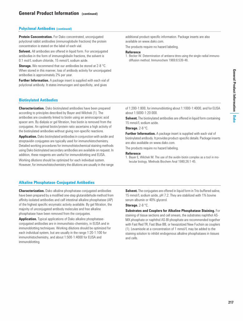
217
General Product Information
DakoGeneral Product Information (continued)
Polyclonal Antibodies (continued)
Protein Concentration. For Dako concentrated, unconjugated polyclonal rabbit antibodies (immunoglobulin fractions) the protein concentration is stated on the label of each vial. Solvent. All antibodies are offered in liquid form. For unconjugated antibodies in the form of immunoglobulin fractions, the solvent is 0.1 mol/L sodium chloride, 15 mmol/L sodium azide.Storage. We recommend that our antibodies be stored at 2-8 °C. When stored in this manner, loss of antibody activity for unconjugated antibodies is approximately 2% per year.Further Information. A package insert is supplied with each vial of polyclonal antibody. It states immunogen and specificity, and gives
additional product-specific information. Package inserts are also available on www.dako.com. The products require no hazard labeling.Reference1. Becker W. Determination of antisera titres using the single radial immuno-
diffusion method. Immunochem 1969;6:539-46.
Biotinylated Antibodies
Characterization. Dako biotinylated antibodies have been prepared according to principles described by Bayer and Wilchek (1). The antibodies are covalently linked to biotin using an aminocaproic acid spacer arm. By dialysis or gel filtration, free biotin is removed from the conjugates. An optimal biotin/protein ratio ascertains a high activity of the biotinylated antibodies without giving non-specific reactions.Application. Dako biotinylated antibodies in conjunction with avidin and streptavidin conjugates are typically used for immunohistochemistry. Detailed working procedures for immunohistochemical staining methods using Dako biotinylated secondary antibodies are available on request. In addition, these reagents are useful for immunoblotting and ELISA. Working dilutions should be optimized for each individual system. However, for immunohistochemistry the dilutions are usually in the range
of 1:200-1:800, for immunoblotting about 1:1000-1:4000, and for ELISA about 1:5000-1:20 000.Solvent. The biotinylated antibodies are offered in liquid form containing 15 mmol/L sodium azide.Storage. 2-8 ºC.Further Information. A package insert is supplied with each vial of biotinylated antibody. It provides product-specific details. Package inserts are also available on www.dako.com. The products require no hazard labeling.Reference1. Bayer E, Wilchek M. The use of the avidin-biotin complex as a tool in mo-
lecular biology. Methods Biochem Anal 1980;26:1-45.
Alkaline Phosphatase-Conjugated Antibodies
Characterization. Dako alkaline phosphatase-conjugated antibodies have been prepared by a modified one-step glutaraldehyde method from affinity-isolated antibodies and calf intestinal alkaline phosphatase (AP) of the highest specific enzymatic activity available. By gel filtration, the majority of unconjugated antibody molecules and free alkaline phosphatase have been removed from the conjugates.Application. Typical applications of Dako alkaline phosphatase-conjugated antibodies are in immunohisto-chemistry, in ELISA and in immunoblotting techniques. Working dilutions should be optimized for each individual system, but are usually in the range 1:20-1:100 for immunohistochemistry, and about 1:500-1:4000 for ELISA and immunoblotting.
Solvent. The conjugates are offered in liquid form in Tris-buffered saline, 15 mmol/L sodium azide, pH 7.2. They are stabilized with 1% bovine serum albumin or 40% glycerol. Storage. 2-8 °C. Substrates and Couplers for Alkaline Phosphatase Staining. For staining of tissue sections and cell smears, the substrates naphthol AS-MX phosphate or naphthol AS-BI phosphate are recommended together with Fast Red TR, Fast Blue BB, or hexazotized New Fuchsin as couplers (1). Levamisole at a concentration of 1 mmol/L may be added to the staining solution to inhibit endogenous alkaline phosphatases in tissues and cells.
USCatPathology.book Page 217 Tuesday, March 8, 2016 12:14 AM

218
General Product Information (continued)
www.
dako
.com
Dako
Gene
ral P
rodu
ct In
form
atio
n
Alkaline Phosphatase-Conjugated Antibodies (continued)
For immunoblotting, the substrates and couplers mentioned above for staining of tissues and cells can be used. Another good combination is 5-bromo-4-chloro-indolyl phosphate as substrate and nitro blue tetrazolium as coupler (2).For ELISA, 4-nitrophenyl phosphate is the substrate most often used (3).Further Information. A package insert is supplied with each vial of conjugate. It provides product-specific details. Package inserts are also available on www.dako.com. The products require no hazard labeling.
References1. Cordell JL, Falini B, Erber WN, Ghosh AK, Abdulaziz Z, Macdonald S,
et al. Immunoenzymatic labeling of monoclonal antibodies using immune complexes of alkaline phosphatase and monoclonal anti-alkaline phos-phatase (APAAP complexes). J Histochem Cytochem 1984;32:219-29.
2. Blake MS, Johnston KH, Russell-Jones GJ, Gotschlich EC. A rapid sensitive method for detection of alkaline phosphatase-conjugated anti-antibody on Western blots. Anal Biochem 1984;136:175-9.
3. Kan LVP, Verspaget HW, Pena AS. ELISA assay for quantitative measure-ment of human immunoglobulins IgA, IgG, and IgM in nanograms. J Immu-nol Methods 1983;57:51-7.
Peroxidase-Conjugated Antibodies
Characterization. Dako peroxidase-conjugated antibodies have been prepared from the chromatographically purified immunoglobulin fraction of antisera or affinity-isolated antibodies and horseradish peroxidase (HRP) of the highest specific enzymatic activity available. The coupling reaction is a modification, developed at Dako, of the two-step glutaraldehyde method of Avrameas and Ternynck (1). The reaction is gentle, efficient, highly reproducible and gives conjugate molecules of molecular weight predominantly 200 000-240 000.Specificity. The specificity of the antibodies is ascertained by crossed immunoelectrophoresis and, when applicable, by ELISA. Application. Dako peroxidase conjugates are generally used for light and electron microscopy, enzyme-linked immunosorbent assays (ELISA), immunoblotting techniques, and amplification of immunoprecipitates in agarose gels. Working dilutions should be optimized for each individual system, but are usually for histological work in the range 1:20-1:200, for ELISA 1:500-1:2000, and for enzymatic amplification of immuno-precipitates and immunoblotting about 1:100-1:1000. Intended/ recommended use and recommended dilutions are stated in the package insert. Solvent. Peroxidase conjugates are sold in liquid form with preservative added. Storage. Dako peroxidase-conjugated antibodies are very stable if kept undiluted at 2-8 °C.Chromogens for Peroxidase Staining. For light and electron microscopy, diaminobenzidine (DAB) is recommended. Considerable enhancement of staining intensity can be obtained very simply by using DAB in conjunction with imidazole and heavy metal salts (2,3). For immunoblotting, tetramethylbenzidine (TMB) used as described by McKimm-Breschkin (4) produces a stable color of high intensity. The most sensitive stain for amplification of immunoprecipitates in agarose gels is 3-amino-9-ethylcarbazole (AEC) (5,6). DAB (7), TMB (4) and AEC can with advantage be prepared as stock solutions, thus easing the work, and especially for DAB, eliminating possible staining variations due to variable amounts of impurities present in small aliquots of DAB powder. Hydrogen
peroxide should not be added to the staining solution until shortly before use.Even at very high dose levels - 4-8 g/kg - DAB produces only minimal toxic effects in male rats and male mice, and has no effects in female mice (8).For ELISA, orthophenylenediamine (OPD) and 3,3'-5,5'- tetra-methylbenzidine (TMB) are good and very sensitive chromogens. Further Information. A package insert is supplied with each vial of conjugate. It provides product-specific details. Package inserts are also available on www.dako.com. The products require no hazard labeling.References1. Avrameas S, Ternynck T. Peroxidase labelled antibody and Fab conjugates
with enhanced intracellular penetration. Immunochemistry 1971;8:1175-9.
2. Trojanowski JQ, Obrocka MA, Lee VMY. A comparison of eight different chromogen protocols for the demonstration of immunoreactive neurofila-ments or glial filaments in rat cerebellum using the peroxidase-antiperoxi-dase method and monoclonal antibodies. J Histochem Cytochem 1983;31:1217-23.
3. Scopsi L, Larsson LI. Increased sensitivity in peroxidase immunocytochemis-try. A comparative study of a number of peroxidase visualization methods employing a model system. Histochemistry 1986;84:221-30.
4. McKimm-Breschkin JL. The use of tetramethylbenzidine for solid phase immunoassays. J Immunol Methods 1990;135: 277-80.
5. Graham RC, Lundholm U, Karnovsky MJ. Cytochemical demonstration of peroxidase activity with 3-amino-9-ethyl-carbazole. J Histochem Cytochem 1965;13:150-2.
6. Broe MK, Ingild A. Amplification of immunoprecipitates in agarose gels by horseradish peroxidase-labelled antibody. Scand J Immunol 1983;17:255-8.
7. Pelliniemi LJ, Dym M, Karnovsky MJ. Peroxidase histochemistry using di-aminobenzidine tetrahydrochloride stored as a frozen solution. J Histochem Cytochem 1980;28:191-2.
8. Weisburger EK, Russfield AB, Homburger F, Weisburger JH, Boger E, Van Dongen CG, et al. Testing of twenty-one environmental aromatic amines or derivatives for long-term toxicity or carcinogenicity. J Environ Pathol Toxicol 1978;2:325-56.
USCatPathology.book Page 218 Tuesday, March 8, 2016 12:14 AM

219
General Product Information
DakoGeneral Product Information (continued)
Fluorescein-Conjugated Antibodies for Tissue Staining
Dako fluorescein-conjugated antibodies for tissue staining meet the stringent requirements suggested at international conferences on standardization in immunofluorescence.Characterization. Chromatographically purified immunoglobulin fractions of antisera or purified monoclonal antibodies are conjugated with fluorescein isothiocyanate isomer 1 (FITC). After conjugation, unreacted FITC is completely removed by gel filtration on Sephadex G-25. A further purification is carried out by ion exchange chromatography. This process removes unconjugated antibody molecules and antibody molecules to which more than 4 molecules of FITC are attached. Thus, our conjugates consist of optimally labeled antibody molecules, and require no absorption with tissue powders prior to use. The fluorescein/protein ratio, measured as the absorbance ratio A 495 nm/A 278 nm, is 0.65 ± 0.05 for all preparations, corresponding to a molar FITC/protein ratio of 2.3.Specificity and Performance Testing. Before conjugation, the specificity of the antibodies is ascertained by crossed immuno-electrophoresis and ELISA, when applicable. After conjugation, the specificity and fluorescence are controlled by direct and indirect immunofluorescence methods.Working Dilutions. Working dilutions of conjugates will depend on the type and the condition of the fluorescence microscope being used, and also on the tissue under investigation. For these reasons it is advisable for each individual laboratory to test various dilutions of a conjugate in order to find the optimal dilution. On human bone marrow/tonsil, the working dilutions of FITC-conjugated anti-human IgA, IgG and IgM are typically 1:20-1:40. No non-specific fluorescence is seen at a dilution of 1:20. Solvent. For fluorescein-conjugated polyclonal antibodies, the solvent is phosphate-buffered saline, 15 mmol/L sodium azide, pH 7.2. For fluorescein-conjugated monoclonal antibodies the solvent is Tris-buffered saline, 15 mmol/L sodium azide, pH 7.2. They are stabilized with 1% bovine serum albumin.
Storage. Fluorescein-conjugated antibodies should be stored in the dark at 2-8 °C.During storage a small precipitate may occasionally develop causing a fine granular non-specific staining. By a simple filtration (0.22 µm cellulose acetate filter), the original high quality of the conjugate will be restored. Conjugates should not be stored in diluted form.Retardation of Fluorescence Fading. During microscopy a pronounced fading of the fluorescence emitted from FITC occurs. The addition of various chemicals to the mountant used for the immunofluorescence preparations is an efficient, simple and inexpensive means of retarding fading. For a detailed discussion of the preparation and features of anti-fading mountants, please see references 1, 2 and 3.Further Information. A package insert is supplied with each vial of conjugate. It provides product-specific details. Package inserts are also available on www.dako.com.The products require no hazard labeling.References1. Krenik KD, Kephart GM, Offord KP, Dunette SL, Gleich GJ. Comparison of
antifading agents used in immunofluorescence. J Immunol Methods 1989;117:91-7.
2. Valnes K, Brandtzaeg P. Retardation of immunofluorescence fading during microscopy. J Histochem Cytochem 1985;33:755-61.
3. Longin A, Souchier C, French M, Bryon P-A. Comparison of antifading agents used in fluorescence microscopy: image analysis and laser confocal microscopy study. J Histochem Cytochem 1993;41:1833-40.
USCatPathology.book Page 219 Tuesday, March 8, 2016 12:14 AM

USCatPathology.book Page 220 Tuesday, March 8, 2016 12:14 AM

IndexesAlphabetical Index 223Synonym List 241Antibody Clone Index 244Product Code System 246Product Code Index 247
USCatPathology.book Page 221 Wednesday, March 9, 2016 10:52 PM

USCatPathology.book Page 222 Tuesday, March 8, 2016 12:14 AM

223
Alphabetical IndexIndexes
Alphabetical Index This index lists all products available from Dako. More detailed information appears on the pages mentioned for each individual product.For antibody clones, see pp 244-245.
Abbreviations: Labels:a Anti- AP Alkaline phosphataseGp Guinea pig Biotin Biotin with a 7-atom spacer armGt Goat FITC Fluorescein isothiocyanateHu Human HRP Horseradish peroxidaseMo MouseRb RabbitRt RatSw Swine
Code Source Product See Page
AAR162 Acid-Fast Bacteria (AFB) Stain Kit, Artisan (50 Tests/100 Tests) 194AR362 Acid-Fast Bacteria (AFB) Light Green Stain Kit, Artisan (50 Tests) 192M0635 Mo a Hu Actin (Muscle), Clone HHF35 73IR700 Mo a Hu Actin (Muscle), Clone HHF35, Ready-to-Use, FLEX, for Autostainer Link Instruments 41 73IS700 Mo a Hu Actin (Muscle), Clone HHF35, Ready-to-Use, FLEX, for Dako Autostainer Instruments 56 73M0874 Mo a Actin (Sarcomeric), Clone Alpha-Sr-1 73M0851 Mo a Hu Actin (Smooth Muscle), Clone 1A4 73IR611 Mo a Hu Actin (Smooth Muscle), Clone 1A4, Ready-to-Use, FLEX, for Autostainer Link Instruments 41 73IS611 Mo a Hu Actin (Smooth Muscle), Clone 1A4, Ready-to-Use, FLEX, for Dako Autostainer Instruments 56 73
Adhesion Molecule-1, see: CD31, Endothelial CellM3501 Mo a Adrenocorticotropin (ACTH), Clone 02A3 73K4069 ADVANCE/HRP, Rabbit/Mouse (55 Tests) 61 132K4068 ADVANCE/HRP, Rabbit/Mouse (550 Tests) 61 132K3464 AEC Substrate-Chromogen, Ready-to-Use (1100 Tests) 133K3461 AEC+ Substrate-Chromogen, Ready-to-Use (150 Tests) 54 133K3469 AEC+ Substrate-Chromogen, Ready-to-Use (1100 Tests) 54 133
AFP, see: Alpha-1-FetoproteinM3628 Rb a Hu Akt-pS473, Phosphorylation Site Specific, Clone 14-5 73F0117 Rb a Hu Albumin/FITC 73AR178 Alcian Blue/PAS/Hematoxylin Stain Kit, Artisan (50 Tests/100 Tests) 194AR160 Alcian Blue, pH 2.5, Stain Kit, Artisan (50 Tests/100 Tests) 194AR169 Alcian Blue/PAS Stain Kit, Artisan (50 Tests/100 Tests) 194Y5417 ALK DNA Probe, Fluorochrome-Conjugated 172G111200 ALK BA P5, SureFISH 175G111400 ALK BA P20, SureFISH 175G211400 ALK BA P20 x 6, SureFISH 175G111900 ALK BA P200, SureFISH 175
ALK Protein, see: CD246, ALK ProteinAlkaline Phosphatase and Peroxidase-Blocking Reagent, see: Dual Endogenous Enzyme Block
A0008 Rb a Hu Alpha-1-Fetoprotein 73 117GA500 Rb a Hu Alpha-1-Fetoprotein, Ready-to-Use, FLEX, for Dako Omnis 26 73IR500 Rb a Hu Alpha-1-Fetoprotein, Ready-to-Use, FLEX, for Autostainer Link Instruments 41 73IS500 Rb a Hu Alpha-1-Fetoprotein, Ready-to-Use, FLEX, for Dako Autostainer Instruments 56 73AR171 Alpha-Amylase Stain Kit, Artisan (50 Tests/100 Tests) 191
Alpha-Methylacyl-Coenzyme A Racemase, see: AMACRM3616 Rb a Hu AMACR, Clone 13H4 74GA060 Rb a Hu AMACR, Clone 13H4, Ready-to-Use, FLEX, for Dako Omnis 26 74IR060 Rb a Hu AMACR, Clone 13H4, Ready-to-Use, FLEX, for Autostainer Link Instruments 41 74IS060 Rb a Hu AMACR, Clone 13H4, Ready-to-Use, FLEX, for Dako Autostainer Instruments 56 74IC004 a Hu AMACR + Cytokeratin HMW + Cytokeratin 5/6, Ready-to-Use, DuoFLEX Cocktail, for Autostainer Link Instruments 115M3562 Mo a Hu Androgen Receptor, Clone AR441 74K3954 Animal Research Kit (ARK)/HRP (150 Tests) 132S0809 Antibody Diluent 133S3022 Antibody Diluent, Background-Reducing 133K8006 Antibody Diluent, EnVision FLEX 53 60 129
Antigen Retrieval, see: Target RetrievalK3954 ARK (Animal Research Kit)/HRP (150 Tests) 132
Artisan Accessories 191AR310 Artisan Link Pro Special Staining System 189
USCatPathology.book Page 223 Wednesday, March 9, 2016 10:53 PM

224
www.dako.com
Alphabetical Index (continued)
Inde
xes
Alph
abet
ical
Inde
x
Artisan Special Stains 192-197AS480 Autostainer Link 48 21 35 35 37S3424 Autostainer Reagent Racks 54S3425 Autostainer Reagent Vials 54S3704 Autostainer Slide Racks (4 Racks) 54
BM7307 Mo a Hu B-Cell-Specific Activator Protein, Clone DAK-Pax-5 74GA650 Mo a Hu B-Cell-Specific Activator Protein, Clone DAK-Pax5, Ready-to-Use, FLEX, for Dako Omnis 26 74IR650 Mo a Hu B-Cell-Specific Activator Protein, Clone DAK-Pax5, Ready-to-Use, FLEX, for Autostainer Link Instruments 41 74IS650 Mo a Hu B-Cell-Specific Activator Protein, Clone DAK-Pax5, Ready-to-Use, FLEX, for Dako Autostainer Instruments 56 74K0598 BCIP/NBT Substrate System (150 Tests) 133Y5407 BCL2 DNA Probe, Fluorochrome-Conjugated 172M0887 Mo a Hu BCL2 Oncoprotein, Clone 124 74IR614 Mo a Hu BCL2 Oncoprotein, Clone 124, Ready-to-Use, FLEX, for Autostainer Link Instruments 41 74IS614 Mo a Hu BCL2 Oncoprotein, Clone 124, Ready-to-Use, FLEX, for Dako Autostainer Instruments 56 74Y5408 BCL6 DNA Probe, Fluorochrome-Conjugated 172M7211 Mo a Hu BCL6 Protein, Clone PG-B6p 75GA625 Mo a Hu BCL6 Protein, Clone PG-B6p, Ready-to-Use, FLEX, for Dako Omnis 26 75IR625 Mo a Hu BCL6 Protein, Clone PG-B6p, Ready-to-Use, FLEX, for Autostainer Link Instruments 41 75IS625 Mo a Hu BCL6 Protein, Clone PG-B6p, Ready-to-Use, FLEX, for Dako Autostainer Instruments 56 75M7260 Mo a Hu BCL10 Protein, Clone 151 75M0872 Mo a Hu Beta-Amyloid, Clone 6F/3D 75M3539 Mo a Hu Beta-Catenin, Clone b-Catenin-1 75GA702 Mo a Hu Beta-Catenin, Clone b-Catenin-1, Ready-to-Use, FLEX, for Dako Omnis 26 75IR702 Mo a Hu Beta-Catenin, Clone b-Catenin-1, Ready-to-Use, FLEX, for Autostainer Link Instruments 41 75IS702 Mo a Hu Beta-Catenin, Clone b-Catenin-1, Ready-to-Use, FLEX, for Dako Autostainer Instruments 56 75X0590 Biotin-Blocking System 134CS702 Bluing Buffer, Dako, for Dako CoverStainer 185M0744 Mo a Bromodeoxyuridine, Clone Bu20a 75S3024 Buffer, PBS, pH 7.0 134S3306 Buffer, Tris-NaCl Solution with Tween 20, pH 7.6 134S1968 Buffer, Tris-Saline, pH 7.6 134S3001 Buffer, Tris-Saline, pH 7.6 134S3006 Buffer, Wash (10x), for Immunohistochemistry 54 134K8007 Buffer, Wash, EnVision FLEX (20x) 53 60 129
CF0254 Rb a Hu C1q Complement/FITC 75F0201 Rb a Hu C3c Complement/FITC 75F0169 Rb a Hu C4c Complement/FITC 75M3517 Mo a Hu CA 19-9, Clone 1116-NS-19-9 76M3520 Mo a Hu CA 125, Clone M11 76GA701 Mo a Hu CA 125, Clone M11, Ready-to-Use, FLEX, for Dako Omnis 26 76IR701 Mo a Hu CA 125, Clone M11, Ready-to-Use, FLEX, for Autostainer Link Instruments 41 76IS701 Mo a Hu CA 125, Clone M11, Ready-to-Use, FLEX, for Dako Autostainer Instruments 56 76
Cadherin, see: E-Cadherin and N-CadherinA0576 Rb a Hu Calcitonin 76GA515 Rb a Hu Calcitonin, Ready-to-Use, FLEX, for Dako Omnis 26 76IR515 Rb a Hu Calcitonin, Ready-to-Use, FLEX, for Autostainer Link Instruments 41 76IS515 Rb a Hu Calcitonin, Ready-to-Use, FLEX, for Dako Autostainer Instruments 56 76M3557 Mo a Hu Caldesmon, Clone h-CD 76GA054 Mo a Hu Caldesmon, Clone h-CD, Ready-to-Use, FLEX, for Dako Omnis 26 76IR054 Mo a Hu Caldesmon, Clone h-CD, Ready-to-Use, FLEX, for Autostainer Link Instruments 42 76IS054 Mo a Hu Caldesmon, Clone h-CD, Ready-to-Use, FLEX, for Dako Autostainer Instruments 56 76
CALLA, see: CD10M3556 Mo a Hu Calponin, Clone CALP 77
Calprotectin, see: Myeloid/Histiocyte AntigenM7245 Mo a Hu Calretinin, Clone DAK-Calret 1 77IR627 Mo a Hu Calretinin, Clone DAK-Calret 1, Ready-to-Use, FLEX, for Autostainer Link Instruments 42 77IS627 Mo a Hu Calretinin, Clone DAK-Calret 1, Ready-to-Use, FLEX, for Dako Autostainer Instruments 56 77M7072 Mo a Hu Carcinoembryonic Antigen, Clone II-7 77GA622 Mo a Hu Carcinoembryonic Antigen, Clone II-7, Ready-to-Use, FLEX, for Dako Omnis 26 77IR622 Mo a Hu Carcinoembryonic Antigen, Clone II-7, Ready-to-Use, FLEX, for Autostainer Link Instruments 42 77IS622 Mo a Hu Carcinoembryonic Antigen, Clone II-7, Ready-to-Use, FLEX, for Dako Autostainer Instruments 56 77
Code Source Product See Page
USCatPathology.book Page 224 Wednesday, March 9, 2016 10:53 PM

225
Alphabetical Index (continued)
Alphabetical IndexIndexes
GA526 Rb a Hu Carcinoembryonic Antigen, Ready-to-Use, FLEX, for Dako Omnis 26 77IR526 Rb a Hu Carcinoembryonic Antigen, Ready-to-Use, FLEX, for Autostainer Link Instruments 42 77IS526 Rb a Hu Carcinoembryonic Antigen, Ready-to-Use, FLEX, for Dako Autostainer Instruments 56 77K1500 Catalyzed Signal Amplification (CSA) System (150 Tests) 132K1497 Catalyzed Signal Amplification (CSA) System II, Biotin Free (150 Tests) 132Y5414 CCND1 DNA Probe, Fluorochrome-Conjugated 173M3571 Mo a Hu CD1a, Clone 010 77IR069 Mo a Hu CD1a, Clone 010, Ready-to-Use, FLEX, for Autostainer Link Instruments 42 77IS069 Mo a Hu CD1a, Clone 010, Ready-to-Use, FLEX, for Dako Autostainer Instruments 56 77M7309 Mo a Hu CD2, Clone AB75 78GA651 Mo a Hu CD2, Clone AB75, Ready-to-Use, FLEX, for Dako Omnis 27 78IR651 Mo a Hu CD2, Clone AB75, Ready-to-Use, FLEX, for Autostainer Link Instruments 42 78IS651 Mo a Hu CD2, Clone AB75, Ready-to-Use, FLEX, for Dako Autostainer Instruments 56 78M7254 Mo a Hu CD3, Clone F7.2.38 78A0452 Rb a Hu CD3 78GA503 Rb a Hu CD3, Ready-to-Use, FLEX, for Dako Omnis 27 78IR503 Rb a Hu CD3, Ready-to-Use, FLEX, for Autostainer Link Instruments 42 78IS503 Rb a Hu CD3, Ready-to-Use, FLEX, for Dako Autostainer Instruments 56 78IC002 a Hu CD3 + CD20cy, Ready-to-Use, DuoFLEX Cocktail, for Autostainer Link Instruments 115M7310 Mo a Hu CD4, Clone 4B12 78IR649 Mo a Hu CD4, Clone 4B12, Ready-to-Use, FLEX, for Autostainer Link Instruments 42 78IS649 Mo a Hu CD4, Clone 4B12, Ready-to-Use, FLEX, for Dako Autostainer Instruments 56 78M3641 Mo a Hu CD5, Clone 4C7 78IR082 Mo a Hu CD5, Clone 4C7, Ready-to-Use, FLEX, for Autostainer Link Instruments 42 78IS082 Mo a Hu CD5, Clone 4C7, Ready-to-Use, FLEX, for Dako Autostainer Instruments 56 78M7255 Mo a Hu CD7, Clone CBC.37 79GA643 Mo a Hu CD7, Clone CBC.37, Ready-to-Use, FLEX, for Dako Omnis 27 79IR643 Mo a Hu CD7, Clone CBC.37, Ready-to-Use, FLEX, for Autostainer Link Instruments 42 79IS643 Mo a Hu CD7, Clone CBC.37, Ready-to-Use, FLEX, for Dako Autostainer Instruments 56 79M7103 Mo a Hu CD8, Clone C8/144B 79GA623 Mo a Hu CD8, Clone C8/144B, Ready-to-Use, FLEX, for Dako Omnis 27 79IR623 Mo a Hu CD8, Clone C8/144B, Ready-to-Use, FLEX, for Autostainer Link Instruments 43 79IS623 Mo a Hu CD8, Clone C8/144B, Ready-to-Use, FLEX, for Dako Autostainer Instruments 56 79M7308 Mo a Hu CD10, Clone 56C6 79GA648 Mo a Hu CD10, Clone 56C6, Ready-to-Use, FLEX, for Dako Omnis 27 79IR648 Mo a Hu CD10, Clone 56C6, Ready-to-Use, FLEX, for Autostainer Link Instruments 43 79IS648 Mo a Hu CD10, Clone 56C6, Ready-to-Use, FLEX, for Dako Autostainer Instruments 56 79M0825 Mo a Hu CD14, Clone TÜK4 79M3631 Mo a Hu CD15, Clone Carb-3 80GA062 Mo a Hu CD15, Clone Carb-3, Ready-to-Use, FLEX, for Dako Omnis 27 80IR062 Mo a Hu CD15, Clone Carb-3, Ready-to-Use, FLEX, for Autostainer Link Instruments 43 80IS062 Mo a Hu CD15, Clone Carb-3, Ready-to-Use, FLEX, for Dako Autostainer Instruments 56 80M7296 Mo a Hu CD19, Clone LE-CD19 80IR656 Mo a Hu CD19, Clone LE-CD19, Ready-to-Use, FLEX, for Autostainer Link Instruments 43 80IS656 Mo a Hu CD19, Clone LE-CD19, Ready-to-Use, FLEX, for Dako Autostainer Instruments 56 80M0755 Mo a Hu CD20cy, Clone L26 80GA604 Mo a Hu CD20cy, Clone L26, Ready-to-Use, FLEX, for Dako Omnis 27 80IR604 Mo a Hu CD20cy, Clone L26, Ready-to-Use, FLEX, for Autostainer Link Instruments 43 80IS604 Mo a Hu CD20cy, Clone L26, Ready-to-Use, FLEX, for Dako Autostainer Instruments 56 80IC002 a Hu CD20cy + CD3, Ready-to-Use, DuoFLEX Cocktail, for Autostainer Link Instruments 115M0784 Mo a Hu CD21, Clone 1F8 80IR608 Mo a Hu CD21, Clone 1F8, Ready-to-Use, FLEX, for Autostainer Link Instruments 43 80IS608 Mo a Hu CD21, Clone 1F8, Ready-to-Use, FLEX, for Dako Autostainer Instruments 56 80M7312 Mo a Hu CD23, Clone DAK-CD23 81GA781 Mo a Hu CD23, Clone DAK-CD23, Ready-to-Use, FLEX, for Dako Omnis 27 81IR781 Mo a Hu CD23, Clone DAK-CD23, Ready-to-Use, FLEX, for Autostainer Link Instruments 43 81IS781 Mo a Hu CD23, Clone DAK-CD23, Ready-to-Use, FLEX, for Dako Autostainer Instruments 56 81M0751 Mo a Hu CD30, Clone Ber-H2 81IR602 Mo a Hu CD30, Clone Ber-H2, Ready-to-Use, FLEX, for Autostainer Link Instruments 43 81IS602 Mo a Hu CD30, Clone Ber-H2, Ready-to-Use, FLEX, for Dako Autostainer Instruments 56 81M0823 Mo a Hu CD31, Endothelial Cell, Clone JC70A 81GA610 Mo a Hu CD31, Endothelial Cell, Clone JC70A, Ready-to-Use, FLEX, for Dako Omnis 27 81IR610 Mo a Hu CD31, Endothelial Cell, Clone JC70A, Ready-to-Use, FLEX, for Autostainer Link Instruments 43 81IS610 Mo a Hu CD31, Endothelial Cell, Clone JC70A, Ready-to-Use, FLEX, for Dako Autostainer Instruments 56 81M7165 Mo a Hu CD34 Class II, Clone QBEnd 10 82
Code Source Product See Page
050360 Alphabetical index US.fm Page 225 Friday, March 11, 2016 9:49 AM
226
www.dako.com
Alphabetical Index (continued)
Inde
xes
Alph
abet
ical
Inde
x
GA632 Mo a Hu CD34 Class II, Clone QBEnd 10, Ready-to-Use, FLEX, for Dako Omnis 27 82IR632 Mo a Hu CD34 Class II, Clone QBEnd 10, Ready-to-Use, FLEX, for Autostainer Link Instruments 43 82IS632 Mo a Hu CD34 Class II, Clone QBEnd 10, Ready-to-Use, FLEX, for Dako Autostainer Instruments 56 82M0846 Mo a Hu CD35, Clone Ber-MAC-DRC 82M0786 Mo a Hu CD43, Clone DF-T1 82GA636 Mo a Hu CD43, Clone DF-T1, Ready-to-Use, FLEX, for Dako Omnis 28 82IR636 Mo a Hu CD43, Clone DF-T1, Ready-to-Use, FLEX, for Autostainer Link Instruments 44 82IS636 Mo a Hu CD43, Clone DF-T1, Ready-to-Use, FLEX, for Dako Autostainer Instruments 57 82M7082 Mo a Hu CD44, Phagocytic Glycoprotein-1, Clone DF1485 82M0701 Mo a Hu CD45, Leucocyte Common Antigen, Clones 2B11 + PD7/26 82GA751 Mo a Hu CD45, Leucocyte Common Antigen, Clones 2B11 + PD7/26, Ready-to-Use, FLEX, for Dako Omnis 28 82IR751 Mo a Hu CD45, Leucocyte Common Antigen, Clones 2B11 + PD7/26, Ready-to-Use, FLEX, for Autostainer Link Instruments 44 82IS751 Mo a Hu CD45, Leucocyte Common Antigen, Clones 2B11 + PD7/26, Ready-to-Use, FLEX, for Dako Autostainer Instruments 57 82M0742 Mo a Hu CD45R0, Clone UCHL1 83M0754 Mo a Hu CD45RA, Clone 4KB5 83M7304 Mo a Hu CD56, Clone 123C3 83IR628 Mo a Hu CD56, Clone 123C3, Ready-to-Use, FLEX, for Autostainer Link Instruments 44 83IS628 Mo a Hu CD56, Clone 123C3, Ready-to-Use, FLEX, for Dako Autostainer Instruments 57 83M7271 Mo a Hu CD57, Clone TB01 83IR647 Mo a Hu CD57, Clone TB01, Ready-to-Use, FLEX, for Autostainer Link Instruments 44 83IS647 Mo a Hu CD57, Clone TB01, Ready-to-Use, FLEX, for Dako Autostainer Instruments 57 83M0753 Mo a Hu CD61, Platelet Glycoprotein IIIa, Clone Y2/51 83M0718 Mo a Hu CD68, Clone EBM11 84M0814 Mo a Hu CD68, Clone KP1 84GA609 Mo a Hu CD68, Clone KP1, Ready-to-Use, FLEX, for Dako Omnis 28 84IR609 Mo a Hu CD68, Clone KP1, Ready-to-Use, FLEX, for Autostainer Link Instruments 44 84IS609 Mo a Hu CD68, Clone KP1, Ready-to-Use, FLEX, for Dako Autostainer Instruments 57 84M0876 Mo a Hu CD68, Clone PG-M1 84GA613 Mo a Hu CD68, Clone PG-M1, Ready-to-Use, FLEX, for Dako Omnis 28 84IR613 Mo a Hu CD68, Clone PG-M1, Ready-to-Use, FLEX, for Autostainer Link Instruments 44 84IS613 Mo a Hu CD68, Clone PG-M1, Ready-to-Use, FLEX, for Dako Autostainer Instruments 57 84M7050 Mo a Hu CD79a, Clone JCB117 84GA621 Mo a Hu CD79a, Clone JCB117, Ready-to-Use, FLEX, for Dako Omnis 28 84IR621 Mo a Hu CD79a, Clone JCB117, Ready-to-Use, FLEX, for Autostainer Link Instruments 44 84IS621 Mo a Hu CD79a, Clone JCB117, Ready-to-Use, FLEX, for Dako Autostainer Instruments 57 84
CD87, see: uPARM3601 Mo a Hu CD99, MIC2 Gene Products, Ewing's Sarcoma Marker, Clone 12E7 84IR057 Mo a Hu CD99, MIC2 Gene Products, Ewing's Sarcoma Marker, Clone 12E7, Ready-to-Use, FLEX, for Autostainer Link Instruments 44 84IS057 Mo a Hu CD99, MIC2 Gene Products, Ewing's Sarcoma Marker, Clone 12E7, Ready-to-Use, FLEX, for Dako Autostainer Instruments 57 84M3527 Mo a Hu CD105, Endoglin, Clone SN6h 85A4502 Rb a Hu CD117, c-kit 85
CD117, see also: c-kitM7228 Mo a Hu CD138, Clone MI15 85GA642 Mo a Hu CD138, Clone MI15, Ready-to-Use, FLEX, for Dako Omnis 28 85IR642 Mo a Hu CD138, Clone MI15, Ready-to-Use, FLEX, for Autostainer Link Instruments 44 85IS642 Mo a Hu CD138, Clone MI15, Ready-to-Use, FLEX, for Dako Autostainer Instruments 57 85M0819 Mo a Hu CD235a, Glycophorin A, Clone JC159 85
CD236R, see: Glycophorin CM7195 Mo a Hu CD246, ALK Protein, Clone ALK1 86GA641 Mo a Hu CD246, ALK Protein, Clone ALK1, Ready-to-Use, FLEX, for Dako Omnis 28 86IR641 Mo a Hu CD246, ALK Protein, Clone ALK1, Ready-to-Use, FLEX, for Autostainer Link Instruments 44 86IS641 Mo a Hu CD246, ALK Protein, Clone ALK1, Ready-to-Use, FLEX, for Dako Autostainer Instruments 57 86M3636 Mo a Hu CDX2, Clone DAK-CDX2 86GA080 Mo a Hu CDX2, Clone DAK-CDX2, Ready-to-Use, FLEX, for Dako Omnis 28 86IR080 Mo a Hu CDX2, Clone DAK-CDX2, Ready-to-Use, FLEX, for Autostainer Link Instruments 45 86IS080 Mo a Hu CDX2, Clone DAK-CDX2, Ready-to-Use, FLEX, for Dako Autostainer Instruments 57 86
CEA, see: Carcinoembryonic AntigenA0485 Rb a Hu c-erbB-2 Oncoprotein 86
c-erbB-2 Oncoprotein Kit, see: HercepTest, HER2 CISH pharmDx Kit and HER2 IQFISH pharmDxc-erbB-3, see: HER3
K0626 Chemiluminescent System for Nucleic Acid Blotting (20 Blots x 150 cm2) 178A0231 Rb a Hu Chorionic Gonadotropin 86GA508 Rb a Hu Chorionic Gonadotropin, Ready-to-Use, FLEX, for Dako Omnis 28 86IR508 Rb a Hu Chorionic Gonadotropin, Ready-to-Use, FLEX, for Autostainer Link Instruments 45 86IS508 Rb a Hu Chorionic Gonadotropin, Ready-to-Use, FLEX, for Dako Autostainer Instruments 57 86
Code Source Product See Page
050360 Alphabetical index US.fm Page 226 Friday, March 11, 2016 9:49 AM

227
Alphabetical Index (continued)
Alphabetical IndexIndexes
M0869 Mo a Hu Chromogranin A, Clone DAK-A3 86CK, see: Cytokeratin
K1906 c-Kit pharmDx (25 Tests) 143K1907 c-Kit pharmDx for the Dako Autostainer 55 143
c-kit, see also: CD117, c-kitS1967 Cleaning Agent, DAB-Away®, for the Dako Autostainer 54SK301 Cleaning Kit, Instrument (Link) 36SL002 Cleaning Reagent, Clear-It, for Special Stains, for use with the Dako Autostainer 54GC207 Cleaning Solution, for ISH on Dako Omnis 24 179GC810 ClearifyTM 23AR309 Clearing Solution, Artisan 191SL002 Clear-It Cleaning Reagent for Special Stains, for use with the Dako Autostainer 54M0785 Mo a Hu Collagen IV, Clone CIV 22 87AR307 Colloidal Iron Stain Kit, Artisan (50 Tests) 192F0254 Rb a Hu Complement C1q/FITC 75F0201 Rb a Hu Complement C3c/FITC 75F0169 Rb a Hu Complement C4c/FITC 75
Complement Receptor 1, see: CD35AR161 Congo Red Stain Kit, Artisan (50 Tests/100 Tests) 195
Control Reagents, see: Negative ControlsCS701 Counterstain, Eosin, Dako, for Dako CoverStainer 185S3302 Counterstain, Hematoxylin (500 mL) 134S3309 Counterstain, Hematoxylin, Mayer's (500 mL) 134CS700 Counterstain, Hematoxylin, Dako, for Dako CoverStainer 185S1962 Counterstain, Methyl Green (500 mL) 134CS704 Cover Glass, Dako, 24 x 50 mm 184CR121 Cover Glass, 24 mm x 60 mm, for automated coverslippers 185CR122 Cover Glass, 24 mm x 55 mm, for automated coverslippers 185CR124 Cover Glass, 24 mm x 40 mm, for automated coverslippers 185CR100 Coverslipper, Dako 184CS100 CoverStainer, Dako 183M3617 Mo a Hu COX-2, Clone CX-294 87K1500 CSA (Catalyzed Signal Amplification) System (150 Tests) 132K1497 CSA II (Catalyzed Signal Amplification) System, Biotin Free 132K1501 CSA II Rabbit Link 132M3642 Rb a Hu Cyclin D1, Clone EP12 87GA083 Rb a Hu Cyclin D1, Clone EP12, Ready-to-Use, FLEX, for Dako Omnis 28 87IR083 Rb a Hu Cyclin D1, Clone EP12, Ready-to-Use, FLEX, for Autostainer Link Instruments 45 87IS083 Rb a Hu Cyclin D1, Clone EP12, Ready-to-Use, FLEX, for Dako Autostainer Instruments 57 87
Cyclooxygenase-2, see: COX-2M3515 Mo a Hu Cytokeratin, Clone AE1/AE3 87GA053 Mo a Hu Cytokeratin, Clone AE1/AE3, Ready-to-Use, FLEX, for Dako Omnis 29 87IR053 Mo a Hu Cytokeratin, Clone AE1/AE3, Ready-to-Use, FLEX, for Autostainer Link Instruments 45 87IS053 Mo a Hu Cytokeratin, Clone AE1/AE3, Ready-to-Use, FLEX, for Dako Autostainer Instruments 57 87M0821 Mo a Hu Cytokeratin, Clone MNF116 87M7237 Mo a Hu Cytokeratin 5/6, Clone D5/16 B4 88GA780 Mo a Hu Cytokeratin 5/6, Clone D5/16 B4, Ready-to-Use, FLEX, for Dako Omnis 29 88IR780 Mo a Hu Cytokeratin 5/6, Clone D5/16 B4, Ready-to-Use, FLEX, for Autostainer Link Instruments 45 88IS780 Mo a Hu Cytokeratin 5/6, Clone D5/16 B4, Ready-to-Use, FLEX, for Dako Autostainer Instruments 57 88IC004 a Hu Cytokeratin 5/6 + AMACR + Cytokeratin HMW, Ready-to-Use, DuoFLEX Cocktail, for Autostainer Link Instruments 115M7018 Mo a Hu Cytokeratin 7, Clone OV-TL 12/30 88GA619 Mo a Hu Cytokeratin 7, Clone OV-TL 12/30, Ready-to-Use, FLEX, for Dako Omnis 29 88IR619 Mo a Hu Cytokeratin 7, Clone OV-TL 12/30, Ready-to-Use, FLEX, for Autostainer Link Instruments 45 88IS619 Mo a Hu Cytokeratin 7, Clone OV-TL 12/30, Ready-to-Use, FLEX, for Dako Autostainer Instruments 57 88M3652 Rb a Hu Cytokeratin 8/18, Clone EP17/EP30 88IR094 Rb a Hu Cytokeratin 8/18, Clone EP17/EP30, Ready-to-Use, FLEX, for Autostainer Link Instruments 45 88M7002 Mo a Hu Cytokeratin 10, Clone DE-K10 88M7003 Mo a Hu Cytokeratin 10/13, Clone DE-K13 88M7046 Mo a Cytokeratin 17, Clone E3 89IR620 Mo a Cytokeratin 17, Clone E3, Ready-to-Use, FLEX, for Autostainer Link Instruments 45 89IS620 Mo a Hu Cytokeratin 17, Clone E3, Ready-to-Use, FLEX, for Dako Autostainer Instruments 57 89M7010 Mo a Hu Cytokeratin 18, Clone DC 10 89GA618 Mo a Hu Cytokeratin 18, Clone DC 10, Ready-to-Use, FLEX, for Dako Omnis 29 89IR618 Mo a Hu Cytokeratin 18, Clone DC 10, Ready-to-Use, FLEX, for Autostainer Link Instruments 45 89IS618 Mo a Hu Cytokeratin 18, Clone DC 10, Ready-to-Use, FLEX, for Dako Autostainer Instruments 57 89
Code Source Product See Page
050360 Alphabetical index US.fm Page 227 Friday, March 11, 2016 9:49 AM

228
www.dako.com
Alphabetical Index (continued)
Inde
xes
Alph
abet
ical
Inde
x
M0888 Mo a Hu Cytokeratin 19, Clone RCK108 89GA615 Mo a Hu Cytokeratin 19, Clone RCK108, Ready-to-Use, FLEX, for Dako Omnis 29 89IR615 Mo a Hu Cytokeratin 19, Clone RCK108, Ready-to-Use, FLEX, for Autostainer Link Instruments 45 89IS615 Mo a Hu Cytokeratin 19, Clone RCK108,Ready-to-Use, FLEX, for Dako Autostainer Instruments 57 89M7019 Mo a Hu Cytokeratin 20, Clone Ks20.8 89GA777 Mo a Hu Cytokeratin 20, Clone Ks20.8, Ready-to-Use, FLEX, for Dako Omnis 29 89IR777 Mo a Hu Cytokeratin 20, Clone Ks20.8, Ready-to-Use, FLEX, for Autostainer Link Instruments 46 89IS777 Mo a Hu Cytokeratin 20, Clone Ks20.8, Ready-to-Use, FLEX, for Dako Autostainer Instruments 57 89M0630 Mo a Hu Cytokeratin, High Molecular Weight, Clone 34bE12 90GA051 Mo a Hu Cytokeratin, High Molecular Weight, Clone 34bE12, Ready-to-Use, FLEX, for Dako Omnis 29 90IR051 Mo a Hu Cytokeratin, High Molecular Weight, Clone 34bE12, Ready-to-Use, FLEX, for Autostainer Link Instruments 46 90IS051 Mo a Hu Cytokeratin, High Molecular Weight, Clone 34bE12, Ready-to-Use, FLEX, for Dako Autostainer Instruments 57 90IC004 a Hu Cytokeratin HMW + AMACR + Cytokeratin 5/6, Ready-to-Use, DuoFLEX Cocktail, for Autostainer Link Instruments 115Z0622 Rb a Cytokeratin, Wide Spectrum Screening 90K5499 Cytology FISH Accessory Kit (20 Tests) 180M0854 Mo a Cytomegalovirus, Clones CCH2 + DDG9 90
DD2-40, see: Podoplanin
S1967 DAB-Away®, Cleaning Agent, for the Dako Autostainer 54K3467 DAB+ (Diaminobenzidine), Liquid (150 Tests) 133K3468 DAB+ (Diaminobenzidine), Liquid (1100 Tests) 54 133S1961 DAB Enhancer 133M0760 Mo a Hu Desmin, Clone D33 90IR606 Mo a Hu Desmin, Clone D33, Ready-to-Use, FLEX, for Autostainer Link Instruments 46 90IS606 Mo a Hu Desmin, Clone D33, Ready-to-Use, FLEX, for Dako Autostainer Instruments 57 90
Diaminobenzidine (DAB), see: DABS0809 Diluent for Antibody 133S3022 Diluent for Antibody, Background Reducing 133K8006 Diluent for Antibody, EnVision FLEX 53 60 129Y5417 DNA Probe/FITC and Texas Red, Fluorochrome-Conjugated, ALK 172Y5407 DNA Probe/FITC and Texas Red, Fluorochrome-Conjugated, BCL2 172Y5408 DNA Probe/FITC and Texas Red, Fluorochrome-Conjugated, BCL6 172Y5414 DNA Probe/FITC and Texas Red, Fluorochrome-Conjugated, CCND1 173Y5406 DNA Probe/FITC and Texas Red, Fluorochrome-Conjugated, IGH 173Y5409 DNA Probe/FITC and Texas Red, Fluorochrome-Conjugated, MALT1 173Y5410 DNA Probe/FITC and Texas Red, Fluorochrome-Conjugated, MYC 174SK110 Doublestain System, EnVision DuoFLEX, for Autostainer Link Instruments 53 130S2003 Dual Endogenous Enzyme Block 54 134
DuoFLEX Cocktails 115-116
EEBV, see: Epstein-Barr Virus
M3612 Mo a Hu E-Cadherin, Clone NCH-38 90GA059 Mo a Hu E-Cadherin, Clone NCH-38, Ready-to-Use, FLEX, for Dako Omnis 29 90IR059 Mo a Hu E-Cadherin, Clone NCH-38, Ready-to-Use, FLEX, for Autostainer Link Instruments 46 90IS059 Mo a Hu E-Cadherin, Clone NCH-38, Ready-to-Use, FLEX, for Dako Autostainer Instruments 57 90
EGFR, see: Epidermal Growth Factor ReceptorK1492 EGFR pharmDx Kit, for Manual Use 144K1494 EGFR pharmDx Kit for the Dako Autostainer 55 144M7299 Mo a Hu EGFR-pY1197, Phosphorylation Site Specific, Clone DAK-H1-1197 91M7298 Mo a Hu EGFR, Wild-Type, Clone DAK-H1-WT 91M0752 Mo a Hu Elastase, Neutrophil, Clone NP57 105AR163 Elastic Stain Kit, Artisan (50 Tests/100 Tests) 195
EMA, see: Epithelial Membrane AntigenEndogenous Enzyme-Blocking Reagent, see: Dual Endogenous Enzyme Block
M3527 Mo a Hu Endoglin, CD105, Clone SN6h 85M0823 Mo a Hu Endothelial Cell, CD31, Clone JC70A 81GA610 Mo a Hu Endothelial Cell, CD31, Clone JC70A, Ready-to-Use, FLEX, for Dako Omnis 27 81IR610 Mo a Hu Endothelial Cell, CD31, Clone JC70A, Ready-to-Use, FLEX, for Autostainer Link Instruments 43 81IS610 Mo a Hu Endothelial Cell, CD31, Clone JC70A, Ready-to-Use, FLEX, for Dako Autostainer Instruments 56 81M7064 Mo a Enterovirus, Clone 5-D8/1 91K4065 EnVision Detection System, Peroxidase/DAB, Rabbit/Mouse (150 Tests) 61 131SK110 EnVision DuoFLEX Doublestain System, for Autostainer Link Instruments (100-150 Tests) 53 130K5361 EnVision G|2 Doublestain System, Rabbit/Mouse (DAB+/Permanent Red) (150 Tests) 61 131
Code Source Product See Page
050360 Alphabetical index US.fm Page 228 Friday, March 11, 2016 9:49 AM

229
Alphabetical Index (continued)
Alphabetical IndexIndexes
K5355 EnVision G|2 System/AP, Rabbit/Mouse (Permanent Red) (50 Tests/500 Tests) 61 131K4063 EnVision+/HRP, Dual Link Rabbit/Mouse (150 Tests) 131K4061 EnVision+/HRP, Dual Link Rabbit/Mouse (1100 Tests) 61 131K4000 EnVision+/HRP, Mouse (150 Tests) 131K4001 EnVision+/HRP, Mouse (1100 Tests) 131K4002 EnVision+/HRP, Rabbit (150 Tests) 131K4003 EnVision+/HRP, Rabbit (1100 Tests) 131K4004 EnVision+ System/HRP, Mouse (AEC+) (150 Tests) 131K4005 EnVision+ System/HRP, Mouse (AEC+) (1100 Tests) 61 131K4006 EnVision+ System/HRP, Mouse (DAB)+ (150 Tests) 131K4007 EnVision+ System/HRP, Mouse (DAB+) (1100 Tests) 61 131K4008 EnVision+ System/HRP, Rabbit (AEC+) (150 Tests) 131K4009 EnVision+ System/HRP, Rabbit (AEC+) (1100 Tests) 61 131K4010 EnVision+ System/HRP, Rabbit (DAB)+ (150 Tests) 131K4011 EnVision+ System/HRP, Rabbit (DAB+) (1100 Tests) 61 131K8006 EnVision FLEX Antibody Diluent 53 60 129GV825 EnVision FLEX DAB+ Substrate Chromogen System, for Dako Omnis 23 128K8008 EnVision FLEX Hematoxylin, for Autostainer Link Instruments 53 129K8018 EnVision FLEX Hematoxylin, for Dako Autostainer Instruments 60 129K8000 EnVision FLEX, High pH, for Autostainer Link Instruments 52 127K8010 EnVision FLEX, High pH, for Dako Autostainer Instruments 59 127GV800 EnVision FLEX, High pH, for Dako Omnis 33 127K8023 EnVision FLEX Mini Kit, High pH, for Autostainer Link Instruments 52 127K8024 EnVision FLEX Mini Kit, High pH, for Dako Autostainer Instruments 59 127GV823 EnVision FLEX Mini Kit, High pH, for Dako Omnis 33 127K8004 EnVision FLEX Target Retrieval Solution, High pH (50x) 53 60 129GV804 EnVision FLEX Target Retrieval Solution, High pH (50x), for Dako Omnis 33K8005 EnVision FLEX Target Retrieval Solution, Low pH (50x) 53 60 129GV805 EnVision FLEX Target Retrieval Solution, Low pH (50x), for Dako Omnis 33 128K8007 EnVision FLEX Wash Buffer (20x) 53 60 129K8002 EnVision FLEX+, High pH, for Autostainer Link Instruments 52 127K8012 EnVision FLEX+, High pH, for Dako Autostainer Instruments 59 127K8021 EnVision FLEX+ Mouse (LINKER), for Autostainer Link Instruments 53 129K8022 EnVision FLEX+ Mouse (LINKER), for Dako Autostainer Instruments 60 129GV821 EnVision FLEX+ Mouse LINKER, for Dako Omnis 33 128K8009 EnVision FLEX+ Rabbit (LINKER), for Autostainer Link Instruments 53 129K8019 EnVision FLEX+ Rabbit (LINKER), for Dako Autostainer Instruments 60 129GV809 EnVision FLEX+ Rabbit LINKER, for Dako Omnis 33 128CS701 Eosin, Dako, for Dako CoverStainer 185M3563 Mo a Hu Epidermal Growth Factor Receptor, Clone H11 91
Epidermal Growth Factor Receptor, see also: EGFRM0804 Mo a Hu Epithelial Antigen, Clone Ber-EP4 91GA637 Mo a Hu Epithelial Antigen, Clone Ber-EP4, Ready-to-Use, FLEX, for Dako Omnis 29 91IR637 Mo a Hu Epithelial Antigen, Clone Ber-EP4, Ready-to-Use, FLEX, for Autostainer Link Instruments 46 91IS637 Mo a Hu Epithelial Antigen, Clone Ber-EP4, Ready-to-Use, FLEX, for Dako Autostainer Instruments 57 91M0613 Mo a Hu Epithelial Membrane Antigen, Clone E29 92IR629 Mo a Hu Epithelial Membrane Antigen, Clone E29, Ready-to-Use, FLEX, for Autostainer Link Instruments 46 92IS629 Mo a Hu Epithelial Membrane Antigen, Clone E29, Ready-to-Use, FLEX, for Dako Autostainer Instruments 57 92M3525 Mo a Hu Epithelial-Related Antigen, Clone MOC-31 92
Epitope Retrieval, see: Target RetrievalY5200 Epstein-Barr Virus (EBER) PNA Probe/Fluorescein 177M0897 Mo a Epstein-Barr Virus, LMP, Clones CS.1-4 92
ER, see: Estrogen ReceptorerbB-3, see: HER3ERBB2 Oncoprotein, see: c-erbB-2 Oncoprotein, HercepTest and HER2 IQFISH pharmDx Kit
M3648 Mo a Hu ERCC1, Clone 4F9, 92IR091 Mo a Hu ERCC1, Clone 4F9, Ready-to-Use, FLEX, for Autostainer Link Instruments 46 92M7314 Rb a Hu ERG, Clone EP111 92GA659 Rb a Hu ERG, Clone EP111, Ready-to-Use, FLEX, for Dako Omnis 29 92IR659 Rb a Hu ERG, Clone EP111, Ready-to-Use, FLEX, for Autostainer Link Instruments 46 92K4071 ER/PR pharmDx Kit for the Dako Autostainer 55 146SK310 ER/PR pharmDx Kit (Link) (50 Tests) 38 146B0357 Rb a Escherichia Coli 117IR084 Rb a Hu Estrogen Receptor a, Clone EP1, Ready-to-Use, FLEX, for Autostainer Link Instruments 46 93
Estrogen Receptor a, see also: ER/PR pharmDx Kits
Code Source Product See Page
050360 Alphabetical index US.fm Page 229 Friday, March 11, 2016 9:49 AM
230
www.dako.com
Alphabetical Index (continued)
Inde
xes
Alph
abet
ical
Inde
x
M7292 Mo a Hu Estrogen Receptor b1, Clone PPG5/10 93GM300 Ethanol Solution, 96%, for In Situ Hybridization on Dako Omnis 24 179
Ets-Related Gene, see: ERGEwing's Sarcoma Marker, see: CD99, MIC2 Gene Products
FFactor VIII-Related Antigen, see: Von Willebrand Factor
S3025 Faramount, Aqueous Mounting Medium 135M3567 Mo a Hu Fascin, Clone 55K-2 93F0111 Rb a Hu Fibrinogen/FITC 93 117K5499 FISH Accessory Kit, Cytology (20 Tests) 180K5799 FISH Accessory Kit, Histology (20 Tests) 180K5326 FISH PNA Kit/Cy3, Telomere (20 Tests) 171K5325 FISH PNA Kit/FITC, Telomere (20 Tests) 171
FITC, see: Fluorescein IsothiocyanateFLEX, EnVision Reagents 128-129FLEX, EnVision Systems 126-127
P5100 Rb a Fluorescein Isothiocyanate (FITC)/HRP, Rabbit F(Ab') 171S3023 Fluorescence Mounting Medium 135GM304 Fluorescence Mounting Medium (Dako Omnis) 24 179M3504 Mo a Hu Follicle-Stimulating Hormone (FSH), Clone C10 93M7157 Mo a Hu Follicular Dendritic Cell, Clone CNA.42 94Y1442 Fragile X Probe/Fluorescein 178K0625 Fuchsin+ Substrate-Chromogen (300 Tests/1100 Tests) 133
GA0568 Rb a Hu Gastrin 94GA519 Rb a Hu Gastrin, Ready-to-Use, FLEX, for Dako Omnis 30 94IR519 Rb a Hu Gastrin, Ready-to-Use, FLEX, for Autostainer Link Instruments 46 94IS519 Rb a Hu Gastrin, Ready-to-Use, FLEX, for Dako Autostainer Instruments 57 94
GCDFP-15, see: Gross Cystic Disease Fluid Protein-15K0620 GenPoint, Catalyzed Signal Amplification System, for In Situ Hybridization (65 Tests) 176AR164 Giemsa Stain Kit, Artisan (50 Tests) 195AR308 Giemsa Stain Kit (Jenner-Wright), Artisan (50 Tests) 192M0761 Mo a Hu Glial Fibrillary Acidic Protein (GFAP), Clone 6F2 94Z0334 Rb a Glial Fibrillary Acidic Protein (GFAP) 94 117GA524 Rb a Glial Fibrillary Acidic Protein (GFAP), Ready-to-Use, FLEX, for Dako Omnis 30 94IR524 Rb a Glial Fibrillary Acidic Protein (GFAP), Ready-to-Use, FLEX, for Autostainer Link Instruments 47 94IS524 Rb a Glial Fibrillary Acidic Protein (GFAP), Ready-to-Use, FLEX, for Dako Autostainer Instruments 57 94C0563 Glycergel®, Aqueous Mounting Medium 135
Glycophorin A, see: CD235a, Glycophorin AM0820 Mo a Hu Glycophorin C, Clone Ret40f 94
Glycoprotein IIIa, see: CD61, Platelet Glycoprotein IIIaAR376 GMS (Grocott's Methenamine Silver) Eosin Stain Kit, Artisan (50 Tests) 193AR176 GMS (Grocott's Methenamine Silver) Stain Kit, Artisan (50 Tests/100 Tests) 196E0466 Rb a Goat Immunoglobulins/Biotinylated 119F0250 Rb a Goat Immunoglobulins/FITC 119P0160 Rb a Goat Immunoglobulins/HRP 119P0449 Rb a Goat Immunoglobulins/HRP 119X0907 Goat Serum (Normal) 121AR166 Gomori's Green Trichrome Stain Kit, Artisan (50 Tests) 195AR167 Gomori's Trichrome Stain Kit, Artisan (50 Tests) 195AR175 Gram Stain Kit, Artisan (50 Tests) 196AR306 Gram Yellow Stain Kit, Artisan (50 Tests) 192M7235 Mo a Hu Granzyme B, Clone GrB-7 94AR376 Grocott's Methenamine Silver (GMS) Eosin Stain Kit, Artisan (50 Tests) 193AR176 Grocott's Methenamine Silver (GMS) Stain Kit, Artisan (50 Tests/100 Tests) 196M3638 Mo a Hu Gross Cystic Disease Fluid Protein-15, Clone 23A3 95GA077 Mo a Hu Gross Cystic Disease Fluid Protein-15, Clone 23A3, Ready-to-Use, FLEX, for Dako Omnis 30 95IR077 Mo a Hu Gross Cystic Disease Fluid Protein-15, Clone 23A3, Ready-to-Use, FLEX, for Autostainer Link Instruments 47 95IS077 Mo a Hu Gross Cystic Disease Fluid Protein-15, Clone 23A3, Ready-to-Use, FLEX, for Dako Autostainer Instruments 57 95A0570 Rb a Hu Growth Hormone 95P0141 Rb a Guinea Pig Immunoglobulins/HRP 119
Code Source Product See Page
050360 Alphabetical index US.fm Page 230 Friday, March 11, 2016 9:49 AM

231
Alphabetical Index (continued)
Alphabetical IndexIndexes
HCS100 H&E, CoverStainer, Dako 183
Hairy Cell Leukaemia, see: Leukaemia, Hairy CellhCG, see: Chorionic Gonadotropin
B0471 Rb a Helicobacter Pylori 95S3302 Hematoxylin (500 mL) 134SK308 Hematoxylin, for Autostainer Link Instruments 36 147 149GC808 Hematoxylin, for Dako Omnis 23 128S3301 Hematoxylin, for Dako Autostainer 54 145 147 149K8008 Hematoxylin, EnVision FLEX, for Autostainer Link Instruments 53 129K8018 Hematoxylin, EnVision FLEX, for Dako Autostainer Instruments 60 129S3309 Hematoxylin, Mayer's (500 mL) 134CS700 Hematoxylin, for Dako CoverStainer 185B0586 Rb a Hepatitis B Virus Core Antigen (HBcAg) 95M7158 Mo a Hu Hepatocyte, Clone OCH1E5 95GA624 Mo a Hu Hepatocyte, Clone OCH1E5, Ready-to-Use, FLEX, for Dako Omnis 30 95IR624 Mo a Hu Hepatocyte, Clone OCH1E5, Ready-to-Use, FLEX, for Autostainer Link Instruments 47 95IS624 Mo a Hu Hepatocyte, Clone OCH1E5, Ready-to-Use, FLEX, for Dako Autostainer Instruments 57 95
HER1 Protein, see: EGFR pharmDx KitSK109 HER2 CISH pharmDx Kit (20 Tests) 154 170K5731 HER2 IQFISH pharmDx (20 Tests) 153 169
HER-2/neu Oncoprotein, see: c-erbB-2 OncoproteinHER-2 Protein IHC Kit, see: HercepTest
M7297 Mo a Hu HER3, Clone DAK-H3-IC 95K5204 HercepTest (35 Tests) 148SK001 HercepTest for Automated Link Platforms (50 Tests) 38 148K5207 HercepTest for the Dako Autostainer 55 148B0114 Rb a Herpes Simplex Virus Type 1 95B0116 Rb a Herpes Simplex Virus Type 2 95
hGH, see: Growth HormoneK5799 Histology FISH Accessory Kit (20 Tests) 180
HIV, p24, see: Human Immunodeficiency VirusM0736 Mo a Hu HLA-ABC Antigen, Clone W6/32 95M0775 Mo a Hu HLA-DP, DQ, DR Antigen, Clone CR3/43 96M0746 Mo a Hu HLA-DR Antigen, Alpha-Chain, Clone TAL.1B5 96
HPV, see: Papillomavirus (Human)HSV, see: Herpes Simplex Virus
M0857 Mo a Human Immunodeficiency Virus (HIV), p24, Clone Kal-1 96S2452 Humidity Control Strips, Hybridizer 166S1801 Hybridization Solution, Nucleic Acid Blotting 178S2450 Hybridizer (110-120 V) 166S2451 Hybridizer (200-240 V) 166 166S2452 Hybridizer Humidity Control Strips 166
IA0262 Rb a Hu IgA 96 117F0204 Rb a Hu IgA/FITC 96 117F0316 Rb a Hu IgA/FITC, Rabbit F(ab')2 96GA510 Rb a Hu IgA, Ready-to-Use, FLEX, for Dako Omnis 30 96IR510 Rb a Hu IgA, Ready-to-Use, FLEX, for Autostainer Link Instruments 47 96IS510 Rb a Hu IgA, Ready-to-Use, FLEX, for Dako Autostainer Instruments 57 96F0200 Rb a Hu IgA, IgG, IgM, Kappa, Lambda/FITC 96 117P0212 Rb a Hu IgA, IgG, IgM, Kappa, Lambda/HRP 96 117IR517 Rb a Hu IgD, Ready-to-Use, FLEX, for Autostainer Link Instruments 47 96IS517 Rb a Hu IgD, Ready-to-Use, FLEX, for Dako Autostainer Instruments 57 96A0423 Rb a Hu IgG 96 117D0336 Rb a Hu IgG/AP 96 117F0315 Rb a Hu IgG/FITC, Rabbit F(ab')2 96 117F0202 Rb a Hu IgG/FITC 96 117P0214 Rb a Hu IgG/HRP 96 117IR512 Rb a Hu IgG, Ready-to-Use, FLEX, for Autostainer Link Instruments 47 96IS512 Rb a Hu IgG, Ready-to-Use, FLEX, for Dako Autostainer Instruments 57 96Y5406 IGH DNA Probe, Fluorochrome-Conjugated 173A0425 Rb a Hu IgM 97 117
Code Source Product See Page
050360 Alphabetical index US.fm Page 231 Friday, March 11, 2016 9:49 AM

232
www.dako.com
Alphabetical Index (continued)
Inde
xes
Alph
abet
ical
Inde
x
F0203 Rb a Hu IgM/FITC 97 117P0215 Rb a Hu IgM/HRP 97 117IR513 Rb a Hu IgM, Ready-to-Use, FLEX, for Autostainer Link Instruments 47 97IS513 Rb a Hu IgM, Ready-to-Use, FLEX, for Dako Autostainer Instruments 57 97K8020 IHC Microscope Slides, FLEX 23 36 54
129 135M3626 Mo a Hu IMP3, Clone 69.1 97M3609 Mo a Hu Inhibin a, Clone R1 97IR058 Mo a Hu Inhibin a, Clone R1, Ready-to-Use, FLEX, for Autostainer Link Instruments 47 97IS058 Mo a Hu Inhibin a, Clone R1, Ready-to-Use, FLEX, for Dako Autostainer Instruments 57 97K0601 In Situ Hybridization Detection Kit (AP), for Biotinylated Nucleic Acid Probes (50 Tests) 177K5201 In Situ Hybridization Detection Kit (AP), for Fluorescein-Labelled PNA Probes (40 Tests) 177SK301 Instrument Cleaning Kit (Link) 36A0564 Gp a Insulin 97IR002 Gp a Insulin, Ready-to-Use, FLEX, for Autostainer Link Instruments 47 97IS002 Gp a Insulin, Ready-to-Use, FLEX, for Dako Autostainer Instruments 58 97G9415A IQFISH Fast Hybridization Buffer 200 180G9416A IQFISH Fast Hybridization Buffer 200 x 6 180G9414A IQFISH Fast Hybridization Buffer 900 180K5731 IQFISH pharmDx, HER2 (20 Tests) 153 169
IRF4 Protein, see: MUM1 ProteinAR158 Iron Stain Kit, Artisan (50 Tests/100 Tests) 196GC207 ISH Cleaning Solution (Dako Omnis) 24 179GM300 ISH Ethanol Solution, 96% (Dako Omnis) 24 179GC102 ISH Lid, for Dako Omnis 24 179GM302 ISH Pepsin (Dako Omnis) 24 179GM301 ISH Pre-Treatment Solution (20x) (Dako Omnis) 24 179GM303 ISH Stringent Wash Buffer (20x) (Dako Omnis) 24 179GC206 ISH Vial with Mixing Ball, 2 mL, for Dako Omnis 24 179
JAR308 Jenner-Wright Giemsa Stain Kit, Artisan (50 Tests/100 Tests) 192AR180 Jones' Basement Membrane (PAS-M) Stain Kit, Artisan (100 Tests) 196AR480 Jones' Basement Membrane H&E (PAS-M) Stain Kit, Artisan (100 Tests) 193AR380 Jones' Basement Membrane Light Green (PAS-M) Stain Kit, Artisan (50 Tests) 193
KA0191 Rb a Hu Kappa Light Chains 98 117F0198 Rb a Hu Kappa Light Chains/FITC 98 117GA506 Rb a Hu Kappa Light Chains, Ready-to-Use, FLEX, for Dako Omnis 30 98IR506 Rb a Hu Kappa Light Chains, Ready-to-Use, FLEX, for Autostainer Link Instruments 47 98IS506 Rb a Hu Kappa Light Chains, Ready-to-Use, FLEX, for Dako Autostainer Instruments 58 98
Keratin, see: CytokeratinKi-1 Antigen, see: CD30
M7240 Mo a Hu Ki-67 Antigen, Clone MIB-1 98GA626 Mo a Hu Ki-67 Antigen, Clone MIB-1, Ready-to-Use, FLEX, for Dako Omnis 30 98IR626 Mo a Hu Ki-67 Antigen, Clone MIB-1, Ready-to-Use, FLEX, for Autostainer Link Instruments 48 98IS626 Mo a Hu Ki-67 Antigen, Clone MIB-1, Ready-to-Use, FLEX, for Dako Autostainer Instruments 58 98M7248 Mo a Rt Ki-67 Antigen, Clone MIB-5 98M7203 Mo a Hu Kip1, Clone SX53G8 106
KIT, see: CD117, c-kit and c-Kit pharmDxKOC, see: IMP3
LL1-Antigen, see: Myeloid/Histiocyte AntigenL523S Protein, see: IMP3
S2700 Label Printer (Dako Autostainer Plus) 136DL412 Label Printer, Universal (Link) 136 185 191A0193 Rb a Hu Lambda Light Chains 98 118F0199 Rb a Hu Lambda Light Chains/FITC 98 118GA507 Rb a Hu Lambda Light Chains, Ready-to-Use, FLEX, for Dako Omnis 30 98IR507 Rb a Hu Lambda Light Chains, Ready-to-Use, FLEX, for Autostainer Link Instruments 48 98IS507 Rb a Hu Lambda Light Chains, Ready-to-Use, FLEX, for Dako Autostainer Instruments 58 98M0638 Mo a Hu Laminin, Clone 4C7 99Z0097 Rb a Laminin 99M7262 Mo a Hu Laminin-5, Gamma-2 Chain, Clone 4G1 99
Code Source Product See Page
050360 Alphabetical index US.fm Page 232 Friday, March 11, 2016 9:49 AM

233
Alphabetical Index (continued)
Alphabetical IndexIndexes
GC202 Large Vial, 30 mL, for Dako Omnis 23M7279 Mo a Hu LAT Protein, Clone LAT-1 99
Leucocyte Common Antigen, see: CD45, Leucocyte Common AntigenM0880 Mo a Hu Leukaemia, Hairy Cell, Clone DBA.44 99
Leukosialin, see: CD43X3021 Levamisole Solution 134
Lewis X Antigen, see: CD15K0640 Liquid Permanent Red Chromogen 133K0675 LSAB2 Kit/HRP, Rabbit/Mouse (1100 Tests) 61 132K0609 LSAB2 Kit/HRP, Rabbit/Mouse, for Rat Tissue (150 Tests) 132M3502 Mo a Hu Luteinizing Hormone (LH), Clone C93 99
Lymphatic Endothelium Marker, see: PodoplaninA0099 Rb a Hu Lysozyme EC 3.2.1.17 99 118
MM0633 Mo a Rb Macrophage, Clone RAM11 100AR314 Maintenance Kit, Artisan 191Y5409 MALT1 DNA Probe, Fluorochrome-Conjugated 173M3625 Mo a Hu Mammaglobin, Clone 304-1A5 100GA074 Mo a Hu Mammaglobin, Clone 304-1A5, Ready-to-Use, FLEX, for Dako Omnis 30 100IR074 Mo a Hu Mammaglobin, Clone 304-1A5, Ready-to-Use, FLEX, for Autostainer Link Instruments 48 100IS074 Mo a Hu Mammaglobin, Clone 304-1A5, Ready-to-Use, FLEX, for Dako Autostainer Instruments 58 100AR173 Masson's Trichrome Stain Kit, Artisan (50 Tests/100 Tests) 196M7052 Mo a Hu Mast Cell Tryptase, Clone AA1 100IR640 Mo a Hu Mast Cell Tryptase, Clone AA1, Ready-to-Use, FLEX, for Autostainer Link Instruments 48 100IS640 Mo a Hu Mast Cell Tryptase, Clone AA1, Ready-to-Use, FLEX, for Dako Autostainer Instruments 58 100M7263 Mo a Hu MCM3 Protein, Clone 101 100M7196 Mo a Hu Melan-A, Clone A103 100IR633 Mo a Hu Melan-A, Clone A103, Ready-to-Use, FLEX, for Autostainer Link Instruments 48 100IS633 Mo a Hu Melan-A, Clone A103, Ready-to-Use, FLEX, for Dako Autostainer Instruments 58 100IC001 a Hu Melan-A + S100 + Tyrosinase, Ready-to-Use, DuoFLEX Cocktail, for Autostainer Link Instruments 116M0634 Mo a Hu Melanosome, Clone HMB-45 101GA052 Mo a Hu Melanosome, Clone HMB-45, Ready-to-Use, FLEX, for Dako Omnis 31 101IR052 Mo a Hu Melanosome, Clone HMB-45, Ready-to-Use, FLEX, for Autostainer Link Instruments 48 101IS052 Mo a Hu Melanosome, Clone HMB-45, Ready-to-Use, FLEX, for Dako Autostainer Instruments 58 101M3505 Mo a Hu Mesothelial Cell, Clone HBME-1 101M0639 Mo a Metallothionein, Clone E9 101S1962 Methyl Green (500 mL) 134
MHC-I, see: HLA-ABC AntigenMHC-II, see: HLA-DP, DQ, DR AntigenMIB-1, see: Ki-67 Antigen, Clone MIB-1MIC2 Gene Products, see: CD99, Ewing's Sarcoma MarkerMicrophthalmia Transcription Factor, see: MITF
K8020 Microscope Slides, FLEX IHC 23 36 54 129135
M3621 Mo a Hu MITF, Clone D5 101GC116 Mixing Device, for Dako Omnis 24 179GC107 Mixing Strip, for Dako Omnis 23
MLH1, see: MutL Protein Homolog 1MMAC, see: PTEN
S3025 Mounting Medium, Aqueous, Faramount 135C0563 Mounting Medium, Aqueous, Glycergel 135S1964 Mounting Medium, Aqueous, Permanent, Ultramount 135CS703 Mounting Medium, Dako, for Dako CoverStainer 185S3023 Mounting Medium, Fluorescence 135CS705 Mounting Medium, Toluene-Free, Dako, for Dako CoverStainer 185GM304 Mounting Medium for FISH (Dako Omnis) 24 179X0931 Mouse IgG1, Control Reagent 121X0943 Mouse IgG2a, Control Reagent 121X0944 Mouse IgG2b, Control Reagent 121X0942 Mouse IgM, Control Reagent 121Z0420 Gt a Mouse Immunoglobulins 119D0486 Gt a Mouse Immunoglobulins/AP 119E0433 Gt a Mouse Immunoglobulins/Biotinylated 119P0447 Gt a Mouse Immunoglobulins/HRP 119
Code Source Product See Page
050360 Alphabetical index US.fm Page 233 Friday, March 11, 2016 9:49 AM

234
www.dako.com
Alphabetical Index (continued)
Inde
xes
Alph
abet
ical
Inde
x
Z0259 Rb a Mouse Immunoglobulins 119D0314 Rb a Mouse Immunoglobulins/AP 119E0354 Rb a Mouse Immunoglobulins/Biotinylated 119E0413 Rb a Mouse Immunoglobulins/Biotinylated, Rabbit F(ab')2 119F0232 Rb a Mouse Immunoglobulins/FITC 119F0261 Rb a Mouse Immunoglobulins/FITC 119P0161 Rb a Mouse Immunoglobulins/HRP 119P0260 Rb a Mouse Immunoglobulins/HRP 119K8021 Mouse (LINKER), EnVision FLEX+, for Autostainer Link Instruments 53 129K8022 Mouse (LINKER), EnVision FLEX+, for Dako Autostainer Instruments 60 129
MSH2, see: MutS Protein Homolog 2MSH6, see: MutS Protein Homolog 6MUC1, see: Epithelial Membrane Antigen
M7313 Mo a Hu MUC2, Clone CCP58 101IR658 Mo a Hu MUC2, Clone CCP58, Ready-to-Use, FLEX, for Autostainer Link Instruments 48 101M7316 Mo a Hu MUC5AC, Clone CLH2 102IR661 Mo a Hu MUC5AC, Clone CLH2, Ready-to-Use, FLEX, for Autostainer Link Instruments 48 102AR168 Mucicarmine Stain Kit, Artisan (50 Tests/100 Tests) 196M7259 Mo a Hu MUM1 Protein, Clone MUM1p 102GA644 Mo a Hu MUM1 Protein, Clone MUM1p, Ready-to-Use, FLEX, for Dako Omnis 31 102IR644 Mo a Hu MUM1 Protein, Clone MUM1p, Ready-to-Use, FLEX, for Autostainer Link Instruments 48 102IS644 Mo a Hu MUM1 Protein, Clone MUM1p, Ready-to-Use, FLEX, for Dako Autostainer Instruments 58 102
Muramidase, see: Lysozyme EC 3.2.1.17M0635 Mo a Hu Muscle Actin, Clone HHF35 73IR700 Mo a Hu Muscle Actin, Clone HHF35, Ready-to-Use, FLEX, for Autostainer Link Instruments 41 73IS700 Mo a Hu Muscle Actin, Clone HHF35, Ready-to-Use, FLEX, for Dako Autostainer Instruments 56 73M3640 Mo a Hu MutL Protein Homolog 1, Clone ES05 102IR079 Mo a Hu MutL Protein Homolog 1, Clone ES05, Ready-to-Use, FLEX, for Autostainer Link Instruments 48 102IS079 Mo a Hu MutL Protein Homolog 1, Clone ES05, Ready-to-Use, FLEX, for Dako Autostainer Instruments 58 102M3639 Mo a Hu MutS Protein Homolog 2, Clone E11 102IR085 Mo a Hu MutS Protein Homolog 2, Clone E11, Ready-to-Use, FLEX, for Autostainer Link Instruments 49 102M3646 Rb a Hu MutS Protein Homolog 6, Clone EP49 103IR086 Rb a Hu MutS Protein Homolog 6, Clone EP49, Ready-to-Use, FLEX, for Autostainer Link Instruments 49 103Y5410 MYC DNA Probe, Fluorochrome-Conjugated 174A0623 Rb a Hu Myelin Basic Protein 103M0747 Mo a Hu Myeloid/Histiocyte Antigen, Clone MAC 387 103A0398 Rb a Hu Myeloperoxidase 103GA511 Rb a Hu Myeloperoxidase, Ready-to-Use, FLEX, for Dako Omnis 31 103IR511 Rb a Hu Myeloperoxidase, Ready-to-Use, FLEX, for Autostainer Link Instruments 49 103IS511 Rb a Hu Myeloperoxidase, Ready-to-Use, FLEX, for Dako Autostainer Instruments 58 103M3512 Mo a MyoD1, Clone 5.8A 103M3559 Mo a Myogenin, Clone F5D 104IR067 Mo a Myogenin, Clone F5D, Ready-to-Use, FLEX, for Autostainer Link Instruments 49 104IS067 Mo a Myogenin, Clone F5D, Ready-to-Use, FLEX, for Dako Autostainer Instruments 58 104M3558 Mo a Hu Myosin Heavy Chain, Smooth Muscle, Clone SMMS-1 104IR066 Mo a Hu Myosin Heavy Chain, Smooth Muscle, Clone SMMS-1, Ready-to-Use, FLEX, for Autostainer Link Instruments 50 104IS066 Mo a Hu Myosin Heavy Chain, Smooth Muscle, Clone SMMS-1, Ready-to-Use, FLEX, for Dako Autostainer Instruments 58 104
NM3613 Mo a Hu N-Cadherin, Clone 6G11 104X0931 Negative Control, Mouse IgG1 121X0943 Negative Control, Mouse IgG2a 121X0944 Negative Control, Mouse IgG2b 121X0942 Negative Control, Mouse IgM 121X0903 Negative Control, Rabbit Immunoglobulin Fraction (Normal) 121X0936 Negative Control, Rabbit Immunoglobulin Fraction (Solid-Phase Absorbed) 121GA750 Negative Control, Universal, Ready-to-Use, for FLEX Ready-to-Use Mouse Primary Antibodies, for Dako Omnis 32 121IR750 Negative Control, Universal, Ready-to-Use, for FLEX Ready-to-Use Mouse Primary Antibodies, for Autostainer Link Instruments 52 121IS750 Negative Control, Universal, Ready-to-Use, for FLEX Ready-to-Use Mouse Primary Antibodies, for Dako Autostainer Instruments 59 121GA600 Negative Control, Universal, Ready-to-Use, for FLEX Ready-to-Use Rabbit Primary Antibodies, for Dako Omnis 32 121IR600 Negative Control, Universal, Ready-to-Use, for FLEX Ready-to-Use Rabbit Primary Antibodies, for Autostainer Link Instruments 52 121IS600 Negative Control, Universal, Ready-to-Use, for FLEX Ready-to-Use Rabbit Primary Antibodies, for Dako Autostainer/Autostainer Plus 59 121
Nerve Cadherin, see: N-Cadherinneu Oncoprotein, see: c-erbB-2 Oncoprotein, HercepTest and HER2 IQFISH pharmDxNeural-Type Cadherin (NCAD), see: N-Cadherin
Code Source Product See Page
050360 Alphabetical index US.fm Page 234 Friday, March 11, 2016 9:49 AM

235
Alphabetical Index (continued)
Alphabetical IndexIndexes
M0762 Mo a Hu Neurofilament Protein, Clone 2F11 104GA607 Mo a Hu Neurofilament Protein, Clone 2F11, Ready-to-Use, FLEX, for Dako Omnis 31 104IR607 Mo a Hu Neurofilament Protein, Clone 2F11, Ready-to-Use, FLEX, for Autostainer Link Instruments 49 104IS607 Mo a Hu Neurofilament Protein, Clone 2F11, Ready-to-Use, FLEX, for Dako Autostainer Instruments 58 104M0873 Mo a Hu Neuron-Specific Enolase (NSE), Clone BBS/NC/VI-H14 104IR612 Mo a Hu Neuron-Specific Enolase (NSE), Clone BBS/NC/VI-H14, Ready-to-Use, FLEX, for Autostainer Link Instruments 49 104IS612 Mo a Hu Neuron-Specific Enolase (NSE), Clone BBS/NC/VI-H14, Ready-to-Use, FLEX, for Dako Autostainer Instruments 58 104
Neutral Endopeptidase 24.11, see: CD10M0752 Mo a Hu Neutrophil Elastase, Clone NP57 105K0698 New Fuchsin Substrate System (1750 Tests) 133
NPM, see: NucleophosminNSE, see: Neuron-Specific Enolase (NSE)
M7305 Mo a Hu Nucleophosmin, Clone 376 105GA652 Mo a Hu Nucleophosmin, Clone 376, Ready-to-Use, FLEX, for Dako Omnis 31 105IR652 Mo a Hu Nucleophosmin, Clone 376, Ready-to-Use, FLEX, for Autostainer Link Instruments 49 105
OOCT3/4, see: Octamer-Binding Transcription Factor 3/4
M3649 Mo a Hu Octamer-Binding Transcription Factor 3/4, Clone N1NK 105IR092 Mo a Hu Octamer-Binding Transcription Factor 3/4, Clone N1NK, Ready-to-Use, FLEX, for Autostainer Link Instruments 49 105GI100 Omnis, Dako 21
Oncoprotein, BCL2, see: BCL2 OncoproteinAR313 Orcein Stain Kit, Artisan (50 Tests) 193
PM7202 Mo a Hu p21WAF1/Cip1, Clone SX118 106M7203 Mo a Hu p27Kip1, Clone SX53G8 106M7001 Mo a Hu p53 Protein, Clone DO-7 106GA616 Mo a Hu p53 Protein, Clone DO-7, Ready-to-Use, FLEX, for Dako Omnis 31 106IR616 Mo a Hu p53 Protein, Clone DO-7, Ready-to-Use, FLEX, for Autostainer Link Instruments 49 106IS616 Mo a Hu p53 Protein, Clone DO-7, Ready-to-Use, FLEX, for Dako Autostainer Instruments 58 106M3629 Rb a Hu p53 Protein, Clone 318-6-11 106
P501S, see: ProsteinP504S, see: AMACR
M3528 Mo a Hu Papillomavirus (Human) (HPV), Clone K1H8 106AR165 PAS (Periodic Acid-Schiff) Stain Kit, Artisan (50 Tests/100 Tests) 197AR169 PAS (Periodic Acid-Schiff)/Alcian Blue Stain Kit, Artisan (50 Tests/100 Tests) 194AR172 PAS (Periodic Acid-Schiff)/Green Stain Kit, Artisan (50 Tests/100 Tests) 197S2801 Pascal Quality Strips (100 Strips) 135AR180 PAS-M (Periodic Acid-Schiff)/Jones' Basement Membrane Stain Kit, Artisan (100 Tests) 196AR480 PAS-M (Periodic Acid-Schiff)/Jones' Basement Membrane H&E Stain Kit, Artisan (100 Tests) 193AR380 PAS-M (Periodic Acid-Schiff)/Jones' Basement Membrane Light Green Stain Kit, Artisan (50 Tests) 193
Pax-5, see: B-Cell-Specific Activator ProteinS3024 PBS (Phosphate-Buffered Saline) pH 7.0 (6 x 1 L) 134
PCNA, see: Proliferating Cell Nuclear AntigenSK006 PD-L1 IHC 22C3 pharmDx 39 155SK005 PD-L1 IHC 28-8 pharmDx 39 158
PECAM-1, see: CD31, Endothelial CellS2002 Pen, for Immunohistochemistry 135S3002 Pepsin, for In Situ Hybridization 135GM302 Pepsin, for In Situ Hybridization on Dako Omnis 24 179AR165 Periodic Acid-Schiff (PAS) Stain Kit, Artisan (50 Tests/100 Tests) 197AR169 Periodic Acid-Schiff (PAS)/Alcian Blue Stain Kit, Artisan (50 Tests/100 Tests) 194AR172 Periodic Acid-Schiff (PAS)/Green Stain Kit, Artisan (50 Tests/100 Tests) 197AR180 Periodic Acid-Schiff (PAS-M)/Jones' Basement Membrane Stain Kit, Artisan (100 Tests) 196AR480 Periodic Acid-Schiff (PAS-M)/Jones' Basement Membrane H&E Stain Kit, Artisan (100 Tests) 193AR380 Periodic Acid-Schiff (PAS-M)/Jones' Basement Membrane Light Green Stain Kit, Artisan (50 Tests) 193CS703 Permanent Mounting Medium, Dako, for Dako CoverStainer 185CS705 Permanent Mounting Medium, Toluene-Free, Dako, for Dako CoverStainer 185K0640 Permanent Red, Liquid Chromogen 133K0695 Permanent Red Substrate System 133
Peroxidase and Alkaline Phosphatase-Blocking Reagent, see: Dual Endogenous Enzyme BlockZ5116 Rb a PGP 9.5 107M7082 Mo a Hu Phagocytic Glycoprotein-1, CD44, Clone DF1485 82S3024 Phosphate-Buffered Saline (PBS), pH 7.0 (6 x 1 L) 134
Code Source Product See Page
050360 Alphabetical index US.fm Page 235 Friday, March 11, 2016 9:49 AM

236
www.dako.com
Alphabetical Index (continued)
Inde
xes
Alph
abet
ical
Inde
x
M7191 Mo a Hu Placental Alkaline Phosphatase, Clone 8A9 107IR779 Mo a Hu Placental Alkaline Phosphatase, Clone 8A9, Ready-to-Use, FLEX, for Autostainer Link Instruments 49 107IS779 Mo a Hu Placental Alkaline Phosphatase, Clone 8A9, Ready-to-Use, FLEX, for Dako Autostainer Instruments 58 107M7077 Mo a Hu Plasma Cell, Clone VS38c 107
Plasma Cell, see also: CD138, Clone MI15Platelet Endothelial Cell Adhesion Molecule-1, see: CD31, Endothelial CellPlatelet Glycoprotein IIIa, see: CD61, Platelet Glycoprotein IIIaPMS2, see: Postmeiotic Segregation Increased 2
K5326 PNA FISH Kit/Cy3, Telomere (20 Tests) 171K5325 PNA FISH Kit/FITC, Telomere (20 Tests) 171K5201 PNA In Situ Hybridization Detection Kit (40 Tests) 177Y5200 PNA Probe/Fluorescein, Epstein-Barr Virus (EBER) 177M3619 Mo a Hu Podoplanin, Clone D2-40 107IR072 Mo a Hu Podoplanin, Clone D2-40, Ready-to-Use, FLEX, for Autostainer Link Instruments 50 107IS072 Mo a Hu Podoplanin, Clone D20, Ready-to-Use, FLEX, for Dako Autostainer Instruments 58 107M3647 Rb a Hu Postmeiotic Segregation Increased 2, Clone EP51 107IR087 Rb a Hu Postmeiotic Segregation Increased 2, Clone EP51, Ready-to-Use, FLEX, for Autostainer Link Instruments 50 107
PR, see: Progesterone ReceptorGM301 Pre-Treatment Solution (20x), for In Situ Hybridization on Dako Omnis 24 179M3569 Mo a Hu Progesterone Receptor, Clone PgR 636 108IR068 Mo a Hu Progesterone Receptor, Clone PgR 636, Ready-to-Use, FLEX, for Autostainer Link Instruments 50 108M3568 Mo a Hu Progesterone Receptor, Clone PgR 1294 108
Progesterone Receptor, see also: ER/PR pharmDx KitsA0569 Rb a Hu Prolactin 108M0879 Mo a Proliferating Cell Nuclear Antigen, Clone PC10 108M0750 Mo a Hu Prostate-Specific Antigen (PSA), Clone ER-PR8 108A0562 Rb a Hu Prostate-Specific Antigen (PSA) 108GA514 Rb a Hu Prostate-Specific Antigen (PSA), Ready-to-Use, FLEX, for Dako Omnis 31 108IR514 Rb a Hu Prostate-Specific Antigen (PSA), Ready-to-Use, FLEX, for Autostainer Link Instruments 50 108IS514 Rb a Hu Prostate-Specific Antigen (PSA), Ready-to-Use, FLEX, for Dako Autostainer Instruments 58 108M3620 Mo a Hu Prostate-Specific Membrane Antigen (PSMA), Clone 3E6 109IR089 Mo a Hu Prostate-Specific Membrane Antigen (PSMA), Clone 3E6, Ready-to-Use, FLEX, for Autostainer Link Instruments 50 109M0792 Mo a Hu Prostatic Acid Phosphatase, Clone PASE/4LJ 109M3615 Mo a Hu Prostein, Clone 10E3 109IR088 Mo a Hu Prostein, Clone 10E3, Ready-to-Use, FLEX, for Autostainer Link Instruments 50 109X0909 Protein Block, Serum Free 134Z5116 Rb a Protein Gene Product 9.5 107
Protein Kinase B (PKB), see: Akt-pS473, Phosphorylation Site SpecificS3004 Proteinase K 135S3020 Proteinase K, Ready-to-Use 54 135S3007 Proteolytic Enzyme, Ready-to-Use 54 135
PSA, see: Prostate-Specific Antigen (PSA)PSMA, see: Prostate-Specific Membrane Antigen
PT200 PT Link 37PT109 PT Link Rinse Station 37PT102 PT Link Tank, for PT100/PT101 37PT202 PT Link Tank, for PT200 37PT103 PT Link Tank Cover, for PT100/PT101 37PT203 PT Link Tank Cover, for PT200 37M3627 Mo a Hu PTEN, Clone 6H2.1 109
QS2801 Quality Control Strips for use with Pascal Target Retrieval Pressure Chamber (100 Strips) 135
RX0903 Rabbit Immunoglobulin Fraction (Normal), Negative Control 121X0936 Rabbit Immunoglobulin Fraction (Solid-Phase Absorbed), Negative Control 121D0487 Gt a Rabbit Immunoglobulins/AP 119E0432 Gt a Rabbit Immunoglobulins/Biotinylated 119P0448 Gt a Rabbit Immunoglobulins/HRP 119M0737 Mo a Rabbit Immunoglobulins, Clone MR12/53 120Z0196 Sw a Rabbit Immunoglobulins 120D0306 Sw a Rabbit Immunoglobulins/AP 120E0353 Sw a Rabbit Immunoglobulins/Biotinylated 120E0431 Sw a Rabbit Immunoglobulins/Biotinylated, Swine F(ab')2 120
Code Source Product See Page
050360 Alphabetical index US.fm Page 236 Friday, March 11, 2016 9:49 AM

237
Alphabetical Index (continued)
Alphabetical IndexIndexes
F0205 Sw a Rabbit Immunoglobulins/FITC 120P0217 Sw a Rabbit Immunoglobulins/HRP 120P0399 Sw a Rabbit Immunoglobulins/HRP 120K8009 Rabbit (LINKER), EnVision FLEX+, for Autostainer Link Instruments 53 129K8019 Rabbit (LINKER), EnVision FLEX+, for Dako Autostainer Instruments 60 129X0902 Rabbit Serum (Normal) 121P0450 Rb a Rat Immunoglobulins/HRP 120M7248 Mo a Rat Ki-67 Antigen, Clone MIB-5 98SK200 Reagent Bottles, User-Fillable, 5 mL, 25 bottles, for Autostainer Link Instruments 36SK201 Reagent Bottles, User-Fillable, 12 mL, 25 bottles, for Autostainer Link Instruments 36SK202 Reagent Bottles, User-Fillable, 25 mL, 25 bottles, for Autostainer Link Instruments 36SK203 Reagent Bottles, User-Fillable, 50 mL, 25 bottles, for Autostainer Link Instruments 36AR409 Reagent Holder, 14 Pack, Artisan 191S3424 Reagent Racks, Dako Autostainer (2 Racks) 54S3425 Reagent Vials, Dako Autostainer (100 Vials) 54M3632 Mo a Hu Renal Cell Carcinoma Marker, Clone SPM314 109GA075 Mo a Hu Renal Cell Carcinoma Marker, Clone SPM314, Ready-to-Use, FLEX, for Dako Omnis 31 109IR075 Mo a Hu Renal Cell Carcinoma Marker, Clone SPM314, Ready-to-Use, FLEX, for Autostainer Link Instruments 50 109IS075 Mo a Hu Renal Cell Carcinoma Marker, Clone SPM314, Ready-to-Use, FLEX, for Dako Autostainer Instruments 58 109G111202 RET BA P5, SureFISH 175G111402 RET BA P20, SureFISH 175G211402 RET BA P20 x 6, SureFISH 175G111902 RET BA P200, SureFISH 175AR182 Reticulin/No Counterstain Stain Kit, Artisan (100 Tests) 197AR179 Reticulin/Nuclear Fast Red Stain Kit, Artisan (50 Tests/100 Tests) 197M7300 Mo a Hu Ribosomal Protein S6-pS240, Phosphorylation Site Specific, Clone DAK-S6-240 110PT109 Rinse Station, PT Link 37G111201 ROS BA P5 SureFISH 175G111401 ROS BA P20, SureFISH 175G211401 ROS BA P20 x 6, SureFISH 175G111901 ROS BA P200, SureFISH 175
SZ0311 Rb a S100 110GA504 Rb a S100, Ready-to-Use, FLEX, for Dako Omnis 31 110IR504 Rb a S100, Ready-to-Use, FLEX, for Autostainer Link Instruments 50 110IS504 Rb a S100, Ready-to-Use, FLEX, for Dako Autostainer Instruments 58 110IC001 a Hu S100 + Tyrosinase + Melan-A, Ready-to-Use, DuoFLEX Cocktail, for Autostainer Link Instruments 116A5114 Rb a Hu S100A4 110M0758 Mo a Hu Serotonin, Clone 5HT-H209 110P0163 Rb a Sheep Immunoglobulins/HRP 120
Sialophorin, see: CD43S3417 Slide Label Kit, Large Flap (3000 Labels) 137DL213 Slide Label Kit, Small Flap (1500 Labels) 137 191S3386 Slide Labels, Large Flap (500 Labels) 137S3393 Slide Labels, Small Flap (500 Labels) 137S3380 Slide Labels, Square Flap (500 Labels) 137S3704 Slide Racks, Autostainer (4 Racks) 54CS119 Slide Rack, Dako CoverStainer (10 Racks) 185GC101 Slide Rack, Dako Omnis (6 Racks) 23GC104 Slide Rack Color Clip, Blue, for Dako Omnis (25 clips) 23GC106 Slide Rack Color Clip, Grey, for Dako Omn is (25 clips) 23GC105 Slide Rack Color Clip, Green, for Dako Omnis (25 clips) 23GC103 Slide Rack Color Clip, Red, for Dako Omnis (25 clips) 23S3003 Slides, Silanized 135GC201 Small Vial, 2 mL, for Dako Omnis 23M0851 Mo a Hu Smooth Muscle Actin, Clone 1A4 73IR611 Mo a Hu Smooth Muscle Actin, Clone 1A4, Ready-to-Use, FLEX, for Autostainer Link Instruments 41 73IS611 Mo a Hu Smooth Muscle Actin, Clone 1A4, Ready-to-Use, FLEX, for Dako Autostainer Instruments 56 73M3558 Mo a Hu Smooth Muscle Myosin Heavy Chain, Clone SMMS-1 104IR066 Mo a Hu Smooth Muscle Myosin Heavy Chain, Clone SMMS-1, Ready-to-Use, FLEX, for Autostainer Link Instruments 50 104IS066 Mo a Hu Smooth Muscle Myosin Heavy Chain, Clone SMMS-1, Ready-to-Use, FLEX, for Dako Autostainer Instruments 58 104A0566 Rb a Hu Somatostatin 110PT102 Spare Tank, for PT Link 37PT103 Spare Tank Cover, for PT Link 37
Code Source Product See Page
050360 Alphabetical index US.fm Page 237 Friday, March 11, 2016 9:49 AM

238
www.dako.com
Alphabetical Index (continued)
Inde
xes
Alph
abet
ical
Inde
x
AR310 Special Staining System, Artisan Link Pro 189Special Stains Reagents 192-197
P0397 Streptavidin/HRP 132S1803 Stringent Wash (2 x SSC), Nucleic Acid Blotting 178GM303 Stringent Wash Buffer (20x), for In Situ Hybridization on Dako Omnis 24 179GC203 Sulfuric Acid, 0.3 M, for Dako Omnis 23M3624 Mo a Hu Survivin, Clone 12C4 110X0901 Swine Serum (Normal) 121M7315 Mo a Hu Synaptophysin, Clone DAK-SYNAP 110IR660 Mo a Hu Synaptophysin, Clone DAK-SYNAP, Ready-to-Use, FLEX, for Autostainer Link Instruments 50 110
Syndecan-1, see: CD138
TS1700 Target Retrieval Solution, Ready-to-Use 134S1699 Target Retrieval Solution, 10x Concentrated 134S2369 Target Retrieval Solution, Citrate pH 6, 10x Concentrated 134S2368 Target Retrieval Solution, pH 9, Ready-to-Use 134S2375 Target Retrieval Solution, pH 9 (10x), (3-in-1) 134S2367 Target Retrieval Solution, pH 9, 10x Concentrated 134K8004 Target Retrieval Solution, High pH (50x), EnVision FLEX 53 60 129K8005 Target Retrieval Solution, Low pH (50x), EnVision FLEX 53 60 129S1968 TBS (Tris-Buffered Saline) pH 7.6 (2 x 5 L) 134S3001 TBS (Tris-Buffered Saline) pH 7.6 (6 x 1 L) 134S3306 TBST (Tris-Buffered Saline with Tween 20) pH 7.6, 10x Concentrated (500 mL) 134
TdT, see: Terminal Deoxynucleotidyl TransferaseK5326 Telomere PNA FISH Kit/Cy3 (20 Tests) 171K5325 Telomere PNA FISH Kit/FITC (20 Tests) 171
TEP1, see: PTENM3651 Rb a Hu Terminal Deoxynucleotidyl Transferase (TdT), Clone EP266 111IR093 Rb a Hu Terminal Deoxynucleotidyl Transferase (TdT), Clone EP266, Ready-to-Use, FLEX, for Autostainer Link Instruments 51 111M0617 Mo a Thrombomodulin, Clone 1009 111M3614 Mo a Hu Thymidylate Synthase, Clone TS106 111M0781 Mo a Hu Thyroglobulin, Clone DAK-Tg6 111A0251 Rb a Hu Thyroglobulin 111 118GA509 Rb a Hu Thyroglobulin, Ready-to-Use, FLEX, for Dako Omnis 31 111IR509 Rb a Hu Thyroglobulin, Ready-to-Use, FLEX, for Autostainer Link Instruments 51 111IS509 Rb a Hu Thyroglobulin, Ready-to-Use, FLEX, for Dako Autostainer Instruments 58 111M7257 Mo a Hu Thyroid Peroxidase, Clone MoAb47 112M3503 Mo a Hu Thyroid-Stimulating Hormone (TSH), Clone 0042 112M3575 Mo a Thyroid Transcription Factor (TTF-1), Clone 8G7G3/1 112IR056 Mo a Thyroid Transcription Factor (TTF-1), Clone 8G7G3/1, Ready-to-Use, FLEX, for Autostainer Link Instruments 51 112IS056 Mo a Thyroid Transcription Factor (TTF-1), Clone 8G7G3/1, Ready-to-Use, FLEX, for Dako Autostainer Instruments 58 112
TIMP-1, see: Tissue Inhibitor of Metalloproteinases 1M7293 Mo a Hu Tissue Inhibitor of Metalloproteinases 1, Clone VT7 112M7186 Mo a Hu Topoisomerase IIa, Clone Ki-S1 112S3306 Tris-Buffered NaCl Solution with Tween 20 (TBST), pH 7.6, 10x Concentrated 134S1968 Tris-Buffered Saline (TBS), pH 7.6 (2 x 5 L) 134S3001 Tris-Buffered Saline (TBS), pH 7.6 (6 x 1 L) 134
Tryptase, see: Mast Cell TryptaseTS, see: Thymidylate SynthaseTTF-1, see: Thyroid Transcription Factor
S1966 Tween 20 134M3623 Mo a Hu Tyrosinase, Clone T311 112IR061 Mo a Hu Tyrosinase, Clone T311, Ready-to-Use, FLEX, for Autostainer Link Instruments 51 112IS061 Mo a Hu Tyrosinase, Clone T311, Ready-to-Use, FLEX, for Dako Autostainer Instruments 58 112IC001 a Hu Tyrosinase + S100 + Melan-A, Ready-to-Use, DuoFLEX Cocktail, for Autostainer Link Instruments 116
US1964 Ultramount, Aqueous, Permanent Mounting Medium 135DL412 Universal Label Printer (Link) 136 185 191K0675 Universal LSAB2 Kit/HRP, Rabbit/Mouse (1100 Tests) 61 132K0609 Universal LSAB2 Kit/HRP, for Rat Tissue, Rabbit/Mouse (150 Tests) 132GA750 Universal Negative Control, Mouse, Ready-to-Use, FLEX, for Dako Omnis 32 121IR750 Universal Negative Control, Mouse, Ready-to-Use, FLEX, for Autostainer Link Instruments 52 121IS750 Universal Negative Control, Mouse, Ready-to-Use, FLEX, for Dako Autostainer Instruments 59 121GA600 Universal Negative Control, Rabbit, Ready-to-Use, FLEX, for Dako Omnis 32 121
Code Source Product See Page
050360 Alphabetical index US.fm Page 238 Friday, March 11, 2016 9:49 AM
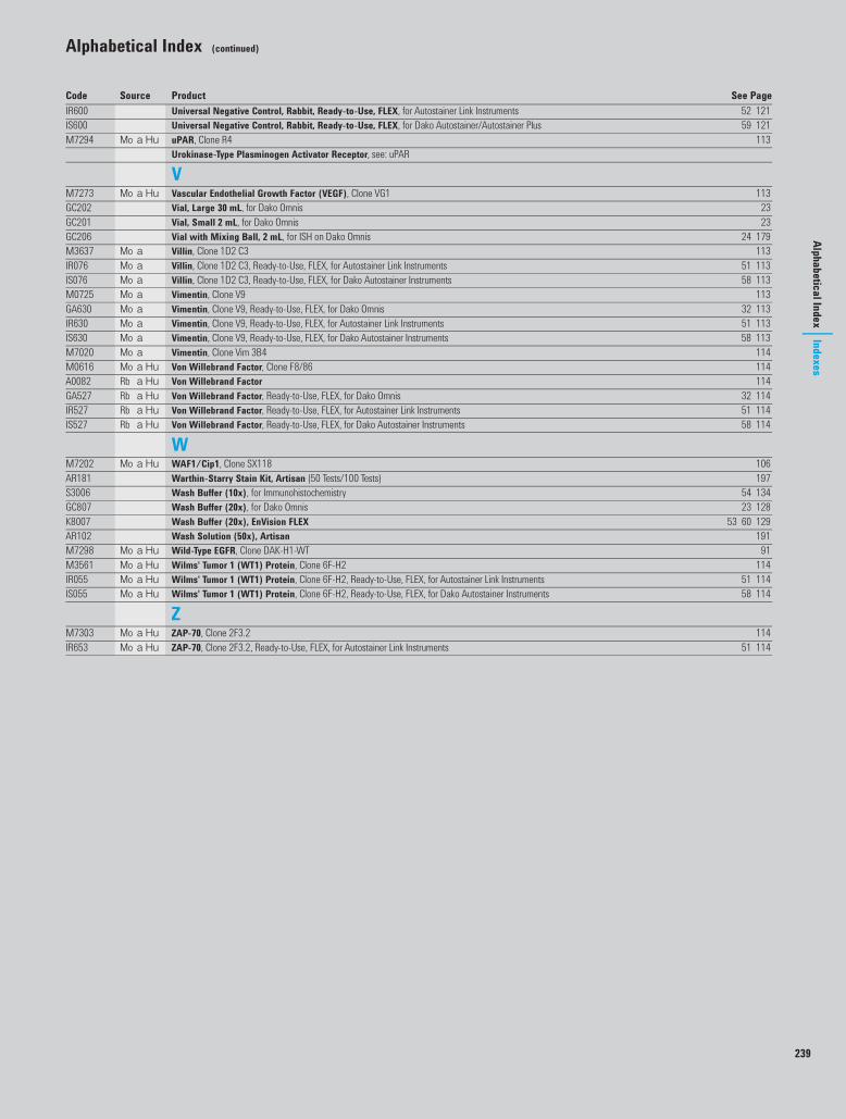
239
Alphabetical Index (continued)
Alphabetical IndexIndexes
IR600 Universal Negative Control, Rabbit, Ready-to-Use, FLEX, for Autostainer Link Instruments 52 121IS600 Universal Negative Control, Rabbit, Ready-to-Use, FLEX, for Dako Autostainer/Autostainer Plus 59 121M7294 Mo a Hu uPAR, Clone R4 113
Urokinase-Type Plasminogen Activator Receptor, see: uPAR
VM7273 Mo a Hu Vascular Endothelial Growth Factor (VEGF), Clone VG1 113GC202 Vial, Large 30 mL, for Dako Omnis 23GC201 Vial, Small 2 mL, for Dako Omnis 23GC206 Vial with Mixing Ball, 2 mL, for ISH on Dako Omnis 24 179M3637 Mo a Villin, Clone 1D2 C3 113IR076 Mo a Villin, Clone 1D2 C3, Ready-to-Use, FLEX, for Autostainer Link Instruments 51 113IS076 Mo a Villin, Clone 1D2 C3, Ready-to-Use, FLEX, for Dako Autostainer Instruments 58 113M0725 Mo a Vimentin, Clone V9 113GA630 Mo a Vimentin, Clone V9, Ready-to-Use, FLEX, for Dako Omnis 32 113IR630 Mo a Vimentin, Clone V9, Ready-to-Use, FLEX, for Autostainer Link Instruments 51 113IS630 Mo a Vimentin, Clone V9, Ready-to-Use, FLEX, for Dako Autostainer Instruments 58 113M7020 Mo a Vimentin, Clone Vim 3B4 114M0616 Mo a Hu Von Willebrand Factor, Clone F8/86 114A0082 Rb a Hu Von Willebrand Factor 114GA527 Rb a Hu Von Willebrand Factor, Ready-to-Use, FLEX, for Dako Omnis 32 114IR527 Rb a Hu Von Willebrand Factor, Ready-to-Use, FLEX, for Autostainer Link Instruments 51 114IS527 Rb a Hu Von Willebrand Factor, Ready-to-Use, FLEX, for Dako Autostainer Instruments 58 114
WM7202 Mo a Hu WAF1/Cip1, Clone SX118 106AR181 Warthin-Starry Stain Kit, Artisan (50 Tests/100 Tests) 197S3006 Wash Buffer (10x), for Immunohistochemistry 54 134GC807 Wash Buffer (20x), for Dako Omnis 23 128K8007 Wash Buffer (20x), EnVision FLEX 53 60 129AR102 Wash Solution (50x), Artisan 191M7298 Mo a Hu Wild-Type EGFR, Clone DAK-H1-WT 91M3561 Mo a Hu Wilms' Tumor 1 (WT1) Protein, Clone 6F-H2 114IR055 Mo a Hu Wilms' Tumor 1 (WT1) Protein, Clone 6F-H2, Ready-to-Use, FLEX, for Autostainer Link Instruments 51 114IS055 Mo a Hu Wilms' Tumor 1 (WT1) Protein, Clone 6F-H2, Ready-to-Use, FLEX, for Dako Autostainer Instruments 58 114
ZM7303 Mo a Hu ZAP-70, Clone 2F3.2 114IR653 Mo a Hu ZAP-70, Clone 2F3.2, Ready-to-Use, FLEX, for Autostainer Link Instruments 51 114
Code Source Product See Page
050360 Alphabetical index US.fm Page 239 Friday, March 11, 2016 9:49 AM

050360 Alphabetical index US.fm Page 240 Friday, March 11, 2016 9:49 AM

241
Synonym List
IndexesSynonym List
Synonym Name Used in Dako Product
3-FL CD15
3-fucosyl-N-acetyllactosamine CD15
5HT serotonin
5-hydroxytryptamine serotonin
14-3-2 protein neuron-specific enolase
40S ribosomal protein S6 ribosomal protein S6-pS240, phosphorylation site specific
55 kDa actin-bundling protein fascin
α-methylacyl-CoA racemase AMACR
A b beta-amyloid
A beta P beta-amyloid
adhesion molecule-1 CD31, endothelial cell
AFP alpha-1-fetoprotein
ALK protein CD246, ALK protein
alpha-methylacyl-coenzyme A racemase
AMACR
amyloid b-peptide beta-amyloid
amyloid beta-protein beta-amyloid
API4 survivin
apoptosis inhibitor 4 survivin
Arc-1 E-cadherin
b-3 integrin CD61, platelet glycoprotein IIIa
b3 integrin chain CD61, platelet glycoprotein IIIa
B23 nucleophosmin
bA4 protein beta-amyloid
baculoviral IAP repeat-containing protein 5
survivin
BIRC5 survivin
BLAST-2 CD23
BSAP B-cell-specific activator protein
C3b receptor CD35
C3bR CD35
C3d-receptor CD21
C4bR CD35
cadherin E-cadherin and N-cadherin
calgranulin myeloid/histiocyte antigen
CALLA CD10
calprotectin myeloid/histiocyte antigen
caudal-type homeobox protein 2 CDX2
CD3 complex CD3
CD44s CD44, phagocytic glycoprotein-1
CD61A CD61, platelet glycoprotein IIIa
CD87 uPAR
CD236R glycophorin C
CEA carcinoembryonic antigen
cell-CAM 120/180 E-cadherin
Synonym Name Used in Dako Product
c-erbB-3 HER3
c-kit CD117, c-kit
CMV cytomegalovirus
COFS4 ERCC1
common acute lymphoblastic leukemia antigen
CD10
complement receptor 1 CD35
complement receptor type 1 CD35
CR1 CD35
CR2 CD21
cyclooxygenase-2 COX-2
cystic fibrosis antigen myeloid/histiocyte antigen
D2-40 podoplanin
diaminobenzidine DAB
EBV Epstein-Barr virus
EBV-receptor CD21
EC 1.14.18.1 tyrosinase
E-CD E-cadherin
ECMRIII CD44, phagocytic glycoprotein-1
EGFR epidermal growth factor receptor
EGFR pY1173 EGFR-pY1197, phosphorylation site specific
EMA epithelial membrane antigen
endoglin CD105, endoglin
endothelial anticoagulant protein thrombomodulin
endothelial cell CD31, endothelial cell
EpCAM epithelial-related antigen
epidermal growth factor receptor EGFR
epidermal growth factor receptor 2 c-erbB-2 oncoprotein, HercepTest, HER2 FISH
epiligrin laminin-5, gamma-2 chain
erbB-1 epidermal growth factor receptor
ERBB2 c-erbB-2 oncoprotein, HercepTest, HER2 FISH
erbB-3 HER3
ets-related gene product ERG
Ewing's sarcoma marker CD99, MIC2 gene product, Ewing's sarcoma marker
excision repair cross-complementation group 1
ERCC1
factor VIII-related antigen von Willebrand factor
FceRII CD23
fetomodulin thrombomodulin
FM thrombomodulin
FOLH prostate-specific membrane antigen
follicular dendritic reticulum cell follicular dendritic cell
FSP1 S100A4
USCatPathology.book Page 241 Friday, March 11, 2016 10:06 AM
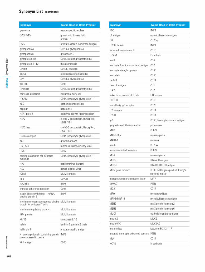
242
Synonym List (continued)
Inde
xes
Syno
nym
Lis
twww.dako.com
g-enolase neuron-specific enolase
GCDFP-15 gross cystic disease fluid protein-15
GCP2 prostate-specific membrane antigen
glycophorin A CD235a, glycophorin A
glycophorin b glycophorin C
glycoprotein IIIa CD61, platelet glycoprotein IIIa
glycoprotein P112 thrombomodulin
GP160 CD105, endoglin
gp200 renal cell carcinoma marker
GPA CD235a, glycophorin A
gpL115 CD43
GPllb/llla CD61, platelet glycoprotein IIIa
hairy cell leukaemia leukaemia, hairy cell
H-CAM CD44, phagocytic glycoprotein-1
hCG chorionic gonadotropin
hep par 1 hepatocyte
HER1 protein epidermal growth factor receptor
HER2 c-erbB-2 oncoprotein, HercepTest, HER2 FISH
HER2/neu c-erbB-2 oncoprotein, HercepTest, HER2 FISH
Hermes antigen CD44, phagocytic glycoprotein-1
hGH growth hormone
HIV, p24 human immunodeficiency virus
HNK-1 CD57
homing-associated cell adhesion molecule
CD44, phagocytic glycoprotein-1
HPV papillomavirus (human)
HSV herpes simplex virus
ICSAT MUM1 protein
Ig-a CD79α
IGF2BP3 IMP3
immune adherence receptor CD35
insulin-like growth factor II mRNA binding protein 3
IMP3
interferon consensus sequence binding protein for activated T cells
MUM1 protein
interferon regulatory factor 4 MUM1 protein
IRF4 protein MUM1 protein
K8/18 cytokeratin 8/18
kalinin laminin-5, gamma-2 chain
kallikrein 3 prostate-specific antigen
K homology domain containing protein overexpressed in cancer
IMP3
Ki-1 antigen CD30
Synonym Name Used in Dako Product
KOC IMP3
L1 antigen myeloid/histiocyte antigen
L26 CD20cy
L523S Protein IMP3
lacto-N-fucopentaose III CD15
L-CAM E-cadherin
leu-3 CD4
leucocyte function-associated antigen CD2
leucocyte sialoglycoprotein CD43
leukosialin CD43
LeuM3 CD14
Lewis X antigen CD15
LFA2 CD2
linker for activation of T cells LAT protein
LNFP III CD15
low affinity IgE receptor CD23
LPS receptor CD14
LPS-R CD14
ly-5 CD45, leucocyte common antigen
lymphatic endothelium marker podoplanin
MAC C5b-9
MAM-1A5 mammaglobin
MART-1 melan-A
mb-1 CD79α
membrane attack complex C5b-9
MGA mammaglobin
MHC-I HLA-ABC antigen
MHC-II HLA-DP, DQ, DR antigen
MIC2 gene product CD99, MIC2 gene product, Ewing's sarcoma marker
microphthalmia transcription factor MITF
MMAC PTEN
MO2 CD14
MPO myeloperoxidase
MRP8/MRP14 myeloid/histiocyte antigen
MSH2 mutS protein homolog 2
MSH6 mutS protein homolog 6
MUC1 epithelial membrane antigen
mucin 2 MUC2
mucin 5AC MUC5AC
muramidase lysozyme EC 3.2.1.17
mutated in multiple advanced cancers PTEN
My4 CD14
NCAD N-cadherin
Synonym Name Used in Dako Product
USCatPathology.book Page 242 Friday, March 11, 2016 10:06 AM

243
Synonym List (continued)Synonym
ListIndexes
neprilysin CD10
nerve cadherin N-cadherin
neu oncoprotein c-erbB-2 oncoprotein, HercepTest, HER2 FISH
neural-type cadherin N-cadherin
neutral endopeptidase 24.11 CD10
nicein laminin-5, gamma-2 chain
NO38 nucleophosmin
NPM nucleophosmin
NSE neuron-specific enolase
OKT4 CD4
oncoprotein, BCL2 BCL2 oncoprotein
OTF 3/4 octamer-binding transcription factor 3/4
p30/32mic2 CD99, MIC2 gene product, Ewing's sarcoma marker
p38 synaptophysin
p55 fascin
p145 CD117, c-kit
p185HER2 c-erbB-2 oncoprotein, HercepTest, HER2 FISH
P501S prostein
P504S AMACR
Pax-5 B-cell-specific activator protein
PCNA proliferating cell nuclear antigen
PECAM-1 CD31, endothelial cell
Pgp-1 CD44, phagocytic glycoprotein-1
phagocytic glycoprotein-1 CD44, phagocytic glycoprotein-1
PIP MUM1 protein
PIP gross cystic disease fluid protein-15
PKB Akt-pS473
platelet/endothelial cell adhesion molecule-1
CD31, endothelial cell
platelet glycoprotein IIIa CD61, platelet glycoprotein IIIa
PMS2 postmeiotic segregation increased 2
POU5F1 octamer-binding transcription factor 3/4
prolactin-inducible protein gross cystic disease fluid protein-15
protein IT cytokeratin 20
protein kinase B Akt-pS473
protein p38 synaptophysin
PSA prostate-specific antigen
PSAP prostatic acid phosphatase
PSM prostate-specific membrane antigen
PSMA prostate-specific membrane antigen
ptyr-1173 EGFR EGFR-pY1197, phosphorylation site specific
Synonym Name Used in Dako Product
PU.1 interaction partner MUM1 protein
RPS6 ribosomal protein S6-pS240, phosphorylation site specific
Rac-α Akt-pS473
RCC renal cell carcinoma marker
S100A8/S100A9 myeloid/histiocyte antigen
sialoglycoprotein alpha CD235a, glycophorin A
sialophorin CD43
sialyl Lea CA 19-9
somatotropin growth hormone
SSEA-1 CD15
stage-specific embryonic antigen-1 CD15
syndecan-1 CD138
syp1 synaptophysin
T1 CD5
T3 CD3
T11 antigen CD2
T200 CD45, leucocyte common antigen
T311 tyrosinase
TCC C5b-9
TdT terminal deoxynucleotidyl transferase
TEP1 PTEN
terminal complement complex C5b-9
Tg thyroglobulin
TGF-b-regulated and epithelial cell- enriched phosphatase
PTEN
TIMP-1 tissue inhibitor of metalloproteinase 1
Tp50 CD2
Tp67 CD5
TPO thyroid peroxidase
transcriptional regulator ERG ERG
tryptase mast cell tryptase
TS thymidylate synthase
TTF-1 thyroid transcription factor
tumor protein 63 p63 protein
urokinase-type plasminogen activator receptor
uPAR
UV20 ERCC1
uvomorulin E-cadherin
vascular permeability factor vascular endothelial growth factor
VEGF vascular endothelial growth factor
VPF vascular endothelial growth factor
X-hapten CD15
Z-associated protein, 70 kDa ZAP-70
Synonym Name Used in Dako Product
USCatPathology.book Page 243 Friday, March 11, 2016 10:06 AM

244
Inde
xes
Antib
ody C
lone
Inde
xwww.dako.com
Antibody Clone IndexThe following list of antibodies has been cataloged alphabetically by clone name.
Clone Antibody Code Page0042 Thyroid-Stimulating Hormone (TSH) M3503 11202A3 Adrenocorticotropin (ACTH) M3501 731A4 Smooth Muscle Actin M0851 731D2 C3 Villin M3637 1131F8 CD21, B Cell M0784 802B11 + PD7/26 CD45, Leucocyte Common Antigen M0701 822F3.2 ZAP-70 M7303 1142F11 Neurofilament Protein M0762 1043E6 Prostate-Specific Membrane Antigen
(PSMA)M3620 109
4B12 CD4 M7310 784C7 CD5 M3641 784C7 Laminin M0638 994F9 ERCC1 M3648 924G1 Laminin-5, Gamma-2 Chain M7262 994KB5 CD45RA M0754 835.8A MyoD1 M3512 1035-D8/1 Enterovirus M7064 915HT-H209 Serotonin M0758 1106F2 Glial Fibrillary Acidic Protein (GFAP) M0761 946F/3D Beta-Amyloid M0872 756F-H2 Wilms' Tumor 1 (WT1) Protein M3561 1146G11 N-Cadherin M3613 1046H2.1 PTEN M3627 1098A9 Placental Alkaline Phosphatase M7191 1078G7G3/1 Thyroid Transcription Factor M3575 11210E3 Prostein M3615 10912C4 Survivin M3624 11012E7 CD99, MIC2 Gene Products, Ewing's
Sarcoma MarkerM3601 84
13H4 AMACR M3616 7414-5 Akt-pS473, Phosphorylation Site
SpecificM3628 73
23A3 Gross Cystic Disease Fluid Protein-15 M3638 9534βE12 Cytokeratin, High MW M0630 9055K-2 Fascin M3567 9356C6 CD10 M7308 7969.1 IMP3 M3626 97101 MCM3 Protein M7263 1001116-NS-19-9 CA 19-9 M3517 76123C3 CD56 M7304 83124 BCL2 Oncoprotein M0887 74151 BCL10 Protein M7260 75304-1A5 Mammaglobin M3625 100318-6-11 p53 Protein M3629 106376 Nucleophosmin M7305 1051009 Thrombomodulin M0617 111A103 Melan-A M7196 100AA1 Mast Cell Tryptase M7052 100AB75 CD2 M7309 78AE1/AE3 Cytokeratin M3515 87
Clone Antibody Code PageALK1 CD246, ALK Protein M7195 86Alpha-Sr-1 Actin (Sarcomeric) M0874 73AR441 Androgen Receptor M3562 74β-Catenin-1 Beta-Catenin M3539 75BBS/NC/VI-H14 Neuron-Specific Enolase (NSE) M0873 104Ber-EP4 Epithelial Antigen M0804 91Ber-H2 CD30 M0751 81Ber-MAC-DRC CD35 M0846 82Bu20a Bromodeoxyuridine M0744 75C8/144B CD8 M7103 79C10 Follicle-Stimulating Hormone (FSH) M3504 93C93 Luteinizing Hormone (LH) M3502 99CALP Calponin M3556 77Carb-3 CD15 M3631 80CBC.37 CD7 M7255 79CCH2 + DDG9 Cytomegalovirus M0854 90CCP58 MUC2 M7313 101CIV 22 Collagen IV M0785 87CLH1 MUC5AC M7316 102CNA.42 Follicular Dendritic Cell M7157 94CR3/43 HLA-DP, DQ, DR Antigen M0775 96CS.1-4 Epstein-Barr Virus, LMP M0897 92CX-294 COX-2 M3617 87D2-40 Podoplanin M3619 107D5 MITF M3621 101D5/16 B4 Cytokeratin 5/6 M7237 88D33 Desmin M0760 90DAK-A3 Chromogranin A M0869 86DAK-Calret 1 Calretinin M7245 77DAK-CD23 CD23 M7312 81DAK-CDX2 CDX2 M3636 86DAK-H1-1197 EGFR-pY1197 M7299 91DAK-H1-WT EGFR, Wild-Type M7298 91DAK-H3-IC HER3 M7292 93DAK-Pax5 B-Cell-Specific Activator Protein M7307 74DAK-S6-240 Ribosomal Protein S6-pS240,
Phosphorylation Site SpecificM7300 110
DAK-SYNAP Synaptophysin M7315 110DAK-Tg6 Thyroglobulin M0781 111DBA.44 Leukaemia, Hairy Cell M0880 99DC 10 Cytokeratin 18 M7010 89DDG9 + CCH2 Cytomegalovirus M0854 90DE-K10 Cytokeratin 10 M7002 88DE-K13 Cytokeratin 10/13 M7003 88DF1485 CD44, Phagocytic Glycoprotein-1 M7082 82DF-T1 CD43 M0786 82DO-7 p53 Protein M7001 106E3 Cytokeratin 17 M7046 89E9 Metallothionein M0639 101E29 Epithelial Membrane Antigen (EMA) M0613 92EBM11 CD68 M0718 84
USCatPathology.book Page 244 Friday, March 11, 2016 10:06 AM
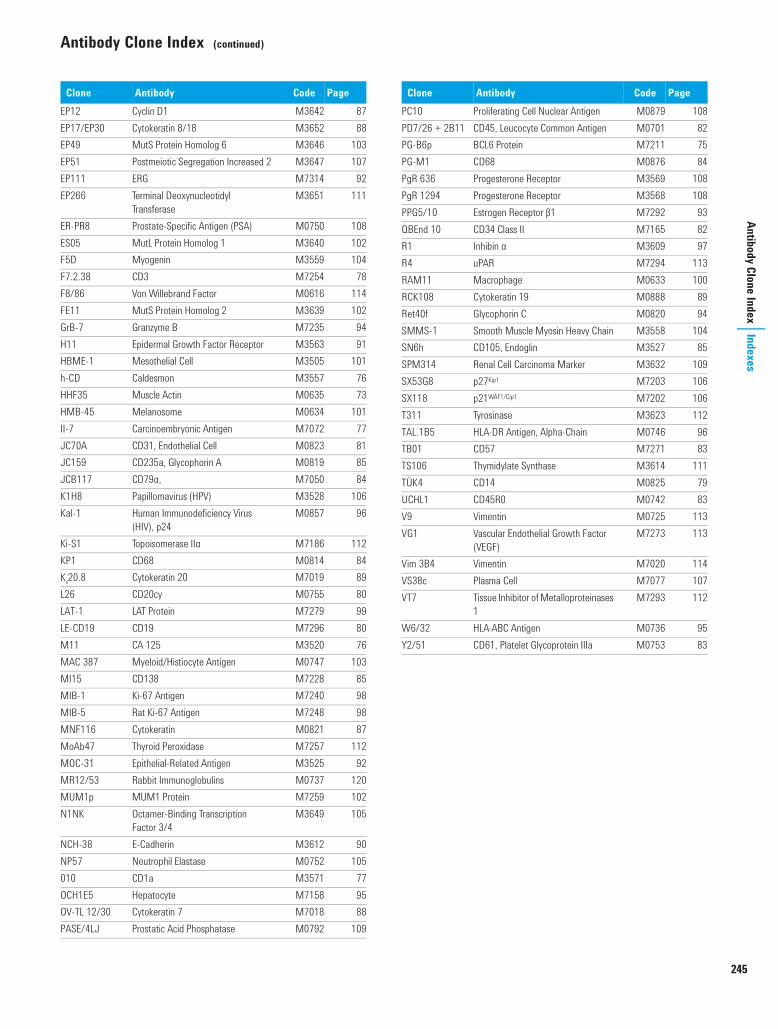
245
Antibody Clone Index (continued)Antibody Clone Index
Indexes
EP12 Cyclin D1 M3642 87EP17/EP30 Cytokeratin 8/18 M3652 88EP49 MutS Protein Homolog 6 M3646 103EP51 Postmeiotic Segregation Increased 2 M3647 107EP111 ERG M7314 92EP266 Terminal Deoxynucleotidyl
TransferaseM3651 111
ER-PR8 Prostate-Specific Antigen (PSA) M0750 108ES05 MutL Protein Homolog 1 M3640 102F5D Myogenin M3559 104F7.2.38 CD3 M7254 78F8/86 Von Willebrand Factor M0616 114FE11 MutS Protein Homolog 2 M3639 102GrB-7 Granzyme B M7235 94H11 Epidermal Growth Factor Receptor M3563 91HBME-1 Mesothelial Cell M3505 101h-CD Caldesmon M3557 76HHF35 Muscle Actin M0635 73HMB-45 Melanosome M0634 101II-7 Carcinoembryonic Antigen M7072 77JC70A CD31, Endothelial Cell M0823 81JC159 CD235a, Glycophorin A M0819 85JCB117 CD79α, M7050 84K1H8 Papillomavirus (HPV) M3528 106Kal-1 Human Immunodeficiency Virus
(HIV), p24M0857 96
Ki-S1 Topoisomerase IIα M7186 112KP1 CD68 M0814 84Ks20.8 Cytokeratin 20 M7019 89L26 CD20cy M0755 80LAT-1 LAT Protein M7279 99LE-CD19 CD19 M7296 80M11 CA 125 M3520 76MAC 387 Myeloid/Histiocyte Antigen M0747 103MI15 CD138 M7228 85MIB-1 Ki-67 Antigen M7240 98MIB-5 Rat Ki-67 Antigen M7248 98MNF116 Cytokeratin M0821 87MoAb47 Thyroid Peroxidase M7257 112MOC-31 Epithelial-Related Antigen M3525 92MR12/53 Rabbit Immunoglobulins M0737 120MUM1p MUM1 Protein M7259 102N1NK Octamer-Binding Transcription
Factor 3/4M3649 105
NCH-38 E-Cadherin M3612 90NP57 Neutrophil Elastase M0752 105010 CD1a M3571 77OCH1E5 Hepatocyte M7158 95OV-TL 12/30 Cytokeratin 7 M7018 88PASE/4LJ Prostatic Acid Phosphatase M0792 109
Clone Antibody Code PagePC10 Proliferating Cell Nuclear Antigen M0879 108PD7/26 + 2B11 CD45, Leucocyte Common Antigen M0701 82PG-B6p BCL6 Protein M7211 75PG-M1 CD68 M0876 84PgR 636 Progesterone Receptor M3569 108PgR 1294 Progesterone Receptor M3568 108PPG5/10 Estrogen Receptor β1 M7292 93QBEnd 10 CD34 Class II M7165 82R1 Inhibin α M3609 97R4 uPAR M7294 113RAM11 Macrophage M0633 100RCK108 Cytokeratin 19 M0888 89Ret40f Glycophorin C M0820 94SMMS-1 Smooth Muscle Myosin Heavy Chain M3558 104SN6h CD105, Endoglin M3527 85SPM314 Renal Cell Carcinoma Marker M3632 109SX53G8 p27Kip1 M7203 106SX118 p21WAF1/Cip1 M7202 106T311 Tyrosinase M3623 112TAL.1B5 HLA-DR Antigen, Alpha-Chain M0746 96TB01 CD57 M7271 83TS106 Thymidylate Synthase M3614 111TÜK4 CD14 M0825 79UCHL1 CD45R0 M0742 83V9 Vimentin M0725 113VG1 Vascular Endothelial Growth Factor
(VEGF)M7273 113
Vim 3B4 Vimentin M7020 114VS38c Plasma Cell M7077 107VT7 Tissue Inhibitor of Metalloproteinases
1M7293 112
W6/32 HLA-ABC Antigen M0736 95Y2/51 CD61, Platelet Glycoprotein IIIa M0753 83
Clone Antibody Code Page
USCatPathology.book Page 245 Friday, March 11, 2016 10:06 AM

246
Inde
xes
Prod
uct C
ode
Syst
emwww.dako.com
Product Code System
To clarify the product coding system, we have included the following brief explanation of the lettering system used with the product code numbers. This is a general guide; there may be some exceptions.
A Polyclonal antibodies to human antigens
AR Artisan Link and reagents
AS Autostainer Link
B Polyclonal antibodies to viral and microbial antigens
C Glycergel Mounting Medium
CR Coverslipper instrument and accessories
CS CoverStainer instrument and accessories
D Alkaline phosphatase (AP)-conjugated products
DL Labeling System
E Biotinylated products
F Fluorescein (FITC)-conjugated antibodies
G SureFISH probes and related reagents
GA FLEX ready-to-use antibodies for Dako Omnis
GC Ancillaries and Accessories for Dako Omnis
GI Dako Omnis
GM IQISH probes and reagents for Dako Omnis
GV Kits for Dako Omnis
IC Antibody Cocktails for Autostainer Link instruments
IR FLEX ready-to-use antibodies for Autostainer Link instruments
IS FLEX ready-to-use antibodies for Dako Autostainer instruments
K Kit systems
M Monoclonal antibodies
P Horseradish peroxidase (HRP)-conjugated products
PT PT Link instrument and accessories
S Equipment and ancillary products
SK Kits and accessories for Autostainer Link instruments
SL Non-immunologic reagent for use with the Dako Autostainer
X Control reagentsNormal animal sera and normal animal immunoglobulin fractions
Y Molecular probes
Z Antibodies to animal antigens
USCatPathology.book Page 246 Friday, March 11, 2016 10:06 AM

247
Product Code IndexIndexes
Product Code Index
Code Product Package Size Order No. Page
AA0008 Polyclonal Rabbit Anti-Human Alpha-1-Fetoprotein 0.2 mL A000829 73 117A0082 Polyclonal Rabbit Anti-Human Von Willebrand Factor 0.2 mL
2 mLA008229A008202
114
A0099 Polyclonal Rabbit Anti-Human Lysozyme EC 3.2.1.17 2 mL A009902 99 118A0191 Polyclonal Rabbit Anti-Human Kappa Light Chains 2 mL A019102 98 117A0193 Polyclonal Rabbit Anti-Human Lambda Light Chains 2 mL A019302 98 118A0231 Polyclonal Rabbit Anti-Human Chorionic Gonadotropin 2 mL A023102 86A0251 Polyclonal Rabbit Anti-Human Thyroglobulin 2 mL A025102 111 118A0262 Polyclonal Rabbit Anti-Human IgA 1 mL A026201 96 117A0398 Polyclonal Rabbit Anti-Human Myeloperoxidase 0.2 mL A039829 103A0423 Polyclonal Rabbit Anti-Human IgG 1 mL A042301 96 117A0425 Polyclonal Rabbit Anti-Human IgM 1 mL A042501 97 117A0452 Polyclonal Rabbit Anti-Human CD3 0.2 mL
1 mLA045229A045201
78
A0485 Polyclonal Rabbit Anti-Human c-erbB-2 Oncoprotein 0.2 mL A048529 86A0562 Polyclonal Rabbit Anti-Human Prostate-Specific Antigen 1 mL A056201 108A0564 Polyclonal Guinea Pig Anti-Insulin 1 mL A056401 97A0566 Polyclonal Rabbit Anti-Human Somatostatin 1 mL A056601 110A0568 Polyclonal Rabbit Anti-Human Gastrin 1 mL A056801 94A0569 Polyclonal Rabbit Anti-Human Prolactin 1 mL A056901 108A0570 Polyclonal Rabbit Anti-Human Growth Hormone 1 mL A057001 95A0576 Polyclonal Rabbit Anti-Human Calcitonin 1 mL A057601 76A0623 Polyclonal Rabbit Anti-Human Myelin Basic Protein 1 mL A062301 103A4502 Polyclonal Rabbit Anti-Human CD117, c-kit 0.2 mL A450229 85A5114 Polyclonal Rabbit Anti-Human S100A4 1 mL A511401 110AR102 Artisan Wash Solution (x 50) 4 x 200 mL AR10211 191AR158 Artisan Iron Stain Kit 50 tests
100 testsAR15892AR15811
196
AR160 Artisan Alcian Blue pH 2.5 Stain Kit 50 tests100 tests
AR16092AR16011
194
AR161 Artisan Congo Red Stain Kit 50 tests100 tests
AR16192AR16111
195
AR162 Artisan Acid-Fast Bacteria (AFB) Stain Kit 50 tests100 tests
AR16292AR16211
194
AR163 Artisan Elastic Stain Kit 50 tests100 tests
AR16392AR16311
195
AR164 Artisan Giemsa Stain Kit 50 tests AR16492 195AR165 Artisan Periodic Acid Schiff (PAS) Stain Kit 50 tests
100 testsAR16592AR16511
197
AR166 Artisan Gomori's Green Trichrome Stain Kit 50 tests AR16692 195AR167 Artisan Gomori's Trichrome Stain Kit 50 tests AR16792 195AR168 Artisan Mucicarmine Stain Kit 50 tests
100 testsAR16892AR16811
196
AR169 Artisan Alcian Blue/PAS Stain Kit 50 tests100 tests
AR16992AR16911
194
AR171 Artisan Alpha-Amylase 50 tests100 tests
AR17192AR17111
191
AR172 Artisan PAS-Green Stain Kit 50 tests100 tests
AR17292AR17211
197
AR173 Artisan Masson's Trichrome Stain Kit 50 tests100 tests
AR17392AR17311
196
AR175 Artisan Gram Stain Kit 50 tests AR17592 196AR176 Artisan Grocott's Methenamine Silver (GMS) Stain Kit 50 tests
100 testsAR17692AR17611
196
AR178 Artisan Alcian Blue/PAS/Hematoxylin Stain Kit 50 tests100 tests
AR17892AR17811
194
AR179 Artisan Reticulin/Nuclear Fast Red Stain Kit 50 tests100 tests
AR17992AR17911
197
USCatPathology.book Page 247 Friday, March 11, 2016 10:06 AM

248
www.
dako
.com
Product Code Index (continued)
Inde
xes
Prod
uct C
ode
Inde
x
AR180 Artisan Jones' Basement Membrane (PAS-M) Stain Kit 100 tests AR18011 196AR181 Artisan Warthin-Starry Stain Kit 50 tests
100 testsAR18192AR18111
197
AR182 Artisan Recticulin/No Counterstain Stain Kit 100 tests AR18211 197AR306 Artisan Gram Yellow Stain Kit 50 tests AR30692 192AR307 Artisan Colloidal Iron Stain Kit 50 tests AR30792 192AR308 Artisan Jenner-Wright Giemsa Stain Kit 50 tests AR30892 192AR309 Artisan Clearing Solution 5 x 100 tests AR30911 191AR310 Artisan Link Pro Special Staining System 1 unit AR31030 189AR313 Artisan Orcein Stain Kit 50 tests AR31392 193AR314 Artisan Maintenance Kit 33 tests AR31411 191AR362 Artisan Acid-Fast Bacteria (AFB) Light Green Stain Kit 50 tests AR36292 192AR376 Artisan Grocott's Methenamine Silver Eosin Stain Kit 50 tests AR37692 193AR380 Artisan Jones' Basement Membrane Light Green (PAS-M) Stain Kit 50 tests AR38092 193AR409 Artisan 14 Pack Reagent Holder 1 unit AR40930 191AR480 Artisan Jones' Basement Membrane H&E (PAS-M) Stain Kit 100 tests AR48011 193AS480 Autostainer Link 48 1 unit AS48030 35
BB0114 Polyclonal Rabbit Anti-Herpes Simplex Virus Type 1 2 mL B011402 95B0116 Polyclonal Rabbit Anti-Herpes Simplex Virus Type 2 2 mL B011602 95B0357 Polyclonal Rabbit Anti-Escherichia Coli 2 mL B035702 117B0471 Polyclonal Rabbit Anti-Helicobacter Pylori 0.2 mL
1 mLB047129B047101
95
B0586 Polyclonal Rabbit Anti-Hepatitis B Virus Core Antigen 1 mL B058601 95
CC0563 Glycergel®, Aqueous Mounting Medium 15 mL C056330 135CR100 Dako Coverslipper 1 unit CR10030 184CR121 Cover Glass, 24 mm x 60 mm 5 x 200 pcs CR12130 185CR122 Cover Glass, 24 mm x 55 mm 5 x 200 pcs CR12230 185CR124 Cover Glass, 24 mm x 40 mm 5 x 200 pcs CR12430 185CS100 Dako CoverStainer 1 unit CS10030 183CS119 Dako CoverStainer Slide Rack 10 racks CS11930 185CS700 Dako Hematoxylin Up to 3000 tests, 1 L CS70030 185CS701 Dako Eosin Up to 3000 tests, 1 L CS70130 185CS702 Dako Bluing Buffer Up to 3000 tests, 1 L CS70230 185CS703 Dako Mounting Medium 473 mL CS70330 185CS704 Dako Cover Glass, 24 x 50 mm 5 x 200 pcs CS70430 185CS705 Dako Toluene-Free Mounting Medium 500 mL CS70530 185
DD0306 Polyclonal Swine Anti-Rabbit Immunoglobulins/AP 1 mL D030601 120D0314 Polyclonal Rabbit Anti-Mouse Immunoglobulins/AP 2 mL D031402 119D0336 Polyclonal Rabbit Anti-Human IgG/AP 1 mL D033601 96 117D0486 Polyclonal Goat Anti-Mouse Immunoglobulins/AP 2 mL D048602 119D0487 Polyclonal Goat Anti-Rabbit Immunoglobulins/AP 1 mL D048701 119DL213 Slide Label Kit, Small Flap 1500 labels DL21330 137DL412 Universal Label Printer (Link) 1 unit DL41230 136
EE0353 Polyclonal Swine Anti-Rabbit Immunoglobulins/Biotinylated 1 mL E035301 120E0354 Polyclonal Rabbit Anti-Mouse Immunoglobulins/Biotinylated 1 mL E035401 119E0413 Polyclonal Rabbit Anti-Mouse Immunoglobulins/Biotinylated, Rabbit F(ab')2 1 mL E041301 119
Code Product Package Size Order No. Page
USCatPathology.book Page 248 Friday, March 11, 2016 10:06 AM

249
Product Code Index (continued)
Product Code IndexIndexes
E0431 Polyclonal Swine Anti-Rabbit Immunoglobulins/Biotinylated, Swine F(ab')2 1 mL E043101 120E0432 Polyclonal Goat Anti-Rabbit Immunoglobulins/Biotinylated 1 mL E043201 119E0433 Polyclonal Goat Anti-Mouse Immunoglobulins/Biotinylated 1 mL E043301 119E0466 Polyclonal Rabbit Anti-Goat Immunoglobulins/Biotinylated 1 mL E046601 119
FF0111 Polyclonal Rabbit Anti-Human Fibrinogen/FITC 2 mL F011102 93 117F0117 Polyclonal Rabbit Anti-Human Albumin/FITC 2 mL F011702 73F0169 Polyclonal Rabbit Anti-Human C4c Complement/FITC 2 mL F016902 75F0198 Polyclonal Rabbit Anti-Human Kappa Light Chains/FITC 2 mL F019802 98 117F0199 Polyclonal Rabbit Anti-Human Lambda Light Chains/FITC 2 mL F019902 98 118F0200 Polyclonal Rabbit Anti-Human IgA, IgG, IgM, Kappa, Lambda/FITC 2 mL F020002 96 117F0201 Polyclonal Rabbit Anti-Human C3c Complement/FITC 2 mL F020102 75F0202 Polyclonal Rabbit Anti-Human IgG/FITC 2 mL F020202 96 117F0203 Polyclonal Rabbit Anti-Human IgM/FITC 2 mL F020302 97 117F0204 Polyclonal Rabbit Anti-Human IgA/FITC 2 mL F020402 96 117F0205 Polyclonal Swine Anti-Rabbit Immunoglobulins/FITC 2 mL F020502 120F0232 Polyclonal Rabbit Anti-Mouse Immunoglobulins/FITC 2 mL F023202 119F0250 Polyclonal Rabbit Anti-Goat Immunoglobulins/FITC 2 mL F025002 119F0254 Polyclonal Rabbit Anti-Human C1q Complement/FITC 2 mL F025402 75F0261 Polyclonal Rabbit Anti-Mouse Immunoglobulins/FITC 2 mL F026102 119F0315 Polyclonal Rabbit Anti-Human IgG/FITC, Rabbit F(ab')2 1 mL F031501 96 117F0316 Polyclonal Rabbit Anti-Human IgA/FITC, Rabbit F(ab')2 1 mL F031601 96
GG111200 SureFISH ALK BA P5 5 µL G111200 175G111201 SureFISH ROS1 BA P5 5 µL G111201 175G111202 SureFISH RET BA P5 5 µL G111202 175G111400 SureFISH ALK BA P20 20 µL G111400 175G111401 SureFISH ROS1 BA P20 20 µL G111401 175G111402 SureFISH RET BA P20 20 µL G111402 175G111900 SureFISH ALK BA P200 200 µL, G111900 175G111901 SureFISH ROS1 BA P200 200 µL G111901 175G111902 SureFISH RET BA P200 200 µL G111902 175G211400 SureFISH ALK BA P20x6 20 µL, 6 vials G211400 175G211401 SureFISH ROS1 BA P20x6 20 µL, 6 vials G211401 175G211402 SureFISH RET BA P20 x 6 20 µL, 6 vials G211402 175G9414A IQFISH Fast Hybridization Buffer 9000 900 µL G9414A 180G9415A IQFISH Fast Hybridization Buffer 200 200 µL G9415A 180G9416A IQFISH Fast Hybridization Buffer 200x6 200 µL, 6 vials G9416A 180GA051 FLEX Monoclonal Mouse Anti-Human Cytokeratin, High Molecular Weight, Clone 34bE12,
Ready-to-Use (Dako Omnis)60 tests, 12 mL8 GA05161 29 90
GA052 FLEX Monoclonal Mouse Anti-Human Melanosome, Clone HMB-45, Ready-to-Use (Dako Omnis)
60 tests, 12 mL8 GA05261 31 101
GA053 FLEX Monoclonal Mouse Anti-Human Cytokeratin, Clone AE1/AE3, Ready-to-Use (Dako Omnis) 60 tests, 12 mL8 GA05361 29 87GA054 FLEX Monoclonal Mouse Anti-Human Caldesmon, Clone h-CD, Ready-to-Use (Dako Omnis) 60 tests, 12 mL8 GA05461 26 76GA059 FLEX Monoclonal Mouse Anti-Human E-Cadherin, Clone NCH-38, Ready-to-Use (Dako Omnis) 60 tests, 12 mL8 GA05961 29 90GA060 FLEX Monoclonal Rabbit Anti-Human AMACR, Clone 13H4, Ready-to-Use (Dako Omnis) 60 tests, 12 mL8 GA06061 26 74GA062 FLEX Monoclonal Mouse Anti-Human CD15, Clone Carb-3, Ready-to-Use (Dako Omnis) 60 tests, 12 mL8 GA06261 27 80GA074 FLEX Monoclonal Mouse Anti-Human Mammaglobin, Clone 304-1A5, Ready-to-Use
(Dako Omnis)60 tests, 12 mL8 GA07461 30 100
GA075 FLEX Monoclonal Mouse Anti-Human Renal Cell Carcinoma Marker, Clone SPM314, Ready-to-Use (Dako Omnis)
60 tests, 12 mL8 GA07561 31 109
GA077 FLEX Monoclonal Mouse Anti-Human Gross Cystic Disease Fluid Protein-15, Clone 23A3, Ready-to-Use (Dako Omnis)
60 tests, 12 mL8 GA07761 30 95
Code Product Package Size Order No. Page
8 Packaged in vials for use with Dako Omnis
USCatPathology.book Page 249 Friday, March 11, 2016 10:06 AM

250
www.
dako
.com
Product Code Index (continued)
Inde
xes
Prod
uct C
ode
Inde
x
GA080 FLEX Monoclonal Mouse Anti-Human CDX2, Clone DAK-CDX2, Ready-to-Use (Dako Omnis) 60 tests, 12 mL8 GA08061 28 86GA083 FLEX Monoclonal Rabbit Anti-Human Cyclin D1, Clone EP12, Ready-to-Use (Dako Omnis) 60 tests, 12 mL8 GA08361 28 87GA500 FLEX Polyclonal Rabbit Anti-Human Alpha-1-Fetoprotein, Ready-to-Use (Dako Omnis) 60 tests, 12 mL8 GA50061 26 73GA503 FLEX Polyclonal Rabbit Anti-Human CD3, Ready-to-Use (Dako Omnis) 60 tests, 12 mL8 GA50361 27 78GA504 FLEX Polyclonal Rabbit Anti-S100, Ready-to-Use (Dako Omnis) 60 tests, 12 mL8 GA50461 31 110GA506 FLEX Polyclonal Rabbit Anti-Human Kappa Light Chains, Ready-to-Use (Dako Omnis) 60 tests, 12 mL8 GA50661 30 98GA507 FLEX Polyclonal Rabbit Anti-Human Lambda Light Chains, Ready-to-Use (Dako Omnis) 60 tests, 12 mL8 GA50761 30 98GA508 FLEX Polyclonal Rabbit Anti-Human Chorionic Gonadotropin, Ready-to-Use (Dako Omnis) 60 tests, 12 mL8 GA50861 28 86GA509 FLEX Polyclonal Rabbit Anti-Human Thyroglobulin, Ready-to-Use (Dako Omnis) 60 tests, 12 mL8 GA50961 31 111GA510 FLEX Polyclonal Rabbit Anti-Human IgA, Ready-to-Use (Dako Omnis) 60 tests, 12 mL8 GA51061 30 96GA511 FLEX Polyclonal Rabbit Anti-Human Myeloperoxidase, Ready-to-Use (Dako Omnis) 60 tests, 12 mL8 GA51161 31 103GA514 FLEX Polyclonal Rabbit Anti-Human Prostate-Specific Antigen, Ready-to-Use (Dako Omnis) 60 tests, 12 mL8 GA51461 31 108GA515 FLEX Polyclonal Rabbit Anti-Human Calcitonin, Ready-to-Use (Dako Omnis) 60 tests, 12 mL8 GA51561 26 76GA519 FLEX Polyclonal Rabbit Anti-Human Gastrin, Ready-to-Use (Dako Omnis) 60 tests, 12 mL8 GA51961 30 94GA524 FLEX Polyclonal Rabbit Anti-Glial Fibrillary Acidic Protein, Ready-to-Use (Dako Omnis) 60 tests, 12 mL8 GA52461 30 94GA526 FLEX Polyclonal Rabbit Anti-Human Carcinoembryonic Antigen, Ready-to-Use (Dako Omnis) 60 tests, 12 mL8 GA52661 26 77GA527 FLEX Polyclonal Rabbit Anti-Human Von Willebrand Factor, Ready-to-Use (Dako Omnis) 60 tests, 12 mL8 GA52761 32 114GA600 FLEX Universal Negative Control, Rabbit, Ready-to-Use (Dako Omnis) 120 tests, 24 mL8 GA60066 32 121GA604 FLEX Monoclonal Mouse Anti-Human CD20cy, Clone L26, Ready-to-Use (Dako Omnis) 60 tests, 12 mL8 GA60461 27 80GA607 FLEX Monoclonal Mouse Anti-Human Neurofilament Protein, Clone 2F11, Ready-to-Use
(Dako Omnis)60 tests, 12 mL8 GA60761 31 104
GA609 FLEX Monoclonal Mouse Anti-Human CD68, Clone KP1, Ready-to-Use (Dako Omnis) 60 tests, 12 mL8 GA60961 28 84GA610 FLEX Monoclonal Mouse Anti-Human CD31, Endothelial Cell, Clone JC70A, Ready-to-Use
(Dako Omnis)60 tests, 12 mL8 GA61061 27 81
GA613 FLEX Monoclonal Mouse Anti-Human CD68, Clone PG-M1, Ready-to-Use (Dako Omnis) 60 tests, 12 mL8 GA61361 28 84GA615 FLEX Monoclonal Mouse Anti-Human Cytokeratin 19, Clone RCK108, Ready-to-Use
(Dako Omnis)60 tests, 12 mL8 GA61561 29 89
GA616 FLEX Monoclonal Mouse Anti-Human p53 Protein, Clone DO-7, Ready-to-Use (Dako Omnis) 60 tests, 12 mL8 GA61661 31 106GA618 FLEX Monoclonal Mouse Anti-Human Cytokeratin 18, Clone DC 10, Ready-to-Use
(Dako Omnis)60 tests, 12 mL8 GA61861 29 89
GA619 FLEX Monoclonal Mouse Anti-Human Cytokeratin 7, Clone OV-TL 12/30, Ready-to-Use (Dako Omnis)
60 tests, 12 mL8 GA61961 29 88
GA621 FLEX Monoclonal Mouse Anti-Human CD79a, Clone JCB117, Ready-to-Use (Dako Omnis) 60 tests, 12 mL8 GA62161 28 84GA622 FLEX Monoclonal Mouse Anti-Human Carcinoembryonic Antigen, Clone II-7, Ready-to-Use
(Dako Omnis)60 tests, 12 mL8 GA62261 26 77
GA623 FLEX Monoclonal Mouse Anti-Human CD8, Clone C8/144B, Ready-to-Use (Dako Omnis) 60 tests, 12 mL8 GA62361 27 79GA624 FLEX Monoclonal Mouse Anti-Human Hepatocyte, Clone OCH1E5, Ready-to-Use (Dako Omnis) 60 tests, 12 mL8 GA62461 30 95GA625 FLEX Monoclonal Mouse Anti-Human BCL6 Protein, Clone PG-B6p, Ready-to-Use
(Dako Omnis)60 tests, 12 mL8 GA62561 26 75
GA626 FLEX Monoclonal Mouse Anti-Human Ki-67 Antigen, Clone MIB-1, Ready-to-Use (Dako Omnis) 60 tests, 12 mL8 GA62661 30 98GA630 FLEX Monoclonal Mouse Anti-Vimentin, Clone V9, Ready-to-Use (Dako Omnis) 60 tests, 12 mL8 GA63061 32 113GA632 FLEX Monoclonal Mouse Anti-Human CD34 Class II, Clone QBEnd 10, Ready-to-Use
(Dako Omnis)60 tests, 12 mL8 GA63261 27 82
GA636 FLEX Monoclonal Mouse Anti-Human CD43, Clone DF-T1, Ready-to-Use (Dako Omnis) 60 tests, 12 mL8 GA63661 28 82GA637 FLEX Monoclonal Mouse Anti-Human Epithelial Antigen, Clone Ber-EP4, Ready-to-Use
(Dako Omnis)60 tests, 12 mL8 GA63761 29 91
GA641 FLEX Monoclonal Mouse Anti-Human CD246, ALK Protein, Clone ALK1, Ready-to-Use (Dako Omnis)
60 tests, 12 mL8 GA64161 28 86
GA642 FLEX Monoclonal Mouse Anti-Human CD138, Clone MI15, Ready-to-Use (Dako Omnis) 60 tests, 12 mL8 GA64261 28 85GA643 FLEX Monoclonal Mouse Anti-Human CD7, Clone CBC.37, Ready-to-Use (Dako Omnis) 60 tests, 12 mL8 GA64361 27 79GA644 FLEX Monoclonal Mouse Anti-Human MUM1 Protein, Clone MUM1p, Ready-to-Use
(Dako Omnis)60 tests, 12 mL8 GA64461 31 102
GA648 FLEX Monoclonal Mouse Anti-Human CD10, Clone 56C6, Ready-to-Use (Dako Omnis) 60 tests, 12 mL8 GA64861 27 79GA650 FLEX Monoclonal Mouse Anti-Human B-Cell-Specific Activator Protein, Clone DAK-Pax5,
Ready-to-Use (Dako Omnis)60 tests, 12 mL8 GA65061 26 74
GA651 FLEX Monoclonal Mouse Anti-Human CD2, Clone AB75, Ready-to-Use (Dako Omnis) 60 tests, 12 mL8 GA65161 27 78GA652 FLEX Monoclonal Mouse Anti-Human Nucleophosmin, Clone 376, Ready-to-Use (Dako Omnis) 60 tests, 12 mL8 GA65261 31 105GA659 FLEX Monoclonal Rabbit Anti-Human ERG, Clone EP111, Ready-to-Use (Dako Omnis) 60 tests, 12 mL8 GA65961 29 92
Code Product Package Size Order No. Page
8 Packaged in vials for use with Dako Omnis
USCatPathology.book Page 250 Friday, March 11, 2016 10:06 AM

251
Product Code Index (continued)
Product Code IndexIndexes
GA701 FLEX Monoclonal Mouse Anti-Human CA 125, Clone M11, Ready-to-Use (Dako Omnis) 60 tests, 12 mL8 GA70161 26 76GA702 FLEX Monoclonal Mouse Anti-Human Beta-Catenin, Clone b-Catenin-1, Ready-to-Use
(Dako Omnis)60 tests, 12 mL8 GA70261 26 75
GA750 FLEX Universal Negative Control, Mouse, Ready-to-Use (Dako Omnis) 120 tests, 24 mL8 GA75066 32 121GA751 FLEX Monoclonal Mouse Anti-Human CD45, Leucocyte Common Antigen,
Clones 2B11 + PD7/26, Ready-to-Use (Dako Omnis)60 tests, 12 mL8 GA75161 28 82
GA777 FLEX Monoclonal Mouse Anti-Human Cytokeratin 20, Clone Ks20.8, Ready-to-Use (Dako Omnis)
60 tests, 12 mL8 GA77761 29 89
GA780 FLEX Monoclonal Mouse Anti-Human Cytokeratin 5/6, Clone D5/16 B4, Ready-to-Use (Dako Omnis)
60 tests, 12 mL8 GA78061 29 88
GA781 FLEX Monoclonal Mouse Anti-Human CD23, Clone DAK-CD23, Ready-to-Use (Dako Omnis) 60 tests, 12 mL8 GA78161 27 81GC101 Dako Omnis Slide Rack 6 racks GC10130 23GC102 Dako Omnis ISH Lid 5 lids GC10230 24 179GC103 Dako Omnis Slide Rack Color Clip, Red 25 clips GC10330 23GC104 Dako Omnis Slide Rack Color Clip, Blue 25 clips GC10430 23GC105 Dako Omnis Slide Rack Color Clip, Green 25 clips GC10530 23GC106 Dako Omnis Slide Rack Color Clip, Gray 25 clips GC10630 23GC107 Dako Omnis Mixing Strip 25 strips GC10730 23GC116 Dako Omnis Mixing Device 1 unit GC11630 24 179GC201 Dako Omnis Small Vial, 2 mL 25 x 2 mL GC20130 23GC202 Dako Omnis Large Vial, 30 mL 25 x 30 mL GC20230 23GC203 Sulfuric Acid, 0.3 M 10 x 22.5 mL GC20330 23GC206 Dako Omnis Vial with Mixing Ball, 2 mL 2 mL GC20630 24 179GC207 ISH Cleaning Solution (Dako Omnis) 100 tests, 10 mL GC20730 24 179GC807 Wash Buffer (20x) (Dako Omnis) 20 x 175 mL, 1700 tests GC80711 23 128GC808 Hematoxylin (Dako Omnis) 8 x 22.5 mL, 600 tests GC80811 23 128GC810 ClearifyTM 3.8 L GC81030 23GI100 Dako Omnis 1 unit GI10030 21GM300 ISH Ethanol Solution, 96% (Dako Omnis) 20 tests, 14 mL GM30011 24 179GM301 ISH Pre-Treatment Solution (20x) (Dako Omnis) 175 mL GM30111 24 179GM302 ISH Pepsin (Dako Omnis) 20 tests, 7 mL GM30211 24 179GM303 ISH Stringent Wash Buffer (20x) (Dako Omnis) 175 mL GM30311 24 179GM304 Fluorescence Mounting Medium (Dako Omnis) 20 tests, 0.8 mL GM30411 24 179GV800 EnVision FLEX, High pH (Dako Omnis) 600 tests GV80011 33 127GV804 EnVision FLEX Target Retrieval Solution, High pH (50x) (Dako Omnis) 3 x 68 mL, 225 tests GV80411 33 128GV805 EnVision FLEX Target Retrieval Solution, Low pH (50x) (Dako Omnis) 3 x 68 mL, 225 tests GV80511 33 128GV809 EnVision FLEX+ Rabbit LINKER (Dako Omnis) 22.5 mL, 75 tests GV80911 33 128GV821 EnVision FLEX+ Mouse LINKER (Dako Omnis) 22.5 mL, 75 tests GV82111 33 128GV823 EnVision FLEX Mini Kit, High pH (Dako Omnis) 150 tests GV82311 33 127GV825 EnVision FLEX DAB+ Substrate Chromogen System (Dako Omnis) 150 tests GV82511 23 128
IIC001 DuoFLEX Cocktail, Anti-S100, Anti-Tyrosinase, Anti-Melan-A, Ready-to-Use (Link) 6 mL IC00106 116IC002 DuoFLEX Cocktail, Anti-CD3, Anti-CD20cy, Ready-to-Use (Link) 6 mL IC00206 115IC004 DuoFLEX Cocktail, Anti-AMACR, Anti-Cytokeratin HMW, Anti-Cytokeratin 5/6, Ready-to-Use
(Link)6 mL IC00406 115
IR002 FLEX Polyclonal Guinea Pig Anti-Insulin, Ready-to-Use (Link) 60 tests, 12 mL2 IR00261 47 97IR051 FLEX Monoclonal Mouse Anti-Human Cytokeratin, High Molecular Weight, Clone 34bE12,
Ready-to-Use (Link)60 tests, 12 mL2 IR05161 46 90
IR052 FLEX Monoclonal Mouse Anti-Human Melanosome, Clone HMB-45, Ready-to-Use (Link) 60 tests, 12 mL2 IR05261 48 101IR053 FLEX Monoclonal Mouse Anti-Human Cytokeratin, Clone AE1/AE3, Ready-to-Use (Link) 60 tests, 12 mL2 IR05361 45 87IR054 FLEX Monoclonal Mouse Anti-Human Caldesmon, Clone h-CD, Ready-to-Use (Link) 60 tests, 12 mL2 IR05461 42 76IR055 FLEX Monoclonal Mouse Anti-Human Wilms' Tumor 1 (WT1) Protein, Clone 6F-H2,
Ready-to-Use (Link)60 tests, 12 mL2 IR05561 51 114
IR056 FLEX Monoclonal Mouse Anti-Thyroid Transcription Factor, Clone 8G7G3/1, Ready-to-Use (Link) 60 tests, 12 mL2 IR05661 51 112
Code Product Package Size Order No. Page
6 Packaged in vials for use with Autostainer Link 482 Packaged in vials for use with Autostainer Link instruments
USCatPathology.book Page 251 Friday, March 11, 2016 10:06 AM

252
www.
dako
.com
Product Code Index (continued)
Inde
xes
Prod
uct C
ode
Inde
x
IR057 FLEX Monoclonal Mouse Anti-Human CD99, MIC2 Gene Products, Ewing's Sarcoma Marker, Clone 12E7, Ready-to-Use (Link)
60 tests, 12 mL2 IR05761 44 84
IR058 FLEX Monoclonal Mouse Anti-Human Inhibin a, Clone R1, Ready-to-Use (Link) 60 tests, 12 mL2 IR05861 47 97IR059 FLEX Monoclonal Mouse Anti-Human E-Cadherin, Clone NCH-38, Ready-to-Use (Link) 60 tests, 12 mL2 IR05961 46 90IR060 FLEX Monoclonal Rabbit Anti-Human AMACR, Clone 13H4, Ready-to-Use (Link) 60 tests, 12 mL2 IR06061 41 74IR061 FLEX Monoclonal Mouse Anti-Human Tyrosinase, Clone T311, Ready-to-Use (Link) 60 tests, 12 mL2 IR06161 51 112IR062 FLEX Monoclonal Mouse Anti-Human CD15, Clone Carb-3, Ready-to-Use (Link) 60 tests, 12 mL2 IR06261 43 80IR066 FLEX Monoclonal Mouse Anti-Human Smooth Muscle Myosin Heavy Chain, Clone SMMS-1,
Ready-to-Use (Link)60 tests, 12 mL2 IR06661 50 104
IR067 FLEX Monoclonal Mouse Anti-Myogenin, Clone F5D, Ready-to-Use (Link) 60 tests, 12 mL2 IR06761 49 104IR068 FLEX Monoclonal Mouse Anti-Human Progesterone Receptor, Clone PgR 636, Ready-to-Use
(Link)60 tests, 12 mL2 IR06861 50 108
IR069 FLEX Monoclonal Mouse Anti-Human CD1a, Clone 010, Ready-to-Use (Link) 60 tests, 12 mL2 IR06961 42 77IR072 FLEX Monoclonal Mouse Anti-Human Podoplanin, Clone D2-40, Ready-to-Use (Link) 60 tests, 12 mL2 IR07261 50 107IR074 FLEX Monoclonal Mouse Anti-Human Mammaglobin, Clone 304-1A5, Ready-to-Use (Link) 60 tests, 12 mL2 IR07461 48 100IR075 FLEX Monoclonal Mouse Anti-Human Renal Cell Carcinoma Marker, Clone SPM314,
Ready-to-Use (Link)60 tests, 12 mL2 IR07561 50 109
IR076 FLEX Monoclonal Mouse Anti-Villin, Clone 1D2 C3, Ready-to-Use (Link) 60 tests, 12 mL2 IR07661 51 113IR077 FLEX Monoclonal Mouse Anti-Human Gross Cystic Disease Fluid Protein-15, Clone 23A3,
Ready-to-Use (Link)60 tests, 12 mL2 IR07761 47 95
IR079 FLEX Monoclonal Mouse Anti-Human MutL Protein Homolog 1, Clone ES05, Ready-to-Use (Link)
60 tests, 12 mL2 IR07961 48 102
IR080 FLEX Monoclonal Mouse Anti-Human CDX2, Clone DAK-CDX2, Ready-to-Use (Link) 60 tests, 12 mL2 IR08061 45 86IR082 FLEX Monoclonal Mouse Anti-Human CD5, Clone 4C7, Ready-to-Use (Link) 60 tests, 12 mL2 IR08261 42 78IR083 FLEX Monoclonal Rabbit Anti-Human Cyclin D1, Clone EP12, Ready-to-Use (Link) 60 tests, 12 mL2 IR08361 45 87IR084 FLEX Monoclonal Rabbit Anti-Human Estrogen Receptor a, Clone EP1, Ready-to-Use (Link) 60 tests, 12 mL2 IR08461 46 93IR085 FLEX Monoclonal Mouse Anti-Human MutS Protein Homolog 2, Clone FE11, Ready-to-Use
(Link)60 tests, 12 mL2 IR08561 49 102
IR086 FLEX Monoclonal Rabbit Anti-Human MutS Protein Homolog 6, Clone EP49, Ready-to-Use (Link)
60 tests, 12 mL2 IR08661 49 103
IR087 FLEX Monoclonal Rabbit Anti-Human Postmeiotic Segregration Increased 2, Clone EP51, Ready-to-Use (Link)
60 tests, 12 mL2 IR08761 50 107
IR088 FLEX Monoclonal Mouse Anti-Human Prostein, Clone 10E3 (Link) 60 tests, 12 mL2 IR08861 50 109IR089 FLEX Monoclonal Mouse Anti-Human Prostate-Specific Membrane Antigen, Clone 3E6 (Link) 60 tests, 12 mL2 IR08961 50 109IR091 FLEX Monoclonal Mouse Anti-Human ERCC1, Clone 4F9, Ready-to-Use (Link) 60 tests, 12 mL2 IR09161 46 92IR092 FLEX Monoclonal Mouse Anti-Human Octamer-Binding Transcription Factor 3/4, Clone N1NK,
Ready-to-Use (Link)60 tests, 12 mL2 IR09261 49 105
IR093 FLEX Monoclonal Rabbit Anti-Human Terminal Deoxynucleotidyl Transferase (TdT), Clone EP266, Ready-to-Use (Link)
60 tests, 12 mL2 IR09361 51 111
IR094 FLEX Monoclonal Rabbit Anti-Human Cytokeratin 8/18, Clone EP17/EP30, Ready-to-Use (Link) 60 tests, 12 mL2 IR09461 45 88IR500 FLEX Polyclonal Rabbit Anti-Human Alpha-1-Fetoprotein, Ready-to-Use (Link) 60 tests, 12 mL2 IR50061 41 73IR503 FLEX Polyclonal Rabbit Anti-Human CD3, Ready-to-Use (Link) 60 tests, 12 mL2 IR50361 42 78IR504 FLEX Polyclonal Rabbit Anti-S100, Ready-to-Use (Link) 60 tests, 12 mL2 IR50461 50 110IR506 FLEX Polyclonal Rabbit Anti-Human Kappa Light Chains, Ready-to-Use (Link) 60 tests, 12 mL2 IR50661 47 98IR507 FLEX Polyclonal Rabbit Anti-Human Lambda Light Chains, Ready-to-Use (Link) 60 tests, 12 mL2 IR50761 48 98IR508 FLEX Polyclonal Rabbit Anti-Human Chorionic Gonadotropin, Ready-to-Use (Link) 60 tests, 12 mL2 IR50861 45 86IR509 FLEX Polyclonal Rabbit Anti-Human Thyroglobulin, Ready-to-Use (Link) 60 tests, 12 mL2 IR50961 51 111IR510 FLEX Polyclonal Rabbit Anti-Human IgA, Ready-to-Use (Link) 60 tests, 12 mL2 IR51061 47 96IR511 FLEX Polyclonal Rabbit Anti-Human Myeloperoxidase, Ready-to-Use (Link) 60 tests, 12 mL2 IR51161 49 103IR512 FLEX Polyclonal Rabbit Anti-Human IgG, Ready-to-Use (Link) 60 tests, 12 mL2 IR51261 47 96IR513 FLEX Polyclonal Rabbit Anti-Human IgM, Ready-to-Use (Link) 60 tests, 12 mL2 IR51361 47 97IR514 FLEX Polyclonal Rabbit Anti-Human Prostate-Specific Antigen, Ready-to-Use (Link) 60 tests, 12 mL2 IR51461 50 108IR515 FLEX Polyclonal Rabbit Anti-Human Calcitonin, Ready-to-Use (Link) 60 tests, 12 mL2 IR51561 41 76IR517 FLEX Polyclonal Rabbit Anti-Human IgD, Ready-to-Use (Link) 60 tests, 12 mL2 IR51761 47 96IR519 FLEX Polyclonal Rabbit Anti-Human Gastrin, Ready-to-Use (Link) 60 tests, 12 mL2 IR51961 46 94IR524 FLEX Polyclonal Rabbit Anti-Glial Fibrillary Acidic Protein, Ready-to-Use (Link) 60 tests, 12 mL2 IR52461 47 94IR526 FLEX Polyclonal Rabbit Anti-Human Carcinoembryonic Antigen, Ready-to-Use (Link) 60 tests, 12 mL2 IR52661 42 77
Code Product Package Size Order No. Page
2 Packaged in vials for use with Autostainer Link instruments
USCatPathology.book Page 252 Friday, March 11, 2016 10:06 AM

253
Product Code Index (continued)
Product Code IndexIndexes
IR527 FLEX Polyclonal Rabbit Anti-Human Von Willebrand Factor, Ready-to-Use (Link) 60 tests, 12 mL2 IR52761 51 114IR600 FLEX Universal Negative Control, Rabbit, Ready-to-Use (Link) 120 tests, 24 mL2 IR60066 52 121IR602 FLEX Monoclonal Mouse Anti-Human CD30, Clone Ber-H2, Ready-to-Use (Link) 60 tests, 12 mL2 IR60261 43 81IR604 FLEX Monoclonal Mouse Anti-Human CD20cy, Clone L26, Ready-to-Use (Link) 60 tests, 12 mL2 IR60461 43 80IR606 FLEX Monoclonal Mouse Anti-Human Desmin, Clone D33, Ready-to-Use (Link) 60 tests, 12 mL2 IR60661 46 90IR607 FLEX Monoclonal Mouse Anti-Human Neurofilament Protein, Clone 2F11, Ready-to-Use (Link) 60 tests, 12 mL2 IR60761 49 104IR608 FLEX Monoclonal Mouse Anti-Human CD21, Clone 1F8, Ready-to-Use (Link) 60 tests, 12 mL2 IR60861 43 80IR609 FLEX Monoclonal Mouse Anti-Human CD68, Clone KP1, Ready-to-Use (Link) 60 tests, 12 mL2 IR60961 44 84IR610 FLEX Monoclonal Mouse Anti-Human CD31, Endothelial Cell, Clone JC70A, Ready-to-Use
(Link)60 tests, 12 mL2 IR61061 43 81
IR611 FLEX Monoclonal Mouse Anti-Human Smooth Muscle Actin, Clone 1A4, Ready-to-Use (Link) 60 tests, 12 mL2 IR61161 41 73IR612 FLEX Monoclonal Mouse Anti-Human Neuron-Specific Enolase, Clone BBS/NC/VI-H14,
Ready-to-Use (Link)60 tests, 12 mL2 IR61261 49 104
IR613 FLEX Monoclonal Mouse Anti-Human CD68, Clone PG-M1, Ready-to-Use (Link) 60 tests, 12 mL2 IR61361 44 84IR614 FLEX Monoclonal Mouse Anti-Human BCL2 Oncoprotein, Clone 124, Ready-to-Use (Link) 60 tests, 12 mL2 IR61461 41 74IR615 FLEX Monoclonal Mouse Anti-Human Cytokeratin 19, Clone RCK108, Ready-to-Use (Link) 60 tests, 12 mL2 IR61561 45 89IR616 FLEX Monoclonal Mouse Anti-Human p53 Protein, Clone DO-7, Ready-to-Use (Link) 60 tests, 12 mL2 IR61661 49 106IR618 FLEX Monoclonal Mouse Anti-Human Cytokeratin 18, Clone DC 10, Ready-to-Use (Link) 60 tests, 12 mL2 IR61861 45 89IR619 FLEX Monoclonal Mouse Anti-Human Cytokeratin 7, Clone OV-TL 12/30, Ready-to-Use (Link) 60 tests, 12 mL2 IR61961 45 88IR620 FLEX Monoclonal Mouse Anti-Cytokeratin 17, Clone E3, Ready-to-Use (Link) 60 tests, 12 mL2 IR62061 45 89IR621 FLEX Monoclonal Mouse Anti-Human CD79a, Clone JCB117, Ready-to-Use (Link) 60 tests, 12 mL2 IR62161 44 84IR622 FLEX Monoclonal Mouse Anti-Human Carcinoembryonic Antigen, Clone II-7, Ready-to-Use
(Link)60 tests, 12 mL2 IR62261 42 77
IR623 FLEX Monoclonal Mouse Anti-Human CD8, Clone C8/144B, Ready-to-Use (Link) 60 tests, 12 mL2 IR62361 43 79IR624 FLEX Monoclonal Mouse Anti-Human Hepatocyte, Clone OCH1E5, Ready-to-Use (Link) 60 tests, 12 mL2 IR62461 47 95IR625 FLEX Monoclonal Mouse Anti-Human BCL6 Protein, Clone PG-B6p, Ready-to-Use (Link) 60 tests, 12 mL2 IR62561 41 75IR626 FLEX Monoclonal Mouse Anti-Human Ki-67 Antigen, Clone MIB-1, Ready-to-Use (Link) 60 tests, 12 mL2 IR62661 48 98IR627 FLEX Monoclonal Mouse Anti-Human Calretinin, Clone DAK-Calret 1, Ready-to-Use (Link) 60 tests, 12 mL2 IR62761 42 77IR628 FLEX Monoclonal Mouse Anti-Human CD56, Clone 123C3, Ready-to-Use (Link) 60 tests, 12 mL2 IR62861 44 83IR629 FLEX Monoclonal Mouse Anti-Human Epithelial Membrane Antigen, Clone E29, Ready-to-Use
(Link)60 tests, 12 mL2 IR62961 46 92
IR630 FLEX Monoclonal Mouse Anti-Vimentin, Clone V9, Ready-to-Use (Link) 60 tests, 12 mL2 IR63061 51 113IR632 FLEX Monoclonal Mouse Anti-Human CD34 Class II, Clone QBEnd 10, Ready-to-Use (Link) 60 tests, 12 mL2 IR63261 43 82IR633 FLEX Monoclonal Mouse Anti-Human Melan-A, Clone A103, Ready-to-Use (Link) 60 tests, 12 mL2 IR63361 48 100IR636 FLEX Monoclonal Mouse Anti-Human CD43, Clone DF-T1, Ready-to-Use (Link) 60 tests, 12 mL2 IR63661 44 82IR637 FLEX Monoclonal Mouse Anti-Human Epithelial Antigen, Clone Ber-EP4, Ready-to-Use (Link) 60 tests, 12 mL2 IR63761 46 91IR640 FLEX Monoclonal Mouse Anti-Human Mast Cell Tryptase, Clone AA1, Ready-to-Use (Link) 60 tests, 12 mL2 IR64061 48 100IR641 FLEX Monoclonal Mouse Anti-Human CD246, ALK Protein, Clone ALK1, Ready-to-Use (Link) 60 tests, 12 mL2 IR64161 44 86IR642 FLEX Monoclonal Mouse Anti-Human CD138, Clone MI15, Ready-to-Use (Link) 60 tests, 12 mL2 IR64261 44 85IR643 FLEX Monoclonal Mouse Anti-Human CD7, Clone CBC.37, Ready-to-Use (Link) 60 tests, 12 mL2 IR64361 42 79IR644 FLEX Monoclonal Mouse Anti-Human MUM1 Protein, Clone MUM1p, Ready-to-Use (Link) 60 tests, 12 mL2 IR64461 48 102IR647 FLEX Monoclonal Mouse Anti-Human CD57, Clone TB01, Ready-to-Use (Link) 60 tests, 12 mL2 IR64761 44 83IR648 FLEX Monoclonal Mouse Anti-Human CD10, Clone 56C6, Ready-to-Use (Link) 60 tests, 12 mL2 IR64861 43 79IR649 FLEX Monoclonal Mouse Anti-Human CD4, Clone 4B12, Ready-to-Use (Link) 60 tests, 12 mL2 IR64961 42 78IR650 FLEX Monoclonal Mouse Anti-Human B-Cell-Specific Activator Protein, Clone DAK-Pax5,
Ready-to-Use (Link)60 tests, 12 mL2 IR65061 41 74
IR651 FLEX Monoclonal Mouse Anti-Human CD2, Clone AB75, Ready-to-Use (Link) 60 tests, 12 mL2 IR65161 42 78IR652 FLEX Monoclonal Mouse Anti-Human Nucleophosmin, Clone 376, Ready-to-Use (Link) 60 tests, 12 mL2 IR65261 49 105IR653 FLEX Monoclonal Mouse Anti-Human ZAP-70, Clone 2F3.2, Ready-to-Use (Link) 60 tests, 12 mL2 IR65361 51 114IR656 FLEX Monoclonal Mouse Anti-Human CD19, Clone LE-CD19, Ready-to-Use (Link) 60 tests, 12 mL2 IR65661 43 80IR658 FLEX Monoclonal Mouse Anti-Human MUC2, Clone CCP58, Ready-to-Use (Link) 60 tests, 12 mL2 IR65861 48 101IR659 FLEX Monoclonal Rabbit Anti-Human ERG, Clone EP111, Ready-to-Use (Link) 60 tests, 12 mL2 IR65961 46 92IR660 FLEX Monoclonal Mouse Anti-Human Synaptophysin, Clone DAK-SYNAP, Ready-to-Use (Link) 60 tests, 12 mL2 IR66061 50 110
Code Product Package Size Order No. Page
2 Packaged in vials for use with Autostainer Link instruments
USCatPathology.book Page 253 Friday, March 11, 2016 10:06 AM

254
www.
dako
.com
Product Code Index (continued)
Inde
xes
Prod
uct C
ode
Inde
x
IR661 FLEX Monoclonal Mouse Anti-Human MUC5AC, Clone CLH2, Ready-to-Use (Link) 60 tests, 12 mL2 IR66161 48 102IR700 FLEX Monoclonal Mouse Anti-Human Muscle Actin, Clone HHF35, Ready-to-Use (Link) 60 tests, 12 mL2 IR70061 41 73IR701 FLEX Monoclonal Mouse Anti-Human CA 125, Clone M11, Ready-to-Use (Link) 60 tests, 12 mL2 IR70161 41 76IR702 FLEX Monoclonal Mouse Anti-Human Beta-Catenin, Clone b-Catenin-1, Ready-to-Use (Link) 60 tests, 12 mL2 IR70261 41 75IR750 FLEX Universal Negative Control, Mouse, Ready-to-Use (Link) 120 tests, 24 mL2 IR75066 52 121IR751 FLEX Monoclonal Mouse Anti-Human CD45, Leucocyte Common Antigen,
Clones 2B11 + PD7/26, Ready-to-Use (Link)60 tests, 12 mL2 IR75161 44 82
IR777 FLEX Monoclonal Mouse Anti-Human Cytokeratin 20, Clone Ks20.8, Ready-to-Use (Link) 60 tests, 12 mL2 IR77761 46 89IR779 FLEX Monoclonal Mouse Anti-Human Placental Alkaline Phosphatase, Clone 8A9,
Ready-to-Use (Link)60 tests, 12 mL2 IR77961 49 107
IR780 FLEX Monoclonal Mouse Anti-Human Cytokeratin 5/6, Clone D5/16 B4, Ready-to-Use (Link) 60 tests, 12 mL2 IR78061 45 88IR781 FLEX Monoclonal Mouse Anti-Human CD23, Clone DAK-CD23 (Link) 60 tests, 12 mL2 IR78161 43 81IS002 FLEX Polyclonal Guinea Pig Anti-Insulin, Ready-to-Use (Dako Autostainer/Autostainer Plus) 30 tests, 6 mL" IS00230 58 97IS051 FLEX Monoclonal Mouse Anti-Human Cytokeratin, High Molecular Weight, Clone 34bE12,
Ready-to-Use (Dako Autostainer/Autostainer Plus)30 tests, 6 mL" IS05130 57 90
IS052 FLEX Monoclonal Mouse Anti-Human Melanosome, Clone HMB-45, Ready-to-Use (Dako Autostainer/Autostainer Plus)
30 tests, 6 mL" IS05230 58 101
IS053 FLEX Monoclonal Mouse Anti-Human Cytokeratin, Clone AE1/AE3, Ready-to-Use (Dako Autostainer/Autostainer Plus)
30 tests, 6 mL" IS05330 57 87
IS054 FLEX Monoclonal Mouse Anti-Human Caldesmon, Clone h-CD, Ready-to-Use (Dako Autostainer/Autostainer Plus)
30 tests, 6 mL" IS05430 56 76
IS055 FLEX Monoclonal Mouse Anti-Human Wilms' Tumor 1 (WT1) Protein, Clone 6F-H2, Ready-to-Use (Dako Autostainer/Autostainer Plus)
30 tests, 6 mL" IS05530 58 114
IS056 FLEX Monoclonal Mouse Anti-Thyroid Transcription Factor, Clone 8G7G3/1, Ready-to-Use (Dako Autostainer/Autostainer Plus)
30 tests, 6 mL" IS05630 58 112
IS057 FLEX Monoclonal Mouse Anti-Human CD99, MIC2 Gene Products, Ewing's Sarcoma Marker, Clone 12E7, Ready-to-Use (Dako Autostainer/Autostainer Plus)
30 tests, 6 mL" IS05730 57 84
IS058 FLEX Monoclonal Mouse Anti-Human Inhibin a, Clone R1, Ready-to-Use (Dako Autostainer/Autostainer Plus)
30 tests, 6 mL" IS05830 57 97
IS059 FLEX Monoclonal Mouse Anti-Human E-Cadherin, Clone NCH-38, Ready-to-Use (Dako Autostainer/Autostainer Plus)
30 tests, 6 mL" IS05930 57 90
IS060 FLEX Monoclonal Rabbit Anti-Human AMACR, Clone 13H4, Ready-to-Use (Dako Autostainer/Autostainer Plus)
30 tests, 6 mL" IS06030 56 74
IS061 FLEX Monoclonal Mouse Anti-Human Tyrosinase, Clone T311, Ready-to-Use (Dako Autostainer/Autostainer Plus)
30 tests, 6 mL" IS06130 58 112
IS062 FLEX Monoclonal Mouse Anti-Human CD15, Clone Carb-3, Ready-to-Use (Dako Autostainer/Autostainer Plus)
30 tests, 6 mL" IS06230 56 80
IS066 FLEX Monoclonal Mouse Anti-Human Smooth Muscle Myosin Heavy Chain, Clone SMMS-1, Ready-to-Use (Dako Autostainer/Autostainer Plus)
30 tests, 6 mL" IS06630 58 104
IS067 FLEX Monoclonal Mouse Anti-Myogenin, Clone F5D, Ready-to-Use (Dako Autostainer/Autostainer Plus)
30 tests, 6 mL" IS06730 58 104
IS069 FLEX Monoclonal Mouse Anti-Human CD1a, Clone 010, Ready-to-Use (Dako Autostainer/Autostainer Plus)
30 tests, 6 mL" IS06930 56 77
IS072 FLEX Monoclonal Mouse Anti-Human Podoplanin Clone D2-40, Ready-to-Use (Dako Autostainer/Autostainer Plus)
30 tests, 6 mL" IS07230 58 107
IS074 FLEX Monoclonal Mouse Anti-Human Mammaglobin, Clone 304-1A5, Ready-to-Use (Dako Autostainer/Autostainer Plus)
30 tests, 6 mL" IS07430 58 100
IS075 FLEX Monoclonal Mouse Anti-Human Renal Cell Carcinoma Marker, Clone SPM314, Ready-to-Use (Dako Autostainer/Autostainer Plus)
30 tests, 6 mL" IS07530 58 109
IS076 FLEX Monoclonal Mouse Anti-Villin, Clone 1D2 C3, Ready-to-Use (Dako Autostainer/Autostainer Plus)
30 tests, 6 mL" IS07630 58 113
IS077 FLEX Monoclonal Mouse Anti-Human Gross Cystic Disease Fluid Protein-15, Clone 23A3, Ready-to-Use (Dako Autostainer/Autostainer Plus)
30 tests, 6 mL" IS07730 57 95
IS079 FLEX Monoclonal Mouse Anti-Human MutL Protein Homolog 1, Clone ES05, Ready-to-Use (Dako Autostainer/Autostainer Plus)
30 tests, 6 mL" IS07930 58 102
IS080 FLEX Monoclonal Mouse Anti-Human CDX2, Clone DAK-CDX2, Ready-to-Use (Dako Autostainer/Autostainer Plus)
30 tests, 6 mL" IS08030 57 86
IS082 FLEX Monoclonal Mouse Anti-Human CD5, Clone 4C7, Ready-to-Use (Dako Autostainer/Autostainer Plus)
30 tests, 6 mL" IS08230 56 78
IS083 FLEX Monoclonal Rabbit Anti-Human Cyclin D1, Clone EP12, Ready-to-Use (Dako Autostainer/Autostainer Plus)
30 tests, 6 mL" IS08330 57 87
IS500 FLEX Polyclonal Rabbit Anti-Human Alpha-1-Fetoprotein, Ready-to-Use (Dako Autostainer/Autostainer Plus)
30 tests, 6 mL" IS50030 56 73
IS503 FLEX Polyclonal Rabbit Anti-Human CD3, Ready-to-Use (Dako Autostainer/Autostainer Plus) 30 tests, 6 mL" IS50330 56 78IS504 FLEX Polyclonal Rabbit Anti-S100, Ready-to-Use (Dako Autostainer/Autostainer Plus) 30 tests, 6 mL" IS50430 58 110IS506 FLEX Polyclonal Rabbit Anti-Human Kappa Light Chains, Ready-to-Use
(Dako Autostainer/Autostainer Plus)30 tests, 6 mL" IS50630 58 98
Code Product Package Size Order No. Page
2 Packaged in vials for use with Autostainer Link instruments" Packaged in vials for use with Dako Autostainer instruments
USCatPathology.book Page 254 Friday, March 11, 2016 10:06 AM

255
Product Code Index (continued)
Product Code IndexIndexes
IS507 FLEX Polyclonal Rabbit Anti-Human Lambda Light Chains, Ready-to-Use (Dako Autostainer/Autostainer Plus)
30 tests, 6 mL" IS50730 58 98
IS508 FLEX Polyclonal Rabbit Anti-Human Chorionic Gonadotropin, Ready-to-Use (Dako Autostainer/Autostainer Plus)
30 tests, 6 mL" IS50830 57 86
IS509 FLEX Polyclonal Rabbit Anti-Human Thyroglobulin, Ready-to-Use (Dako Autostainer/Autostainer Plus)
30 tests, 6 mL" IS50930 58 111
IS510 FLEX Polyclonal Rabbit Anti-Human IgA, Ready-to-Use (Dako Autostainer/Autostainer Plus) 30 tests, 6 mL" IS51030 57 96IS511 FLEX Polyclonal Rabbit Anti-Human Myeloperoxidase, Ready-to-Use
(Dako Autostainer/Autostainer Plus)30 tests, 6 mL" IS51130 58 103
IS512 FLEX Polyclonal Rabbit Anti-Human IgG, Ready-to-Use (Dako Autostainer/Autostainer Plus) 30 tests, 6 mL" IS51230 57 96IS513 FLEX Polyclonal Rabbit Anti-Human IgM, Ready-to-Use (Dako Autostainer/Autostainer Plus) 30 tests, 6 mL" IS51330 57 97IS514 FLEX Polyclonal Rabbit Anti-Human Prostate-Specific Antigen, Ready-to-Use
(Dako Autostainer/Autostainer Plus)30 tests, 6 mL" IS51430 58 108
IS515 FLEX Polyclonal Rabbit Anti-Human Calcitonin, Ready-to-Use (Dako Autostainer/Autostainer Plus)
30 tests, 6 mL" IS51530 56 76
IS517 FLEX Polyclonal Rabbit Anti-Human IgD, Ready-to-Use (Dako Autostainer/Autostainer Plus) 30 tests, 6 mL" IS51730 57 96IS519 FLEX Polyclonal Rabbit Anti-Human Gastrin, Ready-to-Use (Dako Autostainer/Autostainer Plus) 30 tests, 6 mL" IS51930 57 94IS524 FLEX Polyclonal Rabbit Anti-Glial Fibrillary Acidic Protein, Ready-to-Use
(Dako Autostainer/Autostainer Plus)30 tests, 6 mL" IS52430 57 94
IS526 FLEX Polyclonal Rabbit Anti-Human Carcinoembryonic Antigen, Ready-to-Use (Dako Autostainer/Autostainer Plus)
30 tests, 6 mL" IS52630 56 77
IS527 FLEX Polyclonal Rabbit Anti-Human Von Willebrand Factor, Ready-to-Use (Dako Autostainer/Autostainer Plus)
30 tests, 6 mL" IS52730 58 114
IS600 FLEX Universal Negative Control, Rabbit, Ready-to-Use (Dako Autostainer/Autostainer Plus) 60 tests, 12 mL" IS60061 59 121IS602 FLEX Monoclonal Mouse Anti-Human CD30, Clone Ber-H2, Ready-to-Use
(Dako Autostainer/Autostainer Plus)30 tests, 6 mL" IS60230 56 81
IS604 FLEX Monoclonal Mouse Anti-Human CD20cy, Clone L26, Ready-to-Use (Dako Autostainer/Autostainer Plus)
30 tests, 6 mL" IS60430 56 80
IS606 FLEX Monoclonal Mouse Anti-Human Desmin, Clone D33, Ready-to-Use (Dako Autostainer/Autostainer Plus)
30 tests, 6 mL" IS60630 57 90
IS607 FLEX Monoclonal Mouse Anti-Human Neurofilament Protein, Clone 2F11, Ready-to-Use (Dako Autostainer/Autostainer Plus)
30 tests, 6 mL" IS60730 58 104
IS608 FLEX Monoclonal Mouse Anti-Human CD21, Clone 1F8, Ready-to-Use (Dako Autostainer/Autostainer Plus)
30 tests, 6 mL" IS60830 56 80
IS609 FLEX Monoclonal Mouse Anti-Human CD68, Clone KP1, Ready-to-Use (Dako Autostainer/Autostainer Plus)
30 tests, 6 mL" IS60930 57 84
IS610 FLEX Monoclonal Mouse Anti-Human CD31, Endothelial Cell, Clone JC70A, Ready-to-Use (Dako Autostainer/Autostainer Plus)
30 tests, 6 mL" IS61030 56 81
IS611 FLEX Monoclonal Mouse Anti-Human Smooth Muscle Actin, Clone 1A4, Ready-to-Use (Dako Autostainer/Autostainer Plus)
30 tests, 6 mL" IS61130 56 73
IS612 FLEX Monoclonal Mouse Anti-Human Neuron-Specific Enolase, Clone BBS/NC/VI-H14, Ready-to-Use (Dako Autostainer/Autostainer Plus)
30 tests, 6 mL" IS61230 58 104
IS613 FLEX Monoclonal Mouse Anti-Human CD68, Clone PG-M1, Ready-to-Use (Dako Autostainer/Autostainer Plus)
30 tests, 6 mL" IS61330 57 84
IS614 FLEX Monoclonal Mouse Anti-Human BCL2 Oncoprotein, Clone 124, Ready-to-Use (Dako Autostainer/Autostainer Plus)
30 tests, 6 mL" IS61430 56 74
IS615 FLEX Monoclonal Mouse Anti-Human Cytokeratin 19, Clone RCK108, Ready-to-Use (Dako Autostainer/Autostainer Plus)
30 tests, 6 mL" IS61530 57 89
IS616 FLEX Monoclonal Mouse Anti-Human p53 Protein, Clone DO-7, Ready-to-Use (Dako Autostainer/Autostainer Plus)
30 tests, 6 mL" IS61630 58 106
IS618 FLEX Monoclonal Mouse Anti-Human Cytokeratin 18, Clone DC 10, Ready-to-Use (Dako Autostainer/Autostainer Plus)
30 tests, 6 mL" IS61830 57 89
IS619 FLEX Monoclonal Mouse Anti-Human Cytokeratin 7, Clone OV-TL 12/30, Ready-to-Use (Dako Autostainer/Autostainer Plus)
30 tests, 6 mL" IS61930 57 88
IS620 FLEX Monoclonal Mouse Anti-Cytokeratin 17, Clone E3, Ready-to-Use (Dako Autostainer/Autostainer Plus)
30 tests, 6 mL" IS62030 57 89
IS621 FLEX Monoclonal Mouse Anti-Human CD79a, Clone JCB117, Ready-to-Use (Dako Autostainer/Autostainer Plus)
30 tests, 6 mL" IS62130 57 84
IS622 FLEX Monoclonal Mouse Anti-Human Carcinoembryonic Antigen, Clone II-7, Ready-to-Use (Dako Autostainer/Autostainer Plus)
30 tests, 6 mL" IS62230 56 77
IS623 FLEX Monoclonal Mouse Anti-Human CD8, Clone C8/144B, Ready-to-Use (Dako Autostainer/Autostainer Plus)
30 tests, 6 mL" IS62330 56 79
IS624 FLEX Monoclonal Mouse Anti-Human Hepatocyte, Clone OCH1E5, Ready-to-Use (Dako Autostainer/Autostainer Plus)
30 tests, 6 mL" IS62430 57 95
IS625 FLEX Monoclonal Mouse Anti-Human BCL6 Protein, Clone PG-B6p, Ready-to-Use (Dako Autostainer/Autostainer Plus)
30 tests, 6 mL" IS62530 56 75
IS626 FLEX Monoclonal Mouse Anti-Human Ki-67 Antigen, Clone MIB-1, Ready-to-Use (Dako Autostainer/Autostainer Plus)
30 tests, 6 mL" IS62630 58 98
Code Product Package Size Order No. Page
" Packaged in vials for use with Dako Autostainer instruments
USCatPathology.book Page 255 Friday, March 11, 2016 10:06 AM

256
www.
dako
.com
Product Code Index (continued)
Inde
xes
Prod
uct C
ode
Inde
x
IS627 FLEX Monoclonal Mouse Anti-Human Calretinin, Clone DAK-Calret 1, Ready-to-Use (Dako Autostainer/Autostainer Plus)
30 tests, 6 mL" IS62730 56 77
IS628 FLEX Monoclonal Mouse Anti-Human CD56, Clone 123C3, Ready-to-Use (Dako Autostainer/Autostainer Plus)
30 tests, 6 mL" IS62830 57 83
IS629 FLEX Monoclonal Mouse Anti-Human Epithelial Membrane Antigen, Clone E29, Ready-to-Use (Dako Autostainer/Autostainer Plus)
30 tests, 6 mL" IS62930 57 92
IS630 FLEX Monoclonal Mouse Anti-Vimentin, Clone V9, Ready-to-Use (Dako Autostainer/Autostainer Plus)
30 tests, 6 mL" IS63030 58 113
IS632 FLEX Monoclonal Mouse Anti-Human CD34 Class II, Clone QBEnd 10, Ready-to-Use (Dako Autostainer/Autostainer Plus)
30 tests, 6 mL" IS63230 56 82
IS633 FLEX Monoclonal Mouse Anti-Human Melan-A, Clone A103, Ready-to-Use (Dako Autostainer/Autostainer Plus)
30 tests, 6 mL" IS63330 58 100
IS636 FLEX Monoclonal Mouse Anti-Human CD43, Clone DF-T1, Ready-to-Use (Dako Autostainer/Autostainer Plus)
30 tests, 6 mL" IS63630 57 82
IS637 FLEX Monoclonal Mouse Anti-Human Epithelial Antigen, Clone Ber-EP4, Ready-to-Use (Dako Autostainer/Autostainer Plus)
30 tests, 6 mL" IS63730 57 91
IS640 FLEX Monoclonal Mouse Anti-Human Mast Cell Tryptase, Clone AA1, Ready-to-Use (Dako Autostainer/Autostainer Plus)
30 tests, 6 mL" IS64030 58 100
IS641 FLEX Monoclonal Mouse Anti-Human CD246, ALK Protein, Clone ALK1, Ready-to-Use (Dako Autostainer/Autostainer Plus)
30 tests, 6 mL" IS64130 57 86
IS642 FLEX Monoclonal Mouse Anti-Human CD138, Clone MI15, Ready-to-Use (Dako Autostainer/Autostainer Plus)
30 tests, 6 mL" IS64230 57 85
IS643 FLEX Monoclonal Mouse Anti-Human CD7, Clone CBC.37, Ready-to-Use (Dako Autostainer/Autostainer Plus)
30 tests, 6 mL" IS64330 56 79
IS644 FLEX Monoclonal Mouse Anti-Human MUM1 Protein, Clone MUM1p, Ready-to-Use (Dako Autostainer/Autostainer Plus)
30 tests, 6 mL" IS64430 58 102
IS647 FLEX Monoclonal Mouse Anti-Human CD57, Clone TB01, Ready-to-Use (Dako Autostainer/Autostainer Plus)
30 tests, 6 mL" IS64730 57 83
IS648 FLEX Monoclonal Mouse Anti-Human CD10, Clone 56C6, Ready-to-Use (Dako Autostainer/Autostainer Plus)
30 tests, 6 mL" IS64830 56 79
IS649 FLEX Monoclonal Mouse Anti-Human CD4, Clone 4B12, Ready-to-Use (Dako Autostainer/Autostainer Plus)
30 tests, 6 mL" IS64930 56 78
IS650 FLEX Monoclonal Mouse Anti-Human B-Cell-Specific Activator Protein, Clone DAK-Pax5, Ready-to-Use (Dako Autostainer/Autostainer Plus)
30 tests, 6 mL" IS65030 56 74
IS651 FLEX Monoclonal Mouse Anti-Human CD2, Clone AB75, Ready-to-Use (Dako Autostainer/Autostainer Plus)
30 tests, 6 mL" IS65130 56 78
IS656 FLEX Monoclonal Mouse Anti-Human CD19, Clone LE-CD19, Ready-to-Use (Dako Autostainer/Autostainer Plus)
30 tests, 6 mL" IS65630 56 80
IS700 FLEX Monoclonal Mouse Anti-Human Muscle Actin, Clone HHF35, Ready-to-Use (Dako Autostainer/Autostainer Plus)
30 tests, 6 mL" IS70030 56 73
IS701 FLEX Monoclonal Mouse Anti-Human CA 125, Clone M11, Ready-to-Use (Dako Autostainer/Autostainer Plus)
30 tests, 6 mL" IS70130 56 76
IS702 FLEX Monoclonal Mouse Anti-Human Beta-Catenin, Clone b-Catenin-1, Ready-to-Use (Dako Autostainer/Autostainer Plus)
30 tests, 6 mL" IS70230 56 75
IS750 FLEX Universal Negative Control, Mouse, Ready-to-Use (Dako Autostainer/Autostainer Plus) 60 tests, 12 mL" IS75061 59 121IS751 FLEX Monoclonal Mouse Anti-Human CD45, Leucocyte Common Antigen,
Clones 2B11 + PD7/26, Ready-to-Use (Dako Autostainer/Autostainer Plus)30 tests, 6 mL" IS75130 57 82
IS777 FLEX Monoclonal Mouse Anti-Human Cytokeratin 20, Clone Ks20.8, Ready-to-Use (Dako Autostainer/Autostainer Plus)
30 tests, 6 mL" IS77730 57 89
IS779 FLEX Monoclonal Mouse Anti-Human Placental Alkaline Phosphatase, Clone 8A9, Ready-to-Use (Dako Autostainer/Autostainer Plus)
30 tests, 6 mL" IS77930 58 107
IS780 FLEX Monoclonal Mouse Anti-Human Cytokeratin 5/6, Clone D5/16 B4, Ready-to-Use (Dako Autostainer/Autostainer Plus)
30 tests, 6 mL" IS78030 57 88
IS781 FLEX Monoclonal Mouse Anti-Human CD23, Clone DAK-CD23, Ready-to-Use (Dako Autostainer/Autostainer Plus)
30 tests, 6 mL" IS78130 56 81
KK0598 BCIP/NBT Substrate System 150 tests K059811 133K0601 In Situ Hybridization Detection System (AP), for Biotinylated Probes 50 tests K060111 177K0609 Universal LSAB2 Kit/HRP, Rabbit/Mouse 150 tests K060911 132K0620 GenPointTM, Catalyzed Signal Amplification System, for In Situ Hybridization 65 tests K062011 176K0625 Fuchsin+ Substrate-Chromogen 300 tests, 30 mL
1100 tests, 110 mLK062511K062530
133
K0626 Chemiluminescent System for Nucleic Acid Blotting (20 Blots x 150 cm2) 20 blots K062611 178K0640 Dako Liquid Permanent Red 300 tests, 30 mL
1100 tests, 110 mLK064011K064030
133
K0675 Universal LSAB2 Kit/HRP, Rabbit/Mouse(10 x 11 mL Link + 10 x 11 mL Streptavidin/HRP)
1100 tests1100 tests, 10 x 11 mL"
K067511K067589
61 132
Code Product Package Size Order No. Page
" Packaged in vials for use with Dako Autostainer instruments
USCatPathology.book Page 256 Friday, March 11, 2016 10:06 AM

257
Product Code Index (continued)
Product Code IndexIndexes
K0695 Permanent Red Substrate System 10 tablets, 30 mL K069511 133K0698 New Fuchsin Substrate System 1750 tests K069811 133K1492 EGFR pharmDx Kit for Manual Use 35 tests K149211 144K1494 EGFR pharmDx Kit for the Dako Autostainer 50 tests K149489 55 144K1497 CSA II System, Biotin Free 150 tests, 15 mL K149711 132K1500 Catalyzed Signal Amplification (CSA) System 150 tests K150011 132K1501 CSA II Rabbit Link 150 tests, 15 mL K150180 132K1906 c-Kit pharmDx for Manual Use 25 tests K190611 143K1907 c-Kit pharmDx for the Dako Autostainer 35 tests K190787 55 143K3461 AEC+ Substrate-Chromogen, Ready-to-Use 150 tests, 15 mL K346111 54 133K3464 AEC Substrate-Chromogen, Ready-to-Use 1100 tests, 110 mL K346430 133K3467 Liquid DAB+ 150 tests, 15 mL K346711 133K3468 Liquid DAB+ 1100 tests, 110 mL
1100 tests, 10 x 11 mL"K346811K346889
54 133
K3469 AEC+ Substrate-Chromogen, Ready-to-Use 1100 tests, 110 mL K346911 54 133K3954 ARK (Animal Research Kit)/HRP 150 tests K395411 132K4000 EnVision+/HRP, Mouse 150 tests, 15 mL K400011 131K4001 EnVision+/HRP, Mouse 1100 tests, 110 mL K400111 131K4002 EnVision+/HRP, Rabbit 150 tests, 15 mL K400211 131K4003 EnVision+/HRP, Rabbit 1100 tests, 110 mL K400311 131K4004 EnVision+ System/HRP, Mouse (AEC+) 150 tests K400411 131K4005 EnVision+ System/HRP, Mouse (AEC+) 1100 tests K400511 61 131K4006 EnVision+ System/HRP, Mouse (DAB+) 150 tests K400611 131K4007 EnVision+ System/HRP, Mouse (DAB+) 1100 tests K400711 61 131K4008 EnVision+ System/HRP, Rabbit (AEC+) 150 tests K400811 131K4009 EnVision+ System/HRP, Rabbit (AEC+) 1100 tests K400911 61 131K4010 EnVision+ System/HRP, Rabbit (DAB+) 150 tests K401011 131K4011 EnVision+ System/HRP, Rabbit (DAB+) 1100 tests K401111 61 131K4061 EnVision+/HRP, Dual Link, Rabbit/Mouse 1100 tests, 10 x 11 mL" K406189 61 131K4063 EnVision+/HRP, Dual Link Rabbit/Mouse 150 tests, 15 mL K406311 131K4065 EnVision+ Detection System Peroxidase/DAB, Rabbit/Mouse 150 tests K406511 61 131K4068 ADVANCETM/HRP, Rabbit/Mouse 550 tests, 110 mL K406889 61 132K4069 ADVANCETM/HRP, Rabbit/Mouse 55 tests, 11 mL K406987 61 132K4071 ER/PR pharmDx Kit for the Dako Autostainer 50 tests K407111 55 146K5201 PNA ISH Detection Kit 40 tests K520111 177K5204 HercepTest 35 tests K520421 148K5207 HercepTest for the Dako Autostainer 50 tests K520721 55 148K5325 Telomere PNA FISH Kit/FITC 20 tests K532511 171K5326 Telomere PNA FISH Kit/Cy3 20 tests K532611 171K5355 EnVision G|2 System/AP, Rabbit/Mouse (Permanent Red) 50 tests
500 testsK535511K535521
61 131
K5361 EnVision G|2 Doublestain System, Rabbit/Mouse (DAB+/Permanent Red) 150 tests K536111 61 131K5499 Cytology FISH Accessory Kit 20 tests K549911 180K5731 HER2 IQFISH pharmDx 20 tests K573111 153 169K5799 Histology FISH Accessory Kit 20 tests K579911 180K8000 EnVision FLEX, High pH (Link) 400-600 tests K800021 52 127K8002 EnVision FLEX+, High pH (Link) 400-600 tests K800221 52 127K8004 EnVision FLEX Target Retrieval Solution, High pH (50x) 3 x 30 mL K800421 53 60 129K8005 EnVision FLEX Target Retrieval Solution, Low pH (50x) 3 x 30 mL K800521 53 60 129K8006 EnVision FLEX Antibody Diluent 120 mL K800621 53 60 129K8007 EnVision FLEX Wash Buffer (20x) 1 L K800721 53 60 129K8008 EnVision FLEX Hematoxylin (Link) 3 x 45 mL, 400-600 tests K800821 53 129
Code Product Package Size Order No. Page
" Packaged in vials for use with Dako Autostainer instruments
USCatPathology.book Page 257 Friday, March 11, 2016 10:07 AM

258
www.
dako
.com
Product Code Index (continued)
Inde
xes
Prod
uct C
ode
Inde
x
K8009 EnVision FLEX+ Rabbit (LINKER) (Link) 40 mL, 130-200 tests K800921 53 129K8010 EnVision FLEX, High pH (Dako Autostainer/Autostainer Plus) 400-600 tests K801021 59 127K8012 EnVision FLEX+, High pH (Dako Autostainer/Autostainer Plus) 400-600 tests K801221 59 127K8018 EnVision FLEX Hematoxylin (Dako Autostainer/Autostainer Plus) 10 x 13 mL, 400-600 tests K801821 60 129K8019 EnVision FLEX+ Rabbit (LINKER) (Dako Autostainer/Autostainer Plus) 3 x 13 mL, 120-190 tests K801921 60 129K8020 FLEX IHC Microscope Slides 5 x 100 slides K802021 23 36 54 129
135K8021 EnVision FLEX+ Mouse (LINKER) (Link) 40 mL, 130-200 tests K802121 53 129K8022 EnVision FLEX+ Mouse (LINKER) (Dako Autostainer/Autostainer Plus) 3 x 13 mL, 120-190 tests K802221 60 129K8023 EnVision FLEX Mini Kit, High pH (Link) 125-190 tests K802321 52 127K8024 EnVision FLEX Mini Kit, High pH (Dako Autostainer/Autostainer Plus) 125-190 tests K802421 59 127
MM0613 Monoclonal Mouse Anti-Human Epithelial Membrane Antigen, Clone E29 0.2 mL
1 mLM061329M061301
92 92
M0616 Monoclonal Mouse Anti-Human Von Willebrand Factor, Clone F8/86 1 mL M061601 114M0617 Monoclonal Mouse Anti-Thrombomodulin, Clone 1009 1 mL M061701 111M0630 Monoclonal Mouse Anti-Human Cytokeratin, High Molecular Weight, Clone 34bE12 0.2 mL
1 mLM063029M063001
90
M0633 Monoclonal Mouse Anti-Rabbit Macrophage, Clone RAM11 1 mL M063301 100M0634 Monoclonal Mouse Anti-Human Melanosome, Clone HMB-45 0.2 mL
1 mLM063429M063401
101
M0635 Monoclonal Mouse Anti-Human Muscle Actin, Clone HHF35 1 mL M063501 73M0638 Monoclonal Mouse Anti-Human Laminin, Clone 4C7 1 mL M063801 99M0639 Monoclonal Mouse Anti-Metallothionein, Clone E9 1 mL M063901 101M0701 Monoclonal Mouse Anti-Human CD45, Leucocyte Common Antigen, Clones 2B11 + PD7/26 0.2 mL
1 mLM070129M070101
82
M0718 Monoclonal Mouse Anti-Human CD68, Clone EBM11 1 mL M071801 84M0725 Monoclonal Mouse Anti-Vimentin, Clone V9 0.2 mL
1 mLM072529M072501
113
M0736 Monoclonal Mouse Anti-Human HLA-ABC Antigen, Clone W6/32 1 mL M073601 95M0737 Monoclonal Mouse Anti-Rabbit Immunoglobulins, Clone MR12/53 1 mL M073701 120M0742 Monoclonal Mouse Anti-Human CD45R0, Clone UCHL1 1 mL M074201 83M0744 Monoclonal Mouse Anti-Bromodeoxyuridine, Clone Bu20a 1 mL M074401 75M0746 Monoclonal Mouse Anti-Human HLA-DR Antigen, Alpha-Chain, Clone TAL.1B5 1 mL M074601 96M0747 Monoclonal Mouse Anti-Human Myeloid/Histiocyte Antigen, Clone MAC 387 1 mL M074701 103M0750 Monoclonal Mouse Anti-Human Prostate-Specific Antigen, Clone ER-PR8 0.2 mL M075029 108M0751 Monoclonal Mouse Anti-Human CD30, Clone Ber-H2 0.2 mL
1 mLM075129M075101
81
M0752 Monoclonal Mouse Anti-Human Neutrophil Elastase, Clone NP57 1 mL M075201 105M0753 Monoclonal Mouse Anti-Human CD61, Platelet Glycoprotein IIIa, Clone Y2/51 1 mL M075301 83M0754 Monoclonal Mouse Anti-Human CD45RA, Clone 4KB5 1 mL M075401 83M0755 Monoclonal Mouse Anti-Human CD20cy, Clone L26 0.2 mL
1 mLM075529M075501
80
M0758 Monoclonal Mouse Anti-Human Serotonin, Clone 5HT-H209 1 mL M075801 110M0760 Monoclonal Mouse Anti-Human Desmin, Clone D33 0.2 mL
1 mLM076029M076001
90
M0761 Monoclonal Mouse Anti-Human Glial Fibrillary Acidic Protein, Clone 6F2 1 mL M076101 94M0762 Monoclonal Mouse Anti-Human Neurofilament Protein, Clone 2F11 0.2 mL M076229 104M0775 Monoclonal Mouse Anti-Human HLA-DP, DQ, DR Antigen, Clone CR3/43 1 mL M077501 96M0781 Monoclonal Mouse Anti-Human Thyroglobulin, Clone DAK-Tg6 1 mL M078101 111M0784 Monoclonal Mouse Anti-Human CD21, Clone 1F8 1 mL M078401 80M0785 Monoclonal Mouse Anti-Human Collagen IV, Clone CIV 22 1 mL M078501 87M0786 Monoclonal Mouse Anti-Human CD43, Clone DF-T1 1 mL M078601 82M0792 Monoclonal Mouse Anti-Human Prostatic Acid Phosphatase, Clone PASE/4LJ 1 mL M079201 109M0804 Monoclonal Mouse Anti-Human Epithelial Antigen, Clone Ber-EP4 0.2 mL
1 mLM080429M080401
91
Code Product Package Size Order No. Page
USCatPathology.book Page 258 Friday, March 11, 2016 10:07 AM

259
Product Code Index (continued)
Product Code IndexIndexes
M0814 Monoclonal Mouse Anti-Human CD68, Clone KP1 1 mL M081401 84M0819 Monoclonal Mouse Anti-Human CD235a, Glycophorin A, Clone JC159 1 mL M081901 85M0820 Monoclonal Mouse Anti-Human Glycophorin C, Clone Ret40f 1 mL M082001 94M0821 Monoclonal Mouse Anti-Human Cytokeratin, Clone MNF116 1 mL M082101 87M0823 Monoclonal Mouse Anti-Human CD31, Endothelial Cell, Clone JC70A 0.2 mL
1 mLM082329M082301
81
M0825 Monoclonal Mouse Anti-Human CD14, Clone TÜK4 1 mL M082501 79M0846 Monoclonal Mouse Anti-Human CD35, Clone Ber-MAC-DRC 1 mL M084601 82M0851 Monoclonal Mouse Anti-Human Smooth Muscle Actin, Clone 1A4 0.2 mL
1 mLM085129M085101
73
M0854 Monoclonal Mouse Anti-Cytomegalovirus, Clones CCH2 + DDG9 1 mL M085401 90M0857 Monoclonal Mouse Anti-Human Immunodeficiency Virus, p24, Clone Kal-1 1 mL M085701 96M0869 Monoclonal Mouse Anti-Human Chromogranin A, Clone DAK-A3 0.2 mL
1 mLM086929M086901
86
M0872 Monoclonal Mouse Anti-Human Beta-Amyloid, Clone 6F/3D 1 mL M087201 75M0873 Monoclonal Mouse Anti-Human Neuron-Specific Enolase, Clone BBS/NC/VI-H14 0.2 mL
1 mLM087329M087301
104
M0874 Monoclonal Mouse Anti-Sarcomeric Actin, Clone Alpha-Sr-1 1 mL M087401 73M0876 Monoclonal Mouse Anti-Human CD68, Clone PG-M1 0.2 mL
1 mLM087629M087601
84
M0879 Monoclonal Mouse Anti-Proliferating Cell Nuclear Antigen, Clone PC10 1 mL M087901 108M0880 Monoclonal Mouse Anti-Human Leukaemia, Hairy Cell, Clone DBA.44 1 mL M088001 99M0887 Monoclonal Mouse Anti-Human BCL2 Oncoprotein, Clone 124 0.2 mL
1 mLM088729M088701
74
M0888 Monoclonal Mouse Anti-Human Cytokeratin 19, Clone RCK108 1 mL M088801 89M0897 Monoclonal Mouse Anti-Epstein-Barr Virus, LMP, Clones CS.1-4 1 mL M089701 92M3501 Monoclonal Mouse Anti-Adrenocorticotropin (ACTH), Clone 02A3 1 mL M350101 73M3502 Monoclonal Mouse Anti-Human Luteinizing Hormone (LH), Clone C93 1 mL M350201 99M3503 Monoclonal Mouse Anti-Human Thyroid-Stimulating Hormone (TSH), Clone 0042 1 mL M350301 112M3504 Monoclonal Mouse Anti-Human Follicle-Stimulating Hormone (FSH), Clone C10 1 mL M350401 93M3505 Monoclonal Mouse Anti-Human Mesothelial Cell, Clone HBME-1 1 mL M350501 101M3512 Monoclonal Mouse Anti-Human MyoD1, Clone 5.8A 1 mL M351201 103M3515 Monoclonal Mouse Anti-Human Cytokeratin, Clone AE1/AE3 0.2 mL
1 mLM351529M351501
87
M3517 Monoclonal Mouse Anti-Human CA 19-9, Clone 1116-NS-19-9 1 mL M351701 76M3520 Monoclonal Mouse Anti-Human CA 125, Clone M11 1 mL M352001 76M3525 Monoclonal Mouse Anti-Human Epithelial-Related Antigen, Clone MOC-31 1 mL M352501 92M3527 Monoclonal Mouse Anti-Human CD105, Endoglin, Clone SN6h 1 mL M352701 85M3528 Monoclonal Mouse Anti-Human Papillomavirus (HPV), Clone K1H8 1 mL M352801 106M3539 Monoclonal Mouse Anti-Human Beta-Catenin, Clone b-Catenin-1 1 mL M353901 75M3556 Monoclonal Mouse Anti-Human Calponin, Clone CALP 1 mL M355601 77M3557 Monoclonal Mouse Anti-Human Caldesmon, Clone h-CD 1 mL M355701 76M3558 Monoclonal Mouse Anti-Human Smooth Muscle Myosin Heavy Chain, Clone SMMS-1 1 mL M355801 104M3559 Monoclonal Mouse Anti-Myogenin, Clone F5D 1 mL M355901 104M3561 Monoclonal Mouse Anti-Human Wilms' Tumor 1 (WT1) Protein, Clone 6F-H2 1 mL M356101 114M3562 Monoclonal Mouse Anti-Human Androgen Receptor, Clone AR441 1 mL M356201 74M3563 Monoclonal Mouse Anti-Human Epidermal Growth Factor Receptor, Clone H11 1 mL M356301 91M3567 Monoclonal Mouse Anti-Human Fascin, Clone 55K-2 1 mL M356701 93M3568 Monoclonal Mouse Anti-Human Progesterone Receptor, Clone PgR 1294 1 mL M356801 108M3569 Monoclonal Mouse Anti-Human Progesterone Receptor, Clone PgR 636 0.2 mL
1 mLM356929M356901
108
M3571 Monoclonal Mouse Anti-Human CD1a, Clone 010 1 mL M357101 77M3575 Monoclonal Mouse Anti-Thyroid Transcription Factor, Clone 8G7G3/1 0.2 mL
1 mLM357529M357501
112
M3601 Monoclonal Mouse Anti-Human CD99, MIC2 Gene Products, Ewing's Sarcoma Marker, Clone 12E7
1 mL M360101 84
M3609 Monoclonal Mouse Anti-Human Inhibin a, Clone R1 1 mL M360901 97
Code Product Package Size Order No. Page
USCatPathology.book Page 259 Friday, March 11, 2016 10:07 AM

260
www.
dako
.com
Product Code Index (continued)
Inde
xes
Prod
uct C
ode
Inde
x
M3612 Monoclonal Mouse Anti-Human E-Cadherin, Clone NCH-38 0.2 mL1 mL
M361229M361201
90
M3613 Monoclonal Mouse Anti-Human N-Cadherin, Clone 6G11 1 mL M361301 104M3614 Monoclonal Mouse Anti-Human Thymidylate Synthase, Clone TS106 1 mL M361401 111M3615 Monoclonal Mouse Anti-Human Prostein, Clone 10E3 0.2 mL
1 mLM361529M361501
109
M3616 Monoclonal Rabbit Anti-Human AMACR, Clone 13H4 0.2 mL1 mL
M361629M361601
74
M3617 Monoclonal Mouse Anti-Human COX-2, Clone CX-294 1 mL M361701 87M3619 Monoclonal Mouse Anti-Human Podoplanin, Clone D2-40 0.2 mL
1 mLM361929M361901
107
M3620 Monoclonal Mouse Anti-Human Prostate-Specific Membrane Antigen, Clone 3E6 0.2 mL1 mL
M362029M362001
109
M3621 Monoclonal Mouse Anti-Human MITF, Clone D5 0.2 mL M362129 101M3623 Monoclonal Mouse Anti-Human Tyrosinase, Clone T311 0.2 mL M362329 112M3624 Monoclonal Mouse Anti-Human Survivin, Clone 12C4 0.2 mL M362429 110M3625 Monoclonal Mouse Anti-Human Mammaglobin, Clone 304-1A5 0.2 mL M362529 100M3626 Monoclonal Mouse Anti-Human IMP3, Clone 69.1 0.2 mL M362629 97M3627 Monoclonal Mouse Anti-Human PTEN, Clone 6H2.1 0.2 mL M362729 109M3628 Monoclonal Rabbit Anti-Human Akt-pS473, Phosphorylation Site Specific, Clone 14-5 1 mL M362801 73M3629 Monoclonal Rabbit Anti-Human p53 Protein, Clone 318-6-11 0.2 mL
1 mLM362929M362901
106
M3631 Monoclonal Mouse Anti-Human CD15, Clone Carb-3 0.2 mL1 mL
M363129M363101
80
M3632 Monoclonal Mouse Anti-Human Renal Cell Carcinoma Marker, Clone SPM314 1 mL M363201 109M3636 Monoclonal Mouse Anti-Human CDX2, Clone DAK-CDX2 0.2 mL
1 mLM363629M363601
86
M3637 Monoclonal Mouse Anti-Villin, Clone 1D2 C3 1 mL M363701 113M3638 Monoclonal Mouse Anti-Human Gross Cystic Disease Fluid Protein-15, Clone 23A3 1 mL M363801 95M3639 Monoclonal Mouse Anti-Human MutS Protein Homolog 2, Clone FE11 0.2 mL
1 mLM363929M363901
102
M3640 Monoclonal Mouse Anti-Human MutL Protein Homolog 1, Clone ES05 0.2 mL1 mL
M364029M364001
102
M3641 Monoclonal Mouse Anti-Human CD5, Clone 4C7 1 mL M364101 78M3642 Monoclonal Rabbit Anti-Human Cyclin D1, Clone EP12 1 mL M364201 87M3646 Monoclonal Rabbit Anti-Human MutS Protein Homolog 6, Clone EP49 0.2 mL
1 mLM364629M364601
103
M3647 Monoclonal Rabbit Anti-Human Postmeiotic Segregration Increased 2, Clone EP51 0.2 mL1 mL
M364729M364701
107
M3648 Monoclonal Mouse Anti-Human ERCC1, Clone 4F9 0.2 mL1 mL
M364829M364801
92
M3649 Monoclonal Mouse Anti-Human Octamer-Binding Transcription Factor 3/4, Clone N1NK 0.2 mL1 mL
M364929M364901
105
M3651 Monoclonal Rabbit Anti-Human Terminal Deoxynucleotidyl Transferase (TdT), Clone EP266 1 mL M365101 111M3652 Monoclonal Rabbit Anti-Human Cytokeratin 8/18, Clone EP17/EP30 1 mL M365201 88M7001 Monoclonal Mouse Anti-Human p53 Protein, Clone DO-7 0.2 mL
1 mLM700129M700101
106
M7002 Monoclonal Mouse Anti-Human Cytokeratin 10, Clone DE-K10 1 mL M700201 88M7003 Monoclonal Mouse Anti-Human Cytokeratin 10/13, Clone DE-K13 1 mL M700301 88M7010 Monoclonal Mouse Anti-Human Cytokeratin 18, Clone DC 10 0.2 mL M701029 89M7018 Monoclonal Mouse Anti-Human Cytokeratin 7, Clone OV-TL 12/30 0.2 mL
1 mLM701829M701801
88
M7019 Monoclonal Mouse Anti-Human Cytokeratin 20, Clone Ks20.8 0.2 mL1 mL
M701929M701901
89
M7020 Monoclonal Mouse Anti-Vimentin, Clone Vim 3B4 1 mL M702001 114M7046 Monoclonal Mouse Anti-Cytokeratin 17, Clone E3 1 mL M704601 89M7050 Monoclonal Mouse Anti-Human CD79a, Clone JCB117 0.2 mL
1 mLM705029M705001
84
M7052 Monoclonal Mouse Anti-Human Mast Cell Tryptase, Clone AA1 0.2 mL M705229 100M7064 Monoclonal Mouse Anti-Enterovirus, Clone 5-D8/1 1 mL M706401 91M7072 Monoclonal Mouse Anti-Human Carcinoembryonic Antigen, Clone II-7 0.2 mL
1 mLM707229M707201
77
Code Product Package Size Order No. Page
USCatPathology.book Page 260 Friday, March 11, 2016 10:07 AM

261
Product Code Index (continued)
Product Code IndexIndexes
M7077 Monoclonal Mouse Anti-Human Plasma Cell, Clone VS38c 1 mL M707701 107M7082 Monoclonal Mouse Anti-Human CD44, Phagocytic Glycoprotein-1, Clone DF1485 1 mL M708201 82M7103 Monoclonal Mouse Anti-Human CD8, Clone C8/144B 1 mL M710301 79M7157 Monoclonal Mouse Anti-Human Follicular Dendritic Cell, Clone CNA.42 1 mL M715701 94M7158 Monoclonal Mouse Anti-Human Hepatocyte, Clone OCH1E5 1 mL M715801 95M7165 Monoclonal Mouse Anti-Human CD34 Class II, Clone QBEnd 10 0.2 mL
1 mLM716529M716501
82
M7186 Monoclonal Mouse Anti-Human Topoisomerase IIa, Clone Ki-S1 1 mL M718601 112M7191 Monoclonal Mouse Anti-Human Placental Alkaline Phosphatase, Clone 8A9 0.2 mL
1 mLM719129M719101
107
M7195 Monoclonal Mouse Anti-Human CD246, ALK Protein, Clone ALK1 0.2 mL1 mL
M719529M719501
86
M7196 Monoclonal Mouse Anti-Human Melan-A, Clone A103 0.2 mL1 mL
M719629M719601
100
M7202 Monoclonal Mouse Anti-Human p21WAF1/Cip1, Clone SX118 0.2 mL M720229 106M7203 Monoclonal Mouse Anti-Human p27Kip1, Clone SX53G8 1 mL M720301 106M7211 Monoclonal Mouse Anti-Human BCL6 Protein, Clone PG-B6p 0.2 mL
1 mLM721129M721101
75
M7228 Monoclonal Mouse Anti-Human CD138, Clone MI15 1 mL M722801 85M7235 Monoclonal Mouse Anti-Human Granzyme B, Clone GrB-7 1 mL M723501 94M7237 Monoclonal Mouse Anti-Human Cytokeratin 5/6, Clone D5/16 B4 0.2 mL
1 mLM723729M723701
88
M7240 Monoclonal Mouse Anti-Human Ki-67 Antigen, Clone MIB-1 0.2 mL1 mL
M724029M724001
98
M7245 Monoclonal Mouse Anti-Human Calretinin, Clone DAK-Calret 1 0.2 mL1 mL
M724529M724501
77
M7248 Monoclonal Mouse Anti-Rat Ki-67 Antigen, Clone MIB-5 1 mL M724801 98M7254 Monoclonal Mouse Anti-Human CD3, Clone F7.2.38 0.2 mL
1 mLM725429M725401
78
M7255 Monoclonal Mouse Anti-Human CD7, Clone CBC.37 1 mL M725501 79M7257 Monoclonal Mouse Anti-Human Thyroid Peroxidase, Clone MoAb47 0.2 mL M725729 112M7259 Monoclonal Mouse Anti-Human MUM1 Protein, Clone MUM1p 0.2 mL
1 mLM725929M725901
102
M7260 Monoclonal Mouse Anti-Human BCL10 Protein, Clone 151 0.2 mL M726029 75M7262 Monoclonal Mouse Anti-Human Laminin-5, Gamma-2 Chain, Clone 4G1 1 mL M726201 99M7263 Monoclonal Mouse Anti-Human MCM3 Protein, Clone 101 0.2 mL M726329 100M7271 Monoclonal Mouse Anti-Human CD57, Clone TB01 0.2 mL M727129 83M7273 Monoclonal Mouse Anti-Human Vascular Endothelial Growth Factor, Clone VG1 0.2 mL M727329 113M7279 Monoclonal Mouse Anti-Human LAT Protein, Clone LAT-1 0.2 mL M727929 99M7292 Monoclonal Mouse Anti-Human Estrogen Receptor b1, Clone PPG5/10 1 mL M729201 93M7293 Monoclonal Mouse Anti-Human Tissue Inhibitor of Metalloproteinases 1, Clone VT7 0.2 mL M729329 112M7294 Monoclonal Mouse Anti-Human uPAR, Clone R4 0.2 mL M729429 113M7296 Monoclonal Mouse Anti-Human CD19, Clone LE-CD19 0.2 mL M729629 80M7297 Monoclonal Mouse Anti-Human HER3, Clone DAK-H3-IC 0.2 mL M729729 95M7298 Monoclonal Mouse Anti-Human Wild-Type EGFR, Clone DAK-H1-WT 0.2 mL M729829 91M7299 Monoclonal Mouse Anti-Human EGFR-pY1197, Phosphorylation Site Specific,
Clone DAK-H1-11970.2 mL M729929 91
M7300 Monoclonal Mouse Anti-Human Ribosomal Protein S6-pS240, Phosphorylation Site Specific, Clone DAK-S6-240
0.2 mL M730029 110
M7303 Monoclonal Mouse Anti-Human ZAP-70, Clone 2F3.2 1 mL M730301 114M7304 Monoclonal Mouse Anti-Human CD56, Clone 123C3 0.2 mL
1 mLM730429M730401
83
M7305 Monoclonal Mouse Anti-Human Nucleophosmin, Clone 376 1 mL M730501 105M7307 Monoclonal Mouse Anti-Human B-Cell-Specific Activator Protein, Clone DAK-Pax5 1 mL M730701 74M7308 Monoclonal Mouse Anti-Human CD10, Clone 56C6 0.2 mL
1 mLM730829M730801
79
M7309 Monoclonal Mouse Anti-Human CD2, Clone AB75 1 mL M730901 78M7310 Monoclonal Mouse Anti-Human CD4, Clone 4B12 0.2 mL
1 mLM731029M731001
78
M7312 Monoclonal Mouse Anti-Human CD23, Clone DAK-CD23 1 mL M731201 81
Code Product Package Size Order No. Page
USCatPathology.book Page 261 Friday, March 11, 2016 10:07 AM

262
www.
dako
.com
Product Code Index (continued)
Inde
xes
Prod
uct C
ode
Inde
x
M7313 Monoclonal Mouse Anti-Human MUC2, Clone CCP58 0.2 mL1 mL
M731329M731301
101
M7314 Monoclonal Rabbit Anti-Human ERG, Clone EP111 0.2 mL1 mL
M731429M731401
92
M7315 Monoclonal Mosue Anti-Human Synaptophysin, Clone DAK-SYNAP 0.2 mL1 ml
M731529M731501
110
M7316 Monoclonal Mouse Anti-Human MUC5AC, Clone CLH2 0.2 mL1 mL
M731629M731601
102
PP0141 Polyclonal Rabbit Anti-Guinea Pig Immunoglobulins/HRP 2 mL P014102 119P0160 Polyclonal Rabbit Anti-Goat Immunoglobulins/HRP 2 mL P016002 119P0161 Polyclonal Rabbit Anti-Mouse Immunoglobulins/HRP 2 mL P016102 119P0163 Polyclonal Rabbit Anti-Sheep Immunoglobulins/HRP 2 mL P016302 120P0212 Polyclonal Rabbit Anti-Human IgA, IgG, IgM, Kappa, Lambda/HRP 2 mL P021202 96 117P0214 Polyclonal Rabbit Anti-Human IgG/HRP 2 mL P021402 96 117P0215 Polyclonal Rabbit Anti-Human IgM/HRP 2 mL P021502 97 117P0217 Polyclonal Swine Anti-Rabbit Immunoglobulins/HRP 2 mL P021702 120P0260 Polyclonal Rabbit Anti-Mouse Immunoglobulins/HRP 2 mL P026002 119P0397 Streptavidin/HRP 1 mL P039701 132P0399 Polyclonal Swine Anti-Rabbit Immunoglobulins/HRP 1 mL P039901 120P0447 Polyclonal Goat Anti-Mouse Immunoglobulins/HRP 1 mL P044701 119P0448 Polyclonal Goat Anti-Rabbit Immunoglobulins/HRP 1 mL P044801 119P0449 Polyclonal Rabbit Anti-Goat Immunoglobulins/HRP 1 mL P044901 119P0450 Polyclonal Rabbit Anti-Rat Immunoglobulins/HRP 1 mL P045001 120P5100 Polyclonal Rabbit Anti-FITC/HRP, Rabbit F(Ab') 0.5 mL P510050 171PT102 PT Link Tank 1 unit PT10230 37PT103 PT Link Tank Cover 1 unit PT10330 37PT109 PT Link Rinse Station 1 container and lid PT10930 37PT200 PT Link 1 unit PT20027 37PT202 PT Link Tank 1 unit PT20230 37PT203 PT Link Tank Cover 1 unit PT20330 37
SS0809 Antibody Diluent 50 mL
125 mLS080981S080983
133
S1699 Target Retrieval Solution, Concentrated x 10 500 mL S169984 134S1700 Target Retrieval Solution 500 mL S170084 134S1801 Hybridization Solution, Nucleic Acid Blotting 250 mL S180130 178S1803 Stringent Wash (2x SSC), Nucleic Acid Blotting Pkg of 5 S180330 178S1961 DAB Enhancer 30 mL
110 mLS196130S196131
133
S1962 Methyl Green, Counterstain 500 mL S196230 134S1964 Ultramount, Aqueous Permanent Mounting Medium, Ready-to-Use 15 mL S196430 135S1966 Tween 20 100 mL S196630 134S1967 DAB Away® 50 tests S196730 54S1968 Tris-Buffered Saline (TBS), pH 7.6 2 x 5 L S196830 134S2002 Dako Pen 1 unit S200230 135S2003 Dual Endogenous Enzyme Block 15 mL
10 x 11 mL"S200380S200389
54 134
S2367 Dako Target Retrieval Solution, pH 9 (x 10) 500 mL S236784 134S2368 Dako Target Retrieval Solution, pH 9, Ready-to-Use 500 mL S236884 134S2369 Dako Target Retrieval Solution, Citrate pH 6 (x 10) 500 mL S236984 134S2375 Dako Target Retrieval Solution, pH 9 (10x), (3-in-1) 500 mL S237584 134S2450 Hybridizer (110-120 V) 1 unit S245030 166S2451 Hybridizer (200-240 V) 1 unit S245130 166
Code Product Package Size Order No. Page
" Packaged in vials for use with Dako Autostainer instruments
USCatPathology.book Page 262 Friday, March 11, 2016 10:07 AM

263
Product Code Index (continued)
Product Code IndexIndexes
S2452 Hybridizer Humidity Control Strips 20 strips S245230 166S2700 Label Printer (Dako Autostainer Plus) 1 unit S270030 136S2801 Pascal Quality Strips 100 strips S280130 135S3001 Tris-Buffered Saline (TBS) 6 x 1 L S300130 134S3002 Pepsin 6 x 2 g S300230 135S3003 Silanized Slides 100 slides S300330 135S3004 Proteinase K (Concentrate) 2 mL S300402 135S3006 Dako Wash Buffer 10x 1 L S300685 54 134S3007 Proteolytic Enzyme, Ready-to-Use 10 x 11 mL" S300789 54 135S3020 Proteinase K, Ready-to-Use 150 tests, 15 mL
1100 tests, 110 mL1100 tests, 10 x 11 mL"
S302080S302030S302089
54 135
S3022 Antibody Diluent with Background-Reducing Components 50 mL125 mL
S302281S302283
133
S3023 Fluorescence Mounting Medium 15 mL S302380 135S3024 Phosphate-Buffered Saline (PBS), pH 7.0 6 x 1 L S302430 134S3025 Faramount, Aqueous Mounting Medium, Ready-to-Use 15 mL S302580 135S3301 Hematoxylin, for the Dako Autostainer 500 mL S330130 54 145 147 149S3302 Hematoxylin 500 mL S330230 134S3306 Tris-Buffered NaCl Solution with Tween 20 (TBST), pH 7.6, Concentrated x 10 500 mL S330630 134S3309 Hematoxylin, Mayer's 500 mL S330930 134S3380 Slide Labels, Square Flap 500 labels S338030 137S3386 Slide Labels, Large Flap 500 labels S338630 137S3393 Slide Labels, Small Flap 500 labels S339330 137S3417 Slide Label Kit, Large Flap 3000 labels S341730 137S3424 Dako Autostainer Reagent Racks 2 racks S342430 54S3425 Dako Autostainer Reagent Vials 100 vials S342530 54S3704 Autostainer Slide Racks 4 racks S370430 54SK001 HercepTest for Automated Link Platforms 50 tests SK00121 38 148SK005 PD-L1 IHC 28-8 pharmDx 50 tests SK00521 39 158SK006 PD-L1 IHC 22C3 pharmDx 50 tests SK00621 39 155SK109 HER2 CISH pharmDx Kit 20 tests SK10911 154 170SK110 EnVision DuoFLEX Doublestain System (Link) 100-150 tests, 30 mL SK11021 53 130SK200 User-Fillable Reagent Bottles, 5 mL Capacity (Link) 5 mL SK20005 36SK201 User-Fillable Reagent Bottles, 12 mL Capacity (Link) 12 mL SK20110 36SK202 User-Fillable Reagent Bottles, 25 mL Capacity (Link) 25 mL SK20225 36SK203 User-Fillable Bottles, 50 mL Capacity (Link) 50 mL SK20365 36SK301 Instrument Cleaning Kit (Link) 18 runs SK30190 36SK308 Hematoxylin (Link) 45 mL SK30881 36 147 149SK310 ER/PR pharmDx Kit (Link) 50 tests SK31090 38 146SL002 Clear-It Cleaning Reagent for Dako Autostainer 3.8 L SL00230 54
XX0590 Biotin Blocking System 15 mL + 15 mL X059030 134X0901 Swine Serum (Normal) 10 mL X090110 121X0902 Rabbit Serum (Normal) 10 mL X090210 121X0903 Negative Control, Rabbit Immunoglobulin Fraction (Normal) 2 mL
10 mLX090302X090310
121
X0907 Goat Serum (Normal) 10 mL X090710 121X0909 Protein Block, Serum Free 110 mL X090930 134X0931 Control Reagent, Mouse IgG1 1 mL X093101 121X0936 Negative Control, Rabbit Immunoglobulin Fraction (Solid-Phase Absorbed) 2 mL X093602 121X0942 Control Reagent, Mouse IgM 1 mL X094201 121X0943 Control Reagent, Mouse IgG2a 1 mL X094301 121
Code Product Package Size Order No. Page
USCatPathology.book Page 263 Friday, March 11, 2016 10:07 AM

264
www.
dako
.com
Product Code Index (continued)
Inde
xes
Prod
uct C
ode
Inde
x
X0944 Control Reagent, Mouse IgG2b 1 mL X094401 121X3021 Levamisole Solution 15 mL X302130 134
YY1442 Fragile X Probe, Fluorescein-Labeled 0.1 mL Y144230 178Y5200 Epstein-Barr Virus (EBER) PNA Probe/Fluorescein 40 tests, 1 mL Y520001 177Y5406 IGH FISH DNA Probe, Split Signal 0.2 mL Y540629 173Y5407 BCL2 FISH DNA Probe, Split Signal 0.2 mL Y540729 172Y5408 BCL6 FISH DNA Probe, Split Signal 0.2 mL Y540829 172Y5409 MALT1 FISH DNA Probe, Split Signal 0.2 mL Y540929 173Y5410 MYC FISH DNA Probe, Split Signal 0.2 mL Y541029 174Y5414 CCND1 FISH DNA Probe, Split Signal 0.2 mL Y541429 173Y5417 ALK FISH DNA Probe, Split Signal 0.2 mL Y541729 172
ZZ0097 Polyclonal Rabbit Anti-Laminin 1 mL Z009701 99Z0196 Polyclonal Swine Anti-Rabbit Immunoglobulins 2 mL Z019602 120Z0259 Polyclonal Rabbit Anti-Mouse Immunoglobulins 2 mL Z025902 119Z0311 Polyclonal Rabbit Anti-S100 0.2 mL
1 mLZ031129Z031101
110
Z0334 Polyclonal Rabbit Anti-Glial Fibrillary Acidic Protein 0.2 mL1 mL
Z033429Z033401
94 117
Z0420 Polyclonal Goat Anti-Mouse Immunoglobulins 1 mL Z042001 119Z0622 Polyclonal Rabbit Anti-Cytokeratin, Wide Spectrum Screening 1 mL Z062201 90Z5116 Polyclonal Rabbit Anti-PGP 9.5 1 mL Z511601 107
Code Product Package Size Order No. Page
USCatPathology.book Page 264 Friday, March 11, 2016 10:07 AM

DistributionDako Sales Offices and Distributors 267
USCatPathology.book Page 265 Friday, March 11, 2016 10:07 AM

USCatPathology.book Page 266 Friday, March 11, 2016 10:07 AM

267
Dako Sales Offices and DistributorsDistribution
Dako Sales Offices and Distributors
AlgeriaProdiag Sarl31, Haï Braidia16116 HeuraouaTel. +213 20 39 20 38Fax +213 23 95 43 61E-mail: [email protected]
ArgentinaRochem Biocare ArgentinaHerrera 1855 - Oficina A-802Complejo Empresarial Central ParkBarracas, Ciudad Autónoma de Buenos AiresTel. +54 11 4302 5876/6091 8344Fax +54 11 6091 8343Web site: www.rochembiocare.com.arE-mail: [email protected]
AustraliaDako Australia Pty. Ltd.A.B.N. 51 067 225 950Suite 4, Level 4, 56 Berry StreetNorth SydneyNSW 2060Tel. +61 2 9922 0700Fax +61 2 9922 6657Web site: www.dako.comE-mail: : [email protected]
AustriaDako Österreich GmbHMooslackengasse 17A-1190 WienTel. +43 1360 2771 213Fax +43 1253 6722 4966Web site: www.dako.comE-mail: [email protected]
BahrainPlease inquire atZahrawi MedicalSalwa Road/Doha-Qatar(Opposite Mitsubishi showroom)P.O. Box Doha-Qatar 23623Tel. +974 458 0651/3Fax +974 458 0649E-mail: [email protected]
BangladeshDiamed205, Shahid Syed Nazrul Islam ShawraniDhaka-1000Tel. +880-2-71 68 780/781/95 70 366Fax +880-2-95 54 322Web site: www.diamedbd.comE-mail: [email protected]
BelgiumDako Belgium BVBA/SPRLInterleuvenlaan 12BB-3001 HeverleeTel. +32 (0)16 38 72 20Fax +32 (0)16 38 72 21Web site: www.dako.comE-mail: [email protected]
BoliviaPlease inquire atDako do Brasil Ltda.Av. do Café, 2771. Andar - Torre A04311-000 São Paulo - SPBrazilTel. /Fax +55 11 50708300Web site: www.dako.comE-mail: [email protected]
Bosnia and HerzegovinaTuzlafarm d.o.oRudarska 1BA-75000 TuzlaTel. +387 35 321 200Fax +387 35 321 221Web site: www.tuzlafarm.comE-mail: [email protected]
BotswanaPlease inquire atDiagnostechP.O. Box 109Honeydew2040 GautengRepublic of South AfricaTel. +27 11 469 0097Fax +27 86 632 8151E-mail: [email protected]
BrazilDako do Brasil Ltda.Av. do Café, 2771. Andar - Torre A04311-000 São Paulo - SPTel. /Fax +55 11 50708300Web site: www.dako.comE-mail: [email protected]
BruneiPlease inquire atSPD Scientific Pte LtdBlk 192, Pandan Loop #06-20Pantech Industrial ComplexSingapore 128381Tel. +65 6303 9888Fax +65 6775 0995Web site: www.biomediagroup.comE-mail: [email protected]
BulgariaOMNIMED Ltd.9, Tsaritsa Eleonora Str.Office 21618 SofiaTel. +359 87 858 70 04Fax +359 2 955 54 04E-mail: [email protected]
BurundiPlease inquire atLabulax Supplies Ltd.Ramco Courts Msa. Rd.Box 15360-00100 NairobiKenyaTel. +254 20 602 615Fax +254 20 234 3992E-mail: [email protected]
CanadaDako Canada ULC6705 Millcreek Dr., Unit 5Mississauga, Ontario L5N 5M4Tel. +1 800 227 9770Fax +1 866 497 1134Web site: www.dako.comE-mail: [email protected]
ChileGrupo Bíos S.A.Av. Zañartu 1482SantiagoTel. +562 473 6100Fax +562 239 4250Web site: www.grupobios.clE-mail: [email protected]@gupobios.cl
ChinaDako Diagnostics (Shanghai) Co., Ltd.17F, Citic PlazaNo 1350 North Sichuan Rd.Hongkou DistrictShanghai 200080Tel. +86 21 3612 7091Fax +86 21 6628 5319Web site: www.dako.com.cnE-mail: [email protected]
ColombiaRochem BiocareCalle 13 No 60-49 Piso 2BogotáTel. +57 1 446 4343Fax +57 1 571 3140/41E-mail: [email protected]
Costa RicaMAKOL, S.A.Equipos e Implementos MedicosMulticomercial Baden, local # 17Barreal de HerediaP.O. Box 1372-1250, EscazúTel. +506 2293 9694Fax +506 2293 0562Web site: www.makolcr.comE-mail: [email protected]
CroatiaA & B d.o.o.Heinzelova 15 AHR-10000 ZagrebTel. +385 1 2396 888Fax +385 1 2396 899E-mail: [email protected]
CyprusMIKMED LTDIn Vitro Diagnostics Supplier214 Arch. Makarios AvenueIdeal Building, 2nd Floor3030 LimassolTel. +30 210 3218 871Fax +30 210 3213 272E-mail: [email protected]
Czech RepublicYBUX s.r.o.Haasova 27616 00 BrnoTel. +420 541 423 714/Mobile +420 602 581 331Fax +420 541 423 711E-mail: [email protected]
DenmarkDako Denmark A/SProduktionsvej 42DK-2600 Glostrup
Nordic Sales, Order Processingand Customer CareTel. +45 44 85 97 56Fax +45 44 85 84 29Web site: www.dako.comE-mail: [email protected]
General InquiriesTel. +45 44 85 95 00Fax +45 44 85 95 95Web site: www.dako.dkE-mail: [email protected]
USCatPathology.book Page 267 Friday, March 11, 2016 10:07 AM

268
Dako Companies and Distributors (continued)
ww
w.d
ako.
com
Dist
ribut
ion
Dako
Sal
es O
ffice
s and
Dis
tribu
tors
EcuadorSIMED S.A.Av. Amazonas N37-102 y NNUUEdificio Puerta del Sol, Piso 10QuitoTel. +593 2 226 6930/226 1201Fax +593 2 244 8224Web site: www.simed-ecuador.ecE-mail: [email protected]
EgyptLife Trade - Egypt9, Al-Obour Bldgs.Salah Salem Rd.HeliopolisCairoTel. +20 2 24 02 06 01 (10 lines)Fax +20 2 24 02 21 40E-mail: [email protected]
El SalvadorPlease inquire atMAKOL, S.A.Equipos e Implementos MedicosMulticomercial Baden, local # 17Barreal de HerediaP.O. Box 1372-1250 EscazúCosta RicaTel. +506 2293 9694Fax +506 2293 0562Web site: www.makolcr.comE-mail: [email protected]
EstoniaDiamedica OÜAkadeemia tee 21/112618 TallinnTel. +372 68 37 890Direct +372 68 37 895Fax +372 68 37 891E-mail: [email protected]
EthiopiaSub-Saharan Biomedical P.L.C.Bole K.K. Keblele 11 H.No. 292Addis AbebaTel. +251 11 46 71 818E-mail: [email protected]
FinlandAlgol Diagnostics OyP.O. Box 13FI-02611 EspooTel. +358 9 50 991Fax +358 9 595 006Web site: www.dako.comE-mail: [email protected]
FranceDako France S.A.S.Parc Technopolis - ZA CourtaboeufBâtiment Olympe3 Avenue du CanadaCS 8036791978 Les Ulis CedexTel. +33 1 64 53 61 44Fax +33 1 64 53 61 45Web site: www.dako.comE-mail: [email protected]
GermanyDako Deutschland GmbHWeidestrasse 134D-22083 HamburgTel. +49 40 69 69 470Fax +49 40 69 52 741Web site: www.dako.comE-mail: [email protected]
GreeceMikis A. Callifronas4, Eyripidou StreetGR-10559 AthensTel. +30 210 3218 871Fax +30 210 3213 272E-mail: [email protected]
GuatemalaPlease inquire atMAKOL, S.A.Equipos e Implementos MedicosMulticomercial Baden, local # 17Barreal de HerediaP.O. Box 1372-1250 EscazúCosta RicaTel. +506 2293 9694Fax +506 2293 0562Web site: www.makolcr.comE-mail: [email protected]
HondurasPlease inquire atMAKOL, S.A.Equipos e Implementos MedicosMulticomercial Baden, local # 17Barreal de HerediaP.O. Box 1372-1250 EscazúCosta RicaTel. +506 2293 9694Fax +506 2293 0562Web site: www.makolcr.comE-mail: [email protected]
Hong KongGene Company Ltd.Unit A, 8/F, Shell Industrial Building12, Lee Chung StreetChai WanHong Kong, SARTel. +852 2896 6283Fax +852 2515 9371/2557 1283E-mail: [email protected]
HungaryKromat Kft.Péterhegyi út 98HU-1112 BudapestTel. +36 1 248 2110Mobile: +36 20 230 2401Fax +36 1 319 8547E-mail: [email protected]
IcelandMedor ehf.Ofanleiti 2IS-103 ReykjavíkTel. +354 412 7000Fax +354 412 7099Web site: www.medor.isE-mail: [email protected]
IndiaDSS Imagetech Pvt LtdA-5, Mohan Cooperative Indl. EstateMathura RoadNew Delhi 110044Tel. +91 11 3088 6717 / Mobile +91 98 1111 8224Fax +91 11 2695 9382Web site: www.dssimage.comE-mail: [email protected]
IraqAl Zahrawi Scientific BureauAl-Mansur, BaghdadArea 605, Street 13, Villa 7/ATel. +964-7901 98 10 78/7702 71 63 56E-mail: [email protected]
IrelandDako Diagnostics Ireland LtdTel. +353 1 479 0568Fax +353 1 479 0501Web site: www.dako.comE-mail: [email protected]
IsraelEldan Electronic Instruments Co. Ltd.Neopharm Building6 Hashiloach St.P.O.Box 7641Petach-Tikva 4917001Tel. +972-3-937 1102Fax +972-3-937-1100Web site: www.eldan.bizE-mail: [email protected]
ItalyDako Italia SRLVia Piero Gobetti, 2/CIT-20063 Cernusco sul Naviglio (MI)Tel. +39 02 58 078 1Fax +39 02 58 078 294Web site: www.dako.comE-mail: [email protected]
JapanDako Japan G.K.Sumitomo Shibaura Bldg.4-16-36 Shibaura, Minato-kuTokyo, 108-0023Tel. +81 3 5232 9970Fax +81 3 5232 9969Web site: www.dako.jp
KenyaLabulax Supplies Ltd.Ramco Courts Msa. Rd.Box 15360-00100NairobiTel. +254 771 032 515Fax +254 20 234 3992E-mail: [email protected]
KoreaDako Korea LLCIlshin Building 4F, 98Hannam-daero, YongSan-GuSeoul, 04418Tel. +82 2 402 6775Fax +82 2 402 6288Web site: www.dako.comE-mail: [email protected]
KuwaitAdvanced Technology CompanyATC Tower, SalmiyaSalem Al Mubarak StreetBlock 4, Street 1 Opposite Layla TowerP.O. Box 44558KW-32060 HawallyTel. +965 222 47 262Fax +965 257 11 761Web site: www.atc.com.kwE-mail: [email protected]
LatviaDiamedicaA. Deglava 66LV-1035 RigaTel. +371 7 577 833Fax Tel. /+371 7 578 270E-mail: [email protected]
USCatPathology.book Page 268 Friday, March 11, 2016 10:07 AM

Dako Companies and Distributors (continued)
269
Dako Sales Offices and DistributorsDistribution
LebanonSaramed SALKobayyat StreetLiberty Bldg, 1st floorRmeilBeirut 2079 1204Tel. +961 1 577 111Fax +961 1 575 600E-mail: [email protected]
LithuaniaDiamedica UABMoletu pl. 73VilniusLT-14259Tel. +370 5 2790080Fax +370 5 2107286Web site: www.diamedica.ltE-mail: [email protected]
LuxemburgPlease inquire atDako Belgium N.V.Interleuvenlaan 12BB-3001 HeverleeBelgiumTel. +32 016/38.72.20Fax +32 016/38.72.21Web site: www.dako.comE-mail: [email protected]
MacedoniaFarmahemKicevska No 11000 SkopjeTel. +389 2 20 31 193Fax +389 2 20 31 434E-mail: [email protected]
MalaysiaBITA LIFESCIENCE SDN BHD.C-23-05, Dataran 32No. 2, Jalan 19/146300 Petaling JayaSelangor D.E.Tel. +603 7960 8286Fax +603 7960 8262E-mail: [email protected]
MaltaTechnoline Ltd.Edgar Bernard StreetGzira, GZR 06Tel. +356 21 344345Fax +356 21 343952E-mail: [email protected]
MexicoAnalitek, S.A. de C.V.Loma de los Pinos 5505-A Col. Estanzuela ViejaC.P. 64984, Monterrey, N.L.Tel. +52 81 8104 0267Toll free: 01 800 733Fax +52 81 8104 0293Web site: www.analitek.comE-mail: [email protected]
MontenegroGalen-FocusHadzi Milentijeva 3411000 BelgradeTel. /Fax +381 11 24 33 500/30 86 988/34 45 973E-mail: [email protected]
MoroccoREACTING S.A.R.L.33 Avenue Atlas, CIL20200 CasablancaTel. +212 522 39 42 01/03Fax +212 522 39 41 95E-mail: [email protected]
MozambiquePlease inquire atDiagnostechP.O. Box 109Honeydew2040 GautengRepublic of South AfricaTel. +27 11 469 0097Fax +27 86 632 8151E-mail: [email protected]
MyanmarPlease inquire atSPD Scientific Pte LtdBlk 192, Pandan Loop #06-20Pantech Business HubSingapore 128381SingaporeTel. +65 6303 9877Fax +65 6775 0995Web site: www.biomediaholdings.comE-mail: [email protected]
NamibiaPlease inquire atDiagnostechP.O. Box 109Honeydew2040 GautengRepublic of South AfricaTel. +27 11 469 0097Fax +27 86 632 8151E-mail: [email protected]
NetherlandsDako Netherlands bvp/a Interleuvenlaan 12BB-3001 HeverleeBelgiumTel. +31 20 42 11 100Fax +31 20 42 11 101Web site: www.dako.comE-mail: [email protected]
New ZealandGlobal Science - a VWR CompanyP.O. Box 101253North Shore 0745Tel. +64 9 443 5867Fax +64 9 444 7314Toll Free Phone: 0800 734 100Toll Free Fax : 0800 999 002Web site: www.globalscience.co.nzE-mail: [email protected]
NicaraguaPlease inquire atMAKOL, S.A.Equipos e Implementos MedicosMulticomercial Baden, local # 17Barreal de HerediaP.O. Box 1372-1250 EscazúCosta RicaTel. +506 2293 9694Fax +506 2293 0562Web site: www.makolcr.comE-mail: [email protected]
NorwayDako NorgePostboks 4814NO-0422 OsloTel. +47 23 14 05 40Fax +47 23 14 05 42Web site: www.dako.comE-mail: [email protected]
OmanMUSTAFA SULTAN SCIENCE & INDUSTRY CO LLCS & I Dept.P.O. Box 3340OM-112 RuwiTel. +968 2463 6043 (Main)/6043/6197 (Direct)Fax +968 2447 9066Web site: www.mustafasultan.comE-mail: [email protected]
PakistanMEXIMP TechnologiesSuite No. 120, 1st FloorBlock BSaima Trade TowersI.I. Chundrighar RoadKarachiTel. +92 21 3481 2553Fax +92 21 3496 8821E-mail: [email protected]
PanamaBio-LabCalle 22 FPueblo Nuevo, frente a TexacoP.O. Box 6-1900El DoradoTel. +507 229 7111/7336/7256Fax +507 229 6424E-mail: [email protected]
ParaguayPlease inquire atBioQuim Ltda.Circunvalacion Durango 1429, 6º A y B11000 MontevideoUruguayTel. /Fax +598 2 916 8969Web site: www.bioquim.com.uyE-mail: [email protected]
PeruNachaccov E.I.R.L.Jr. Francisco de Zela N. 1544 Of. 201Lince-Lima 14Tel. +51 1 265 0372Fax +51 1 265 6052E-mail: [email protected]
PhilippinesMMJ Biosystems Philippines, Inc.Unit 206 CYA Land Building No. 282EDSA ExtensionPasay City 1300Tel. +632 851 01 92/3Fax +632 853 36 65Web site: www.mmjbiosystems.comE-mail: [email protected]
PolandDako Polska Sp. z o.o.ul. Swietojanska 13481-404 GdyniaTel. +48 58 661 1879Fax +48 58 661 3390Web site: www.dako.comE-mail: [email protected]
USCatPathology.book Page 269 Friday, March 11, 2016 10:07 AM

270
Dako Companies and Distributors (continued)
ww
w.d
ako.
com
Dist
ribut
ion
Dako
Sal
es O
ffice
s and
Dis
tribu
tors
PortugalLusoPalexR Consiglieri Pedroso nº 71D-2º P-2730-055 Queluz de Baixo/LisbonTel. +351 21 434 47 40Fax +351 21 434 47 79/69Web site: www.palexmedical.comE-mail: [email protected]
QatarZahrawi MedicalSalwa Road/Doha-Qatar(Opposite Mitsubishi showroom)P.O. Box Doha-Qatar 23623Tel. +974 4458 0651/3Fax +974 4458 0649Web site: www.zahrawimedical.comE-mail: [email protected]
Republic of BelarusTosilena Ltd.Bldg. 16H, 10A Biruzova Str.220073 MinskTel. +375 17 204 32 16Fax +375 17 204 35 54E-mail: [email protected]
Republic of KazakhstanRC Medical LtdAharipova Str. 103050 000 AlmatyTel. +7 727 329 35 88E-mail: [email protected]
Republic of South AfricaDiagnostechP.O. Box 109Honeydew2040 GautengTel. +27 11 469 0097Fax +27 86 632 8151E-mail: [email protected]
RomaniaRedox Lab Supplies Com SRLStr. Oasului No. 6Otopeni, Jud. ILFOVTel. +40 21 323 16 48/320 08 83Fax +40 21 323 52 06E-mail: [email protected]
RussiaAMTEO3, Bolshaya DecabrskayaRU-123022 MoscowTel. /Fax +7 495 921 3578Web site: www.amteo.ruE-mail: [email protected]
RwandaPlease inquire atLabulax Supplies Ltd.Ramco Courts Msa. Rd.Box 15360-00100 NairobiKenyaTel. +254 20 602 615Fax +254 20 234 3992E-mail: [email protected]
Saudi ArabiaDar Al-Zahrawi Medical Co. LLCBetween McDonald and DajenOpposite Al Jumaih Automotive Co.Behind Al Jazira BankP.O. Box 26669Riyadh 11496Tel. +966 1230 5533Fax +966 1237 4730E-mail: [email protected]
SerbiaGalen-FocusHadzi Milentijeva 3411000 BelgradeTel. /Fax +381 11 24 33 500/30 86 988/34 45 973E-mail: [email protected]
SingaporeSPD Scientific Pte LtdBlk 192, Pandan Loop #06-20Pantech Business HubSingapore 128381Tel. +65 6303 9877Fax +65 6775 0995Web site: www.biomediaholdings.comE-mail: [email protected]
Slovak RepublicHERMES LabSystems, s.r.o.Púchovská 6851/12831 06 Bratislava-RacaTel. +421 2 492 069 38Fax +421 2 448 885 46E-mail: [email protected]
SloveniaLABORMED d.o.o.Periceva 29SLO-1000 LjubljanaTel. +386 1 436 4901Fax +386 1 436 4905Web site: www.labormed.siE-mail: [email protected]
SomaliPlease inquire atLabulax Supplies Ltd.Ramco Courts Msa. Rd.Box 15360-00100 NairobiKenyaTel. +254 20 602 615Fax +254 20 234 3992E-mail: [email protected]
SpainDako Diagnósticos, S.L.U.Edificio Euro 3C/ Federico Mompou, 5 Bajos A 1aE-08960 Sant Just Desvern (Barcelona)Tel. +34 93 344 57 77Fax +34 93 520 78 21Web site: www.dako.comE-mail: [email protected]
Sri LankaA/M Suhada Enterprises (Pvt) Ltd647/3, Kandy RoadKelaniyaTel. +94 112 91 11 95
SwedenDako Sweden ABBox 1449S-114 79 StockholmTel. +46 8 556 20 600Fax +46 8 556 20 619Web site: www.dako.comE-mail: [email protected]
SwitzerlandDako Schweiz GmbHGrabenstrasse 27Postfach 1036CH-6341 BaarTel. +41 41 760 11 66Fax +41 41 760 11 77Web site: www.dako.comE-mail: [email protected]
TaiwanSCN TRADING COMPANY LIMITED9FI, No. 8, Lane 348Sec. 2 Chung Shan RoadChungho DistrictNew Taipei CityTaiwan 235Tel. +886 2 2246 7799Fax +886 2 2247 1188E-mail: [email protected]
TanzaniaPlease inquire atLabulax Supplies Ltd.Ramco Courts Msa. Rd.Box 15360-00100 NairobiKenyaTel. +254 20 602 615Fax +254 20 234 3992E-mail: [email protected]
ThailandMedicare Supply Ltd. Part.60/348 Soi Prasert Manukitch 27Prasert Manukitch RoadJorakebua LadpraoBangkok 10230Tel. +66 2 578 3496/553 6013Fax +66 2 578 3076E-mail: [email protected]
TunisiaWafa LabRue Carthagène Résidence YafaBloc B, No 5 El Manar I2092 TunisTel. +216 71 880 110/880 655Fax +216 71 888 626E-mail: [email protected]
USCatPathology.book Page 270 Friday, March 11, 2016 10:07 AM

Dako Companies and Distributors (continued)
271
Dako Sales Offices and DistributorsDistribution
TurkeySouth RegionN.I.C. Medikal San. Tic. Ltd. Sti.Kazim Dirik Mahallesi,374 Sokak, No: 1D ADB Plaza35040 Bornova-IzmirTel. +90 232 465 3336-40Fax +90 232 465 3391Web site: www.nicmedikal.comE-mail: [email protected] RegionSEM Laboratuar Cihazlari Paz San v. Tic. A.S.Barbaros Mh. Temmuz Sk. No: 534746 AtasehirIstanbulTel. +90 216 571 02 00Fax +90 216 571 02 02Web site: www.semlab.com.trE-mail: [email protected]
UgandaPlease inquire atLabulax Supplies Ltd.Ramco Courts Msa. Rd.Box 15360-00100 NairobiKenyaTel. +254 20 602 615Fax +254 20 234 3992E-mail: [email protected]
UkraineGlobal Biomarketing Group45, Vasilkivska St.03022 KyivTel. +380 44 2570834Fax +380 44 2570860E-mail: [email protected]
United Arab EmiratesAl-Zahrawi MedicalP.O. Box No. 5973DubaiTel. +971 4262 2728Fax +971 4262 5506Web site: www.zahrawimedical.comE-mail: [email protected]
United KingdomDako UK LtdCambridge HouseSt Thomas Place, ElyCambridgeshire CB7 4EXTel. +44 (0)1 353 66 99 11Fax +44 (0)1 353 66 89 89Web site: www.dako.comE-mail: [email protected]
United States of AmericaDako North America, Inc.6392 Via RealCarpinteria, CA 93013Tel. +1 805 566 6655Fax +1 805 566 6688Web site: www.dako.comE-mail: [email protected]
UruguayBioQuim Ltda.Circunvalacíon Durango 1429, 6º A y B11000 MontevideoTel. /Fax +598 2 916 8969Web site: www.bioquim.com.uyE-mail: [email protected]
VenezuelaMacrosearch C.A.Urb. ChuaoCentro Comercial ChuaoPiso 2, Oficina 7Caracas 1061Tel. +58 212 991 6702/2596Fax +58 212 992 3761E-mail: [email protected]
VietnamLifelabs JSC39/11A Ta Quang BuuWard 2, District 8Ho Chi Minh CityTel. +84 8 7300 8586Fax +84 8 7304 8586 Web site: www.lifelabs.vnE-mail: [email protected]
ZimbabwePlease inquire atDiagnostechP.O. Box 109Honeydew2040 GautengRepublic of South AfricaTel. +27 11 469 0097Fax +27 86 632 8151E-mail: [email protected]
Flow Cytometry and Specific ProteinsReagent Partnership Division provides Dako’s clinical diagnostic products within the area of flow cytometry and specific proteins. The Division focuses on two business areas: Retail sales of IVD-approved products within the areas of flow cytometry and specific proteins, including a
broad range of assays for turbidimetry OEM bulk sales and assay development of antibody solutions and kits with special expertise in assay
development and validation for turbidimetric platforms To acquire a product catalog for Flow Cytometry and/or Specific Proteins, please contact [email protected] or visit our homepage www.dako.com/index/products.htm.
USCatPathology.book Page 271 Friday, March 11, 2016 10:07 AM

USCatPathology.book Page 272 Friday, March 11, 2016 10:07 AM

Customer Service● Order by Phone 800 235 5763, Option 1. Call Monday through Friday between 6 am and
5 pm PST. Hardcopy orders required for order $10k or greater on non-credit card pay-ment orders.
● Order by E-mail [email protected]
● Online Ordering Order on the Dako online store at www.dako.com
● Order by Fax 800 566 3256, available 24 hours a day, 7 days a week
● Order by Mail Dako North America, Inc.
C/O Customer Service Group 6392 Via Real Carpinteria, CA 93013, USA
● Technical Inquiries Contact Technical Support Group by phone 800 235 5763, Option 2,
fax 805 566 3746 or e-mail: [email protected].
● Literature Requests To request literature please visit www.dako.com under Knowledge Center or
e-mail: [email protected]
● Corporate Offices General Information by phone
800 235 5743, or fax 805 566 1344.
● Accounting Department Contact Accounting Department by phone
866 653 5280, or fax 800 417 9228
Ordering Information● Fast Delivery Due to the nature of our products, most orders accepted in our system
before 2 PM PST Monday-Friday will be shipped same day for second day or overnight delivery. Some products may not be eligible for shipments on Fridays or subject to manufacturing lead times.
● Standing Orders For your convenience, Dako will schedule periodic shipments against any single
firm purchase order. This service eliminates the burden of placing frequent repeat orders and assures a constant supply of regularly needed products. Standing orders can be cancelled, increased or decreased up to 48 hours prior to scheduled shipment date.
● Confirming Orders In case a phone order is followed by written confirmation, please mark
confirming orders very clearly to avoid duplication in shipment.
● Prices Subject to change without notice.
● Sales Tax Where applicable there will be a sales/use tax.
Tax Exempt Certificates can be submitted to [email protected]
● Payment Terms Payment shall be net 30 days from date of invoice upon approved credit.
Prepaid and credit card orders are accepted. We take Visa, Mastercard, and American Express. Overdue amounts are subjected to carrying interest rate charges at 2% per month
● Shipping Terms Dako ships DDP (Delivered Duty Paid).
● Product Safety The products offered by Dako are intended for use by professionals who
will handle the products and their disposal with all due safety precautions. Material Safety Data Sheets are available for all applicable Dako products. Please direct all safety inquiries to the Safety Department at 800 400 3256, extension 5535, or fax 805 566 1344.
● Warranties Dako warrants that its products will be of merchantable quality and will
conform to the information contained in this catalog and in the relevant Dako specifications and product brochures. Dako makes no warranties for the products’ suitability for any purchaser’s particular use of the products. The suitability for use must be determined by the purchaser or user of the products. Product liability is limited to refunds or replacements. Product performance is guaranteed up to the expiration date of the product.
Customers must notify Dako within 30 business days of receipt of the product of any defects in the product (excluding damages in shipment) in order to request a credit or a replacement product. For Dako Autostainer warranty please refer to the Dako Autostainer page.
● Technical Backup Do you have a question or a concern? Please call! Our expert support
specialists are here to help. Call our toll-free number 800 235 5763, Option 2.
● Returns Should you need to return an item, please call Customer Service at 800 235
5763 to request a return authorization number within 30 days of receipt of the product. Returns without an RMA number will not be accepted. No credit or exchanges will be permitted without prior authorization.
In order to maintain the high quality and integrity of our products, we do not put returned products back into inventory to ship to other customers. Therefore, it is very rare when we are able to accept returns or issue approved return authorizations. We do however recognize that errors do occur and we will work with each customer on a case by case basis to determine the best methods to accommodate such occurrences. If you order a product in error and plan to return for credit or replacement, please note that there will be a 25% return goods fee. Original freight charges will not be credited. No returns for service parts will be accepted.
We do not accept material that has been opened, not in the original packaging, packaging that has been marked on, or with an expiration date within 4 months.
For Returns related to performance, contact the Technical Support Group at 800 424 0021. All performance claims are subject to confirmation by in-house testing at Dako.
The RMA number must clearly appear on the outer return packaging.
● International Inquiries Dako has a global distribution network to respond to all customers’ inquiries
and provide service and support. Please refer to Dako Sales Offices and Distributors list in the catalog for specifics.

www.dako.comDistributors in more than 100 countries
Dako North America, Inc.6392 Via RealCarpinteria, CA 93013, USA
Customer Service: 800 235 5763Technical Support: 800 424 0021OEM: 877 636 3256
© D
ako 0
8005
01A
PR16
Trusted Answers. Together