Agilent BioHPLC Columns
208
Agilent BioHPLC Columns Your essential resource for biomolecule analysis
-
Upload
khangminh22 -
Category
Documents
-
view
6 -
download
0
Transcript of Agilent BioHPLC Columns

Agilent BioHPLC ColumnsYour essential resource for biomolecule analysis

2 Agilent BioHPLC ColumnsAgilent BioHPLC Columns
CrossLab is an Agilent capability that integrates services, consumables and lab-wide resource management to help laboratories improve efficiency, optimize operations, increase instrument uptime, develop user skill, and more.
Agilent CrossLab supports Agilent and select non-Agilent instruments and provides consultative support for workflow enablement, lab analytics, compliance, inventory management and asset management, including relocation services.
Learn more about Agilent CrossLab, and see examples of insight that leads to great outcomes, at www.agilent.com/crosslab
Look out for CrossLab stories from the lab to find out how we can help.
Who We Are. What We Do.Across the lab, around the world, with you every step of the way.
Real stories from the labAgilent CrossLab service engineers strive to deliver insight at every interaction, helping users improve efficiency, optimize resources, increase instrument uptime, and develop user skill. Learn how Agilent CrossLab has brought insight and value to customers around the world.
www.agilent.com/chem/crosslabstories

www.agilent.com/chem/advancebio
Table of Contents
(Continued)
Biocolumn Selection Guidelines 1What is a biomolecule? 2What is a biocolumn? 3Column selection flowchart 5
Biomolecule Separations 6Protein separations 6Glycan separations 12Peptide separations 14AdvanceBio columns: for faster, more consistent biopharmaceutical analysis 16DNA and RNA oligonucleotide separations 17Amino acid analysis 19Amino acids 20
Method Development Guidelines 21Primary structure analysis methods 21Reversed-phase LC/MS methods 23Charge variant analysis methods 24Charge variant analysis methods with Agilent Buffer Advisor software 26Aggregation and fragment analysis methods 27Glycan and hydrophilic/glycopeptide analysis 29Titer determination and cell culture optimization methods 30High sensitivity capillary column methods 31
Agilent Instruments for Protein Identification and Impurity Profiling 321260 Infinity II bio-inert LC 321290 Infinity II LC with high speed pump 331260 Infinity II LC with binary pump 341260 Infinity II Prime LC 35
Primary Structure Analysis 36Accurately determine amino acid sequence and post-translational modifications, and analyze impurities of peptides and oligonucleotides 36
AdvanceBio RP-mAb 39ZORBAX 300 Å StableBond 44ZORBAX 300 Å Extend-C18 53Poroshell 300 57AdvanceBio Peptide Mapping 62Agilent peptide quality control standard 66PLRP-S 67AdvanceBio Desalting-RP 73AdvanceBio Oligonucleotide 74AdvanceBio Oligonucleotide standards 76
Intact Analysis Using Hydrophobic Interaction Chromatography 78AdvanceBio HIC 78
Charge Variant Analysis 80Purify proteins and other charged molecules 80Applying IEX to charge variant analysis 80Bio MAb HPLC columns 83Bio IEX HPLC columns 88PL-SAX Strong Anion-Exchange columns 95PL-SCX Strong Cation-Exchange columns 99Bio-Monolith Ion-Exchange HPLC columns 102
Aggregation and Fragment Analysis 106Accurately determine biomolecule aggregation, fragmentation, and chemical ligation/modification 106Applying SEC to aggregation studies 106Applying SEC to quantitation and molecular weight determination 107Which SEC column is right for your application? 109AdvanceBio SEC 110AdvanceBio SEC standards 116Bio SEC-3 117Bio SEC-5 123ProSEC 300S 127ZORBAX GF-250 and GF-450 gel filtration columns 128

Table of Contents (continued)
Agilent BioHPLC Columns
Glycosylation Characterization 130AdvanceBio Glycan Mapping columns 132N-Glycan standards 136Hydrophilic and glycopeptide analysis 138
Titer Determination 140Bio-Monolith HPLC columns 140
Cell Culture and Amino Acid Analysis 145Agilent solutions for spent media analysis 146AdvanceBio Amino Acid Analysis (AAA) 147AdvanceBio Amino Acid Analysis standards and kit 149AdvanceBio MS Spent Media 150ZORBAX Eclipse Amino Acid Analysis (AAA) 152
Protein Depletion 155Agilent protein fractionation system and proteomics reagents 155Multiple affinity removal system 156Multiple affinity removal system starter kits 159
Specialty Dimensions 160Capillary and nano columns 160ZORBAX Bio-SCX Series II 166
MicroBore (1.0 mm id) columns 1692D-LC 172Offline column regeneration 172Comprehensive 2D-LC 174Purification—Prep HPLC 176mRP-C18 high-recovery protein columns 178ZORBAX PrepHT 180PLRP-S for prep to process 182Bio MAb and Bio IEX 188PL-SAX and PL-SCX for prep to process 190Peptide purification 194VariTide RPC columns for synthetic peptides 194VariPure IPE 195Load & Lock preparative HPLC columns 196Bio SEC 198
Agilent Solutions 200
Agilent CrossLab Services 202
Tips and tools
For applications examples spanning the biocolumn portfolio, please see the Critical Quality Attributes Application Compendium at www.agilent.com/chem/cqa-applications

1www.agilent.com/chem/advancebio
Biocolumn Selection Guidelines
Biocolumn Selection Guidelines
Biotherapeutics have enormous potential to improve human health. The number of approved protein and antibody therapeutics continues to grow around the world as this important therapeutic class addresses unmet medical needs. But discovery and development of biopharmaceuticals is difficult. Scientists face many challenges and must not only stay abreast of advances in knowledge and improvements in technology, but also navigate the maze of shifting government regulations. Making good decisions fast is critical. At every stage in the process, from disease research to QA/QC and manufacturing, Agilent can help you make the right choices for moving therapeutics successfully to market. And it’s not just because we build reliable instruments and consumables that provide accurate, reproducible results. We understand the biopharmaceutical workflow and provide families of products that work together seamlessly—as engines of research, discovery, and development—to move candidate biopharmaceuticals forward.
Given that protein biopharmaceuticals are very heterogeneous, they will require a number of chromatographic methods to accurately characterize the active pharmaceutical ingredient (API). Methods include size exclusion chromatography for the quantitation of dimers and aggregates, and ion-exchange chromatography for charge variant analysis. As part of the full characterization, it will be necessary to look at primary amino acid sequence and any post-translational modification to the sequence that may occur during purification or formulation steps. To enable complete, reproducible, and high-quality analysis for key characterization workflows, Agilent provides a broad range of columns and supplies.
This comprehensive guide will help you find the right column for your characterization workflow. We have also included advice and tips on method development, solvent choice, mobile phase modification, optimization, and many example separations, all to assist you in column selection and method development.
Agilent has complete solutions for your needs. These include the Agilent 1260 Infinity II bio-inert LC with a metal-free sample path and the Agilent 1290 Infinity II LC, designed to provide highest speed, resolution, and ultrasensitivity for UHPLC applications. Biomolecules may be complex in structure, but their analysis is simplified by using Agilent HPLC columns, systems, and supplies.

s s
ss
2 Agilent BioHPLC Columns
Biocolumn Selection Guidelines
What is a biomolecule?
Biomolecules are compounds made by living organisms. They can range in size from amino acids and small lipids to large polynucleotides, such as DNA or RNA.
In this section, we deal with the separation of:
Proteins—separation based on size with size exclusion chromatography, charge with ion-exchange chromatography, and hydrophobicity with reversed-phase or hydrophobic interaction chromatography.
Peptides—biocolumns for the analysis and purification of the full range of peptides, including hydrophobic, hydrophilic, basic, and acidic peptides across the full size range. Also, columns for peptide mapping by HPLC and UHPLC.
DNA/RNA oligonucleotides—reversed-phase and ion-exchange options for DNA and RNA oligos, and with particle pore sizes to cover the full range of oligonucleotide sizes, from small synthetic oligos to large plasmids.
Amino acids—the AdvanceBio Amino Acid Analysis columns provide a high-efficiency solution for analysis of 24 amino acids. Typical analysis times range from 14 minutes, with a 75 mm column, to 24 minutes with a 150 mm column.
Heavy chain
Light chain Fab
Glycosylation site
Monoclonal antibody
Fc

3www.agilent.com/chem/advancebio
Biocolumn Selection Guidelines
What is a biocolumn?
Biochromatography columns, or biocolumns, are liquid chromatography columns used for the separation of biological compounds, such as peptides and proteins, oligonucleotides and polynucleotides, and other biomolecules and complexes. Biocolumns are specifically designed for biomolecule analysis with larger pore sizes to accommodate the larger molecule sizes. Media are designed to minimize nonspecific binding of analytes for improved recovery. Separation mechanisms are chosen to either retain biological function so bioactivity is not lost during analysis, or to deliberately denature for primary structure characterization.
Agilent’s biocolumn offering provides solutions for all the major characterization techniques required for your biomolecule analysis. These include:
Titer determination Use unique technology, such as AdvanceBio Bio-Monolith Protein A, to perform titer determination and cell line optimization.
Oligonucleotide analysis Robust, high-efficiency solutions for DNA/RNA analysis.
Amino acid and cell culture analysis Analyze critical cell culture media components with either an LC/UV-based workflow with AdvanceBio AAA or an LC/MS-based workflow with AdvanceBio MS Spent Media.
Glycan analysis Agilent hydrophilic interaction chromatography (HILIC) columns deliver accurate and reproducible glycan and glycopeptide analysis.
Charge variant analysis Agilent ion-exchange columns include optimized chemistries for monoclonal antibody analysis, such as Bio MAb and Bio IEX for accurate isoform analysis.
Aggregate and fragment analysis AdvanceBio SEC accurately measures aggregates (such as dimers, trimers, and tetramers, and so on) and separates low molecular excipients and impurities from larger molecular weight proteins.
Peptide mapping Detect and identify key post-translational modifications in digested protein samples using AdvanceBio Peptide Mapping.
Intact analysis using hydrophobic interaction Agilent AdvanceBio HIC columns will resolve various protein variants (PTMs) including oxidation in mAbs and drug-antibody species observed in ADCs.
Intact and subunit purity using reversed-phase Use key technologies such as AdvanceBio RP-mAb, ZORBAX RRHD 300 Å, and PLRP-S, for confidence in results from primary structural characterization through analysis of intact or fragmented proteins.

4 Agilent BioHPLC Columns
Biocolumn Selection Guidelines
Agilent AdvanceBio columns are designed to advance accuracy and speed for your characterization of monoclonal antibodies and other intact proteins, aggregation with SEC, charge variants with IEX, intact mass, primary structure, and post-translational modifications (PTMs) by reversed-phase, and cleaved glycan analysis by hydrophilic interaction chromatography.
This guide provides more details on the complete Agilent biocolumn portfolio, along with information on choices within the AdvanceBio family to accurately characterize biotherapeutics.
Tips and tools
To learn more about our AdvanceBio family of columns and various tools to advance your characterization needs, visit: www.agilent.com/chem/advancebio

5www.agilent.com/chem/advancebio
Biocolumn Selection Guidelines
Column selection flowchartThe flowchart below indicates the page numbers that will take you to the selection guides in the individual chapters to help you choose the best column for your biomolecule application.
There are various guidelines that can be followed to help with the selection of the optimum column for a biomolecule separation. The starting point is the size of the molecule, as this determines the pore size of the HPLC method used for the separation. Secondly, consider the solubility of the molecule. Thirdly, note the separation mechanism, size, hydrophobicity, and charge.
Aggregation and fragment analysis
Size exclusion chromatography (SEC)
Protein depletion Multiple affinity removal (MARS)
Charge variant analysis Ion-exchange (IEX)
Titer determination (affinity) Protein A
Primary structure analysis Reversed-phase
Glycosylation characterization
Hydrophilic interaction chromatography (HILIC)
See Page 106
See Page 157
See Page 80
See Page 140
See Page 67
See Page 95
See Page 36
See Page 130
Monoclonal antibody or other proteins
Impurity analysisSynthetic peptides and synthetic oligonucleotides
RP
Anion exchange

6 Agilent BioHPLC Columns
Biomolecule Separations
Hydrogen bonds
Ionic interactions
Hydrophobic interactions
Disulfide bridges
Hydrogen bonds
Pleated sheet Alpha helix
Pleated sheet
Alpha helix
Amino acids
Primary protein structure A sequence of amino acids.
Secondary protein structureOccurs when the sequence of amino acids are linked by hydrogen bonds.
Tertiary protein structure Occurs when certain attractions are present between alpha helices and pleated sheet.
Quaternary protein structure A protein consisting of more than one amino acid chain.
Figure 1. Schematic showing the various levels of protein structure.
Biomolecule Separations
Proteins are complex molecules that require multiple techniques to provide full characterization. They exist as three-dimensional structures and these structures confer their biological activity.
The sequence of the amino acid chains defines the primary structure of the protein. Hydrogen bonding between amino acids of the primary structure then confers a secondary structure, typically in the form of alpha helices and pleated sheets. A further series of interactions, hydrogen bonding, ionic, hydrophobic, and disulfide bridges, between regions of the secondary structure, then provides the tertiary protein structure, or three-dimensional conformation. If the protein is composed of a number of amino acid chains, the interaction between these chains gives the quaternary structure.
When looking at methods for protein characterization, it is therefore clear from Figure 1 that techniques will be required that characterize the protein in its native state, without disrupting the tertiary and quaternary structures. We also need techniques for assessing the primary amino acid sequence, in the fully denatured state, with the three-dimensional structure stripped away.
Protein separations

7www.agilent.com/chem/advancebio
Biomolecule Separations
The environment of the protein can influence, stabilize, or disrupt its structure. Factors to consider include pH, temperature, salt concentrations, aqueous or organic solvent content, and for some proteins, the presence of a stabilizing small molecule or metal ions. Protein structure can also be disrupted by the use of sulfhydryl reducing agents to break -S-S- bonds or chaotropic agents, such as urea or guanidine HCI. With the complexity of proteins and the intramolecular interactions that determine the three-dimensional structure, you can also expect that there will be intermolecular associations between protein molecules and other molecular entities and the surfaces with which they come into contact. This can result in protein complexes, aggregation (with possible precipitation), and deposition on surfaces, including those of the HPLC column and system. Therefore, you should consider the handling and environment in which the protein is maintained.
Protein Column Selection Guide
Application Technique Agilent Columns Notes
Primary structure analysis
UHPLC/HPLC reversed-phase separations
AdvanceBio RP-mAb PLRP-S ZORBAX RRHD 300 Å Poroshell 300 Å ZORBAX 300 Å AdvanceBio Peptide Mapping
Reversed-phase separations require (or cause) denaturing of the protein to obtain detailed information about the amino acid sequence and amino acid modifications (including post-translational modifications).
Charge variant analysis
Ion-exchange separations
Agilent Bio IEX Agilent Bio MAb PL-SAX PL-SCX
The ratio of individual amino acids determines the net charge of the protein molecule. The pH at which the net charge is zero is called the isoelectric point (pI). When the solution pH is less than the pI, the protein will be positively charged (acidic), and when the solution pH is greater than the pI, the protein is negatively charged (basic). For ion-exchange analysis, we recommend the eluent pH be at least one pH unit away from its pI. Protein analysis using ionexchange columns requires buffered mobile phase and either salt gradients or pH gradients for elution.
Aggregation and fragment analysis
Size exclusion separations
AdvanceBio SECBio SEC-3 Bio SEC-5
Aggregates in protein biopharmaceuticals are of major concern, as they can induce an immunogenic response and can influence the composition of the final formulation.
Glycosylation characterization
Hydrophilic interaction chromatography
AdvanceBio Glycan Mapping ZORBAX RRHD 300 HILIC
Understanding glycosylation and glycan structures of proteins and mAbs is growing in importance due to the effect of immunogenicity and safety of the biotherapeutic. HILIC chromatography provides orthogonal information to reversed-phase columns as it retains the hydrophilic portion of the sample.
Titer determination Affinity separation Bio-Monolith Protein ABio-Monolith Protein G
To monitor monoclonal antibody titer and yield from cell-culture supernatants before expensive preparative and large amounts of protein A are employed, a small (analytical) scale procedure is necessary to determine the titer of monoclonal antibody for the optimal time for harvest of the monoclonal antibody products.
Protein depletion Affinity purification MARS Human-14 MARS Human-7 MARS Human-6 MARS Human-6 High Capacity MARS Human-2 MARS Human-1 MARS Mouse-3
Remove the high-abundance proteins from biological samples. Removal of these abundant proteins improves the subsequent LC/MS and electrophoretic analysis of the sample by effectively expanding the dynamic range.

8 Agilent BioHPLC Columns
Biomolecule Separations
min
mAU
0
2.5
5
7.5
10
12.5
15
17.5
0.14
8
1.38
4
1.64
8
Oxidized insulin chains are resolved from insulin in under two minutes using the
ZORBAX RRHD 300SB-C18 2.1 x 50 mm, 1.8 μm column.
Column: ZORBAX RRHD 300SB-C18 857750-902 2.1x50mm,1.8μm
Mobile phase: A: 0.1% TFA B: 0.01% TFA + 80% ACN
Flow rate: 1.0 mL/min
Gradient: 33–50% B, 0–4 min
Detector: 1290 Infinity LC with diode array detector at 280 nm
Sample: Insulin, insulin chain A and chain B, oxidized (BSA, Sigma-Aldrich, Corp., 1 mg/mL)
Oxidized insulin chain B
Oxidized insulin chain A
Insulin
Subspecies of oxidized insulin chain B
Higher resolution of oxidation
Column: AdvanceBio RP-mAb C4 795775-904 2.1x100mm,3.5μm
Mobile phase: A: 0.1% TFA in water:IPA (98.2) B: IPA:ACN:Mobile phase A (70:20:10)
Flow rate: 1.0 mL/min
Gradient: 10–58% B in 4 min, 1 min wash at 95% B, 1 min re-equilibration at 10% B
Temperature: 80 °C
Detector: UV, 254 nm
Sample: 5 μL injection of humanized recombinant Trastuzumab Variant IgG1 intact from Creative Biolabs (1 mg/mL)
AdvanceBio RP-mAb C4 provides a sharp peak and resolves fine detail in less than two minutes.
1.3 1.4 1.5 1.6 1.7 1.8 1.9 2 2.1 2.2
0
20
40
60
80
100
120
140
1.3 1.4 1.5 1.6 1.7 1.8 1.9 2 2.1 2.2
-4-2024681012
mAU
mAU
Characterization in less than 2 minutes
High speed, high resolution separation of Trastuzumab Variant IgG1

9www.agilent.com/chem/advancebio
Biomolecule Separations
VLC_MD_Mab
0
50
100
150
200
250
300
mAU
4 6 8 10 12 14 min
Tips and tools
Agilent recognizes the extraordinarily complicated and labor-intensive work that you do. We can help. Further information can be found in BioPharma Workflow Solutions: How Agilent Helps Resolve Complex Analytical Challenges (publication 5991-5235EN). www.agilent.com/search
Column: Bio SEC-3, 300 Å 5190-2511 7.8x300mm,3μm
Flow rate: 1.0 mL/min
Injection: 5 μL
Temperature: Ambient
Detector: UV, 220 nm
Buffer: Sodium phosphate buffer 150 mM, pH 7.0
Isocratic: 0–100% buffer from 0–30 min
Monomer
Dimer
Intact mAb monomer and dimer separation

10 Agilent BioHPLC Columns
Biomolecule Separations
Separation of charge variants of human IgG1 with salt gradient
Column: Bio MAb, PEEK 5190-2407 4.6x250mm,5μm
Mobile phase: A: 10 mM Na2HPO4, pH 5.5 B: A + 0.5 M NaCl
Flow rate: 0.85 mL/min
Gradient: 10 to 35% B from 0 to 25 min
Detector: UV, 225 nm
Instrument: 1260 Infinity Bio-inert Quaternary LC or 1100 series LC
Sample: 5 μL of 1 mg/mL of intact or C-terminal digested IgG1
Separation of intact and C-terminal digested IgG1 using an Agilent Bio MAb 5 μm column.
mAU
120100
80604020
0nim520251015
14
mAU
10
64
8
12
20
015.75.2 12.5 15 17.5 20 22.5 min5
Intact
Lys-0
Lys-1
Lys-2
Digested

11www.agilent.com/chem/advancebio
Biomolecule Separations
AdvanceBio Bio-Monolith Protein A chromatogram of a Trastuzumab-producing CHO
clone, clone 9, and of a Trastuzumab originator diluted in 50 mM Na-phosphate pH
7.4 to 0.2 mg/mL. Note that the supernatant was diluted 1:1 in phosphate buffer.
Column: Bio-monolith Protein A 5069-3639 5.2 x 4.95 mm
Mobile phase: A: 50 mM phosphate, pH 7.4 B: 100 mM citric acid, pH 2.8
Flow rate: 1 mL/min
Gradient: Time (min) % B 0 to 0.5 0 (binding) 0.6 to 1.7 100 (elution) 1.8 to 3.5 0 (regeneration)
Injection volume: 50 μL
Detector: UV, 280 nm
Fraction collection: Time-based
min0 0.5 1 1.5 2 2.5 3
mAU
0
500
1000
1500
2000
2500
3000
3500
Binding Elution Regeneration
Trastuzumab originator
Clone 9
Titer determination of IgG1 from supernatant of CHO-cell

12 Agilent BioHPLC Columns
Biomolecule Separations
Glycan separations
Glycan profiling Glycan analysis is required for the characterization of biotherapeutics as the glycosylation pattern can affect the safety and efficacy of the final product.
The intact glycoprotein is treated with an enzyme such as PNGase F to cleave the glycans from the protein. The glycans are then labeled with a fluorescent dye, as they are not inherently visible by UV or fluorescence. Following labeling, a cleanup step is performed to remove excess reagent and deglycosylated protein from the sample mixture. The purified, released glycan sample is then most commonly analyzed with hydrophilic interaction chromatography (HILIC) with either fluorescence or mass spectrometry detection.
The chromatographic profile is characteristic of the starting glycoprotein samples, and can vary widely in complexity. The AdvanceBio Glycan Mapping columns are well suited to delivering high resolution separation in a short amount of time.
Tips and tools
Agilent bio-inert supplies provide a metal-free sample flow path, which minimizes interactions with your biomolecules.
Visit: www.agilent.com/chem/bio-inert-uhplc

13www.agilent.com/chem/advancebio
Biomolecule Separations
0 1 2 3 4 5 6 7 8 9 min
LU
0
0.2
0.4
0.6
0.8
1
1
2
4
3
5
7
6
8
9 10
11
Fast, high resolution glycan mapping (1.8 μm column). This standard is used to test all
AdvanceBio Glycan Mapping columns.
Column: Agilent AdvanceBio Glycan Mapping 859700-913 2.1x150mm,1.8μm
Mobile phase: A: 100 mM NH4Formate, pH 4.5 B: ACN
Injection volume: 2 μL in 70:30 ACN:100 mM NH4Formate
Fluorescence Excitation = 260 detection: Emission = 430
Instrument: Agilent 1290 Infinity LC with Agilent 1260 Infinity FLD
Sample: 2-AB labeled N-linked Human IgG glycan library (p/n 5190-6996)
Flow Rate Time %A %B (mL/min)
0 25 75 1.0
12 40 60 1.0
12.15 60 40 0.5
12.5 60 40 0.5
12.9 25 75 0.5
13.05 25 75 1.0
15 25 75 1.0
1. G02. G0F3. G0FB4. G1F[6]5. G1F[3]6. G1FB7. G28. G2F9. G2FB10. G1FS111. A1F
Peak Glycan Structure
1 G0
2 G0F
3 G0FB
4 G1F
5 G1F’
6 G1FB
7 G1FB Man6
8 G2
Peak Glycan Structure
9 G2F
10 G2FB
11 G1FS1
12 A1
13 A1F
14 A1FB
15 A2
16 A2F
17 A2FB
2-AB labelling reagent
Fucose
Mannose
Galactose
N-acetylglucosamine
N-acetylneuramic acid
Super-fastglycananalysis:lessthantenminuteswith1.8μmparticles

14 Agilent BioHPLC Columns
Biomolecule Separations
Peptide separations
Peptide mappingPeptide mapping is required for the characterization of proteins. It is used to confirm the identity of a protein and to identify and quantify post-translational modifications.
The purified protein is first digested using an enzyme, such as trypsin, yielding a range of peptide fragments. The specificity of the enzyme cleavage produces a fingerprint of peptides which is characteristic of that protein. Identification of the peptide fragments confirms the identity of the protein, and changes in the profile of the peptide digest can be used to identify post-translational modifications to the protein that may have occurred during the manufacturing or purification processes.
Reversed-phase UHPLC/HPLC is the preferred technique for the analysis of peptide digests with either MS or UV detection. LC/MS is used for the identification of the peptide fragments and determination of sequence coverage whereas LC/UV is more commonly used for peptide map comparisons in the monitoring/QC segments.
Peptide digests are complex mixtures, and for complete coverage, that is, resolution of the individual peptides, a high efficiency/high resolution column is required. AdvanceBio Peptide Mapping columns are designed to provide high-resolution peptide maps for protein identification and determination of post-translation modifications. These columns let you quickly resolve and identify amino acid substitutions/modifications in a protein primary sequence.

15www.agilent.com/chem/advancebio
Biomolecule Separations
1
2
3
4
5
6
7
8 9
10
2.5 5 7.5 10 12.5 15 17.5 20 0
20
40
60
80
100
120
140
Test mix used for every batch of AdvanceBio Peptide Mapping media. The mixture contains
10 hydrophilic, hydrophobic, and basic peptides, ranging in molecular weight from 757 to
2845 Da. Every column is also tested with a small-molecule probe to ensure efficiency.
1. Bradykin frag (1-7)2. Bradykin acetate3. Angiotensin II4. Neurotensin5. Angiotensin I
6. Renin7. [Ace-F-3,-2 H-1] Angiotensin (1-14)8. Ser/Thr Protein phosphotase (15-31)9. [F14] Ser/Thr Protein phosphotase (15-31)10. Mellitin (Honey bee venom)
Column: AdvanceBio Peptide Mapping 653750-902 2.1x150mm,2.7μm
Flow rate: 0.5 mL/min
Injection: 3 μL
Gradient: A, water (0.1% TFA), B, ACN (0.1% TFA), 0-25 min, 15–65% B; 25–26 min, 65–95% B
Temperature: 55 °C
Detector: 220 nm
Sample: Peptide Mapping Standards Mix (0.5-1.0 μg/μL per peptide) p/n 5190-0583
Quality assurance testing with Agilent peptide mix
Tips and tools
Agilent InfinityLab well plates and sealing mats are the ideal sample containers for your high-throughput LC/MS applications.
Visit: www.agilent.com/chem/well-plates

16 Agilent BioHPLC Columns
Biomolecule Separations
AdvanceBio columns: for faster, more consistent biopharmaceutical analysisAdvanceBio Peptide Mapping columns are part of Agilent’s growing state-of-the-art family of biocolumns. They are designed to deliver consistent, exceptional performance for the separation and characterization of peptides and proteins, antibodies, conjugates, new biological entities, and biopharmaceuticals. The science behind AdvanceBio columns helps to advance accuracy and productivity that support faster analysis and efficiency in your lab.
For ordering information on the Agilent Peptide Mapping solution, turn to Page 62.
Tips and tools
For more information on Agilent biocolumns, visit: www.agilent.com/chem/bioHPLC

17www.agilent.com/chem/advancebio
Biomolecule Separations
DNA and RNA oligonucleotide separationsThere is a renewed interest in oligonucleotides (oligos) as they are used in more and more applications, including potential therapeutics. The synthesis workflow is similar to that used for the more established synthetic peptide production, that is, an activated solid phase synthesis resin is used with sequential addition of specific nucleotides to build the desired sequence.
The nucleotide building blocks are protected at the 5’ hydroxyl end with a dimethoxytrityl (DMT) group and the cleaved target oligo will have this protected group still attached. As DMT is hydrophobic, it is a useful handle that can be used for the first stage. To increase the stability of the oligonucleotide, particularly to enzyme degradation, it may be chemically modified, for example by replacing oxygen with sulfur to produce phosphorothioates. When using chemical synthesis to produce biomolecules, the coupling efficiency of each additional cycle is never 100%. The sample, after cleavage from the solid phase synthesis support, will contain deletion sequences, oligos where one or more residues are missing, and a certain amount of larger oligos produced by double coupling or branching. The sample mixture is complex and high-efficiency techniques are required for its analysis.

18 Agilent BioHPLC Columns
Biomolecule Separations
There are three UHPLC/HPLC techniques that are routinely used for oligonucleotide separations:
Trityl-on—This procedure is relatively simple to perform and separates the full-length target oligo, which still has the DMT group attached, from the deprotected failure sequences. The analytical information obtained is limited and this is considered to be a purification method.
Ion-exchange separations of the trityl-off, deprotected oligos—This method uses the negative charge on the backbone of the oligo to facilitate the separation. Resolution is good for the shorter oligos, but decreases with increasing chain length. Aqueous eluents are used, but oligos are highly charged and high concentrations of salt are needed to achieve elution from the column.
Ion-pair reversed-phase separation of the trityl-off, deprotected oligos—This technique uses organic solvents and volatile ion-pairing agents, and is suitable for LC/MS. The technique is best performed with high-efficiency particles. Conditions that fully denature the oligos and prevent association with complementary sequences are required. Thus, the separation is best performed at elevated temperatures.
DNA and RNA Oligonucleotide Column Selection
Application Technique Agilent Columns Notes
Trityl-on/trityl-offoligonucleotides
Trityl-on PLRP-S 50 μm media Separates due to differences in hydrophobicity. Ideal for the separation of trityl-on from trityl-off oligos and is also used for ion-pair reversed-phase separations of deprotected oligos.
Deprotected oligonucleotides
Ion-pair reversed-phase separation of the trityl-off, deprotected oligos
PLRP-S 3 μm to 50 μmAdvanceBio Oligonucleotide
Aggregation and fragment analysis
Ion-exchange separations of the trityl-off, deprotected oligos
PL-SAX 1000 Å Separates deprotected oligos under denaturing high pH conditions. The quaternary amine functionality on the polymeric particles enables ion-exchange separations at high pH, improving chromatography for self-complementary sequences.

19www.agilent.com/chem/advancebio
Biomolecule Separations
min 2 4 6 8 10
mAU
0
50
100
150
200
250
300
350
Glut
amic
acid
Aspa
ragin
e Se
rine
Glut
amin
e Hi
stidi
ne Glyc
ine Ar
ginin
e
Tyro
sine
Alan
ine
Cyst
ine
Valin
e M
ethi
onin
eNo
rvali
ne Tryp
toph
an Ph
enyla
lanin
e Iso
leucin
e Le
ucin
e
Aspa
rtic a
cid
DAD = 338 nm
1 nmol amino acid standards4.6 x 100 mm column
DAD = 262 nmTh
reon
ine
Lysin
e Hy
drox
ypro
line
Sarc
osin
e Pr
olin
e
Amino acid analysisThe AdvanceBio AAA high efficiency column separates amino acids following an updated and improved protocol. During the production of proteins the cell culture medium is monitored to ensure that the correct nutrient balance and levels are maintained for the expression of the product protein. Amino acids are critical components of the feedstock and so must be monitored and adjusted during the production process. Reversed-phase chromatography is the primary technique used for amino acid analysis. Total analysis from injection to injection can be achieved in 14 minutes (9 minute analysis time) on shorter, 75 mm columns and 24 minutes (18 minute analysis time) on the 150 mm column. Sensitivity (5 to 50 pmol with diode array or fluorescence detectors) and reliability are achieved using both OPA- and FMOC-derivatization chemistries in one fully automated procedure using any Agilent LC.
AdvanceBio Amino Acid Analysis
Column: AdvanceBio Amino Acid Analysis 655950-802 4.6x100mm,2.7μm
Mobile phase: A = 10mM Na2HPO4 and 10mM Na2B4O7, pH 8.2 B = Acetonitrile:methanol:water (45:45:10, v:v:v:)
Flow rate: 1.5 mL/min
Injection: 3 μL
Gradient: Time %B
0 2
0.35 2
13.4 57
13.5 100
15.7 100
15.8 2
18 stop
Column temperature: 40 °C
Detector: UV, 338 and 262 nm
Separation of 1 nmol amino acid standards using the AdvanceBio Amino Acid Analysis column

20 Agilent BioHPLC Columns
Biomolecule Separations
Ser
Serine
S105.0987.08C3H7NO3
Amino acids
Basic 1-Letter amino acid code3-Letter amino acid code
Chemical structure
Chemical name
Molecular weight
Mol Wt-H2O
Molecular formula
Nonpolar (hydrophobic)
Polar, uncharged
Acidic
Isoleucine Tryptophan Proline Valine
His
Histidine
H155.16137.14C6H9N3O2
Arg
Arginine
R174.20156.19C6H14N4O2
Lys
Lysine
K146.19128.17C6H14N 2O2
Ile
Isoleucine
I131.18113.16C6H13NO2
Phe
Phenylalanine
F165.19147.18C9H11NO2
Leu
Leucine
L131.17113.16C6H13NO2
Trp
Tryptophan
W204.23186.21C11H12N 2O2
Ala
Alanine
A89.0971.08C3H7NO2
Met
Methionine
M149.21131.20C5H11NO2S
Pro
Proline
P115.1397.12C5H9NO2
Cys
Cysteine
C121.16103.14C3H7NO2S
Asn
Asparagine
N132.12114.10C4H8N2O3
Val
Valine
V117.1599.13C5H11NO2
Gly
Glycine
G75.0757.05C2H5NO2
Ser
Serine
S105.0987.08C3H7NO3
GlnQ146.15128.13C5H10N2O3
Glutamine
Tyr
Tyrosine
Y181.19163.17C9H11NO3
Asp
Aspartic Acid
D133.10115.09C4H7NO4
Glu
Glutamic Acid
E147.13129.11C5H9NO4
Thr
Threonine
T119.12101.10C4H9NO3

21www.agilent.com/chem/advancebio
Method Development Guidelines
Peptides and polypeptidesMol Wt <10 kDa
AdvanceBio Peptide Mapping
Also consider: ZORBAX 300 Å StableBond C18
Mobile phase: A: 95% H2O:5% ACN with 0.1% TFA B: 5% H2O:95% ACN with 0.085% TFA
Gradient: 3 to 60% in 30 min
Temperature: 40 °C
2.1 mm id columns
Particle size 2.7 μm SPP
Flow rate 0.5 mL/min
2.1 mm id columns
Particle size 1.8 μm TPP 3.5 μm TPP 5 μm TPP 5 μm SPP
Flow rate 0.5 mL/min 0.2 mL/min 0.2 mL/min 1.0 mL/min
2.1 mm id columns
Particle size 3.5 μm SPP 1.8 μm TPP 3.5 μm TPP 5 μm TPP
Flow rate 0.8 mL/min 0.3 mL/min 0.2 mL/min 0.2 mL/min
2.1 mm id columns
Particle size 3.5 μm SPP 5 μm SPP
Flow rate 1.0 mL/min
4.6 mm id columns
Particle size 2.7 μm SPP
Flow rate 2.0 mL/min
4.6 mm id columns
Particle size 3.5 μm TPP 5 μm TPP
Flow rate 1.0 mL/min 1.0 mL/min
4.6 mm id columns
Particle size 3.5 μm SPP 3.5 μm TPP 5 μm TPP
Flow rate 4.0 mL/min 1.0 mL/min 1.0 mL/min
4.6 mm id columns
Particle size 3.5 μm SPP
Flow rate 5.0 mL/min
ProteinsMol Wt >10 kDa
ZORBAX 300 ÅStableBond C8
Also consider: PLRP-S
ZORBAX 300 Å StableBond C18 ZORBAX 300 Å StableBond C3 Poroshell 300 StableBond C18 Poroshell 300 StableBond C8 Poroshell 300 StableBond C3
Mobile phase: A: 95% H2O:5% ACN with 0.1% TFA B: 5% H2O:95% ACN with 0.085% TFA
Gradient: 5 to 70% in 20 min
Temperature: 80 °C
Initial bonded phase
Initial separation conditions
Reduced/fragmentedMol Wt <50 kDa
AdvanceBioRP-mAb StableBond C8
Also consider: PLRP-S
AdvanceBio RP-mAb C4 AdvanceBio RP-mAb diphenyl
ZORBAX 300 Å diphenyl ZORBAX 300 Å StableBond C8 ZORBAX 300 Å StableBond C3
ZORBAX 300 Å StableBond C18
Mobile phase: A: 100% H2O with 0.1% TFA B: 10% H2O:10% ACN:80% n-PA
with 0.08% TFA
Gradient: 5 to 40% in 10 min
Temperature: 60 °C
IntactMol Wt >150 kDa
AdvanceBioRP-mAb C4
Also consider: PLRP-S
AdvanceBio RP-mAb StableBond C8
AdvanceBio RP-mAb diphenyl Poroshell 300 StableBond C3 Poroshell 300 StableBond C8
Mobile phase: A: 98% H2O:2% IPA with 0.1% TFA B: 10% H2O:0% ACN:70% IPA
with 0.08% TFA
Gradient: 10 to 60% B in 5 min
Temperature: 80 °C
Method Development GuidelinesPrimary structure analysis methods
This section on column selection strategy for primary structure analysis provides some critical details on method development for mAb, proteins, and peptides.
SPP = superficially porous particle, TPP = totally porous particle

22 Agilent BioHPLC Columns
Method Development Guidelines
Start at low pH with simple aqueous/organic gradient
Optimize sample solubility
Increase the temperature
Optimize mobile phase pH
Solvent choices to solubilize proteins and peptides
Typically, a water:acetonitrile with 0.1% trifluoroacetic acid (TFA) gradient is used to elute all components of interest. A typical high resolution gradient on a 300 Å pore size column requires 30 to 50 min. An AdvanceBio RP-mAb column requires a shorter analysis time and a higher flow rate, and still provides exceptional resolution. To improve resolution, increase the gradient time, decrease column length, or increase flow rate.
For LC/MS methods, TFA can reduce detector sensitivity and is often replaced with ammonium formate/formic acid.
For best peak shape and recovery at any pH, it is important to completely solubilize a sample. Highly acidic or neutral solvents can be used with AdvanceBio RP-mAb, ZORBAX 300 Å StableBond, Poroshell 300 StableBond, and AdvanceBio Peptide Mapping, while neutral solvents and dilute
bases can be used with ZORBAX 300Extend-C18 and Poroshell 300Extend-C18.
Separations of proteins and peptides are influenced by temperature and higher column temperature can dramatically improve both resolution and recovery of proteins and hydrophobic and aggregating peptides.
AdvanceBio RP-mAb: Up to 90 °C ZORBAX 300 StableBond, Poroshell 300 StableBond: Up to 80 °C
AdvanceBio Peptide Mapping: Up to 60 °C
Try mid and high pH if low pH does not work If an optimized, low pH method does not provide an ideal separation, then mid or high pH mobile phase can be used. At high pH, selectivity is often very different because acidic amino acids become negatively charged and some basic amino acids may lose their charge. ZORBAX
300Extend-C18 is an excellent choice for mid to high pH separation.
Water/phosphate buffer
Dilute acid (TFA, acetic acid or HCI)
Neutral pH, 6–8 M guanidine-HCI or isthiocyanate
Acetic acid 5%/6 M urea
Dilute acid + aqueous/organic solvents (ACE, MeOH, THF)
Dilute base (ammonium hydroxide)
DMSO or 0.1%–1% in DMSO
Formamide
Weakest
Strongest
Column: ZORBAX 300Extend-C18 4.6x150mm,5μm 773995-902
Mobile phase: A: 20 mM nH4OH in H2O B: 20 mM nH4OH in 80% ACN
Gradient: 5–60% B in 30 min
Temperature: 25–30 °C (<60 °C)
Flow rate: 1 mL/min

23www.agilent.com/chem/advancebio
Method Development Guidelines
Analytical LC/MS applications
High sensitivity/proteomics applications
2.1 mm id columns provide good sensitivity when sample size is not limited. With Poroshell columns, smaller 1 mm column ids may be used.
Capillary columns are used for high sensitivity protein and peptide applications. The 0.5 mm id columns are used for protein and protein digest separations, while the 0.3 mm id columns are most often used for protein digests.
These can be analyzed at high pH with an ammonium hydroxide mobile phase. Nano columns (0.1 and 0.075 mm id) are often used in 2D LC/MS systems for proteomics and the initial choice is C18 bonded phase.
Reversed-phase LC/MS methods
LC/MS of proteins and peptides is used to provide information for protein characterization, to accurately identify post-translational modifications of proteins, and to determine the molecular weight of synthetic and natural peptides. LC/MS is also used to provide protein identification in 2D separations for proteomics applications. Therefore, LC/MS of proteins and peptides is a critical separation area, which requires some special column and mobile phase recommendations. Smaller column sizes are often used for LC/MS and TFA is generally not used in mobile phase because of reduced sensitivity in the MS with this mobile phase additive.

Bio IEX columns, cation-exchange (SCX, WCX), monoclonal antibodies, proteins, peptides, and glycoproteins
Bio IEX columns, anion-exchange (SAX, WAX),
oligonucleotides, and proteins
24 Agilent BioHPLC Columns
Method Development Guidelines
Bio MAb columns and monoclonal antibodies
Recommended initial conditions
Charge variant analysis methods
This section on column selection strategy for charge variant analysis provides some critical details on method development for mAb, proteins, and peptides.
Mobile phase: A: 20 mM sodium phosphate, pH 5.5 B: Buffer A + 200 mM NaCl
Gradient: 10–35% B in 50 min
Sample size: 2 mg/mL, 5 μL injection
Temperature: Ambient
Detection: UV, 220 nm
Mobile phase: A: 20 mM sodium phosphate, pH 5.0 for WCX or pH 6.0 for SCX B: Buffer A + 500 mM NaCl
Gradient: 1–100% B in 30 min for 50 mm columns, 60 min for 250 mm columns
Sample size: 2 mg/mL, 5 μL injection
Temperature: Ambient
Detection: UV, 220/280 nm
Mobile phase: A: 20 mM Tris-HCI, pH 8.5 B: A + 500 mM NaCl
Gradient: 1–100% B in 30 min for 50 mm columns, 60 min for 250 mm columns
Sample size: 2 mg/mL, 5 μL injection
Temperature: Ambient
Detection: UV, 220/280 nm
2.1 mm id columns 4.6 mm id columns
Particlesize,μm Flow rate, mL/min Particlesize,μm Flow rate, mL/min
5 0.1–0.5 1.7 0.1–0.3
1.7 0.1–0.8 3 0.1–0.5
3.5 0.1–0.8
10 0.1–1.0
Selectflowratebasedoncolumndiameterandparticlesize

25www.agilent.com/chem/advancebio
Method Development Guidelines
Temperature
Optimize conditions
Ionic strength
Selection of buffers and pH Selection of buffers and pH
Additives Additives
Agilent Bio MAb and IEX columns are stable up to 80 °C. However, many proteins and biomolecules are heat labile. Be sure to establish the temperature stability of your sample before routinely using high temperature for separation.
Some separations may require a specific buffer, ionic strength, pH, and temperature.
Certain ionic strength is required to sustain the function of columns. Usually, a minimal concentration of 10 to 20 mM salt is required. However, greater than 20 mM strength may prevent the adsorption of biomolecules onto the column. Commonly used salts are sodium and
potassium chloride and acetate. For elution, a typical salt concentration is 400 to 500 mM.
Note: Never use water alone for washing columns as it causes a significant increase in backpressure.
Buffers play a key role in the optimization of separations. Phosphate buffers are typically used for antibodies and many biomolecules. The following are also recommended: MES,
tris, and ACES buffers. Use buffers of pH 5.0 to 6.5. pH can be adjusted usually by +/- 0.2 units. For some specific proteins, buffers with higher pH (>pH 6.5) may be needed.
Phosphoric acid, acetic acid, HCl, and NaOH can be used to adjust pH.
pH gradients can also be used for elution.
For anion-exchange, acetate and phosphate buffers of pH 8.0 to 9.0 are recommended. pH can be adjusted usually by +/- 0.2 units. For some specific proteins,
buffers with higher or lower pH may be needed. Phosphoric acid, acetic acid, HCl, and NaOH can be
used to adjust pH.
pH gradients can also be used for elution.
Organic solvents Acetonitrile, ethanol, methanol, and other similar solvents can be used up to 50%.
Detergents Nonionic, anionic, and zwitterionic detergents can be used. Cationic detergents are not recommended.
Organic solvents Acetonitrile, ethanol, methanol, and other similar solvents can be used up to 50%.
Detergents Nonionic, cationic, and zwitterionic detergents can be used. Anionic detergents are not recommended.

26 Agilent BioHPLC Columns
Method Development Guidelines
Recommended initial conditions
2.1 mm id columns 4.6 mm id columns
Particlesize,μm Flow rate, mL/min Particlesize,μm Flow rate, mL/min
1.7 0.1–0.3 1.7 0.1–0.3
3 0.1–0.5 3 0.1–0.5
5 0.1–0.8 5 0.1–0.8
10 0.1–1.0 10 0.1–1.0
Note: Always start with a low flow rate and default to the recommended operating limit of the column.
Note: Similarly, the above approaches can be applied for Agilent WAX and SCX columns with modifications. To access the Critical Quality Attributes Application Compendium, visit: www.agilent.com/chem/cqa-applications
Salt gradient (see application note: 5991-0656EN)
Columns: Bio WCX, 4.6 x 250 mm, 10 μm Bio WCX, 4.6 x 250 mm, 5 μm
Mobile phase: A: water B: 1.6 M NaCl C. 40.0 mM NaH2PO4
D. 40.0 mM Na2HPO4
By combining predetermined proportions of C and D, 20 mM buffer solutions at the desired pH range are produced.
Gradient: 0 to 50% B, 0 to 20 min (constant pH, for example, pH 6.0) 50% B, 20 to 25 min 0% B, 25 to 35 min
Temperature: Ambient
Injection volume: 10 μL
Detection: UV, 220 nm
Instrument: 1260 Infinity bio-inert LC
Sample: Ovalbumin, ribonuclease A, cytochrome c, lysozyme
Sample conc: 2 mg/mL (in 20 mM sodium phosphate buffer, pH 6.0)
pH gradient (see application note: 5990-9629EN)
Column: Bio MAb, 4.6 x 250 mm, 5 μm
Mobile phase: A: water B: 1.6 M naCl C. 40.0 mM NaH2PO4
D. 40.0 mM Na2HPO4
By combining predetermined proportions of C and D, buffer solutions at the desired pH range are produced at the selected buffer strengths.
Gradient: pH 6.0 to 8.0, 0 to 20 min 0 to 800 mM NaCl, 20 to 25 min 800 mM NaCl, 25 to 30 min
Temperature: Ambient
Injection volume: 10 μL
Detection: UV, 220 nm
Instrument: 1260 Infinity II bio-inert LC
Sample: IgG monoclonal antibody
Sample conc: 2 mg/mL (in 20 mM sodium phosphate buffer, pH 6.0)
Charge variant analysis methods with Agilent Buffer Advisor software
Agilent Buffer Advisor is a software tool that enables more reproducible and precise ion exchange methods. For example, the software can generate methods using quaternary mixing for a salt gradient with constant pH, or for a pH gradient with a salt gradient for cleanup. These methods can be directly imported into Agilent LC acquisition software.
Selectflowratebasedoncolumndiameterandparticlesize

27www.agilent.com/chem/advancebio
Method Development Guidelines
AdvanceBioSEC(2.7μm) BioSEC-3(3μm) BioSEC-5(5μm)
Pore size Mol Wt range, kDa
Pore size Mol Wt range, kDa
Particlesize,μm Flow rate, mL/min
130 Å 0.1–120 100 Å 0.1–100 100 Å 0.1–100
300 Å 5–1,250 150 Å 0.5–150 150 Å 0.5–150
300 Å 5–1,250 300 Å 5–1,250
500 Å 15–5,000
1000 Å 50–7,500
2000 Å >10,000
Aggregation and fragment analysis methods
This section on column selection strategy for aggregation analysis provides some critical details on method development for mAb, proteins, and peptides.
Choose initial columns and conditions for size-based separation of biomolecules, aggregation analysis—peptides, polypeptides, and proteins
Select column based on molecular weight range and pore size
Recommended initial separation conditions
Peptides, polypeptides, proteins Mol Wt >0.1–1,250 kda
Peptides, polypeptides, proteins Mol Wt >0.1–10,000 kda
Columns: AdvanceBio SEC Bio SEC (3 μm and 5 μm)
Mobile phase: Phosphate buffer 150 mM, pH 7.0*
Gradient: Isocratic in 15 to 60 min range
Temperature: Recommended 10 to 30 °C, maximum 80 °C
Flow rate: 0.1 to 0.4 mL/min for 4.6 mm id columns 0.1 to 1.25 mL/min for 7.8 mm id columns 1.0 to 10.0 mL/min for 21.2 mm id columns
Sample size: ≤5% of total column volume
* Other aqueous buffers with high and low salt can be used
For additional information, see:Resolve Protein Aggregates and Degradants With Speed and Confidence (publication 5991-2898EN)www.agilent.com/search

28 Agilent BioHPLC Columns
Method Development Guidelines
After the initial chromatogram, additional changes may be needed to improve the separation, maintain protein solubility, or to decrease sample interaction with the chromatographic media. The ionic strength of the mobile phase can be adjusted up or down to attain an optimized separation. pH can also be adjusted usually +/- 0.2 units. If further optimization is necessary, the upward or downward range should be expanded. A change of temperature or addition of an organic solvent can also be used.
For protocols requiring additional salt, these buffers are typical:
100 to 150 mM sodium chloride in 50 mM sodium phosphate, pH 7.0
100 to 150 mM sodium sulfate in 50 mM sodium phosphate, pH 7.0
50 to 100 mM urea in 50 mM sodium phosphate, pH 7.0
Other similar salts (for example, KCl) and guanidine hydrochloride can also be used.
pH range:
2.0 to 8.5
Potential organic solvent additions include:
5 to 10% ethanol (or other similar solvents) in 50 mM sodium phosphate, pH 7.0
5% DMSO in 50 mM sodium phosphate, pH 7.0
Particular care must be taken to avoid excessive pressure changes due to the high viscosity of some aqueous/organic solvent mixtures. Use reduced flow rate or increased temperature to help alleviate potential problems.
Temperature:
Typically, SEC separations are run at 20 to 30 °C. Separation of proteins and peptides may require higher temperatures to improve both resolution and recovery of proteins and hydrophobic peptides.
Maximum temperature of Bio SEC columns is 80 °C.

29www.agilent.com/chem/advancebio
Method Development Guidelines
Glycan and hydrophilic/glycopeptide analysis
Initial bonded phase
Initial separation conditions
Glycans Mol Wt <5 kDa
Glycopeptides and Hydrophilic Peptides Mol Wt <10 kDa
AdvanceBioGlycanMapping,1.8μm
For fast separations and high throughput analysis
Mobile phase:A: 100 mM ammonium formate, pH 4.5 B: ACN
Gradient:25 to 40% A in 12 min
Temperature:40 °C
Mobile phase:A: 100 mM ammonium formate, pH 4.5 B: ACN
Gradient:25 to 60% A in 35 min
Temperature:40 °C
Mobile phase:A: ACN B: 50 mM ammonium formate, pH 4.5
Gradient:95 to 0% A in 15 min
Temperature:Ambient
AdvanceBioGlycanMapping,2.7μm
For high resolution and 250 mm column lengths
Superficially porous for separation efficiency at lower pressure
ZORBAX RRHD 300-HILIC
Also consider: AdvanceBio Glycan Mapping columns
Columns Columns Columns
Dimensions
2.1 x 100 mm 2.1 x 150 mm
Dimensions
2.1 x 100 mm 2.1 x 150 mm 2.1 x 250 mm 4.6 x 100 mm 4.6 x 150 mm 4.6 x 250 mm
Dimensions
2.1 x 50 mm 2.1 x 100 mm
Application
High throughput Speed with resolution
Application
UHPLC speed Robust methods
UHPLC resolution HPLC speed
Robust methods HPLC resolution
Application
Speed Resolution

30 Agilent BioHPLC Columns
Method Development Guidelines
Titer determination and cell culture optimization methods
Agilent Bio-Monolith Protein A recommended conditions
Note: Additional salts such as sodium chloride can be added to mobile phases, up 150 mM. Higher salts should be determined experimentally.
Columns: Bio-Monolith Protein A (p/n 5069-3639)
Mobile phase: A: 50 mM phosphate, pH 7.4; B: 100 mM citric acid, pH 2.8 mM, or 500 mM acetic acid, pH 2.6
Gradient: Time (mins) %A %B 0 to 0.5 100 0 Binding 0.6 to 1.7 0 100 Eluting 1.8 to 3.5 100 0 Re-equilibrating
Temperature: Ambient
Flow rate: 1 mL/min
Injection volume: Variable (50 μL, optimized for CHO cell culture supernatant contains IgG1)
Detection: UV, 280 nm
Sample: IgG1 (1 to 20 mg/mL) and CHO cell supernatant contains IgG1 (up to 20 mg/mL total protein)
Tips and tools
Agilent recognizes that there are many different factors that affect the quality of mAb and protein separations. To enable you to gain the best results, we have developed a series of ‘how to’ guides. For more information, see:
Keys for Enabling Optimum Peptide Characterizations: A Peptide Mapping “How to” Guide (publication 5991-2348EN)
Ion-exchange Chromatography for Biomolecule Analysis: A “How to” Guide (publication 5991-3775EN)
Size Exclusion Chromatography for Biomolecule Analysis: A “How to” Guide (publication 5991-3651EN)
For more on the above, and other guides that will help in your characterization, go to: www.agilent.com/chem/getbioguides

31www.agilent.com/chem/advancebio
Method Development Guidelines
High sensitivity capillary column methods
Mobile phase considerations for reversed-phase methodsFor LC/MS methods where the column eluent passes directly from the column to the MS detector the mobile phases must contain only volatile salts, and additives. And for maximum sensitivity there must be no ion suppression or adduct formation.
Low pH
TFA is generally not used for LC/MS separations of proteins and peptides as it suppresses ionization and increases the limit of detection. The first step is normally to replace TFA with 0.1 to 1% formic acid. Acetic acid up to 1% can also be used as an alternative mobile phase modifier. At low pH, the best separation may still be obtained with TFA in the mobile phase but with a reduction in sensitivity. In some cases, the TFA can be displaced postcolumn with an alternative acid, such as propionic acid by the use of a simple online desalting/counter ion-exchange.
High pH is less often used for protein characterization but LC/MS can also be done at high pH with 10 to 20 mM NH4OH as a mobile phase additive.
Mid and high pH

32 Agilent BioHPLC Columns
Agilent Instruments for Protein Identification and Impurity Profiling
Agilent Instruments for Protein Identification and Impurity Profiling
1260 Infinity II bio-inert LC
1260 Infinity II bio-inert LC
True bio-inertness for efficient biomolecule analysis The only UHPLC that provides a metal-free sample flow path. Other advantages include:
100% bio-inertness
– No stainless steel: for utmost sample integrity
– pH 1 to pH 13 (pH 14 short-term) for highest instrument uptime
– Handles 2 M salt and 8 M urea
– Easy-to-use capillary technology
UHPLC capability
– 600 bar
Robust and easy to use
– Low surface activity, corrosion resistance, active seal wash, and quaternary buffer mixing
Ideal for protein identification
For best results, use with AdvanceBio Peptide Mapping, Bio MAb, and AdvanceBio SEC columns.

33
Agilent Instruments for Protein Identification and Impurity Profiling
www.agilent.com/chem/advancebio
1290 Infinity II LC with high speed pump
1290 Infinity II LC with high speed pump
Unmatched performance for ultimate confidence in your resultsBest-in-class resolution per time, dispersion, sensitivity, accuracy, and precision in LC/UV and LC/MS. Combines innovative active damping, microfluidic mixing, and optofluidic waveguides detection technology to achieve:
– UHPLC power range with up to 1300 bar and 5 mL/min
– The fastest and easiest method transfer using ISET, Agilent’s unique Intelligent System Emulation Technology
– UHPLC productivity with HPLC ownership costs
Use for impurity profiling, peptide mapping or ultra-fast gradients
For best results, use with ZORBAX RRHD 300 Å, 1.8 μm column.
Achieve optimal performance and efficiency using Agilent LC supplies
Agilent InfinityLab supplies work seamlessly with InfinityLab LC series instruments to achieve the highest performance, operational efficiency, and lab safety. Smart consumables track usage for added confidence in your analysis.
Visit: www.agilent.com/chem/infinitylab or discover the InfinityLab LC Supplies catalog at www.agilent.com/chem/catalog

34 Agilent BioHPLC Columns
Agilent Instruments for Protein Identification and Impurity Profiling
1260 Infinity II LC with binary pump
1260 Infinity II LC with binary pump
Raising the standard in analytical HPLC with 600 bar 100% HPLC compatibility plus UHPLC capability
– UHPLC performance with HPLC ownership costs
– Supports LC and LC/MS applications, with any narrow and standard bore analytical column (2.1 to 4.6 mm id)
– Superior gradient accuracy by high-pressure mixing
Use for any standard UHPLC application

35
Agilent Instruments for Protein Identification and Impurity Profiling
www.agilent.com/chem/advancebio
1260 Infinity II Prime LC
1260 Infinity II Prime LC
Highest confidence and accuracy in your everyday analysisHighest quaternary UHPLC performance facilitating seamless method transfer and automated buffer blending
– UHPLC power range with up to 800 bar and 5 mL/min
– BlendAssist, the easiest tool for accurate buffer and additive blending
– UHPLC productivity with HPLC ownership costs
Use for method development or walk-up systems with accurate buffer blending
Tips and tools
For a closer look at these advanced systems, visit: www.agilent.com/chem/BioHPLC

s s
ss
36 Agilent BioHPLC Columns
Primary Structure Analysis
Primary Structure Analysis
Accurately determine amino acid sequence and post-translational modifications, and analyze impurities of peptides and oligonucleotidesFor full characterization of a protein, such as a monoclonal antibody, it is necessary to look at the primary amino acid sequence and any post-translational modifications to the sequence that may have occurred during the purification or formulation steps of manufacture. To perform this type of analysis, denaturing conditions are required, so reversed-phase LC is normally the technique of choice.
Agilent offers the most comprehensive range of wide-pore, 300 Å, 450 Å, and larger, reversed-phase BioHPLC columns, all backed by technical support experts and application chemists around the globe. The family includes 1.8, 3.5, and 5 μm porous particles for pressures from 400 to 1200 bar, three different superficially porous particles for UHPLC separations at lower pressure, and polymeric columns for analysis under a wide variety of conditions, including formic acid mobile phases for MS analysis.
For impurity analysis of either peptides or oligonucleotides, Agilent offers both silica-based and polymeric column options with high pH tolerance. Polymeric column options have the added benefit of being scalable from analytical to preparative separations.

37
Primary Structure Analysis
www.agilent.com/chem/advancebio
– AdvanceBio RP-mAb columns are the only reversed-phase columns designed especially for mAb characterizations. 450 Å pore size Poroshell technology and the right bonded phase selectivity provide fast, high resolution characterization of intact mAbs and mAb fragments.
– PLRP-S columns include macroporous polymer particles that deliver HPLC separations over the widest pH range. With three wide-pore sizes and eight particle sizes, the PLRP-S columns provide optimum solutions for analytical prep separations of peptides, proteins, and protein complexes.
– ZORBAXRRHD300Å1.8μmcolumns deliver UHPLC performance for reversed-phase separations of intact proteins, protein fragments, and digests with 1200 bar stability.
– ZORBAX300Å3.5,and5μmcolumns are made from fully porous materials for HPLC and prep separations; many of the bonded phases scalable from the 1.8 μm particle.
– Poroshell 300 columns are the industry’s first superficially porous small particle columns for fast polypeptide and protein separations.
– AdvanceBio Peptide Mapping columns quickly resolve and identify amino acid modifications in primary structure. With their 2.7 μm particles, and C18 functionality, AdvanceBio Peptide Mapping columns deliver excellent retention, resolution, and peak shape for basic hydrophobic peptides.
– AdvanceBio Oligonucleotide columns feature high efficiency, 2.7 µm superficially porous particles for high resolution analysis of oligonucleotides and their impurities. Using exclusive technology, the particles are chemically modified to make them resistant to the high pH mobile phase conditions necessary for oligonucleotide separations.

38 Agilent BioHPLC Columns
Primary Structure Analysis
Reversed-phase column selection
Application Agilent Columns Notes
Monoclonal antibodies and mAb fragments AdvanceBio RP-mAb Based on Poroshell technology featuring superficially porous particles that reduce diffusion distances and allow higher flow rates and steeper gradients to be used thus reducing run times—even on 600 bar systems.450 Å pore size provides full access to the bonded phase by large molecules ensuring the best possible chromatography. Robust bonded phases designed for monoclonal antibody separations provide a range of selectivities that allow resolution to be optimized.
C4SB-C8Dipheny
PLRP-S 1000 Å Macroporous polymeric PLRP-S delivers excellent mAb separations, especially with formic acid mobile phases for MS detection.
Intact proteins, monoclonal antibodies, mAb fragments and polypeptides
ZORBAX 300 Å, 1.8 μm Optimized packing processes achieve stability up to 1200 bar for use with the 1290 Infinity II LC. RRHD 1.8 μm columns are available in 50 and 100 mm lengths for fast or high resolution separations of the most complex samples.StableBond C18 is ideal for complex protein and protein digest separations.
RRHD 300SB-C18RRHD 300SB-C8RRHD 300SB-C3RRHD 300-Diphenyl
ZORBAX 300 Å, 3.5, and 5 μm
Ideal for use with HPLC systems.StableBond C3 and CN are useful for larger, more hydrophobic compounds.
300SB-C18300SB-C8300SB-C3300SB-CN
PLRP-S Polymeric PLRP-S offers alternate selectivity to silica-based columns, along with excellent peak shape in formic acid mobile phases.
100 Å300 Å1000 Å4000 Å
Poroshell 300 5 μm Poroshell particles with 300 Å pores enable rapid HPLC separations of intact proteins.
300SB-C18 300SB-C8300SB-C3300Extend-C18
Proteins in protein digests AdvanceBio Peptide Mapping
An ideal 120 Å pore size for identifying a wide molecular weight range of peptides. Tested with a challenging peptide mix to ensure performance. The unique Agilent Poroshell technology enables shorter run times and better resolution of the full peptide sequence.
Synthetic peptide impurity analysis PLRP-S 100 Å, 300 Å Scalable from analytical to preparative separations.Superior pH stability enables extreme pH use, including for sanitization.
Oligonucleotide analysis PLRP-S 100 Å, 300 Å, 1000 Å, 4000 ÅPL-SAX 1000 Å, 4000 Å
Multiple pore size options depending on oligonucleotide length.High temperature stability.Scalable from analytical to preparative separations.For more information on PL-SAX, please see Page 95.
AdvanceBio Oligonucleotide
Ideal for high-resolution analytical scale separations.
Part of the AdvanceBio family
AB
AB
AB
AB
AB
AB

39
Primary Structure Analysis
www.agilent.com/chem/advancebio
AdvanceBio RP-mAb
Reversed-phase columns developed for the unique challenges of monoclonal antibody characterization
– Improved accuracy: superficially porous particles (3.5 μm) with wide pores (450 Å) increase mAb resolution while maintaining compatibility with all LC instruments.
– Speed: shorter analysis times compared to columns packed with fully porous particles.
– Flexible method development: range of chemistries—SB-C8, C4, and diphenyl.
– Lower costs: robust Poroshell packed bed and 2 μm inlet frit extend column lifetime by helping prevent inlet blockage.
Analysis of intact and reduced monoclonal antibodies are critical measurements for characterizing therapeutic proteins and understanding their efficacy and stability. Poor chromatographic separations can result in rework and even compromise the accuracy of the characterization.
Long analysis times negatively impact the throughput of a laboratory and lead to delays in making decisions based on the results of characterization. To eliminate these problems, Agilent has developed a new reversed-phase column to optimize the performance of intact and reduced mAb analysis. The AdvanceBio RP-mAb column is based on Poroshell technology with unique engineering for pore size and bonded phases.
Tips and tools
For more information on the characterization of monoclonal antibody primary structure, see: Better Characterization of Biomolecules using Agilent AdvanceBio Reversed-Phase Columns (publication 5991-2032EN)
www.agilent.com/search
0.25 μm
3.0 μm 3.5 μm

40 Agilent BioHPLC Columns
Primary Structure Analysis
Large biomolecules, such as monoclonal antibodies, are typically separated slowly to reduce the potential peak broadening of these slow diffusing analytes. However, the Poroshell technology used in AdvanceBio RP-mAb columns features superficially porous particles made with a thin layer of porous silica, 0.25 μm thick, on a 3.0 μm solid silica core. This morphology reduces the diffusion distance, allowing higher flow rates and steeper gradients to be used—even on 600 bar systems. The wide 450 Å diameter of the pores in the thin layer provides full access to the bonded phase by the large monoclonal antibody molecules ensuring the best possible chromatography. A choice of robust bonded phases designed for monoclonal antibody separations, C4, SB-C8, and a unique diphenyl, provide a range of selectivities that allow resolution to be optimized.
AdvanceBio RP-mAb columns deliver higher resolution and shorter run times to provide fast, accurate, and reproducible results when analyzing monoclonal antibodies for biopharma discovery, development, and QA/QC applications.
ColumnSpecifications
Bonded Phase Pore Size Temperature Limits* pH Range* Endcapped
AdvanceBio RP-mAb C4 450 Å 90 °C 1.0–8.0 Yes
AdvanceBio RP-mAb SB-C8 450 Å 90 °C 1.0–8.0 No
AdvanceBio RP-mAb Diphenyl 450 Å 90 °C 1.0–8.0 Yes
Specifications represent typical values only.
* Columns are designed for optimal use at low pH. At pH 6–8, highest column stability for all silica-based columns is obtained by operating at temperatures <40 °C and using low buffer concentrations in the range of 0.01–0.02 m.

41
Primary Structure Analysis
www.agilent.com/chem/advancebio
High speed, high resolution separation of Trastuzumab Variant IgG1
Column: AdvanceBio RP-mAb C4 795775-904 2.1x100mm,3.5μm
Mobile phase: A: 0.1% TFA in water:IPA (98.2) B: IPA:ACN:mobile phase A (70:20:10)
Flow rate: 1.0 mL/min
Gradient: 10–58% B in 4 min, 1 min wash at 95% B, 1 min re-equilibration at 10% B
Temperature: 80 °C
Detector: UV, 254 nm
Sample: 5 μL injection of humanized recombinant Trastuzumab Variant IgG1 intact from Creative Biolabs (1 mg/mL)
AdvanceBio RP-mAb C4 provides a sharp peak and resolves fine detail in less than 2 minutes.
Characterization in less than 2 minutes
1.3 1.4 1.5 1.6 1.7 1.8 1.9 2 2.1 2.2
0
20
40
60
80
100
120
140
1.3 1.4 1.5 1.6 1.7 1.8 1.9 2 2.1 2.2
-4-2024681012
mAU
mAU

42 Agilent BioHPLC Columns
Primary Structure Analysis
1.7 1.8 1.9 2 2.1 2.2 2.3 2.4 2.5
-4
-2
0
2
4
6
8
10
12
mAU
min
1.3 1.4 1.5 1.6 1.7 1.8 1.9 2 2.1 2.2-505
10
1.3 1.4 1.5 1.6 1.7 1.8 1.9 2 2.1 2.2-505
10
1.3 1.4 1.5 1.6 1.7 1.8 1.9 2 2.1 2.2-505
10
1.3 1.4 1.5 1.6 1.7 1.8 1.9 2 2.1 2.2-505
10
min
min
min
min
mAU
mAU
mAU
mAU
Column: AdvanceBio RP-mAb C4 795775-904 2.1x100mm,3.5μm
Mobile phase: A: 0.1% TFA in water:IPA (98:2) B: IPA:ACN:mobile phase A (70:20:10)
Flow rate: 1.0 mL/min
Gradient: 10–58% B in 4 min, 1 min wash at 95% B, 1 min re-equilibration at 10% B
Temperature: 80 °C
Detector: UV, 254 nm
Sample: 5 μL injection of humanized recombinant Trastuzumab Variant IgG1 intact from Creative Biolabs (1 mg/mL)
Column: AdvanceBio RP-mAb Diphenyl 795775-944 2.1x100mm,3.5μm
Mobile phase: A: 0.1% TFA in water: IPA (98:2) B: IPA:ACN:mobile phase A (70:20:10)
Flow rate: 1.0 mL/min
Gradient: 10–58% B in 4 min, 1 min wash at 95% B, 1 min re-equilibration at 10% B
Temperature: 80 °C
Detector: UV, 254 nm
Sample: 5 μL injection of humanized recombinant Trastuzumab Variant IgG1 intact from Creative Biolabs (1 mg/mL)
Specifically designed for mAb separations, AdvanceBio RP-mAb provides superior peak
shape and resolution when compared to other columns used for intact protein separations.
The unique selectivity of AdvanceBio RP-mAb Diphenyl resolves even more fine detail.
Superior to other protein columns
More fine details resolved
AdvanceBio RP-mAb C4
Other manufacturer’s column C4, 400 Å, 3.4 μm
Other manufacturer’s column C4, 200 Å, 3.6 μm
Other manufacturer’s column C4, 300 Å, 2.6 μm
Separation of intact humanized recombinant Trastuzumab IgG1
Selective diphenyl phase

2 2.25 2.5 2.75 3 3.25 3.5 3.75 4 4.250
50
100
150
200
250
300
350
400
mAU
min
43
Primary Structure Analysis
www.agilent.com/chem/advancebio
Column: AdvanceBio RP-mAb SB-C8 785775-906 2.1x100mm,3.5μm
Mobile phase: A: 0.1% TFA in water B: n-Propanol:ACN:mobile phase A (80:10:10)
Flow rate: 0.8 mL/min
Gradient: 5-40% B in 5 min, 1 min wash at 95% B, 1 min re-equilibration at 10% B
Temperature: 60 °C
Detector: UV, 220 nm
Sample: 1 μL injection of Fc/Fab, papain-digested humanized recombinant Trastuzumab Variant IgG1 from Creative Biolabs (2 mg/mL)
The wide-pore Poroshell technology of the AdvanceBio RP-mAb column delivers high
efficiency, a short analysis time, and low pressure, at temperatures below 80 °C—the
typical temperature of many reversed-phase methods.
High accuracy, low backpressure
Pressure: 505 bar
AdvanceBio RP-mAb
Size (mm) ParticleSize(μm)AdvanceBio RP-mAb C4 USP L26
AdvanceBio RP-mAb SB-C8 USP L7
AdvanceBio RP-mAb Diphenyl USP L11
4.6 x 150 3.5 793975-904 783975-906 793975-944
4.6 x 100 3.5 795975-904 785975-906 795975-944
4.6 x 50 3.5 799975-904 799975-906 799975-944
2.1 x 150 3.5 793775-904 783775-906 793775-944
2.1 x 100 3.5 795775-904 785775-906 795775-944
2.1 x 75 3.5 797775-904 787775-906 797775-944
2.1 x 50 3.5 799775-904 789775-906 799775-944
The Poroshell advantage

44 Agilent BioHPLC Columns
Primary Structure Analysis
ZORBAX 300 Å StableBondAgilent ZORBAX 300 Å StableBond columns are an ideal choice for the reproducible separations of proteins and peptides for two key reasons. First, wide-pore, 300 Å columns are necessary for an efficient separation of proteins and peptides, or other large molecules, to allow these analytes to completely access the bonded phase. Second, 300StableBond columns are unmatched in their durability at low pH, such as with TFA-containing mobile phases typically used for protein and peptide separations. For LC/MS separations at low pH, 300StableBond columns can also be used with formic acid and acetic acid mobile phase modifiers. These columns are available in five different bonded phases (C18, C8, C3, CN, and diphenyl (DP)) for selectivity and recovery optimization of proteins and polypeptides. To further increase sample recovery and improve efficiency for difficult proteins, 300StableBond columns can be used up to 80 °C. StableBond 300SB-C18 and 300SB-C8 columns are an ideal choice for complex protein and protein digest separations. These columns are also available in capillary (0.3 and 0.5 mm id) and nano (0.075 and 0.10 mm id) dimensions for reversed-phase LC/MS separations of protein digests. Capillary and nano columns can be used for either 1D or 2D proteomics separations.
UHPLCColumnSpecifications
Bonded Phase Pore Size Temp Limits* pH Range* Endcapped
ZORBAX RRHD 300SB-C18 300 Å 90 °C 1.0–8.0 No
ZORBAX RRHD 300SB-C8 300 Å 80 °C 1.0–8.0 No
ZORBAX RRHD 300SB-C3 300 Å 80 °C 1.0–8.0 No
ZORBAX RRHD 300-Diphenyl 300 Å 80 °C 1.0–8.0 Yes
ZORBAX 300SB-C18 300 Å 80 °C 1.0–8.0 No
ZORBAX 300SB-C8 300 Å 80 °C 1.0–8.0 No
ZORBAX 300SB-C3 300 Å 80 °C 1.0–8.0 No
ZORBAX 300SB-CN 300 Å 80 °C 1.0–8.0 No
Specifications represent typical values only.
* 300StableBond columns are designed for optimal use at low pH. At pH 6–8, the highest column stability for all silica-based columns is obtained by operating at temperatures <40 °C and using low buffer concentrations in the range of 0.01–0.02 M. At mid or high pH, 300Extend-C18 is recommended.
Sterically Protected 300StableBond bonded phase

45
Primary Structure Analysis
www.agilent.com/chem/advancebio
mAU250
200
150
100
500
mAU
250
350
150
500
0 2 4 6 8 10 min
0 2 4 6 8 10 min
mAU100
80
60
40
20
0 1.2 1.4 1.6 1.8 2.0 2.2 2.4 2.6 2.8 min
Higher resolution of intact monoclonal antibody
Column: ZORBAX RRHD 300SB-C8 857750-906 2.1x50mm,1.8μm
Mobile phase: A: H2O:IPA (98:2) + 0.1% TFA (v/v) B: IPA:ACN:H2O (70:20:10) + 0.1% TFA (v/v)
Flow rate: Between 0.5 mL/min and 1.0 mL/min
Gradient: Multisegmented and linear elution
Temperature: 80 °C
Detector: 1290 Infinity LC with autosampler, binary pump and thermostatted column compartment, and diode array detector (DAD)
Sample: UV, 225 nm
Gradient A 0.5 mL/min optimized resolution
Gradient B 1.0 mL/min high speed analysis time
Shoulder
Increased sensitivity
Resolved
Expanded view
Tips and tools
For more information on better characterization of biomolecules using AdvanceBio Reversed-Phase columns, see the white paper on this topic (publication 5991-2032EN).
www.agilent.com/search

46 Agilent BioHPLC Columns
Primary Structure Analysis
min1.0 1.5 2.0 2.5 3.0 3.5 4.0 4.5 5.0
mAU
0
50
100
150
200
250
300
350
LC fragment
LC
HC1
HC
Column: ZORBAX RRHD 300SB-C8 858750-906 2.1x150mm,1.8μm
Mobile phase: A: H2O + 0.1% TFA (v/v) B: n-propanol:ACN:H2O (80:10:10) + 0.1% TFA (v/v)
Flow rate: 0.5 mL/min
Injection: 3 μL (from 2.5 mg/mL sample)
% Solvent B Time (min) 20 0 35 3 40 4 40 5 90 5.1 90 5.5 25 6
Gradient: Multisegmented
Temperature: 75 °C
Detector: UV, 225 nm
Instrument: 1290 Infinity LC with autosampler, binary pump, thermostatted column compartment, and diode array detector (DAD)
For consecutive chromatographic runs, a 2-min postrun was added to re-equilibrate
the column.
Reduced and alkylated mAb—separation of light chain and heavy chain variants
Tips and tools
Typical mobile phases for protein and peptide separations combine a very low pH with TFA (or other acids) to solubilize proteins. StableBond columns have extremely long lifetimes under these conditions. They are available in 300 Å pore size for proteins up to 100–500 kDa.

47
Primary Structure Analysis
www.agilent.com/chem/advancebio
min
0 0.5 1 1.5 2 2.5 3 min
100
200
300
400
100
300
500
700
0 5
bar750
500
250
mA
mA
Improved reproducibility of monoclonal antibodies
Column: ZORBAX RRHD 300SB-C8 857750-906 2.1x50mm,1.8μm
Mobile phase: A: H2O:IPA (98:2) + 0.1% TFA (v/v) B: IPA:ACN:H2O (70:20:10) + 0.1% TFA
Flow rate: 1.0 mL/min
Temperature: 80 °C
Detector: 1290 Infinity LC with diode array detector at 225 nm
Sample: mAb
Pre- and post-150injection column blankrun overlays
Gradient wash and re-equilibration region
1st injection
150th injection
Excellent column reproducibility and protein
recovery using ZORBAX 300SB-C8.
Gradient
Particlesize,μm Flow rate, mL/min
0.00 25
3.00 35
4.00 90
5.00 25

48 Agilent BioHPLC Columns
Primary Structure Analysis
0 1 2 3 4 5 6 7
0 25 50 75 100
DAD1 A, Sig=210,4 Ref=off (Z:\CHEM32\1\DATA\IGG2_\0000019.D)
2.79
2
3.07
0
3.22
5
3.41
6
3.56
5
3.74
0
3.96
8
4.28
7
4.48
4
4.70
2
4.75
3
0 1 2 3 4 5 6 7
0 25 50 75 100
DAD1 A, Sig=210,4 Ref=off (Z:\CHEM32\1\DATA\IGG2_\0000034.D)
1.44
3
1.60
9
1.71
9
1.96
5
2.02
7
2.18
2
2.30
8 2
.592 2
.863
3.04
0
0 1 2 3 4 5 6 7
0 25 50 75 100
DAD1 A, Sig=210,4 Ref=off (Z:\CHEM32\1\DATA\IGG2_\0000025.D)
2.32
4
2.64
4
3.17
9
3.27
8
3.51
4
3.77
3 4
.168
4.60
5
4.77
4
4.91
4
0 1 2 3 4 5 6 7
0 25 50 75 100
DAD1 A, Sig=210,4 Ref=off (Z:\CHEM32\1\DATA\IGG2_\0000038.D)
2.29
2
2.91
3
3.42
2
3.82
9
Unique selectivity choices for mAb characterization
Mobile phase: A: H2O (0.1% TFA) (v/v) B: 80% nPA:10% ACN:10% H2O (0.08% TFA) (v/v)
Injection volume: 3 μL (from 2.5 mg/mL sample)
Flow rate: 1.0 mL/min (3.5 μm*), 1.0 mL/min (1.8 μm)
Gradient: 25-35% B, 90% wash
Temperature: 80 °C
Detector: UV, 215 nm
* Broad peaks at lower flow rates
300SB-C18
300SB-C3
300SB-C8
300-Diphenyl
Columns: ZORBAX RRHD 300SB-C18 858750-902 2.1x100mm,1.8μm
ZORBAX RRHD 300SB-C3 858750-909 2.1x100mm,1.8μm
ZORBAX RRHD 300SB-C8 858750-906 2.1x100mm,1.8μm
ZORBAX RRHD 300-Diphenyl 858750-944 2.1x100mm,1.8μm

LCBP026Time (min)
1 2
3
4
4, 5
5
4
5
4
5
6
7 8
42 8 10 12 14 16 18 200 6Time (min)
42 8 10 12 14 16 18 200 6
49
Primary Structure Analysis
www.agilent.com/chem/advancebio
LCSB005
0 1,000 2,000 3,000 4,000 5,000 6,000 7,000 8,00040
50
60
70
80
90
100
Peptides/proteins: effect of elevated temperature
Short-chain ZORBAX 300SB-C3 is stable at low pH, high temperature
1. Leucine enkephalin2. Angiotensin II3. RNase A4. Insulin (BOV)5. Cytochrome c6. Lysozyme7. Myoglobin8. Carbonic anhydrase
Ambient
ZORBAX 300 SB-C3
% k
Phe
nylh
epta
ne re
mai
ning
Volume of mobile phase (mL)
45 °C
35 °C
Competitor C4
60 °C
Column: ZORBAX RRHD 300SB-C3 883995-909 4.6x150mm,5μm
Mobile phase: A: 5:95 ACN:water with 0.10% TFA (v/v%) B: 95:5 ACN:water with 0.085% TFA (v/v%)
Flow rate: 1.0 mL/min
Gradient: 15–53% in 20 min, posttime 12 min
Temperature: Ambient–60 °C
Detector: UV, 215 nm
Sample: Polypeptides
Column: ZORBAX 300SB-C3 883995-909 4.6x150mm,5μm
Mobile phase: Gradient 0-100% B in 80 min A: 0.5% TFA in water B: 0.5% TFA in acetonitrile Isocratic retention test conditions: 1-phenylheptane 50% A, 50% B
Flow rate: 1.0 mL/min
Temperature: 60 °C

50 Agilent BioHPLC Columns
Primary Structure Analysis
Column A: ZORBAX RRHD 300SB-C18 883995-902 4.6x150mm,5μm
Column B: ZORBAX 300SB-C8 883995-906 4.6x150mm,5μm
Column C: ZORBAX 300SB-C3 858750-909 4.6x150mm,5μm
Column D: ZORBAX 300SB-CN 858750-905 4.6x150mm,5μm
Mobile phase: Linear gradient, 25–70% B in 40 min A: 0.1% TFA in water B: 0.09% TFA in 80% acetonitrile:20% water
Flow rate: 1.0 mL/min
Temperature: 60 °C
Sample: 3 μg each protein
The 300SB-C18, C8, C3, and CN bonded phases all provide a different
separation of this group of polypeptides. This adds an important parameter
for quickly optimizing protein separations. The 300SB-CN column offers
unique selectivity for more hydrophilic polypeptides.
1. RNase2. Insulin3. Cytochrome c4. Lysozyme5. Parvalbumin6. CDR7. Myoglobin8. Carbonic
anhydrase9. S-100b10. S-100a
LCSB006
1
2, 3
4 56
87
910
14
3
25
6, 7
89
10
143
25 7
68
9
10
1 43
25 7
68
910
Time (min)
A
B
C
D
0 5 20 25 30 35 401510
Four different 300SB bonded phases optimize separation of large polypeptides

51
Primary Structure Analysis
www.agilent.com/chem/advancebio
Tips and tools
To get the most out of your Agilent instruments, visit: www.agilent.com/chem/getbioguides
(Continued)
ZORBAX 300 Å StableBond
Description Size (mm) Particle Size (μm)
300SB-C18 USP L1
300SB-C8 USP L7
300SB-CN USP L10
300SB-C3 USP L56
300-Diphenyl USP L11
Standard columns (no special hardware required)
Semipreparative 9.4 x 250 5 880995-202 880995-206 880995-205 880995-209
Analytical 4.6 x 250 5 880995-902 880995-906 880995-905 880995-909
Analytical 4.6 x 150 5 883995-902 883995-906 883995-905 883995-909
Analytical 4.6 x 50 5 860950-902 860950-906 860950-905 860950-909
Rapid Resolution 4.6 x 150 3.5 863973-902 863973-906 863973-905 863973-909
Rapid Resolution 4.6 x 100 3.5 861973-902 861973-906
Rapid Resolution 4.6 x 50 3.5 865973-902 865973-906 865973-905 865973-909
Solvent Saver Plus 3.0 x 150 3.5 863974-302 863974-306 863974-309
Solvent Saver Plus 3.0 x 100 3.5 861973-306
Narrow Bore 2.1 x 250 5 881750-902
Narrow Bore 2.1 x 150 5 883750-902 883750-906 883750-905 883750-909
Narrow Bore RR 2.1 x 150 3.5 863750-906
Narrow Bore RR 2.1 x 100 3.5 861775-902 861775-906
Narrow Bore RR 2.1 x 50 3.5 865750-902 865750-906
Narrow Bore RRHD 2.1 x 100 1.8 858750-902 858750-906 858750-909 858750-944
Narrow Bore RRHD 2.1 x 50 1.8 857750-902 857750-906 857750-909 857750-944
MicroBore 1.0 x 250 5 861630-902
MicroBore RR 1.0 x 150 3.5 863630-902 863630-906
MicroBore RR 1.0 x 50 3.5 865630-902 865630-906
MicroBore guard, 3/pk 1.0 x 17 5 5185-5920 5185-5920
Guard cartridge, 2/pk 9.4 x 15 7 820675-124 820675-124 820675-124 820675-124
Guard cartridge, 4/pk 4.6 x 12.5 5 820950-921 820950-918 820950-923 820950-924
Part of the AdvanceBio family
AB
AB

52 Agilent BioHPLC Columns
Primary Structure Analysis
ZORBAX 300 Å StableBond
Description Size (mm) Particle Size (μm)
300SB-C18 USP L1
300SB-C8 USP L7
300SB-CN USP L10
300SB-C3 USP L56
300-Diphenyl USP L11
Guard cartridge, 4/pk 2.1 x 12.5 5 821125-918 821125-918 821125-924 821125-924
Guard hardware kit 840140-901 840140-901 840140-901 840140-901
Guard hardware kit 820999-901 820999-901 820999-901 820999-901
PrepHTcartridgecolumns(requireendfittingskit820400-901)
PrepHT cartridge 21.2 x 250 7 897250-102 897250-106 897250-105 897250-109
PrepHT cartridge 21.2 x 150 7 897150-102 897150-106 897150-109
PrepHT cartridge 21.2 x 150 5 895150-902 895150-906 895150-909
PrepHT cartridge 21.2 x 100 5 895100-902 895100-906 895100-909
PrepHT cartridge 21.2 x 50 5 895050-902 895050-906 895050-909
PrepHT endfittings, 2/pk 820400-901 820400-901 820400-901 820400-901
PrepHT guard 17.0 x 7.5 5 820212-921 820212-918 820212-924 820212-924
Guard cartridge hardware 820444-901 820444-901 820444-901 820444-901
Capillary glass-lined columns
Capillary 0.5 x 250 5 5064-8266
Capillary 0.5 x 150 5 5064-8264
Capillary 0.5 x 35 5 5064-8294
Capillary RR 0.5 x 150 3.5 5064-8268
Capillary RR 0.5 x 35 3.5 5065-4459
Capillary 0.3 x 250 5 5064-8265
Capillary 0.3 x 150 5 5064-8263
Capillary 0.3 x 35 5 5064-8295
Capillary RR 0.3 x 150 3.5 5064-8267 5065-4460
Capillary RR 0.3 x 100 3.5 5064-8259 5065-4461
Capillary RR 0.3 x 35 3.5 5064-8270 5065-4462
Capillary RR 0.3 x 50 3.5 5064-8300 5065-4463
Nano columns (PEEK fused silica)
Nano RR 0.1 x 150 3.5 5065-9910
Nano RR 0.075 x 150 3.5 5065-9911
Nano RR 0.075 x 50 3.5 5065-9924 5065-9923
Trap/guard, 5/pk 0.3 x 5 5 5065-9913 5065-9914
Trap/guard hardware kit 5065-9915 5065-9915

Silica Support
0 SiC18
0Si
C18
53
Primary Structure Analysis
www.agilent.com/chem/advancebio
ZORBAX 300 Å Extend-C18 – Rugged, high and low pH separations of polypeptides and peptides from pH 2-11.5
– Different selectivity possible at high and low pH
– High efficiency and good recovery of hydrophobic peptides at high pH
– Ideal for LC/MS with ammonium-hydroxide-modified mobile phase
ZORBAX 300 Å Extend-C18 is a wide-pore HPLC column for high-efficiency separations of peptides from pH 2-11.5. The unique, bidentate bonded phase provides excellent lifetime and reproducibility at high and low pH. At high pH, retention and selectivity of peptides and polypeptides can change dramatically as a result of changes in charge on molecules. Excellent recoveries of hydrophobic polypeptides have been achieved at room temperature and high pH. LC/MS sensitivity of peptides and polypeptides can also be improved at high pH using a simple ammonium-hydroxide-containing mobile phase.
UHPLCColumnSpecifications
Bonded Phase Pore Size Temp Limits*
pH Range* Endcapped
ZORBAX 300 Å Extend-C18 60 °C 300 Å 60 °C 2.0-11.5 Double
Specifications represent typical values only
* Temperature limits are 60 °C up to pH 8, 40 °C from pH 8-11.5.
Tips and tools
Selecting the right column is only part of the total solution. Don’t forget key supplies such as our wide range of LC lamps. Visit: www.agilent.com/chem/lamps for more.
Novel bidentate C18-C18 bonding for Extend-C18 bonded phase

54 Agilent BioHPLC Columns
Primary Structure Analysis
LC/MS analysis of angiotensin
Column: ZORBAX Extend-C18 773700-902 2.1x150mm,5μm
Mobile phase: Acidic conditions: A: 0.1% TFA in water B: 0.085% TFA in 80% acetonitrile (ACN) Basic conditions: A: 10 mM NH4OH in water B: 10 mM NH4OH in 80% ACN
Flow rate: 0.2 mL/min
Gradient: 15–50% B in 15 min
Temperature: 35 °C
MS conditions: Pos. ion ESI; Vf 70 V, Vcap 4.5 kV, N2, 35 psi, 12 L/min, 325 °C
Sample: Angiotensin I, II, III, 2.5 μL sample (50 pmol each)
A
Angiotensin I
Max: 10889
Low pH
B
Angiotensin I
Max: 367225
High pH
Both small and large peptides demonstrate selectivity changes at high and low pH. At high pH, due to a change in charge, all three angiotensins can be resolved.
In addition, the spectral clarity of angiotensin I is dramatically improved at high pH with the ammonium hydroxide mobile phase. The Extend-C18 column can be
used for the analysis of small peptides at high pH as well.
Reference: B.E. Boyes. Separation and Analysis of Peptides at High pH Using RP-HPLC/ESI-MS, 4th WCBP, San Francisco, CA, Jan. 2000.
LC30003
Time (min)
13.2
61
12.0
50
5.49
9
8.47
7
9.62
1
TIC (2
00-1
500
m/z)
2.500
5.0 7.5 10 12.5
1.0E6
2.0E6
3.0E6
4.0E6
5.0E6
m/z
227.
125
0.9
340.
339
8.0
433.
0 649.
565
9.7
720.
376
7.3 84
4.8
1025
.2
1179
.1
1277
.913
26.3
1373
.3
1468
.8
458.
0
+3
A II + A III
+2
433.
0
649.
065
9.8
+3
+2
1296
.6
+1
50000
1000
20
40
60
80
100
Time (min)
TIC (2
00-1
500
m/z)
2.500
5.0 7.5 10 12.5
1.0E7
2.0E7
3.0E7
4.0E7
5.0E7
m/z50000
1000
20
40
60
80
100
AI
A IIIA II
A I

55
Primary Structure Analysis
www.agilent.com/chem/advancebio
LC30001
0 10,000 20,000 30,000 50,00040,0000
2
4
6
8
Long life at high pH
Column: ZORBAX Extend-C18 773450-902 4.6x150mm,5μm
Mobile phase: 20% 20 mM NH4OH, pH 10.5 80% methanol
Flow rate: 1.5 mL/min
Temperature: Aging 24 °C Tests 40 °C
4-Chloro-1-nitrobenzene
Trimipramine
Column volumes of mobile phase
k Va
lues
Each 10,000 column volume is approximately one working month.
The Extend column can be used for high pH separations of
peptides. At high and low pH, very different selectivity can occur.
Just by changing pH, a complementary method can be developed
and it is possible to determine if all peaks are resolved. The Extend
column can be used at high and low pH, and so the complementary
separation can be investigated with one column. Better MS
sensitivity for this sample is also achieved at high pH.
LCBP017Time (min)
LHG
A
B
LHLLLL
LLVF
LLL-NH2
LHG
LHL
LLL
LLVF
LLL-NH2
.5E7
1E7
1.5E7
2E7
2.5E7
3E7
42 8 10 12 14 16 18 200
0 6
.5E7
1E7
1.5E7
2E7
2.5E7
3E7
42 8 10 12 14 16 18 200
0 6
Use ZORBAX Extend-C18 for alternate selectivity at high pH
Column: ZORBAX Extend-C18 773700-902 2.1x150mm,5μm
Mobile phase: A: 0.1% TFA in water B: 0.085% TFA in 80% ACN
A: 20 mM NH4OH in water B: 20 mM NH4OH in 80% ACN
Flow rate: 0.25 mL/min
Gradient: 5-60% B in 20 min
Temperature: 25 °C
MS conditions: Pos. ion ESI; Vf 70 V, Vcap 4.5 kV, N2, 35 psi, 12 L/min, 300 °C 4 μL (50 ng each peptide)

56 Agilent BioHPLC Columns
Primary Structure Analysis
ZORBAX 300 Å Extend-C18
Description Size (mm) Particle Size (µm) Part No.
Analytical 4.6 x 250 5 770995-902
Analytical 4.6 x 150 5 773995-902
Rapid Resolution 4.6 x 150 3.5 763973-902
Rapid Resolution 4.6 x 100 3.5 761973-902
Rapid Resolution 4.6 x 50 3.5 765973-902
Narrow Bore RR 2.1 x 150 3.5 763750-902
Narrow Bore RR 2.1 x 100 3.5 761775-902
Narrow Bore RR 2.1 x 50 3.5 765750-902
Guard cartridge, 4/pk 4.6 x 12.5 5 820950-932
Guard cartridge, 4/pk 2.1 x 12.5 5 821125-932
Guard hardware kit 820999-901
Capillary glass-lined columns
Capillary RR 0.3 x 150 3.5 5065-4464
Capillary RR 0.3 x 100 3.5 5065-4465
Capillary RR 0.3 x 75 3.5 5065-4466
Capillary RR 0.3 x 50 3.5 5065-4467
Tips and tools
Guard columns and filters help protect your column and instruments from particulates that can cause blockages, which increase system pressure and negatively impact performance—interrupting your daily workflow. Agilent’s new Fast guards for UHPLC and Bio LC columns help protect the column, leading to longer column lifetimes, minimizing interruptions in your workflow. For more information, visit: www.agilent.com/chem/fastguards

57
Primary Structure Analysis
www.agilent.com/chem/advancebio
Poroshell 300 – High speed separations of biomolecules with superficially porous particles
– 300 Å pores provide high efficiency and recovery with proteins (up to 1,000 kDa)
– Achieve long lifetime at low pH with Poroshell 300SB and at high pH with 300Extend-C18
– Optimize recovery and selectivity with four different bonded phases—300SB-C18, 300SB-C8, 300SB-C3, and 300Extend-C18
Poroshell 300 columns are ideal for fast separations of proteins and peptides because the 5 μm diameter superficially porous particle allows for fast flow rates to be used while maintaining sharp, efficient peaks. Poroshell columns with StableBond bonded phases provide excellent stability and selectivity choices with TFA and formic acid mobile phases. The Poroshell 300Extend-C18 column can be used from pH 2-11 for unique separations. These columns can also be used for analytical protein separations and LC/MS separations.
Peptides and proteins are typically separated slowly to reduce the potential peak broadening of these slow diffusing analytes. However, Poroshell columns use a superficially porous particle made with a thin layer of porous silica, 0.25 μm thick, on a solid core of silica. This reduces the diffusion distance for proteins, making rapid HPLC separations of peptides and proteins up to 500-1,000 kDa possible with 400/600 bar HPLC systems, including the 1260 Infinity II bio-inert LC.
UHPLCColumnSpecifications
Bonded Phase Pore Size Temp Limits* pH Range* Endcapped
Poroshell 300SB-C18, C8, C 3 300 Å 90 °C 1.0–8.0 No
Poroshell 300Extend-C18 300 Å 40 °C above pH 860 °C above pH 8
2.0–11.0 Yes
Specifications represent typical values only
* 300StableBond columns are designed for optimal use at low pH. At pH 6–8, the highest column stability for all silica-based columns is obtained by operating at temperatures < 40 °C and using low buffer concentrations in the range of 0.01–0.02 M. At mid or high pH, 300Extend-C18 is recommended. Poroshell 300 columns
Solid core, 4.5 µm diameter
Porous shell, 0.25 µm thick
Part of the AdvanceBio family
AB
AB

58 Agilent BioHPLC Columns
Primary Structure Analysis
Poroshell 300 columns separate proteins and peptides in seconds
This separation of eight polypeptides and proteins is completed in less than 60 seconds. Each
peak is sharp and efficient.
1. Angiotensin II2. Neurotensin3. RNase4. Insulin5. Lysozyme6. Myoglobin7. Carbonic anhydrase8. Ovalbumin
Tips and tools
Further information can be found in:
Characterization of Glycosylation in the Fc Region of Therapeutic Recombinant Monoclonal Antibody (publication 5991-2323EN)
Using the High-pH Stability of ZORBAX Poroshell 300Extend-C18 to Increase Signal-to-Noise in LC/MS (publication 5989-0683EN)
www.agilent.com/search
LCPO001Time (min)0.50 1.0
1
2
34
5
67
8
Column: Poroshell 300SB-C18 660750-902 2.1x75mm,5μm
Mobile phase: A: 0.1% TFA in H2O B: 0.07% TFA in ACN
Flow rate: 0.3 mL/min
Gradient: 5–100% B in 1.0 min
Temperature: 70 °C, 260 bar
Detector: UV, 215 nm
Sample: Proteins and peptides

59
Primary Structure Analysis
www.agilent.com/chem/advancebio
min0 0.5 1 1.5 2 2.5 3 3.5 4
0
200
400
600
800
min1.5 1.75 2 2.25 2.5 2.75 3 3.25 3.5 3.75
mAU
0
50
100
150
200
250
2.144
2.250
2.295 2.5
092.6
56
2.410
2.703
2.903
Rapid high resolution analysis of fragmented IgG
Time (min) % B 0 20 4 45 8 45 9 90 10 20
Temperature: 80 °C
Detector: UV, 280 nm
Instrument: 1200 Infinity series with high performance autosampler, binary pump, thermostatted column compartment (TCC), and diode array detector (DAD) coupled to a 6224 Accurate-Mass TOF LC/MS
Reversed-phase separation of IgG1 after papain digestion showing two primary peaks
of the Fc and Fab fragments. The inset details partially resolved peaks representing
variants of the Fc and Fab fragments (arrows).
Expanded view Fc glycoform resolution (arrows)
Fc region
Fab region
Column: Poroshell 300SB-C3 660750-909 2.1x75mm,5μm
Mobile phase: A: Water (5% AcOH, 1.0% FA, 0.05% TFA) B: 70/20/10 IPA:ACN:water (5% AcOH, 1.0% FA, 0.05% TFA)
Flow rate: 1.0 mL/min
Injection volume: 2 μL
Gradient: Segmented

60 Agilent BioHPLC Columns
Primary Structure Analysis
With narrow bore diameters of 2.1 mm, 1.0 mm, and 0.5 mm, Poroshell columns make an ideal LC/MS partner. When the sample is very limited, the 1.0 mm or
0.5 mm id Poroshell columns are an excellent choice for high sensitivity LC/MS analyses. Sensitive MS molecular weight determinations are possible with as
little as 0.5 to 5 pmole of protein on Poroshell columns. The columns have also been used for rapid MS identification of intact proteins, even in the presence of
stabilizers and tissue culture media.
MicroBore Poroshell 300 columns provide maximum sensitivity for LC/MS
LCPO003Time (min)
0.50 4.01.0 1.5 2.0 2.5 3.0 3.5
0.50.751.02.55.0
20000000
40000000
60000000
80000000
1EB
Column: Poroshell 300SB-C18 661750-902 1.0x75mm,5μm
Mobile phase: A: Water + 0.1% formic acid B: ACN + 0.1% formic acid
Flow rate: 600 μL/min
Gradient: 20–100% B in 5.5 min
Temperature: 80 °C
MS conditions: LC/MS: Pos. ion ESI; Vcap 6,000 V Drying gas flow: 12 L/min Drying gas temperature: 350 °C Nebulizer: 45 psi Fragmentor voltage: 140 V Scan: 600-2,500 Stepsize: 0.15 amu Peak width: 0.06 min
Sample: 1 μL
Tips and tools
Agilent offers an extensive selection of vials and sample containment solutions including polypropylene and deactivated and siliconized glass. To see the full range, see publication 5990-9022EN.
www.agilent.com/chem/vials-productivity

61
Primary Structure Analysis
www.agilent.com/chem/advancebio
500
453
406
359
312
265
218
171
124
77
300 21 42 63 84 105 126 147 168 189 210
0 21 42 63 84 105 126 147 168 189 210
0 21 42 63 84 105 126 147 168 189 210
Time (s)
1
234
56
7
8
490
444
398
352
306
260
214
168
122
76
30
Time(s)
Prot_Elution
1
2 34
56 7
8
480
435
390
345
300
255
210
165
120
75
30
Time (s)
1
234
5 6
7
8
Protein elution patterns
Column: Poroshell 300SB-C8 660750-906 2.1x75mm,5μm
Mobile phase: A: 0.1% TFA in H2O B: 0.1% TFA in ACN
Flow rate: 1.0 μL/min
Gradient: 20 to 70% B in 3 min
Detector: UV, 214 nm
1. Glycoprotein X, mol wt ~22 kDa2. Protein I, mol wt ~4 kDa3. Glucagon, mol wt ~3.5 kDa4. Biosynthetic human insulin, mol wt ~6 kDa5. Protein J, mol wt ~3 kDa6. Protein K, mol wt ~6 kDa7. Glycoprotein Y, mol wt ~45 kDa8. Glycoprotein Z, mol wt ~30 kDa
Column temperature 40 °C
Column temperature 60 °C
Column temperature 75 °C
Poroshell 300
Description Size (mm) Partical Size (μm)
Poroshell 300SB-C18 Poroshell 300SB-C8 Poroshell 300SB-C3 Poroshell 300Extend-C18
Narrow Bore 2.1 x 75 5 660750-902 660750-906 660750-909 670750-902
MicroBore 1.0 x 75 5 661750-902 661750-906 661750-909 671750-902
Capillary 0.5 x 75 5 5065-4468
Guard cartridge, 4/pk 2.1 x 12.5 5 821075-920 821075-918 821075-924
Guard hardware kit 820999-901 820999-901 820999-901
MicroBore guard, 3/pk 1.0 x 17 5 5185-5968 5185-5968 5185-5968 5185-5968
Resp
onse
(mV)
Resp
onse
(mV)
Resp
onse
(mV)

AN
N P
T
I
L
KA
N
DP
MH
T
B R
R A
A
N
DP
T
B R
R
R
62 Agilent BioHPLC Columns
Primary Structure Analysis
AdvanceBio Peptide Mapping
– Greater analytical confidence: each batch of AdvanceBio Peptide Mapping media is tested with a rigorous peptide mix to ensure suitability and reproducibility, and to enable the identification of key peptides in complex peptide maps.
– Save time: two to three times faster than fully porous HPLC columns.
– Every instrument works harder: 4.6, 3.0, and 2.1 mm id columns are stable to 600 bar, enabling you to get the most from your UHPLC instruments. They can also deliver excellent performance for your legacy 400 bar instruments, too.
– Increased flexibility: achieve increased MS sensitivity with formic acid mobile phases on any HPLC.
These advanced biocolumns feature a 120 Å pore size with superficially porous 2.7 μm particles. They are specially tested with a challenging peptides mix to ensure reliable peptide mapping performance. In addition, AdvanceBio Peptide Mapping columns deliver exceptional resolution and speed for UHPLC, and excellent results for conventional HPLC.
ColumnSpecifications
Bonded phase Pore Size Temp Limits pH Range Endcapped
EC-C18 120 Å 60 °C 2.0–8.0 Double
Specifications represent typical values only
Tips and tools
Do you want to know how scientists are using the AdvanceBio Peptide Mapping column? See:
Amano, M. et al. Detection of Histidine Oxidation in a Monoclonal Immunoglobulin gamma (IgG) 1 Antibody. Analytical Chemistry, 2014, 86 (15): 7536 -7543
Leah G. Luna and Katherine Coady, Identification of X. laevis Vitellogenin Peptide Biomarkers for Quantification by Liquid Chromatography Tandem Mass Spectrometry. J. Anal Bioanal Tech, 2014, 5:3

63
Primary Structure Analysis
www.agilent.com/chem/advancebio
High resolution peptide map of erythropoietin digest
Time (min) % B 0 3 28 45 33 60 34 95
Temperature: 55 °C
Sample: 5 μL (2 μg/μL)
min0 10 20 30 40 50 60
mAU
020406080
100120140160
min0 2 4 6 8 10 12
mAU
0
20
40
60
80
100
120
140
160
AdvanceBio Peptide Mapping column optimization for achieving a faster peptide mapping
analysis. Gradient 10–40% B, DAD: 215 nm, 40 °C. Top panel, 75 min separation on a
2.1 x 150 mm column generated 59 peptide peaks (flow rate 0.2 mL/min, 211 bar). Bottom
panel, optimized 14 min separation on a 2.1 x 100 mm column generated 57 peptide peaks
(flow rate 0.6 mL/min, 433 bar).
Fast and efficient peptide mapping of IgG
Column: AdvanceBio Peptide Mapping 655750-902 2.1x100mm,2.7μm
AdvanceBio Peptide Mapping 653750-902 2.1x150mm,2.7μm
Mobile phase: A: H2O + 0.1% FA (v/v) B: 90% ACN + 0.1% FA (v/v)
Flow rate: Various
Injection volume: 15 μL
Temperature: 40 °C
Detector: UV, 215/220 nm
Sample: 1290 Infinity LC and 6530 accurate mass quadrupole time-of-flight LC/MS
0 5 10 15 20 25 30 min0
50
100
150
200
250
300
mAU
2.1 x 100 mm AdvanceBio Peptide Mapping column
2.1 x 150 mm AdvanceBio Peptide Mapping column
Column: AdvanceBio Peptide Mapping 651750-902 2.1x250mm,2.7μm
Mobile phase: A: H2O + 0.1% formic acid (v/v) B: Acetonitrile + 0.1% formic acid (v/v)
Flow rate: 0.4 mL/min
Gradient:
%B-50%
%B-50%

64 Agilent BioHPLC Columns
Primary Structure Analysis
1
2
3
4
5
6
7
8 9
10
2.5 5 7.5 10 12.5 15 17.5 20 0
20
40
60
80
100
120
140
Column: AdvanceBio Peptide Mapping 653750-902 2.1x150mm,2.7μm
Flow rate: 3 μL
Gradient: A, H2O (0.1% TFA), B, ACN (0.1% TFA), 0–25 min, 15–65% B; 25–26 min, 65–95% B
Temperature: 55 °C
Detector: 220 nm
Sample: Peptide Mapping Standards mix (0.5-1.0 μg/μL per peptide) p/n 5190-0583
1. Bradykin frag (1-7)2. Bradykin acetate3. Angiotensin II4. Neurotensin5. Angiotensin I6. Renin7. [Ace-F-3,-2 H-1] Angiotensin (1-14)8. Ser/Thr Protein phosphatase (15-31)9. [F14] Ser/Thr Protein phosphatase (15-31)10. Mellitin (honey bee venom)
Test mix used for every batch of AdvanceBio Peptide Mapping media. The mixture contains
10 hydrophilic, hydrophobic, and basic peptides, ranging in molecular weight from 757 to
2845 Da. Every column is also tested with a small-molecule probe to ensure efficiency.
Quality assurance testing with Agilent peptide mix
Bio bright spot Learn how this lab manager was able to dramatically reduce downtime and improve user confidence.
www.agilent.com/chem/story25
Real stories from the lab

65
Primary Structure Analysis
www.agilent.com/chem/advancebio
Superior reproducibility, lot-to-lot and run-to-run. A 2.1 x 250 mm AdvanceBio Peptide Mapping
column was used for maximum resolution.
Lot-to-lot reproducibility after 200 injections
Column: AdvanceBio Peptide Mapping 651750-902 2.1x250mm,2.7μm
Flow rate: 0.5 mL/min
Injection: 1 μL
Gradient: A, H2O (0.1% TFA), B, ACN (0.08% TFA), 0–8 min, 10–60% B; 8.1–9 min, hold 95% B
Temperature: 55 °C
Detector: 220 nm
Sample: Sigma HPLC peptide standards
1
1
2
2
3
3
4
4
5
5
6 7 8
0
50 100
150
200Norm
23 4 5
1 2 3 4 5 6 7 80
50
100
150
200 1
Injection RT2 (min)
RT3 (min)
RT4 (min)
RT5 (min)
1 3.39 4.36 4.59 4.90
200 3.52 4.48 4.70 5.02
Injection PW2 PW3 PW4 PW5
1 0.020 0.021 0.020 0.022
200 0.020 0.021 0.019 0.021
Injection RT2 (min)
RT3 (min)
RT4 (min)
RT5 (min)
1 3.36 4.29 4.52 4.85
200 3.24 4.18 4.41 4.74
Injection PW2 PW3 PW4 PW5
1 0.019 0.020 0.019 0.020
200 0.019 0.020 0.019 0.020
Inj. 1 (395 bar)
1. Gly-Tyr2. Val-Tyr-Val3. Met Enk4. Angio II5. Leu Enk
Inj. 1 (397 bar)
Inj. 200 (388 bar)
Inj. 200 (405 bar)
Silica lot PEP1227229
Silica lot B12169

66 Agilent BioHPLC Columns
Primary Structure Analysis
AdvanceBio Peptide Mapping
Description Part No.
4.6 x 150 mm, 2.7 μm 653950-902
3.0 x 150 mm, 2.7 μm 653950-302
2.1 x 250 mm, 2.7 μm 651750-902
2.1 x 150 mm, 2.7 μm 653750-902
2.1 x 100 mm, 2.7 μm 655750-902
4.6 x 5 mm, Fast guard* 850750-911
3.0 x 5 mm, Fast guard* 853750-911
2.1 x 5 mm, Fast guard* 851725-911
* Fast guards extend column lifetime without slowing down the separation or affecting resolution.
Agilent Peptide Quality Control Standard
Description Part No.
Peptide quality control standard, 71 μg in 2 mL vial 5190-0583
Agilent peptide quality control standard
Use Agilent’s ten-peptide quality control standard, the same standard Agilent uses to QC its columns, to evaluate your column performance over its lifetime. It can be used for HPLC or LC/MS. Approximately 20 injections per vial.
Tips and tools
Peptide mapping is a powerful technique and the most widely used identity test for proteins, particularly those produced by recombinant means. There are several considerations to be made in addition to column selection for reproducible and accurate peptide maps, including protein digestion, sample preparation, method optimization, and so on. For fundamental techniques used in peptide mapping procedures and considerations when optimizing your peptide mapping separations, see Keys for Enabling Optimum Peptide Characterizations: A Peptide Mapping “How to” Guide (publication 5991-2348EN).
www.agilent.com/search

67
Primary Structure Analysis
www.agilent.com/chem/advancebio
PLRP-S – Contain durable and resilient polymer particles that deliver reproducible results over
longer lifetimes
– Thermally and chemically stable
– Comply with USP L21 designation
– Used in bioscience, chemical, clinical research, energy, environmental, food and agriculture, material science, and pharmaceutical industries
– Pore sizes (100 Å to 4000 Å) for separations of small molecules to large complexes and polynucleotides
The PLRP-S family of columns consists of a range of pore sizes and particle sizes, all with identical chemistry and fundamental adsorptive characteristics. The particles are inherently hydrophobic. Therefore, no bonded phase or alkyl ligand is required for reversed-phase separations. This gives a highly reproducible material that is free from silanols and heavy metal ions. Columns within the extensive product range are suitable for micro separations, including bottom-up and top-down proteomics, analytical separations, and preparative purifications. In addition, process columns can be packed with bulk media.
Scanning electron micrographs (SEM) ofPLRP-S10μmparticles.
The difference in pore size is clearly seen.A is the small pore 100 ÅB the larger pore 300 ÅC the gigaporous 4000 Å
A B C
ColumnSpecifications
pH range 1-14
Buffer content Unlimited
Organic modifier 1-100%
Temperature limits 200 °C
Maximum pressure 3 µm: 275 bar/4000 psi
5 µm, 8 µm, and 10 µm: 207 bar/3000 psi
10–15 µm, 15–20 µm, and 30 µm: 103 bar/1500 psi
PLRP-S Applications
Pore size Application
100 Å Small molecules/synthesis
300 Å Recombinant peptides/proteins
1000 Å Large proteins
4000 Å DNA/high speed

68 Agilent BioHPLC Columns
Primary Structure Analysis
0VLC0048
15
0 15
min
min
HPLC of 25 bp DNA ladder
Column: PLRP-S, 2.1 x 150 mm
Mobile phase: A: 100 mM TEAA B: 100 mM TEAA in 50% water:50% ACN
Flow rate: 200 μL/min
Gradient: 12.5–50% B in 150 min
Polyethylene glycols
Column: PLRP-S 100 Å PL1111-3500 4.6x150mm,5μm
Mobile phase: A: H20 B: ACN
Flow rate: 1 μL/min
Injection volume: 10 μL
Gradient: 10–30% B in 12 min, held at 30% B for 3 minutes
Detector: ELS (neb=50 °C, evap=70 °C, gas=1.6 SLM)
Sample conc: 1 mg/mL
TimeVLCPLRPSResp
HPLC
PLRP-S 100 Å
PLRP-S 300 Å
PLRP-S 1000 Å
PLRP-S 4000 Å
PEG 400
PEG 1080
Resp
onse
, UV,
260
nm

69
Primary Structure Analysis
www.agilent.com/chem/advancebio
Exploiting chemical stability—TFA concentration
Column: PLRP-S 100 Å PL1512-5500 4.6x250mm,5μm
Mobile phase: A: TFA (various %) in water B: TFA (various %) in ACN
Flow rate: 1.0 mL/min
Gradient: Linear 12–40% B in 15 min
Detector: ELS (neb=75 °C, evap=85 °C, gas=1.0 SLM)
1. Angiotensin III2. Angiotensin II3. Angiotensin I
321
0.1%
0.02%
0.0025%
min0 15VLC0068
0VLC0049
10 min
1
23
Exploiting chemical stability— NH4OH concentration
Column: PLRP-S 100 Å PL1512-5500 4.6x250mm,5μm
Mobile phase: A: NH4OH (various mM) in water B: NH4OH (various mM) in ACN
Flow rate: 1.0 mL/min
Gradient: Linear 10–100% B in 15 min
Detector: ELS (neb=80 °C, evap=85 °C, gas=1.0 SLM)
1. Angiotensin II2. Angiotensin I3. Angiotensin III
1 mM
10 mM
100 mM

70 Agilent BioHPLC Columns
Primary Structure Analysis
0
4
5
VLC005030min
1 2
3
Whey proteins in dairy samples—milk
Column: PLRP-S 300 Å PL1512-3801 4.6x150mm,8μm
Mobile phase: A: 0.1% TFA in 99% water: 1% ACN B: 0.1% TFA in 1% water: 99% ACN
Flow rate: 1.0 mL/min
Injection volume: 10 μL
Gradient: 36–48% B, 0–24 min, 48–100% B, 24–30 min 100% B, 30–35 min, 100-36% B, 35–40 min
Detector: UV, 220 nm
1. a-Lactalbumin2. b-Lactoglobulin (B chain)3. b-Lactoglobulin (A chain)
2
31
min0 24 VLC0074
Alberta Peptide Institute test mix
Column: PLRP-S 100 Å PL1512-5500 4.6x250mm,5μm
Mobile phase: A: 0.1% TFA in 99% water:1% ACN B: 0.1% TFA in 70% water:30% ACN
Flow rate: 1 μL/min
Gradient: 0–100% B in 30 min
Detector: UV, 220 nm
1. Ala3-Gly4 (free amino)2. Gly3-Gly4 (Na-acetylated)3. Ala3-Gly4 (Na-acetylated)4. Val3-Gly4 (Na-acetylated)5. Val3-Val4 (Na-acetylated)

71
Primary Structure Analysis
www.agilent.com/chem/advancebio
0 12 VLC0051min 0 18min
1
2
1
2
Temperature as a tool to enhance mass transfer and improve resolution of oligonucleotides in ion-pair reversed-phase HPLC
Column: PLRP-S 100 Å PL1512-1300 4.6x50mm,3μm
Mobile Phase: A: 100 mM TEAA B: 100 mM TEAA in 25% ACN
Flow Rate: 1.0 mL/min
Gradient: 5% change in buffer B over 5 min
Temperature: 35 °C, 60 °C, or 80 °C
Detector: UV, 254 nm
1. 29-mer2. 30-mer
0
0
25
min
mV
12.5
0
0
25
minm
V12.5
0
0
25 12
1 2
1 2
80 °C
60 °C
35 °C
min
mV
12.5
VLC0072
Large fibrous proteins
Column: PLRP-S 300 Å PL1512-3801 4.6x150mm,8μm
Mobile phase: A: 0.25% TFA in water B: 0.25% TFA in 5% water:95% ACN
Flow rate: 1.0 mL/min
Injection volume: 10 μL
Gradient: 20–60% B in 15 min
Detector: UV, 220 nm
1. Collagen (120,000 mol wt)2. Fibrinogen (340,000 mol wt)
PLRP-S 300 Å PLRP-S 1000 Å

72 Agilent BioHPLC Columns
Primary Structure Analysis
Tips and tools
For microbore columns ordering information, turn to Page 169.
For PLRP-S prep to process columns and media ordering information, turn to Page 182.
PLRP-S HPLC Columns
Size (mm) Particle Size(μm)
PLRP-S 100 Å USP L21
PLRP-S 300 Å USP L21
PLRP-S 1000 Å USP L21
PLRP-S 4000 Å USP L21
4.6 x 250 8 PL1512-5800 PL1512-5801 PL1512-5802
4.6 x 150 8 PL1512-3800 PL1512-3801 PL1512-3802 PL1512-3803
4.6 x 50 8 PL1512-1801 PL1512-1802 PL1512-1803
4.6 x 250 5 PL1512-5500 PL1512-5501
4.6 x 150 5 PL1111-3500 PL1512-3501
4.6 x 50 5 PL1512-1500 PL1512-1501 PL1512-1502 PL1512-1503
4.6 x 150 3 PL1512-3300 PL1512-3301
4.6 x 50 3 PL1512-1300 PL1512-1301
2.1 x 250 8 PL1912-5801
2.1 x 150 8 PL1912-3801 PL1912-3802 PL1912-3803
2.1 x 50 8 PL1912-1801 PL1912-1802 PL1912-1803
2.1 x 250 5 PL1912-5500 PL1912-5501
2.1 x 150 5 PL1912-3500 PL1912-3501
2.1 x 50 5 PL1912-1500 PL1912-1501 PL1912-1502 PL1912-1503
2.1 x 150 3 PL1912-3300 PL1912-3301
2.1 x 50 3 PL1912-1300 PL1912-1301
1.0 x 50 8 PL1312-1802
1.0 x 50 5 PL1312-1500 PL1312-1502
1.0 x 10 5 PL1C12-2502
1.0 x 150 3 PL1312-3300
1.0 x 50 3 PL1312-1300
PLRP-S guard cartridges for 3.0 x 5.0 mm, 2/pk
PL1612-1801 PL1612-1801 PL1612-1801 PL1612-1801
Guard cartridge holder for 3.0 x 5.0 mm cartridges
PL1310-0016 PL1310-0016 PL1310-0016 PL1310-0016
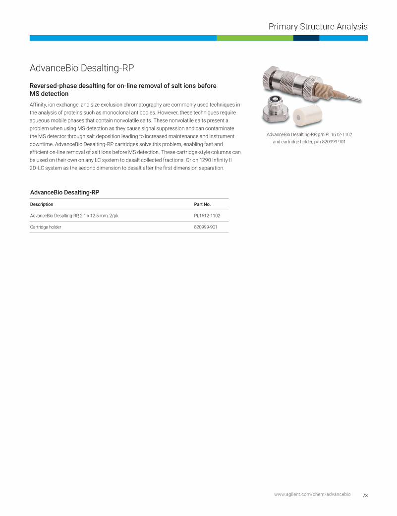
73
Primary Structure Analysis
www.agilent.com/chem/advancebio
AdvanceBio Desalting-RP
Reversed-phase desalting for on-line removal of salt ions before MS detectionAffinity, ion exchange, and size exclusion chromatography are commonly used techniques in the analysis of proteins such as monoclonal antibodies. However, these techniques require aqueous mobile phases that contain nonvolatile salts. These nonvolatile salts present a problem when using MS detection as they cause signal suppression and can contaminate the MS detector through salt deposition leading to increased maintenance and instrument downtime. AdvanceBio Desalting-RP cartridges solve this problem, enabling fast and efficient on-line removal of salt ions before MS detection. These cartridge-style columns can be used on their own on any LC system to desalt collected fractions. Or on 1290 Infinity II 2D-LC system as the second dimension to desalt after the first dimension separation.
AdvanceBio Desalting-RP
Description Part No.
AdvanceBio Desalting-RP, 2.1 x 12.5 mm, 2/pk PL1612-1102
Cartridge holder 820999-901
AdvanceBio Desalting-RP, p/n PL1612-1102 and cartridge holder, p/n 820999-901

74 Agilent BioHPLC Columns
Primary Structure Analysis
AdvanceBio Oligonucleotide
To successfully separate trityl-off, deprotected oligonucleotides, you need columns that have high resolving power and are robust enough to withstand challenging conditions.
Agilent AdvanceBio oligonucleotide columns feature high efficiency, 2.7 μm superficially porous Poroshell particles. Using technology exclusive to Agilent, we chemically modify the particles to make them exceedingly resistant to high pH mobile phases. We also bond them with an end-capped C18 phase that delivers excellent selectivity for oligonucleotides. What’s more, we test every batch of AdvanceBio oligonucleotide media with a resolution standard to ensure consistent performance.
The family of AdvanceBio products are designed to deliver consistent, exceptional performance for the complete characterization of proteins, antibodies, conjugates, new biological entities and biopharmaceuticals.
ColumnSpecifications
Bonded Phase Particle Size Pore Size Temp Limit pH Range Endcapped Pressure Limit
C18 2.7 µm 100 Å 65 °C 3.0–11.0 Double 600 bar

5
4
3
2
1
0
0.30.2 0.4 0.5 0.6 0.7 0.8 0.9 1 1.1 1.2 1.3 1.4 1.5 1.6 1.7 1.8 1.9 2 2.1 2.2 2.3 2.4 2.5 2.6 2.7 2.8
Response vs. acquisition time (min)
Resp
onse
Resp
onse
UV-expansive baseline
TIC-expansive baseline
B
C
5
4
3
2
1
0
x102
x102
800
700
600
500
400
300
200
100
0
2
UV full scale
A
3.138
4 6 8Time (min)
Abso
rban
ce (U
V 26
0 m
m)
FLP-
19
FLP-
18
FLP-
17
FLP-
16FL
P-15
FLP-
14
FLP-
13FL
P-12
FLP-
11
FLP-
10
FLP-
9
FLP-
8
FLP-
7
FLP-
6
FLP-
3 to
FLP
-5
FLP-
2FL
P-1
FLP
4.54
3.53
2.52
1.51
0.50
0.4 0.5 0.6 0.7 0.8 0.9 1 1.1 1.2 1.3 1.4 1.5 1.6 1.7 1.8 1.9 2 2.1 2.2 2.3 2.4 2.5 2.6
Counts vs. acquisition time (min)
Coun
ts
x105
******TTTGCTGCTGTTTTGCTGT
75
Primary Structure Analysis
www.agilent.com/chem/advancebio
Column: AdvanceBio Oligonucleotide 2.1 x 50 mm (p/n 659750-702)
Mobile phase: A: HFIP:TEA (400 mM:15mM) in water B: MeOH:mobile phase A (50:50)
Flow rate: 0.4 mL/min
Gradient: 30–40% B in 0.5 min; 40–70% B in 5 min
Sample: 25 mer DNA
Temperature: 65 °C
Detection: UV at 260 nm
Detection: MS
Min range: 400 m/z
Max range: 1,700 m/z
Scan rate: 3.00 spectra/s
Ion polarity: -ve
VCap: 3,500
Nozzle voltage: 1,000 V
Fragmentor: 200
Deconvoluted data from TIC of a 25-mer DNA oligonucleotide separated
by the AdvanceBio Oligonucleotide column
AdvanceBio Oligonucleotide
Description id (mm) Length (mm) Particle Size (µm) Part No.
Conventional 2.1 150 2.7 653750-702
Conventional 2.1 100 2.7 655750-702
Conventional 2.1 50 2.7 659750-702
UG Guard, 600 bar 2.1 5 2.7 821725-921
Conventional 4.6 150 2.7 653950-702
Conventional 4.6 100 2.7 655950-702
Conventional 4.6 50 2.7 659950-702
UG Guard, 600 bar 4.6 5 2.7 820750-921

0 1 2 3 4 5 6 7 8 9 min
20
15
10
5
0
mAU 0.2
05 0
.359
3.2
91 5.6
66
8.0
92
8.5
69
76 Agilent BioHPLC Columns
Primary Structure Analysis
Column: AdvanceBio Oligonucleotide 2.1 x 50 mm (p/n 659750-702)
Mobile phase: A: 100 mM TEAA in water B: 100 mM TEAA in acetonitrile
Gradient: 6 to 8% B in 12 min
Stop time: 13 min
Post run: 5 min
Flow rate: 0.6 mL/min
Sample: Agilent Oligonucleotide Resolution Standard (p/n 5190-9028)
Temperature: 65 °C
Injection: 0.5 μL
Detection: UV at 260 nm
AdvanceBio Oligonucleotide standards
To ensure performance for your separations, every batch of AdvanceBio Oligonucleotide media is tested with the Agilent Oligonucleotide Resolution standard. The Oligonucleotide Resolution standard containing 14, 17, 20, and 21 mer synthetic oligonucleotides is designed to demonstrate N / N-1 resolution.

0 1 2 3 4 5 6 7 8 9 min
20
15
10
5
0
mAU
0.1
89
0.3
23 0
.439
0.5
02
2.1
60
4.0
36 5.4
06
6.3
88
7.1
59
7.7
82
77
Primary Structure Analysis
www.agilent.com/chem/advancebio
Agilent also offers an Oligonucleotide Ladder standard containing 15, 20, 25, 30, 35, and 40 mer synthetic oligodeoxythymidines, an excellent tool for demonstrating column selectivity and reproducibility.
AdvanceBio Oligonucleotide Standards
Description Part No.
Oligonucleotide Resolution Standard 5190-9028
Oligonucleotide Ladder Standard 5190-9029
Column: AdvanceBio Oligonucleotide 2.1 x 50 mm (p/n 659750-702)
Mobile phase: A: 100 mM TEAA in water
B: 100 mM TEAA in acetonitrile
Gradient: 10 to 14% B in 10 min
Stop time: 11 min
Post run: 5 min
Flow rate: 0.6 mL/min
Column temperature: 65 °C
Sample: Agilent Oligonucleotide Ladder standard (p/n 5190-9029)
Injection: 10 μL
Detection: UV at 260 nm

78 Agilent BioHPLC Columns
Intact Analysis Using Hydrophobic Interaction Chromatography
AdvanceBio HIC
AdvanceBio HIC columns deliver high resolution, robust, and reproducible separations of native proteins at the intact level.
Built using the capabilities of the ZORBAX fully porous particles and proprietary bonding technology, these columns provide new levels of hydrophobicity and versatile single chemistry to address particularly challenging molecules such as monoclonal antibodies (mAbs), antibody drug conjugates (ADCs), and other recombinant proteins.
Together with the 1260 Infinity II bio-inert LC system, the AdvanceBio HIC provides uncompromised performance and data consistency during characterization and validation.
– Optimized selectivity: ideal for mAb oxidation and ADC DAR ratios.
– Single chemistry: reduces the need for multiple-column screening for different CQAs.
– Enhanced robustness: improved column lifetime for ultimate confidence in your data.
– Proven performance: every batch of media is tested with NIST mAb.
– High quality: each column is individually tested to ensure packing efficiency.
– Greater productivity: shorter columns reduce analysis time while maintaining separation performance.
ColumnSpecifications
Pore Size Particle Size Temperature Limit pH Range Pressure Limit Flow Rate*
450 Å 3.5 µm 60 °C (at pH 7) 2.0–8.0 (at 35 °C) 400 bar (typical operating pressure<200 bar)
0.5–1.0 mL/min (4.6 mm id)
*In some cases, lowering the flow rate to 0.3 mL/min and extending gradient time may further improve resolution.
Intact Analysis Using Hydrophobic Interaction Chromatography

79
Intact Analysis Using Hydrophobic Interaction Chromatography
www.agilent.com/chem/advancebio
Cytoch
rome C
Myoglobin
Ribonuclease
A
Lysozy
me
α-Chymotry
psinogen
A
Retention time [min]
1 2 3 4 5 6 7 8 9 10 11 12 13 14 15 16 17 18 19 20 21 22 23 24
Column: AdvanceBio HIC 4.6 x 100 mm, 3.5 µm
Eluent A: 2 M Ammonium Sulfate, 50 mM Sodium Phosphate, pH 7.0
Eluent B: 50 mM Sodium Phosphate, pH 7.0
Gradient: Time (min) %A %B 0 100 0 20 0 100 25 0 100 30 100 0 40 100 0
Flow rate: 0.5 mL/min
Temperature: 30 °C
Injection volume: 5 µL
Detection: UV, 220 nm
Column: AdvanceBio HIC 4.6 x 100 mm, 3.5 µm
Eluent A: 2 M Ammonium Sulfate, 50 mM Sodium Phosphate, pH 7.0
Eluent B: 50 mM Sodium Phosphate, pH 7.0
Gradient: Time (min) %A %B %C 0 50 45 5 20 0 75 25 25 0 75 25 30 50 45 5 40 50 45 5
Flow rate: 0.5 mL/min
Temperature: 30 °C
Injection volume: 5 µL
Detection: UV, 220 nm
1 2 3 4 5 6 7 8 9 10 11 12 13 14 15 16 17 18 19 20 21 22 23 24Retention time [min]
Agilent AdvanceBio HIC Columns
Description Part No.
AdvanceBio HIC, 4.6 x 100 mm, 450 Å, 3.5 µm 685975-908
AdvanceBio HIC, 4.6 x 30 mm, 450 Å, 3.5 µm 681975-908

80 Agilent BioHPLC Columns
Charge Variant Analysis
Charge Variant AnalysisPurify proteins and other charged molecules
Applying IEX to charge variant analysis
Ion-exchange chromatography (IEX) is a highly sensitive technique that allows you to separate ions and polar molecules based on their charge. Like SEC, IEX can be used to separate proteins in their native state.
During production and purification, antibodies can exhibit changes in charge heterogeneity as a result of amino acid substitutions, glycosylation, phosphorylation, and other post-translational or chemical modifications. Because these changes can impact stability and activity—or cause immunologically adverse reactions—the analysis of charge heterogeneity in monoclonal antibody (mAb) preparations is critical to biopharmaceutical manufacture.
In protein analysis, charge variations at a given pH indicate a change in the primary molecular structure—resulting in additional forms of the protein in question. These are called isoforms (or charge variants), and can be resolved by IEX chromatography. IEX is also useful as a preparative technique.
Because these changes can impact stability and activity—or cause immunologically adverse reactions—the analysis of charge variants is critical to biopharmaceuticals.
As a leading supplier to the biopharmaceutical industry, Agilent understands that quality and consistency are critical to providing safe, highly efficacious therapeutics. Agilent ion-exchange BioHPLC columns offer the speed, resolution, and reproducibility you need to quickly and cost-effectively get life-changing products into the hands of those who need them.

81
Charge Variant Analysis
www.agilent.com/chem/advancebio
The pages that follow describe Agilent’s family of weak and strong ion-exchangers—both anionic and cationic.
– Nonporous Bio IEX columns are designed for high resolution, high efficiency, and high recovery separations.
– Bio MAb columns are optimized for separating charge isoforms of monoclonal antibodies.
– Porous IEX columns (PL-SAX and PL-SCX) are chemically stable, and are available in two pore sizes, allowing you to separate peptides, oligonucleotides, and very large proteins.
– Bio-Monolith IEX columns are uniquely suited for separating antibodies, viruses, and DNA.
– Buffer Advisor software is an ideal solution for automated protein separation by ionic strength gradients.
Tips and tools
For more information about the Agilent Buffer Advisor software, see publication 5991-1408EN
www.agilent.com/search

82 Agilent BioHPLC Columns
Charge Variant Analysis
Ion-exchange column selection
Application Agilent Columns Notes
Monoclonal antibodies Bio MAb Thorough characterization of monoclonal antibodies includes the identificationand monitoring of acidic and basic isoforms. Bio MAb HPLC columns feature a unique resin specifically designed for high resolution charge-based separations of monoclonal antibodies.
Peptides and proteins Bio IEX Bio IEX columns are packed with polymeric, nonporous, ion-exchangeparticles. The Bio IEX columns are designed for high resolution, high recovery,and highly efficient separations.
Proteins, peptides, and deprotectedsynthetic oligonucleotides
PL-SAX The strong anion-exchange functionality, covalently linked to a fully porous chemically stable polymer, extends the operating pH range. In addition, the anion-exchange capacity is independent of pH. For synthetic oligonucleotides, separations using denaturing conditions of temperature, organic solvent, and high pH are all possible. The 5 μm media deliver separations at high resolution while the 30 μm media are used for medium pressure liquid chromatography
• 1000 Å• 4000 Å
Globular proteins and peptides PL-SAX 1000 Å
Very large biomolecules/high speed PL-SAX 4000 Å
Small peptides to large proteins and very large biomolecules
PL-SCX PL-SCX is a macroporous PS/DVB matrix with a very hydrophilic coating and strongcation-exchange functionality. This process is controlled to provide the optimumdensity of strong cation-exchange moieties for the analysis, separation, andpurification of a wide range of biomolecules. The 5 μm media deliver separationsat higher resolution while the 30 μm media are used for medium pressureliquid chromatography.
• 1000 Å• 4000 Å
Antibodies (IgG, IgM), plasmid DNA, viruses, phages, and other macro biomolecules
Bio-Monolith Strong cation-exchange, strong and weak anion-exchange phases. Bio-MonolithHPLC columns are compatible with InfinityLab LC series.
• Bio-Monolith QA• Bio-Monolith DEAE• Bio-Monolith SO3
Viruses, DNA, large proteins Bio-Monolith QA
Plasmid DNS, bacteriophages Bio-Monolith DEAE
Proteins, antibodies Bio-Monolith SO3

83
Charge Variant Analysis
www.agilent.com/chem/advancebio
Bio MAb HPLC columns – A packing support composed of a rigid, spherical, highly cross-linked polystyrene
divinylbenzene (PS/DVB) nonporous bead
– Particles grafted with a hydrophilic polymeric layer, virtually eliminating nonspecific binding of antibody proteins
– A different process is used to layer the weak cation-exchange phase to the particle giving it a higher density than the Bio WCX column particles
– Specifically designed for the separation of charge isoforms of monoclonal antibodies
Thorough characterization of monoclonal antibodies includes the identification and monitoring of acidic and basic isoforms. Bio MAb HPLC columns feature a unique resin specifically designed for high resolution, charge-based separations of monoclonal antibodies. These columns are compatible with aqueous solution buffers, acetonitrile/acetone/methanol, and water mixtures. Commonly used buffers are phosphate, tris, MES, and acetate.
Bio MAb columns are available in 1.7, 3, 5, and 10 μm sizes, providing higher resolution with smaller particles.
Tips and tools
Are you looking to increase your throughput for charge variant analysis of monoclonal antibodies? If so, see:
Reducing Cycle Time for Charge Variant Analysis of Monoclonal Antibodies (publication 5991-4722EN)
www.agilent.com/search
ColumnSpecifications
Bonded Phase id Particle Size pH Stability Operating Temperature Limit
Flow Rate
Weak cation-exchange (carboxylate)
2.1 and 4.6 mm 1.7, 3, 5, and 10 μm
2–12 80 °C 0.1–1.0 mL/min
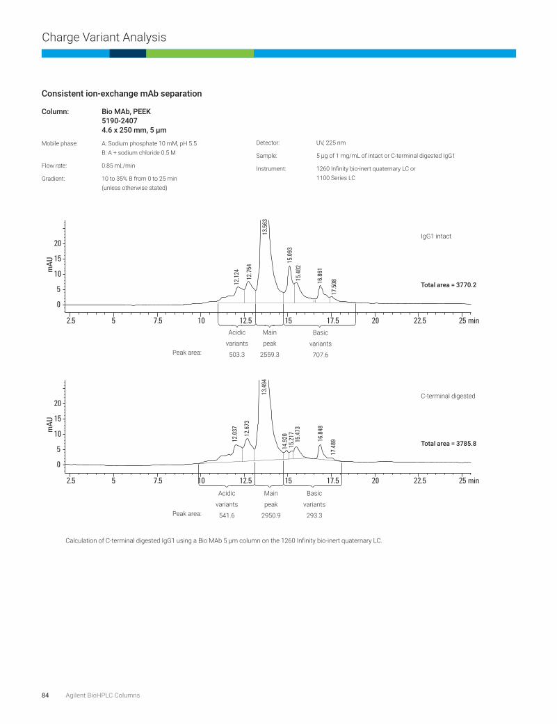
84 Agilent BioHPLC Columns
Charge Variant Analysis
Consistent ion-exchange mAb separation
Column: Bio MAb, PEEK 5190-2407 4.6x250mm,5μm
Mobile phase: A: Sodium phosphate 10 mM, pH 5.5 B: A + sodium chloride 0.5 M
Flow rate: 0.85 mL/min
Gradient: 10 to 35% B from 0 to 25 min (unless otherwise stated)
Detector: UV, 225 nm
Sample: 5 μg of 1 mg/mL of intact or C-terminal digested IgG1
Instrument: 1260 Infinity bio-inert quaternary LC or 1100 Series LC
min2.5 5 7.5 10
12.12
412
.037
12.67
3
13.49
4
14.92
015
.217
15.47
3
16.84
8
17.48
9
12.75
4
13.56
3
15.09
315
.482
16.86
1
17.50
8
12.5 15 17.5 20 22.5 25
mAU
mAU
0
5
10
15
20
min2.5 5 7.5 10 12.5 15 17.5 20 22.5 25
0
5
10
15
20
Calculation of C-terminal digested IgG1 using a Bio MAb 5 μm column on the 1260 Infinity bio-inert quaternary LC.
C-terminal digested
IgG1 intact
Total area = 3770.2
Peak area:
Peak area:
Basic
variants
707.6
Basic
variants
293.3
Main
peak
2559.3
Main
peak
2950.9
Acidic
variants
503.3
Acidic
variants
541.6
Total area = 3785.8

85
Charge Variant Analysis
www.agilent.com/chem/advancebio
Reproducibility and precision—Bio MAb columns enable precise quantitation, robust methods
WCX separation of Fab and Fc fragments of Trastuzumab
Time (min) % B 0 0 25 100 27 100 30 0
Time (min) % B 0 0 39.5 80 40 100 50 100
50.5 2
60 2
Data acquisition: 214, and 280 nm
Acquisition rate: 20 Hz
Flow cell: 60 mm path
Column temperature: 30 °C
Sample thermostat: 50 °C
Posttime: 5 min
Temperature: 30 °C
Instrument: 1100 series
mAU
1000
800
600
400
200
0
18 20 22 24 26 28 30 32 min
mAU
Time (min)14 16 18 20 22
0
2
4
6
13.28
3 13.61
7
15.06
9
17.82
7
22.69
8
24
Column: Bio MAb, PEEK 5190-2411 2.1x250mm,5μm
Mobile phase: A: 20 mM MES, pH 5.6 B: 20 mM MES, pH 5.6 + 300 mM NaCl
Flow rate: 170 μL/min
Injection volume: 16 μL
Gradient:
Column: Bio MAb, PEEK 5190-2407 4.6x50mm,5μm
Mobile phase: A: 10 mM Sodium phosphate buffer, pH 6.0 B: 10 mM Sodium bicarbonate buffer, pH 9.5
Flow rate: 1.0 mL/min
Injection volume: 10 μL (needle with wash, flush port active for 7 s)
Gradient:
Main peak
Basic variants
Fab (-KTH)
Fc
Fab
Fab (deam)
Acidic variants

86 Agilent BioHPLC Columns
Charge Variant Analysis
Method development using Buffer Advisor software—determination of optimum pH
min
mAU
0
50
100
150
200
250
1 1
1
1 11
33
33 3 3
2 2
2
22
2
pH 5.8pH 6
pH 6.2pH 6.4
pH 6.6
pH 6.8
1. Ribonuclease A2. Cytochrome c3. Lysozyme
pH scouting for the separation of a three-protein mix using dynamically mixed quaternary gradients
Column: Bio MAb, PEEK 5190-2407 4.6x250mm,5μm
Instrument: 1260 Infinity Bio-inert Quaternary LC
Buffer: A: H2O B: NaCl 3 M C: MES (2-N-morpholino) ethanesulfonic acid monohydrate) 60 mM D: MES-Na (2-(N-morpholino) ethanesulfonic acid sodium salt) 35 mM
Sample: Mix of three proteins, dissolved in PBS (phosphate buffered saline), pH 7.4
Ribonuclease A: 13,700 Da, pI 9.6 Cytochrome c: 12,384 Da, pI 10-10.5 Lysozyme: 14,307 Da, pI 11.35
Flow rate: 1 mL/min
Gradient: 0 min–20 mM NaCl 5 min–20 mM NaCl 30 min–500 mM NaCl 35 min–1,000 mM NaCl 36 min–20 mM NaCl
Injection volume: 10 μL
Thermostat: 4 °C
Temperature TCC: 25 °C
DAD: 280 nm/4 nm Reference: OFF
Peak width: >0.05 min (1.0 s respose time)(5 Hz)

87
Charge Variant Analysis
www.agilent.com/chem/advancebio
Column: Bio MAb, stainless steel 5190-2413 4.6x250mm,10μm
Mobile phase: A: 10 mM Sodium phosphate, pH 6.0 B: A + 1.0 M Sodium chloride
Flow rate: 1.0 mL/min
Gradient: 0–100% B in 42 min
Temperature: 25 °C
Detector: UV, 214 nm
The combination of well-controlled resin production, column surface
chemistry, and column packing virtually eliminates retention time
variations from column-to-column and lot-to-lot.
Agilent Bio MAb HPLC columns
Size (mm) ParticleSize(μm) Bio MAb PEEK Pressure Limit Bio MAb Stainless Steel Pressure Limit
21.2 x 250 5 5190-6885 275 bar, 4000 psi
10 x 250 5 5190-6884 275 bar, 4000 psi
4.6 x 250 10 5190-2415 275 bar, 4000 psi 5190-2413 275 bar, 4000 psi
4.6 x 50 10 5190-2416 275 bar, 4000 psi
4.6 x 250 5 5190-2407 400 bar, 5800 psi 5190-2405 400 bar, 5800 psi
4.6 x 50 5 5190-2408 400 bar, 5800 psi
4.6 x 50 3 5190-2403 551 bar, 8000 psi
4.6 x 50 1.7 5190-2401 600 bar, 8700 psi
4.0 X 10, guard 10 5190-2414 275 bar, 4000 psi
4.0 X 10, guard 5 5190-2406 413 bar, 6000 psi
4.0 X 10, guard 3 5190-2404 551 bar, 8000 psi
4.0 X 10, guard 1.7 5190-2402 600 bar, 8700 psi
2.1 x 250 10 5190-2419 275 bar, 4000 psi
2.1 x 50 10 5190-2420 275 bar, 4000 psi
2.1 x 250 5 5190-2411 400 bar, 5800 psi
2.1 x 50 5 5190-2412 400 bar, 5800 psi
0 2
1 2 3
4 6 8 10 12 14 min16 18
1. Cytochrome c2. Lysozyme3. Ribonuclease A
Virtually eliminate retention time variations
Lot A
Lot B
Lot C

88 Agilent BioHPLC Columns
Charge Variant Analysis
Bio IEX HPLC columns – Highly cross-linked and rigid nonporous poly(styrene divinylbenzene) (PS/DVB) particles
are grafted with a hydrophilic polymeric layer, eliminating nonspecific binding
– Uniform, densely packed ion-exchange functional groups are chemically bonded to the hydrophilic layer (multiple ion-exchange groups per anchor) to increase column capacity
– Particles, coating, and bonding are resistant to high pressures, promoting higher resolution and faster separations
– Multiple ion-exchange groups are captured on one anchor to increase cap
Bio IEX HPLC columns are packed with polymeric, nonporous, ion-exchange particles and are designed for high resolution, high recovery, and highly efficient separations of peptides, oligonucleotides, and proteins.
The Bio IEX family includes strong cation-exchange (SCX), weak cation-exchange (WCX), strong anion-exchange (SAX), and weak anion-exchange (WAX) phases. All phases are available in 1.7, 3, 5, and 10 μm nonporous particles.
Tips and tools
For further information on optimizing your charged variant analysis, refer to: Ion-exchange chromatography for biomolecule analysis: a “how-to” guide (publication 5991-3775EN), and Agilent ion-exchange BioHPLC columns, characterize charged variants of proteins with speed and confidence (publication 5991-2449EN)
www.agilent.com/search
ColumnSpecifications
Bonded Phase id Particle Size pH Stability Operating Temperature Limit
Flow Rate
SCX (strong cation-exchange)–SO3H 2.1 and 4.6 mm 1.7, 3, 5, and 10 μm 2–12 80 °C 0.1–1.0 mL/min
WCX (weak cation-exchange)–COOH
SAX (strong anion-exchange)–N(CH3)3
WAX (weak cation-exchange)–N(C2H5)2

89
Charge Variant Analysis
www.agilent.com/chem/advancebio
Column: Bio WCX, stainless steel 5190-2443 4.6x50mm,3μm
Bio SCX, stainless steel 5190-2423 4.6x50mm,3μm
Mobile phase: A: Water B: Sodium chloride 1.5 M C: Monosodium phosphate 40 mM D: Disodium phosphate 40 mM By combining predetermined proportions of C and D as determined by the Buffer Advisor software, buffer solutions at the desired pH range and strength were created.
Flow rate: 1.0 mL/min
Injection volume: 10 μL
Gradient: Conditions for chromatograms shown: pH 5.0 to 7.0, 10 to 25 mM buffer strength sodium chloride (NaCl) 0 to 500 mM, 0 to 15 min sodium chloride (NaCl) 500 mM, 15 to 20 min DOE experiments pH 5.0 to 7.0 0 to 200 mM, 0 to 250 mM, and 0 to 300 mM NaCl
Temperature: Ambient
Detector: UV, 220 nm
Sample: IgG monoclonal antibody
Sample conc: 2 mg/mL (in sodium phosphate buffer 20 mM, pH 6.0)
Instrument: 1260 Infinity bio-inert quaternary LC
Automated method development for optimized charge variant separations. Optimizing buffer
strength at pH 6.5 from the screening chromatograms of a monoclonal IgG separation.
0 1 2 3 4 5 min
10 mM
15 mM
20 mM
25 mM
Shortened and simplified charge variant workflow

90 Agilent BioHPLC Columns
Charge Variant Analysis
Column: Bio WCX, stainless steel 5190-2445 4.6x250mm,5μm
Bio WCX, stainless steel 5190-2443 4.6x50mm,3μm
Mobile phase: A: Sodium phosphate 20 mM, pH 6.5 B: A + sodium chloride 1.6 M
Flow rate: 1.0 mL/min
Injection volume: 10 μL
Gradient: 0 to 50% B
Temperature: Ambient
Detector: UV, 220 nm
Sample conc: 0.5 mg/mL
Instrument: 1260 Infinity bio-inert quaternary LC
Protein separation on a Bio WCX 4.6 x 50 mm, 3 μm column versus a Bio WCX 4.6 x 250 mm, 5 μm
column (flow rate 1.0 mL/min). Faster analysis times were achieved through smaller particle size and
shorter column length—samples eluted from the longer column in 17 min, and in just 12 min from the
shorter column.
1
1
2
2
3
3
4
4
0 5 10 15 20 25 30 min
0 5 10 15 20 25 30 min
1. Ovalbumin2. Ribonuclease A3. Cytochrome c4. Lysozyme
800 mM NaCl
0 mM NaCl
0 mM NaCl
800 mM NaCl
Bio WCX 3 μm, 4.6 × 50 mm
Bio WCX 5 μm, 4.6 × 250 mm
Achieve faster analysis time with smaller particles and shorter column lengths—speed up your separation by 30%

91
Charge Variant Analysis
www.agilent.com/chem/advancebio
Smaller particle sizes provide increased resolution
Column: Bio WCX, stainless steel 5190-2443 4.6x50mm,3μm
Bio WCX, stainless steel 5190-2441 4.6x50mm,1.7μm
Mobile phase: A: Sodium phosphate 20 mM, pH 6.5 B: A + sodium chloride 1.6 M
Injection volume: 10 μL
Gradient: 0 to 50% B
Temperature: Ambient
Detector: UV, 220 nm
Sample conc: 0.5 mg/mL
Instrument: 1260 Infinity bio-inert quaternary LC
Left and middle: Protein separation on a Bio WCX 3 μm column versus a Bio WCX 1.7 μm column (flow rate 1.0 mL/min).
Right: By increasing the flow rate to 1.7 mL/min, the separation time was reduced to less than 3 minutes. (A Bio WCX column was used.)
Reduce analysis time—without sacrificing peak shape and resolution—by increasing flow rate.
0 1 2 3 4 5 6
1
2
34
min 0 1 2 3 4 5 6 min
1
2
3 4
0 1 2 3 4 5 6 min
1
2
34
1. Ovabumin2. Ribonuclease A3. Cytochrome c4. Lysozyme
800 mM NaCl 800 mM NaCl 800 mM NaCl
Bio WCX 3 μm Bio WCX 1.7 μm Bio WCX 1.7 μm
0 mM NaCl 0 mM NaCl 0 mM NaCl

92 Agilent BioHPLC Columns
Charge Variant Analysis
Column: Bio WAX, PEEK 5190-2487 4.6x250mm,5μm
Buffer: A: 20 mM tris, pH 7.6 B: 20 mM tris, pH 7.6 + 2 M NaCl, 1 M KCl, 1 M CH3COONa, 1 M [(CH3 )4N]Cl
Gradient 1 M: 5 min–100% A 20 min–70% B 25 min–100% B
Gradient 2 M: 5 min–100% A 20 min–35% B 25 min–50% B 25.01 min–100% B
Stop time: 30 min
Posttime: 20 min
Temperature: 25 °C
Flow rate: 0.5 mL/min
Injection volume: 5 μL
DAD: 280 nm
Peak width: 0.025 min (0.5 s response time) (10 Hz)
For further information see application note
5990-9614EN (www.agilent.com/search)
Figure 1. Protein separation by AEX by a linear gradient using 2 M NaCl as eluting salt.
Figure 2. Protein separation by AEX by a linear gradient using 1 M KCl as eluting salt.
Figure 3. Protein separation by AEX by a linear gradient using 1 M [(CH3)4N]Cl) as eluting salt.
1. Myoglobin, pl 6.92. Conalbumin Isoform A, pl 6.243. Conalbumin Isoform B, pl 6.094. a-Lactalbumin, pl 4.55. Trypsin Inhibitor, pl 4.5
RSD (%) RSD Area (%)Myoglobin 0.073 0.413Conalbumin Isoform A 0.080 0.809Conalbumin Isoform B 0.066 n/aa-Lactalbumin 0.081 n/aTrypsin Inhibitor 0.075 1.024
RSD (%) RSD Area (%)Myoglobin 0.105 0.066Conalbumin Isoform A 0.027 n/aConalbumin Isoform B 0.015 n/aa-Lactalbumin 0.023 0.66Trypsin Inhibitor 0.33 1.492
RSD (%) RSD Area (%)Myoglobin 0.077 2.346Conalbumin Isoform A 0.035 n/aConalbumin Isoform B 0.033 n/aa-Lactalbumin 0.040 4.606Trypsin Inhibitor 0.074 0.628
min0 5 10 15 20 25
mAU
0
10
20
30
40
50
60
70
-10
1
2
34
5
min0 5 10 15 20 25
mAU
0
10
20
30
40
50
60
70
801
2
3
4
5
min0 5 10 15 20 25
mAU
1
0
20
40
60
80
100
120
-20
2
3
4
5
Analysis of proteins by anion-exchange columns using the Agilent 1260 Infinity bio-inert quaternary LC system

93
Charge Variant Analysis
www.agilent.com/chem/advancebio
Bio IEX HPLC Columns, PEEK
Size (mm) ParticleSize(μm) Pressure Limit Bio SCXPart No.
Bio WCXPart No.
Bio SAXPart No.
Bio WAXPart No.
4.6 x 250 10 275 bar, 4000 psi 5190-2435 5190-2455 5190-2475 5190-2495
4.6 x 50 10 275 bar, 4000 psi 5190-2436 5190-2456 5190-2476 5190-2496
4.6 x 250 5 400 bar, 5800 psi 5190-2427 5190-2447 5190-2467 5190-2487
4.6 x 50 5 400 bar, 5800 psi 5190-2428 5190-2448 5190-2468 5190-2488
2.1 x 250 10 275 bar, 4000 psi 5190-2439 5190-2459 5190-2479 5190-2499
2.1 x 50 10 275 bar, 4000 psi 5190-2440 5190-2460 5190-2480 5190-2500
2.1 x 250 5 400 bar, 5800 psi 5190-2431 5190-2451 5190-2471 5190-2491
2.1 x 50 5 400 bar, 5800 psi 5190-2432 5190-2442 5190-2462 5190-2492
Tips and tools
Use Agilent solvent filters to remove particles from self-prepared buffers and mobile phases.
Visit: www.agilent.com/chem/solvent-filters-degassers

94 Agilent BioHPLC Columns
Charge Variant Analysis
Bio IEX HPLC Columns, Stainless Steel
Size (mm) ParticleSize(μm) Pressure Limit Bio SCXPart No.
Bio WCXPart No.
Bio SAXPart No.
Bio WAXPart No.
21.2 x 250 5 5190-6879 5190-6881 5190-6883 5190-6877
10 x 250 5 5190-6878 5190-6880 5190-6882 5190-6876
4.6 x 250 10 275 bar, 4000 psi 5190-2433 5190-2453 5190-2473 5190-2493
4.6 x 250 5 413 bar, 6000 psi 5190-2425 5190-2445 5190-2465 5190-2485
4.6 x 150 3 5190-6875
4.6 x 50 3 551 bar, 8000 psi 5190-2423 5190-2443 5190-2463 5190-2483
4.6 x 50 1.7 600 bar, 8700 psi 5190-2421 5190-2441 5190-2461 5190-2481
4.0 x 10, guard 10 275 bar, 4000 psi 5190-2434 5190-2454 5190-2474 5190-2494
4.0 x 10, guard 5 413 bar, 6000 psi 5190-2426 5190-2446 5190-2466 5190-2486
4.0 x 10, guard 3 551 bar, 8000 psi 5190-2424 5190-2444 5190-2464 5190-2484
4.0 x 10, guard 1.7 600 bar, 8700 psi 5190-2422 5190-2442 5190-2462 5190-2482
Tips and tools
For further information see:
Optimizing protein separations with Agilent weak cation-exchange columns (publication 5990-9628EN) Faster separations using Agilent weak cation-exchange columns (publication 5990-9931EN) pH Gradient elution for improved separation of monoclonal antibody variants (publication 5990-9629EN) Optimizing protein separations with cation-exchange chromatography using Agilent Buffer Advisor (publication 5991-0565EN)
www.agilent.com/search

95
Charge Variant Analysis
www.agilent.com/chem/advancebio
PL-SAX Strong Anion-Exchange columns – Small particles deliver excellent chromatographic performance
– Wide range of particle sizes and two pore sizes for flexible analysis to scale up purification
– Exceptional stability for long column lifetime
PL-SAX -N(CH3)3+ is ideal for the anion-exchange HPLC separations of proteins, peptides,
and deprotected synthetic oligonucleotides under denaturing conditions. The strong anion-exchange functionality, covalently linked to a chemically stable fully porous polymer, extends the operating pH range. In addition, the anion-exchange capacity is independent of pH. For synthetic oligonucleotides, separations using denaturing conditions of temperature, organic solvent, and high pH are all possible. PL-SAX delivers improved chromatography for self-complementary or G-rich sequences that may associate to form aggregates or hairpin structures. The 5 μm material provides high-efficiency separations of n and n-1 sequences. A wide range of particle sizes and column geometries permits analysis scale-up to purification. The strong anion-exchange functionality provides a material with exceptional chemical and thermal stability, even with sodium hydroxide eluents, leading to long column lifetime.
ColumnSpecifications
Bonded Phase id Particle Size (mm) PoreSize(μm) pH Stability Operating Temperature Limit
Strong anion-exchange 2.1, 4.6, 7.5, 25, 50, and 100
5, 8, 10, and 30 1000 Å and 4000 Å
1–14 80 °C

96 Agilent BioHPLC Columns
Charge Variant Analysis
Standard ion-exchange protein separation
Column: PL-SAX 1000 Å PL1551-1502 4.6x50mm,5μm
Mobile phase: A: 10 mM tris HCl, pH 8 B: A + 350 mM sodium chloride, pH 8
Gradient: 0–100% B in 20 min
Flow rate: 1.0 mL/min
Detector: UV, 220 nm
Analysis of choline kinase
Column: PL-SAX 4000 Å PL1551-1803 4.6x50mm,8μm
Mobile phase: A: 20 mM tris 5% ethylene glycol, pH 7.5 (The following are required to retain enzyme activity) 1.0 mM ethylene glycol tetraacetic acid 2.0 mM ß-mercaptoethanol 0.2 mM phenylmethylsulfonyl fluoride B: A + 1 M potassium chloride
Flow rate: 3.0 mL/min
Detector: UV, 280 nm
Separation courtesy of T Porter, Purdue University, USA
0 min 40
0
%B100
3
421
min0 25VLC0070
1. Myoglobin2. Bovine carbonic anhydrase3. Ovalbumin4. Soybean trypsin inhibitor
Enzyme activity

97
Charge Variant Analysis
www.agilent.com/chem/advancebio
Analysis of representative whey proteins
Column: PL-SAX 1000 Å PL1551-1802 4.6x50mm,8μm
Mobile phase: A: 20 mM tris HCI, pH 7 B: A + 500 mM sodium acetate, pH 7
Flow rate: 1.0 mL/min
Gradient: Linear 0-50% B in 10 min
Detector: UV, 280 nm
Reliable separations of synthetic oligonucelotides— high resolution separation of a poly-t-oligonucleotide size standard spiked with 10-mer, 15-mer, 30-mer, and 50-mer (main peaks)
Column: PL-SAX 1000 Å PL1551-1802 4.6x50mm,8μm
Mobile phase: A: 7:93 v/v ACN: 100 mM TEAA, pH 8.5 B: 7:93 v/v ACN: 100 mM TEAA, 1 M ammonium chloride, pH 8.5
Gradient: 0–40% B in 10 min, followed by 40–70% B in 14 min and 70–100% B in 25 min
Flow rate: 1.0 mL/min
Temperature: 60 °C
Detector: UV, 220 nm
High-resolution separation of poly-t-oligonucleotide. With the gradient
used here, baseline separation of the n-1 from n was easily obtained up
to the 15-mer.
0
1
2
3
4
min 18
1. Carbonic anhydrase2. a-Lactalbumin3. b-Lactoglobulin B4. b-Lactoglobulin A
0
0
2000 15
30 50
10
VLC0045min 50

98 Agilent BioHPLC Columns
Charge Variant Analysis
PL-SAX Strong Anion-Exchange Columns
Size (mm) ParticleSize(μm) Pressure Limit PL-SAX1000 Å
PL-SAX4000 Å
100 x 300 10 207 bar, 3000 psi PL1851-2102 PL1851-2103
50 x 150 30 207 bar, 3000 psi PL1751-3702 PL1751-3703
50 x 150 10 207 bar, 3000 psi PL1751-3102 PL1751-3103
25 x 150 30 207 bar, 3000 psi PL1251-3702 PL1251-3703
25 x 150 10 275 bar, 4000 psi PL1251-3102 PL1251-3103
25 x 50 10 207 bar, 3000 psi PL1251-1102 PL1251-3103
4.6 x 250 30 207 bar, 3000 psi PL1551-5702 PL1551-5703
4.6 x 150 30 207 bar, 3000 psi PL1551-3702 PL1551-3703
4.6 x 250 10 207 bar, 3000 psi PL1551-5102 PL1551-5103
4.6 x 150 10 207 bar, 3000 psi PL1551-3102 PL1551-3103
4.6 x 150 8 207 bar, 3000 psi PL1551-3802 PL1551-3803
4.6 x 50 8 207 bar, 3000 psi PL1551-1802 PL1551-1803
4.6 x 50 5 207 bar, 3000 psi PL1551-1502 PL1551-1503
2.1 x 150 8 207 bar, 3000 psi PL1951-3802 PL1951-3803
2.1 x 50 8 207 bar, 3000 psi PL1951-1802 PL1951-1803
2.1 x 50 5 207 bar, 3000 psi PL1951-1502 PL1951-1503
1.0 x 50 5 207 bar, 3000 psi PL1351-1502 PL1351-1503
PL-SAX Strong Anion-Exchange Bulk Media
Size (mm) ParticleSize(μm) Pressure Limit PL-SAX1000 Å
PL-SAX4000 Å
100 g 30 207 bar, 3000 psi PL1451-4702 PL1451-4703
100 g 10 207 bar, 3000 psi PL1451-4102 PL1451-4103

99
Charge Variant Analysis
www.agilent.com/chem/advancebio
PL-SCX Strong Cation-Exchange columns – Optimal design for effective separation of biomolecules
– Pore sizes allow use of a range of solute sizes
– Exceptional stability for long column lifetime
PL-SCX -SO3 is a macroporous PS/DVB matrix with a very hydrophilic coating and strong cation-exchange functionality. This process is controlled to provide the optimum density of strong cation-exchange moieties for the analysis, separation, and purification of a wide range of biomolecules, from small peptides to large proteins. Two pore sizes are available, 1000 Å and 4000 Å, to provide good mass-transfer characteristics for a range of solute sizes. The 5 μm media deliver separations at higher resolution with the 30 μm media used for medium pressure liquid chromatography.
ColumnSpecifications
Bonded Phase id (mm) ParticleSize(μm) Pore Size pH Stability Operating Temperature Limit
Strong cation-exchange 2.1, 4.6, 7.5, 25, 50, and 100
5, 8, 10, and 30 1000 Å and 4000 Å 1–14 80 °C

100 Agilent BioHPLC Columns
Charge Variant Analysis
0
1
23
4
VLC004625min
Standard protein separation
Column: PL-SCX 1000 Å PL1545-1502 4.6x50mm,5μm
Mobile phase: A: 20 mM potassium dihydrogen phosphate, pH 6.0 B: A + 1 M sodium chloride
Gradient: 0–100% B in 20 min
Flow rate: 1.0 mL/min
Detector: UV, 280 nm
1. Myoglobin2. Chymotrypsinogen A3. Cytochrome c4. Lysozyme

101
Charge Variant Analysis
www.agilent.com/chem/advancebio
PL-SCX Strong Cation-Exchange Bulk Media
Size (mm) ParticleSize(μm) PL-SCX1000 Å
PL-SCX4000 Å
100 g 30 PL1445-4702 PL1445-4703
100 g 10 PL1445-4102 PL1445-4102
Tips and tools
PL-SAX and PL-SCX columns and bulk media are also available for prep to process.
Turn to Pages 190–195.
PL-SCX Strong Cation-Exchange Columns
Size (mm) ParticleSize(μm) Pressure Limit PL-SCX1000 Å
PL-SCX4000 Å
100 x 300 10 207 bar, 3000 psi PL1845-2102 PL1845-2103
50 x 150 30 207 bar, 3000 psi PL1745-3703 PL1745-3703
50 x 150 10 207 bar, 3000 psi PL1745-3102 PL1745-3103
25 x 150 30 207 bar, 3000 psi PL1245-3702 PL1245-3703
25 x 150 10 207 bar, 3000 psi PL1245-3102 PL1245-3103
25 x 50 10 207 bar, 3000 psi PL1245-1102 PL1245-1103
4.6 x 250 30 207 bar, 3000 psi PL1545-5703 PL1545-5703
4.6 x 150 30 207 bar, 3000 psi PL1545-3702 PL1545-3703
4.6 x 250 10 207 bar, 3000 psi PL1545-5102 PL1545-5103
4.6 x 150 10 207 bar, 3000 psi PL1545-3102 PL1545-3103
4.6 x 150 8 207 bar, 3000 psi PL1545-3802 PL1545-3803
4.6 x 50 8 207 bar, 3000 psi PL1545-1802 PL1545-1803
4.6 x 50 5 207 bar, 3000 psi PL1545-1502 PL1545-1503
2.1 x 150 8 207 bar, 3000 psi PL1945-3802 PL1945-3803
2.1 x 50 8 207 bar, 3000 psi PL1945-1802 PL1945-1803
2.1 x 50 5 207 bar, 3000 psi PL1945-1502 PL1945-1503
1.0 x 50 5 207 bar, 3000 psi PL1345-1502 PL1345-1503

102 Agilent BioHPLC Columns
Charge Variant Analysis
Bio-Monolith Ion-Exchange HPLC columns – Polymer-based, monolith HPLC columns designed for macro biomolecule separations
– Flow-rate independent separations; no diffusion, no pores, and no void volume make transport between mobile and stationary phase very rapid
– Monolith disk is 5.2 x 4.95 mm (100 μL column volume) with continuous channels, eliminating diffusion mass transfer
– Extremely fast separations speed up method development time and decrease costs; locking in method parameters takes significantly less time and buffer
Bio-Monolith Ion-Exchange HPLC columns provide high resolution and rapid separations of antibodies (IgG, IgM), plasmid DNA, viruses, phages, and other macro biomolecules. The product family offers strong cation-exchange, strong and weak anion-exchange, and Protein A phases. Bio-Monolith HPLC columns are compatible with InfinityLab LC series.
Bio-Monolith HPLC Column Selection Guide
Column Description Key Applications Part No.
Bio-Monolith QA The quaternary amine bonded phase (strong anion-exchange) is fully charged over a working pH range of 2–13, binding negatively charged biomolecules.
– Adenovirus process monitoring and quality control – IgM purification monitoring and quality control – Monitoring DNA impurity removal – Monitoring endotoxin removal – HSA purity
5069-3635
Bio-Monolith DEAE The diethylaminoethyl bonded phase (weak anion-exchange) offers increased selectivity of biomolecules with negative charge over a working pH range of 3–9.
– Process monitoring and quality control of bacteriophage manufacturing and purification
– Process monitoring and quality control of plasmid DNA purification
5069-3636
Bio-Monolith SO3 The sulfonyl bonded phase (strong cation-exchange) is fully charged over a working pH range of 2–13, binding positively charged biomolecules.
– Fast and high resolution analytical separations of large molecules such as proteins and antibodies
– Hemoglobin A1c fast analytics
5069-3637
Bio-Monolith Ion-Exchange HPLC column

103
Charge Variant Analysis
www.agilent.com/chem/advancebio
ColumnSpecifications
Dimensions 5.2 mm x 4.95 mm
Column volume 100 μL
Maximum pressure 150 bar (15 MPa, 2,200 psi)
Temperature min/max Operating: 2–40 °C
Storage: 2–8 °C
Recommended pH Operating range: 2–13
Cleaning-in-place: 1–14
Materials of construction Hardware: stainless steel
Packing: poly(glycidyl methacrylate-co-ethylene dimethacrylate) highly porous monolith
Colorringidentifier Bio-Monolith QA: blue
Bio-Monolith DEAE: green
Bio-Monolith SO3: red
Shelf life/expiration date SO3, QA, DEAE: 24–36 months

104 Agilent BioHPLC Columns
Charge Variant Analysis
4 6 8 10 12 14 16 18 min
468
101214
mAU
4
1 2 3
6 8 10 12 14 16 18 min
468
101214
mAU
4.483
4.968
6.357
6.834
7.832
9.072
12.17
0
16.41
0
4.284 4.5
204.6
884.9
45 5.653
6.482
7.059 7.6
04 8.162
9.271
10.33
5 16.90
1
Baseline expansion of a separation of protein standards
Column: Bio-Monolith CM15, 5.5 x 15 mm
Mobile phase: A: 10 mM sodium hydrogen phosphate, pH 6.0 B: A + 500 mM sodium chloride or just 500 mM sodium hydrogen phosphate, pH 6.0
Flow rate: 2 mL/min
Gradient: 0.5 min hold with mobile phase A followed by a linear gradient to 45% B in 15 min (elapsed time 15.5 min), then 60% B at 15.6 min continued to 20 min. Column flushed with 100% B for 15 min before re-equilibration for the next run.
pH gradient: A: 5 mM sodium hydrogen phosphate, buffer, pH 5.5 and B: 40 mM sodium hydrogen phosphate (not buffered, pH 8.9). 2% B/min at 1 mL/min for 15 min, followed by a column wash with 90% B for 5 min.
Detector: UV, 220 nm
Sample: One mg each/mL in mobile phase A 1. RNAse from bovine pancreas (pI 9.6) 2. Cytochrome c from bovine heart (pI 10.37-10.8) 3. Lysozyme from chicken egg (pI 11.35) (0.5 mg)
Instrument: 1200 Infinity series with diode array detectorB shows a better resolution of protein contaminants.
A with NaCl gradient
B with Na2HPO4 gradient

105
Charge Variant Analysis
www.agilent.com/chem/advancebio
Monitor phage production during fermentation
Column: Bio-Monolith DEAE 5069-3636 5.2 x 4.95 mm
Mobile phase: A: 125 mM sodium phosphate buffer, pH 7.0 B: 125 mM sodium phosphate buffer + 1 M sodium chloride, pH 7.0
Flow rate: 1 mL/min
Gradient: 100% buffer A (2.5 min) 0–100% buffer B (10 min) 100% buffer A (2 min)
Detector: UV, 280 nm
Instrument: High pressure gradient HPLC system, Agilent 1200 Infinity series
As phage proliferation progresses, the genomic DNA (gDNA) concentration increases as the host cells are lysed. In the late stages of fermentation, gDNA begins
to degrade into fragments. These gDNA fragments cannot be easily removed by purification media, and so it is critical to stop the fermentation cycle before the
degradation of the gDNA. The chromatogram above represents three samples taken from the bioreactor at 36, 158, and 191 minutes. Peak 1 represents phage, media,
and host cells, peak 2 the intact gDNA, and peak 3 the fragmented gDNA.
Phages and other components
Rela
tive
Abso
rban
ce (m
AU)
% G
radi
ent
0
5
10
15
20
25
30
35
40
45
50
0
20
40
60
80
100
120
1 3
2
0 2 4 6 8 10 12
min
Time: 36 minTime: 158 minTime: 191 min% buffer B

106 Agilent BioHPLC Columns
Aggregation and Fragment Analysis
Aggregation and Fragment Analysis
Accurately determine biomolecule aggregation, fragmentation, and chemical ligation/modification
Applying SEC to aggregation studies
Size exclusion chromatography (SEC) is a technique for separating proteins, oligonucleotides, and other complex biopolymers by size, using aqueous eluents. In particular, it is an essential tool for quantification of aggregates present in protein biotherapeutics. Manufacture of a biopharmaceutical, such as a monoclonal antibody, is a complex process and aggregation of the protein is an issue that can arise during cell culture, isolation, purification, and formulation. The presence of dimers and higher aggregates can affect both efficacy and safety of the final product; quantification of aggregate content must be carried out during process development to establish the product’s critical quality attributes (CQA) as well as during final product characterization to ensure the extent of aggregation is minimized and controlled at safe levels.
The size, type, and content of aggregates present in protein biopharmaceuticals can affect both efficacy and formulation—or worse, induce an immunogenic response. Aggregate formation occurs through a variety of mechanisms, including disulfide bond formation and noncovalent interactions.
Because the size of protein aggregates, including dimers, is sufficiently different from the protein monomer, you can separate the various forms using SEC. In fact, SEC with UV or light scattering is a standard technique for quantifying protein aggregation.

107
Aggregation and Fragment Analysis
www.agilent.com/chem/advancebio
Applying SEC to quantitation and molecular weight determinationFor proteins and other molecules of discrete molecular weight, SEC can be used to detect and quantitate monomers, dimers, aggregates, and fragments. SEC can also separate oligonucleotide mixtures.
For biopolymers containing varying chain lengths, such as starches and other polysaccharides, SEC can provide data on molecular weight distribution and branching (with the proper detectors)
As a leading manufacturer of SEC columns and instruments for over 30 years, Agilent is continually developing new SEC products that will provide even higher resolution and quicker separations. This section highlights Agilent’s broad family of SEC columns for protein biopolymer analysis:
– AdvanceBio SEC is available in two particle sizes for SEC-UV or SEC-LS measurements. The 2.7 µm particles are available in 130 Å or 300 Å pore sizes to cover a wide variety of sample sizes. The 1.9 µm particles have a 200 Å pore and offer the highest resolution. Both particles have a hydrophilic polymer coating, resulting in minimal secondary interactions. This makes them robust for challenging samples like ADCs, as well as for routine analysis.
– Bio SEC-3 and Bio SEC-5 columns are available in a variety of pore sizes, and are well suited for protein analysis with UV or MS detection—especially when determining the presence of dimers and aggregates in therapeutic biologicals. Note that 3 μm Bio SEC-3 columns provide higher resolution than our industry-standard 5 μm Bio SEC-5 columns.
– ProSEC 300S columns work well with globular proteins under high salt conditions.
– ZORBAX GF-250 and GF-450 columns are legacy products that should be employed where protocols still require use of USP designation L35. Alternatively, we recommend using Bio SEC columns for improved performance.
– PL aquagel-OH columns can be used to analyze biopolymers of broad molecular weights, such as PEGs, oligo—and polysaccharides, starches, and gums. Please see aqueous and polar GPC/SEC columns product guide (publication 5990-7995EN).

108 Agilent BioHPLC Columns
Aggregation and Fragment Analysis
Size Exclusion Chromatography (SEC)
Application Agilent Columns Notes USP Designation
Peptides, proteins, aggregate analysis AdvanceBio SEC Robust hydrophilic polymer coating yielding minimal secondary interactions; 2.7 µm particles with 130 Å or 300 Å, or 1.9 µm particles with 200 Å pore size for highest resolution.
L59
Bio SEC-3 Higher resolution and faster separation from 3 µm particles, with 100 Å, 150 Å, and 300 Å pore sizes.
L59
Large biomolecules and samples with multiple molecular weight components
Bio SEC-5 More pore size options (100 Å, 150 Å, 300 Å, 500 Å, 1000 Å,and 2000 Å) to cover a wider range of analytes.
L59
Globular proteins, antibodies ProSEC 300S Single column option for protein analysis in high salt conditions. L33
Proteins, globular proteins ZORBAX GF-250/450 Legacy products that should be employed where protocols still require use of USP designation L35.
L35
Tips and tools
Use Agilent solvent filters to remove particles from self-prepared buffers and mobile phases.
Visit: www.agilent.com/chem/solvent-filters-degassers

100D 1kD 10kD 100kD 1,000kD 10,000kD
109
Aggregation and Fragment Analysis
www.agilent.com/chem/advancebio
Which SEC column is right for your application?Agilent has a wide selection of SEC columns that give you the choices you need to perfect separations based on your analytes and method parameters. This chart offers an overview of the pore size ranges that yield the best results for common molecule types. We recommend that you begin your method development with AdvanceBio SEC columns.
Peptides Globular proteins Protein conjugates and large biomolecules
InsulinMyoglobin
Ovalbumin
BSAIgG Thyro
globulin
Bio SEC 300 Å
Bio SEC 2000 Å
AdvanceBio SEC 300 Å
Bio SEC 150 Å
Bio SEC 1000 Å
ZORBAX GF-250
ZORBAX GF-450
AdvanceBio SEC 200 Å
Bio SEC 100 Å
Bio SEC 500 Å
AdvanceBio SEC 130 Å

110 Agilent BioHPLC Columns
Aggregation and Fragment Analysis
AdvanceBio SEC
AdvanceBio SEC columns deliver accurate, precise quantitation for mAb aggregation and protein analysis. These innovative size exclusion chromatography columns were designed and engineered by Agilent to improve lab productivity by providing robust, reliable methods that eliminate sample reanalysis. Consistent results are achieved from column to column, batch to batch, and lab to lab, ensuring that methods can be transferred across departments and locations to put an end to uncertainty.
– Increased analytical speed to help you meet vital deadlines
– Increased resolution for more accurate quantitation
– Increased sensitivity to quantitate aggregates, even at low levels
– Increased reproducibility to eliminate rework
– Uniquely designed standards for AdvanceBio SEC columns, providing optimal calibration and performance verification
ColumnSpecifications
Pore Size Particle Size Mol Wt Range pH Range Max Pressure Flow Rate
130 Å 2.7 µm 100–120,000 2–8.5 400 bar (typical operating pressure < 200 bar)
0.1–2.0 mL/min (7.8 mm id)
0.1–0.7 mL/min (4.6 mm id)
300 Å 2.7 µm 5,000–1,250,000 2–8.5 400 bar (typical operating pressure < 200 bar)
0.1–2.0 mL/min (7.8 mm id)
0.1–0.7 mL/min (4.6 mm id)
200 Å 1.9 µm 2,000–700,000 2–8 620 bar 0.1– 0.7 mL/min (4.6 x 150 mm)
0.1– 0.5 mL/min (4.6 x 300 mm)

111
Aggregation and Fragment Analysis
www.agilent.com/chem/advancebio
AdvanceBio SEC, 2.7 µm
Description 130 Å 300 Å
Analytical Columns
4.6 x 300 mm, 2.7 µm PL1580-5350 PL1580-5301
4.6 x 150 mm, 2.7 µm PL1580-3350 PL1580-3301
7.8 x 300 mm, 2.7 µm PL1180-5350 PL1180-5301
7.8 x 150 mm, 2.7 µm PL1180-3350 PL1180-3350
Analytical Guards
4.6 x 50 mm, 2.7 µm PL1580-1350 PL1580-1301
7.8 x 50 mm, 2.7 µm PL1180-1350 PL1180-1301
AdvanceBio SEC, 1.9 µm
Description 200 Å
Analytical Columns
4.6 x 300 mm, 1.9 µm PL1580-5201
4.6 x 150 mm, 1.9 µm PL1580-3201
Guard Column
4.6 x 30 mm, 1.9 µm PL1580-1201
Reducing complexity drives better lab economics
Learn how the CrossLab team helped a large pharma lab manage different types of instruments, from different manufacturers.
www.agilent.com/chem/story92
Real stories from the lab

2 4 6 8 10 12 140
100
200
300
400
5001
2 3 4
5
mAU
min
2 4 6 8 10 12 140
25
50
75
100
125
150
175
mAU
min
1 2
3
4
5
2 4 6 8 10 12 14 min
0
10
20
30
40
50
60
70
mAU
1
23
45
112 Agilent BioHPLC Columns
Aggregation and Fragment Analysis
1. Thyroglobulin2. Bovine y-globulin3. Ovalbumin (chicken)4. Myoglobin (equine)5. Vitamin B12
1. Thyroglobulin2. Bovine y-globulin3. Ovalbumin (chicken)4. Myoglobin (equine)5. Vitamin B12
1. Thyroglobulin2. Bovine y-globulin3. Ovalbumin (chicken)4. Myoglobin (equine)5. Vitamin B12
AdvanceBio SEC 300 Å and 130 Å separation of the BioRad Gel
Filtration mix. The 300 Å provides increase resolution.
AdvanceBio SEC 130 Å separation of protein and peptide mix
showing the resolution of small peptides and proteins.
SEC molecular weight standard separations
Column: AdvanceBio SEC 7.8 x 300 mm
Sample: BioRad Gel Filtration standard #1511901
Mobile phase: A: 150mM sodium phosphate, pH 7.0

5
mAU
2 4 6 8 10 12 min
45
40
35
30
25
20
15
10
0
113
Aggregation and Fragment Analysis
www.agilent.com/chem/advancebio
AdvanceBio SEC recommended starting conditions
Column: AdvanceBio SEC 300 Å, 2.7 µm 7.8 x 300 mm (p/n PL1180-5301)
Flow rate: 1 mL/min
Mobile phase: 150mM phosphate buffer, pH 7.0
Wavelength: 220 nm
Temperature: ambient
Injection volume: 5 µL
Sample: lgG
AdvanceBio SEC 2.7 µm Operating Parameters
Mobile phase compatibility 150 mM phosphate buffer, pH 7.0 (recommended starting conditions) Other aqueous buffers with high and low salt can be used.Mixtures of water and acetonitrile can be used. (Check solubility of buffer components and system pressure.)
pH stability 2 to 8.5
Operating temperature 20–30 °C (recommended)80 °C (maximum)
Typical operating pressure < 200 bar (2,900 psi) (single column)
Maximum pressure 400 bar (5,800 psi)
Workingflowrate 0.1 to 2.0 mL/min for 7.8 mm i.d. columns (recommended)0.1 to 0.7 mL/min for 4.6 mm i.d. columns (recommended)For two columns in series, lower flow rates may be necessary to ensure maximum pressure does not exceed 400 bar (5,800 psi).
Note: Working at extremes of the operating parameters is likely to reduce column lifetime.
High-resolution separation of an IgG sample, showing the monomer, aggregates,
and degradation products.

2 4 6 8 10 12 14010203040
2 4 6 8 10 12 14010203040
Run 1
Run 1300
min
min
mAU
mAU
1.70 1.80 1.90 2.00 2.10 2.20 2.30 2.40 2.50 2.60
0 200 400 600 800 1000 1200 1400
Reso
lutio
n fa
ctor
(mon
omer
/ d
imer
)
Injection number
114 Agilent BioHPLC Columns
Aggregation and Fragment Analysis
The profile of an IgG sample did not change—even after 1300 injections.
Resolution factors and quantitation of the IgG monomer and dimer also
remained within working range throughout the column lifetime.
Plot showing the resolution between IgG monomer
and dimer over a 1300 injection sequence.
Column: AdvanceBio SEC 300 Å 7.8 x 300 mm
Mobile phase: 150 mM sodium phosphate, pH 7.0
Sample: IgG

115
Aggregation and Fragment Analysis
www.agilent.com/chem/advancebio
AdvanceBioSEC,1.9μm
Instrument: 1260 Infinity II bio-inert LC system
Software: Agilent OpenLab CDS
Flow rate: 0.35 mL/min
Eluent: 50 mM sodium phosphate, 200 mM NaCl, pH 7.0
Temperature: 25 °C
Injection volume: 1 μL
Detection: UV, 220 nm
The 1.9 μm silica particles modified by unique Agilent hydrophilic
bonding chemistry ensure high resolution and separation
efficiency. In addition, they minimize secondary interactions with
mAbs, ADCs, and other proteins.
AdvanceBio SEC columns demonstrate better resolution and
separation efficiency, compared to competitive columns.
5.5 6 6.5 7 7.5 8 8.5 9 9.5 10
x102
1.2
10.8
0.60.4
0.2
0
5.5 6 6.5 7 7.5 8 8.5 9 9.5 10
x102
1.2
10.8
0.60.4
0.2
0
x102
1.2
10.8
0.60.4
0.2
0
5.5 6 6.5 7 7.5 8 8.5 9 9.5 10
5.6 6 6.4 6.8 7.2 7.6 8 8.4 8.8 9.2
6 6.4 6.8 7.2 7.6 8 8.4 8.8 9.2 9.6
5.8 6.2 6.6 7 7.4 7.8 8.2 8.6 9 9.4
A: Agilent AdvanceBio SEC 4.6 x 300 mm, 200 Å 1.9 µm
B: Competitor SEC column 1 4.6 x 300 mm, 200 Å 1.7 µm
C: Competitor SEC column 2 4.6 x 300 mm, 250 Å 2.0 µm
Retention time [min]
Resp
onse
[mAU
]Re
spon
se [m
AU]
Resp
onse
[mAU
]
monomer
LMW1LMW2dimer
Peak Width 50% Resolution
Back Pressure (bar)Monomer LMW1 LMW2 Dimer/Monomer Monomer/LMW1
Agilent Advance Bio SEC 1.9 μm 0.159 0.154 0.148 2.79 2.28 340
Competitor SEC column 1
0.172 0.166 0.160 2.46 2.09 354
Competitor SEC column 2
0.194 0.182 0.169 2.49 1.83 260
Size exclusion chromatograms of SigmaMAb mixed
with LMW1 and LMW2 fragments.

116 Agilent BioHPLC Columns
Aggregation and Fragment Analysis
AdvanceBio SEC standards
130 Å AdvanceBio SEC protein standards A protein mix consisting of 5 carefully selected proteins (Ovalbumin, Myoglobin, Aprotinin, Neurotensin, Angiotensin II) designed to calibrate Agilent’s 130 Å AdvanceBio size exclusion columns. This standard can be used regularly to calibrate the column and ensure ideal system performance in various applications involving protein purification and analysis.
300 Å AdvanceBio SEC protein standards A protein mix consisting of 5 carefully selected proteins (Thyroglobulin, g-globulin, Ovalbumin, Myoglobin, Angiotensin II) designed to calibrate Agilent’s 300 Å AdvanceBio size exclusion columns. This standard can be used regularly to calibrate the column and ensure ideal system performance in various applications involving protein purification and analysis.
AdvanceBio SEC Standards
Description Size Part No.
130 Å 1.5 mL vial 5190-9416
300 Å 1.5 mL vial 5190-9417
AdvanceBio SEC protein standards, p/n 5190-9416 and p/n 5190-9417

117
Aggregation and Fragment Analysis
www.agilent.com/chem/advancebio
Bio SEC-3Higher resolution for faster peptide and protein separations
Bio SEC-3 columns offer speed and resolution advantages over other SEC columns, thanks to their small, efficient particles.
– Faster separations than large-particle SEC columns
– High resolution: sharper peaks and better protein recovery
– Exceptional loading capacity and recovery due to proprietary hydrophilic layer
– Flexible method development: compatible with most aqueous buffers
– Excellent stability under both high- and low-salt conditions
– Reliable, consistent performance: narrowly dispersed particles; proprietary hydrophilic layer provides for minimal secondary interactions
– Robust particles compatible with multi-detectors including light scattering
– MS-compatible
Bio SEC-3 columns help you achieve more consistent SEC separations. Each column is packed with spherical, narrowly dispersed 3 μm silica particles coated with a proprietary hydrophilic layer for high recovery and minimal secondary interactions, which provides more consistent separations. This thin polymeric layer is chemically bonded to pure, mechanically stable silica under controlled conditions, ensuring a highly efficient and stable size exclusion particle.
Tips and tools
Deactivated/silanized vials have inert surfaces that will not interact with metals, biologicals or proteins, and will not cause pH shifts. Avoid standard polypropylene vials for biological or light-sensitive compounds.

118 Agilent BioHPLC Columns
Aggregation and Fragment Analysis
ColumnSpecifications
Pore Size Particle Size Mol Wt Range pH Range Max Pressure Flow Rate
100 Å 3 μm 100–100,000 2–8.5 137 bar, 2000 psi 1.0–10.0 mL/min (21.2 mm id)
0.2–1.2 mL/min (7.8 mm id)
0.1–0.4 mL/min (4.6 mm id)
150 Å 3 μm 500–150,000 2–8.5 137 bar, 2000 psi 1.0–10.0 mL/min (21.2 mm id)
0.2–1.2 mL/min (7.8 mm id)
0.1–0.4 mL/min (4.6 mm id)
300 Å 3 μm 5,000–1,250,000 2–8.5 137 bar, 2000 psi 1.0–10.0 mL/min (21.2 mm id)
0.2–1.2 mL/min (7.8 mm id)
0.1–0.4 mL/min (4.6 mm id)

119
Aggregation and Fragment Analysis
www.agilent.com/chem/advancebio
Calibration curves—Bio SEC-3
Column: Bio SEC-3 7.8x300mm,3μm
Mobile phase: Sodium phosphate 150 mM, pH 7.0
Flow rate: 1.0 mL/min
Detector: UV
Pore Size
Proteins Mol Wt 300 Å 150 Å 100 Å
Thyroglobulin 670,000 6.34 5.50 5.63
g-Globulin 150,000 8.03 6.24 5.74
BSA 67,000 8.90 7.00 6.03
Ovalbumin 45,000 9.57 7.70 6.41
Myoglobin 17,000 10.12 8.50 7.10
Ribonuclease A 12,700 10.40 8.80 7.46
Vitamin B12 1,350 11.90 11.40 10.20
1,000,000
100,000
10,000
1,000
1004 5 6 7 8 9 10 11 12 13
Retention volume (mL)
Mol
ecul
ar W
eigh
t (M
ol W
t)
300 Å
150 Å
100 Å

120 Agilent BioHPLC Columns
Aggregation and Fragment Analysis
Column A: Bio SEC-3, 100 Å 5190-2503 4.6x300mm,3μm
Column B: Bio SEC-3, 100 Å 5190-2508 4.6x300mm,3μm
Column C: Bio SEC-3, 100 Å 5190-2513 4.6x300mm,3μm
Mobile phase: Sodium phosphate 100 mM + sodium chloride 150 mM, pH 6.8
Flow rate: 0.35 mL/min
Gradient: 10–58% B in 4 min, 1 min wash at 95% B, 1 min re-equilibration at 10% B
Detector: UV, 220 nm
Sample: Bio-Rad gel filtration standards mix
1. Thyroglobulin aggregates2. Thyroglobulin3. IgA4. g-globulin
5. Ovalbumin6. Myoglobin7. Vitamin B12
Pore size choiceThe choice of media pore size will influence the resolution in SEC. As the separation is based on differences in molecular size in solution, the sample must be able to permeate the porous structure of the particles. If the pore size is too small, the samples will be excluded from the pores and elute in the void volume of the column, and if too large, then all will be able to fully permeate the particles and so there will be very little separation.
PoreSz_Proteins
0 2.5
100Å
150Å
300Å 1
2
3
4
56
7
5.0 7.5 10.0 12.5 15.0 17.5 20.0
0
500
1000
1500
2000
2500mAU
Pore size choice: proteins

121
Aggregation and Fragment Analysis
www.agilent.com/chem/advancebio
PoreSz_Mouse
0 2.5 5.0 7.5 10.0 12.5 15.0 17.5 20.0
0
200
800
1000
1200
1400
1
2
3
4
300Å
150Å
100Å
mAU
400
600
Column A: Bio SEC-3, 100 Å 5190-2503 4.6x300mm,3μm
Column B: Bio SEC-3, 100 Å 5190-2508 4.6x300mm,3μm
Column C: Bio SEC-3, 100 Å 5190-2513 4.6x300mm,3μm
Mobile phase: Sodium phosphate 100 mM + sodium chloride 150 mM, pH 6.8
Flow rate: 0.35 mL/min
Gradient: 10-58% B in 4 min, 1 min wash at 95% B, 1 min re-equilibration at 10% B
Detector: UV, 220 nm
Sample: Mouse IgG
1. Dimer2. Monomer3. Monomer fragment4. Azide
Pore size choice: mouse IgG

122 Agilent BioHPLC Columns
Aggregation and Fragment Analysis
Bio SEC-3
Size (mm) ParticleSize(μm)Bio SEC-3100 ÅUSP L59
Bio SEC-3150 ÅUSP L59
Bio SEC-3300 ÅUSP L59
21.2 x 300 3 5190-6850 5190-6851 5190-6852
21.2 x 50, guard 3 5190-6854 5190-6855 5190-6856
7.8 x 300 3 5190-2501 5190-2506 5190-2511
7.8 x 150 3 5190-2502 5190-2507 5190-2512
7.8 x 50, guard 3 5190-2505 5190-2510 5190-2515
4.6 x 300 3 5190-2503 5190-2508 5190-2513
4.6 x 150 3 5190-2504 5190-2509 5190-2514
4.6 x 50, guard 3 5190-6846 5190-6847 5190-6848
Tips and tools
To further understand molecular weight determination and aggregation analysis using the 1260 Infinity Multi-detector Bio-SEC Solution along with BioSEC-3 columns, see:
Detailed Aggregation Characterization of Monoclonal Antibodies Using the Agilent 1260 Infinity Multi-Detector Bio-SEC Solution with Advanced Light Scattering Detection (publication 5991-3954EN), and Determination of Protein Molecular Weight and Size Using the Agilent 1260 Infinity Multi-Detector Bio-SEC Solution with Advanced Light Scattering Detection (publication 5991-3955EN)
www.agilent.com/search

123
Aggregation and Fragment Analysis
www.agilent.com/chem/advancebio
Bio SEC-5
– Exceptional resolution for large molecules
– High stability and efficiency due to a proprietary neutral hydrophilic layer
– Improved peak capacity and resolution due to specially designed packing that increases pore volume
– Rugged performance: outstanding reproducibility and column lifetime
– Excellent stability, even under high-pH, high-salt, and low-salt conditions
– Flexible method development: compatible with most aqueous buffers
– Broad applicability: up to 2000 Å pore size for vaccines and high molecular weight biomolecules
– MS-compatible
For large biomolecules and samples with components of multiple molecular weights, Bio SEC-5 columns are an ideal choice. They are packed with 5 μm silica particles coated with a proprietary, neutral, hydrophilic layer for maximum efficiency and stability, with six different pore sizes to provide optimum resolution over the molecular weight range.
ColumnSpecifications
Pore Size Particle Size Mol Wt Range pH Range Max Pressure Flow Rate
100 Å 5 μm 100–100,000 2–8.5 137 bar, 2000 psi 1.0–10.0 mL/min (21.2 mm id)
0.2–1.2 mL/min (7.8 mm id)
0.1–0.4 mL/min (4.6 mm id)
150 Å 5 μm 500–150,000 2–8.5 137 bar, 2000 psi 1.0–10.0 mL/min (21.2 mm id)
0.2–1.2 mL/min (7.8 mm id)
0.1–0.4 mL/min (4.6 mm id)
300 Å 5 μm 5,000–1,250,000 2–8.5 137 bar, 2000 psi 1.0–10.0 mL/min (21.2 mm id)
0.2–1.2 mL/min (7.8 mm id)
0.1–0.4 mL/min (4.6 mm id)
500 Å 5 μm 15,000–5,000,000 2–8.5 137 bar, 2000 psi 1.0–10.0 mL/min (21.2 mm id)
0.2–1.2 mL/min (7.8 mm id)
0.1–0.4 mL/min (4.6 mm id)
1000 Å 5 μm 50,000–7,500,000 2–8.5 137 bar, 2000 psi 1.0–10.0 mL/min (21.2 mm id)
0.2–1.2 mL/min (7.8 mm id)
0.1–0.4 mL/min (4.6 mm id)
2000 Å 5 μm >10,000,000 2–8.5 137 bar, 2000 psi 1.0–10.0 mL/min (21.2 mm id)
0.2–1.2 mL/min (7.8 mm id)
0.1–0.4 mL/min (4.6 mm id)

124 Agilent BioHPLC Columns
Aggregation and Fragment Analysis
Analysis of monoclonal antibody
Column: Bio SEC-3, 300 Å 5190-2511 7.8x300mm,3μm
Column: Bio SEC-5, 300 Å 5190-2526 7.8x300mm,5μm
Mobile phase: Sodium phosphate 150 mM, pH 7.0
Flow rate: 1 mL/min
Detector: UV, 220 nm
Sample: Humanized monoclonal antibody
The 3 μm column gives higher definition of the fragmentation plan.
Tips and tools
There are many things to consider when developing aggregation analysis for your proteins; the effect of solute size and molecular weight, column selection choices, important mobile phase considerations, and more. For a guide on all of the above, see:
Size exclusion chromatography for biomolecule analysis: A “How to” guide (publication 5991-3651EN)
www.agilent.com/search
Mono_Antibody
0 2 4 6
12 3
1 2
3
8 10 12 14 min
0
20
80
100
120
mAU
16 18
40
60
1. Dimer2. Monomer3. Monomer fragment
5 μm
3 μm
Comparing Bio SEC-3 and Bio SEC-5

125
Aggregation and Fragment Analysis
www.agilent.com/chem/advancebio
4100
1,000
10,000
100,000
1,000,000
5 6 7 8 9 10 11 12 13
Column: Bio SEC-5 7.8x300mm,5μm
Mobile phase: Sodium phosphate 150 mM, pH 7.0
Flow rate: 1.0 mL/min
Detector: UV, 214 nm
Retention Volume
Proteins Mol Wt 1000 Å 500 Å 300 Å 150 Å 100 Å
Thyroglobulin 670,000 10.07 8.23 7.03 5.82 5.77
g-Globulin 150,000 10.88 9.80 8.57 6.55 5.79
BSA 67,000 11.13 10.44 9.44 7.29 6.00
Ovalbumin 45,000 11.28 10.83 9.89 7.90 6.40
Myoglobin 17,00 0 11.44 11.28 10.42 8.66 7.05
Ribonuclease A 12,700 11.52 11.41 10.58 8.93 7.32
Vitamin B12 1,350 12.00 12.59 11.78 11.49 10.30
Uracil (totalpermeation marker)
112 12.08 12.68 12.21 12.13 11.41
1000 Å
500 Å
300 Å
150 Å
100 Å
Retention volume (mL)
Mol
ecul
ar W
eigh
t (M
ol W
t)
Calibration curves—Bio SEC-5
1
2 3
4
1
2
3 4
Lot 1
Lot 2
Lot 3
min min 0 2 4 6 8 10 12 14
AU
.10
.20
.30
.40
.50
.60
.70
.80
.00
0 2 4 6 8 10 12 14
Column: Bio SEC-5, 150 Å 5190-2521 7.8x300mm,5μm
Mobile phase: Sodium phosphate 150 mM, pH 7.0
The four-protein mixture shows excellent retention time reproducibility over 300
injections and on three columns from different manufacturing lots.
1. Thyroglobulin2. BSA3. Ribonuclease A4. Uracil
3rd run
After 300 injections
Exceptional lot-to-lot reproducibility

126 Agilent BioHPLC Columns
Aggregation and Fragment Analysis
Bio SEC-5
Size (mm) ParticleSize(μm)Bio SEC-5100 ÅUSP L59
Bio SEC-5150 ÅUSP L59
Bio SEC-5300 ÅUSP L59
Bio SEC-5500 ÅUSP L59
Bio SEC-51000 ÅUSP L59
Bio SEC-52000 ÅUSP L59
21.2 x 300 5 5190-6863 5190-6864 5190-6865 5190-6866 5190-6867 5190-6868
21.2 x 50, guard 5 5190-6869 5190-6870 5190-6871 5190-6872 5190-6873 5190-6874
7.8 x 300 5 5190-2516 5190-2521 5190-2526 5190-2531 5190-2536 5190-2541
7.8 x 150 5 5190-2517 5190-2522 5190-2527 5190-2532 5190-2537 5190-2542
7.8 x 50, guard 5 5190-2520 5190-2525 5190-2530 5190-2535 5190-2540 5190-2545
4.6 x 300 5 5190-2518 5190-2523 5190-2528 5190-2533 5190-2538 5190-2543
4.6 x 150 5 5190-2519 5190-2524 5190-2529 5190-2534 5190-2539 5190-2544
4.6 x 50, guard 5 5190-6857 5190-6858 5190-6859 5190-6860 5190-6861 5190-6862
Tips and tools
Update your SEC technology to AdvanceBio SEC 300 Å for higher resolution and fewer secondary interactions.
See Page 110 for more information.

127
Aggregation and Fragment Analysis
www.agilent.com/chem/advancebio
ProSEC 300S – Stable performance: mechanically robust silica particles that do not bleed during use
– Easy method development: extended linear resolving range eliminates the need for pore size selection—a single column to analyze most globular proteins
– Choices to help you perfect your separation: two column ids to suit multi-detector SEC
– Increased sensitivity when used with light-scattering detectors, to identify dimers, trimers, and aggregates
The ProSEC 300S column is specifically designed as a single column solution for globular protein analysis. The pore size selection and optimization provides an extended linear resolving range so that this single column can be used for analysis across the full range of globular proteins.
The particles are extremely robust and do not fragment during use to leach particulates. This gives exceptionally stable baselines making this column an ideal choice for use with light scattering detectors.
Two column dimensions, 7.5 mm id and 4.6 mm id, to suit multi-detector size exclusion chromatography, provide an option for the analysis of small masses.
ProSEC 300S
Dimensions ParticleSize(μm) Part No.
7.5 x 600 5 PL1147-8501
7.5 x 300 5 PL1147-6501
4.6 x 250 5 PL1547-5501
Guard columns
7.5 x 50 5 PL1147-1501
4.6 x 50 5 PL1547-1501
ProSEC300SColumnSpecifications
Bonded Phase Pore Size
Particle Size
Protein Mol Wt Range
pH Range Flow Rate Max Pressure
ProSEC 300S 300 Å 5 µm 1,500–800,000 2–7.5 <1.5 mL/min
(7.5 mm id) 250 bar, 3700 psi
<0.5 mL/min (4.6 mm id)

128 Agilent BioHPLC Columns
Aggregation and Fragment Analysis
ZORBAX GF-250 and GF-450 gel filtration columns – Legacy products to be used where protocols state USP designation L35
– Semiprep and prep column dimensions
– Compatible with organic modifiers and denaturants
– Wide usable pH range (3 to 8)
ZORBAX GF-250 and GF-450 size exclusion (gel filtration) columns are ideal for size separations of proteins and other biomolecules. The separation range is 4,000-900,000 for globular proteins when using GF-250 and GF-450 columns in series. The GF-250/GF-450 size exclusion columns have a hydrophilic diol bonded phase for high recovery of proteins (typically >90%) and a unique zirconia modification of the silica for a pH operating range from 3 to 8. The GF-250 and GF-450 columns are packed with precisely sized porous silica microspheres with narrow pore size and particle size distributions. The result is an efficient, rugged, and reproducible size exclusion column that can be used for both analytical and preparative separations of proteins with flow rates of up to 3 mL/min. These columns are compatible with organic modifiers (<25%) and denaturants in the mobile phase to reduce protein aggregation. Some common applications include separations of protein monomers, dimers and aggregates, desalting, protein molecular weight estimation, and separations of modified proteins.
UHPLCColumnSpecifications
Bonded Phase Pore Size Particle Size Mol Wt Range Surface Area pH Range Flow Rate Max Pressure
ZORBAX GF-250 150 Å 4 μm 4,000–400,000 140 m2/g 3.0–8.0 <3.0 mL/min 350 bar
ZORBAX GF-450 300 Å 6 μm 10,000–900,000 50 m2/g 3.0– 8.0 <3.0 mL/min 350 bar
Specifications represent typical values only
GF-250 gel filtration columns

129
Aggregation and Fragment Analysis
www.agilent.com/chem/advancebio
ZORBAX GF-250 (USP L35) and GF-450 (USP L35) Gel Filtration Columns
Description Size (mm) ParticleSize(μm) Part No.
GF-250, 150 Å 9.4 x 250 4 884973-901
GF-250, 150 Å 4.6 x 250 4 884973-701
GF-450, 300 Å 9.4 x 250 6 884973-902
Guard Columns (hardware required)
GF-450 Diol, guard cartridge, 2/pk 9.4 x 15 6 820675-111
GF-250 Diol, guard cartridge, 4/pk 4.6 x 12.5 6 820950-911
GF-450 Diol, guard cartridge, 2/pk 9.4 x 15 6 820675-111
Prep guard hardware kit 840140-901
Guard hardware kit 820999-901
PrepHT Columns
PrepHT GF-250, 150 Å 21.2 x 250 6 877974-901
PrepHT GF-450, 300 Å 21.2 x 250 6 877974-910
PrepHT endfittings, 2/pk 820400-901
PrepHT guard cartridge, 2/pk 17.0 x 7.5 5 820212-911
Guard cartridge hardware 820444-901
Tips and tools
Column user guides are excellent resources, with instructions for use and column care, as well as suggested starting methods:
www.agilent.com/chem/biolc-columns-user-guides

130 Agilent BioHPLC Columns
Glycosylation Characterization
Glycosylation CharacterizationPost-translational modifications to the primary amino acid sequence, including glycosylation, have functional consequences and can impact efficacy and immunogenicity of a biopharmaceutical. The structure of the glycan also contributes to the half-life of the protein in plasma and the ability of the monoclonal antibody to trigger the immune response required for efficacy. Regulatory authorities consider glycosylation to be one of the critical quality attributes and, therefore, it must be characterized and quantified, with acceptable ranges determined, as part of the development process for a glycoprotein innovator, biosimilar or biobetter pharmaceutical.
There are a number of analytical methods that are used to obtain information about the structure and form of protein glycosylation.
– For structural identification, including identification of glycosylation sites, mass spectrometry detection is used with reversed-phase and hydrophilic interaction chromatography (HILIC).
– The sialic acid containing glycans will also impart more charge to the protein and can be characterized by ion-exchange chromatography.
Having characterized the glycoprotein and glycopeptide fragments to obtain information about the number and position of the glycosylation sites, it is then necessary to identify and quantify the individual glycans. To do this, the glycans must be cleaved from the protein and analyzed using HILIC columns. As glycans have no chromaphore, derivatization with a fluorophore is carried out to enable FLD detection to map and quantify the glycans.

131
Glycosylation Characterization
www.agilent.com/chem/advancebio
Hydrophilic Interaction Column Selection
Application Agilent Columns Notes
Glycans cleaved from a glycoproteinincluding monoclonal antibodies
AdvanceBio Glycan Mapping Amide bonded phase for rapid equilibration and enhanced selectivity for glycans.
1.8 μm Based on a fully porous particle for high-speed separations and high throughput applications. Stability to 1200 bar for use with the 1290 Infinity II LC.
2.7 μm Based on Poroshell technology to give a superficially porous particle with reduced diffusion distances. This gives high-resolution separations at lower pressures, and enables the use of longer column lengths for increased separation efficiency.
Hydrophilic peptides and glycopeptides ZORBAX RRHD 300 Å, 1.8 μm A 300 Å silica particle to provide an orthogonal separation to the ZORBAX RRHD 300 Å, 1.8 μm reversed-phase columns.
AdvanceBio Glycan Mapping The amide bonded phase provides an alternative HILIC functionality for small hydrophilic peptides and glycopeptides.
Part of the AdvanceBio family
AB
AB
AB
Benefit from a complete Agilent N-glycan workflow
Agilent provides a single source offering for instruments and consumables, from sample to trusted answer.
With the addition of ProZyme products and services, our expertise now covers the complete N-glycan analysis workflow. The Agilent portfolio now includes Gly-X N-glycan sample preparation technology with InstantPC, providing best-in-class throughput and analytical sensitivity. Standalone reagents for glycobiology research, such as glycan standards and glycoenzymes, and a wide range of analytical services are also available.
For more information, please visit: www.agilent.com/chem/better-together

132 Agilent BioHPLC Columns
Glycosylation Characterization
AdvanceBio Glycan Mapping columnsAdvanceBio Glycan Mapping columns, standards, and sample preparation products for the selective removal of the N-glycans from a glycoprotein, including monoclonal antibodies.
Speed of analysis—1.8 μm columns provide high throughput N-glycan analysis where speed is the primary concern either due to the number of samples or to the immediate requirement for data.
Resolution—high resolution separations are achieved using the 2.7 μm particles packed in the 250 mm column. This increased resolution enables accurate quantitation of target glycans and changes to the protein glycosylation profile, which may have occurred during expression.
Comprehensive methods—for sample preparation, chromatographic analysis, and data interpretations to ensure reproducibility, and accuracy of identification and quantitation
Simplicity of ordering—a single part number to order the full sample preparation workflow for protein solubilization to purification of 2-AB labeled glycans, plus kits for each part of the sample preparation workflow for versatility.
The mapping of the N-linked glycan component of a glycoprotein, including monoclonal antibodies, requires the N-glycans to be enzymatically cleaved, using PNGase F, from the protein amino acid backbone. The cleaved N-glycans can be analyzed by hydrophilic interaction chromatography with MS detection, or after derivatization with a fluorophore, 2-aminobenzamide (2-AB) analyzed using HPLC/UHPLC using either FLD or MS. The AdvanceBio Glycan columns provide both speed of analysis, 1.8 μm, and resolution, 2.7 μm, for the identification and quantitation of the cleaved glycans.
ColumnSpecifications
Bonded Phase id (mm) Particle Endcapped pH Stability Operating Temperature
Pressure Limit
Amide HILIC 2.1 and 4.6 1.8 µm, fully porous No 2–7 40 °C 1200 bar
Amide HILIC 2.1 and 4.6 2.7 µm, superficially porous No 2–7 40 °C 600 bar

133
Glycosylation Characterization
www.agilent.com/chem/advancebio
Speed of analysisThe AdvanceBio Glycan Mapping 1.8 μm columns are recommended for high throughput analysis where short run times are required.
The AdvanceBio Glycan Mapping column delivers better resolution, narrower bands, and
higher peak capacity than the non-Agilent sub-2 μm column in a 2.1 x 150 mm configuration.
Column: AdvanceBio Glycan Mapping 859700-913 2.1x150mm,1.8μm
Column B: Competitor sub-2 μm glycan column
Mobile phase: 100 mM NH4Formate, pH 4.5 B: ACN
Injection volume: 2 μl in 70:30 ACN: 100 mM NH4Formate
Sample thermostat: 10 °C
FLD: Excitation = 260 Emission = 430
Flow rate: 0.35 mL/min
Instrument: 1290 Infinity LC with 1260 Infinity fluorescence detector (FLD)
Sample: 2-AB labeled N-linked Human IgG glycan library (p/n 5190-6996)
0 2.5 5 7.5 10 12.5 15 17.5 20 22.50
0.20.40.60.8
1.2LU
LU
1
9.61
2
10.5
35 11
.169
11.66
8 1
2.017
12.3
48 1
2.984
13.3
90 1
3.966
14.3
73 17.0
76
0 2.5 5 7.5 10 12.5 15 17.5 20 22.5 min
min
00.20.40.60.8
1.21
16.0
00 1
6.006
17.0
05
18.2
40 1
8.247
18.5
67 1
8.926
19.5
50 2
0.587
23.6
61
AdvanceBio glycan mapping column, 1.8 μm
Competitor sub-2 μm glycan column
Time %A %B Flow Rate mL/min
0 20 80 0.525 40 60 0.526 100 0 0.527 40 80 0.5
Superior results—in 40% less time than the competition

134 Agilent BioHPLC Columns
Glycosylation Characterization
ResolutionThe AdvanceBio Glycan Mapping 2.7 μm media, in longer column lengths, are recommended for high resolution separations.
The N-glycans cleaved from fetuin using PNGase F were analyzed after 2-AB derivatization
using UHPLC —FLD. The peak assignment by MS shows that the N-glycans cleaved from
fetuin are complex biantennary and triantennary glycans containing N-acetylneuramic
acid (NeuAc) but no fucose. Fetuin 2-AB N-glycans analyzed using HILIC-UHPLC with peak
assignments determined by MS.
Column: AdvanceBio Glycan Mapping 859700-913 2.1x150mm,1.8μm
Instrument: 1290 Infinity binary LC
Buffer: A: 100 mM ammonium formate in water, pH 4.5
B: Acetonitrile
MS conditions: Gas temperature: 250 °C Sheath gas temperature: 250 °C Gas flow: 8 L/min Sheath gas flow: 8 L/min Nebulizer: 25 psi Vcap: 3,500 V Nozzle: 1,000 V Fragmentor: 200 V Skimmer: 45 V Oct 1 RF Vpp: 550 Collision energies: 15 and 30 V Mode: MS and targeted MS/MS
Peak Area %1 0.62 6.43 4.44 1.55 2.36 34.67 26.48 15.09 8.8
×101
8 9 10 11 12 13 14 15 16 17 18 19 20 21 22 23 24 25 26 27 28 29 30
1
23
4 5
6
7
8
9
0.51.01.52.02.53.03.54.04.55.05.56.0
Superior results—in 40% less time than the competition

135
Glycosylation Characterization
www.agilent.com/chem/advancebio
Peak Oxford Structure
1 A2G2S1
2, 3 A2G2S2
4 A3GGS2
5 A3G3S3, A3G3S2 (trace)
6 A3G3S3, A3G3S2 (trace)
7 A3G3S3, A3G3S4 (trace)
8 A3G3S4, A3G3S3
9 A3G3S4
Instrument conditions
Antibody Standard Gradient Fetuin Gradient Ovalbumin Gradient
Starting flow rate 0.5 mL/min 0.5 mL/min 0.5 mL/min
Gradient 0 min 85% B 0 min 75% B 0-6 min 85% B
5 min 75% B 45 min 50% B 10 min 80% B
35 min 64% B 47 min 40% B, flow 0.5 mL/min
60 min 70% B
40 min 50% B 47.01 min, flow 0.25 mL/min
65 min 50% B, flow 0.5 mL/min
42 min, flow 0.5 mL/min 42.01 min, flow 0.25 mL/min
49 min 0% B 65.01 min,flow 0.25 mL/min
43 min, 0% B 51 min 0% B 68 min 0% B
48 min 0% B 51.01 min 75% B, flow 0.25 mL/min
73 min 0% B
50 min 85% B 50.01 min, flow 0.25 mL/min
52.00 min, flow 0.5 mL/min flow
74 min 85% B, 0.25 mL/min
51 min, flow 0.5 mL/min 75.00 min, flow 0.5 mL/min
Stop time 51 min 52 min 75 min
Posttime 20 min 20 min 20 min
Injection volume 5 µL 1 µL 1 µL
Thermostat autosampler 5 °C
FLD Excitation = 260 nm Emission = 430 nm
Peak width >0.013 min (0.25 s resp. time) (37.04 Hz)
Fucose
Mannose
Galactose
N-acetylglucosamine
N-acetylneuramic acid
Detailed information of N-glycan ovalbumins

136 Agilent BioHPLC Columns
Glycosylation Characterization
N-Glycan standardsAgilent provides the reference materials, IgG N-linked glycan standard, and dextran ladder standard, needed as part of the workflow to ensure optimum performance of both sample preparation and the LC system. The two standards are available with the 2-AB label attached and also without the 2-AB label for use as sample preparation reference materials.
The IgG N-linked glycan standard is used as a QA test for every batch of the AdvanceBio Glycan mapping media to ensure each column meets the stringent reproducibility requirements for this demanding analysis.
The dextran ladder standard is used for calibrating the system based on elution times of the glucose units (GU) in the dextran homologous series and for reporting out GU relative retention data.
This analysis uses the Agilent dextran ladder standard, together with an AdvanceBio Glycan
Mapping column to correlate retention times of unknown glycans.
Column: AdvanceBio Glycan Mapping 859700-913 2.1x150mm,1.8μm
Mobile phase: A: 100 mM NH4Fc, pH 4.5 B: ACN
FLD: Excitation = 260 nm Emission = 430 nm
Injection volume: 2 μL (10 pmol total glycan/1 μL 75:25 ACN:water)
Sample: 2-AB (p/n 5190-6998) labeled dextran ladder
0 2 4 6 8 10 12 14 min0
1
2
3
4
Time %A %B Flow Rate (mL/min)
0 25 75 1.0
12 40 60 1.0
12.15 60 40 0.5
12.5 60 40 0.5
12.9 25 75 0.5
13.05 25 75 1.0
15 25 75 1.0
Separation of a 2-AB labeled dextran ladder

137
Glycosylation Characterization
www.agilent.com/chem/advancebio
AdvanceBioGlycanMapping,1.8μm,Stableto1200Bar
Size (mm) Part No.
2.1 x 150 859700-913
2.1 x 100 858700-913
2.1, 1.8 μm, Fast Guard 821725-905
N-Glycan Standards
Size (mm) Part No.
Dextran ladder standard, 10 μg, 0.5 mL vial 5190-6997
2-AB labeled dextran ladder standard, 200 pmol 5190-6998
IgG N-linked glycan library, 20 μg, 0.5 mL 5190-6995
2-AB labeled IgG N-linked glycan library, 200 pmol 5190-6996
AdvanceBioGlycanMapping,2.7μm,SuperficiallyPorous, Stable to 1200 Bar
Size (mm) Part No.
4.6 x 250 680975-913
4.6 x 150 683975-913
4.6 x 100 685975-913
2.1 x 250 651750-913
2.1 x 150 683775-913
2.1 x 100 685775-913
2.1, 2.7 μm, Fast Guard 821725-906

138 Agilent BioHPLC Columns
Glycosylation Characterization
Hydrophilic and glycopeptide analysisPeptide analysis demands high selectivity and run-to-run reproducibility as provided by reversed-phase chromatography. However, reversed-phase columns have limited retention and selectivity for hydrophilic peptides, including glycopeptides. The ZORBAX RRHD 300-HILIC, 1.8 μm columns provide increased retention of hydrophilic and glycopeptides compared to reversed-phase columns so that valuable information is not lost when doing peptide mapping experiments.
The two techniques are orthogonal and provide complementary information for protein primary structure analysis.
– A ZORBAX 300 Å particle for analysis across the range of peptide sizes
– The 1.8 μm particle delivers UHPLC performance with 1200 bar stability
– Provides UHPLC orthogonality when used with the ZORBAX RRHD 300 Å reversed-phase columns
Peptide mapping is used for characterization and impurity profiling of protein biotherapeutics. Reversed-phase UHPLC/HPLC is routinely used but when the digest contains hydrophilic peptides, such as glycopeptides, valuable information may be missed. The ZORBAX RRHD 300-HILIC column retains the hydrophilic glycopeptides and, when coupled with mass spectrometry, provides identification of this important group of protein fragments.
ColumnSpecifications
Bonded Phase id (mm) Particle Size Endcapped pH Stability Operating Temperature
Pressure Limit
Silica 2.1 1.8, fully porous No 1-8 40 °C 1200 bar

139
Glycosylation Characterization
www.agilent.com/chem/advancebio
Figure UV shows the separation of an erythropoietin (EPO) peptide map using the ZORBAX RRHD 300-HILIC 2.1 x 100 mm column and Figure MS shows the
extracted compound chromatograms of matched EPO. Seven peptides were identified from the HILIC-MS data that were not identified by RP-MS. HILIC is
orthogonal to RP and provides additional resolution of the hydrophilic glycopeptides, in a protein enzyme digest.
Column: ZORBAX RRHD 300-HILIC 858750-901 2.1x100mm,1.8μm
Mobile phase: A: 100% ACN B: 50 mM ammonium formate, pH 4.5
Flow rate: 0.4 mL/min
Injection: 5 μg
Detector: UV, 280 nm
Instrument: 1290 Infinity LC, 6224 accurate-mass time-of-flight, dual ESI source in positive ion mode
Sample: Glycopeptide from digested EPO protein (1 mg/mL)
x105
5.55
4.54
3.53
2.52
1.51
0.50
0 1 2 3 4 5 6 7 8 9 10 11
0
0
0
0
050
mAU
40
30
20
10
02 4 6 8 10 12 min
ZORBAXRRHD300-HILIC1.8μmColumns
Size (mm) id (mm) ParticleSize(μm) Part No.
ZORBAX RRHD 300-HILIC 2.1 x 100 1.8 858750-901
ZORBAX RRHD 300-HILIC 2.1 x 50 1.8 857750-901
Gradient
Time %A %B
0 95 5
15 0 100
15.1 95 5
25 95 n/a
MSUV
Glycopeptide identification in a protein tryptic digest

140 Agilent BioHPLC Columns
Titer Determination
Titer Determination
Affinity chromatography is a powerful technique that takes advantage of highly specific molecular interactions, frequently between specific proteins (for example, antigen/antibody). Agilent offers several specialty affinity products, monolithic Protein A and monolithic Protein G columns for the isolation and quantitation of IgG and a series of multiple affinity removal systems for the elimination of high abundance proteins in biological samples.
– Designed for the analytical separation of all IgG (human and mouse)
– Flow rate independent separations; no diffusion, no pores, and no void volume make transport between mobile and stationary phase very rapid
– Extremely fast separations speed up method development time and decrease costs
– Locking in method parameters takes significantly less time and buffer
Bio-Monolith Protein A and Protein G HPLC columns are part of the Bio-Monolith column family. Protein A and Protein G Bio-Monolith columns are compatible with HPLC and preparative LC systems, including 1100, 1200, and 1260 bio-inert quaternary LC.
Bio-Monolith HPLC columns
Tips and tools
For more information on salt tolerance for mAb binding and acidic buffers compatibility for mAb elution on Bio-Monolith Protein A columns, see publication 5991-2990EN.
www.agilent.com/search
Bio-Monolith Protein A column, 5069-3639

141
Titer Determination
www.agilent.com/chem/advancebio
ColumnSpecifications
Dimensions 5.2 mm x 4.95 mm
Column volume 100 μL
Maximum pressure 150 bar (15 MPa, 2,200 psi)
Temperature min/max Operating: 2-40 °C
Storage: 2-8 °C
Recommended pH Operating range: 2-13
Cleaning-in-place: 1-14
Materials of construction Hardware: stainless steel
Packing: poly (glycidyl methacrylate-co-ethylene dimethacrylate) highly porous monolith
Colorringidentifier Bio-Monolith Protein A: whiteBio-Monolith Protein G: orange
Shelf life/expiration date 12 months
Bio-Monolith Protein A and Protein G
Description Part No.
Bio-Monolith Protein A, 4.95 x 5.2 mm 5069-3639
Bio-Monolith Protein G, 4.95 x 5.2 mm 5190-6900
Tips and tools
Further information can be found in:
mAb Titer Analysis with the Agilent Bio-Monolith Protein A Column (publication 5991-5135EN)
Agilent Bio-Monolith Protein A Monitors Monoclonal Antibody Titer from Cell Cultures (publication 5991-2990EN)
Cell Clone Selection Using the Agilent Bio-Monoltih Protein A Column and LC/MS (publication 5991-5124EN)
Cell Culture Optimization Using an Agilent Bio-Monolith Protein A Column and LC/MS (publication 5991-5125EN)
www.agilent.com/search

142 Agilent BioHPLC Columns
Titer Determination
BindingAffinityofBio-MonolithProteinAandGforDifferentIgG Subclasses
Antibody Protein A Protein G
Human
Human IgG1 ++++ ++++
Human IgG2 ++++ ++++
Human IgG3 – ++++
Human IgG4 ++++ ++++
Human IgA ++ –
Human IgD ++ –
Human IgE ++ –
Human IgM ++ –
Mouse
Mouse IgG1 + ++
Mouse IgG2a ++++ ++++
Mouse IgG2b ++++ +++
Mouse IgG3 + +++
Mouse IgM +/– –
Antibody fragments
Human Fab + +
Human F(ab’)2 + +
Human scFv + +
Human Fc + +
Human κ + +
Human λ + +
++++ = Strong affinity+++ = Moderate affinity ++ = Weak affinity+ = Slight affinity– = No affinity

143
Titer Determination
www.agilent.com/chem/advancebio
Calibration curves of modified humanized Trastuzumab
(panel A: 0-2 mg/mL, and B: 25-200 mg/mL)
SDS PAGE analysis of fractions from the separation
Key:
Lane 1: Whole serum before separation
Lane 2: IgG standard
Lane 3: Peak 1 (flow-through fraction)
Lane 4: Peak 2 (Protein A-bound fraction;
for example, IgG1)
Column: Bio-Monolith Protein A 5069-3639 5.2 x 4.95 mm
Mobile phase: A: 50 mM phosphate, pH 7.4 B: 100 mM citric acid, pH 2.8
Flow rate: 1 mL/min
Injection volume: Variable (50 μL, optimized for CHO cell culture supernatant contains IgG1)
Gradient: Time (min) %A %B 0 to 0.5 100 0 Binding 0.6 to 1.7 0 100 Eluting 1.8 to 3.5 100 0 Re-equilibrating
Temperature: Ambient
Detector: UV, 280 nm
Fraction collection: Time-based
Sample: IgG1 (1-20 mg/mL) and CHO cell supernatant contains IgG1 (up to 20 mg/mL total protein)
RT (min) Peak Area
1 383 1.666
2 372 1.666
3 365 1.665
4 389 1.667
5 383 1.666
6 378 1.666
7 379 1.668
8 377 1.666
9 376 1.667
10 377 1.667
Mean 378 1.667
S 6.52 0.001
%RSD 1.73 0.060
0
2000
4000
6000
8000
10000
12000
14000
16000
18000
0 50 100 150 200
0
1000
2000
3000
4000
5000
6000
7000
8000
9000
0 0.2 0.4 0.6 0.8 1 1.2 1.4 1.6 1.8 2
Concentration (mg/mL)
Concentration (mg/mL)
Rapid humanized monoclonal antibody quantitation
R2 = 0.9999
R2 = 0.99 9
IgG1
Area
Area

144 Agilent BioHPLC Columns
Titer Determination
Flow Rate Versus Peak Relative Area on Unbound Proteins and IgG1
Flow Rate (mL/min)
Unbound Area(mAu/S)
IgG1 Area(mAu/S)
Unbound Relative area (%)
IgG1 Relative area (%)
Pressure (bar)
1.0 1230 738 63 37 32
1.5 840 492 63 37 47
2.0 636 363 64 36 68
Binding of IgG1 with the Bio-Monolith Protein
A column evaluated at several flow rates. More
sample was loaded for this study to easily observe
changes in chromatogram and signal integration.
Column: Bio-Monolith Protein A 5069-3639 5.2 x 4.95 mm
Mobile phase: A: Sodium phosphate buffer, 20 mM, pH 7.4 B: Citric acid, 0.1 M, pH 2.8
Flow rate: 1.0, 1.5, and 2.0 mL/min
Injection: 4 μL (from 2.5 mg/mL IgG1 spiked with 20 mg/mL of E.coli supernatant) detector: UV, 280 nm
Gradient: 0% B for 0.5 min, 100% B from 0.6-1.7 min, 0% B from 1.8-3 min
Temperature: 25 °C
Sample: Humanized IgG1 and E.coli lysate
Instrument: 1260 Infinity bio-inert LC
0 0.5 1 1.5 2 2.5 3
mAU
0
200
400
600
800
Retention time (min)
Abso
rban
ce (U
V, 2
80 n
m)
E. coli proteins (unbound)
IgG1
2.0 mL/min
1.5 mL/min
1.0 mL/min
IgG1
IgG1
No impact on binding efficiency with high flow rate

145
Cell Culture and Amino Acid Analysis
www.agilent.com/chem/advancebio
Cell Culture and Amino Acid Analysis
AdvanceBio columns from Agilent make it easier for your biotech lab to analyze amino acids and other small metabolites in spent cell culture media—with or without sample derivatization. Columns for both solutions are tested with amino acids to ensure quality and performance. Simply choose the workflow that suits your needs.
Choose the Agilent AdvanceBio Amino Acid Analysis kit for industry-standard LC/UV analysis
– Get automated online derivatization of amino acids with reverse-phase LC separation and UV detection.
– Use any Agilent LC system.
– Minimize your investment in instrumentation and expertise.
Choose Agilent AdvanceBio MS Spent Media columns for fast, underivatized LC/MS analysis
– Analyze amino acids and other cell culture metabolites with a single method: HILIC LC separation with MS detection.
– Eliminate the need to derivatize your sample.
– Use any LC/MS system.
– No need for baseline chromatographic resolution with MS detection.
Tips and tools
Agilent InfinityLab well plates and sealing mats are the ideal sample containers for your high-throughput LC/MS applications.
Visit: www.agilent.com/chem/well-plates

Leuc
ine
Isol
euci
ne
Rs = 3.0
Phe
Val
Tyr Al
aTh
rG
ly Ser As
nG
ln
Arg
Lys
Glu H
isAs
p
Met
Trp
Counts vs. acquisition time (min)
146 Agilent BioHPLC Columns
Cell Culture and Amino Acid Analysis
AdvanceBio
Amino Acid Analysis
Derivatization?
Fast, reliable results
Ready to use reagents
AdvanceBio Amino Acid Analysis
LC/UV
Amino acids Amino acids and cell culture metabolites
AdvanceBio MS Spent Media
LC/MS
Yes No
Agilent solutions for spent media analysis

147
Cell Culture and Amino Acid Analysis
www.agilent.com/chem/advancebio
AdvanceBio Amino Acid Analysis (AAA)
Agilent’s AdvanceBio Amino Acid Analysis (AAA) columns deliver fast, sensitive, and reproducible separations of amino acids in protein hydrolysates and cell culture media.
AdvanceBio AAA includes proven reagents for amino acid derivatization, a ready-to-use amino acid standards kit, columns based on Agilent’s innovative Poroshell technology, and expert support from Agilent. Alongside Agilent InfinityLab LC Series instruments, AdvanceBio AAA provides a complete solution for amino acid analysis.
These columns are part of the Agilent AdvanceBio family—designed as innovative solutions for biomolecule characterization.
– Reliable results: high resolution separations delivered by efficient Poroshell particle morphology.
– Reduced costs: long column lifetimes from robust, high-pH resistant, chemically modified silica.
– Increased flexibility: compatibility with both HPLC and UHPLC systems via 2.7 μm diameter particles.
– Quality control: AdvanceBio AAA columns are batch tested with amino acid standards to ensure quality.
– Easy ordering: standards and reagents available as kits.
– Automated online derivatization: with Agilent analytical injection systems.
ColumnSpecifications
Bonded Phase Particle Size Pore Size Temp Limit pH Range Endcapped Pressure Limit
C18 2.7 µm 100 Å 60 °C 3.0–11.0 Double 600 bar
Tips and tools
To learn about Agilent’s end-to-end solution for amino acid analysis, please visit: www.agilent.com/chem/aaa-how-to-guide

148 Agilent BioHPLC Columns
Cell Culture and Amino Acid Analysis
LC/UV
Column: Agilent AdvanceBio Amino Acid Analysis 4.6 x 100 mm p/n 655950-802
Column temperature: 30 °C
Mobile phase: Low pH, positive ion mode MS detection: A = 10 mM Na2HPO4, 10 mM Na2B4O7, pH 8.2 B = acetonitrile:methanol:water, 45:45:10 (v:v:v)
Flow rate: 1.5 mL/min
Gradient: Time (min) %B 0 2 0.35 2 13.4 57 13.5 100 15.7 100 15.7 2 18 end
Sample: Protein hydrolysate
Detection: Agilent 1260 Infinity II DAD WR
Ultraviolet chromatogram of amino acids from protein hydrolysate. The resolution
between leucine and isoleucine is 4.35, which easily meets the European Pharamacopoeia
requirement that resolution be greater than 1.5. [European Pharmacopoeia 9.0 (2.2.56)
Amino Acid Analysis.]
AdvanceBio Amino Acid Analysis (AAA) Columns
Description Part No.
AdvanceBio Amino Acid Analysis 100 Å, 3.0 x 100 mm, 2.7 μm 695975-322
AdvanceBio Amino Acid Analysis 100 Å, 4.6 x 100 mm, 2.7 μm 655950-802
AdvanceBio Amino Acid Analysis 100 Å, 3.0 x 5 mm, 2.7 μm (3-pack guards) 823750-946
AdvanceBio Amino Acid Analysis 100 Å, 4.6 x 5 mm, 2.7 μm (3-pack guards) 820750-931

149
Cell Culture and Amino Acid Analysis
www.agilent.com/chem/advancebio
AdvanceBio Amino Acid Analysis standards and kit
All necessary derivatization reagents and amino acid standards for quantitation are included in a single part number. Individual components may be reordered as needed.
AdvanceBio Amino Acid Analysis (AAA) Standards and ReagentsDescription Part No.
Standards and reagents kit 5190-9426
Kit contents (can be ordered separately)
Buffer, borate, 100 mL 5061-3339
FMOC reagent, 10 ampoules, 1 mL each, for AAA 5061-3337
OPA reagent, 10 mg/mL, 6 ampoules, 1 mL each 5061-3335
Dithiodiproprionic acid (DTDPA), 5g 5062-2479
AA standard, 1 nmol, 10/pk 5061-3330
AA standard, 250 pmol, 10/pk 5061-3331
AA standard, 100 pmol, 10/pk 5061-3332
AA standard, 25 pmol, 10/pk 5061-3333
AA standard, 10 pmol, 10/pk 5061-3334
AA supplement, 1 g each 5062-2478
– Glycine
– L-cysteine
– L-histidine
– L-tyrosine
– L-leucine
– L-methionine
– L-serine
– L-alanine
– L-phenylalanine
– L-glutamic acid
– L-proline
– L-isoleucine
– L-arginine
– L-threonine
– L-valine
– L-lysine
– L-aspartic acid
Buffer, borate, 100 mL, 5061-3339
AA supplement, 1 g each, 5062-2478
Each amino acid standard contains the following amino acids:

150 Agilent BioHPLC Columns
Cell Culture and Amino Acid Analysis
AdvanceBio MS Spent Media
Agilent AdvanceBio MS Spent Media columns are HILIC columns that deliver fast, sensitive, and reproducible separations of underivatized amino acids and other polar metabolites found in bioprocessor cell culture media for mass spectrometric detection.
Alongside Agilent InfinityLab LC series instruments and Agilent MS instruments, AdvanceBio MS Spent Media provides a complete solution for spent media analysis.
AdvanceBio MS Spent Media analysis is a new addition to the Agilent AdvanceBio family, designed as innovative solutions for biomolecule production and characterization.
– Fast, MS-based workflow
– No sample derivatization needed, saving time and resources
– PEEK-lined stainless steel column hardware for an inert flow path to achieve excellent peak shape and recovery of challenging ionic metabolites
– Baseline chromatographic resolution of leucine and isoleucine isomers
– Columns are tested with amino acids to ensure quality and performance
– Excellent analytical sensitivity with MS-friendly mobile phases
– Compatibility with both HPLC and UHPLC systems via 2.7 µm Poroshell particle
ColumnSpecifications
Bonded Phase Pore Size Particle Size Temperature Limit pH Range Pressure Limit
HILIC-Z 100 Å 2.7 µm 80 °C (at pH 7) 3.0–11.0 (at 35 °C) 600 bar

151
Cell Culture and Amino Acid Analysis
www.agilent.com/chem/advancebio
AdvanceBio MS Spent Media
Description Part No.
AdvanceBio MS Spent Media 100 Å, 2.1 x 50 mm, 2.7 μm 679775-901
AdvanceBio MS Spent Media 100 Å, 2.1 x 100 mm, 2.7 μm 675775-901
AdvanceBio MS Spent Media 100 Å, 2.1 x 150 mm, 2.7 μm 673775-901Le
ucin
eIs
oleu
cine
Rs = 3.0
Phe
Val
Tyr Al
aTh
rG
ly Ser As
nG
ln
Arg
Lys
Glu H
isAs
p
Met
Trp
Counts vs. acquisition time (min)
LC/MS
Column: Agilent AdvanceBio MS Spent Media 2.1 x 100 mm p/n 675775-901
Column temperature: 30 °C
Mobile phase: Low pH, positive ion mode MS detection: A = 10% 200 mM ammonium formate in water pH 3, 90% water B = 10% 200 mM ammonium formate in water pH 3, 90% acetonitrile Final salt concentration is 20 mM. We recommend preparing mobile phases from a concentrated buffer stock to ensure robust and consistent mobile phases.
Flow rate: 0.5 mL/minGradient: Time %B (Low pH, % B (High pH, (min) positive ion mode)) negative ion mode) 0 1.00 100 15 80 80 15.5 100 100 20 100 100
Sample: Cell culture media, diluted five-fold with mobile phase BDetection: Agilent 6230 time-of-flight LC/MS

152 Agilent BioHPLC Columns
Cell Culture and Amino Acid Analysis
ZORBAX Eclipse Amino Acid Analysis (AAA)
– Tested for amino acid analysis
– Uses well-known OPA and FMOC precolumn derivatization chemistry
– Easily automated using a detailed online, derivatization protocol available for use with the InfinityLab LC series
The ZORBAX Eclipse AAA column separates amino acids following an updated and improved protocol. Total analysis from injection-to-injection can be achieved in 14 min (9 min analysis time) on shorter, 75 mm length columns and 24 min (18 min analysis time) on the 150 mm column length. Sensitivity (5-50 pmol with DAD, FLD) and reliability are achieved using both OPA and FMOC derivatization chemistries in one fully automated procedure using the InfinityLab LC series.
For high speed amino acid analysis on UHPLC systems, ZORBAX Eclipse Plus C18 1.8 μm columns give excellent results.

153
Cell Culture and Amino Acid Analysis
www.agilent.com/chem/advancebio
Tips and tools
Further information can be found in: Automatic Precolumn Derivatization of Amino Acids and Analysis by Fast LC using the Agilent 1290 Infinity LC System (publication 5990-5599EN)
www.agilent.com/search
ZORBAX Eclipse Amino Acid Analysis (AAA) Columns
Description Size (mm) ParticleSize(μm) Part No.
Analytical routine sensitivity 4.6 x 150 5 993400-902
Analytical routine sensitivity,high resolution using FLD
4.6 x 150 3.5 963400-902
Analytical routine sensitivity,high throughput
4.6 x 75 3.5 966400-902
Solvent Saver high sensitivity,high resolution
3.0 x 150 5 961400-302
Guard cartridges, 4/pk 4.6 x 12.5 5 820950-931
Guard hardware kit 820999-901
ZORBAX Eclipse Plus
Description Size (mm) ParticleSize(μm)
EclipsePlus C18USP L1
Narrow bore RRHD, 1200 ba 2.1 x 50 1.8 959757-902
Narrow bore RRHT, 600 bar 2.1 x 50 1.8 959741-902

154 Agilent BioHPLC Columns
Cell Culture and Amino Acid Analysis
High resolution of 24 amino acids
Column: ZORBAX Eclipse AAA 963400-902 4.6x150mm,3.5μmMobile phase: A: 40 mM Na2HPO4, pH 7.8 B: ACN:MeOH:water, 45:45:10 v/vFlow rate: 2 mL/minTemperature: 40 °CDetector: FluorescenceSample: 24 amino acids
1. Asp2. Glu3. Asn4. Ser5. Gln6. His7. Gly8. Thr9. Cit10. Arg11. Ala12. Tyr
13. Cys14. Val15. Met16. Nva17. Trp18. Phe19. Ile20. Leu21. Lys22. Hyp23. Sar24. Pro
This high resolution separation of 24 amino acids is achieved in 18 minutes. If the Rapid Resolution
4.6 x 75 mm Eclipse AAA column is selected, these amino acids are resolved in 9 minutes.
Tips and tools
Quick reference guides list the common supplies you should have on hand to keep your Agilent InfinityLab LC series operating at peak efficiency. Download your free copy at www.agilent.com/chem/getguides
LCPAH01
Time (min)
1
2
3
4
5
6
7
8
9
1011 12
14
15
16 1718
19
20
21
22 23
24
20 2510 150 5

155
Protein Depletion
www.agilent.com/chem/advancebio
Protein DepletionTo more easily isolate and identify proteins in biological samples, such as serum, plasma, and cerebrospinal fluid (CSF), the multiple affinity removal system is designed to chromatographically eliminate interfering high-abundance proteins from biological samples. Removal of these abundant proteins improves the subsequent LC/MS and electrophoretic analysis of the sample by effectively expanding the dynamic range.
Agilent protein fractionation system and proteomics reagents
– LC/MS analysis of biological samples
– Preparation for electrophoretic analysis
– Sample preparation for biomarker discovery
– Instrument and workflow validation
– Cost-effective immunodepletion
– Sample desalting, concentration, and fractionation
For sample fractionation and desalting, the Agilent mRP-C18 high-recovery protein column is designed to simultaneously desalt, concentrate, and fractionate in one easy step with extremely high recovery of samples as compared to conventional RP-HPLC columns that are fully compatible with LC/MS analysis.
In addition, validated reagents for sample preparation in biomarker discovery and other proteomics applications are also available, including a complex standard, and proteomics grade trypsin. For your convenience, these reagents are fully compatible with Agilent LC/MS methods and require no additional sample pretreatments.
Large volume requirements and custom column dimensions can also be fulfilled with our custom configurations.

156 Agilent BioHPLC Columns
Protein Depletion
Multiple affinity removal system
The multiple affinity removal system enables the identification and characterization of high-value, low abundant proteins and biomarkers found in serum, plasma, and other biological fluids.
The multiple affinity removal system reproducibly and specifically removes up to fourteen high-abundant proteins found in human biological fluids and three high-abundant proteins found in mouse biological fluids.
The multiple affinity removal system is available in a variety of LC column dimensions and in spin cartridge format. When combined with Agilent optimized buffers, convenient spin filters, and concentrators, the multiple affinity removal system creates an automated, integrated depletion solution compatible with most LC instruments (columns), and bench-top centrifuges (spin cartridges).
Samples depleted using the multiple affinity removal system are ready for downstream analyses such as 2D gel electrophoresis, LC/MS, and other analytical techniques.
Tips and tools
For more information on how to reduce your cycle time for affinity chromatography, see:
Reducing Cycle Time for Affinity Removal of High-Abundant Proteins in Human Plasma. Alternating Column Regeneration Using an Agilent 1200 Infinity Series Quick-Change Bio-inert 2-position/10-port Valve and an Agilent 1290 Infinity Flexible Cube (publication 5991-4721EN)
www.agilent.com/search
Multiple affinity removal system

157
Protein Depletion
www.agilent.com/chem/advancebio
MultipleAffinityRemovalSystemSelectionGuide
Product Proteins RemovedTotal Protein Removed
Dimensions Load Capacity Part No.
MARS Human-14 Albumin, IgG, antitrypsin, IgA, transferrin, haptoglobin, fibrinogen, alpha2-macroglobulin, alpha1-acid glycoprotein, IgM, apolipoprotein Al, apolipoprotein. All, complement C3, transthyretin
94% Spin cartridge 8–10 μL 5188-6560
4.6 x 50 mm 20 μL 5188-6557
4.6 x 100 mm 40 μL 5188-6558
10.0 x 100 mm 250 μL 5188-6559
MARS Human-7 Albumin, IgG, IgA, transferrin, haptoglobin, antitrypsin, fibrinogen 88–92% Spin cartridge 12–14 μL 5188-6408
4.6 x 50 mm 30–35 μL 5188-6409
4.6 x 100 mm 60–70 μL 5188-6410
10.0 x 100 mm 250–300 μL 5188-6411
MARS Human-6 Albumin, IgG, IgA, transferrin, haptoglobin, antitrypsin 85–90% Spin cartridge 7–10 μL 5188-5230
4.6 x 50 mm 15–20 μL 5185-5984
4.6 x 100 mm 30–40 μL 5185-5985
MARS Human- High Capacity
Albumin, IgG, IgA, transferrin, haptoglobin, antitrypsin 85–90% Spin cartridge 14–16 μL 5188-5341
4.6 x 50 mm 30–40 μL 5188-5332
4.6 x 100 mm 60–80 μL 5188-5333
10.0 x 100 mm up to 340 μL 5188-5336
MARS Human-2 Albumin, IgG 69% Spin cartridge 50 μL 5188-8825
4.6 x 50 mm 100 μL 5188-8826
MARS Human-1 Albumin 50–55% Spin cartridge 65 μL 5188-5334
4.6 x 50 mm 130 μL 5188-6562
MARS Mouse-3 Albumin, IgG, transferrin 80% Spin cartridge 25–30 μL 5190-2534
4.6 x 50 mm 37–50 μL 5188-5217
4.6 x 100 mm 75–100 μL 5188-5218

158 Agilent BioHPLC Columns
Protein Depletion
High abundance proteins removed by Agilent multiple affinity removal columns and spin cartridges
6% remaining for analysis
Transferrin
Fibrinogen
IgA
Haptoglobin
a1-Antitrypsin
Complement C3
a1-Acid glycoprotein
a2-Macroglobulin
Apolipoprotein AI
Apolipoprotein AII
IgM
Transthyretin
Albumin
IgG
15%
Tips and tools
Learn more about Agilent’s complete services portfolio at www.agilent.com/chem/services

159
Protein Depletion
www.agilent.com/chem/advancebio
Multiple affinity removal system starter kitsThe LC Column and Spin Cartridge reagent Starter Kits include all the required supplies to use with the multiple affinity removal system. These buffers provide optimal conditions for column longevity and sample reproducibility.
– The kits provide enough Buffer A and Buffer B for approximately 200 sample depletions using the 4.6 x 50 mm LC columns, approximately 100 sample depletions using the 4.6 x 100 mm LC columns and 200 spin cartridge uses.
– Buffer A, the loading buffer, minimizes protein-protein interactions, allowing low-abundant proteins often bound to high-abundant proteins to pass through the column, while the targeted high-abundant proteins bind to their associated antibodies.
– Buffer B, the elution buffer, then disrupts the antibody-protein interaction eluting the high-abundant proteins off the column.
MultipleAffinityRemovalSystemStarterKits
Description Part No.
High concentration sample dilution buffer, 50 mL 5188-8283
LC column reagent starter kit includes: 5185-5986
Buffer A, for loading, washing, and equilibrating, 1 L 5185-5987
Buffer B, for eluting, 1 L 5185-5988
0.22 μm cellulose acetate, 25/pk, 1 L 5185-5990
Spin concentrators, 5K MWCO, 4 mL, 25/pk 5185-5991
Multiple affinity removal spin cartridge reagent kit includes: 5188-5254
Buffer A, for loading, washing, and equilibrating, 1 L 5185-5987
Buffer B, for eluting, 1 L 5185-5988
2 x spin filters, 0.22 μm cellulose acetate, 25/pk 5185-5990
Spin concentrators, 5K MWCO, 4 mL, 25/pk 5185-5991
Luer-Lok adapters, 2/pk 5188-5249
Plastic syringe, 5 mL, Luer-Lok, 2/pk 5188-5332
6 x microtube, 1.5 mL, screw top, 100/pk 5188-5251
Caps and plugs, 6/pk 5188-5252
PTFE needles, Luer-Lok, 10/pk 5188-5253
LC column reagent starter kit, 5185-5986
Luer-Lok syringe, 5188-5250
Luer-Lok adapters, 5188-5249
Luer-Lok needles, 5188-5253

160 Agilent BioHPLC Columns
Specialty Dimensions
Specialty DimensionsCapillary and nano columns
– Highest sensitivity for your smallest sample sizes
– Compatible with all LC/MS interfaces
– Internal diameters of 0.5, 0.3, 0.1, and 0.075 mm
– 300 Å pore sizes for biomolecule analysis
– Ideal for 1D and 2D (proteomics) applications
ZORBAX capillary (0.5 and 0.3 mm id) and nano columns (0.1 and 0.075 mm id) are available in a wide variety of phases, and dimensions. These columns are ideal for very sample-limited applications because they provide enhanced sensitivity by reducing on-column sample dilution.
Nano columns
Tips and tools
Agilent offers various e-seminars and on-site training to help you learn how to be a more effective chromatographer.
For more information, visit: www.agilent.com/chem/education

161
Specialty Dimensions
www.agilent.com/chem/advancebio
A ZORBAX nano HPLC column, 0.075 mm id, is used for high-sensitivity
LC/MS analysis of a protein digest sample.
Sample-limited applications require capillary column dimensions to minimize
on-column sample dilution and to enhance sensitivity. The 0.3 mm capillary in
this example provides 100 times more sensitivity than the standard 4.6 mm
column. Agilent nanobore (0.1 mm to 0.075 mm id) columns can provide up to
2,000 times more sensitivity for your most limited sample applications.
Column: ZORBAX 300SB-C18 5065-9911 0.075x150mm,3.5μm
Mobile phase: A: Water + 0.1% formic acid B: ACN + 0.1% formic acid
Flow rate: 600 nL/min
Gradient: 2% B to 52% B in 25 min
Detector: Positive ion nano Electrospray MS
Sample: Digest of eight proteins 100 fm (1 μL)
Column: ZORBAX SB-C18 5064-8255 0.3x150mm,5μm
Column: ZORBAX SB-C18 5064-8256 0.5x150mm,5μm
Column: ZORBAX SB-C18 863600-902 1.0x150mm,3.5μm
Column: ZORBAX SB-C18 883975-902 4.6x150mm,5μm
Sample: Biphenyl 200 ng
LCCN002Time (min)
mAU
0.3 mm
0.5 mm
1.0 mm
0 2.5 5.0 7.5 10.0 12.5 15.0 17.5
0
400
300
200
600
800
1000
1200
1400
4.6 mm
LCSB008Time (min)
Intensity
2.5 5.0 7.5 10.0 12.5 15.0 17.5 20.0 22.5
0.5
1.5
1.0
High sensitivity protein digest analysis by LC/MS
High sensitivity with capillary columns

162 Agilent BioHPLC Columns
Specialty Dimensions
Sample preparation of human serum: Major serum proteins removed using multiple
affinity removal column, 4.6 x 100 mm (p/n 5185-5985)
Followed by 1D gel digest
Column: ZORBAX 300SB-C18 Trap:0.3x5mm,5μm,5065-9913 Analytical:0.3x150mm,5μm,5064-8263
Mobile phase: A: Water + 0.1% formic acid B: Acetonitrile + 0.1% formic acid
Flow rate: 6 μL/min
Gradient: 0 min 3% B 5 min 3% B (loading) 50 min 45% B 52 min 80% B 57 min 80% B 60 min 3% B
Sample: Band from 1D in gel digest
LCBP014
020 25 30 35 40 45 50 55
1
2
3
x107
Time (min)
Base peak chromatogram
Proteins Identified1. a-1-antichymotrypsin2. Antithrombin-III precursor3. Complement factor B precursor
Human serum: low abundance protein isolation and identification from 1D gel band by LC/MS

163
Specialty Dimensions
www.agilent.com/chem/advancebio
Column: ZORBAX 300SB-C18 5064-8268 0.5x150mm,3.5μm
Mobile phase: A: Water + 0.1% formic acid B: Acetonitrile + 0.1% formic acid
Flow rate: 5.5 μL/min
Gradient: 5-55% B in 50 min, to 85% B from 55-57 min
Detector: UV, 206 nm
MS conditions: LC/MS: Pos. ion ESI with LC/MSD trap Vcap: 4,000 V Drying gas flow: 7 L/min Drying gas temperature: 250 °C Nebulizer: 15 psi Capillary exit volt: 50 V max Accum time: 300 ms Total averages: 3 Isolation width: 3 m/z Frag amplitude: 1.0 V
Sample: Beta case in digest 100 nL (4 pmol)
LCBP037Time (min)
20406080
100
10 15 20 25 30 35 40 45 500
Time (min)
343.0559.5
688.4
1032.0
1275.4 1582.0
747.3
982.5 1022.9
1106.7 1490.81690.0
[MH2-H20]2+[MH2-H2PO4]2+
0.2
0.4
0.6
0.8
x107
10 15 20 25 30 35 40 45 500.0
m/z
0.51.01.52.02.5x105
400 800 160016000.0
m/z
0.5
1.0
1.5
2.0
2.5x10⁴
400 800 160016000.0
% R
elat
ive
abun
danc
eUV
at 2
06 n
m
% R
elat
ive
abun
danc
e%
Rel
ativ
e ab
unda
nce
Full Scan MSMS
phosphopeptide
MS/MS of [m+2H] 2+ at m/z 1032UV
Peptide phosphorylation sites, LC and LC/MS using capillary LC columns

164 Agilent BioHPLC Columns
Specialty Dimensions
LCSB007
Time (min)20 25 30 35 40 45 501510
0
20
40
60
80
100
Time (min)20 25 30 35 40 45 501510
0.0
0.2
0.4
0.6
0.8x1
0
A ZORBAX 300SB-C18 capillary column (0.3 mm id) is used for the
separation of the protein digest. Detection is by both UV and Electrospray
MS. MS detection can be used for identification of peptide fragments.
Column: ZORBAX 300SB-C18 5064-8263 0.3x150mm,5μm
Mobile phase: 5-55% B in 50 min, to 85% B from 55-57 min A: Water + 0.1% formic acid B: Acetonitrile + 0.1% formic acid
Flow rate: 5.5 μL/min
Detector: UV, 206 nm
MS conditions: LC/MS: Pos. ion ESI with LC/MSD trap, Vcap 4,000 V
Drying gas flow: 7 L/min Drying gas temperature: 250 °C Nebulizer: 15 psi Capillary exit volt: 50 V Max accum time: 300 ms Total averages: 3 Isolation width: 3 m/z Frag amplitude: 1.0 V
Sample: Beta casein digest 100 nL (4 pmol)
UV
% R
elat
ive
abun
danc
eUV
at 2
06 n
mMS
Capillary columns for HPLC analyses with UV and MS detection

165
Specialty Dimensions
www.agilent.com/chem/advancebio
Tryptic digest of bovine serum albumin (BSA). The base peak
chromatograms show a selection of fractions from a 2D HPLC separation.
Single chromatograms represent peptides from BSA eluting at a given salt
concentration followed by enrichment and reversed-phase chromatography.
Column: ZORBAX 300SB-C18 5065-9913 0.3x5mm,5μm
Column: ZORBAX 300SB-C18 5065-9911 0.075x150mm,3.5μm
Mobile phase: Quaternary pump: 3% acetonitrile:0.1% formic acid Nano pump: A; water, 0.1% formic acid, B; ACN, 0.1% formic acid
Flow rate: Quaternary pump: 30 μL/min Nano pump: 300 nL/min
Gradient: Quaternary pump: isocratic Nano pump: 6 min = 3% B, 120 min = 60% B, 125 min = 80% B, 130 min = 80% B, 131 min = 3% B, 140 min = 3% B
MS conditions: Source: nano ESI, drying gas flow: 5 L/min, drying gas temp: 225 °C Ion trap: skim: 1:35 V, cap exit offset: 115 V, octupole 1:12 V, octupole 2:3.5 V, trap drive: 80 V. ICC: on, averages: 4, max accum time: 150 ms; target 60.000, ion mode positive, MS/MS mode
Sample: Tryptic digest of bovine serum albumin Volume: 1 to 8 μL Salt step elution: 8 mL of 10 mM to 100 mM KCI (10 mM increments), 125 mM, 150 mM, 200 mM, 300 mM, 500 mM, 1 M
LCCN004Time (min)
5 10 15 20 25 30 35 40
0.5
1.0x10⁶
0.5
1.0x10⁶
42
6x10⁵
42
6x10⁵
30 mM KCI
75 mM KCI
60 mM KCI
120 mM KCI
Proteins in a complex sample by 2D HPLC with nano HPLC columns

166 Agilent BioHPLC Columns
Specialty Dimensions
ZORBAX Bio-SCX Series IIZORBAX Bio-SCX Series II columns are designed for optimized 2D separations of peptides and proteins using LC/MS. This packing is based on ultrapure 3.5 μm ZORBAX silica particles, bonded with a bio-friendly polymer that is functionalized with sulfonic acid groups. This gives strong retention and good peak shape in the ion-exchange step of 2D analysis of peptides and proteins.
HPLCColumnSpecifications
Bonded Phase Pore Size Surface Area pH Range Functionality Max Pressure
ZORBAX Bio-SCX Series II 300 Å 90 m2/g 2.5-8.5 Sulfonic acid 350 bar
Nano columns

167
Specialty Dimensions
www.agilent.com/chem/advancebio
The ZORBAX Bio-SCX Series II column retains smaller peptides more strongly than
some other SCX columns. The result is increased resolution of more hydrophilic
peptide fragments and more accurate identification when these columns are used in
2D HPLC analysis.
Column: ZORBAX Bio-SCX Series II 5065-9912 0.3x35mm,3.5μm
Mobile phase: 95% 40 mM NaCl: 5% ACN, 0.3% formic acid
Flow rate: 5 μL/min
Detector: UV, 230 nm
Sample: Synthetic dipeptides
LCIE002
Time (min)
mAU
420 6 8 10
12
02040
Time (min)
mAU
420 6 8 10
12
02040
1. Trp - Tyr
2. Mct - Trp
More retention of small peptides
Tips and tools
Agilent bio-inert supplies provide a metal-free sample flow path, which minimizes interactions with your biomolecules.
Visit: www.agilent.com/chem/bio-inert-uhplc

168 Agilent BioHPLC Columns
Specialty Dimensions
ZORBAX HPLC Capillary Columns (Glass-Lined Stainless Steel)
Description Size (mm) Particle Size (mm) 300SB-C18 300SB-C8 300Extend-C18
Capillary 0.5 x 250 5 5064-8266
Capillary 0.5 x 150 5 5064-8264
Capillary RR 0.5 x 150 3.5 5064-8268
Capillary 0.5 x 35 5 5064-8294
Capillary RR 0.5 x 35 3.5 5065-4459
Capillary 0.3 x 250 5 5064-8265
Capillary 0.3 x 150 5 5064-8263
Capillary 0.3 x 35 5 5064-8295
Capillary RR 0.3 x 150 3.5 5064-8267 5065-4460 5065-4464
Capillary RR 0.3 x 100 3.5 5064-8259 5065-4461 5065-4465
Capillary RR 0.3 x 75 3.5 5064-8270 5065-4462 5065-4466
Capillary RR 0.3 x 50 3.5 5064-8300 5065-4463 5065-4467
Replacement screens, 10/pk 5065-4427 5065-4427 5065-4427
ZORBAX Nano HPLC Columns (PEEK)
Description Size (mm) Particle Size (mm)300SB-C18 USP L1
300SB-C8 USP L7
Nano RR 0.1 x 150 3.5 5065-9910
Nano RR 0.075 x 150 3.5 5065-9911
Nano RR 0.075 x 50 3.5 5065-9924 5065-9923
Trap/guard, 5/pk 0.3 x 5 5 5065-9913 5065-9914
Trap/guard hardware kit 5065-9915 5065-9915
ZORBAX 300SB-C18 trap/guard, 5065-9913

169
Specialty Dimensions
www.agilent.com/chem/advancebio
MicroBore (1.0 mm id) columns – High sensitivity for small sample sizes
– Compatible with LC/MS interfaces
– Wide variety of bonded phases
– Silica and polymeric particles
MicroBore (1.0 mm id) columns are a good choice when sample sizes are limited. They can improve detection limits by five times, compared to 2.1 mm id columns when the same sample mass is used. This increase in sensitivity can be critical. MicroBore columns use low flow rates (typically ~50 μL/min). Therefore, these columns are ideal for use with detectors requiring low flow rates such as some mass spectrometers and with capillary LC systems.
Optimum performance is achieved when MicroBore columns are used with UHPLC/HPLC Microbore systems. A wide variety of bonded phases is available for up to 400 bar including StableBond, 300SB-C18, 300SB-C8, and Poroshell columns. Polymeric reversed-phase, PLRP-S, and ion-exchange PL-SAX and PL-SCX are also available for applications requiring exceptionally stable wide-pore particles. Guard columns are also now available with an adjustable tube depth-stop to provide perfect zero dead volume connection every time.
Sterically protected 300StableBond bonded phase
LCMB001Time (min)
20 30 40 5010 25 35 45 55150 5
This example of a tryptic digest separated on a MicroBore column demonstrates the
high sensitivity and resolution possible with 1.0 mm id columns.
Column: ZORBAX 300SB-C18 863630-902 1.0x150mm,3.5μm
Mobile phase: Gradient: 2-60% B in 60 min A: 0.1% TFA B: 0.075% TFA:80% ACN
Flow rate: 50 μL/min
Temperature: 50 °C
Detector: UV, 215 nm
Sample: Tryptic digest of rhGH 2 μL
Separation of a tryptic digest

170 Agilent BioHPLC Columns
Specialty Dimensions
Peptide separation on PLRP-S 100 Å 5 μm columns
Standard curve data-point graphs on PLRP-S columns. Reduced column internal diameter reduces the limit of detection and enables quantitation of
lower amounts of sample.
Column: PLRP-S100Å5μm 150 mm x various id
Mobile phase: A: 0.01 M tris HCI, pH 8 B: A + 0.35 M NaCl, pH 8
Flow rate: 1 mL/min
Gradient: linear 20% ACN, 0.1% TFA to 50% ACN, 0.1% TFA over 15 min
Injection volume: 0.5 μL
Sample conc: 0.25 mg/mL
Detector: UV, 220 nm
30
0
10
20
40
50
60
70
80
90
50 100 1500 50 100 1500
100
0
20
40
60
80
120
40
0
20
60
80
100
120
140
160
50 100 1500 50 100 1500
80
0
40
50
60
70
90
30
20
10
21
3
4
ABC
min0 8
Peak identification
Peak identification
A. 1.0 mm id (flow rate 47 μL/min)
B. 2.1 mm id (flow rate 200 μL/min)
C. 4.6 mm id (flow rate 1 mL/min)
1. Oxytocin2. Angiotensin II 3. Angiotensin I 4. Insulin
Insulin Oxytocin Angiotensin II Angiotensin I
1.0 mm
2.1 mm
4.6 mm
Microbore HPLC for sensitive peptide analysis

171
Specialty Dimensions
www.agilent.com/chem/advancebio
MicroBore (1.0 mm id)
Description
Size (mm)
Particle Size (mm)
300SB-C18 USP L1
300SB-C8 USP L7
MicroBore 1.0 x 250 5 861630-902
MicroBore RR 1.0 x 150 3.5 863630-902 863630-906
MicroBore RR 1.0 x 50 3.5 865630-902 865630-906
MicroBore guard, 3/pk 1.0 x 17 5 5185-5920 5185-5920
Description
Size (mm)
Particle Size (mm)
Poroshell 300SB-C18
Poroshell 300SB-C8
Poroshell 300SB-C3
Poroshell 300Extend-C18
MicroBore 1.0 x 75 5 661750-902 661750-906 661750-909 671750-902
MicroBore guard, 3/pk 1.0 x 17 5 5185-5968 5185-5968 5185-5968
Description
Size (mm)
Particle Size (μm)
PLRP-S 100 Å USP L21
PLRP-S 300 Å USP L21
PLRP-S 1000 Å USP L21
PLRP-S 4000 Å USP L21
MicroBore 1.0 x 150 3 PL1312-3300
MicroBore 1.0 x 50 8 PL1312-1802 PL1312-1803
MicroBore 1.0 x 50 5 PL1312-1500 PL1312-1501 PL1312-1502 PL1312-1503
MicroBore 1.0 x 50 3 PL1312-1300 PL1312-1301
Description
Size (mm)
Particle Size (μm)
PL-SAX 1000 Å
PL-SAX 4000 Å
PL-SCX 1000 Å
PL-SCX 4000 Å
MicroBore 1.0 x 50 5 PL1351-1502 PL1351-1503 PL1345-1502 PL1345-1503

172 Agilent BioHPLC Columns
Specialty Dimensions
– Combine two orthogonal LC techniques into a single analysis
– Provides greater peak capacity compared even with UHPLC methods
– Interfaces size exclusion and ion-exchange methods to MS
The heterogeneity and complexity of biomolecules necessitates the use of multiple LC techniques to identify and characterize a target biopharmaceutical. By combining two orthogonal techniques into one analysis, for example hydrophilic interaction and reversed-phase, or cation-exchange and reversed-phase, you can achieve unmatched separating power to enable identification and analysis of critical quality attributes.
Applications for 2D-LC range from simple online desalting of a fraction obtained from a Protein A capture of a monoclonal antibody, to interfacing hydrophilic interaction and reversed-phase peptide mapping methods to characterize the full spectrum of peptide fragments—hydrophilic, glyco-, and hydrophobic peptide fragments.
Achieve increased productivity by using 2D-LC to shorten analysis times and maximize data generation/interpretation:
– Method scouting and application switching
– Offline column regeneration
– Online impurity analysis
– Heart-cutting 2D-LC
– Comprehensive 2D-LC
For robust charge variant analysis using ion-exchange LC, a robust column cleanup and equilibration is required. This adds time to the total analysis and so ways of decreasing this time to increase sample throughput are needed. One way of doing this is to use offline column regeneration, which has been shown to reduce cycle time by as much as 40%.
2D-LC
Offline column regeneration

173
Specialty Dimensions
www.agilent.com/chem/advancebio
A schematic showing the valve arrangement required for offline column regeneration as would be used for mAb charge variant analysis using the 1260 Infinity II
bio-inert LC, and two Bio MAb PEEK, 2.1 x 250 mm, 5 μm columns.
Good inter- and intra-column precision of retention time and area is achieved for the offline regeneration of the Bio MAb PEEK column.
Intra-column (n = 6) and inter-column (n = 12).
Intra- (n = 6) and Inter-column (n = 12) Precision of Retention Time and Area
Intra-column % RSD RT Intra-column % RSD Area Inter-column % RSD RT Inter-column % RSD Area
CV1 0.205 2..50 0.247 3.39
CV2 0.183 1.91 0.218 1.63
CV3 0.247 1.13 0277 2.56
CV4 0.302 6.73 0.286 6.67
CV5 0.301 1.63 0.255 1.41
CV6 0.252 2.78 0.213 2.93
Column 1
Column 2
AutosamplerInfinityIIQuaternaryPump2
InfinityIIQuaternaryPump1 Detector
Waste

174 Agilent BioHPLC Columns
Specialty Dimensions
1290 Infinity II multicolumn thermostat
Waste
6530 accurate-mass Q-TOF LC/MS
1D columnZORBAX RRHD 300-HILIC
1D pump1290 Infinity II high speed pump
1D DAD1260 Infinity II diode array detector HS
2D columnZORBAX Eclipse Plus C18
2D pump1290 Infinity II high speed pump
1290 Infinity II multisampler
Comprehensive 2D-LC
1 1D pump to autosampler Calibration capillary (G1312-67500)
2 Autosampler to 1D column (1.6 µL heat exchanger) Stainless steel, 0.17 mm
3 Tee 1 to 2D-LC valve Stainless steel, 0.12 x 200 mm
4 Tee 1 to 1D DAD Stainless steel, 0.12 x 140 mm
5 2D pump to 2D-LC valve Stainless steel, 0.17 mm
6 2D-LC valve to 2D column (1.6 µL heat exchanger) Stainless steel, 0.12 x 270 mm
7 Tee 2 to waste Stainless steel, 0.12 x 340 mm
8 Tee 2 to detector (AJS source or DAD 2D) Stainless steel, 0.075 x 340 mm (5067-4783)
InfinityLab 2D-LC system configuration for comprehensive 2D analysis of monoclonal antibody digests, and peptide
mapping, using HILIC and RPLC-MS.

T62
T5
T26
T30
T11T32
T57
T10
T33
T46T58
T25T15 T40
T35T7
T27T2T13
T41
T47T59
T34T56T22
T23T29
T21T1
T16T18
T31 T54T50
T43
T14T45
T18T13
T42
175
Specialty Dimensions
www.agilent.com/chem/advancebio
LCxLC contour plot for the analysis of the tryptic digest of trastuzumab generated with MS total ion count data. The two dimensions, HILIC in the first and RP in the second, had good orthogonality for this analysis.
Agilent has a wide range of phases available in the column sizes required. If you cannot find the combination of pore size, particle size, phase, and column size you require, contact our custom columns team who have a wealth of expertise in producing columns to meet specific performance criteria.
Tips and tools
To find out more about the fundamentals of 2D-LC, see our 2D-LC primer: Principles, Practical Implementation and Applicati ons of Two-Dimensional Liquid Chromatography (publication 5991-2359EN)
www.agilent.com/chem/2DLC-Primer
2 D, R
PLC,
15
s
D, HILIC, 58 min

176 Agilent BioHPLC Columns
Specialty Dimensions
Purification—Prep HPLCAgilent has a comprehensive range of silica and polymeric HPLC columns and media designed for biomolecule purification. There are high efficiency small particle prep columns optimized for the purification of μg and mg amounts of a biopharmaceutical drug candidate, and fully porous bulk media, to pack development and process columns to purify g, kg, and multi-kg of API.
Some columns are specifically designed to address the needs of high-efficiency purification, while other products provide easy scale-up from small particle analytical columns to full-scale API production. Table 1 shows prep column/media options and the quantity of product that can be purified.
Biopharmaceutical Lifecycle
Discoveryµ mg
highefficiency
Developmentg
Productionkg multi-kg
high throughput
Reversed-phase
mrP-C18
ZORBAX Prep HT300 Å StableBond
VariTide RPC
PLRP-S 100 Å, 300 Å,1000 Å, 4000 Å
Ion-exchange
Bio MAb
Bio IEX
PL-SAX
PL-SCX
Size exclusion Bio SEC-3 Bio SEC-5
Polymeric Prep HPLC columns
Agilent columns and media for biomolecule purification—chromatographic type, product family, and purification scale.

177
Specialty Dimensions
www.agilent.com/chem/advancebio
PurificationColumnSelection
Application Technique Notes Agilent Columns
Proteomics Reversed-phase A specialist high recovery column for proteomics applications. It is designed for μg scale purification with maximum recovery.
mRP-C18
All biomolecules Reversed-phase High efficiency 300 Å silica-based particles. ZORBAX PrepHT 300SB
Synthetic peptides Reversed-phase Polymeric material designed for the purification of synthetic peptides. It is a high efficiency single-column solution for the full range of synthetic peptides, acidic, basic, hydrophobic, and hydrophilic, and covers the size range of peptides produced by both solution and solid phase synthesis.
VariTide RPC
All biomolecules Reversed-phase The premium polymeric reversed-phase family with a range of pore sizes and particle sizes to enable high efficiency laboratory scale purification using small particle prep columns, and scale-up to high-yield production purification with larger particles at the process scale. Use PLRP-S when purification will be scaled up to produce APIs and will need regulatory documentation.
PLRP-S
– 3 μm and 5 μm for high efficiency – 8 μm, 10 μm, 10-15 μm, 15-20 μm, 30 μm, and 50 μm particles for larger scale and low
pressure purification
Monoclonal antibodies Ion-exchange A nonporous weak cation-exchanger Bio MAb
All biomolecules Ion-exchange Nonporous ion-exchangers Bio IEX
SAX, WAX, SCX, and WCX functionalities to provide options for purification of acidic and basic moleculesNonporous 5 μm particle for highest efficiency lab prep
All biomolecules Ion-exchange A fully porous strong anion-exchanger PL-SAX
– 5 μm particle size for high-efficiency separations – 8 μm, 10 μm, and 30 μm particles for larger scale medium and low pressure purification
A fully porous strong cation-exchanger PL-SCX
– 5 μm particle size for high-efficiency separations – 8 μm, 10 μm, and 30 μm particles for larger scale medium and low pressure purification
All biomolecules Size exclusion Silica based SEC materials with a range of pore sizes Bio SEC-3 and 5
– 3 μm, and 5 μm particle sizes for high efficency – Pore sizes for 100 Å to 2000 Å cover a wide range of sample molecular sizes
Tips and tools
Further information can be found in:
Biomolecule Purification (publication 5990-8335EN)
www.agilent.com/search

178 Agilent BioHPLC Columns
Specialty Dimensions
mRP-C18 high-recovery protein columnsThe mRP (macroporous reversed-phase) C18 high-recovery protein column is designed for high recovery, high-resolution separation, fractionation, and simultaneous desalting of complex protein samples, such as immunodepleted serum or plasma proteins.
– Greater than 95 to 99% protein sample recovery has been observed with immunodepleted serum using the multiple affinity removal system—LC column
– Can load up to 380 μg of total protein mass without reducing chromatographic resolution of the proteins
– Column packed with macroporous C18-bonded ultrapure 5 μm particle silica designed to reduce or eliminate strong adsorption of proteins
– Maximum operating pressure of 250 bar (4,000 psi)
– Compatible with water and all common organic solvents
mRP-C18 High-Recovery Protein Columns
Description Protein Load Capacity Part No.
mRP-C18, 0.5 x 100 mm 10 ng-5 μg 5188-6510
mRP-C18, 2.1 x 75 mm 8-85 μg 5188-6511
mRP-C18, 4.6 x 50 mm 40-380 μg 5188-5231
mRP-C18 high-recovery protein column, 4.6 x 50 mm, 5188-5231

MRPC18
Time (min)0
1234567
mAU
10 20 30 40 50 60 70 80Time (min)
0012345678
mAU
10 20 30 40 50 60
Time (min)0
0
510152025
mAU
10 20 30 40 50Time (min)
005
10152025
mAU
10 20 30 40 50 60
179
Specialty Dimensions
www.agilent.com/chem/advancebio
Protein fractionation of complex samples on the mRP column
mRP-C18, 4.6 x 50 mm
Hela membrane prep
“Top-6” depleted human serum Human brain membrane lipid raft prep (500 μg)
Hela cell lysate (352 μg)Highest recovery
Lipids

180 Agilent BioHPLC Columns
Specialty Dimensions
– Easy scale-up from analytical to preparative scale with ZORBAX phases
– Fast preparative separations, up to 2,000 mg
– 5 to 7 μm particles for high efficiency and high yield
– Easy to install finger-tight connections seal up to 5,000 psi/350 bar
– Use to maintain selectivity of the analytical phase in your prep separations
High purity, high recovery, and high throughput can be easily achieved with ZORBAX PrepHT columns. Available in a variety of bonded phases—StableBond 300 Å, C18, C8, C3, and CN—for optimized resolution and loadability under any conditions.
ZORBAX PrepHT columns are packed with 5 and 7 μm particle sizes for very high resolution. The high resolution allows high loadability, high yield, and high purity of compounds. The larger diameter columns and mechanically stronger ZORBAX particles allow for flow rates up to 100 mL/min, thus increasing throughput.
ZORBAX PrepHT columns are designed for rapid scale-up from analytical to preparative scale without losing resolution. For complex separations on larger columns (21.2 mm id, 150 mm length and longer), Agilent has carefully chosen the 7 μm particle size to achieve a balance between high efficiency and high loadability.
ZORBAX PrepHT
ZORBAX 300 Å StableBond Prep HT Cartridge columns
ZORBAX 300 Å StableBond
Description Size (mm) Particle Size (µm) 300SB-C18USP L1
300SB-C8USP L7
300SB-CNUSP L10
300SB-C3USP L56
PrepHT Cartridge Columns (require endfittings kit 820400-901)
PrepHT cartridge 21.2 x 250 7 897250-102 897250-106 897250-105 897250-109
PrepHT cartridge 21.2 x 150 7 897150-102 897150-106 897150-109
PrepHT cartridge 21.2 x 150 5 895150-902 895150-906 895150-909
PrepHT cartridge 21.2 x 100 5 895100-902 895100-906 895100-909
PrepHT cartridge 21.2 x 50 5 895050-902 895050-906 895050-909
PrepHT endfittings, 2/pk 820400-901 820400-901 820400-901 820400-901
PrepHT guard cartridge, 2/pk 17.0 x 7.5 5 820212-921 820212-918 820212-924 820212-924
Guard cartridge hardware 820444-901 820444-901 820444-901 820444-901

181
Specialty Dimensions
www.agilent.com/chem/advancebio
min9.25 9.75 10.25 10.75 11.25
mAU
0
500
1,000
1,500
min2 6 8 10 12
mAU
0
500
1,000
1,500
9.522
10.561
2,000 A
B
4
Column: ZORBAX SB-C18 880975-202 9.4x250mm,5μm
A: Semipreparative RP-HPLC of PEG lysozyme reaction mixture on a ZORBAX semipreparative 300 SB-C18 column
B: Zoom-in showing fraction collection
DAD1 B, Sig=280Lysozyme (Peak 1)
PEGylated Lysozyme (Peak 2)
Semipreparative RP-HPLC of PEG lysozyme reaction mixture

182 Agilent BioHPLC Columns
Specialty Dimensions
– Discovery stage to multi-kg CGMP production reduces method development time
– Chemical stability for separations, optimization, sanitation, and regeneration increases selectivity and column lifetime
– Single batch packing of multiple columns reduces system downtime and validation costs
The PLRP-S media, rigid poly(styrene/divinylbenzene) particles, are available in a range of pore sizes for small molecule, synthetic biomolecule, and macromolecule purification. Their thermal and chemical stability makes them ideal for purifications that require extreme conditions for sample preparation, compound elution, and column regeneration.
Capacity and resolution are two key parameters for maximizing the throughput of a purification. With a wide choice of pore sizes and extended range of operating conditions, PLRP-S provides more options to achieve the optimum process. Particle sizes range from 3 μm to 50 μm for scale-up from the μg/mg discovery stage to multi-kg CGMP production. Excellent chemical stability, up to 1 M NaoH, permits sanitation and regeneration that increase column lifetime. PLRP-S media batch sizes of up to 600 L are available, providing single batch packing of multiple columns.
As part of our commitment to quality and continuity of supply, all manufacturing is carried out under a fully documented process, and facility audits are routinely conducted.
PLRP-S for prep to process

183
Specialty Dimensions
www.agilent.com/chem/advancebio
PLRP-S Prep to Process Application Guide
PLRP-S Media Pore Size
Application 100 Å 300 Å 1000 Å 4000 Å
Synthetic biomolecules, peptides, oligonucleotides ü ü
Recombinant biomolecules, peptides, proteins ü ü
Large biomolecules, antibodies, DNA fragments ü ü
Small molecules, unstable compounds including metal sensitivity
ü
UHPLCColumnSpecifications
pH range 1–14
Buffer content Unlimited
Organicmodifier 1–100%
Temperature limits 200 °C
Maximum pressure 5–8 μm: 3,000 psi (210 bar)
3 μm: 4,000 psi (300 bar)

184 Agilent BioHPLC Columns
Specialty Dimensions
Column: PLRP-S 100 Å PL1512-1300 4.6x50mm,3μm
Mobile phase: A: 100 mM Triethylammonium acetate (TEAA) B: 100 mM TEAA in 25:75 Acetonitrile:water
Flow rate: 1 mL/min
Gradient: 25% B 0 min, 35% B 2 min, 45% B 22.5 min, 45% B 23 min, 25% B 23.05 min, 25% B 26 min
Temperature: 80 °C
0
0
80
min 12.5VLC0053
0
0
200
min
mV
mV
25
Purity (CE): 90.54%Recovery: 83.20%
Purification of a 25-mer trityl-off oligonucleotide and analytical quantitation of the fraction using PLRP-S 100 Å, 4.6 x 50 mm

185
Specialty Dimensions
www.agilent.com/chem/advancebio
Fractionanalysis—theconcentrationoverloadpurification
HPLC analysis of the fractions collected across
the peak showed that fractions 1 to 4 contained
only the peptide of interest and that the level of the
critical impurity increased with increasing fraction
number. Using the high efficiency PLRP-S column
it was possible to obtain from the crude, 91.7%
pure, a recovery of 97% with 100% purity. For more
information, see application note 5990-7736EN.
Column: PLRP-S 100 Å PL1512-5100 4.6x250mm,10μm
Mobile phase: 0.1% TFA in 21% ACN:79% water
Flow rate: 1 mL/min (360 cm/h)
Sample: 30 μL containing 1.5 mg of crude peptide
100
0
% Purity (bradykinin)
% Purity (impurity)
0 30minVLCbradykinin
Crude bradykinin prep load
HPLC purity %
% H
PLC
purit
y
Fraction number

186 Agilent BioHPLC Columns
Specialty Dimensions
Prep to Process PLRP-S
Size (mm) ParticleSize(μm) PLRP-S 100 Å PLRP-S 300 Å PLRP-S 1000 Å PLRP-S 4000 Å
100 x 300 30 PL1812-3102 PL1812-3103
100 x 300 15-20 PL1812-6200 PL1812-6201
100 x 300 10-15 PL1812-6400 PL1812-6401
100 x 300 10 PL1812-6100 PL1812-6101
100 x 300 8 PL1812-6800 PL1812-6801
50 x 300 8 PL1712-6800 PL1712-6801
50 x 150 30 PL1712-3702 PL1712-3703
50 x 150 15-20 PL1712-3200 PL1712-3201
50 x 150 10-15 PL1712-3400 PL1712-3401
50 x 150 10 PL1712-3100 PL1712-3101 PL1712-3102 PL1712-3103
50 x 150 8 PL1712-3800 PL1712-3801
25 x 300 15-20 PL1212-6200 PL1212-6201
25 x 300 10-15 PL1212-6400 PL1212-6401
25 x 300 10 PL1212-6100 PL1212-6101
25 x 300 8 PL1212-6800 PL1212-6801
25 x 150 30 PL1212-3702 PL1212-3703
25 x 150 10 PL1212-3100 PL1212-3101 PL1712-3102 PL1712-3103
25 x 150 8 PL1212-3800 PL1212-3801
25 x 50 10 PL1212-1102 PL1212-1103
PLRP-S Method Development Columns
4.6 x 250 30 PL1512-5702 PL1512-5703
4.6 x 250 15-20 PL1512-5200 PL1512-5201
4.6 x 250 10-15 PL1512-5400 PL1512-5401
4.6 x 250 10 PL1512-5100 PL1512-5101 PL1512-5102 PL1512-5103
4.6 x 250 8 PL1512-5800 PL1512-5801
4.6 x 150 30 PL1512-3702 PL1512-3703
4.6 x 150 15-20 PL1512-3200 PL1512-3201
4.6 x 150 10-15 PL1512-3401
4.6 x 150 10 PL1512-3100 PL1512-3101 PL1512-3102 PL1512-3103
4.6 x 150 8 PL1512-3800 PL1512-3801

187
Specialty Dimensions
www.agilent.com/chem/advancebio
PLRP-S Bulk Media
Particle Size (µm) Unit PLRP-S 100 Å PLRP-S 300 Å PLRP-S 1000 Å PLRP-S 4000 Å
50 1 kg PL1412-6K00 PL1412-6K01 PL1412-6K02
100 g PL1412-4K00 PL1412-4K01 PL1412-4K02
30 100 g PL1412-4702 PL1412-4703
15-20 1 kg PL1412-6200 PL1412-6201
100 g PL1412-4200 PL1412-4201
10-15 1 kg PL1412-6400 PL1412-6401
100 g PL1412-4400 PL1412-4401
10 1 kg PL1412-6100 PL1412-6101
100 g PL1412-4100 PL1412-4101 PL1412-4102 PL1412-4103
8 1 kg PL1412-6800 PL1412-6801
Custom column and bulk media ordering.If you do not see the combination of pore size/particle size and column dimension or the bulk media quantity you require in these tables, contact your local sales office for assistance with our custom ordering process.
Tips and tools
Column user guides are excellent resources, with instructions for use and column care, as well as suggested starting methods:
www.agilent.com/chem/biolc-columns-user-guides

188 Agilent BioHPLC Columns
Specialty Dimensions
Bio MAb and Bio IEX
Analytical to high efficiency prep – Nonporous particle to eliminate mass transfer and provide highest efficiency to purify
even closely related impurities
– Five functionalities: SAX, WAX, SCX, WCX, and a CX optimized specifically for mAbs to provide maximum resolution for increased sample load
– Scale-up from analytical to semiprep and prep with the same 5 μm particle
The outer hydrophilic layer on these particles reduces nonspecific interaction and delivers high sample recovery.
With analytical, semiprep, and prep columns packed with the same 5 μm particles, any unexpected charge variant seen during process development of a biopharmaceutical can quickly be purified for further characterization and identification.
Bio MAb particle
Bio IEX particle
Ion-exchange groups (SCX, WCX, SAX, WAX)
Hydrophilic layer
Charge profile obtained by LC
DifferentSimiliar
Isolation of impurities by fraction collection
LC/MS based characterization
No further action

189
Specialty Dimensions
www.agilent.com/chem/advancebio
Bio MAb HPLC Columns
Size (mm) ParticleSize(μm) Part No.
21.2 x 250 5 5190-6885
10 x 250 5 5190-6884
Bio IEX HPLC Columns, Stainless Steel
Size (mm) ParticleSize(μm) Bio SCX Part No.
Bio WCX Part No.
Bio SAX Part No.
Bio WAX Part No.
21.2 x 250 5 5190-6879 5190-6881 5190-6883 5190-6877
10 x 250 5 5190-6878 5190-6880 5190-6882 5190-6878
Tips and tools
You can connect your Agilent BioHPLC column with InfinityLab Quick Connect fittings and metal-free bio-inert capillaries.
Learn more: www.agilent.com/chem/5991-7469EN

190 Agilent BioHPLC Columns
Specialty Dimensions
PL-SAX and PL-SCX for prep to process – Ion-exchange purifications over a wider pH range extend applications
– HPLC flow rates and rapid equilibration reduce purification cycle times
– Large pore size for improved mass transfer delivers high speed, high resolution purifications
These rigid, strong ion-exchange materials are extremely hydrophilic and are designed for purification of biomolecules. The PL-SAX and PL-SCX materials are totally polymeric and are chemically and thermally stable over a full range of HPLC conditions. The strong ion-exchange functionalities, covalently linked to a chemically stable polymer, facilitate ion-exchange purifications over a wider pH range. This stability can be exploited for column sanitation and cleanup. Thermal stability also enables the use of denaturing conditions and stabilizing/solubilizing agents for the purification of target compounds, as encountered in the purification of synthetic oligonucleotides with self-complementary sequences.
Both the 1000 Å and 4000 Å wide-pore materials are mechanically stable and robust and can be operated over a wide range of linear velocities, with fast loading of dilute solutions and wash cycles. HPLC flow rates, and rapid equilibration reduce purification cycle times.
Packing in dynamic axial compression (DAC) column hardware is straightforward and high efficiency columns are achieved with excellent reproducibility and lifetimes. The 1000 Å pore size is for high-capacity purifications and the 4000 Å gigaporous particles with improved mass transfer are intended for large biomolecules and high speed, high resolution purifications.

191
Specialty Dimensions
www.agilent.com/chem/advancebio
UHPLCColumnSpecifications
PL-SAX PL-SCX
Matrix Fully polymeric Fully polymeric
Pore sizes 1000 Å, 4000 Å 1000 Å, 4000 Å
Particle sizes 10 μm, 30 μm 10 μm, 30 μm
Bead form Rigid spherical Rigid spherical
Functionality Quaternary amine Sulfonic acid
Pressure stability 3,000 psi 3,000 psi
Temperature stability 80 °C 80 °C
pH range 1–14 1–14
Eluent compatibility All anion-exchange buffers All cation-exchange buffers
Packed bed density 0.39 g/mL 0.39 g/mL
Column: PL-SAX1000Å,8μm
Mobile phase: A: 93% 100 mM TEAA, pH 7:7% Can B: 93% 100 mM TEAA, 3.24 M ammonium acetate, pH 7:7% ACN
Flow rate: 1.5 mL/min
Gradient: 0–100% B in 20 min
Temperature: 60 °C
Detector: UV, 290 nm
0
0
80
min 35
%B
VLC0054
91-merPurification of a large oligonucleotide

192 Agilent BioHPLC Columns
Specialty Dimensions
Column: PL-SAX 4000 Å PL1551-1803 4.6x50mm,8μm
Mobile phase: A: 10 mM tris HCL, pH 8 B: A + 500 mM NaCl, pH 8
Flow rate: 4.0 mL/min
Gradient: Linear 0–100% B in 2 min
Temperature: 60 °C
Detector: UV, 280 nm
min0 30
Preparative fraction
Preparative fractionation of a culture filtrate containing amyloglucosidases

193
Specialty Dimensions
www.agilent.com/chem/advancebio
Prep to Process PL-SAX and PL-SCX
Dimensions Particle Size (µm) PL-SAX 1000 Å PL-SAX 4000 Å PL-SCX 1000 Å PL-SCX 4000 Å
50 x 150 30 PL1751-3702 PL1751-3703 PL1745-3702 PL1745-3703
50 x 150 10 PL1751-3102 PL1751-3103 PL1745-3102 PL1745-3103
25 x 150 30 PL1251-3702 PL1251-3703 PL1245-3702 PL1245-3703
25 x 150 10 PL1251-3102 PL1251-3103 PL1245-3102 PL1245-3103
25 x 50 10 PL1251-1102 PL1251-1103 PL1245-1102 PL1245-1103
7.5 x 150 8 PL1151-3802 PL1151-3803
7.5 x 50 8 PL1151-1802 PL1151-1803 PL1145-1802 PL1145-1803
PL-SAX and PL-SCX Method Development Columns
4.6 x 250 30 PL1551-5702 PL1551-5703 PL1545-5702 PL1545-5703
4.6 x 250 10 PL1551-5102 PL1551-5103 PL1545-5102 PL1545-5103
4.6 x 150 30 PL1551-3702 PL1551-3703 PL1545-3702 PL1545-3703
4.6 x 150 10 PL1551-3102 PL1551-3103 PL1545-3102 PL1545-3103
PL-SAX and PL-SCX Bulk Media
Particle Size (µm) Unit PL-SAX 1000 Å PL-SAX 4000 Å PL-SCX 1000 Å PL-SCX 4000 Å
30 100 g PL1451-4702 PL1451-4703 PL1445-4702 PL1445-4703
10 100 g PL1451-4102 PL1451-4103 PL1445-4102 PL1445-4103
Custom column and bulk media ordering.If you do not see the combination of pore size/particle size and column dimension or the bulk media quantity you require in thesetables, contact your local sales office for assistance with our custom ordering process.
Prep to Process PL-SAX and PL-SCX columns and bulk media

194 Agilent BioHPLC Columns
Specialty Dimensions
Peptide purification
VariTide RPC columns for synthetic peptides – A single column to cover the full range of synthetic peptides
– Small particle size for maximum efficiency, even with 1 and 2 in prep columns
– Bulk media to pack 1 and 2 in prep columns for the purification of mg to g quantities
VariTide RPC columns and media are part of the VariPep peptide solution. This is the recommended option for cost-effective separation and purification of synthetic peptides using generic methods.
VariTide is a cost-effective solution for the production of synthetic peptides. This column lets you manage the cost and efficiency of high-volume synthetic peptide purification, from μg to g scale. VariTide provides a solution for peptide houses that manufacture small quantities of hundreds or thousands of peptides where manufacturing time is the economic driving force.
VariTide RPC columns
VariTide RPC Columns for Synthetic Peptides
Size (mm) Part No.
21.2 x 250 PL1E12-5A05
10.0 x 250 PL1012-5A05
4.6 x 250 PL1512-5A05
VariTide RPC Bulk Media
Description Part No.
100 g PL1412-4A05
1 kg PL1412-6A05

195
Specialty Dimensions
www.agilent.com/chem/advancebio
VariPure IPE – Prepacked for convenience
– Removal of ion-pairing agents for improved productivity
– High performance and economy for excellent efficiency
VariPure IPE is a polymer-supported quaternary-amine resin with a bicarbonate counter ion, designed for removing acidic ion-pair reagents, such as trifluoroacetic acid (TFA), formic acid, or acetic acid. VariPure IPE is a high performance and economical acid-removal material conveniently supplied as prepacked SPE type devices. The particle size, capacity, and device geometry are matched to provide sufficient residence time to achieve effective ion-air extraction under gravity flow. For acid-labile peptides, removal of the ion-pairing agent prevents acid degradation of the peptide during post-HPLC work-up, and increases the yield of purified product
VariPure IPE
Loading Counter-ion Removal Capacity Unit Part No.
100 mg per 3 mL tube ~5 mL 0.1% TFA 50/pk Pl3540-d603VP
500 mg per 6 mL tube ~25 mL 0.1% TFA 50/pk Pl3540-C603VP
1 g per 20 mL tube ~50 mL 0.1% TFA 25/pk Pl3540-P603VP
25 g Pl3549-3603VP
Crude peptide screen
Column: VariTide RPC PL1512-5A05 4.6 x 250 mm
Mobile phase: Acidic A: 0.1% TFA in 95% water: 5% ACN B: 0.1% TFA in 50% water: 50% Can Basic A: 5% ACN, 95% 20 mM ammonium carbonate, pH 9.5 B: 50% ACN, 50% 20 mM ammonium carbonate, pH 9.5
Flow rate: 1.0 mL/min (360 cm/h)
Gradient: 0–100% B in 30 min
Detector: UV, 220 nm
0VLC0052
30min
Acidic
BasicMajor impurity not separated at acidic pH

196 Agilent BioHPLC Columns
Specialty Dimensions
Load & Lock preparative HPLC columns Agilent offers a complete range of laboratory scale Load & Lock columns and the Mobile Packing Station. Designed to let you easily and quickly pack your own preparative high- efficiency columns, this is the right solution for development applications of pharmaceutical compounds, peptides, and natural products. Our Load & Lock columns have a unique fluid/sample distribution system to maximize productivity. This improves column performance by diffusing the sample more efficiently over the complete bed surface.
– Highest performance: achieve superior results with a unique flow distribution system
– Maximum flexibility: all the 1 inch, 2 inch, and 3 inch Load & Lock columns conveniently use the same mobile packing station and perform both Dynamic Axial Compression (DAC) or Static Axial Compression (SAC)
– Greater convenience: pack or unpack your column in a few minutes
– Maximum mobility: column and packing station are combined in one easy-to-move skid, wherever it’s needed
Dual mode packing formats ensure easy operation and the delivery of consistent, high-performance results by using the DAC or SAC mode.
Agilent laboratory scale Load & Lock columns combine excellent packed-bed stability with enhanced flow distribution to deliver the highest quality purification possible with maximum speed, flexibility, and ease of operation.
Requiring only compressed air, the Load & Lock packing station uses no power supply, making it safe to use with any type of solvent and the solution of choice for hazardous environments. The quick-release single-bolt clamp offers speedy and easy packing and unpacking within minutes.

197
Specialty Dimensions
www.agilent.com/chem/advancebio
Load & Lock Preparative HPLC Columns
Description Water Jacket Size (mm) Part No.
Load & Lock column No 27.0 x 500 PCG93LL500X25
Yes 27.0 x 500 PCG93LL500X25WJ
Spare parts kit PCG931AAKIT
Mobile packing station (air-driven hydraulic) PCG93LLSTAND123

198 Agilent BioHPLC Columns
Specialty Dimensions
Bio SECPurification based on size
– Six pore sizes to provide size separations across the range of biopharmaceuticals
– Scale-up from 3 μm, and 5 μm analytical columns to lab prep with the same particles
– The thin hydrophilic polymer layer minimizes nonspecific interactions and provides good peak shapes and increased sample capacity
These silica based SEC materials have optimized pore sizes and pore volume to provide high resolution separations under HPLC conditions of pressure and flow rate. The 3 μm particles with 100 Å, 150 Å, and 300 Å pore sizes provide highest efficiency for lab prep, and the 5 μm particles provide a wide range of pore sizes for fractionation of larger biomolecules and conjugates.
Tips and tools
Column user guides are excellent resources, with instructions for use and column care, as well as suggested starting methods:
www.agilent.com/chem/biolc-columns-user-guides

199
Specialty Dimensions
www.agilent.com/chem/advancebio
Bio SEC-3 HPLC Columns for Faster Peptide and Protein Separations
Size (mm) Particle Size (µm) Bio SEC-5100 ÅUSP L33
Bio SEC-5150 ÅUSP L33
Bio SEC-5300 ÅUSP L33
21.2 x 300 3 5190-6850 5190-6851 5190-6852
Prep guards
21.2 x 50 3 5190-6854 5190-6855 5190-6856
Bio SEC-5 HPLC Columns for Size-Based Biomolecules
Size (mm) Particle Size (µm) Bio SEC-5100 ÅUSP L33
Bio SEC-5150 ÅUSP L33
Bio SEC-5300 ÅUSP L33
Bio SEC-5500 ÅUSP L33
Bio SEC-51000 ÅUSP L33
Bio SEC-52000 ÅUSP L33
21.2 x 300 5 5190-6863 5190-6864 5190-6865 5190-6866 5190-6867 5190-6868
Prep guards
21.2 x 50 5 5190-6869 5190-6870 5190-6871 5190-6872 5190-6874 5190-6875

200 Agilent BioHPLC Columns
Agilent Solutions
Agilent offers a comprehensive portfolio of innovative solutions—from improving lab productivity for chemical analysis, to vacuum systems, to workflow solutions developed to facilitate unlocking the complexities of living systems. We help you stay current with emerging applications and approved methods, while our integrated workflow solutions deliver accurate, comprehensive data helping you make informed decisions accelerating your time to results.
Customers who have been with Agilent for a long time have experienced our commitment firsthand. Now, we look forward to demonstrating how our approach to relentless innovation can work to your advantage, too.
Put more than 40 years of relentless innovation behind your every result
Agilent Solutions
Cell metabolismUnderstanding cellular metabolism is critical to understanding many cellular processes. With Agilent you can analyze metabolism in live cells, in real time, and generate the most physiologically relevant data.
AcademiaThrough our commitment to achievement that spans international borders and scientific disciplines, we strive to help researchers build momentum with their research and gain greater recognition as meaningful contributors in their field.
Biologics and biosimilarsBiotherapeutics have enormous potential to improve human health. The number of approved protein and antibody therapeutics continues to grow around the world as this important therapeutic class addresses unmet medical needs.
Materials testing and research Agilent provides you the ability to identify the nature and source of undesired odors in finished goods, characterize microcrystalline irregularities in composites, and quantify trace impurities in metal alloys.
Geochemistry, mining and metalsWhether determining major or trace analytes, determining gold, silver and platinum elements, analyzing plating solutions, or performing geochemical mapping, Agilent provides ease-of-use and reliable analysis instruments required for the most refined and the most difficult samples.
Homeland securityIf you are involved in the detection of Chemical Warfare Agents (CWA), Biological Warfare Agents (BWA), Toxic Industrial Chemicals (TIC), or in the monitoring of food, air, soil, and water, Agilent has a fast, flexible and reliable solution to meet your needs.

201
Agilent Solutions
www.agilent.com/chem/advancebio
MetabolomicsAgilent metabolomics solutions are used for disease research, analytical toxicology, environmental analysis, agriculture, biofuel development, and nutrition. Metabolomics results are combined with gene expression and/or proteomics studies to provide a comprehensive understanding of the biology.
Clinical researchWith our comprehensive portfolio of industry-leading LC/MS, GC/MS, and ICP-MS instruments as well as our RapidFire and sample preparation automation offerings, Agilent can help you take advantage of the growing world of MS analysis in clinical research.
ProteomicsProteomics studies structure and function of proteins, and their interactions within a complex biological system. Protein analysis represents many challenges, your proteomics research goals need to be met by complete, optimized and accessible workflows for fast, accurate and reproducible results.
Energy and chemicalsAgilent has custom methods and solutions for your specific needs to achieve the highest productivity and profitability for your lab, while meeting the industry’s stringent regulatory requirements.
Semiconductor analysisAgilent analytical instruments offer the highest performance for trace element analysis across the range of samples in the semiconductor industry, including silicon wafers by surface metal extraction (SME), solar PV silicon, high purity process chemicals, ultrapure water, organic solvents, and photoresists.
ForensicsWhether testing for poisons, drug screening, analyzing recreational drugs, or checking a crime scene for explosive residue - Agilent Technologies leads the industry with robust instruments that provide the ability to confirm and quantify thousands of substances.
Food testing and agricultureThe food and agriculture industry faces ever-increasing demands for more sensitive, productive analytical solutions. Our instruments, systems, and supplies are used throughout the food production chain. Agilent develops systems and applications that meet the latest requirements.
GenomicsWhether you are using gene expression, cloning and quantitative PCR, comparative genomic hybridization to detect copy number variation, or selecting for the human exome to sequence regions of interest, Agilent provides the innovation and reliability your research requires.
Small molecule pharmaceuticalsThe pharmaceutical industry faces constant challenges, as business, science, and regulatory pressures evolve and collide. From personalized medicine, to CROs, the process of bringing drugs to market today is dramatically different than it was ten years ago.
Vacuum solutionsUnderstanding your vacuum needs is critical in providing solutions that are reliable and enhance your productivity. With Agilent Technologies by your side, you discover the solid scientific expertise and creativity we possess in developing tailored solutions for your needs.
Specialty chemicalsAgilent’s portfolio of instruments, accessories, support and supplies—along with our large, diverse network of Channel Partners—means we can help you solve virtually any measurement problem you might face.
EnvironmentalAgilent offers reliable and efficient environmental analysis and regulatory expertise. May it be contaminants in waste water, purity of drinking water, measuring indoor air quality, responding to natural or man-made disasters, or identifying emerging contaminants.
Tips and tools
To find out more, visit: www.agilent.com/en/solutions

202 Agilent BioHPLC Columns
Agilent Services and Support
Agilent CrossLab Services
Maximize uptime with end-to-end support Trust Agilent CrossLab service experts to deliver valuable insights and keep your instruments running at top performance. Our industry-leading services—tailored to meet your needs—include Technology Refresh, application consulting, repairs, preventive maintenance, compliance verification, and education. Ask us how we can support your laboratory today.
Agilent CrossLab service plansProtect against costly repairs and downtime Today’s labs face complex challenges that require the support of a true service partner. Agilent CrossLab service plans maximize your lab’s productivity and keep your operations running smoothly, day after day. Whatever your laboratory configuration, we can provide the level of support that best meets your needs and budget.
Agilent compliance servicesEquipment qualification that meets the most stringent requirementsEnterprise Edition Compliance was developed to streamline qualification delivery compliance across your entire lab. Used worldwide in regulated labs, including standards organizations and regulatory agencies, Enterprise Edition enables you to:
– Improve qualification efficiency by harmonizing protocols across platforms to ensure greater efficiency and minimize regulatory risk
– Standardize your compliance operation with robust test designs that work with all of your instruments
– Add, remove, or reconfigure tests based on your unique user requirements
– Reduce staff review time with consistently formatted, computer generated, tamper-proof reports
Get the Agilent service guaranteeShould your instrument require service while covered by an Agilent service agreement, we guarantee repair or we will replace your instrument for free.
No other company offers this level of commitment to keep your lab up and running at peak efficiency.

203
Agilent Services and Support
www.agilent.com/chem/advancebio
Agilent UniversityGet the knowledge you and your team need to succeedKeeping skill levels high is a challenge, and directly impacts instrument uptime, lab productivity, and analytical accuracy. Agilent University offers comprehensive learning opportunities—from beginner- level courses to advanced classes—for every member of your team via defined learning paths.
The Agilent Value Promise—10 years of guaranteed value
Technical support at work for youHave a hardware, software, application, instrument repair, or troubleshooting question? Agilent’s technical experts are available to answer your questions. With years of laboratory experience, our technical support specialists can provide in-depth knowledge and experience.
For questions about supplies found in this catalog, contact your local Agilent office or authorized Agilent distributor, or visit: www.agilent.com/chem/techsupport
Extended services The Agilent Value Promise guarantees at least 10 years of instrument life with 7 years of guaranteed support. CrossLab Extended Services provide customers with aging instruments consistent diagnostic, repair, maintenance, and education service quality. CrossLab Extended Services are delivered by Agilent engineers using the latest tools, procedures, and genuine Agilent parts.
For more detailed information, please contact your local Agilent services and support representative, or visit: www.agilent.com/chem/extended-services
Agilent CommunityConnect, collaborate, and share perspectivesThe Agilent Community is the best place to collaborate with colleagues about applications, discuss Agilent products, and find in-depth content relevant to your analysis. Visit: community.agilent.com

For Research Use Only. Not for use in diagnostic procedures.
This information is subject to change without notice.
© Agilent Technologies, Inc. 2019 Published in the USA, June 11, 2019 5994-0974EN
Contact us: www.agilent.com/chem/contactus
Buy online: www.agilent.com/chem/store
Get social with Agilent: www.agilent.com/chem/social
Explore our full range of catalogs: www.agilent.com/chem/catalog