Case–control study of risk factors for breast cancer in Nigerian women
152
ESTROGENS, GENETIC POLYMORPHISMS AND BREAST CANCER RISK IN NIGERIAN WOMEN by Michael N. Okobia MBBS, University of Benin, Nigeria, 1986 MPH, University of Pittsburgh, 2001 Submitted to the Graduate Faculty of Graduate School of Public Health in partial fulfillment of the requirements for the degree of Doctor of Philosophy University of Pittsburgh 2005
Transcript of Case–control study of risk factors for breast cancer in Nigerian women
ESTROGENS, GENETIC POLYMORPHISMS AND BREAST CANCER RISK IN NIGERIAN WOMEN
by
Michael N. Okobia
MBBS, University of Benin, Nigeria, 1986
MPH, University of Pittsburgh, 2001
Submitted to the Graduate Faculty of
Graduate School of Public Health in partial fulfillment
of the requirements for the degree of
Doctor of Philosophy
University of Pittsburgh
2005
UNIVERSITY OF PITTSBURGH
GRADUATE SCHOOL OF PUBLIC HEALTH
This dissertation was presented
by
Michael N. Okobia
It was defended on
April 6, 2005
and approved by
Lewis H. Kuller, MD, DrPH, Professor, Department of Epidemiology, Graduate School of Public Health, University of Pittsburgh
Candace M. Kammerer, PhD, Associate Professor, Department of Human Genetics, Graduate School of Public Health, University of Pittsburgh
Joseph M. Zmuda, PhD, Assistant Professor, Department of Epidemiology, Graduate School of Public Health, University of Pittsburgh
Victor G. Vogel, MD, MHS, Professor of Medicine, School of Medicine, University of Pittsburgh Clareann H. Bunker, MPH, PhD, Associate Professor, Department of Epidemiology,
Graduate School of Public Health, University of Pittsburgh Dissertation Director
ii

Copyright © Michael N. Okobia
2005
iii
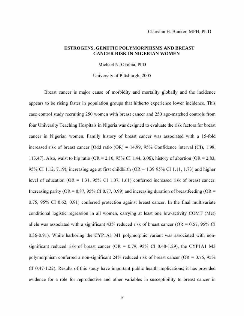
Clareann H. Bunker, MPH, Ph.D
ESTROGENS, GENETIC POLYMORPHISMS AND BREAST CANCER RISK IN NIGERIAN WOMEN
Michael N. Okobia, PhD
University of Pittsburgh, 2005
Breast cancer is major cause of morbidity and mortality globally and the incidence
appears to be rising faster in population groups that hitherto experience lower incidence. This
case control study recruiting 250 women with breast cancer and 250 age-matched controls from
four University Teaching Hospitals in Nigeria was designed to evaluate the risk factors for breast
cancer in Nigerian women. Family history of breast cancer was associated with a 15-fold
increased risk of breast cancer [Odd ratio (OR) = 14.99, 95% Confidence interval (CI), 1.98,
113.47]. Also, waist to hip ratio (OR = 2.10, 95% CI 1.44, 3.06), history of abortion (OR = 2.83,
95% CI 1.12, 7.19), increasing age at first childbirth (OR = 1.39 95% CI 1.11, 1.73) and higher
level of education (OR = 1.31, 95% CI 1.07, 1.61) conferred increased risk of breast cancer.
Increasing parity (OR = 0.87, 95% CI 0.77, 0.99) and increasing duration of breastfeeding (OR =
0.75, 95% CI 0.62, 0.91) conferred protection against breast cancer. In the final multivariate
conditional logistic regression in all women, carrying at least one low-activity COMT (Met)
allele was associated with a significant 43% reduced risk of breast cancer (OR = 0.57, 95% CI
0.36-0.91). While harboring the CYP1A1 M1 polymorphic variant was associated with non-
significant reduced risk of breast cancer (OR = 0.79, 95% CI 0.48-1.29), the CYP1A1 M3
polymorphism conferred a non-significant 24% reduced risk of breast cancer (OR = 0.76, 95%
CI 0.47-1.22). Results of this study have important public health implications; it has provided
evidence for a role for reproductive and other variables in susceptibility to breast cancer in
iv
indigenous African women, thus contributing to the global epidemiologic literature on risk
factors for breast cancer in populations of African ancestry. It has also provided data suggesting
protection for breast cancer for women harboring the low-activity COMT (Met) allele of the
codon 158 polymorphism of the COMT gene. In addition, the findings of this study will serve a
useful resource tool in future research and policy decisions aimed at breast cancer control and
prevention in these populations.
v
TABLE OF CONTENTS 1. INTRODUCTION .................................................................................................................. 1 2. CASE-CONTROL STUDY OF RISK FACTORS FOR BREAST CANCER IN NIGERIAN
WOMEN..................................................................................................................................... 9 2.1. ABSTRACT.................................................................................................................. 10 2.2. INTRODUCTION ........................................................................................................ 11 2.3. METHODS ................................................................................................................... 12
2.3.1. Study population ................................................................................................... 12 2.3.2. Recruitment of study participants ......................................................................... 12 2.3.3. Data collection ...................................................................................................... 13 2.3.4. Anthropometric measurements ............................................................................. 14 2.3.5. Data analysis ......................................................................................................... 14
2.4. RESULTS ..................................................................................................................... 15
2.4.1. Socio-demographic characteristics of the study population ................................. 15 2.4.2. Reproductive characteristics ................................................................................. 18 2.4.3. Anthropometric measurements ............................................................................. 21 2.4.4. Controlling for additional risk factors................................................................... 21 2.4.5. Analysis of menopausal status .............................................................................. 22
2.5. DISCUSSION............................................................................................................... 22
3. ASSOCIATION OF CATECHOL-O-METHYLTRANSFERASE (COMT) GENE AND
BREAST CANCER RISK IN NIGERIAN WOMEN.............................................................. 38 3.1. ABSTRACT.................................................................................................................. 39 3.2. INTRODUCTION ........................................................................................................ 40 3.3. MATERIALS AND METHODS.................................................................................. 42
3.3.1. Subjects ................................................................................................................. 42 3.3.2. Sample donation and preparation.......................................................................... 43 3.3.3. PCR and RFLP analysis........................................................................................ 44 3.3.4. Statistical Analysis................................................................................................ 45
3.4. RESULTS ..................................................................................................................... 46
vi
3.4.1. Demographic characteristics................................................................................. 46 3.4.2. Allele and genotype frequencies........................................................................... 47 3.4.3. COMT genotypes and breast cancer risk .............................................................. 48
3.5. DISCUSSION............................................................................................................... 51
4. CYTOCHROME P4501A1 GENETIC POLYMORPHISMS AND BREAST CANCER RISK
IN NIGERIAN WOMEN ......................................................................................................... 65 4.1. ABSTRACT.................................................................................................................. 66 4.2. INTRODUCTION ........................................................................................................ 67 4.3. MATERIALS AND METHODS.................................................................................. 69
4.3.1. Recruitment of study participants ......................................................................... 69 4.3.2. Biological samples ................................................................................................ 70 4.3.3. DNA extraction..................................................................................................... 70 4.3.4. PCR and Genotyping ............................................................................................ 71 4.3.5. Statistical Analysis................................................................................................ 72
4.4. RESULTS ..................................................................................................................... 73
4.4.1. Descriptive epidemiology ..................................................................................... 73 4.4.2. Association of CYP1A1 M1 polymorphism and breast cancer risk ..................... 74 4.4.3. Association of CYP1A1 M3 polymorphism and breast cancer risk ..................... 76
4.5. DISCUSSION............................................................................................................... 78
5. GENERAL DISCUSSION/FUTURE RESEARCH............................................................. 95 6. PUBLIC HEALTH SIGNIFICANCE OF STUDY ............................................................ 111 7. SUMMARY........................................................................................................................ 114 8. APPENDIX A: STUDY QUESTIONNAIRE .................................................................... 117 9. APPENDIX B: CONSENT FORM .................................................................................... 131 10. APPENDIX C: MAP OF NIGERIA SHOWING STUDY SITES................................. 136 BIBLIOGRAPHY....................................................................................................................... 137
vii
LIST OF TABLES Table 2-1: Conditional logistic regression comparing cases and controls. Significant predictors of
breast cancer risk [Numbers (N), Percentages (%)], [Means (S.D.)], age-adjusted odds ratio, 95% confidence interval for cases and controls.................................................................... 30
Table 2-2: Conditional logistic regression comparing cases and controls. Non-significant
predictors of breast cancer risk [Numbers (N), Percentages (%)], [Means (S.D.)], age-adjusted odds ratio, 95% confidence intervals...................................................................... 31
Table 2-3: Multivariate conditional logistic regression comparing cases and controls................ 32 Table 3-1: Conditional logistic regression comparing cases and controls. Significant predictors of
breast cancer risk [Numbers (N), Percentages (%)], [Means (S.D.)], age-adjusted odds ratio, 95% confidence interval ....................................................................................................... 58
Table 3-2: COMT allele and genotype frequencies for breast cancer cases and controls ............ 59 Table 3-3: Distribution of polymorphisms of the COMT gene in relation to breast cancer......... 60 Table 4-1: CYP1A1 Polymorphisms analysis by restriction enzyme digest ................................ 83 Table 4-2: Conditional logistic regression comparing cases and controls. Significant predictors of
breast cancer risk [Numbers (N), Percentages (%)], [Means (S.D.)], age-adjusted odds ratio, 95% confidence interval ....................................................................................................... 86
Table 4-3: CYP1A1 M1 Allele and Genotype frequencies for Breast Cancer Study Participants87 Table 4-4: Distribution of genotype frequencies of the CYP1A1 M1 polymorphism in relation to
breast cancer risk................................................................................................................... 88 Table 4-5: CYP1A1 M3 Allele and Genotype frequencies for Breast Cancer Study Participants89 Table 4-6: Distribution of genotype frequencies of the CYP1A1 M3 polymorphism in relation to
breast cancer risk................................................................................................................... 90
viii
LIST OF FIGURES Figure 2-1: Age distribution of breast cancer patients.................................................................. 29 Figure 3-1: COMT NlaIII Restriction Digest ............................................................................... 57 Figure 4-1: CYP1A1 M1 MspI Restriction Digest ....................................................................... 84 Figure 4-2: CYP1A1 M2 BsrdI Restriction Digest....................................................................... 84 Figure 4-3: CYP1A1 M3 MspI Restriction Digest ....................................................................... 85 Figure 4-4: CYP1A1 M4 BsaI Restriction Digest ........................................................................ 85 Figure 10-1: Map of Nigeria showing the Breast Cancer Study Sites........................................ 136
ix

ACKNOWLEDGMENTS
First and foremost, I thank Almighty God, for all His mercies, protection, and guidance
thus far in life. God has been very generous to me since birth and has stood between me and
disappointment, failure and death since my childhood days. Secondly, I thank my parents for all
their care, pains and anxieties in nurturing me since childhood. They taught me the first lessons
of humility, honesty and hardwork and implore me to show devotion to endeavors in life. To my
lovely and devoted wife, Esther and my lovely children; Nelson, Isioma and Amaka, I say thank
you for all your perserverance and patience with me. Even while in Nigeria, I was always on the
road to the Nigerian study sites; the feelings of desertion you experience are sincerely
appreciated. My academic advisor, Dr. C.H. Bunker, single handedly brought me to experience
the academic paradise of Pittsburgh; I sincerely thank you. I appreciate the pivotal role you have
continued to play in expanding my academic horizon. You have forever changed my worldview
for good. I say thank you to all my Committee Members; Drs. Kuller, Zmuda, Kammerer, and
Vogel for all your assistance in making this dissertation a reality. Staff and graduate students of
the Department of Epidemiology, University of Pittsburgh were wonderful in their assistance
throughout the conduct of this study; I appreciate all your efforts. Special thanks to the staff of
Dr. Zmuda’s Molecular Epidemiology laboratory; you made this dissertation possible through
your constant assistance and making sure I had all the equipments and reagents I needed for the
laboratory studies. To all the consultants and staff of the Departments of Surgery at the Nigerian
Study Sites, including University of Benin Teaching Hospital, Benin City, University of Nigeria
Teaching Hospital, Enugu, Nnamdi Azikiwe University Teaching Hospital, Nnewi; and
University of Port Harcourt Teaching Hospital, Port Harcourt, I say thank you for all your
x

assistance during the conduct of this study in Nigeria. Finally, to the founding fathers of this
great University of Pittsburgh over two centuries ago, I say thank you for your courage and
foresight.
Pittsburgh, April 2005
Michael N. Okobia
xi
1. INTRODUCTION
Breast cancer is the most common form of cancer among women, and the leading cause
of death related death among women globally (Parkin et al, 1999). The lifetime risk of
developing breast cancer is reportedly 1 in 8 for women in North America, and 1 in 12 for
Western Europe. The incidence of breast cancer in women has been rising since the 1940s and
the rise is occurring more rapidly in population groups that hitherto enjoyed a low incidence of
the disease (Parkin et al, 1999). For many years, breast cancer incidence and mortality rates have
been highest in North America and Northern Europe, intermediate in Southern Europe and Latin
America, and lowest in Asia and Africa (Parkin et al., 1997). The steep rise in incidence in most
countries of Northern Europe and North America from the early 1980s has been ascribed to the
introduction of mammographic screening for breast cancer (Kelsey et al. 1993). In recent years,
steep increases in breast cancer incidence and mortality rates have been reported in many Asian
and Central European countries. Thus the magnitudes of the differences in incidence rates
between countries such as Japan and the United States are less than they were previously (Kelsey
et al., 1993). Over time, the world breast cancer burden has increased steadily with an almost
doubling of the annual number of estimated new cases over a 20-year span, the increase being
seen both in the developed as well as the developing countries (Parkin et al., 1988); Parkin et al.,
1985; Parkin et al., 1999; Sasco AJ, 2001).
International differences in breast cancer incidence rates has been hypothesized to be
partially related to variation in such factors as body weight (De Waard et al 1977), some aspects
of diet (Amstrong et al., 1975), hormone levels (Henderson et al., 1991), and reproductive
1
characteristics especially age at menarche (Henderson et al., 1991), and possibly menstrual cycle
length, parity, and lactation (Wang et al., 1992). Differences in hormone levels among women in
various countries have also been thought to play a role (Bernstein et al., 1993). Studies of
migrants to the United States suggest that environmental factors rather than genetic factors are
mainly responsible for the variation in breast cancer incidence rates among countries.
Incidence of Breast Cancer in US Populations
There are racial/ethnic variations in the incidence and mortality rates of breast cancer among
the various ethnic/racial groups in the United States. Breast cancer is the most common cancer
among women in every major ethnic group in the United States. Estimates of age-adjusted
incidence rates per 100,000 by race/ethnicity in California for the 1988-1989 were 110.6 for
white women, 96.3 for black women, 59.2 for Hispanic women, and 52.8 for Asian American
women (Hoegh et al., 1992). Incidence rates are higher in whites than blacks above the age of
40-45 years, but below this age range, blacks are at slightly higher risk than whites (Hoegh et al.,
1992). Younger average age at first birth among blacks appears to be one of the factors
accounting for this phenomenon. High parity may be associated with breast cancer that is
diagnosed in women younger than 45-50 years of age, but it appears to be protective against
breast cancer diagnosed at older ages (Kelsey et al., 1993); thus the high parity of blacks than
whites could be partly responsible for these risk differentials.
Incidence trends of breast cancer in the US
Between 1973 and 1998, incidence rates of invasive breast cancer increased for women
age 40 and over, although rates grew more than two-and-a-half times faster among women age
2

50 and older than for women in their 40s (Howe et al., 2001). Incidence rates for invasive breast
cancer did not increase for women under age 40 during this time. Incidence rates of ductal
carcinoma-in-situ (DCIS) increased for women of all ages during this same time period, although
rates grew fastest in women over age 50 (Ernster et al., 1996). The perceptions of increasing
numbers of breast cancer cases in young women in the late 1980s and early 1990s are largely due
to the growth and aging of the US population, as many “baby boomer” women reached ages 25-
40 at that time (Howe et al., 2001). Since 1985, breast cancer incidence rates among women
under age 40 have actually declined significantly at an average 1.3% per year. It is important to
note that in the past decade, incidence rates of breast cancer have remained relatively unchanged
in women of all racial and ethnic groups.
There has been an important reduction in breast cancer death rates in the US in recent
years beginning in the late 1990s (Howe et al., 2001). This decline in breast cancer mortality has
been attributed to both improvements in breast cancer treatments and the benefits of
mammography screening (Ries et al., 2001).
Incidence of Breast Cancer in African Populations
Although there are no accurate data on the incidence rate of breast cancer in most of sub-
Saharan Africa, data emerging from the few cancer registries within the region gives estimates of
the incidence of the disease within various countries in the region. Reports from the Ibadan
cancer registry in Nigeria estimated the incidence of breast cancer in Nigeria in 1976 to be 15.3
per 100,000 but this rose to 33.6 per 100,000 by 1993 (Ihekwaba FN, 1992). The age
standardized incidence rate (world standard population) from the Abijan cancer registry in Ivory
Coast is 21.4 per 100,000 (Echimane et al, 2000). Other incidence figures from sub-Saharan
3
Africa are 20.4 per 100,000 for the Harare cancer registry in Zimbabwe (Chokunoga et al., 2000)
and 16.4 per 100,000 from Kyadondo County in Uganda (Wabinga et al., 2000). Some other
registries have reported lower incidence figures of 10.9 per 100,000 in Conarkry, Guinea
(Koulibaly et al., 1997), 10.2 per 100,000 from Bamako Mali (Bayo et al., 1990) and 3.4 per
100,000 from Gambia cancer registry (Bah et al., 1990). Although the above incidence figures
are much lower than the incidence of 79.3 per 100,000 in African American women in the U.S.
(SEER, 1988-1992), there is general consensus that there is gross under-reporting due to low
awareness, poverty, sociocultural factors and the absence of breast cancer-screening programs in
countries within sub-Saharan Africa.
For the past two centuries, it has been suspected that sex hormones particularly estrogens
may play some role in the etiology of breast cancer. It was, however, in the early 1970s that the
role of these hormones in the causation of the disease was demonstrated by MacMahon et al.
(1973). In 1983, Pike and colleagues (1983) observed that when the age-incidence curve was
plotted on a log-log scale, the curve produced assumed a straight line until approximately age 50
years, when a decrease in the curve is noted, indicating that the premenopausal period probably
creates a fertile period for the pathophysiological processes culminating in the manifestation of
the disease. Since then, a lot of studies have been conducted in an attempt to explain the role of
female hormones in the etiology and biological behavior of breast cancer. Several reproductive
risk factors have been implicated in the etiology of the disease, including age at menarche and
menopause, menstrual irregularity, age at first full-term pregnancy, parity, breastfeeding, and age
at last childbirth. Other related hormonal factors include use of hormonal contraceptives,
hormone replacement therapy and environmental exposure to hormone-related substances
(xenohormones).
4
Although recent studies have provided evidence for familial clustering of breast cancer,
high penetrance genes are thought to account for only 5-10% of all cases of the disease (Johnson
et al., 1995), indicating that over 90% of cases of breast cancer may be accounted for by low-
penetrance genes acting in concert with various environmental factors.
Cytochrome P4501A1 (CYP1A1) and cytochrome P4501B1 (CYP1B1) genes are
involved in the hydroxylation of estradiol and estrone to catechol estrogen intermediates.
Catechol estrogens particularly 4-hydroxyestadiol and 16-hydroxyestradiol have been shown to
induce DNA damage via formation of estrogen catechol-DNA adducts as well as the generation
of superoxide radicals that have been associated with single strand DNA breaks and other toxic
effects on proteins and other cellular macromolecules (Han et al. 1994). On the other hand, the 2-
hydroxy catechol estrogens including 2-OH estradiol and 2-OH estrone are devoid of estrogenic
activity. COMT is one of the several phase II enzymes involved in the conjugation and
inactivation of catechol estrogens. These enzymes are widely distributed in the body particularly
in target organs prone to estrogen-induced carcinogenesis including the breast. In addition,
different functional polymorphisms influencing the activity of these enzymes have been
described and evidence is accumulating that these polymorphisms may determine inter-
individual differences in exposure to estrogen-related carcinogenic metabolites.
In the past few decade efforts have been made to relate the above actions of estrogens and
its metabolites in animal models to human breast carcinogenesis. Although studies in African
American women have highlighted most of the reproductive risk factors for breast cancer in
blacks, there is little data in the literature on the role of these variables in breast cancer
susceptibility in African populations south of the Sahara. In the past decade, studies in molecular
epidemiology have been devoted to quantifying the contribution of low-penetrance genes to
5
breast cancer risk in various populations. Most of these studies have been conducted in
Caucasian populations, with few emerging reports in Asian populations. Very little literature
exist on the role of these genetic polymorphisms on breast cancer risk in populations of African
descent in the US, partly because of the low participation rate of African-American women in
such studies. The few studies that have recruited African-American women often are of very
small sample sizes. Overall, there has been a lot of inconsistency in the reported effects of these
polymorphic variants on breast cancer risk in various populations partly because these
polymorphisms have different allele frequencies in various populations. For example studies
have shown that the G to A transition mutation in the COMT gene confer increased breast cancer
risk in Asian populations (Yim et al., 2001, Huang et al., 1999) but not in most Caucasian
populations (Millikan et al., 1998; Lavigne et al., 1997) studied to date despite the fact that the
low-activity allele of the COMT gene has a much higher prevalence in Caucasian populations
compared with Asian populations. In addition, some CYP1A1 polymorphic variants such as the
CYP1A1 M1 is associated with increased risk of breast cancer in African-American women but
not in Caucasians.
Overall, the existing literature on risk factors for breast cancer is inadequate particularly
in populations of African descent. The present study, recruiting 250 Nigerian women with
histologically confirmed breast cancer and 250 aged-matched control subjects, and aimed at
evaluating the epidemiological and genetic risk factors for breast cancer in Nigeria women was
designed to test the following hypothesis:
6
Hypothesis 1
Women with breast cancer will have lifetime/reproductive experiences associated with
higher estrogen exposure compared to those without the disease. We speculate that women with
breast cancer will have earlier age at menarche, later age at menopause, later age at first full-term
pregnancy, lower parity, and shorter overall duration of breastfeeding compared to women
without the disease. In addition, we hypothesize that women with breast cancer will be more
likely to have positive history of breast cancer in first- and second-degree relatives compared to
the control subjects.
Hypothesis 2
Women with breast cancer are more likely to harbor the low-activity COMT (Met) allele
of catechol-O-methyltransferase (COMT), the gene encoding the phase II enzyme responsible for
the detoxification of the carcinogenic catechol estrogens particularly 4-hydroxyestradiol to its
biologically inactive intermediate, 4-methoxyestradiol for subsequent excretion. We speculate
that this slower rate of detoxification might lead to the accumulation of genotoxic metabolites
such as 4-hydroxyestradiol thereby exposing women with this variant allele to increased risk of
breast cancer.
Hypothesis 3
We also hypothesized that women with breast cancer will experience lower rate of 2-
hydroxylation of estradiol compared to control women. This lower 2-hydroxylation rate is based
on the speculation that these cancer-bearing women will harbor cytochrome P450 1A1
(CYP1A1) polymorphisms that encode enzymes with decreased catalytic activity. This will
result in lower lifetime exposure to 2-hydroxyestradiol (a non-carcinogenic metabolite), which is
7

converted to 2-methoxyestradiol. 2-Methoxyestradiol has been shown to exert anti-angiogenic,
anti-tubulin, and antiproliferative properties on tumor cells.
8
2. CASE-CONTROL STUDY OF RISK FACTORS FOR BREAST CANCER IN NIGERIAN WOMEN
To be submitted to International Journal of Cancer
Okobia M.N.1,4
Bunker C.H. 1 Zmuda JM1
Kammerer CM2
Vogel VG3
Uche E.E.O. 5Anyanwu S.NC. 6
Ezeome E.R. 7 Kuller L.H. 1 Ferrell R.E 2
From the Department of Epidemiology1, Department of Human Genetics2, Graduate School of
Public Health, Department of Medicine3, University of Pittsburgh, Pittsburgh, PA 15261, U.S.A.;
Department of Surgery, University of Benin Teaching Hospital, Benin City, Nigeria4;
Department of Surgery, University of Port Harcourt Teaching Hospital, Port Harcourt, Nigeria5;
Nnamdi Azikiwe University Teaching Hospital, Nnewi, Nigeria6; and Department of Surgery,
University of Nigeria Teaching Hospital, Enugu, Nigeria7.
9
2.1. ABSTRACT
This study was aimed at evaluating the risk factors for breast cancer in Nigerian women.
A case-control design recruiting 250 women with breast cancer and 250 age-matched female
controls was adopted for the study. Both cases and controls were drawn from four University
Teaching Hospitals located in Midwestern and Southeastern Nigeria. Data on the clinical and
epidemiological characteristics of the respondents were collected using interviewer-administered
structured questionnaires followed by the anthropometric measurements. The mean ages of the
cases and controls were 46.1 and 47.1 years, respectively. Fifty-seven percent of the cases were
premenopausal while 43% were post- menopausal. Using conditional logistic regression, the
effects of the various risk factors for breast cancer in the study population were assessed.
Positive family history of breast cancer in first- and second-degree relatives was associated with
a 15-fold increased risk of breast cancer [Odd ratio (OR) = 14.99, 95% Confidence interval (CI),
1.98, 113.47]. Also, waist to hip ratio (OR = 2.10, 95% CI 1.44, 3.06), history of abortion (OR =
2.83, 95% CI 1.12, 7.19), increasing age at first childbirth (OR = 1.39 95% CI 1.11, 1.73) and
higher level of education (OR = 1.31, 95% CI 1.07, 1.61) conferred increased risk of breast
cancer. Increasing parity (OR = 0.87, 95% CI 0.77, 0.99) and increasing duration of
breastfeeding (OR = 0.75, 95% CI 0.62, 0.91) conferred protection against breast cancer. The
findings from this study have shown that family history and reproductive characteristics are
significant predictors of breast cancer risk in Nigerian women.
Key words: breast cancer, risk factors, Nigeria women.
10

2.2. INTRODUCTION
Breast cancer is currently the most common malignancy in Nigerian women and the
incidence seems to be on the increase1. It has overtaken carcinoma of the cervix and if the
present trend is maintained, it will become, for Nigeria, and most other developing countries, the
most important non-communicable disease of the new millennium.
The actual cause of breast cancer is unknown but studies have implicated age, gender,
heredity, reproductive factors, diet and anthropometric characteristics as possible etiological
factors. Most of the available literature on the role of these risk factors in breast susceptibility is
drawn from Caucasian populations. While these factors may be at play in Nigerian women, it is
important to note that there is considerable variation in the geographical, racial and ethnic
distribution of the disease attributed to environmental and genetic factors2. Anecdotal
observations by clinicians in Nigeria suggest that the epidemiological characteristics of the
disease in Nigerian women differ from that in Caucasian populations. For example, most women
with breast cancer in Nigeria are multiparous and they practice prolonged lactation, factors,
which are thought to be protective against the disease.
While several investigators have reported the clinical and pathological characteristics of
breast cancer in Nigerian women,3-6 little is known about risk factors of the disease in this
population3,7,8. Yet it is the nature of the disease that each society, race and population must seek
to define the characteristics of the disease among its people and evolve appropriate management
strategy. This study is aimed at identifying risk factors for breast cancer in Nigerian women.
Identification of these factors may enhance the ability to prevent the disease by permitting better-
focused health education and other preventive strategies.
11
2.3. METHODS
2.3.1. Study population
The study participants consisted of 250 breast cancer cases and 250 age- and sex-matched
(within 5 years) controls recruited from four University Teaching Hospitals in Midwestern and
Southeastern Nigeria including the University of Benin Teaching Hospital, Benin-City;
University of Nigeria Teaching Hospital, Enugu; Nnamdi Azikiwe University Teaching Hospital,
Nnewi; and University of Port Harcourt Teaching Hospital, Port Harcourt. The study protocol
and consent forms were approved by the Institutional Review Boards of these four hospitals and
the University of Pittsburgh. The cases consisted of prevalent and incident cases of breast cancer
that were seen within these hospitals during the period of study. Breast cancer was defined as
histologically confirmed malignant breast disease. Hospital-based, age-and sex-matched controls
were recruited at the time of their outpatient clinic visits or in-patient wards at each of the
hospitals where cases were recruited. The age match was within 1 to 5 years. Eligible controls
were women who were being treated for non-hormonal and non-cancerous lesions.
2.3.2. Recruitment of study participants
Physicians at the various study sites reviewed in-patient and outpatient medical records for
information regarding past medical history and current medical complaint to identify potential study
participants. Brief information about the study was provided to these potential study participants
after which those willing to participate in the study were asked to contact the investigators for
further information about the study.
12
2.3.3. Data collection
Data was collected during one 30-minute visit. First, key details of the study in respect of
objectives of the study, study protocol, risks and benefits, confidentiality and rights of
participants were explained to all potential participants. Those willing to participate signed
informed consent after which questionnaires were administered by the investigators. The
interview was conducted in English; however when participants did not understand English,
literate adult relatives, who as a rule accompany patients to the hospital, explained contents of
the questionnaire and consent forms to the study participants. Each potential participant had an
option to refuse participation in the study. An opportunity was also granted for participants to ask
any questions.
The questionnaire was designed to gather demographic data including age, religion,
educational and marital status and occupational history. Information in respect of use of alcohol,
cigarette smoking, history of breast and other cancers in first- and second-degree relatives and
position among siblings were also obtained. Reproductive characteristics related to age at
menarche, age at first childbirth, duration of breastfeeding of each child, use of hormonal and
surgical contraceptives, history of abortion, age at menopause and use of hormone replacement
therapy were also noted. For case participants, age at diagnosis of breast cancer and treatment
received were also noted. In addition knowledge of study participants about breast cancer was
also obtained.
13
2.3.4. Anthropometric measurements
Height, weight, waist and hip measurements were taken while the subject was standing.
The height was measured with a vertical tape attached to the wall; the weight was taken using a
calibrated scale; the waist measurement was made at the umbilicus; and, the hips were measured
at the widest part of the buttocks.
2.3.5. Data analysis
All questionnaires were reviewed for missing or incorrect data before the end of the
interview section with each participant. All forms were reviewed for suspicious data, and returned to
the participant’s file to be confirmed or corrected the following day, if necessary. The data analysis
was done using the Statistical Analysis System (SAS) software (Version 8.0). Descriptive analysis
was carried out to characterize the demographic variables of the study participants. For logistic
regression analysis, the variables were classified as follows: family history of breast cancer in first-
and second-degree relatives (yes/no), waist/hip ratio dichotomized based on the median value in
controls, abortion (yes/no), age at first childbirth (< 20 years, 20-24 years, 25-29 years, and ≥ 30
years), education (< 8 years, 8 through 11 years, 12 years or completed High School,
Vocational/Technical Training, Some College, Completed College, and Postgraduate), parity (none,
1, 2, 3, 4, and ≥ 5), and duration of breastfeeding (1-12 months, 13-24 months, 25-36 months, 37-48
months, and ≥ 49 months). Other categorical variables that were entered into the logistic regression
include cigarette smoking (yes/no), alcohol consumption (yes/no), regularity of menses (yes/no), use
of hormone contraceptive pills (yes/no), age at menarche (≤ 12 years, 13, 14, 15, 16, 17, and ≥ 18
years), and age at menopause (≤ 45 years, 45-50 years, 51-55 years, and ≥ 55 years). Body mass
14
index (BMI), calculated as weight divided by the square of height (kg/m2) was dichotomized based
upon the median value of BMI among the controls (24.65 kg/m2).
Conditional logistic regression was used to assess the strength of the association between
each of the hypothesized risk factors and breast cancer risk. Each matched case was paired with
the corresponding control to enable differences between the cases and controls to be calculated.
First each variable was assessed alone. The strength of significant variables were further assessed
by building multivariate models.
2.4. RESULTS
2.4.1. Socio-demographic characteristics of the study population
Age distribution
There were 250 cases (all females) of histologically confirmed breast cancer and 250 aged-
and sex-matched controls that were recruited for this study. The participation rate in this study was
very high as less than 1% of the patients were excluded from the study. Reasons for exclusion
included refusal to allow a blood draw and patient’s unwillingness to participate in the study. The
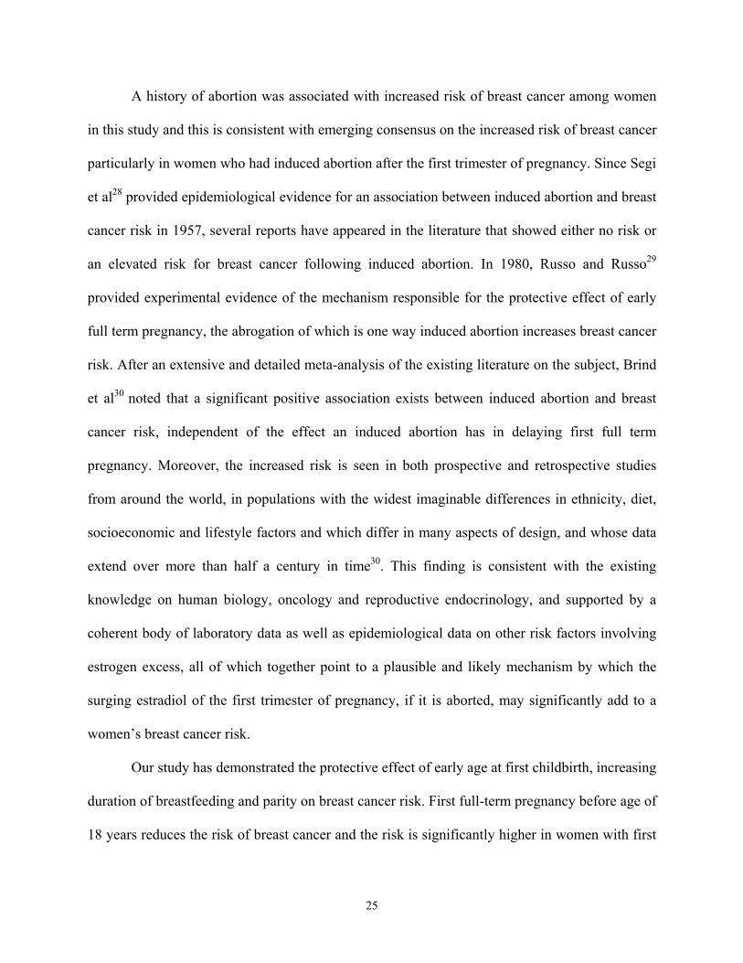
age at diagnosis for the breast cancer cases ranged from 17 years to 95 years with a mean of
46.1±12.6 years. The peak age of the cases were in the 45-49 year age range [43 patients (17.2%0],
closely followed by the 40-44 year age range [41 patients (16.4%)]. Eighty patients (32.0%) were
below the age of 40 years while 22 patients (8.8%) were aged 65 years and older as shown in Figure
2-1. The mean age of the controls was 47.1 years.
Educational status
Fewer cases [57 (22.8%)] than controls [91 (36.4%)] had less than eight years of
education while more cases [136 (54.4%)] than controls [113 (45.2%)] completed High School
15
or post High School education. In a conditional logistic regression model controlling for age,
higher level of education (completed High School or post High School education) conferred a
significant 31% increased risk of breast cancer (OR = 1.31, 95% CI 1.07, 1.61) as shown in
Table 2-1.
Marital status
Majority of the cases [195 (78.0%)] and controls [190 (76.0%)] were married or living as
married. Fewer cases [24 (9.6%)] than controls [36 (14.4%)] were never married. There were
more women who had divorced among the cases [10 (4.0%)] compared with the controls [2
(0.8%)]. Two-third of the patients (67%) presented in hospital with advanced stages of breast
cancer (Manchester Stages III and IV).
Usual adult occupation
The majority of the study participants were engaged in trading; 82 cases (32.8%) versus
100 controls (40.0%). Most of the traders were engaged in petty trading involving sale of food
items and domestic wares. Slightly more cases [81 (32.4%) than controls [74 (29.6%) were
employed in the public service. The most common jobs in the public service included teaching,
secretarial duties and nursing. Similar proportions of cases and controls were engaged in farming
at a subsistence level usually involving cultivation of food crops. The other less common
occupations included catering, fashion designing and hairdressing.
16
Family history of cancer
A much higher number of cases (19) than controls (6) reported positive family history of
various types of cancer. More cases (15) than controls (1) reported family history of breast
cancer. Six of the family breast cancers in the cases were in first-degree relatives, comprising
four in sisters and two in mothers while seven were in second degree relatives consisting of four
in aunts, one in a grandmother, one in a cousin and one in a niece. Two of the cases reported
history of breast cancer in two relatives; one was that of breast cancer in her sister and
grandmother while the second reported history of breast cancer in her sister and cousin. The only
control with positive family history of breast cancer was reported in a sister. Other cancers
reported in family members in the cases include one case each of carcinoma of the cervix,
carcinoma of the prostate, and one patient that reported history of liver cancer in her father and
brother. The other cancers reported by control participants include two cases of squamous cell
carcinoma of the skin, one case of oral cancer and one case of carcinoma of the larynx. There
was one case of an unspecified abdominal cancer in a relative of one of the controls. As shown in
Table 2-1, family history of breast cancer was strongly associated with a 15-fold increased risk
of breast cancer (OR = 14.99, 95% CI 1.98, 113.47) in a conditional logistic regression age.
Use of alcohol
Alcohol consumption was slightly more common among the cases [83 (33.2%)]
compared with the controls [70 (28.0%)]. Most of them were occasional drinkers and the types
of alcohol consumed include beer, palm-wine, and locally brewed gin. Alcohol consumption was
17
associated with a non-significant 29% increased risk of breast cancer among the study population
(OR = 1.29, 95% CI 0.87, 1.90) as shown in Table 2-2.
Cigarette smoking
Cigarette smoking was not a common practice among study participants as only two of
the cases and two of the controls reported having smoked cigarettes regularly for more than six
months. Cigarette smoking was not associated with increased risk of breast cancer.
2.4.2. Reproductive characteristics
Age at menarche
The age at menarche in cases (range10-20 years and mean 14.75 years) did not differ
significantly from that of controls (range 10-20 years, mean 14.5 years). As shown in Table 2-2,
later age at menarche (>14 years) was associated with a non-significant 11% increased risk of
breast cancer (OR = 1.11, 95% CI 0.98, 1.26).
Age at first childbirth
There were 210 parous women among the cases and 209 parous women among the
controls. The age at first childbirth ranged from 14-44 years (mean 23.18 years) for the cases and
14-42 years (mean 21.87 years) for the control participants. Fewer cases [48 (22.8%)] had their
first childbirth before the age of 20 years compared with controls [73 (34.93%)]. More cases [29
(13.81%)] than control participants [13 (6.22%)] had their first childbirth after the age of 30
years. Older age at first childbirth was associated with a significant 39% increased risk of breast
cancer in a model controlling for age (OR = 1.39, 95% CI 1.11, 1.73).
18
Duration of breastfeeding
All parous women among the study participants breastfed their babies ranging from 2-
216 months (mean 65.49 months) among the cases and 2-312 months for the controls (mean
80.96 months). More cases [93 (44.50%)] than controls [68 (32.69%)] breastfed for 48 months
and below while fewer cases [116 (55.50%)] than control participants [140 (67.31%)] breastfed
for over 48 months. Breastfeeding was associated with a significant 25% reduction in breast
cancer risk (OR = 0.75, 95% CI 0.62, 0.91).
Parity
The parity of the cases ranged from 0-11 with a mean parity of 4 while parity among the
controls ranged from 0-13 with a mean of 5. More cases (97) than controls (69) had five children
and below while fewer cases (113) than controls (140) had more than five children. Higher parity
was associated with a significant 12% reduced risk of breast cancer (OR = 0.88, 95% CI 0.81,
0.96).
Age at menopause
About equal number of cases [107 (42.8%)] and controls [108 (43.2%)] were
postmenopausal. Fewer cases (11) than controls (17) attained menopause before the age of 45
years while more cases (48) than controls (38) became menopausal between the ages of 45 and
49 years. Slightly fewer cases (41) than controls (47) attained menopause between the ages of 50
and 54 years. About equal number of cases (7) and controls (6) attained menopause after the age
19
of 55 years. Older age at menopause was associated with a 7% non-significant increased risk of
breast cancer (OR = 1.07, 95% CI 0.71, 1.60).
Regularity of menses
Information about menstrual regularity was available in 227 cases, of which 27 (11.89%)
reported irregular menses. Among the controls 231 provided information about menstrual
regularity and 31 of them (13.42%) had irregular menses. Irregular menses was associated a 19%
non-significant reduced risk of breast cancer (OR = 0.81, 95% CI 0.45, 1.44).
Use of hormonal contraceptives
Use of hormonal contraceptives was reported among 43 (17.2%) of the cases and 32
(12.8%) control participants. Hormone contraceptive use conferred a 40% non-significant
increased risk of breast cancer among study participants (OR = 1.40, 95% CI 0.84, 2.34).
Birth order
Information about birth order was available in 222 cases and 233 controls. The birth
order of the cases ranged from 1-9 while the range is from 1-11 for the controls. About equal
number of cases (136) and controls (135) were in the 1-3 birth order category while slightly
fewer cases (68) than controls (77) were in the 4-6 category. Birth order was not associated with
increased of breast cancer in the study population.
20
2.4.3. Anthropometric measurements
Body mass index (BMI)
One hundred and thirty-six cases (54.4%) and 125 controls (50.0%) had BMI below
24.65. Slightly fewer cases [114 (45.6%)] than controls [125 (50.0%)] had BMI above 24.65 (the
median BMI for controls). Body mass index was not associated with increased risk of breast
cancer (OR = 0.83, 95% CI 0.58, 1.19).
Waist/Hip ratio
Median waist to hip ratios for the cases and controls were 0.92 and 0.90 respectively.
Fewer cases [89 (35.6%)] than controls [133 (53.2%)] had waist/hip ratio of 0.90 and below
while more cases [161 (64.4%)] than controls [117 (46.8%)] had waist/hip ratio above 0.90.
Higher waist/hip ratio was associated with a 2.0 fold increased risk of breast cancer in a logistic
regression model (OR = 2.0, 95% CI 1.39, 2.87) as shown in Table 2-1.
2.4.4. Controlling for additional risk factors
Significant predictors of breast cancer identified in the univariate conditional logistic
regression model including family history of breast cancer, waist/hip ratio, abortion, education,
marital status, age at first childbirth, parity and duration of breastfeeding were entered into the
model and various combinations of risk factors were found to be significant as shown in Table 2-
3. Overall, family history of breast cancer, waist/hip ratio, age at first childbirth, duration of
breastfeeding, and education remain significant with three variables in the model while family
21
history of breast cancer, waist/hip ratio, age at first childbirth and education were retained in the
model controlling for four additional factors.
2.4.5. Analysis of menopausal status
Further stratified analysis on the basis of menopausal status revealed that educational status
(OR 1.45; 95% CI 1.09, 1.95), use of hormone contraceptives (OR 2.67; 95% CI 1.04, 6.82), and
late age at first childbirth (OR 1.72; 95% CI 1.16, 2.53), were associated with significantly
increased risk of breast cancer risk in postmenopausal women but not in premenopausal women.
Longer duration of breastfeeding conferred a 33% reduced risk of breast cancer also in
postmenopausal women (OR 0.67; 95% CI 0.48, 0.92). In premenopausal women, higher waist
to hip ratio was associated with a significant 4-fold increased risk of breast cancer.
2.5. DISCUSSION
This study was aimed at examining the role of reproductive and other epidemiological
risk factors in susceptibility to breast cancer among women in Midwestern and Southeastern
Nigeria. We were interested in this subject because of the paucity of data on risk factors for
breast cancer in sub-Saharan Africa. Establishing the risk factors of the disease is the first major
step in understanding the etiology of the disease and designing appropriate control and
preventive measures. Much of what has been reported about risk factors for breast cancer has
come from studies conducted in populations in the other parts of the world. It is known that
environmental factors may play considerable role in breast cancer susceptibility and most of the
populations studied live in different geographical areas. We were therefore interested in
22
assessing the risk factors that may be at play in a black population in Midwestern and
Southeastern Nigeria. This population provides a particularly interesting environment for this
study because of the very high population density in Midwestern and Southeastern Nigeria9, with
a very high participation rate of over 99%. This is in contrast to the findings of other
investigators who have reported low recruitment rates particularly for blacks in the United
States10.
The age of the breast cancer patients in this study ranged from 17 to 90 years with a mean
of 46.1±12.6 years. Fifty-seven percent of the cases were premenopausal while 43% were
postmenopausal and majority of the patients presented in hospital with advanced stages of the
disease. The disease pattern in this study reflects the general picture in most sub-Saharan African
populations. The mean age of in the cases in this study is slightly higher than that reported by
Anyanwu4 in Eastern Nigeria (44 years), slightly lower than reports from Ibadan in Western
Nigeria (48 years)3 but much lower than figures among blacks patients in Durban, South Africa
(54±10.9 years)11 and Chris Hani Baragwanath Hospital in Soweto (50 years)12. Thus, in sub-
Saharan African countries breast cancer occurs at a much earlier age than in the case of patients
in most developed populations. Interestingly, African-American patients also tend to present at a
younger age. In one study, 33% were under 50 years of age, compared with 25% in the case of
white patients studied13. The proportion of postmenopausal women among the cases in this study
is higher than the 20% previously reported from Ibadan in Nigeria3 but considerably less than
two-thirds reported in Caucasians14,15.
Among the relevant etiological factors identified in this study, family history of breast
cancer in first- and second-degree relatives conferred a 15-fold increased risk of breast cancer,
controlling for age but this risk was attenuated to eight-fold when additional factors including
23
waist/hip ratio, age at first childbirth and education were added to the model. About 30.4% of
breast cancer cases in this study fell within the category of familial and hereditary breast cancer
using the criteria of Lynch and associates.15 This is in keeping with reports in other populations
with detailed characterization of pedigree suggesting that familial and hereditary breast cancer
constitute about 32% of the total incidence of breast cancer.16
Waist/hip ratio, a surrogate marker for central adiposity was associated with a 2-fold
increased risk of breast cancer in this study and it remained significant in premenopausal women
when participants were further stratified by menopausal status. Epidemiological studies of body
fat distribution using waist/hip ratio have produced contradictory results; some being positively
related breast cancer,6,17-21 others showing no association; 22,23 inconsistencies in these findings
being related to differences in study design and sample size. Abdominal obesity is linked to
hyperinsulinemia in both pre- and post-menopausal women and estrogen production is increased
in the presence of abdominal obesity24,25. Increased estrogen bioactivity is thought to be related
to increased estrogen production in fat deposits and also to decreased estrogen binding because
of decreased levels of sex hormone binding globulin and increased triglyceride levels. Women
with abdominal obesity also show an increase in unbound testosterone levels and may in addition
show an increased production of testosterone and dihydrotestoterone.26 Insulin levels affect
plasma lipid levels, and dyslipidemia is increased in the presence of abdominal obesity. The
relative importance of abdominal obesity and hyperinsulinemia is uncertain in relation to a role
in mammary carcinogenesis, but in a subset of women, the metabolic/endocrine concomitants of
hyperinsulinemia associated with changes in the bioactivity of insulin-like growth factors (IGFs)
in breast tissue, might act synergistically with increased estrogen bioactivity27.
24
A history of abortion was associated with increased risk of breast cancer among women
in this study and this is consistent with emerging consensus on the increased risk of breast cancer
particularly in women who had induced abortion after the first trimester of pregnancy. Since Segi
et al28 provided epidemiological evidence for an association between induced abortion and breast
cancer risk in 1957, several reports have appeared in the literature that showed either no risk or
an elevated risk for breast cancer following induced abortion. In 1980, Russo and Russo29
provided experimental evidence of the mechanism responsible for the protective effect of early
full term pregnancy, the abrogation of which is one way induced abortion increases breast cancer
risk. After an extensive and detailed meta-analysis of the existing literature on the subject, Brind
et al30 noted that a significant positive association exists between induced abortion and breast
cancer risk, independent of the effect an induced abortion has in delaying first full term
pregnancy. Moreover, the increased risk is seen in both prospective and retrospective studies
from around the world, in populations with the widest imaginable differences in ethnicity, diet,
socioeconomic and lifestyle factors and which differ in many aspects of design, and whose data
extend over more than half a century in time30. This finding is consistent with the existing
knowledge on human biology, oncology and reproductive endocrinology, and supported by a
coherent body of laboratory data as well as epidemiological data on other risk factors involving
estrogen excess, all of which together point to a plausible and likely mechanism by which the
surging estradiol of the first trimester of pregnancy, if it is aborted, may significantly add to a
women’s breast cancer risk.
Our study has demonstrated the protective effect of early age at first childbirth, increasing
duration of breastfeeding and parity on breast cancer risk. First full-term pregnancy before age of
18 years reduces the risk of breast cancer and the risk is significantly higher in women with first
25
full term pregnancy after the age of 35 years31. Most studies have also found that for first births
over the entire childbearing period, the higher a woman’s age at first birth, the higher the risk32.
While some studies have reported no protective effect for early age at first full term pregnancy
others have found that age over 30 years at first child birth was associated with an increased risk
of breast cancer relative to nulliparous women33.
The effect of parity on breast cancer risk is not clearly understood. In most studies, high
parity is found to be associated with low rates of breast cancer, but the extent to which this
relationship can be explained by an inverse association between parity and age at first birth
varies between studies34. Several studies have reported a protective effect of parity independent
of the effect of age at first full term pregnancy. Kvale et al35 found a consistent and highly
significant inverse association between high parity and breast cancer. The apparent protective
effect of high parity was found in all subgroups of the patients according to demographic
variables and could not be explained by other reproductive factors. There appears to be
consistency in this finding across studies conducted in both high-risk, intermediate-risk and low-
risk areas. The protective effect of parity seems stronger in postmenopausal than in
premenopuasal women, possibly on account of the confounding effect of time since last birth in
younger women.
The long-term protective effects of pregnancy are contrasted with the observation that the
risk of carcinogenesis is actually increased in the short term after a pregnancy36. It is known that
hormones induce carcinogenesis by inducing cell proliferation, which is an essential component
of carcinogenesis. This hypothesis is consistent with the observation that increased cell
proliferation results in a larger pool of cells that are susceptible to defective DNA repair. This in
turn leads to mutations, which are subsequently propagated through increased mitotic activity
26
present in proliferating cells, and can result in cancer formation. However, it has been shown that
full term pregnancy induces differentiation of cells in the terminal duct lobular unit (TDLU) in
the breast and this effect produces the long term effect of slowing the cell cycle in the epithelial
cells of this location, which allows more time for DNA repair, which in turn will lead to
decreased carcinogenesis37.
A number of epidemiological studies have investigated the relationship between
breastfeeding and breast cancer risk. Our finding of a 25% reduction in risk conferred by
breastfeeding is consistent with reports in the literature. Overall, studies suggest a 20-30 percent
reduction in risk among women who have ever breastfed38. More consistently, a longer duration
of breastfeeding has been associated with breast cancer risk reductions as great as 40-60
percent39. Recently, age at first lactation has been identified as the arbiter of risk, with an earlier
age at initiation of lactation being associated with a stronger reduction in risk for premenopausal
women and possibly for postmenopausal women40. However, because of the very strong
correlation between age at first birth and age at first lactation, the independent effect of age at
first lactation is difficult to isolate. It is notable that in countries with low risk of breast cancer,
the protection conferred by lactation appears to be stronger and to be sustained throughout the
postmenopausal period as well.
Higher education conferred an increased risk of breast cancer in this study, in keeping
with reports in the literature41. Although, we did not measure socioeconomic status, an
established risk factor for breast cancer in most studies42,43, education is a strong surrogate for
socioeconomic status. The quest for higher education delays age at marriage and age at first
childbirth and it is associated with reduced parity and reduced duration of breastfeeding; these
factors have been shown to reduce breast cancer risk.
27

In conclusion, this study has examined the relationship between various factors and
breast cancer risk in Nigerian women. It has demonstrated increased breast cancer risk associated
with family history of breast cancer, abdominal adiposity, abortion and higher education and the
reduced risk conferred by various reproductive variables; these findings are consistent with
reports in the literature. However, studies with larger sample sizes are recommended for better
characterization of the role of these risk factors in breast cancer risk in Nigerian women. This
will provide an enabling framework for developing breast cancer risk assessment tools for the
population with the aim of identifying high-risk individuals for primary and secondary
prevention.
28
Figure 2-1: Age distribution of breast cancer patients
29
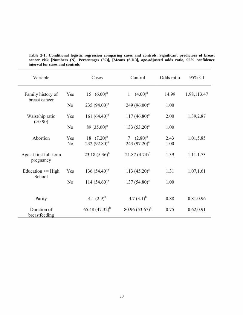
Table 2-1: Conditional logistic regression comparing cases and controls. Significant predictors of breast cancer risk [Numbers (N), Percentages (%)], [Means (S.D.)], age-adjusted odds ratio, 95% confidence interval for cases and controls
Variable
Cases
Control
Odds ratio
95% CI
Family history of
breast cancer
Yes
15 (6.00)a
1 (4.00)a
14.99
1.98,113.47
No 235 (94.00)a 249 (96.00)a 1.00
Waist/hip ratio (>0.90)
Yes
161 (64.40)a
117 (46.80)a
2.00
1.39,2.87
No 89 (35.60)a 133 (53.20)a 1.00
Abortion
Yes
18 (7.20)a
7 (2.80)a
2.43
1.01,5.85 No 232 (92.80)a 243 (97.20)a 1.00
Age at first full-term pregnancy
23.18 (5.36)b
21.87 (4.74)b
1.39
1.11,1.73
Education >= High
School
Yes
136 (54.40)a
113 (45.20)a
1.31
1.07,1.61
No 114 (54.60)a 137 (54.80)a 1.00
Parity
4.1 (2.9)b
4.7 (3.1)b
0.88
0.81,0.96
Duration of
breastfeeding
65.48 (47.32)b
80.96 (53.67)b
0.75
0.62,0.91
30
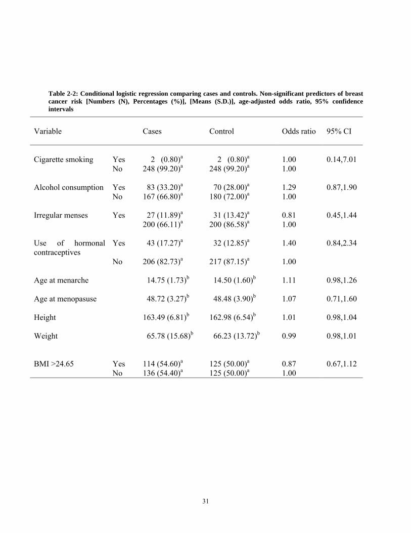
Table 2-2: Conditional logistic regression comparing cases and controls. Non-significant predictors of breast cancer risk [Numbers (N), Percentages (%)], [Means (S.D.)], age-adjusted odds ratio, 95% confidence intervals
Variable
Cases
Control
Odds ratio
95% CI
Cigarette smoking
Yes
2 (0.80)a
2 (0.80)a
1.00
0.14,7.01
No 248 (99.20)a 248 (99.20)a 1.00
Alcohol consumption Yes 83 (33.20)a 70 (28.00)a 1.29 0.87,1.90 No 167 (66.80)a 180 (72.00)a 1.00
Irregular menses Yes 27 (11.89)a 31 (13.42)a 0.81 0.45,1.44 200 (66.11)a 200 (86.58)a 1.00
Use of hormonal contraceptives
Yes 43 (17.27)a 32 (12.85)a 1.40 0.84,2.34
No 206 (82.73)a 217 (87.15)a 1.00
Age at menarche 14.75 (1.73)b 14.50 (1.60)b 1.11 0.98,1.26
Age at menopasuse 48.72 (3.27)b 48.48 (3.90)b 1.07 0.71,1.60
Height 163.49 (6.81)b 162.98 (6.54)b 1.01 0.98,1.04
Weight 65.78 (15.68)b 66.23 (13.72)b 0.99 0.98,1.01
BMI >24.65
Yes
114 (54.60)a
125 (50.00)a
0.87
0.67,1.12
No 136 (54.40)a 125 (50.00)a 1.00
31
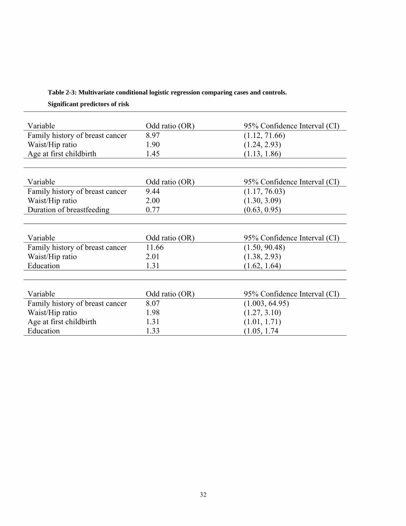
Table 2-3: Multivariate conditional logistic regression comparing cases and controls.
Significant predictors of risk
Variable
Odd ratio (OR)
95% Confidence Interval (CI)
Family history of breast cancer 8.97 (1.12, 71.66) Waist/Hip ratio 1.90 (1.24, 2.93) Age at first childbirth 1.45 (1.13, 1.86)
Variable
Odd ratio (OR)
95% Confidence Interval (CI)
Family history of breast cancer 9.44 (1.17, 76.03) Waist/Hip ratio 2.00 (1.30, 3.09) Duration of breastfeeding 0.77 (0.63, 0.95)
Variable
Odd ratio (OR)
95% Confidence Interval (CI)
Family history of breast cancer 11.66 (1.50, 90.48) Waist/Hip ratio 2.01 (1.38, 2.93) Education 1.31 (1.62, 1.64)
Variable
Odd ratio (OR)
95% Confidence Interval (CI)
Family history of breast cancer 8.07 (1.003, 64.95) Waist/Hip ratio 1.98 (1.27, 3.10) Age at first childbirth 1.31 (1.01, 1.71) Education 1.33 (1.05, 1.74
32
REFERENCES
1. Solanke TF. An overview of cancer in Nigeria. In : Solanke TF, Adebamowo CA (eds)
Report of the Workshop on State of the Art in Oncology in Ibadan and Ife. Ibadan.
National Headquarters of Cancer Registries in Nigeria. 1996: 7-12.
2. Parkin DM, Whelan SL, Ferlay J, Young J, (eds) Breast Cancer. In: Cancer in five
continents. Vol VII. IARC Scientific Publication No. 143. Lyon: International Agency
for Research on Cancer, 1997:858-859.
3. Ihekwaba FN. Breast cancer in Nigerian women. Br J Surg 1992; 79:771-775.
4. Anyanwu SN. Breast cancer in Eastern Nigeria: a ten-year review. W Afr J Med, 2000;
19(2):120-125.
5. Okobia MN, Osime U. Clinicopathological study of carcinoma of the breast in Benin
City. AJRH, 2001; 5(2):56-62.
6. Ikpatt OF, Kupio T, Collan Y. Proliferation in African breast cancer: biology and
prognostication in Nigerian breast cancer material. Modern Pathology, 2002; 15(8):783-
789.
7. Adebamowo CA, Ogundiran TO, Adenipekun AA, Oyesegun RA, Campbell OB, Akang
EE, Rotimi CN, Olopade OI. Waist-hip ratio and breast cancer risk in urbanized Nigerian
women. Breast Cancer Research 2002; 5:R18-R24.
8. Adebamowo CA, Ogundiran TO, Adenipekun AA, Oyesegun RA, Campbell OB, Akang
EE, Rotimi CN, Olopade OI. Obesity and height and breast cancer risk in urban Nigerian
women with breast cancer. Ann Epid 2003; 13(6):455-461.
33
9. National Population Commission – 1991 Population Census of Nigeria
10. Royal C, Baffoe-Bonnie A, Kittles R, Powell I, Bennett J, et al. Recruitment experience
in the first phase of the African American Hereditary Prostate Cancer (AAHPC) Study.
Ann Epidemiol 2000; 10:S68-S77.
11. Pegoraro RJ, Nirmul D, Bryer JV, Jordan JP, Joubert SM. Clinical patterns of
presentation of breast cancer in women of different racial groups in South Africa. S Afr
Med J 1985; 68:808-810.
12. Walker AR, Walker BF, Funani S, Walker AJ. Characteristics of black women with
breast cancer in Soweto, South Africa. Cancer J 1989; 2:316-319.
13. Joslyn SA, West MM. Racial differences in breast carcinoma survival. Cancer 2000;
88:114-123.
14. Saracci R, Repetto F. Epidemiology of breast cancer. Semin Oncol 1978; 5:342-350.
15. Doll R, Muir C, Waterhouse J, eds. Cancer Incidence in Five Continents. Vol 2. New
York: Springer-Verlag, 1970.
16. Lynch HT, Marcus JN, et al. Familial breast cancer, family cancer syndromes, and
predisposition to breast neoplasia. In: Bland KI, Coperland EM III (eds) The Breast:
Comprehensive Management of Benign and Malignant Diseases. Phildelphia, WB
Saunders, 1991, chap 13.
17. Ballard-Barbash R, Schatzkin A, Carter CL, et al. Body fat distribution and breast cancer
in the Framingham study. J Natl Cancer Inst 1990; 82:286-290.
18. Folsom AR, Kaye SA, Prineas RJ, Potter JD, Gapstur SM, Wallace R. Increased
incidence of carcinoma of the breast associated with abdominal adiposity in
postmenopausal women. Am J Epidemiol 1990; 131:794-803.
34
19. Schapira DV, Kumar NB, Lyman GH, Cox CE. Abdominal obesity and breast cancer
risk. Ann Intern Med 1990; 112:182-186.
20. Bruning PE, Bonfrer JMG, Hart AAM, et al: Body measurements, estrogen availability
and the risk of human breast cancer: a case-control study. Int J Cancer 1992; 51:14-19.
21. Den Tonkelaar I, Seidell JC, Collette HJA. Body fat distribution in relation to breast
cancer in women participating in the DOM-project. Breast Cancer Res Treat 1995; 34:55-
61.
22. Petrek JA, Peters M, Cirrincione C, Rhodes D, Bajorunas D. Is body fat topography a risk
factor for breast cancer. Ann Intern Med 1993; 118:356-362.
23. Swanson CA, Coates RJ, Schoenberg JB, et al. Body fat and breast cancer risk among
women under age 45 years. Am J Epidemiol 1996; 143:698-706.
24. Zimmet PZ. Hyperinsulinemia – how innocent a bystander? Diabetes Care 1993;
16(Suppl 3):56-70.
25. Ballard-Barbash R. Anthropometry and breast cancer. Cancer 1994; 74:1090-1100.
26. Kirschner MA, Samojlik E, Drejka M, et al. Androgen-estrogen metabolism in women
with upper body versus lower body obesity. J Clin Endocrinol Metab 1990; 70:473-479.
27. Stoll BA. Breast cancer: the obesity connection. Br J Cancer 1994; 69:799-801.
28. Segi M, Fukushima I, Fujisaku S, et al. An epidemiological study on cancer in Japan.
GANN 1957; 48:1-63.
29. Russo J, Russo IH. Susceptibility of the mammary gland to carcinogenesis. Am J Pathol
1980; 100:497-512.
35
30. Brind J, Chinchilli VM, Severs WB, Summy-Long J. Induced abortion as an independent
risk factor for breast cancer: a comprehensive review and meta-analysis. J Epidemiol
Community Health 1996; 50:481-496.
31. MacMahon B, Cole P, Lin TM. Age at first birth and breast cancer risk. Bull WHO 1970;
43:209-221.
32. Negri E, La Vecchia C, Bruzzi P. Risk factors for breast cancer: pooled results from three
Italian case-control studies. Am J Epidemiol 1988; 128:1207-1215.
33. Adami H-O, Bergstrom R, Lund E, Meirik O. Absence of association between
reproductive variables and the risk of breast cancer in young women in Sweden. Br J
Cancer 1990; 62:122-126.
34. Thomas DB. Epidemiology and related studies of breast cancer etiology. In: Lilienfeld
AM, ed. Reviews in cancer epidemiology. Vol 1. New York: Elsevier North Holland,
1980:153-217.
35. Kvale G, Heuch I. A prospective study of reproductive factors and breast cancer. Am J
Epidemiol 1987; 126:842-850.
36. Pathak DR, Whittemore AS. Combined effects of body size, parity, and menstrual effects
on breast cancer incidence in seven countries. Am J Epidemiol 1992; 135:153-168.
37. Russo J, Russo IH. The etipathogenesis of breast cancer prevention. Cancer Lett 1995;
90:81-89.
38. Romieu I, Hernandez-Avila M, Lazcano E et al. Breast cancer and lactation history in
Mexican women. Am J Epidemiol 1996; 143:543-552.
39. Yang CP, Weiss NS, Band PR et al. History of lactation and breast cancer risk. Am J
Epidemiol 1993; 138:1050-1056.
36
40. Freudenheim JL, Marshall JR, Vena JE et al. Lactation history and breast cancer risk. Am
J Epidemiol 1997; 146:932-938.
41. Braaten T, Weiderpass E, Kumle M, Adami H, Lund E. Education and risk of breast
cancer in the Norwegian-Swedish women’s lifestyle and health cohort study. Int J Cancer
2004; 110(4):579-583.
42. Banquet CR, Horm JW, Gibbs T, Greenwald P. Socioeconomic factors and cancer
incidence among blacks and whites. J Natl Cancer Inst 1991; 83:551-557.
43. Faggio F, Zanetti R, Costa G. Cancer risk and social inequalities in Italy. J Epidemiol
Community Health 1994; 48:447-452.
37
3. ASSOCIATION OF CATECHOL-O-METHYLTRANSFERASE (COMT) GENE AND BREAST CANCER RISK IN NIGERIAN WOMEN
To be submitted to British Journal of Cancer
Okobia M.N.1,4
Bunker C.H. 1 Zmuda JM1
Kammerer CM2
Vogel VG3
Uche E.E.O. 5Anyanwu S.NC. 6
Ezeome E.R. 7 Kuller L.H. 1 Ferrell R.E 2
From the Department of Epidemiology1, Department of Human Genetics2, Graduate School of
Public Health, Department of Medicine3, University of Pittsburgh, Pittsburgh, PA 15261, U.S.A.;
Department of Surgery, University of Benin Teaching Hospital, Benin City, Nigeria4;
Department of Surgery, University of Port Harcourt Teaching Hospital, Port Harcourt, Nigeria5;
Nnamdi Azikiwe University Teaching Hospital, Nnewi, Nigeria6; and Department of Surgery,
University of Nigeria Teaching Hospital, Enugu, Nigeria7.
38
3.1. ABSTRACT
Life-long estrogen exposure has been recognized as a predictor of breast cancer risk in
women. Since polymorphisms in candidate genes involved in estrogen metabolism may
contribute to determining life-time exposure to estrogen and its biologically diverse metabolites,
we utilized a polymerase chain reaction (PCR)-based restriction fragment length polymorphism
(RFLP) assay to assess the relationship between a G to A transition polymorphism in the
catechol-O-methyltransferase (COMT) gene and breast cancer risk in a case-control study
recruiting 250 Nigerian women with breast cancer and their age-matched controls. The
frequencies of the COMT (Val/Val), COMT (Val/Met) and COMT (Met/Met) genotypes in the
control subjects were 0.53, 0.40 and 0.07 respectively. In the final multivariate logistic
regression model in all women, carrying at least one of low-activity allele of the COMT gene
(COMT [Val/Met] + COMT [Met/Met]) was associated with a significant 33% reduced risk of
breast cancer (0R = 0.57, 95% CI = 0.36-0.91). In premenopausal women, harboring at least one
low- activity COMT (Met) allele conferred a non-significant 31% reduced risk of breast cancer
(OR = 0.69, 95% CI 0.40-1.18) while there was a 14% reduced risk of postmenopausal breast
cancer (OR = 0.86, 95% CI 0.46-1.61) for carrying at least one low-activity COMT (Met) allele.
Our results suggest that harboring the COMT polymorphism appears to confer some protection
against breast cancer risk in this population. To the best of our knowledge, this is the first study
evaluating the association between this genotype and breast cancer risk in indigenous African
populations. We therefore suggest more studies to further assess the contribution of this
polymorphism to breast cancer susceptibility in sub-Saharan African populations.
39
3.2. INTRODUCTION
The standard mechanistic paradigm of estrogen-mediated carcinogenesis via estrogen
receptor α-induced cell proliferation1,2 has been expanded to encompass emerging research data
supporting a complementary genotoxic pathway mediated by the generation and redox cycling of
reactive oxygen species through the metabolic effects of estrogen metabolites such 4-, and 16α-
hydroxy catechols and the estrogen quinones that result from the oxidation of catechols3,4. This
paradigm shift is necessitated by evidence of estrogen-induced carcinogenesis in several animal
models including the Syrian hamster kidney cells5,6, the uterus of CD-1 mice7, mouse mammary
gland8, and pituitary of rats9 following exposure to these estrogen metabolites.
Although several enzymes have been implicated in the hepatic and extrahepatic
hydroxylation of endogenous estrogens in animals and humans, attention has focused on the 2-,
4-, and 16-hydroxylation pathways because of the known actions of metabolites in these
pathways in both humans and animals. 4-Hydroxyestradiol has been shown to exhibit strong
carcinogenic activity in several animal models including the male Syrian hamster kidney5,6 and
rat pituitary9. 4-Hydroxyestradiol carcinogenicity has been attributed to its ability to bind the
classical estrogen receptor10, undergo metabolic redox cycling11 to generate superoxide radicals
and chemically-reactive quinone and semiquinone intermediates which damage DNA and other
cellular macromolecules12, induce cell transformation and initiate tumorigenesis4. On the other
hand, the 2-hydroxyl metabolites including 2-hydroxyestradiol and 2-hydroxyestrone lack
carcinogenic activity and in fact, 2-methoxyestradiol (a product of subsequent O-methylation of
2-hydroxyestradiol) is a very potent inhibitor of tumor cell proliferation13 and angiogenesis14.
40
The recent finding that chronic administration of quercetin (a substrate and inhibitor of
catechol-O-methyltransferase) to male Syrian hamsters significantly increased the severity of
estradiol-induced kidney tumors15, and the correlation of this effect with inhibition of enzymatic
O-methylation of catechol estrogens during quercetin administration16 suggest that factors that
decreased the rate of O-methylation (mediated by catechol-O-methyltransferase [COMT]) of
estrogen catechols might increase the rate of breast and other tumors in both animals and
humans. In addition recent studies have shown that there are marked person-to-person variations
in catechol-O-methyltransferase activity in red blood cells17 and liver samples18, and the
distribution of catechol-O-methyltransferase activity in the American population appears to
follow a polymorphic bimodal pattern19. All these led to the hypothesis that women harboring
the low-activity allele of the COMT gene might have a higher risk of estrogen-associated breast
cancer due to decreased formation of antitumorigenic 2-methoxyestradiol and retarded
inactivation of catechol estrogen intermediates (particularly 4-hydroxyestradiol which is
hormonally active and potentially genotoxic).
Available data from association studies conducted in predominantly Caucasian20-23 and
Oriental populations24,25 have shown inconsistencies with some demonstrating some increased
risk with the low-activity allele while others show some protection against breast cancer risk in
women carrying the low-activity alleles. There are few reports on the role of this genetic variant
in populations of African descent mainly among African American women and most of these
studies suffer from inadequate sample size with the risk of spurious associations. To the best of
our knowledge, there are no reports on the role of this genetic variant on breast cancer risk in
sub-Saharan African populations. This case-control study was designed to examine the
41
association between the G to A transition mutation in codon 158 of the COMT gene and breast
cancer risk in Nigerian women, a population of indigenous African women.
3.3. MATERIALS AND METHODS
3.3.1. Subjects
All the subjects for the study were recruited from four University Teaching Hospitals located
in Midwestern and Southeastern Nigeria, including University of Benin Teaching Hospital,
Benin City; Nnamdi Azikiwe University Teaching Hospital, Nnewi; University of Nigeria
Teaching Hospital, Enugu; and University of Port Harcourt Teaching Hospital, Port Harcourt.
Study protocols were approved by the Ethics and Research Committees of the Nigerian hospitals
and the Institutional Review Board (IRB) of the University of Pittsburgh. Recruitment was
conducted between September 2002 and April 2004. Two hundred and fifty women with breast
cancer and 250 age- and institution-matched controls were recruited during the period of study.
Cases consisted of women with histologically confirmed breast cancer and were recruited during
surgical out-patient clinic visits or in-patient hospitalizations for treatment of breast cancer
during the period. Control subjects were women attending surgical outpatient clinics or
hospitalized for non-malignant surgical diseases such as road traffic accident and other injuries,
abdominal conditions such as intestinal obstructions, inflammatory disorders such as
cholecystitis, choledocholithiasis and appendicitis and urological diseases such as urolithiasis
and urinary tract infections. Patients being managed for malignant diseases or hormonal
disorders were excluded from the study. About 0.5% of eligible patients refused sample donation
and were also excluded from the study.
42
Patient recruitment was carried out in the outpatient surgical clinics and surgical wards of
the participating Nigerian hospitals. Written informed consent was obtained from study
participants after detailed explanation of key points of the study including study objectives, risks
and benefits, confidentiality and the rights of participants. Data was collected using interviewer-
administered questionnaires targeted at demographic history including age, sex, religion,
occupation, exposure to chemical fertilizers and pesticides, and rearing of domestic animals.
Obstetric and gynecological history including age at menarche, age at first full-term pregnancy,
parity, breastfeeding, age at menopause (for postmenopausal subjects), history of use of
hormonal contraceptives and hormone replacement therapy and surgical oophorectomy.
Information about social habits such as cigarette smoking and alcohol consumption was also
obtained. Anthropometric measurements including height, weight, waist and hip circumference
were taken at the end of the interview.
3.3.2. Sample donation and preparation
Thirty milliliters of whole blood was collected in two 15 ml plain vacutainers and 10ml
of whole blood was also collected in one 10 ml K3 EDTA vacutainer tube from each of the study
participants. The blood was stored in ice packs and centrifuged within 10 h of collection. Serum
and clots from the plain vacutainer tubes were separated; the serum was aliquoted into ten 2 ml
tubes while the clot was transferred into two plain 20 ml tubes. Samples in the K3 EDTA
vacutainer tubes were also centrifuged and buffy coat collected and stored in 3 ml tubes. All the
samples were stored at –20 oC in the various study sites in Nigeria and later transferred to the
Nigerian coordinating center in Polar Pack –20 C ice packs for frozen shipments where the
samples were stored at –20 oC and later shipped to University of Pittsburgh in dry ice using
43
FEDEX services. Samples were stored at –70 oC at the University of Pittsburgh until DNA
extraction.
DNA extraction from buffy coats (and clots for subjects with no buffy coats) was carried
out using QIAamp DNA Mini Kits (for buffy coats) and QIAamp DNA Midi Kits (for blood
clots) protocols26,27. The extracted DNA was stored at 4 oC until used for PCR and RFLP
analysis.
3.3.3. PCR and RFLP analysis
Genomic DNAs from the cases and control subjects were analyzed for the presence of the
G to A transition polymorphism in the COMT gene by a PCR-based Restriction Fragment
Length Polymorphism (RFLP) assay. PCR amplification of a 323 bp fragment of the COMT
gene, including part the part of the exon 4 that contains the polymorphism was carried out using
forward primer: CCT GCT CTT TGG GAG AGG T and reverse primer: GTC TGA CAA CGG
GTC AGG TA. A 50 µl PCR reaction mixture containing 2 µl of genomic DNA, 8 µl of
deoxynucleotide triphosphates, 1 µl each of forward and reverse primers, 5 µl of 10X buffer, 1.5
µl of MgCl and 0.2 µl of Taq polymerase was placed in a MJ Research DYAD thermocycler.
After denaturing for 5 min at 95 oC, the DNA was amplified for 35 cycles at 95 oC for 30 s, 58
oC for 30 s, and 72 oC for 30 s, followed by a 5 min extension at 72 oC. A positive control
containing genomic DNA (University of Pittsburgh Molecular Epidemiology Laboratory control)
and a negative control containing everything except DNA were included in the PCR experiment.
Five µl of each PCR product, including the controls, were ran on a 1% agarose gel to ensure that
the expected 323 bp product was generated.
44
Restriction digest for the DNA fragment was carried out using Nla III restriction enzyme.
Fifteen µl of the PCR product was digested for 16 h overnight at 37 C with 5 units of Nla III
(New England Biolabs). The product of the restriction digest was mixed with 10 µl of orange
loading dye and ran on a 3% agarose gel (with Ethidium bromide) electrophoresis in a 1X Tris-
Borate-EDTA buffer at 200V for 60 min. Although the presence of a G at position 1947
(COMT-codon 158) generated a unique 185 bp and 138 bp fragments, the 138 bp fragment was
divided into unique 96 bp and 42 bp fragments when position 1947 contains an A as shown in
Figure 3-1. The gels were visualized by UV and the RFLP gel electrophoresis products were read
by two independent persons who were unaware of the identities of samples as either cases or
controls.
3.3.4. Statistical Analysis
Statistical analysis was carried out using the Statistical Analysis System (SAS) software
(Version 8.0). Conditional logistic regression was used to assess the association between the
COMT genotypes and breast cancer risk. First all women were considered together in the
analysis irrespective of menopausal status. Because there is evidence suggesting an interaction
between some putative risk factors for breast cancer such as menopausal status and obesity,
stratified analysis in premenopausal and postmenopausal women were carried out. Risk factors
that were identified as significant predictors of breast cancer risk were controlled for in the final
conditional logistic regression model involving all women. Further analyses controlling for these
risk factors in stratified analysis based on menopausal status could not be carried out as some of
the cells contain zero subjects.
45
3.4. RESULTS
3.4.1. Demographic characteristics
Participants in this study consisted of 250 women with breast cancer and 250 age- and
institution-matched controls drawn from four University Teaching Hospitals in Midwestern and
Southeastern Nigeria. The mean age of the cases and controls were 46.1 years and 47.1 years
respectively. The risk factors identified for breast cancer in this population include family history
of breast cancer in first- and second-degree relatives (OR = 14.99, 95% CI 1.98-113.47),
waist/hip ratio greater than the median in the controls (OR = 2.00, 95% CI 1.39-2.87), history of
abortion (OR = 2.43, 95% CI 1.01-5.85), older age at first full term pregnancy (OR = 1.39, 95%
CI 1.11-1.73) and higher level of education (High School and above) (OR = 1.31, 95% CI 1.07-
1.61) as shown in Table 3-1. High parity (OR = 0.88, 95% CI 0.81-0.96) and longer duration of
breastfeeding (OR = 0.75, 95% CI 0.62-0.91) conferred protection against breast cancer in these
women.
Of the 250 breast cancer cases and 250 control subjects, PCR-based RFLP assays
employing Nla III restriction enzyme were successful in 231 cases and 229 controls respectively.
Figure 3-1 illustrates the results of the PCR-based RFLP assay for the COMT polymorphism on
eight representative samples. It shows that the COMT (Val/Val), COMT (Val/Met), and COMT
(Met/Met) are easily distinguishable using this technique.
46
3.4.2. Allele and genotype frequencies
All women
As shown in Table 3-2, the frequencies of the COMT (Val) and COMT (Met) alleles
among cases were 0.76 and 0.24 respectively and 0.73 and 0.27 respectively for control subjects
for the entire sample. The frequencies of the COMT (Val) allele in the cases was slightly higher
compared to the controls while the frequency of the COMT (Met) allele among the control
subjects was slightly higher than in the cases but these were not significantly different.
As shown in Table 3-2, the COMT genotype frequencies were also not significantly
different from between the cases and control subjects. The frequencies of the COMT (Val/Val),
COMT (Val/Met), and COMT (Met/Met) in the cases 0.58, 0.38, and 0.05 respectively while the
frequencies of these genotypes in the control subjects were 0.53, 0.40, and 0.07 respectively.
Premenopausal women
The frequency of the COMT (Val) allele in the cases (0.77) was slightly higher than that
in the controls (0.72). Similarly, the frequency of the COMT (Met) allele was slightly lower in
the cases (0.23) compared to the control subjects (0.28). There were slight differences in the
distribution of the COMT genotypes in premenopausal women. The COMT (Val/Val) genotype
was slightly higher in the control participants (0.59) compared to the cases (0.52). Conversely,
the COMT (Val/Met) and COMT (Met/Met) genotypes were slightly lower in the cases
compared to the controls as shown in Table 3-2.
47
Postmenopausal women
The COMT (Val) allele was slightly higher in the cases (0.76) compared to the controls
(0.74) while the COMT (Met) allele frequency was higher in the controls (0.27) compared to the
cases (0.24). The distribution of the COMT (Val/Val), COMT (Val/Met), and COMT (Met/Met)
genotypes were similar in the cases and controls. The frequencies of the COMT (Val/Val),
COMT (Val/Met), and COMT (Met/Met) genotypes in the cases were 0.55, 0.41, and 0.04
respectively while the frequencies of these genotypes in the controls were 0.54, 0.39, and 0.07,
respectively.
3.4.3. COMT genotypes and breast cancer risk
The strength of the association between the COMT genotypes and breast cancer risk was
assessed using conditional logistic regression. First all women were considered together followed
by subgroup analysis based on menopausal status.
All women
Table 3-3 shows the results of the conditional logistic regression analysis. Slightly more
cases [133 (57.88%)] than controls [121 (52.84%)] were homozygous for the high-activity
COMT (Val/Val) genotype while fewer cases [87 (37.66%)] than controls [91 (39.74%)] carried
the heterozygous intermediate-activity COMT (Val/Met) genotype. Carrying the heterozygous
intermediate-activity COMT (Val/Met) genotype was associated with a reduced risk of breast
cancer although this was not statistically significant (OR = 0.70, 95% CI 0.45-1.10). Also fewer
cases [11 (4.76%)] than controls [17 (7.42%)] were homozygous for the low-activity COMT
(Met/Met) genotype. When the intermediate-activity COMT (Val/Met) and low-activity COMT
48
(Met/Met) genotypes were combined in the logistic regression model, carrying at least one low-
activity COMT (Met) allele was associated with a 26% reduced risk of breast cancer although
this was not statistically significant (OR = 0.70, 95% CI 0.45-1.10).
Because it is has been shown that some of the known risk factors for breast cancer such
as obesity can modulate the effect of COMT genotypes on breast cancer risk, a model controlling
for significant risk factors for breast cancer that were identified in the descriptive analysis was
developed. In the final conditional logistic regression model, carrying at least one low-activity
COMT (Met) allele was associated with a significant 43% reduced risk of breast cancer (OR =
0.57, 95% CI 0.36-0.91) as shown in Table 3-3.
Premenopausal women
Of the 142 premenopausal breast cancer cases and 142 premenopausal controls, the PCR-
based RFLP assay was successful in 128 cases and 128 controls. More cases [75 (58.59%)] than
controls [67 (52.34%)] were homozygous for the high-activity COMT (Val/Val) genotype.
Slightly fewer cases [46 (35.94%)] than controls [51 (39.84%)] carried the heterozygous
intermediate-activity COMT (Val/Met) genotype. Heterozygosity was associated with a 33%
reduced risk of premenopausal breast cancer (OR = 0.67, 95% CI 0.37-1.21) although this was
not statistically significant. Slightly fewer cases [7 (5.47%)] than controls [10 (7.81%)] were
homozygous for the low-activity COMT (Met/Met) genotype. There was an 11% non-significant
reduced risk of premenopausal breast cancer associated with this genotype (OR = 0.89, 95% CI
0.46-1.73). A model combining the heterozygous intermediate-activity COMT (Val/Met) and the
homozygous low-activity COMT (Met/Met) genotypes conferred a non-significant 21% reduced
risk of premenopausal breast cancer (OR = 0.69, 95% CI 0.40-1.18). Adjustment for descriptive
49
risk factors in the conditional logistic regression model could not be carried out in subgroup
analysis, as some of the cells contained zero subjects, thereby creating instability in the
multivariate model.
Postmenopausal women
Slightly more cases [57 (55.34%)] than control subjects [54 (54.00%)] were homozygous
for the high-activity COMT (Val/Val) genotype. Also slightly fewer cases [42 (40.78%)] than
controls [39 (39.00%)] carried the intermediate-activity COMT (Val/Met) genotype. There was a
17% reduced risk of postmenopausal breast cancer associated with carrying this genotype but
this did not attain statistical significance (OR = 0.83, 95% CI 0.42-1.65). Although fewer cases
[4 (3.88%)] than controls [7 (7.00%) were homozygous for the low-activity COMT (Met/Met)
genotype, this was not associated with risk of postmenopausal breast cancer (OR = 1.00, 95% CI
0.45-2.23)]. Combining individuals carrying the intermediate-activity COMT (Val/Met) and low-
activity COMT (Met/Met) genotypes as a group in the logistic regression model conferred a non-
significant 14% reduced risk of postmenopausal breast cancer (OR = 0.86, 95% CI 0.46-1.61).
50

3.5. DISCUSSION
This case-control study was designed to evaluate the association between the codon 158
G to A transition polymorphism in the COMT gene and breast cancer risk in Nigerian women, a
population of exclusively African women. The frequency of 7.4% for the COMT-LL (Met/Met)
genotype in the Nigerian control subjects in this study is 3-4 times lower than the 22-28%
reported in US Caucasians20,21,23,29 and half the 13% in African Americans23 but close to the
9.8% found among Korean women24 and 2 times higher than the 3.2% reported in Taiwanese
control subjects25. The frequency of the COMT-HH (Val/Val) genotype among our control
subjects (52.8%) is higher than corresponding figures in US Caucasians (22-36%)20,21,23,29,
African Americans (42%)23 but lower than the figures in Koreans (62%)24 but similar to the
frequency of this genotype among the Taiwanese (53%)25.
In the final logistic regression model controlling for family history of breast cancer,
waist/hip ratio, age at first full-term pregnancy, and education, we found a significant reduced
risk of breast cancer for carriers of at least one COMT-L (Met) allele (OR = 0.56, 95% CI 0.34-
0.92) and a non-significant 13% reduced risk for homozygous carriers. This reduced risk for
carrying at least one low activity COMT (Met) allele of the COMT gene was also noted in
subgroup analysis based on menopausal status (31% in premenopausal women and 14% in
postmenopausal women) although these were not statistically significant. This is in agreement
with reports of some investigators20-22 but in agreement with that of others21-25. In the Carolina
Breast Cancer Study recruiting both Caucasians (389 cases and 379 controls) and African-
51
American women (265 cases and 263 controls), Millikan et al.20 were unable to demonstrate any
significant association between COMT (Val/Met) polymorphism and breast cancer risk overall
or in subgroup analysis. In particular, there was a non-significant 20% reduced risk of breast
cancer in African American women harboring both the heterozygous COMT (Val/Met) genotype
(adjusted RRs = 0.8, 95% CI 0.5-1.2) and homozygous COMT (Met/Met) genotype (adjusted
RRs = 0.8, 95% CI 0.4-1.5) in a multivariate unconditional logistic regression model controlling
for significant descriptive risk factors identified in their population. Risk for breast cancer was
also not related to the COMT (Val/Met) polymorphism among Caucasian women in their study
(adjusted RR = 0.8, 95% CI 0.6-1.1 for the COMT [Val/Met] genotype and adjusted RR = 0.7,
95% CI 0.5-1.1 for the COMT [Met/Met] genotype). These authors also did not observe a
positive association for COMT genotype among women who were physically inactive (recently
or at age 12), who reported use of HRT or OC or who smoked cigarettes. In fact, their
observation that women who were physically inactive showed a slightly inverse relationship with
breast cancer risk was contrary to their hypothesis that women with low physical activity and
low COMT activity will be at increased risk of breast cancer.
Two studies among Caucasian populations have reported conflicting associations
between the COMT codon 158 polymorphism and breast cancer risk particularly in subgroup
analysis. Although Lavigne et al.21 (nested case control study recruiting 113 cases and 113
controls) noted no significant overall relationship between the COMT (Val/Met) polymorphism
and breast cancer risk (overall associations with heterozygosity and homozygosity for the
COMT (Met) allele were 1.30, 95% CI, 0.66-2.58 and 1.45, 95% CI, 0.69-2.58, respectively),
they however reported that postmenopausal women homozygous for the COMT (Met) allele had
a greater than two-fold increase in risk (OR = 2.18, 95% CI 0.93-5.11). Further stratified analysis
52
in postmenopausal women based on body mass index (BMI) showed significant associations
only among those whose BMI was greater than 24.47 kg/m2 (OR = 3.58, 95% CI 1.07-11.98).
An inverse association between COMT (Val/Met) polymorphism was however noted in
premenopausal women in their study (OR = 0.57, 95% CI 0.14-2.40 for the COMT [Val/Met]
genotype and OR = 0.24, 95% CI 0.04-1.51 for the COMT [Met/Met) genotype.
On the other hand, in a case control study of 281 case patients and 289 control subjects in
western New York, Thompson et al.22 found that among premenopausal women with breast
cancer, those with at least one low-activity allele showed significantly increased risk (OR=2.4;
95% CI, 1.4-4.3) and the risk was stronger on further stratification based on BMI (OR=5.7; 95%
CI, 1.1-30.1 for the heaviest premenopausal women. There was an inverse association between
low-activity alleles and postmenopausal breast cancer, which was most pronounced among those
who were COMT (Met/Met) (OR=0.40; 95% CI, 0.2-0.7); and this risk was strongest on further
stratification based on body mass index (BMI) in the leanest women with at least one low-
activity allele (OR=0.3; 95% CI, 0.1-0.7). When COMT (Met/Val) individuals were combined
with individuals who were COMT (Met/Met), having one or two low-activity alleles
significantly decreased risk (OR=0.5; 95% CI, 0.3-0.9). Because the catechol estrogens are
products of estrogen metabolism by CYP1A1 and CYP1A2, which are both induced by smoking,
Ambrosone et al.23 have also presented data evaluating the role of COMT on breast cancer in
smoking and nonsmoking women. It is interesting that increased risk was observed only among
postmenopausal women who smoked and that inverse associations were significant only among
postmenopausal nonsmokers.
The results of the two studies among the Asian populations appear to demonstrate a more
consistent association between the COMT polymorphism and breast cancer risk. Among Korean
53
women, Yim et al.24 showed that in the total study population, subjects with at least one COMT-
L (Met) allele had an almost two-fold risk of breast cancer compared with the COMT-HH
(Val/Val) genotype individuals (OR=1.7; 95% CI=1.04-2.78). Categorization by menopausal
status revealed no difference in the distribution of COMT genotypes between the different
menopausal groups. In a multigenic study on the combined impact of CYP17, CYP1A1, and
COMT on breast cancer risk among Taiwanese women, Huang et al.25 compared the risk
associated with harboring the high risk variants of these three genes in a case-control study
recruiting 150 breast cancer cases and 150 hospital-based controls. The homozygous variants of
the three polymorphisms evaluated (CYP17 A2/A2, CYP1A1 MspI vt/vt, and COMT L/L) were
designated as the high-risk variants based on findings in their study. Individually, breast cancer
risk associated with the susceptibility genotypes varied for the three genes and was much higher
for COMT (P<0.05) than for CYP17 (P>0.05), and intermediate value for CYP1A1 (P<0.05). To
determine whether the combined profiles of these estrogen-metabolizing genes may be
associated with breast cancer, they examined breast cancer risk associated with combinations of
these high-risk genotypes using women with the three putative low-risk genotypes as the
reference groups. The authors found that the presence of at least one putative high-risk genotype
was associated with increased risk of breast cancer. The risk of breast cancer increased
significantly as the number of putative high-risk genotypes increased (p=0.006, based on Mantel
extension test for a linear trend). Notably, none of the controls harbored all three high-risk
genotypes. When genotype data was combined with other risk factors for breast cancer in the
population for further analyses, it was discovered that harboring a high-risk COMT genotype is a
stronger predictor of breast cancer risk than harboring a high-risk CYP17 or CYP1A1,
54
suggesting that in Taiwanese women inactivation of catechol estrogens may be more important
than their formation in breast cancer development.
Overall, it is obvious that marked differences exist in the association of COMT genotypes
and breast cancer risk even among studies in the same population. Some of these differences may
be due to small sample size in some of the studies (Lavigne et al.20, 113 cases and 113 controls;
Yim et al.24, 163 cases and 163 controls; and Huang et al.25, 150 cases and 150 controls).
Although, our study sample size of 231 cases and 229 controls has 90% power to detect
significant relationships between the COMT polymorphism and breast cancer risk in our study
population, this was calculated based on the distribution of COMT genotypes in Caucasian
populations. As already noted, the frequency of homozygous COMT (Val/Val), COMT
(Val/Met), and COMT (Met/Met) genotypes in Caucasian controls (27%, 48%, and 25%
respectively) differs significantly from the distribution of these genotypes in our control subjects
(52.84%, 39.74%, and 7.42% respectively). Based on the above distribution, we speculate that a
higher sample size will be required for detection of effects of COMT heterozygosity and
homozygosity on breast cancer risk in our population compared to the Caucasian population.
This is often one of the setbacks in early studies in populations with previously unknown
genotype distriburions. In addition, differences in statistical analysis may also contribute to
difficulties in comparing results of various studies. Although all the above-cited studies were
matched on age, only Lavigne et al.20 performed conditional logistic regression. It is known that
ORs may be overestimated when matched studies are analyzed with unconditional logistic
regression. The effect of genotypes such as COMT on breast cancer risk may vary from one
population to the other as a result of marked differences in the distribution of heterozygosity and
homozygosity of the genotypes in the populations. The finding of interaction between COMT
55
genotypes and menopausal status and obesity may also partly explain the differences in reports
from various populations. In the developed countries with a much higher percentage of
postmenopausal breast cancer and a higher proportion of obese women, COMT may be expected
to have more impact on risk of the disease compared to the Nigerian population with a smaller
percentage of postmenopausal women in a predominantly lean population. It should be noted that
for a genotype such as COMT with a very high frequency of heterozygosity and homozygosity in
various populations, a strong positive link to breast cancer risk would have created an
evolutionary disadvantage for humans.
About 70% of breast cancer patients in our study presented with advanced breast cancer
(Manchester Stages III and IV). It is possible that we missed individuals with rapidly progressive
breast cancer who may have died without reaching hospital. There is also no breast cancer-
screening program in Nigeria. Therefore patients with early preclinical disease may have been
missed in our study. We do not know how this would have influenced out results. Progress in
molecular cancer genetics is occurring at a very rapid rate. It is expected that new procedures
utilizing rapidly advancing high through put techniques will soon replace studies based on single
nucleotide polymorphisms (SNPs) in molecular cancer research. It is hoped that future molecular
epidemiology cancer research in the next few decades will clarify our understanding of the
contribution of low penetrance genes in the genesis of breast and other cancers.
56
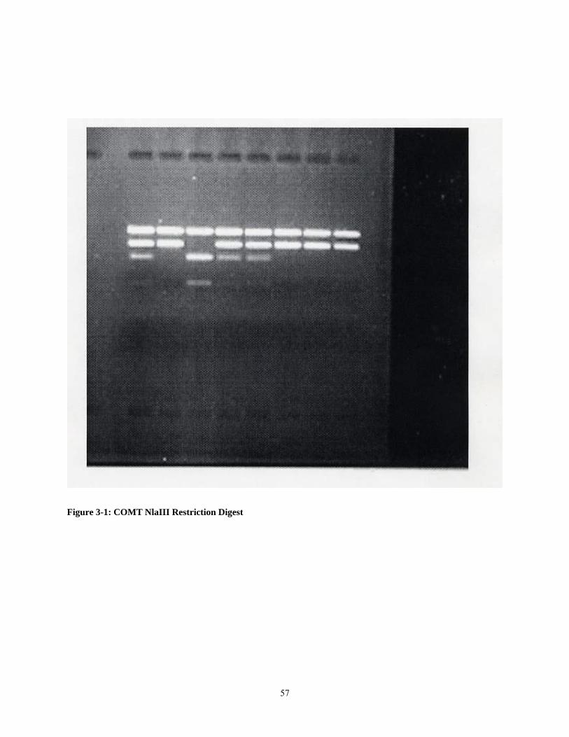
Figure 3-1: COMT NlaIII Restriction Digest
57

Table 3-1: Conditional logistic regression comparing cases and controls. Significant predictors of breast cancer risk [Numbers (N), Percentages (%)], [Means (S.D.)], age-adjusted odds ratio, 95% confidence interval
Variable
Cases
Control
Odds ratio
95% CI
Family history of breast cancer
Yes
15 (6.00)a
1 (4.00)a
14.99
1.98,113.47
No 235 (94.00)a 249 (96.00)a 1.00 Waist/hip ratio (>0.90)
Yes
161 (64.40)a
117 (46.80)a
2.00
1.39,2.87
No 89 (35.60)a 133 (53.20)a 1.00 Abortion
Yes
18 (7.20)a
7 (2.80)a
2.43
1.01,5.85
No 232 (92.80)a 243 (97.20)a 1.00 Age at first full-term pregnancy
23.18 (5.36)b
21.87 (4.74)b
1.39
1.11,1.73
Education >= High School
Yes
136 (54.40)a
113 (45.20)a
1.31
1.07,1.61
No 114 (54.60)a 137 (54.80)a 1.00 Parity
4.1 (2.9)b
4.7 (3.1)b
0.88
0.81,0.96
Duration of breastfeeding
65.48 (47.32)b
80.96 (53.67)b
0.75
0.62,0.91
58
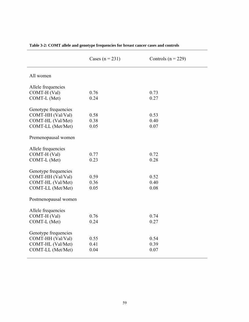
Table 3-2: COMT allele and genotype frequencies for breast cancer cases and controls
Cases (n = 231)
Controls (n = 229)
All women
Allele frequencies COMT-H (Val) 0.76 0.73 COMT-L (Met) 0.24 0.27 Genotype frequencies COMT-HH (Val/Val) 0.58 0.53 COMT-HL (Val/Met) 0.38 0.40 COMT-LL (Met/Met)
0.05
0.07
Premenopausal women
Allele frequencies COMT-H (Val) 0.77 0.72 COMT-L (Met) 0.23 0.28 Genotype frequencies COMT-HH (Val/Val) 0.59 0.52 COMT-HL (Val/Met) 0.36 0.40 COMT-LL (Met/Met)
0.05 0.08
Postmenopausal women
Allele frequencies COMT-H (Val) 0.76 0.74 COMT-L (Met) 0.24 0.27 Genotype frequencies COMT-HH (Val/Val) 0.55 0.54 COMT-HL (Val/Met) 0.41 0.39 COMT-LL (Met/Met)
0.04 0.07
59
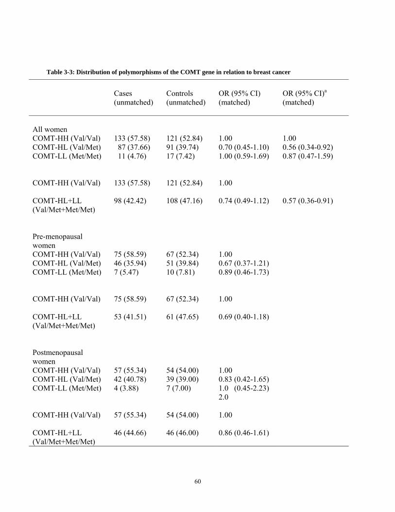
Table 3-3: Distribution of polymorphisms of the COMT gene in relation to breast cancer
Cases (unmatched)
Controls (unmatched)
OR (95% CI) (matched)
OR (95% CI)a
(matched)
All women
COMT-HH (Val/Val) 133 (57.58) 121 (52.84) 1.00 1.00 COMT-HL (Val/Met) 87 (37.66) 91 (39.74) 0.70 (0.45-1.10) 0.56 (0.34-0.92) COMT-LL (Met/Met) 11 (4.76) 17 (7.42) 1.00 (0.59-1.69) 0.87 (0.47-1.59)
COMT-HH (Val/Val) 133 (57.58) 121 (52.84) 1.00
COMT-HL+LL (Val/Met+Met/Met)
98 (42.42) 108 (47.16) 0.74 (0.49-1.12) 0.57 (0.36-0.91)
Pre-menopausal women
COMT-HH (Val/Val) 75 (58.59) 67 (52.34) 1.00 COMT-HL (Val/Met) 46 (35.94) 51 (39.84) 0.67 (0.37-1.21) COMT-LL (Met/Met) 7 (5.47) 10 (7.81) 0.89 (0.46-1.73)
COMT-HH (Val/Val) 75 (58.59) 67 (52.34) 1.00
COMT-HL+LL (Val/Met+Met/Met)
53 (41.51) 61 (47.65) 0.69 (0.40-1.18)
Postmenopausal women
COMT-HH (Val/Val) 57 (55.34) 54 (54.00) 1.00 COMT-HL (Val/Met) 42 (40.78) 39 (39.00) 0.83 (0.42-1.65) COMT-LL (Met/Met) 4 (3.88) 7 (7.00) 1.0 (0.45-2.23)
2.0
COMT-HH (Val/Val) 57 (55.34) 54 (54.00) 1.00
COMT-HL+LL (Val/Met+Met/Met)
46 (44.66) 46 (46.00) 0.86 (0.46-1.61)
60
REFERENCES
1. Kelsey JL. Gammon MD. The epidemiology of breast cancer. CA Cancer J Clin 1991;
41:146-165.
2. Feigelson HS, Henderson BE. Estrogens and breast cancer. Carcinogenesis, 1996;
17:2279-2284.
3. Davis DL, Bradlow HL, Wolff M, Woodruff T, Hoel DG, Anton-Culver H. Medical
hypothesis: Xenoestrogens as preventable causes of breast cancer. Environ Hlth Perspect
1993; 101:372-377.
4. Yager JD, Liehr JG. Molecular mechanisms of estrogen carcinogenesis Ann Rev
Pharmacol Toxicol 1996; 36:203-232.
5. Hammond DK, Zhu BT, Wang MY, Ricci MJ, Liehr JG. Cytochrome P450 metabolism
of estradiol in hamster liver and kidney. Toxicol Appl Pharmacol 1997; 145:54-60.
6. Zhu BT, Bui QD, Weisz J, Liehr JG. Conversion of estrone to 2- and 4-hydroxyestrone
by hamster kidney and liver microsomes: implications for the mechanisms of estrogen-
induced carcinogenesis. Endocrinology, 1994; 135:1772-1779.
7. Newbold RR, Bullock BC, MacLachlan JA. Uterine adenocarcinoma in mice following
developmental treatment with estrogen. Cancer Res 1990; 50:7677- 7681.
8. Bradlow HL, Hershcopf RJ, Martucci CP, Fishman J. Estradiol 16α-hydroxylation in the
mouse correlates with mammary tumor incidence and presence of mammary tumor virus:
A possible model for the hormonal etiology of breast cancer in humans. Proc Natl Acad
Sci USA 1985; 82:6295-6299.
61
9. Bui QD, Weisz J. Monooxygenase mediating catecholestrogen formation by rat anterior
pituitary is an estrogen-4-hydroxylase. Endocrinology, 1988; 124:1085-1087.
10. van Aswegen CH, Purdy RH, Wittliff JL. Binding of 2-hydroxyestradiol and 4-
hydroxyestradiol to estrogen receptor human breast cancers. J Steroid Biochem 1989;
32:485-492.
11. Liehr JG, Roy D. Free radical generation by redox cycling of estrogens. Free Radical
Biol Med 1990; 8:415-423.
12. Cavalieri EL, Stack DE, Devanesan PD, Todorovic R, Dwiredy I, Higginbotham S,
Johansson SL, Patil KD, Gross ML, Gooden JK, Ramanathan R, Cerny L, Rogan EG.
Molecular origin of cancer: catechol estrogen-3,4-quinones as endogenous tumor
initiators. Proc Natl Acad Sci USA 1997; 94:10937-10942.
13. Hamel E, Lin CM, Flymn E, D’Amato RJ. Interaction of 2-methoxyestradiol, an
endogenous mammalian metabolite, with unpolymerized tubulin and tubulin polymers.
Biochemistry, 1996; 35:1304-1310.
14. Klauber N, Parangi S, Flymn E, Hamel E, D’Amato RJ. Inhibition of angiogenesis and
breast cancer in mice by the microtubulin inhibitors 2-methoxyestradiol and taxol. Cancer
Res 1997; 57:81-86.
15. Zhu BT, Liehr JG. Quercetin increases the severity of estradiol-induced tumorigenesis in
hamster kidney. Toxicol Appl Pharmacol 1994; 125:149-158.
16. Zhu BT, Liehr JG. Inhibition of catechol-O-methyltransferase-catalyzed O-methylation
of 2- and 4-hydroxyestradiol by quercetin: possible role in estradiol-induced
tumorigenesis. J Biol Chem 1996; 271:1357-1363.
62
17. Weinshilboum RM, Raymond FA. Inheritance of low erythrocyte catechol-O-
methyltransferase acitivity in man. Am J Hum Genet 1977; 29:125-135.
18. Weinshilboum R. Methyltransferase pharmacogenetics. Pharmacol Ther 1989; 43:77-90.
19. Boudikova B, Szumlanski C, Weinshilboum R. Human liver catechol-O-
methyltransferase pharmacogenetics. Clin Pharmacol Ther 1990; 48:381-389.
20. Millikan RC, PittmanGS, Tse CKJ, Duell E, Newman B, et al. Catechol-O-
methyltransferase and breast cancer risk. Carcinogenesis 1998; 19:1943-1947.
21. Lavigne JA, Helzlsouer KJ, Huang HY, Strickland PT, Bell DA, et al. An association
between the allele coding for a low activity variant of catechol-O-methyltransferase and
the risk of breast cancer. Cancer Res 1997; 57:5493-5497.
22. Thompson PA, Shields PG, Freudenheim JL, StoneA, Vena JE, Marshall JR, et al.
Genetic polymorphisms in catechol-O-methyltransferase, menopausal status, and breast
cancer risk. Cancer Res 1998; 58:2107-2110.
23. Ambrosone CB, Thompson PA, Shields PG, Freudenheim JL, Stone A, Marshall JR, et
al. Catechol-O-methyltransferase (COMT) genetic polymorphisms, smoking and breast
cancer risk. Am J Epidemiol 1998; 147:2107-2110.
24. Yim DS, Park SK, Yoo YK, Yoon KS, Chung HH, Kang HJ, et al. Relationship between
the Val Met polymorphism of catechol-O-methyltransferase and breast cancer.
Pharmacogenetics 2001; 11:279-286.
25. Huang CS, Chern HD, Chang KJ, Cheng CW, Hsu SM, Shen CY. Breast cancer risk
associated with genotype polymorphism of the estrogen metabolizing genes, CYP17,
CYP1A1, and COMT: A multigenic study on cancer susceptibility. Cancer Res 1999;
59:4870-4875.
63

26. QIAamp DNA Mini Kit and QIAamp DNA Blood Mini Kit Handbook, February 2003.
27. QIAamp DNA Mini Kit and QIAamp DNA Blood Midi Kit Handbook, February 2003.
64
4. CYTOCHROME P4501A1 GENETIC POLYMORPHISMS AND BREAST CANCER RISK IN NIGERIAN WOMEN
To be submitted to the Breast Journal
Okobia M.N.1,4
Bunker C.H. 1 Zmuda JM1
Kammerer CM2
Vogel VG3
Uche E.E.O. 5Anyanwu S.NC. 6
Ezeome E.R. 7
Ferrell R.E 2
Kuller L.H. 1 From the Department of Epidemiology1, Department of Human Genetics2, Graduate School of
Public Health, Department of Medicine3, University of Pittsburgh, Pittsburgh, PA 15261, U.S.A.;
Department of Surgery, University of Benin Teaching Hospital, Benin City, Nigeria4;
Department of Surgery, University of Port Harcourt Teaching Hospital, Port Harcourt, Nigeria5;
Nnamdi Azikiwe University Teaching Hospital, Nnewi, Nigeria6; and Department of Surgery,
University of Nigeria Teaching Hospital, Enugu, Nigeria7.
65
4.1. ABSTRACT
In this case-control study based on 250 women with breast cancer and 250 age-matched
controls, we sought to evaluate the role of four polymorphic variants in the CYP1A1 gene in
breast cancer susceptibility in Nigerian women. Heterozygosity for the CYP1A1 M1 genotype
(CYP1A1 M1 [T/C]) was associated with a 21% reduced risk of breast cancer (OR = 0.79, 95%
CI 0.46-1.40) while homozygosity for the genotype (CYP1A1 M1 [C/C]) conferred a non-
significant 9% reduced risk of breast cancer. These risk profiles were not significantly altered in
subgroup analysis by menopausal status. While heterozygosity for the CYP1A1 M3 genotype
(T/C) conferred a non-significant 24% reduced risk of breast cancer (OR = 0.76, 95% CI 0.47-
1.22), homozygosity for the variant was associated a non-significant 1.95-fold increased risk of
breast cancer (OR = 1.95, 95% CI 0.24-6.01). Subgroup analysis showed a non-significant 11%
reduced risk in premenopausal heterozygous carriers (OR = 0.89, 95% CI 0.45-1.44) and a non-
significant 6% increased risk of postmenopausal breast cancers for carriers of the CYP1A1 M3
(T/C) genotype. The CYP1A1 M2 (isoleucine to valine) polymorphism in exon 7 and CYP1A1
M4 (threonine to asparagine) variant in codon 461 of the CYP1A1 gene were found to be very
rare in our study subjects. This study has shown that while the CYP1A1 M1 polymorphism
conferred reduced risk of breast cancer, homozygosity for the CYP1A1 M3 (C/C) was associated
with increased risk of breast cancer although these risks did not attain statistical significance.
66
4.2. INTRODUCTION
Cytochrome P450 1A1 (CYP1A1) gene plays a central role in the 2-hydroxylation of
estradiol and estrone (the two estrogens in humans) to 2-hydroxy catechol metabolites for
subsequent O-methylation to 2-methoxy intermediates. While the 2-hydroxylation products (2-
OH estradiol catechol and 2-OH estrone catechol) are devoid of estrogenic activities1 and the 2-
methoxy derivatives shown to possess anti-proliferative and anti-angiogenic properties2-4,
another mutually exclusive pathway of 16α-hydroxylation leads to metabolites with strong
estrogenic properties and have been linked to estrogen-induced carcinogenesis in both laboratory
animals and humans5-7.
The CYP1A1 gene is highly polymorphic in human populations and ethnic differences in
the distribution of these polymorphisms have been reported in various populations. Four
polymorphisms including M1 (a threonine to cysteine substitution in the 3’ non-coding region)8,
M2 (isoleucine to valine variant in codon 462 in the heme-binding domain in exon 7)9, M3 (an
A-T to G-C transition mutation in the 3’ non-coding region 300 base pairs from the
polyadenylation site)10, and M4 (a threonine to asparagine substitution in codon 461)11 have been
described in various populations. It has been demonstrated by Crofts et al.12 that the exon 7
polymorphism has a role in gene function by increasing both enzyme activity and mRNA levels
in Asians. The same investigators found an increase in CYP1A1 mRNA in Caucasians who carry
the double heterozygous genotype for the CYP1A1 M1 and CYP1A1 M2 (exon 7)
polymorphisms, but no evidence for a functional significance of the exon 7 polymorphism alone.
Association of CYP1A1 polymorphisms and breast cancer risk has been reported in various
67
populations with inconsistencies even within populations. Taoli et al.13,14 observed an increased
risk of breast cancer among a small sample of African American women (20 cases and 81
controls) but not among Caucasian women who harbor the CYP1A1 M1 variant. Rebbeck et al.15
found no association between the exon 7 polymorphism (M2) and breast cancer risk among US
Caucasian women (96 cases and 146 controls) while Ambrosone and colleagues16 noted an
increased risk of postmenopausal breast cancer among Caucasian women with the variant exon 7
allele (216 cases and 282 controls). While Ishibe et al.17 reported that neither the CYP1A1 M1
nor CYP1A1 M2 polymorphisms was independently associated with overall breast cancer risk,
these investigators noted an interaction between these variants and smoking; current smoking
and adolescent-onset smoking interacted with CYP1A1 M1 and CYP1A1 M2, respectively to
increase breast cancer risk among Caucasian women in a case-control study nested in the Nurses’
Health Study. Studies in Asian populations have also reported conflicting results. While Huang
et al.18 noted an increased risk among homozygous carriers of the CYP1A1 M1 polymorphism
among postmenopausal Chinese women in Taiwan, Miyoshi and colleagues19 reported a
significant 40% and 34% reduced risk of breast cancer for the CYP1A1 M1 and CYP1A1 M2
variants in Japanese women.
Apart from the small data from Taoli et al.13,14 noted above, we are unaware of any other
association study of CYP1A1 genetic polymorphisms and breast cancer risk in women of
African descent in the literature. Because of the wide ethnic differences in the distribution of
CYP1A1 gene polymorphisms, this study recruiting 250 women with breast cancer and 250 age-
matched control subjects seeks to evaluate the association between CYP1A1 M1, M2, M3, and
M4 polymorphisms and breast cancer risk in an exclusively African population drawn from
Midwestern and Southeastern Nigeria.
68
4.3. MATERIALS AND METHODS
4.3.1. Recruitment of study participants
Two hundred and fifty cases of breast cancer and 250 age-matched women without the
disease were recruited from four University Teaching Hospitals located in Midwestern and
Southeastern Nigeria between September 2002 and April 2004 for this case-control study. Study
protocols were approved by the Ethics and Research Committees of the Nigerian institutions and
the Institutional Review Board (IRB) of the University of Pittsburgh. The cases were women
with breast cancer being managed at the surgical out-patient clinics and surgical wards of the
Nigerian hospitals. Control subjects were women without evidence of breast and other malignant
diseases who were being treated for other surgical disorders including road traffic accidents,
intestinal obstructions, chronic gallbladder diseases, appendicitis and non-malignant leg ulcers.
Eligible study participants were identified by with the assistance of physicians in these
hospitals who informed them about the study and referred those willing to the study
investigators. Details of the study including objectives, risks and benefits, confidentiality and
right of participation were explained to eligible participants. Potential study participants were
free to discuss participation with close family members. Written informed consent was obtained
those that accepted to participate in the study.
Interviewer-administered questionnaires detailing sociodemographic characteristics
including age, sex, occupation, exposure to chemical fertilizers and pesticides and obstetric and
gynecological variables such as age at menarche, parity, age at first full-term pregnancy (FFTP),
age at menopause, use of hormone contraceptives and hormone replacement therapy and history
69
of abortions and surgical oophorectomy were used for data collection, which lasted 30 minutes.
Height, weight, waist and hip circumferences were measured at the end of the interview.
4.3.2. Biological samples
Forty milliliters of whole blood collected in two 15 ml plain vacutainer tubes and one 10 ml
K3-EDTA vacutainer tube was obtained from each study participant. Samples were centrifuged
within 10 h of collection; buffy coat separated from plasma and red cells in the K3-EDTA
vacutainer tubes were carefully pippeted into 3 ml tubes while clots separated from serum in the
plain vacutainer tubes were turned into two 20 plain tubes and stored at –20 oC in each of the
Nigerian study sites until transferred in polar ice packs to the Nigerian coordinating center at the
University of Benin Teaching Hospital where samples were stored at –20 oC. The samples were
later transferred to the University of Pittsburgh in dry ice and stored at –80 oC until DNA
extraction.
4.3.3. DNA extraction
DNA extraction was from buffy coats and blood clots for participants in whom buffy coat
was unavailable. QIAamp DNA Mini Kit20 and QIAamp DNA Midi Kit21 protocols were used
for DNA extraction from buffy coats and blood clots respectively. The DNA was stored at 4 C
until amplified by polymerase chain reaction (PCR) and used for restriction fragment length
polymorphism (RFLP) analysis.
70
4.3.4. PCR and Genotyping
Polymerase chain reaction (PCR) amplification and restriction endonuclease digest were
carried out for each of the four CYP1A1 variants (CYP1A1 M1, M2, M3 and M4). Primers used
for the PCR reaction, fragment lengths amplified and restriction enzymes used are detailed in
Table 4-1. For the CYP1A1 M1 variant, a 348 base pair (bp) fragment containing part of exon 7
and the adjoining 3’ non-coding region of intron 7 was amplified using the following primers
(Forward [C1]: 5’ CCG CTG CAC TTA AGC AGT CT 3’, Reverse [C2]: 5’AGG GCG TAA
GTC AGC ACA GT 3’. For the CYP1A1 M2 and CYP1A1 M4 polymorphisms, a 377 bp
fragment containing part of exon 7 bearing the M2 and M4 variants was amplified using the
following primers (Forward [C3]: 5’ GCA TTG ATC CTC CTG TCC AT, Reverse [C4]: 5’
AGG CAT GCT TCA TGG TTA GC 3’). A 400 bp fragment encompassing part of 3’ non-
coding region of the gene was amplified for the CYP1A1 M3 polymorphism using the following
primers (Forward [C5]: 5’ GGC CTC TGA GAA GCT CTG AA 3’, Reverse [C6]: 5’ GTC
CTG GTG CCT GGA TAT GT 3’).
For each of the four polymorphisms, a 50 µl PCR reaction mixture containing 2 µl of
genomic DNA, 8 µl of deoxynucleotide triphosphates, 1 µl each of forward and reverse primers,
5 µl of 10X buffer, 1.5 µl of MgCl and 0.2 µl of Taq polymerase was placed in a MJ Research
DYAD thermocycler. After denaturing for 5 min at 95 oC, the DNA was amplified for 35 cycles
at 95 oC for 30 s, 58 oC for 30 s, and 72 oC for 30 s, followed by a 5 min extension at 72 oC. A
positive control containing genomic DNA (University of Pittsburgh Molecular Epidemiology
Laboratory control) and a negative control containing everything except DNA were included in
71
the PCR experiment. Five µl of each PCR product, including the controls, were run on a 1%
agarose gel to ensure that the expected fragments product was generated.
Restriction digest for each of the four variants of the CYP1A1 gene was carried out using
restriction endonucleases detailed in Table I: CYP1A1 M1, MspI; CYP1A1 M2, BsrDI;
CYP1A1 M3, MspI; and CYP1A1 M4, BsaI. Digestion for the CYP1A1 M1 MspI variant
carried out at 37 oC overnight for 16 h revealed a band of 348 bp for the CYP1A1 M1 (T) allele
and two bands of 230 bp and 118 bp for the CYP1A1 M1 (C) allele (Fig 4-1). BsrDI digest for
the CYP1A1 M2 polymorphism, carried out for 16 h overnight at 65 oC, revealed a 377 bp
fragment for the G allele and two bands of 237 bp and 140 bp for the A allele as shown in Figure
4-2. For the CYP1A1 M3 variant, a 400 bp fragment for the T allele and two fragments of 330
bp and 70 bp for the C allele were detected following a 16 h digestion at 37 oC and ran on 3%
agarose gel electrophoresis stained with ethidium bromide as shown in Figure 4-3. Digestion for
the CYP1A1 M4 variant with BsaI restriction endonuclease overnight for 16 h at 50 oC yielded a
377 bp fragment for the A allele and two fragments of 277 bp and 150 bp for the C allele (Figure
4-4).
4.3.5. Statistical Analysis
Data analysis was carried out using Statistical Analysis Systems (SAS) software (Version
8.0). Assessment of association between socio-demographic characteristics and obstetric and
gynecological variables and breast cancer risk was first carried out using conditional logistic
regression. Because PCR amplification and RFLP analysis for the CYP1A1 M2 and M4 variants
revealed that all individuals genotyped for CYP1A1 M2 variant carried the wild type CYP1A1
M2 (A) allele except for one individual with the heterozygous CYP1A1 (A/G) genotype, and all
72
the study subjects genotyped for the CYP1A1 M4 polymorphism were homozygous for the C
allele (CYP1A1 M4 [C/C]), we restricted analysis of association between CYP1A1 genotypes
and breast cancer risk to the CYP1A1 M1 and CYP1A1 M3 variants. Conditional logistic
regression was used to evaluate association between these two CYP1A1 variants and breast
cancer risk. First, all women were combined in univariate conditional logistic regression model
restricted to each of the variants. Next variables that were found to be significant predictors of
breast cancer risk in the descriptive analysis were entered into the multivariate conditional
logistic regression models together with the genotype data. Subgroup analysis based on
menopausal status was carried out separately for premenopausal and postmenopausal women.
4.4. RESULTS
4.4.1. Descriptive epidemiology
Results of conditional logistic regression models evaluating the association of various
socio-demographic and obstetric and gynecological variables are shown in Table 4-2. Family
history of breast cancer (OR = 14.99, 95% CI 1.98-113.47), waist/hip ratio (OR = 2.00, 95% CI
1.39-2.87), history of abortion (OR = 2.43, 95% CI 1.01-5.85), older age at first full-term
pregnancy (OR = 1.39, 95% CI 1.11-5.85) and education above High School (OR = 1.31, 95%
CI 1.07-1.61) were associated with increased risk of breast cancer. A 12% reduced risk of breast
cancer was conferred by high parity (OR = 0.88, 95% CI 0.81-0.96); longer duration of
breastfeeding was also associated with a significant 25% reduced risk of breast cancer (OR =
0.75, 95% CI 0.62-0.91).
73
4.4.2. Association of CYP1A1 M1 polymorphism and breast cancer risk
PCR-based RFLP assay for the CYP1A1 M1 polymorphism was successful in 220 cases
and 218 control subjects out of 250 cases and 250 controls that were recruited for the study. The
CYP1A1 M1 variant was found to be highly polymorphic in the study population as shown in
Table 4-3.
All women
Allele frequencies for the CYP1A1 M1 (T) and CYP1A1 M1 (C) alleles were 0.77 and
0.23 respectively among the cases and 0.76 and 0.24 respectively among the control subjects.
Frequencies for the CYP1A1 M1 (T/T), CYP1A1 M1 (T/C) and CYP1A1 M1 (C/C) genotypes
for the cases (0.61, 0.31, and 0.07 respectively) were not significantly different from the
distribution of these genotypes in the control subjects (0.60, 0.33, and 0.08, respectively). When
compared with the homozygous CYP1A1 M1 (T/T) genotype carriers, individuals harboring the
heterozygous CYP1A1 M1 (T/C) genotype had a non-significant 21% reduced risk of breast
cancer (OR = 0.79, 95% CI 0.46-1.40) in the final multivariate conditional logistic regression
model controlling for other identified risk factors for breast cancer as shown in Table 4-4.
Homozygosity for the CYP1A1 M1 (C/C) allele also conferred a non-significant 11% reduced
risk of breast cancer in the final model (OR = 0.91, 95% CI 0.50-1.66). Combining heterozygous
CYP1A1 M1 (T/C) and CYP1A1 M1 (C/C) carriers was associated with a non-significant 21%
reduced risk of breast cancer.
74
Premenopausal women
The distribution of the CYP1A1 M1 (T) allele and CYP1A1 M1 (C) allele in
premenopausal breast cancer cases was not significantly different from the frequency of these
alleles in the control subjects. While the allele frequencies in the cases were 0.78 and 0.22
respectively for the T and C alleles, the frequencies of the T and C alleles in the control subjects
were 0.75 and 0.25. Slightly more cases (78 [62.40%]) than controls (71 [57.26%]) were
homozygous for CYP1A1 M1 (T/T) genotype while fewer cases (40 [32.00%]) than controls (43
[34.68%]) carried the heterozygous CYP1A1 M1 (T/C) genotype. Seven cases (5.60%) and 10
controls (8.06%) were homozygous for the CYP1A1 M1 (C) variant. There was a non-significant
38% reduced risk of premenopausal breast cancer among carriers of the heterozygous CYP1A1
M1 (T/C) genotype (OR = 0.62, 95% CI 0.31-1.24). Similarly, homozygosity for the CYP1A1
M1 (C) allele conferred a non-significant 37% reduced risk of premenopausal breast cancer (OR
= 0.63, 95% CI 0.28-1.44). Although, there was a 42% reduced risk of premenopausal breast
cancer when the CYP1A1 M1 (T/C) and CYP1A1 M1 (C/C) genotypes were pooled together,
this was also not statistically significant (OR = 0.58, 95% CI 0.31-1.09).
Postmenopausal women
Of the 108 cases of postmenopausal breast cancer patients and 108 age-matched controls,
the PCR-based RFLP assay was successful in 103 cases and 104 control subjects. Slightly fewer
cases (62 [60.19%]) than controls (58 [61.70%]) were homozygous for the CYP1A1 M1 (T)
allele. Almost equal number of cases (31 [30.10%]) and controls (29 [30.85%]) harbored the
heterozygous CYP1A1 M1 (T/C) genotype while slightly more cases (10 [9.71%]) than controls
75
(7 [7.45%]) were homozygous for the CYP1A1 M1 (C) allele. Heterozygosity for the CYP1A1
M1 variant (CYP1A1 M1 [T/C]) was not related to the risk of postmenopausal breast cancer (OR
= 1.00, 95% CI 0.49-2.05) while homozygosity for the CYP1A1 M1 (C) allele (CYP1A1 M1
(C/C) conferred a non-significant 16% increased risk of postmenopausal breast cancer (OR =
1.16, 95% CI 0.55-2.44). Combining individuals heterozygous for the CYP1A1 M1 genotype 1
[T/C]) and homozygous carriers of the variant (CYP1A1 M1 [C/C]) was associated with a non-
significant 6% increased risk of postmenopausal breast cancer (OR = 1.06, 95% CI 0.55-2.01).
4.4.3. Association of CYP1A1 M3 polymorphism and breast cancer risk
All women
As shown in Table 4-5, the CYP1A1 M3 variant is highly polymorphic in the study
population with allele frequencies of 0.88 and 0.12 for the CYP1A1 M3 (T) and CYP1A1 M3
(C) alleles respectively among the cases and 0.87 and 0.13 for the CYP1A1 M3 (T) and CYP1A1
M3 (C) alleles respectively among the control subjects. Slightly more cases (176 [76.86%]) than
control subjects (172 [75.77%]) were homozygous for the CYP1A1 M3 (T) allele while almost
equal number of cases (52 [22,71%]) and control subjects (53 [23.35%]) carried the
heterozygous CYP1A1 M3 (T/C) genotype. Only one case (0.44%) and two controls (0.88%)
harbored the homozygous CYP1A1 M3 (C/C) genotype. In the final multivariate conditional
logistic regression model, heterozygosity for the CYP1A1 M3 genotype (CYP1A1 M3 [T/C])
conferred a non-significant 24% reduced risk of breast cancer (OR = 0.76, 95% CI 0.47-1.22)
while homozygosity for the CYP1A1 M3 (C) allele (CYP1A1 M3 [C/C]) was associated with a
non-significant 95% increased risk of breast cancer (OR = 1.95, 95% CI 0.24-6.01) as shown in
Table 4-6.
76
Premenopausal women
Of the 142 cases of premenopausal breast cancer and the 142 age-matched
premenopausal controls, the PCR-based RFLP assay was successful in 126 cases and 126
controls. The distribution of the CYP1A1 M3 (T) and CYP1A1 M3 (C) alleles in the cases (0.87
and 0.13 respectively) is not significantly different from the distribution of these alleles in the
control subjects (0.85 and 0.15, respectively). As shown in Table 4-5, slightly more cases (95
[75.40]) than controls (90 [71.43%]) were homozygous for the CYP1A1 M3 (T) allele while
fewer cases (30 [23.81%]) than controls (35 [27.78%]) carried the heterozygous CYP1A1 M3
(T/C) genotype. While heterozygosity was associated a non-significant 19% reduced risk of
premenopausal breast cancer (OR = 0.81, 95% CI 0.45-1.44), homozygosity for the CYP1A1 M1
(C) allele was not related to premenopausal breast cancer risk (OR = 1.00, 95% CI .025-4.00).
Combining heterozygous and homozygous carriers of the CYP1A1 M3 (C) allele did not alter
the premenopausal breast cancer risk as shown in Table 4-6 (OR = 0.82, 95% CI 0.46-1.43).
Postmenopausal women
The PCR-based RFLP assay was successful in 103 out of 108 postmenopausal breast
cancer cases and 99 out of 108 postmenopausal control subjects. CYP1A1 M3 (T) allele and
CYP1A1 M3 (C) allele frequencies were similar in cases (0.89 and 0.11, respectively) and
control subjects (0.90 and 0.10, respectively) as shown in Table 4-5. Slightly fewer cases (81
[78.64%]) than controls (80 [80.81%]) were homozygous for the CYP1A1 M3 (T) allele while
slightly more cases (22 [21.36%]) than controls (18 [18.18%]) carried the heterozygous CYP1A1
M3 (T/C) genotype as shown in Table 4-6. Compared with the homozygous CYP1A1 M3 (T/T)
77

carriers, heterozygosity (CYP1A1 M3 (T/C) was associated with a non-significant 6% increased
risk of postmenopausal breast cancer (OR = 1.06, 95% CI 0.54-1.20). Risk associated with the
homozygous CYP1A1 M3 (C/C) genotype could not be assessed as none of the postmenopausal
breast cancer cases carried the homozygous CYP1A1 M3 (C/C) genotype.
4.5. DISCUSSION
CYP1A1 may be involved in breast cancer via estrogen-related mechanisms. Estrogens
are metabolized by two competing pathways to form either inactive 2-hydroxy-estradiol and
estrone or active 16α-hydroxy- estradiol and estrone, respectively; level of the latter are often
elevated in breast cancer, and may be related to tumorigenesis22. CYP1A1 polymorphism may
affect the distribution of these metabolites and thus determine susceptibility to cancer23,24. To
date, four polymorphisms have been described within this gene; M1, M2, M3, and M4. It has
been suggested that there is wide ethnic variation in the distribution of polymorphisms of the
CYP1A1 gene. For example, the CYP1A1 M2 (Exon 7) polymorphism was found to be rare in
our study population with only one heterozygous (CYP1A1 M2 [A/G]) carrier in the entire
sample. This same polymorphism has been reported to have allele frequencies of 0.69 and 0.31
among the Japanese8, and 0.88 and 0.12 among a racially mixed African American population25.
Studies in Asian populations indicate that more than 10% of the population possesses variant
genotypes of this polymorphism18. Allele frequencies of this polymorphism in Caucasians have
been reported to be low (0.97 and 0.03)15. Also we did not find any carriers of the CYP1A1 M4
(A) allele in our study subjects; all were homozygous for the CYP1A1 M4 (C) allele. On the
other hand, the CYP1A1 M1 and CYP1A1 M3 variants were highly polymorphic among the
78
Nigeria study subjects. Allele frequencies for the CYP 1A1 M1 (T) and CYP1A1 (C) alleles
among our control subjects were 0.76 and 0.24 respectively with genotype frequencies of 0.60,
0.33, and 0.08 for the CYP1A1 M1 (T/T), CYP1A1 M1 (T/C), and CYP1A1 M1 (C/C)
genotypes respectively. The homozygous CYP1A1 M1 (C) frequency of 8% in this study agrees
with the findings of Cosma et al.26 These investigators reported the frequency of the CYP1A1
M1 (C/C) to be 6-7% in a small sample of African American women (21 cases and 85 controls).
Our finding is however lower than the 13% reported for the homozygous CYP1A1 M1 (C/C)
genotype among Asians but higher than the 2% reported in Caucasian populations26.
The CYP1A1 M3 polymorphism has been reported previously only in African American
women. We have found this variant to also be polymorphic in Nigerian women occurring with
allele frequencies of 0.87 and 0.13 for the CYP1A1 M3 (T) and CYP1A1 M3 (C) alleles and
among the control subjects. Among a small sample of African American women, Crofts et al.27
observed a 17% carrier rate for the CYP1A1 M3 variant with CYP1A1 M3 (T) and CYP1A1 M3
(C) allele frequencies of 0.91 and 0.09 respectively.
Breast cancer risk association studies with CYP1A1 variants in the literature have been
conflicting. We did not observe any significant association between the two common CYP1A1
polymorphisms in our study population (CYP1A1 M1 and CYP1A1 M3) and breast cancer risk
overall, or in subgroup analysis based on menopausal status. In particular, the CYP1A1 M1
polymorphism was associated with a non-significant 19% reduced risk of breast cancer (OR =
0.79, 95% CI 0.46-1.40). This same polymorphism was reported to confer almost 10-fold
increased risk of breast cancer among African American women by Taioli et al.14 (OR = 9.7,
95% CI 2.0-47.9) although their study was of small sample size (21 cases and 85 controls).
These investigators were unable to demonstrate any association of CYP1A1 M1 polymorphism
79
and breast cancer risk in Caucasian women in their study (OR = 1.7, 95% CI 0.6-4.9 for
heterozygous carriers of the CYP1A1 M1 [T/C] genotype).
Rebbeck et al.15 observed no association of breast cancer risk with the exon 7
polymorphism among 96 incident breast cancer cases and 146 controls, whereas Ambrosone and
colleagues16 reported a significant increase in risk among postmenopausal women with the
variant exon 7 allele (based on 216 cases and 282 controls). In addition, an interaction was
observed in the latter study with smoking, suggesting that this polymorphism may be important
in increasing breast cancer risk among light smokers (defined as <30 pack-years). In a
prospective study of 466 cases (over 99% Caucasians) and an equal number of controls nested in
the Nurses’ Health Study, Ishibe et al.17 were unable to detect an overall increase in breast cancer
risk with the variant CYP1A1 genotypes. There was however a suggestion of a role of the variant
alleles in breast cancer development in association with smoking. An increase in breast cancer
risk among smokers was limited to those who were variant for the CYP1A1 M1 polymorphism
and who had smoked greater than 29 pack-years contrary to the findings of Ambrosone et al.16
noted above.
In a case-control study recruiting 150 postmenopausal Chinese women with breast cancer
and an equal number of controls, Huang et al.18 found that the homozygous variant of the
CYP1A1-MspI polymorphism was a significant predictor of breast cancer risk, independent of
other established risk factors (adjusted odds ratio 1.98; 95% CI, 1.01-3.99). In a recent study
among Japanese women, Miyoshi et al.19 found that carriers with variant allele 6235C (MspI
polymorphism) or variant allele 462Val (exon 7) have significantly lower risk of breast cancer,
that is, the ORs were 0.60 for the 6235C (MspI polymorphism) carriers and 0.66 for allele
462Val (exon 7) carriers compared with noncarriers. They also noted linkage disquilibrium
80
between the two variants because most of the variant 462Val carriers were associated with the
variant allele 6235C (MspI polymorphism) carriers by haplotype analysis. These findings are
inconsistent with those reported for other ethnicities; that is, carriers of these variant alleles have
a breast cancer risk equivalent to noncarriers in Caucasian women13,17,28 and carriers of variant
allele 6235C (MspI) have a significantly higher breast cancer risk than noncarriers in African-
American women13,14 and similar to the findings in postmenopausal Chinese women in Taiwan18.
The authors admitted that reasons for their finding are currently unknown but postulated that this
may be partly explained by the differences in other genetic and environmental factors among
these ethnicities
Some of the limitations of our study have to do with recall and other biases associated
with case-control studies. However, the genetic constitution of individuals is constant and not
susceptible to recall bias. Control subjects for our study were recruited from hospital patients;
this group of control patients may not be exactly representative of the general population from
which the cases were drawn. It should however be noted that with limited communication
facilities in developing countries such as Nigeria, population controls are often difficult to
recruit. Sample size may also be a factor of concern. Although we calculated sample size based
on available data in the literature, some of the studies used were of small sample size and may
not be very reliable for accurate sample size determination. Over 70% of breast cancer patients
presented with advanced stages of the disease (Manchester Stages III and IV). It is possible that
we may have missed patients with rapidly progressive breast cancer who may have died before
reaching the stage of fungating tumors seen in most of our patients. There is currently no breast
cancer-screening program in Nigeria; it is possible that we also missed patients with early stage
81
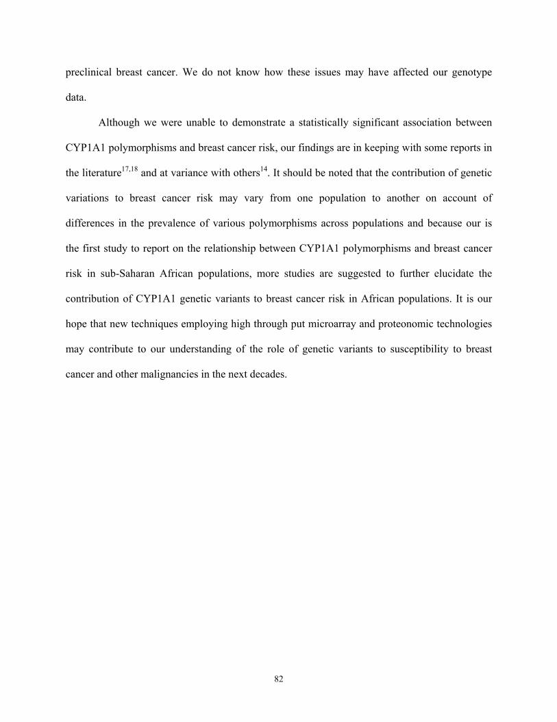
preclinical breast cancer. We do not know how these issues may have affected our genotype
data.
Although we were unable to demonstrate a statistically significant association between
CYP1A1 polymorphisms and breast cancer risk, our findings are in keeping with some reports in
the literature17,18 and at variance with others14. It should be noted that the contribution of genetic
variations to breast cancer risk may vary from one population to another on account of
differences in the prevalence of various polymorphisms across populations and because our is
the first study to report on the relationship between CYP1A1 polymorphisms and breast cancer
risk in sub-Saharan African populations, more studies are suggested to further elucidate the
contribution of CYP1A1 genetic variants to breast cancer risk in African populations. It is our
hope that new techniques employing high through put microarray and proteonomic technologies
may contribute to our understanding of the role of genetic variants to susceptibility to breast
cancer and other malignancies in the next decades.
82
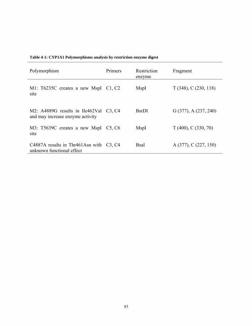
Table 4-1: CYP1A1 Polymorphisms analysis by restriction enzyme digest
Polymorphism
Primers
Restriction enzyme
Fragment
M1: T6235C creates a new MspI site
C1, C2
MspI
T (348), C (230, 118)
M2: A4889G results in Ile462Val and may increase enzyme activity
C3, C4
BsrDI
G (377), A (237, 240)
M3: T5639C creates a new MspI site
C5, C6
MspI
T (400), C (330, 70)
C4887A results in Thr461Asn with unknown functional effect
C3, C4
BsaI
A (377), C (227, 150)
83
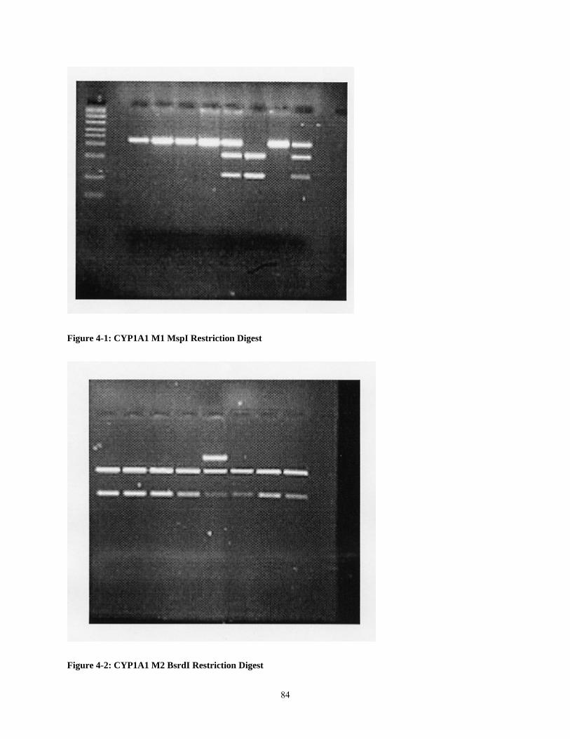
Figure 4-1: CYP1A1 M1 MspI Restriction Digest
Figure 4-2: CYP1A1 M2 BsrdI Restriction Digest
84
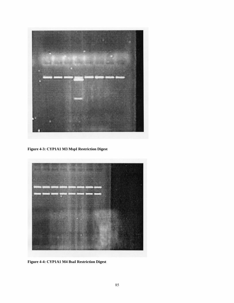
Figure 4-3: CYP1A1 M3 MspI Restriction Digest
Figure 4-4: CYP1A1 M4 BsaI Restriction Digest
85
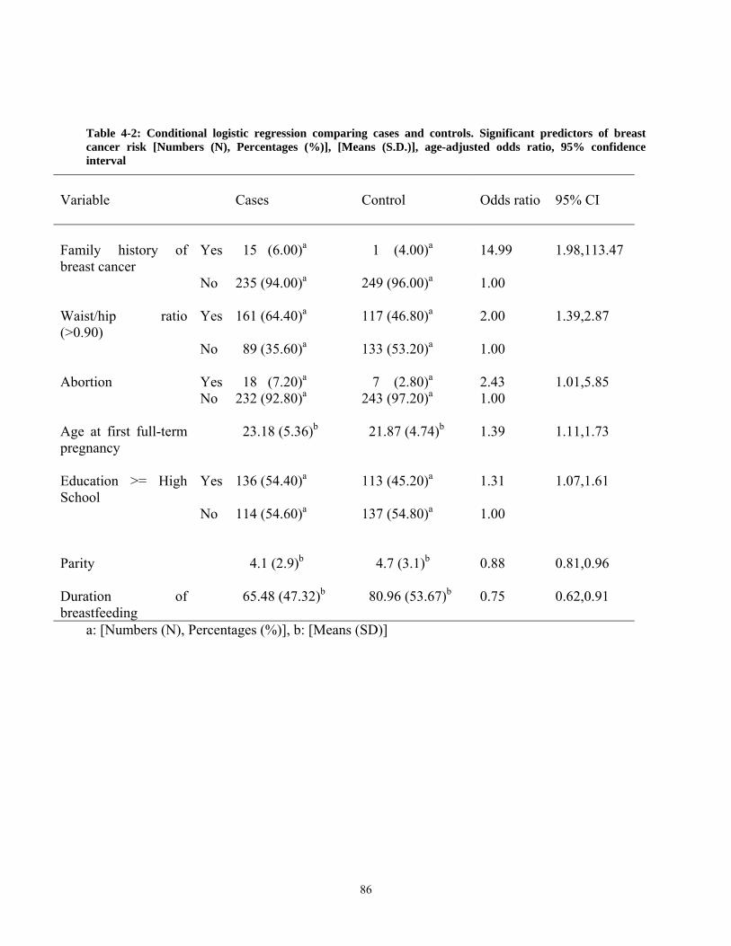
Table 4-2: Conditional logistic regression comparing cases and controls. Significant predictors of breast cancer risk [Numbers (N), Percentages (%)], [Means (S.D.)], age-adjusted odds ratio, 95% confidence interval
Variable
Cases
Control
Odds ratio
95% CI
Family history of breast cancer
Yes
15 (6.00)a
1 (4.00)a
14.99
1.98,113.47
No 235 (94.00)a 249 (96.00)a 1.00 Waist/hip ratio (>0.90)
Yes
161 (64.40)a
117 (46.80)a
2.00
1.39,2.87
No 89 (35.60)a 133 (53.20)a 1.00 Abortion
Yes
18 (7.20)a
7 (2.80)a
2.43
1.01,5.85
No 232 (92.80)a 243 (97.20)a 1.00 Age at first full-term pregnancy
23.18 (5.36)b
21.87 (4.74)b
1.39
1.11,1.73
Education >= High School
Yes
136 (54.40)a
113 (45.20)a
1.31
1.07,1.61
No 114 (54.60)a 137 (54.80)a 1.00 Parity
4.1 (2.9)b
4.7 (3.1)b
0.88
0.81,0.96
Duration of breastfeeding
65.48 (47.32)b
80.96 (53.67)b
0.75
0.62,0.91
a: [Numbers (N), Percentages (%)], b: [Means (SD)]
86
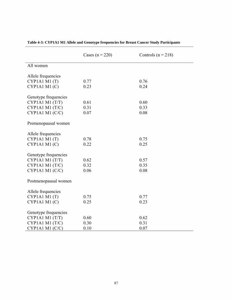
Table 4-3: CYP1A1 M1 Allele and Genotype frequencies for Breast Cancer Study Participants
Cases (n = 220)
Controls (n = 218)
All women
Allele frequencies CYP1A1 M1 (T) 0.77 0.76 CYP1A1 M1 (C) 0.23 0.24 Genotype frequencies CYP1A1 M1 (T/T) 0.61 0.60 CYP1A1 M1 (T/C) 0.31 0.33 CYP1A1 M1 (C/C) 0.07 0.08 Premenopausal women
Allele frequencies CYP1A1 M1 (T) 0.78 0.75 CYP1A1 M1 (C) 0.22 0.25 Genotype frequencies CYP1A1 M1 (T/T) 0.62 0.57 CYP1A1 M1 (T/C) 0.32 0.35 CYP1A1 M1 (C/C) 0.06 0.08 Postmenopausal women
Allele frequencies CYP1A1 M1 (T) 0.75 0.77 CYP1A1 M1 (C) 0.25 0.23 Genotype frequencies CYP1A1 M1 (T/T) 0.60 0.62 CYP1A1 M1 (T/C) 0.30 0.31 CYP1A1 M1 (C/C) 0.10 0.07
87
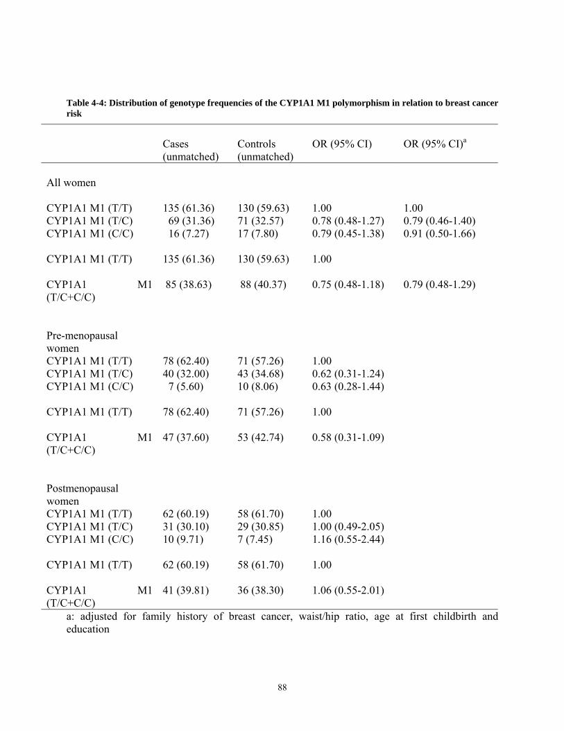
Table 4-4: Distribution of genotype frequencies of the CYP1A1 M1 polymorphism in relation to breast cancer risk
Cases (unmatched)
Controls (unmatched)
OR (95% CI)
OR (95% CI)a
All women
CYP1A1 M1 (T/T) 135 (61.36) 130 (59.63) 1.00 1.00 CYP1A1 M1 (T/C) 69 (31.36) 71 (32.57) 0.78 (0.48-1.27) 0.79 (0.46-1.40) CYP1A1 M1 (C/C) 16 (7.27) 17 (7.80) 0.79 (0.45-1.38) 0.91 (0.50-1.66) CYP1A1 M1 (T/T) 135 (61.36) 130 (59.63) 1.00
CYP1A1 M1 (T/C+C/C)
85 (38.63) 88 (40.37) 0.75 (0.48-1.18) 0.79 (0.48-1.29)
Pre-menopausal women
CYP1A1 M1 (T/T) 78 (62.40) 71 (57.26) 1.00 CYP1A1 M1 (T/C) 40 (32.00) 43 (34.68) 0.62 (0.31-1.24) CYP1A1 M1 (C/C) 7 (5.60) 10 (8.06) 0.63 (0.28-1.44) CYP1A1 M1 (T/T) 78 (62.40) 71 (57.26) 1.00
CYP1A1 M1 (T/C+C/C)
47 (37.60) 53 (42.74) 0.58 (0.31-1.09)
Postmenopausal women
CYP1A1 M1 (T/T) 62 (60.19) 58 (61.70) 1.00 CYP1A1 M1 (T/C) 31 (30.10) 29 (30.85) 1.00 (0.49-2.05) CYP1A1 M1 (C/C) 10 (9.71) 7 (7.45) 1.16 (0.55-2.44) CYP1A1 M1 (T/T) 62 (60.19) 58 (61.70) 1.00
CYP1A1 M1 (T/C+C/C)
41 (39.81) 36 (38.30) 1.06 (0.55-2.01)
a: adjusted for family history of breast cancer, waist/hip ratio, age at first childbirth and education
88
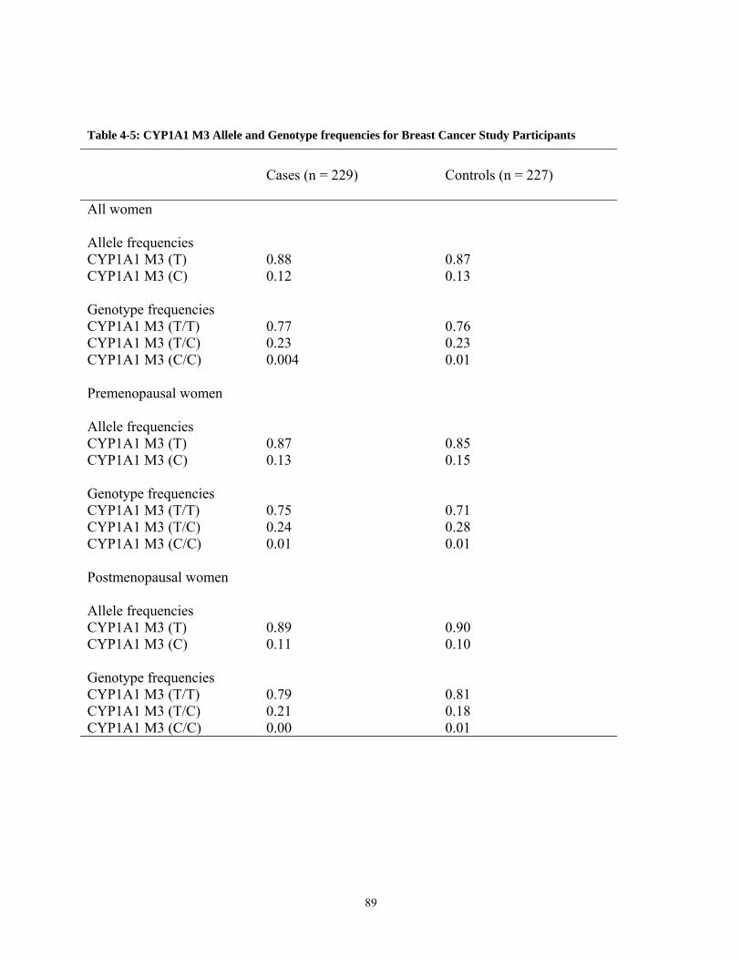
Table 4-5: CYP1A1 M3 Allele and Genotype frequencies for Breast Cancer Study Participants
Cases (n = 229)
Controls (n = 227)
All women
Allele frequencies CYP1A1 M3 (T) 0.88 0.87 CYP1A1 M3 (C) 0.12 0.13 Genotype frequencies CYP1A1 M3 (T/T) 0.77 0.76 CYP1A1 M3 (T/C) 0.23 0.23 CYP1A1 M3 (C/C) 0.004 0.01 Premenopausal women
Allele frequencies CYP1A1 M3 (T) 0.87 0.85 CYP1A1 M3 (C) 0.13 0.15 Genotype frequencies CYP1A1 M3 (T/T) 0.75 0.71 CYP1A1 M3 (T/C) 0.24 0.28 CYP1A1 M3 (C/C) 0.01 0.01 Postmenopausal women
Allele frequencies CYP1A1 M3 (T) 0.89 0.90 CYP1A1 M3 (C) 0.11 0.10 Genotype frequencies CYP1A1 M3 (T/T) 0.79 0.81 CYP1A1 M3 (T/C) 0.21 0.18 CYP1A1 M3 (C/C) 0.00 0.01
89
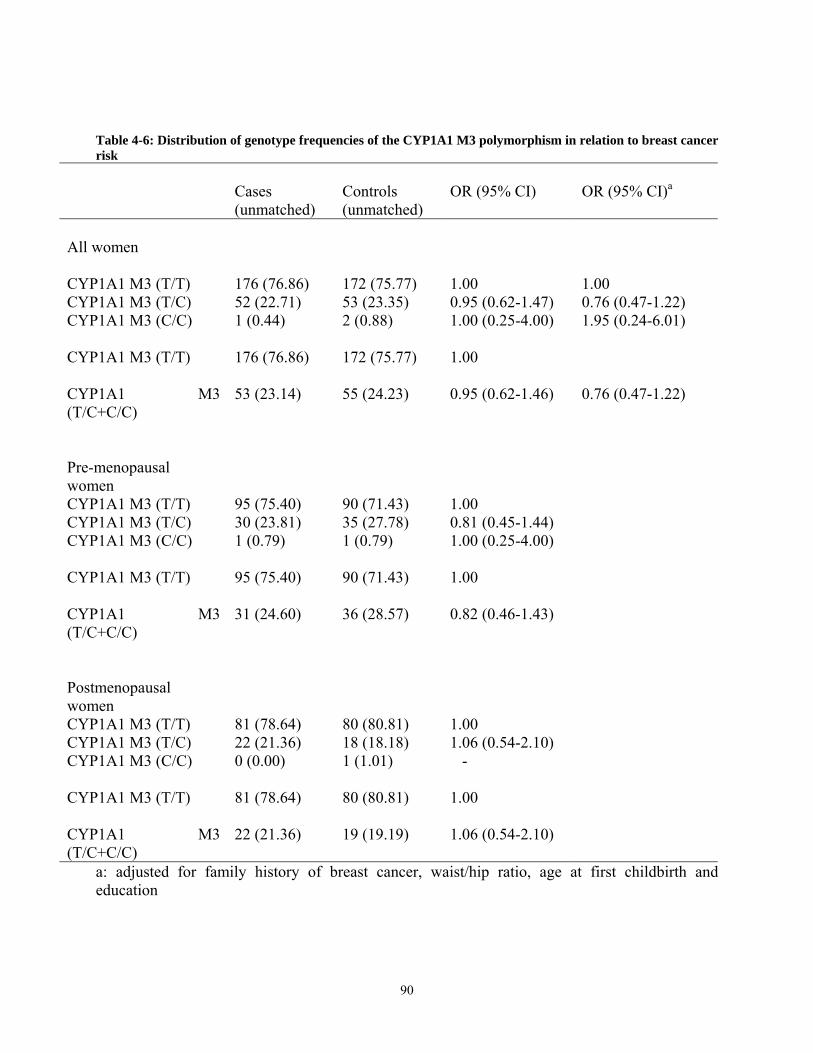
Table 4-6: Distribution of genotype frequencies of the CYP1A1 M3 polymorphism in relation to breast cancer risk
Cases (unmatched)
Controls (unmatched)
OR (95% CI)
OR (95% CI)a
All women
CYP1A1 M3 (T/T) 176 (76.86) 172 (75.77) 1.00 1.00 CYP1A1 M3 (T/C) 52 (22.71) 53 (23.35) 0.95 (0.62-1.47) 0.76 (0.47-1.22) CYP1A1 M3 (C/C) 1 (0.44) 2 (0.88) 1.00 (0.25-4.00) 1.95 (0.24-6.01) CYP1A1 M3 (T/T) 176 (76.86) 172 (75.77) 1.00
CYP1A1 M3 (T/C+C/C)
53 (23.14) 55 (24.23) 0.95 (0.62-1.46) 0.76 (0.47-1.22)
Pre-menopausal women
CYP1A1 M3 (T/T) 95 (75.40) 90 (71.43) 1.00 CYP1A1 M3 (T/C) 30 (23.81) 35 (27.78) 0.81 (0.45-1.44) CYP1A1 M3 (C/C) 1 (0.79) 1 (0.79) 1.00 (0.25-4.00) CYP1A1 M3 (T/T) 95 (75.40) 90 (71.43) 1.00
CYP1A1 M3 (T/C+C/C)
31 (24.60) 36 (28.57) 0.82 (0.46-1.43)
Postmenopausal women
CYP1A1 M3 (T/T) 81 (78.64) 80 (80.81) 1.00 CYP1A1 M3 (T/C) 22 (21.36) 18 (18.18) 1.06 (0.54-2.10) CYP1A1 M3 (C/C) 0 (0.00) 1 (1.01) - CYP1A1 M3 (T/T) 81 (78.64) 80 (80.81) 1.00
CYP1A1 M3 (T/C+C/C)
22 (21.36) 19 (19.19) 1.06 (0.54-2.10)
a: adjusted for family history of breast cancer, waist/hip ratio, age at first childbirth and education
90
REFERENCES
1. Kawajiri K, Nakachi K, Imai K, Yoshii A, Shinoda N, Wanatabe J. Identification of
genetically high risk individuals to lung cancer by DNA polymorphisms of the
cytochrome P4501A1 gene. FEBS Lett 1990; 263:131-133.
2. Hayashi S, Wanatabe J, Nakachi K, Kawajiri K. Genetic linkage of lung-associated MspI
polymorphism with amino acid replacement in the heme binding region of the human
cytochrome P4501A1 gene. J Biochem 1991; 110:407-411.
3. Crofts F, Cosma GN, Currie D, Taioli E, Toniolo P, Garte SJ. A novel CYP1A1 gene
polymorphism in African Americans. Carcinogenesis 1993; 14:1729-1731.
4. Cascorbi I, Brockmoller J, Roots I. A C4887A polymorphism in exon 7 of human
CYP1A1: population frequency, mutation linkages, and impact on lung cancer
susceptibility. Cancer Res 1996; 56:4965-4969.
5. Schneider J, Huh MM, Bradlow LH, Fishman J. Antiestrogen action of 2-hydrxyestrone
on MCF-7 human breast cancer cells. J Biol Chem 1984; 259:4840-4845.
6. Fotsis T, Zhang Y, Pepper MS, Aldercreutz H, Montesano R, Nawroth PP, Schwelgerer
L. The endogenous estrogen metabolite 2-methoxyestradiol inhibits angiogenesis and
suppresses tumor growth. Nature 1994; 368:237-239.
7. Cushman M, He HM, Katzenellenbogan JA, Lin CM, Hamel E. Synthesis, antitubulin
and antimitotic activity, and cytotoxicity of analogs of 2-methoxyestradiol, an
91
endogenous mammalian metabolite of estradiol that inhibits tubulin polymerization by
binding to the colchicines binding site. J Med Chem 1995; 38:2041-2049.
8. Klauber N, Parangi S, Flymn E, Hamel E, D’Amato RJ. Inhibition of angiogenesis and
breast cancer in mice by the microtubulin inhibitors 2-methoxyestradiol and taxol. Cancer
Res 1997; 57:81-86.
9. Musey PI, Collins DC, Bradlow HL, et al. Effect of diet on oxidation of 17beta- estradiol
in vivo. J Clin Endocrinol Metab 1987; 65:782-795.
10. Gierthy JF, Lincoln DWII, Kampcik SJ, et al. Enhancement of 2- and 16 alpha-estradiol
hydroxylation in MCF-7 human breast cancer cells by 2,3,7,8-tetrachlorodibenzo-p-
dioxin. Biochem Biophys Res Commun 1988; 157:515-520.
11. Wanatabe H. Effects of 17 alpha-ethinylestradiol on biliary metabolites of 17 beta[6,7-
3H]estradiol in the rat. Biochem Biophys Acta 1971; 231:399-405.
12. Crofts F, Taioli E, Cosma GN, Currie D, Toniolo P, Garte SJ. Functional significance of
different human CYP1A1 genotypes. Carcinogenesis (Lond.), 1994; 12:2961-2963.
13. Taioli E, Trachman J, Chen X, Toniolo P, Garte SJ. A CYP1A1 restriction fragment
length polymorphism is associated with breast cancer in African American women.
Cancer Res 1995; 55:3757-3758.
14. Taioli E, Bradlow HL, Garbers SV, Sepkovic DW, Osborne MP, et al. Role of estradiol
metabolism and CYP1A1 polymorphisms in breast cancer risk. Cancer Detect Prev 1999;
23(3):232-237.
15. Rebbeck TR, Rosvold EA, Duggan DJ, Zhang J, Buetow KH. Genetics of CYP1A1:
coamplification of specific alleles by Polymerase Chain Reaction and association with
breast cancer. Cancer Epidemiol Biomarkers Prev 1994; 3:511-514.
92
16. Ambrosone CB, Freudenheim JL, Graham S, Marshall JR, Vena JE, Brasue JR, et al.
Cytochrome P4501A1 and Glutathione S-Transferase (M1) genetic polymorphisms and
postmenopausal breast cancer risk. Cancer Res 1995; 55:3483-3485.
17. Ishibe N, Hankinson SE, Colditz GA, Spiegelman D, Willet WC, et al. Cigarrete
smoking, cytochrome P4501A1 polymorphisms, and breast cancer risk in the Nurses’
Health Study. Cancer Res 1998; 58:667-671.
18. Huang CS, Shen CY, Chang KJ, Hsu SM, Chern HD. Cytochrome P4501A1
polymorphism as a susceptibility factor for breast cancer in postmenopausal Chinese
women in Taiwan. Br J Cancer 1999; 80(11):1838-1843.
19. Miyoshi Y, Takahashi Y, Egawa C, Noguchi S. Breast cancer risk associated with
CYP1A1 genetic polymorphisms in Japanese women. The Breast J 2002; 8(4):209-215.
20. QIAamp DNA Mini Kit and QIAamp DNA Blood Mini Kit Handbook, February 2003.
21. QIAamp DNA Midi Kit and QIAamp DNA Blood Midi Kit Handbook, February 2003.
22. Fishman J, Schneider J, Hershcopf RJ, Bradlow HL. Increased estrogen 16α-hydroxylase
activity in women with breast cancer and endometrial cancer. J Steroid Biochem Mol
Biol 1984; 20:1077-1081.
23. Yager JD, Liehr JG. Molecular mechanisms of estrogen carcinogenesis Ann Rev
Pharmacol Toxicol 1996; 36:203-232.
24. Zhu BT, Conney AH. Functional role of estrogen metabolism in target cells: review and
perspectives. Carcinogenesis, 1998; 19:1-27.
25. Peterson DD, McKinney CE, Ikeya K, Smith HH, et al. Human CYP1A1 gene:
cosegregation of the enzyme inducibility phenotype and an RFLP. Am J Hum Genet
1991; 48:720-725.
93

26. Cosma GN, Crofts F, Currie D, Wirgin I, Toniolo P, Garte SJ. Racial differences in
restriction fragment length polymorphisms and Mrna inducibility of the human CYP1A1
gene. Cancer Epidemiol Biomark Prevent 1993; 2:53-57.
27. Crofts F, Cosma GN, Currie D, Taioli E, Toniolo P, Garte SJ. A novel CYP1A1 gene
polymorphism in African Americans. Carcinogenesis 1993; 14(9):1729-1731.
28. Bailey LR, Roodi N, Verrier CS, Yee CJ, Dupont WD, Parl FF. Breast cancer and
CYP1A1, GSTM1, and GSTT1 polymorphisms: evidence of a lack of association in
Caucasians and African Americans. Cancer Res 1998; 58:65-70.
94
GENERAL DISCUSSION/FUTURE RESEARCH
A rapidly growing global population, an aging world population profile with changing
reproductive characteristics, a western-oriented diet heavily laden with animal fat, a sedentary
lifestyle and a work environment exposed to various chemical carcinogens have all contributed
partly to the rising global burden of breast cancer, a disease predominantly affecting the worlds’
female sex. Breast cancer is the third most frequent cancer in the world (796,000 cases in 1990)
and by far the most common malignancy of women (21% of all new cases). Worldwide, the ratio
of mortality to incidence is about 61% (Parkin et al., 1999a). As a result, breast cancer ranks as
the fifth cause of death from cancer overall, although it is still the leading cause of cancer
mortality in women (the 314,000 annual deaths represent 14.1% of cancer deaths in females).
Recent World Health Organization (WHO) statistics indicate that with an annual incidence rate
increase of about 0.5% in western countries, and rates as high as 5% in China and other Eastern
Asian countries and probably similar increases in sub-Saharan Africa and South America, the
global burden of breast cancer in 2010 would be 1.45 million new cases, an 82% increase over
the figure in 1990 (Parkin et al., 1999a). This increasing disease burden, likely to be born by the
dwindling economies of the developing countries with the least resources to fight the disease
calls for a reawakening of global efforts to combat the disease. We may not be able to
completely eliminate breast cancer, but emerging evidence from the developed world provide
evidence that we can at least slow the growth in the incidence of the disease and reduce
morbidity and mortality in those afflicted by breast cancer.
Globally the prognosis for breast cancer is reported as generally rather good. The highest
crude survival is reported by the SEER program, 84%, consistent with the age-adjusted estimate
95
for North America of 73%. Survival rates are high in Japan (74%) and Australia/New Zealand
(68%) and lower in Europe (53% to 63%, consistent with the EUROCARE crude rate of 67%).
Elsewhere survival ranges from 49% to 61% (Parkin et al., 1999a).
An issue of concern to the US in particular is the Black/White disparities in breast cancer
burden within the US. Although, there are conflicting reports in the literature as to the degree of
this disparity accounted for by racial differences in socioecomonic status, mammographic
screening, and access to health care, there appear to be a consensus that a certain proportion
remains explained by race. Black women diagnosed with breast cancer experience a 5-year
relative survival of only 62%, compared with 79% for white women (Miller et al., 1992). Part of
the poorer breast cancer survival for black women is due to more, advanced stage of disease at
presentation, but some analyses that have controlled or stratified for stage have still found black
women’s survival to be poorer than that of their white counterparts (Rain et al., 1986;
McWhorter et al., 1987; Kimmick et al., 1991; Fisher et al., 1993). Research concerning
differences in breast cancer survival between black and white women suggests that the biological
characteristics of tumors may differ between the races (Fisher et al., 1993; Dayal et al., 1982).
Recent data from the National Cancer Institute Black/White Cancer Survival Study showed that
after adjusting for stage, treatment, comorbid illness, and pathologic and sociodemographic
variables, blacks continued to demonstrate a slightly increased, but not statistically significant,
risk of death (hazard ratio=1.3; 95% CI 1.0-1.8) (Eley et al., 1994). Current efforts to eliminate
health disparities among the different racial groups in the US will help show whether or not this
Black/White differences will remain in the next decades to come.
A coalition of global efforts based upon sound public health principles and led by
epidemiology hold the key to success in the fight to reduce the global burden of breast and other
96

cancers. The last half-century has seen the integration of disciplinary efforts to better understand
the process of carcinogenesis. Recent advances in the traditional disciplines of bacteriology,
virology, biochemistry and molecular biology and rapid progress in the Human Genome Project,
and the application of tools of molecular biology to epidemiological studies will markedly
influence the road map in the search for etiologic factors for breast cancer. For these efforts to
have lasting impact on reducing global breast cancer burden, epidemiological research has to
adopt a global approach. This is because there is evidence supporting a multifactorial etiology
for breast cancer. In fact, it is being speculated that breast cancer is not a single disease entity but
a final common pathway for the manifestation of a variety of genetic insults resulting in loss of
cellular regulation and integration. While breast cancer occurs in all continents and countries
across the globe, it is the nature of the disease that risk factors may differ from one part of the
world to the other. Available data suggests that reproductive characteristics such as age at
menarche, parity, duration of breastfeeding and practices such as use of exogenous estrogens for
birth control and hormone replacement therapy (HRT) differ considerably between the
developed world and developing countries(Collaborative Group on Hormonal Factors for Breast
Cancer, 1997). In addition, prevalence of obesity (a risk factor associated with many chronic
diseases including breast cancer) varies widely across the globe.
The choice of Nigerian women for this study was meant to serve dual purposes.
Sub-Saharan Africa, of which Nigeria (population: 130 million) (Nigeria National Population
Commission, 2003) is one of the component countries, is a region of the world grossly under-
represented in the epidemiologic literature. Until recently, it used to be said that breast cancer
was very rare in Nigeria; simply because the disease has not be adequately studied in the
population. We believe that studying breast cancer risk factors among Nigerian women will not
97
only provide data about the disease among populations in sub-Saharan Africa but will contribute
to global efforts at control and prevention. In addition, there is evidence that the Nigerian
population under study share considerable genetic ancestry with Nigerian women. We suspect
that predictors of breast cancer risk particularly genetic risks may be similar in the two
populations. Studying breast cancer risk among African American women in the US has been
hampered by low participation rate which has been attributed to factors such as mistrust and
suspicion of the healthcare system, economic limitations, low level of education, concerns about
drug toxicity, cultural/religious beliefs/attitudes, poor access to health care, lack of awareness
about studies, past negative experiences, and physicians’ attitude (Cox et al., 1998; Harris et al.,
1996; Shavers-Hornaday et al., 1997; Swanson et al., 1995; Thomas et al., 1994; Robinson et al.,
1996). Studying the role of low-penetrance genes such as cytochrome P450 (CYP1A1) and
catechol-O-methyltransferase (COMT) gene polymorphisms in breast cancer susceptibility in
Nigerian women provides an excellent opportunity for investigating the contribution of genetic
factors to breast cancer burden in populations of African descent. The high participation rate of
the study population is encouraging although infrastructural inadequacy creates setbacks in the
conduct of such studies.
Article 1
The first article evaluates the descriptive epidemiology of breast cancer risk in the study
population. Breast cancer in our study population was found to be predominantly premenopausal
(56.8%). Risk factors associated with increased risk of breast cancer in our study population
include family history of breast cancer, higher waist/hip ratio, abortion, age at first full-term
pregnancy, and higher level of education while increasing parity and longer duration of
98
breastfeeding conferred protection against the disease. These findings are similar to
characteristics of the disease reported in studies comparing the risk profiles of breast cancer in
African American and Caucasian women in the US. African American women under the age of
45 years have a greater incidence of breast cancer than Caucasian women in this young age
range. These rates equalize during the fifth decade of life, and for women over the age of 55
years, incidence rates for Caucasian women surpass those of African Americans (Newman,
2005). Available clinical data indicate that 30%-40% of African American breast cancer patients
seen in clinical practice are younger than 50 years, compared with 20% of Caucasian breast
cancer patients in this age range (National Cancer Data Base, 1994). Factors accounting for this
race-related risk profile are not clearly understood.
In a landmark study, Pike et al (1983) proposed the model of “breast tissue age” and this
has been supported by a further extension of the Pike model by Pathaks and Whittemore (1992).
The theory of Pike assumes that breast carcinoma vary in proportion to a power of the
accumulated “breast tissue age” (Pike et al., 1983). In that model, the breast tissue ages at a
constant rate between age at menarche and age at first full-term pregnancy (FFTP), at which time
the hormonal milieu of pregnancy causes a one-time increase in breast tissue age but lowers the
rate of subsequent “breast tissue aging”. This lower rate continues until the perimenopausal
years, at which time it decreases linearly until menopause. After menopause the breast tissue
continues to age, but at a much lower rate. This model predicts that at a given age, a woman with
an FFTP in the preceding 5-10 years is at increased risk relative to a nulliparous woman. Studies
of Janerich and Hoff (1982), and Pathak et al (1986) have confirmed this theory. Carrying a
pregnancy to the third trimester confers a protective effect on lifetime risk for breast carcinoma if
99

that pregnancy occurs early in life. However, if a woman delivers her first child close to the age
at which menopause occurs, her lifetime risk is actually higher than if she was nulliparous.
Pathaks and Whittemore (1992) extended Pike’s single-birth model to a multiple birth
model, by incorporating a smaller increase in risk at each additional full term pregnancy with a
subsequent lowering of the rate at which breast tissue ages. Rosner et al (1994) fitted the
extended Pike model to prospective data from the Nurse’s Health Study and obtained breast
carcinoma incidence curves for various combinations of age at FFTP, total parity, and ages at
subsequent births. The predicted incidence curves for these hypothetical scenarios show lower
risk for nulliparous women relative to multiparous women until ages 42-45 years, at which time
a crossover occurs and the multiparous are at lower risk relative to the nulliparous women. The
crossover for women with a single birth at age 20 years does not occur until age 55 years. This
shift to a later age for the crossover effect for women with a single birth relative to multiparous
women would be expected based on the hypothesized decreased rate of breast tissue aging after
each subsequent pregnancy (Pathak et al., 2000).
Another factor that may contribute to higher breast cancer risk in young black women is the
higher rate of abortions performed before the first full-term pregnancy and higher rate of use of oral
contraceptive pills during the same period compared to young white women (Romieu et al., 1990).
Although abortion was associated with increased risk of breast cancer in our patients, the abortion
rates were low and data is lacking on the number of abortions performed before first full-term
pregnancy in our patients. Increasing incidence of abortion and use of oral contraceptives among the
younger cohorts of Nigerian women is of concern to the risk of breast cancer in the population in the
future. Of concern also is the changing lifestyle of the younger cohorts of Nigerian women.
Increasing westernization with more consumption of animal fat and sedentary lifestyle may lead to
100
obesity epidemic in the future as is being witnessed in the developed world. In addition, early
menarche, delay in childbearing and reduced parity may add to drive up longer period of lifetime
unopposed estrogen exposure (an established risk factor for breast cancer).
Article 2
In the second article, we evaluated the association between the G to A transition
polymorphism in codon 158 of the COMT gene and breast cancer risk in the study population. In
the final conditional logistic regression model controlling for identified risk factors for breast
cancer, harboring at least one low-activity COMT (Met) allele was associated with a significant
43% reduced risk of breast cancer (OR = 0.57, 95% CI 0.36-0.91). Similar reduced risks were
observed in both premenopausal and postmenopausal breast cancer risk although further
subgroup analysis were precluded by the low frequency of the COMT (Met) allele in the study
population. We noted a 2 times lower frequency of the COMT (Met/Met) compared with the
13% reported in African Americans and 3-4 times lower than the 22-28% in Caucasians
(Millikan et al, 1998). Our finding of reduced risk of breast cancer both in the final conditional
logistic regression multivariate model and in univariate analyisis in premenopausal and
postmenopausal women agrees with the 20% reduced risk (although not significant) in both
premenopausal and postmenopausal African American women and the 20% and 30% reduced
risk (also not significant) of premenopausal and postmenopausal breast cancer risk among
Caucasian women reported by Millikan et al (1998). The findings of 2-fold increased risk (not
significant) of postmenopausal breast cancer among Caucasian women by Lavigne et al. (1997)
and 5.7 fold increased risk of premenopausal breast cancer risk in Caucasian women by
Thompson et al. (1998) is contrary to our findings. As noted previously, Asian women appear to
101
show a more consistent increased risk of breast cancer in carriers of the low-activity COMT
(Met) allele (Yim et al., 2001).
Factors responsible for the inconsistencies in the reports in the literature with respect to
association studies of the codon 158 COMT polymorphism are not easy to explain. For example,
how do we explain the protection conferred by the COMT gene variant for breast cancer in our
study subjects. In the first instance, this finding is contrary to our hypothesis of increased breast
cancer risk among carriers of the variant allele because of the known functions of the COMT
gene and the reported decreased methylation activity of the gene caused by the codon 158
polymorphism. We have already noted the much lower frequency of the COMT (Val/Met) and
COMT (Met/Met) variants in our population compared with Caucasians. This reduced frequency
of the variant allele might suggest a smaller impact of this polymorphism on the methylation of
estrogen catechols in our subjects. Another possible explanation for our finding may be that the
codon 158 Valine/Methionine polymorphism is in linkage disequilibrium with other
polymorphisms of functional significance in the detoxification of biologically harmful estrogen
catechols. We noted that sample size impacted subgroup analysis in our study. Although, our
sample size of 231 cases and 229 controls with successful genotype data is larger than the sample
size reported in most of the other studies noted above, the lower prevalence of the COMT
(Met/Met) genotype in our population implies the need for larger sample size to be able to detect
similar sizes of effects noted in either Caucasian or African American women. Using our COMT
(Met/Met) frequency of 0.07 detected in our control subjects will require a sample size of 304
premenopausal cases and 304 premenopausal controls if we wish to detect an odd ratio of 2.4
(reported by Thompson et al., 1998). Our sample size of 142 premenopausal cases and 142
premenopausal controls is just about half the calculated sample size. Sample size is always an
102
issue of contention in early studies in population groups with no pre-existing data and our
experience with the COMT gene highlights the importance of prior pilot studies. Our finding will
serve this purpose for future disease association studies of this polymorphic variant in the
Nigerian population.
Efforts to explain the discrepancy between the protective effect reported by Millikan et al.
(1998) and the increased risk reported by Lavigne et al. (1997) and Thompson et al. (1998) (both
reports contrasting with each other) among Caucasian women in the US have been less rewarding.
As noted by Thompson et al. (1998), one possible explanation for the inconsistencies may be
differences in the ethnic backgrounds of study participants. It should be noted that Caucasian
Americans are of different European ethnicities which may affect the distribution of the COMT
variant genotypes, a factor that can partly account for the divergent findings. Differences in these
findings may also be the result of small sample size of the studies. As noted previously, the study of
Lavigne et al. (1997) had only 113 cases and 113 controls in contrast to that of Thompson et al.
(1998) (281 cases and 289 controls) and Millikan et al. (1998) (Caucasians: 389 cases and 379
controls, African Americans (265 cases and 263 controls). Another possible explanation may be the
action of other compensating enzymes such as Glutathione S-transferase (GSTs), uridine diphospho-
glucuronosyltransferase (UGT) 1A1, and the highly polymorphic manganese superoxide dismutase
(MNSOD) genes that are induced when COMT activity is very low. Lower oxidative damage in the
presence of COMT-(Met/Met) genotype may also be of importance; the level of 8-hydroxy-2’-
deoxyguanosine in breast tissue was higher in patients with COMT (Val/Val) genotype as shown by
Markides et al. (1998).
The fact that the COMT gene is highly polymorphic in some populations means that the
population attributable risk for breast cancer resulting from the COMT genetic polymorphism may
103
be more marked in populations of higher prevalence of the homozygous COMT (Met/Met) genotype
such as Caucasian populations (Millikan et al., 1998) compared with effects on risk in populations of
lower prevalence as demonstrated in the Nigerian population and in Asian populations (Yim et al.,
2001). In addition, environmental factors such as the prevalence of obesity in the population may
also modulate the effect of the genotype on breast cancer risk. The implication of the above is that as
part of the global strategy for the control and prevention there is need for studies of the prevalence of
these polymorphisms in diverse population groups across the world to properly define the role of the
COMT gene in breast cancer risk in various populations.
Article 3
Our interest in the third article is to assess the role of four polymorphic variants (M1, M2,
M3, and M4) in the cytochrome P450 (CYP1A1) gene in breast cancer susceptibility. CYP1A1 is
involved in the 2-hydroxylation of estrogens to 2-hydroxy catechol metabolites for subsequent
O-methylation to 2-methoxy derivatives including 2-methoxyestradiol. There is currently little
information on the activities of these various polymorphic variants in the CYP1A1 gene in
human populations, efforts are underway to characterize the biochemical properties of these
variants to enhance research into their possible role in disease risk. There is evidence suggesting
enhanced hydroxylation activity for the exon 7 polymorphism in some populations such as the
Japanese (Nakachi et al., 1993) but not in African Americans (Taioli et al., 1995). This study has
demonstrated the high prevalence of the CYP1A1 M1 and CYP1A1 M3 variants and the rarity of
the CYP1A1 M2 and CYP1A1 M4 polymorphims in the Nigerian population. The CYP1A1 M1
(C/C) genotype frequency of 8% was found to be twice the 3.5% reported in African Americans
but about 3-4 times higher than the 2% noted in Caucasians (Taioli et al., 1995). Our finding of a
104
non-significant 19% reduced risk of breast cancer in our study subjects is in sharp contrast to the
almost 10 times increased risk reported by Taioli et al. (1995) in African Americans although it
must be pointed out that their small sample size of 21 cases and 85 cases creates room for
spurious associations. Also, the finding of a significant 1.98 times increased risk of
postmenopausal breast cancer risk in Chinese women in Taiwan (Huang et al., 1999) contradicts
our finding of a non-significant 16% increased risk of breast cancer in postmenopausal women in
our study. The finding of significant 40% and 42% reduced risk of breast cancer among Japanese
women harboring the heterozygous and homozygous variants of this genotype respectively
(Miyoshi et al., 2002) also reflects the inconsistencies associated with association studies
between CYP1A1 M1 variant and breast cancer risk in various populations. It must however be
appreciated that the 51% CYP1A1 M1 (T/C) heterozygosity and the 17% CYP1A1 M1 (C/C)
homozygosity reported among Japanese patients is about twice the frequency of these genotypes
in the Nigerian population.
Allele frequencies for the CYP1A1 M3 (T) and CYP1A1 M3 (C) alleles among our study
participants was slightly lower than the figures reported by Crofts et al. (1993) among African
American women. The CYP1A1 M3 variant is said to be African American-specific and has not
been reported in Caucasians. We were unable to demonstrate any significant association between
this novel polymorphism and breast cancer risk overall or in subgroup analysis in keeping with
the findings of Taioli et al. (1995) who first described this variant in African American women
although we noticed a higher frequency of the heterozygous CYP1A1 M3 (T/C) genotype
compared with the figure reported by these investigators. We were unable to compute risks
associated with the CYP1A1 M2 (Exon 7) and CYP1A1 M4 polymorphic variants due to their
rarity in our population.
105
The interest in the 2-hydroxylation of estrogens has heightened with the recent finding
that 2-methoxyestradiol, the O-methylation product of this pathway possesses anti-tumor
properties. 2-Methoxyestradiol inhibits the proliferation of several cancer cell lines in vitro
(Fotsis et al., 1994), and human breast cancer cell lines (estrogen receptor positive or negative)
were particularly sensitive to a cytotoxic effect of 2-methoxyestradiol (Cushman et al., 1995).
Additional studies indicated that 2-methoxyestradiol disrupted microtubule function and was a
potent inhibitor of angiogenesis (Fotsis et al., 1994). By altering microtubule stability, inducing
apoptosis and inhibiting angiogenesis in tumor cells and downregulating cytokine and PGE2
induced in-situ aromatase synthesis in the breast (Purohit et al., 1999), 2-methoxyestradiol
distinguishes itself as the new focus for new generation anti-cancer chemotherapy for both
hormone dependent and hormone-independent breast cancer. In fact, 2-methoxyestradiol is
undergoing a phase I trial as an angiogenesis inhibitor and search is ongoing for synthetic
derivatives of this metabolite for anti-cancer chemotherapy (Brem et al., 1987).
Knowledge of estrogen metabolism has also provided new insight in appreciating the
interaction between obesity and high fat diet in mediating breast carciongenesis. It is now well
documented that lean women or women on low fat diets have a lower risk of breast cancer than
obese women or women consuming high fat diets. This finding is explained by the fact that in
lean women or those on low fat diets, metabolism of estrogens via the 2-hydroxylation pathway
predominates with subsequent formation of 2-methoxyestrogens (Longcope et al., 1987).
Consequently, this causes downregulation of cytokine and PGE2 receptors in breast adipose
stromal cells and reduces in situ estrogen synthesis via peripheral aromatization. In contrast, in
obese women or subjects on high fat diets that are associated with reduced synthesis of 2-
methoxyestrogens, any cytokines or PGE2 within the breast could result in increased production
106
of estrogens and subsequent risk of tumor development. There is also evidence suggesting that
pesticides, which have been implicated in breast cancer, can also decrease estrogen 2-
hydroxylation but increase 16-[alpha]-hydroxylation (Bradlow et al., 1995).
Some of the limitations of this study include recall and other biases associated with case
control studies although it must be noted that genotype data is constant and unlikely to be
affected by problems with recall. We calculated sample size for this study based on data on
African American women; the genotype frequencies used for the calculation are similar to our
findings among the Nigerian patients. Based on the genotype frequencies of the CYP1A1 M1
variant, we estimated that 35 CYP1A1 M1 (C/C) homozygous cases and controls would be
required to demonstrate the odds ratio of 9.7 noted by Taioli et al (1995). Among our 220 cases
and 218 control subjects with successful PCR-based RFLP assay, the number of cases and
controls with the homozygous CYP1A1 M1 (C/C) genotype were 16 and 17 respectively, about
half of the number calculated above. If the risk profile associated with the CYP1A1 M1
polymorphism reported by Taioli et al. (1995) were to be correct, we would have required twice
our current sample size of 220 cases and 218 controls to detect similar effect size found in their
study. However, the sample size of 21 cases and 85 controls recruited by Taioli et al. (1995)
seem too small for conclusive statements to be made. In addition, theirs was probably the only
study of the association of this polymorphism with breast cancer risk in populations of African
descent.
To the best of our knowledge, ours is the first study to investigate the association between
CYP1A1 genetic polymorphisms and breast cancer risk in indigenous African women. We have
provided baseline data with which future studies can be compared. The distribution of the
genotype frequencies of the CYP1A1 variants reported here will provide the basis for estimating
107
sample sizes required for further studies on the role of these variants on breast cancer risk in sub-
Saharan African populations.
Future Research
This study has provided us opportunity for an exploratory study to investigate the risk
factors for breast cancer in Nigerian women, a population of exclusively indigenous African
women, a population about whom little is known about breast cancer susceptibility. We have had
a lot of difficulties; however we have learnt a lot of lessons within the past 3 years of this study.
First, because of the multicenter design, we have been able to establish loci for future research on
breast and other cancers within the Nigerian population. We have shown in the first article on the
descriptive epidemiology of breast cancer in the population that reproductive variables and other
factors similar to reports in other populations predict breast cancer risk in this population, thus
establishing that lifetime estrogen exposure remains the main arbiter of breast cancer risk in
indigenous African women as in other populations. Our exploratory data on the association
between the COMT and CYP1A1 genetic polymorphims and breast cancer risk in these women
has provided a reference on which future studies would depend. We have pointed out limitations,
which might have affected our results, and we hope to avoid them in future studies. The rapid
progress of the Human Genome Project and accompanying fascinating progress in the health
sciences including molecular epidemiology, molecular biology, genetic engineering, biostatistics
and computer science and clinical medicine in the past half-decade has created an avalanche of
opportunities for integrated efforts in the battle against breast cancer. Although it is becoming
obvious that disease association studies based on single nucleotide polymorphisms are falling out
of favor in molecular epidemiologic research, such studies have provided the impetus for the
108
future. We are moving to a stage where hard science combined with high through put research
tools including microarray techniques and proteomics technology will drive the search for
etiological factors in cancer and other chronic diseases. We hope to remain in the team of the
future by building on the foundation provided by this exploratory study.
As a preliminary follow up to this study, we hope to further explore the association
between CYP1A1 genetic polymorphisms and breast cancer risk using the haplotype approach
combining allele and genotype data for CYP1A1 M1, CYP1A1 M2, CYP1A1 M3, and CYP1A1
M4 derived from this study. We also hope to apply the same technique in evaluating the
association between CYP1B1 M1, CYP1B1 M2, CYP1B1 M3, and CYP1B1 M4 polymorphic
variants of the cytochrome P4501B1 (CYP1B1) gene and breast cancer risk in this population.
We have generated allele frequency and genotype data for the tetranucleotide simple tandem
repeat polymorphism in the 3’ noncoding region of the aromatase gene. We will also evaluate the
association between repeats lengths and breast cancer risk in our study population. By combining
the genotype and haplotype data, we hope to build a multigenic model of breast cancer risk in
these subjects. Polymorphisms in other relevant genes in the pathway of estrogen synthesis and
metabolic pathway including the CYP17 beta-hydroxysteroid dehydrogenase (CYP17),
cytochrome P450 3A4 (CYP3A4), glutathione-S-transferase (GSTs), uridine diphospho-
glucuronosyltransferase (UTG) 1A1, N-acetyltransferases (NATs) and manganese superoxide
dismutase (MNSOD) genes will be investigated as part of the ongoing study. We will also
examine polymorphisms in the estrogen receptor polymorphisms and variations in DNA repair
genes including XRCC1, and the effect of epigenetic modifications such as promoter
hypermethylation of various genes in transcriptional silencing and how this relates to breast
cancer risk. As mentioned previously, there is need to expand the sample size of our study to
109

increase its robustness for multigenic modeling. We hope to achieve this by recruiting more
study participants from the Nigerian institutions; a sample size of 1000 cases and 1000 control
subjects is being estimated for this future study. We hope to expand our capacity for robust
comparisons by collaboration with other investigators with similar interests working with US
populations as well as populations of African descent in the Caribbean.
110
PUBLIC HEALTH SIGNIFICANCE OF STUDY
This study was focused on identifying risk factors for breast cancer in Nigerian women,
an indigenous sub-Saharan African population. The results have demonstrated that
sociodemographic characteristics, reproductive variables and anthropometric measures are
significant predictors of breast cancer risk in the study population. In particular, family history of
breast cancer in first- and second-degree relatives, higher level of education, waist/hip ratio,
abortion and later age at first full-term pregnancy were associated with increased risk of breast
cancer while high parity and long duration of breastfeeding conferred protection against the
disease in Nigerian women. We further evaluated the contribution of polymorphisms in catechol-
O-methyltransferase (COMT) and cytochrome P450 1A1 (CYP1A1) genes in breast cancer
susceptibility among the study population. Our results provide evidence suggesting that
harboring at least one low activity COMT (Met) allele of the codon 158 Valine to Methionine
polymorphism of the COMT gene significantly reduced the risk of breast cancer. The study also
demonstrated the rarity of the CYP1A1 M2 and CYP1A1 M4 polymorphisms of the CYP1A1
gene in the study population. Although the CYP1A1 M1 and CYP1A1 M3 genes were found to
be highly polymorphic, we were unable to demonstrate significant association between these
polymorphisms and breast cancer risk in the study population.
While these variables are similar to the risk factors for breast cancer described in African
American women and US Caucasians and other populations across the globe, we recognized
notable exceptions. Firstly, there was a preponderance of premenopausal breast cancer among
the study population similar to the distribution of the disease in African American women but in
contrast to the higher proportion of postmenopausal breast cancer in Caucasian populations.
111
Secondly, the use of oral contraceptives and postmenopausal hormone replacement therapy was
very low among the Nigerian women in this study in contrast to the higher figures reported in
Western populations. Thirdly, the Nigerian women in this population were predominantly lean
population with mean body mass index (BMI) below figures reported in the developed countries.
Fourthly, the frequency of the COMT (Met) allele of the codon 158 polymorphism (Valine to
Methionine substitution) of the COMT gene was lower in our study population compared to
figures reported in African American women and Caucasians in the US. Also the proportion of
the homozygous genotype of the African American-specific CYP1A1 M3 polymorphism of the
CYP1A1 gene was found to be higher in the Nigerian population than the reported figures in
African American women.
Although, it is consolatory to note that the epidemic of global obesity has not spread to
sub-Saharan Africa, there is need for policy guidelines necessary to prevent the obesity
phenomenon from spreading to the developing countries. In addition, the adoption of abortion as
means of family planning should be discouraged; effort should be directed to alternative methods
that do not endanger the life of women through increasing breast cancer risk. Also, physical
inactivity appear to be creeping into the lives of populations of developing countries as sedentary
office jobs replace the traditional agrarian economies. Alcohol consumption and cigarette
smoking rates although low in the study population are likely to be on the increase in these
populations in this century. Both factors have been linked to increased risk of breast cancer in
developed Western populations. The global anti-smoking campaign should be expanded to the
developing countries as the Tobacco companies shift their operations to these countries.
112

Overall, the findings of this study has contributed to knowledge about risk factors for
breast cancer in populations of African ancestry in general and Nigerian population in particular
and has provided baseline data for future epidemiologic studies in sub-Saharan African
populations.
113
SUMMARY
The study titled “Estrogens, Genetic Polymorphisms and Breast Cancer Risk in Nigerian
Women” was designed to evaluate the role of reproductive variables (surrogate measures of
lifetime estrogen exposure), family history, anthropometric variables and genetic polymorphisms
in breast cancer susceptibility in Nigerian women. The first article of the study evaluated the
descriptive epidemiology of breast cancer. Two hundred and fifty women with histologically-
confirmed breast cancer and their age matched controls were recruited from four University
Teaching Hospitals in Midwestern and Southeastern Nigeria for the study. Recruitment was
successfully conducted during a 20-month period between September 2002 and April 2004. The
association between various reproductive variables, sociodemographic characteristics and
anthropometric measurements and breast cancer risk in the study population was assessed using
conditional logistic regression models.
Results showed that a positive family history of breast cancer was associated with
increased risk of breast cancer. Higher level of education above High School also increased the
risk of breast cancer in these women. While reproductive variables such as late age at first full-
term pregnancy and abortion were associated with increased risk of breast cancer, high parity
and long duration of breastfeeding were protective against the development of the disease.
Among the anthropometric measurements evaluated for association with breast cancer risk, only
waist/hip ratio was found to be significantly associated with increased breast cancer risk. The
population was found to be lean with mean body mass index (BMI) below figures reported in
114
most Western populations. Although there was a tendency towards increased risk with increasing
height, the difference did not attain statistical significance.
The second article was designed to investigate the hypothesis that harboring the low-
activity COMT (Met) allele of the codon 158 valine to methionine substitution polymorphism of
the catechol-O-methyltransferase (COMT) gene will increase the risk of breast cancer. Although
biological samples were obtained from the 250 women with breast cancer and the 250 age-
matched controls recruited from the Nigerian institutions, the polymerase chain reaction (PCR)-
based restriction length polymorphism (RFLP) assay was successful in 231 cases and 229 control
subjects. It was found that the frequency of the COMT (Met) allele was lower in the Nigerian
study population with a correspondingly higher prevalence of the COMT (Val) allele compared
to the reported distribution of these alleles in both African American and Caucasian women in
the US. After controlling for risk factors for breast cancer identified in the descriptive analysis, it
was found that harboring at least one low activity COMT (Met) allele of the COMT
polymorphism (COMT valine to methionine substitution in codon 158) was associated with
significant decrease in breast cancer risk in the final multivariate logistic regression model.
Although, this is contrary to our initial hypothesis, we speculate that this polymorphism may be
in linkage disequilibrium with other yet to be identified polymorphisms that might be associated
with reduced breast cancer risk in our study population.
The third article investigated the association between four polymorphisms in the
cytochrome P450 1A1 (CYP1A1) gene and the risk of breast cancer in the study population
using a PCR-based RFLP assay. The assays were successful in 220 cases and 218 controls.
While the CYP1A1 M1 and CYP1A1 M3 polymorphisms were found to be common in our study
populations, the CYP1A1 M2 and CYP1A1 M4 variants were found to be very rare as only one
115
control subject carried the heterozygous CYP1A1 M2 (A/G) genotype. All the study participants
were homozygous for the CYP1A1 M4 (C) allele.
A conditional logistic regression model was used to evaluate the association between
these polymorphisms and breast cancer risk. We were unable to demonstrate significant
association between the CYP1A1 M1 and CYP1A1 M3 polymorphisms and breast cancer risk,
both overall and in subgroup analysis based on menopausal status. We however noted a higher
frequency of the African American-specific CYP1A1 M3 (C) allele in our study subjects
compared with reported figures in African American women.
Results of this study have important public health implications. First, it has provided
evidence for a role for reproductive and sociodemographic variables in susceptibility to breast
cancer in indigenous African women; thus contributing to the global epidemiologic literature on
risk factors for breast cancer in populations of African ancestry. It has also provided data
suggesting protection for breast cancer for women harboring at least one low activity COMT
(Met) allele of the COMT gene. As noted previously, the study provides baseline data that will
serve as an important resource tool for similar epidemiologic studies in sub-Saharan African
populations. In particular, it provides genotype data for the determination of sample size in
studies evaluating the role of polymorphisms in the COMT and CYP1A1 genes and breast cancer
risk. In addition, the findings of this study will serve a useful resource tool in policy decisions
aimed at breast cancer control and prevention in these populations.
116

APPENDIX A: STUDY QUESTIONNAIRE
Date ____/____/_____ ID No. NB02 __ __ __ __ __ __ __ Day Mon Year IRB # 010767 Approved _08_/28/_01__ Renewal 08/27/ 02 Data Status: 1. Final complete________ 2. Final incomplete______ ESTROGENS, GENETIC POLYMOPHISMS AND BREAST CANCER RISK Baseline Questionnaire Consent form signed: 1. No ___ 2. Yes ___ IF NO, DO NOT PROCEED UNTIL STUDY HAS BEEN EXPLAINED AND CONSENT FORM HAS BEEN SIGNED. Please complete: (Note: You need not answer questions you feel uncomfortable answering) Date of Birth: _____/_____/______ Day Month Year Do you wish to participate in this study? 1. No ___ 2. Yes ___ If yes, previously diagnosed with breast cancer 1. No ___ 2. Yes ___ ____ Status: 1. Agrees to participate 2. Undecided/postponed 3. Withdrawn _____ FOR OFFICE USE ONLY Interviewer ID# ____________ Nigerian referral source ___ ___ Data Entry Date (1) ____/____/____ Data Entry ID# _________ (2) ____/____/____
117

ID No. NB 02__ __ __ __ __ __ __
THE FOLLOWING QUESTIONS ASK ABOUT YOUR GENERAL BACKGROUND, WORK HISTORY, AND SMOKING HISTORY. 1. Place of Birth - Country of Birth? ___________________________________________ 2a. What tribe/ethnic group are you? Edo Bini/Benin___ E.Ishan___E.Ora/Owan___E.Etsako/Afemai___E.Akoko___Urhobo___ Itsekiri___Ijaw/Izon___Ibo Bendel___I.Imo/Anambra/Ebonyi/Abia___Efik___Kalabari___ Yoruba___Other(Specify)___ 2b. What is your religion?__________________________________ 3. What is the highest grade or level of schooling you completed? (Mark only one response)____ 1. Less the 8 years 5. Some university 2. 8 thru 11 years 6. University graduate 3. 12 years or completed secondary school 7. Postgraduate 4. Post secondary training other than university (vocational or technical training) 4. What is your current marital status? (Check only one; if common-law marriage, check 1.)____ 1. Married or living as married 4. Separated 2. Widowed 5. Never married 3. Divorced 5. Which of these categories best describes your current working situation?____ 1. Homemaker 5. Extended sick leave 2. Working 6. Disabled 3. Unemployed 7. Other (specify) _____________________ 4. Retired 6. What has been your usual adult occupation? That is, at what type of occupation have you worked the longest during your adult life? Usual adult occupation: _________________________________________________________ 7. What were your usual activities and duties in this occupation? Usual activities or duties: _________________________________________________________ 8. In what type of business or industry were you usually employed in this occupation? Business or industry: _________________________________________________________
118

ID No. NB 02__ __ __ __ __ __ __ 9. How many years have you worked in this occupation? ____ ____ Number of years worked in occupation 10. List other jobs you have had:__________________________________________________________ 11. Have you ever worked as an agricultural worker or pesticide worker? ____ 1. No (Go to Question 11j) 2. Yes 11b. If yes, circle your years of age when you worked as an agricultural worker/grounds keeping more than one month? (For example, if the man worked on a farm from ages 18-25, and age 45-51, draw a circle around the numbers 18 19 and a circle around 20 21 22 23 24 25 and a circle around 45 46 47 48 49 and a circle around 50 51.) List crops or livestock tended. 10 11 12 13 14 15 16 17 18 19 ______________________________________ 20 21 22 23 24 25 26 27 28 29 ______________________________________ 30 31 32 33 34 35 36 37 38 39 ______________________________________ 40 41 42 43 44 45 46 47 48 49 ______________________________________ 50 51 52 53 54 55 56 57 58 59 ______________________________________ 60 61 62 63 64 65 66 67 68 69 ______________________________________ 70 71 72 73 74 75 76 77 78 79 ______________________________________ 80 81 82 83 84 85 86 87 88 89 ______________________________________ 11c. Were chemical fertilizers used on the estate/farm/holding/plantation where you worked?___ 1. No (Go to Question 11f) 2. Yes 3. Don't know (go to question 11f) 11d. If yes, circle all age periods when you worked on an estate/farm/holding/plantation when chemical fertilizers were used. Age periods 10-19 30-39 50-59 70-79 20-29 40-49 60-69 80 and older
119

ID No. NB02 __ __ __ __ __ __ __ 11e. What chemical fertilizers were used? ______________________________________________________________________________ ______________________________________________________________________________ ______________________________________________________________________________ 11f. Were pesticides used on the estate/farm/holding/plantation where you worked?___ 1. No (Go to Question 11j) 2. Yes 3. Don't know (Go to Question 11j) 11g. Circle all age periods when you worked on an estate/farm/holding/plantation when pesticides were used. Age periods 10-19 30-39 50-59 70-79 20-29 40-49 60-69 80 and older 11h. What chemical pesticides were used when you worked on an estate/farm/holding/plantation? ____________________________________________________________________________________ ____________________________________________________________________________________ ____________________________________________________________________________________ 11i. Circle your years of age when you worked as a pesticide sprayer in agriculture, grounds keeping, business or residential work for more than one month? (For example, if the man worked with pesticides from ages 18-25, and age 45-51, draw a circle around the numbers 18 19 and a circle around 20 21 22 23 24 25 and a circle around 45 46 47 48 49 and a circle around 50 51.) List pesticides used. 10 11 12 13 14 15 16 17 18 19 ______________________________________ 20 21 22 23 24 25 26 27 28 29 ______________________________________ 30 31 32 33 34 35 36 37 38 39 ______________________________________ 40 41 42 43 44 45 46 47 48 49 ______________________________________ 50 51 52 53 54 55 56 57 58 59 ______________________________________ 60 61 62 63 64 65 66 67 68 69 ______________________________________ 70 71 72 73 74 75 76 77 78 79 ______________________________________ 80 81 82 83 84 85 86 87 88 89 ______________________________________
120

ID No. NB 02__ __ __ __ __ __ __ 11j. Check the animals you keep? 1. ____ chickens 5. ____ cows 2. ____ ducks 6. ____ goats 3. ____ pigeons 7. ____ other 4. ____ other fowl 12. Have you ever smoked cigarettes regularly for six months or longer?____ 1. No (Go to Question 20) 2. Yes 13. At what age did you start smoking cigarettes regularly? ____ (Enter age first started smoking in the space provided) 14. Do you smoke cigarettes regularly now? ____ 1. No 2. Yes (Go to Question 16) 15. At what age did you last stop smoking cigarettes regularly?____ (Enter age last stopped smoking in the space provided) 16. During periods when you smoked, how many cigarettes did or do you usually smoke per day?____ 1. 1-10 3. 21-30 5. 41-60 7. 81 or more 2. 11-20 4. 31-40 6. 61-80 17. During periods when you smoked, did or do you more often smoke filter or non-filter cigarettes?____ 1. Filter more often 2. Non-filter more often 3. Both about equally 18. Do you now or did you ever smoke a pipe regularly for a year or longer? ____ 1. Never smoked a pipe 3. Currently do smoke a pipe 2. Did smoke a pipe but currently do not smoke 19. Do you now or did you ever smoke cigars regularly for a year or longer? 1. Never smoked cigars 3. Currently do smoke cigars 2. Did smoke cigars but currently do not smoke
121

ID No. NB02 __ __ __ __ __ __ __
20a. Do you drink alcohol? Yes___(1) No___(2) b. which type of alcohol do you usually take? 1. Beer_____ 2. Palm wine_________ 3. Ogogoro/whiskey/Liquor__________ 4. wine______ c. How often do you drink alcohol? 1. Daily____ 2. Two to 3 times a week_____ 3. Less than twice a week 4. Occasionally____ 21. What is your position among your mother’s children (both alive and late)?
22a. Number of whole sisters
Number of half sisters
22b. Number of whole brothers
Number of half brothers
# Living
# Deceased
# Living
# Deceased
# Living
# Deceased
# Living
# Deceased
22c. Number of whole sisters who live in:
22d. Number of half sisters who live in:
Benin-city or w/I 10 km
Elsewhere in Nigeria
Benin-City or w/I 10km
Elsewhere in Nigeria
THE FOLLOWING QUESTIONS ASK ABOUT YOUR FAMILY MEDICAL HISTORY AND YOUR PERSONAL MEDICAL HISTORY. 22c. Is your mother living?____ 1. No, deceased 3. Yes living elsewhere in Nigeria 2. Yes living in/near Benin-City. 4. Yes, living outside Nigeria
122

IDNo.NB02_____________
23a. Have parents, children, brothers, sisters, half-brother, or half-sisters ever been diagnosed as having any type of cancer? (Do not include Basal-Cell Skin Cancer) 1. No (Go to question 23c) 2. Yes 23b. Please complete this chart for each relative (mother, father, children, brothers, sisters, half-brother, or half-sisters) diagnosed with cancer? (Do not include Basal-Cell Skin Cancer) (If you have more than four relatives diagnosed with cancer, please include a separate page with this information.) Male/Female Type of cancer Age at diagnosis 1st Relative ______________________________________________________________________ 2nd Relative ______________________________________________________________________ 3rd Relative _______________________________________________________________________ 4th Relative _______________________________________________________________________ 23c. Have any of your relatives been diagnosed with fibrocystic disease of the breast? Yes___ No___ 24. What is or was your weight at these ages? (Enter the weight in pounds in the space provided) Weight at Age 50? ___ ___ ___ Weight at Age 20?___ ___ ___ Current Weight?___ ___ ___ 25. How tall are you? (Record your height in feet and inches in the space provided.) Feet _____ Inches _____ _____ 26. During the last 12 months, have you regularly used aspirin or aspirin-containing products, such Bufferin, Anacin, cafenol, disprin, phensic, alka seltzer? (Please do not include aspirin-free products such as Tylenol and Panadol.) ____1. No (Go to Question 28) 2. Yes
123

ID No. NB 02__ __ __ __ __ __ __ 27. During the last 12 months, how many pills of aspirin or aspirin-containing products did you usually take per day, per week, or per month? ____ 1. 1 per day 4. 2 per week 7. 2-3 per month 2. 2 or more per day 5. 3-4 per week 3. 1 per week 6. Less than 2 per month 28. During the last 12 months, have you regularly used ibuprofen-containing products, such as Advil, Nuprin, Motrin, olfen, feldene, indocin? ____ 1. No (Go to Question 30) 2. Yes 29. During the last 12 months, how many pills of ibuprofen-containing products did you usually take per day, per week, or per month? ____ 1. 1 per day 4. 2 per week 7. 2-3 per month 2. 2 or more per day 5. 3-4 per week 3. 1 per week 6. Less than 2 per month 30. Has the doctor ever told you that you have any of the following conditions? (Mark Yes, No or Don’t Know for each condition) 1 2 3 No Yes Don’t Know ___ ___ _____ High blood pressure (hypertension) ___ ___ _____ Coronary heart disease/heart attack ___ ___ _____ Stroke ___ ___ _____ Emphysema ___ ___ _____ Chronic bronchitis ___ ___ _____ Diabetes ___ ___ _____ Colorectal polyp(s) ___ ___ _____ Arthritis ___ ___ _____ Osteoporosis ___ ___ _____ Hepatitis ___ ___ _____ Cirrhosis ___ ___ _____ Diverticulitis/diverticulosis ___ ___ _____ Gall bladder stones or inflammation
124

ID No. NB 02__ __ __ __ __ __ __
31. Have you ever been diagnosed as having breast cancer? 1. No (Go to Question 33) 2. Yes, age at diagnosis __________ 32. Have you ever been diagnosed with any type of cancer other than breast cancer? (Do not include Basal-Cell Skin Cancer).
What type of cancer did you have? How old were you when you were diagnosed with this cancer?
1st Cancer
_____________________ Type of cancer
____________ Age
2nd Cancer
_____________________ Type of cancer
____________ Age
3rd Cancer
____________________ Type of cancer
____________ Age
33. At what age did you first began your menstrual period? ______________ 34a. Are your menstrual periods regular? 1. No______________ 2. Yes_______________ 34b. Date of Last Menstrual Period --------------------------------------- 35a. Have you ever (or are you currently) taking female hormones for birth control? 1. No________ 2. Yes__________ 3. Don’t know________________
125

ID No. NB02 __ __ __ __ __ __ __ b. Dates or time period, name of pill Name of birth control pill Age at onset of use Duration of use (months)
36a. Have you ever taken female hormones other than for birth control? 1. No_____ 2. Yes____ 3. Don’t know___ b. Dates or time period, name and reason:______________________________________________________ ____________________________________________________________________________________________ 37.a. At what age did you experience menopause (absence of menstrual period for more than 1 year)? ___________ Years b. Did you receive any medications because of menopause? 1. No________ 2. Yes_____________ 3. Don’t know_____________ If the subject indicates that medication was received because of menopause, complete questions 37c and 37d. If the subject did not receive any medication because of menopause, continue with question 38a. c. What type of medicine? (List names of medications below when possible) Tranquilizer? 1. No________ 2. Yes__________ 3. Don’t know_____________ Pain Killer 1. No________ 2. Yes__________ 3. Don’t know_____________ Female Hormones 1. No________ 2. Yes__________ 3. Don’t know_____________
126

ID No. NB02 __ __ __ __ __ __ __ d. How many months have you taken female hormones
Name of Hormone (HRT) Age at onset of use Duration of use (months)
e. Are you currently taking the female hormones? 1. No_________ 2. Yes_________ 38a. Have you had any of the following operations? Caesarean section (operation for delivery) 1. No_____ 2. Yes________ 3. Don’t know_______ Tubal ligation (tubes tied for family planning) 1. No_____ 2. Yes________ 3. Don’t know_______ Hysterectomy (womb removed at operation) 1. No_____ 2.Yes________ 3. Don’t know_______ If the subject indicates that she had hysterectomy, complete questions 38b. If the subject did not have hysterectomy, continue with question 38c. b. Did they remove the ovaries with the womb? 1. No_____ 2. Yes_______ 3. Don’t know_______
c. What type of medicine were you given after the operation? (List the names of the medications when possible). Pain Killer? 1. No_______ 2. Yes__________ 3. Don’t know_______________ Antibiotics? 1. No_______ 2. Yes__________ 3. Don’t know_______________ Female Hormones? 1. No_______ 2. Yes__________ 3. Don’t know_______________ d. How many months have you taken female hormones? __________months e. Are you currently taking female hormones? 1. No________ 2. Yes__________
127

ID No. NB02 __ __ __ __ __ __ __
39a. How many children do you have? ________________ 39b. History of childbirths/pregnancies and breastfeeding (Include all abortions, indicate whether spontaneous or induced). Child/pregnancy No.
Date of birth Duration of pregnancy Duration of breastfeeding (months)
128

ID No. NB02 __ __ __ __ __ __ __
40. At what age did you have your first live birth _____________ 41. At what age did you have your first pregnancy over 6 months (alive or late)? ____________ 42a. Did you breastfeed? 1. No_____________ 2. Yes________________ b. How many months did you breastfeed your children? ______________ 43. Have you had any of the following surgical procedures of the breast? 1. Biopsy 4. Breast surgery, type unknown 2. Surgery for benign disease 5. None (Go to Question 45) 3. Mastectomy for breast cancer 6. Don’t know (Go to Question 45) 44. How old were you when you had a surgical procedure for breast cancer for the first time? 1. Less than 30 3. 40-49 5. 60-69 2. 30-39 4. 50-59 6. 70-older 45a. During the past year, have you had a breast examination? 1. No_______ 2. Yes, once _________ 3. Yes, more than once_____ 4. Don’t know______ b. During the past month, have you done a Self-breast examination? 1.No________ 2. Yes, once________ 3. Yes, more than once_____ 4. Don’t know______ 46. How would you rate your awareness of breast disease? (e.g. painful/painless breast lump, bloody nipple discharge or ulceration)? 1. Very high_______ 2. High_________ 3. Low___________ 4. Very low 47. What is the date you completed this questionnaire? _____/_____/_____ Day Month Year
129

ID No. NB02 __ __ __ __ __ __ __ Following Measurements with light clothing, without shoes 48. Height ________cm ____ ___ ___. ___ 49. Weight ________kg ____ ___ ___. ___ 50. Waist _________cm (at umbilicus) ___ ___. ___ 51. Hips __________cm (at widest part of buttocks) ___ ___. __
130
APPENDIX B: CONSENT FORM
University of Pittsburgh IRB # 010767 Graduate School of Public Health NB0 2 __ __ __ __ __ __ __
CONSENT TO ACT AS A SUBJECT IN A CLINICAL STUDY Title: Estrogens, Genetic Polymorphisms and Breast Cancer Risk INVESTIGATORS: University of Pittsburgh, Pittsburgh, Pennsylvania 15261, USA Clareann H. Bunker, Ph. D., Assistant Professor, Department of Epidemiology (412) 624-3467.
Robert E. Ferrell, Ph.D., Professor, Department of Human Genetics. (412) 624-3018
Lewis H. Kuller, M.D., Dr.PH, Professor and Chairperson, Department of Epidemiology (412) 624-3054
Department of Surgery, Uviversity of Benin/University of Benin Teaching Hospital, Benin-City, Nigeria Michael N. Okobia, MBBS, Senior Lecturer, Tel: 234 52 600621. Department of Surgery, Nnamdi Azikiwe University/Nnamdi Azikiwe University Teaching Hospital, Nnewi, Nigeria Stanley N.C. Anyanwu, M.B.B.S., FMCS, FWACS, Associate Professor, Tel: 234 46 251912 Department of Surgery, University of Nigeria/University of Nigeria Teaching Hospital, Enugu, Nigeria. Ezeome E.R., M.B.B.S., FWACS, Lecturer/Consultant Surgeon. Department of Surgery, University of Port Harcourt/University of Port Harcourt Teaching Hospital, Port Harcourt, Nigeria Uche E.E.O., M.B.B.S., SCGS, Ph.D, FWACS, Lecturer/Consultant Surgeon.
SOURCE OF SUPPORT: U.S. Department of Defense (Pending) DESCRIPTION: You are being asked to participate in a genetic research study involving breast cancer. We want to study the genetic factors in the body, called genes or DNA, which may help cause breast cancer in some women. We plan to compare these genes in women who have breast Cancer with these genes in women who do not have breast cancer. You are being asked to be in the study because you have breast cancer or because you are a hospital patient without breast cancer. The study will require one visit. At the visit, a breast examination will be conducted. If you do not have cancer and the doctor finds that your breast has a lump, you will not be eligible
131

NB0 2 __ __ __ __ __ __ __
to continue in the study. If you are eligible to continue, we will record your height, weight, and other physical measurements. We will also ask questions from the Breast Cancer Questionnaire about your history of cancer, and medical, work, and smoking history. This will take about 30 minutes. Blood and urine samples will be collected from you to test for factors, e.g. female hormones, related to breast cancer. Female hormones are substances in the body which are related to the female organs, e.g. the breasts, ovaries and uterus. The DNA tests will be done on the blood sample. A vial with a needle at the top will be stuck into a vein in your arm to draw a small blood sample (about four teaspoons). Part of your blood and urine samples will be stored indefinitely at the University of Pittsburgh for possible future research tests to help understand the causes of breast cancer. If you have breast cancer, we also request your permission for us to review your medical record and obtain a copy of the report of the doctor’s examination of your breast tissue under the microscope at the pathology department of University of Benin Teaching Hospital/Nnamdi Azikiwe University Teaching Hospital/University of Nigeria Teaching Hospital/University of Port Harcourt Teaching Hospital. This review of your medical record will be completed within the next year. If you agree to participate in the research project, use of your biological and genetic material will be under the control of the principal investigator of this research project. RISKS AND BENEFITS: There are no benefits for participation in this study. Your blood will be taken from your arm by one of the local doctors working on the study. There may be a little discomfort or a bruise on your arm. You may feel dizzy or faint, but this would be rare. The breast examination will be performed by a specialist surgeon. The DNA tests and other future research tests are not ordinary medical tests which doctors use to help decide your care. Instead, these are special tests that are being done for research and the results have no known benefit to you. Therefore, you will not receive these results. However, as a result of these tests, we expect to learn about factors that may cause breast cancer. This knowledge may be of benefit to others in the future. To minimize the risk of social stigma associated with DNA tests, the results of these tests will be kept strictly at the University of Pittsburgh. The result of these tests will not be made available to your doctor. The results of the DNA test will not bear your name, they will be identified by code numbers and will only be used for research purposes
132

NB0 2 __ __ __ __ __ __ __ COSTS AND PAYMENTS: Your biological sample or genetic material may lead, in the future, to new inventions or products. If the research investigators are able to develop new products from the use of your biological sample or genetic material, there are currently no plans to share with you any money or other rewards that may result from the development of the new product. SAMPLE PROVISION: Our knowledge of the causes of breast cancer are limited and new factors may be studied over time. We will save your samples for up to 20 years in order to try to identify genes, or factors in the blood, or factors in the urine, which will help us to understand why some women get breast cancer while others do not. We don't know exactly what will be important to test, but factors which may be studied include hormone levels in the blood and urine, infectious agents, and genes which may influence the effects of hormones, vitamin D, and dietary factors. The results of these further tests will be for research and will not be provided to you. We will use these samples for other types of studies, i.e., studies not involving breast cancer, only after asking again for your consent. Do you accept to have your blood and urine samples stored for up to 20 years for future study? Yes No Your refusal to sign the sample consent form does not make you ineligible to participate in this study. Your biological samples will be stored without identifiers in University of Pittsburgh for up to 20 years or until used in approved investigations. Storage arrangements may change in response to emergency conditions or approved change in protocol. Your biological samples will be under the control of the principal investigator of this project. Your biological samples may be provided to secondary investigators without identifiers. You may request that your biological samples be destroyed at any time and such request are to be made in writing to the principal investigator, or any of the co-investigators in Nigeria who will then pass the request to the coordinating center at Department of Epidemiology, University of Pittsburgh. Upon receipt of such a request, your samples will be pulled out from the storage using your identifier number. These samples will then be destroyed according to University of Pittsburgh protocol for disposal of biological samples. CONFIDENTIALITY: Any information obtained about you in this research, including history, and laboratory data will be kept confidential. All records obtained from you in the course of this research including questionnaires and medical chart collection sheets will be transferred from the various study sites in Nigeria personally by the principal investigator and stored in a locked file cabinet in Nigeria. The records will be carried personally by the principal investigator as a hand luggage from Nigeria to the University of Pittsburgh. All records pertaining to you will be stored in a locked file cabinet at the University of Pittsburgh, and will only be accessible to the investigators listed on the first page of this informed consent form. We will give health information, for example the results of your breast examination, to your doctor or hospital at
133
NB0 2 __ __ __ __ __ __ __
your written request. You do understand that research records, like hospital records may be subpoenaed by court order in the United States, but this is unlikely to occur and would not affect you in any way. Your blood and urine samples will not have your name on it, but it will be identified with a code number to insure confidentiality during analysis and storage at the University of Pittsburgh. The result of the DNA testing (genotyping) will not be placed in your medical record or given to your doctor, it will be used only for research purposes. All the information that will be obtained from you will be stored in the computer database using identification numbers without your name. The questionnaires, medical chart collection sheets, data and other paper documents used in this study will be stored for five years after which they will be destroyed according to the University of Pittsburgh protocol for the disposal of research documents. The U.S. Army Medical Research and Materiel Command are eligible to review research records as part of their responsibility to protect human subjects in research. You consent to the publication of study results so long as the information is anonymous and/or disguised so that no one could identify you. The University Research and Conduct Office may review these research records. RIGHT TO WITHDRAW: You understand that you are free to refuse to participate in this study or to withdraw at any time. Your decision will not adversely affect your care at the University of Pittsburgh, the University of Benin Teaching Hospital/Nnamdi Azikiwe University Teaching Hospital/University of Nigeria Teaching Hospital/University of Port Harcourt Teaching Hospital or your employment, nor will it cause a loss of benefits, which you might otherwise receive. You retain the right to have your blood samples and its DNA destroyed should you decide to withdraw from the research study. COMPENSATION FOR ILLNESS OR INJURY: Should you be injured as a direct result of participating in this research project, you will be provided with medical care at no cost to you for that injury. You will not receive any injury compensation, only medical care. You should also understand that this is not a waiver of release of your legal rights. You should discuss this issue thoroughly with the principal investigator before you enroll in this study. VOLUNTARY CONSENT: I certify that I have read the preceding, or that it has been read to me. Dr. Michael Okobia , who may be contacted at the University of Benin Teaching Hospital, 234 52 600621; or Dr. Stanley N.C. Anyanwu, Nnamdi Azikiwe University Teaching Hospital, 234 46 251912; or Dr. Emmanuel R. Ezeome, University of Nigeria Teaching Hospital, 234 42 254623; or Dr. Emmanuel E.O. Uche, University of Port Harcourt Teaching Hospital, 234 84 238923; or Dr. Clareann Bunker, University of Pittsburgh (412-624-3467) will answer any questions I have about this study. Any questions I have about my rights as a research subject will be answered by the Human Subject Protection Advocate, Institutional Review Board (IRB) Office, University of Pittsburgh (412-578-8570).
134

NB0 2 __ __ __ __ __ __ __ By signing this form, I agree to participate in this research study. I,_____________________________________, consent to participate in this study. (Please Print Name) I understand the nature, purpose, potential risks and benefits associated with Participation, and all of my questions I have been answered. Date ____________________ Signature _____________________ Date ____________________ Witnessed by __________________ Permanent Address of Participant: ____________________________________________ ________________________________________________________________________ INVESTIGATOR CERTIFICATION I certify that I have explained to the above individual the nature and purpose, the potential benefits, and possible risks associated with participating in this research study, have answered any questions that have been raised, and have witnessed the above signature. Date _______________________ ____________________________________ Investigator’s signature
135

APPENDIX C: MAP OF NIGERIA SHOWING STUDY SITES
Figure 10-1: Map of Nigeria showing the Breast Cancer Study Sites
136
BIBLIOGRAPHY Amstrong B, Doll R. Environmental factors and cancer incidence and mortality in different countries with special reference to dietary practices. Int J Cancer 1975; 15:617-631. Bah E, Hall AJ, Inskip HM. The first two years of the Gambian National Cancer Registry. Br J Cancer 1990; 62:647-650. Bayo S, Parkin DM, Koumare AK, Diallo AN, Ba T, Soumare S, et al. Cancer in Mali, 1987-1988. Int J Cancer 1990; 45:679-684. Bernstein L, Ross RK. Endogenous hormones and breast cancer risk. Epidemiol Rev 1993; 15:48-65. Bradlow HL, Davis DL, Lin G, Sepkovic DW, Tiwari R. Effects of pesticides on the ratio of 16[alpha]/2-hydroxyestrone: a biological marker of breast cancer risk. Environ Health Perspect Suppl 1995; 103:147-150. Brem S. Angiogenesis antagonists: current clinical trials. Angiogenesis 1998; 2:9-20. Echimane AK, Ahnoux AA, Adoubi I, Hien S, M’Bra K, D’Horpock A, Diomande M,
Anongba D, Mensah-Adoh I, Parkin DM. Cancer incidence in Abidjan, Ivory Coast. First results from the Cancer Registry, 1995-1997. Cancer, 2000 August 1; 89(3):653-663.
Chokunoga E, Levy LM, Bassett MT, Mauchaza BG, Thomas DB, Parkin DM. Cancer incidence in the African population of Harare, Zimbabwe: second results from the Cancer Registry 1993-1995. Int J Cancer 2000; 85:54-59. Collaborative group on hormonal factors for breast cancer. Breast cancer and hormone replacement therapy: collaborative reanalysis of data from 51 epidemiological studies of 52,705 women with breast cancer and 108,411 women without breast cancer. Lancet 1997; 350:1047-1059. Cox JD. Paternalism, informed consent and Tuskegee. Int J Radiat Oncol Biol Phys 1998; 40:1-2. Crofts F, Cosma GN, Currie D, Taioli E, Toniolo P, Garte SJ. A novel CYP1A1 gene polymorphism in African Americans. Carcinogenesis 1993; 14:1729-1731. Cushman M, He HM, Katzenellenbogan JA, Lin CM, Hamel E. Synthesis, antitubulin and antimitotic activity, and cytotoxicity of analogs of 2-methoxyestradiol, an
137
endogenous mammalian metabolite of estradiol that inhibits tubulin polymerization by binding to the colchicines binding site. J Med Chem 1995; 38:2041-2049.
Dayal HH, Power RN, Chiu C. Race and socioeconomic status in survival from breast cancer. J Chron Dis 1982; 85:675-683. De Waard F, Cornelius JP, Aoki K, et al. Breast cancer incidence according to weight and height in two cities of the Netherlands and in Aichi Prefecture, Japan. Cancer 1977; 40:1269-1275. Eley JW, Hill HA, Chen VW, Austin DF, Wesley MN, et al. Racial differences in survival from breast cancer: Results of the National Cancer Institute Black/White Cancer Survival Study. JAMA 1994; 272:947-954. Ernster VL, Barclay J, Kerlikowske K, Grady D, Henderson IC. Incidence and treatment of ductal carcinoma of the breast. JAMA 1996; 275:913-918. Fisher ER, Anderson S, Redmond C, Fisher B. Pathological findings from the National Breast Project protocol B-06. Cancer 1993; 71:2507-2514. Fotsis T, Zhang Y, Pepper MS, Aldercreutz H, Montesano R, Nawroth PP, Schwelgerer L. The endogenous estrogen metabolite 2-methoxyestradiol inhibits angiogenesis and suppresses tumor growth. Nature 1994; 368:237-239. Han X, Liehr JG. DNA single strand breaks in kidneys of Syrian Hamsters treated with steroidal estrogens. Hormone-induced free radical damage preceding renal malignancy. Carcinogenesis, 1994; 15:977-1000. Harris Y, Gorelick PB, Samuels P, Bempong I. Why African Americans may not be participating in clinical trials. J Natl Med Assoc 1996; 88:630-634. Henderson BE, Bernstein L. The international variation in breast cancer rates: an epidemiological assessment. Breast Cancer Res Treat 1991; 18(suppl 1):S11-17. Hoegh H, McHendry CJ, Wright WE, et al. Cancer incidence by race/ethnicity
California, 1988-1989. (Unpublished report, 1992). California Department of Health Services, Sacramento, CA.
Howe HL, Wingo PA, Thun MJ, Ries LAG, Rosenberg HM, Feigal EG, Edwards BK. The Annual Report to the nation on the status of cancer (1973-1998), Featuring cancers with recent increasing trends. JNCI 2001; 93(11):824-842. Huang CS, Chern HD, Chang KJ, Cheng CW, Hsu SM, Shen CY. Breast cancer risk
associated with genotype polymorphism of the estrogen metabolizing genes, CYP17, CYP1A1, and COMT: A multigenic study on cancer susceptibility. Cancer Res 1999; 59:4870-4875.
138
Ihekwaba FN. Breast cancer in Nigerian women. Br J Surg 1992; 79:771-775. Janerich DT, Hoff MB. Evidence for a crossover in breast cancer risk factors. Am J Epidemiol 1982; 116:737-742. Johnson N, Lancaster T, Fuller A, et al. The prevalence of a family history of cancer in general practice. Family Practice, 1995; 12(3):287-289. Kelsey JL. Gammon MD, John EM. Reproductive factors and breast cancer. Epidemiol Rev 1993; 15:36-47. Kimmick G, Muss HB, Case D, et al. A comparison of treatment outcomes for black
patients and white patients with metastatic breast cancer. Cancer 1991; 67:2850-2853. Koulibaly M, Kabba IS, Cisse A, Diallo SB, Diallo MB, Keita N, et al. Cancer incidence
in Conarkry, Guinea. First results from the Cancer Registry 1992-1995. Int J Cancer 1997; 70:39-45.
Lavigne JA, Helzlsouer KJ, Huang HY, Strickland PT, Bell DA, et al. An association
between the allele coding for a low activity variant of catechol-O-methyltransferase and the risk of breast cancer. Cancer Res 1997; 57:5493-5497.
Longcope C, Gorback S, Goldin B, Woods M, Dwyer J, Morrill A, Warram J. The effect
of a low fat diet on estradiol metabolism. J Clin Endocrinol Metab 1987; 64:1246-1250. MacMahon B, Cole P, Brown J. Etiology of human breast cancer: A review. J Natl Cancer Inst 1973; 50:21-42. Markides CSA, Roy D, Liehr JG. Concentration dependence of prooxidant and
antioxidant properties of catechol estrogens. Arch Biochem Biophysics 1998; 360:105-112.
McWhorter WP, Mayer WJ. Black/white differences in type of initial breast cancer treatment and implications for survival. Am J Public Health 1987; 77:1515-1517. Miller BA, Ries LAG, Hankey BF, Kosary CL, Edwards BK, eds. Cancer Statistics
Review: 1973-1989. Bethesda, Md: National Cancer Institute; 1992. NIH publication 92-2789.
Millikan RC, PittmanGS, Tse CKJ, Duell E, Newman B, et al. Catechol-O- methyltransferase and breast cancer risk. Carcinogenesis 1998; 19:1943-1947. Miyoshi Y, Takahashi Y, Egawa C, Noguchi S. Breast cancer risk associated with
CYP1A1 genetic polymorphisms in Japanese women. The Breast J 2002; 8(4):209-215.
139
Nakachi K, Imai K, Hayashi S, Kawajiri K. Polymorphisms of the CYP1A1 and glutathione S-transferase genes associated with susceptibility to lung cancer in relation to cigarette dose in a Japanese population. Cancer Res 1993; 53:2994-2999.
National Cancer Data Base. Patterns of diagnosis and treatment, 1995-2000. National Cancer Database. http://web.facs.org/ncdbbmr/ncdbbenchmarks.cfm, 1994. Newman L. Breast cancer in African American women. The Oncologist 2005; 10:1-14. Nigeria National Populaiton Commission, Abuja, 2003. Parkin DM, Pisani P, Ferlay J. Global Cancer Statistics. CA Cancer J Clin 1999a; 49:33- 64. Parkin DM, Whelan SL, Ferlay J, Young J, editors, Breast Cancer. In: Cancer in five
continents. Vol VII. IARC Scientific Publication No. 143. Lyon: International Agency for Research on Cancer, 1997:858-859.
Parkin DM, Laaa E, Muir CS. Estimates of the worldwide frequency of sixteen major cancers in 1980. Int J Cancer 1988; 41:184-197. Parkin DM, Pisani P, Ferlay J. Estimates of the worldwide incidence of eighteen major cancers in 1985; 54:594-606. Parkin DM, Pisani P, Ferlay J. Estimates of the worldwide incidence of 25 major cancers in 1990. Int J Cancer 1999b; 80:827-841. Pathak DR, Osuch JR, Jianping HE. Breast carcinoma etiology: Current knowledge and
new insights into the effects of reproductive and hormonal risk factors in black and white populations. Cancer 2000; 88(S5):1230-1238.
Pathak DR, Speizer FE, Willet WC, Rosner B, Lipnick RJ. Parity and breast cancer risk: possible effect on age at diagnosis. Int J Cancer 1986; 37:21-25. Pathak DR, Whittemore AS. Combined effects of body size, parity, and menstrual effects
on breast cancer incidence in seven countries. Am J Epidemiol 1992; 135:153-168. Pike MC, Krailo MD, Henderson BE, Casagrande JT, Hoel DG. ‘Hormonal’ risk factors,
‘breast tissue age’ and the age-incidence of breast cancer. Nature, 1983; 303:767-770. Purohit A, Singh A, Ghilchik MW, Reed MJ. Inhibition of tumor necrosis factor [alpha]- stimulated aromatase activity by microtubule-stabilizing agents, paclitaxel and 2- methoxyestradiol. Biochem Biophys Res Commun 1999; 261:214-217. Rain RP, Greenberg RS, Whitaker JP. Racial differences in survival of women with breast cancer. J Chron Dis 1986; 39:631-642.
140
Ries LAG, Eisner MP, Kosary CL, Hankey BF, Miller BA, Clegg L, Edwards BK (eds).
SEER Cancer Statistics Review, 1973-1998. National Cancer Institute, Bethseda, MD. 2001.
Robinson SB, Ashley M, Haynes MA. Attitudes of African Americans regarding prostate cancer clinical trials. J Community Health 1996; 21:77-87. Romieu I, Berlin J, et al. Oral contraceptives and breast cancer. Review and meta- analysis. Cancer 1990; 66:2253-2263. Rosner B, Colditz GA, Willett WC. Reproductive risk factors in a prospective study of breast cancer: the Nurse’s Health Study. Am J Epidemiol 1994; 139:819-835. Sasco AJ. Epidemiology of breast cancer: an environmental disease? APMIS 2001; 109(5):321-332. Shavers-Hornaday VL, Lynch CF, Burneister LF, Torner JC. Why are African Americans
under-represented in medical research studies? Impediments to participation. Ethn Health 1997; 2:31-45.
Swanson GM, Ward AJ. Recruiting minorities into clinical trials: toward a participant- friendly system. J Natl Cancer Inst 1995; 87:1747-1759. Taioli E, Trachman J, Chen X, Toniolo P, Garte SJ. A CYP1A1 restriction fragment
length polymorphism is associated with breast cancer in African American women. Cancer Res 1995; 55:3757-3758.
Thomas CR, Pinto HA, Roach III, M, Vaughn CB. Participation in clinical trials: is it
state-of-the art treatment for African Americans and other people of color? J Natl 1994; 86:177-182.
Thompson PA, Shields PG, Freudenheim JL, StoneA, Vena JE, Marshall JR, et al.
Genetic polymorphisms in catechol-O-methyltransferase, menopausal status, and breast cancer risk. Cancer Res 1998; 58:2107-2110.
Wabinga HR, Parkin DM, Wabwire Mangen F, Nambooze S. Trends in cancer incidence in Kyadondo County, Uganda 1960-1997. Br J Cancer 2000; 82(9):1585-1592. Wang QS, Ross RK, Yu MC et al. A case-control study of breast cancer in Tianjin, China. Cancer Epidemiol Biomarkers Prev 1992; 1:435-439. Yim DS, Park SK, Yoo YK, Yoon KS, Chung HH, Kang HJ, et al. Relationship between the Val Met polymorphism of catechol-O-methyltransferase and breast cancer.
Pharmacogenetics 2001; 11:279-286.
141