Carreck et al, 2013
40
REVIEW ARTICLE Standard methods for Apis mellifera anatomy and dissection Norman L Carreck 1,2* , Michael Andree 3 , Colin S Brent 4 , Diana Cox-Foster 5 , Harry A Dade † , James D Ellis 6 , Fani Hatjina 7 and Dennis vanEnglesdorp 8 1 International Bee Research Association, Unit 6, Centre Court, Treforest, CF37 5YR, UK. 2 Laboratory of Apiculture and Social Insects, School of Life Sciences, University of Sussex, Falmer, Brighton, East Sussex, BN1 9QG, UK. 3 University of California Cooperative Extension, Oroville, CA 95965, USA. 4 USDA, Arid Land Agricultural Research Center, Maricopa, AZ, USA. 5 Department of Entomology, Penn State University, University Park, Pennsylvania PA 16802, USA. 6 Honey Bee Research and Extension Laboratory, Department of Entomology and Nematology, University of Florida, Steinmetz Hall, Natural Area Dr., P.O. Box 110620, Gainesville, FL, 32611, USA. 7 Hellenic Institute of Apiculture, Hellenic Agric. Organization ‘DEMETER’, Nea Moudania, Greece. 8 Department of Entomology, University of Maryland, College Park, MD 20742 USA. † Deceased. Received 3 July 2013, accepted subject to revision 15 July 2013, accepted for publication 1 August 2013 *Corresponding author: Email: [email protected] Summary An understanding of the anatomy and functions of internal and external structures is fundamental to many studies on the honey bee Apis mellifera. Similarly, proficiency in dissection techniques is vital for many more complex procedures. In this paper, which is a prelude to the other papers of the COLOSS BEEBOOK, we outline basic honey bee anatomy and basic dissection techniques. Métodos estandar para la disección y anatomía de Apis mellifera Resumen El conocimiento de la anatomía y las funciones de las estructuras internas y externas es fundamental para muchos estudios sobre la abeja de la miel Apis mellifera. Del mismo modo, el dominio de técnicas de disección es vital para muchos procedimientos más complejos. En este trabajo, que es un preludio de los demás documentos del BEEBOOK COLOSS, describimos la anatomía básica de abejas y las técnicas básicas de disección. 西方蜜蜂解剖学和解剖的标准方法 摘要 在西方蜜蜂的很多研究中都很有必要了解蜜蜂解剖学和内外结构的功能。同样地,熟练的解剖技术对于许多复杂的研究也很重要。作为COLOSS BEEBOOK的开篇,本文概述了基本的蜜蜂解剖学和基本的解剖技术。 Keywords: COLOSS, BEEBOOK, honey bee, Apis mellifera, anatomy, dissection, autopsy Journal of Apicultural Research 52(4): (2013) © IBRA 2013 DOI 10.3896/IBRA.1.52.4.03 Footnote: Please cite this paper as: CARRECK, N L; ANDREE, M; BRENT, C S; COX-FOSTER, D; DADE, H A; ELLIS, J D; HATJINA, F; VANENGELSDORP, D (2013) Standard methods for Apis mellifera anatomy and dissection. In V Dietemann; J D Ellis; P Neumann (Eds) The COLOSS BEEBOOK, Volume I: standard methods for Apis mellifera research. Journal of Apicultural Research 52(4): http://dx.doi.org/10.3896/IBRA.1.52.4.03
Transcript of Carreck et al, 2013

REVIEW ARTICLE
Standard methods for Apis mellifera anatomy and
dissection
Norman L Carreck12 Michael Andree3 Colin S Brent4 Diana Cox-Foster5 Harry A Dadedagger James D Ellis6
Fani Hatjina7 and Dennis vanEnglesdorp8 1International Bee Research Association Unit 6 Centre Court Treforest CF37 5YR UK 2Laboratory of Apiculture and Social Insects School of Life Sciences University of Sussex Falmer Brighton East Sussex BN1 9QG UK 3University of California Cooperative Extension Oroville CA 95965 USA 4USDA Arid Land Agricultural Research Center Maricopa AZ USA 5Department of Entomology Penn State University University Park Pennsylvania PA 16802 USA 6Honey Bee Research and Extension Laboratory Department of Entomology and Nematology University of Florida Steinmetz Hall Natural Area Dr PO Box 110620 Gainesville FL 32611 USA 7Hellenic Institute of Apiculture Hellenic Agric Organization lsquoDEMETERrsquo Nea Moudania Greece 8Department of Entomology University of Maryland College Park MD 20742 USA daggerDeceased Received 3 July 2013 accepted subject to revision 15 July 2013 accepted for publication 1 August 2013
Corresponding author Email normancarreckbtinternetcom
Summary
An understanding of the anatomy and functions of internal and external structures is fundamental to many studies on the honey bee
Apis mellifera Similarly proficiency in dissection techniques is vital for many more complex procedures In this paper which is a prelude to
the other papers of the COLOSS BEEBOOK we outline basic honey bee anatomy and basic dissection techniques
Meacutetodos estandar para la diseccioacuten y anatomiacutea de
Apis mellifera
Resumen
El conocimiento de la anatomiacutea y las funciones de las estructuras internas y externas es fundamental para muchos estudios sobre la abeja de
la miel Apis mellifera Del mismo modo el dominio de teacutecnicas de diseccioacuten es vital para muchos procedimientos maacutes complejos En este
trabajo que es un preludio de los demaacutes documentos del BEEBOOK COLOSS describimos la anatomiacutea baacutesica de abejas y las teacutecnicas baacutesicas
de diseccioacuten
西方蜜蜂解剖学和解剖的标准方法
摘要
在西方蜜蜂的很多研究中都很有必要了解蜜蜂解剖学和内外结构的功能同样地熟练的解剖技术对于许多复杂的研究也很重要作为COLOSS
BEEBOOK的开篇本文概述了基本的蜜蜂解剖学和基本的解剖技术
Keywords COLOSS BEEBOOK honey bee Apis mellifera anatomy dissection autopsy
Journal of Apicultural Research 52(4) (2013) copy IBRA 2013 DOI 103896IBRA152403
Footnote Please cite this paper as CARRECK N L ANDREE M BRENT C S COX-FOSTER D DADE H A ELLIS J D HATJINA F VANENGELSDORP D (2013) Standard methods for Apis mellifera anatomy and dissection In V Dietemann J D Ellis P Neumann (Eds) The COLOSS BEEBOOK Volume I standard methods for Apis mellifera research Journal of Apicultural Research 52(4) httpdxdoiorg103896IBRA152403
2 Carreck et al
Table of Contents Page No
1 Introduction 3
2 Anatomy 3
3 Dissection 3
31 Apparatus and materials 3
311 The dissecting microscope 3
312 Dissecting instruments 3
313 Reagents 25
314 Material and its preservation 25
315 Mounting bees for dissection 25
32 Dissection of the worker bee 26
321 The general dissection
26
322 The abdomen 26
3221 Exposing the viscera (Plates 1 and 8A and 8B) 26
3222 Displaying the alimentary canal (Plate 9) 27
3223 The underlying organs (Plate 10) 27
3224 Rapid removal of alimentary canal and associated glands in the abdomen
28
323 The thorax 29
3231 Exposing the flight muscles (Plates 8A and 12) 29
3232 Oesophagus and glands (Plate 9) 29
3233 The nervous system in the thorax (Plate 10) 29
324 The head (Plates 8 9 10) 30
3241 Exposing the brain and other structures 30
3242 Displaying the glands and antennal lobes (Plates 9 10)
30
325 Lateral dissection of the worker 30
3251 The head 30
3252 The thorax 30
3253 The abdomen 31
326 Dissection of laying worker (Plate 17) 31
33 Dissection of the head all castes 31
331 Preparation 31
332 Workers head from the anterior aspect (Plate 13)
31
3321 The glands 31
3322 The brain 31
333 Workers head from the posterior aspect (Plate 13)
31
Page No
3331 The glands 31
3332 The brain and suboesophageal ganglion 32
334 Isolation of the retrocerebral complex 32
3341 Dissection of the RCC from the head capsule 32
3342 Isolation of the RCC and CA 33
3343 Dissection of the brain from the head capsule 33
335 The head of the drone 33
336 The head of the queen 33
34 Dissection of the drone 33
341 Preparation for dissection 33
342 Immature drone dissection from the dorsal aspect (Plate 14)
33
3421 Exposing the viscera 33
3422 The reproductive apparatus 33
3423 Maturing drone (Plate 15) 34
3424 Mature drone 34
3425 The everted endophallus (Plate 15)
34
35 Dissection of the queen 34
351 Preparation 34
352 Dissection of the fertile queen (Plate 16) 34
3521 Exposing the viscera (Plate l6A) 34
3522 Displaying the reproductive organs (Plate l6B) 34
3523 Weight of ovaries 35
3524 Number of ovarioles
35
3525 Diameter of the spermatheca 35
353 Dissection of the fertile queen from the ventral aspect 35
354 Dissection of the virgin queen (Plate 17) 35
355 Spermatozoa in the spermatheca 37
36 Dissection of the juvenile forms 37
361 Dissection of the larva (Plate 19) 37
362 Dissection of the prepupa 37
363 Pupae
37
4 Acknowledgements 37
5 References 37
1 Introduction This paper is placed first in the COLOSS BEEBOOK because an
understanding of honey bee anatomy is essential for much of the
work described in the other papers Similarly basic dissection
techniques are also fundamental to many facets of the study of honey
bees Man has kept honey bees for many thousands of years and
they have long held a fascination for those keen on understanding
natural history The development of our understanding of the
anatomy of honey bees has been outlined by Crane (1999) In the
modern era two textbooks have become standard those by
Snodgrass (1956 2004) and Dade (1962 2009) and these are still
readily available For the purposes of this paper we have therefore
tried to give essential information only and we suggest that the
reader seeking further information consults these Much of the section
on dissection here is taken from Dadersquos (1962) work and for
reference we have reproduced his fine plates and retained the same
numbering for these and the figures The plates themselves are also
available separately from the International Bee Research Association
in enlarged and laminated form for use at the laboratory bench
httpibrastoreorgukindexphp
main_page=product_infoampcPath=4ampproducts_id=176
Those seeking further information about the functions of the
structures shown here are suggested to consult two other books
those by Goodman (2003) and Stell (2012) Specific techniques of
dissection for the diagnosis of nosema infection and tracheal mite
infestation are given in the relevant BEEBOOK papers (Fries et al
2013 Sammataro et al 2013)
2 Anatomy
Table 1 summarises the external and internal anatomy of the honey
bee The list of structures is not exhaustive but is presented based on
the structures discussed in the text and shown in the figures and
plates of Dade (1962) which are reproduced here for reference using
his original numbering Only notable muscles tergites sternites
ganglia and other ldquominorrdquo or ldquorepeatedrdquo structures labelled in the
figures or plates are included in the table Most information regarding
structure definition or function is from Dade (1962) but supplemented
with information from Goodman (2003) the social insect anatomy
glossaries at antbaseorg (2008) and the Hymenoptera Anatomy
Ontology Portal (2013) If column 2 shows ldquonot labelledrdquo the structure
is not labelled in any plate or figure though we felt it necessary to
define Though unlabelled the plate or figure where the structure is
shown is included in column 2 If ldquonot shownrdquo then the structure is
not shown in any plate or figure but is worth mentioning nonetheless
We list in column 3 the caste(s) in Dade (1962) figures or plates for
which the structures are shown ldquoGenericrdquo means that the image is
The COLOSS BEEBOOK anatomy and dissection 3
presented stylized for two or more castes For example the abdomen
is shown for the drone worker and queen in different figures or
plates However it is shown generically (no specific caste) in other
figures or plates Columns 4 and 5 list the life stage (egg embryo
larva prepupa pupa adult) and structure location (head thorax and
or abdomen and internalexternal) shown in the figures or plates
That does not mean the structures are unique to that life stage or
figure or plate only that they were presented as such in Dade (1962)
3 Dissection
31 Apparatus and materials
311 The dissecting microscope
The type of instrument required for dissection is a prismatic dissecting
microscope and suitable lighting source Instruments of this kind embody
prisms which erect the image so that we can see our tools moving in
the correct directions A compound microscope is not suitable as it
inverts the image Binocular instruments give stereoscopic vision
enabling us to perceive depth in the object as well as to use both
eyes The most useful magnification is about x 20 and higher powers
are neither useful nor desirable
312 Dissecting instruments
Only a few instruments are needed (Fig 36) but it is important that
three of them should be of exactly the right kind
The scissors should be cuticle scissors not less than 90 mm
in length and not much longer with very fine points which
cut cleanly right up to their tips Looked at sideways they
should be very slim
Forceps need to have very fine points and grip very firmly at
their extreme tips
A very sharp and finely-pointed knife is the third important
tool The Swann-Morton scalpel No 3 with replaceable Swann
-Morton No 11 blades of the correct shape is widely used
A pair of needles mounted in metal handles
Pasteur pipettes
Coarse forceps
A stout wire bent into an L-shape its long limb being about
150 mm long and the short one 20 mm long The best
material is brass rod 5 mm thick This brass wire has to be
heated (Section 315 Plate 1A)
Two or three dissecting dishes need to be made from flat round
metal tins about 75 mm in diameter These are to be filled with
melted beeswax to within 6 mm of the top of the rim the wax
must then be allowed to solidify The surface of the wax has to
be re-melted frequently and this is done most conveniently
by turning a Bunsen burner flame downwards over the dish
4 Carreck et al
Table 1 External and internal anatomy of the honey bee The figure and plate numbers apply both to this paper and to Dade (1962)
Structure
singular(plural)
Figures (F) or Plates (P)
where labelled
Caste presented in
Figures (F) or Plates (P)
Life stage shown in
Figures (F) or Plates (P)
Structure
location Definitionfunction
abdomen(s) F20 22 P4 5 8-10 14-17
drone (P14 15) worker (P4 8
9 10) queen (P16 17) generic (F20
22 P5)
adult 3rd main body section Third tagma of the body It is located after the thorax
abdominal muscle(s) P10 worker adult abdomen internal Muscles responsible for general abdominal movement and movement of internal abdominal
organs
acid gland(s) of sting P10 11 worker (P10) generic (P11)
adult abdomen internal A long forked tubule with slightly expanded trips The single proximal tube widens for form the
venom sac in which the gland secretion is stored Also known as the ldquovenom glandrdquo
acinus(-ni) not labelled One of the small sac-like dilations composing a compound gland For example they are present
in the hypopharyngeal glands and look like a string of onions
adult(s) not labelled The mature stage of honey bee
alkaline gland(s) of sting
P10 11 worker (P10) generic (P11)
adult abdomen internal White strap-shaped organ that empties its secretion into the sting chamber
antenna(-ae) F5 28 P2 18B C drone (F28) all (P2) generic
(F5 P18B C)
embryo (F5) larva (P18B C)
adult (F28 P2)
head external Important paired sensory organs on bees They aid in tactile olfactory and gustatory sensory
perception
antenna(-ae) cleaner P6 generic adult foreleg thorax external
A notch with comb structure on the basitarsus of the foreleg of the honey bee When the leg is bent
the fibula and notch form a structure through which the bee pulls its antennae to clean it
antennal lobe(s) F24 P9 10 13 worker (P9 10 13) generic
(F24)
adult head internal Areas in the deutocerebrum composed of bundles of nerve fibres that connect with the sense organs
of the antennae
antennal vesicle(s) F20 generic adult head internal Small pulsating area under the base of the antennae Small vessels run from the vesicles into the
antennae to supply them with haemolymph
anus(-ni) P5 16C 19 generic (P5 19) queen (P16C)
adult (5P 16C) larva (P19)
abdomen external An orifice in the proctiger through which wastes are expelled
aorta(-ae) F20 P12 generic adult head thorax internal Continuous with the heart it is a blood vessel that runs through the thorax to the head where its
end opens below the brain thus supplying haemolymph to the brainhead
apodeme(s) P12 worker adult abdomen internal Peg-like ingrowth extensions of thickened plate edges of the exoskeleton They serve as points of
attachment for muscles and may support internal organs
arcus(-ci) P6 generic adult legs thorax external An arc on the ventral surface of the ldquofootrdquo or pretarsus to which the dorsally-located manubrium
is attached
arolium(-ia) P6 generic adult legs thorax external A pad on the pretarsus that is normally folded and raised between the claws Bees unfold the arolium
for adhesion when walking on a smooth surface
auricle(s) P7 worker adult hind legs thorax external
A sloping shelf on the end of the basitarsus (ie in the pollen press) It has a textured surface and
a fringe of hairs to facilitate pollen processing
basalare(s) and basalare muscle(s)
F9 generic adult thorax external (basalare) and internal
(basalare muscle)
A plate on the thorax at the base of the wing that is hinged to a pleurite and attached to a muscle
Contraction of the muscle (basalar muscle) causes the basalare to swing inward on its hinge thus pulling the leading edge of the wing down during
flight
basement membrane(s)
not shown all internal Supportive layer for epidermal cells composed of basal lamina and collagen fibres It separates the
haemocoel from the exoskeleton
basitarsus(-rsi) P6 7 worker (P7) generic (P6)
adult legs thorax external
The first and largest tarsomere (or subsection of the tarsus)
bulb(s) of endophallus(-lli)
P14 15 drone adult abdomen internal An ovoid body on the distal end of the endophallus being crescent-shaped and having roughly
triangular sclerotized plates in its walls and trough-like internal projections Dorsal and lateral plates of bulb are seen in P15
bulb(s) of sting F12 P11 generic adult abdomen internal Inflated organ between the ramus and shaft of sting It is continuous with the stylet and full of
venom
The COLOSS BEEBOOK anatomy and dissection 5
Structure
singular(plural)
Figures (F) or Plates (P)
where labelled
Caste presented in
Figures (F) or Plates (P)
Life stage shown in
Figures (F) or Plates (P)
Structure
location Definitionfunction
bursa(-ae) F32 33 queen adult abdomen internal A wide membranous pouch at the anterior end of the sting chamber
brain(s) F20 23 24 P13 worker (P13) generic (F20
23 24)
larva (F23) adult (P13 F20
24)
head internal The centre of sensory perception in the bee It principally receives stimuli from the eye and
antennae and transmits the nervous impulses to the motor centres (ganglia) of the ventral nerve cord
cardo(-dines) F3 4A P2D 3 worker (P2D) generic (F3 4A
P3)
adult head external The basal hinge of the maxilla located on the proximal end of the proboscis The cardines are
instrumental in extending the proboscis
cervix(-ices) not labelled but shown in P15
drone adult abdomen internal The part of the shaft of the endophallus in drone honey bees between the horns and bulb
chitin(s) not shown all external Tough protective polysaccharide that forms much of the insect exoskeleton
cibarium(-ia) F15 P13 worker (P13) generic (F15)
adult head internal Food chamber in the bee mouth Muscles attached to it cause it to act as a sucking pump thus raising
fluid through the proboscis
clasper(s) P15 drone adult abdomen external External part of the reproductive organs of drones They are reduced to small sclerites attached to
the sternite of A9
claw(s) P6 generic adult legs thorax external
Pair of strong recurved structures present on the pretarsus (foot) They provide secure footing on
rough surfaces Also known as unguis
clypeus(-pei) P2C worker adult head external A plate of the exoskeleton on the ldquofacerdquo of the bee
compound eye(s) P2C 9 13 worker adult head external The two large eyes on the head of the bee The external surface of both eyes is an elongated oval
strongly convex and consists of the lenses of thousands of ommatidia
corbicula(-ae) P7 worker adult hind leg thorax external
The ldquopollen basketrdquo or pollen carrying apparatus on the hind leg of the worker bee
corna(-nua) See ldquohornrdquo Synonymous with ldquohornrdquo
corpus (-pora) allatum (-ta)
P13 worker adult head internal Paired endocrine glands behind the brain They produce juvenile hormone
corpus (-pora) cardiacum (-iaca)
not shown head internal Two small knots of tissue just in front of the corpora allata on either side of the aorta They
pour the hormones they produce into the haemolymph stimulating larval prothoracic endocrine glands to produce ecdysone
corpus (-pora) pedunculatum (-ta)
not shown head internal Also called ldquomushroom bodiesrdquo Buried in protocerebrum under the ocelli they contain small
groups of nerve cells They coordinate the actions of the insect according to information received from the sense organs
coxa(-ae) P5 7 worker (P7) generic (P5)
adult legs thorax external Segment of the leg closest to the thorax
crop(s) P8B 9 worker adult abdomen internal Also known as the ldquohoney stomachrdquo the crop is a transparent bag in which foraging bees store
nectar after it has been collected from flowers and while it is being transported to the hive The crop is the last section of the foregut
crystalline cone(s) F26 generic adult head internal A transparent area behind the ommatidium lens that is surrounded by pigment cells
cuticle(s) not shown all external The main layer of the exoskeleton It is secreted by epidermal cells which lie beneath it It has two
layers exocuticle and endocuticle It is covered by the epicuticle
deutocerebrum(s) not shown head internal Part of the brain It is composed of bundles of nerve fibres connected with the sense organs of
the antennae
dorsal diaphragm(s) F20 P8C worker (P8C) generic (F20)
adult abdomen internal A thin transparent membrane spread over the roof of the abdomen and attached to the apodemes
of the tergites and sternites Responsible (with the ventral diaphragm) for setting up circulation inside the abdomen and for drawing blood from
the thorax into the abdomen
duct(s) of hypopharyngeal gland(s)
F15 generic adult head internal Ducts leading from the hypopharyngeal gland to the back of the hypopharynx The duct carries the
secretions of the hypopharyngeal gland to the mouth of the bee
Table 1 contrsquod
6 Carreck et al
Table 1 contrsquod
Structure
singular(plural)
Figures (F) or Plates (P)
where labelled
Caste presented in
Figures (F) or Plates (P)
Life stage shown in
Figures (F) or Plates (P)
Structure
location Definitionfunction
duct(s) of salivary gland(s)
F15 generic adult head internal Ducts leading from the salivary glands of the thorax to the salivarium in the bee mouth at the
base of the glossa
duct(s) of spermatheca(-ae)
F32 33 queen adult abdomen internal Duct connecting the spermatheca to the vagina At mating the drones deposit spermatozoa into
the oviducts The spermatozoa then migrate up the spermathecal duct into the spermatheca
duct(s) of venom gland(s)
F12 generic adult abdomen internal A tube that runs from the venom gland to the bulb of the sting
egg(s) P20 generic egg Also known as ldquoovumrdquo the egg contains the embryo the youngest juvenile form of the honey
bee They are about 15 mm long and 13 mm in diameter at the apex
egg cell(s) F31 queen adult abdomen internal The cells present in the ovarioles that later become the mature eggs
ejaculatory duct(s) P14 drone adult abdomen internal The duct in the endophallus through which spermatozoa pass at the moment of copulation
embryo(s) P20 generic egg egg internal The youngest juvenile form of the honey bee It is present in the egg
endocuticle(s) not shown all external The innermost layer of cuticle It is a soft layer composed mostly of chitin
endophallus(-lli) P15 drone adult abdomen internal The part of the drone reproductive organ that is inserted into the queen at copulation It is stored
inverted in the abdomen
epicuticle(s) not shown all external The thin waxywaterproof layer covering the exoskeleton It has the following layers (from
outside to inside) cement wax polyphenol and cuticulin It reduces water loss and blocks invasion of foreign matter
epidermis(es) not shown all internal Single layer of epithelial cells that secrete the rest of the layers of the exoskeleton They are located
below the endocuticle and are attached to the basement membrane
epipharynx(-nges) F4A 15 P13 worker (P13) generic (F4A
15)
adult head internal A soft pad on the inner surface of the labrum that is shaped to fit closely (ie form an airtight seal)
against the proboscis when the proboscis is in use
exocuticle(s) not shown all external The outermost layer of cuticle It is hard and composed mainly of sclerotin and chitin It hardens
in a process called sclerotization thus forming the sclerites
exoskeleton(s) not shown all external The protective outer shell of the bee From outside to inside it is composed of the following layers
epicuticle cuticle (exocuticle and endocuticle) epidermis and basement membrane It protects the internal organs and serves as locations for
muscle attachment
fat body(ies) P10 worker adult abdomen internal A layer of conspicuous creamy cells concentrated principally on the floor and roof of
the abdomen The fat body contains fat cells which are mainly fat but also may contain protein and glycogen
femur(-mora) P6 7 worker (P7) generic (P6)
adult legs thorax external
From the thorax the 3rd segment of each bee leg
fibula(-ae) P6 generic adult foreleg thorax external
A jointed spur present on the foreleg tibia that is part of the antenna cleaner complex
flabellum(-lla) P3 generic adult head external The small rounded scoop at the end of the glossa Also known as the labellum
flagellum(-lla) F28A drone adult head external The largest part of the bee antenna It contains 11 segments in queens and workers and 12
segments in drones
fold(s) forewing(s) P6 generic adult wing external Folds on the trailing edge of the forewing to which the hamuli on the leading edge of the hind
wings connect
follicle cell(s) F31 queen adult abdomen internal Cells that cluster around the egg cells in the ovariole to form a sheath around each egg cell
They disappear once the egg is mature thus leaving the network of markings on the exterior of the egg
The COLOSS BEEBOOK anatomy and dissection 7
Table 1 contrsquod
Structure
singular(plural)
Figures (F) or Plates (P)
where labelled
Caste presented in
Figures (F) or Plates (P)
Life stage shown in
Figures (F) or Plates (P)
Structure
location Definitionfunction
food canal(s) F4B generic adult head internal A tube formed when the galeae and labial palps are brought close together It surrounds the glossa
and is used to suck nectar honey water or other liquids
foramen(-mina) P2D worker adult head external A hole below the occiput on the posterior portion of the head through which the organs inside the
head are connected to the thorax
foregut(s) not labelled head thorax abdomen internal
The mouth cavity oesophagus and crop of the honey bee It is lined with cuticle Also called
stomodaeum in embryonic and larval bees
foreleg(s) F5 P6 generic embryo (F5) adult (P6)
thorax external The front pair of the three pairs of legs of the honey bee These are located on the prothorax
and contain the antenna cleaner Also called proleg
forewing(s) not labelled but seen in P6 thorax external The larger of the paired wings on both sides of the body The forewing is located on the
mesothorax
fossa(-ae) P2D 3 worker (P2D) generic (P3)
adult head external A U-shaped hollow on the posterior portion of the head
frons(-ntes) P2C worker adult head external An area on the anterior portion of the head (face) It is the segment best seen below the
ocelli but above and beside the antennae Also called the ldquobrowrdquo
furca(-ae) P9 10 12 worker (P9 10) generic (P12)
adult thorax internal Hardened processes of the thoracic sternites that reach inside the thorax They may protect internal
organs or have other functions
furcula(-ae) of the sting(s)
P11 generic adult abdomen internal Small recurved process rising from the base of the bulb as two branches which unite after curving
over it dorsally It provides sites to which muscles attach in the sting
galea(-ae) F4B P2D E 3 worker (P2D E) generic (F4B
P3)
adult head external Paired parts of the proboscis that with the labial palps form the food canal
ganglion(-ia) F23 P10 11 12 worker (P10 11) generic
(F23 P12)
larva (F23) adult (P10-12)
thorax abdomen internal
Nerve centres located in the thorax and abdomen They are responsible primarily for movement
and organ control
gena(-ae) P2C 13 worker adult head external The ldquocheekrdquo region or lateral plates on the bee head
glossa(-ae)
F4B 15 P2D E 3 worker (P2D E) generic (F4B
15 P3)
adult head external The bee ldquotonguerdquo the distal tip of which contains the flabellum It is a hollow tube of thin tough
membrane and is flattened and curled at its sides It is covered with small hairs
glossal rod(s) F4B generic adult head internal A slender rod that stiffens the glossa and which can be drawn backwards by muscles in the
prementum
hamulus(-li) P6 generic adult wing external Hooks on the leading edge of the hind wing that latch onto a fold on the trailing edge of the fore-
wing thus joining the fore- and hind wings
head(s) P2 drone (P2B) queen (P2A)
worker (PC-F)
adult 1st main body section
First tagma of the bee body It is located before the thorax
heart(s) F20 P8C worker (P8C) generic (F20)
adult abdomen internal Elongated organ lying just under the roof of the abdomen and attached to the dorsal diaphragm
It has muscular walls and small holes (ostia) which allow haemolymph into the heart The heart pumps the haemolymph forward through the
abdomen and into the thoracic aorta
haemocoel(s) not shown all internal The internal body cavity of the bee
haemolymph not shown all internal Bee blood
hind gut(s) not labelled abdomen internal The Malpighian tubules small intestines rectum and anus of the bee Also called proctodeum in
embryonic and larval bees
hind leg(s) F5 P7 worker (P7) generic (F5)
embryo (F5) adult (P7)
thorax external The last pair of the three pairs of legs of the honey bee These are located on the metathorax
and contain the pollen press pollen brush and corbicula (pollen basket)
hind wing(s) not labelled but seen in P6 thorax external The smaller of the paired wings on both sides of the body The hind wing is located on the
metathorax
8 Carreck et al
Table 1 contrsquod
Structure
singular(plural)
Figures (F) or Plates (P)
where labelled
Caste presented in
Figures (F) or Plates (P)
Life stage shown in
Figures (F) or Plates (P)
Structure
location Definitionfunction
horn(s) of endophallus also
corna(-nua) or pneumophysis(-ses)
P15 drone adult abdomen internal Conspicuous projections on either side of the drone vestibule
hypopharynx(-nges) F15 generic adult head internal A plate on the floor of the cibarium It is hardened and the front lobe bends downward
hypopharyngeal gland(s) P9 13 worker adult head internal Paired glands in the head with ducts opening at the base of the hypopharynx They produce components
of brood food
ileum(-lei) not labelled but seen in F17 P8B 9
worker (P8B 9) generic (F17)
adult abdomen internal Part of the hindgut Also called the ldquosmall intestinerdquo It is a narrow tube surrounded by circumferential
muscle fibres It is pleated into 6 longitudinal folds
imago(-gines) not labelled The adult or sexually developed insect
instar(s) P20 generic larva and pupa The developmental stage of the bee between each moult until sexual maturity (adulthood) is
reached
Johnston organ(s) not shown head internal A sense organ located in the antennae pedicel The cells are arranged around the nerve trunk
and are believed to be speed-of-flight indicators and also sensitive to gravity and electromagnetic fields
labellum(-lla) not labelled as labellum but shown in P3
The small rounded scoop at the end of the glossa Also known as the flabellum
labial palp(s) F4B P2D E 3 worker (P2D E) generic (F4B
P3)
adult head external Paired parts of the proboscis that with the galea form the food canal
labium(-ia) F5 P18B C generic embryo (F5) larva (P18B C)
head external The ldquolower liprdquo of the bee from which the inner members of the proboscis are all derived These
include the postmentum prementum labial palps glossa two paraglossae and the labellum
labrum(-ra) F5 P2C E 13 18B C worker (P2C E 13) generic (F5
P18B C)
embryo (F5) larva (P18B C)
adult (P2C E 13)
head external A sclerotized flap hinged to the clypeus on the anterior side of the head
lacinia(-ae) F4A P3 generic adult head external Part of the maxillae They press against the epipharynx when the proboscis is in use This
forms an airtight joint to facilitate sucking though the proboscis
lancet(s) F12 P11 18D generic adult (F12 P11) prepupa
(18D)
abdomen external Paired hardened shafts that are part of the sting apparatus They are pointed and barbed on the
distal end When the sting is used muscles cause the lancets to dig into the victim at the sting site
lancet track(s) P5 11 generic adult abdomen internal A semicircular path on which the ramus of the lancet runs in the process of deploying the sting
larva(-ae) P20 generic larva The immature stage of the honey bee that emerges from the egg Larvae spend all their time feeding
and growing This stage immediately precedes the prepupal stage of bee development
lateral pouch(es) of bursa(-ae)
F32 queen adult abdomen internal Bulbous sacs located on both sides of the bursa (1 sac per side)
lens(es) F26 generic adult head external The outer layer of the eye both on the ocelli and the ommatidium
ligula(-ae) not shown The region where the base of the glossa and the small pair of paraglossal lobes join the prementum
lip(s) proventriculus(-li)
F16A-D generic adult abdomen internal 4 triangular flaps on the apex of the proventriculus can be closedopened They are fringed with hairs
The hairs filter pollen and other particles from nectar
longitudinal commissure(s)
P10 worker adult abdomen internal Twin nerve trunks that connect ganglia
longitudinal muscle(s) thorax(-aces)
P8A 12 worker (P8A) generic (P12)
adult thorax internal Two bundles of muscles that run side by side from the mesothorax tergite and 1st phragma to
the 2nd phragma Contraction of this muscle squeezes the anterior and posterior ends of the thorax together raising the roof of the thorax and
forcing the wings down
lorum(-ra) P3 generic adult head external V-shaped submentum that join the stipites together It is located between the cardines
The COLOSS BEEBOOK anatomy and dissection 9
Table 1 contrsquod
Structure singular(plural)
Figures (F) or Plates (P) where labelled
Caste
presented in Figures (F) or
Plates (P)
Life stage
shown in Figures (F) or
Plates (P)
Structure location
Definitionfunction
lumen(-mina) of the proventriculus(-li)
F16B generic adult abdomen internal The inside space of the proventriculus
Malpighian tubule(s) F17 P8A B 9 19 worker (P8A B 9) generic (F17
P19)
adult (F17 P8A B 9) larva
(P19)
abdomen internal Function as the ldquokidneysrdquo in the bee They filter nitrogenous wastes from the haemolymph and
pass it through the small intestines to the rectum
mandible(s) F5 P2C E 3 13 18B C worker (P2C E P13) all (P3)
generic (F5 P18B C)
embryo (F5) larva (P18B C)
adult (P2C E 3 13)
head external The ldquojawsrdquo which are hinged to the genae They are strong spoon-shaped organs in the worker
concave and ridged on the inner side The queenrsquos mandibles are toothed and along with the dronersquos are unspecialized Mandibles have many
functions in the worker bee
mandible groove(s) P3 generic adult head external A straight depression on the mandible down which secretions from the mandibular gland flow
mandible orifice(s) of gland
P3 generic adult head external An opening on the mandible that leads to the mandibular gland
mandibular gland(s) P13 worker adult head internal A pair of glands above the mandibles They are single somewhat lobate sacs lying under the genae
They are developed in the worker rudimentary in the drone and very large in the queen The secretion of the mandibular glands in queens is
known as ldquoqueen substancerdquo which has multiple important functions in the colony
manubrium(-ria) P6 generic adult legs thorax external
A dorsal plate on the pretarsus It has 5 or 6 long bristles and is attached to the arcus of the arolium
maxilla(-ae) F5 P2D E 18B C worker (P2D E) generic (F5 P18
B C)
embryo (F5) larva (P18B C)
adult (P2D E)
head external Part of the proboscis composed of the stipites galeae laciniae and the maxillary palps
maxillary palp(s) P3 generic adult head external Small appendages that are part of the maxilla of the bee They are vestiges with no clear function
median oviduct(s) F32 33 P16C queen adult abdomen internal The duct formed where the two lateral oviducts join Eggs pass from the ovarioles into the lateral
oviducts and then into the median oviduct The median oviduct opens into the vagina
mesenteron(s) labelled as ventriculus in P19
generic larva abdomen internal The midgut of the immature bee It becomes the ventriculus in the adult bee
mesothorax(-aces) T2 P4 generic adult thorax external The second thoracic segment The middle legs and forewing are attached to this segment
metathorax(-aces) T3 P4 generic adult thorax external The third thoracic segment The hind legs and hind wing are attached to this segment
micropyle(s) not shown egg external A location at the apex of the egg that is not covered by follicle cells but rather by a thin
membrane It is this area through which spermatozoa penetrate when the egg is fertilized
middle leg(s) F5 generic embryo thorax external The middle pair of legs on the thorax It has a characteristic spine on the distal end of the tibia
Also called mesoleg
midgut(s) labelled as ventriculus andor mesenteron P9
abdomen internal Also called ventriculus or mesenteron the latter in embryonic and larval bees
mucus gland(s) P14 15 drone adult abdomen internal Club-shaped sacs in the drone associated with the endophallus It produces mucus and increases
in size as the drone matures The mucus may be deposited in the mated queen to prevent the escape of spermatozoa
Nasanov gland(s) not labelled Synonymous with scent gland
nerve(s) P10 worker adult head thorax abdomen legs
antennae internal
Cells responsible for sensory transmission and processing toin the ganglia and brain and for
muscle movement
nurse cell(s) F31 queen adult abdomen internal Clusters of cells that follow each egg cell in the ovarioles The nurse cells provide nutrients to the
growing eggs and are absorbed as the egg approaches full size
oblong plate(s) F12 P5 10 18D generic adult (F12 P5 10) prepupa
(P18D)
abdomen external One of three pairs of plates in the sting which are moved by muscles and cause the sting to deploy
occiput(s) P2D worker adult head external The upper posterior region of the head
10 Carreck et al
Table 1 contrsquod
Structure singular(plural)
Figures (F) or Plates (P) where labelled
Caste
presented in Figures (F) or
Plates (P)
Life stage
shown in Figures (F) or
Plates (P)
Structure location
Definitionfunction
ocellus(-lli) F24 P2C 9 13 worker (P2C 9 13) generic
(F24)
adult head external The three simple eyes on the dorsal surface of the head between the two compound eyes They
consist of a lens above a layer of simple elongated retinal cells Used to measure light intensity
oenocyte(s) not shown Type of cell in the fat body Function possibly linked to wax production
oesophagus(-gi) P9 12 13 worker (P9 13) generic (P12)
adult head thorax internal
The tube connecting the mouth to the crop It is part of the bee foregut
ommatidium(-ia) F26 P13 worker (P13) generic (F26)
adult head internal Elongated bodies of the compound eyes tapering towards their inner ends It consists of a lens
crystalline cone (surrounded by pigment cells) a bundle of eight retinula cells (also surrounded by pigment cells)
optic lobe(s) F24 P9 13 18B worker (P9 13) generic (F24
P18B)
adult (F24 P9 13) larva
(P18B)
head internal Bundles of crossing nerve fibres (chiasmata) connected with the thousands of units in the
compound eyes The lobes are part of the protocerebrum which is one of the brainrsquos three component parts
ostium(-ia) P8C worker adult abdomen internal One-way valves in the heart that allow haemolymph to enter the heart when it is dilated
but confined it and force it forward when the heart contracts
ovariole(s) F31 queen adult abdomen internal A long tubule located in the ovary It contains the egg cells nurse cells and follicle cells Groups of
ovarioles (150 or more) coalesce to form the ovary
ovary(-ies) P10 16A-C worker (P10) queen (P16A-C)
adult abdomen internal Paired organs in the queen each consisting of a bundle of about 150 ovarioles They are greatly
reduced in the worker
oviduct(s) lateral F32 33 P16B C queen adult abdomen internal A broad tube that is attached to both ovaries The two lateral oviducts connect directly to ovarioles
on their posterior side and to one another to form the median oviduct on their anterior side
ovum(-va) not labelled Another term for ldquoeggrdquo
paraglossa(-ae) P3 generic adult head external Part of the proboscis they are a pair of small appendages of the ligula
pecten(-tines) not labelled Another term for ldquorastellumrdquo
pedicel(s) F28A drone adult head external The second segment of the bee antenna
peduncle(s) not labelled Another term for ldquopetiolerdquo
petiole(s) not labelled but shown in P4 5
thorax abdomen external
The restricted (narrowed) area linking the thorax to the abdomen Often called the ldquowaistrdquo or
ldquopedunclerdquo
phallotreme(s) not shown abdomen internal Genital opening of endophallus
pharynx(-nges) F3 15 generic adult head internal The gullet-cavity behind the cibarium
phragma(ae) P12 generic adult thorax internal Fence-like ridges that are ingrowths of the exoskeleton They serve as strong places of
attachment for muscles and struts to stiffen the shell in places where added strength is needed
pigment cell(s) F26 generic adult head internal Cells in the ommatidium that are located around the crystalline cone and the retinula cells They
appear to exclude the light which enters neighbouring ommatidia thus ensuring stimulation is applied only by the light entering a single
ommatidium
pit(s) anterior head P2C worker adult head external Two depressions at the edges of the clypeus that mark the position of the upper ends of the tentoria
pit(s) posterior head P2D worker adult head external Two depressions adjoining the foramen that indicate the position of the ends of the tentoria
planta(-ae) P6 generic adult legs thorax external
A sclerite of the pretarsus between the unguitractor and arolium
pleurite(s) P4 5 worker (P4) generic (P5)
adult thorax external In honey bees this mainly refers to lateral plates (sclerites) of the thoracic exoskeleton The legs
on the pro- meso- and metathorax are articulated to the pleurites located on the respective thoracic segments Pleurites are absent in abdomen
pneumophysis(-ses) labelled as horn in P15 Synonymous with ldquohornrdquo
pollen brush(es) P7 worker adult hind legs thorax external
On the hind leg it is the inner side of the flat broad basitarsi It is covered with rows of closely
set stiff hairs It is used for brushing pollen from the abdomen after visits to flowers
The COLOSS BEEBOOK anatomy and dissection 11
Table 1 contrsquod
Structure
singular(plural)
Figures (F) or Plates (P)
where labelled
Caste presented in
Figures (F) or Plates (P)
Life stage shown in
Figures (F) or Plates (P)
Structure
location Definitionfunction
pollen press(es) P7 worker adult hind leg thorax external
The tibio-tarsal joint on the hind leg of the worker bee modified for use as a pollen manipulator
postcerebral gland(s) P9 13 worker adult head internal Glands that lie behind the brain Produces enzymes that are a component of bee saliva
postmentum(-ta) P3 generic adult head external Part of the proboscis It is articulated to the middle of the lorum and between the cardines
pouch(es) proventriculus(-li)
F16B-D generic adult abdomen internal Pockets behind the proventricular lips in which pollen is collected after being filtered from nectar
Pollen masses pass from here into the ventriculus when full
prementum(-ta) F15 P2D E 3 worker (P2D E) generic
(F15 P3)
adult head external A sclerite of the proboscis to which is joined the labial palps glossa and two paraglossae
prepupa(ae) P20 generic prepupa The immature stage of bee development between the larval and pupal stages The prepupal stage
occurs in capped cells
pretarsus(-si) P6 generic adult legs thorax external The bee ldquofootrdquo
proboscis(es) P2C 3 worker (P2) generic (P3)
adult head external An anatomical cluster composed of the labium and maxillae It has many component parts It use
used by the bee to suck in liquids (nectar water honey etc) for exchanging food with other bees and for removing water from nectar
proctiger(-ra) P5 11 generic adult abdomen external The remains of the 10th abdominal segment It carries the anus and is fixed to the sting
proctodeum(-ea) P19 generic larva abdomen internal The hindgut of the embryo and larva It does not connect with the midgut (mesenteron) until the
full grown larva has taken its last meal and is ready to pupate It becomes the Malpighian tubules small intestine rectum and anus in the
adult bee
propodeum(-ea) A1 P4 worker adult thorax external The first abdominal segment located immediately behind the metathorax Its tergite (A1 tg) nearly
encircles the rear part of the thorax and narrows to fit around the petiole Its sternite (A1 st) is a small strap underneath the petiole The precursor
for this is shown for larvae in P18A and for prepupae in P18 E (in both labelled A1)
prothorax(-aces) T1 P4 worker adult thorax external The first thoracic segment The two forelegs are attached to the pleurites of this segment Its
tergite encircles the bee neck like a collar A lobe on either side projects backwards to cover the first spiracle
protocerebrum(-ra) F24 generic adult head internal Part of the brain It is primarily composed of the optic lobes
proventriculus(-li) F16 P9 worker (P9) generic (F16)
adult abdomen internal Valve between the crop and ventriculus that prevents the collected nectar from running into
the stomach It also comprises a filtering apparatus for extracting pollen
pupa(-ae) P20 generic pupa The immature stage of bee development that occurs after the prepupal stage and before the
adult stage This stage occurs in capped cells and is when the grub-like body of the prepupa begins to develop into that of an adult
pyloric valve(s) F17 generic adult abdomen internal Valve that regulates the passage of material from the ventriculus into the intestine
quadrate plate(s) F12 P11 generic adult abdomen external One of three pairs of plates in the sting which are moved by muscles and cause the sting to deploy
ramus(-mi) F12 generic adult worker Proximal end of the lancet It is flexible and runs on a semicircular track Pressure by the triangular
plate pushes the ramus round its track thus forcing the lancet in the same direction
rastellum(-lla) P7 worker adult hind legs thorax external
A row of wide and pointed spines on the distal end of the tibia It is used to rake pollen out of pollen
brushes and prevent the pollen mass from escaping the pollen press Also known as ldquopectenrdquo
rectal pad(s) P8A 9 worker adult abdomen internal Six partly chitinized pads arranged around the rectum They reabsorb ions and water (used to
collect wastes by the Malpighian tubules) from the rectum
12 Carreck et al
Table 1 contrsquod
Structure
singular(plural)
Figures (F) or Plates (P)
where labelled
Caste presented in
Figures (F) or Plates (P)
Life stage shown in
Figures (F) or Plates (P)
Structure
location Definitionfunction
rectum(-ta) P5 8A B 9 16A generic (P5) worker (P8A
B 9) queen (P16A)
adult abdomen internal Part of the hind gut into which the contents of the small intestines empty and out of which wastes
pass through the anus It can expand greatly to hold wastes when bees are unable to leave the colony to defecate
retaining hair(s) or auricle(s)
P7 worker adult hind legs thorax external
Hairs on the fringe of the auricle that keep pollen from falling out of the pollen press
retina(-ae) of ocellus(-lli)
P9 13 worker adult head internal Part of the ocellus that detects relative intensity of flight which falls on the lens
retinula(-ae) cell(s) F26 generic adult head internal A bundle of 8 cells in the ommatidium They are surrounded by pigment cells The edges of the
retinula cells which meet in the axis of the ommatidium combine to form a long narrow rhabdom
rhabdom(s) F26 generic adult head internal A transparent rod that is formed by the hollow left when the edges of 8 retinula cells meet in the axis
of the ommatidium It is striated and likely diffuses light laterally into the retinula cells
salivarium(-ia) F15 generic adult head internal A pouch under the hypopharynx into which opens the common duct of the postcerebral and thoracic
salivary glands
salivary gland(s) thorax
P9 worker (P9) adult thorax internal Glands in the thorax which are partially responsible for producing enzymes in the saliva
scape(s) F28A drone adult head external The first antennal segment
scent canal(s) P12 worker adult abdomen external Structure on the 7th abdominal tergite onto which the pheromone of the scent gland is released
scent gland(s) P4 worker adult abdomen internal Lies under the front part of the 7th abdominal tergite Secretes a pheromone into the scent
canal which is dissipated by the bees when they fan their wings Also called Nasanov gland
sclerite(s) not labelled all external Hardened plates of cuticle
sclerotin(s) not labelled all external A tanned protein present in the exocuticle
scopa(-ae) not labelled abdomen external Pollen-carrying apparatus on bee body Also known as ldquocorbiculardquo or ldquopollen basketrdquo for honey
bees
scutal fissure(s) P5 generic adult thorax external A fissure (line that divides a sclerite) between the scutum and scutellum
scutellum(-lla) T3 tg P4 5 worker (P4) generic (P5)
adult thorax external The tergite of the 3rd thoracic segment It is visible as a prominent ldquorollrdquo behind the scutum
scutum(-ta) T2 tg P4 5 worker (P4) generic (P5)
adult thorax external The tergite of the 2nd thoracic segment It is strongly domed and covers most of the thorax
semen P15 drone adult abdomen internal A mixture of spermatozoa and glandular secretions produced by the drone reproductive
organs
seminal vesicle(s) P14 15 drone adult abdomen internal Two curved sausage-shaped organs which increase in length and girth as they receive
spermatozoa from the testes Their walls are muscular and lined with glandular tissue When the drone is sexually mature the vesicles are
packed with spermatozoa and glandular secretions
sense hair(s) F28C 29B drone (F28C) generic (F29B)
adult head external Sensilla with fine bristles projecting from the cuticle on a smooth surface or from in a pit They
are tactile organs with many located on the antenna
sense peg(s) not shown Similar to sense hairs they are sensilla with short stout pegs rather than a bristle projecting from
the cuticle on a smooth surface or from in a pit They are tactile organs (mechanoreceptors)
sense plate(s) F28C 29A drone (F28C) generic (F29A)
adult head antennae external
Sensilla that consist of a hollow in the cuticle capped by a thin plate level with the surrounding
surface They are chemoreceptors
sensillum(-lla) F29 generic adult head external A sense cell or cells with a nerve fibre connected with the central nervous system and its distal end
in close connection to the cuticle There are several types and they are involved in various types of sensory perception (principally mechano- and
chemoreceptors)
seta(-ae) not labelled all external A sensillum that is multicellular consisting of trichogen (hair shaft) tormogen (the ldquosocketrdquo
holding the shaft) and sense cells
The COLOSS BEEBOOK anatomy and dissection 13
Table 1 contrsquod
Structure
singular(plural)
Figures (F) or Plates (P)
where labelled
Caste presented in
Figures (F) or Plates (P)
Life stage shown in
Figures (F) or Plates (P)
Structure
location Definitionfunction
silk gland(s) F5 P19 generic embryo (F5) larva (P19)
abdomen internal Long kinked glands that extend through most of the larvarsquos body They unite in a common duct
which opens in the spinneret on the labium They become the thoracic salivary glands in the adult bee The larva uses the glands to produce a silk
cocoon while entering the prepupal phase
sinus(es) dorsal and ventral
not labelled abdomen internal The space between the dorsalventral diaphragms and the body wall It is important for haemolymph
circulation in the beersquos body
small intestine(s) F17 P8A 16A worker (P8) queen (P16A)
generic (F17)
adult abdomen internal Derived from the proctodeum it is a narrow tube surrounded by circumferential muscle fibres The
tube is pleated into six longitudinal folds and is coiled It follows the ventriculus and Malpighian tubules and precedes the rectum in the alimentary
tract Also called ileum
spermatheca(-ae) F33 P16B C queen adult abdomen internal A spherical sac which holds the spermatozoa in a mated queen The spermatozoa can be stored and
kept alive here for the life of the queen
spermathecal gland(s) F33 queen adult abdomen internal A gland with two branches that loop over the dorsal surface of the spermatheca The common
duct of these two branches joins the spermathecal duct and the gland is believed to produce a nutrient secretion for the spermatozoa
spermathecal valve and pump
F33 queen adult abdomen internal A fixture on the duct of the spermatheca that has muscles which draw spermatozoa out of the
spermatheca and forces it into the vagina where it is available to fertilize an egg
spermatozoon(-zoa) F30 drone adult abdomen internal The male gametes They are also called sperm They are slender threads about frac14 mm long They
have head and tail regions using their tails to swim
spiracle(s) sp F5 P5 generic embryo (F5) adult (P5)
thoraxabdomen external
The beersquos breathing holes Spiracles are arranged on the lateral sides of the various body segments
They open to the tracheal system and facilitate air exchange into and out of the body
spiracle plate(s) of sting A8 tg
P5 generic adult abdomen external The remains of the 8th abdominal tergite It composes part of the sting
sternite(s) st P4 5 worker (P4) generic (P5)
adult thoraxabdomen external
Ventral plates of the exoskeleton There is one for each visible body segment of the thorax and
abdomen They are labelled 1-3 on the thorax and 1-7 on abdomen
sting apparatus(es) P10 11 16B C worker (P10) queen (P16B
C) generic (P11)
adult abdomen internal A number of abdominal parts that collectively compose the honey bee sting These include
modified sclerites muscles glands etc The honey bee sting is used in defence
sting chamber(s) P5 16C generic (P5) queen (P16C)
adult abdomen external The area covered by the 7th abdominal segment that contains the sting apparatus
sting shaft(s) P4 5 10 worker (P4 10) generic
(P5)
adult abdomen external The part of the sting that is deployed into the stung victim It is composed of three parts the
stylet and two lancets
sting sheath(s) P5 11 18D generic (P5 11 18D)
adult (P5 10) prepupa (P18D)
abdomen external Two soft extensions of the oblong plates about the same length of the sting shaft It may produce a
glandular secretion that is involved in the alarm response
stipes(-pites) P3 generic adult head external Part of the maxillae which in turn is part of the proboscis The two stipites are joined together by
the transverse lorum
stomodaeum(-ea) F5 P19 generic embryo (F5) larva (P19)
head thorax abdomen internal
The foregut of the immature honey bee embryo and larva It becomes the mouth cavity oesophagus
and crop in the adult bee
stylet(s) F12 P18D generic prepupa abdomen external A slender rigid and sharply pointed rod that with the two lancets compose the sting shaft The
stylet is barbed at its point The two lancets and stylet form a canal down which the venom flows when the sting is deployed
subalare(s) and subalare muscle (s)
F9 generic adult abdomen external (subalare) and
internal (subalare muscle)
A plate on the thorax that is hinged to the notched pleurite of the mesothorax Inside the body this
plate is attached to the subalare muscle the other end of which is anchored to the coxa of the second leg Contraction of the subalare muscle
pulls down the subalare and with it the trailing edge of the wing
14 Carreck et al
Table 1 contrsquod
Structure
singular(plural)
Figures (F) or Plates (P)
where labelled
Caste presented in
Figures (F) or Plates (P)
Life stage shown in
Figures (F) or Plates (P)
Structure
location Definitionfunction
suboesophageal ganglion(-ia)
F23 24 P13 worker (P13) generic (F23
24)
larva (F23) adult (F24 P13)
head internal The ganglion in the head that sends nerves to the mandibles and proboscis
taenidium(-ia) not shown all internal Spiral thickenings of the cuticle in the walls of trachea They keep the trachea open or dilated
tagma(-mata) head thorax abdomen
The three major body regions of the bee the head thorax and abdomen
tarsomere(s) P6 generic adult legs thorax external The five subdivisions of the tarsus of which the first and largest is the basitarsus
tarsus(-si) P7 worker adult legs thorax external A part of the bee leg that contains 5 subdivisions It follows the tibia
tegula(-ae) P4 5 worker (P4) generic (P5)
adult thorax external A large scale on the mesothorax that overlaps and protects the root of the forewing
tentorium(-ia) F3 4A P3 13 generic (F3 4A P3)
worker (P13)
adult head internal Struts which strengthen the head framework
tergite(s) tg P4 5 8C worker (P4 8C) generic
(P5)
adult thoraxabdomen external
Dorsal plates of the exoskeleton There is one for each visible body segment of the thorax and
abdomen They are labelled 1-3 on the thorax and 1-7 on abdomen
testis(-tes) P14 15 19 drone (P14 15) generic
(P19)
adult (P14 15) larva (P19)
abdomen internal Two bundles of tubules in which spermatozoa are produced and matured They occupy much of the
abdomen in newly-emerged drones but they shrink to a smaller size in sexually-mature drones This occurs when the testes empty their contents
into the seminal vesicles
thoracic collar(s) T1 tg
P4 5 worker (P4) generic (P5)
adult thorax external A notable thoracic tergite located on the prothorax It encircles the neck like a collar A lobe (ldquoloberdquo in
P4 and ldquolobe covering spiraclerdquo in P5) on each side projects backwards to cover the spiracle through which tracheal mites (Acarapis woodi) enter
thorax(-aces) T1-3 P4 5 worker (P4) generic (P5)
adult 2nd main body section The 2nd tagma of the body It occurs after the head and before the abdomen It is the centre of
bee locomotion being the tagma where the wings and legs are attached
tibia(-ae) P6 7 worker (P7) generic (P6)
adult legs thorax external The 4th segment of the bee leg It is proximal to the tarsus and distal to the femur
tormogen(s) not labelled all external Cell that secretes the cuticle of the ldquosocketrdquo holding the trichogen of the seta
trachea(-ae) or tracheal trunk (s)
F21 22 P9 worker (P9) generic (F21
22)
larva (F21) adult (F22 P9)
all internal Tubes in the body connected to spiracles through which respiratory air is delivered to organs They
are maintained in a dilated state by spiral thickenings of cuticle (taenidia) in their walls They resemble the tracheal of terrestrial vertebrates They expand
into tracheal sacs
tracheal sac(s) not labelled but seen in F22
all internal Large air bags formed by longitudinal tracheal trunks They serve as bellows to move air into and
out of the body
tracheoles(s) not labelled all internal The smallest of the branches that radiate from the tracheal trunks They have no spiral thickening of
the cuticle (taenidia) They are closely applied to the tissues Here oxygen brought to the tracheoles is dissolves in the haemolymph and carbon dioxide
is removed
triangular plate(s) F12 P5 11 18D generic adult (F12 P5 11) prepupa
(P18D)
abdomen external One of three pairs of plates in the sting which are moved by muscles and cause the sting to deploy
trichogen(s) not labelled all external The ldquohair shaftrdquo of the seta
tritocerebrum(-ra) not labelled head internal The smallest of the three component parts of the brain It is obscured by the other parts and sends
nerves to the labrum and frons (for the cibarium)
trochanter(s) P7 worker adult thorax external The second leg segment It is proximal to the femur and distal to the coxa
unguis(-ues) thorax external Synonymous with ldquoclawsrdquo
unguitractor plate(s) P6 generic adult thorax external The sclerite that corresponds to the site of insertion of the tendon which flexes the pretarsus
valvefold(s) F33 P16C queen adult abdomen internal A muscular fold in the floor of the vagina that projects upwards as a flap It may be used to
press eggs against the opening of the duct as sperm are released
The COLOSS BEEBOOK anatomy and dissection 15
Table 1 contrsquod
Structure
singular(plural)
Figures (F) or Plates (P)
where labelled
Caste presented in
Figures (F) or Plates (P)
Life stage shown in
Figures (F) or Plates (P)
Structure
location Definitionfunction
vagina(-ae) F32 queen adult abdomen internal The sac-like pouch that follows the median oviduct Eggs pass through the vagina and the queen can
release spermatozoa from the spermatheca through a duct to fertilize the egg
vaginal orifice(s) F32 queen adult abdomen internal Opening leading to the vagina
vas(-sa) deferens(-ntia) P14 15 drone adult abdomen internal Coiled tubes leading from the testes to the seminal vesicles Spermatozoa pass through the vas
deferentia
venom canal(s) F12 generic adult abdomen internal The canal formed by the pair of lancets and the stylet This is the canal through which venom
passes on its way to the victim
venom gland(s) labelled as ldquoacid glandrdquo Synonymous with ldquoacid glandrdquo
venom sac(s) P10 11 worker (P10) generic (P11)
adult abdomen internal A single tube proximal to the venom glands (acid glands) that widens to form a large club-shaped
sac in which the secretion of the venom gland is stored The sac tapers to a narrow duct which connects with and opens into the bulb of the sting
ventral diaphragm(s) F20 generic adult A thin transparent membrane spread over the floor of the abdomen and attached to the
apodemes of the sternites Responsible (with the dorsal diaphragm) for setting up circulation inside the abdomen and for drawing blood from the
thorax into the abdomen
ventriculus(-li) F17 P8A B 9 16A 19 worker (P8A B 9) queen
(P16A) generic (F17 P19)
adult (F17 P8A B 9 16A) larva
(P19)
abdomen internal Also known as mid-gut or stomach It is derived from the embryonic mesenteron It is a long wide
tube lying in a loop in the abdomen It is constricted at intervals by contracted circumferential muscles It is lined with epithelium cells which aid in digestion
and nutrient uptake
vertex(-tices) P2C worker adult head external The crown or top of the head
vertical muscle(s) thorax(-aces)
P8A 12 worker (P8A) generic (P12)
adult thorax internal Two bundles of muscles that run side by side from the domed scutum of the mesothorax to the
sternite of the same When they contract the roof of the thorax is pulled down thus raising the wings
vestibule(s) not labelled but shown in P15
drone adult abdomen internal The part of the shaft of the endophallus in drone honey bees between the horns and the genital
opening at A9
wax gland(s) w P4 worker adult abdomen internal Glands located on inner sides of abdominal sternites 4-7 that secrete wax
wax mirror(s) P12 worker adult abdomen external Areas on abdominal sternites 4-7 onto which wax is secreted by the wax gland
16 Carreck et al
Fig 3 The proboscis folded cardines swung back
Fig 4 A The proboscis extended cardines swung forward laciniae
pressed against the epipharynx B cross-section of proboscis
Fig 5 Development of embryonic mouthparts showing their progressive
arrangement round the mouth and the final fusion of the two parts of
the labium
Fig 9 Diagram greatly simplified showing the feathering action of
the direct wing muscles A left forewing seen from outside the body
the 2nd axillary sclerite (2ax) of the wing is articulated in a notch on
the pleurite the basalare and subalare are hinged to the pleurite and
float on the membrane (dotted) their direct muscles are inside the
thorax B cross-section of part of thorax basalare pulled down by its
muscle drawing down with it the leading edge of the wing
C basalare muscle relaxed while the subalare muscle (hidden) pulls
down the subalare and with it the trailing edge of the wing
Fig 11 The principal abdominal muscles A the diagram shows half
of some abdominal segments laid out B section through the sternites
showing how the protractor and retractor muscles act on the plates
C half of cross-section of the abdomen showing how the compressor
and dilator muscles work p protractors r retractors c compressors
d dilators t muscles in anterior segments which produce torsion
Fig 12 The sting mechanism movements of the plates and lancets
The diagram shows the sting flattened out the moving parts in thick
lines On the left side the protractor muscle (198) contracts pulling
the quadrate plate forward and thus causing the triangular plate to
swing on its articulation with the oblong plate the movement is
transmitted to the ramus of the lancet (1r) which slides on the ramus
of the oblong plate (2r) and causes the lancet to be protracted On
the right hand side the lancet is simultaneously retracted by the reverse
movements produced by the contraction of the retractor muscle
(199) The small drawings show on the right a longitudinal section
through part of the bulb and shaft on the left a cross-section
through the shaft v duct of venom gland b bulb l lancet
o oblong plate q quadrate plate t triangular plate 1r first ramus
2r second ramus st stylet c venom canal
The COLOSS BEEBOOK anatomy and dissection 17
Fig 15 The cavity of the mouth and the associated structures A
longitudinal section through the head diagrammatic
Fig 16 The proventriculus A anterior aspect lips closed B ditto
lips open to show the short spines and long hairs of the lips and the
lumen partly closed by the muscles below the lips also the pouches
which open into the lumen C part of the proventriculus laid out after
slitting up on one side three of the four lips are shown with the pollen
pouches between them D sketch of a longitudinal section on the left
through a lip on the right through a pouch
Fig 17 The pyloric region Half of the canal is cut away showing the
interior of the other half with the junction of the ventriculus and the
small intestine and the valve also the insertion of the Malpighian
tubules Note the small recurved setae lining the valve also the
proliferating digestive cells of the ventricular epithelium
Fig 20 Diagram illustrating the action of the heart and diaphragms
Fig 21 The larval tracheal system
Fig 22 The principal tracheal sacs and trunks of the adult bee
18 Carreck et al
Fig 23 The larval nervous system The six ganglia of the head are
already combined to form the brain and suboesophageal ganglion
Those of T2 and T3 and A1 and A2 are still discrete that of A7 is still
discrete but those of A8 to A10 are already fused
Fig 24 The brain and principal nerves of the head anterior aspect
The labral nerves come from the very small tritocerebrum (H3)
concealed behind the antennal lobes The mandibular nerve comes
from H4 Two other pairs of nerves (shown but not flagged) come
from H5 and H6 and go to the maxillae and labium respectively
Fig 26 An ommatidium A longitudinal section B transverse section
through the rhabdom and retinula cells C von Frischs polaroid model
of B consisting of 8 triangles with axes of polarization shown by
double arrows
Fig 28 A antenna of worker B jointing of segments C sense
plates and sense hairs on the antenna
Fig 29 Sensilla A a sense plate in section B a sense hair c cuticle
cc cap cell sc sense cells e epidermis bm basement membrane
n nerve nf nerve fibre
Fig 30 A spermatozoa as they appear in a stained smear 1 2 coiled
inactive 3 to 7 stages in uncoiling The total length of a spermatozoon is
about 025 mm the head is about 10 μm long and 05 μm in diameter
B structure of the head and part of the tail (A drawn from smear
B simplified after Rothschild) Bar represents 100 microm
The COLOSS BEEBOOK anatomy and dissection 19
Fig 31 A an ovariole with eggs and nurse cells in all stages of
development B an egg with its nurse cells drawn from a microtome
section a plug of the eggs cell plasm is in direct contact with the
nurse cells through an opening in the layer of follicle cells
Fig 32 The reproductive tract of a queen dorsal aspect
Diagrammatic
Fig 33 A the spermatheca and vagina with adjoining organs of a
queen B the spermathecal valve and pump in section and external
view showing muscles
Fig 36 Dissecting instruments a scalpel with No 11 blade
b watchmakers forceps HH pattern c cuticle scissors d bent wire
e needle in chuck-top holder f pipette with teat g coarse forceps
20 Carreck et al
Plate 3 Mouthparts proboscis parts and suspension mandibles all castes Plate 4 Thorax and abdomen of worker external features from all aspects
(For details of lateral aspect of thorax see Plate 5)
Plate 1 Preparation for dissection and use of instruments
A anchoring bee in pool of melted wax B bee fixed in wax
C methods of using scissors and scalpel D holding dissecting dish and steadying
scissors against thumb
Plate 2 External anatomy of the head Faces of queen (A) drone (B) worker (C)
Other aspects of workerrsquos head posterior (D) lateral (E) dorsal (F)
The COLOSS BEEBOOK anatomy and dissection 21
Plate 7 The hind leg inner and outer surfaces and details of the tibio-tarsal
joint (pollen press)
Plate 8 Dissection of the worker from the dorsal aspect Stage 1
A roofs of the head thorax and abdomen removed and underlying organs
undisturbed abdomen shows condition of bee confined to hive with full rectum
for details of head see Plate 9 B abdomen shows condition of bee returning to
the hive rectum empty crop full of nectar C roof of abdomen inverted showing
heart and dorsal diaphragm attached to tergites
Plate 5 Above lateral aspect of thorax Below lateral dissection of sting
chamber from the left side
Plate 6 Above the wings with details of the hamuli Centre the foreleg with
the antenna cleaner the antenna cleaner open and closed Below the foot
(pretarsus) left to right ventral lateral dorsal aspects
22 Carreck et al
Plate 11 The sting apparatus Below dorsal aspect as in Plate 10 Above
ventral aspect revealed when apparatus is lifted out and turned over
Plate 12 Above transverse and longitudinal lsquosemisectionsrsquo of the thorax to
show the indirect flight muscles etc Below the fifth abdominal sternite with
lsquowax mirrorsrsquo and the seventh abdominal tergite with the canal of the scent
gland
Plate 9 Dissection of worker Stage 2 Glands of head lifted out indirect flight
muscles removed from thorax to expose underlying organs and alimentary
canal displayed
Plate 10 Dissection of worker Stage 3 Head canted back to show some parts
more clearly glands removed alimentary canal removed to expose nervous
system sting apparatus and floor of abdomen
The COLOSS BEEBOOK anatomy and dissection 23
Plate 15 Dissection of maturing drone viscera undisturbed Below right the
reproductive apparatus removed and laid out Below left two stages in induced
eversion of the endophallus
Plate 16 Dissection of the fertile queen A Stage 1 viscera undisturbed
B ovaries laid out and alimentary canal removed C longitudinal lsquosemisectionrsquo
right side viewed from the left
Plate 13 Dissections of head of worker Above from the anterior aspect
Stages 1 and 2 Below from the posterior aspect
Plate 14 Dissection of immature (newly emerged) drone Stage 1 viscera
undisturbed Stage 2 testes laid out and alimentary canal removed to expose
the complete reproductive apparatus
24 Carreck et al
Plate 19 Dissection of larva from lateral aspect Plate 20 Stages in development from egg to pupa
Plate 17 Reproductive organs of normal worker laying worker and virgin
queen Compare with Plate 16
Plate 18 External anatomy of larva and prepupa A larva lateral aspect
B face of larva C head from left side D posterior segments of prepupa
ventral aspect showing sting initials E head and thorax of prepupa from left
side showing the rapid reorganization of thoracic segments and head
The COLOSS BEEBOOK anatomy and dissection 25
If a Bunsen is not available a hand gas tool may be used or a
butane blow-lamp otherwise the whole of the wax will have to be
melted When dissections are done to prepare glands (eg
mandibular glands) for chemical analyses wax contamination can
be a problem In this instance a clean glass Petri dish can be
used There the bee cannot be fixed but with some training this
is not a problem
313 Reagents
Alcohol has a variety of uses The stock should be 95
Industrial Denatured Alcohol (IDA) The best dissecting fluid is
30 or 50 made by diluting the stock with water (30 parts
spirit to 65 parts water by volume or 50 to 45 respectively)
Water cannot be used by itself because it will not wet the
hairy bodies of bees
Formalin (38-40 solution of formaldehyde) will be needed
as preserving fluid Caution toxic compound
Bees can be killed with many volatile substances such as ethyl
acetate or chloroform but freezing is also a good method of
killing bees (see Human et al 2013)
Glacial acetic acid will be required if Carls solution is preferred
for preservation it has many advantages (see section 314)
Glycerol diluted with water is an excellent preserving fluid for
finished dissections
Insect saline solution is a good preservative for dissections
75 g NaCl 238 g Na2HPO4 272 g KH2PO4 in 1 l of distilled
water Insect saline is preferable to alcohol as it preserves the
tissues for the time of dissection but it does not harden the
tissues as alcohol does
Either saline solution or Gracersquos insect tissue culture medium
should be used when observing organs in vivo These
solutions are isotonic relative to the organisms body fluid
Sterile bee saline is appropriate for honey bees and can be
easily made Sterile bee saline is composed of 130 mM NaCl
6 mM KCl 4 mM MgCl2 5 mM CaCl2 160 mM sucrose 25
mM glucose 10 mM 4-(2-hydroxyethyl)-1-piperazineethane-
sulfonic acid in distilled water (pH 67 500 mOsmol) (Richard
et al 2008) Alternatively Gracersquos Insect Tissue culture
medium can be purchased from various biological supply
companies or made according to Grace (1962)
314 Material and its preservation
For guidance on how to collect bees see the BEEBOOK paper on
miscellaneous methods (Human et al 2013) Freshly killed bees will
be available for dissection at all times and even in the depths of
winter can be collected from the cluster With the exception of queens
and drones preserved bees are more satisfactory for dissection Bees
outside of preservation fluid deteriorate quickly so bees that are
intended for autopsies need to be collected alive and appropriately
preserved Black bees or dark hybrids are easier to dissect than pale
races the internal sclerotized parts being more deeply coloured and
therefore more conspicuous Queens and drones must be dissected
immediately after killing their reproductive organs become brittle
when they are preserved by any method and thus break up when
they are disturbed
Alcohol is commonly supposed to be a good preservative This is a
fallacy It does not penetrate sufficiently rapidly to prevent the onset
of decomposition which ensues very rapidly after death and it
renders tissues very brittle It is therefore necessary to add to the
alcohol some substance which has great penetrating power and which
tends to toughen tissues Two such fluids are commonly used
formalin and acetic acid
Formol alcohol is prepared by mixing the following
95 alcohol 70 parts by volume
formalin 5 parts
water 25 parts
The formalin penetrates quickly and hardens soft tissues making
them leathery and easy to handle This fluid can be used not only for
preserving material but also for fixing and hardening finished
dissections of freshly killed bees prior to mounting them in glycerol
For preserving material however many prefer Carls solution
This is made up of
95 alcohol 17 parts
formalin 6 parts
water 28 parts
glacial acetic acid 2 parts
The acetic acid should be added just before using ie 2 parts of
acetic acid to 51 parts of the remaining mixed constituents
Larvae and pupae can be extracted from their cells without
damage by floating them This is quite easy if a jet of water from a
fine nozzle is directed into the cells They should be carefully handled
using a small spoon or section lifter and at once put into formol
alcohol or Carls solution They should be dissected two days later
(see section 361)
315 Mounting bees for dissection
Insects are usually dissected with their backs uppermost though in
some cases another posture may be more appropriate for some
special reason
1 Take a freshly killed or preserved worker and cut off its
wings legs and proboscis with scissors
2 If the bee has been preserved dry it as well as possible by
rolling it gently on blotting paper
3 Consult Plate 1
4 Seize the bee by the thorax back uppermost with coarse
forceps
5 Take the bent wire with the other hand
6 Heat its short limb in a flame
7 Apply the hot wire to the wax in the middle of the dissecting
dish thus forming a small pool of melted wax somewhat
bigger than the bee
8 Place the bee quickly in the pool hold it there with the cool
end of the wire and withdraw the forceps
9 Reheat the wire and melt a little of the wax near the sides
and ends of the bee
10 Push this melted wax against the body so that it piles up
slightly and makes good contact
This will ensure that the specimen is firmly anchored and will not
come adrift during dissection The bee should be sunk nearly halfway
in the wax Plate 1A shows this operation and B the embedded bee
All this must be done quickly and without overheating the insect A
better posture is obtained if when lowering the bee into the pool of
wax the tip of the abdomen touches the wax first it will adhere and
then the body can be drawn forward slightly thus stretching the
abdomen a little Whole insects or parts like the head can be
prepared in any posture that may be desired
11 Pour insect saline or dissecting fluid (diluted alcohol) on at
once enough to cover the bee
The fluid floats and supports the internal organs and of course
prevents them from drying
12 Place the dish under the microscope it then is ready for work
13 Focus the microscope and adjust the spot lamp
14 Instruments should be laid out ready for use
32 Dissection of the worker bee
321 The general dissection
Plates 8 to 10 illustrate the dissection of the whole body from the
dorsal aspect in three stages In practice it will be found convenient
to begin with the abdomen and to complete its examination before
starting on the thorax and head and the directions which follow are
arranged in that order
For the study of the alimentary canal the heart the tracheal sacs
and the ovaries in both normal and laying workers bees should be
dissected immediately after killing These organs become brittle in
preserved specimens For all other purposes preserved bees are
much more satisfactory Bees with fully distended abdomina will show
the heart and nervous system to the best advantage The specimen is
prepared fixed in the dissecting dish back uppermost and covered
with dissecting fluid as described in Section 31 and illustrated in
Plate 1
322 The abdomen
Turn the dish so that the head of the insect points to 10 oclock
During operations remember to move the dish to suit the
convenience of the right hand and scissors (see section 34)
3221 Exposing the viscera (Plates 1 and 8 A and B)
To open the abdomen
1 Steady the dish with the left hand and the scissors by resting
them against the left thumb on the edge of the dish (Plate 1)
2 Insert one point of the scissors under the overlapping edge of
the tergite of A5 on the right side of the body
3 Cut through the body wall
4 Continue snipping through the right side working forwards
towards the thorax
Keep the inner blade of the scissors as far as possible parallel with the
side of the insect thus avoiding thrusting it in deeply and damaging
the viscera
5 Turn the dish clockwise to suit the scissors hand when the
corner at the front end of the abdomen is reached
6 Cut across the broad front of the abdomen to the opposite
corner
7 Then again turn the dish and work down the left side
8 Turn the dish again when the tergite of A6 is reached
9 Cut across the tergite taking great care not to damage the
soft organs underneath and so complete the circuit at the
beginning of the first incision
10 Lift off the roof of the abdomen gently with the point of a
needle it should be free and easily removed
If it resists one or more of the infolded parts of the tergites have not
been severed These uncut parts must be found and cut through
using the inner scissors point as a probe while gently lifting the roof
with the needle When it is clear that the roof is free do not pull it off
roughly but lift it gently with two needles There may be slight
resistance from tracheae but these will break without doing any
damage If however it seems that the internal organs are being
pulled out or disturbed take the roof by its edge with the fine
forceps and with the needle in the other hand break the tracheae
which will show as fine threads stretching between the roof and the
organs below Finally lift off the roof and turn it over If the work has
been done neatly it will come off in one piece
11 Examine the underside of the roof (Plate 8C) as it lies in the
dissecting fluid
Observe
111 the heart with its closed posterior chamber and its
ostioles (there are five pairs but the anterior pair may
have been lost) in the mid-line of the roof
112 the dorsal diaphragm transparent but clearly visible
and its attachments to the apodemes of the tergites
Carreck et al 26
113 the pericardial fat cells large numbers of small creamy
bodies clustered against the heart
114 the dorsal sheet of the fat body forming a pad between
the heart and the body wall
115 some of the abdominal muscles may be seen as flat
nearly transparent bands stretched across the tergites
In preserved bees the heart and dorsal diaphragm occasionally adhere
to the viscera and thus tear away from the roof Having examined all
these organs lay aside the roof and
12 look at the contents of the abdomen
121 The appearance of the undisturbed viscera is very
variable depending on the state of the alimentary canal
(Plate 8 A and B) In a bee which has been confined to
the hive for some time or a young bee which has not
yet flown the rectum is greatly distended by
accumulated faeces the bulk of which are yellow pollen
husks (A) If the rectum has been damaged by
instruments during the opening operations some of the
faeces will have escaped and will litter the dissection If
the bee has just returned to the hive after a flight the
rectum will be empty and shrunken to very small
proportions if she has brought home a load of nectar or
water the crop (honey stomach) will be expanded into a
large transparent globe (B) if it is empty it will appear
as a small semi-opaque pear-shaped body
122 part at least of the ventriculus will be visible as a broad
corrugated tube
123 a loop of the small intestine will be found connected to
the forward end of the rectum its other end which
joins the ventriculus may not be visible
124 the slender tangled threads which spread all over the
abdomen are the Malpighian tubules
125 in a freshly killed bee the tracheal sacs will be seen as
large bags silvery with included air (which escapes
when a needle point is inserted) obscuring parts of the
other organs In preserved bees the sacs are almost
invisible filmy membranes the air having been
dissolved by the preserving fluid When air-filled sacs
obscure the view they should be pulled out with
forceps
126 Tracheae in large numbers appear as silvery tubules in
all parts of the body
13 Clear away debris (faeces fragments of tissues etc) which
collects in the abdominal cavity from time to time during
dissection
This is done by irrigation with clear dissecting fluid a jet of which is
directed into the cavity with the pipette
The COLOSS BEEBOOK anatomy and dissection 27
3222 Displaying the alimentary canal (Plate 9)
1 Take a needle in each hand
2 Pass them under the rectum and ventriculus
3 Lift up the alimentary canal gently tearing away the network
of investing tracheae in which it hangs
4 Lay it over to the left side as shown in Plate 9
5 Carefully tease out the tracheae and Malpighian tubules to
permit the canal to lie loosely showing all its parts
6 Identify and examine the parts Notice
61 the six rectal pads which appear as whitish bars on the
wall of the rectum
62 the small intestine as a narrow coiled tube with six
longitudinal pleats
63 at its junction with the ventriculus about one hundred
Malpighian tubules are inserted This is the pyloric region
of the canal
64 the ventriculus in which food masses in the course of
digestion can usually be seen showing as dark areas
where the corrugations of the ventriculus are smoothed
out If the ventriculus is torn with a needle this food
mass will exude as a brownish gelatinous substance
65 the proventriculus which will be visible through the walls
of the crop if it is full of nectar In any case tear open
the wall of the crop using needles and turn up the
proventriculus so that its four triangular lips may be
clearly seen If they are closed the lips meet to form a
cross If they are partly open an aperture like a four
pointed star is seen
66 the forward end of the crop which narrows into
67 the oesophagus which enters the thorax through the
petiole
3223 The underlying organs (Plate 10)
1 Grasp the alimentary canal with forceps
2 Stretch it
3 Cut through the oesophagus with scissors
4 Treat the rectum in the same way cutting through it as far
back as possible
5 Carefully cut away the remaining small triangle of the roof
which was left at the tip of the abdomen
6 Lower the side walls with scissors giving a better view of the
floor
7 Flush out the cavity with the pipette
8 Compare with Plate 10
9 Identify and examine the parts Notice
91 the ventral diaphragm which may not be noticed at first
but closer inspection will reveal it as a transparent film
which very slightly obscures the view of the chain of
ganglia and other underlying features In a later
dissection the diaphragm may be studied more carefully
it is attached to the apodemes of the sternites its
anterior end extends into the thorax and is attached to
the furca of T2 and T3 while its posterior end is
anchored to the spiracle plate of A8
10 Tear out the diaphragm with the fine forceps taking care not
to damage other structures in the process
11 Observe the now more clearly visible organs lying on the floor
of the abdomen
111 the chain of five ganglia is the most conspicuous it is
connected by twin longitudinal commissures The last
the 7th ganglion is attached to the sting apparatus and
comes away with the latter when it is torn out of the
workers body after stinging
112 the main lateral nerves which spring from the ganglia
can be seen running out to right and left those of the
7th may be seen passing to the muscles of the sting
113 the fat body spreads widely over the floor of the
abdomen being particularly well developed over the
wax glands of the sternites of A4 to A7 Smaller clusters
of fat cells occur along the sides of the abdomen The
fat body is highly developed in young bees and winter
bees where the cells are large and plump but in old
foragers they are shrunken
114 the abdominal muscles show clearly some of the larger
sets being very conspicuous as broad V-shaped pairs of
bands stretching between the thickened forward
margins of adjacent sternites
115 the ovaries are difficult to see and since they encircle
the alimentary canal they are torn away when it is lifted
out To prevent this after removing the roof of the
abdomen
1151 lift the alimentary canal slightly from the right-
hand side
1152 look sideways under it the right ovary with its
oviduct will be seen as an almost transparent
narrow flat tube running to the root of the
sting
1153 gently disengage the ovary from the tracheae
which tie it down and attach it to the other
viscera
1154 repeat this operation from the other side thus
freeing the left ovary
1155 go on with the dissection removing the canal
The ovaries will then be seen lying or floating in
the abdominal cavity their oviducts disappearing
behind the sting their distal ends separated In
the undisturbed abdomen the tips of the ovaries
are joined and attached to the heart (Plate 17)
28 Carreck et al
116 the sting if not wholly visible can be examined in situ
by removing more of the wall of the abdomen at the tip
(Plate 10)
Identify the parts flagged in Plate 11 dorsal aspect The whole
apparatus can be removed intact very easily by passing needles below
it and lifting it out the small muscles which hold it giving way without
offering noticeable resistance The extracted apparatus can now be
turned over as it lies in clear fluid and its ventral aspect (Plate 11)
can be examined Very rarely the sting apparatus is laterally reversed
the only evidence of this being that the positions of the venom gland
and the alkaline gland are reversed The powerful muscles of the sting
apparatus conceal the plates which constitute the system of levers
actuating the lancets The plates can be exposed by removing the
muscles by maceration Note that the sting apparatus is arched it can
be flattened by tearing away the proctiger which is firmly attached to
the oblong plates and it is then easier to examine and also to mount
as a microscopical preparation
3224 Rapid removal of alimentary canal and associated
glands in the abdomen
There may be occasions when rapid removal of abdominal organs
(alimentary canal and sting glands) is needed For instance one may
want to quantify the percentage of bees exhibiting a certain pathology
such as scaring of the pyloric valve (Bailey 1981) In these cases it
may be more economical and efficient to expose the abdominal
organs without mounting the bee in wax Instead after blotting
individual bees on a paper towel the intact bee can be held by the
thorax ventral side uppermost Using scissors two shallow incisions
are made along the lateral sides of the sclerites (Fig 42) The
incisions should start at the posterior end of the abdomen (between
tergite and sternite A6 Plate 4) and end near the petiole It is helpful
to lay the scissors flat against the sclerites while cutting between the
tergites and sternites When the incisions are complete remove the
abdomen from the thorax and placing the abdomen in a petri dish
Fig 42 An individual bee being held for rapid removal of abdominal
organs Photo Michael Andree
containing the appropriate preservation solution to submerge the
abdomen With the aid of a compound microscope remove the
sclerites by grabbing the edge of the most anterior sternite (A2 st
Plate 4) with forceps and peel the sternites (st A2 - A6 Plate 4) back
exposing the gastro-intestinal tract and other abdominal organs
(Fig 43) The digestive tract and sting gland organs can be removed
from the tergites by gently teasing them away from the integument
beginning at the anterior end and working back toward the sting
gland (Fig 44) Examination of the desired organs can then
commence
323 The thorax
In a freshly killed bee it is not possible to see the structure of the
indirect wing muscles or to remove them without great difficulty for
they are extremely soft and elastic In bees which have been
preserved however these great muscles are hardened and tough
very easy to examine and to handle Therefore preserved bees should
always be used for work on the thorax
3231 Exposing the flight muscles (Plates 8A and 12)
The roof of the thorax is best taken off with the knife
1 Insert the extreme point only as shown in Plate 1
2 Make a short slit in the body wall by an outward and forward
stroke
3 Continue this along the dotted line in the Plate all round the
domed roof of T2
4 Make a longitudinal slit along the mid-line
Usually the roof is very firmly attached to the flight muscles and must
be detached again with the point of the knife
5 Keep the blade in a horizontal position pass its point under
the body wall through the longitudinal slit and separate the
roof from the muscles by small forward movements gradually
working the point further under the body wall When the first
half of the roof is nearly free steady it with forceps while
completing the separation If this is done carefully the
muscles will be undisturbed and undamaged and will have
the appearance shown in Plate 8
6 Remove the other half of the roof
7 Take off the remainder of the roof along the second dotted
line in Plate 1
This is not attached to the muscles and will come off easily
8 If necessary remove more of the side walls of the thorax
down to the level of the wings
The indirect flight muscles are now exposed
3232 Oesophagus and glands (Plate 9)
1 Remove the flight muscles simply grip bunches of them with
the forceps and pull them out (this is virtually impossible in a
freshly-killed bee)
The COLOSS BEEBOOK anatomy and dissection 29
2 Observe
21 the attachment of the longitudinal muscles to the 2nd
phragma which is an extension inwards of the tergite of
the second segment
22 the oesophagus below the longitudinal muscles it passes
from abdomen to head
23 the salivary glands of the thorax (derived from the silk
glands of the larva)
24 the aorta is a delicate tube which is destroyed by the
removal of the indirect flight muscles it can be found by
careful lateral dissection
3233 The nervous system in the thorax (Plate 10)
1 Remove the oesophagus and salivary glands with forceps
2 Observe
21 the combined furcae of T2 and T3 which are now conspicuous
Fig 44 The alimentary canal and sting gland organs removed from a
worker bee using the rapid autopsy procedure
Photo Michael Andree
Fig 43 The contents of the abdomen revealed after the sclerites
have been removed Photo Michael Andree
23 the mandibular glands
Beware of rupturing them and thus losing their contents
3 Cut away the frons down to the level of the antennae
4 Observe
41 the antennal lobes
42 nerves running into the antennae
They can be seen if the head is lying in a favourable position
as in Plate 10
325 Lateral dissection of the worker
Mount a preserved worker on its right side
3251 The head
The dissection is chiefly useful to demonstrate the suspension of the
proboscis but also shows different aspects of the organs which are
the principal subjects of other dissections
1 Take off the left gena
2 Work carefully downwards lowering the walls
3 Note
31 the tentoria two powerful struts running from the
foramen to the clypeus and joined at their feet by a
small transverse bridging piece
32 the fossa of which the upturned wall lies just below
33 the gena
34 the cardo its end will be found articulated to a knob on
the edge of this wall
35 the oesophagus it enters the head through the foramen
and then expands into the pharynx
36 the hypopharyngeal plate of which the long extensions
embrace
37 the pharynx
38 the cibarium its dilator muscles connects it to
39 the clypeus
The organs revealed by this dissection will be recognized from the
diagrams in Figs 4 and 15
3252 The thorax
1 Remove the left side of the thorax
2 Excavate removing the flight muscles and lowering the walls
as the work proceeds
3 Observe
31 the attachment of the vertical muscles to the floor and
roof of the thorax
32 the attachment of the longitudinal muscles to the roof
and first phragma in front and to the second phragma in
the rear
33 the aorta which can be found when approaching the mid
-line of the thorax between the right and left sets of
longitudinal muscles and below it
34 the oesophagus
They form a strong strut and bridge across the thorax and
protect
22 the great second ganglion which can be seen below the
furcae
23 the first ganglion lies in front of and partly concealed by
the furca of T1 at the anterior end of the thorax To see
this it will probably be necessary to remove more of the
body wall in this region Both ganglia can be exposed by
removing the overlying parts of the furcae using the
point of the knife and forceps Sheets of semi-
transparent muscles attached to the furca will also have
to be removed this must be done cautiously for the
ganglia are very easily damaged Notice the thick twin
commissures joining the ganglia
24 commissures also run forward from the first ganglion to
the suboesophageal in the head and from the second
ganglion to the abdomen where they join the abdominal
chain
25 nerves from the second ganglion can also be seen
passing to the propodeum and into the abdomen where
they serve A2
324 The head (Plates 8 9 10)
3241 Exposing the brain and other structures
1 Cut along the dotted line of Plate 1C using the point of the knife
The isolated portion of the wall will come away easily Conspicuous on
the summit of the brain are the purple retinae of the three ocelli
2 Extend the opening to each side cutting away part of the
compound eyes (Plate 8A)
Both knife and scissors can be used for this part of the work
3 In a freshly killed bee clear away the large tracheal sac filled
with air which obscures the brain
4 Observe
41 the protocerebrum bearing the ocelli
42 the optic lobes connected with the compound eyes
The pigmented parts of the compound eyes indicate the
radiating ommatidia
43 the hypopharyngeal glands in front of the brain
They are conspicuous in young nurse bees and winter bees in
which the acini are large and white but in foragers they are
much shrunken and may be difficult to find
44 another branch of the hypopharyngeals
45 a small part of the postcerebral glands behind the brain
3242 Displaying the glands and antennal lobes (Plates 9 10)
1 Lift out the glands with the point of a needle
2 Note
21 the form of the hypopharyngeals like strings of onions
22 the quite different branched structure of the
postcerebrals
30 Carreck et al
35 the ganglia
36 the commissures
37 the anterior end of the ventral diaphragm which enters
the thorax through the petiole
4 Remove everything else
5 Observe
51 the direct wing muscles on the inner surface of the right
hand wall of the thorax
A special preparation to show the direct muscles can easily be made
by slicing off one side of a thorax with a razor blade and trimming
away debris
3253 The abdomen
1 Examine the sting chamber
2 Cut a window in the side of the abdomen starting at the rear
edge of the tergite of A6
3 Enlarge the opening towards the posterior end of the
abdomen severing the muscles which hold the isolated parts
of the body wall by passing the point of the knife under them
4 Observe
41 the spiracle plate of A8 it is found overlying the sting
apparatus and has something of the appearance of a
dogs head (Plate 5) The spiracle plate is bound to
42 the quadrate plate near the dogs nose
43 the proctiger attached to the sting apparatus and also to
44 the rectum
5 Remove the contents of the abdomen lower the walls
6 Survey the abdominal muscles of the right side (some of them
are shown in Fig 11)
326 Dissection of the laying worker (Plate 17)
See note on obtaining material in section 314 Laying workers should
be dissected when freshly killed or within two or three days after
preservation or the ovaries will become too brittle to handle
1 Remove the roof of the abdomen
If the crop is full and the ovaries are well developed the anterior
extremities of the ovaries may be seen crossing the crop but this is
not usual
2 Proceed as though dissecting out the ovaries of a normal
worker (see section 3923) The laying workers ovaries are
much more easily seen as the ovarioles contain strings of eggs
3 Break the tracheae binding the ovaries to the alimentary canal
4 With care remove the alimentary canal without tearing the
ovaries apart and they are then displayed as shown in Plate 17
33 Dissection of the head all castes
331 Preparation
Heads are removed and mounted for dissection from both anterior
and posterior aspects Beeswax alone is too soft to hold the head
firmly and a stiffer cement must be used A wax-resin mixture is
The COLOSS BEEBOOK anatomy and dissection 31
suitable and may be used to mount the heads on small pieces of
acetate or Perspexreg sheet these in turn being temporarily anchored
in the dissecting dish by melting the wax in the dish at their corners
with the toe of the bent wire The heads should be embedded
deeply with their upper surfaces almost level with the surface of the
cement
332 Workers head from the anterior aspect (Plate 13)
3321 The glands
1 Cut through the wall of the mask with the point of the knife
across the vertex round the margins of the compound eyes
and round the edges of the mask excluding the mandibles
labrum and clypeus
2 Snip off the antennae near to their insertion
3 Lift off the mask which remains connected by tentoria
4 Cut around the small pits in the suture surrounding the clypeus
The mask will then lift off
5 Hold the clypeus down firmly by the cibarial muscles (dilators
of the cibarium)
6 Disengage the cibarial muscles by using the point of the knife
in the same way as when taking the roof off the thorax
7 Notice
71 the hypopharyngeal glands In a young bee five or six
days old or in a winter bee which has not yet nursed
brood they will have the appearance shown in the Plate
the acini being plump and creamy white they will
almost fill the space in front of the brain as well as
sending branches to the back of the brain They can be
lifted out to show their string-of-onions structure In
foraging bees which have completed their nursing duties
these glands are greatly shrunken almost to the point of
disappearance leaving only thin thread-like remains
72 the mandibular glands beware of rupturing them and
thus losing their contents
3322 The brain
1 Remove the hypopharyngeal glands to expose the brain
2 Identify the structures flagged in the plate 13
3 Cut down the compound eyes to the level of the optic lobes
and note the indications of the radiating ommatidia
4 Remove the roof of the cibarium
5 Examine the floor of the mouth cavity finding
51 the hypopharynx with the two pores marking the ends of
52 the ducts of the brood-food glands
333 Workers head from the posterior aspect (Plate 13)
3331 The glands
1 Remove the proboscis
2 Remove the wall of the occiput and postgenae by the same
means as in the preceding dissection
3 Lift off the back wall of the head cut down the tentoria to
give a clear view
4 Clear the tentoria in the normal way
5 Identify the structures flagged in the plate 13 The
postcerebral glands are more translucent than the
hypopharyngeals note their branching form
3331 The brain and suboesophageal ganglion
1 Remove the glands
The cut end of the oesophagus will be seen projecting from the space
between the brain and the suboesophageal ganglion
2 If the corpora allata are not visible pull the oesophagus out a
little way with forceps when they should come into view
3 Trace
31 the principal nerves (Fig 24)
32 the ducts of the glands (Fig 15)
334 Isolation of the retrocerebral complex
The retrocerebral complex (RCC) is a set of neurosecretory organs
that sit at the base of the brain and play a crucial role in controlling
insect behaviour and physiology The complex consists of the corpora
allata (CA) and corpora cardiaca (CC) both of which are paired
organs In honey bees the complex is located close to the
subesophageal ganglion (Fig 453) and forms an incomplete ring
around the aorta and oesophagus (AO-ES) (Hannan 1955)
The CA are responsible for the production and release of juvenile
hormone (Goodman and Cusson 2012) The CC are a conduit for
neurosecretory cells of the brain responsible for storing and releasing
factors such as prothoracicotropic hormone adipokinetic hormone and
other regulators of metabolism and muscular activity (Woodring et al
1994 Lorenz et al 1999 Takeuchi et al 2003 Audsley and Weaver
2006 Boerjan et al 2010)
Isolation of the RCC permits quantification of these various
hormonal factors including the in vitro measurement of the rate of
synthesis and release of juvenile hormone (Rachinsky and Hartfelder
1990 Huang et al 1991 Hartfelder et al 2013)
3341 Dissection of the RCC from the head capsule
1 Sedate a live adult bee by chilling
2 Decapitate the bee by gently pulling on the head to elongate
the neck
3 Sever the neck as close to the thorax as possible
4 Secure the head face side down to a wax surface using a
pair of pins through the eyes
Angle the pins away from the head to maximize accessibility (Fig 451)
5 Using a micro-scalpel or ndashscissors cut the area (postgena)
that encircles the occipital foramen
32 Carreck et al
6 Widen the circle out to the inner margins of the eyes
7 Gently tilt up the cuticle from the anterior end to gain access
to any connective tissue bound to the cuticle
8 Sever these connections
Be sure to minimally disturb the posterior end and leave the oesophagus
intact
9 Remove the cuticle plate
10 Add a droplet of incubation medium if dissecting to measure
the rate of juvenile hormone biosynthesis by the paired corpora
allata (see section 35 of the BEEBOOK paper on physiology
and biochemistry (Hartfelder et al 2013)) or another
appropriate physiological liquid (eg Ringer solution see Table
1 of the BEEBOOK paper on cell cultures (Genersch et al
2013)) to ensure the interior of the head stays moist
(Fig 452)
11 Examine the posterior end of the brain where the
suboesophogeal ganglion completes a circle around the aorta
and oesophagus the retrocerebral complex will be found in
this area also tightly associated with AO-ES
12 If the glands are not immediately visible gently pull on the
oesophagus and the complex should come into view
The RCC has a slightly blue colour that is distinct from the whiter
nervous tissue around it (Fig 453)
Fig 45 Dissection of the retrocerebral complex of an adult honey bee
1 Head capsule viewed from behind 2 cuticle removed giving a
posterior view of the brain 3 enlarged image of Fig 452 with
the position of the retrocerebral complex marked by a red circle
4 the isolated retrocerebral complex showing the spherical corpora
allata connected to the corpora cardiaca which sits like a saddle over
the foregut Photos Colin Brent
3342 Isolation of the RCC and CA
1 Sever the section of AO-ES just anterior to where the complex
is attached
2 Remove the whole mass from the head
3 Grasp the AO-ES rather than the RCC to avoid damaging the
CA and CC
4 Use forceps to gently push the complex off of the AO-ES
leaving an isolated RCC
5 Transfer the AO-ESRCC to an incubation medium if dissecting
to measure the rate of juvenile hormone biosynthesis by the
paired corpora allata (see section 35 of the BEEBOOK paper
on physiology and biochemistry (Hartfelder et al 2013)) or
another appropriate physiological liquid (eg Ringer solution
see Table 1 of the BEEBOOK chapter on cell cultures
(Genersch et al 2013)) to prevent desiccation
For some assays it is desirable to leave attached tracheal elements
intact to enhance the buoyancy of the complex (Fig 454) Avoid grasping
the glands with forceps as the endocrine tissue is readily damaged and
often the glands will adhere to the forceps and become difficult to
dislodge The CA or entire RCC can be moved between different media by
suspending the glands in a droplet of liquid held by a small wire hoop
3343 Dissection of the brain from the head capsule
1 Flash freeze the bees in liquid nitrogen
2 Disconnect the head from the body
3 Place the head on dry ice to ensure it stays frozen
4 Cut away the face plate using a micro-scalpel or scissors
5 Make the incision from just above the epistomal sulcus along
the inner margin of the eyes and over the ocelli
6 Gently lift the face away severing any underlying connections
to reveal the brain below
7 Cut the connections between the brain and optic lobes
8 Lift the brain away from the head capsule
If the brain is to be further subdivided prior to analysis ensure that it
stays chilled during the dissection
335 The head of the drone
Proceed as with the worker The hypopharyngeal glands are absent
and the postcerebrals are reduced to rudiments adhering to the
posterior wall of the head The mandibular glands are also vestigial
Clusters of fat cells in some parts of the head may be mistaken for
glands The brain proper (part of the protocerebrum under the ocelli)
is smaller than the workers though the very large optic lobes give a
misleading appearance of size
336 The head of the queen
Proceed as with the worker The hypopharyngeal glands are lacking
(vestiges of their ducts may sometimes be found) The postcerebrals
are like the workers The mandibular glands (source of queen substance)
The COLOSS BEEBOOK anatomy and dissection 33
are much larger than the workers The brain is somewhat smaller
than that of the worker
34 Dissection of the drone
341 Preparation for dissection
Immature drones which have just emerged from their cells or which
can be caught actually emerging should be killed and dissected at once
Mature drones cannot be killed without partial eversion of the endophallus
ensuing Partly matured drones if their development has not proceeded
too far can usually be killed without inducing eversion though ejaculation
often begins Preserved material is useless for dissection of the
reproductive organs but preserved drones may be used for the dissection
of the head of the thorax to show the powerful flight muscles and of
the abdomen to show the immensely powerful abdominal muscles
342 Immature drone dissection from the dorsal aspect
(Plate 14)
3421 Exposing the viscera
1 Proceed as in the case of the worker (see section 3221)
to take off the roof of the abdomen
In crossing over at the anterior end it will be difficult to avoid damage
to the testes which will then be slightly frayed out in that region
2 Notice the resistance to the scissors offered by the greatly
developed abdominal muscles
3 Take care in all dissections of the drone to avoid damage to
the mucus glands
If these are pierced mucus will exude coagulate and obscure the work
4 Compare the undisturbed viscera with the plate 14 and
identify the organs
Note the enormous testes composed of bundles of tubules which can
be shown by teasing out one of the testes
3422 The reproductive apparatus
1 Lay out the testes as shown in Plate 14 (where only one
testis is drawn)
If they are not very gently handled they will break off at the vasa
deferentia
2 Grasp the ventriculus with forceps and draw it out backwards
until the crop appears
3 Cut through the oesophagus with scissors also through the rectum
4 Remove the alimentary canal
5 Flush out the abdominal cavity with clean dissecting fluid from
the pipette
This exposes the rest of the reproductive apparatus
6 Identify the parts flagged in Plate 14
The whole of the apparatus can be removed for more detailed
examination by cutting through the body wall round the genital
aperture with the point of the knife It can then be lifted out very
gently to avoid damage fixed in formol-alcohol and preserved
3423 Maturing drone (Plate 15)
Treat as in the preceding dissection The testes are reduced in size
finally becoming thin triangular translucent scales The seminal
vesicles having received spermatozoa are increased in size so are
the mucus glands If eversion has not occurred there may yet be
partial ejaculation the evidence of which may be seen in the swelling
of the ejaculatory duct near the bulb
3424 Mature drone
1 Catch a flying drone on the alighting board of a hive or at a
DCA (see section 134 of the BEEBOOK paper on behavioural
methods (Scheiner et al 2013))
2 Kill it (see section 212 on immobilising killing and storing
adult Apis mellifera in the BEEBOOK paper on miscellaneous
methods (Human et al 2013))
3 Cut off the everted endophallus
4 Mount the insect for dissection
5 Remove the roof of the abdomen
6 Look for the testes now shrunken to mere yellowish or
greenish scales
7 In the second stage of the dissection compare the condition
of the rest of the apparatus (except the endophallus which is
now outside the body) with that of the immature and
maturing drone The ejaculatory duct will be seen passing out
through the genital aperture
3425 The everted endophallus (Plate 15)
1 Kill a mature drone
2 Examine the partially everted endophallus and compare it
with the drawing in Plate 15
3 Take the abdomen between the thumb and forefinger and
compress it laterally eversion will continue until the bulb
reaches the tip of the organ when sperm and mucus will be
liberated
In living drones notice how easily eversion is induced by the slightest
pressure and the snap when this occurs
35 Dissection of the queen
351 Preparation
Queens must be dissected immediately after killing If they are
preserved in alcohol the ovaries become extremely brittle and cannot
be handled for proper examination They can however be placed at
-20˚C alive without alcohol and they can be dissected even months
afterwards The remainder of the body may be preserved however
and can then be dissected at leisure (see section 3105)
352 Dissection of the fertile queen (Plate 16)
3521 Exposing the viscera (Plate l6A)
1 Mount the queen for dissection dorsal surface uppermost
34 Carreck et al
2 Open the abdomen carefully avoiding damage to the
underlying organs when taking off the roof
3 When the roof appears to be quite free raise the front edge
of it slightly with the forceps
4 Gently detach with a needle held in the right hand the soft
organs which cling to the roof
If this is not done very carefully the ovaries will be pulled out and
spoiled The undisturbed viscera will then have the appearance shown
in Plate 16A At their anterior ends the ovarioles of the two ovaries
are joined and at this point were attached to the heart from which
they were detached before lifting the roof
5 Identify the parts flagged in the plate and note
51 the enormous ovaries that fill about two-thirds of the
abdomen
52 the rectum that is always empty
3522 Displaying the reproductive organs (Plate 16B)
1 Grip the ventriculus with forceps and stretch it drawing it
towards the rear end of the abdomen
2 Cut the canal in front of the crop and through the rectum
3 Remove the alimentary canal
This must be done without damaging the ovaries
4 Very carefully slip a needle under one of the ovaries and lift it
turning it slightly outwards
The tracheae which bind and suspend the ovary will now be seen
stretched
5 Break the tracheae with a needle held in the other hand until
the ovary is freed sufficiently to lie over to the side as shown
in the plate
6 Repeat this operation with the other ovary
7 Lower the sides of the body wall near the tip of the abdomen
8 Flush out the body cavity with the pipette
9 Note
91 the paired oviducts which converge to the root of the
sting where they join
92 the median oviduct
93 the spermatheca
94 the spermathecal gland and investment of silvery tracheae
95 the acid gland of the sting which is much longer than the
workers
96 the sting apparatus is firmly anchored and cannot be
lifted out as it can from a worker dissection The shaft of
the sting is curved not straight like the workers
Queens used for dissection are often discarded ones a year or two
old sometimes older In these old queens the fat body and Malpighian
tubules are discoloured by the accumulation of waste products (this
can be seen in the pericardial fat cells it is not necessary to spoil a
good dissection by removing the viscera to expose the fat cells on the
floor of the abdomen)
3523 Weight of ovaries
The ovaries can be weighed immediately after dissection (wet weight)
or after dehydration in an incubator (in separate Eppendorf tubes at
60oC for 24 hours (dry weight) Dehydration requires time and it does
not give an immediate result
3524 Number of ovarioles
The number of ovarioles (in one ovary) can vary among honey bee
races For example it has been reported that there are an average of
130-155 ovariolesovary in A m macedonica (Hatjina et al 2013) In
A m ligustica there are between 135-175 (Woyke 1971
Casagrande-Ialoretto et al 1984) and in Am carnica they range
between 145-160 (Hatjina et al 2013) The number of ovarioles can
be evaluated at any time during the life of a fertilized queen but
preferably a few months after mating Left and right ovaries contain
almost the same number of ovarioles so there is no need to perform
the count for both ovaries It is recommended to always count ovaries
on the same side of all queens The number of ovarioles can be
estimated by two main methods histological preparations or real-time
counting The steps for each method are described below (Table 2
Fig 46) These two methods have advantages and disadvantages as
described in Table 3
3525 Diameter of the spermatheca
The diameter of the spermatheca is evaluated with or without the
tracheal net which surrounds it The diameter is a direct estimation of
the volume and an indirect estimation of the number of spermatozoa
Digital photographs and measurements with the use of an Image
Analysis system give very accurate results Without this system
measurements of the diameter can be performed using an eye-piece
ocular micrometer
1 Measure two cross diameters because spermathecae do not
always have a perfect spherical shape
2 Calculate average value in mm
Before the tracheal net is removed the full spermatheca should have
a diameter gt 12 mm One can use this measure for spermatheca
volume to calculate the theoretical maximum storage capacity and
percentage filled for spermatheca (see Tarpy et al 2011)
3 Remove the tracheal net
The spermatheca of a mated queen is white milky colour while the
one of an unfertilized queen is transparent (Fig 47)
353 Dissection of the fertile queen from the ventral aspect
This more difficult operation will demonstrate that part of the
reproductive tract which was concealed by the sting apparatus in the
preceding dissection This part of the tract including the median oviduct
vagina and bursa together with the sting apparatus which lies above
them are firmly attached to the floor of the abdomen from which they
can be detached only by careful patient use of the point of the knife
The COLOSS BEEBOOK anatomy and dissection 35
1 Mount the insect with the ventral surface upwards
2 Cut away the floor of the abdomen using scissors starting at
the anterior end and working backwards removing the
sternites until the lateral oviducts are uncovered
3 Continue cutting through the body wall at the extreme sides
of the abdomen towards the tip
4 Grip the floor with forceps at the forward edge
5 Insert the point of the knife and very carefully separate the
sternites from the soft parts beneath them
6 Remove the floor piecemeal as it becomes possible to cut
away parts of it to facilitate the work
When the dissection is finished it will be possible easily to identify the
bursa and lateral pouches the latter in this position of the queen are
uppermost The remaining structures are more difficult to identify but
will become clearer after soaking in glycerol
354 Dissection of the virgin queen (Plate 17)
Dissect from the dorsal aspect and compare with the fertile queen
The ovaries are small and undeveloped the ovarioles containing no
eggs The spermatheca contains fluid and collapses easily Compare
also with the laying worker
Fig 46 Two different methods of seeing the ovarioles
a the histological preparation (transversal section from Jackson et al
2011) each ellipse represents and ovariole and b real-time
stereomicroscopy counting (from Hatjina 2012)
a b
Fig 47 Spermatheca with the tracheal net Photo Fani Hatjina
36 Carreck et al
Steps of
procedure
Histological preparations Real-time counting
1 The ovaries are dissected in saline solution and then are immediately
transferred to alcoholic Bouinrsquos fixative prepared by combining 150 ml
80 ethanol 60 ml commercially prepared formaldehyde 15 ml
glacial acetic acid and 1 g of saturated picric acid solution for
fixation Jackson et al (2011)
The ovaries are dissected in saline solution
2 After overnight fixation at room temperature dehydration is
completed by transferring the ovaries to 100 ethanol (two
changes 15 minutes each) Dehydrated ovaries are then immersed
into a 11 solution of 100 ethanol xylene for one hour then
transferred to xylene for storage until infiltration with paraffin wax
in a 60degC oven (two changes of paraffin approximately 60 minutes
each in duration)
Ovaries are removed dyed by electrophoresis gel stain
and then rinsed with alcohol and transferred to a microscope
slide (Rodes and Somerville 2003)
A transverse section cuts the ovary into two parts Most
ovarioles contain eggs at the same stage of maturation
3 Finally they are embedded in paraffin wax and the wax blocks
containing the embedded ovaries are sectioned using a rotary
microtome Sections of 10 μm-thickness should be mounted in a
pool of water at 48degC and collected on slides The slides can stay
on a slidewarmer overnight and then stored at room temperature
in a covered box until staining Immediately prior to staining
paraffin is removed from sections by immersion in xylene (three
changes 5 minutes each) and rehydrated in a graded series of
ethanol of descending concentrations 5 minutes per change
(100 100 95 70 with lithium carbonate 50 and 30)
After a brief dip in distilled water sections can then stained for 2
minutes in trichrome stain prepared by adding phosphotungstic
acid (1 g) orange G (2 g) aniline blue WS (1 g) and acid fuchsin
(3 g) to 200 ml distilled water and differentiated in 95 ethanol
prior to dehydrating in 100 ethanol clearing in CitriSolv and
coverslipping with Permount (Fisher Scientific) Slides are stored
flat for a week at room temperature prior to microscopy to ensure
adequate drying of the Permount mounting medium
The upper part of the ovary is then used for separation
and counting of the ovarioles under a stereomicroscope at
200x or 250x magnification (Hatjina 2012)
4 Ovarioles in several tissue sections from each ovary are counted
and the average number is noted
Ovariole numbers are noted only once per ovary (left and
right)
Table 2 Estimating the number of ovarioles using histological preparations or real-time counting
Histological preparations Real-time counting
Advantages The preparations are permanent
Photographs can be taken and the ovarioles
can be counted at any time
Greater accuracy in the estimation can be
achieved
The procedure is very quick and simple
It does not require special chemicals or
equipment such as a microtome
You count each ovary only once
Disadvantages The method requires special chemicals and
equipment such as a microtome
The preparation of the samples is vey long
Handling the ovary so many times can be
destructive
The method is destructive for the ovary
The ovariole count needs to be done at the
time of the dissection
You cannot have more than two different
estimations of the same ovary as the ovarioles
are destroyed
Table 3 Advantages and disadvantages of estimating the number of ovarioles using histological preparations or real-time counting
355 Spermatozoa in the spermatheca
To obtain spermatozoa or check their presence
1 Anaesthetize the queen and kill her (see section 212 on
immobilising killing and storing adult Apis mellifera in the
BEEBOOK paper on miscellaneous methods (Human et al 2013))
2 Dissect quickly in 015 salt solution
3 Remove the spermatheca
4 Put the spermatheca into a drop of the salt solution in a
watch glass or on a microscope slide
5 Tear open the spermatheca with needles
If spermatozoa are present they will emerge as a creamy mass with
something of the appearance of a tuft of cotton wool
6 Spread this out with needles and examine under a microscope
with 16 mm objective or higher power See Fig 30
Alternatively
1 Pinch off the top of the abdomen
2 Pick out the spermatheca (easily seen with the naked eye)
3 Burst it under a coverslip
4 Examine under a microscope with 16 mm objective or higher
power See Fig 30
36 Dissection of the juvenile forms
361 Dissection of the larva (Plate 19)
1 Choose large coiled larvae
In the fresh state they are too fragile to handle while if they are fully
hardened the fat cells form a solid mass which cannot be broken up
without destroying the viscera They must therefore be hardened
2 Immerse in formol-alcohol for only two or three days before
dissection
In any case the removal of the fat is a rather tedious process
requiring care and patience to take it away piecemeal
3 Lay the coiled larva on the side since it cannot be
straightened out for dissection
4 Remove the body wall using scissors
5 Disengage the fat in small fragments
If this is done successfully the structures shown in Plate 19 will be
revealed The plate shows a drone larva more convenient for this
work on account of its larger size The ovaries are very small in
worker larvae much larger in queen larvae they occupy the same
position as the testes which are still larger
362 Dissection of the prepupa
Prepupae show the same structures as larvae but after the faeces are
discharged the ventriculus is reduced to a narrow flat strap hardly
recognizable as the same organ as the enormously distended
ventriculus of the feeding larva
363 Pupae
In the early stages pupae may be dissected like larvae but from the
dorsal aspect Older pupae can be treated like adult insects
4 Acknowledgements
Mention of trade names or commercial products in this article is solely
for the purpose of providing specific information and does not imply
recommendation or endorsement by the US Department of
Agriculture USDA is an equal opportunity provider and employer The
COLOSS (Prevention of honey bee COlony LOSSes) network aims to
explain and prevent massive honey bee colony losses It was funded
through the COST Action FA0803 COST (European Cooperation in
Science and Technology) is a unique means for European researchers
to jointly develop their own ideas and new initiatives across all
scientific disciplines through trans-European networking of nationally
funded research activities Based on a pan-European intergovernmental
framework for cooperation in science and technology COST has
contributed since its creation more than 40 years ago to closing the
gap between science policy makers and society throughout Europe
and beyond COST is supported by the EU Seventh Framework
Programme for research technological development and demonstration
activities (Official Journal L 412 30 December 2006) The European
Science Foundation as implementing agent of COST provides the
COST Office through an EC Grant Agreement The Council of the
European Union provides the COST Secretariat The COLOSS network
is now supported by the Ricola Foundation - Nature amp Culture
5 References
ANTBASEORG (2008) American Museum of Natural History
httpantbaseorgindexhtm
AUDSLEY N WEAVER R J (2006) Analysis of peptides in the brain
and corpora cardiacandashcorpora allata of the honey bee
Apis mellifera using MALDI-TOF mass spectrometry Peptides 27
512-520 httpdxdoiorg101016jpeptides200508022
BAILEY L (1981) Honey bee pathology Academic Press London UK
193 pp
BOERJAN B CARDOEN D BOGAERTS A LANDUYT B SCHOOFS L
VERLEYEN P (2010) Mass spectrometric profiling of (neuro)-
peptides in the worker honey bee Apis mellifera
Neuropharmacology 58 248-258
httpdxdoiorg101016jneuropharm200906026
CASAGRANDE-JALORETTO D C BUENO O C STORT A C (1984)
Numero de ovariolos em rainhas de Apis mellifera Naturalia 9 73-79
CRANE E E (1999) The world history of beekeeping and honey hunting
Duckworth London UK pp 559-561 ISBN 0 7156 2827 5
DADE H A (1962) Anatomy and dissection of the honey bee
International Bee Research Association Cardiff UK 178 pp
ISBN 0-86098-214-9
DADE H A (2009) Anatomy and dissection of the honey bee (revised
Edition) International Bee Research Association Cardiff UK 196
pp ISBN 0-86098-214-9
The COLOSS BEEBOOK anatomy and dissection 37
FRIES I CHAUZAT M-P CHEN Y-P DOUBLET V GENERSCH E
GISDER S HIGES M MCMAHON D P MARTIacuteN-HERNAacuteNDEZ R
NATSOPOULOU M PAXTON R J TANNER G WEBSTER T C
WILLIAMS G R (2013) Standard methods for nosema research In
V Dietemann J D Ellis P Neumann (Eds) The COLOSS BEEBOOK
Volume II Standard methods for Apis mellifera pest and
pathogen research Journal of Apicultural Research 52(1)
httpdxdoiorg103896IBRA152114
GENERSCH E GISDER S HEDTKE K HUNTER W B MOumlCKEL N
MUumlLLER U (2013) Standard methods for cell cultures in Apis
mellifera research In V Dietemann J D Ellis P Neumann (Eds)
The COLOSS BEEBOOK Volume I standard methods for Apis
mellifera research Journal of Apicultural Research 52(1)
httpdxdoiorg103896IBRA152102
GOODMAN L J (2003) Form and function in the honey bee
International Bee Research Association Cardiff UK 220 pp ISBN
0-86098-243-2
GOODMAN W G CUSSON M (2012) The juvenile hormones In L I
Gilbert (Ed) Insect Endocrinology Academic Press Oxford UK
pp 310-365 httpdxdoiorg101016B978-0-12-384749-2-10008-1
GRACE T D C (1962) Establishment of four strains of cells from insect
tissues grown in vitro Nature 195(4843) 788
HANAN B B (1955) Studies of the retrocerebral complex in the honey
bee Part I Anatomy and histology Annals of the Entomological
Society of America 48 315-320
HARTFELDER K GENTILE BITONDI M M BRENT C GUIDUGLI-
LAZZARINI K R SIMOtildeES Z L P STABENTHEINER A DONATO
TANAKA Eacute WANG Y (2013) Standard methods for physiology
and biochemistry research in Apis mellifera In V Dietemann
J D Ellis P Neumann (Eds) The COLOSS BEEBOOK Volume I
standard methods for Apis mellifera research Journal of
Apicultural Research 52(1)
httpdxdoiorg103896IBRA152106
HATJINA F (2012) Greek honey bee queen quality certification Bee
World 89 18-20
HATJINA F BIEŃKOWSKA M CHARISTOS L CHLEBO R COSTA C
DRAŽIĆ M FILIPI J GREGORC A IVANOVA E N KEZIC N
KOPERNICKY J KRYGER P LODESANI M LOKAR V
MLADENOVIC M PANASIUK B PETROV P P RAŠIĆ S
SMODIS-SKERL M I VEJSNAEligS F WILDE J (2013) Examples of
different methodology used to access the quality characteristics of
honey bee queens Journal of Apicultural Research (in press)
HUANG Z Y ROBINSON G E TOBE S S YAGI K J STRAMBI C
STRAMBI A STAY B (1991) Hormonal regulation of behavioural
development in the honey bee is based on changes in the rate of
juvenile hormone biosynthesis Journal of Insect Physiology 37
733-741 httpdxdoiorg1010160022-1910(91)90107-B
38 Carreck et al
HUMAN H BRODSCHNEIDER R DIETEMANN V DIVELY G ELLIS J
FORSGREN E FRIES I HATJINA F HU F-L JAFFEacute R KOumlHLER A
PIRK C W W ROSE R STRAUSS U TANNER G TARPY D R
VAN DER STEEN J J M VEJSNAEligS F WILLIAMS G R ZHENG H-Q
(2013) Miscellaneous standard methods for Apis mellifera
research In V Dietemann J D Ellis P Neumann (Eds) The
COLOSS BEEBOOK Volume I standard methods for Apis mellifera
research Journal of Apicultural Research 52(4)
httpdxdoiorg103896IBRA152410
HYMENOPTERA ANATOMY ONTOLOGY PORTAL (2013) Hymenoptera
Anatomy Ontology
httpportalhymaoorgprojects32publicontology
JACKSON J T TARPY D R FAHRBACH S E (2011) Histological
estimates of ovariole number in honey bee queens Apis mellifera
reveal lack of correlation with other queen quality measures
Journal of Insect Science 11 82
httpdxdoiorg101016S0093-691X(99)00094-1
LORENZ M W KELLNER R WOODRING J HOFFMANN K H GADE G
(1999) Hypertrehalosaemic peptides in the honeybee
(Apis mellifera) purification identification and function Journal of
Insect Physiology 45 647ndash53
httpdxdoiorg101016S0022-1910(98)00158-9
RACHINSKY A HARTFELDER K (1990) Corpora allata activity a prime
regulating element for caste-specific juvenile hormone titre in
honey bee larvae (Apis mellifera carnica) Journal of Insect
Physiology 36 189-194
httpdxdoiorg1010160022-1910(90)90121-U
RICHARD F J AUBERT A GROZINGER C M (2008) Modulation of
social interactions by immune stimulation in honey bee Apis
mellifera workers BMC Biology 6 50
SAMMATARO D DE GUZMAN L GEORGE S OCHOA R (2013)
Standard methods for tracheal mites research In V Dietemann
J D Ellis P Neumann (Eds) The COLOSS BEEBOOK Volume II
Standard methods for Apis mellifera pest and pathogen research
Journal of Apicultural Research 52(4)
httpdxdoiorg103896IBRA152420
SCHEINER R ABRAMSON C I BRODSCHNEIDER R CRAILSHEIM K
FARINA W FUCHS S GRUumlNEWALD B HAHSHOLD S KARRER M
KOENIGER G KOENIGER N MENZEL R MUJAGIC S
RADSPIELER G SCHMICKLl T SCHNEIDER C SIEGEL A J
SZOPEK M THENIUS R (2013) Standard methods for
behavioural studies of Apis mellifera In V Dietemann J D Ellis
P Neumann (Eds) The COLOSS BEEBOOK Volume I standard
methods for Apis mellifera research Journal of Apicultural
Research 52(4)httpdxdoiorg103896IBRA152404
SNODGRASS R E (1956) Anatomy of the honey bee Cornell
University Press Ithica USA 334 pp ISBN 1-904846-05-X
SNODGRASS R E (2004) Anatomy of the honey bee Northern Bee
Books Mytholmroyd UK 334 pp ISBN 1-904846-05-X
STELL I (2012) Understanding bee anatomy a full colour guide The
Catford Press Catford UK 200 pp ISBN 978-0-9574228-0-3
TAKEUCHI H YASUDA A YASUDA-KAMATANI Y KUBO T
NAKAJIMA T (2003) Identification of a tachykinin-related
neuropeptide from the honey bee brain using direct MALDI-TOF
MS and its gene expression in worker queen and drone heads
Insect Molecular Biology 12 291ndash298
httpdxdoiorg101046j1365-2583200300414x
TARPY D R KELLER J J CAREN J R DELANEY D A (2011)
Experimentally induced variation in the physical reproductive
potential and mating success in honey bee queens Insectes
Sociaux 58(4) 569-574
httpdxdoiorg101007s00040-011-0180-z
WOODRING J DAS S GAumlDE G (1994) Hypertrehalosemic factors
from the corpora cardiaca of the honey bee (Apis mellifera)
and the paper wasp (Polistes exclamans) Journal of Insect
Physiology 40 685-692
httpdxdoiorg1010160022-1910(94)90095-7
WOYKE J (1971) Correlations between the age at which honeybee
brood was grafted characteristics of the resultant queens and
results of insemination Journal of Apicultural Research 10(1) 45-
55
The COLOSS BEEBOOK anatomy and dissection 39
Journal of Apicultural Research 52(4) copy IBRA 2013
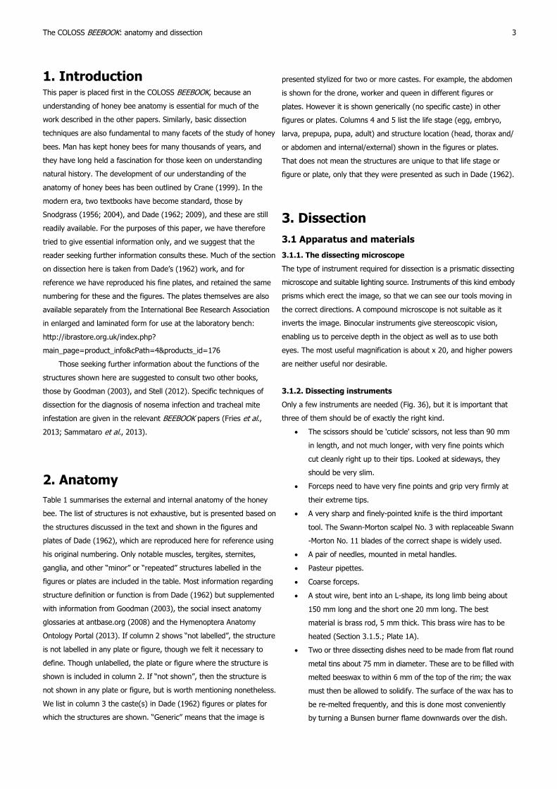
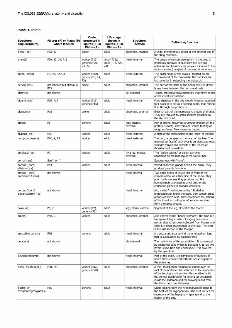
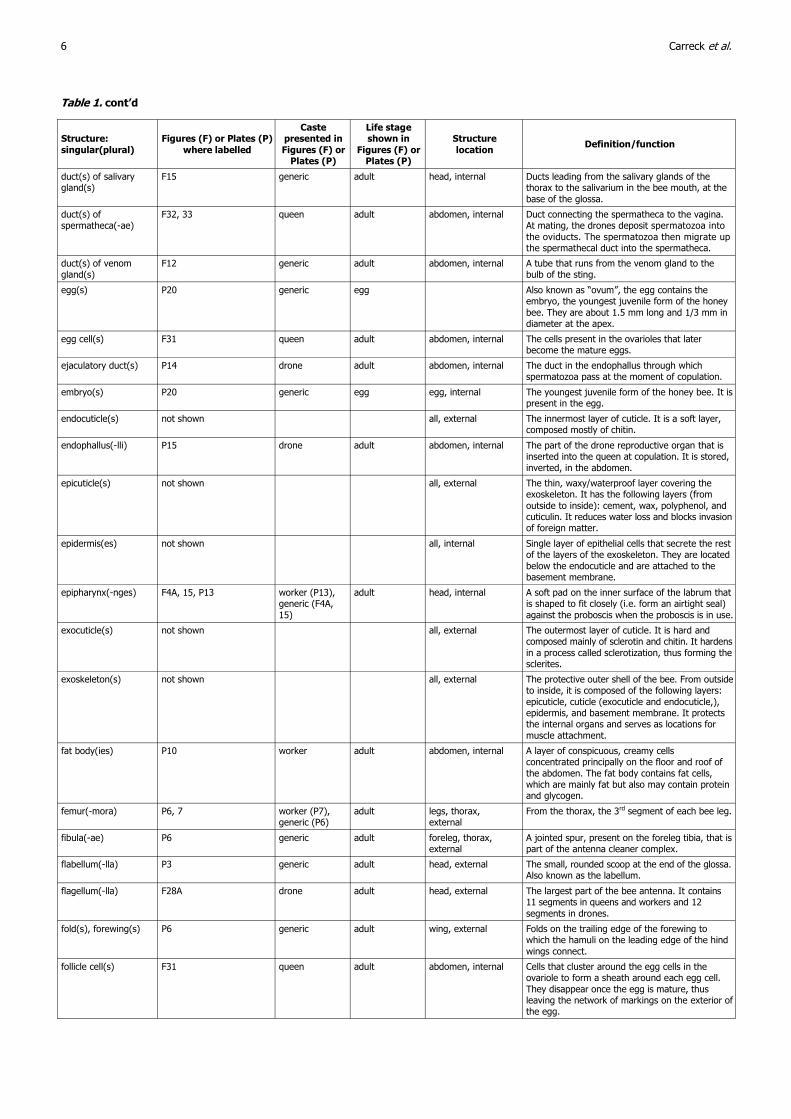
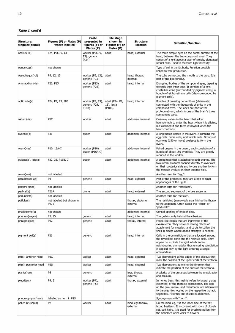
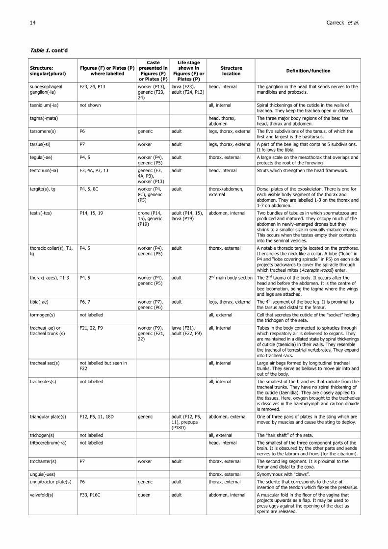
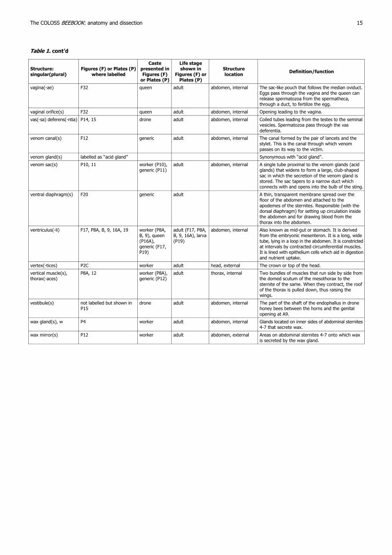
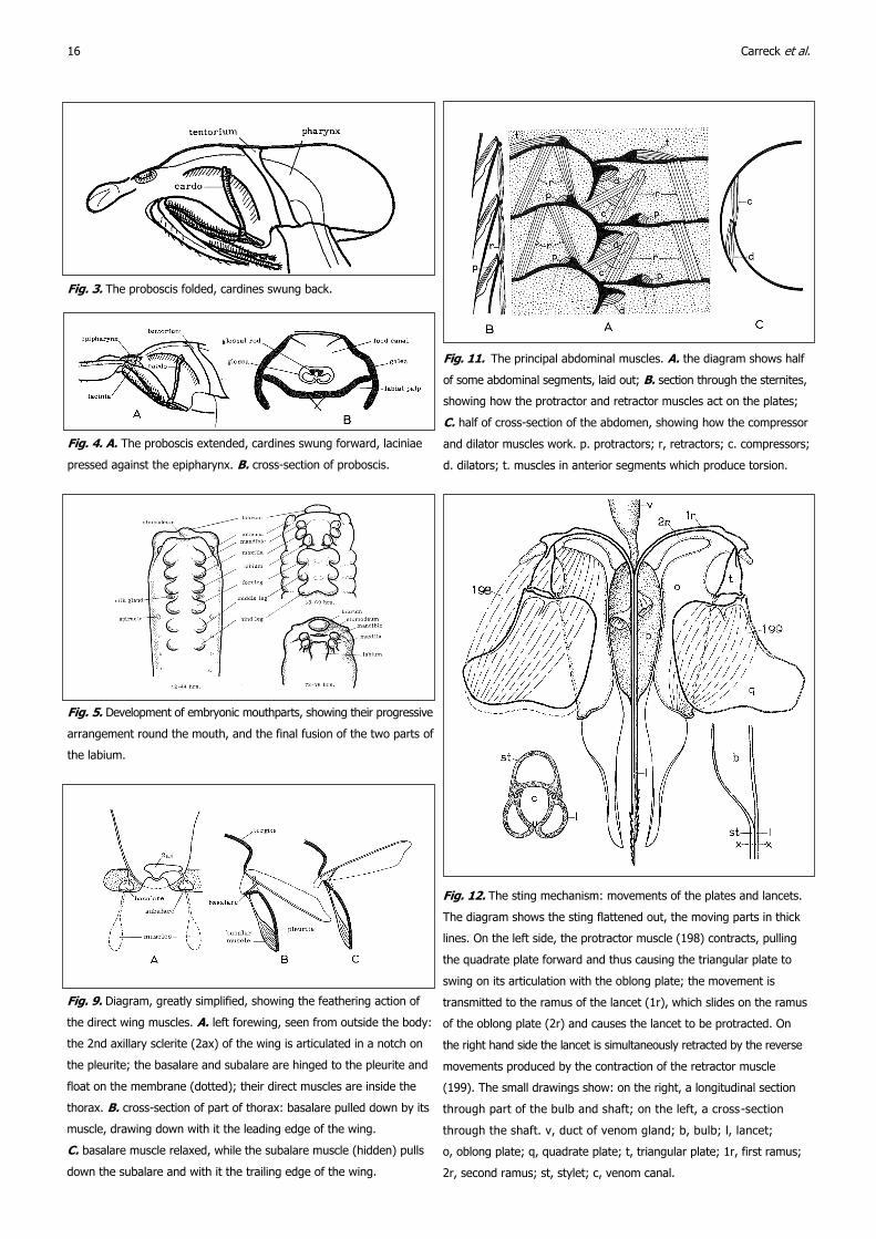
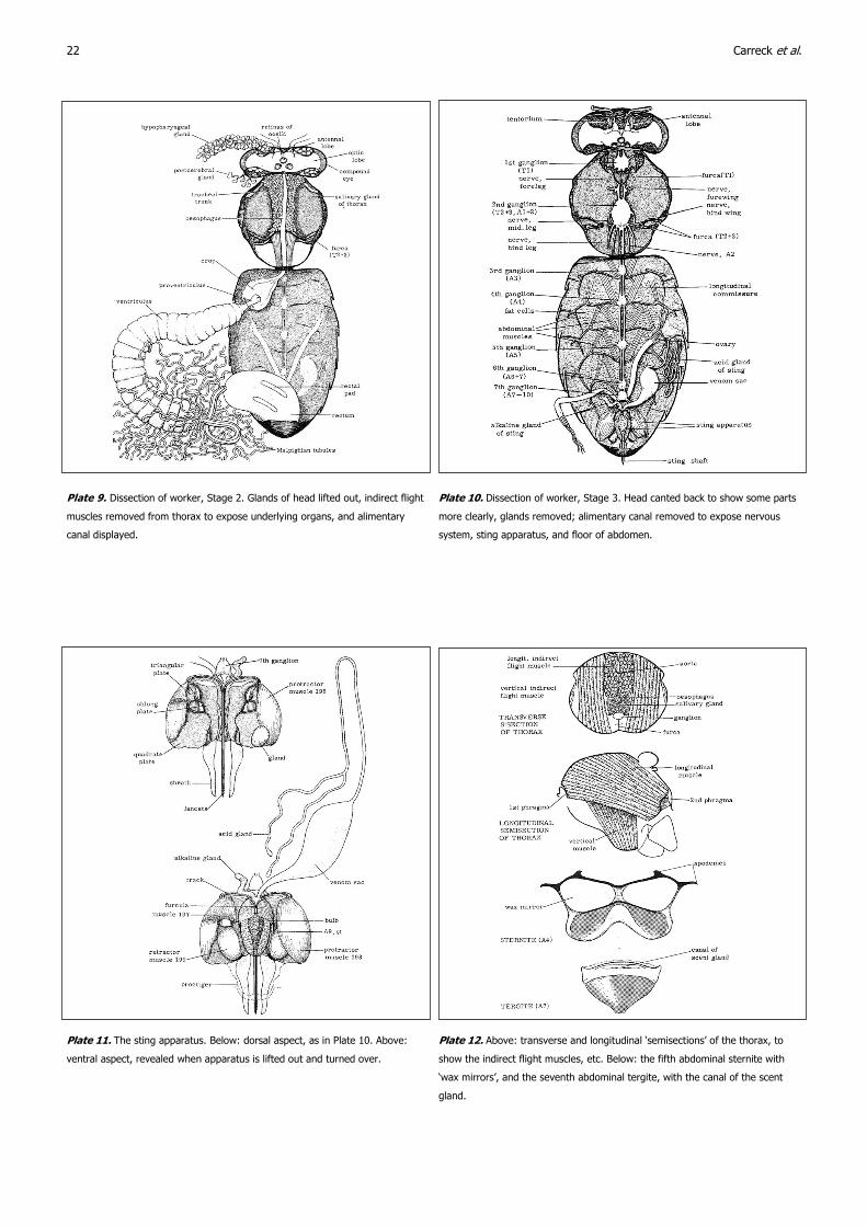
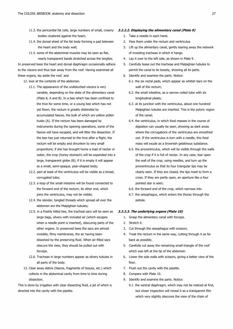

Journal of Apicultural Research 52(4) copy IBRA 2013