Jaquet&al 2013
17
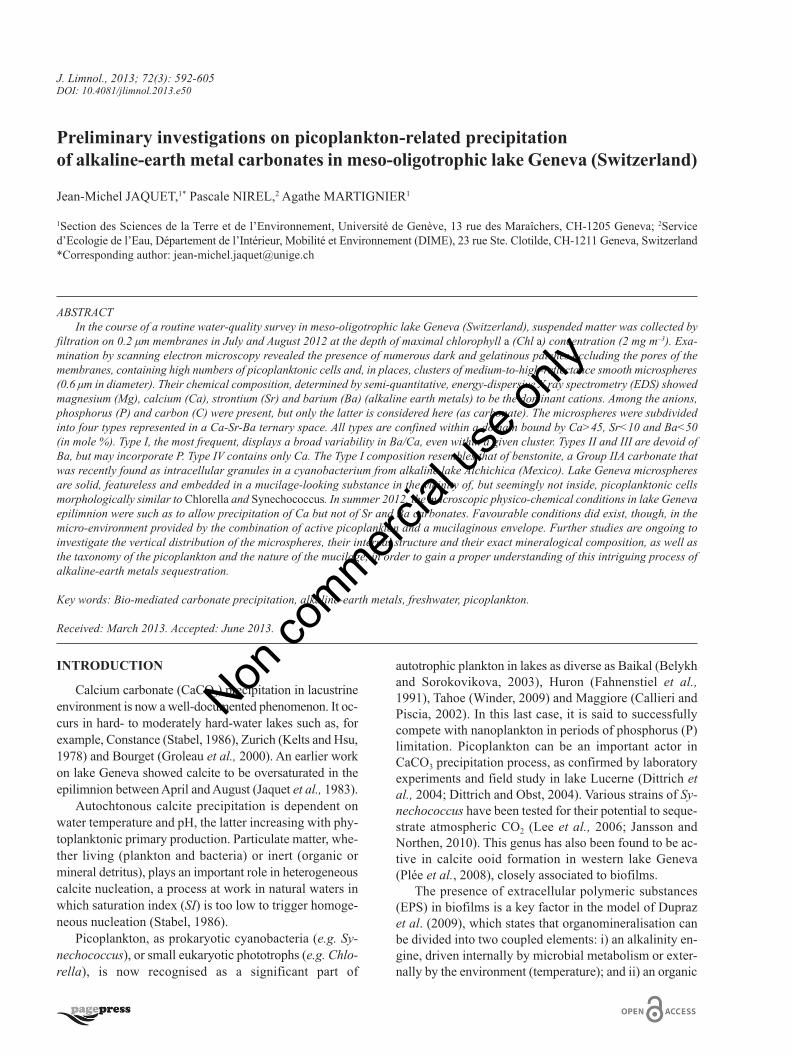
J. Limnol., 2013; 72(3): 592-605 DOI: 10.4081/jlimnol.2013.e50 INTRODUCTION Calcium carbonate (CaCO 3 ) precipitation in lacustrine environment is now a well-documented phenomenon. It oc- curs in hard- to moderately hard-water lakes such as, for example, Constance (Stabel, 1986), Zurich (Kelts and Hsu, 1978) and Bourget (Groleau et al., 2000). An earlier work on lake Geneva showed calcite to be oversaturated in the epilimnion between April and August (Jaquet et al., 1983). Autochtonous calcite precipitation is dependent on water temperature and pH, the latter increasing with phy- toplanktonic primary production. Particulate matter, whe- ther living (plankton and bacteria) or inert (organic or mineral detritus), plays an important role in heterogeneous calcite nucleation, a process at work in natural waters in which saturation index (SI) is too low to trigger homoge- neous nucleation (Stabel, 1986). Picoplankton, as prokaryotic cyanobacteria (e.g. Sy- nechococcus), or small eukaryotic phototrophs (e.g. Chlo- rella), is now recognised as a significant part of autotrophic plankton in lakes as diverse as Baikal (Belykh and Sorokovikova, 2003), Huron (Fahnenstiel et al., 1991), Tahoe (Winder, 2009) and Maggiore (Callieri and Piscia, 2002). In this last case, it is said to successfully compete with nanoplankton in periods of phosphorus (P) limitation. Picoplankton can be an important actor in CaCO 3 precipitation process, as confirmed by laboratory experiments and field study in lake Lucerne (Dittrich et al., 2004; Dittrich and Obst, 2004). Various strains of Sy- nechococcus have been tested for their potential to seque- strate atmospheric CO 2 (Lee et al., 2006; Jansson and Northen, 2010). This genus has also been found to be ac- tive in calcite ooid formation in western lake Geneva (Plée et al., 2008), closely associated to biofilms. The presence of extracellular polymeric substances (EPS) in biofilms is a key factor in the model of Dupraz et al. (2009), which states that organomineralisation can be divided into two coupled elements: i) an alkalinity en- gine, driven internally by microbial metabolism or exter- nally by the environment (temperature); and ii) an organic Preliminary investigations on picoplankton-related precipitation of alkaline-earth metal carbonates in meso-oligotrophic lake Geneva (Switzerland) Jean-Michel JAQUET, 1* Pascale NIREL, 2 Agathe MARTIGNIER 1 1 Section des Sciences de la Terre et de l’Environnement, Université de Genève, 13 rue des Maraîchers, CH-1205 Geneva; 2 Service d’Ecologie de l’Eau, Département de l’Intérieur, Mobilité et Environnement (DIME), 23 rue Ste. Clotilde, CH-1211 Geneva, Switzerland *Corresponding author: [email protected] ABSTRACT In the course of a routine water-quality survey in meso-oligotrophic lake Geneva (Switzerland), suspended matter was collected by filtration on 0.2 μm membranes in July and August 2012 at the depth of maximal chlorophyll a (Chl a) concentration (2 mg m –3 ). Exa- mination by scanning electron microscopy revealed the presence of numerous dark and gelatinous patches occluding the pores of the membranes, containing high numbers of picoplanktonic cells and, in places, clusters of medium-to-high-reflectance smooth microspheres (0.6 μm in diameter). Their chemical composition, determined by semi-quantitative, energy-dispersive X ray spectrometry (EDS) showed magnesium (Mg), calcium (Ca), strontium (Sr) and barium (Ba) (alkaline earth metals) to be the dominant cations. Among the anions, phosphorus (P) and carbon (C) were present, but only the latter is considered here (as carbonate). The microspheres were subdivided into four types represented in a Ca-Sr-Ba ternary space. All types are confined within a domain bound by Ca>45, Sr<10 and Ba<50 (in mole %). Type I, the most frequent, displays a broad variability in Ba/Ca, even within a given cluster. Types II and III are devoid of Ba, but may incorporate P. Type IV contains only Ca. The Type I composition resembles that of benstonite, a Group IIA carbonate that was recently found as intracellular granules in a cyanobacterium from alkaline lake Alchichica (Mexico). Lake Geneva microspheres are solid, featureless and embedded in a mucilage-looking substance in the vicinity of, but seemingly not inside, picoplanktonic cells morphologically similar to Chlorella and Synechococcus. In summer 2012, the macroscopic physico-chemical conditions in lake Geneva epilimnion were such as to allow precipitation of Ca but not of Sr and Ba carbonates. Favourable conditions did exist, though, in the micro-environment provided by the combination of active picoplankton and a mucilaginous envelope. Further studies are ongoing to investigate the vertical distribution of the microspheres, their internal structure and their exact mineralogical composition, as well as the taxonomy of the picoplankton and the nature of the mucilage, in order to gain a proper understanding of this intriguing process of alkaline-earth metals sequestration. Key words: Bio-mediated carbonate precipitation, alkaline-earth metals, freshwater, picoplankton. Received: March 2013. Accepted: June 2013. Non commercial use only
-
Upload
independent -
Category
Documents
-
view
1 -
download
0
Transcript of Jaquet&al 2013
J. Limnol., 2013; 72(3): 592-605DOI: 10.4081/jlimnol.2013.e50
INTRODUCTION
Calcium carbonate (CaCO3) precipitation in lacustrineenvironment is now a well-documented phenomenon. It oc-curs in hard- to moderately hard-water lakes such as, forexample, Constance (Stabel, 1986), Zurich (Kelts and Hsu,1978) and Bourget (Groleau et al., 2000). An earlier workon lake Geneva showed calcite to be oversaturated in theepilimnion between April and August (Jaquet et al., 1983).
Autochtonous calcite precipitation is dependent onwater temperature and pH, the latter increasing with phy-toplanktonic primary production. Particulate matter, whe-ther living (plankton and bacteria) or inert (organic ormineral detritus), plays an important role in heterogeneouscalcite nucleation, a process at work in natural waters inwhich saturation index (SI) is too low to trigger homoge-neous nucleation (Stabel, 1986).
Picoplankton, as prokaryotic cyanobacteria (e.g. Sy-nechococcus), or small eukaryotic phototrophs (e.g. Chlo-rella), is now recognised as a significant part of
autotrophic plankton in lakes as diverse as Baikal (Belykhand Sorokovikova, 2003), Huron (Fahnenstiel et al.,1991), Tahoe (Winder, 2009) and Maggiore (Callieri andPiscia, 2002). In this last case, it is said to successfullycompete with nanoplankton in periods of phosphorus (P)limitation. Picoplankton can be an important actor inCaCO3 precipitation process, as confirmed by laboratoryexperiments and field study in lake Lucerne (Dittrich etal., 2004; Dittrich and Obst, 2004). Various strains of Sy-nechococcus have been tested for their potential to seque-strate atmospheric CO2 (Lee et al., 2006; Jansson andNorthen, 2010). This genus has also been found to be ac-tive in calcite ooid formation in western lake Geneva(Plée et al., 2008), closely associated to biofilms.
The presence of extracellular polymeric substances(EPS) in biofilms is a key factor in the model of Duprazet al. (2009), which states that organomineralisation canbe divided into two coupled elements: i) an alkalinity en-gine, driven internally by microbial metabolism or exter-nally by the environment (temperature); and ii) an organic
Preliminary investigations on picoplankton-related precipitationof alkaline-earth metal carbonates in meso-oligotrophic lake Geneva (Switzerland)
Jean-Michel JAQUET,1* Pascale NIREL,2 Agathe MARTIGNIER1
1Section des Sciences de la Terre et de l’Environnement, Université de Genève, 13 rue des Maraîchers, CH-1205 Geneva; 2Serviced’Ecologie de l’Eau, Département de l’Intérieur, Mobilité et Environnement (DIME), 23 rue Ste. Clotilde, CH-1211 Geneva, Switzerland*Corresponding author: [email protected]
ABSTRACTIn the course of a routine water-quality survey in meso-oligotrophic lake Geneva (Switzerland), suspended matter was collected by
filtration on 0.2 µm membranes in July and August 2012 at the depth of maximal chlorophyll a (Chl a) concentration (2 mg m–3). Exa-mination by scanning electron microscopy revealed the presence of numerous dark and gelatinous patches occluding the pores of themembranes, containing high numbers of picoplanktonic cells and, in places, clusters of medium-to-high-reflectance smooth microspheres(0.6 µm in diameter). Their chemical composition, determined by semi-quantitative, energy-dispersive X ray spectrometry (EDS) showedmagnesium (Mg), calcium (Ca), strontium (Sr) and barium (Ba) (alkaline earth metals) to be the dominant cations. Among the anions,phosphorus (P) and carbon (C) were present, but only the latter is considered here (as carbonate). The microspheres were subdividedinto four types represented in a Ca-Sr-Ba ternary space. All types are confined within a domain bound by Ca>45, Sr<10 and Ba<50(in mole %). Type I, the most frequent, displays a broad variability in Ba/Ca, even within a given cluster. Types II and III are devoid ofBa, but may incorporate P. Type IV contains only Ca. The Type I composition resembles that of benstonite, a Group IIA carbonate thatwas recently found as intracellular granules in a cyanobacterium from alkaline lake Alchichica (Mexico). Lake Geneva microspheresare solid, featureless and embedded in a mucilage-looking substance in the vicinity of, but seemingly not inside, picoplanktonic cellsmorphologically similar to Chlorella and Synechococcus. In summer 2012, the macroscopic physico-chemical conditions in lake Genevaepilimnion were such as to allow precipitation of Ca but not of Sr and Ba carbonates. Favourable conditions did exist, though, in themicro-environment provided by the combination of active picoplankton and a mucilaginous envelope. Further studies are ongoing toinvestigate the vertical distribution of the microspheres, their internal structure and their exact mineralogical composition, as well asthe taxonomy of the picoplankton and the nature of the mucilage, in order to gain a proper understanding of this intriguing process ofalkaline-earth metals sequestration.
Key words: Bio-mediated carbonate precipitation, alkaline-earth metals, freshwater, picoplankton.
Received: March 2013. Accepted: June 2013.
Non co
mmercial
use o
nly

593Phytoplankton-related precipitation of alkaline-earth metal carbonates
matrix in which mineralisation will take place. Cyanobac-teria (Pereira et al., 2009; Dittrich and Sibler, 2010) andsome strains of Chlorella (Watanabe et al., 2006) areknown to secrete extracellular polysaccharides, whichmay act as binding sites for Ca2+ and CO3
2–. The impor-tance of biofilms in freshwater carbonate precipitation hasbeen demonstrated by Rogerson et al. (2008) in their ex-periments on tufa systems.
In contrast to CaCO3, data on precipitation of other al-kaline-earth metals (AEM) such as strontium (Sr) and ba-rium (Ba) are scarcer. This is understandable, as theconcentration of these elements in lake water is muchlower (molar ratio Ba:Sr:Mg:Ca is of the order of1:40:1900:8200 in lake Geneva), with saturation indicesfor Sr and Ba carbonates well below zero under normalphysico-chemical conditions.
In the marine environment, biogenic Ba preferablyprecipitates as sulfate (barite) within dead phytoplanktonaggregates (abiotic precipitation) (Jeandel et al., 2000),or under the direct influence of biological processes (Gon-zalez-Muñoz et al., 2012).
In lake Constance, Sr has been found to co-precipitatewith calcium (Ca) in calcite, while a Ca-independent Sruptake by biota is negligible (Stabel et al., 1986; Stabel,1989). According to Stabel et al. (1991), the water of thislake is always undersaturated with respect to SrCO3
(strontianite), not to mention BaCO3 (witherite).Barium and Sr sulfates (barite and celestite) are known
to be present as biominerals in the terminal vacuoles offreshwater Desmids such as Closterium and Micrasterias(Brook, 1980; Wilcock et al., 1989; Krejci et al., 2011). Ba-rium carbonate (as witherite) has been reported to be preci-pitated as fibrous crystals by bacteria (Sanchez-Moral et al.,2004). Another avenue for precipitation of AEM is given bythe prostomatid ciliates Loxodes and Remanella, whosemüllerian vesicles contain, respectively, Ba and Sr salts (Fin-lay et al., 1983; McGrath et al., 1989; Lynn, 2010).
It is only recently that a well-identified amorphouscarbonate containing magnesium (Mg), Ca, Sr and Ba (at-tributed to a benstonite-like phase) was found in highlyalkaline lake Alchichica microbialites by Couradeau et al.(2012). In this case, the carbonate is precipitated as intra-cellular inclusions in a novel cyanobacterium (candidatusGloeomargarita lithophora).
Within the framework of ongoing water-quality surveysundertaken by the Service de l’écologie de l’eau (SECOE)in Petit-Lac (the southwestern part of lake Geneva), suspen-ded matter was collected in 2012 by filtration at the depthof maximum chlorophyll a (Chl a) concentration. Exami-nation by scanning electron microscopy (SEM) coupled toenergy-dispersive X-ray spectrometry (EDS) revealed, inJuly and August samples, the presence of Mg-Ca-Sr-Ba-containing microspheres, seemingly related to mucilagepackaging picoplanktonic cells.
We present here preliminary investigations conductedby SEM/EDS on the microsphere samples currently avai-lable. This study is part of a series of geomicrobiologicalanalyses conducted in lake Geneva (Jaquet et al., 1982;Plée et al., 2008; Glas-Haller, 2010).
METHODS
Petit-Lac represents the southwest portion of Lémanor lake Geneva. After undergoing eutrophication in the1980s and 1990s (Anneville and Pelletier, 2000), thishard-water lake is now in the process of re-oligotrophica-tion. Data on physico-chemistry, phytoplankton, zooplan-kton and fish production is available in the French-SwissCommission yearly reports (CIPEL, 2011; Lazarotto andKlein, 2012). Recent studies on phytoplankton can befound in Anneville et al. (2002) and Gallina et al. (2011).
Monthly measurements of physico-chemistry and watersamples were taken at GE3 station (6.22°E/46.30°N) at 10different depths ranging 0 to 70 m with a plastic Niskin® bot-tle. Also, continuous recording of Chl a and turbidity wasdone by means of a Wet Labs® probe (Wet Labs, Philomath,OR, USA). Phytoplankton was collected by means of an in-tegrating sampler over the top 20 m, and taxa were identifiedand counted following Utermöhl method. A larger water vo-lume (10-15 L) was sampled at the depth of maximum Chla concentration, followed by gentle vacuum filtration in thelab within a few hours on return from the field. For gravi-metry, Whatman® GF/F membranes (Whatman Ltd., Maid-stone, UK) were used, and Schleicher & Schuell®
nitrocellulose (NC 60; 0.6 μm) (Schleicher & Schuell Bio-Science GmbH, Dassel, Germany) as well as Millipore® po-lycarbonate (PC GTTP; 0.2 μm) (Millipore, Billerica, MA,USA) for SEM/EDS. Filters were air-dried, and no fixationwas applied on this first set of samples. Sub-samples of filterswere mounted on aluminum stubs covered with double-sidedconductive carbon tape. A coating of gold (ca 15 nm) or car-bon (ca 15 nm) was then deposited on the samples prior toimaging with a Jeol® JSM 7001F SEM (Jeol Ltd., Tokyo,Japan) (Department of Earth Sciences, Section of Earth andEnvironmental Sciences, University of Geneva, Switzerland)with an acceleration voltage of 15 kV. Semi-quantitative el-emental analyses were performed with a JED2300 EDS de-tector (Jeol Ltd.; working distance: 10 mm).
Carbonate species concentrations and calcite satura-tion index [SI=log (IAP/KS)] were calculated using thesoftware Aqion 2.5.1 (www.aqion.de), based on USGSPHREEQC (Parkhurst and Appelo, 1999) and the thermo-dynamical database wateq4f (Ball and Nordstrom, 1991).
This preliminary study concerns two suspended mattersamples in which microspheres were present. They werecollected in 2012: i) on 10th July at 6 m depth [polycar-bonate (PC) filter numbers 16 and 17, carbon coated] andii) on 7th August at 6 m depth [(PC filters 20, 21 (carboncoated) and 30 (gold coated)]. Clusters of microspheres
Non co
mmercial
use o
nly

594 J-M Jaquet et al.
were identified visually on the stubs at a magnification of1000x and numbered as specimens (Spx_y-z, where x=1for 10.07.12, x=2 for 07.08.12; y=cluster number andz=microsphere number).
In order to verify the analytical results obtained byEDS on the microspheres, the mineral benstonite [Mg Ca6
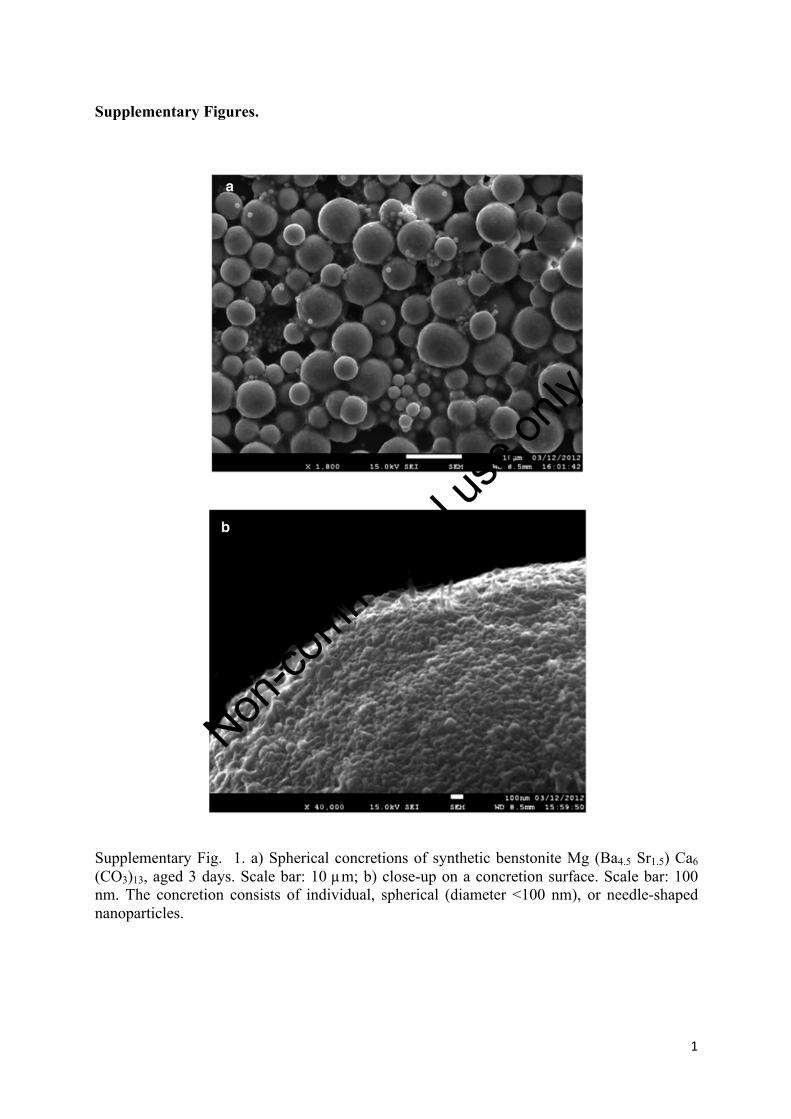
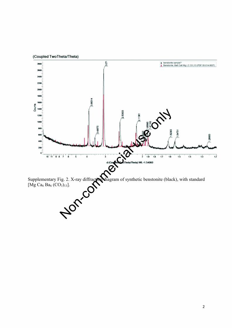
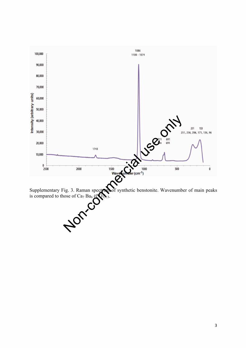
(BaxSry) (CO3)13] (with x+y=6) was synthesised followinga modification of Hood and colleagues’ procedure (Hoodand Steidl, 1973; Hood et al., 1974). The precipitate wasaged for three days prior to SEM/EDS analysis. It consistsof spherical aggregates of nanoparticles (SupplementaryFig. 1), similar in shape to CaCO3 or SrCO3 polymorphsreported by Wu et al. (2004), Sun et al. (2006) and Sondiand Matijevic (2003). The precipitate attribution to ben-stonite was confirmed by X-ray diffraction (XRD) (Sup-plementary Fig. 2) and Raman spectroscopy(Supplementary Fig. 3).
RESULTS
Limnological context
The microsphere-containing suspended matter sam-ples were collected both times in fairly similar conditions
(Fig. 1a): at 6 m in the metalimnion, corresponding to thepeak concentration of Chl a (2 mg m–3 on both dates) andwith a turbidity around 1 NTU.
The water was saturated, with respect to calcite,down to 7 m in July and 10 m in August (Tab. 1, Fig.1b). Other interesting features are the molar ratios (insolution) Sr/Ca, around 0.0050, similar to lake Con-stance values as mentioned by Stabel et al. (1986), andSr/Ba, which is higher in the epilimnion (43-45.5; Fig.1c), practically mimicking the Chl a profile (Fig. 1a).Analytical results (Tab. 1) indicate, at 5 m depth, highertemperature, pH, Sr/Ba and calcite saturation values(0.34 vs 0.13) in August. These Sr/Ba maximums werenot observed during our other campaigns in 2012 (datanot shown).
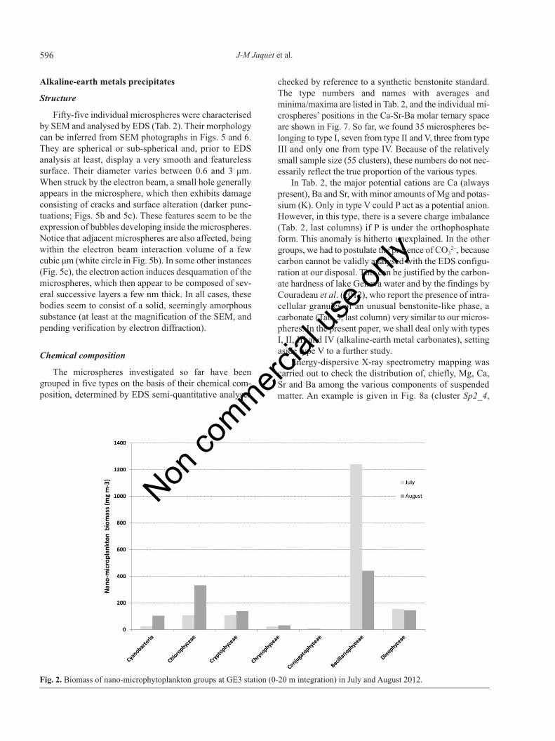
Fig. 2 shows the phytoplankton groups found in Julyand August integrated over the top 20 m of the water col-umn. Because of the integration, this may not representexactly the taxa living at 6 m depth. Moreover, apart fromChlorella sp., it does not include picoplankton, which isomnipresent in the SEM images. In terms of biomass, di-atoms (Bacillariophyceae) largely dominate in July, with
Fig. 1. a) Temperature and chlorophyll a concentration profiles at GE3 station on 10th July (dashed line) and 7th August (solid line) 2012;b) corresponding calcite saturation index (samples with saturation index>0 are saturated with respect to calcite); c) Sr/Ba molar ratio.
Non co
mmercial
use o
nly
595Phytoplankton-related precipitation of alkaline-earth metal carbonates
lesser amounts of Chlorophyceae, Cryptophyceae andDinophyceae. In August, the diatoms biomass decreasesat the expense of the Chlorophyceae (mostly Chlorellasp., with abundance around 2 103 cell mL–1). No measure-ments were carried out for water samples at the Chl amaximum. However, for lake Geneva in August (upper15 m), Personnic et al. (2009) report picocyanobacterialabundance values up to 3 105 cell mL–1.
Overall morphology of suspended matter
The general aspect of the suspended matter collectedon 10th July (concentration: 2.6 mg L–1) and 7th August(concentration: 1.8 mg L–1) is shown in Figs. 3 and 4, re-spectively. An obvious feature of both samples is the pres-ence of dark patches, very likely of mucilaginous nature,which obliterate the membrane pores.
Low reflectance picoplanktonic cells, either spherical(Fig. 5a, right) or ellipsoidal (Fig. 6a), are densely packedwithin the mucilage patches. Their dimension is of theorder of the μm. In places, the patches contain clusters ofhigh-reflectance, spherical or subspherical bodies (Fig.5a), with diameters varying between 0.6 and 3 μm. These
microspheres are described in detail below. The mucilageembedding the clusters and picoplanktonic is electroni-cally quasi-transparent, and its limits can be quite sharpand sub-circular (white arrows in Fig. 6a and 6b), sug-gesting an originally spherical shape prior to the deposi-tion on the filter and subsequent collapse through drying.The presence of this organic matrix is further confirmedby a fortunate feature stemming from electron damage,visible on Fig. 6c: whereas most of the microspheres’ con-tours are blurred by the organic coating (Hernández Mar-iné et al., 2004), the cracks reveal the presence ofunderlying, bare, sharply delimited microspheres (withinwhite frames in Fig. 6c).
Calcium carbonate is present as isolated crystals (>5μm) or aggregates (Fig. 6a), showing signs of corrosion(Fig. 5a). These do not have any visible relationship withmucilage.
In July (Fig. 3), suspended matter contained, in addi-tion to picoplankton, nano- and microplankton (Fig. 2):centric and pennate diatoms and chrysophyte siliceousscales. Ceratium hirundinella and diatoms (Achnanthessp.) (Fig. 4) were present in August.
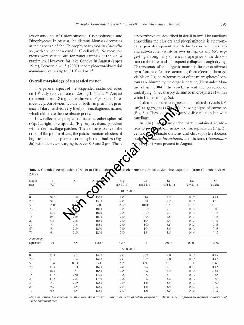
Tab. 1. Chemical composition of water at GE3 station (selected elements) and in lake Alchichica aquarium (from Couradeau et al.,2012).
Depth T pH Alkalinity Mg Ca Sr Ba SI(m) (°C) (μM L-1) (μM L-1) (μM L-1) (μM L-1) (μM L-1) calcite
10.07.2012
0 20.6 8.52 1570 235 910 5.2 0.12 0.482.5 20.0 8.55 1590 235 930 5.2 0.12 0.515* 16.0* 8.14* 1730* 235* 1004* 5.3* 0.12* 0.13*
7.5 13.3 7.92 1810 235 1059 5.4 0.13 -0.0810 12.2 7.86 1820 235 1059 5.3 0.13 -0.1615 10.6 7.84 1870 240 1094 5.5 0.13 -0.1520 9.6 7.83 1880 240 1104 5.4 0.13 -0.1630 7.4 7.84 1880 240 1109 5.5 0.13 -0.1850 6.4 7.86 1890 240 1104 5.5 0.13 -0.1870 6.4 7.86 1890 240 1124 5.5 0.14 -0.17
Alchichicaaquarium 24 8.9 13617 6955 47 0.013 0.001 0.158
05.08.2012
0 22.4 8.5 1480 232 868 5.0 0.12 0.452.5 21.8 8.52 1480 233 882 5.0 0.12 0.475* 19.4* 8.38* 1560* 232* 924* 5.0* 0.11* 0.34*
7.5 17.4 8.11 1620 241 994 5.1 0.11 0.1210 16.4 8 1650 235 986 5.2 0.12 -0.0115 13.6 7.91 1720 238 1052 5.1 0.13 -0.0520 11.3 7.89 1790 236 1072 5.2 0.13 -0.0930 8.2 7.88 1880 249 1145 5.5 0.13 -0.0950 6.7 7.9 1880 244 1123 5.4 0.13 -0.1270 6.3 7.9 1880 245 1131 5.5 0.13 -0.12
Mg, magnesium; Ca, calcium; Sr, strontium; Ba, barium; SI, saturation index of calcite (aragonite in Alchichica). *Approximate depth of occurrence ofstudied microspheres.
Non co
mmercial
use o
nly
596 J-M Jaquet et al.
Alkaline-earth metals precipitates
Structure
Fifty-five individual microspheres were characterisedby SEM and analysed by EDS (Tab. 2). Their morphologycan be inferred from SEM photographs in Figs. 5 and 6.They are spherical or sub-spherical and, prior to EDSanalysis at least, display a very smooth and featurelesssurface. Their diameter varies between 0.6 and 3 μm.When struck by the electron beam, a small hole generallyappears in the microsphere, which then exhibits damageconsisting of cracks and surface alteration (darker punc-tuations; Figs. 5b and 5c). These features seem to be theexpression of bubbles developing inside the microspheres.Notice that adjacent microspheres are also affected, beingwithin the electron beam interaction volume of a fewcubic μm (white circle in Fig. 5b). In some other instances(Fig. 5c), the electron action induces desquamation of themicrospheres, which then appear to be composed of sev-eral successive layers a few nm thick. In all cases, thesebodies seem to consist of a solid, seemingly amorphoussubstance (at least at the magnification of the SEM, andpending verification by electron diffraction).
Chemical composition
The microspheres investigated so far have beengrouped in five types on the basis of their chemical com-position, determined by EDS semi-quantitative analyses
checked by reference to a synthetic benstonite standard.The type numbers and names with averages andminima/maxima are listed in Tab. 2, and the individual mi-crospheres’ positions in the Ca-Sr-Ba molar ternary spaceare shown in Fig. 7. So far, we found 35 microspheres be-longing to type I, seven from type II and V, three from typeIII and only one from type IV. Because of the relativelysmall sample size (55 clusters), these numbers do not nec-essarily reflect the true proportion of the various types.
In Tab. 2, the major potential cations are Ca (alwayspresent), Ba and Sr, with minor amounts of Mg and potas-sium (K). Only in type V could P act as a potential anion.However, in this type, there is a severe charge imbalance(Tab. 2, last columns) if P is under the orthophosphateform. This anomaly is hitherto unexplained. In the othergroups, we had to postulate the presence of CO3
2–, becausecarbon cannot be validly analysed with the EDS configu-ration at our disposal. This can be justified by the carbon-ate hardness of lake Geneva water and by the findings byCouradeau et al. (2012), who report the presence of intra-cellular granules of an unusual benstonite-like phase, acarbonate (Tab. 3, last column) very similar to our micros-pheres. In the present paper, we shall deal only with typesI, II, III and IV (alkaline-earth metal carbonates), settingaside type V to a further study.
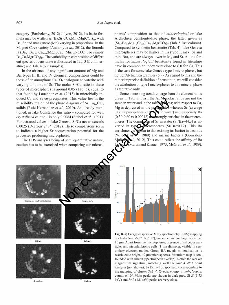
Energy-dispersive X-ray spectrometry mapping wascarried out to check the distribution of, chiefly, Mg, Ca,Sr and Ba among the various components of suspendedmatter. An example is given in Fig. 8a (cluster Sp2_4,
Fig. 2. Biomass of nano-microphytoplankton groups at GE3 station (0-20 m integration) in July and August 2012.
Non co
mmercial
use o
nly

597Phytoplankton-related precipitation of alkaline-earth metal carbonates
Fig. 3. General view of suspended matter from 10th July 2012(6 m depth), deposited on 0.2 μm polycarbonate membrane, car-bon coated. Scale bar: 10 μm. Centric and pennate diatoms,CaCO3 as white aggregates. Darker parts are interpreted as mu-cilage packaging picoplanktonic cells (see Figs. 5-7 for furtherdetails). Sp1_1 cluster of microspheres is visible at the centre ofthe figure.
Fig. 4. General view of suspended matter from 7th August 2012(6 m depth), deposited on 0.2 μm polycarbonate membrane, goldcoated. Scale bar: 10 μm. Apical horn of Ceratium hirundinella.Numerous Achnanthes sp. diatoms, CaCO3 are also visible.Darker parts are believed to be mucilage packaging picoplank-tonic cells (see Fig. 6a and 6b for further details). Two clustersof microspheres are labelled Sp2_1 and Sp2_2 (in white frames).
Tab. 2. Microspheres average composition (atom %).
Type I II III IV VName CaSrBa CaSr CaPSr Ca CaPElement Mean N Min Max Mean N Min Max Mean N Min Max Mean N Mean N Min Max
Mg 0.5 35 0.0 2.2 0.4 7 0.3 0.8 1.2 3 0.8 1.7 1.0 1 3.5 7 1.4 10.6P 1.2 3 0.0 2.0 5.0 3 3.4 53.5 7 45.0 60.2S 2.5 1 2.5 2.5 12.4 2 1.3 23.4K 8.4 2 7.2 9.5Ca 32.0 35 19.5 64.1 90.1 7 87.0 92.4 82.7 3 76.3 87.5 99.0 1 37.1 7 23.0 43.1Sr 4.7 35 0.4 10.1 9.5 7 7.3 12.2 10.3 3 6.6 16.0Ba 62.8 35 30.7 75.5 0.0 7
As 100%*
Ca 32.2 19.6 64.4 90.5 87.3 92.8 88.9 82.1 94.2 100.0 100.0Sr 4.7 0.4 10.1 9.5 7.3 12.3 11.1 7.1 17.2 0.0 0.0Ba 63.1 30.9 76.0 0.0 0.0 0.0 0.0
N, number of analyses for a given element; Min, minimum; Max, maximum; Mg, magnesium; P, phosphorus; S, sulphur; K, potassium; Ca, calcium; Sr,strontium; Ba, barium. *Three last rows: Ca, Sr and Ba normalised to 100%.
Tab. 3. Composition (atom %, alkaline-earth elements) of benstonite varieties.
Ratio (Ba5.5Sr0.5) (Ba4.5Sr1.5) (Ba4.5Sr1.5) (BaSr)6 (Ba2.7Sr1.0Mg1.4Ca0.9)theoretical* theoretical* synthetic° Anthony et al. Couradeau et al.
1 2 EDS (2012) (2012)
Mg 2.3 2.4 1.3 2.3 18.0Ca 22.6 23.7 20.8 22.9 53.2Sr 4.1 13.0 11.2 4.1 8.1Ba 71.0 60.9 66.7 70.7 20.7
EDS, energy dispersive X-ray spectrometry; Mg, magnesium; Ca, calcium; Sr, strontium; Ba, barium. *Computed from formula; °analysed semi-quan-titatively by EDS on a polished surface. First row: variable cations, in addition to fixed Mg1 Ca6; last column: recomputed from Table S2 in Couradeauet al. (2012).
Non co
mmercial
use o
nly
598 J-M Jaquet et al.
07.08.2012). In this specimen, Ca and Ba are exclusivelyconcentrated in the microspheres, some of which may berather small. The Sr and silicon (Si) maps are similar dueto peak overlaps (Fig. 8b).
DISCUSSION
Chemical composition in terms of dissolvedalkaline-earth metals
The concentrations of Ca, Sr, Ba and their seasonalvariations are quite similar in lake Geneva and lake Con-stance (Stabel et al., 1991), where these elements are de-pleted in epilimnion during summer. In lake Geneva, theratios [Me]5m/[Me]70m computed from Tab. 1 were, in Au-gust 2012, Mg: 0.95, Ca: 0.82, Sr: 0.90 and Ba: 0.85.Hence, it seems that Ca and Ba are slightly more depleted
than Mg and Sr, as an echo to the composition of the mi-crospheres, Ca and Ba representing more than 90% of thecations (Tab. 2) in type I microspheres. In lake Constance,Sr and Ba are said to co-precipitate with calcite at an al-most constant stoichiometry in particulate material (molarSr/Ca=0.0084; atom Sr/Ca=0.018). This could also be thecase in lake Geneva, with additional scavenging by thebiologically mediated precipitation of specific amorphousAEM carbonates. In contrast, there is no sign of a de-crease in Ba concentration towards the bottom (Tab. 1),as in lake Biwa, through adsorption on hydrous man-ganese oxides (Sugiyama et al., 1992).
Physico-chemical framework for microspheresprecipitation
It is not known whether amorphous AEM carbonate
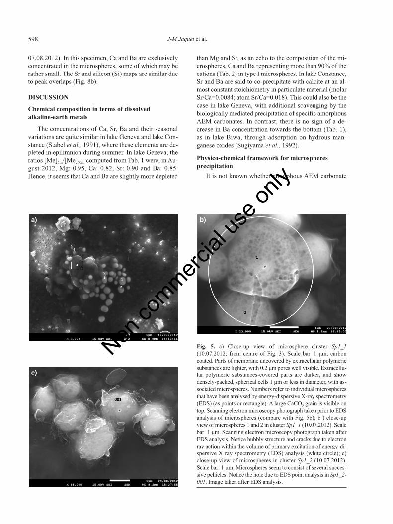
Fig. 5. a) Close-up view of microsphere cluster Sp1_1(10.07.2012; from centre of Fig. 3). Scale bar=1 μm, carboncoated. Parts of membrane uncovered by extracellular polymericsubstances are lighter, with 0.2 μm pores well visible. Extracellu-lar polymeric substances-covered parts are darker, and showdensely-packed, spherical cells 1 µm or less in diameter, with as-sociated microspheres. Numbers refer to individual microspheresthat have been analysed by energy-dispersive X-ray spectrometry(EDS) (as points or rectangle). A large CaCO3 grain is visible ontop. Scanning electron microscopy photograph taken prior to EDSanalysis of microspheres (compare with Fig. 5b); b ) close-upview of microspheres 1 and 2 in cluster Sp1_1 (10.07.2012). Scalebar: 1 μm. Scanning electron microscopy photograph taken afterEDS analysis. Notice bubbly structure and cracks due to electronray action within the volume of primary excitation of energy-di-spersive X ray spectrometry (EDS) analysis (white circle); c)close-up view of microspheres in cluster Sp1_2 (10.07.2012).Scale bar: 1 μm. Microspheres seem to consist of several succes-sive pellicles. Notice the hole due to EDS point analysis in Sp1_2-001. Image taken after EDS analysis.
a)
c)
b)
Non co
mmercial
use o
nly
599Phytoplankton-related precipitation of alkaline-earth metal carbonates
(AAEMC) microspheres existed at other depths and datesthan those found by chance in summer 2012. We onlyknow they occurred in the thermocline, near the Chl aconcentration or biomass maximum, in waters saturatedwith respect to calcite and at a depth where the dissolvedSr/Ba molar ratio was maximal (Fig. 1). It is, therefore,not unreasonable to postulate that there is a link betweenmicrosphere precipitation and phytoplanktonic primaryproduction (PP) (primary production and phytoplanktonbiomass maxima generally coincide in lake Geneva) (Ta-donleke, 2012). This is substantiated by the close spatialassociation of microspheres and picoplankton visible inSEM images (Figs. 5a and 6a).
If the macroscopic physico-chemical conditions (sat-uration index>0; Tab. 1) enable the precipitation of calcitecrystals, the situation is different for the AAEMC. Al-
though we were not able to find a value for the solubilityconstant Ks of crystalline benstonite (let alone for itsamorphous, various species), we can approach the ques-tion by considering Stabel et al.’s (1991) data: these au-thors have computed, for lake Constance in summer, the[Sr2+]=3.16 10–5 M L–1 and [Ba2+]=7.08 10–5 M L–1 con-centrations necessary to allow precipitation of strontianite(SrCO3; Ks=10–9.03) and witherite (BaCO3; Ks=10–8.3). Ex-tending these figures to our case, it is obvious that Sr andBa concentrations given in our Tab. 1 are much lower,meaning that these AEM carbonates cannot precipitateunder the macroscopic pH and temperature conditions inlake Geneva. This is likely to apply also to benstonite-likeminerals, the precipitation of which will necessitatemicro-environments with extreme conditions (elevatedpH, increased ionic concentration and alkalinity).
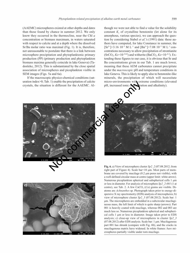
Fig. 6. a) View of microsphere cluster Sp2_2 (07.08.2012; fromright part of Figure 4). Scale bar=10 μm. Most parts of mem-brane are covered by mucilage (0.2 μm pores not visible), witha well-defined circular mass at centre (upper limit: white arrow).Numerous picoplankton spherical and subspherical cells 1 μmor less in diameter. For analysis of microsphere Sp2_2-001 (1 atcentre), see Tab. 2. A few CaCO3 (Ca) grains are visible. Di-atoms are Achnanthes sp. Photograph taken prior to energy-di-spersive X ray spectrometry (EDS) analysis of microspheres; b)view of microsphere cluster Sp2_3 (07.08.2012). Scale bar: 1μm. The microspheres are embedded in a subcircular mucilagi-neous mass, the left limit of which is quite sharp (arrows). Part001 is heavily coated with mucilage, whereas 002 and 003 aremuch less so. Numerous picoplankton spherical and subspheri-cal cells 1 μm or less in diameter. Image taken prior to EDSanalysis; c) close-up view of microspheres in cluster Sp2_3(07.08.2012) after EDS analysis. Scale bar: 1 μm. Mucilagenouspart 001 has shrunk (compare with Fig. 6b), and the cracks inmucilagenous matrix have widened. In white frames: bare mi-crospheres partially visible under torn mucilage.
a)
c)
b)
Non co
mmercial
use o
nly
600 J-M Jaquet et al.
Micro-environmentThis micro-environment is provided by the combination
of picoplanktonic cells and a mucilage-like matrix in whichmicrospheres have been observed to be embedded. Mu-cilage or EPS (exopolymeric substances) excreted bymicro-organisms consists mainly of polysaccharides, whichmay contain functional groups for metal binding (Tien,2002; Dittrich and Sibler, 2006, 2010). For instance, Al-varado Quiroz et al. (2006) have reported the strong bindingcapacity of sticky EPS-derived acid polysaccharide com-pounds for thorium (IV). Likewise, Acharya et al. (2009)showed that the ability of Synechococcus elongatus to binduranium was due to its complexation with ligands withinthe EPS coating cell surface. Extracellular polymeric sub-stances are known to stabilise microbial cells against high-energy environments and to provide a chemically protectivemicroenvironment. They also serve to bind and concentrateCa2+ and Mg2+ ions and raise the alkalinity of the surround-ing water (Decho et al., 2005; Couradeau et al., 2012).
Mucilage has not yet been chemically characterised inthe samples under study, but we interpret the low re-flectance portions of the polycarbonate membranes (Fig. 3)as extra-cellular mucilage because of the following char-acteristics: obliteration of the 0.2 μm pores (Figs. 5a and6a); coating of microspheres and presence of numerous pi-coplanktonic cells (Fig. 6b) in their midst; patch size reach-ing several tens of micrometers, thereby ruling out theirattribution to flattened cells; and, of course, existence ofAAEMC precipitates, which were never found outside thedark parts of the membranes.
The taxonomic attribution of the picoplankton associ-ated with the mucilage will be clarified in a follow-upstudy. At the present stage, the densely packed, coccoidcells visible in July sample (Fig. 5a) could be tentativelyattributed to two taxa: a member of i) the Chlorellaceae(Chlorella, Dictyosphaerium) or ii) the Chroococcaceae(Aphanocapsa, Aphanothece). Chlorella has been identi-fied in the integrated phytoplankton counts, and thesecolonial taxa may have mucilaginous envelopes (Bock etal., 2011; Watanabe et al., 2006). Grouped under a smalleukaryotes category, they have been found in summer inlakes Annecy, Bourget and Geneva (Personnic et al.,2009). The cyanophyta Aphanocapsa and Aphanothecehave also been counted in the integrated phytoplanktonsample. In August, most of the micro-organisms foundclose to the AAEMC microspheres (Fig. 6a) seem mor-phologically similar to a species of Synechococcus (Cal-lieri and Stockner 2002), a cyanobacterium known for itsassociation with EPS (Alvarado Quiroz et al., 2006). Be-sides, it is possible that Synechococcus and Chlorella co-exist in the so-called association Z reported by Callieri etal. (2006) in lake Maggiore. Epifluorescent microscopy,necessary to discriminate between pro- and eukaryotes(Callieri, 2010), could not be done at the time of sampling
in summer 2012. However, water samples collected at thesame GE3 station in June 2013 were examined by a com-bination of SEM and confocal laser scanning microscopy.Under blue excitation we found coccoid and rod-shapedcells appearing yellow [PE-cells of Callieri (2010)], andlarger (1-2 μm) cocci emitting in light red (pico-eukary-otes). This finding somewhat supports the taxonomic at-tribution presented for the summer samples.
On the basis of only SEM images, we cannot knowwhether the microspheres have precipitated next to, or atthe expense of, picoplanktonic cells. The microspheres’aggregation in clusters and their almost perfectly sphericalshape would stand in favour of the former alternative,whereas the presence of a pellicle surrounding an internalpart might indicate precipitation at the cell surface, as ob-served by Schultze-Lam and Beveridge (1994) for a Syne-chococcus strain.
The mineral phase
Medium-magnification SEM images do not reveal anyclear features on the microspheres’ surface (Fig. 6c). Theyseem to be solid and compact inside, and the bubbly tex-ture seen in Fig. 5b is an artifact clearly due to EDS analy-sis. This points towards an amorphous nature for themicrospheres, characterising also the AAEMC intracellu-lar granules of lake Alchichica (Couradeau et al., 2012).Interestingly, Addadi et al. (2003) stress the role of amor-phous CaCO3 (ACC or vaterite) in biomineralisation, astemporary storage deposits in various vesicles and tran-sient precursor of calcite or aragonite (Konhauser andRiding, 2012; Mori et al., 2009; Gower, 2008). Finally,similar spherical shapes have been reported in amorphousbarite precursors (Gonzalez-Muñoz et al., 2012).
Strictly speaking, types I to IV microspheres shouldtake place in a tetraedric space with Mg, Ca, Sr and Baapices to account for the composition given in Tab. 2. How-ever, we have chosen to reduce this representation to a ter-nary Ca-Sr-Ba system: never exceeding a few atompercents, the Mg analytical results are somewhat doubtfuland thus can be temporarily disregarded. The ternary plotof Fig. 7a shows the microspheres to be confined withinthe domain Ca>45; Sr<10; Ba<50 (expressed here as %).The Ba/Ca molar ratio varies between 0 and 1.1, indicatinga very broad mixture of these cations compared to a rela-tively narrow Sr variability. In terms of a possible influenceof the sampling date on the composition, the domains ofoccurrence of July and August microspheres on Fig. 7bbeing separated in places, while overlapping in others, thequestion cannot be solved for the moment. The plot of Fig.7c has been used to establish a provisional typology of themicrospheres (Tab. 4). The reality of five discrete types willhave to be checked through the analysis of further micros-pheres, with consideration of the limited accuracy of EDSanalyses. This will establish whether the chemical compo-
Non co
mmercial
use o
nly
601Phytoplankton-related precipitation of alkaline-earth metal carbonates
sition forms a continuous series. What is clear from Fig.7c, though, is the impossibility of subdividing type I, as agreat variability in the Ba/Ca ratio exists between micros-pheres within a given cluster (represented in the figure as aline joining the squares). Type III differs from type II by
the presence of P and S (Tab. 2). Type IV is, so far, repre-sented by only one individual.
We have attributed type I microspheres to a benstonite-like amorphous phase. The benstonite mineral species ispart of the Nickel-Struntz 05.AB Alkali-earth carbonates
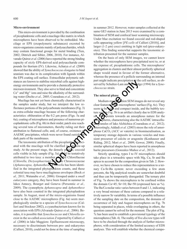
Fig. 7. Ternary diagram representation of Ca, Sr and Ba molarproportions in lake Geneva microspheres. a) The 55 analysed mi-crospheres are all located in the domain limited by Ca>0.45,Sr≤0.1 and Ba<0.5. Dotted lines labelled with underlined numbersrepresent the Ba/Ca molar ratio; b) microspheres distribution ac-cording to the date. Open squares, July; black squares, August; c)microspheres typology I: {Ca Sr Ba}; II: {Ca Sr}; III: {Ca P Sr};IV: {Ca}; V: {Ca P}. The individual microspheres within a clusterare linked by a black line. Isolated squares indicate that only oneanalysis was performed. Stippled circle: domain of groups IV andV. Grey ellipse: domain of groups II and III.
a)
c)
b)
Tab. 4. Indices of approximate stoichiometric formulae for microspheres.
Mgw Cax (Sry Baz) (CO3)13
Microspheres Mg Ca Sr Ba
Type I: average 0.18 7.57 0.55 4.71Type I: max Ba 0.09 5.76 0.64 6.51Type I: max Sr 0.24 6.28 1.27 5.21Type I: min Ba 0.35 10.83 0.31 1.52Type II: average 0.09 12.31 0.59 0.00Lake Alchichica 2.4 6.9 1.0 2.7Benstonite (from Tab. 3) 1.0 6.0 1.5 4.5
Mg, magnesium; Ca, calcium; Sr, strontium; Ba, barium. Lake Alchichica benstonite-like phase stoichiometry is recomputed from Couradeau et al. (2012).
Non co
mmercial
use o
nly
602 J-M Jaquet et al.
category (Barthelemy, 2012; Jolyon, 2012). Its basic for-mula may be written as (Ba,Sr)6(Ca,Mn)6Mg(CO3)13, withBa, Sr and manganese (Mn) varying in proportions. In theMagnet-Cove variety (Anthony et al., 2012), the formulais (Ba5.27Sr0.73)Ca6.00(Mg0.79Ca0.12Mn0.09)(CO3)13 or simplyBa6Ca6Mg(CO3)13. The variability in composition of differ-ent species of benstonite is illustrated in Tab. 3 (from liter-ature) and Tab. 4 (our samples).
In the absence of any significant amount of Mg andBa, types II, III and IV chemical compositions could bethose of an amorphous CaCO3 analogous to vaterite withvarying amounts of Sr. The molar Sr/Ca ratio in thesetypes of microspheres is around 0.05 (Tab. 5), equal tothat found by Lauchnor et al. (2013) in microbially in-duced Ca and Sr co-precipitates. This value lies in themiscibility region of the phase diagram of SrxCa1-xCO3
solids (Ruiz-Hernandez et al., 2010). As already men-tioned, in lake Constance this ratio – computed for wellcrystallised calcite – is only 0.0084 (Stabel et al., 1991).For ostracod valves in lake Geneva, Sr/Ca never exceeds0.0025 (Decrouy et al., 2012). These comparisons seemto indicate a higher Sr sequestration potential for theprocesses producing microspheres.
The EDS analyses being of semi-quantitative nature,caution has to be exercised when comparing our micros-
pheres’ composition to that of mineralogical or lakeAlchichica benstonite-like phase, the latter given as(Sr1.0Ba2.7Mg1.4Ca0.9)Ca6.0Mg(CO3)13 (Tab. 5, last column).Compared to synthetic benstonite (Tab. 4), lake Genevamicrospheres may be higher in Ca (type I, max. Sr andmin. Ba), and are always lower in Mg and Sr. All the for-mulas for mineralogical benstonite found in literaturehave in common an index very close to 6.0 for Ca. Thisis the case for some lake Geneva type I microspheres, butnot for Alchichica granules (6.9). As regard to this and therather imprecise definition of benstonite, we will considerthe attribution of type I microspheres to this mineral phaseas tentative only.
Some interesting trends emerge from the element ratiosgiven in Tab. 5. First, the AEM molar ratios are not thesame in water and in the microspheres; with respect to Ca,Mg is depressed in the precipitates whereas Sr (average0.06 in precipitates vs 0.0054 in water) and especially Ba(0.30-0.60 vs 0.00012) are strongly enriched in the micros-pheres. The dominance of Sr in water (Sr/Ba=44.3) is in-verted in type I microspheres (Sr/Ba=0.12). This Baenrichment is similar to that existing (as barite) in desmids(Wilcock et al., 1989) and marine bacteria (Gonzalez-Muñoz et al., 2012). This could reflect the affinity of Bafor biota (Martin and Knauer, 1973; McGrath et al., 1989).
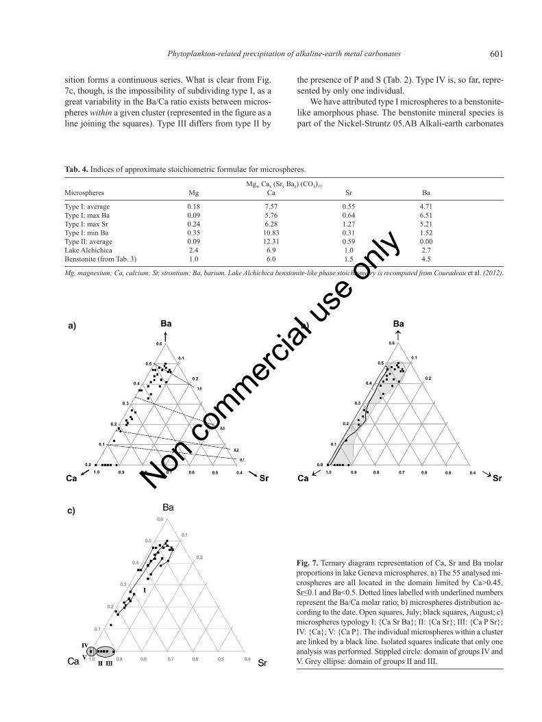
Fig. 8. a) Energy-dispersive X ray spectrometry (EDS) mappingof cluster Sp2_4 (07.08.2012), embedded in mucilage. Scale bar:10 μm. Apart from the microspheres, presence of siliceous par-ticles and picoplanktonic cells (1 μm diameter, visible in sec-ondary electron mode). Group IIA metals mineralisation isrestricted to bright, >2 μm microspheres. Strontium map is con-founded with silicon (spectral peak overlap). Notice the weakermagnesium signature, matching well the Sp2_4 -001 pointanalysis (not shown); b) Extract of spectrum corresponding tothe mapping of cluster Sp2_4. X-axis: energy in keV; Y-axis:counts × 103. Main peaks are shown in dark grey. Si K (1.73keV) and Sr L (1.8 keV) peaks are very close.
a) b)
Non co
mmercial
use o
nly

603Phytoplankton-related precipitation of alkaline-earth metal carbonates
An explanation for the differential enrichment of Sr and Bain the microspheres could be traced in Rogerson et al.(2008), who found that ions other than Ca can be chelatedinto biofilm EPS, and that this process was highly selectivein favour of those ions with small charge density.
Second, with respect to lake Alchichica AAEMC gran-ules, microspheres in lake Geneva are much lower in Mg(Mg/Ca=0.02 vs 0.56) and much higher in Ba(Ba/Ca=0.30-0.60 vs 0.11). This reflects the highly alka-line character of lake Alchichica waters (Tab. 1, bottomrow), with high Mg and low Ca, Sr and Ba concentrations.
CONCLUSIONS
Type I amorphous microspheres (0.6-3 μm diameter)containing Mg, Ca, Sr and Ba found in the epilimnion oflake Geneva in summer 2012 have been tentatively inter-preted as a benstonite-like mineral phase. Types II, III andIV, without Ba and with variable Sr, could be ascribed toa vaterite-like phase, a precursor of calcite. All these typesare characteristically precipitated within a mucilaginousmatrix embedding picoplanktonic cells.
Type I AAEMC microspheres’ chemical composition,determined semi-quantitatively by EDS, bears some anal-ogy to that of cyanobacterial intracellular inclusions foundin alkaline lake Alchichica (Mexico) microbialites. In lakeGeneva, however, the microspheres are pelagic, seem-ingly extracellular, and contain less Mg but more Ba.
Discovered a posteriori in dried suspended matter de-posited on filters, these precipitates could be studied onlyin a preliminary manner. In the context of the little-knownSr and Ba cycles in freshwater, their occurrence raises aseries of questions, which will be dealt with in a dedicatedstudy planned for 2013. Is the typology found in 2012 apermanent feature, and how can it be explained? What is
the taxonomic attribution of the picoplanktonic cells spa-tially associated with microspheres within the mucilage?Precipitating in epilimnion, do the microspheres survivein hypolimnion, and possibly in the sediment? Finally, theinternal structure and exact composition of the AAEMCprecipitates (as carbonate or phosphate in type V?) andassociated mucilage should be investigated with adequatetools in order to gain a proper understanding of this pecu-liar process of AEM sequestration.
ACKNOWLEDGMENTS
We are indebted to Sophie Lavigne, who carried outphytoplankton counting and determination. We thankFrançois Gischig, Cédric Schnyder (Muséum d’HistoireNaturelle, Geneva, Switzerland) and Marie-CarolinePinget (Department of Mineralogy, Université deGenève, Geneva, Switzerland) for preparation, Ramanspectroscopy and X-ray diffraction of benstonite sam-ples, respectively. Mathieu Coster was of great help inthe synthesis of benstonite. Vincent Ebener and NathalieDupont have aptly and faithfully collected samples inthe field, and water analyses were carried out by the Ser-vice de l’écologie de l’eau (SECOE) analytical labora-tory. The availability of SEM facilities at the Departmentof Geology (Université de Genève) is gratefully ac-knowledged. We are thankful to the reviewers and Jour-nal's editors for their help in improving an earlier versionof the manuscript.
REFERENCES
Acharya C, Joseph D, Apte SK, 2009. Uranium sequestrationby a marine cyanobacterium, Synechococcus elongatusstrain BDU/75042. Bioresource Technol. 100:2176-2181.
Addadi L, Raz S, Weiner S, 2003. Taking advantage of disorder:
Tab. 5. Selected element atom and molar ratios of water at 5 m depth, in microspheres and various species of benstonite-like phases.
Cation Water Water Microspheres Microspheres Microspheres (Sr0.5Ba5.5)* (Ba4.5Sr1.5)* (Ba2.7Sr1.0Mg1.4Ca0.9)*
10.07.2012 07.08.2012 type I type II type III theoretical synthetic Couradeau et al.5 m 5 m (average) (average) 1 EDS (2012)
Mg/Ca° 0.14 0.15 0.01 0.01 0.01 0.10 0.06 0.340.23 0.25 0.02 0.01 0.02 0.17 0.11 0.56
Sr/Ca 0.012 0.012 0.15 0.11 0.12 0.55 0.54 0.150.0053 0.0055 0.07 0.05 0.06 0.25 0.24 0.07
Ba/Ca 0.00041 0.00041 1.96 1.01 2.57 3.20 0.390.00012 0.00012 0.62 0.29 0.75 0.93 0.11
Sr/Mg 0.081 0.078 9.81 22.7 8.56 5.41 8.41 0.450.022 0.022 3.10 6.29 2.40 1.50 2.33 0.12
Sr/Ba 28.3 29.4 0.07 0.21 0.17 0.3944.3 46.1 0.12 0.33 0.26 0.61
EDS, energy dispersive X-ray spectrometry; Mg, magnesium; Ca, calcium; Sr, strontium; Ba, barium. *First row: variable cations, in addition to fixedMg1 Ca6; °top: atom ratio; bottom: molar ratio.
Non co
mmercial
use o
nly

604 J-M Jaquet et al.
amorphous calcium carbonate and its role in biomineraliza-tion. Adv. Mater. 15:959-970.
Alvarado Quiroz NG, Hung C-C, Santschi P, 2006. Binding ofthorium (IV) to carboxylate, phosphate and sulfate func-tional groups from marine exopolymeric substances (EPS).Mar. Chem. 100:337-353.
Anneville O, Pelletier J-P, 2000. Recovery of Lake Geneva fromeutrophication. Arch. Hydrobiol. 148:607-624.
Anneville O, Souissi S, Ibanez F, Ginot V, Druart JC, Angeli N,2002. Temporal mapping of phytoplankton assemblages inLake Geneva: annual and interannual changes in their pat-terns of succession. Limnol. Oceanogr. 47:1355-1366.
Anthony JW, Bideaux RA, Bladh KW, Nichols MC, 2012.Handbook of mineralogy. Mineralogical Society of Americaed., Chantilly: 628 pp.
Ball JW, Nordstrom DK, 1991. User’s manual for WATEQ4F,with revised thermodynamic data base and test cases for cal-culating speciation of major, trace, and redox elements innatural waters. Available from: http://wwwbrr.cr.usgs.gov/projects/GWC_chemtherm/pubs/wq4fdoc.pdf
Barthelemy D, 2012. Mineralogy database. Available from:http://webmineral.com/
Belykh OI, Sorokovikova EG, 2003. Autotrophic picoplanktonin Lake Baikal: Abundance, dynamics, and distribution.Aquat. Ecosyst. Health 6:251-261.
Bock C, Krienitz L, Pröschold T, 2011. Taxonomic reassessmentof the genus Chlorella (Trebouxiophyceae) using molecularsignatures (barcodes), including description of seven newspecies. Fottea 11:293-312.
Brook AJ, 1980. Barium accumulation by desmids of the genusClosterium (Zygnemaphyceae). Brit. Phycol. J. 15:261-264.
Callieri C, 2010. Single cells and microcolonies of freshwaterpicocyanobacteria: a common ecology. J. Limnol. 69:257-277.
Callieri C, Caravati E, Morabito G, Oggioni A, 2006. The uni-cellular freshwater cyanobacterium Synechococcus andmixotrophic flagellates: evidence for a functional associa-tion in an oligotrophic, subalpine lake. Freshwater Biol.51:263-273.
Callieri C, Piscia R, 2002. Photosynthetic efficiency and sea-sonality of autotrophic picoplankton in Lago Maggiore afterits recovery. Freswater Biol. 47:941-956.
Callieri C, Stockner JC, 2002. Freshwater autotrophic pi-coplankton: a review. J. Limnol. 61:1-14.
CIPEL, 2011. General conclusions on the state of Lake Genevain 2010. Commission Internationale pour la Protection duLéman ed., Nyon.
Couradeau E, Benzerara K, Gérard E, Moreira D, Bernard S,Brown Jr GE, López-García P, 2012. An early-branching mi-crobialite cyanobacterium forms intracellular carbonates.Science 336:459-462
Decho AW, Visscher PT, Reid RP, 2005. Production and cyclingof natural microbial exopolymers (EPS) within a marinestromatolite. Palaeogeogr. Palaeocl. 219:71-86.
Decrouy L, Vennemann TV, Ariztegui D, 2012. Mg/Ca andSr/Ca of ostracod valves from living species of LakeGeneva. Chem. Geol. 314-317:45-56.
Dittrich M, Kurz P, Wehrli B, 2004. The role of autotrophic pic-ocyanobacteria in calcite precipitation in an oligotrophiclake. Geomicrobiol. J. 21:45-53.
Dittrich M, Obst M, 2004. Are picoplankton responsible for cal-cite precipitation in lakes? Ambio 33:559-564.
Dittrich M, Sibler S, 2006. Influence of H+ and calcium ions onsurface functional groups of Synechococcus PCC 7942 cells.Langmuir 22:5435-5442.
Dittrich M, Sibler S, 2010. Calcium carbonate precipitation bycyanobacterial polysaccharides, p. 51-63. In: H.M. Pedleyand M. Rogerson (eds.), Tufas and speleothems: unravellingthe microbial and physical controls. Geological Society ofLondon Publ.
Dupraz C, Reid RP, Braissant O, Decho AW, Norman RS, Viss-che PT, 2009. Processes of carbonate precipitation in mod-ern microbial mats. Earth-Sci. Rev. 96:141-162.
Fahnenstiel GL, Carrick HJ, Rogers CE, Sicko-Goad L, 1991.Red fluorescing phototrophic picoplankton in the LaurentianGreat Lakes: what are they and what are they doing? Int.Rev. Hydrobiol. 76:603-616.
Finlay BJ, Hetherington NB, Davison W, 1983. Active biologi-cal participation in lacustrine barium chemistry. Geochim.Cosmochim. Ac. 47:1325-1329.
Gallina N, Anneville O, Beniston M, 2011. Impacts of extremeair temperatures on cyanobacteria in five deep peri-alpinelakes. J. Limnol. 70:186-196.
Glass-Haller L, 2010. Microbial and geochemical characteriza-tion of a contaminated freshwater ecosystem (the case ofVidy Bay, Lake Geneva, Switzerland). University of Genevaed., Geneva: 186 pp.
Gonzalez-Muñoz MT, Martinez-Ruiz F, Morcillo F, Martin-Ramos JD, Paytan A, 2012. Precipitation of baryte by ma-rine bacteria: a possible mechanism for marine bariteformation. Geology 40:675-678.
Gower L, 2008. Biomimetic model systems for investigating theamorphous precursor pathway and its role in biomineraliza-tion. Chem. Rev. 108:4551-4627.
Groleau A, Sarazin G, Vinçon-Leite B, Tassin B, Quiblier-Llo-béras C, 2000. Tracing calcite precipitation with specificconductance in a hard water alpine lake (Lake Bourget).Water Res. 43:4151-4160.
Hernández Mariné M, Clavero E, Roldán M, 2004. Microscopymethods applied to research on cyanobacteria. Limnetica23:179-185.
Hood WC, Steidl PF, 1973. Synthesis of benstonite at room tem-perature. Am. Mineral. 58:341-343.
Hood WC, Steidl PF, Tschopp DG, 1974. Precipitation ofnorsethite at room temperature. Am. Mineral. 59:471-474.
Jansson C, Northen T, 2010. Calcyfying cyanobacteria. The po-tential of biomineralization for carbon capture and storage.Curr. Opin. Biotech. 21:1-7.
Jaquet J-M, Favarger P-Y, Peter A, Vernet J-P, 1983. [Premièresdonnées sur la matière en suspension dans le Léman]. [Bookin French]. Institut Forel, University of Geneva ed., Geneva:83 pp. Available from: http://archive-ouverte.unige.ch/vital/access/manager/Repository/unige:27100
Jaquet J-M, Nembrini G, Garcia J, Vernet J-P, 1982. The man-ganese cycle in Lac Léman (Switzerland): the role of Met-allogenium. Hydrobiologia 91-92:323-340.
Jeandel C, Tachikawa K, Bory A, Dehairs F, 2000. Biogenic bar-ium in suspended and trapped material as a tracer of exportproduction in the tropical NE Atlantic (EUMELI sites). Mar.Chem. 71:125-142.
Non co
mmercial
use o
nly

605Phytoplankton-related precipitation of alkaline-earth metal carbonates
Jolyon R, 2012. Mindat.org, the mineral and locality database.Available from: http://www.mindat.org
Kelts K, Hsu KJ, 1978. Freshwater carbonate precipitation, p.295-323. In A. Lerman (ed.), Lakes: chemistry, geology,physics. Springer-Verlag.
Konhauser K, Riding R, 2012. Bacterial biomineralization, p.105-130. In: A.H. Knoll, D.E. Canfield and K.O. Konhauser(eds.), Fundamentals of geobiology. Blackwell Publ.
Krejci MR, Wassermann B, Finney L, McNulty I, Legnini D,Vogt S, Joester D, 2011. Selectivity of biomineralization ofbarium and strontium. J. Struct. Biol. 176:192-202.
Lauchnor EG, Schulz LN, Bugni S, Mitchell AC, CunninghamAB, Gerlach R, 2013. Bacterially induced calcium carbonateprecipitation and strontium coprecipitation in a porousmedia flow system. Environ. Sci. Technol. 47:1557-1564.
Lazarotto J, Klein A, 2012. Physical-chemical changes in thewaters of Lake Geneva. Available from: http://www.cipel.org/wp-content/uploads/2012/11/Evolution-physico-chim-ique.pdf
Lee BD, Apel WA, Walton MR, 2006. Calcium carbonate for-mation by Synechococcus sp. strain PCC 8806 and Syne-chococcus sp. strain PCC 8807. Bioresource Technol.97:2427-2434.
Lynn DH, 2010. The ciliated protozoa: characterization, classi-fication, and guide to the literature. Springer, Amsterdam:605 pp.
Martin JH, Knauer GA, 1973. The elemental composition ofplankton. Geochim. Cosmochim. Ac. 37:1639-1653.
McGrath M, Davison W, Hamilton-Taylor J, 1989. Biogeochem-istry of barium and strontium in a softwater lake. Sci. TotalEnviron. 87/88:287-295.
Mori Y, Enomae T, Isogai A, 2009. Preparation of pure vateriteby simple mechanical mixing of two aqueous salt solutions.Mater. Sci. Eng. C29:1409-1414.
Parkhurst DL, Appelo CAJ, 1999. User’s guide to PHREEQC(version 2): a computer program for speciation, batch reac-tion, one-dimensional transport, and inverse geochemicalcalculations. US Department of the Interior ed., denver: 309pp. Available from: ftp://brrftp.cr.usgs.gov/pub/dlpark/geochem/unix/phreeqc/manual.pdf
Pereira S, Zille A, Micheletti E, Moradas-Ferreira P, De PhilippisR, Tamagnini P, 2009. Complexity of cyanobacterial ex-opolysaccharides : composition, structures, inducing factorsand putative genes involved in their biosynthesis and assem-bly. FEMS Microbiol. Rev. 33:917-941.
Personnic S, Domaizon I, Dorrigo U, Berdjeb L, Jacquet S,2009. Seasonal and spatial variability of virio-, bacterio-,and picoplanktonic abundance in three perialpine lakes. Hy-drobiologia 627:99-116.
Plée K, Ariztegui D, Martini R, Davaud E, 2008. Unravellingthe microbial role in ooid formation – results of an in situexperiment in modern freshwater Lake Geneva in Switzer-land. Geobiology 6:341-350.
Rogerson M, Pedley HM, Wadhawan JD, Middleton R, 2008.New insights into biological influence on the geochemistry
of freshwater carbonate deposits. Geochim. Cosmochim. Ac.72:4976-4987.
Ruiz-Hernandez SE, Grau-Crespo R, Ruiz-Salcador AR, DeLeuw NH, 2010. Thermochemistry of strontium incorpora-tion in aragonite from atomistic simulations. Geochim. Cos-mochim. Ac. 74:1320-1328.
Sanchez-Moral S, Luque L, Cañaveras JC, Laiz L, Jurado V,Hermosin B, Saiz-Jimenez C, 2004. Bioinduced barium pre-cipitation in St. Callixtus and Domitilla catacombs. Ann. Mi-crobiol. 54:1-12.
Schultze-Lam S, Beveridge TJ, 1994. Nucleation of celestite andstrontianite on a cyanobacterial S-Layer. Appl. Environ. Mi-crob. 60:447-453.
Sondi I, Matijevic E, 2003. Homogenous precipitation by en-zyme-catalyzed reactions. 2. Strontium and Barium carbon-ates. Chem. Mater. 15:1322-1326.
Stabel H-H, 1986. Calcite precipitation in Lake Constance. Lim-nol. Oceanogr. 31:1081-1093.
Stabel H-H, 1989. Coupling of strontium and calcium cycles inLake Constance. Hydrobiologia 176-177:323-329.
Stabel H-H, Kleiner J, Merkel P, Sinemus HW, 1991. [Stof-fkreislaüfe ausgewählter Spurenelemente im Bodensee].[Article in German]. Vom Wasser 76:73-91.
Stabel H-H, Küchler-Krischun J, Kleiner J, Merkel P, 1986. Re-moval of strontium by coprecipitation in Lake Constance.Naturwissenschaften 73:551-553.
Sugiayma M, Hori T, Kihara S, Matsui M, 1992. A geochemicalstudy on the specific distribution of barium in Lake Biwa,Japan. Geochim. Cosmochim. Ac. 56:597-605.
Sun D-M, Wu Q-S, Ding Y-P, 2006. A novel method for crystalcontrol: synthesis and design of strontium carbonate withdifferent morphologies by supported liquid membrane. Sol.St. Phen. 39:544-549.
Tadonleke RD, 2012. Primary production and chlorophyll a bio-mass in Lake Geneva. Campagne 2011:78-84.
Tien C-J, 2002. Biosorption of metal ions by freshwater algaewith different surface characteristics. Process Biochem.38:605-613.
Watanabe K, Imase M, Sasaki K, Ohmura N, Saiki H, TanakaH, 2006. Composition of the sheath produced by the greenalga Chlorella sorokiniana. Lett. Appl. Microbiol. 42:538-543.
Wilcock JR, Perry CC, Williams RJP, Brook AJ, 1989. Biolog-ical minerals formed from strontium and barium sulphates.II. Crystallography and control of mineral morphology indesmids. P. Roy. Soc. Lond. B Bio. 238:203-221.
Winder M, 2009. Photosynthetic picoplankton dynamics in LakeTahoe: temporal and spatial niche partitioning amongprokariotic and eukaryotic cells. J. Plankton Res. 31:1307-1320.
Wu Q-S, Sun D-M, Liu H-J, Ding Y-P, 2004. Abnormal poly-morph conversion of calcium carbonate and nano-self as-sembly of vaterite by a supported liquid membrane system.Cryst. Growth Des. 4:717-720.
Non co
mmercial
use o
nly
1
Supplementary Figures.
Supplementary Fig. 1. a) Spherical concretions of synthetic benstonite Mg (Ba4.5 Sr1.5) Ca6 (CO3)13, aged 3 days. Scale bar: 10 µm; b) close-up on a concretion surface. Scale bar: 100 nm. The concretion consists of individual, spherical (diameter <100 nm), or needle-shaped nanoparticles.
Non-co
mmercial
use o
nly
2
Supplementary Fig. 2. X-ray diffraction diagram of synthetic benstonite (black), with standard [Mg Ca6 Ba6 (CO3)13].
Non-co
mmercial
use o
nly
3
Supplementary Fig. 3. Raman spectrum of synthetic benstonite. Wavenumber of main peaks is compared to those of Ca7 Ba6 (CO3)13.
Non-co
mmercial
use o
nly