Carol Tanner PhD Thesis - UWSpace
319
Biochemical and Microbiological Investigations of Inhibitors for β-Lactamases from Human Pathogenic Bacteria by Carol Anne Tanner A thesis presented to the University of Waterloo in fulfillment of the thesis requirement for the degree of Doctor of Philosophy in Chemistry Waterloo, Ontario, Canada, 2019 ©Carol Anne Tanner 2019
-
Upload
khangminh22 -
Category
Documents
-
view
0 -
download
0
Transcript of Carol Tanner PhD Thesis - UWSpace

Biochemical and Microbiological
Investigations of Inhibitors for
β-Lactamases from Human Pathogenic
Bacteria
by
Carol Anne Tanner
A thesis
presented to the University of Waterloo
in fulfillment of the
thesis requirement for the degree of
Doctor of Philosophy
in
Chemistry
Waterloo, Ontario, Canada, 2019
©Carol Anne Tanner 2019

ii
Author’s Decarlation
I hereby declare that I am the sole author of this thesis. This is a true copy of the thesis, including
any required final revisions, as accepted by my examiners.
I understand that my thesis may be made electronically available to the public.

iii
Abstract
The majority of antibiotics prescribed for treatment of bacterial infections are β-lactam antibiotics.
Resistance to these has evolved in a few different ways, notably by regulating permeability and by the
expression of β-lactamases which hydrolyze the antibiotic before it reaches its target. Three of the
classes of β-lactamases (classes A, C and D) are serine-β-lactamases (SBLs) and the fourth class
(Class B) consists of metallo-β-lactamases (MBLs) that rely on one or two zinc ions for their catalytic
activity. Bacteria producing β-lactamases that are capable of hydrolyzing the β-lactam bond in all of
the classes of β-lactam antibiotics including penicillins, monobactams, cephalosporins and
carbapenems are of great clinical concern. As a consequence of the increasing prevalence of
resistance, there is much interest in the discovery of inhibitors for such clinically important β-
lactamases as well as in the discovery of β-lactam antibiotics that are less susceptible to inactivation
by β-lactamases.
Described in this thesis are the kinetic properties of new chromogenic cephalosporin-type
substrates that are susceptible to hydrolysis by clinically important SBLs and MBLs but that exhibit a
much more pronounced colour change upon hydrolysis than does the commercially available and
widely used chromogenic cephalosporin called nitrocefin. Some of these substrates also offer more
favourable kinetic properties for assaying MBLs in vivo.
Also in this thesis, biochemical as well as microbiological investigations of several classes of SBL
and MBL inhibitors are describes as well as one class of cephalosporins that exhibit inhibition of
MBLs and surprising antibacterial potency against certain clinically significant MBL-producing
Gram negative bacteria.
More specifically, 6-phosphonomethylpyridine-2-carboxylates (PMPCs) and a number of
derivatives thereof, synthesized previously in this research group have been shown in this thesis
research to be potent inhibitors (low to submicromolar Ki) of the major Class B1 MBLs, IMP-1,
VIM-2, NDM-1 and SPM-1 as well as the Class B3 MBL L1, all of which are dizinc enzymes, and
somewhat less potent inhibitors of the monozinc Class B2 MBL, SFH-1, which is of lesser clinical
significance. These compounds that are expected to exhibit metal-binding characteristics were found
to exhibit a time-dependent inhibition mechanism which fits a kinetic mechanism that is consistent
with slow binding to the active site and even slower release of the inhibitor without expulsion of the
metal ions form the MBL active site. Microbiological investigations were also carried out involving

iv
combinations of PMPCs with the carbapenem antibiotic meropenem and demonstrated an ability of
the PMPCs to lower the MICs of meropenem against of MBL-producing clinical strains of Eschericia
coli, Klebsiella pneumoniae, Pseudomonas aeruginosa, Acinetobacter baumannii and
Stenotrophomonas maltophilia thus encouraging further research on even more potent PMPCs for
potential clinical use in combination with carbapenems.
Another class of MBL inhibitors that was studied consist of cephalosporin derivatives that
incorporate an aromatic thioester linked to C3ʹ of the cephalosporin core. It was found that inhibition
of the Class B3 MBL L1 was likely the consequence largely of the binding of an arylthioacid
conjugate base to the active site zinc ions after its expulsion from the hydrolysis product of the
cephalosporin. This led to a study of the MBL-inhibitory properties of a series of synthetic
arylthioacids, a class of compounds that have not been well studied as metallo enzyme inhibitors.
These compounds were found to be poor inhibitors of Class B1 MBLs (IMP-1, VIM-2, NDM-2 and
SPM-1) but good inhibitors of the B3 MBL L1. These observations are consistent with inhibition of
Class B1 MBLs arising not from the arylthicarboxylate released but from binding the cephalosporin-
derived metal-binding species formed upon expulsion of the arylthiocarboylate from the active site.
A further enzyme kinetic study revealed that synthetic samples of pyridine-2,6-bis(carbothioic)
acid and 6-thiocarboxy-picolinic acid, previously known as Fe3+-binding siderophores produced by
some species of Pseudomonas, were good inhibitors of Class B1 and B3 MBLs and also exhibited an
ability to lower the MIC of meropenem against MBL-producing clinically important Gram negative
bacteria.
One cephalosporin called UW-123 with at C3ʹ-arylacylthio group and a siderophore mimic
attached to the amide group at C7 was studied in some detail microbiologically and found to exhibit
good standalone antibiotic activity against certain MBL-producing Gram-negative bacteria especially
those producing the widespread MBL VIM-2. UW-123 was also shown to be bacteriostatic and to
bind preferentially to induce filamentation of E. coli cells. It was found to bind most strongly to PBP
3 and 1a but also significantly to PBP1b and 4 in P. aeruginosa. In E. coli, UW-123 bound most
tightly to PBP3 but also significantly to PBP1a/b and PBP2.
Finally, the ability of a cyclobutanone mimic of penem antibiotics JJ05-1058, previously prepared
in this laboratory, to inhibit both SBLs and MBLs was demonstrated and the ability of this compound
to bind to the low molecular weight penicillin binding proteins was observed suggesting that such

v
compound may have some promise as broad spectrum MBL/SBL inhibitors and possible also as
antibiotics that inhibit penicillin binding proteins.

vi
Acknowledgements
First and foremost I would like to thank Dr. Gary Dmitrienko for sharing his knowledge and
expertise in the field of β-lactam resistance these many years. His patience and assistance with the
thesis writing process has made the production of this document tolerable.
I would also like to thank our collaborators Dr. James Spencer and Dr. Philip Hinchliffe without
whom many of the insightful conclusions about binding modes could not have been achieved. My
advisory committee Dr. Guy Guillemette, Dr. Thorsten Dieckmann, Dr. Richard Manderville, and Dr.
Stefen Siemann have been excellent sources of advice and thought provoking questions, challenging
me to push the boundaries of my knowledge and expertise. My external examiner, Dr. Steven Seah,
and my internal-external examiner, Dr. Trevor Charles should be thanked for the time and effort they
put forth in reviewing my thesis and offering suggestions.
I am also grateful to Cathy Van Esch, a fount of knowledge for the inner workings of graduate
studies in the Department of Chemistry.
The ladies of the Dmitrienko biochemistry lab, Dr. Geneviève Labbé, Valerie Goodfellow, and Dr.
Laura Marrone taught me everything I know about running experiments and living life to the fullest –
no amount of thanks could be enough. The excellent undergraduate thesis students that have passed
through this lab over the years, Melinda Lam, Karan Malik, Alicia Tjahjadi, and Jessica Duong must
be thanked and credited for giving us a reason to purify more proteins, actually performing the
purification, and breaking the silence that took over the lab in 2015.
To Dr. Anthony Krismanich, Dr. Ahmad Ghavami, Dr. Ahmed Desoky, Dr. Nan Chen, Dr. Glenn
Abbott, Dr. Jarrod Johnson, Karan Teckwani and Peizhi Qui, this work would not have been possible
without your synthetic talents and the religious observation of the 10 am coffee break. Upon the
dispersal of the Dmitrienko organic lab, I thank my friends throughout the department for keeping me
company as I continued to observe – though less religiously – the daily coffee break.
The biological nature of this work necessitated use of equipment or facilities in the Biology
Department, and I would like to thank Dr. Trevor Charles, Dr. Josh Neufeld, Katja Engel, Sura Ali,
Dr. Brenden McConkey, and Dr. Roland Hall for training me and allowing me to use your facilities to
complete my work.

vii
Finally, I would like to thank my husband, Taylar Cameron for his unending love and support
throughout my graduate studies and his understanding when I say that I have done enough dishes for
one day. Our beloved cats, Lily and Pickles, must also be thanked for being my primary source of
purr-crastination.

viii
Table of Contents
Author’s Decarlation .......................................................................................................................... ii
Abstract ............................................................................................................................................. iii
Acknowledgements ........................................................................................................................... vi
Table of Contents ............................................................................................................................ viii
List of Figures .................................................................................................................................. xii
List of Schemes ................................................................................................................................ xv
List of Tables .................................................................................................................................. xvi
List of Abbreviations....................................................................................................................... xix
Chapter 1 ............................................................................................................................................ 1
1.1 Antibiotic Classes and Targets ................................................................................................. 1
1.1.1 Overview of Antibiotics .................................................................................................... 1
1.1.2 β-Lactam Antibiotics ......................................................................................................... 4
1.1.3 Bacterial Resistance .......................................................................................................... 7
1.2 Penicillin Binding Proteins ...................................................................................................... 9
1.2.1 Bacterial Cell Wall Structure ............................................................................................ 9
1.2.2 PBP Functional and Structural Classification ................................................................. 10
1.2.3 PBP Expression in Bacterial Strains ............................................................................... 15
1.2.4 Role of PBPs in Bacterial Resistance to β-Lactams ........................................................ 17
1.3 β-Lactamases .......................................................................................................................... 18
1.3.1 Role of β-Lactamases in Bacterial Resistance to β-Lactams .......................................... 19
1.3.2 Classification of β-Lactamases........................................................................................ 20
1.3.3 Class A β-Lactamases ..................................................................................................... 23
1.3.4 Class C β-Lactamases ..................................................................................................... 28

ix
1.3.5 Class D β-Lactamases ...................................................................................................... 31
1.3.6 Class B β-Lactamases ...................................................................................................... 33
1.3.7 Established Methods for Assaying β-lactamases ............................................................. 37
1.4 β-Lactamase Inhibitors ........................................................................................................... 38
1.4.1 β-Lactams as β-Lactamase Inhibitors .............................................................................. 38
1.4.2 Serine-β-Lacamase Inhibitors .......................................................................................... 40
1.4.3 Metallo-β-Lactamase Inhibitors....................................................................................... 47
1.4.4 Dual Serine- and Metallo-β-Lactamase Inhibitors .......................................................... 57
1.5 Clinical Significance of β-Lactamase Inhibition .................................................................... 58
Chapter 2 Phylogenetic Analysis, Purification, and Biochemical Characterization of β-Lactamases
.............................................................................................................................................................. 60
2.1 Introduction ............................................................................................................................ 60
2.2 Methods .................................................................................................................................. 62
2.2.1 Protein Sequence Alignment ........................................................................................... 62
2.2.2 Enzyme Purification ........................................................................................................ 63
2.2.3 Characterization of Chromogenic Substrates .................................................................. 74
2.2.4 Biochemical Characterization .......................................................................................... 76
2.3 Results .................................................................................................................................... 78
2.3.1 Metallo-β-Lactamase Protein Sequence Alignment ........................................................ 78
2.3.2 Protein Purification .......................................................................................................... 84
2.3.3 Characterization of Chromogenic Substrates .................................................................. 88
2.3.4 Biochemical Characterization .......................................................................................... 93
2.4 Discussion ............................................................................................................................... 99
2.5 Future Work .......................................................................................................................... 105

x
Chapter 3 Inhibition of Metallo-β-Lactamases by Phosphonomethyl Pyridine Carboxylates ....... 106
3.1 Introduction .......................................................................................................................... 106
3.2 Methods ................................................................................................................................ 109
3.2.1 Enzyme Kinetics ........................................................................................................... 109
3.2.2 Microbial Experiments .................................................................................................. 113
3.3 Results .................................................................................................................................. 114
3.3.1 Enzyme Kinetics ........................................................................................................... 114
3.3.2 Potentiation of meropenem against MBL producing Gram negatives .......................... 123
3.4 Discussion ............................................................................................................................ 124
3.5 Future Work ......................................................................................................................... 132
Chapter 4 Thioacids and 3ʹ-Acylthio-Cephalosporins as B3 Inhibitors ......................................... 134
4.1 Introduction .......................................................................................................................... 134
4.2 Methods ................................................................................................................................ 137
4.2.1 Synthetic Thioacids and 3ʹ-acylthiocephalosporins ...................................................... 137
4.2.2 β-Lactamase Enzyme Kinetics ...................................................................................... 140
4.2.3 Antimicrobial Susceptibility ......................................................................................... 141
4.2.4 Mode of Action ............................................................................................................. 142
4.3 Results .................................................................................................................................. 146
4.3.1 Released Phenyl Thioacids as β-Lactamase Inhibitors ................................................. 146
4.3.2 MICs of UW-123 .......................................................................................................... 155
4.3.3 Bacterial Synergy of UW-123 with Meropenem .......................................................... 163
4.3.4 Mode of Action Studies of UW-123 ............................................................................. 164
4.3.5 Pyridine thioacids – a more potent alternative to phenyl thioacids ............................... 173
4.4 Discussion ............................................................................................................................ 179

xi
4.4.1 Phenyl Thioacid Inhibitors of B3 MBLs ....................................................................... 179
4.4.2 3ʹ-Acylthiocephalosporins as delivery systems for phenyl thioacid inhibitors of L1 .... 182
4.4.3 UW-123 ......................................................................................................................... 183
4.5 Future Work .......................................................................................................................... 185
Chapter 5 Other β-Lactamase Inhibitors......................................................................................... 187
5.1 Introduction .......................................................................................................................... 187
5.2 Methods. ............................................................................................................................... 191
5.2.1 β-Lactamase Enzyme Kinetics ...................................................................................... 191
5.2.2 Penicillin Binding Protein Binding Assay ..................................................................... 193
5.3 Results .................................................................................................................................. 194
5.3.1 Inhibition of β-Lactamases ............................................................................................ 194
5.3.2 Competitive binding to Penicillin binding proteins ....................................................... 198
5.4 Discussion ............................................................................................................................. 200
5.5 Future Work .......................................................................................................................... 202
Appendix A Structures of β-Lactamase Inhibitors ......................................................................... 203
Appendix B Statistical Methods for IC50 Averages ........................................................................ 206
Appendix C Clinical Isolates and Control Strains .......................................................................... 207
Appendix D KI Graphs of DPA, PMPC-3, and PMPC-4 ............................................................... 209
Appendix E Protein Sequence Alignments ..................................................................................... 212
Bibliography ................................................................................................................................... 242

xii
List of Figures
Figure 1.1 Critically important antimicrobials for human medicine. ..................................................... 2
Figure 1.2 Representative antibiotics from each major class of β-lactams ............................................ 4
Figure 1.3 Chemical structures of imipenem and cilastatin. .................................................................. 6
Figure 1.4 Core structures of β-lactam antibiotic families. .................................................................... 7
Figure 1.5 Gram positive and Gram negative cellular envelope composition. ...................................... 9
Figure 1.6 Structure of Lipid II, the peptidoglycan precursor ............................................................. 11
Figure 1.7 3-Dimensional structure of Class A SBLs, exemplified by KPC-2 .................................... 24
Figure 1.8 Locations of phenotype changing mutations in TEM-1 ..................................................... 26
Figure 1.9 Overall MBL αββα fold ...................................................................................................... 34
Figure 1.10 Penicillins that are inhibitory towards some SBLs. .......................................................... 39
Figure 1.11 Mechanism-based serine-β-lactamase inhibitors. ............................................................. 41
Figure 1.12 Other mechanism-based inhibitors: penicillanic acid sulfones, 6-halo penicillins, and
alkylidene penams........................................................................................................................... 42
Figure 1.13 A selection of the diazabicyclooctanes that are clinically available or in development. .. 43
Figure 1.14 Transition state analog inhibitors of SBLs: boronates and phosph(on)ates. ..................... 45
Figure 1.15 Cyclobutanones: β-lactam mimics that are inhibitory towards β-lactamases. .................. 46
Figure 1.16 Demetallating inhibitors of MBLs. ................................................................................... 48
Figure 1.17 Dicarboxylate inhibitors of MBLs. ................................................................................... 49
Figure 1.18 Thiol inhibitors of MBLs. ................................................................................................. 51
Figure 1.19 D-Captopril bound to various MBLs from all subclasses................................................. 53
Figure 1.20 Thioester and other non-thiol MBL inhibitors that contain sulphur. ................................ 55
Figure 1.21 Structurally unique MBL inhibitors.................................................................................. 57
Figure 1.22 Structural evidence for cyclic boronate inhibition of SBLs and MBls ............................. 58
Figure 2.1 Chromogenic and fluorogenic substrates for β-lactamases excluding the UW series
substrates. ........................................................................................................................................ 60
Figure 2.2 Chromogenic β-lactamase substrates synthesized by the Dmitrienko group. .................... 61
Figure 2.3 Chromogenic β-lactamase substrates synthesized by the Dmitrienko group. .................... 75
Figure 2.4 Phylogenetic tree of NDM Variants ................................................................................... 79
Figure 2.5 Phylogenetic Tree of VIM variants .................................................................................... 81
Figure 2.6 Phylogenetic tree of IMP family variants ........................................................................... 83

xiii
Figure 2.7 SDS-PAGE of pure β-lactamases. ...................................................................................... 87
Figure 2.8 Visual appearance of 200 μL of increasing concentrations of unhydrolyzed and hydrolyzed
chromogenic substrates synthesized by the Dmitrienko lab ............................................................ 89
Figure 2.9 Spectral scans of chromogenic substrates before and after hydrolysis ............................... 90
Figure 2.10 Stability spectra of 100 μM chromogenic substrates before and after hydrolysis. ........... 92
Figure 2.11 DMSO sensitivity of β-lactamases .................................................................................... 93
Figure 2.12 Michaelis-Menten plots of CTX-M-15 kinetic parameter determination. ........................ 94
Figure 2.13 Michaelis-Menten plots of VIM-1, SFH-1, OXA-23, and OXA-48 kinetic parameter
determination. .................................................................................................................................. 96
Figure 2.14 NDM-1 crystal structure with residues that are changed among the variants highlighted 99
Figure 2.15 Overlay of VIM-1 and VIM-2 crystal structures with variable residues within 10 Å of the
Zn atoms highlighted ..................................................................................................................... 100
Figure 3.1 MBL inhibitors based off the DPA core structure. ........................................................... 107
Figure 3.2 PC1 from S. aureus covalently inactivated by a phosphonate monoester ........................ 108
Figure 3.3 Phosph(on)ate based inhibitors of β-lactamases. .............................................................. 109
Figure 3.4 Structures of picolinic acid derivatives used in this chapter. ............................................ 110
Figure 3.5 Dose response curves of MBLs with PMPCs and related compounds. ............................ 118
Figure 3.6 Time-dependent KI graphs for PMPC-1 against representative MBLs ............................. 121
Figure 3.7 Binding of 3-(4-hydroxypiperidin-1-yl)-aminophthalic acid and PMPC-1 to the active site
IMP-1. ........................................................................................................................................... 127
Figure 3.8 Crystal structures of PMPCs bound to L1 ......................................................................... 127
Figure 3.9 Binding of the S-enantiomer of PMPC-3 to the active site of L1. .................................... 128
Figure 3.10 Fits of initial and steady state rates on representative progress curves of MBLs inhibited
by PMPC-1 .................................................................................................................................... 130
Figure 3.11 Binding of PMPC-1 to the active site of IMP-1, highlighting opportunities for
development of future PMPCs. ..................................................................................................... 133
Figure 4.1 Protection of meropenem hydrolysis by synthetic and commercial cephalosporins. ........ 136
Figure 4.2 Generation of a MBL inhibitor from 8-thioxo-cephalosporins. ........................................ 137
Figure 4.3 Potent inhibitors of CcrA and L1 determined from natural product screen. ..................... 137
Figure 4.4 Structure of pyridine based thioacids and related compounds presented in this chapter. . 138

xiv
Figure 4.5 Serine-β-lactamase inhibitors used in microbiological experiments in the Dmitrienko lab.
...................................................................................................................................................... 141
Figure 4.6 MH agar spot plates of thioacid potentiation of meropenem against L1 and L2 producing S.
maltophilia .................................................................................................................................... 151
Figure 4.7 UW-123 time kill curves against wild type E. coli ........................................................... 165
Figure 4.8 SDS-PAGE of UW-123 bound to P. aeruginosa PBPs competing with Bocillin. ........... 168
Figure 4.9 Dose response curves of the relative competitive binding of UW-123 to PBPs from P.
aeruginosa with Bocillin. ............................................................................................................. 169
Figure 4.10 SDS-PAGE of UW-123 bound to E. coli PBPs competing with Bocillin. ..................... 171
Figure 4.11 Dose response curves of the relative competitive binding of UW-123 to PBPs from E.
coli with Bocillin. ......................................................................................................................... 172
Figure 4.12 Dose response curves of MBLs and GC-1 inhibited by PMTC and PDTC ................... 176
Figure 4.13 Spot plates of UWB 33 on MH agar to determine the MBCs of thioacid potenticated .. 181
Figure 4.14 Binding of thiols to the L1 active site ............................................................................. 182
Figure 4.15 Bis-PDTC-cephalosporin................................................................................................ 186
Figure 5.1 Examples of cyclobutanone mimics of β-lactam antibiotics. ........................................... 188
Figure 5.2 Conformations of C3-substituted cyclobutanones and the steric interactions of the
hydrates. ........................................................................................................................................ 189
Figure 5.3 X-Ray crystal structures of cyclobutanones bound to β-lactamases. ............................... 191
Figure 5.4 Inhibition of SBLs by JJ05-1058. ..................................................................................... 195
Figure 5.5 Inhibition of MBLs by JJ05-1058. ................................................................................... 196
Figure 5.6 SDS-PAGE of JJ05-1058 bound to P. aeruginosa PBPs competing with Bocillin.......... 198
Figure 5.7 JJ05-1058 competition for PBP binding with Bocillin. .................................................... 199
Figure 5.8 Time Dependent KI graphs for DPA against representative MBLs ................................. 209
Figure 5.9 Time Dependent KI graphs for PMPC-3 against representative MBLs ............................ 210
Figure 5.10 Time Dependent KI graphs for PMPC-4 against representative MBLs .......................... 211

xv
List of Schemes
Scheme 1.1 Generalized hydrolysis of β-lactam by a β-lactamase. ....................................................... 8
Scheme 1.2 Reactions catalyzed by PBPs: glycosyl transfer, transpeptidation, carboxypeptidation, and
endopeptidation. .............................................................................................................................. 12
Scheme 1.3 Inactivation of PBPs by a β-lactam................................................................................... 13
Scheme 1.4 Mechanism of penicillin hydrolysis by Class A β-lactamases. ......................................... 25
Scheme 1.5 Mechanism of cephalosporin hydrolysis by Class C β-lactamases. ................................. 30
Scheme 1.6 Mechanism of penicillin hydrolysis catalyzed by Class D SBLs. .................................... 32
Scheme 1.7 Hydrolysis of carbapenems by B1 and B3 MBLs............................................................. 37
Scheme 1.8 Hydrolysis of carbapenems by B2 MBLs. ........................................................................ 37
Scheme 1.9 Inhibition of SBLs by cephalosporins and carbapenems. ................................................. 40
Scheme 1.10 Inhibition of Class A SBLs by mechanism-based inhibitors eg. clavulanate. ................ 42
Scheme 1.11 Avibactam mediated inhibition of SBLs and other reactions with Avibactam catalyzed
by β-lactamases. .............................................................................................................................. 44
Scheme 1.12 Covalent adducts formed by boronates, phosphonates, and cyclobutanones that mimic
the transition-state of β-lactam acyl-enzyme formation with serine-β-lactamases. ........................ 47
Scheme 1.13 Activation of rhodanine to a mercaptocarboxylate MBL inhibitor. ................................ 54
Scheme 2.1 Panbio homogeneous immunoassay technology. ............................................................. 61
Scheme 2.2 Hydrolysis of nitrocefin by β-lactamases. ........................................................................ 89
Scheme 3.1 Enzyme inhibition mechanisms that can exhibit time-dependent inhibition. ................. 120
Scheme 4.1 Formazan dye strategy for a chromogenic cephalosporin. ............................................. 135
Scheme 4.2 Abridged method for thioacid synthesis. ........................................................................ 138
Scheme 5.1 Comparison of hemiketal formation with TS and intermediate in acyl enzyme
mechanism. .................................................................................................................................... 187
Scheme 5.2 Parallells between the binding of penems and cyclobutanones to SBLs and MBLs. ..... 190

xvi
List of Tables
Table 1.1 Mode of action of established antibiotics. ............................................................................. 3
Table 1.2 Penicillin binding proteins present in E. coli and P. aeruginosa ......................................... 16
Table 1.3 Richmond and Sykes functional classification scheme for β-lactamases. ........................... 21
Table 1.4 Functional and molecular classification of β-lactamases. .................................................... 22
Table 1.5 Amino acid residues that coordinate zinc ions in the three subclasses of metallo-β-
lactamases. ...................................................................................................................................... 34
Table 2.1 Enzyme and substrate concentrations used for DMSO stability studies. ............................. 76
Table 2.2 Amino acid differences between NDM-1 and other NDM variants. ................................... 80
Table 2.3 CTX-M-15 purification table ............................................................................................... 84
Table 2.4 KPC-2 purification table. ..................................................................................................... 85
Table 2.5 VIM-1 purification table ...................................................................................................... 85
Table 2.6 GC-1 purification table. ....................................................................................................... 86
Table 2.7 OXA-48 purification table. .................................................................................................. 87
Table 2.8 Masses of pure β-lactamases as predicted using Expasy ProtParam and determined by mass
spectroscopy ................................................................................................................................... 88
Table 2.9 Molar extinction coefficients for UW substrates ................................................................. 91
Table 2.10 Kinetic parameters of CTX-M-15 with and without its His-tag against NC and UW
substrates. ........................................................................................................................................ 95
Table 2.11 Kinetic parameters of serine-β-lactamases (except CTX-M-15) against NC and UW
substrates. ........................................................................................................................................ 97
Table 2.12 Kinetic parameters of metallo-β-lactamases with NC and UW substrates ........................ 98
Table 2.13 Literature values for serine-β-lactamase kinetic parameters with nitrocefin. .................. 103
Table 2.14 Literature values for metallo-β-lactamase kinetic parameters with nitrocefin. ................ 104
Table 3.1 Kinetic parameters and conditions for PMPC inhibitor screening..................................... 111
Table 3.2 Conditions of time-dependent KI determination. ............................................................... 112
Table 3.3 Screening of PMPCs and related commercial compounds against MBLs ......................... 115
Table 3.4 Screening of PMPCs and related commercial compounds against SBLs. ......................... 116
Table 3.5 IC50’s of PMPCs against MBLs ......................................................................................... 119
Table 3.6 KI of DPA, PMPC-1, -3, and -4 against select MBLs........................................................ 122
xvii
Table 3.7 On and off rates of metallo-β-lactamase inhibition by PMPCs. ......................................... 122
Table 3.8 Sensitization of MBL producing Gram negatives to meropenem by DPA and PMPCs. ... 124
Table 4.1 Structures of phenyl thioacids presented in this chapter .................................................... 138
Table 4.2 Structures of cephalosporins presented in this chapter ....................................................... 139
Table 4.3 Relationships indicated by FIC .......................................................................................... 142
Table 4.4 pH of DMSO stocks of TA-1 when prepared with and without NaOH. ............................ 147
Table 4.5 Phenyl thioacid screening against L1 when prepared in the presence and absence of NaOH
(presented as % inhibition). ........................................................................................................... 148
Table 4.6 Phenyl thioacid inhibitor screening against SBLs .............................................................. 149
Table 4.7 Phenyl thioacid inhibitor screening against MBLs............................................................. 150
Table 4.8 Potentiation of meropenem against S. maltophilia strains by TA-1, TA-5, and TA-6 as
determined using agar spot plate technique................................................................................... 152
Table 4.9 MICs of 3ʹ-acylthiocephalosporins and control β-lactams against β-lactamase producing
strains of S. maltophilia. ................................................................................................................ 154
Table 4.10 Potentiation of meropenem by 3ʹ-acylthiocephaloporins (Ceph) against β-lactamase
producing strains of S. maltophilia................................................................................................ 155
Table 4.11 MICs of UW-123 in the presence and absence of 4 mg/L BLI-489 against select bacteria
from the UW collection. ................................................................................................................ 156
Table 4.12 MICs of UW-123 against select strains from UW collection and its potentiation by
avibactam. ..................................................................................................................................... 157
Table 4.13 MICs of UW-123 in the presence and absence of an undisclosed avibactam-like inhibitor
against a Merck panel of IMP producers. ...................................................................................... 159
Table 4.14 MICs of UW-123 in the presence and absence of an undisclosed avibactam-like inhibitor
against a Merck panel of NDM-1 producers. ................................................................................ 161
Table 4.15 MICs of UW-123 in the presence and absence of an undisclosed avibactam-like inhibitor
against a Merck panel of VIM producers. ..................................................................................... 162
Table 4.16 Synergy of UW-123 and meropenem against select strains from the UW collection. ..... 163
Table 4.17 Sucrose stabilized, β-lactam treated E. coli under 1000x magnification by phase contrast
microscopy. ................................................................................................................................... 166
Table 4.18 IC50s of the competitive binding of UW-123 to PBPs from P. aeruginosa with Bocillin.
....................................................................................................................................................... 170
xviii
Table 4.19 IC50s of the competitive binding of UW-123 to PBPs from E. coli with Bocillin. .......... 173
Table 4.20 Pyridine thioacid inhibitor screening against SBLs. ........................................................ 174
Table 4.21 Pyridine thiocarboxylate inhibitor screening against MBLs ............................................ 175
Table 4.22 IC50s of pyridine thiocarboxylates against class B and C β-lactamases. .......................... 177
Table 4.23 Sensitization of MBL producing Gram negatives to meropenem by pyridine
thiocarboxylates. ........................................................................................................................... 178
Table 5.1 Kinetic parameters and conditions for inhibitor screening. ............................................... 192
Table 5.2 IC50 of β-Lactamases with JJ05-1058 with variable pre-incubation times and detergent
concentrations. .............................................................................................................................. 197
Table 5.3 IC50s of JJ05-1058 competing with Bocillin for binding to P. aeruginosa PBPs. ............. 200
Table 5.4 Comparison of literature values for JJ05-1058 IC50s to those presented in this study. ...... 201

xix
List of Abbreviations
6-MPA 6-Methyl picolinic acid
aa Amino acid
ACE Angiotensin-converting enzyme
AMA Aspergillomarasmine A
BLI β-Lactamase inhibitor
BSA Bovine Serum Albumin
CAZ Ceftazidime
CDC Centers for Disease Control
clav Clavulanate
CNMR 13C Nuclear Magnetic Resonance Spectroscopy
CTX Cefotaxim
DBO Diazabicyclooctanes
DHP-1 Renal dehydropeptidase I
DMF Dimethylformamide
DMSO Dimethylsulfoxide
DNA Deoxyribonucleic acid
dNTP Deoxynucleotide triphosphate
DPA Dipicolinic acid, specifically 2,6-dipicolinic acid
DPMP Diphosphonomethyl pyridine
EDTA Ethylenediaminetetraacetic acid
ESBL Extended spectrum ß-lactamase
ESI-MS Electrospray ionization mass spectrometry
ESKAPE Enterococcus faecium, Staphylococcus aureus, Klebsiella pneumoniae, Acinetobacter
baumannii, Pseudomonas aeruginosa, and the Enterobacter species
FDA Food and Drug Administration
FIC Fractional Inhibitory Concentration
HEPES (4-(2-hydroxyethyl)-1-piperazineethanesulfonic acid)
HMM High molecular mass (regarding PBPs)
HNMR Proton Nuclear Magnetic Resonance Spectroscopy
HRV 3C Human rhinovirus 3C protease
xx
IC50 50% Inhibitory concentration
IgM Immunoglobulin M
Imi Imipenem
IPTG Isopropyl β-D-1-thiogalactopyranoside
IR Inhibition resistant
kcat Catalytic constant
kDa Kilodalton
KI Inhibition constant
KM Michaelis constant
LB Luria Bertani
LMM Low molecular mass (regarding PBPs)
LMW Low molecular weight (regarding SDS PAGE standards)
MBC Minimum Bactericidal Concentration
MBL Metallo-β-lactamase
MBP Maltose binding protein
m-DAP meso-Diaminopimelic acid
Mero Meropenem
MES 2-(N-morpholino)ethanesulfonic acid
MH(A/B) Mueller Hinton (Agar/Broth)
MIC Minimum inhibitory concentration
MRSA Methicilin resistant Staphylococcus aureus
NAG N-acetylglucosamine
NAM N-acetylmuramic acid
NaPi Sodium phosphate (buffer)
NC Nitrocefin
NCBI National Center for Biotechnology Information
Ni NTA Nickel coordinated Nitrilotriacetic acid
nPB Non-penicillin binding (domain)
OD Optical density
PA Picolinic acid
PAR 4-(2-pyridylazo)resorcinol
xxi
PB Penicillin binding (domain)
PBP Penicillin binding protein
PCMB para-chloromercuribenzoate
PCR Polymerase chain reaction
PDCA Pyridine dicarboxylic acid
PDTC Pyridine dithiocarboxylate
penG Penicillin G or Benzylpenicillin
PMPC Phosphonomethyl pyridine carboxylate
PMS Phenazine methosulfate
PMTC Pyridine monothiocarboxylate
Q-TOF Quadripole time of flight (with respect to mass spectrometry)
RNA Ribonucleic acid
RT Room temperature
SAR Structure activity relationship
SBL Serine-β-lactamase
SDS PAGE Sodium dodecyl sulphate polyacrylamide gel electrophoresis
SIT Spiro-indolino-thiadiazoles
spp Species (plural)
sul Sulbactam
tazo Tazobactam
TB Terrific broth
TPEN N,N,N',N'-tetrakis(2-pyridylmethyl)ethylenediamine
TS Transition State
UPLC Ultra Performance Liquid Chromatography
UV Ultraviolet
UWB University of Waterloo bacterial collection
WHO World Health Organization
Throughout this thesis, the three and one letter codes for amino acids are used in accordance with
IUPAC convention.


1
Chapter 1
1.1 Antibiotic Classes and Targets
1.1.1 Overview of Antibiotics
Prior to the advent of antibiotics, approximately one third of deaths were the result of bacterial
infection.1 The discovery of penicillin by Sir Alexander Fleming in 1929 and its subsequent
identification and use as a clinical therapeutic in 1940 and 1941 respectively by Chain and Florey,
heralded the age of antibiotics.2–4 The sulfa drugs were discovered in the 1930’s in Germany, and
represent the first synthetic antibiotics.5 Over the next few decades the discovery and development of
a wide variety of antibiotics burgeoned, reducing the relative number of deaths from bacterial
infection substantially.1,6 Among the bacterial pathogens that remain problematic, are the nosocomial
“ESKAPE” pathogens: Enterococcus faecium, Staphylococcus aureus, Klebsiella pneumoniae,
Acinetobacter baumannii, Pseudomonas aeruginosa, and the Enterobacter species (spp.). These
pathogens are considered important for the study of antibiotic susceptibility due to the incidence of
infections from these as well as their pathogenesis, transmission, and resistance.7 These ESKAPE
pathogens also represent 5 of the 9 pathogens designated by the World Health Organization (WHO)
as critical or high priority targets for the development of new antibiotics.8 Other high priority
pathogens on the WHO list are Helicobacter pilori, Campylobacter spp., Salmonellae, and Neisseria
gonorrhoeae.
In addition to designating which target pathogens are most in need of new antibiotics, the WHO
has designated which current antibiotics are most important. Critically important antibiotics have
been defined as those that are the only therapy (or one of limited available therapies) for serious
bacterial infections and can either treat bacterial infections that can be transmitted from non-human
sources or acquire resistance from non-human sources.9 As of 2017, the critically important antibiotic
classes are aminoglycosides, ansamycins, β-lactams (carbapenems, 3rd, 4th, and 5th generation
cephalosporins, monobactams, and penicillins), glycopeptides, glycylcyclines, isoniazid, lipopeptides,
macrolides, oxazolidinones, fosfomycin, polymixins, and quinolones.9 Representative drugs from
each of these classes are shown in Figure 1.1

2
Figure 1.1 Critically important antimicrobials for human medicine.
The structural diversity of antibiotics seen in Figure 1.1 correlates with target diversity.10 The most
common target systems are protein synthesis and cellular envelope stability with several molecular
targets existing within each system. Protein synthesis can be inhibited through binding of antibiotics
to either the 30S or 50S subunit of the ribosome.11 Destabilization of the cellular envelope can occur
through several mechanisms targeting either the cell wall or the bacterial membranes. The cell wall
integrity can be compromised by either capping the polymerizing cell wall subunit or inhibiting the

3
enzymes responsible for subunit synthesis or polymerization.12 The membrane integrity can be
compromised by pore formation causing membrane depolarization or dissolution by detergent-like
antibiotics.13,14
Table 1.1 Mode of action of established antibiotics. 15
Drug / class Target System Molecular Target
β-lactams Cellular envelope Penicillin binding proteins
Fosfomycin Cellular envelope MurA (NAM synthesis)
Vancomycin Cellular envelope Peptidoglycan
Daptomycin Cellular envelope (Gram positive) Cytoplasmic membrane
Colistin Cellular envelope (Gram negative) Outer membrane
Isoniazid Cellular envelope (Mycobacteria) InhA (Mycolic acid synthesis)
Quinolones DNA replication Gyrase and Topoisomerase IV
Ansamycins Transcription RNA polymerase
Aminoglycosides Protein synthesis 30S ribosomal subunit
Tetracyclines Protein synthesis 30S ribosomal subunit
Chloramphenicol Protein synthesis 50S ribosomal subunit
Macrolides Protein synthesis 50S ribosomal subunit
Oxazolidinones Protein synthesis 50S ribosomal subunit
Sulfonamide Folic acid synthesis Dihydropteroate synthetase
Despite the wide array of established antibiotics, the β-lactams are the most prescribed class of
antibiotic consisting of 50 % of antibiotic usage in Canada in 2017.16,17 There are 4 main structural
classes of β-lactam antibiotics: penicillins, cephalosporins, carbapenems, and monobactams (Figure
1.2) which can be distinguished by the size and heteroatom composition of the ring fused to the β-
lactam or by the lack of a fused ring.
β-Lactam antibiotics exert their antibacterial activity through inhibition of critical cell wall
synthesis proteins known as penicillin binding proteins (PBPs). PBPs are enzymes that can be
membrane anchored or associated with peptidoglycan, which catalyze the elongation and crosslinking
of peptidoglycan to form the rigid component of the cell wall. Each bacterial strain has a different
complement of PBPs and generally, each class of β-lactams preferentially inhibits one or more
PBPs.18
4
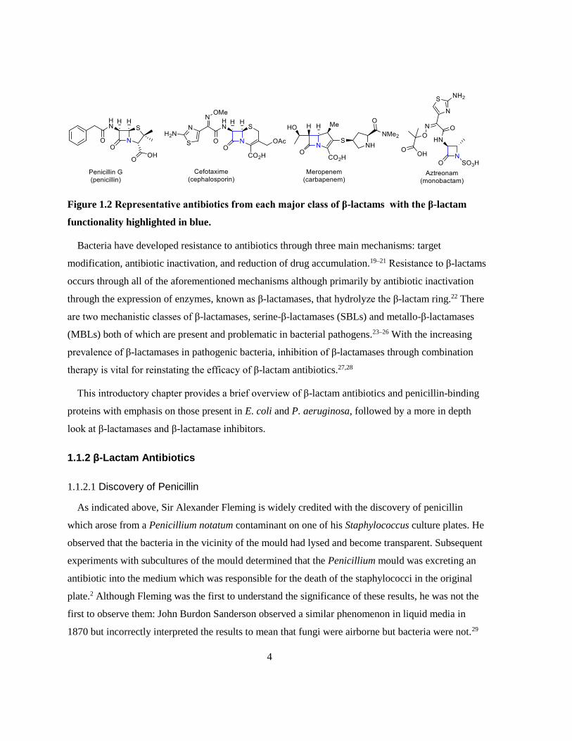
Figure 1.2 Representative antibiotics from each major class of β-lactams with the β-lactam
functionality highlighted in blue.
Bacteria have developed resistance to antibiotics through three main mechanisms: target
modification, antibiotic inactivation, and reduction of drug accumulation.19–21 Resistance to β-lactams
occurs through all of the aforementioned mechanisms although primarily by antibiotic inactivation
through the expression of enzymes, known as β-lactamases, that hydrolyze the β-lactam ring.22 There
are two mechanistic classes of β-lactamases, serine-β-lactamases (SBLs) and metallo-β-lactamases
(MBLs) both of which are present and problematic in bacterial pathogens.23–26 With the increasing
prevalence of β-lactamases in pathogenic bacteria, inhibition of β-lactamases through combination
therapy is vital for reinstating the efficacy of β-lactam antibiotics.27,28
This introductory chapter provides a brief overview of β-lactam antibiotics and penicillin-binding
proteins with emphasis on those present in E. coli and P. aeruginosa, followed by a more in depth
look at β-lactamases and β-lactamase inhibitors.
1.1.2 β-Lactam Antibiotics
1.1.2.1 Discovery of Penicillin
As indicated above, Sir Alexander Fleming is widely credited with the discovery of penicillin
which arose from a Penicillium notatum contaminant on one of his Staphylococcus culture plates. He
observed that the bacteria in the vicinity of the mould had lysed and become transparent. Subsequent
experiments with subcultures of the mould determined that the Penicillium mould was excreting an
antibiotic into the medium which was responsible for the death of the staphylococci in the original
plate.2 Although Fleming was the first to understand the significance of these results, he was not the
first to observe them: John Burdon Sanderson observed a similar phenomenon in liquid media in
1870 but incorrectly interpreted the results to mean that fungi were airborne but bacteria were not.29

5
Inspired by Sanderson’s observations, Joseph Lister, John Tyndall, and Thomas Henry Huxley also
explored the antibacterial properties of Penicillium spp. through the 1870’s.30 Lister disproved
Sanderson’s hypothesis that bacteria could not be airborne and suggested that Penicillium spp. make
liquid media less favourable for bacterial growth.31 Tyndall then suggested that they made liquid
media unfavourable by covering the surface of the media, thereby depriving the bacteria of oxygen.32
Huxley disproved Tyndall’s hypothesis, showing that oxygen deprivation was not the cause of the
antibacterial effect but rather the mould in solution was making the media unfavourable for growth.33
Throughout the late-1800’s and early-1900’s, several scientists noted the antibacterial properties of
moulds and the abilities of extracts of moulds to be used as treatments for bacterial infections, but
none of them had identified the causative agent for these effects.34 It was Fleming’s work in 1928 that
finally established that the antibacterial effect of this mould was being caused be a compound
excreted from Penicillium notatum which was not excreted from other moulds.2
In the summary of Fleming’s 1929 paper, he suggests that penicillin could be an effective antibiotic
with clinical applications, although he did not do any studies on infected patients (human or animal).2
The clinical usefulness of penicillin was established by Chain and Florey in their 1940 paper where
they showed the efficacy of penicillin in treating mice infected with Streptococcus pyrogenes,
Staphylococcus aureus, or Clostridium septique (modern name: Clostridium septicuma).4 Human
trials of penicillin began in January 1941 when a penicillin infusion was injected into a terminally ill
breast cancer patient to determine toxicity in humans – she showed no adverse effects initially and
within a few hours developed a high temperature and died. It was determined that the cause of the
increased temperature was an impurity, not penicillin itself. The first patient treated with penicillin
was a policeman with combined staphylococcal and streptococcal septicemia who was “desperately
and pathetically ill”. He showed improvement within the first day and was almost healthy after five
days. Unfortunately, they had exhausted Florey’s supply of penicillin and could not treat the man any
further – he died a month later from the infection. After shifting the trials towards children (less
a The strain referred to in Chain et al. 19404 is denoted “Cl. septique Nat. coll. type-cultures No. 458”. The
NCTC entry for No. 458 is Bordetella bronchiseptica which does not cause the gas gangrene described by
Chain et al..553 It is probable that this is a typographical error in which the NCTC entry should be No. 547 (548
does not exist) which is Clostridium septicum (formerly Vibrio septique), the strain used by Henderson and
Gorer in the paper referenced by Chain et al. in the methods section.554 The change in nomenclature from V.
septique to C. septicum appears to have occurred in 1935 and references to C. septique occur throughout the
1940’s (referring to NCTC No. 547).555–558

6
penicillin was required for smaller patients), there was a string of successes in which the patient was
saved or only died because the treatment was too late.3 Fleming, Chain, and Florey were awarded a
Nobel Prize in Physiology or Medicine in 1945 for their contributions to the discovery and
development of penicillin.
1.1.2.2 Modern β-Lactams
In the decades that followed Fleming’s discovery of penicillin, a host of other antibiotics were
discovered and developed.35 The next leap forward in β-lactam antibiotics was discovery of
cephalosprin C in 1948 from a strain of Cephalosporium (modern name: Acermonium) fungus by
Guiseppe Brotzu.36 The natural cephalosporins had modest antibacterial activity but had a stable ring
system that made them ideal for chemical modification which lead to the four generations of semi-
synthetic cephalosporins which were much more potent than their natural counterparts.34,37 In 1976, a
new family of β-lactams were discovered in extracts from Streptomyces olivaceus, the olivanic acid
family carbapenems.38 Another family of carbapenems, the thienamycin carbapenems, was
discovered around the same time from Streptomyces cattleya; although, neither of these families of
natural carbapenems were stable enough for clinical use.39,40 A semi-synthetic effort in the late-1970’s
by scientists at Merck yielded imipenem (Figure 1.3), a carbapenem that had a broader spectrum of
activity than any of the cephalosporins, and was resistant to hydrolysis by many of the enzymes that
hydrolyze penicillins and cephalosporins.40–42 Imipenem, like thienamycin, is susceptible to
inactivation by renal dehydropeptidase I (DHP-I) and has to be administered in combination with
cilastatin, a DHP-I inhibitor, to prevent degradation and the formation of nephrotoxic metabolites.40
Figure 1.3 Chemical structures of imipenem and cilastatin.
To date, there are four major structural classes of β-lactam antibiotics: penicillins (penams),
cephalosporins (cephems), carbapenems, and monobactams. Other β-lactam classes include penems,

7
cephams, penamycins, oxapenems, cephamycins, oxacephems, oxacephamycins, and carbacephems
(Figure 1.4).34,43
Figure 1.4 Core structures of β-lactam antibiotic families.
While antibiotics from all four major classes of β-lactams are still used clinically, many pathogens
have developed resistance to some or all of them.
1.1.3 Bacterial Resistance
In human medicine, antimicrobials are one of the most prescribed drugs, although the Centers for
Disease Control (CDC) has estimated that as many as 50 % of these prescriptions are unnecessary.44
As a result of this systemic misuse through over-prescribing, not abiding by treatment regimens, and
addition to feed stocks for food animals; bacteria have developed resistance to all of the antibiotic
classes mentioned in Table 1.1.35 The first report of the incidence of bacterial resistance to antibiotics
was in 1940 when enzyme (β-lactamase)-mediated resistance to penicillin was observed in E. coli.45 It
has been suggested that the existence of β-lactamases pre-dates the antibiotic era by millions of years
since penicillin is a naturally occurring antibiotic used in signaling and potentially germ warfare
among microbes.46,47 In response to spreading resistance, the use of some antibiotics, such as third-
generation cephalosporins and carbapenems, has been restricted to hospitals.48
Bacteria can acquire resistance through random chromosomal mutation or horizontal gene transfer.
These adaptations lead to resistance through one of three general mechanisms: target modification,

8
compound modification, or accessibility. Target modification can occur through mutation or
enzymatic alteration, target bypass or replacement, or target protection through the expression of
molecules that compete for drug binding. Compound modification often involves the expression of an
enzyme or group of enzymes that chemically modify the antibiotic compound rendering it inactive
against its target. Resistance through moderating accessibility is often the result of decreasing
permeability of the outer membrane by reducing the expression of outer membrane porins (uptake
proteins), increasing the expression of efflux pumps, or the formation of biofilms.19–21
Resistance to β-lactams can occur through several of the mechanisms listed above. Methicillin-
resistant Staphylococcus aureus (MRSA) acquires its β-lactam resistance through target replacement
with the expression of PBP2a, a PBP that is not present in wild type S. aureus and is not inactivated
by most β-lactam antibiotics.49 Imipenem resistance in Pseudomonas aeruginosa is known to occur
through decreased production of OprD, an outer membrane porin responsible for the uptake of basic
amino acids, as well as through the increased expression of multidrug efflux pumps.50–52 The
predominant mechanism of β-lactam resistance occurs through the production of β-lactamases,
enzymes that hydrolyze the β-lactam ring thereby inactivating the antibiotic as demonstrated in
Scheme 1.1.
Scheme 1.1 Generalized hydrolysis of β-lactam by a β-lactamase.
There are approximately 2000 currently known β-lactamases, of which, several hundred are
carbapenemases – enzymes capable of hydrolytically inactivating carbapenems.53–55 The most
problematic carbapenemases are the metallo-β-lactamases (MBLs) which are uninhibited by all
clinically available β-lactamase inhibitors. Additionally, MBLs are often carried on mobile genetic
elements with multiple β-lactamases and resistance factors for other antibiotics.56 The increasing
global prevalence of MBL-expressing pathogens has increased the need for a clinically viable MBL
inhibitor.

9
1.2 Penicillin Binding Proteins
1.2.1 Bacterial Cell Wall Structure
The cellular envelopes of bacteria are responsible for creating a stable barrier between the cell and
its environment that is rigid enough to maintain cell shape and resist lysis in dilute solutions while
being permeable enough to transport nutrients in and waste out. There are three types of cellular
envelope: Gram-positive, Gram-negative, and atypical which are broadly classified by Gram staining.
The cellular envelope of Gram-positive bacteria consists of an inner cell membrane coated in a thick
layer of peptidoglycan – the rigid component of bacterial cell walls. Gram-negative bacteria have a
similar inner membrane coated in a much thinner layer of peptidoglycan which is covered by another
membrane layer referred to as the outer membrane. Both Gram positive and Gram negative cellular
envelopes have a space between the inner membrane and peptidoglycan known as the periplasm that
contains numerous proteins in a reducing environment.57 Atypical bacteria lack peptidoglycan either
as part of their native state (eg. Mycoplasma spp.) or due to degradation of their peptidoglycan (L-
forms).58Atypical bacteria will not be discussed further as they do not respond to β-lactam antibiotics
due to their lack of peptidoglycan; however, more information on this topic can be found in
reviews.59–61
Figure 1.5 Gram positive and Gram negative cellular envelope composition. 62,63 Membrane
graphics were generated using ChemDraw Pro v. 17.0.
The bacterial cell wall is constructed using several enzymes, key among them, the penicillin
binding proteins (PBPs). These proteins are responsible for the synthesis and recycling of
peptidoglycan as well as some regulation of its size and the extent of crosslinking. As should be
expected, these proteins are primarily located on the periplasmic face of the inner (cytosolic)

10
membrane in complexes with other PBPs and proteins involved in cell shape maintenance and
septation.64 Some of the smaller PBPs such as E. coli PBP 4 are associated with peptidoglycan instead
of being membrane bound.65 The localization and function of PBPs as well as other proteins involved
in cell shape and septation in rod shaped bacteria has been reviewed by den Blaauwen.64
1.2.2 PBP Functional and Structural Classification
Historically, penicillin-binding proteins were defined as membrane associated proteins that would
bind penicillin G. Much of the early work in this field was performed using gel based assays with 14C
isotopically labelled penicillin G, and as such, the nomenclature for PBPs has been broadly defined
by molecular weight where the heaviest PBP was denoted PBP 1 and lighter ones followed in
sequence.66 An appreciable difference in the necessity and functionality of PBPs was observed and
they were split into two groups: high molecular mass (HMM) and low molecular mass (LMM). The
naming of PBPs by molecular mass has proven inconvenient as different organisms express different
quantities of PBPs and functionality is not strictly correlated with mass; thus PBP 2 from one
organism may have a different function than PBP 2 from another organism.
1.2.2.1 Reactions Catalyzed by PBPs
Peptidoglycan (or murein) is a highly crosslinked polymer made from the precursor, lipid II, which
is synthesized in the cytoplasm before being flipped into the periplasm using a flippase. Lipid II is
composed of three major groups: the membrane anchor, the glycan backbone, and the pentapeptide,
as shown in Figure 1.6. The membrane anchor is embedded in the inner membrane, keeping this
substrate in close proximity to the HMM PBPs which are responsible for its polymerization as they
are also membrane bound. The polymerization of lipid II into peptidoglycan is achieved by sequential
additions of the N-acetylmuramic acid-N-acetylglucosamine (NAM-NAG) disaccharide portion of the
molecule to form a glycan backbone. Finally, the pentapeptide is the portion of peptidoglycan that is
crosslinked to increase the structural integrity of the cell wall. In the majority of Gram negative
bacteria, the sequence of the pentapeptide is L-Ala-γ-D-Glu-m-DAP-D-Ala-D-Ala (m-DAP: meso-
diaminopimelic acid).67
11
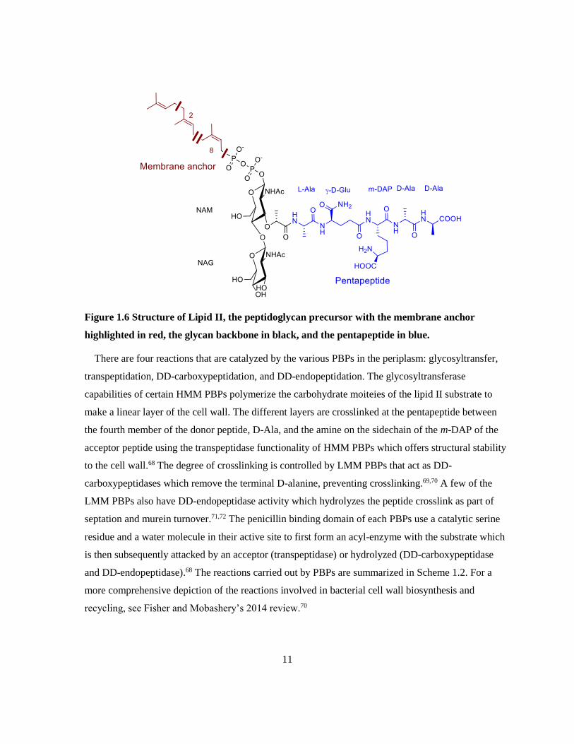
Figure 1.6 Structure of Lipid II, the peptidoglycan precursor with the membrane anchor
highlighted in red, the glycan backbone in black, and the pentapeptide in blue.
There are four reactions that are catalyzed by the various PBPs in the periplasm: glycosyltransfer,
transpeptidation, DD-carboxypeptidation, and DD-endopeptidation. The glycosyltransferase
capabilities of certain HMM PBPs polymerize the carbohydrate moiteies of the lipid II substrate to
make a linear layer of the cell wall. The different layers are crosslinked at the pentapeptide between
the fourth member of the donor peptide, D-Ala, and the amine on the sidechain of the m-DAP of the
acceptor peptide using the transpeptidase functionality of HMM PBPs which offers structural stability
to the cell wall.68 The degree of crosslinking is controlled by LMM PBPs that act as DD-
carboxypeptidases which remove the terminal D-alanine, preventing crosslinking.69,70 A few of the
LMM PBPs also have DD-endopeptidase activity which hydrolyzes the peptide crosslink as part of
septation and murein turnover.71,72 The penicillin binding domain of each PBPs use a catalytic serine
residue and a water molecule in their active site to first form an acyl-enzyme with the substrate which
is then subsequently attacked by an acceptor (transpeptidase) or hydrolyzed (DD-carboxypeptidase
and DD-endopeptidase).68 The reactions carried out by PBPs are summarized in Scheme 1.2. For a
more comprehensive depiction of the reactions involved in bacterial cell wall biosynthesis and
recycling, see Fisher and Mobashery’s 2014 review.70

12
Scheme 1.2 Reactions catalyzed by PBPs: glycosyl transfer, transpeptidation,
carboxypeptidation, and endopeptidation. 68,70,73–75

13
The critical role of PBPs in the synthesis of bacterial cell walls makes them an ideal target for
antibiotics, namely β-lactams. The inhibition of a PBP by a β-lactam occurs when the active site
serine of the PBP binds and attacks the amide of the β-lactam, forming an acyl enzyme as shown in
Scheme 1.3. Unlike with its natural substrate, the acyl enzyme formed between a PBP and a β-lactam
is very slow to hydrolyze with a half-life of around 10 hr, leading to covalent inactivation of the
PBP.73
Scheme 1.3 Inactivation of PBPs by a β-lactam.
1.2.2.2 Classification of PBPs
A classification system for the PBPs was developed by Goffin and Ghuysen based on the
nomenclature of E. coli PBPs which focused on the functionality and amino acid sequence of HMM
PBPs.68 This system has been further refined by Sauvage et al. to include classification of LMM PBPs
and PBPs from Actinomycetes and Cyanobacteria using structure and function as classification
determinants.18 For simplicity, this section will only cover PBPs in Gram positive and Gram negative
bacterial pathogens with most attention being paid to E. coli, P. aeruginosa, B. subtilis, S. aureus, and
S. pneumoniae. Several excellent reviews are recommended for more detailed information on the
structural and functional classification of PBPs.18,68,73,76
The high molecular mass PBPs are multimodular, membrane bound proteins that can be classified
as one of two classes: Class A consists of any PBP with a transglycosylase functionality at the N-
terminal and Class B consists of PBPs with a C-terminal transpeptidase module and an N-terminal
module that is involved in the cell cycle. The nomenclature of these two classes are based upon the
numbering of E. coli PBPs for Gram negative organisms, while B. subtilis, E. faecium, and S.
pneumoniae were used in combination to describe the PBPs for Gram positive organisms.68 B. subtilis

14
will be used where possible as the example for describing PBPs classification in Gram positive
organisms as it contains the widest variety for a Gram positive organism.
Class A PBPs are further divided into 7 subclasses based on sequence analysis of the pencillin
binding (PB) cores and are denoted A1-A7. Subclasses A1 and A2 contain the major bifunctional
transpeptidases/transglycosylases in Gram negative organisms of which E. coli PBP 1a and 1b are the
prototypes respectively.77–80 The bifunctional transpeptidases/transglycosylases in Gram positive
organisms comprise subclasses A3, A4, and A5 as exemplified by B. subtilis PBPs 1, 2c, and 4
respectively. Subclass A6 contains the “outliers” such as E. coli PBP 1c which contains both modules
characteristic of Class A PBPs; however, the PB domain is very substrate specific and does not seem
to act as a transpeptidase.81,82 Finally, subclass A7, which was added by Sauvage, contains
monofunctional glycosyltransferases such as MgtA from E. coli.83,84 It is worth noting that subclass
A7 enzymes do not appear in traditional gel based assays as they lack a PB core and that subclass A6
enzymes may not appear under certain conditions due to their evolutionarily divergent PB core.
The class B PBPs are multimodular like the class A PBPs with a C-terminal penicillin-binding
transpeptidase module; however, the non-penicillin binding (nPB) module in class B PBPs is
implicated in cellular morphology and division rather than transglycosylation.18,64 Five subclasses of
class B PBPs are present in Gram positive and Gram negative organisms: B1-B5, defined by Goffin
and Ghuysen using sequence alignment.68 The subclass B1 PBPs are most commonly found in Gram
positive organisms and are characterized by their low affinity for β-lactams, most famously, PBP 2a
from methicillin resistant S. aureus. B1 PBPs have been thoroughly reviewed by Zapun et al..85
Subclass B2 PBPs are predominantly found in Gram negatives, such as E. coli PBP 2, and are
associated with rod shaped bacteria as the nPB module is specific to the elongase complex.64
Consequently, selective inhibition of subclass B2 PBPs causes the formation of spherical cells.66,86
The B3 subclass PBPs, such as E. coli PBP 3 are part of the divisome, and their selective inhibition
causes filamentation.64,86 The Gram positive counterpart to B3 PBPs are B4 PBPs, such as B. subtilis
PBP 2b, in that they contribute to cell division.87,88 The B5 PBPs are Gram positive PBPs such as B.
subtilis PBP 2a, most of which have no known function for the nPB module.68,89,90 B. subtilis PBP 2a
is known to be involved in maintaining rod shape along with PBP H, another B5 PBP.91 This function
in elongation cannot be a sole characteristic of B5 PBPs as many cocci express these.18
15
The final class of PBPs are the Class C PBP, commonly referred to as the low molecular mass
(LMM) or non-essential PBPs. Class C PBPs are monofunctional PBPs that play a role in
peptidoglycan regulation and recycling through carboxypeptidase and/or endopeptidase
mechanisms.18,76 As with Class A and Class B PBPs, the nomenclature of Class C PBPs is based on
the separation pattern of E. coli PBPs; however, there is no differentiation between Gram positive and
Gram negative organisms in this class. The Class C PBPs are divided into three types: type 4, type 5,
and type 7.18 Type 4 PBPs, whose archetype is PBP 4 in E. coli, function as both a DD-
carboxypeptidase and a DD-endopeptidase and have been implicated in maintaining normal cell
morphology and in recycling the cell wall.65,76,92,93 These PBPs do not contain a transmembrane helix
and are thought to be loosely associated with the cytoplasmic membrane (Gram positive), the inner
leaflet of the outer membrane (Gram negative) or directly with peptidoglycan since they overproduce
in the soluble form for E. coli and can be washed off B. subtilis with 1 M KCl.18,65,94–96 In Neisseria
gonorrhoeae, the type 4 PBP, PBP 3, has been found to be associated with both the outer membrane
and peptidoglycan.97,98 Type 5 PBPs are often the most abundant PBPs in cells and are the strict DD-
carboxypeptidases.99–101 Functionally, type 5 PBPs have been implicated in normal septum formation
and in the maintenance of normal cell diameter.92,102,103 In E. coli, this type is represented by PBP 5
and PBP 6 of which PBP 5 performs the normal regulatory functions of this type, and PBP 6 is
involved in the onset of stationary phase.69,104,105 The final type of Class C PBPs is type 7 which are
structurally similar to the penicillin binding domain of type 5 PBPs and as such exhibit the same
carboxypeptidase/endopeptidase activity but are largely absent in Gram positive organisms.18 Much
like type 4 PBPs, these are loosely membrane associated and, as of yet, their function in cell cycle or
morphological regulation is unknown.101,106 In E. coli, this class is represented by PBP 7 and its
proteolytic product, PBP 8.107
1.2.3 PBP Expression in Bacterial Strains
In this study, assays of both E. coli and P. aeruginosa penicillin binding proteins were employed,
so it is important to discuss which PBPs are present in these organisms, how they compare, and to
examine any controversy in the literature. Much of the comparison of E. coli and P. aeruginosa PBPs
was performed by Noguchi et al. in 1979; however, this study was limited to the PBPs that react with
radiolabeled benzylpenicillin and remain on an SDS PAGE gel.108 Using binding kinetics, thermal
sensitivity, correlations in β-lactam specificity, and morphological impacts as criteria, they
determined that E. coli PBPs 1a, 1b, 2, 3, 4, and 5 correspond with P. aeruginosa PBPs 1b, 1a, 2, 3, 4,

16
and 5 respectively. It was also noted that expression of PBPs may vary between P. aeruginosa strains
with PBP 2 being nearly invisible on the gels of certain strains, and PBP 3x appearing on others.108
Recently, P. aeruginosa PBPs have been studied individually, bringing the understanding of the
function, and in some cases structure, of these proteins to the level of E. coli PBPs. Comparative
genetic sequence alignment of P. aeruginosa PBP 1a with PBP 1a and 1b from E. coli determined
that P. aeruginosa PBP 1a most closely correlates with E. coli PBP 1a, not 1b as was previously
suggested.108,109 Several studies have confirmed that PBP 2, 3, 4, and 5 from P. aeruginosa have the
same function and similar structures as their E. coli counterparts.99,110–113 Gene names can be used as a
clearer way to compare PBPs across species as they more closely correlate structure and function than
electrophoretic mobility, although some inconsistencies in naming still occur. Table 1.2 is a
compilation of the PBPs present in E. coli and P. aeruginosa with their respective gene names. It is
worth noting that there is no relationship between the proteins encoded by pbpC in E. coli and P.
aeruginosa, it is seemingly just an unfortunate naming coincidence.81,114
Table 1.2 Penicillin binding proteins present in E. coli and P. aeruginosa 76,111
E. coli Protein Genes P aeruginosa Protein
PBP 1a ponA PBP 1a
PBP 1b mrcB PBP 1b
PBP 1c pbpC†
PBP 2 pbpA PBP 2
PBP 3 ftsI PBP 3
pbpC† PBP 3x
PBP 4 dacB PBP 4
PBP 5 dacA
PBP 6 dacC PBP 5/6
PBP 6b dacD
PBP 7/8 pbpG PBP 7
†The genes denoted as pbpC are not homologs
17
Aside from the naming coincidence with the pbpC genes in E. coli and P. aeruginosa, there is
controversy in the literature with respect to the names of the high molecular weight PBPs in P.
aeruginosa. The first report of P. aeruginosa PBPs labelled them as 1a, 1b, 2, 3, 3’, 4, 4’, 5, and 7/8,
some of which are only conditionally present, notably 3’ which also goes by the names 3x and
3a.68,76,108,114,115 While there is some differences between the original labelling of the Class B and C
PBPs and the current labelling, the nomenclature of these PBPs has become consistent across the
literature. The point of contention seems to be whether the two Class A PBPs are 1a and 1b or 1b and
1c. The origin of the 1b/c nomenclature seems to be a publication by Moyá et al. in 2010, in which
four classic papers about P. aeruginosa PBPs are referenced in the introduction with respect to the
PBPs present in P. aeruginosa; none of these papers mention PBP 1c and all use the 1a/b
nomenclature.18,108,116–118 Since the paper by Moyá, several other papers have used the 1b/c
nomenclature, mostly in reference to the proteins inhibited by ceftolozane (which is the subject of
Moyá et al. 2010)119–121 Additionally, PBPs 1b and 1c are named in the mode of action description of
ceftolozane on Drugbank; although, PBP 1c is not listed in the targets section.122 Papers describing
new experiments on P. aeruginosa PBPs since the release of Moyá et al. 2010 tend to use the 1a/b
nomenclature, including another paper by Moyá, which may suggest that the 2010 paper had a
typographical error that has been carried through later literature on the subject.113,116,123,124 Putting this
to rest, Dhar et al. released a review in 2018 that included a survey of the P. aeruginosa genome
database, and found no genes corresponding to PBP 1c.76 For this work, the major PBPs in P.
aeruginosa will be labelled as PBPs 1a, 1b, 2, 3, 4, 5/6 according to the most common nomenclature,
excluding those that are not visible.
1.2.4 Role of PBPs in Bacterial Resistance to β-Lactams
Penicillin-binding proteins contribute to bacterial resistance to β-lactams through either being
inherently resistant to inhibition by β-lactams or through inducing the production of β-lactamases
upon inhibition. The most well-known of the β-lactam resistant PBPs is PBP 2a from Staphylococcus
aureus, a Class B PBP that confers methicillin resistance.125,126 PBP 2a is encoded by the mecA gene
which is found in a mobile genetic element that integrates into the chromosome of Staphylococci and
whose origin appears to be Staphyolococcus fleurettii.127,128 β-Lactam resistance by PBP 2a
acquisition is primarily due to its poor affinity for β-lactams allowing it to compensate for other
inhibited transpeptidases.129–133Additionally, PBP 2a accounts for 43% of expressed PBPs in
methicillin resistant S. aureus cells while the number of other PBPs expressed per cell remains

18
constant.134 Similar β-lactam resistant PBPs exist in other bacteria and are covered in several
excellent reviews.73,85,135
In bacteria that do not produce inherently resistant PBPs, the inhibition or mutation of certain
native PBPs has been shown to up-regulate the production of Class C chromosomal β-lactamases
such as AmpC. Chromosomal ampC genes has been found in many Gram negative pathogens
including E. coli, P. aeruginosa, K. pneumoniae, E. cloacae, and Serratia marcescens and can be
constitutively expressed (as in E. coli) or induced by β-lactams.136–139 β-Lactam induced production of
the β-lactamese AmpC occurs primarily through one of two pathways: AmpR regulation or two
component CreBC(D) regulation both of which are thoroughly reviewed by Zeng and Lin.140 The
AmpR regulation pathway involves the inhibition of a LMM PBP (usually PBP 4 or 5) which shifts
the equilibrium of peptidoglycan recycling, releasing NAG-anhydro-NAM-pentapeptides which are
transported into the cytoplasm through the AmpG transporter.113,141–144 Once in the cytoplasm, the
NAG is removed from the NAG-anhydro-NAM-pentapeptides by NagZ and the peptide portion is
shortened by AmpD, creating a molecule that interacts with the AmpR regulator, derepressing AmpC
production.139,143–146 The CreBC(D) system has been shown to regulate the expression of OXA-12 and
other chromosomal β-lactamases in Aeromonas jandaei and to interact with AmpR regulation in PBP
4 mutants of P. aeruginosa to confer higher β-lactam resistance than the AmpR pathway
alone.144,147,148 The CreBC(D) two component regulation system responds to PBP 4 inhibition or
mutation in P. aeruginosa through an unknown signal causing the CreC inner membrane histidine
kinase to phosphorylate CreB, the response regulator, which induces CreD overexpression in P.
aeruginosa or induces β-lactamase production in A. jandaei.144,147–149 It is not well understood how
CreD overexpression influences β-lactam resistance, but it is known to contribute to cell envelope
stability and biofilm formation.148,150
Despite the importance of PBPs in conferring and inducing β-lactam resistance, these tend to be
narrow spectrum resistance mechanisms, making the expression of extended spectrum β-lactamases a
more important resistance mechanism.
1.3 β-Lactamases
β-Lactamases are periplasmic enzymes that efficiently hydrolyze β-lactam antibiotics, conferring
resistance to the bacterium. There are two general mechanisms by which these enzymes hydrolyze β-
19
lactams: using a catalytic active site serine or zinc ions. The serine β-lactamases (SBLs) react with β-
lactams in a mechanism similar to that of PBPs, except they are capable of rapidly hydrolyzing the
acyl enzyme. A common misconception is that β-lactamases evolved from PBPs as a response to the
use of β-lactams as antibiotics in humans. It is thought that β-lactamases and PBPs evolved from a
common DD-peptidase ancestor hundreds of millions of years ago and clinical use of β-lactam
antibiotics has just selected for resistance, making it more prevalent.151,152 Additionally, the use of β-
lactams in a clinical setting had been shown to accelerate the evolution of β-lactamases with about
170 β-lactamases having been identified by 1997 and around 2000 having been identified today.53–
55,152
1.3.1 Role of β-Lactamases in Bacterial Resistance to β-Lactams
The first observation of a β-lactamase capable of conferring resistance to a bacterium was reported
in 1940 by Abraham and Chain, although they state in their letter that Fleming had also observed the
resistance due to these enzymes in his 1929 paper detailing the discovery of penicillin.2,45 Early β-
lactamases were referred to as penicillinases or cephalosporinases depending upon their ability to
hydrolyze penicillins or cephalosporins respectively. When semisynthetic penicillins were introduced
in the 1970’s as less β-lactamase susceptible alternatives to natural product penicillins, they were
lauded for their efficacy against highly resistant pathogens such as Pseudomonas spp., although
resistance was observed in clinical trials.34,153 Likewise, the third-generation of semi-synthetic
cephalosporins, such as ceftazidime in 1979, were thought to be highly resistant to hydrolysis by β-
lactamases but some Gram-negative pathogens developed resistance in clinical trials.154,155 Many of
first identified β-lactamases had relatively narrow-spectrum activity against either penicillins or
cephalosporins and the few that were capable of hydrolyzing the more stable β-lactams were
chromosomally encoded.156,157
In the early 1980’s, enzymes were discovered on transferrable plasmids in Klebsiella spp. isolates
that were capable of hydrolyzing many of the available β-lactams which represented the first of the
extended spectrum β-lactamases.156 In the 1980’s, extended-spectrum β-lactamases (ESBLs) were
considered those capable of efficiently hydrolyzing common semi-synthetic penicillins and first-
generation cephalosporins but exhibiting poor activity against third-generation cephalosporins and
monobactams.156 The definition of the term ESBL has evolved with the advent of new β-lactams and

20
the identification of β-lactamases with even broader spectrum activity such that it now includes only
enzymes that are, at least, capable of conferring resistance to penicillins, first-, second- and third-
generation cephalosporins, and monobactams.158 The β-lactam therapy of choice for ESBL-producers
has been carbapenems for the last few decades, although ESBLs that also hydrolyze carbapenems
(carbapenemases, e.g. KPC-2) and metallo-β-lactamases have rendered this option less effective.159
Recently, mobile genetic elements coding for multiple β-lactamases as well as resistance enzymes
for other classes of antibiotic have been discovered in Gram negative pathogens.160,161 One such K.
pneumoniae isolate harbouring 4 resistance plasmids was found to be carrying genes for 6 β-
lactamases, conveying resistance to all β-lactams, as well as genes coding for resistance to gentamicin
(and related compounds), streptomycin, sulfonamides, aminoglycosides, trimethoprim,
quinolones/fluoroquinolones, macrolides, and fosfomycin in addition to 9 efflux pumps with a wide
range of specificity.162 Multidrug resistant bacteria are becoming more and more prevalent and have
moved from being associated only with nosocomial infections to community-acquired infections as
well.163 The Infectious Disease Society of America has declared that antimicrobial resistance is “one
of the greatest threats to human health worldwide”, and with over 50% of prescribed antibiotics being
β-lactams, combatting β-lactam resistance is of the utmost importance.16,17,164
1.3.2 Classification of β-Lactamases
The earliest nomenclature for the enzymes that were capable of hydrolyzing penicillins was
“penicillinase” in accordance with the naming conventions of the time.45 The discovery of
cephalosporins in the mid-1950’s and the enzymes capable of hydrolyzing them brought about a new
term – cephalosporinase.165,166 These functional names failed to accurately categorize the enzymes
that they were describing as they only indicated whether or not that enzyme could hydrolyze that type
of substrate, omitting enzymes that hydrolyze both cephalosporins and penicillins. Additionally, there
was no distinction between broad spectrum enzymes that could hydrolyze every member of a class of
β-lactams and a narrow spectrum one that could only hydrolyze a few members of a class (e.g. Only
first-generation cephalosporins, but not second-generation).
After some early efforts to establish a classification system,167–169 Richmond and Sykes came up
with the first widely accepted classification scheme in 1973.157 This scheme split the known β-
lactamases into five classes denoted by the Roman numerals I-V according to their substrate

21
specificity as well as their sensitivity to inhibition by both cloxacillin and p-chloromercuribenzoate as
illustrated in Table 1.3.157 Unlike the previous nomenclature, this scheme allows for classification of
β-lactamases that can hydrolyze both cephalosporins and penicillins and uses additional information
such as the susceptibility to inhibitors to further subdivide. The classes proposed by Richmond and
Sykes were subdivided into lettered subgroups according to their relative activities with six
substrates.157
Table 1.3 Richmond and Sykes functional classification scheme for β-lactamases. 157
Sensitivity to Inhibition
Class Preferred Substrate Cloxacillin p-chloromercuribenzoate
I cephalosporin S R
II penicillin S R
III cephalosporin/penicillin S R
IV cephalosporin/penicillin R S
V cephalosporin/penicillin R R
S = sensitive to inhibition, R = resistant to inhibition
While the Richmond and Sykes classification system was adequate for the time, it introduced
confusion since Roman numerals were also used in the names of β-lactamases to distinguish between
two β-lactamases produced by the same organism (e.g. BcI and BcII from Bacillus cereus). Ambler
proposed a similification of the classification of β-lactamases in 1980 based on their structure and
changed designations from Roman numerals to letters. The β-lactamases analysed by Ambler fell into
two groups: Class A and Class B which used an active site serine residue and divalent zinc ions
respectively to perform the hydrolysis.170 The serine-β-lactamases (SBLs) studied by Amber: PC1,
BcI, TEM-1, and B. licheniformis 749/C, had a high sequence homology and thus did not require
further division although Ambler addressed the likelihood of other classes existing.170 Another
molecular class, Class C, was proposed by Jaurin and Grundström in 1981 to encompass
chromosomally encoded SBLs from Enterobacteriaceae such as AmpC which had low sequence
homology with the class A β-lactamases.171 The final molecular class, Class D, was proposed in 1987
by Ouelette et al. on the basis of the lack of sequence homology between the OXA-type SBLs and the
Class A and C SBLs.172

22
Table 1.4 Functional and molecular classification of β-lactamases. 173,174
Bush-
Jacoby Ambler
Distinctive
substrate(s)
Inhibited by Characteristics
Representative
enzymes Clav/Tazo EDTA
1 C Cephalosporins - - Greater hydrolysis of
cephalosporins than PenG;
hydrolyze cephamycins
E. coli AmpC, P99,
ACT-1, CMY-2,
MIR-1, DHA-1
1e C Cephalosporins - - Increased hydrolysis of
ceftazidime and often other
oxyimino-β-lactams
GC-1, CMY-10
2a A Penicillins + - Greater hydrolysis of PenG
than cephalosporins
PC1, BcI
2b A Penicillins, early
cephalosporins
+ - Similar hydrolysis of PenG
and cephalosporins
TEM-1, SHV-1
2be A Extended-spectrum
cephalosporins
+ - Increased hydrolysis of
oxyimino-β-lactams
(cefotaxime, ceftazidime,
cefepime)
CTX-M-15, SHV-2A,
SHV-12, TEM-3
2br A Penicillins - - Resistance to clav/sul/tazo TEM-30, SHV-10
2ber A Extended-spectrum
cephalosporins,
monobactams
- - Increased hydrolysis of
oxyimino-β-lactams
combined with resistance to
clav/sul/tazo
TEM-50
2c A Carbenicillin + - Increased hydrolysis of
carbenicillin
PSE-1, CARB-3
2ce A Carbenicillin,
cefepime
+ - Increased hydrolysis of
carbenicillin, cefepime, and
cefpirome
RTG-4
2d D Cloxacillin ± - Increased hydrolysis of
cloxacillin and oxacillin
OXA-1, OXA-10
2de D Extended-spectrum
cephalosporins
± - Hydrolyzes cloxacillin or
oxacillin and oxyimino-β-
lactams
OXA-11, OXA-45
2df D Carbapenems ± - Hydrolyzes cloxacillin or
oxacillin and carbapenems
OXA-23, OXA-48
2e A Extended-spectrum
cephalosporins
+ - Hydrolyzes cephalosporins.
Inhibited by clav but not
aztreonam
CepA, L2
2f A Carbapenems ± - Increased hydrolysis of
carbapenems, oxyimino-β-
lactams, cephamycins
KPC-2, KPC-3, SME-
1, IMI-1
3a B1 Carbapenems - + Broad-spectrum hydrolysis
including carbapenems but
not monobactams
BcII, IMP, NDM,
VIM, SPM-1
B2 Carbapenems - + Broad-spectrum hydrolysis
including carbapenems but
not monobactams
L1, GOB-1, FEZ-1
3b B3 Carbapenems - + Preferential hydrolysis of
carbapenems
CphA, Sfh-1
4 ? Penicillins - - SAR-2
Adapted from 173,174 with representative enzymes focused towards ones used in this work.175–177 clav =
clavulanic acid, tazo = tazobactam, sul = sulbactam, PenG = penicillin G = benzylpenicillin.
23
Some small updates have been made to the Ambler molecular class classification system since the
1980’s including subdividing the Class B’s into three subclasses (B1, B2, and B3) based on the amino
acids used to bind the zincs as well as rearranging the phylogenetic tree of the Class B’s to emphasize
the relationship between B1 and B2.178,179
The Ambler molecular classification scheme, along with its many updates, is the most widely used
classification system to this day because of its simplicity and the limited amount of information
(primary sequence) needed for classification of new β-lactamases. Throughout the late-1980’s and
early-1990’s, Bush and Jacoby championed another functional classification system, culminating in
their 2010 update that divides the β-lactamases into 4 groups (one of which is for “others”) and 17
subgroups based upon their preferred substrate(s) and their inhibition by clavulanic acid/tazobactam
and EDTA.173,174,180–182 While this scheme excels at categorizing β-lactamases according to the finer
details of their function, it is exceedingly complicated and related enzymes can be scattered across
subgroups (e.g. TEM-type β-lactamases are spread across 4 functional subgroups: 2b, 2be, 2br, and
2ber), leading to it’s lack of widespread use.173 For the purposes of this work, the Ambler class will
be referred to as the molecular class and the Bush-Jacoby class will be referred to as the functional
class.
The names of β-lactamases are abbreviations that indicate the source organism (Bc = Bacillus
cereus), the target substrate (IMP = imipenem), the location where a family of β-lactamases was first
discovered (NDM = New Delhi Metallo-β-lactamase), the name of the patient (TEM = Temoneira),
or a combination of thereof (CTX-M = cefotaxime, isolated from Munich). The number that follows
the abbreviation is often assigned sequentially to variants within a family as they are discovered. An
in depth listing of the nomenclature conventions of β-lactamases was compiled by Jacoby in 2006.183
1.3.3 Class A β-Lactamases
When the molecular classes of β-lactamases were first defined, only four Class A SBLs were
identified: the chromosomally encoded PC-1 from S. aureus, BcI from B. cereus, and 749/C from B.
linchiformis as well as the plasmid encoded TEM-1 (R-TEM) from E. coli.170 This class tends to
prefer penicillins as their substrate rather than cephalosporins. Much of the work on this class has
been on the TEM-type Class A SBLs due to their clinical significance as a result of being plasmid
encoded. The mobility of plasmids between bacterial strains and species facilitated the rapid

24
dissemination of TEM-1 from one E. coli isolate in Greece in the early 1960s into other
Entereobacteriaceae, P. aeruginosa, Haemophilus influenzae, and Neisseria gonorrhoeae worldwide
in only a few years.184 To this day, TEM-type β-lactamases are responsible for almost 90% of
ampicillin resistance in Gram negative pathogens.185
Class A SBLs are composed of two globular domains: one composed entirely of α-helices (pink)
and another composed of 5 β-strands (green) surrounded by α-helices (blue) as depicted in Figure 1.7.
The active site sits between the two domains with the β3 strand defining one wall and an Ω-loop
(Arg164-Asp179) as the other wall shown in green and purple respectively in Figure 1.7.186 An
oxyanion hole composed of the backbone NH groups of Ser70 and Ala237 orients the substrates
correctly by binding the carbonyl oxygen of the β-lactam amide.187 The catalytic serine residue,
Ser70, as well as other critical active site residues such as Lys73, Glu166, and Lys234 are invariant
among all Class A SBLs.
Figure 1.7 3-Dimensional structure of Class A SBLs, exemplified by KPC-2 .188 This figure was
generated using UCSF Chimera v. 1.12 from protein databank file 2OV5.
Early studies on the mechanism of SBLs determined that an acyl enzyme was formed and that it
was likely that a serine residue, then referred to as Ser44, was the nucleophile.189–193 In the early
1990’s, X-ray crystallography and site directed mutagenesis were used to further elucidate the
mechanism and the discussion shifted to whether Glu166 or Lys73 was the general base used to
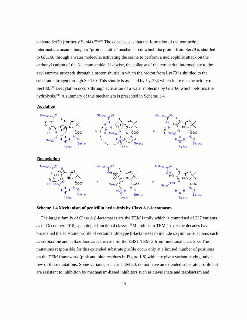
25
activate Ser70 (formerly Ser44).194,195 The consensus is that the formation of the tetrahedral
intermediate occurs though a “proton shuttle” mechanism in which the proton from Ser70 is shuttled
to Glu166 through a water molecule, activating the serine to perform a nucleophilic attack on the
carbonyl carbon of the β-lactam amide. Likewise, the collapse of the tetrahedral intermediate to the
acyl enzyme proceeds through a proton shuttle in which the proton from Lys73 is shuttled to the
substrate nitrogen through Ser130. This shuttle is assisted by Lys234 which increases the acidity of
Ser130.196 Deacylation occurs through activation of a water molecule by Glu166 which peforms the
hydrolysis.194 A summary of this mechanism is presented in Scheme 1.4.
Scheme 1.4 Mechanism of penicillin hydrolysis by Class A β-lactamases.
The largest family of Class A β-lactamases are the TEM family which is comprised of 237 variants
as of December 2018, spanning 4 functional classes.55Mutations to TEM-1 over the decades have
broadened the substrate profile of certain TEM-type β-lactamases to include oxyimino-β-lactams such
as cefotaxime and ceftazidime as is the case for the EBSL TEM-3 from functional class 2be. The
mutations responsible for this extended substrate profile occur only at a limited number of positions
on the TEM framework (pink and blue residues in Figure 1.8) with any given variant having only a
few of these mutations. Some variants, such as TEM-30, do not have an extended substrate profile but
are resistant to inhibition by mechanism-based inhibitors such as clavulanate and tazobactam and

26
represent another functional class, 2br. Like TEM-type ESBLs, the inhibitor resistant (IR) TEMs
contain a couple mutation(s) from a small set (yellow and blue residues in Figure 1.8), some of which
are common in ESBLs as well (blue/bold).184 The frequency and function of the common TEM
mutations has recently been reviewed by Grigorenko et al..197 Complex mutant TEMs such as TEM-
50 have mutations that confer both the extended substrate profile and inhibition resistance, and
represent another functional class, 2ber.173
Figure 1.8 Locations of phenotype changing mutations in TEM-1 that contribute to ESBL
(pink), IR (yellow), or a combination of ESBL and IR phenotypes (blue). The catalytic triad
(STXK) in the active site is highlighted in grey.184 This figure was generated using UCSF
Chimera v. 1.12 from protein databank file 5VHI.
The mutations that confer ESBL character to the TEM variants widen the active site so they can
better accommodate the larger oxyimino side chains of the third generation cephalosporins or
improve their alignment in the active site. Inhibitor resistance is a consequence of mutations that
disrupt the active site through misalignment of Ser70 and Ser130 or disrupting the H-bonding
network around Ser130. This causes decreased turnover and apparent decreased affinity, particularly
with inhibitors such as clavulanic acid, altering the balance between acylation and rearrangement.198

27
The second largest family of Class A SBLs is the SHV-family with 228 variants from primarily the
2b and 2be functional classes with a few 2br variants.55 Unique to this family, the A146V mutationb
in SHV-38 confers limited carbapenemase activity, but is still categorized in the 2be functional
class.199 Unlike the TEM-family β-lactamases which are found in many different Gram negative
pathogens, members of the SHV-family are found primarily in K. pneumoniae and other
Enterobacteriaceae.176
Although the TEM- and SHV-type Class A SBLs are clinically important, they are becoming
overshadowed by another family: the CTX-M’s. Named for their ability to hydrolyze cefotaxime (and
their first identification in Munich, Germany), this family is entirely composed of ESBLs.183,200 The
vast majority of the CTX-M-type SBLs belong to the 2be functional class, although recently, a CTX-
M-variant, CTX-M-190, was discovered in a clinical isolate in China and demonstrates significant
resistance to tazobactam and sulbactam but not clavulanate.201,202 This family of ESBLs has
disseminated worldwide, primarily in Enterobacteriaceae, in large part due to its presence on
transferable plasmids.203 Of the 224 identified CTX-M-variants, CTX-M-15 is the most prevalent in
most parts of the world although CTX-M-14 is most prevalent in Southeast Asia, China, South Korea,
Japan, and Spain. It has been predicted that in the coming years, CTX-M-27 will out-compete the
other CTX-M-variants as it confers increased resistance for ceftazidime compared to CTX-M-14 and
has been shown to be more transmissible in nosocomial E. coli clones compared to CTX-M-15.203
Other Class A ESBLs include: ACI-1, BEL-type, BES-1, CARB-10, CfxA-type, CGA-1, CIA-type,
CphA-type, CSP-1, DES-1, ERP-type, FAR-1, GES-type, KLUC-type, LRG-1, LUT-type, OHIO-1,
OXY-type, PER-type, RAHN-type, SFO-1, SGM-1, SPU-1, TLA-type, and VEB-type.55 As is the
case for the TEM- and SHV-type SBLs, not all members of a family are ESBLs, but the families
listed above include at least one ESBL. Despite the wide diversity of ESBLs, CTX-M-, TEM-, and
SHV-type ESBLs are the most common in clinical pathogens and are consequently the most
important targets for inhibition.185
The hydrolysis of cephalosporins is problematic since cephalosporins represent the single most
prescribed class of antibiotic in Canada but the ESBLs listed above are not capable of hydrolyzing
carbapenems and as such, those infections can be treated with β-lactams.17 In 1996, a patient in North
b The A146V mutation is also present in SHV-168; however, the mature protein is the same as SHV-38.559

28
Carolina presented with a carbapenem resistant K. pneumoniae infection which marked the first
instance of a Class A carbapenemase: KPC-1.204 In 2008, a 73-year-old man in a Toronto-area
hospital presented with the first Canadian case of a KPC-producing infection despite having no
known risk factors (e.g. recent travel to the USA) – he succomed to the infection before an
appropriate therapy could be determined.205 KPC-producing Enterobacteriaceae are now found
worldwide, particularly in the USA, China, South/Central America, the Middle East and
Mediterranean Europe, with KPC-2 and KPC-3 being the most prevalent variants.206,207
Class A carbapenemases comprise the 2f functional group with the KPC’s being the most
prevalent. The members of this group (as of 2007), including NMC-A, GES-, SME-, and IMI-type
carbapenemases, have been thoroughly reviewed by Walther-Rasmussen and Høiby.208 Of these, the
GES-type enzymes are generally the least efficient carbapenemases (the authors suggest that they are
more appropriately 2e than 2f) while NMC-A is the most efficient carbapenemase. Additionally,
many of these enzymes have higher affinity for meropenem than imipenem (as much as 33-fold
greater affinity for SME-2).208 Other Class A carbapenemases have been recently discovered
including FRI-1 and BKC-1 from clinical isolates of E. cloacae and K. pneumoniae respectively, as
well as FPH-1 from a opportunistic pathogen that rarely causes infection found in a Utah river
(Francisella philomiragia), VCC-1 from Vibrio cholerae found in shrimp imported to Canada from
India, and BIC-1 from an environmental Pseudomonas fluorescens sample in France.209–213
Class A carbapenemases harbour mutations that make the active site wider and shallower. These
changes are stabilized by a disulfide bond between C69 and C238 which is characteristic of Class A
carbapenemases. Additionally, the active site residues 237T, 274H, and 220R interact with the
carboxyl.214
1.3.4 Class C β-Lactamases
In Ambler’s original classification system, β-lactamases were divided based on whether they used a
serine or bound zinc ion(s) to hydrolyze β-lactams.170 All of the SBLs with known sequences at that
time were coincidentally Class A’s, so when a SBL was sequenced shortly afterwards that had very
low sequence homology with the Class A’s, another class was established.171 Class C SBLs differ
functionally from Class A SBLs in that their preferred substrate is cephalosporins rather than
penicillins (in most cases). This establishes them as their own functional group: group 1.173

29
Many of the Class C SBLs are chromosomally encoded by the ampC gene in Gram negative
bacteria but tend to be expressed in low levels once expression is induced by a β-lactam. Strains that
produce high levels of a Class C SBL often harbour mutations in genes involved in the induction
pathway, particularly ampD or ampR, that cause them to become hyperinducible or constitutive
hyperproducers.215 The chromosomal AmpC-type β-lactamases tend to be named for the organism
from which it arises (e.g. E. coli AmpC or P. aeruginosa AmpC) although some have adopted a short
form where they are referred to by the strain identifier (e.g. P99 from E. cloacae P99).173,216 Plasmid
mediated AmpC enzymes have also been discovered such as the ACC, BIL, CMY, DHA, FOX, LAT,
and MIR family β-lactamases.217 The most prevalent plasmid mediated Class C SBLs are related to
CMY-2, although plasmid mediated Class C SBLs are far less prevalent than those that are coded
chromosomally in resistant clinical isolates.218
Hydrolysis of cephalosporins is considered one of the characteristics of Class C SBLs, although
there are a few that are markedly better at hydrolyzing third-generation cephalosporins such as
ceftazidime which define the functional group 1e.173 These “extended-spectrum cephalosporinases”
often have mutations in the Ω-loop, R2 loop, or H-9 helix that open parts of the active site to better
bind substrates with bulky sidechains.215 The first cephalosporinase discovered to have an extended-
spectrum was GC-1 from E. cloacae GC1 which has a three amino acid (Ala208 Val209 Arg210)
insertion in the Ω-loop relative to P99 from E. cloacae, increasing its flexibility.219 Other ESBL Class
C’s include members from the ADC, CMY, EC, OCH, and PDC families and SRT-1.55
Class C SBLs are structurally similar to Class A SBLs in many ways – they have a similar overall
fold and many of the active site residues are the same. The canonical numbering system for Class Cs
differs slightly from Class A, the critical active site residues in Class C are Ser64, Lys67, Lys315, and
Tyr150 instead of Ser70, Lys73, Lys234, and Ser130 from Class A.220 From a mechanistic standpoint,
Class A and Class C SBLs are very similar in broad strokes: both use a catalytic serine residue to
form a tetrahedral acyl-enzyme followed by deacylation using a water molecule found in the active
site. Looking in greater detail, differences start to arise – the rate limiting step in Class A β-
lactamases is acylation wherease it is deacylation in Class C, the water molecule involved in
deacylation approaches from opposite faces of the β-lactam, and Class C β-lactamases do not contain
a residue analogous to Glu166 from Class A.220–223

30
Throughout the years, several groups have attempted to address how Class C β-lactamases function
without Glu166. It was originally thought that Tyr150 in its anionic form could act as the general base
for acylation; however, this was disproved by NMR experiments that show that the pKa of Tyr150 is
greater than 11 and site-directed mutagenesis experiments that showed that Tyr150 is not critical for
β-lactam turnover.220,224,225 Using hydrid quantum mechanics / molecular mechanics (QM/MM),
Tripathi and Nair determined that Lys67 acts as the general base for acylation and Tyr150 acts as a
proton shuttle for the collapse of the tetrahedral intermediate to form the initial acyl enzyme as
depicted in Scheme 1.5.226 Following formation of the initial acyl enzyme, many cephalosporins
undego rearrangement to eliminate the R2 group.
Scheme 1.5 Mechanism of cephalosporin hydrolysis by Class C β-lactamases. 226,227
The mechanism of deacylation has also been debated through the years. As with acylation, Tyr150
was originally thought to be the general base for activating the water molecule which was disproven
by NMR experiments220,224 Substrate-assisted catalysis, in which the ring nitrogen activates the water

31
was also considered, although Patera et. al. note that the oxygen of Tyr150 is ideally placed to act as
the general base if it were in its anionic form.228,229 Tripathi and Nair point out that the NMR studies
commonly used to eliminate the theories involving Tyr150 were performed on the apo enzyme and do
not reflect the nature of the enzyme active site when the β-lactam is bound.227 In computational
studies, the estimated pKa of Tyr150 in the acyl enzyme has supported the idea that it likely exists as
the phenolate or partially protonated. This, with QM/MM studies supports the deacylation mechanism
in which the water molecule is activated by Lys67 using Tyr150 as a proton shuttle.227,230
1.3.5 Class D β-Lactamases
Among the penicillinases, a small group were set apart by their ability to hydrolyze oxacillin,
cloxacillin, and other isoxazoyl β-lactams. Once members of this family had been sequenced, they
were found to have little sequence homology with Class A and C SBLs and were defined as their own
molecular class: Class D.172 As of 2000, only 20 OXA-variants had been identified;231 however, this
group has grown exponentially in recent years: 50 variants had been identified by 2005,221 250 by
2013,232 and there are currently over 700 identified OXA-variants.55 Several excellent reviews have
been written about the Class D β-lactamases over the years.232–234
The Class D β-lactamases can be chromosomally encoded or plasmid borne, and are found in many
Gram negative pathogens, notably A. baumannii and K. pneumoniae.234 The early Class D β-
lactamases are narrow spectrum enzymes, defining functional class 2d, and are often related to OXA-
1, OXA-2, or OXA-10. There are many extended spectrum variants that are closely related to the
narrow spectrum variants, particularly OXA-10 which define the functional class 2de. Some ESBL
Class Ds, such as OXA-18, OXA-45, and OXA-53 are not structurally related to the narrow spectrum
variants.235 As with Class A, there are Class D variants with carbapenemase activity (functional class
2f). The carbapenemases are further grouped by structural similarity, such as OXA-23-like, OXA-48-
like, OXA-58-like, and OXA-143-like.234 There is no unified numbering system for the amino acid
sequences of Class D SBLs, so all amino acid numbering in this section will refer to OXA-10 since it
was the first Class D crystal structure and is one of the best studied in this class.
Class D β-lactamases share many mechanistic similarities with Class A β-lactamases: a general
base activates the catalytic serine to form the acyl enzyme, the same general base activates a water
molecule for deacylation from the α face of the substrate. The general base in Class A β-lactamases,

32
Glu166, is not present in Class D β-lactamases and early crystallographic studies suggested that a
lysine residue in the active site, Lys70 in OXA-10, acted as the general base.231,236 This suggestion
was found to be incomplete later that year when it was discovered that the lysine is carboxylated in
the active form of the enzyme.237 It is widely accepted that the carboxylated lysine acts as the general
base for both the acylation and deacylation half reactions as depicted in Scheme 1.6.
Scheme 1.6 Mechanism of penicillin hydrolysis catalyzed by Class D SBLs. 238
Unlike Class A β-lactamases, Class D β-lactamases have a highly hydrophobic active site which is
thought to promote the carboxylation of the lysine by lowering its pKa and provide favourable
interactions for substrates with bulky hydrophobic groups such as oxacillin.232 The post-translational
modification of the active site lysine is known to be reversible, pH dependent, and necessary for
enzyme activity.239 To prevent spontaneous decarboxylation, the carboxylated lysine is stabilized by
hydrogen bonds and salt bridges with nearby residues – notably Trp154 and Ser67.232
Prior to the crystallographic and mechanistic studies, Class D β-lactamases were notoriously
difficult to work with for kinetic studies due to their biphasic kinetics (burst phase followed by a
slower steady state) and irreproducibility. The revelation that a critical active site residue needed to be
carboxylated, and that assay buffers should be supplemented with bicarbonate to mimic physiological
conditions made kinetic assays reproducible and gave typical progress curves.240 The carboxylated
lysine was not the only factor that complicates kinetics with Class D β-lactamases, many of the

33
OXA’s are thought to exist as dimers in solution. Paetzel et. al. demonstrated that optimal enzyme
activity was achieved when OXA-10 was in its dimeric form, and suggested that the biphasic kinetics
could be, in part, the result of shifting of the dimerization equilibrium.236 For OXA-10, dimerization
is heavily dependent on the presence of divalent cationic metals; however, this is not true for all
dimeric OXA’s – OXA-48’s dimerization is metal ion independent.241 Although many OXA’s are
dimeric (e.g. OXA-2,242 OXA-10,236,237 OXA-13,243 OXA-29,244 OXA-46,245 and OXA-48241) there
are also several monomeric OXA’s (e.g. OXA-1,246 OXA-24,247 OXA-85,248, and OXA-205249
1.3.6 Class B β-Lactamases
The Class B, or metallo-, β-lactamases are characterized by the active site metal ions used to
perform catalysis and their ability to efficiently hydrolyze carbapenems. Within this class, there are
three subclasses: B1, B2, and B3 which are primarily differentiated by the amino acid residues used
to coordinate the zinc ions and the number of those zinc ions in the active form of the enzyme. The
subclass B1 MBLs, which represent the majority of Class B MBLs, have 3 His residues that
coordinate Zn1 and an Asp, Cys, and His that coordinate Zn2. A hydroxide molecule is also
coordinated between the two zinc ions as well, a second water is coordinated to Zn2 such that the
coordination about Zn1 is tetrahedral and the coordination about Zn2 is trigonal bipyramidal.250–253
The B1 MBLs also have a unique structural feature: a loop (residues 61-66) that closes over the active
site and contributes to reaction rate.254 In the B2 MBLs, one of the His residues that coordinates Zn1
is replaced with an Asn residue, decreasing the binding affinity of that site such that CphA is often a
monozinc enzyme. The difference between the B1 and B3 MBLs reside primarily in the Zn2 binding
site where Cys221 is replaced with His121.255,256 A few B3s, such as GOB-1, also have His116
substituted with a Gln.178,257 Unlike the B1 (and B3) MBLs, the molecule that coordinates Zn1 is a
water molecule, a water molecule also occupies the Zn2 binding site.258,259 In the functional
classification scheme, B1 and B3 MBLs occupy group 3a which hydrolyze penicillins,
cephalosporins, and carbapenems efficiently while group 3b which contains the B2 MBLs only
efficiently hydrolyze carbapenems.173

34
Table 1.5 Amino acid residues that coordinate zinc ions in the three subclasses of metallo-β-
lactamases.
MBL Subclass Zn1 ligands Zn2 ligands
B1 His116 His118 His196 Asp120 Cys221 His263
B2 Asn116 His118 His196 Asp120 Cys221 His263
B3 His/Gln116 His118 His196 Asp120 His121 His263
Residues numbered using the standard MBL numbering scheme178,260
Unlike the previously described classes of β-lactamase, the Class B β-lactamases use an activated
water molecule coordinated by zinc ion(s) to perform β-lactam hydrolysis rather than a serine residue.
The MBLs also have a different overall protein fold: they are an αββα sandwich with the active site
nestled in the crevice between the two β-sheets as depicted in Figure 1.9.26 The structural and
mechanistic differences between the metallo-β-lactamases and serine-β-lactamases have suggested
that they evolved independently of one another.
Figure 1.9 Overall MBL αββα fold with the N-terminal αβ motif in blue and green and the C-
terminal αβ motif in purple and pink. This figure was generated using UCSF Chimera v. 1.12
from protein databank file 5N5G, an unpublished structure of VIM-1.

35
The serine-β-lactamases are thought to have evolved from a DD-peptidase similar to the PBPs,
whereas the MBLs are thought to have evolved from a superclass of zinc hydrolases with a possible
divergence that differentiated the B3 group from the B1 and B2 groups.151,179,261,262 Other proteins
from the zinc hydrolase superfamily (sometimes referred to as MBL fold proteins) include
flavoproteins, glyoxylase II, SNM1 which repairs DNA interstrand crosslinks, and many more.261
The first metallo-β-lactamase was identified by Abraham and Newton in the mid-1950’s when it
was realized that the penicillinase they were pursuing from Bacilus cereus 569H was a mixture of two
enzymes termed BcI and BcII.263,264 The first of these, BcI, is a Class A penicillinase discussed briefly
in 1.3.3. The second, BcII, was found to be a metal-dependent cephalosporinase that lost activity
upon treatment with EDTA.265 For decades, BcII was the only known MBL until L1 was discovered
in Pseudomonas maltophilia (also known as Xanthomonas maltophilia, now Stenotrophomonas
maltophilia) in 1982.266 Shortly after, two more MBLs were identified: CcrA from Bacteroides
fragilis and CphA from Aeromonas hydrophilia.267,268 All of the early MBLs: BcII (B1), L1 (B3),
CcrA (B1), and CphA (B2) were chromosomally encoded so despite their ability to efficiently
hydrolyze cephalosporins and carbapenems, they were not considered clinically important. Recently,
L1 has become considered much more clinically relevant since S. maltophilia has become
increasingly prevalent (third most common non-fermentative Gram negative pathogen after P.
aeruginosa and A. baumannii) and highly resistant to most antibiotics.269
In 1991, the first mobile MBL, IMP-1 (B1), was identified in a P. aeruginosa isolate from Japan
encoded in a class 1 integron.270 Since then, 70 IMP variants have been identified in several Gram
negative pathogens including other Pseudomonas spp., Klebsiella spp., Citrobacter spp.,
Acinetobacter spp., and E. cloacae. Pathogens expressing IMP are found worldwide, but are most
prevalent in southeast Asia and Australia.271–276 Another major family of transferrable MBLs was
identified in 1997 in a P. aeruginosa isolate from Verona, Italy – VIM-1 (B1).277 The VIM-family of
MBLs has quickly become the most prevalent MBL in Europe in a variety of Gram negative pathogen
with over 50 VIM-variants having been identified to date.55,278
A relatively new family of MBLs, the NDMs, has risen to fame in recent years over their ability to
confer resistance to almost every clinically used β-lactam, presence on highly multidrug resistant
plasmids, and rapid dissemination into nosocomial and community acquired pathogens. The first
report of a pathogen harbouring NDM-1 came in 2009 from a K. pneumoniae strain in New Delhi that
36
expressed NDM-1 (B1 MBL), CMY-4 (Class A SBL), and an erythromycin esterase. In that same
report, they also noted that an E. coli strain isolated from the patient’s feces was also carrying and
NDM-1 gene, leading to speculation about in vivo conjugation.279 Two years after the first report,
NDM-1 was found found in clinical isolates worldwide, primarily E. coli, K. pneumoniae, A.
baumannii, and P. aeruginosa, and in environmental isolates in India (many of which are potential
human pathogens).56,280,281 NDM-1-producing isolates often produce other resistance proteins – one
was shown to have genes for 3 SBLs, as well as aminoglycoside, fluoroquinolone, sulfonamide,
trimethoprim, fosfomycin, macrolide, chloramphenicol, and rifampin resistance.56,282,283
Through the 2000’s and early 2010’s, several novel MBLs were discovered in the chromosomes of
pathogens such as FEZ-1 (B3)284 and SFH-1 (B2)285 or on mobile genetic elements such as integrons
or tranferable plasmids such as SPM-1 (B1/B2 hybrid),286,287 GIM-1 (B1),288 SIM-1 (B1),289 KHM-1
(B1),290 DIM-1 (B1),291 AIM-1 (B3),292 and FIM-1 (B1).293 Of these, only SPM-, GIM-, and FIM-
producing pathogens are reported with any regularity in epidemiological surveys of metallo-β-
lactamase-producers with VIM-, IMP-, and NDM-producers being far more prevalent.207,294–297
The hydrolytic mechanisms of MBLs are divided into dizinc (B1/B3) and monozinc (B2). For both
groups, the predominant questions address the identity and nature of the nucleophile and the identity
of the proton source for protonating the ring nitrogen. The history and key studies in this area have
been thoroughly reviewed by Meini et. al..298
The primary candidates for the nucleophile in dizinc MBLs are Asp120 (which coordinates Zn2) or
the water/hydroxide bound between the zinc ions.250,299,300 The consensus is that Zn1 lowers the pKa
of the water molecule enough that it exists as a hydroxide ion which acts as the nucleophile for the
ring opening.298 Asp120 was seriously considered as the proton source for the second step of the
mechanism; however, this would require transient dissociation from Zn2. Also, studies with BcII and
L1 mutants have demonstrated that Asp120 is not the proton source.301–303 It has been suggested that
the proton source is most likely the non-hydrolytic water molecule that coordinates Zn2 and that the
two tautomeric products of carbapenems occur through protonation of different resonance
structures.298 A summary of the mechanism of B1 and B3 hydrolysis of carbapenems can be found in
Scheme 1.7.

37
Scheme 1.7 Hydrolysis of carbapenems by B1 and B3 MBLs. 298
There has been much less controversy regarding the B2 mechanism – the water molecule bound in
the Zn1 binding site is activated by His118, forming a hydroxide that attacks the amide.258,259 As with
the dizinc MBLs, the water that coordinates Zn2 acts as the proton source which can protonate either
resonance structure to form the two carbapenem tautomers.298 A summary of the mechanism of
carbapenem hydrolysis by B2 MBLs can be found in Scheme 1.8.
Scheme 1.8 Hydrolysis of carbapenems by B2 MBLs. 298
1.3.7 Established Methods for Assaying β-lactamases
Early methods for assaying β-lactamase activity used traditional analytical chemistry techniques
adapted to this unique system such as pH indicators, iodometric titration, or manometric quantitation
of the CO2 evolved. Upon hydrolysis by as β-lactamase, penicillin becomes penicillanic acid which
can be quantified using pH indicators such as phenyl red using spectrophotometry.304 Later methods
were developed that used a pH meter to monitor penicillanic acid formation.305 The iodometric
titration exploited the ability of penicilloic acid to reduce iodine, turning the blue starch/iodine
complex clear. The earliest version of the iodometric assay involved adding a known excess of the
starch/iodine indicator after hydrolysis had preceeded for a set amount of time, and determining the

38
amount of unreduced indicator by titration with thiosulfate.306 Later versions of this assay added the
β-lactamase to a solution containing both the indicator and the substrate and monitoring the colour
change from blue to clear spectrophotometrically in either a continuous or discontinuous
manner.307,308 The manometric method quantifies the hydrolysis of penicillin in sodium bicarbonate
by the CO2 evolved from the bicarbonate in response to the pH change caused by penicillanic acid.309
The hydrolysis of all β-lactam antibiotics causes a shift in the absorption peak in the UV-range. In
the early 1970’s, a novel cephalosporin, termed nitrocefin, was developed that showed a visible
colour change from yellow to red upon hydrolysis.310 This allowed for the direct quantitation of the
hydrolysis of a cephalosporin using spectrophotometry. By the mid-1970’s UV-spectrophotometers
had become affordable enough for direct quantitation of β-lactam hydrolysis by spectrophotometry.311
Since then, several chromogenic substrates have been developed, including PADAC, CENTA, and
Chromacef for visual or spectrophotometric determination of β-lactamases, although nitrocefin
remains the standard for chromogenic substrates.312–315
In the early 2000’s a fluorogenic cephalosporin was developed that releases an umbelliferone
fluorophore upon hydrolysis for use in cell imaging and localization studies.316 Recently, another
fluorogenic cephalosporin, FC4, was developed that releases a 7-hydroxycoumarin fluorophore. FC4
was found to be an excellent substrate for fluorescence based kinetic assays of MBLs and has also
found limited use with SBLs.317,318
1.4 β-Lactamase Inhibitors
1.4.1 β-Lactams as β-Lactamase Inhibitors
The first inhibitor of β-lactamases was actually a β-lactam itself: cephalosporin C which was found
to be resistant to inactivation by a penicillinase in 1955.165 Cephalosporin C was later shown to
protect benzylpenicillin (Penicillin G) from hydrolysis by a penicillinase by competitive inhibitionof
the enzyme.263 The inhibition of β-lactamases by β-lactams relies entirely upon the substrate
specificity of the different classes of β-lactamase. Many of the early β-lactamases were narrow-
spectrum Class A SBLs, and consequently were able to efficiently hydrolyze penicillins but not
cephalosporins. As such, β-lactams that are inhibitory towards β-lactamases due to unfavourable
substrate specificity are often referred to as “poor” or “slow” substrates.

39
Methicillin was the first semi-synthetic penicillin developed that was resistant to hydrolysis by
some penicillinases and acted as a competitive inhibitor of others.319 Other penicillins, such as
nafcillin, cloxacillin, and quinacillin were also determined to inhibit β-lactamases (Figure 1.10).320–322
A structural study by Fink et al. in 1987 suggested that inhibition of β-lactamases by methicillin and
related compounds was due to steric interactions of the bulky aromatic sidechains causing the acyl
enzyme to adopt a conformation that cannot be deacylated.322
Figure 1.10 Penicillins that are inhibitory towards some SBLs.
Some cephalosporins and carbapenems are capable of inhibiting Class A and Class C β-lactamases
through rearrangements that stabilize the acyl enzyme. With cephalosporins, this rearrangement
occurs if X is a sufficiently good leaving group and favours elimination over deacylation (Scheme
1.9A) which has been kinetically observed with the Class A SBLs PC1 and RTEM-2 as well as the
Class C SBLs E. cloacae 908 R and P99.323–326 The elimination product of ceftazidime has been
captured in crystal structures of AmpC (C) where steric clashes prevented the correct conformation
for deacylation and in deacylation impaired mutants of CTX-M-14 (A) and OXA-160 (D).327–329
Carbapenems are known to demonstrate biphasic kinetics with Class A SBLs, a phenomenon that was
attributed to tautomerization of the acyl enzyme from a Δ2-pyrroline to a Δ1-pyrroline (Scheme
1.13B) as observed using NMR methods.330–332 In crystal structures of carbapenems bound to TEM-1
(A), SHV-1 (A), and AmpC (C) the Δ1-pyrroline tautomer is not observed but rather the rotation of a
carbonyl formed upon acyl enzyme formation out of the oxanion hole (Scheme 1.13C).333–335 This
rotation disrupts the ideal geometry for acyl enzyme hydrolysis, leading to a long-lived acyl-enzyme
intermediate and consequently, inactivation of the β-lactamase.

40
Scheme 1.9 Inhibition of SBLs by cephalosporins and carbapenems.
Although metallo-β-lactamases are typically efficient carbapenemases, a series of carbapenems, the
1β-methylcarbapenems, have been discovered that are poor substrates for MBLs and potent
inhibitors. The most potent in this series, J-110,441, was inhibitory towards TEM-1 (A), IMP-1 (B1),
BcII (B1), CcrA (B2), L1 (B3), and a Class C cephalosporinase from E. cloacae with KIs ranging
from 0.0037 μM to 2.54 μM. This compound was found to be a modest antibiotic against several
MBL-producing strains but was potently synergistic with imipenem, reducing the MIC as much as
64-fold.336 Unfortunately, this mechanism of MBL inhibition by J-110,441 has not been elucidated
and the compound has not led to a clinically useful MBL inhibitor.
1.4.2 Serine-β-Lacamase Inhibitors
Most of the β-lactamases discovered early in the history of β-lactam antibiotic use, particularly
those that were prevalent in clinical isolates, were serine-β-lactamases. As a result of this, much of
the early work to find a clinically useful β-lactamase inhibitor focused on SBL inhibition. The first
success came with the mechanism-based inhibitors, clavulanate, sulbactam, and tazobactam (Figure
1.11) which are inhibitory towards many of the Class A SBLs and some Class D SBLs. Much of the

41
work over the next few decades focused on transition state analogs such as the boronates and
phosphonates, although none of these made it into clinical use. More recently, the
diazobicyclooctanes were discovered in the mid-2000’s which inhibit Class A, C and some Class D
SBLs – these have been approved for clinical use since 2015.337,338 Finally, a cyclic boronate,
Varborbactam, was approved for clinical use in combination with meropenem in August of 2017 as a
inhibitor of Class A and C SBLs.
1.4.2.1 Mechanism Based Inhibitors
Clavulanic acid was the first of the mechanism-based inhibitors to be discovered in the late-1960’s
but was not disclosed until the late-1970’s as part of a program by Beecham pharmaceuticals to
identify natural product β-lactmase inhibitors.38,339 This compound was a potent inhibitor of
penicillinases (Class A SBLs) but was ineffective against the chromosomal cephalosporinases (Class
C SBLs).340 Shortly after the disclosure of clavulanic acid, a sulfone derivative of penicillin,
sulbactam, was discovered by Pfizer which had a similar spectrum of inhibitory activity as clavulanic
acid but slightly inferior potency.341,342 Nearly a decade later, tazobactam, a triazole-substituted
derivative of sulbactam was prepared by a group led by R. Micetich at SynPhar labs in Edmonton,
Alberta, and was determined to have increased potency against Class C SBLs while also improving
upon sulbactam’s inhibition of Class A SBLs.342,343
Figure 1.11 Mechanism-based serine-β-lactamase inhibitors.
The mechanism by which these compounds inhibit SBLs has been thoroughly studied and appears
in many reviews. The most complete analysis of the mechanism was done by Brown et al. in
1996.23,344–349 As summarized in Scheme 1.10, the first step for inhibition by these compounds is
similar to the hydrolysis of β-lactams by SBLs: the catalytic serine (Ser70) attacks the β-lactam bond
to form the acyl enzyme. From this point, the enzyme can deacylate, releasing the free enzyme, or
rearrange to form the imine intermediate. The imine can then tautomerize to form the cis or trans
enamine which is transiently inhibitory or alternatively can react with another serine (Ser130),
irreversibly crosslinking the active site and inactivating the β-lactamase. Other side reactions

42
including decarboxylation of the imine or enamines, and hydrolysis of the imine to an aldehyde which
is slowly hydrolyzed can also occur.
Scheme 1.10 Inhibition of Class A SBLs by mechanism-based inhibitors eg. clavulanate. 344,346,348
Other mechanism-based inhibitors such as other sulfone derivatives of penicillin,350–352 6-halo
penicillins,189,353–355 and alkylidene penams356–360 have also been studied but have not been brought
into clinical use.
Figure 1.12 Other mechanism-based inhibitors: penicillanic acid sulfones, 6-halo penicillins,
and alkylidene penams. 189,350,351,354,356,358
1.4.2.2 Diazabicyclooctanes
The major drawback of the clinically available mechanism-based inhibitors is that they only inhibit
some Class A and C SBLs which greatly limits their usefulness for highly resistant pathogens. A new
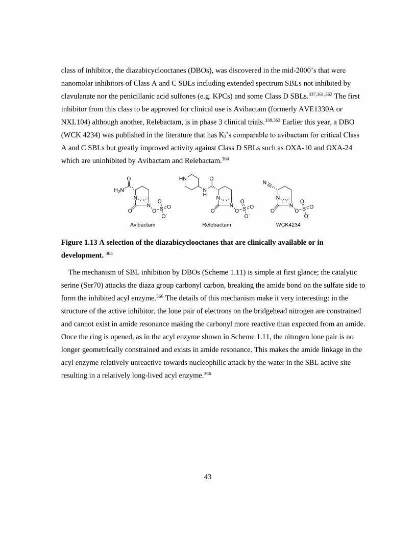
43
class of inhibitor, the diazabicyclooctanes (DBOs), was discovered in the mid-2000’s that were
nanomolar inhibitors of Class A and C SBLs including extended spectrum SBLs not inhibited by
clavulanate nor the penicillanic acid sulfones (e.g. KPCs) and some Class D SBLs.337,361,362 The first
inhibitor from this class to be approved for clinical use is Avibactam (formerly AVE1330A or
NXL104) although another, Relebactam, is in phase 3 clinical trials.338,363 Earlier this year, a DBO
(WCK 4234) was published in the literature that has KI’s comparable to avibactam for critical Class
A and C SBLs but greatly improved activity against Class D SBLs such as OXA-10 and OXA-24
which are uninhibited by Avibactam and Relebactam.364
Figure 1.13 A selection of the diazabicyclooctanes that are clinically available or in
development. 365
The mechanism of SBL inhibition by DBOs (Scheme 1.11) is simple at first glance; the catalytic
serine (Ser70) attacks the diaza group carbonyl carbon, breaking the amide bond on the sulfate side to
form the inhibited acyl enzyme.366 The details of this mechanism make it very interesting: in the
structure of the active inhibitor, the lone pair of electrons on the bridgehead nitrogen are constrained
and cannot exist in amide resonance making the carbonyl more reactive than expected from an amide.
Once the ring is opened, as in the acyl enzyme shown in Scheme 1.11, the nitrogen lone pair is no
longer geometrically constrained and exists in amide resonance. This makes the amide linkage in the
acyl enzyme relatively unreactive towards nucleophilic attack by the water in the SBL active site
resulting in a relatively long-lived acyl enzyme.366

44
Scheme 1.11 Avibactam mediated inhibition of SBLs and other reactions with Avibactam
catalyzed by β-lactamases. 361,366,367
Acylation of the catalytic serine can slowly reverse in many SBLs to regenerate the active inhibitor.
Some SBLs, such as KPC-2, are capable of hydrolyzing and inactivating Avibactam by desulfonating
(through either a one or two step mechanism), then hydrolyzing the imine, followed by carbamate
hydrolysis but these are slow processes shown in Scheme 1.11.361 MBLs are not inhibited by DBOs,
but have been shown to slowly inactivate the DBOs through the hydrolysis depicted in Scheme 1.11.
The subsequent desulfonation in the MBL mechanism was only observed at low levels and it was not
conclusively determined whether or not it was the result of enzymatic catalysis.367
1.4.2.3 Transition State Analogs and β-Lactam Mimics
Mechanism based inhibitors and DBOs both inhibit though the opening of an amide ring (either β-
lactam or DBO) forming a stable acyl-enzyme complex. Not all inhibitors of SBLs require a cyclic
amide, others function by forming a covalent complex with the active site serine that mimics the

45
tetrahedral intermediate formed during catalysis or mimics the natural substrate without being able to
complete the catalytic cycle.
The earliest β-lactamase inhibitor to be discovered was boric acid in 1978 which inhibited BcI (A)
with a KI of 1 mM.368 Boronate inhibitors were vastly improved in the 1980’s with simple aromatic
boronic acids such as 1-1 and 1-2 from Figure 1.14 that inhibited Class A and C SBLs with KI’s as
low as 0.5 μM.369–371 Increasing the complexity of boronic acids to include two R groups (one of
which is derived from a cephalothin R group – highlighted in blue) such as 1-3, improved the potency
of these inhibitors to 60 nM (IC50) against Class A SBLs.372 Cyclization of the boronate improved
potency even more with Vaborbactam (RPX7009) inhibiting Class A and C SBLs with KI’s as low as
30 nM.373 Vaborbactam was first reported and patented in 2010 and was approved for clinical use in
combination with meropenem in 2017.374,375 Recently, boronic acid inhibitors have also been shown
to be inhibitory towards a Class D SBL, OXA-24/40, although the inhibition is much less potent than
against Class A and C SBLs.376 The inhibition of Class A, C, and D SBLs by phosphonates (1-4) and
both cyclic and linear phosphates (1-5 and 1-6 respectively) have been thoroughly studied by the Pratt
group; however the kinetics are reported in terms of rate constants making it difficult to compare their
efficacy to that of other inhibitors reported in terms of KI or IC50.377–386
Figure 1.14 Transition state analog inhibitors of SBLs: boronates and phosph(on)ates.
369,371,372,374,377,381,385

46
Many β-lactam mimics have been studied in the context determining the pharmacophore of β-
lactam antibiotics and diversifying pharmacophores, some of which (imidazolidinones and
cyclobutanones) were determined to be inhibitory towards β-lactamases. The topic of β-lactam
mimics has been well reviewed by Jungheim and Ternansky in 1993 and cyclobutanones in particular
have been recently reviewed by Devi and Rutledge.387,388 The first cyclobutanones (such as 1-7 from
Figure 1.15) were synthesized by Gordon et al. in 1981 and were found to have no inhibitory activity
towards R-TEM β-lactamase.389 A few years later, Lowe and Swain prepared a series of
cyclobutanones, including 1-8, that inhibited R-TEM and B. cereus 568/H β-lactamases in a slow,
time-dependent manner but they did not report a value that indicates inhibitory potency.390 Most
recently, Johnson et al. from the Dmitrienko laboratory described the synthesis of
dichlorocyclobutanones such as JJ05-1058 and JJ05-802, that weakly inhibited all classes of β-
lactamases with the best activity being demonstrated against Class C.
Figure 1.15 Cyclobutanones: β-lactam mimics that are inhibitory towards β-lactamases. 389–391
Both the transition-state analogs and the cyclobutanones inhibit SBLs by forming a reversible
covalent acyl enzyme with the catalytic serine (Ser70) of the β-lactamase that mimics the tetrahedral
transition-state of β-lactam binding as shown in Scheme 1.12. Crystal structures of Vaborbactam, 1-4,
and JJ05-802 bound to the active sites of SBLs have been obtained which confirm the formation of a
covalent bond between the catalytic serine and the inhibitor as well as the release of the p-nitrophenyl
group from the phosphonate.373,379,391 A crystal structure of JJ05-1085 bound to the active site of the
B1 MBL SPM-1 has also been determined.392
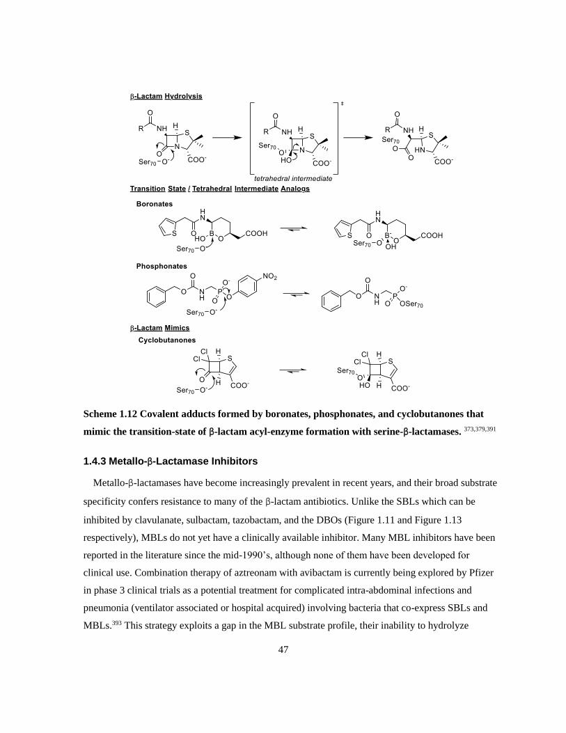
47
Scheme 1.12 Covalent adducts formed by boronates, phosphonates, and cyclobutanones that
mimic the transition-state of β-lactam acyl-enzyme formation with serine-β-lactamases. 373,379,391
1.4.3 Metallo-β-Lactamase Inhibitors
Metallo-β-lactamases have become increasingly prevalent in recent years, and their broad substrate
specificity confers resistance to many of the β-lactam antibiotics. Unlike the SBLs which can be
inhibited by clavulanate, sulbactam, tazobactam, and the DBOs (Figure 1.11 and Figure 1.13
respectively), MBLs do not yet have a clinically available inhibitor. Many MBL inhibitors have been
reported in the literature since the mid-1990’s, although none of them have been developed for
clinical use. Combination therapy of aztreonam with avibactam is currently being explored by Pfizer
in phase 3 clinical trials as a potential treatment for complicated intra-abdominal infections and
pneumonia (ventilator associated or hospital acquired) involving bacteria that co-express SBLs and
MBLs.393 This strategy exploits a gap in the MBL substrate profile, their inability to hydrolyze

48
monobactams, while protecting the antibiotic from the SBLs that can degrade it with avibactam. In
vitro studies on over 60 000 clinical isolates from 40 countries have shown that the aztreonam and
avibactam combination is a potent combination against MBL-producing Enterobacteriaceae and P.
aeruginosa.394 Despite the promise of a treatment for MBL-producing infections in the near future,
the pursuit of an MBL inhibitor (for combination therapy with carbapenems) continues to be an
important area of research. The following section covering MBL inhibitors is by no means exhaustive
but covers many of the main themes in MBL inhibitor development; several excellent reviews exist in
the literature.27,395–398 Common strategies for MBL inhibition involve completely or patially de-
metallating the active site with chelators, binding the zinc ions with dicarboxylates or sulphur groups
without metal ion removal. Inhibitors have either been designed rationally or been discovered by
screening natural product or known chemical libraries for activity using high-throughput methods.
1.4.3.1 Demetallating chelators
When trying to inactivate a protein that uses metal ions to perform its catalytic function, an obvious
strategy is to remove the metal from the protein. For decades, this has been used in the classification
of β-lactamases as MBLs since only the MBLs would be inactivated by EDTA.399 Although EDTA is
adequate as a characterization tool, EDTA inactivation of MBLs often requires hours of incubation
which, in combination with its promiscuity, makes it a poor drug candidate. Other common
commercial chelators such as o-phenanthroline, PAR, TPEN, and DPA have been investigated as
inactivators of IMP-1 although even the most effective of these inactivators, DPA, had only removed
one zinc ion from 13% of the IMP-1 moelcules after 3 hours of incubation.400
Figure 1.16 Demetallating inhibitors of MBLs. 400–402
More recently, a natural product, Aspergillomarasmine A (AMA), was found to be a potent
inactivator of NDM-1 and VIM-2, capable of removing one zinc ion from NDM-1 in as little as one
hour. While AMA was shown to potentiate meropenem against NDM and VIM producing bacterial
strains, it was much less effective against strains that produce AIM, IMP, or SPM – type MBLs,

49
making it a relatively narrow-spectrum MBL inactivator.401 The same group also discovered the
potential for spiro-indolino-thiadiazoles (SIT) to inactivate MBLs with IC50s for NDM-1 as low as 6.6
μM. Despite the favourable response from NDM-1, VIM-2 and IMP-7 were unaffected by even the
most potent compound from this series, SIT-Z5, likely due to their zinc ions being more tightly
bound.402–405 Demetallating chelators pose problems for clinical use since compounds that are potent
enough to pull the zinc ions out of MBLs will potentially have toxicity problems in humans and those
that are weak enough to be non-toxic have very narrow-spectrum activity. As a potentially less toxic
alternative to demetallating chelators, inhibitors that can bind the zincs rather than remove them are
desirable.
1.4.3.2 Dicarboxylates
The earliest MBL inhibitors in this class were the succinic acid derivatives, the most potent of
which is 1-9 (Figure 1.17).This compound was reported to be an extremely potent inhibitor of IMP-1
with an IC50 of 3 nM, although neither the article nor the patent that disclose this compound address
its ability to inhibit other MBLs.406,407 Further analogs of the succinic acids have been pursued by
chemists at Merck who have proven that these compounds can potentiate meropenem against E. coli
strains transformed with an IMP-1 expressing plasmid, although none of those compounds approach
the potency of 1-9.408 Olsen et al. explored the inhibitory potency of analogs of 1-9 with BcII and L1
and did not observe inhibition, suggesting that these compounds may not have a sufficiently broad-
spectrum to be clinically useful.409
Figure 1.17 Dicarboxylate inhibitors of MBLs. 406,410–414

50
The succinic acid derivatives have flexibility in the bond connecting the carboxylic acid groups,
although this is not necessary as several conformationally rigid dicarboxylates have been found to be
MBL inhibitors. DPA (2,6-PA), which is capable of abstracting one zinc ion from IMP-1 at long
incubation times, also inhibits MBLs in a time dependent competive manner at short incubation
times.400,415 Horsfall et al. demonstrated that other substitution patterns of DPA are also inhibitory,
notably 2,4-PA which inhibits the B2 MBL CphA with a KI of 5 μM but has little effect on B1 and B3
MBLs. Their study also demonstrated that the monocarboxylate, 2-picolinic acid, is capable of
partially inhibiting all MBL subclasses at 100 μM.410 Feng et al. explored nitrogen containing
heterocycles as MBL inhibitors, and found that those with carboxylate groups on either side of the
nitrogen (such as 1-10) were inhibitors of all MBL subclasses with IC50s of compound 1-10 ranging
from 0.64 μM to 7.1 μM.413
Although the nitrogen in the picolinic acid derivatives is advantageous for zinc binding (see
Chapter 3 for more details), it is not a necessary feature for the aromatic dicarboxylate inhibitors.
Researchers at Meiji Seika pharmaceuticals have demonstrated that dicarboxylate inhibitors derived
from phthalic acid such as 1-11 and 1-12 are capable of inhibiting IMP-1. Although the IC50s for 1-11
and 1-12 (0.97 μM and 0.27 μM respectively) indicate that these are much more potent than the DPA
derivatives, they have only been tested against IMP-1, so it is unknown whether or not these represent
broad-spectrum MBL inhibitors.411,414,416
Another discovery in this area at Meiji Seika pharmaceuticals is ME1071, a maleic acid derivative
that has been demonstrated inhibit IMP-1, VIM-2, and NDM-1. The inhibition constants determined
for ME1071 remains consistent between publications for IMP-1 (KI = 0.41 μM or 0.46 μM);
however, that of VIM-2 varies greatly (KI = 120 μM or 1 μM), while that of NDM-1 has only been
reported once (KI = 24 μM). When correlated with the bacterial data, it becomes clear that this
compound is most active against IMP variants and least active against NDM variants.412,417
1.4.3.3 Sulphur Containing Inhibitors
The affinity of sulphur compounds for zinc ions is one of the fundamental relationships in biology,
allowing for zinc binding to proteins through cysteine containing zinc fingers, the Lewis acidity of
zinc ions in proteins, some forms of redox activity, and much more.418 This relationship can also be
exploited for inhibition of proteins, such as MBLs, that use zinc in their catalysis.

51
The most common sulphur moiety used in MBL inhibitors is the thiol (Figure 1.18) which often
accompanies a carboxylate. Early mercaptocarboxylates identified as MBL inhibitors included
compound 1-13 and R-thiomandelic acid, both of which were inhibitors of multiple subclasses of
MBL with submicromolar IC50s and KIs respectively.251,419 In 2003, members of the Dmitrienko lab
tested the inhibitory potential of 23 commercial thiols, the most potent of which was mercaptoacetic
acid against IMP-1 and also inhibited BcII 5/B/6 and CcrA. This study concluded that thiols with an
α-carboxylate (or similar anionic group) were classical competitive inhibitors, while those without an
anionic group behave as time-dependent competitive inhibitors under neutral conditions due to the
deprotonation necessary to make the most stable EI complex. Additionally, it was observed that
esterifying the carboxylates, adding an amine, and using an aromatic molecule as the core structure all
reduced inhibitory potency against IMP-1.420 Jin et al. found that using aliphatic chains to distance the
thiol from an aromatic group increased potency as compound 1-14 from Figure 1.18 exhibits IC50s of
1.2 μM and 1.1 μM with IMP-1 and VIM-2 respectively while the equivalent compound with only
one carbon between the phenyl ring and the carboxylate had IC50s of 16.4 μM and 14.3 μM
respectively.421
Figure 1.18 Thiol inhibitors of MBLs. 251,419–425
The most thoroughly studied MBL inhibitor is captopril, a known inhibitor of angiotensin-
converting enzyme (ACE) in humans that has been used for many years to treat hypertension.426 Two
stereoisomers of captopril have been used in most investigations: D-captopril (Figure 1.18) and L-
captopril, referring to the stereochemistry of the proline ring. Crystal structures of MBLs have been
acquired with both stereoisomers bound; however, the D-isomer is frequently the more potent
52
inhibitor (2- to 61- fold more potent) and consequently generates higher occupancy crystal structures
of the enzyme-inhibitor complexes.423,427 Although D-captopril is able to inhibit enzymes in all
subclasses of MBL, some MBLs such as SPM-1 (B1) and FEZ-1 (B3) are resistant to inhibition.256,423
Additionally, D-captopril does not bind to all MBLs in the same manner: the sulphur atom binds
between the zinc atoms in all MBLs except in the monozinc CphA (B2) in which the carboxylate
binds the zinc atom as seen in Figure 1.19E. Binding even differs within subclasses, for example the
B1 MBLs BcII (Figure 1.19A) and VIM-2 (Figure 1.19B) bind in very similar modes with the proline
ring stacked over the His residue that coordinates Zn2 (His240) and the carbonyl H-bonded to
Asn210 whereas in IMP-1 (Figure 1.19C) the Asn210 interaction is weaker (4.4 Å as opposed to <3
Å) but the tryptophan on the capping loop, Trp45, makes the proline stacking tighter.423 The most
different of the B1 MBL structures is BlaB (Figure 1.19D) in which the proline ring sits over one of
the histadine residues that coordinates Zn1 (His118) rather than the His residue that coordinates Zn2
(His 240).428 The two structures of B3 MBLs differ greatly since L1 (Figure 1.19F) is inhibited by D-
captopril with a KI of 8 μM, corresponding to D-captopril binding the active site tightly, whereas
FEZ-1 (Figure 1.19G) is only weakly inhibited with a KI of 400 μM and in the crystal structure, D-
captopril is well removed from the active site.255,256

53
Figure 1.19 D-Captopril bound to various MBLs from all subclasses : B1 MBLs (A) BcII, (B)
VIM-2, (C) IMP-1, (D) BlaB, the B2 MBL (E) CphA with the putative Zn1 binding site
highlighted in blue, and the B3 MBLs (F) L1, and (G) FEZ-1 (This figure was generated using
UCSF Chimera v. 1.12 from protein databank files 4C1C, 4C1E, 4C1G, 1M2X, 2QDS, 2FU8,
and 1JT1 respectively).255,256,423,428,429 All structures are oriented with Zn1 (or equivalent binding
site) towards the bottom of the image.
Recent work on thiol inhibitors of MBLs has focused on taking advantage of their ability to inhibit
all subclasses of MBL and reducing the in vivo toxicity. The mercaptophosphonate 1-15 (Figure 1.18)
addressed cross-class inhibition of MBLs by introducing a phosphonate which coordinates the zinc
ion of CphA in a tetrahedral geometry while also making contacts with nearby His and Lys residues
(PDB: 3IOG) while the thiol is predicted to bind the zincs in the B1 and B3 enzymes as observed for
D-captopril.424 The bisthiazolidines, particularly L-CS319 (Figure 1.18), were shown to inhibit all
subclasses of MBL with KIs ranging from 0.26 μM to 41 μM with most KIs being in the low
micromolar range for the 7 tested MBLs (B1: NDM-1, VIM-2, IMP-1, BcII; B2: Sfh-1; B3: L1,
GOB-18). Crystal structures of Sfh-1 (B2) and L1 (B3) were obtained with L-CS319 and showed the

54
thiol equidistant between the zinc ions in the L1 strucutre (PDB: 5EVK) and the carboxylate
coordinating the zinc ion in Sfh-1 (PDB: 5EW0), consistent with the binding modes of D-captopril.425
Unpublished structures of the B1 MBLs BcII, NDM-1, and VIM-2 with L-CS319 bound (PDB:
4NQ5, 4U4L, 4U4A) have been acquired and demonstrate the expected mode of binding: the thiol
replacing the bridging hydroxide.
Scheme 1.13 Activation of rhodanine to a mercaptocarboxylate MBL inhibitor. 430,431
One of the challenges of using thiols as MBL inhibitors is that thiols can inhibit critical human
metallo-proteins such as ACE and some can cause toxicity as a result of their reactivity.432,433
ML302F is a potent MBL inhibitor that is intended to be administered as ML302 with the thiol
protected. ML302F was found to inhibit 6 B1 MBLs (NDM-1, VIM-1, VIM-2, SPM-1, IMP-1, and
BcII) with sub-micromolar IC50s and was not inhibitory towards a small panel of human metallo-
enzymes including ACE. The authors suspect that ML302 is converted to ML302F by MBLs;
however, their experiments to confirm this were inconclusive due to the potent inhibitory effect of
ML302F. As expected, ML302F binds to VIM-2 with the thiol bridging the zinc ions with the
carboxylate also interating with Zn2 and several of the surrounding residues.430,431 Although
protecting the thiol of ML302F should reduce potential toxicity (as demonstrated by no toxicity
against human HeLa cells up to 100 μM)430, rhodanines are known to be promiscuous inhibitors and
may cause problems in complex systems such as humans.434
The next most common sulphur containing functionality used in MBL inhibitors is the thioester as
seen in compounds 1-16, 1-17, and 1-18 in Figure 1.20, the idea being that the thioester could be
recognized as the amide of a β-lactam substrate, particularly when the β-carbon from the sulphur is a
carboxylate.435 Each of these compounds was reported as an MBL inhibitor in the late 1990’s, and
today would not be considered potent enough to publish. Compound 1-16 inhibited L1 (B3) and
CphA (B2) with reasonable potency (IC50 = 10 μM, 30 μM respectively) but was not capable of
significantly inhibiting BcII (B1) nor CfiA (B1).435 The same group demonstrated that compound 1-

55
17 inhibited BcII and CfiA with IC50s of 23 μM and 724 μM respectively in the presence of 1 μM
zinc and all inhibition was abolished when the Zn2+ concentration was increased to 100 μM. Although
it was weakly effective against B1 MBLs, this inhibitor was very potent against L1 with an IC50 of
less than 1.9 μM in high and low zinc conditions.436 Compound 1-18 was developed by Merck
scientists as a potent IMP-1 inhibitor (IC50 = 0.0004 μM), although their publications are inconsistent
about the stereochemistry of the active inhibitor.437,438 The stereochemistry denoted in Figure 1.20 is
consistent with the Greenlee paper which actually addresses the differences in the activity of the R
and S isomers.437 The extreme potency of 1-18 against IMP-1 (B1) did not translate to the only other
MBL tested, CcrA (B1), which has an IC50 of only 180 μM.437,438 No crystal structures have been
published with a thioester inhibitor bound to an MBL.
Figure 1.20 Thioester and other non-thiol MBL inhibitors that contain sulphur. 435–437,439–442
Other MBL inhibitors have been discovered with sulphur containing moieties, some of which fit
better into the other categories laid out in this section such as the demetallating chelator SIT-Z5 from
Figure 1.16 and the dicarboxylate 1-10 from Figure 1.17.402,413 Inspired by the inhibition of MBLs
with DPA and thiols, Roll et al. investigated the inhibition of CcrA (B1) and L1 (B3) with thioacid
and methyl thioester derivates of DPA and found that PDTC (Figure 1.20) is the most potent inhibitor
of these two MBLs with IC50s of 0.14 μM and 0.6 μM respectively.439 Faridoon et al. used fragment-
based screening to suggest that 3-mercapto-1,2,3-triazoles and N-acylated thiosemicarbazides could
be inhibitory towards IMP-1. After investigation of several compounds from each group, they

56
determined that the triazoles were not inhibitory and the thiosemicarbazoles were moderate inhibitors.
The most potent inhibitor from this series, 1-19 in Figure 1.20, inhibited IMP-1 with a KI of 14 μM
which is comparable to the inhibitory potency of L-captopril.440
Two groups of inhibitors have been published with sulfones as a key feature of their structure: the
N-arylsulfonyl hydrazones (1-20 in Figure 1.20) and sulfonyl triazoles (1-21 in Figure 1.20).441,442
The N-arylsulfonyl hydrazones were developed as IMP-1 inhbitors, the most potent of which (1-20)
inhibited IMP-1 with an IC50 of 1.6 μM. These inhibitors were unfortunately not broad-spectrum as
many in this series were unable to inhibit BcII 5/B/6, and even 1-20 only elicited 30 % inhibition at
25 μM.441 The sulfonyl triazoles were discovered as VIM-2 inhibitors using high-throughput
screening of a click-chemistry library, the most potent compound in this series, 1-21, inhibited VIM-2
in a competitive manner with a KI of 0.41 μM (IC50 = 3.3 μM) but was uninhibitory towards IMP-
1.442 Compound 1-21 was used as a lead to develop a more potent inhibitor which was able to inhibit
VIM-2 with a KI of 0.01 μM but all 47 compounds developed in this series were uninhibitory towards
IMP-1.443
1.4.3.4 Other Metallo-β-Lactamase Inhibitors
Identification of structurally unique inhibitors of MBLs occurs primarily through screening large
databases of natural products, pharaceuticals, and chemical collections from pharmaceutical
companies. Scientists from GlaxoSmithKline screened their natural products database and identified
three tricyclic natural products as MBL inhibitors, the most potent of which is SB236049 which
inhibits BcII (B1) and CfiA (B2) with IC50s of 0.3 μM and 2 μM respectively. This compound was
not as effective against IMP-1 (IC50 = 151 μM) and all three compounds in this series were inactive
against L1 (B3).444 Chemists at Merck screened their chemical collection to identify biphenyl
tetrazoles as inhibitors of MBLs, the most potent derivative of which was L-161,189 which inhibited
a CcrA (B1) with an IC50 of 0.3 μM.445
57
Figure 1.21 Structurally unique MBL inhibitors. 442,444,445
Minond et al. screened a collection of pharmacologically active compounds to identify inhibitors of
VIM-2. They found mitroxantrone (Figure 1.21), a DNA intercalator and topoisomerase IIα inhibitor
used to treat advanced multiple sclerosis and certain cancers, to be an uncompetitive inhibitor of
VIM-2 (KI = 1.5 μM) and uninhibitory towards IMP-1.442,446–449 The other pharmacologically active
compound identified from the screen as a VIM-2 inhibitor was para-chloromercuribenzoate (pCMB)
which is known to react with cysteine residues. pCMB inhibits both VIM-2 and IMP-1, likely by
covalently modifying the cysteine that coordinates Zn2 (Cys172 in VIM-2, Cys158 in IMP-1).260,442
Inhibitors of metallo-β-lactamases vary widely in structure and specificity. Many of the inhibitors
presented in this section were only tested against one or two MBLs and do not offer any insight into
whether or not they would be useful as broad-spectrum inhibitors. Of those that were tested widely,
the thiol (particularly the mercaptocarboxylates) and dicarboxylate functionalities had the broadest
spectrum of inhibition. The mercaptophosphonate 1-7 in Figure 1.18 introduces a phosphonate into a
thiol inhibitor, a novel solution to the problem of thiols being less inhibitory towards B2 MBL. The
dithioacid derivative of DPA (PDTC) in Figure 1.20 is also an interesting approach to that problem,
potently inhibiting both a B2 and B3 MBL. The phosphonate and thioacid moieties are understudied
in the literature considering their potential to be broadly inhibitory: this thesis addresses furthering the
understanding of the prospects for these functionalities in MBL inhibitors (see Chapter 3 and Chapter
4 respectively).
1.4.4 Dual Serine- and Metallo-β-Lactamase Inhibitors
Although it is useful to inhibit either SBLs or MBLs, this poses a challenge for clinical applications
against pathogens that produce both types of β-lactamases. It is challenging enough to get
combination therapies involving only two compounds approved but is significantly more difficult to
get approval for a three-compound combination (antibiotic, SBL inhibitor, MBL inhibitor). Ideally, a
dual serine- and metallo-β-lactamase inhibitor would be administered in combination with a broad-
spectrum β-lactam (such as meropenem) which would protect it from all β-lactamases. The only dual
SBL/MBL inhibitors that have been discovered are the cyclic boronates, notably CB-1 and CB-2
(Figure 1.22).450,451

58
Figure 1.22 Structural evidence for cyclic boronate inhibition of SBLs and MBls exemplified by
CB-1 bound to the SBL CTX-M-15 (A) and CB-2 bound to the B1 MBL VIM-2 (B). This figure
was generated using UCSF Chimera v. 1.12 from protein databank files 5T66 and 5FQC.450,451
Kinetic studies on the cyclic boronates CB-1 and CB-2 have determined that while both show
activity against SBLs and MBLs, CB-1 is more potent against SBLs and CB-2 is more potent against
MBLs.451 Crystal structures of CTM-M-15 (PDB: 5T66) and OXA-10 (PDB: 5FQ9) have been
determined with CB-1 bound, and VIM-2 (PDB: 5FQC) and BcII (PDB: 5FQB) have been
determined with CB-2 bound (Figure 1.22).450,451 The crystal structures of the SBLs illustrated that the
inhibition of SBLs occurs through nucleophilic attack of the boronate by the catalytic serine (Ser73 in
CTX-M-15) forming a covalent adduct.450 In binding the MBLs, such as VIM-2, CB-2 was found to
coordinate Zn1 with both boronate hydroxyl groups and Zn2 with the oxygen in the ring.451 Although
the cyclic boronate pharmacophore is capable of being inhibitory towards both SBLs and MBLs, not
all cyclic boronates behave in this manner. The newly FDA approved β-lactamase inhibitor
Vaborbactam (Figure 1.14) is a cyclic boronate with activity against only Class A and C β-lactamases
as covered in 1.4.2.3.375
1.5 Clinical Significance of β-Lactamase Inhibition
Combination therapy with the mechanism-based inhibitors has proven to be a useful strategy for
combating β-lactam resistance in bacteria that express certain SBLs although resistance to these
inhibitors is already prevalent. The recent introductions of Avibactam and Vaborbactam into clinical
use have addressed resistance from many, but not all, SBLs including some ESBLs. These solutions
to an ever-growing problem provide hope that β-lactams can remain a viable option for antimicrobial
therapy for decades to come. The biggest threat to β-lactams as a therapeutic option is the
increasingly widespread expression of MBLs which are capable of breaking down last-line of defense

59
β-lactams, such as the carbapenems, and are unaffected by the current SBL inhibitors. Metallo-β-
lactamase inhibitors are necessary for the future of antimicrobial therapy, especially in the light of
increasingly common multidrug resistant pathogens. Ideally, novel β-lactamase inhibitors should aim
to inhibit both SBLs and MBLs since it is not uncommon for resistant pathogens to express multiple
β-lactamases from both mechanistic families.
Inhibition of PBPs and β-lactamases – specifically metallo-β-lactamases – is crutial for treating
increasingly problematic resistant pathogens. This work explores the potential use of several classes
of MBL inhibitors and inhibitor releasing cephalosporins in increasing the efficacy of clinical
antibiotics, such as meropenem, against β-lactamase-producing pathogens.

60
Chapter 2
Phylogenetic Analysis, Purification, and Biochemical
Characterization of β-Lactamases
2.1 Introduction
The objective of the research described in this chapter was to overexpress and purify adequate
amounts of a variety of β-lactamases for kinetic testing and to characterize their ability to hydrolyze
chromogenic cephalosporins as well as their stability to organic co-solvents commonly used in kinetic
assays. While the hydrolysis of any β-lactam can be detected using spectrophotometry, it is
advantageous to use chromogenic substrates that exhibit larger changes in absorbance in the visible
range rather than the UV range upon hydrolysis of the β-lactam bond. This means that these
substrates can be assayed in affordable 96-well plates made of polystyrene rather than those made
from quartz or expensive UV-transparent plastics. An additional benefit of the assay wavelength
being relatively high is that it is uncommon for other compounds in the assay, such as cosolvents or
inhibitors, to interfere with the assay as they typically absorb only in the UV range.
Figure 2.1 Chromogenic and fluorogenic substrates for β-lactamases excluding the UW series
substrates.
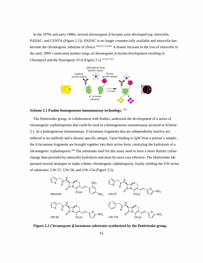
61
In the 1970s and early 1980s, several chromogenic β-lactams were developed (eg. nitrocefin,
PADAC, and CENTA (Figure 2.1)). PADAC is no longer commercially available and nitrocefin has
become the chromogenic substrate of choice.310,312–314,452 A drastic increase in the cost of nitrocefin in
the early 2000’s motivated another surge of chromogenic β-lactam development resulting in
Chromacef and the fluorogenic FC4 (Figure 2.1).315,317,453
Scheme 2.1 Panbio homogeneous immunoassay technology. 454
The Dmitrienko group, in collaboration with Panbio, undertook the development of a series of
chromogenic cephalosporins that could be used in a homogeneous immunoassay pictured in Scheme
2.1. In a homogeneous immunoassay, β-lactamase fragments that are independently inactive are
tethered to an antibody and a disease specific antigen. Upon binding to IgM from a patient’s sample,
the β-lactamase fragments are brought together into their active form, catalyzing the hydrolysis of a
chromogenic cephalosporin.454 The substrates used for this assay need to have a more distinct colour
change than provided by nitrocefin hydrolysis and must be more cost effective. The Dmitrienko lab
pursued several strategies to make a better chromogenic cephalosporin, finally yielding the UW series
of substrates: UW-57, UW-58, and UW-154 (Figure 2.2).
Figure 2.2 Chromogenic β-lactamase substrates synthesized by the Dmitrienko group.

62
The UW series of substrates have the advantage of a shorter and more cost effective synthetic
method while maintaining similar kinetic parameters to nitrocefin with many β-lactamases. In some
cases, the kinetic parameters of the UW substrates make them better for kinetic assays than nitrocefin
through either raising the KM sufficiently to make substrate depletion less of a problem in assays
performed at KM concentrations, or higher kcat values which require less enzyme to achieve
reasonable rates, allowing savings on precious enzyme stocks.
2.2 Methods
Unless otherwise stated, all spectrophotometric readings were done using a SpectraMax 190 plate
reader (Molecular Devices, Sunnyvale, CA) with SoftMax 6.0 (Molecular Devices, Sunnyvale, CA)
software. Analysis of kinetic data was performed using GraphPad Prism 5.00 for Windows
(GraphPad Software, San Diego, CA). The 96-well plates were always clear, flat bottomed,
polystyrene plates and were purchased from either Corning (Corning, NY) or Greiner Bio-One
(Monroe, NC) for enzyme kinetics. Plates for bacteria experiments were purchased from either
Corning (Corning, NY) or Sarstedt (Nümbrecht, Germany). Initially, cation adjusted Mueller Hinton
broth powder was purchased from Sigma (Burlington, MA) when problems with inconsistency were
encountered between batches late in 2017, the supplier was changed to BD (Franklin Lakes, NJ).
Other bacterial media components were purchased from BioBasics (Markham, ON). Most buffer salts
were purchased from BioShop (Burlington, ON), phosphate buffer salts from Alfa Aesar (Haverhill,
MA), and other chemicals from companies now owned by MilliporeSigma (Burlington, MA) such as
EMD and Sigma. The dialysis membrane used throughout was SpectraPor dialysis membrane with a
6-8 kDa molecular weight cutoff from the Spectrum labs division of Repligen (Waltham, MA).
2.2.1 Protein Sequence Alignment
The protein sequences of IMP-1, NDM-1, and VIM-2 were acquired from the NCBI Database in
FASTA form and entered into the BLAST Explorer functionality of phylogeny.fr which searched the
NCBI database prior to 9 Nov 2017 for similar, non-redundant protein sequences.455,456 The 100
closest matches were displayed and the whole protein sequence for every variant within that family
was selected to make a phylogenetic tree using the “One Click” tool. A check was done to ensure all
variants with sequences in the NCBI database as of 20 Jan 2018 were in the data set, and any variants
that were missing were manually added to the data set prior to tree generation. The “One Click”
analysis tool aligned sequences using MUSCLE, curated these results with Gblocks, determined

63
phylogenetic relationships with PhyML. The phylogenetic tree was rendered with TreeDyn.457–461 The
sequence alignment was downloaded as a Clustal file then formatted using BioEdit v. 7.0.5.3.462
Formatting of the phylogenetic tree was done using both the TreeDyn web applet on phylogeny.fr and
the TreeDyn v. 198.3 desktop program.
2.2.2 Enzyme Purification
Characterization of the inhibition of serine- and metallo-β-lactamases by PMPCs (Chapter 3)
necessitated the purification and acquisition of a variety of enzymes. Pure protein samples of the
MBLs IMP-1, L1, and SFH-1 as well as the SBL OXA-23 were generous gifts from Dr. James
Spencer of the School of Cellular and Molecular Medicine at the University of Bristol. Dr. Spencer
also provided expression plasmids for VIM-1 and OXA-48. Two batches of pure SPM-1 were
obtained; one from Dr. Spencer, and the other from Dr. Christopher Schofield of the Department of
Chemistry at the University of Oxford. The experiments presented in this thesis were performed with
Dr. Schofield’s SPM-1 preparation. NDM-1 with the first 41 amino acids truncated including the
signal peptide and the 13 amino acids that follow it, from the Schofield group at the University of
Oxford was used thoughout these studies.317 Dr. Natalie Strynadka of the Department of Biochemistry
and Molecular Biology at the University of British Columbia provided the expression plasmid for
CTX-M-15 and pure NDM-1 stocks that were used in preliminary experiments not presented in this
work. Expression plasmids for VIM-2, GC-1, and KPC-2 were provided by Dr. Patrice Nordmann of
L’Université Paris-Sud, Dr. Michiyoshi Nukaga of Josai International University in Togane City,
Chiba, Japan, and Dr. Focco van den Akker of Case Western Reserve University respectively.
Purification of proteins was carried out with the assistance of Dr. Geneviève Labbé and various
undergraduate research students as part of their projects.
2.2.2.1 Preparation for Protein Purification
Media for Bacterial Cultures
Luria-Bertani (LB) and Terrific broth (TB) were made using standard recipes from Molecular
Cloning Laboratory Manual 2nd Ed. with tryptone, yeast extract, and NaCl. Salts for buffering TB
were prepared separately from the media and added after autoclaving.463
Calcium chloride competent E. coli for Transformation

64
LB broth (5 mL) was inoculated with 50 μL E. coli cells (BL21, BL21 DE3, or BL21 PLysS as
appropriate) and incubated overnight at 37 oC shaking at 225 rpm. The overnight culture was
inoculated into fresh LB in a ratio of 1:20 (culture:media) and shaken at 225 rpm at 37 oC until the
OD was between 0.25 and 0.3 (approximately 1.5 hr) the chilled on ice for 15 min. Cells were
isolated by centrifugation in sterile 50 mL conical vials at 1600 xg for 10 min at 4 oC. The
supernatant was decanted and discarded. Cells were resuspended and pooled in 30 mL cold 0.1 M
CaCl2 and allowed to accommodate for 30 min on ice. Centrifugation and resuspension were repeated
twice as previously described. The final resuspension was performed with 6 mL cold 0.1 M CaCl2 in
15 % glycerol which was then aliquoted and flash frozen on dry ice for storage at -80 oC.
2.2.2.2 Analysis of Protein Purification
Nitrocefin Activity Assay
Aliquots of protein collected during purification were diluted as appropriate in the buffer used in
the respective purification step and incubated in a flat bottom 96 well plate at 30 oC for 5-10 min
before addition to nitrocefin (final concentration 100 μM for GC-1, and 25 μM for CTX-M-15). The
activity was determined by monitoring this reaction at 482 nm for 5 min at 30 oC and measuring the
initial rate. At least two replicates of this assay were generally performed.
Bradford Assay
A standard curve of bovine serum albumin (BSA) from 0.05 mg/mL to 0.5 mg/mL was made fresh
each day. Four technical replicates of standards and appropriately diluted samples were
simultaneously incubated with Bradford reagent (BioRad, Hercules, CA) in a 96 well plate for 5 min
at room temperature. The absorbance was then read at 595 nm using either a Powerwave XS2
(BioTek) plate reader or the SpectraMax 190.
SDS PAGE
Protein samples or diluted protein samples were added to 5x SDS PAGE loading buffer (0.25 M
Tris-HCl, 10 % SDS, 5 % 2-mercaptoethanol, 30 % glycerol, 0.02 % bromophenol blue, pH 6.8) then
boiled for 5 min. If condensation was observed on the lid of the tubes, they were centrifuged briefly.
The prepared sample (10-20 μL) was loaded onto 12.5 % polyacrylamide:bisacrylamide gel and run
at 200 V with a low molecular weight protein standard (GE Healthcare UK, Buckinghamshire, UK or
BioBasic, Markham, ON) in at least one of the wells. Gels were stained with Coomassie Blue

65
(BioRad, Hercules, CA) containing stain (50 % (v/v) methanol, 10 % (v/v) acetic acid, 0.025 % (w/v)
Coomassie Blue R-250) then destained with 50 % methanol, 10 % acetic acid. Gels were soaked in
water prior to visualization using either the white plate with a Gel Doc EZ Imager (BioRad, Hercules,
CA) or a camera. Purification of all β-lactamases in this chapter was monitored using SDS PAGE.
Gels from individual purifications will not be presented in this work, only a summary gel.
Mass Spectrometry
The molecular weights of purified proteins were determined by electrospray ionization mass
spectrometry (ESI-MS) in positive ion mode at the University of Waterloo Mass Spectroscopy
Facility. Pure proteins with their affinity tags cleaved were exchanged into 20 mM ammonium acetate
using Amicon Ultra Ultracel 10 K membrane centrifugal filters (Millipore). VIM-2 and KPC-2 were
then appropriately diluted in 1:1 MeOH:H2O, 0.2 % FA and injected into the Micromass Q-TOF
Ultima Global mass spectrometer equipped with a Z-spray electrospray ionization source
(Micromass) with the aid of a syringe pump. CTX-M-15, GC-1, NDM-1 (received from the Schofield
group at Oxford), SPM-1 (received from the Spencer group at Bristol), L1 (also received from
Spencer), and OXA-48 were appropriately diluted in 1:1 MeOH:H2O, 0.1 % FA then injected into the
Thermo Scientific Q-Exactive Orbitrap mass spectrometer (Thermo Fisher Scientific Inc.) by
electrospray ionization with the aid of a syringe pump. Protein deconvolution was done using Thermo
Protein Deconvolution v. 1.0 (Thermo Fisher Scientific Inc.).
2.2.2.3 CTX-M-15
The transformation and purification of CTX-M-15 was done with the assistance of Jessica Duong
as part of her undergraduate research.
Transformation
The cloning of CTX-M-15 into a pET28 vector with a thrombin cleavable N-6His tag was
performed by Dr. Strynadka’s lab before being sent to the Dmitrienko lab. The CTX-M-15 plasmid
was transformed into CaCl2 competent E. coli BL21 DE3 cells by incubating them together for 40
min on ice before heat shocking at 42 oC for 1.5 min. The cells were allowed to recover from
transformation with the addition of 200 μL of LB media then incubated for 2 hours at 37 oC.
Transformants were plated on LB agar supplemented with 50 μg/mL kanamycin and incubated
overnight at 37 oC then stored at 4 oC until ready to proceed to liquid cultures.

66
Growth of Liquid Cultures for CTX-M-15 Expression
One colony from the CTX-M-15 transformation was inoculated into 50 mL TB media
supplemented with 50 μg/mL kanamycin which was incubated at 37 oC overnight, shaking at 200
rpm. Two 4 L flasks containing 1 L TB media supplemented with 50 μg/mL kanamycin were
inoculated with 10 mL each of the overnight culture then incubated at 37 oC, shaking at 220 rpm until
the culture had reached an OD600 of approximately 0.9 (this should have been 0.6, but was
overgrown). Expression of CTX-M-15 was then induced with 1 mM isopropyl β-D-1
thiogalactopyranoside (IPTG) overnight, shaking at 200 rpm at room temperature. Cells were
harvested by centrifugation in a JA-10 rotor at 6000 xg for 20 min at 4 oC yielding 25.7 g of pellet.
The pellet was then washed twice with 54 mL of 0.85 % NaCl and cells were harvested again by
centrifugation in a F34-6-38 rotor at 6000 xg for 5 min at 4o C then flash frozen on dry ice and stored
at -80 oC. Cell pellets were thawed then resuspended in 20 mL of Lysis buffer (50 mM Tris HCl, 300
mM NaCl, 20 mM imidazole, 10 % (v/v) glycerol, 0.2 % NP-40, 1 mg/mL lysozyme, 2 μg/mL DNase
I, pH 8.0) and lysed using a large probe on a Heat Systems Ultrasonic processor W-255 sonicator set
to 50 % cycle and 50 % power 5 times for 30 sec each with 1 min rest on ice between bursts. Cellular
debris was separated from the lysate by centrifugation at 20 000 xg for 60 min at 4 oC using a JA-25.5
rotor. The supernatant was retained and stored at 4 oC overnight.
Affinity Chromatography
An Omnifit column housing filled with HisPurTM Ni-NTA Superflow Agarose (Thermo Scientific)
was mounted on a BioCad Sprint Perfusion Chromatography system and equilibrated with 1 column
volume of CTX-M-15 Elution buffer (50 mM Tris HCl, 300 mM NaCl, 300 mM imidazole, 10 %
(v/v) glycerol, pH 8.0) then 12 column volumes of CTX-M-15 Chelating buffer (50 mM Tris HCl,
300 mM NaCl, 20 mM imidazole, 10 % (v/v) glycerol, pH 8.0) through 2 different lines at 1 mL/min.
The lysate was filtered through at 0.2 μm syringe filter to sterilize and remove any particulates before
loading onto the column. Over the course of 3 purifications, 9.5 mL of lysate was applied to the
column through the pump at 0.5 mL/min then that line was flushed with CTX-M-15 Chelating buffer
for 2 column volumes. The column was then washed with 5 column volumes of CTX-M-15 Chelating
buffer at 1 mL/min. CTX-M-15 was eluted from the column using a gradient from 100 % CTX-M-15
Chelating buffer to 100 % CTX-M-15 Elution buffer over 5 column volumes with an additional
column volume of CTX-M-15 Elution buffer at the end to ensure complete elution. Fractions were

67
collected every 3 min starting from the loading of the protein onto the column. The 2-3 fractions from
each day that contained the highest CTX-M-15 activity by nitrocefin assay were pooled and dialyzed
into 50 mM HEPES pH 7.2 until both the imidazole and NaCl concentrations were calculated to be
below 10 nM. Dialyzed CTX-M-15 was concentrated using an Amicon Ultra-15 centrifugal filter
with a 10 kDa molecular weight cutoff then stored in 50 % (v/v) glycerol at -20 oC.
Thrombin Cleavage of His Tag
Cleavage of the His tag from CTX-M-15 was performed using a Thrombin Cleavage Capture kit
(Novagen) which uses biotinylated thrombin and streptavidin agarose. An 80 μL aliquot of
concentrated CTX-M-15 was incubated with 20 μL 1:100 diluted thrombin in Thrombin Cleavage
Buffer (20 mM Tris HCl, 150 mM NaCl, 2.5 mM CaCl2, pH 8.4) at room temperature for 4 hr. The
reaction was stopped with the addition of 25 μL streptavidin agarose slurry and incubated for 30 min
at room temperature to bind all of the thrombin. Cleaved CTX-M-15 was eluted using a spin filter at
1000 xg for 10 min then stored at -20 oC in 50 % (v/v) glycerol.
2.2.2.4 KPC-2
PCR Amplification of KPC-2
The gene for KPC-2 was amplified from a KPC-2 containing pBr322 vector supplied by Dr. Focco
van den Akker to introduce restriction sites for insertion into pET28a.214 Two sets of primers were
used to code for an N-terminal 6-His tag: one that adds the His tag onto the signal peptide of KPC-2
and another one that adds the His tag directly onto KPC-2 without coding for the signal peptide.
Polymerase chain reactions (PCR) were prepared using a PWO DNA polymerase kit (Roche) with 1
mM dNTP mixture, 0.25 μM of the Nde1 forward primer and EcoR1 reverse primer, 1 μL or 5 μL of
KPC-2 template, and 2.5 units of PWO polymerase in 10 mM Tris HCl, 25 mM KCl, 5 mM
(NH4)2SO4, 2 mM MgSO4, pH 8.85 then vortexed and centrifuged briefly. PCR was initiated with a 2
min denaturation at 95 oC, followed by 34 cycles consisting of denaturation at 92 oC for 15 sec,
annealing at 65 oC for 30 sec, and elongation at 72 oC for 1 min. The final cycle of PCR ended with a
7 min elongation at 72 oC. PCR products were purified on a 1% agarose gel in TAE (40 mM Tris, 20
mM acetic acid, 1 mM EDTA, pH 8.3) where bands were excised, and DNA was isolated using a
Quiagen Gel Extraction kit.
Cloning KPC-2 into pET28a

68
Both PCR products (His-SP-KPC-2 and His-KPC-2) and a pET28a vector were digested with
FastDigest EcoR1 and FastDigest Nde1 in FastDigest Green buffer (Fermentas). The digestion of the
PCR products was incubated for 1 hr at 37o C while the digestion of the vector was only incubated for
10 min at 37 oC before purification on 1 % agarose gel in TAE where bands were excised, and
digested DNA was isolated using a Qiagen Gel Extraction kit. The digested pET28a vector was
ligated with each of the two digested PCR products using T4 DNA Ligase for 2 hr at 37 oC.
From this point on, the purification of KPC-2 was taken over by the undergraduate research student
Melinda Lam under the supervision of Dr. Laura Marrone.
Transformation
The two KPC-2 containing plasmids, His-SP-KPC-2 and HIS-KPC-2, were transformed into 50 μL
of CaCl2 competent E. coli BL21 DE3 cells by incubating cells and plasmid on ice for 30 minutes
followed by heat shock at 42 oC for 20 seconds. Transformation reactions were placed back on ice for
2 min then allowed to recover in 950 μL of LB media for 1 hr at 37 oC while shaking. Transformants
were plated on LB agar supplemented with 50 μg/mL kanamycin and incubated at 37 oC overnight.
Small scale expression tests of these transformants indicated that the construct without a signal
peptide was not active and this avenue was not pursued further.
Growth of Liquid Cultures for KPC-2 Expression
Initial cultures were prepared by inoculating 5 mL of LB media supplemented with 50 μg/mL
kanamycin with His-SP-KPC-2 transformants then incubating these overnight at 37 oC while shaking.
The entirety of this initial culture was used to inoculate 0.5 L LB media supplemented with 50 μg/mL
kanamycin which was then grown at 37 oC, shaking at 220 rpm overnight. Small scale tests indicated
that expression of KPC-2 was best when not induced with IPTG so this step was forgone. Bacterial
cells were harvested by centrifugation at 6000 rpm for 15 min at 4 oC then subjected to periplasmic
lysis at room temperature for 10-15 min in 50 mM HEPES, 0.5 M NaCl, pH 7.5 and 0.1 μg of
lysozyme. DNase I and RNase I were added to the lysate to degrade any DNA and RNA released by
sphereoplast lysis then the periplasmic lysate was isolated from spheroplasts and cellular debris by
centrifugation at 12 000 xg for 10 min.
Chromatographic Purification of KPC-2
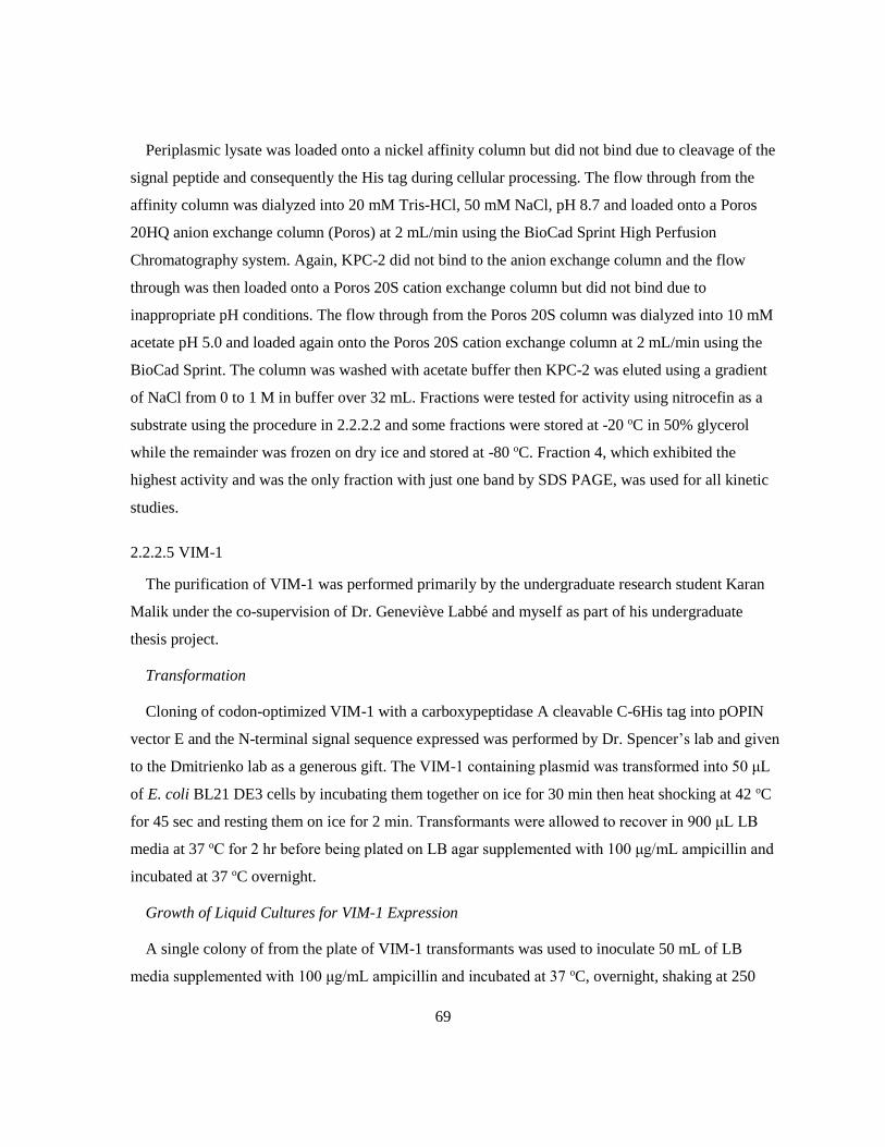
69
Periplasmic lysate was loaded onto a nickel affinity column but did not bind due to cleavage of the
signal peptide and consequently the His tag during cellular processing. The flow through from the
affinity column was dialyzed into 20 mM Tris-HCl, 50 mM NaCl, pH 8.7 and loaded onto a Poros
20HQ anion exchange column (Poros) at 2 mL/min using the BioCad Sprint High Perfusion
Chromatography system. Again, KPC-2 did not bind to the anion exchange column and the flow
through was then loaded onto a Poros 20S cation exchange column but did not bind due to
inappropriate pH conditions. The flow through from the Poros 20S column was dialyzed into 10 mM
acetate pH 5.0 and loaded again onto the Poros 20S cation exchange column at 2 mL/min using the
BioCad Sprint. The column was washed with acetate buffer then KPC-2 was eluted using a gradient
of NaCl from 0 to 1 M in buffer over 32 mL. Fractions were tested for activity using nitrocefin as a
substrate using the procedure in 2.2.2.2 and some fractions were stored at -20 oC in 50% glycerol
while the remainder was frozen on dry ice and stored at -80 oC. Fraction 4, which exhibited the
highest activity and was the only fraction with just one band by SDS PAGE, was used for all kinetic
studies.
2.2.2.5 VIM-1
The purification of VIM-1 was performed primarily by the undergraduate research student Karan
Malik under the co-supervision of Dr. Geneviève Labbé and myself as part of his undergraduate
thesis project.
Transformation
Cloning of codon-optimized VIM-1 with a carboxypeptidase A cleavable C-6His tag into pOPIN
vector E and the N-terminal signal sequence expressed was performed by Dr. Spencer’s lab and given
to the Dmitrienko lab as a generous gift. The VIM-1 containing plasmid was transformed into 50 μL
of E. coli BL21 DE3 cells by incubating them together on ice for 30 min then heat shocking at 42 oC
for 45 sec and resting them on ice for 2 min. Transformants were allowed to recover in 900 μL LB
media at 37 oC for 2 hr before being plated on LB agar supplemented with 100 μg/mL ampicillin and
incubated at 37 oC overnight.
Growth of Liquid Cultures for VIM-1 Expression
A single colony of from the plate of VIM-1 transformants was used to inoculate 50 mL of LB
media supplemented with 100 μg/mL ampicillin and incubated at 37 oC, overnight, shaking at 250

70
rpm. Two flasks with a 4 L capacity containing 1 L of LB media supplemented with 100 μg/mL
ampicillin were each inoculated with 20 mL of overnight culture and shaken at 225 rpm and 37 oC.
As the OD600 of the culture reached 0.4, expression of VIM-1 was induced with 1 mM IPTG for 4
hours shaking at 225 rpm and 37 oC. Cells were harvested by centrifugation at 6000 rpm for 20 min at
4 oC; nitrocefin tests determined that the majority of the VIM-1 activity was in the supernatant.
Affinity Chromatography
VIM-1 containing supernatant was filtered through a 0.45 μm filter to remove particulates, then
imidazole was added to the solution to a final concentration of 20 mM. A HisPurTM Ni-NTA
Superflow Agarose (Thermo Scientific) in an Omnifit column housing mounted on a BioCad Sprint
Perfusion Chromatography system was equilibrated with VIM-1 Chelating buffer (50 mM Tris-HCl,
300 mM NaCl, 20 mM imidazole, 10 % (v/v) glycerol, pH 8.0) before 200 mL VIM-1 supernatant
was loaded onto the column at 4 mL/min through the pump. The column was washed with 90% VIM-
1 Chelating buffer and 10 % VIM-1 Elution buffer (50 mM Tris-HCl, 300 mM NaCl, 150 mM
imidazole, 10 % (v/v) glycerol, pH 8.0) until the OD280 was reading less than 0.1. VIM-1 was eluted
from the column using VIM-1 Elution buffer then dialyzed 50 mM Tris-HCl, 150 mM NaCl, pH 8.0
at 4 oC. Pure His-tagged VIM-1 was stored in 50 % glycerol at -20 oC. Due to time constraints and
low yield, the tag was not cleaved; however, the kinetic parameters of His-VIM-1 did not differ
significantly from literature values.464 The concentrations of VIM-1 fractions were determined by
A280 (ε = 29910 M-1cm-1) using NanoDrop (Thermo Scientific) instead of Bradford due to low
concentrations. The concentration of the final VIM-1 stock was determined by Bradford as described
in 2.2.2.2.
2.2.2.6 VIM-2
Dr. Geneviève Labbé cloned VIM-2 out of the pNOR 2001 vector provided by Dr. Patrice
Nordmann (South Paris University, Paris, France) into a pET28a vector and purified the protein as
previously described.453,465
2.2.2.7 GC-1
Transformation
The cloning of GC-1 into the high expression plasmid pCS100 was performed and published by Dr.
M. Nukaga prior to it being obtained by the Dmitrienko lab.219 The recommended E. coli strain for

71
purification from this plasmid was AS266-51 which is an ampC deficient C600 with a mutant ampD
to prevent AmpC contamination of the GC-1.219,466 When transformations of GC-1 containing
pCS100 were attempted into competent E. coli AS266-51, colonies formed on 50 μg/mL
chloramphenicol LB agar plates; however, activity was not observed upon growth and induction in
LB broth. In later trials, a side by side transformation of 5 μL of pCS100-GC-1 into E. coli AS266-
51, E. coli BL21 DE3, and E. coli BL21 PLysS was performed by incubating the plasmid with CaCl2
competent cells for 30 min on ice before heat shocking at 42 oC for 45 sec. Cells were returned to ice
before the addition of 950 μL of LB. Cells were allowed to recover for 1 hr at 37 oC. Transformants
were then plated on LB agar supplemented with 50 μg/mL chloramphenicol and incubated at 37 oC
overnight. It was determined from induction of small scale liquid cultures that BL21 DE3 was the
best strain for growing GC-1 despite the potential for a minor AmpC contaminant.
Growth of Liquid Cultures for GC-1 Overexpression
A single colony of pCS100-GC-1 transformed E. coli BL21 DE3 was used to inoculate 70 mL TB
broth supplemented with 50 μg/mL chloramphenicol which was incubated at 37 oC shaking at 225
rpm overnight. Six flasks with a 4 L capacity containing 1 L of TB broth supplemented with 50
μg/mL chloramphenicol were then inoculated with 10 mL each of the overnight culture which were
incubated at 37 oC shaking at 190 rpm until they reached an optical density at 600 nm of
approximately 0.6. As each flask reached the desired optical density, it was induced with 1 mM IPTG
and returned to incubator overnight. Cells were harvested by centrifugation at 6000 rpm for 20 min at
4 oC in a JA-10 rotor yielding 54.6 g of pellet. The activity of both the supernatant and a crude lysis
of the pellet (using 0.1 mg/mL lysozyme incubated for 10 min at room temperature) were determined
using a nitrocefin based activity assay as described in 2.2.2.2. It was determined that the majority of
the GC-1 activity was in the supernatant.
Fractionation by Ammonium Sulfate
Solid ammonium sulfate was dissolved in the supernatant to a final concentration of 70% (w/v) at
room temperature then stirred at 4 oC for 2 days. The precipitated proteins were extracted by
centrifugation at 13500 xg for 60 min at 4 oC in a JA-10 rotor. The extracted proteins, including GC-
1, were then slowly resuspended at 100 rpm in 8 mL of 50 mM NaPi pH 7.0 per gram of pellet at 4
oC. Any remaining precipitated proteins were extracted by centrifugation at 13500 xg for 60 min at 4
oC then discarded after the presence of GC-1 in the supernatant was confirmed using the nitrocefin

72
assay described above. The supernatant (230 mL) was dialyzed against 50 mM NaPi pH 7.0 at 4 oC
until the ammonium sulfate concentration was calculated to be below acceptable limits for ion
exchange.
Ion Exchange Chromatography
The dialyzed protein solution was diluted in an equal volume of 50 mM MES pH 5.0 then the pH
was adjusted to approximately 5.0 with 6 N HCl. The acidified protein was then filtered through a
0.45 μm filter to ensure no particulates remained in the solution. A POROS CM-Sepharose column
mounted on a BioCad Sprint Perfusion Chromatography system was equilibrated in 50 mM MES pH
5.0 then loaded at 3 mL/min with the acidified protein ensuring the flow through did not have β-
lactamase activity. The column was then washed with 3 column volumes of 50 mM MES pH 5.0 at 3
mL/min followed by 1 column volume each of 50 mM MES, 30 mM NaCl pH 5.0 and 50 mM MES,
50 mM NaCl pH 5.0 at 5 mL/min. A final wash was performed with 2.5 column volumes of 50 mM
MES, 100 mM NaCl pH 5.0. GC-1 was eluted from the column using a salt gradient from 50 mM
MES, 100 mM NaCl pH 5.0 to 50 mM MES, 500 mM NaCl pH 5.0 over 4 column volumes at 5
mL/min. Fractions containing the highest GC-1 activity by the nitrocefin assay were pooled and
concentrated using Amicon Ultra-15 membrane filters with a 10 kDa molecular weight cutoff. An
equal volume of glycerol was added to the concentrated GC-1 for storage at -20 oC.
Size Exclusion Chromatography
Dr. Geneviève Labbé performed the size exclusion chromatography to increase the purity of the
GC-1 preparations by first dialyzing the glycerol stock into 50 mM MES, 0.2 M NaCl pH 5.5. The
dialyzed GC-1 was then concentrated using a Pall Macrosep Advance Centrifugal device with a 10
kDa molecular weight cutoff down to 1.5 mL (2 mL after filter was rinsed). The concentrated GC-1
solution was centrifuged at 14 000 rpm for 5 min to remove dust and particulates before injecting it
onto a Superdex 200 26/60 size exclusion column mounted on an AKTA Explorer HPLC system.
GC-1 was eluted using isocratic 50 mM MES, 0.2 M NaCl, pH 5.5 at 2 mL/min. Collected fractions
with the highest β-lactamase activity by nitrocefin assay were pooled and concentrated using another
Pall Macrosep Advance Centrifugal device with a 10 kDa molecular weight cutoff. Concentrated GC-
1 was diluted with an equal volume of glycerol for storage at -20 oC.

73
2.2.2.8 OXA-48
The purification of OXA-48 was performed primarily by Alicia Tjahjadi as part of her
undergraduate thesis project under the co-supervision of Dr. Geneviève Labbé and myself.
Transformation
Cloning of codon-optimized OXA-48 with a 3C Protease cleavable N-6His tag into pOPIN vector
F without expression of the signal sequence was performed by Dr. Spencer’s lab and given to the
Dmitrienko lab as a generous gift. The OXA-48 containing plasmid was transformed into 50 μL of
CaCl2 competent E. coli BL21 DE3 cells by incubating them together on ice for 30 min before heat
shocking them at 42 oC for 45 sec then resting them on ice for 2 min. Transformants were allowed to
recover in 950 μL of LB for 2 hr at 37 oC and then were plated on LB agar supplemented with 100
μg/mL ampicillin which was incubated at 37 oC overnight.
Growth of Liquid Cultures for OXA-48 Expression
A single colony from the plate of OXA-48 transformants was used to inoculate 50 mL of LB broth
supplemented with 100 μg/mL ampicillin in a 250 mL flask and incubated overnight at 37 oC, shaking
at 250 rpm. Two flasks with a 4 L capacity containing 1 L of LB media supplemented with 100
μg/mL ampicillin were each inoculated with 20 mL of overnight culture and incubated at 37 oC,
shaking at 225 rpm. Once an OD600 of approximately 0.6 was reached, expression of OXA-48 was
induced with 1 mM IPTG and incubated at 25 oC, shaking at 225 rpm overnight. Cells were harvested
by centrifugation at 3000 xg for 20 min at 4 oC. Cell pellets (11.4 g total) were resuspended in 40 mL
of OXA-48 Lysis buffer (50 mM Tris-HCl, 300 mM NaCl, 20 mM imidazole, 10 % (v/v) glycerol,
0.2 % (v/v) NP-40, 0.02 % (v/v) 1-thioglycerol, pH 8.0) then lysed using a large probe on a Heat
Systems Ultrasonic processor W-255 sonicator set to 50 % cycle and 50 % power 5 times for 30 sec
each with a 1 min rest on ice between bursts. Cellular debris was separated from the lysate by
centrifugation at 48 000 xg for 60 min at 4 oC and the supernatant was retained.
Affinity Chromatography
OXA-48 chromatography was performed in two runs each with the lysate of a different flask.
Imidazole was added to each lysate to a final concentration of 20 mM before being loaded at 2
mL/min onto HisPurTM Ni-NTA Superflow Agarose (Thermo Scientific) in an Omnifit column
housing mounted on a BioCad Sprint Perfusion Chromatography system that had been equilibrated
74
with OXA-48 Chelating buffer (50 mM Tris-HCl, 300 mM NaCl, 20 mM imidazole, 10 % (v/v)
glycerol, 0.01 % (v/v) 1-thioglycerol, pH 8.0). The column with OXA-48 bound was washed at 3
mL/min with OXA-48 Chelating buffer then at 4 mL/min with 90 % OXA-48 Chelating buffer and 10
% OXA-48 Elution buffer (50 mM Tris-HCl, 300 mM NaCl, 150 mM imidazole, 10 % (v/v) glycerol,
0.01 % (v/v) 1-thioglycerol, pH 8.0) over a total of 55-80 mL. His-tagged OXA-48 was eluted from
the column with 100 % OXA-48 Elution buffer while 10 mL fractions were collected until no activity
was detected in fractions by nitrocefin assay. Fractions containing the highest OXA-48 activity were
dialyzed into OXA-48 Storage buffer (50 mM Tris-HCl, 150 mM NaCl, 10 mM EDTA, 20 % (v/v)
glycerol, pH 8.0).
Cleavage of His-tag
The most active fraction from the two days (day 1 fraction 2) was chosen for tag cleavage. His-
tagged OXA-48 and His-tagged 3C Protease were both dialyzed at 4 oC using SpectraPor dialysis
membranes with a molecular weight cutoff of 8-10 kDa into Dialysis buffer (50 mM Tris-HCl, 150
mM NaCl, pH 8.0). The dialyzed OXA-48 was incubated with 1 mg of 3C Protease per 100 mg of
OXA-48 at 4 oC overnight to allow complete cleavage of the N-terminal tag. The protein mixture was
loaded onto Ni-NTA Superflow Agarose (Thermo Scientific) in an Omnifit column housing mounted
on a BioCad Sprint Perfusion Chromatography system that had been equilibrated with Dialysis buffer
at 4 mL/min. OXA-48 was collected in the flow through over 4 fractions then the 3C Protease was
eluted from the column with Protease Elution buffer (50 mM Tris-HCl, 300 mM NaCl, 150 mM
imidazole, pH 8.0). Pure OXA-48 with the His-tag cleaved was dialyzed as above, then stored at -20
oC in 50 % glycerol.
2.2.3 Characterization of Chromogenic Substrates
Chromogenic β-lactams: nitrocefin, UW-57, UW-58, and UW-154, were synthesized as an E/Z
mixture by Dr. Ahmed Ghavami as previously described, and their structures (Figure 2.3) and purity
were confirmed by 1H NMR, 13C NMR, and ESI-MS.453 Powders of these substrates were dissolved
in DMSO as a necessary step to isomerize completely to the E isomer to a final concentration of 40
mM and stored at -20 oC. If it was suspected that stocks had started to degrade (usually after years in
the freezer or if the colour changed), they were checked by UPLC or kinetic comparison to previous
data.

75
Figure 2.3 Chromogenic β-lactamase substrates synthesized by the Dmitrienko group.
2.2.3.1 Molar Extinction Coefficient Determination
Substrates were diluted to 100 μM in 50 mM HEPES pH 7.2 supplemented with 50 μg/ml BSA,
0.01 % Triton X-100, and 100 μM ZnCl2. One sample of this was read directly while the other was
completely hydrolyzed using 90 nM VIM-2 for 10 min. Spectral scans were performed using a Cary
50 spectrophotometer in quartz cuvettes from 250-750 nm in triplicate. The wavelength
corresponding to the greatest difference between the absorbance of the product and the substrate was
determined. A range of substrate concentrations from 1 μM to 100 μM were prepared in 50 mM
HEPES pH 7.2 supplemented with 50 μg/mL BSA and 0.01 % Triton X-100. Hydrolyzed substrates
were made by incubating with 27 nM VIM-2 until fully hydrolyzed. Greiner 96-well plates were
loaded with 200 μL volumes of both hydrolyzed and unhydrolyzed substrates then read at the
wavelength to be used for kinetics (NC, 482 nm; UW-57, 520 nm; UW-58, 534 nm; UW-154. 533
nm).
2.2.3.2 Substrate and Product Stability
Substrates were prepared as in 2.2.3.1 and the stability of both substrate and product were assessed
by taking scans from 325-750 nm (NC, UW-57, UW-58) in a SpectraMax 190 plate reader or from
250-750 nm in a Cary 50 spectrophotometer at 0 hr, 3 hr, 24 hr, and 96 hr. The 96-well plate
containing 200 μL samples of NC, UW-57, and UW-58 substrate and product was lidded and sealed
with Parafilm between reads to minimize evaporation.
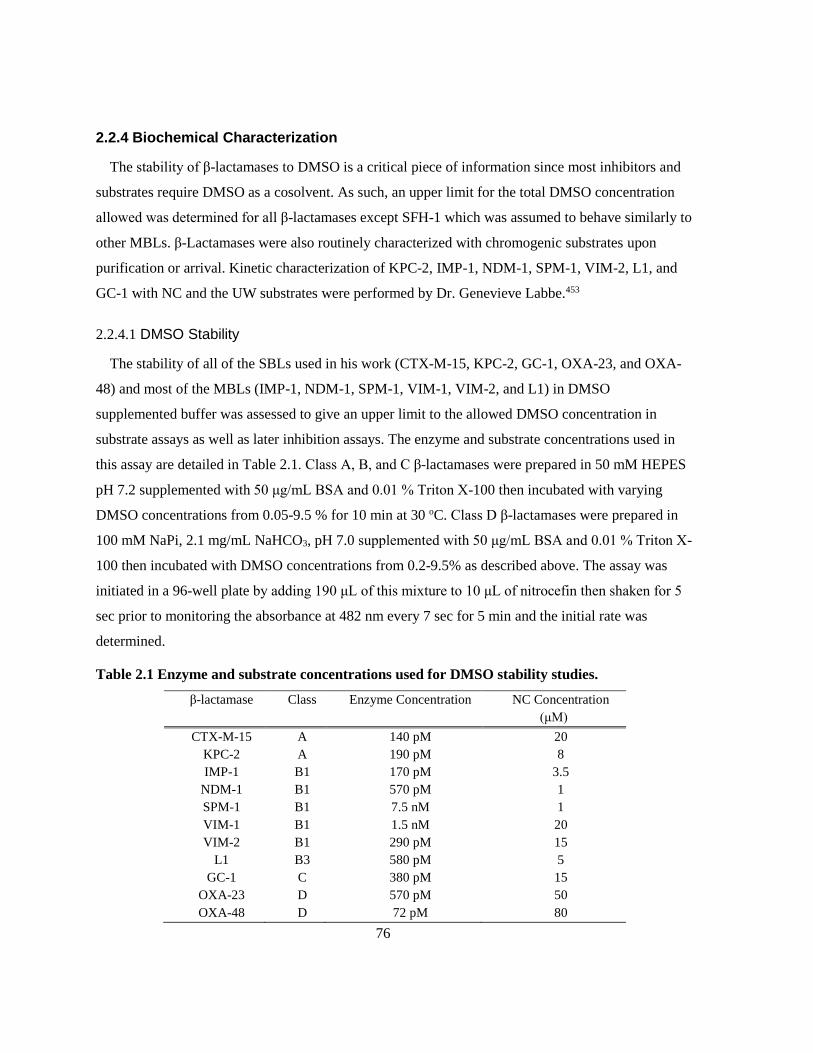
76
2.2.4 Biochemical Characterization
The stability of β-lactamases to DMSO is a critical piece of information since most inhibitors and
substrates require DMSO as a cosolvent. As such, an upper limit for the total DMSO concentration
allowed was determined for all β-lactamases except SFH-1 which was assumed to behave similarly to
other MBLs. β-Lactamases were also routinely characterized with chromogenic substrates upon
purification or arrival. Kinetic characterization of KPC-2, IMP-1, NDM-1, SPM-1, VIM-2, L1, and
GC-1 with NC and the UW substrates were performed by Dr. Genevieve Labbe.453
2.2.4.1 DMSO Stability
The stability of all of the SBLs used in his work (CTX-M-15, KPC-2, GC-1, OXA-23, and OXA-
48) and most of the MBLs (IMP-1, NDM-1, SPM-1, VIM-1, VIM-2, and L1) in DMSO
supplemented buffer was assessed to give an upper limit to the allowed DMSO concentration in
substrate assays as well as later inhibition assays. The enzyme and substrate concentrations used in
this assay are detailed in Table 2.1. Class A, B, and C β-lactamases were prepared in 50 mM HEPES
pH 7.2 supplemented with 50 μg/mL BSA and 0.01 % Triton X-100 then incubated with varying
DMSO concentrations from 0.05-9.5 % for 10 min at 30 oC. Class D β-lactamases were prepared in
100 mM NaPi, 2.1 mg/mL NaHCO3, pH 7.0 supplemented with 50 μg/mL BSA and 0.01 % Triton X-
100 then incubated with DMSO concentrations from 0.2-9.5% as described above. The assay was
initiated in a 96-well plate by adding 190 μL of this mixture to 10 μL of nitrocefin then shaken for 5
sec prior to monitoring the absorbance at 482 nm every 7 sec for 5 min and the initial rate was
determined.
Table 2.1 Enzyme and substrate concentrations used for DMSO stability studies.
β-lactamase Class Enzyme Concentration NC Concentration
(μM)
CTX-M-15 A 140 pM 20
KPC-2 A 190 pM 8
IMP-1 B1 170 pM 3.5
NDM-1 B1 570 pM 1
SPM-1 B1 7.5 nM 1
VIM-1 B1 1.5 nM 20
VIM-2 B1 290 pM 15
L1 B3 580 pM 5
GC-1 C 380 pM 15
OXA-23 D 570 pM 50
OXA-48 D 72 pM 80

77
2.2.4.2 Kinetic Parameter Determination
CTX-M-15
The determination of kinetic parameters for CTX-M-15 was performed for both the tagged and
cleaved versions of the protein in both HEPES and phosphate buffers. CTX-M-15 (tagged: 44 pM, tag
cleaved: 152 pM) was prepared in 50 mM HEPES pH 7.2 or 100 mM NaPi pH 7.2 supplemented with
50 μg/mL BSA and 0.01 % Triton X-100 then incubated for 10 min at 30 oC in a 96-well plate. The
reaction was initiated by addition of CTX-M-15 mix to 16 concentrations of substrate (500 μM – 1
μM) in triplicate then shaking for 5 sec before monitoring the absorbance every 6-7 sec for 5 min
(NC: 482 nm, UW-57: 520 nm, UW-58: 534 nm, UW-154: 533 nm) and determining initial rates.
Rates were converted into molar units using Beer’s Law and molar extinction coefficients for the
hydrolysis of each substrate determined in 2.2.3.1 (NC: 11 470 M-1cm-1, UW-57: 10 630 M-1cm-1,
UW-58: 12 420 M-1cm-1, UW-154: 10 870 M-1cm-1) then fitted to equations for Michaelis-Menten
kinetics (Equation 2.1) or substrate inhibition (Equation 2.2) as appropriate where v is the rate of
product formation and [S] is the concentration of substrate.467
Equation 2.1 𝒗 =𝑽𝒎𝒂𝒙[𝑺]
𝑲𝑴+[𝑺]
Equation 2.2 𝒗 =𝑽𝒎𝒂𝒙[𝑺]
𝑲𝑴+[𝑺](𝟏+[𝑺]
𝑲𝑰)
VIM-1
Kinetic parameters for VIM-1 were determined using methods similar to that of CTX-M-15 using
50 mM HEPES pH 7.2, a tagged VIM-1 final concentration of 1.6 nM for NC and 640 pM for the
UW substrates and a substrate concentration range from 500 μM to 0.5 μM.
SFH-1
Kinetic parameters for SFH-1 were determined using methods similar to that of CTX-M-15 using
50 mM HEPES pH 7.2, SFH-1 final concentrations of 9 nM (nitrocefin) and 1.8 nM (UW substrates),
and a substrate concentration range from 500 μM to 0.5 μM.
78
OXA-23
Kinetic parameters for OXA-23 were determined using methods similar to that of CTX-M-15 using
100 mM NaPi, 2 mg/mL NaHCO3, pH 7.0; OXA-23 final concentrations of 600 pM (nitrocefin), 1.2
nM (UW-57, UW-154), and 3 nM (UW-58); and a substrate concentration range from 500 - 0.5 μM.
OXA-48
Kinetic parameters for OXA-48 were determined using methods similar to that of CTX-M-15 using
100 mM NaPi, 2 mg/mL NaHCO3, pH 7.0; OXA-48 final concentrations of 87 pM (nitrocefin), 347
pM (UW-57), 173 pM (UW-58, UW-154); and a substrate concentration range from 500 μM to 5 μM.
2.3 Results
2.3.1 Metallo-β-Lactamase Protein Sequence Alignment
Alignments of the protein sequences of NDM-1, IMP-1, and VIM-2 with other variants from each
family was done using a variety of web-based programs on phylogeny.fr. This was not done for SPM-
1 or L1 as they are no other MBL variants in their families.
2.3.1.1 NDM Family Alignment
The amino acid sequence of the NDM family of MBLs is highly conserved resulting in a
phylogenetic tree where branches generally indicate a single amino acid change as seen in Figure 2.4.

79
Figure 2.4 Phylogenetic tree of NDM Variants generated using phylogeny.fr and TreeDyn.
Most of the 19 variants within the NDM family are within 1-2 amino acids of NDM-1 with the
exception of NDM-10 and NDM-18 which differ by 5 amino acids. A full list of the amino acid
changes in the variants relative to NDM-1 can be found in Table 2.2.
The complete sequence alignment for NDM-1 and all of its variants can be found in Appendix E.
The most common mutation found among the variants is the M154L mutation which is present in 9 of
the 19 variants. Mutation of D130 to either G or N is also fairly common. Other mutations were either
unique to a variant or were only found in a small subset of the variants.
80
Table 2.2 Amino acid differences between NDM-1 and other NDM variants.
NDM Variant Mutation Relative to NDM-1 numbering
NDM-2 P28A
NDM-3 D95N
NDM-4 M154L
NDM-5 V88L, M154L
NDM-6 A230V
NDM-7 D130N, M154L
NDM-8 D130G, M154L
NDM-9 E152K
NDM-10 R32S, G36D, G69S, A74T, G200R
NDM-11 M154V
NDM-12 M154L, G222D
NDM-13 D95N, M154L
NDM-14 D130G
NDM-15 M154L, A230V
NDM-16 R264H
NDM-17 V88L, M154L, E170K
NDM-18 D48_L49insQRFGD
NDM-19 D130N, M154L, A230V
2.3.1.2 VIM Family Alignment
Whereas NDM family contains only 19 variants including NDM-1, the VIM family contains 55
variants making it likely that there will be more than just 1 or 2 amino acids difference between
variants. As seen in the phylogenetic tree in Figure 2.5, there are two main branches of the tree; those
that are VIM-1 like, and those that are VIM-2 like. There are 25 amino acids different between VIM-
1 and VIM-2: F2L, L4V, L5I, K7S, L12M, I16V, I19V, F25H, V27G, D28E, S29P, S37N, V145A,
S192N, I200V, Y201H, R205S, I223V, Q228K, Q234E, F235V, K250Q, T253A, T260K, V265A.
Within the two groupings, variation is fairly consistent with only a few amino acids differing between
variants in that group. It is worth noting that VIM-7 is widely divergent from both the VIM-1 like and
VIM-2 like variants. The complete sequence alignment as well as a table summarizing the amino acid
changes of the VIM variants relative to VIM-2 can be found in Appendix E.

81
Figure 2.5 Phylogenetic Tree of VIM variants generated by phylogeny.fr and TreeDyn.

82
2.3.1.3 IMP Family Alignment
The IMP family is the largest of the MBL families with 70 assigned variants. Of these 70 variants,
IMP-36, IMP-39, IMP-50, and IMP-65 do not have protein sequences in the NCBI database and IMP-
47 has the same amino acid sequence as IMP-8, so IMP-47 was left out of the phylogenetic analysis.
The phylogenetic tree generated from this analysis, seen in Figure 2.6, is much more branched than
that of the NDM variants or the VIM variants, indicative of the wide sequence variety in this family.
The small subset of IMP variants found near IMP-1 on the phylogenetic tree are very similar;
however, at the extremities of the tree, variants can differ by over 50 amino acids from IMP-1. The
full protein sequence alignment and a table summarizing the amino acid changes relative to IMP-1
can be found in Appendix E.
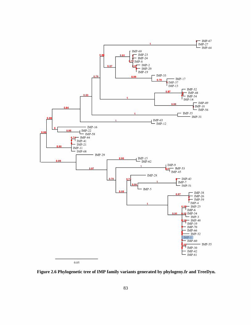
83
Figure 2.6 Phylogenetic tree of IMP family variants generated by phylogeny.fr and TreeDyn.

84
The analysis of the relationships between NDM-1, IMP-1, VIM-2, and their respective variants
indicates that there is little sequence similarity between MBL families. The NDM variants are all
closely related differing by only a few amino acids between NDM-1 and any given NDM variant.
Due to both its sequence similarity and clinical significance NDM-1 should be a suitable
representative of this family. The VIM family contains two major groupings: VIM-1-like and VIM-2-
like. One variant from each these groupings should be represented in screening to check if both
branches respond similarly to substrates and inhibitors - VIM-1 and VIM-2 were selected. The
amount of variability in the IMP family makes it unreasonable to test representatives of each branch
of the phylogenetic tree, so IMP-1 was used as it is among the most prevalent in clinical isolates and
is readily available.276
2.3.2 Protein Purification
2.3.2.1 CTX-M-15 Purification
His tagged CTX-M-15 was purified by affinity chromatography before tag cleavage with thrombin.
This was an efficient purification in which 12 % of the active protein from the crude was collected in
fractions that were used for concentration and cleavage (Table 2.3). The reduction of CTX-M-15
from the 829 mg present in the lysate to the 34 mg eluted from the Ni-NTA column can be explained
though its use in method development and its presence in fractions that were not pooled. The apparent
loss of CTX-M-15 from the thrombin cleavage is due to only a small portion of the protein having
undergone tag cleavage. The remainder of the tagged CTX-M-15 was stored at -20 oC in glycerol for
possible His-tag cleavage in the future.
Table 2.3 CTX-M-15 purification table
Total activity
(μmol/min)
% Yield Total Protein
(mg)
Specific activity
(μmol/min/mg)
Fold
Purification
Lysate 14000 100 829 17.1 1
Ni-NTA 1700 12 34 49.4 2.9
Concentration 1600 11 34 45.7 2.7
Thrombin Cleavage 56 0.4 0.78 71.6 4.2

85
2.3.2.2 KPC-2 Purification
The cloning of KPC-2 into a plasmid to introduce a His-tag was futile as the tag was removed during
cellular processing along with the signal peptide. The active flow through from the Poros 20HQ
column was not formally assayed with NC, run on SDS PAGE, nor aliquoted for future checks. As a
result of this lack of testing, it is unknown whether or not this step contributed to purification;
although, it is likely that it is responsible for the removal of some anionic proteins. This in
conjunction with the purification by the Poros 20S column resulted in > 95 % pure protein by SDS
PAGE (Figure 2.7).
Table 2.4 KPC-2 purification table.
Total activity
(μmol/min)
% Yield Total Protein
(mg)
Specific activity
(μmol/min/mg)
Fold
Purification
Lysate 54 100 4.4 12 1
Poros 20HQ - 20S 0.25 0.46 0.40 0.62 0.05
2.3.2.3 VIM-1 Purification
VIM-1 was not overexpressed well in E. coli BL21 DE3 cells; however, what protein was expressed
was found predominantly in the media since the signal peptide was included in the construct. The
media was loaded onto a Ni-NTA affinity column and VIM-1 eluted across 3 fractions, one of which
was further concentrated yielding 0.5 mg VIM-1 (Table 2.5). Potentially much more VIM-1 could
have been purified from the remaining 1.8 L of media; however, the amount that was produced was
adequate for the present study.
Table 2.5 VIM-1 purification table
Total activity
(μmol/min)
% Yield Total Protein
(mg)
Specific activity
(μmol/min/mg)
Fold
Purification
Media Supernatant 38.9 100 92 0.42 1
Ni-NTA 0.84 2.2 0.56 0.84 3.5
Concentration 0.61 1.6 0.51 0.61 2.8
2.3.2.4 GC-1 Purification
Early testing of the lysate fractions (media, periplasm, and cytosol) determined that most of the
GC-1 was exported into the media when it was overexpressed. Since the GC-1 construct did not

86
include an affinity tag, a 70% ammonium sulfate cut was used to precipitate GC-1 which was then
resolubilized, removing 200 mg of various proteins (Table 2.6). This mixture was then purified by ion
exchange and cleaned by size exclusion chromotography yielding 2.5 mg of pure GC-1.
Table 2.6 GC-1 purification table.
Total activity
(μmol/min)
% Yield Total Protein
(mg)
Specific activity
(μmol/min/mg)
Fold
Purification
Media Supernatant 65319 100 660 99 1
Ammonium Sulfate
Soluble Fraction 26024 39.8 460 57 0.6
CM Sepharose 11962 18.3 9.2 1293 13
Sephadex 200 1859 2.8 2.5 774 7.5
Early attempts at purification of KPC-2, IMP-1, and VIM-2 were performed using classical
purification techniques such as those used in this GC-1 purification. Each of those purifications failed
with all of the protein being lost on either the ion exchange or size exclusion columns. Considering
this early experience, it came as a surprise that this purification proceded relatively well, yielding 2.5
mg of pure GC-1. The good yield of GC-1 as compared to those earlier purifications is likely because
the GC-1 purification was performed using an FPLC system while the others were performed using
gravity columns.
2.3.2.5 OXA-48 Purification
OXA-48 was overexpressed well and was contained within the cells since no signal peptide was
present in the construct. A lack of characterization at the time of purification necessitated the sample
of frozen lysate being tested 3 years after the purification was completed. It is probable that this delay
skewed the results since the target protein has been exposed to proteolytic enzymes for an extended
period. For this reason, the non-physical values for % yield (2.5 million %) and fold purification (270
000 fold) were not included in Table 2.7.

87
Table 2.7 OXA-48 purification table.
Total activity
(μmol/min)
Total Protein (mg) Specific activity
(μmol/min/mg)
Lysate 0.34* 350 0.00097*
Ni-NTA 16700 52 180
His-tag cleavage 8300 32 260
*3 year delay in analysis of this sample. Proteolytic degradation is likely.
2.3.2.6 SDS PAGE of Purified β-Lactamases
A summary SDS-PAGE gel of all pure β-lactamases used in this thesis is presented in Figure 2.7 with
the enzymes purified in this laboratory on the left side of the gel and any gifted enzymes on the right
side of the gel. The β-lactamases purified at U Waterloo: KPC-2, VIM-1, VIM-2, and OXA-48, were
determined to be >95 % pure by SDS-PAGE as is evident by the presence of only a single band in
each well. CTX-M-15 and GC-1 samples appear to have minor contaminants, the CTX-M-15
contaminant is likely His-tagged CTX-M-15. Many of the β-lactamases received from other
researchers: NDM-1, SPM-1, SFH-1, L1, and OXA-23 were also determined to be >95 % pure. Some
small contaminant bands are evident in the IMP-1 sample and can likely be attributed to HRV 3C
protease and IMP-1 with an uncleaved His-tag that were not successfully removed from the sample.
Figure 2.7 SDS-PAGE of pure β-lactamases. 1 LMW Marker, 2 CTX-M-15, 3 KPC-2, 4 VIM-1,
5 VIM-2, 6 GC-1, 7 OXA-48, 8 LMW Marker, 9 IMP-1, 10 NDM-1, 11 SPM-1, 12 SFH-1, 13 L1,
14 OXA-23, 15 LMW Marker.

88
2.3.2.7 Confirmation of β-Lactamase Identity by Mass Spectrometry
The masses of proteins purified in this laboratory were confirmed by ESI mass spectrometry. In
addition, samples of NDM-1, SPM-1, and L1 were tested. This was used to confirm the successful
removal of the His-tags from CTX-M-15 and OXA-48 using thrombin and 3C protease respectively.
By the same logic, this confirms that the His tag was cleaved from KPC-2 during cellular processing
and was not present during purification.
Table 2.8 Masses of pure β-lactamases as predicted using Expasy ProtParam and determined
by mass spectroscopy
β-Lactamase Predicted Denatured
Mass (Da)
Mass Determined by
Mass Spectrometry (Da)
CTX-M-15 28 407 28 406
KPC-2 28 720 28 719
NDM-1 24 318 24 317
VIM-2 25 515 25 515
SPM-1 27 884 27 883
L1 28 706 28 703
GC-1 39 573 39 572
OXA-48 28 301 28 300
Some of the β-lactamases provided by other labs were also analyzed by ESI mass spectrometry as
either preliminary experiments for other work or to determine the specifics of the sample sequence
given to us. The mass of NDM-1 was determined because the sample provided to us was denoted as
Δ36 NDM-1 which implied that the first 36 amino acids were truncated; however, that did not match
any constructs published by that group. Upon determination of the mass, it was confirmed that the
first 41 amino acids including the signal peptide were excluded from the construct, consistent with the
published construct.317
In addition to the masses presented in Table 2.8, the native mass of SPM-1 was also determined to
be 28 009 Da (theoretical: 28 015 Da)468 which is consistent with 2 Zn ions in the active site. This
was performed in ammonium acetate buffer instead of 1:1 H2O:MeOH 0.1 % formic acid.
2.3.3 Characterization of Chromogenic Substrates
The colour change observed in the chromogenic β-lactams is produced by the hydrolysis of the β-
lactam ring by β-lactamases as shown in Scheme 2.2

89
Scheme 2.2 Hydrolysis of nitrocefin by β-lactamases.
Hydrolyzed and unhydrolyzed versions of the 4 substrates used in this work were plated at various
concentrations on a 96-well plate to illustrate the colour change (Figure 2.8). Nitrocefin shows a
characteristic change from yellow to red, UW-57 goes from yellow to red/brown, and both UW-58
and UW-154 turn from yellow to purple. This colour change can be exploited for kinetic analysis of
β-lactamases capable of hydrolyzing cephalosporins (ESBLs, Class C SBLs, and MBLs).
Figure 2.8 Visual appearance of 200 μL of increasing concentrations of unhydrolyzed and
hydrolyzed chromogenic substrates synthesized by the Dmitrienko lab (A: 10 μM, B: 25 μM,
C:50 μM, D: 75 μM, E: 100 μM, F: 200 μM). 1: unhydrolyzed NC, 2: hydrolyzed NC, 3:
unhydrolyzed UW-57, 4: hydrolyzed UW-57, 5: unhydrolyzed UW-58, 6: hydrolyzed UW-58, 7:
unhydrolyzed UW-154, 8: hydrolyzed UW-154.
At the same concentrations, both the unhydrolyzed and hydrolyzed forms of the UW substrates are
more more readily detectable visually. Additionally, the colour change of the UW substrates is more

90
distinctive since they are on opposite sides of the colour wheel instead of going to a neighbouring
colour (red-orange).
2.3.3.1 Molar Extinction Coefficient
The wavelength at which the greatest difference between substrate and product curves occurred,
λmax, was determined using spectral scans of both the unhydrolyzed and hydrolyzed versions of the
substrates produced by the Dmitrienko lab (Figure 2.9). The λmax is the optimal wavelength for kinetic
analysis as it is where the highest sensitivity occurs. The λmax was determined to be 482 nm for NC,
520 nm for UW-57, 534 nm for UW-58, and 533 nm for UW-154.
250 300 350 400 450 500 550 600 650 700 750
-0.5
0.0
0.5
1.0
1.5
2.0
Wavelength (nm)
Ab
so
rban
ce
250 300 350 400 450 500 550 600 650 700 750
-0.5
0.0
0.5
1.0
1.5
2.0
Wavelength (nm)
Ab
so
rban
ce
250 300 350 400 450 500 550 600 650 700 750
-0.5
0.0
0.5
1.0
1.5
2.0
Wavelength (nm)
Ab
so
rban
ce
250 300 350 400 450 500 550 600 650 700 750
-0.5
0.0
0.5
1.0
1.5
2.0
Wavelength (nm)
Ab
so
rban
ce
A B
C D
Figure 2.9 Spectral scans of chromogenic substrates before and after hydrolysis with the
unhydrolyzed represented by a yellow and the hydrolyzed represented in red/purple for
nitrocefin (A), UW-57 (B), UW-58 (C), and UW-154 (D) at 100 μM. This figure was generated
using GraphPad Prism v. 5.03.

91
Estimates for the molar extinction coefficients for the product can be determined from these scans:
NC ε482 = 15 990 M-1cm-1, UW-57 ε520 = 16 520 M-1cm-1, UW-58 ε534 = 16 590 M-1cm-1, and UW-154
ε533 = 17 140 M-1cm-1; however, it is more accurate to determine the molar extinction coefficients
from several different concentrations of substrate. In order to simplify further calculations, the molar
extinction coefficient of a 200 μL volume in a standard flat bottom 96-well plate (Greiner Bio One)
was determined and used for future calculations as well as the molar extinction coefficient in
conventional units as listed in Table 2.9. For the purposes of this work, it is most appropriate to use
the difference between molar extinction coefficient of the product and substrate, Δε. The differences
between the estimates and the calculated extinction coefficients illustrate the inaccuracy of single
point determinations and may have been exacerbated by these being from different batches of
different ages.
Table 2.9 Molar extinction coefficients for UW substrates
Molar extinction coefficient (M-1well-1)
Pathlength Substrate Substrate Product Difference
200 μL in Costar
655101 96-well
plate (0.571 cm)
NC 679 ± 8 7230 ± 50 6550 ± 50
UW-57 350 ± 10 6420 ± 30 6070 ± 40
UW-58 392 ± 5 7490 ± 19 7090 ± 20
UW-154 533 ± 2 6740 ± 20 6200 ± 20
Molar extinction coefficient (M-1cm-1)
1 cm NC 1190 ± 10 12 660 ± 80 11 470 ± 90
UW-57 620 ± 20 11 240 ± 50 10 630 ± 80
UW-58 686 ± 8 13 110 ± 30 12 420 ± 40
UW-154 933 ± 3 11 800 ± 30 10 870 ± 30
2.3.3.2 Substrate and Product Stability
The stability of both the unhydrolyzed and hydrolyzed versions of each substrate was assessed
using spectral scans at 0 hr, 3 hr, 24 hr, and 96 hr (Figure 2.10). The unhydrolyzed versions of the
substrates tend to be stable within the time frame of a day of assays and start to degrade within 24 hr.
The apparent hydrolysis of the substrates at 96 hr can be observed for NC, UW-57, and UW-58. The
products of the substrate hydrolysis are much less stable than the unhydrolyzed substrate often
showing a significant decrease in maximal absorbance at only 3 hr (with the exception of nitrocefin).

92
250 300 350 400 450 500 550 600 650 700 7500.0
0.2
0.4
0.6
0.8
1.0
Wavelength (nm)
Ab
so
rban
ce
250 300 350 400 450 500 550 600 650 700 7500.0
0.2
0.4
0.6
0.8
1.0
Wavelength (nm)
Ab
so
rban
ce
250 300 350 400 450 500 550 600 650 700 7500.0
0.2
0.4
0.6
0.8
Wavelength (nm)
Ab
so
rban
ce
250 300 350 400 450 500 550 600 650 700 7500.0
0.2
0.4
0.6
0.8
Wavelength (nm)
Ab
so
rban
ce
250 300 350 400 450 500 550 600 650 700 7500.0
0.5
1.0
Wavelength (nm)
Ab
so
rban
ce
250 300 350 400 450 500 550 600 650 700 7500.0
0.5
1.0
Wavelength (nm)
Ab
so
rban
ce
250 300 350 400 450 500 550 600 650 700 7500.0
0.5
1.0
1.5
2.0
Wavelength (nm)
Ab
so
rban
ce
250 300 350 400 450 500 550 600 650 700 7500.0
0.5
1.0
1.5
2.0
Wavelength (nm)
Ab
so
rban
ce
A B
C D
E F
G H
Figure 2.10 Stability spectra of 100 μM chromogenic substrates before and after hydrolysis.
NC (A: unhydrolyzed, B: hydrolyzed), UW-57 (C: unhydrolyzed, D: hydrolyzed), UW-58 (E:
unhydrolyzed, F: hydrolyzed), and UW-154 (G: unhydrolyzed, H: hydrolyzed) at 0 hr (red), 3
hr (green), 24 hr (blue), 96 hr (purple). This figure was generated using GraphPad Prism v.
5.03.

93
2.3.4 Biochemical Characterization
2.3.4.1 DMSO Stability
The relative activity of β-lactamases incubated for 10 min in various concentrations of DMSO is
presented in Figure 2.11 with stability being defined as a <10 % reduction in activity relative to a no
DMSO added control (0.05 %- 0.2 % DMSO present in NC). Of the SBLs, CTX-M-15, OXA-23, and
OXA-48 are only stable up to 2 % DMSO while KPC-2 and GC-1 are stable up to 5 % DMSO. VIM-
1 is the least DMSO stable of the MBLs, dipping below the acceptable activity reduction after only 1
% DMSO. The other MBLs are much more stable with SPM-1 and VIM-2 being stable up to 2.5 %
DMSO, IMP-1 and L1 being stable up to 5 % DMSO, and NDM-1 appearing to be activated in the
presence of DMSO. This suggests that in most cases, the presence of 2 % DMSO is tolerated well by
β-lactamases.
0 2 4 6 8 100
20
40
60
80
100
120CTX-M-15
KPC-2
GC-1
OXA-23
OXA-48
% DMSO
% A
cti
vit
y
0 2 4 6 8 100
20
40
60
80
100
120IMP-1
NDM-1
SPM-1
VIM-1
VIM-2
L1
% DMSO
% A
cti
vit
yA B
Figure 2.11 DMSO sensitivity of β-lactamases separated into (A) SBLs and (B) MBLs with a
cutoff line at 10 % activity reduction. This figure was generated using GraphPad Prism v. 5.03.
2.3.4.2 Determination of Kinetic Parameters
The determination of kinetic parameters was done by non-linear regression of the Michaelis-
Menten plots fitting to either Michaelis-Menten (Equation 2.1) or substrate inhibition (Equation 2.2)
equations as appropriate. The hydrolysis of NC and UW substrates in 50 mM HEPES pH 7.2 and 100
mM NaPi pH 7.2 by both tagged and cleaved CTX-M-15 is presented in Figure 2.12. These graphs
show that the tagged CTX-M-15 is more prone to substrate/product inhibition in both buffer systems
as seen by the reduction in rate of hydrolysis at high substrate concentrations. Substrate/product

94
inhibition is also observed when the His-tag cleaved CTX-M-15 but only in the HEPES buffer
system. The concentration of DMSO in these experiments was kept constant.
HEPES NaPi
Cleaved Tagged Cleaved Tagged
0 200 400 6000.00
0.02
0.04
0.06
0.08
[NC] (µM)
Rate
(
M/s
ec)
0 200 400 6000.00
0.02
0.04
0.06
0.08
0.10
[NC] (µM)
Rate
(
M/s
ec)
0 100 200 300 400 5000.00
0.02
0.04
0.06
0.08
0.10
[NC] (µM)
Rate
(
M/s
ec)
0 100 200 300 400 5000.0
0.1
0.2
0.3
[NC] (µM)
Rate
(
M/s
ec)
0 200 400 6000.000
0.005
0.010
0.015
0.020
[UW-57] (µM)
Rate
(
M/s
ec)
0 200 400 6000.00
0.01
0.02
0.03
0.04
[UW-57] (µM)
Rate
(
M/s
ec)
0 100 200 300 400 5000.00
0.01
0.02
0.03
0.04
[UW-57] (µM)
Rate
(
M/s
ec)
0 100 200 300 400 5000.00
0.05
0.10
0.15
[UW-57] (µM)
Rate
(
M/s
ec)
0 200 400 6000.000
0.005
0.010
0.015
[UW-58] (µM)
Rate
(
M/s
ec)
0 200 400 6000.000
0.005
0.010
0.015
0.020
[UW-58] (µM)
Rate
(
M/s
ec)
0 100 200 300 400 5000.00
0.01
0.02
0.03
0.04
0.05
[UW-58] (µM)
Rate
(
M/s
ec)
0 100 200 300 400 5000.00
0.05
0.10
0.15
[UW-58] (µM)
Rate
(
M/s
ec)
0 200 400 6000.00
0.01
0.02
0.03
0.04
[UW-154] (µM)
Rate
(
M/s
ec)
0 200 400 6000.00
0.01
0.02
0.03
0.04
0.05
[UW-154] (µM)
Rate
(
M/s
ec)
0 100 200 300 400 5000.00
0.01
0.02
0.03
0.04
0.05
[UW-154] (µM)
Rate
(
M/s
ec)
0 100 200 300 400 5000.00
0.05
0.10
0.15
[UW-154] (µM)
Rate
(
M/s
ec)
Figure 2.12 Michaelis-Menten plots of CTX-M-15 kinetic parameter determination. Each point
represents three replicate measurements, error bars are often smaller than the markers. This
figure was generated using GraphPad Prism v. 5.03.
The kinetic parameters determined by the non-linear regression of the graphs in Figure 2.12 can be
found in Table 2.10. It is evident that while substrate/product inhibition in HEPES is a slight problem
for CTX-M-15 with the tag cleaved, the KI is at least 10x greater than the KM for all substrates except
UW-154 where they are both about 25 μM. Overall, the KM and kcat for NC is about 3 times greater

95
than those for the UW substrates (particularly UW-57 and UW-58) and the kcat/KM is similar for all
substrates within a given set of conditions.
Table 2.10 Kinetic parameters of CTX-M-15 with and without its His-tag against NC and UW
substrates.
CTX-M-15
Preparation
Buffer Substrate KM (μM) KI (μM) kcat (s-1) kcat/KM
(μM-1s-1)
Cleaved HEPES NC 17.3 ± 0.8 580 ± 40 580 ± 10 33 ± 2
UW-57 5.6 ± 0.4 1110 ± 140 120 ± 2 21 ± 1
UW-58 5.8 ± 0.6 2400 ± 800 78 ± 2 13 ± 1
UW-154 24 ± 8 25 ± 8 640 ± 150 30 ± 10
NaPi NC 35.7 ± 0.9 1400 ± 90 716 ± 8 20.1 ± 0.5
UW-57 10.0 ± 0.2 223.6 ± 0.7 22.4 ± 0.3
UW-58 11.2 ± 0.3 270 ± 2 24.1 ± 0.7
UW-154 12.1 ± 0.2 280 ± 1 23.2 ± 0.5
Tagged HEPES NC 12 ± 1 150 ± 20 3270 ± 150 250 ± 30
UW-57 5.9 ± 0.6 1600 ± 400 790 ± 30 130 ± 20
UW-58 3.5 ± 0.6 330 ± 60 400 ± 20 110 ± 20
UW-154 4.3 ± 0.2 730 ± 60 1120 ± 20 260 ± 20
NaPi NC 57 ± 4 600 ± 70 9700 ± 400 170 ± 10
UW-57 15.4 ± 0.5 2100 ± 200 2780 ± 30 181 ± 6
UW-58 18.8 ± 0.8 930 ± 80 3580 ± 70 190 ± 9
UW-154 30 ± 3 150 ± 20 4660 ± 290 160 ± 20
Combinations without a KI value were fitted to Equation 2.1 rather than Equation 2.2.
The kinetic parameters of all other β-lactamases used in this thesis have also been determined for
NC and the UW substrates. Many of these experiments were published by Dr. Geneviève Labbé using
the β-lactamase preparations described above and have been included in Table 2.11.453 Michaelis-
Menten plots for the β-lactamases not included in the publication by Ghavami et al. are presented in
Figure 2.13 and show typical Michaelis-Menten behaviour with the exception of OXA-48 and VIM-1
which display substrate/product inhibition.
The kinetic parameters for the SBLs used in this thesis against NC and UW substrates are listed in
Table 2.11. Generally, KPC-2 and GC-1 β-lactamases have the lowest KM values for NC and the
highest KM values for UW-154, a trend that is mirrored in the kcat values. This KM trend does not hold
for Class D β-lactamases which have significantly higher KM values for NC than any other tested
substrate by 3 – 4 fold. The kcat values for the Class D SBLs scale with the KM values as seen in KPC-
2 and GC-1. Unlike the other SBLs, GC-1 is susceptible to substrate/product inhibition.

96
VIM-1 SFH-1 OXA-23 OXA-48
0 200 400 6000.00
0.01
0.02
0.03
0.04
0.05
[NC] (µM)
Rate
(
M/s
ec)
0 200 400 6000.000
0.005
0.010
0.015
[NC] (µM)
Rate
(
M/s
ec)
0 200 400 6000.00
0.02
0.04
0.06
[NC] (µM)
Rate
(
M/s
ec)
0 200 400 6000.00
0.02
0.04
0.06
0.08
0.10
[NC] (µM)
Rate
(
M/s
ec)
0 200 400 6000.00
0.01
0.02
0.03
0.04
[UW57] (µM)
Rate
(
M/s
ec)
0 200 400 6000.000
0.005
0.010
0.015
[UW57] (µM)
Rate
(
M/s
ec)
0 200 400 6000.00
0.02
0.04
0.06
0.08
[UW57] (µM)
Rate
(
M/s
ec)
0 200 400 6000.00
0.01
0.02
0.03
0.04
[UW58] (µM)
Rate
(
M/s
ec)
0 200 400 6000.00
0.02
0.04
0.06
0.08
[UW58] (µM)
Rate
(
M/s
ec)
0 200 400 6000.00
0.02
0.04
0.06
0.08
[UW58] (µM)
Rate
(
M/s
ec)
0 200 400 6000.000
0.005
0.010
0.015
0.020
0.025
[UW154] (µM)
Rate
(
M/s
ec)
0 200 400 6000.00
0.01
0.02
0.03
[UW154] (µM)
Rate
(
M/s
ec)
0 200 400 6000.00
0.02
0.04
0.06
0.08
[UW154] (µM)
Rate
(
M/s
ec)
0 200 400 6000.00
0.01
0.02
0.03
0.04
0.05
[UW-57] (µM)
Rate
(
M/s
ec)
0 200 400 6000.00
0.01
0.02
0.03
0.04
[UW-58] (µM)
Rate
(
M/s
ec)
0 200 400 6000.00
0.01
0.02
0.03
[UW-154] (µM)
Rate
(
M/s
ec)
Figure 2.13 Michaelis-Menten plots of VIM-1, SFH-1, OXA-23, and OXA-48 kinetic parameter
determination. Each point represents three replicate measurements, error bars are often
smaller than the markers. This figure was generated using GraphPad Prism v. 5.03.
The KI values for this inhibition tend to be at least 10x greater than the KM values (except for UW-57
where KI is only about 4x larger than the KM) so they should not be problematic for kinetic
experiments performed at KM concentrations of substrate. The catalytic efficiencies (kcat/KM) of all
substrates tend to be fairly consistent for each SBL.
The kinetic parameters for the MBLs with NC and the UW substrates (Table 2.12) tend to follow
the same trend seen for the SBLs. The KM values for all substrates tend to be fairly consistent and any

97
variation is generally mirrored in the kcat values leading to consistent catalytic efficiencies. The only
exceptions to this are the NC values for SPM-1, SFH-1, and L1 in which the KM is slightly (1.1 to
1.5-fold) smaller than it is for other substrates and the kcat is significantly (5 to 10-fold) smaller
leading to much lower catalytic efficiencies.
Table 2.11 Kinetic parameters of serine-β-lactamases (except CTX-M-15) against NC and UW
substrates.
Enzyme Class Substrate KM (μM) KI (μM) kcat (s-1) kcat/KM
(μM-1s-1)
KPC-2a A NC 8.1 ± 0.2 520 ± 3 64 ± 2
UW-57 12.6 ± 0.2 463 ± 2 36.8 ± 0.6
UW-58 14.1 ± 0.3 607 ± 3 43.1 ± 0.9
UW-154 20.9 ± 0.5 799 ± 7 38 ± 1
GC-1a C NC 15.4 ± 0.8 390 ± 20 920 ± 20 60 ± 4
UW-57 35 ± 4 130 ± 20 1700 ± 100 49 ± 6
UW-58 20.3 ± 0.7 390 ± 20 1460 ± 30 72 ± 3
UW-154 35 ± 2 270 ± 30 2200 ± 90 63 ± 4
OXA-23 D NC 51 ± 2 95 ± 1 1.86 ± 0.08
UW-57 6.3 ± 0.2 8.85 ± 0.06 1.41 ± 0.05
UW-58 15.2 ± 0.2 24.44 ± 0.07 1.60 ± 0.02
UW-154 15.7 ± 0.3 23.2 ± 0.1 1.48 ± 0.03
OXA-48 D NC 82 ± 2 980 ± 50 1650 ± 20 20.0 ± 0.6
UW-57 9.2 ± 0.2 242.8 ± 0.8 26.3 ± 0.7
UW-58 21.5 ± 0.5 351 ± 2 16.3 ± 0.4
UW-154 20.5 ± 0.4 3787 ± 1 18.8 ± 0.4 aThese assays were performed by Dr. Geneviève Labbé and were reported by Ghavami et al.453
Combinations without a KI value were fitted to Equation 2.1 rather than Equation 2.2.
As was seen for the SBLs, some enzymes are subject to substrate/product inhibition; in this case
VIM-1, SPM-1 and NDM-1. For SPM-1 and NDM-1, the KMs are extremely low and the KI values
for the inhibition are around 100x higher. This substrate/product inhibition should not be a problem
for inhibition kinetics performed near KM but should be kept in mind for those performed in excess
substrate (usually only 10x KM). This consideration is irrelevant for NDM-1 as it is only inhibited by
UW-57 and inhibition kinetics were performed with NC. For VIM-1, only UW-58 and UW-154
display significant substrate/product inhibition, which is not a problem for kinetic assays performed
with NC.

98
Table 2.12 Kinetic parameters of metallo-β-lactamases with NC and UW substrates
Enzyme Class Substrate KM (μM) KI (μM) kcat (s-1) kcat/KM
(μM-1s-1)
IMP-1a B1 NC 3.5 ± 0.2 236 ± 3 67 ± 4
UW-57 2.5 ± 0.1 229 ± 2 92 ± 4
UW-58 3.0 ± 0.1 235 ± 2 78 ± 3
UW-154 6.8 ± 0.2 576 ± 5 85 ± 3
NDM-1a B1 NC 0.88 ± 0.06 43.7 ± 0.5 50 ± 3
UW-57 1.43 ± 0.09 1300 ± 100 120 ± 2 84 ± 5
UW-58 2.15 ± 0.09 185 ± 1 86 ± 4
UW-154 2.6 ± 0.1 237 ± 2 91 ± 4
SPM-1a B1 NC 1.2 ± 0.1 630 ± 90 4.9 ± 0.1 4.1 ± 0.4
UW-57 1.5 ± 0.1 270 ± 20 25.8 ± 0.5 17 ± 1
UW-58 2.3 ± 0.2 210 ± 20 51 ± 1 22 ± 2
UW-154 2.6 ± 0.2 130 ± 10 58 ± 2 22 ± 2
VIM-1 B1 NC 31.5 ± 0.6 25.2 ± 0.1 0.80 ± 0.02
UW-57 71 ± 2 74.3 ± 0.6 1.04 ± 0.03
UW-58 120 ± 10 450 ± 70 113 ± 9 1.0 ± 0.1
UW-154 90 ± 9 280 ± 30 88 ± 6 1.0 ± 0.1
VIM-2a B1 NC 13.9 ± 0.3 471 ± 3 33.9 ± 0.8
UW-57 12.5 ± 0.5 369 ± 4 30 ± 1
UW-58 15.1 ± 0.3 529 ± 3 35.0 ± 0.7
UW-154 17.5 ± 0.9 590 ± 10 34 ± 2
SFH-1 B2 NC 120 ± 10 1.60 ± 0.05 0.013 ± 0.001
UW-57 200 ± 10 23.2 ± 0.5 0.115 ± 0.006
UW-58 220 ± 10 24.5 ± 0.6 0.112 ± 0.007
UW-154 132 ± 4 13 ± 1 0.10 ± 0.01
L1a B3 NC 5.5 ± 0.5 69 ± 1 13 ± 1
UW-57 8.5 ± 0.1 279 ± 1 32.8 ± 0.4
UW-58 15.2 ± 0.4 388 ± 3 25.5 ± 0.7
UW-154 16.7 ± 0.6 425 ± 6 25 ± 1 aThese assays were performed by Dr. Geneviève Labbé and were reported by Ghavami et al.453
Combinations without a KI value were fitted to Equation 2.1 rather than Equation 2.2.
The kinetic parameters determined in this chapter, particularly the KM s, will be used to inform the
design of the experiments in future chapters.

99
2.4 Discussion
The phylogenetic analysis of NDM variants showed that they are all closely related to NDM-1,
often only differing by 1 or 2 amino acids. Each of the amino acids that differ across the NDM
variants have been highlighted in green in the NDM-1 structure in Figure 2.14.
Figure 2.14 NDM-1 crystal structure with residues that are changed among the variants
highlighted in green. This figure was generated using UCSF Chimera v. 1.12 from protein
databank file 3SPU Chain A.
Of the mutable residues, G222 and E152 are the closest to the active site (within 7 Å of Zn1) and
are most likely to cause a difference in substrate hydrolysis or inhibition. The E152 mutation has been
studied in NDM-9 and results in a 3-fold increase in the affinity (KM) and 2-fold increase in
hydrolytic activity (kcat) for meropenem while decreasing the affinity for nitrocefin 6-fold and its
hydrolytic activity 2-fold relative to NDM-1.469 The G222 mutation, which is located on the mobile
loop that comes over the active site, was studied in NDM-12 which also contains an M154 mutation.
In the NDM-12 variant, the affinity of meropenem is largely unchanged relative to NDM-1 but the
hydrolytic activity is decreased 3-fold. In microbial experiments, this showed little difference in MICs
against transformed E. coli DH5α; often only 1 two-fold dilution different for a varied collection of β-
lactams which is often considered to be within standard error for this type of experiment.470 M154 is
also quite close to the active site (within 10 Å of Zn1) and is the most prevalent mutation being found

100
in 10 of the 19 NDM variants. A study of 4 variants with the M154 mutation (NDM-4, NDM-5,
NDM-7, and NDM-8) concluded that this mutation does not have significant effects on the kinetics
except that substrate inhibition becomes apparent at high concentrations of nitrocefin.471 The small
changes in kinetic parameters that result from these mutations as well as their physical location
suggests that they should not significantly affect the binding of inhibitors. Generally, these variants
are similar enough that if a representative of the family (NDM-1) is susceptible to an inhibitor, then
all variants should be susceptible to a similar extent.
Figure 2.15 Overlay of VIM-1 and VIM-2 crystal structures with variable residues within 10 Å
of the Zn atoms highlighted in a darker colour and labelled relative to VIM-2. This figure was
generated using UCSF Chimera v. 1.12 from the following protein databank files: VIM-1 (blue,
PDB: 5N5G), VIM-2 (tan, PDB: 4BZ3).
The phylogenetic tree in Figure 2.5 shows that the VIM variants can be split into two major and
three minor groups: VIM-1-like and VIM-2-like being the major groups, and VIM-12-like, VIM-13-
like, and VIM-7 being the minor groups. Many of the mutations that occur in this family occur in the
signal peptide region (1-20 aa) and are not present in the mature protein. The mutations between
VIM-2 and VIM-1 within 10 Å of the Zn atoms, I200V, Y201H, and R205S, have been highlighted in
Figure 2.15. A crystal structure of VIM-1 bound to hydrolyzed meropenem (PDB: 5N5I) has also
been obtained and of the three mutations listed above, only S205 falls within 5 Å of the hydrolysis
product. In VIM-2, when this is an Arg residue, the polarity of the amino acid is reversed and comes
much closer to the amide oxygen on the side chain of meropenem. This would change the kinetics of
101
meropenem hydrolysis between VIM-1 and VIM-2 as one has favourable contacts with the product
and the other has unfavourable contacts.
A comparison of substrate and inhibition kinetics between VIM-2 and four VIM-1 like variants
(VIM-1, VIM-4, VIM-5, and VIM-38) was reported by Makena et al. and determined that these
variants behave similarly with various substrates but differ greatly in their behaviour with
isoquinoline-based metal chelators.472 This similarity between the substrate kinetics of VIM-1 and
VIM-2 was also observed in this lab where their KM values for nitrocefin are relatively close (31 μM
and 13.9 μM453 respectively). From an epidemiological standpoint, VIM-1 is by far the most prevalent
VIM variant in clinical isolates although VIM-2 is also found in multiple isolates.278 VIM-1 is
woefully understudied as a protein with crystal structures having been released very recently and two
publications with original kinetic data (one of which uses VIM-1 with an uncleaved C-terminal His-
tag).464,472 Conversely, VIM-2 is extensively studied and is often the go-to VIM for studies with
MBLs. The observation by Makena that they do not necessarily behave in a similar manner with
inhibitors in addition to the structural observations, suggest that VIM-1 should be included in initial
screens of inhibitors to determine whether or not VIM-2 alone will be an adequate representative of
the VIM family for the inhibitors in this study.472
On first inspection of the phylogenetic tree in Figure 2.6, the IMP variants do not appear to have
any major groupings like the VIM family with the exception of the grouping around IMP-1. A 2014
analysis of the diversity of IMP variants identified 5 main branches of the phylogenetic tree; however
only 42 IMP variants were identified at that time, and there are now 70 identified IMP variants and
the phylogenetic tree has increased in complexity.473 A study by Matsumura et al. on the global
epidemiology of IMP producing clinical isolates showed that IMP-1 and IMP-4 (which only has 10
amino acids different from IMP-1) are the most prevalent IMP variants.276 Purification and
characterization of an adequate number of IMP variants was beyond the scope of the present study
and so characterization of substrates and inhibitors were limited to IMP-1 due to its clinical
significance and availability.
None of the purifications in this chapter were the same as one another. Of the proteins purified in
this work, the yield of final usable protein was greatest for OXA-48 which yielded 32 mg of pure
protein from 2 L of liquid culture once the tag was cleaved. A similar yield of CTX-M-15 could be
achieved as there is 34 mg of concentrated tagged protein; however, as a result of the limited supply

102
of thrombin at the time of this investigation, only a small portion of the protein was subjected to tag
cleavage. Other His-tagged β-lactamases did not give high yields for various reasons: VIM-1 did not
overexpress well in the cells and only a small portion of the available culture was used in purification,
and KPC-2 did not bind to the Ni-NTA column leading us to believe that the His-tag was cleaved in
the cells, a hypothesis later confirmed by ESI-MS. GC-1, the only β-lactamase that was never tagged,
yielded 2.5 mg of pure protein after ammonium sulfate precipitation, ion exchange, and size exclusion
chromatography. The SDS-PAGE presented in Figure 2.7 confirms the purity of each β-lactamase
used in this thesis, and the masses in Table 2.8 confirm their identity. Each of these preparations
yielded an adequate amount of protein for the kinetics presented in this thesis although the tagged
purifications (like CTX-M-15) were by far preferred to untagged purifications (like GC-1) due to the
ease of the purification.
The four substrates produced in this laboratory (NC, UW-57, UW-58, and UW-154) were deemed
adequate for the study of the β-lactamases used in this work as they each demonstrate a maximal
difference between the substrate and product spectra in the visible range, making them condusive to
use in polystyrene 96-well plates. These substrates also proved to be stable within the time constraints
of a normal day of assays (as long as 3 hr at room temperature) and the relative instability of the
product should not interfere with assays as most are only run for 5-10 min. To minimize any possible
deterioration of the substrate, they will be kept on ice after dilution until they are ready to be used.
While each of the substrates can be hydrolyzed by all of the β-lactamases used in this thesis, some
are better than others in specific cases. Most assays presented in this thesis will be run using
nitrocefin as the substrate due to its widespread use in the literature, facilitating comparison. The
exception to this is SFH-1 which behaves similarly with UW-57 and UW-58 but has a kcat 15-fold
lower than NC which translates into more SFH-1 being required to achieve readable rates with NC.
Due to the extremely limited supply of SFH-1 and the amount of it required for any given assay, it
will be assayed with UW-58 for inhibition studies.
It was important to establish how tolerant the various β-lactamases are to DMSO since the majority
of the inhibitors used in this work are soluble in DMSO. The cutoff for stability was set at 10 %
activity reduction since that is considered in the literature to be the acceptable error for enzyme
kinetics and has been used as a cutoff in similar experiments.474–476 All β-lactamases were stable up to
at least 2% DMSO with the exception of VIM-1 which was only stable up to 1 % DMSO. This will

103
be considered the upper limit of DMSO concentration eventhough some β-lactamases are stable at
DMSO concentrations much higher than 2 %.
As can be seen in Table 2.13, the values for various kinetic parameters with nitrocefin vary widely
in the literature for SBLs. CTX-M-15 and KPC-2 are widely studied Class A β-lactamases in
enzymatic assays and the KM determined in our lab fits within the expected range for KPC-2 but is 2-
fold lower than any of the literature KMs for CTX-M-15. Conversely, our kcat for CTX-M-15 is
consistent with literature values while our kcat for KPC-2 is about 10x those found in the literature.
The class D SBLs, OXA-23 and OXA-48 are generally studied with meropenem as opposed to
nitrocefin; however, our values for KM fall within the expected variance based on the spread seen in
the literature for Class A SBLs. The kcat for OXA-23 obtained in this study is much lower than the
literature value (93 s-1 and 1500 s-1 respectively) but considering the literature value is for a OXA-23-
MBP fusion protein, differences are expected.477 The two literature values for the OXA-48 KM vary
four fold and our value falls halfway between them, although the reported kcat values range from 143
s-1 to 1610 s-1.241,478 GC-1 has been previously studied with cephalothin, cefuroxime, ceftazidime,
benzylpenicillin, ampicillin, and aztreonam; however, studies with nitrocefin have only been
performed by members of the Dmitrienko lab, so there is no literature value for comparison.219,479
Table 2.13 Literature values for serine-β-lactamase kinetic parameters with nitrocefin.
Enzyme Class KM (μM) kcat (s-1) kcat/KM (μM-1s-1) Source
CTX-M-15 A 35
47
88
17
582
190
7619
570
16.6
4
86
33
Faheem480
Ourghanlian481
Maryam482
this study
KPC-2 A 24
11
5
6
8.1
78
42
30
52
520
3.3
3.9
6.0
8.7
64
Yigit204
Ourghanlian481
Papp-Wallace483
Papp-Wallace484
Ghavami453 (this group)
GC-1 C 15.4 920 60 Ghavami453 (this group)
OXA-23 D 109
51
1500
93
13.8
1.83
Torol477
this study
OXA-48 D 120
36
82
940
143
1610
7.7
4
19.6
Docquier241
Stojanoski478
this study

104
Similar to the SBLs, the literature values for the kinetic parameters of MBLs with nitrocefin vary
widely. The KM and kcat values obtained by members of the Dmitrienko lab for NDM-1, VIM-2, and
L1 are consistent with literature values. The literature values for NDM-1 vary by more than an order
of magnitude, likely because its KM approaches the lower limit for detection of NC by
spectrophotometry (rates cannot be determined for NC concentrations below 0.5 μM). The KM of
IMP-1 from the Dmitrienko lab is approximately 2-16-fold lower than literature values although, the
kcat is fairly consistent.
Table 2.14 Literature values for metallo-β-lactamase kinetic parameters with nitrocefin.
Enzyme Class KM (μM) kcat (s-1) kcat/KM (μM-1s=1) Source
IMP-1 B1 20
12
27
8.2
55.7
3.5
467
276
63
13.
2794
236
23
23
2.3
16
50
67
Yamaguchi302
Siemann420
Laraki403
Moali485
van Berkel317
Ghavami453 (this group)
NDM-1 B1 3
1.3
8.8
1.1
3.7
11
0.9
12.5
15
25.3
16
2.6
65
43.7
4.2
12
2.9
15
0.69
5.9
50
Kim486
Thomas487
van Berkel317
Yang488
Yang488
Makena471
Ghavami453 (this group)
SPM-1 B1 4
16
1.2
0.5
0.7
4.9
0.1
0.04
4.1
Murphy489
van Berkel317
Ghavami453 (this group)
VIM-1 B1 15
17
31
130
95
25
8.7
5.6
1.0
Makena472
Franceschini464
this study
VIM-2 B1 13.8
18
7.2
50
13.9
206
770
226
510
471
15
43
31
10
34
Marchiaro490
Docquier404
van Berkel317
Makena472
Ghavami453 (this group)
SFH-1 B2 106
120
0.06
1.57
0.00057
0.013
Fonseca491
this study
L1 B3 4
5.5
41
69
10
13
Crowder492
Ghavami453 (this group)

105
SPM-1 also gave a lower KM when compared to literature values as well as a higher kcat leading to
catalytic efficiency that is as much as 100 x greater than literature (4.1 vs 0.04).317 The obtained KM
of SFH-1 is consistent with literature value (120 μM and 106 μM respectively); however, we obtained
a much higher kcat value.491 Despite our determination of a high kcat for SFH-1, it is still too low for
efficient use in kinetic assays using NC which is why UW-58 was used as it has a higher kcat.
The VIM-1 purified in this laboratory was found to exhibit stability issues in certain storage
conditions, one of the least stable conditions was used for this determination and likely contributed to
the discrepancy between the kinetic parameters determined here and the literature. A Michaelis-
Menten parameter determination was performed for NC with the ideal storage condition and gave
values consistent to the literature (KM 18.6 μM, kcat 48 s-1, kcat/KM 2.6 s-1μM-1) however, this was done
with 2 technical replicates with only 7 concentrations of NC. This suggests that any further studies
performed with VIM-1 should pay closer attention to the stability problem with this MBL.
The values for the various kinetic parameters obtained in this laboratory (either in this study or in
Ghavami 2015)453 will be used to inform later experiments as they are most representative of the
conditions and constructs used.
2.5 Future Work
As it currently stands, all of the chromogenic substrates in the literature are cephalosporins which
limits their usefulness since not all β-lactamases are capable of hydrolyzing cephalosporins. A
chromogenic version of a penicillin and a carbapenem could be used in conjugation with one of the
cephalosporins as a diagnostic tool to visually determine if a bacterial strain is resistant to any or all
of these β-lactam subclasses. This could additionally make kinetics more affordable for β-lactamases
that do not hydrolyze cephalosporins well such as SFH-1, OXA-23, OXA-48, and some non-ESBL
class A and C β-lactamases since lower concentrations would likely be used for assays performed at
KM concentrations. Development of chromogenic substrates based on other β-lactam scaffolds would
also require acquisition or purification of more class A and C β-lactamases: ideally AmpC, P99,
CMY-2, and L2 since they are present in the resistant clinical isolates in our collection.

106
Chapter 3
Inhibition of Metallo-β-Lactamases by Phosphonomethyl Pyridine
Carboxylates
3.1 Introduction
The objective of this chapter is to assess the inhibitory potency of pyridine-based inhibitors of
metallo-β-lactamases and the potential applications of this inhibition in combination therapy.
Previous studies in collaboration with this laboratory concerning the inhibition of bacterial and fungal
Class II fructose-1,6-bisphosphate aldolase demonstrated that 6-phosphonomethyl-pyridine-2-
carboxylates (PMPCs) are moderately inhibitory (IC50 57-130 μM).493 Early docking studies
suggested that these compounds may also be inhibitory towards MBLs, a hypothesis which was
bolstered by literature reports of picolinic acid derivatives and phosphonates inhibiting MBLs.
Dipicolinic acid (DPA) has been used for decades in the identification and characterization of β-
lactamases as MBLs, the logic being that if a β-lactamase was inactivated by a metal-binding agent
such as DPA, it must be an MBL.494 The characterization of IMP-1 and VIM-1 at the turn of the
millennium with multiple chelators showed that DPA is one of the more effective inactivators of these
MBLs. Upon further exploration of this inactivation, the authors determined that DPA forms a
transient ternary complex with the metal-containing enzyme.403,464 A few years later, a study was
performed on IMP-1 to assess the efficacy of a wide range of chelators and to explore the potential
activity of its monozinc form. The most potent IMP-1 chelator from this study, DPA, was shown to
be able to extract one of the zinc ions from the IMP-1 active site only when applied in large excess
for 10-24 hours. When treated for only 3 hours, 87% of the di-zinc form of IMP-1 remained,
suggesting that metal ion removal is slow and the inhibition observed at short incubation times is not
the result of Zn extraction.400 When SPM-1 was characterized in 2003, it was determined that DPA
bound preferentially to one of the Zn ions over the other, unlike IMP-1 and VIM-1 whose kinetics
suggest equal affinity for both Zn ions.403,464,489 Later structural studies on SPM-1 determined that it is
structurally a B1/B2 hybrid, which, with the earlier observation suggest that different subclasses of
MBLs may react differently with DPA.287,489

107
The many reports of MBL inhibition or inactivation by DPA inspired development of DPA based
inhibitors. Horsfall et al. studied the inhibitory potency of other pyridinedicarboxylic acids (PDCA)
such as 2,3-PDCA, 2,4-PDCA, 2,5-PDCA, and 3,4-PDCA in addition to the standard 2,6-PDCA
(DPA) and picolinic acid (PA) against MBLs from all three subclasses using imipenem as a reporter
substrate. They found that the Class B2 MBL, CphA, was most potently inhibited by these
compounds after 30 minutes of pre-incubation at room temperature, but the inhibition was generally
weak (uninhbited by 100 μM 2,3-PDCA, 2,5-PDCA, and 3,4 PDCA and 8 %, 12%, and 38% residual
activity when inhibited by 100 μM of PA, DPA, and 2,4-PDCA respectively). Additionally, they
determined that inhibition of CphA by these compounds was pH dependent and occurred through a
competitive mechanism.410 Thioacid and thioester derivatives of DPA have also been found to be
inhibitory towards Class B3 MBLs, CcrA and L1, but not SBLs.439 This will be explored in more
detail in Chapter 4. The most in depth work on the inhibitory action of DPA analogs was reported by
Chen et al. in 2017 after the present study of MBL inhibition by PMPCs was completed and details
the synthesis of 47 DPA analogs as well as their inhibitory potency against NDM-1. The most potent
inhibitor from this study (compound 2 in Figure 3.1) gave in IC50 for NDM-1 of 0.08 μM was also a
good inhibitor for IMP-1 and VIM-2 with IC50s of 0.24 μM and 0.21 μM respectively.495
Figure 3.1 MBL inhibitors based off the DPA core structure. 2,4-PDCA,410 1,439 and 2495 are the
most potent novel inhibitors from their respective publications.
The known phosphonate β-lactamase inhibitors are primarily mechanism based SBL inhibitors.
The first of these, the phosphonate monoesters such as compound 3 from Figure 3.3, was reported in
1991 by the Pratt group at Wesleyan as an inhibitor of three Class A SBLs: PC1, BC1, and a TEM-
type.377 This inhibitor series was then found to also inhibit the Class C SBL P99 and a crystal
structure of it bound to the active site of PC1 was generated.378,379 The PC1 crystal structure
confirmed the hypotheses that the phosphonate monoesters inactivate SBLs by forming a covalent

108
bond between the phosphorous atom and the catalytic serine residue (Ser70) and that inhibitor
potency is dependent upon leaving group stability.379
Figure 3.2 PC1 from S. aureus covalently inactivated by a phosphonate monoester (compound
3 from Figure 3.3). This figure was generated using UCSF Chimera v. 1.12 from the protein
databank file 1BLH.379
More analogs of the phosphonate monoesters were generated to explore the requirements of the
part of the molecule that bonds to the serine residue, yielding compound 4 from Figure 3.3.
Compound 4 was found to be 20 fold more potent against the TEM-type SBL, 20 fold less potent
against P99, and maintain similar potency with PC1.380 Once it was thoroughly established that
phosphonates could be SBL inhibitors, two publications probed the possibility of phosphates as SBL
inhibitors. Compounds such as compound 7 from Figure 3.3 were found to inhibit OXA-1, P99, and a
TEM-type with a suggested mechanism that differs from the phosphonate monoesters: the serine OH
attacks the carbon of the ester rather than the phosphorous atom.385,386 Further studies also identified
bicyclic and monocylic phosph(on)ates as inhibitors of P99, a TEM-type SBL, OXA-1, and OXA-10
with the bicyclics acting by a mechanism similar to the phosphates expect with the rings being
reformed upon reversal, and the monocyclics undergoing a two-step mechanism. 381–384
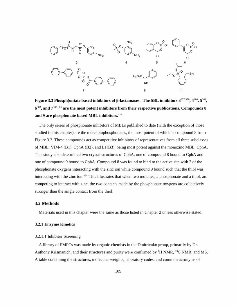
109
Figure 3.3 Phosph(on)ate based inhibitors of β-lactamases. The SBL inhibitors 3377,378, 4380, 5381,
6383, and 7385,386 are the most potent inhibitors from their respective publications. Compounds 8
and 9 are phosphonate based MBL inhibitors.424
The only series of phosphonate inhibitors of MBLs published to date (with the exception of those
studied in this chapter) are the mercaptophosphonates, the most potent of which is compound 8 from
Figure 3.3. These compounds act as competitive inhibitors of representatives from all three subclasses
of MBL: VIM-4 (B1), CphA (B2), and L1(B3), being most potent against the monozinc MBL, CphA.
This study also determined two crystal structures of CphA, one of compound 8 bound to CphA and
one of compound 9 bound to CphA. Compound 8 was found to bind to the active site with 2 of the
phosphonate oxygens interacting with the zinc ion while compound 9 bound such that the thiol was
interacting with the zinc ion.424 This illustrates that when two moieties, a phosphonate and a thiol, are
competing to interact with zinc, the two contacts made by the phosphonate oxygens are collectively
stronger than the single contact from the thiol.
3.2 Methods
Materials used in this chapter were the same as those listed in Chapter 2 unless otherwise stated.
3.2.1 Enzyme Kinetics
3.2.1.1 Inhibitor Screening
A library of PMPCs was made by organic chemists in the Dmitrienko group, primarily by Dr.
Anthony Krismanich, and their structures and purity were confirmed by 1H NMR, 13C NMR, and MS.
A table containing the structures, molecular weights, laboratory codes, and common acronyms of

110
these compounds can be found in Appendix A, an abridged figure can be found in Figure 3.4. PMPC-
3 and PMPC-4 were prepared as racemic mixtures and were tested as such. Synthetic picolinate
derivatives, were prepared as 100 mM stocks in DMSO with the exception of: PMP and DPMP which
were prepared as 50 mM stocks in 50 mM HEPES pH 7.2. Commercial picolinate derivatives, DPA,
PA, and 6-MPA (Sigma), were prepared in DMSO, the same as synthetic picolinate derivatives. All
inhibitor stocks were stored at -20 oC and were remade when necessary.
Figure 3.4 Structures of picolinic acid derivatives used in this chapter.
Enzyme stocks for class A, B, and C β-lactamases were prepared in 50 mM HEPES pH 7.2
supplemented with 50 μg/mL BSA and 0.01 % Triton X-100 while the stocks for class D enzymes
were prepared in 100 mM NaPi, 2.1 mg/mL NaHCO3, pH 7.0 supplemented with 50 μg/mL BSA and
0.01 % Triton X-100. Enzyme specific conditions are listed in Table 3.1; the total DMSO
concentration was kept below 2 %.
Enzyme stocks were pre-incubated with a small selection of inhibitor concentrations from 1 mM,
500 μM, 100 μM, 10 μM, and 1 μM in addition to the appropriate DMSO control in triplicate for 10
min at 30 oC in flat bottomed microplates. After pre-incubation, inhibited enzyme stock was added to
nitrocefin/UW-58 concentrations at or near the KM to a final volume of 200 μL and was monitored at
482 nm (NC) or 534 nm (UW-58) every 5-7 sec for 5 min (30 min for SFH-1) using a SpectraMax
190 plate reader (Molecular Devices, Sunnyvale, CA). The initial rate of each reaction was
determined using SoftMax 6.0 software (Molecular Devices, Sunnyvale, CA) over a minimum of 5
points. Percent inhibition was calculated relative to a control containing the same concentration of
DMSO.

111
Table 3.1 Kinetic parameters and conditions for PMPC inhibitor screening.
Enzyme Molecular
class Substrate KM (μM)
Enzyme
Conc. in assay
[Substrate]
in assay (μM)
KPC-2 A NC 8.1 ± 0.2453 211 pM 8
GC-1 C NC 15.4 ± 0.8453 41 pM 15
OXA-48 D NC 81 ± 2 79 pM 80
IMP-1 B1 NC 3.5 ± 0.2453 186 pM 3.5
NDM-1 B1 NC 0.88 ± 0.06453 620 pM 1
SPM-1 B1 NC 1.2 ± 0.1453 8.17 nM 1
VIM-1 B1 NC 19 ± 1 1.15 nM 15
VIM-2 B1 NC 13.9 ± 0.3453 313 pM 15
SFH-1 B2 UW-58 220 ± 10 1.86 nM 100
L1 B3 NC 5.5 ± 0.5453 637 pM 5
3.2.1.2 IC50 Determination
A more thorough analysis of inhibition was performed for enzyme/inhibitor concentrations that
showed at least 50 % inhibition at 100 μM. Assays were performed as described in 3.2.1.1 using 7 or
14 different concentrations of inhibitor. Inhibitor concentrations were determined using data from the
inhibitor screen ensuring both plateaus and the hill of the plot were well defined. Each measurement
was performed on 3-4 separate days unless otherwise indicated. Initial rates were normalized then
fitted to Equation 3.1 using GraphPad Prism version 5.00 for Windows (GraphPad Software, San
Diego, CA) by non-linear least squares regression.
Equation 3.1 𝒚 =𝟏𝟎𝟎
𝟏+𝟏𝟎(𝒍𝒐𝒈𝑰𝑪𝟓𝟎−[𝑰])+𝒔
Where y is the normalized initial rate, [I] is the log of the inhibitor concentration, and s is the Hill
slope. Measurements and uncertainties from each day were averaged using the statistical method
described in Appendix B.
3.2.1.3 Determination of Inhibition Constant, KI
Assays to determine the KI for the most potent enzyme/inhibitor combinations were performed in
50 mM HEPES pH 7.2 supplemented with 50 μg/mL BSA and 0.01 % Triton X-100 in 96-well flat

112
bottomed microplates at a final volume of 200 μL. Enzyme mixture was added to nitrocefin (>>KM)
containing dilutions of inhibitor as determined from IC50 experiments in triplicate; specific conditions
can be found in Table 3.2. Assay was monitored at 482 nm every 6 sec for 10 min at 30 oC in a
SpectraMax 190 plate reader (Molecular Devices, Sunnyvale, CA).
Table 3.2 Conditions of time-dependent KI determination.
Enzyme [Enzyme] in assay [NC] in assay
IMP-1 60 pM 25 μM
NDM-1 600 pM 15 μM
VIM-2 39.2 pM 100 μM
L1 308 pM 50 μM
Progress curves were fitted to Equation 3.2 by non-linear regression using GraphPad Prism version
5.00 for Windows (GraphPad Software, San Diego, CA) to determine kobs, the apparent first-order
rate constant at steady state, for each inhibitor concentration.496,497
Equation 3.2 [𝑷]𝒕 = 𝒗𝒔𝒕 +(𝒗𝒐−𝒗𝒔)(𝟏−𝒆−𝒌𝒐𝒃𝒔𝒕)
𝒌𝒐𝒃𝒔+ 𝑪
Where [P]t is the concentration of product at time t, vo is the initial rate, vs is the steady state rate,
and C is a constant used to mitigate any background absorbance. The values of kobs were then plotted
against their respective concentration of inhibitor [I] to determine which mechanism described in
Scheme 3.1 this data fits. The resulting linear plot defined by Equation 3.3 suggests that the data fits
Scheme 3.1A.
Equation 3.3 𝒌𝒐𝒃𝒔 = 𝒌−𝟎 (𝟏 +[𝑰]
𝑲𝑰𝒂𝒑𝒑)
Where k-o is the dissociation rate constant of EI, the enzyme:inhibitor complex, and KIapp is the
apparent inhibition constant. The value of KIapp can be determined by dividing the value of the y-
intercept, k-o, by the value of the slope, k-o/KIapp. KI was then determined using the value of KI
app and
Equation 3.4.
Equation 3.4 𝑲𝑰𝒂𝒑𝒑
= 𝑲𝑰 (𝟏 +[𝑺]
𝑲𝑴)

113
Finally, kon and koff, the association and dissociation rate constants of EI can be determined using
Equation 3.5, where koff = k-o from Equation 3.3.
Equation 3.5 𝑲𝑰 =𝒌𝒐𝒇𝒇
𝒌𝒐𝒏
3.2.2 Microbial Experiments
3.2.2.1 Bacterial Strains
Detailed information regarding the clinical isolates and control strains used in this study can be
found in Appendix C. The Pseudomonas putida strain expressing VIM-2 (UWB24), Escherichia coli
strain expressing both CTX-M-15 and NDM-1 (UWB75), Pseudomonas aeruginosa strain expressing
VIM-2 (UWB78), and E. coli strain expressing both CTX-M-15 and IMP-1 (UWB93), were obtained
from the collection of clinical isolates at Calgary Laboratory Services, Calgary, Alberta and were
provided by Dr. Johann Pitout and Dr. Dylan Pillai. A Klebsiella pneumoniae strain expressing
NDM-1 (UWB116), was provided by Dr. Allison McGeer from Mount Sinai Hospital in Toronto,
Ontario.
3.2.2.2 Minimum Inhibitory Concentration (MIC) Determination
MIC values for meropenem with the bacterial strains in 3.2.2.1 were assayed by broth
microdilution several times in cation adjusted Mueller Hinton broth (MHB) (Sigma) according to the
Clinical Laboratory Standards Institute (CLSI) guidelines.498 Overnight cultures in MHB were grown
shaking at 225 rpm and 37 oC then diluted to approximately 0.5 McFarland as measured by OD600.
The 0.5 McFarland culture was diluted 100-fold in MHB, making the inoculum. Microtiter plates
containing media and two-fold dilutions of meropenem were inoculated with equal volumes of
inoculum to a final volume of 200 μL. The growth control (100 μL) that was diluted 1000-fold was
plated on Mueller Hinton agar (MHA). Microtiter plates were shaken for 10 sec before being
incubated overnight at 37 oC in humid conditions; agar plates were also incubated overnight at 37 oC.
MICs were determined by measuring the OD600 using a Powerwave XS2 (BioTek) plate reader with
an absorbance of 0.07 considered the breakpoint for growth then confirmed visually.

114
3.2.2.3 Potentiation of β-lactam Antibiotics by PMPCs
Potentiation experiments were performed using the method described in 3.2.2.2 with a gradient of
two-fold dilutions of inhibitor, dissolved in DMSO, in the microtiter plates perpendicular to the
meropenem gradient.
3.3 Results
3.3.1 Enzyme Kinetics
3.3.1.1 Inhibitor Screening
The inhibitors used in this chapter were first screened against all of the purified β-lactamases
available in the Dmitrienko lab to test for inhibition as well as to give a rough estimate of the IC50. To
maximize the possibility of seeing inhibition, the inhibitors were preincubated with the enzyme for 10
minutes prior to being added to nitrocefin at concentrations near KM. These screens were performed
in the presence of Triton X-100 to reduce the probability of observing inhibition as a result of
aggregation.
The data shown in Table 3.3 indicates that DPA, all of the PMPCs, and DPMP are reasonably
potent inhibitors of both B1 and B3 MBLs. The representative B2 MBL, SFH-1, was not inhibited to
the same degree by the PMPCs as the other MBLs but its response to DPA was consistent. The
PMPCs that significantly inhibited SFH-1 were PMPC-3 and PMPC-4 which both reduced its activity
by just over 50 % at 10 μM. NDM-1 also demonstrated comparable sensitivity with PMPC-3 and
PMPC-4. Other MBLs were most potently inhibited by PMPC-4 with VIM-1, and VIM-2 being
inhibited by more than 50 % at 1 μM. PMPC-1 is consistently a more potent inhibitor than DPA, and
PMPC-2 is comparable to it against NDM-1, SPM-1, VIM-1, SFH-1, and L1. With IMP-1 and VIM-
2, PMPC-2 is less effective at 10 μM than PMPC-1 was, suggesting that the carboxylate at the 3
position of the pyridine ring may interfere with binding for some MBLs. PA, 6-MPA, and PMP are
poor inhibitors that only show activity at the highest concentrations and therefore do not warrant
further study. The IC50 of PA with VIM-2 will be determined to get an idea of how this compares in
more detailed experiments.

115
Table 3.3 Screening of PMPCs and related commercial compounds against MBLs
% Inhibition
IMP-1 NDM-1 SPM-1 VIM-1 VIM-2 SFH-1 L1
DPA
100 μM 93.0 91.2 81.1 72.9 97.9 91.9 92.2
10 μM 46.1 82.7 30.0 52.4 84.3 59.7 62.2
1 μM 17.4 32.3 18.8 -14.5 9.9 4.4 -2.7
PA
100 μM 19.7 46.4 44.1 37.4 74.6 46.4
10 μM 16.5 37.7 18.1 -12.6 26.4 12.5
1 μM 17.2 25.9 22.1 -19.9 8.2 -0.6
6-MPA
100 μM 20.5 32.7 18.0 -11.7 8.5 8.0
10 μM 25.9 36.4 14.2 -3.5 4.7 -5.0
1 μM -13.7 7.0 8.5 -9.5 2.4 -3.7
PMPC-1
100 μM 98.4 99.0 81.7 87.5 101.2 61.6 77.4
10 μM 66.2 97.6 26.1 76.2 93.7 41.7 90.8
1 μM 2.3 53.4 25.1 39.9 24.7 22.0 32.0
PMPC-2
100 μM 89.0 101.6 84.0 79.3 97.2 57.7 105.5
10 μM 12.4 91.6 41.4 86.4 39.5 40.6 82.3
1 μM 0.4 61.5 19.0 42.8 11.9 26.1 24.0
PMPC-3
100 μM 95.9 101.3 88.7 87.5 96.1 85.2 90.9
10 μM 63.6 108.2 41.2 80.0 78.6 54.0 87.0
1 μM 26.1 69.7 26.7 33.9 14.7 37.0 32.1
PMPC-4
100 μM 96.7 100.2 106.5 103.9 101.1 71.6 100.5
10 μM 92.0 103.0 70.3 90.8 99.7 51.1 92.9
1 μM 24.6 84.3 21.8 61.3 64.7 35.1 39.6
PMP
1 mM 60.7 45.3 14.9 85.0 40.4
100 μM 8.5 -14.3 14.4 32.7 2.0
10 μM -3.1 -8.3 10.0 0.4 -3.6
DPMP
1 mM 96.7 98.6 73.9 99.3 97.5
100 μM 54.3 97.3 38.4 88.8 33.6 83.5
10 μM 1.0 87.3 16.9 35.0 19.7 33.8
Bolded values are >50 % inhibited.

116
A selection of SBLs was screened to ascertain whether or not these are metallo-enzyme specific
inhibitors. As seen in Table 3.4, these compounds do not inhibit SBLs at concentrations comparable
to their inhibition of MBLs.
Table 3.4 Screening of PMPCs and related commercial compounds against SBLs.
% Inhibition
KPC-2 GC-1 OXA-48
DPA
500 μM 28.1 6.5 0.7
100 μM -5.0 -16.4 10 μM -8.1 -13.7
PA
500 μM 10.3 1.3 2.3
100 μM -2.4 -7.9
10 μM -12.1 -12.9
6-MPA
500 μM 12.3 -1.4 1.1
100 μM -6.4 -11.2 10 μM -5.7 -3.7
PMPC-1
500 μM 33.2 25.1 0.6
100 μM 4.6 -3.9
10 μM 1.0 -5.2
PMPC-2
500 μM 14.5 42.8 1.6
100 μM 6.0 1.1
10 μM -1.1 -14.1
PMPC-3
500 μM 13.9 34.8 3.2
100 μM 3.8 4.2
10 μM -4.6 -6.3
PMPC-4
500 μM 56.7 47.2 4.8
100 μM 19.4 14.3 10 μM -4.6 0.7
PMP 1 mM 24.0 17.7
500 μM 12.2 9.7 -2.4
DPMP 1 mM 22.2 25.9
500 μM 19.6 16.1 -2.1
Some inhibition can be detected at 500 μM for KPC-2 and GC-1, with the greatest effect being
observed with PMPC-4 (57 % and 47 % inhibition respectively). The inhibition of SBLs by PMPCs
was not considered potent enough to warrant further study.

117
3.3.1.2 IC50 Determination
A more in-depth screen of MBL inhibition by PMPCs was performed by determining IC50s when
the inhibitor was preincubated with enzyme for 10 min prior to addition to nitrocefin. Trends in the
efficacy of inhibitors for each enzyme can best be demonstrated by the dose response curves in Figure
3.5. Generally, DPMP (and PA and PMP where applicable) are the poorest inhibitors as seen by their
curves being shifted to the right of the others. Little difference can be seen between PMPC-1, PMPC-
3, and PMPC-4 against most enzymes, although PMPC-4 is notably more potent than the others
against VIM-2. Of the PMPCs, PMPC-2 is the least potent, falling well to the right of the curves of
other PMPCs for IMP-1, VIM-1, and VIM-2.
In most cases, the curve shape and Hill slope is fairly consistent for the different inhibitors against
any given enzyme with the exception of DPA with SFH-1 and DPMP with both SPM-1 for which the
Hill slopes are much higher. This difference may be indicative of inhibitor binding to multiple sites or
non-specific inhibition.499

118
IMP-1
-4 -2 0 2 40
50
100
log([Inhibitor] (µM))
% A
cti
vit
y
NDM-1
-4 -2 0 2 40
50
100
log([Inhibitor] (µM))
% A
cti
vit
y
SPM-1
-4 -2 0 2 40
50
100
log([Inhibitor] (µM))
% A
cti
vit
y
VIM-1
-4 -2 0 2 40
50
100
log([Inhibitor] (µM))
% A
cti
vit
yVIM-2
-4 -2 0 2 40
50
100
log([Inhibitor] (µM))
% A
cti
vit
y
SFH-1
-4 -2 0 2 40
50
100
log([Inhibitor] (µM))
% A
cti
vit
y
L1
-4 -2 0 2 40
50
100
log([Inhibitor] (µM))
% A
cti
vit
y
Figure 3.5 Dose response curves of MBLs with PMPCs and related compounds. The
enzyme/inhibitor combinations in Table 3.5 are presented here with the inhibitors represented
as follows: DPA (▬), PA (▬), PMPC-1 (▬), PMPC-2 (▬), PMPC-3 (▬), PMPC-4 (▬), PMP
(▬), and DPMP (▬). The graphs in this figure were generated using GraphPad Prism v. 5.03.
The data in Table 3.5 is a numeric representation of the graphs in Figure 3.5 which allows for
comparison between enzymes. From this it can be seen that NDM-1 is the MBL that is most
susceptible to inhibition by PMPCs with IC50 values of 0.31 μM to 0.55 μM for the PMPCs, all of
which are lower than the IC50 values for any other enzyme/inhibitor combination. SPM-1 and SFH-1
are poorly inhibited relative to the other MBLs with SFH-1 being less susceptible to inhibition than
SPM-1. The B1 MBLs are inhibited most potently by PMPC-4 with PMPC-1 being less than 2-fold
less potent than PMPC-4 against all B1 MBLs except VIM-2. Unlike the B1 MBLs, the B2 MBL,

119
SFH-1, was most potently inhibited by PMPC-3 than PMPC-4 (IC50s of 0.56 μM and 7.3 μM
respectively) and PMPC-1 is 5-fold less potent than the most potent inhibitor, PMPC-3. The B3 MBL
L1, is very similarly inhibited by all of the PMPCs such that the 95% confidence intervals for each
PMPC inhibitor encompasses the IC50 values of all of the other PMPC inhibitors. As was suggested
from the screening experiments, PA, PMP, and DPMP are relatively weak inhibitors of MBLs. Using
VIM-2 as a model system (since it showed some susceptibility to all inhibitors in screening), PMP is
the weakest inhibitor followed by PA, and DPMP with IC50 values of 170 μM, 32 μM, and 18 μM
respectively. With IMP-1, VIM-2, and SFH-1, PMPC-2 is a weaker inhibitor than DPA suggesting
that this is not necessarily a good compound on which to base further structural optimization.
Table 3.5 IC50’s of PMPCs against MBLs
IC50 (μM)
(95% Confidence Interval)
IMP-1 NDM-1 SPM-1 VIM-1 VIM-2 SFH-1 L1
DPA 10
(7-15)
2.6
(2.0-3.4)
18
(11-29)
5.3 *
(5.1-5.5)
5.2
(2.9-9.3)
6.4 *
(5.5-7.4)
5.2
(4.8-5.7)
PA 101
(50-202)
32
(28-37)
PMPC-1 3.9
(3.2-4.8)
0.37
(0.25-0.55)
6.8
(5.6-8.3)
0.82 *
(0.78-0.85)
1.3
(0.7-2.3)
29 *
(21-40)
1.5
(1.0-2.2)
PMPC-2 21
(19-24)
0.55
(0.44-0.59)
13
(8-23)
1.4 *
(1.3-1.5)
14.1
(13.6-14.7)
36 *
(20-65)
1.9
(1.0-3.4)
PMPC-3 7.2
(5.2-10.1)
0.32
(0.21-0.49)
9.1
(6.1-13.6)
1.27 *
(1.21-1.32)
1.9
(1.3-2.9)
5.6 *
(3.8-8.1)
2.1
(1.6-2.6)
PMPC-4 2.9
(2.3-3.7)
0.31
(0.20-0.47)
5.4
(4.4-6.5)
0.80 *
(0.77-0.83)
0.46
(0.31-0.70)
7.3 *
(6.0-9.0)
1.6
(0.9-2.8)
PMP 170 *
(150-190)
DPMP 108 *
(103-114)
2.0 *
(1.8-2.2)
150 *
(100-210)
18 *
(17-19)
33 *
(31-34)
* From a single experiment of three technical replicates.
From these data, it was determined that the inhibition should be better characterized for
enzyme/inhibitor combinations with low IC50 values. The enzyme/inhibitor combinations selected for
this analysis were IMP-1, NDM-1, VIM-2, and L1 with the inhibitors DPA, PMPC-1, PMPC-3, and
PMPC-4. The stock of purified VIM-1 was extremely limited and still has the C-terminal His tag
attached, thus it was excluded from further study.
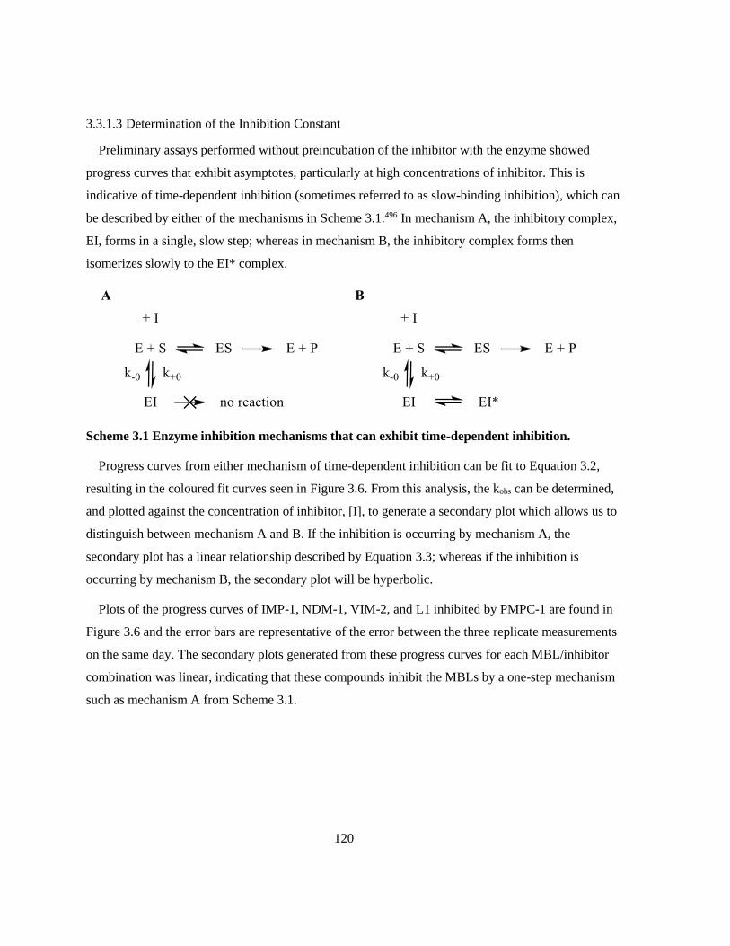
120
3.3.1.3 Determination of the Inhibition Constant
Preliminary assays performed without preincubation of the inhibitor with the enzyme showed
progress curves that exhibit asymptotes, particularly at high concentrations of inhibitor. This is
indicative of time-dependent inhibition (sometimes referred to as slow-binding inhibition), which can
be described by either of the mechanisms in Scheme 3.1.496 In mechanism A, the inhibitory complex,
EI, forms in a single, slow step; whereas in mechanism B, the inhibitory complex forms then
isomerizes slowly to the EI* complex.
Scheme 3.1 Enzyme inhibition mechanisms that can exhibit time-dependent inhibition.
Progress curves from either mechanism of time-dependent inhibition can be fit to Equation 3.2,
resulting in the coloured fit curves seen in Figure 3.6. From this analysis, the kobs can be determined,
and plotted against the concentration of inhibitor, [I], to generate a secondary plot which allows us to
distinguish between mechanism A and B. If the inhibition is occurring by mechanism A, the
secondary plot has a linear relationship described by Equation 3.3; whereas if the inhibition is
occurring by mechanism B, the secondary plot will be hyperbolic.
Plots of the progress curves of IMP-1, NDM-1, VIM-2, and L1 inhibited by PMPC-1 are found in
Figure 3.6 and the error bars are representative of the error between the three replicate measurements
on the same day. The secondary plots generated from these progress curves for each MBL/inhibitor
combination was linear, indicating that these compounds inhibit the MBLs by a one-step mechanism
such as mechanism A from Scheme 3.1.

121
0 2 4 6 8 10
0
1
2
3
4
0
25
15
10
7.5
5
2.5
1
Time (min)
[Pro
du
ct]
(µ
M)
0 2 4 6 8 10
0
2
4
0
75
50
25
15
10
5
1
Time (min)
[Pro
du
ct]
(µ
M)
0 10 20 300.0
0.5
1.0
[I]
k o
bs
0 20 40 60 800.0
0.2
0.4
0.6
0.8
1.0
[I]
k o
bs
0 2 4 6 8 10
0
2
4
6
0
10
5
2.5
1
0.75
0.5
0.1
Time (min)
[Pro
du
ct]
(µ
M)
0 2 4 6 8 10
0
1
2
3
4
0
10
7.5
5
2.5
1
0.5
0.1
Time (min)
[Pro
du
ct]
(µ
M)
0 2 4 60.0
0.5
1.0
1.5
[I]
k o
bs
0 2 4 6 8 100.0
0.2
0.4
0.6
0.8
1.0
[I]
k o
bs
KI = 0.5 ± 0.1
R2 = 0.9760
KI = 1.1 ± 0.2
R2 = 0.9944KI = 0.067 ± 0.010
R2 = 0.9893
KI = 0.54 ± 0.05
R2 = 0.9918
A B
C D
Figure 3.6 Time-dependent KI graphs for PMPC-1 against representative MBLs : IMP-1 (A),
NDM-1 (B), VIM-2 (C), and L1 (D). The graphs in this figure were generated using GraphPad
Prism v. 5.03.

122
The graphs corresponding to those in Figure 3.6 for DPA, PMPC-3, and PMPC-4 can be found in
Appendix D.
The KI values in Table 3.6 were determined from further analysis of the linear regression of the
secondary plots. Consistent with the IC50 data, PMPC-4 is the most potent inhibitor as it has the
lowest KI for each enzyme; as much as 14.2 times more potent than PMPC-1. The general trends for
inhibitor potency within each enzyme system seen in the IC50 data also holds up here, although the
scaling is not consistent (ie. PMPC-4 was 8-fold more potent than DPA against NDM-1 in terms of
IC50 values, however the KI is almost 100-fold lower). The KI values for PMPC-1 and PMPC-3 are
generally similar while the KI values for DPA are 1.5-49 times greater than those of PMPC-1. Finally,
NDM-1 is most sensitive to PMPCs with KI values from 34 nM to 74 nM.
Table 3.6 KI of DPA, PMPC-1, -3, and -4 against select MBLs.
KI (µM)
DPA PMPC-1 PMPC-3 PMPC-4
IMP-1 2.8 ± 0.4 1.1 ± 0.2 1.5 ± 0.1 0.4 ± 0.2
NDM-1 3.3 ± 0.6 0.067 ± 0.010 0.074 ± 0.008 0.034 ± 0.006
VIM-2 0.8 ± 0.3 0.54 ± 0.05 0.61 ± 0.04 0.038 ± 0.009
L1 1.1 ± 0.3 0.5 ± 0.1 0.41 ± 0.10 0.4 ± 0.1
The first order rate constant for the dissociation of the EI complex, koff, for each enzyme/inhibitor
combination was determined from the y-intercept of the secondary plot. The second order rate
constant for the association of the EI complex, kon was calculated from the known values of koff and
the KI; the results of these analyses are found in Table 3.7.
Table 3.7 On and off rates of metallo-β-lactamase inhibition by PMPCs.
kon (M-1sec-1)
koff (sec-1)
DPA PMPC-1 PMPC-3 PMPC-4
IMP-1 500 ± 100
0.0014 ± 0.0002
1400 ± 300
0.0015 ± 0.0002
980 ± 90
0.00147 ± 0.00008
4000 ± 2000
0.0014 ± 0.0006
NDM-1 1300 ± 300
0.0043 ± 0.0003
51 000 ± 9000
0.0034 ± 0.0003
54 000 ± 8000
0.0040 ± 0.0004
110 000 ± 20 000
0.0036 ± 0.0002
VIM-2 5000 ± 2000
0.004 ± 0.001
8000 ± 800
0.0043 ± 0.0002
9100 ± 700
0.0055 ± 0.0002
70 000 ± 20 000
0.0027 ± 0.0005
L1 1200 ± 400
0.0013 ± 0.0003
6000 ± 2000
0.0029 ± 0.0005
5000 ± 1000
0.0019 ± 0.0004
5000 ± 2000
0.0020 ± 0.0004
123
The koff values for the various inhibitors against any one enzyme are fairly consistent, ranging from
0.0013 sec-1 to 0.0055 sec-1, while the kon values vary greatly. The kon values for PMPC-4 are 2-7.5-
fold larger than the inhibitor with the next highest kon for B1 MBLs. This indicates that the EI
complex forms fastest with PMPC-4, and the koff values show that it dissociates at approximately the
same rate as the other inhibitors, resulting in stronger inhibition. This trend does not hold up with L1
which has very similar kon values across the PMPCs (5000 M-1sec-1 – 6000 M-1sec-1). The lack of
increased kon for PMPC-4 with L1 is consistent with the KI and IC50 data which suggests that PMPC-4
does not hold a particular advantage over the other PMPCs against L1, unlike the B1 MBLs.
3.3.2 Potentiation of meropenem against MBL producing Gram negatives
It is one thing to inhibit enzymes in vitro, but another thing entirely to penetrate the Gram-negative
cell wall and restore antibiotic potency. Five strains MBL producing clinical isolates including two E.
coli strains, two Pseudomonas spp., and a K. pneumoniae were selected to determine whether or not
the PMPCs could potentiate meropenem activity in vivo. Strains UWB93 and UWB75 co-express the
SBL CTX-M-15 which does not significantly hydrolyze meropenem and thus should not significantly
contribute to the meropenem MIC when the MBL is inhibited.
The Clinical Laboratory Standards Institute (CLSI) defines Enterobactericiae (E. coli and K.
pneumoniae) as susceptible to meropenem when the MIC is at or below 1 mg/L and as resistant at or
above 4 mg/L. Pseudomonas spp. are considered susceptible to meropenem at or below 2 mg/L and
are resistant at or above 8 mg/L. The data in Table 3.8 indicates that DPA tends to bring the MIC of
meropenem into susceptible range with two-fold less inhibitor than PMPCs although it tends to
require 16 mg/L DPA or more to achieve susceptibility. PMPC-4 is the least effective of the tested
inhibitors against each strain and only brings UWB93 and UWB116 into the susceptible range, others
showed a decrease in MIC at high concentrations of PMPC-4, but not enough to be clinically useful.
UWB116 is particularly sensitive to potentiation by PMPCs with meropenem MICs entering the
susceptible range with only 16-32 mg/L of inhibitor. None of the tested inhibitors had intrinsic
antibacterial activity at the concentrations used in this assay.

124
Table 3.8 Sensitization of MBL producing Gram negatives to meropenem by DPA and PMPCs.
Strain β-lactamase
Meropenem
MIC (mg/L) Inhibitor
Meropenem MIC (mg/L) in the presence of
Inhibitor
4
mg/L
8
mg/L
16
mg/L
32
mg/L
64
mg/L
128
mg/L
E. coli
UWB93
IMP-1
CTX-M-15
4 DPA 4 4 2 1 0.25 <0.03
PMPC-1 4 4 4 2 0.5 0.25
PMPC-2 4 4 4 4 2 0.5
PMPC-3 4 4 4 4 2 0.5
PMPC-4 4 4 4 2 2 0.5
E. coli
UWB75
NDM-1
CTX-M-15
128 DPA 128 128 64 16 <0.5 <0.5
PMPC-1 128 128 64 32 4 <0.5
PMPC-2 128 128 64 64 16 4
PMPC-3 128 128 128 64 32 2
PMPC-4 128 128 128 64 64 8
K. pneumoniae
UWB116
NDM-1 32 DPA 16 4 0.5 <0.125 <0.125 <0.125
PMPC-1 16 8 4 0.25 <0.125 <0.125
PMPC-2 16 8 2 0.5 <0.125 <0.125
PMPC-3 8 8 1 <0.125 <0.125 <0.125
PMPC-4 16 8 2 0.5 <0.125 <0.125
P. putida
UWB24
VIM-2 128 DPA 128 128 128 64 4 2
PMPC-1 128 128 128 64 32 4
PMPC-2 128 128 128 128 64 32
PMPC-3 128 128 128 128 64 32
PMPC-4 128 128 128 128 64 32
P. aeruginosa
UWB78
VIM-2 128 DPA 64 64 64 32 8 4
PMPC-1 128 128 64 64 16 8
PMPC-2 128 128 128 64 64 16
PMPC-3 128 128 128 128 64 8
PMPC-4 128 128 64 64 64 16
Bolded MIC values are susceptible to meropenem according to CLSI guidelines.500
Italicized MIC values are intermediate (neither resistant nor susceptible) to meropenem according to CLSI
guidelines.500
3.4 Discussion
Previous studies have indicated that DPA is an effective inhibitor of both IMP-1 and NDM-1.400,495
Due to the structural similarity of DPA and the PMPCs, it was considered likely that the PMPCs
would also inhibit MBLs. The initial synthesis of PMPC-1 and PMPC-2 was directed at inhibition of
Class II fructose-1,6-bisphosphate aldolase, a metallo-enzyme.501

125
The PMPCs and related pyridine compounds are poor inhibitors of SBLs, only inhibiting KPC-2
and GC-1 at the highest tested concentration, 500 μM. In most cases, this inhibition is modest,
reducing the activity of these enzymes by 10-40 % relative to the uninhibited control. The only tested
compound that managed to elicit a response of greater than 50 % inhibition against SBLs was PMPC-
4 which is also the most potent inhibitor against the MBLs.
The MBLs are potently inhibited by PMPCs, and to a lesser extent by other picolinic acid
derivatives depicted in Figure 3.4. Of the commercial picolinic acid derivatives: PA, DPA, and 6-
MPA, the most potent inhibitor of MBLs is DPA. The improved activity of DPA over PA indicates
that the presence of the carboxylate at the 6-position is beneficial as it decreases the IC50 by 6 to
greater than 20-fold. A study on the inhibitory activity of commercial picolinate derivatives against
MBLs by Horsfall et al. showed that 100 μM DPA was able to reduce the activity of IMP-1 to 2 %
relative to an uninhibited control in the absence of added zinc while 100 μM PA was only able to
reduce the activity by 31%.410 In the presence of 100 μM ZnCl2, the inhibition of IMP-1 by DPA is
substantially diminished (72 % residual activity at 100 μM DPA) suggesting that DPA in solution
with Zn2+ is being sequestered and is not available for inhibition.410 The data presented by Horfall et
al. is consistent with the data presented in Table 3.3 for DPA in which 7 % activity remains after
incubation with 100 μM DPA but is not consistent with the data for PA (80.3 % residual activity at
100 μM PA). The data in Horsfall et al. with VIM-2 and L1 are not comparable as they appear to be
run in the presence of 100 μM ZnCl2, although this is not explicitly detailed in the Methods section of
their paper.410 Substitution of the carboxylate on DPA at the 6 position with a methyl group
completely abolishes activity, suggesting that the presence of a polar group at the 6-position is
favourable for inhibition.
The inhibition is further improved when the 6-carboxylate is substituted by a phosphonomethyl
group, decreasing the IC50 values by another 2.6-7-fold. Substitution of both DPA carboxylates by
phosphonomethyl groups as in DPMP leads to a reduction in activity for all MBLs except NDM-1
which has comparable IC50 values for DPA and DPMP (2.59 μM and 2.03 μM respectively).
Furthermore, if the carboxylate from PMPC-1 is excluded (PMP) the inhibitory activity is completely
abolished, emphasizing the importance of the carboxylate at the 2-position.
Within the PMPCs, two substitution sites were explored: the 3-position of the pyridine ring
(PMPC-2), and the methylene group of the phosphonate (PMPC-3 and PMPC-4). The addition of a

126
carboxylate to PMPC-1 at the 3-position of the ring to form PMPC-2 provides no improvement in the
inhibition of NDM-1 or L1 and increases the IC50 2-11-fold against IMP-1, SPM-1, and VIM-2. This
suggests that the addition of an anionic group to the 3-position of the pyridine ring increases
specificity but does not improve the potency of these inhibitors. The addition of either a small polar
group (PMPC-3) or large hydrophobic group (PMPC-4) to the methylene group of the PMPC-1
phosphonate did not significantly affect the inhibition in terms of IC50 with the exception of the 2.8-
fold decrease in the IC50 from PMPC-1 (1.29 μM) to PMPC-4 (0.464 μM) for VIM-2. It should be
noted that PMPC-3 and PMPC-4 were synthesized as racemic mixtures so they may be twice as
potent as reported if the binding to the active site is selective for only one enantiomer.
The two VIM variants, VIM-1 and VIM-2, showed similar IC50 values for most of the compounds
tested. The only compound for which their IC50’s varied significantly was PMPC-2 which is also the
only compound in this set that may have two modes of inhibition: one as a PMPC and the other as a
phthalate. Phthalates (specifically 3-(4-hydroxypiperidin-1-yl)-aminophthalic acid) are known to
inhibit B1 MBLs by coordinating one carboxylate group, which would be equivalent to the
carboxylate at the 2 position in PMPCs, with the two active site Zn ions and the other carboxylate (the
3 position in PMPC-2) interacts with Lys161 in IMP-1 (Figure 3.7A). Crystallographic studies done
in collaboration with Dr. Jim Spencer (University of Bristol) yielded structures of IMP-1 with PMPC-
1 and L1 with PMPC-1 and PMPC-3. In the IMP-1 structure, PMPC-1 coordinates its pyridine
nitrogen and carboxylate with Zn2. The carboxylate also hydrogen bonds with Lys161, the
phosphonate hydrogen bonds to the bridging water/hydroxide and interacts weakly with Ser80 in
IMP-1, and the aromatic ring has an edge-face interaction with Trp28 (Figure 3.7C).414,415 The
potential for two binding modes for PMPC-2 may explain the discrepancy in the potency trend
mentioned above. If PMPC-2 preferentially binds in a manner similar to PMPC-1, it is expected that
the IC50 would be similar to the other PMPCs as it is for NDM-1, L1, and VIM-1 since they should be
making the same contacts. If PMPC-2 preferentially binds in a manner similar to the phthalates, this
may result in significantly different IC50 as seen with IMP-1, SPM-1, and VIM-2 since the contacts
made in the active site are completely different. This dichotomy greatly complicates the kinetics as
inhibition may be a result of multiple binding modes, each with differing affinities.

127
Figure 3.7 Binding of 3-(4-hydroxypiperidin-1-yl)-aminophthalic acid and PMPC-1 to the active
site IMP-1. (A) 3-(4-hydroxypiperidin-1-yl)-aminophthalic acid bound to IMP-1 with contacts
from the groups also present in PMPC-2 shown (PDB 3WXC). (B) Overlay of the binding of the
two inhibitors. (C) PMPC-1 bound to IMP-1 with contacts that would also be present in PMPC-
2 (PDB 5HH4). This figure was generated using Chimera v. 1.12 from the aforementioned
protein databank files.
Structural studies of PMPC-1 and PMPC-3 with L1 yielded some unexpected results. Despite the
kinetics suggesting that IMP-1 and L1 are inhibited by the same mechanism, crystal structures in
Figure 3.8 show that in L1, the phosphonate of the PMPC replaces Zn2 while in IMP-1 both Zn ions
are retained (Figure 3.7). The removal of one zinc ion from L1 is uncommon and has only been
previously reported when a L1 crystal was soaked for 10 min with 10 mM EDTA.255 Studies have
been performed in the literature to determine the dissociation constant (KD) of Zn2 in both L1 and
IMP-1 and suggest that Zn2 is more tightly bound in IMP-1 than in L1 (KD of 0.7 μM at pH 5.61 and
2.6 μM at pH 7.0 respectively).302,502
Figure 3.8 Crystal structures of PMPCs bound to L1 , namely PMPC-1 (A, green) and PMPC-3
(C, purple) with a comparative overlay of the two structures (B) to illustrate similarities in the
mode of binding. This figure was generated using Chimera v. 1.12 from PDB: 5HH5 and 5HH6.

128
It has been shown that the KD of Zn2 depends heavily on pH and while these two KDs were not
determined at the same pH, the KD of IMP-1 was determined at a range of pHs (4.33-5.61) and
decreases as pH increases.302 This suggests that if the trend holds, 0.7 μM may by an over estimation
of the KD of Zn2 in IMP-1 at pH 7 which may explain why it retains its Zn while L1 does not in the
crystallization experiments with PMPC-1. Additionally, the KD values were determined for the
proteins in solution, and they may be significantly different with respect to one another in the
crystalline state. The replacement of Zn2 in L1 by the phosphonate group of the PMPCs results in a
very strong interaction between one of the phosphonate oxygens and Zn1 (1.8 Å). This interaction is
notably stronger that the interactions between Zn1 and the three His residues that coordinate it to the
active site (2.1 Å average). Additionally, the two PMPCs, PMPC-1 and PMPC-3 bind to L1 in the
same manner, overlaying on each other almost perfectly as seen in Figure 3.8B.
Figure 3.9 Binding of the S-enantiomer of PMPC-3 to the active site of L1. This figure was
generated using Chimera v. 1.12 from the protein databank file 5HH6.
Unlike PMPC-1 which has no stereocenters, the C6 methine group of PMPC-3 is chiral and was
synthesized as a racemic mixture. The crystal structure of L1 with PMPC-3 bound shows that the
binding of PMPC-3 to the active site is stereospecific for the S-enantiomer. By mapping the protein
surface, as in Figure 3.9, it can be seen that the 6’-hydroxyl group points towards the opening of the
129
active site and that the R-enantiomer would likely have unfavourable contacts with the protein
surface. The relative openness of this part of the binding pocket indicates that large substituents such
as that of PMPC-4 would be well tolerated but their binding would also be stereospecific. Due to the
stereoselectivity of the L1 active site, it is probable that the IC50 and KI values for PMPC-3 and
PMPC-4 should be half that which was detailed above. It is uncertain whether or not the other MBLs
require the same enantiomer as L1 for inhibition by PMPC-3 and PMPC-4.
In the case of binding of PMPC-4 to IMP-1, it is clear from qualitative modelling that the bulky N-
thienoyl-N-benzyl groupcan only be accommodated in the binding site if the stereochemistry is of the
R configuration. This is due to the prediction of very bad contacts with the zinc ions and the bridging
hydroxide ion if the stereochemistry is of the S configuration. Thus, it is reasonable to suggest that
only the R-isomer of PMPC-4 contributes to inhibition and that the effective IC50 and KI values with
IMP-1 should be half what is reported above.
In the case of PMPC-3, the hydroxyl group in the S-isomer makes close contacts with the bridging
hydroxide as well as with the aspartate carboxylate that is a ligand to Zn2. These may however be
tolerated since they may involve hydrogen bonding interactions. In the R-isomer, no contacts are
predicted between the hydroxyl group and the protein. Thus it is possible that both stereoisomers
contribute to the observed enzyme inhibition.
In early experiments performed with no pre-incubation with the inhibitors (DPA, PMPC-1, PMPC-
3, and PMPC-4) and three B1 MBLs (IMP-1, NDM-1, and VIM-2) and one B3 MBL (L1) exhibited
burst kinetics wherein there is an initial fast rate and a slower steady state rate (Figure 3.10). The
burst kinetics are most evident in mid-range inhibitor concentrations of the progress curves in Figure
3.6 and are not caused by substrate depletion as only 10 % or less of the substrate is consumed at the
end of a 10 min assay. This, in conjunction with the observation that IC50’s taken at different
preincubation times stabilize at 10 min (data not shown), indicates that DPA and the PMPCs inhibit
MBLs by a time dependent mechanism rather than classical competitive inhibition.

130
IMP-1
0 2 4 6 8 10
0.0
0.5
1.0
1.5
2.0
7% Substrateconsumed
vo
vs
Time (min)
[Pro
du
ct]
(µ
M)
NDM-1
0 2 4 6 8 10
0.0
0.5
1.0
1.5
2.0
10% Substrateconsumed
vo
vs
Time (min)
[Pro
du
ct]
(µ
M)
VIM-2
0 2 4 6 8 10
0.0
0.5
1.0
1.5
1.2% Substrateconsumed
vo
vs
Time (min)
[Pro
du
ct]
(µ
M)
L1
0 2 4 6 8 10
0.0
0.5
1.0
1.5
2.0
3% Substrateconsumed
vo
vs
Time (min)
[Pro
du
ct]
(µ
M)
Figure 3.10 Fits of initial and steady state rates on representative progress curves of MBLs
inhibited by PMPC-1 (25 μM for IMP-1, 1 μM for NDM-1, 5 μM for VIM-2, and 7.5 μM for
L1). The graphs in this figure were generated using GraphPad Prism v. 5.03.
Time dependent kinetics can be the result of either slow-binding or slow-tight-binding. The
possibility of slow-tight-binding inhibition was excluded since the concentrations of inhibitor
required to see inhibition are in great excess relative to the concentration of the MBL being
inhibited.496 Secondary plots of the rate of change between the initial rate, vo and the steady state rate,
vs, denoted as kobs, and the concentration of inhibitor were used to determine that inhibition occurs
through the formation of an EI complex, and that that complex does not isomerize further.
Determination of the inhibition constant, KI, showed that PMPC-1, PMPC-3, and PMPC-4 were
similarly potent against IMP-1, VIM-2, and L1 with PMPC-4 being the most potent. NDM-1 is the
most susceptible to inhibition by the PMPC’s with KI values ranging from 34-74 nM. The KI values
for DPA against each of the MBLs is larger than those of the PMPCs by 1.3-45-fold indicating that
the PMPCs are more effective inhibitors of MBLs than DPA.
Slow binding inhibitors exhibit lower rates of association (kon) than classical inhibitors which tend
to have association rates of 106-108 M-1sec-1 and in order to be effective inhibitors, the dissociation
rates (koff) must be even slower.496 The dissociation rates determined for DPA, PMPC-1, PMPC-3,
and PMPC-4 are very consistent across the MBLs as they all tend to be on the order of 10-3 sec-1.
Since the dissociation rates are so consistent, any differences in the efficacy of an inhibitor is
predicated by the association rate, which can vary by a hundred-fold for different inhibitors against
the same enzyme. The kon are highest for NDM-1 and approach being fast enough for classical
kinetics with PMPC-4 (106 M-1sec-1). This is consistent with the structural characteristics of NDM-1
in that it has the largest and most open active site allowing it to bind larger inhibitors faster than
enzymes with tighter active site like IMP-1 which has the slowest association rates for every
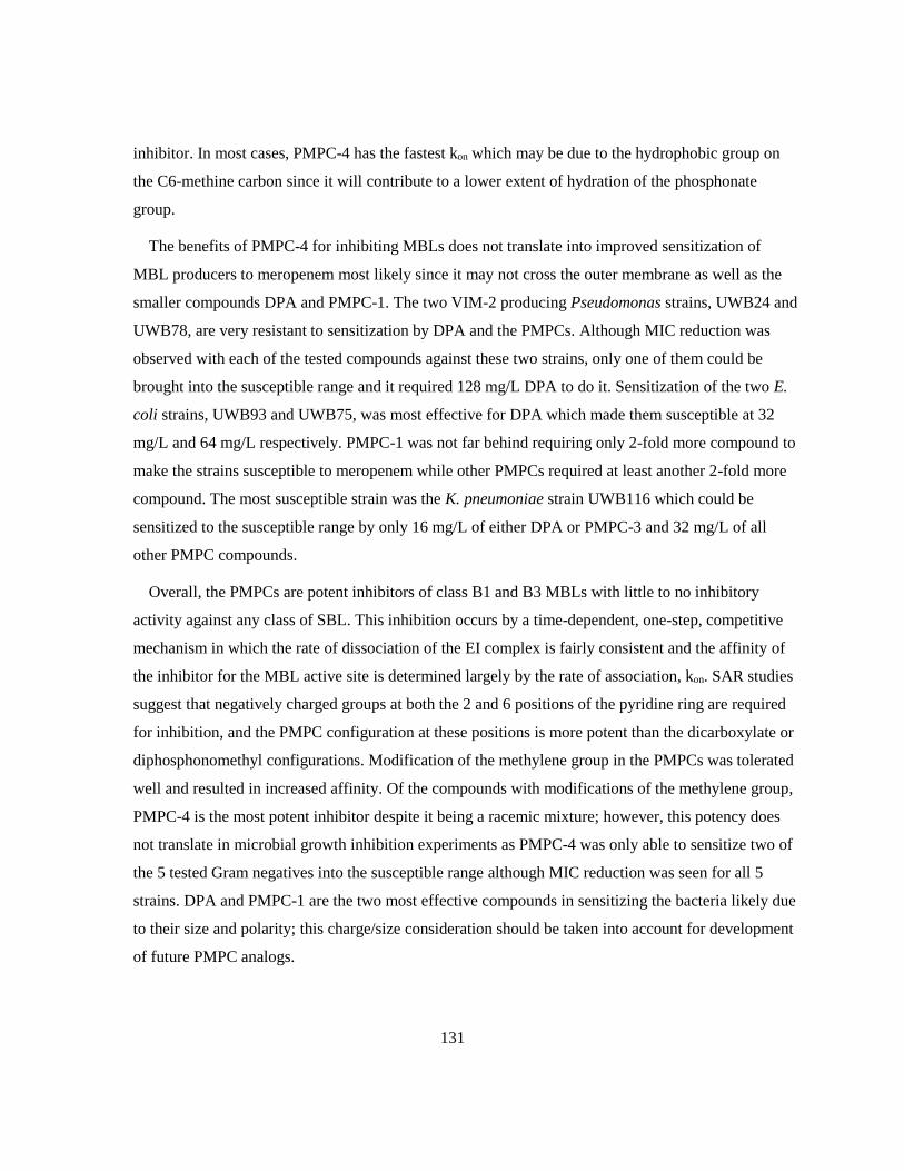
131
inhibitor. In most cases, PMPC-4 has the fastest kon which may be due to the hydrophobic group on
the C6-methine carbon since it will contribute to a lower extent of hydration of the phosphonate
group.
The benefits of PMPC-4 for inhibiting MBLs does not translate into improved sensitization of
MBL producers to meropenem most likely since it may not cross the outer membrane as well as the
smaller compounds DPA and PMPC-1. The two VIM-2 producing Pseudomonas strains, UWB24 and
UWB78, are very resistant to sensitization by DPA and the PMPCs. Although MIC reduction was
observed with each of the tested compounds against these two strains, only one of them could be
brought into the susceptible range and it required 128 mg/L DPA to do it. Sensitization of the two E.
coli strains, UWB93 and UWB75, was most effective for DPA which made them susceptible at 32
mg/L and 64 mg/L respectively. PMPC-1 was not far behind requiring only 2-fold more compound to
make the strains susceptible to meropenem while other PMPCs required at least another 2-fold more
compound. The most susceptible strain was the K. pneumoniae strain UWB116 which could be
sensitized to the susceptible range by only 16 mg/L of either DPA or PMPC-3 and 32 mg/L of all
other PMPC compounds.
Overall, the PMPCs are potent inhibitors of class B1 and B3 MBLs with little to no inhibitory
activity against any class of SBL. This inhibition occurs by a time-dependent, one-step, competitive
mechanism in which the rate of dissociation of the EI complex is fairly consistent and the affinity of
the inhibitor for the MBL active site is determined largely by the rate of association, kon. SAR studies
suggest that negatively charged groups at both the 2 and 6 positions of the pyridine ring are required
for inhibition, and the PMPC configuration at these positions is more potent than the dicarboxylate or
diphosphonomethyl configurations. Modification of the methylene group in the PMPCs was tolerated
well and resulted in increased affinity. Of the compounds with modifications of the methylene group,
PMPC-4 is the most potent inhibitor despite it being a racemic mixture; however, this potency does
not translate in microbial growth inhibition experiments as PMPC-4 was only able to sensitize two of
the 5 tested Gram negatives into the susceptible range although MIC reduction was seen for all 5
strains. DPA and PMPC-1 are the two most effective compounds in sensitizing the bacteria likely due
to their size and polarity; this charge/size consideration should be taken into account for development
of future PMPC analogs.

132
3.5 Future Work
One of the possible explanations for the discrepancy between the kinetic results and the
crystallography with respect to the replacement of Zn2 in L1 is that it may be due to the zinc ion
affinities differing in the crystalline state in comparison to the solution state. To establish whether or
not the zinc ion is abstracted in solution state, mass spectrometry experiments will be attempted.
Preliminary studies on SPM-1 have demonstrated that the dizinc form of the enzyme can be detected
as the primary species by ESI-MS under non-denaturing conditions. It is likely that L1 could also be
detected by native ESI-MS as the dizinc enzyme. If L1 retains 2 Zn ions when treated with PMPCs
prior to analysis by ESI-MS, then the replacement is a crystallographic artifact; however, a mono-Zn
L1 by this method does not confirm the crystal structure as the interaction of the Zn may have been
weakened enough by the PMPC to allow it to dissociate in the MS. If a mono-Zn spectrum was
obtained, further studies using ICP-MS, as described by King et al., would be required to determine if
Zn2 is actually being removed from the active site.401
Experiments performed in this chapter used racemic mixtures of PMPC-3 and PMPC-4; however,
the crystal structure of L1 with PMPC-3 demonstrated stereoselectivity for the S-enantiomer. It
should be noted that the stereoselectivity of the crystalline form of the enzyme may not be
representative of the stereoselectivity of the solution state enzyme. As such, enantiomerically pure
(both R and S) samples of PMPC-3 and PMPC-4 should be synthesized as tested with the MBLs to
determine if the S-enantiomer is the active inhibitor for all of them as well as how potent the pure
stock is. Once the issue of the stereochemistry at the C6-methylyne group is sorted out, new analogs
in the PMPC series can be rationally developed using the crystal structures of IMP-1 and L1 with
PMPCs bound.
The crystal structure of PMPC-1 bound to IMP-1 suggests a number of strategies for enhancing the
affinity of the PMPCs for MBL active sites. As indicated in Figure 3.11C, a substituent on C3 would
extend out of the active site so substituents that would increase the uptake into cells could be attached
here. The results with PMPC-2 demonstrate that the addition of an anionic group to this position is
disadvantageous; however, addition of an ether linker to either an amine or a siderophore may be
tolerated.

133
Figure 3.11 Binding of PMPC-1 to the active site of IMP-1, highlighting opportunities for
development of future PMPCs. (A) Schematic of the binding interactions involved in PMPC-1
binding to IMP-1. (B) The hydrophobic pocket of IMP-1 highlighted in purple. (C) Positions on
the PMPC core that could be modified to optimize binding to B1 MBLs. This figure was
generated using Chimera v. 1.12 from the protein databank file 5HH4.
Substituents at C4 and in the α-orientation on the C6 methine would not be well tolerated due to
steric clashes with Trp64 and the zinc ions/ligands respectively. Hydrophobic substitutions on C5 or
in the β-orientation on the C6 methine would point towards the hydrophobic pocket highlighted in
purple in Figure 3.11B. This hydrophobic pocket is well conserved within the B1 MBLs, so these
hydrophobic groups would improve inhibition against more that just IMP-1.

134
Chapter 4
Thioacids and 3ʹ-Acylthio-Cephalosporins as B3 Inhibitors
4.1 Introduction
Cephalosporins such as cefotaxime and ceftazidime account for 12% of all antibiotic prescriptions
in Canada and are considered highly important by the WHO.9,16,17 Over the past few decades,
cephalosporin resistance has become increasingly prevalent to the point where early generation
cephalosporins are no longer prescribed for Gram negative infections.44 The first report of an ESBL-
producer was in 1983 in Germany which was followed by reports of ESBL-producers around the
world in the late-80’s.503,504 Resistance to extended spectrum cephalosporins (generations 3-5) in
Canadian hospitals has been monitored since 1995 by the Canadian Nosocomial Infection
Surveillance Program and it was determined that 116 ESBL producers in 12 Canadian hospitals in a
one year period (Sept 1999- Sept 2000).505 Shortly thereafter, a study in Calgary showed that ESBLs
were becoming increasingly prevalent in community acquired infections.506 The most prevalent
ESBLs are CTX-M-15-like, SHV-type, and KPC-type, although MBLs such a NDM-1 are increasing
in prevalence.17,507 Recently avibactam (see Figure 1.13), an inhibitor effective against SBLs, has
been approved by the FDA which (at least temporarily) solves that part of the ESBL problem.508
In a collaboration between the Dmitrienko lab and Panbio Inc. in Australia discussed in 2.1 several
strategies to develop a novel chromogenic β-lactamase substrate including one strategy that involved
the release of a thioacid as described in Scheme 4.1 were explored. In this strategy, a 3ʹ-
acylthiocephalosporin is hydrolyzed by a β-lactamase, followed by a rearrangement that expels a
thioacid which in turn, reacts with a tetrazolium salt in the presence of 1-methoxy phenazine
methosulfate (PMS) to form a red formazan dye.

135
Scheme 4.1 Formazan dye strategy for a chromogenic cephalosporin.
The formazan dye strategy was not successful in making a chromogenic cephalosporin, since the
3ʹ-acylthiocephalosporins were poor substrates for β-lactamases. As part of their characterization,
early compounds in this series, including UW-19 (Scheme 4.1), were found to protect meropenem
from hydrolysis by metallo-β-lactamases. In the enzyme assays depicted in Figure 4.1, co-
administration of UW-19 with meropenem extended the half-life of meropenem three-fold while
another compound in this series, AK075, extended its half-life 5.5-fold. To confirm whether or not
this was a characteristic of cephalosporins, ceftiofur and moxalactam were tested for comparable
activity and were determined that they do not protect meropenem. This interesting and potentially
advantageous behaviour inspired a synthetic effort that resulted in over 70 novel cephalosporins
which were assessed based on their synergy with meropenem in microbiological experiments. While
some experiments were performed to elucidate the reason that these cephalosporins protected
meropenem, the work was not completed as it coincided with a personnel turnover in the Dmitrienko
lab.
136
Figure 4.1 Protection of meropenem hydrolysis by synthetic and commercial cephalosporins.
The novel cephalosporins UW-19 and AG075 protect meropenem from hydrolysis by IMP-1,
extending its half-life 3-fold and 5.5-fold respectively (A). The commercial cephalosporin
ceftiofur and oxacephamycin moxalactam are not capable of conferring the same protection to
meropenem (B).
The mechanism presented in Scheme 4.1 suggests that after the cephalosporin is hydrolyzed by a β-
lactamase, the hydrolysis product expels an aromatic thioacid as its conjugate base. There are a
number of examples in the literature of thiols and thioesters being potent inhibitors of MBLs as
reviewed by McGeary et al.397 but relatively few examples of the interaction of thioacids with MBLs.
Tsang et al. have reported that the thioacid formed upon hydrolysis of an 8-thioxo-cephalosporin is an
inhibitor of BcII a B1 MBL that seldom arises in clinical samples (Figure 4.2).509

137
Figure 4.2 Generation of a MBL inhibitor from 8-thioxo-cephalosporins. 509
More recently, researchers at Pfizer Global R&D reported using a high throughput bioassay to
screen 19,366 natural product extracts including those from actinomycete cultures to identify two
potent inhibitors of the Class B1 MBL CcrA (from Bacterioides fragilis) and the Class B3 MBL L1
from (S. maltophilia) PMTC and PDTC.439
Figure 4.3 Potent inhibitors of CcrA and L1 determined from natural product screen. 439
The goals of the research described in this Chapter were as follows:
1) Estimate the potential of aromatic thiocids as inhibitors of MBLs
2) Determine the potency of PMTC and PDTC as inhibitors of clinically important MBLs
3) Gain insights in to the mechanism of inhibition of MBLs by 3ʹ-acylthiocephalosporins
4) Gain insights into the potency and mechanism of a 3ʹ-acylthiocephalsoporin, UW-123 (Table
4.2), that has significant activity as a stand-alone anditbiotic against MBL-producing clinical
isolates.
4.2 Methods
Materials used in this chapter were the same as those listed in Chapter 2 unless otherwise stated.
Thiobenzoic acid (TBA) is the only inhibitor that was not synthesized in this lab but was purchased
from Sigma.
4.2.1 Synthetic Thioacids and 3ʹ-acylthiocephalosporins
A library of thioacids was synthesized by Dr. Ahmed Desoky and Dr. Glenn Abbott in the
Dmitrienko group using a new synthetic method shown in Scheme 4.2, and their structures and purity
were confirmed by 1HNMR, 13CNMR, and ESI-MS.

138
Scheme 4.2 Abridged method for thioacid synthesis.
A table containing the structures, molecular weights, laboratory codes, and common acronyms of
these compounds can be found in Appendix A, an abridged depiction can be found in Table 4.1.
Table 4.1 Structures of phenyl thioacids presented in this chapter
R1 R2 R3 R4 R5
TBA H H H H H
TA-1 H H Br H H
TA-2 H OMe H H H
TA-3 Cl H Cl H H
TA-4 Cl H Cl H Cl
TA-5 H NO2 H H H
TA-6 H NO2 H NO2 H
TA-7 H H NHAc H H
TA-8 H H NHBoc H H
Samples of PMTC, PDTC, and I-PDTC, a benzene variant of PDTC, were synthesized using the
same strategy.
Figure 4.4 Structure of pyridine based thioacids and related compounds presented in this
chapter.
The primary goal for the study of the thioacids was to determine whether or not they are inhibitory
towards β-lactamases when released from a cephalosporin. While it is useful to know the in vitro
inhibitory properties of the thioacids, ultimately, the potency of the cephalosporin against β-lactamase
producing bacteria is more important. In order to assess this potency, a series of 3ʹ-

139
acylthiocephalosporins were made by Dr. Desoky and other organic chemists in the Dmitrienko
group. In total, over 70 compounds in this series were made. Only a small subset of these compounds
are presented in this work. The 3ʹ-acylthiocephalosporins presented in this work, found in Table 4.2,
were all made by Dr. Ahmed Desoky and their structures have been confirmed using the methods
described for the compounds in Table 4.1.
Table 4.2 Structures of cephalosporins presented in this chapter
R1 R2
TE-1
TE-2
TE-3
TE-4
TE-5
UW-123
The R1 sidechains for TE-5 and UW-123 were chosen to optimize uptake into Gram negative
bacteria. For TE-5, the R1 sidechain is the same as that from ceftazidime – a highly successful
antibiotic against clinically important Gram negative pathogens.9 The R1 sidechain of UW-123 is a
catechol siderophore mimic intended to increase uptake into Pseudomonas strains through Fe3+
uptake porins. This sidechain has also been used in BAL30072, a monobactam in Phase 3 clinical
trials for treatment of Gram negative infections, especially Pseudomonas aeruginosa.510 Catechol-
type siderophores such as enterobactin and pyoverdine are known to facilitate iron uptake in E. coli,
140
K. pneumoniae, and P. aeruginosa which suggests that these siderophore mimics may increase uptake
in other bacterial pathogens as well as in Pseudomonas.511
4.2.2 β-Lactamase Enzyme Kinetics
4.2.2.1 Thioacid Solubility
Thioacids were dissolved in DMSO using a combination of vortexing and sonicating to make 100
mM, 50 mM, or 25 mM stocks. The stock concentration was chosen for each thioacid was the highest
concentration where all solid appeared to dissolve. Upon freezing, these stocks precipitated and the
resulting solid would not dissolve back into the DMSO so they were used as fine suspensions. To test
whether or not the solubility of these compounds could be improved by deprotonation of the thioacid,
5 mM solutions of TA-1 were prepared from the precipitated freezer stocks in the presence of 0, 1,
and 6 molar equivalents of NaOH. Each of the solutions was then diluted to 50 μM in DMSO and to
100 μM in 50 mM HEPES pH 7.2 the latter is intended to mimic kinetic assay conditions. The pH of
each of the prepared solutions of TA-1 was determined using colorpHast® pH 0-14 indicator strips
(EM-Reagents, Gibbstown, NJ).
4.2.2.2 Inhibitor Screening
The assays in this section were performed as described in 3.2.1.1 for CTX-M-15, KPC-2, GC-1,
OXA-48, IMP-1, NDM-1, SPM-1, VIM-2, and L1 with the following stipulations:
The phenyl thioacids were screened against L1 in the presence and absence of one molar equivalent
of NaOH to assess differences and determine the best conditions to be used with other β-lactamases.
Screening of the other β-lactamases against the synthetic phenyl thioacids (TA-1 – TA-8) listed in
Table 4.1 was carried out in the presence of NaOH. Later studies with the inhibitors presented in
Figure 4.4 were also carried out with one molar equivalent of NaOH to assist in complete dissolution.
4.2.2.3 IC50 Determination
The assays in this section with PMTC, PDTC, and I-PDTC were performed as described in 3.2.1.2
with the most concentrated stock being prepared with one molar equivalent of NaOH.

141
4.2.3 Antimicrobial Susceptibility
4.2.3.1 Bacterial Strains
Detailed information regarding bacterial strains can be found in Appendix C. The bacterial strains
used by Merck are part of their collections.
4.2.3.2 MIC Determination
MICs were determined using methods described in 3.2.2.2. Some compounds were prepared as
DMSO stocks, in which case the final concentration of DMSO was 1.25 %. Commercial antibiotics
that were used for comparison: meropenem (Mero) (A. G. Scientific, San Diego, CA), imipenem
(Imi), ceftazidime (CAZ) (Sigma), cephotaxim (CTX) (Sigma). Later experiments with UW-123 were
performed in the presence and absence of a serine-β-lactamase inhibitor. In the case of the
experiments performed in the Dmitrienko lab, this inhibitor was either BLI-489 or avibactam (Figure
4.5), in the Merck panels, it was an undisclosed inhibitor from the same functional class as avibactam.
Figure 4.5 Serine-β-lactamase inhibitors used in microbiological experiments in the Dmitrienko
lab. 337,512
4.2.3.3 Checkerboard Synergy Assay
Checkerboard synergy assays were performed against meropenem as described in 3.2.2.3 with final
DMSO concentrations of 1.67 %. To ensure complete dissolution of the thioacids, one molar
equivalent of NaOH was added to the initial DMSO stock. Some compounds precipitated in MH
media, in these cases, after growing overnight at 37 oC in a 96-well plate, 10 μL from each well was
spotted onto MH agar then incubated overnight to assess the viability of any remaining bacteria. By
comparing the minimum bactericidal concentrations (MBCs) generated by spot plate for meropenem
and the same MICs measured by OD600, correlation between the two techniques was established.

142
Fractional inhibitory concentrations (FICs) were determined using Equation 4.1 with the caveat
that if one compound does not have an MIC in the range of the experiment, it is assumed to be the
next dilution above the limit.
Equation 4.1 𝑭𝑰𝑪 =𝒍𝒐𝒘𝒆𝒔𝒕 𝑴𝑰𝑪 𝑿𝒄𝒐𝒎𝒃𝒊𝒏𝒆𝒅
𝑴𝑰𝑪 𝑿𝒂𝒍𝒐𝒏𝒆+
𝒍𝒐𝒘𝒆𝒔𝒕 𝑴𝑰𝑪 𝒀𝒄𝒐𝒎𝒃𝒊𝒏𝒆𝒅
𝑴𝑰𝑪 𝒀𝒂𝒍𝒐𝒏𝒆
The determined FIC values indicate what type of relationship exists between the two compounds as
listed in Table 4.3.
Table 4.3 Relationships indicated by FIC
FIC Relationship
≤ 0.5 Synergistic
0.5 < FIC ≥ 1 Additive
1 < FIC > 2 Indifferent
≥ 2 Antagonistic
4.2.4 Mode of Action
The experiments performed in this section were adapted from Moya et al. and Page et al..116,513
4.2.4.1 Time-Kill Kinetics
The UW-123 used in these experiments was from a different stock than the one used in the MIC
experiments above. An MIC was determined for the strain being used and was found to be different
from the one above. These experiments were set up using the MIC of this stock: 1 mg/L.
4.2.4.1.1 Monitoring by Optical Density
MH agar plates were streaked with a wild type E. coli (UWB59) and incubated at 37 oC overnight
then stored at 4 oC. Single colonies were used to inoculate 1 mL of MH broth which was shaken at
250 rpm overnight at 37 oC. O/N cultures of E. coli (UWB59) were diluted 1/100 in MH broth and
grown to an OD600 of approximately 0.2 in an incubator at 37 oC and 250 rpm. Dilutions of antibiotic
were prepared at 4x, 2x, 1x, 0.5x, 0.25x, and 0x MIC in MH with 1.25% DMSO. Cultures were then
added to 96 well plates by a 1/20 dilution with the antibiotic preparations. Plates were shaken in the
plate reader for 10 sec on low speed then read at 600 nm every hour for 8 hours and again at 24 hours
then incubated at 37 oC between reads. Same day experiments were performed in quadruplicates with
143
only MH in the wells on the edge of the plate. Three separate day replicates were performed.
Normalization of data was performed by subtracting the absorbance of the sterility control from that
of the wells with bacteria and antibiotic at that concentration and time to eliminate background
effects.
4.2.4.1.2 Monitoring by Colony Forming Units
The setup of this experiment was similar to that detailed in 4.2.4.1.1 except that the growth
component of the assay was performed in snap cap tubes with a final volume of 1 mL instead of in a
microplate with a 200 μL final volume using only 2x, 1x, 0.5x, 0.25x, and 0x MIC. The tubes were
shaken at 250 rpm for the duration of their incubation at 37 oC with aliquots taken at 0 hr, 0.5 hr, 1 hr,
2 hr, 4 hr, 8 hr, and 24 hr. Aliquots were immediately diluted and plated on MH agar then incubated
at 37 oC overnight. Several dilutions were plated for each time and concentration combination to
ensure at least one readable plate was obtained, totaling 119 agar plates. Due to the amount of
supplies required to perform this experiment, only a single replicate was performed.
4.2.4.2 Phase Contrast Microscopy
The setup of this experiment was similar to that of 4.2.4.1.1 with the 1/100 dilution of cultures
being made in both MH broth and MH broth supplemented with 0.3 M sucrose. Likewise, dilutions of
antibiotic (128 mg/L – 0.0625 mg/L) were also made in MH broth in the presence and absence of 0.3
M sucrose. After 2 hr of incubation at 37 oC in a microplate, 100 μL was taken from each well and
pelleted at 5000 rpm for 5 min in an Eppendorf 5415 C centrifuge and the supernatant was decanted.
The pellets were kept on ice until about 0.5 μL of the pellet (resuspended in the small amount of
retained supernatant) was visualized under oil immersion at 1000x magnification on a Zeiss Axioskop
2 plus phase contrast microscope.
4.2.4.3 Penicillin Binding Protein Competitive Binding Assay
4.2.4.3.1 Preparation of PBP Containing Membrane Fragments
P. aeruginosa Membrane Fragments
Procedures for PBP membrane preparation and Bocillin binding assay were adapted from Moyá et
al..116 P. aeruginosa PA01 cells were obtained from Dr. Stephen Seah of the University of Guelph
and designated UWB96. An overnight culture of PA01 cells was prepared by inoculating two tubes
144
containing 2 mL Luria Bertani (LB) broth with scrapings from a glycerol stock and shaking them at
250 rpm, 37 oC, overnight (16-20 hr). The 4 mL of PA01 overnight culture were used to inoculate 500
mL LB broth which was then incubated at 37 oC, shaking at 185 rpm until the OD600 reached 1 (~5
hr). Cells were then centrifuged in a JA-10 rotor at 4400 xg for 10 min at 4 oC and the supernatant
was discarded. The pellets were washed in 150 mL of Buffer A (20 mM KH2PO4, 140 mM NaCl, pH
7.5) then centrifuged again at 4400 xg for 10 min at 4 oC and the supernatant was discarded. The cells
in the pellet were then lysed using a large probe on a Heat Systems Ultrasonic processor W-255
sonicator set to 50 % cycle and 50 % power 5 times for 30 sec each with 1 min rest on ice between
bursts. The lysate was then centrifuged in a JA-25.5 rotor at 12 000 xg for 10 min at 4 oC and the
supernatant and pellets were retained separately and frozen on dry ice then stored at -80 oC. The
supernatant was thawed and transferred into 10 mL Oak Ridge polypropylene tubes with a nylon
screw cap for use in a T-1270 rotor of a Sorvall WX100 Ultra centrifuge. Samples were centrifuged at
150 000 xg for 1 h at 4 oC and the supernatant was discarded. The pellets were resuspended in 1 mL
Buffer A and pooled. Total membrane protein concentration was determined by Bradford assay as
described in 2.2. The P. aeruginosa PBP membrane preparation was then aliquoted, frozen on dry ice,
and stored at -80 oC.
E. coli Membrane Fragments
A similar but modified procedure was used for the isolation of membrane fragments from E. coli
(ATCC 25922, UWB 102). An overnight culture of E. coli cells was prepared by inoculating 5 mL of
LB broth with scrapings from a glycerol stock and shaking them at 200 rpm, 37 oC, overnight (16-20
hr). The 5 mL of E. coli overnight culture was used to inoculate 1 L LB broth which was then
incubated at 37 oC, shaking at 200 rpm until the densely grown. Cells were then centrifuged in a f10
4x1000 LEX rotor in a Sorvall RC 6+ centrifuge at 4400 xg for 10 min at 4 oC and the supernatant
was discarded. The pellets were washed in 150 mL of Buffer B (20 mM NaH2PO4, 140 mM NaCl, pH
7.5) then centrifuged again at 4400 xg for 10 min at 4 oC and the supernatant was discarded. The cells
in the pellet were resuspended in 40 mL of Buffer B, then lysed using a small probe on a Microson
Ultrasonic Cell Disruptor set to 5 power, 5 times for 30 sec each at 4 oC with 1 min rest on ice
between bursts. The lysate was then centrifuged in a f13 14x50cy rotor at 12 000 xg for 5 min at 4 oC
and the pellets were retained separately and frozen on dry ice then stored at -80 oC. The supernatant
was transferred into 70 mL Beckman polypropylene tubes with an aluminum screw cap for use in a
Type 45 Ti rotor of a Beckman-Coulter Class S Optima L-90K Ultracentrifuge. Samples were

145
centrifuged at 134 032 xg (34 000 rpm) for 1 h at 4 oC with maximum acceleration and slow
deceleration then the supernatant was discarded. The pellets were resuspended in 1 mL Buffer B then
aliquoted and flash frozen on dry ice to be stored at -80 oC. Upon testing this preparation, it was too
dilute to quantify PBP 2 or 3 – the following procedural additions were to generate a more
concentrated stock. The retained pellet from the sonication was thawed and suspended in another 40
mL Buffer B then lysed more completely using a large probe on a Heat Systems Ultrasonic processor
W-255 sonicator set to 50 % cycle and 50 % power 5 times for 30 sec each with 1 min rest on ice
between bursts. The lysate was then centrifuged in an f13 14x50cy rotor at 10 000 xg for 5 min at 4
oC. The supernatant was decanted into 70 mL Beckman polypropylene tubes with an aluminum screw
cap and balanced against a pooled solution of the previous aliquots, with Buffer B. Both samples
were centrifuged at 134 032 xg (34 000 rpm) for 1 h at 4 oC with maximum acceleration and slow
deceleration in a Type 45 Ti rotor. Each pellet was resuspended in 20 mL of Buffer B, then they were
pooled together and centrifuged again at 134 032 xg for 1 h at 4 oC with maximum acceleration and
slow deceleration in a Type 45 Ti rotor. The pellet was then soaked in 500 μL of Buffer B overnight
at 4 oC to allow complete resuspension before aliquoting and freezing on dry ice for storage at -80 oC.
Total membrane protein concentration was determined by Bradford assay as described in 2.2.
4.2.4.3.2 PBP Competitive Binding Assay
Two-fold dilutions of UW-123 ranging from 128 mg/L to 0.0039 mg/L were prepared in Buffer B
(20 mM NaPi, 140 mM NaCl, pH 7.5) and DMSO such that the final concentration of DMSO in each
sample was 1.28 % while aliquots of the PBP membrane preparation were thawed on ice. The PBP
membrane preparation (10 μL) was incubated with a 3x concentrated stock of UW-123 (5 μL) for 30
min at 37 oC after vortexing. In a darkened room, 5 μL of the fluorescent penicillin, Bocillin
(Thermo), was added and returned to the incubator for 30 min at 37 oC after vortexing. Samples were
prepared for SDS-PAGE by adding 5 μL of 5x SDS-PAGE Buffer to each sample, vortexing, and
boiling for 3 min before spinning down any condensation. Each sample (20 μL) as well as 5 μL of
low molecular weight marker was loaded onto two 10 % SDS-PAGE gel which were run at 80 V in
the dark until the blue dye front had reached the bottom of the gel. Gels were uncased directly onto a
Pharos (BioRad, Hercules, CA) gel reading system and visualized at λex= 488 nm and λem= 530 nm.
Band densities were determined using Image Lab software (BioRad, Hercules, CA) and were
normalized and fitted to Equation 3.1 using GraphPad Prism 5.00 for Windows (GraphPad Software,
San Diego, CA) with the added constraint that the bottom plateau is set to 0 since it is not well

146
defined. Gels were then stained with Coomassie Blue R-250 (BioRad, Hercules, CA) containing stain
overnight then destained with 50 % methanol, 10 % acetic acid in water. Coomassie stained gels were
visualized on a Gel Doc EZ Imager (BioRad, Hercules, CA).
4.3 Results
Preliminary studies aimed at probing the mechanism of MBL inhibition by 3ʹ-
acylthiocephalsopsorins were carried out by Dr. Laura Marrone in this research group. She
discovered that, for UW-19, the IC50 for inhibition of nitrocefin hydrolysis by IMP-1 was 3.1 μM.
Following this, a complete hydrolysis of UW-19 was carried out using the SBL KPC-2 and the
hydrolysis product was separated from the enzyme. The hydrolysis product, at 60 μM was found to
cause 48% reduction in the catalytic activity of IMP-1. In a separate experiment, thiobenzoate itself
(TBA) at 60 μM was found to reduce the catalytic activity of IMP-1 by 56%. Thus, neither
thiobenzoate alone nor the hydrolysis products, presumed to be TBA and the cephalosporin-derived
elimination product (shown in Scheme 4.1) were sufficiently effective as inhibitors to explain the
inhibitory potency of UW-19.
This led to the proposal the the inhibition by UW-19 was likely a consequence of tight binding of
the intact cephalosporin (low KM, estimated to be 0.07 μM) followed by slow release of the
cephalosporin-derived elimination product from the active site resulting in a low kcat.514
4.3.1 Released Phenyl Thioacids as β-Lactamase Inhibitors
In an effort to ascertain if the inhibitory activity of 3ʹ-acylthiocephalosporins could be altered by
substitutions on the thioacids, a selection of phenyl thioacids were tested for their inhibitory activity
towards β-lactamases. These compounds are not very soluble directly in either DMSO or water at pHs
that can be readily withstood by the β-lactamases, necessitating the addition of NaOH.
To determine if the addition of NaOH would cause a significant pH change in the stocks and
assays, the pH of 5 mM and 50 μM DMSO stocks of TA-1 were determined when prepared with one
and six molar equivalents of NaOH and without. As demonstrated in Table 4.4, when 5 mM TA-1
was prepared without NaOH, it was neutral (pH 7) whereas the TA-1 that was prepared with 1
equivalent of NaOH was slightly basic (pH 8). When an excess of NaOH was used in TA-1 stock
preparation, the solution becomes more basic (pH 9). Upon 100-fold dilution in DMSO without
additional NaOH, each of these stocks became pH 9.

147
Table 4.4 pH of DMSO stocks of TA-1 when prepared with and without NaOH.
[TA-1] (mM) equivalents of NaOH pH
5 0 7
5 1 8
5 6 9
0.05 0 9
0.05 1 9
0.05 6 9
It was important to know if the addition of NaOH would change the conditions of the kinetic
assays. To this end, the 5 mM stocks of TA-1 prepared with 0, 1, and 6 equivalents of NaOH were
diluted 50-fold in 50 mM HEPES pH 7.2 as they would be in kinetic assays. Once diluted, both
solutions of 100 μM TA-1 were pH 7 confirming that the addition of NaOH to the thioacid stock does
not change the pH of the kinetic assays. This experiment was not repeated for 50 μM stocks, although
the results of the 100 μM TA-1 prepared with excess NaOH suggest that the HEPES will be able to
buffer the 50 μM stocks as well.
4.3.1.1 Inhibitor Screening
A very preliminary screen suggested that the Class B3 MBL L1 would be the most susceptible of
our β-lactamases to inhibition by thioacids, so it was chosen as the test system to determine whether
or not stocks should be prepared with NaOH. A range of concentrations (100 μM, 10 μM, and 1 μM)
of each thioacid listed in Table 4.1 were screened for inhibiton of L1 when the stocks were prepared
without NaOH and with 1 molar equivalent of NaOH (Table 4.5).

148
Table 4.5 Phenyl thioacid screening against L1 when prepared in the presence and absence of
NaOH (presented as % inhibition).
0 equivalents NaOH 1 equivalent NaOH
[TA] 100 μM 10 μM 1 μM 100 μM 10 μM 1 μM
TBA -14.2 24.1 19.7 86.1 40.4 12.1
TA-1 25.4 42.4 36.6 91.2 61.4 13.4
TA-2 26.6 10.0 18.2 5.7 45.4 23.3
TA-3 13.9 20.8 10.3 91.7 51.1 8.1
TA-4 -18.9 -18.1 -12.2 19.6 0.4 -3.0
TA-5 68.4 50.5 23.8 86.9 70.0 17.5
TA-6 67.4 44.4 16.7 91.5 72.8 15.5
TA-7 1.7 3.3 6.0 -4.7 8.9 3.9
TA-8 16.6 8.5 3.0 34.9 6.7 1.8
Bolded values indicate >50 % inhibition.
The data in Table 4.5 suggest that phenyl thioacids are more inhibitory when prepared in the
presence of 1 molar equivalent of NaOH. Regardless of whether or not the stocks were made with
NaOH, thioacids containing nitro group(s), TA-5 and TA-6, are the most potent L1 inhibitors. These
were the only compounds to reduce the activity of L1 by more than 50% when NaOH was not used to
aide in dissolution, and to elicit >70% inhibition at 10 μM when NaOH was used. The other phenyl
thioacids that significantly inhibited L1 were TA-1 which had a bromine at C4, and TA-3 which has
chlorines at both C2 and C4.
Inspection of the data collected without the addition of NaOH shows unusual trends for some
thioacids: TBA, TA-1, TA-3, and TA-7 are more inhibitory at 10 μM than they are at 100 μM. It is
probable that while the solutions are clear and the no scatter is observed in the absorbance, these
thioacids may be forming colloids at 100 μM, reducing their availability for the L1 active site. The
only case of this when the thioacids were treated with NaOH is TA-2 which has an OMe group at C3.
Studies on other β-lactamases were carried out using phenyl thioacid stocks that have been prepared
using NaOH since it did not display as much atypical behavior (colloid formation) and was more
potently inhibitory.
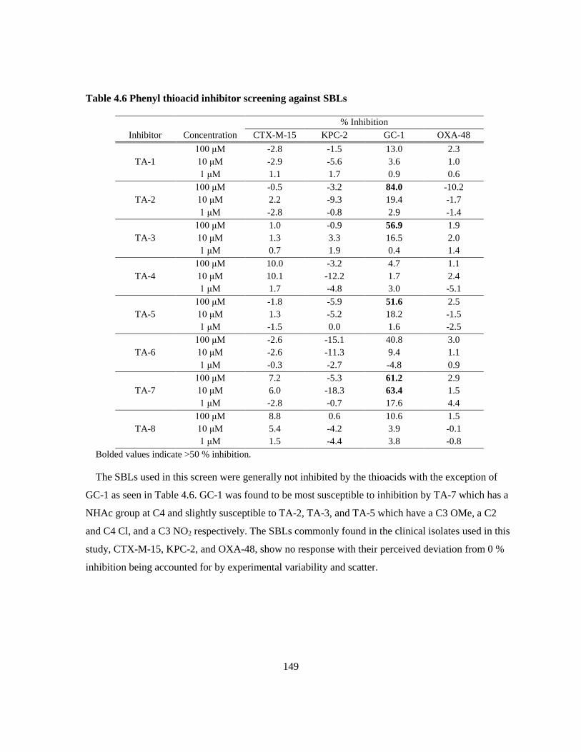
149
Table 4.6 Phenyl thioacid inhibitor screening against SBLs
% Inhibition
Inhibitor Concentration CTX-M-15 KPC-2 GC-1 OXA-48
TA-1
100 μM -2.8 -1.5 13.0 2.3
10 μM -2.9 -5.6 3.6 1.0
1 μM 1.1 1.7 0.9 0.6
TA-2
100 μM -0.5 -3.2 84.0 -10.2
10 μM 2.2 -9.3 19.4 -1.7
1 μM -2.8 -0.8 2.9 -1.4
TA-3
100 μM 1.0 -0.9 56.9 1.9
10 μM 1.3 3.3 16.5 2.0
1 μM 0.7 1.9 0.4 1.4
TA-4
100 μM 10.0 -3.2 4.7 1.1
10 μM 10.1 -12.2 1.7 2.4
1 μM 1.7 -4.8 3.0 -5.1
TA-5
100 μM -1.8 -5.9 51.6 2.5
10 μM 1.3 -5.2 18.2 -1.5
1 μM -1.5 0.0 1.6 -2.5
TA-6
100 μM -2.6 -15.1 40.8 3.0
10 μM -2.6 -11.3 9.4 1.1
1 μM -0.3 -2.7 -4.8 0.9
TA-7
100 μM 7.2 -5.3 61.2 2.9
10 μM 6.0 -18.3 63.4 1.5
1 μM -2.8 -0.7 17.6 4.4
TA-8
100 μM 8.8 0.6 10.6 1.5
10 μM 5.4 -4.2 3.9 -0.1
1 μM 1.5 -4.4 3.8 -0.8
Bolded values indicate >50 % inhibition.
The SBLs used in this screen were generally not inhibited by the thioacids with the exception of
GC-1 as seen in Table 4.6. GC-1 was found to be most susceptible to inhibition by TA-7 which has a
NHAc group at C4 and slightly susceptible to TA-2, TA-3, and TA-5 which have a C3 OMe, a C2
and C4 Cl, and a C3 NO2 respectively. The SBLs commonly found in the clinical isolates used in this
study, CTX-M-15, KPC-2, and OXA-48, show no response with their perceived deviation from 0 %
inhibition being accounted for by experimental variability and scatter.

150
Table 4.7 Phenyl thioacid inhibitor screening against MBLs.
% Inhibition
Inhibitor Concentration IMP-1 NDM-1 SPM-1 VIM-2 L1
TA-1
100 μM 33.0 -21.5 27.0 13.5 91.2
10 μM 6.9 -13.6 -1.1 3.5 61.4
1 μM 3.9 -4.7 0.8 2.1 13.4
TA-2
100 μM 32.8 22.8 38.4 12.1 5.7
10 μM 1.4 12.1 4.7 -4.9 45.4
1 μM 7.2 16.7 4.8 -2.0 23.3
TA-3
100 μM 18.3 -13.5 -1.7 8.7 91.7
10 μM 8.3 -5.0 -5.0 4.7 51.1
1 μM -3.6 -4.0 -2.7 0.4 8.1
TA-4
100 μM 0.5 -11.5 -3.3 -7.7 19.6
10 μM 2.7 -7.7 -2.5 -8.9 0.4
1 μM 4.4 2.0 7.0 -5.7 -3.0
TA-5
100 μM 49.9 -0.6 19.9 37.5 86.9
10 μM 9.9 -5.2 18.2 11.3 70.0
1 μM 0.8 -7.4 -9.2 3.2 17.5
TA-6
100 μM 78.8 5.0 35.9 61.4 91.5
10 μM 19.9 -13.4 -1.0 3.5 72.8
1 μM 6.0 -10.1 -2.5 -2.3 15.5
TA-7
100 μM 38.2 53.6 29.0 17.3 -4.7
10 μM 19.6 69.2 37.9 19.4 8.9
1 μM 7.2 18.7 8.4 8.9 3.9
TA-8
100 μM 2.7 6.2 -1.6 -1.8 34.9
10 μM -0.1 -5.8 1.6 -3.7 6.7
1 μM 1.6 -1.2 9.8 0.5 1.8
Bolded values indicate >50 % inhibition.
The phenyl thioacids were also screened against MBLs as potential inhibitors using similar
methods. The data in Table 4.7 shows that the B1 MBLs are slightly more inhibited than the SBLs at
the highest concentrations of phenyl thioacids. IMP-1 and VIM-2 were 78.8% and 61.4 % inhibited
by TA-6 (3,5-dinitro) respectively, consistent with L1 which is also inhibited most potently by TA-6.
NDM-1 was only inhibited by more than 50% by TA-7 (4-NHAc) which is more consistent with GC-
1 than L1 which was uninhibited by this thioacid. Throughout Table 4.7 there are examples of the B1
MBLs starting to show slight inhibition (~30%) at 100 μM with TA-1, TA-2, TA-5, TA-6, and TA-7.
While it is encouraging that these can be inhibited by phenyl thioacids, the inhibition is too weak to
be clinically relevant. In addition to those presented below, the Class B2 MBL SFH-1 was screened
with TA-1 and was not significantly inhibited up to 316 μM.

151
4.3.1.2 Meropenem potentiation by phenyl thioacids
The most potent L1 inhibitors (TA-1, TA-5, and TA-6) were assayed for potentiation of
meropenem against 10 S. maltophilia strains. Of these strains, 7 are known to produce both the Class
B3 MBL L1 and the Class A SBL L2, 2 produce only L2, and one has not been sequenced to identify
coded β-lactamases. These compounds were not found to be soluble in MH broth at 1.25 % (TA-1,
TA-5) or 1.67% (TA-6) DMSO, necessitating spotting 10 μL from each well onto MH agar to
distinguish between growth and precipitate as depicted in Figure 4.6.
Figure 4.6 MH agar spot plates of thioacid potentiation of meropenem against L1 and L2
producing S. maltophilia (UWB 45) for 128 mg/L TA-1 (B), TA-5 (C), or TA-6 (D). Plate A is the
unsupplemented control. The values along the perimeter of the agar plates indicate the
concentration of meropenem in the sample starting from 128 mg/L in the upper right corner
progressing downwards then left in 2-fold serial dilutions to 0.25 mg/L. The sample labelled “0”
is the inhibitor and the sample labelled “gr” is the growth control which contains neither
meropenem nor thioacid.
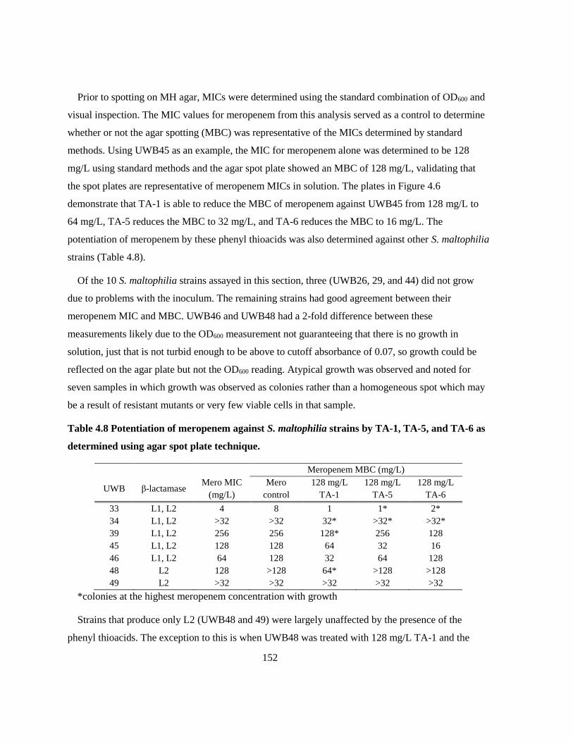
152
Prior to spotting on MH agar, MICs were determined using the standard combination of OD600 and
visual inspection. The MIC values for meropenem from this analysis served as a control to determine
whether or not the agar spotting (MBC) was representative of the MICs determined by standard
methods. Using UWB45 as an example, the MIC for meropenem alone was determined to be 128
mg/L using standard methods and the agar spot plate showed an MBC of 128 mg/L, validating that
the spot plates are representative of meropenem MICs in solution. The plates in Figure 4.6
demonstrate that TA-1 is able to reduce the MBC of meropenem against UWB45 from 128 mg/L to
64 mg/L, TA-5 reduces the MBC to 32 mg/L, and TA-6 reduces the MBC to 16 mg/L. The
potentiation of meropenem by these phenyl thioacids was also determined against other S. maltophilia
strains (Table 4.8).
Of the 10 S. maltophilia strains assayed in this section, three (UWB26, 29, and 44) did not grow
due to problems with the inoculum. The remaining strains had good agreement between their
meropenem MIC and MBC. UWB46 and UWB48 had a 2-fold difference between these
measurements likely due to the OD600 measurement not guaranteeing that there is no growth in
solution, just that is not turbid enough to be above to cutoff absorbance of 0.07, so growth could be
reflected on the agar plate but not the OD600 reading. Atypical growth was observed and noted for
seven samples in which growth was observed as colonies rather than a homogeneous spot which may
be a result of resistant mutants or very few viable cells in that sample.
Table 4.8 Potentiation of meropenem against S. maltophilia strains by TA-1, TA-5, and TA-6 as
determined using agar spot plate technique.
Meropenem MBC (mg/L)
UWB β-lactamase Mero MIC
(mg/L)
Mero
control
128 mg/L
TA-1
128 mg/L
TA-5
128 mg/L
TA-6
33 L1, L2 4 8 1 1* 2*
34 L1, L2 >32 >32 32* >32* >32*
39 L1, L2 256 256 128* 256 128
45 L1, L2 128 128 64 32 16
46 L1, L2 64 128 32 64 128
48 L2 128 >128 64* >128 >128
49 L2 >32 >32 >32 >32 >32
*colonies at the highest meropenem concentration with growth
Strains that produce only L2 (UWB48 and 49) were largely unaffected by the presence of the
phenyl thioacids. The exception to this is when UWB48 was treated with 128 mg/L TA-1 and the

153
MIC of meropenem was reduced at least 4-fold. For most of the L1-producing strains, TA-1 was the
most effective phenyl thioacid in potentiating meropenem yielding at least a 2-fold reduction in MBC.
The most susceptible strain to potentiation was UWB 33 whose meropenem MBC was reduced 8-fold
in the presence of TA-1 and TA-5, and 4-fold in the presence of TA-6. While the potentiation of
meropenem by these compounds may be underwhelming in comparison to that of the PMPCs
described in Chapter 3, this assay requires the phenyl thioacids to cross the outer membrane and find
its target without getting diverted by other potential targets. The intended application of these
thioacids would involve them being delivered to their targets as part of a 3ʹ-acylthiocephalosporin and
released upon hydrolysis, reducing the likelihood of off target interactions and potentially increasing
their potency.
4.3.1.3 MICs of 3ʹ-acylthiocephalosprins against Stenotrophomonas maltophilia
To assess the intended thioacid delivery system, the 3ʹ-acylthiocephalosprins, and the impact of
substitutions on the thioacid leaving group in vivo, a selection of 3ʹ-acylthiocephalosporins with a
simple phenylacetyl or 3-fluorophenylacectyl on the C7-amido nitrogen group and a variety of
thioacid leaving groups at C3” were used in this study. While the most potent thioacids were the 3-
nitro (TA-5) and 3,5-dinitro (TA-6) substituted phenyl thioacids, cephalosporins with these groups on
the right-hand side were not available at the time of testing. Two S. maltophilia strains that are known
not to produce L1 but produce the other β-lactamase present in each of these strains, L2, was used as
a control. Three commercial β-lactams were also tested, meropenem (Mero), ceftazidime (CAZ), and
cefotaxime (CTX), to give a relative measure of the potency of the novel cephalosporins. The only β-
lactam for which there is a CLSI guideline for resistance is ceftazidime which is seldom used as a
therapy for S. maltophilia infections due to their nearly ubiquitous expression of
cephalosporinases.500,515–517

154
Table 4.9 MICs of 3ʹ-acylthiocephalosporins and control β-lactams against β-lactamase
producing strains of S. maltophilia.
MIC (mg/L)
UWB β-Lactamases Mero CAZ CTX TE-1 TE-2 TE-3 TE-4 TE-5 UW-123
26 L1, L2 256 256 256 >128 >128 >128 >128 64 16
29 NS 64 32 128 64 >128 128 64 8 4
33 L1, L2 4 16 8 64 128 >128 128 4 32
34 L1, L2 8 8 16 64 128 128 64 4 2
39 L1, L2 128 128 256 128 >128 >128 128 16 16
43 L1, L2 32 16 16 32 128 64 32 4 0.5
44 L1, L2 <1 <1 <1 128 64 64 32 2 0.25
45 L1, L2 64 64 128 32 >128 >128 128 16 4
46 L1, L2 32 4 32 64 128 128 64 4 2
48 L2 64 <1 16 128 >128 >128 >128 8 1
49 L2 16 2 16 64 128 128 64 4 2
NS – not sequenced, the presence of β-lactamases is unknown
Bolded MIC values are susceptible to β-lactams according to CLSI guidelines.500
Italicized MIC values are intermediate (neither resistant nor susceptible) to β-lactams according to CLSI
guidelines.500
The simple 3ʹ-acylthiocephalosporins, TA-1 – 4 are generally poor antibiotics for S. maltophilia
strains with MICs no lower than 32 mg/L. Of these compounds, TE-1 and TE-4 are the most potent
against this panel although studies with other bacterial strains and variable right-hand side groups
have distinguished the 2,4-dichloro substitution pattern as the most potent as a standalone antibiotic.
Through optimization of the group on the left-hand side, the potency of these compounds was
improved from TE-4, though TE-5, to UW-123. TE- 5 is more potent than meropenem against 9 of
the 11 S. maltophilia strains in Table 4.9 and UW-123 is generally 2 to 4-fold more potent than TE-5
and is only less potent than meropenem against one strain, UWB33.
In addition to being poor antibiotics against S. maltophilia strains, the early 3ʹ-
acythiocephalosporins, TE-1 through TE-4, were ineffective at potentiating meropenem beyond a 2-
fold MIC improvement as seen in Table 4.10. Although little potentiation of meropenem by these
compounds is observed in terms of MIC reduction, the combination of the two antibiotics did
significantly decrease the density of the cultures in the wells. This was particularly evident towards
the higher ends of both the cephalosporin and meropenem concentration ranges. The discrepancies
between the meropenem MICs reported in Table 4.9 and those in Table 4.10 are likely due to more
bacteria being present in the inoculum when the latter was prepared. Ideally, there should be 500 000

155
CFU/mL in the inoculum and while the colony counts for the experiment described in Table 4.10
(UWB26: 850 000 CFU/mL, UWB45: 320 000 CFU/mL, and UW49: 830 000 CFU/mL), they were
not considered out of range, nor were those for the experiment in Table 4.9 (UWB26: 590 000
CFU/mL, UWB45: 260 000 CFU/mL, and UWB49: 190 000 CFU/mL). For most organisms, minor
differences like this in the colony counts do not significantly impact the MICs; however, the S.
maltophilia strains in this lab have been found to be difficult in this respect in the past.
Table 4.10 Potentiation of meropenem by 3ʹ-acylthiocephaloporins (Ceph) against β-lactamase
producing strains of S. maltophilia.
Strain β-lactamase Ceph
Ceph MIC
(mg/L)
Meropenem MIC (mg/L) in the presence of Ceph
0
mg/L
4
mg/L
8
mg/L
16
mg/L
32
mg/L
64
mg/L
128
mg/L
UWB 26 L1, L2 TE-1 >128 512 512 512 512 512 256 256
TE-2 >128 512 512 512 512 512 256 256
TE-3 >128 512 256 256 256 256 256 256
TE-4 >128 512 512 512 512 512 256 256
UWB 45 L1, L2 TE-1 >128 128 128 128 128 128 64 64
TE-2 >128 128 128 128 128 128 128 128
TE-3 >128 128 128 128 128 128 128 64
TE-4 >128 128 128 128 128 128 128 64
UWB 49 L2 TE-1 >128 >64 >64 >64 >64 >64 >64 64
TE-2 >128 >64 >64 >64 >64 >64 >64 >64
TE-3 >128 >64 >64 >64 >64 >64 >64 >64
TE-4 >128 >64 >64 >64 >64 >64 >64 64
Bolded values indicate potentiation relative to meropenem control.
Optimization of the 3ʹ-acylthiocephalosporins involved over 70 novel cephalosporins in pursuit of
an MBL resistant β-lactam antibiotic that could be co-administered with meropenem. These
compounds were screened using a meropenem synergy assay with MBL producing Gram negative
pathogens. From this significant synthetic and microbiological effort, one compound distinguished
itself for its potency as a standalone antibiotic against not only S. maltophilia, but other MBL
producers as well and its moderate synergistic activity with meropenem: UW-123.
4.3.2 MICs of UW-123
In order to assess UW-123’s potential as a standalone antibiotic, several MIC panels were
performed in the presence and absence of an SBL inhibitor by both our lab and Merck.
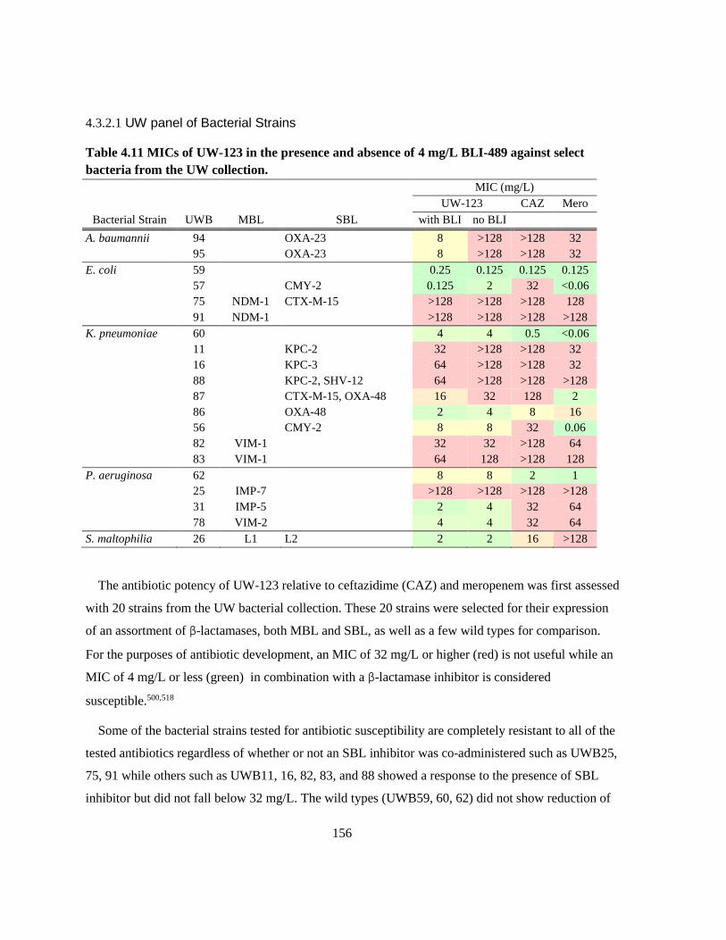
156
4.3.2.1 UW panel of Bacterial Strains
Table 4.11 MICs of UW-123 in the presence and absence of 4 mg/L BLI-489 against select
bacteria from the UW collection. MIC (mg/L) UW-123 CAZ Mero
Bacterial Strain UWB MBL SBL with BLI no BLI
A. baumannii 94 OXA-23 8 >128 >128 32
95 OXA-23 8 >128 >128 32
E. coli 59 0.25 0.125 0.125 0.125
57 CMY-2 0.125 2 32 <0.06
75 NDM-1 CTX-M-15 >128 >128 >128 128
91 NDM-1 >128 >128 >128 >128
K. pneumoniae 60 4 4 0.5 <0.06
11 KPC-2 32 >128 >128 32
16 KPC-3 64 >128 >128 32
88 KPC-2, SHV-12 64 >128 >128 >128
87 CTX-M-15, OXA-48 16 32 128 2
86 OXA-48 2 4 8 16
56 CMY-2 8 8 32 0.06
82 VIM-1 32 32 >128 64
83 VIM-1 64 128 >128 128
P. aeruginosa 62 8 8 2 1
25 IMP-7 >128 >128 >128 >128
31 IMP-5 2 4 32 64
78 VIM-2 4 4 32 64
S. maltophilia 26 L1 L2 2 2 16 >128
The antibiotic potency of UW-123 relative to ceftazidime (CAZ) and meropenem was first assessed
with 20 strains from the UW bacterial collection. These 20 strains were selected for their expression
of an assortment of β-lactamases, both MBL and SBL, as well as a few wild types for comparison.
For the purposes of antibiotic development, an MIC of 32 mg/L or higher (red) is not useful while an
MIC of 4 mg/L or less (green) in combination with a β-lactamase inhibitor is considered
susceptible.500,518
Some of the bacterial strains tested for antibiotic susceptibility are completely resistant to all of the
tested antibiotics regardless of whether or not an SBL inhibitor was co-administered such as UWB25,
75, 91 while others such as UWB11, 16, 82, 83, and 88 showed a response to the presence of SBL
inhibitor but did not fall below 32 mg/L. The wild types (UWB59, 60, 62) did not show reduction of

157
the UW-123 MIC in the presence of the SBL inhibitor, confirming that it is a true wild type and does
not produce an SBL. Conversely, all of the strains that produce only SBLs (except UWB56) showed
improved UW-123 MICs in the presence of the SBL inhibitor. This improvement can be as much as
16-fold as it as for UWB57 whose MIC dropped from 2 mg/L to 0.125 mg/L with the addition of 4
mg/L BLI-489. The strains that produced MBLs were much more sensitive to UW-123 than they
were to either ceftazidime and meropenem with the most pronounced difference being against the
IMP-5 and VIM-2 producing strains of P. aeruginosa (UWB31, 78). This effect could be a result of
UW-123 being insensitive to MBLs or turning over very slowly in MBLs and thus losing less
antibiotic to hydrolysis by MBL than other β-lactams. Unlike strains that produced IMP and VIM β-
lactamases, those that produced NDM-1 were not anymore susceptible to UW-123 than they were to
ceftazidime or meropenem which may be a result of the MICs being out of range or NDM-1 being
insensitive to UW-123.
To determine whether or not UW-123 could also be potentiated by the clinical SBL inhibitor
avibactam, three MBL producing strains (UWB75, 78, and 93) were assayed with both UW-123 and
ceftazidime against various concentrations of avibactam. The strains were chosen because they are
the most β-lactam resistant strains for each NDM, IMP, and VIM producers in the UW collection.
The standard administration of avibactam in microbiological assays is 4 mg/L; however, higher
concentrations can be assayed to explore the possibility of better synergy at higher concentrations.
Table 4.12 MICs of UW-123 against select strains from UW collection and its potentiation by
avibactam.
Cephalosporin MIC (mg/L)
Bacterial Strain UWB MBL SBL Antibiotic
0 mg/L
Avi
4 mg/L
Avi
8 mg/L
Avi
E. coli 75 NDM-1 CTX-M-15 UW-123 >128 128 64
CAZ >1024 >1024 >1024
93 IMP-1 CTX-M-15 UW-123 >128 64 64
CAZ 1024 512 256
P. aeruginosa 78 VIM-2 UW-123 4 8 8
CAZ 128 128 128

158
Two of the MBL producers co-express CTX-M-15, a common SBL in clinical pathogens. The
strains that produced the SBL showed improved sensitivity to UW-123 when co-administered with 4
mg/L of avibactam and administration of 8 mg/L avibactam did not offer a significant advantage over
4 mg/L as demonstrated in Table 4.12. The strain that only produced an MBL did not show
significant change in the MIC in response to the administration of either concentration of avibactam.
This is consistent with the previous result that BLI-489 does not potentiate UW-123 against VIM-2
producing P. aeruginosa (UWB78). As was also seen in Table 4.11, UW-123 is consistently much
more potent than ceftazidime against these MBL producers regardless of whether or not an SBL
inhibitor was co-administered.
4.3.2.2 Merck panel of Bacterial Strains
UW-123 was found to be superior to both meropenem and ceftazidime against the MBL producing
strains from the UW collection but required an SBL inhibitor to improve potency against strains that
express SBLs. The UW collection does not have a lot of variation within each of the MBL families,
so a sample of UW-123 was sent to Merck for testing against IMP, NDM, and VIM producing strains.
The Merck testing was performed on 25 MBL producing strains each of the families mentioned above
using ceftazidime and imipenem as comparators. The BLI used in these experiments was not
disclosed but is known to be a DBO like avibactam but is not the DBO that they were pursuing for
FDA approval.
IMP producing strains
Many of the IMP producing strains in the Merck panel were not sequenced at high enough resolution
at the time of these assays to determine the specific variant with the exception of the five K.
pneumoniae strains that produce IMP-26. The lack of specific typing to the IMPs also suggests that
the screening may have missed any SBLs in many of these strains. Contrary to expectations, a few of
the IMP producing strains in Table 4.13 demonstrate an antagonistic relationship between the BLI
and UW-123.
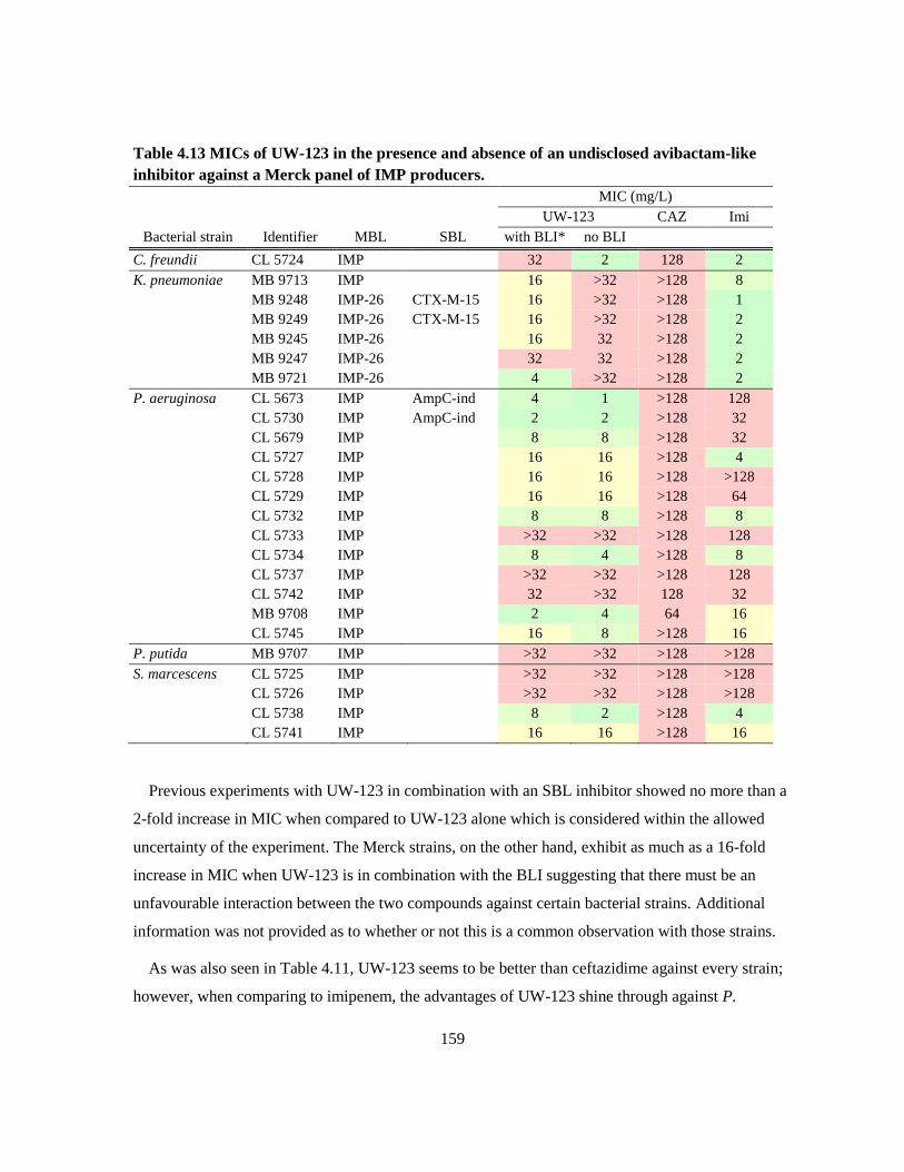
159
Table 4.13 MICs of UW-123 in the presence and absence of an undisclosed avibactam-like
inhibitor against a Merck panel of IMP producers.
MIC (mg/L)
UW-123 CAZ Imi
Bacterial strain Identifier MBL SBL with BLI* no BLI
C. freundii CL 5724 IMP 32 2 128 2
K. pneumoniae MB 9713 IMP 16 >32 >128 8
MB 9248 IMP-26 CTX-M-15 16 >32 >128 1
MB 9249 IMP-26 CTX-M-15 16 >32 >128 2
MB 9245 IMP-26 16 32 >128 2
MB 9247 IMP-26 32 32 >128 2
MB 9721 IMP-26 4 >32 >128 2
P. aeruginosa CL 5673 IMP AmpC-ind 4 1 >128 128
CL 5730 IMP AmpC-ind 2 2 >128 32
CL 5679 IMP 8 8 >128 32
CL 5727 IMP 16 16 >128 4
CL 5728 IMP 16 16 >128 >128
CL 5729 IMP 16 16 >128 64
CL 5732 IMP 8 8 >128 8
CL 5733 IMP >32 >32 >128 128
CL 5734 IMP 8 4 >128 8
CL 5737 IMP >32 >32 >128 128
CL 5742 IMP 32 >32 128 32
MB 9708 IMP 2 4 64 16
CL 5745 IMP 16 8 >128 16
P. putida MB 9707 IMP >32 >32 >128 >128
S. marcescens CL 5725 IMP >32 >32 >128 >128
CL 5726 IMP >32 >32 >128 >128
CL 5738 IMP 8 2 >128 4
CL 5741 IMP 16 16 >128 16
Previous experiments with UW-123 in combination with an SBL inhibitor showed no more than a
2-fold increase in MIC when compared to UW-123 alone which is considered within the allowed
uncertainty of the experiment. The Merck strains, on the other hand, exhibit as much as a 16-fold
increase in MIC when UW-123 is in combination with the BLI suggesting that there must be an
unfavourable interaction between the two compounds against certain bacterial strains. Additional
information was not provided as to whether or not this is a common observation with those strains.
As was also seen in Table 4.11, UW-123 seems to be better than ceftazidime against every strain;
however, when comparing to imipenem, the advantages of UW-123 shine through against P.

160
aeruginosa. Approximately two thirds of the P. aeruginosa strains were more susceptible to UW-123
than imipenem, even without the addition of the BLI which did little to nothing for this subset. The
IMP producing strains of K. pneumoniae tend to be more sensitive to imipenem than UW-123 as well
as more responsive to the BLI than the P. aeruginosa strains. The addition of BLI brought the MICs
of UW-123 nearer to those of imipenem for most K. pneumoniae strains. The tested strains of S.
marcescens were similarly susceptible to UW-123 and imipenem which is in and of itself
encouraging that a cephalosporin can be equally potent as a carbapenem.
NDM producing strains
The only NDM variant represented in the NDM panel from Merck is NDM-1 because it is by far the
most clinically important variant (and was at the time of this panel the only one found in clinical
isolates). Many of the NDM producing strains co-express at least one SBL, necessitating the use of
the SBL inhibitor since UW-123 is known to be susceptible to SBLs. This increases the complexity of
the problem since none of the clinically available classes of SBL inhibitors actually inhibit all SBLs.
The most common SBLs in this panel, CTX-M-15 and CMY-2, have been previously found to be
susceptible to avibactam, and should be similarly susceptible to other inhibitors of that class.361,519
The data in Table 4.14 suggest that UW-123 is not as effective against NDM-1 producers as it was
against IMP producers: imipenem is much more effective against most of the strains in this panel that
UW-123 regardless of whether or not the BLI is present. This is consistent with the observation from
Table 4.11 that UW-123 was completely ineffective against NDM-1 producers. Both NDM-1
producing E. coli strains used in the UW panel gave out of range or top of range MICs for each of the
tested antibiotics which prevented meaningful conclusions from being drawn; however, the
overwhelming result from the Merck panel is that NDM-1 producing strains of E. coli are much more
susceptible to carbapenems than to UW-123.

161
Table 4.14 MICs of UW-123 in the presence and absence of an undisclosed avibactam-like
inhibitor against a Merck panel of NDM-1 producers.
MIC (mg/L)
UW-123 CAZ Imi
Bacterial strain Identifier MBL SBL
with
BLI*
no
BLI
A. baumannii CLB 30003A NDM-1 >32 >32 >128 128
E. coli CLB 30022 NDM-1 CMY-2 16 16 >128 2
CLB 30048 NDM-1 CTX-M-15 32 >32 >128 >128
CLB 30005 NDM-1 CTX-M-15, CMY-2 32 >32 >128 8
CLB 30009 NDM-1 CTX-M-15, CMY-2 32 >32 >128 8
CLB 30016 NDM-1 CTX-M-15, CMY-2 >32 >32 >128 8
CLB 30026 NDM-1 CTX-M-15, CMY-2 >32 >32 >128 8
CLB 30028 NDM-1 CTX-M-15, CMY-2 >32 >32 >128 16
CLB 30042 NDM-1 CTX-M-15, CMY-2 32 >32 >128 8
CLB 30008 NDM-1 16 16 >128 8
E. cloacae CLB 30004 NDM-1 CTX-M-15, ACT/MIR >32 >32 >128 4
CLB 30018 NDM-1 CTX-M-15, ACT/MIR >32 >32 >128 4
CLB 30030 NDM-1
CTX-M-15, ACT/MIR,
OXA-181 2 >32 >128 16
CLB 30031 NDM-1
CTX-M-15, ACT/MIR,
OXA-181 2 >32 >128 16
MB 9867 NDM-1 TEM-1, CTX-M-15 4 >32 >128 4
K. pneumoniae CLB 30046 NDM-1 CMY-2 32 32 >128 16
CLB 30039 NDM-1 CTX-M-15 >32 >32 >128 16
CLB 30001 NDM-1 CTX-M-15, CMY-2 16 >32 >128 16
CLB 30006 NDM-1
CTX-M-15, CMY-2,
DHA >32 >32 >128 64
CLB 30012 NDM-1 CTX-M-15, DHA-1 32 >32 >128 8
CLB 30019 NDM-1 CTX-M-15, DHA-1 32 >32 >128 8
CLB 30010 NDM-1 DHA-1 16 16 >128 8
CLB 30034 NDM-1 SHV-12, CTX-M-15 32 32 >128 8
CLB 30011 NDM-1 SHV-2A, DHA-1 32 >32 >128 2
M. morganii CLB 30047 NDM-1 DHA-1 16 >32 >128 8
The only subset that yielded promising results from the NDM Merck panel was the E. cloacae
strains: three of the five strains showed a reduction in the MIC of UW-123 in the presence of BLI that
made it similarly potent to imipenem. This suggests that it may not be NDM-1 that is causing the lack

162
of sensitivity to UW-123 but rather other features of pathogenic Enterobacteriaceae that are
preventing it from reaching its target. This trend was also observed in the UW panel as well as the
IMP panel in which clinical isolates of K. pneumoniae and E. coli are much less susceptible to UW-
123 than other strains, even in the presence of a BLI.
VIM producing strains
Table 4.15 MICs of UW-123 in the presence and absence of an undisclosed avibactam-like
inhibitor against a Merck panel of VIM producers.
MIC (mg/L)
UW-123 CAZ Imi
Bacterial strain Identifier MBL SBL with BLI* no BLI
C. freundii MB 9163 VIM-1 4 16 >128 2
MB 9864 VIM-32 CMY-81 32 32 >128 8
E. cloacae MB 9787 VIM-1 ACT/MIR 4 1 >128 4
MB 9158 VIM-1 8 8 >128 4
MB 9160 VIM-1 0.125 2 >128 4
MB 9178 VIM-5 >32 >32 >128 4
K. pneumoniae MB 9773 VIM-1 KPC-2 4 16 >128 64
MB 9151 VIM-1 0.5 0.5 >128 64
MB 9152 VIM-1 1 2 >128 16
MB 9153 VIM-1 1 8 >128 32
MB 9155 VIM-1 16 8 >128 32
MB 9156 VIM-1 1 1 >128 64
MB 9159 VIM-1 0.5 0.5 >128 64
MB 9161 VIM-1 4 32 >128 32
MB 9166 VIM-1 2 4 >128 2
MB 9171 VIM-26 1 0.5 >128 32
MB 9172 VIM-26 0.25 1 >128 64
MB 9175 VIM-27 8 16 >128 64
MB 9177 VIM-5 4 >32 >128 16
P. aeruginosa MB 9732 VIM 8 8 64 >128
MB 9758 VIM >32 >32 >128 >128
MB 9344 VIM-2 16 16 16 16
MB 9345 VIM-2 4 4 16 128
MB 9346 VIM-2 32 >32 64 >128
MB 9347 VIM-2 16 16 128 >128

163
The Merck panel of VIM producers includes at least 6 VIM variants and 3 of the strains also
express an SBL. Despite the lack of SBLs in these strains, 11 strains responded to the BLI and
reduced the MIC of UW-123. The VIM panel was the most susceptible of the Merck panels to UW-
123 with 10 of the 25 strains having MICs of 4 mg/L or less without BLI and 16 of 25 with BLI as
seen in Table 4.15. This is a distinct improvement over the 6 of 25 IMP producers with UW-123
MICs of 4 mg/L or lower. Similar to other panels, ceftazidime was ineffective against almost all
strains making any response to UW-123 a vast improvement over other cephalosporins.
The K. pneumoniae strains from the IMP and NDM panels were less susceptible to UW-123 than
imipenem both in the presence and absence of the BLI. In the VIM panel, only two of the 13 K.
pneumoniae strains were less susceptible to UW-123 than imipenem in the absence of BLI while the
addition of BLI brings the MIC of UW-123 to or lower than that of imipenem for all of them.
Likewise, all of the P. aeruginosa strains in the VIM panel were more or equally susceptible to UW-
123 than both ceftazidime and imipenem regardless of whether or not it was assayed with the BLI.
4.3.3 Bacterial Synergy of UW-123 with Meropenem
Compounds in this series were developed to be co-administered with a carbapenem as a protective
agent against MBLs. UW-123 was the best antibiotic from this series; however, early synergy
screening assays with meropenem were often out of range.
Table 4.16 Synergy of UW-123 and meropenem against select strains from the UW collection.
Strain β-lactamase
UW-123 MIC
(mg/L)
Meropenem MIC (mg/L) in the presence of UW-123
FIC*
0
mg/L
1
mg/L
2
mg/L
4
mg/L
8
mg/L
16
mg/L
32
mg/L
K. pneumoniae
UWB11 KPC-3 >32 64 32 32 64 32 32 64 0.27
K. pneumoniae
UWB16 KPC-2 >32 128 32 32 32 32 32 32 0.28
E. coli
UWB75
NDM-1
CTX-M-15 >32 128 128 128 128 128 64 64 0.75
P. aeruginosa
UWB31 VIM-2 8 64 32 32 16 - - - 0.13
P. aeruginosa
UWB78 VIM-2 4 64 32 16 - - - - 0.35
S. maltophilia
UWB26 L1, L2 2 128 128 64 32 - - - 0.25
*Synergistic (FIC ≤ 0.5), Additive (0.5 < FIC ≤ 1), Indifferent (1 < FIC < 2), Antagonistic (FIC ≥ 2)

164
The synergy of UW-123 and meropenem was assayed against 6 β-lactamase producing strains
using a microplate checkerboard synergy assay. UW-123 was able to potentiate meropenem against
K. pneumoniae and P. aeruginosa strains at even the lowest concentrations (1 mg/L) as seen in Table
4.16. For the chosen E. coli and S. maltophilia strains, higher concentrations of UW-123 were
required to decrease the MIC of meropenem against those bacteria. This combination of antibiotics
demonstrates synergy against the S. maltophilia strain but not the E. coli against which they are
additive. It is possible that not all E. coli strains will be resistant to synergistic effects of these two
antibiotics as UWB75 among the most resistant strains in our collection.
4.3.4 Mode of Action Studies of UW-123
To get a better understanding of how UW-123 works as an antibiotic, a series of experiments were
performed to establish whether or not it is lethal to cells, what (if any) morphological changes occur
in response to treatment, and the specific target(s) and potency against common pathogens.
4.3.4.1 Time Kill Curves
β-Lactam antibiotics are widely accepted to be bactericidal as treatment with therapeutic
concentrations results in cell lysis. Studies have shown that this bactericidal activity is concentration
dependent such that lower concentrations of β-lactam are bacteriostatic if only one critical PBP is
inhibited while higher concentrations are bactericidal as a result of inhibition of multiple PBPs.520,521
In this work, time-kill curves for UW-123 were first generated by OD600 to establish the expected
behavior at specific timepoints then by CFU to determine viability. The time-kill curve produced by
reading the culture density, depicted in Figure 4.7A shows a reproducible “bump” in the curve at
around 3 hr corresponding to morphological change. This method of assaying growth in the presence
of antibiotic is good as a starting point for comparing different classes of antibiotics (data no shown)
but gives little insight into the viability of the cells in the culture

165
0 2 4 6 8 100.0
0.2
0.4
0.6
0.8
22 24 26
4 mg/L
2 mg/L
1 mg/L
0.5 mg/L
0.25 mg/L
0 mg/L
Time (hr)
OD
600
0 2 4 6 8 100
5
10
15
15 20 25 30
Time (hr)
log
(CF
U/m
L)
Figure 4.7 UW-123 time kill curves against wild type E. coli (UWB 59) monitored by (A) OD600
and (B) CFUs. The graphs in this figure were generated using GraphPad Prism v. 5.03.
Plating UW-123 treated cultures at various timepoints on MH agar gives insight into cell viability
after antibiotic treatment. Bactericidal activity is defined as a 3-log unit reduction in the number of
viable cells relative to the starting timepoint of the untreated control. The experiment in Figure 4.7B
started with 107 viable cells per mL of liquid culture. At 4 hours, the number of viable cells in 1 mL
of culture had fallen to its minimal value of 105.5 when treated with either 1 or 2 mg/L UW-123 (1x
and 2x MIC respectively), a 1.5 log unit reduction. This suggests that higher concentrations of UW-
123 would be required to induce a bactericidal effect and that at these concentrations, UW-123
behaves as a bacteriostatic antibiotic. Later experiments performed with other β-lactam antibiotics
showed that bactericidal activity occurs at 4x MIC and greater (data not shown).
4.3.4.2 Morphology
Inhibition of certain HMM PBPs results in characteristic morphological changes which can be
observed by phase contrast microscopy. The OD600 measurement of the time-kill curves of many
antibiotics suggested that most morphological changes occurred at 2-3 hr after treatment as indicated
by a transient “bump” in the curves. This prompted the decision to stop the growth at 2 hr and
determine the morphology of the cells by phase contrast microscopy. This form of microscopy was
chosen over fluorescence or traditional light microscopy because it does not require staining or fixing
of the bacteria and it has been used to determine bacterial morphologies in the literature.117,513,522 A
DMSO control was performed to ensure that any morphological changes were the result of the
antibiotic rather than the solvent. Additionally, aztreonam and meropenem were tested as controls for
known E. coli PBP 3 and PBP 2 inhibitors respectively.
A B

166
Table 4.17 Sucrose stabilized, β-lactam treated E. coli under 1000x magnification by phase
contrast microscopy.
Control Aztreonam Meropenem UW-123
Untreated
0.0078 mg/L
0.00195 mg/L
0.25 mg/L
DMSO
0.03125 mg/L
0.0078 mg/L
1 mg/L
Cell density should not be considered in analysis of these microscopy images; samples were not
prepared quantitatively.
The treatment of cells with 1.25 % DMSO did not induce significant morphological changes –
Table 4.17 illustrates that some cells may have elongated slightly which that may be within normal
variation when compared to the untreated control. As expected, inhibition of PBP 3 in E. coli by
aztreonam induced filamentation at concentrations above the MIC (0.0156 mg/L) but not below the
MIC. Some elongation can be observed below the MIC but is minimal compared to the filamentation
above the MIC. Inhibition of PBP 2 in E. coli by meropenem induced the expected spheroplasting of
the cells. The addition of 0.3 M sucrose to the MH broth is supposed to stabilize osmotically unstable
spheroplasts.522 When E. coli cells were treated with 0.0156 mg/L meropenem in the presence and
absence of sucrose, the sucrose free media yielded many spheroplasted cells while the sucrose media
yielded only cellular debris (data not shown).
UW-123 induced filamentation at all assayed concentrations (128 mg/L – 0.0625 mg/L); however,
normal morphology cells can be observed in samples treated with 0.25 mg/L or less UW-123 as seen
in Table 4.17. Contrary to the effect of sucrose on meropenem treated cells, UW-123 treated cells

167
grown in the presence of sucrose were much more stable at concentrations in excess of the MIC such
as 128 mg/L, maintaining the groups of filaments observed for 1 mg/L in Table 4.17 as opposed to
the only one cell could be found in the culture treated with 128 mg/L UW-123 in the absence of
sucrose (data not shown). The formation of filaments by treatment with UW-123 suggests that UW-
123 primarily inhibits subclass B3 PBPs, specifically PBP 3 in E. coli.
4.3.4.3 Competition of UW-123 with Bocillin for PBP Binding
To confirm the hypothesis that UW-123 acts as an inhibitor of subclass B3 PBPs, membrane
fragments from two wild-type Gram negative organisms, E. coli and P. aeruginosa, were isolated.
The wild-type E. coli strain used in this experiment is an ATCC control strain, unlike the one used
above and was acquired after the previous experiments were performed. Membrane fragments from
this particular E. coli strain were required for work not presented in this thesis and some of which was
used to assay UW-123. In addition to E. coli, membrane fragments from P. aeruginosa were isolated
due to the marked increase in potency of UW-123 over ceftazidime and meropenem against P.
aeruginosa strains from our lab.
P. aeruginosa PBPs
Variable concentrations of the isolated membrane fragments from P. aeruginosa were assayed
using the SDS-PAGE Bocillin assay described in 4.2.4.3.2 to determine the optimal concentration of
fragment to use in the assay (data not shown). Multiple SDS-PAGE gels of varying acrylamide
contents (10%, 12.5%, and 15%) were also tested to ensure optimal separation. The optimal
conditions for this assay were determined to be 1.4 mg/mL protein per well, run on a 10% SDS-
PAGE gel. Using these conditions, the membrane fragments were first incubated with UW-123,
allowing adequate time for the covalent bonds to form, then Bocillin was added and incubated for the
same amount of time allowing selective labelling of uninhibited PBPs. Identification of PBPs used
the Rf values of the bands from gel A and the Coomassie stained LMW marker.

168
Figure 4.8 SDS-PAGE of UW-123 bound to P. aeruginosa PBPs competing with Bocillin. Gel A
is the Bocillin fluorescent read, Gel B is the Coomassie stained read. The lanes correspond to
the following concentrations of UW-123: 2) 0 mg/L; 3) 128 mg/L; 4) 64 mg/L; 5) 32 mg/L; 6) 16
mg/L; 7) 8 mg/L; 8) 4 mg/L; 9) 2 mg/L – streak in the lane thus discluded; 10) 1 mg/L; 11) 0.5
mg/L; 12) 0.25 mg/L; 13) 0.125 mg/L; 14) 0.0625 mg/L; 15) 0.0313 mg/L; 16) 0.0156 mg/L; 17)
0.0078 mg/L; 18) 0.0039 mg/L; 19) 0 mg/L. Lanes 1 and 20 contained low molecular weight
marker.
The visualization of the fluorescence is dependent upon the covalent binding of Bocillin to the
various PBPs, and a decrease in intensity of any band relative to the control is indicative of UW-123
competing for the active site of that PBP. As can be seen in Figure 4.8A, UW-123 binds to P.
aeruginosa PBP 1a and PBP 3 at low concentrations and PBP 1b and PBP 4 at slightly higher
concentrations. Graphing the relative intensities of the bands gives a clearer picture of the binding
(Figure 4.9).
A
B

169
PBP 1a
-3 -2 -1 0 1 2 30
25
50
75
100
125
log([UW-123] (mg/L))
Rela
tive %
Bo
cill
in B
ind
ing
PBP 1b
-3 -2 -1 0 1 2 30
25
50
75
100
125
log([UW-123] (mg/L))
Rela
tive %
Bo
cill
in B
ind
ing
PBP 2
-3 -2 -1 0 1 2 30
25
50
75
100
125
log([UW-123] (mg/L))
Rela
tive %
Bo
cill
in B
ind
ing
PBP 3
-3 -2 -1 0 1 2 30
25
50
75
100
125
log([UW-123] (mg/L))
Rela
tive %
Bo
cill
in B
ind
ing
PBP 4
-3 -2 -1 0 1 2 30
25
50
75
100
125
log([UW-123] (mg/L))
Rela
tive %
Bo
cill
in B
ind
ing
PBP 5/6
-3 -2 -1 0 1 2 30
25
50
75
100
125
log([UW-123] (mg/L))
Rela
tive %
Bo
cill
in B
ind
ing
Figure 4.9 Dose response curves of the relative competitive binding of UW-123 to PBPs from P.
aeruginosa with Bocillin. The graphs in this figure were generated using GraphPad Prism v.
5.03.

170
The weak intensity of PBP2 in the gels resulted in a high degree of uncertainty in the measurement
of the band densities resulting in scatter and poor curve fitting (Figure 4.9). Looking at the gel in
Figure 4.8A, it is evident that PBP2 was not inhibited by UW-123 since the corresponding band
appears to be only slightly weaker in intensity when treated with 128 mg/L UW-123 (well 2) than
when treated with no UW-123 (well 1). The curve fitting of PBPs 1a, 1b, 3, and 4 was good, although
PBP 1a and PBP 3 required constraints for the upper plateau since it was not well defined. The fitting
of the dose response curves from Figure 4.9 yielded the IC50s presented in Table 4.18.
Table 4.18 IC50s of the competitive binding of UW-123 to PBPs from P. aeruginosa with
Bocillin.
PBP IC50 (mg/L)
1a 0.13 ± 0.05
1b 0.6 ± 0.1
2 NI
3 0.027 ± 0.005
4 0.8 ± 0.3
5/6 NI
NI – Not significantly inhibited
As expected, the most potently inhibited PBP was the subclass B3 PBP 3 with an IC50 of 0.027
mg/L. The other PBPs were an order of magnitude less potently inhibited than PBP 3 with IC50s
ranging from 0.13 mg/L to 0.8 mg/L indicating that the antibiotic potency is primarily due to PBP 3
inhibition in P. aeruginosa.
E. coli PBPs
In the same manner as for the P. aeruginosa PBP assay, the optimal conditions for the E. coli PBP
assay were determined to be 0.32 mg/mL protein per well, run on a 10% SDS-PAGE gel. Unlike the
P. aeruginosa PBPs, PBP 1a and 1b could not be resolved for E. coli. Close inspection of the gels in
Figure 4.10A shows two bands at this position; however, they did not migrate uniformly and cannot
be isolated from one another for band density analysis. Additionally, the signal for PBP 5/6 is so
intense that it may impact the readings for PBP 4 since these two bands are not well resolved. It
should be noted that the visualization in Figure 4.10A has been optimized for PBP 2 and PBP 3 - the
high fluorescence signal at the bottom of the gel was minimized when quantifying PBP 5/6.

171
Figure 4.10 SDS-PAGE of UW-123 bound to E. coli PBPs competing with Bocillin. Gel A is the
Bocillin fluorescent read, Gel B is the Coomassie stained read. The lanes correspond to the
following concentrations of UW-123: 2) 0 mg/L; 3) 128 mg/L; 4) 64 mg/L; 5) 32 mg/L; 6) 16
mg/L; 7) 8 mg/L; 8) 4 mg/L; 9) 2 mg/L; 10) 1 mg/L; 11) 0.5 mg/L – water entered sample during
boiling; 12) 0.25 mg/L; 13) 0.125 mg/L; 14) 0.0625 mg/L; 15) 0.0313 mg/L; 16) 0.0156 mg/L; 17)
0.0078 mg/L; 18) 0.0039 mg/L; 19) 0 mg/L. Lanes 1 and 20 contained low molecular weight
marker.
As above, the visualization of the fluorescence is indicative of covalently bound Bocillin in the
active sites of the various PBPs, and a decrease in intensity of any band relative to the control is
indicative of UW-123 competing for the active site of that PBP. As can be seen in Figure 4.10A, UW-
123 binds to E. coli PBP 3 at low concentrations and PBP 1a/b and PBP 2 at higher concentrations.
Graphing the relative intensities of the bands gives a clearer picture of the binding (Figure 4.11).
A
B

172
PBP 1a/b
-3 -2 -1 0 1 2 30
50
100
150
log([UW-123] (µg/mL))
Rela
tive %
Bo
cill
in B
ind
ing
PBP 2
-3 -2 -1 0 1 2 30
50
100
150
log([UW-123] (µg/mL))
Rela
tive %
Bo
cill
in B
ind
ing
PBP 3
-3 -2 -1 0 1 2 30
50
100
150
log([UW-123] (µg/mL))
Rela
tive %
Bo
cill
in B
ind
ing
PBP 4
-3 -2 -1 0 1 2 30
50
100
150
log([UW-123] (µg/mL))
Rela
tive %
Bo
cill
in B
ind
ing
PBP 5/6
-3 -2 -1 0 1 2 30
50
100
150
log([UW-123] (µg/mL))
Rela
tive %
Bo
cill
in B
ind
ing
Figure 4.11 Dose response curves of the relative competitive binding of UW-123 to PBPs from
E. coli with Bocillin. The graphs in this figure were generated using GraphPad Prism v. 5.03.
The affinity of UW-123 for the active sites of PBP 1a/b and PBP 2 appear to be similar with both
curves having midpoints between 0.1 and 1 mg/L. PBP 3 was much more susceptible to inhibition by
UW-123 with only the lowest assayed concentration (0.0039 mg/L) representing the upper plateau.

173
As was expected, there is a lot of scatter in the PBP 4 data; however, visual inspection of the gel
confirms that the band intensity is consistent throughout most of the wells, only decreasing at the
highest UW-123 concentrations. Likewise, little inhibition of PBP 5/6 was observed, although the
intensity of these bands steadily decreased with increasing UW-123.
The dose-response plots in Figure 4.11 were fitted using nonlinear regression to determine the IC50s
presented in Table 4.19. The curves for PBP 4 and PBP 5/6 did not go low enough (PBP 4 and PBP
5/6 were not inhibited enough) to determine an IC50 with certainty.
Table 4.19 IC50s of the competitive binding of UW-123 to PBPs from E. coli with Bocillin.
PBP IC50 (mg/L)
1a/b 0.45 ± 0.08
2 1.0 ± 0.2
3 0.02 ± 0.01†
4 NI
5/6 NI
NI – Not significantly inhibited
†Top of curve not well defined, constrained to 110 (comparable to top of curve for 1a/b and 2)
As the morphological data suggested, PBP 3 is the most potently inhibited E. coli PBP and it level
of inhibition is comparable to that of PBP 3 from P. aeruginosa – both 0.02-0.03 mg/L. Likewise, the
inhibition of PBP 1 a/b from E. coli is close to the average inhibition of the Class A PBPs from P.
aeruginosa (0.45 mg/L and 0.37 mg/L respectively). Unlike P. aeruginosa, significant inhibition of
PBP 2 from E. coli was observed, although it is weaker than the inhibition of PBP 3 and the Class A
PBPs.
4.3.5 Pyridine thioacids – a more potent alternative to phenyl thioacids
Phenyl thioacids are at best moderate inhibitors of MBLs, by increasing the potency of the released
thioacid inhibitor the efficacy of these cephalosporins could be further improved against MBL
producing strains. From Chapter 3, we know that pyridine rings with negatively charged groups at the
2 and 6 positions are potent inhibitors of all subclasses of MBLs. Additionally, Roll et al. have
demonstrated that pyridine thiocarboxylates (also referred to as pyridine thioacids) are potent
inhibitors of B3 MBLs; however, their analysis did not include other MBL subclasses.439 This section
explores the potential inhibitory activity of two pyridine thiocarboxylates, PMTC and PDTC, and the

174
benzene derivative of PDTC, I-PDTC (structures depicted in Figure 4.4) against a variety of β-
lactamases.
4.3.5.1 Inhibitor Screening
Like the phenyl thioacids, the pyridine thiocarboxylates required the addition of NaOH to the
stocks to ensure complete dissolution - this was most pronounced for I-PDTC. These compounds
were first screened for SBL inhibition to establish whether or not they are universal β-lactamase
inhibitors. As expected, CTX-M-15, KPC-2, and OXA-48 are not inhibited by any of the tested
compounds as seen in Table 4.20. Some mild inhibition of CTX-M-15 was observed with I-PDTC at
100 μM, but this is not enough inhibition to warrant further study.
Table 4.20 Pyridine thioacid inhibitor screening against SBLs.
% Inhibition
Inhibitor Concentration CTX-M-15 KPC-2 GC-1 OXA-48
PMTC 100 μM -0.3 -2.1 54.3 -0.4
10 μM 1.2 -2.9 44.1 -0.9
1 μM -0.5 -1.2 5.6 0.3
PDTC 100 μM 3.8 3.0 69.5 1.2
10 μM -3.1 -3.0 37.6 -1.1
1 μM -2.1 -2.8 1.4 1.1
I-PDTC 100 μM 37.1 -5.1 85.4 6.4
10 μM 2.2 -3.4 50.2 -1.5
1 μM -1.1 -0.5 6.3 -2.4
Bolded values are >50 % inhibited.
As was also observed for the phenyl thioacids in Table 4.6, GC-1 is the only SBL that is
significantly susceptible to inhibition by this subset of thioacids. The data in Table 4.20 shows that I-
PDTC is the most potent inhibitor of GC-1 from this series, suggesting that the pyridine core is not
necessary or beneficial for GC-1 inhibition. GC-1 is also indifferent to the number of thiocaboxylate
groups on the molecule as evidenced by the negligible difference between the inhibitory potency of
PMTC and PDTC.
Although the pyridine core was not advantageous for GC-1 inhibition, it yielded very potent MBL
inhibition as demonstrated in Table 4.21. The most potent inhibitor in this series appears to be PMTC
which completely inhibits IMP-1, NDM-1, and VIM-2 at all assayed concentrations and demonstrated
>50% inhibition at all concentrations for SFH-1 and L1. PDTA is slightly less effective, inhibiting

175
most MBLs by about 73 % at 1 μM as opposed to the >95% seen with most B1 MBLs at 1 μM
PMTC. SPM-1 is less potently inhibited that other B1 MBLs, demonstrating complete inhibition at 10
μM PMTC and only 42% inhibition at 1 μM while the other B1 MBLs are still completely inhibited
at 1 μM.
Table 4.21 Pyridine thiocarboxylate inhibitor screening against MBLs
% Inhibition
Inhibitor Concentration IMP-1 NDM-1 SPM-1 VIM-2 SFH-1 L1
PMTC
100 μM 99.8 99.2 99.5 99.9 96.7 99.7
10 μM 99.9 100.0 99.5 99.9 96.1 99.9
1 μM 97.3 96.8 42.5 99.6 62.4 84.2
PDTC
100 μM 98.6 94.1 101.9 99.9 93.6 100.2
10 μM 100.0 99.7 90.2 99.8 96.9 99.0
1 μM 71.5 73.1 45.9 74.5 73.6 78.2
I-PDTC
100 μM 61.8 27.2 54.8 31.8 41.2 61.3
10 μM 12.7 5.3 0.3 -0.6 13.1 35.1
1 μM 6.5 7.3 -7.3 -0.6 -4.4 22.7
Bolded values are >50 % inhibited.
The inhibitor with a benzene core, I-PDTC, only inhibits MBLs at the highest assayed concentrations.
The drastic drop in inhibitory potency from PDTC to I-PDTC suggests that the pyridine core is
essential of potent inhibition.
4.3.5.2 IC50 Determination
A wider range of PMTC and PDTC concentrations were assayed with GC-1, the B1 MBLs, and L1
to give a more accurate measure of inhibition. These assays were performed with three technical
replicates but was only performed once to allow enough material for bacterial studies. The dose
response curves generated from these experiments are shown in Figure 4.12.

176
GC-1
-4 -2 0 2 4
50
100
log([Inhibitor] (µM))
% A
cti
vit
y
IMP-1
-6 -4 -2 0 2 4
50
100
log([Inhibitor] (µM))
% A
cti
vit
y
NDM-1
-6 -4 -2 0 2 4
50
100
log([Inhibitor] (µM))
% A
cti
vit
y
SPM-1
-6 -4 -2 0 2 4
50
100
log([Inhibitor] (µM))
% A
cti
vit
y
VIM-2
-6 -4 -2 0 2 4
50
100
log([Inhibitor] (µM))
% A
cti
vit
y
L1
-6 -4 -2 0 2 4
50
100
log([Inhibitor] (µM))
% A
cti
vit
y
Figure 4.12 Dose response curves of MBLs and GC-1 inhibited by PMTC and PDTC (red and
blue respectively). The graphs in this figure were generated using GraphPad Prism v. 5.03.
It is evident from the dose response curves in Figure 4.12 that PMTC and PDTC are very similarly
potent inhibitors against all assayed enzymes as the curves are overlapping throughout the vertical
portion of the curve. This steepness of this region is well conserved for each enzyme; however, there
is a distinct difference between enzymes. The curves for IMP-1 and VIM-2 are much steeper (Hill
slope >4) than SPM-1 and L1 (Hill slope ~1.3) with NDM-1 and GC-1 having intermediate slopes
which may be indicative of differing inhibition mechanisms for the different enzymes.499
Fitting of these dose response curves to Equation 3.1 yielded the IC50 values listed in Table 4.22.
These data confirm that PMTC and PDTC have similar potency with IC50 values differing by no more
than 25% (SPM-1). It is probable that with more repeats of this experiment, the IC50 values for PMTC
and PDTC will fall within the uncertainty of one another. Despite the differences observed in the
steepness of the dose response curves, the pyridine thiocarboxylates exhibit consistent potency across
the MBLs with IC50 values ranging from 0.41 μM to 0.79 μM for the assayed MBLs with the

177
exception of SPM-1 which was slightly less susceptible to inhibition. A dose response curve was also
generated for SFH-1; however, the assayed concentrations were not low enough to obtain the upper
plateau region. The estimates for the IC50 of these compounds against SFH-1 are consistent with those
of the majority of the MBLs (0.70 μM and 0.47 μM for PMTC and PDTC respectively).
Table 4.22 IC50s of pyridine thiocarboxylates against class B and C β-lactamases.
IC50 (μM)
β-lactamase Class PMTC PDTC
GC-1 C 4.61 ± 0.08 4.2 ± 0.1
IMP-1 B1 0.79 ± 0.02 0.63 ± 0.02
NDM-1 B1 0.45 ± 0.02 0.50 ± 0.02
SPM-1 B1 1.33 ± 0.06 1.01 ± 0.06
VIM-2 B1 0.60 ± 0.01 0.48 ± 0.02
L1 B3 0.41 ± 0.02 0.48 ± 0.02
In addition to being potent MBL inhibitors, PMTC and PDTC are inhibitors of GC-1, albeit a 10-
fold less potent than against MBLs. Despite the reduced potency against GC-1 relative to the MBLs,
PMTC and PDTC are similarly potent against GC-1 with IC50s of 4.61 μM and 4.2 μM respectively.
This inhibition broadens the spectrum of targets from solely MBLs to MBLs and some SBLs. To
further explore the potential of the pyridine thiocarboxylates as β-lactamase inhibitors, they were
assayed as an adjuvant to potentiate meropenem against β-lactam resistant pathogens.
4.3.5.3 Potentiation of meropenem by pyridine thiocarboxylates
The potentiation of meropenem was assayed using a microplate checkerboard assay with two-fold
dilutions of meropenem along the long axis of the plate and two-fold dilutions of the inhibitor along
the short axis. The addition of sodium hydroxide to stock solutions of pyridine thiocarboxylates was
required for complete dissolution prior to dilution in MH broth. A small amount of precipitate was
observed in the wells corresponding to the highest concentration of the inhibitors after overnight
growth. Spotting on agar plates was not used to differentiate between growth and precipitate in these
assays because comparison to other wells containing the same concentration of inhibitor allowed for
visual differentiation.
Each of the strains in Table 4.23 demonstrate potentiation of meropenem by the pyridine
thiocarboxylates at the highest assayed concentrations as evidenced by a 4-fold or greater reduction in

178
the MIC. The most susceptible strain was the IMP-1 and CTX-M-15 producing E. coli, UWB 93,
which was reduced to intermediate inhibition by only 4 mg/L PDTC and 8 mg/L PMTC. This strain
was made completely susceptible to meropenem (MIC of 1 mg/L or less) in the presence of 16 mg/L
PDTC and 64 mg/L PMTC. Most other strains were only brought into intermediate or susceptible
range but the highest concentrations of adjuvant. This is a significant accomplishment for UWB 75
and UWB 24 which have meropenem MICs of 128 mg/L and require a 32 to 64-fold reduction of
their MIC to enter the intermediate range.
Table 4.23 Sensitization of MBL producing Gram negatives to meropenem by pyridine
thiocarboxylates.
Strain β-lactamase
Meropenem
MIC (mg/L) Inhibitor
Meropenem MIC (mg/L) in the presence of
Inhibitor
4
mg/L
8
mg/L
16
mg/L
32
mg/L
64
mg/L
128
mg/L
E. coli
UWB93
IMP-1
CTX-M-15
4 PMTC 4 2 2 2 0.5 0.25
PDTC 2 2 1 0.25 0.25 0.125
E. coli
UWB75
NDM-1
CTX-M-15
128 PMTC 128 64 64 64 8 4
PDTC 64 64 64 16 4 2
K. pneumoniae
UWB116
NDM-1 32 PMTC 16 16 8 8 2 0.25
PDTC 16 16 8 8 2 0.25
I-PDTC 32 16 16 16 16 8
P. putida UWB24 VIM-2 128 PMTC 128 128 128 128 64 4
PDTC 128 128 128 64 4 2
P. aeruginosa
UWB78
VIM-2 128 PMTC 128 128 128 128 32 32
PDTC 128 128 128 64 32 32
Bolded MIC values are susceptible to β-lactams according to CLSI guidelines.500
Italicized MIC values are intermediate (neither resistant nor susceptible) to β-lactams according to CLSI
guidelines.500
The most effective compound at potentiating meropenem is PDTC which elicits the same MIC
reductions with at least 2-fold less compound for most bacterial strains. The only strain where the two
pyridine thiocarboxylates are equivalent was UWB 116, this was also the only strain for which I-
PDTC was assayed. The benzene core of I-PDTC abolishes much of the potency of PDTC, such that
PDTC elicits a 32-fold greater reduction in MIC than I-PDTC at 128 mg/L. Additionally, I-PDTC
does not reduce the MIC of meropenem against UWB 116 enough to bring it into intermediate
resistance.

179
4.4 Discussion
4.4.1 Phenyl Thioacid Inhibitors of B3 MBLs
The phenyl thioacids proved to be moderate inhibitors of L1 but inhibited few other β-lactamases.
This inhibition was improved substantially when the most concentrated DMSO stock of the inhibitor
was prepared in the presence of one molar equivalent of NaOH. Upon addition of the NaOH, it is
assumed that the thiocarboxylate group is deprotonated. Although the initial DMSO solution did not
seem to contain precipitate, it is possible that the aromatic thioacids are in an aggregated state and
that deprotonation breaks up the aggregate resulting in a higher effective concentration of the
conjugate base of the thioacid to act as an inhibitor.
The phenyl thioacids with halogen substituents on the aromatic ring TA-1, TA-3, and TA-4
demonstrated decreased potency at high concentrations when prepared in the absence of NaOH.
Preliminary dose response curves of these compounds under the same conditions gave unusual “U-
shaped” curves (data not shown). Most of the literature regarding U-shaped dose response curves
involves hormesis, the overcompensation of cells or organisms in response to deviation from
homeostasis, which does not apply to in vitro enzyme assays.523 In this case, it is likely that the U-
shaped dose response curves are indicative of the halogenated phenyl thioacids forming aggregates or
colloids at high concentrations resulting in reduced availability of the free inhibitor for binding to the
enzyme active site.524
The most potent set of phenyl thioacid inhibitors of L1 were those with one or more nitro group on
the phenyl ring, TA-5 (at C3) and TA-6 (at C3 and C5), while the second most potent set of inhibitors
has a halogen at C4, TA-1 (Br at C4) and TA-3 (Cl at C2 and C4). TA-4 which has an additional
chlorine at C6 is a poor inhibitor. It is not clear if this effect is steric or electronic. TA-2 which has a
methoxy group (inductively electron withdrawing but pi-electon donating) at C3 is a poor inhibitor of
L1. This suggests that it is beneficial to have π-electron with drawing groups (e.g. NO2) meta to the
thioester rather than a π-electron donating groups (e.g. OMe).
Upon testing with a broader panel of MBLs, TA-6 which has nitro groups at C3 and C5 had the
greatest breadth of inhibitory activity as it inhibited IMP-1 and VIM-2 by more than 50% at 100 μM
and showed limited activity with SPM-1. Other trends seen with L1 do not hold up: TA-2 and TA-7
are more potent against the B1 MBLs than both TA-1 and TA-3, suggesting that the hydrophobic

180
substituent is preferred over the halogens in these MBLs. This may be the result of the C3 substituent
weakly interacting with the loop that closes over the B1 active sites. NDM-1 does not behave the
same way as other B1 MBLs with these inhibitors, it is only significantly inhibited by TA-7 which
may be a result having a much more flexible loop than IMP-1 or VIM-2.
Unlike the PMPCs, the phenyl thioacids are capable of inhibiting at least one of the SBLs, GC-1,
although the Class A and Class D SBLs were not affected. The mode of inhibition of GC-1 by
aromatic thioacids is unclear.
In bacterial studies, the phenyl thioacids were able to potentiate meropenem against L1 producing
strains of S. maltophilia. Most strains were more potentiated by TA-1 than either of the phenyl
thioacids with nitro substituents. This is contrary to the observation that the nitro substituents
improved the inhibitory potency of the phenyl thioacids in enzyme kinetic assays and suggests that
TA-1 may cross the outer membrane better than TA-5 or -6 or it may have fewer off-target
interactions. Since meropenem is a bactericidal antibiotic, the MBCs were reflective of the MICs with
no more than a 2-fold difference between the MIC and MBC of meropenem. This suggests that since
the thioacids are not antibiotics themselves and should not disrupt the ability of meropenem to kill
bacteria, the MBC reduction would also be reflected in the MICs if they were readable. The only
point in the MBC experiment where it becomes uncertain whether or not they are reflective of MICs
is when the growth spots are only colonies. Due to the low count of viable bacteria in these spots, it is
likely that these wells would not have reached the growth cutoff. This is most pronounced against
UWB 33 which had only colonies in many of the spots as seen in Figure 4.13.

181
Figure 4.13 Spot plates of UWB 33 on MH agar to determine the MBCs of thioacid potenticated
meropenem when treated with no adjuvant (A), 128 mg/L TA-1 (B), 128 mg/L TA-5 (C), and
128 mg/L TA-6.
The inhibition of L1 by the phenyl thioacids may be the result of the thiocarboxylate sulfur atom
replacing the catalytically essential hydroxide ion that is normally shared as a ligand by the the two
Zn2+ ions in the active site. This behavior, depicted in Figure 4.14, has been previously demonstrated
for the sulfur atom of numerous thiols including D-captopril, N-(3-mercaptopropanoyl)-D-alanine,
and the bisthiazolidine derivatives but a crystal structure of a thioacid in the L1 active site has never
been acquired.255,425,429

182
Figure 4.14 Binding of thiols to the L1 active site : D-captopril (PDB: 2FU8), N-
mercaptopropanoyl)-D-alanine (PDB: 2QDT), bisthiazolidine (PDB: 5EVB). This figure was
generated from the aforementioned protein databank files using Chimera v. 1.12.
4.4.2 3ʹ-Acylthiocephalosporins as delivery systems for phenyl thioacid inhibitors of
L1
Precipitation of an adjuvant is not ideal for assays since the true accessible concentration of the
compound is unknown. The challenges of determining the potentiation of meropenem by the phenyl
thioacids would be a problem for potential drug development; however, the intended application of
these compounds is through a 3ʹ-acylthiocephalosporin. Unlike the phenyl thioacids, the synthetic
cephalosporins were completely soluble, and use of a cephalosporin as a delivery system ensures that
the thioacid is release at its target. The release of the thioacid at its target circumvents any solubility
problems, reduces the potential for off-target reactions, and the thioacid should be in the active,
deprotonated form. Simple 3ʹ-acylthiocephalosporins with leaving groups at C3ʹ similar to the
thioacids discussed above were less effective at potentiating meropenem against S. maltophilia strains
than the thioacid alone. This suggests that these cephalosporins may be having difficulty crossing the
outer membrane or that they are poor substrates that are not being turned over enough to release an
adequate amount of inhibitor.
S. maltophilia is known to be a highly antibiotic resistant pathogen with low outer membrane
permeability, efficient efflux mechanisms, and chromosomally encoded β-lactamases (L1 and L2).525–
527 This resistance is particularly prevalent for β-lactams due to the chromosomally encoded β-
lactamases such that only 26.5% of S. maltophilia isolates from a Greek hospital were susceptible to
either ceftazidime and treatment with β-lactams is only recommended when the patient exhibits
adverse effect to other antibiotics such as trimethoprim/sulfamethoxazole.515,517 This is what makes

183
the antimicrobial potency of TE-5 and UW-123 against β-lactamase producing strains of S.
maltophilia interesting: S. maltophilia strains that co-produce L1 and L2 have been widely reported to
be immune to β-lactam monotherapy (with sporadic reports of limited ceftazidime efficacy).279,526–534
4.4.3 UW-123
The most potent standalone antibiotic to come from the extensive medicinal chemistry effort of
developing the 3ʹ-acylthiocephalospoins was UW-123. The increased potency of this compound
relative to the other 3ʹ-acylthiocephalosporins is most likely a result of more efficient uptake into the
bacteria due to the siderophore mimic in the amido sidechain on the C7 nitrogen. In assays where
UW-123 was not co-administered with any other antibiotic or adjuvant, it was shown to be less
effective against strains that express extended spectrum SBLs. This is, in part, due to these enzymes
being excellent at hydrolyzing cephalosporins, but more importantly, it is due to the phenyl thioacids
not being inhibitory towards Class A and Class D SBLs. The vast majority of bacterial strains in the
Dmitrienko lab inventory that produce SBLs produce either Class A or Class D ESBLs, so efficacy of
UW-123 against strains producing Class C SBLs, particularly Class C ESBLs, was not established.
4.4.3.1 Efficacy of UW-123 against bacterial pathogens
In the UW panel, UW-123 was more potent than either ceftazidime or meropenem against strains
that produce only MBLs due to the inhibition of the MBL by the released thioacid inhibitor. The only
case where this was not true was against strains that produce NDM-1. These strains, UWB 75 and 91,
are highly resistant to all β-lactam antibiotics and require potent inhibition of NDM-1 for sensitization
as demonstrated by the PMPCs in Table 3.8. Since the phenyl thioacids were not inhibitory towards
B1 MBLs in vitro, least of all NDM-1, it is probable that they are also not inhibitory in vivo, leading
to the insensitivity of these strains to UW-123.
The insensitivity of NDM-1 producers was also observed in the Merck panel of NDM-1 producers
in Table 4.14 in which UW-123 was less potent than either ceftazidime or imipenem against all
strains in the absence of BLI. While many of the NDM-1 producing strains demonstrated a reduction
in UW-123 MIC in the presence of BLI consistent with the widespread SBL co-expression, only 3 of
the strains were sensitized enough to be considered susceptible. Unlike NDM-1 producers which were
not susceptible (MIC ≤ 4 mg/L) to UW-123 alone, both the IMP and VIM producing strains from the
Merck panels had 6 and 9 strains that were susceptible to UW-123 without the aid of a BLI.

184
The VIM panel was the most susceptible of the Merck panels to UW-123 and was also the most
responsive to the addition of the BLI, increasing the number of susceptible strains from 9 to 15 of 25
upon its addition (Table 4.15). This demonstrates that the VIM-producing strains in the UW panel in
Table 4.12 are not necessarily representative of all VIM-producers as the K. pneumoniae VIM-1
strains (UWB 82 and 83) were highly resistant to UW-123 while the VIM-2 producing P. aeruginosa
(UWB 78) was only moderately susceptible. Although the Merck panel of VIM-producers contained
at least 6 VIM variants, these were heavily weighted towards VIM-1-like variants with only 4-6 of the
25 strains being from the VIM-2 branch of the family. Unfortunately, all of the potential strains that
produce a VIM-2-like variant were P. aeruginosa strains with no VIM-1-like variants being
represented in this organism so although the VIM-2 strains were less susceptible to UW-123, it is not
certain whether that is a characteristic of P. aeruginosa or the VIM-2-like variants.
Across both the NDM and VIM panels from Merck, the BLI performed as expected, reducing the
MIC of UW-123 against a few of the strains. In the VIM panel, there was an occasional discrepancy
in which UW-123 became less effective in the presence of the BLI, although this only happened in 3
of the 25 strains and only one of those was actually a significant (increase of ≥ 4-fold in MIC). The
IMP-1 panel had more of these anomalies with 5 of the 25 strains demonstrating this behavior and
three of those being a significant decrease in efficacy. It is possible that this BLI has off-target
interactions in some strains (such as inducing the expression of an SBL or efflux pump) that increase
the resistance of the cell to UW-123 or β-lactams in general.
In assays with a small set of β-lactamase-producing bacteria from the Dmitrienko lab collection,
UW-123 was found to behave synergistically with meropenem against most strains (Table 4.16). The
only strain against which they did not show synergy was UWB75, and E. coli that produces both
NDM-1 and CTX-M-15. This is consistent with the results from the Merck panel of NDM-1-
producers, suggesting that UW-123 is highly susceptible to NDM-1 and does not reach its target in
the cell in high concentrations.
4.4.3.2 Mode of action of UW-123
At the assayed concentrations, UW-123 is a bacteriostatic antibiotic at concentrations as high as 2x
MIC that induces filamentation in E. coli cells. Other literature cephalosporins such as CXA-101 and
ceftazidime have also been found to be bacteriostatic at 2x MIC, approaching but not attaining a 3-log
unit reduction at 8 hr.116 In those studies, a bactericidal load of antibiotic was achieved at 8 hour

185
incubation with 4x MIC and only slightly surpassed the 3-log unit cutoff. This, in addition to the
observation that when the killing curve is collected using OD measurements, the 4x MIC sample has
much less growth at 24 hr than the 2x MIC sample (Figure 4.7) suggests that going to higher
concentrations in the agar plate count experiments would likely yield a set of conditions under which
UW-123 is bactericidal.
The morphological changes associated with inhibition of PBP 2 and PBP 3 in E. coli were
confirmed using meropenem and aztreonam respectively. These experiments were carried out in the
presence of 0.3M sucrose to increase the likelihood of observing delicate morphologies through
osmotic stabilization. Consistent with the literature, inhibition of PBP 2 by meropenem created
spheroplasts and inhibition of PBP 3 by aztreonam induced filamentation (Table 4.17).535
Cephalosporins, such as ceftazidime, have also be known to induce filamentation through PBP 3
inhibition.535 The filamentation of E. coli cells in the presence of UW-123 is indicative of a β-lactam
antibiotic that inhibits subclass B3 PBPs such as PBP 3 in E. coli.
Studies to quantify the binding of UW-123 to the PBPs of E. coli and P. aeruginosa confirmed that
UW-123 inhibits the B3 PBP, PBP3, most potently in each organism.
4.5 Future Work
The success of UW-123 as a more potent cephalosporin against MBL producing Gram negative
pathogens suggests that this is an area of study that warrants further pursuit.
The data presented in section 4.3.1.1 suggest that the phenyl thioacids are moderate inhibitors of L1
and have some inhibitory activity against IMP-1 and VIM-2. The kinetic studies in this section
operated under the assumption that NaOH only deprotonates the thioacid and does not cause any side
reactions in DMSO. A thorough literature search has not yielded any studies that have been carried
out under these conditions. To confirm whether or not the addition of NaOH is just deprotonating or
changing the structure of the phenyl thioacids, structural studies (such as 1HNMR or MS) would need
to be performed on the reaction mixture over time as a decrease in colour intensity is observed.
The proposed mechanism for the 3ʹ-acylthiocephalosporins suggests that upon hydrolysis, a
rearrangement occurs that results in the release of a thioacid, a phenomenon that has yet to be
confirmed. Reaction mixtures of either base hydrolyzed or enzyme hydrolyzed 3ʹ-
acylthiocephalosporin could be analyzed by ESI-MS to determine whether or not the thioacid is

186
released. This study could also be used to determine whether or not the enzyme active site contributes
to the rearrangement. If a thioacid is released, further studies kinetic studies to better quantify the
inhibition (IC50) and to determine the mechanism by which inhibition occurs would be warranted.
Much like the phenyl thiocaids, there is more kinetics work to be done with the pyridine thioacids:
additional replicates of the IC50s should be done to establish reproducibility and mechanistic studies
should be pursued. Additionally, the Dmitrienko group has acquired an expression plasmid for His-
tagged AmpC which should be used to generate pure AmpC protein and tested for inhibition with
PMTC and PDTC to determine whether or not the inhibition of GC-1 is characteristic of Class C
SBLs or if it is GC-1 specific.
The the pyridine thioacids were found to be potent β-lactamase inhibitors; however, they pose both
solubility and reactivity problems in a clinical setting. Delivery of these compounds, particularly the
dithioacid, PDTC, using a cephalosporin has been considered with a bis-PDTC-cephalosporin (Figure
4.15) having been synthesized.
Figure 4.15 Bis-PDTC-cephalosporin
Some preliminary experiments have been conducted with this compound and it was found to be
ineffective alone and in combination with avibactam (only for UWB 75, 93) against 5 MBL
producing Gram negatives (UWB: 24, 26, 75, 78, 93). For each of these, the MIC of the bis-PDTA-
cephalosporin was >128 mg/L alone and came down to 128 mg/L when in combination with 4 mg/L
avibactam against UWB 93. It is likely that this compound is too large to adequately penetrate the
outer cell membrane of Gram negative bacteria since it is 860 Da. Another strategy for delivery of
these compounds may need to be considered such as attempting to deliver only the monothioacid,
PMTC, which displays similar potency in vitro and only requires protection on one thioacid.

187
Chapter 5
Other β-Lactamase Inhibitors
5.1 Introduction
In the late 1960’s and early 1970’s a number of reports appeared in the literature concerning the
inhibition of serine proteases by electrophilic carbonyl compounds such as aldehydes and
trifluoromethylketones.536–538 It was argued the such inhibitors formed hemiacetal or hemiketal
adducts with the enzyme active site serine hydroxyl group and that such adducts interacted
particularly favourably with the enzyme active site since they were mimics of either the tetrahedral
intermediate involved in the catalytic mechanism or mimics of the structurally related transition state
involved in forming them. (Scheme 5.1) Hence, such compounds were referred to as transition state
analogues. 539–541
Scheme 5.1 Comparison of hemiketal formation with TS and intermediate in acyl enzyme
mechanism.
Inspired by these observations, this laboratory as well as several other academic and industrial
research laboratories explored the concept of creating cyclobutanones that mimic β-lactam antibiotics
as potential inhibitors of β-lactamases as well as potential antibiotics that bind to penicillin binding
proteins (PBPs), in the late 1970’s to mid 1980’s (Figure 5.1).389,390,542–545

188
Figure 5.1 Examples of cyclobutanone mimics of β-lactam antibiotics. 389,542–546
In this group, racemic CYB-1: (1S,4S,5S)-7,7-dichloro-6-oxo-2-thiabicyclo[3.2.0]heptane-4-
carboxylic acid (Figure 5.1) was prepared and tested as a potential inhibitor of the Class A SBL Bc-I
from Bacillus cereus. This compound exhibited no SBL inhibition at concentrations up to 1 mM.
Likewise, the cyclobutanones prepared in this context by others were not potent inhibitors of Class A
SBLs nor good antibiotics. Since at that time potent SBL inhibitors such as clavulanic acid and
penicillanic acid sulfones such as sulbactam (Figure 1.11) were coming into clinical use, all research
concerning the cyclobutanones as SBL inhibitors was abandoned.
With the emergence of clinically important bacteria that produce new SBLs exhibiting
carbapenemase activity and that are poorly inhibited by clavulanate and the penicillanic acid sulfones,
as well as the carbapenem-hydrolyzing MBLs in the late 90’s, this laboratory began to revisit the
cyclobutanones as potential inhibitors of both the SBLs and the MBLs. Computational, spectroscopic
and crystallographic studies in this group led to the realization that the cyclobutanone-type penicillin
mimic A (Figure 5.2) had a strong preference for an endo conformation of the five-membered ring
which forces the essential carboxylate group into a pseudo-axial orientation that is not favourable for
binding to the active sites of β-lactamases.546

189
Figure 5.2 Conformations of C3-substituted cyclobutanones and the steric interactions of the
hydrates. The colour-coded (Cl = green, S = yellow, O = red, C = grey, H = white) ball-and-
stick ORTEP plots were generated using Mercury 3.7 software and are based on atomic
coordinates from original X-ray crystallographic data generated by Dr. Abdeljalil Assoud in
the Department of Chemistry X-ray crystallographic laboratory at the University of Waterloo.
It was also discovered that the preferred conformation of the five-membered ring could be
controlled by introduction of a heteroatom in the α-orientation (Figure 5.2) which creates an anomeric
effect between the heteroatom and the sulfur lone pairs of electrons leading to a preference for the
exo-conformation and a spatial orientation of the carboxylate group that is more favourable for
binding to β-lactamases. The compounds incorporating two chlorine atoms at C7, the C=O of the
cyclobutanone were found to be prone to exist predominantly as a hydrate in water and as a hemiketal
in methanol. This tendency was especially pronounced for those compounds that possess a
heteroatom in the α-orientation adjacent to the sulfur atom (Figure 5.2).
It was envisaged that SBL inhibition would occur via hemiketal formation with the active site
serine hydroxyl group of SBLs and MBL inhibition might arise through binding to the zinc ions as a
hydrate as shown below in Scheme 5.2B.

190
Scheme 5.2 Parallells between the binding of penems and cyclobutanones to SBLs and MBLs.
Proposed binding modes of tetrahedral intermediates in the β-lactamase-catalyzed hydrolysis of
a penem (A) and the potential for cyclobutanones as broad-spectrum SBL and MBL inhibitors.
In a collaborative study with the protein X-ray crystallography group of Natalie Strynadka at UBC
it was possible to demonstrate the formation of a hemiketal linkage with the active site serine
hydroxyl group of the Class D SBL OXA-10 (Figure 5.3A) It was also possible to demonstrate that
the cyclobutanone CYB-2 and the unsaturated cyclobutanone JJ05-1058 (Figure 5.1) exhibit
significant inhibition of SBLs of Classes A, C and D and the MBL IMP-1.391

191
Figure 5.3 X-Ray crystal structures of cyclobutanones bound to β-lactamases. (A) CYB-2
bound to OXA-10 as a serine hemiketal. (B)JJ05-1058 bound to the B1 MBL SPM-1 as a
hydrate. This figure was generated using Chimera v. 1.12 from the protein databank files 3LCE
and 5NDB respectively)391,392
More recently, in a collaborative study with colleagues Christopher Schofield at the University of
Oxford and James Spencer at the University of Bristol in the UK, this group has been able to show
that the unsaturated cyclobutanone JJ05-1058 binds in its hydrated form to the active site of the Class
B1 MBL SPM-1 with the hydroxide ion that bridges between the two zinc ions retained rather than
being displaced by one of the oxygens groups of the hydrate as had been speculated in earlier studies
in this group (Figure 5.3B).392
The goal of the present study of cyclobutanone-type β-lactamase inhibitors was to expand the
understanding of the spectrum of activity of the unsaturated cyclobutanone-type inhibitor JJ05-1058
as an inhibitor of clinically important SBLs and MBLs as well as to gain some initial insight into the
possibility of binding to PBPs which might suggest possible antibacterial activity.
5.2 Methods.
5.2.1 β-Lactamase Enzyme Kinetics
One of the most active cyclobutanone analogues from the Johnson 2010 paper seen in Figure 5.1
was tested against a wider array of β-lactamases. Solid JJ05-1058 (compound 5)391 was provided by
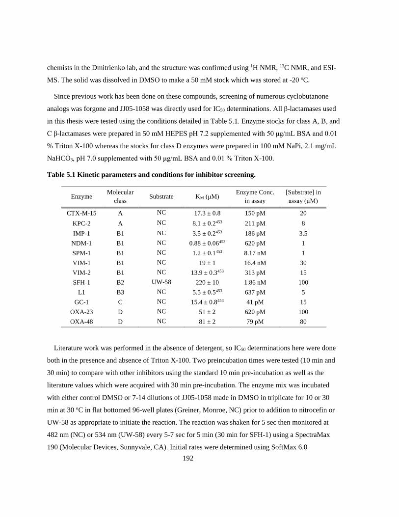
192
chemists in the Dmitrienko lab, and the structure was confirmed using 1H NMR, 13C NMR, and ESI-
MS. The solid was dissolved in DMSO to make a 50 mM stock which was stored at -20 oC.
Since previous work has been done on these compounds, screening of numerous cyclobutanone
analogs was forgone and JJ05-1058 was directly used for IC50 determinations. All β-lactamases used
in this thesis were tested using the conditions detailed in Table 5.1. Enzyme stocks for class A, B, and
C β-lactamases were prepared in 50 mM HEPES pH 7.2 supplemented with 50 μg/mL BSA and 0.01
% Triton X-100 whereas the stocks for class D enzymes were prepared in 100 mM NaPi, 2.1 mg/mL
NaHCO3, pH 7.0 supplemented with 50 μg/mL BSA and 0.01 % Triton X-100.
Table 5.1 Kinetic parameters and conditions for inhibitor screening.
Enzyme Molecular
class Substrate KM (μM)
Enzyme Conc.
in assay
[Substrate] in
assay (μM)
CTX-M-15 A NC 17.3 ± 0.8 150 pM 20
KPC-2 A NC 8.1 ± 0.2453 211 pM 8
IMP-1 B1 NC 3.5 ± 0.2453 186 pM 3.5
NDM-1 B1 NC 0.88 ± 0.06453 620 pM 1
SPM-1 B1 NC 1.2 ± 0.1453 8.17 nM 1
VIM-1 B1 NC 19 ± 1 16.4 nM 30
VIM-2 B1 NC 13.9 ± 0.3453 313 pM 15
SFH-1 B2 UW-58 220 ± 10 1.86 nM 100
L1 B3 NC 5.5 ± 0.5453 637 pM 5
GC-1 C NC 15.4 ± 0.8453 41 pM 15
OXA-23 D NC 51 ± 2 620 pM 100
OXA-48 D NC 81 ± 2 79 pM 80
Literature work was performed in the absence of detergent, so IC50 determinations here were done
both in the presence and absence of Triton X-100. Two preincubation times were tested (10 min and
30 min) to compare with other inhibitors using the standard 10 min pre-incubation as well as the
literature values which were acquired with 30 min pre-incubation. The enzyme mix was incubated
with either control DMSO or 7-14 dilutions of JJ05-1058 made in DMSO in triplicate for 10 or 30
min at 30 oC in flat bottomed 96-well plates (Greiner, Monroe, NC) prior to addition to nitrocefin or
UW-58 as appropriate to initiate the reaction. The reaction was shaken for 5 sec then monitored at
482 nm (NC) or 534 nm (UW-58) every 5-7 sec for 5 min (30 min for SFH-1) using a SpectraMax
190 (Molecular Devices, Sunnyvale, CA). Initial rates were determined using SoftMax 6.0
193
(Molecular Devices, Sunnyvale, CA) then normalized and fitted to Equation 3.1 using GraphPad
Prism version 5.00 for Windows (GraphPad Software, San Diego, CA) by non-linear least squares
regression. The concentration of DMSO present in the final assay mixture was kept under 2 % which
set an upper limit for JJ05-1058 concentrations at 1000 μM. Due to this upper limit, the plateau at the
high inhibitor concentration end was not always present or well defined.
5.2.2 Penicillin Binding Protein Binding Assay
Preparation of PBP Containing Membrane Fragments
Procedures for PBP membrane preparation and Bocillin binding assay were adapted from Moyá et
al..116 P. aeruginosa PA01 cells were obtained from Dr. Stephen Seah of the University of Guelph.
An overnight culture of PA01 cells was prepared by inoculating two tubes containing 2 mL Luria
Bertani (LB) broth with scrapings from a glycerol stock and shaking them at 250 rpm, 37 oC,
overnight (16-20 hr). The 4 mL of PA01 overnight culture were used to inoculate 500 mL LB broth
which was then incubated at 37 oC, shaking at 185 rpm until the OD600 reached 1 (~5 hr). Cells were
then centrifuged in a JA-10 rotor at 4400 xg for 10 min at 4 oC and the supernatant was discarded.
The pellets were washed in 150 mL of Buffer A (20 mM KH2PO4, 140 mM NaCl, pH 7.5) then
centrifuged again at 4400 xg for 10 min at 4 oC and the supernatant was discarded. The cells in the
pellet were then lysed using a large probe on a Heat Systems Ultrasonic processor W-255 sonicator
set to 50 % cycle and 50 % power 5 times for 30 sec each with 1 min rest on ice between bursts. The
lysate was then centrifuged in a JA-25.5 rotor at 12 000 xg for 10 min at 4 oC and the supernatant and
pellets were retained separately and frozen on dry ice then stored at -80 oC. The supernatant was
thawed and transferred into Oak Ridge polypropylene tubes with a nylon screw cap for use in a T-
1270 rotor of a Sorvall WX100 Ultra centrifuge. Samples were centrifuged at 150 000 xg for 1 h at 4
oC and the supernatant was discarded. The pellets were resuspended in 1 mL Buffer A and pooled.
Total membrane protein concentration was determined by Bradford assay as described in 2.2. The P.
aeruginosa PBP membrane preparation was then aliquoted, frozen on dry ice, and stored at -80 oC.
PBP Competitive Binding Assay
Two-fold dilutions of JJ05-1058 ranging from 128 mg/L to 0.0039 mg/L were prepared in Buffer B
(20 mM NaPi, 140 mM NaCl, pH 7.5) and DMSO such that the final concentration of DMSO in each
sample was 1.28 % while aliquots of the PBP membrane preparation were thawed on ice. The PBP
membrane preparation (10 μL) was incubated with a 3x concentrated stock of JJ05-1058 (5 μL) for 30

194
min at 37 oC after vortexing. In a darkened room, 5 μL of the fluorescent penicillin, Bocillin, was
added and returned to the incubator for 30 min at 37 oC after vortexing. Samples were prepared for
SDS-PAGE by adding 5 μL of 5x SDS-PAGE Buffer to each sample, vortexing, boiling for 3 min
before spinning down any condensation. Each sample (20 μL) as well as 5 μL of low molecular
weight marker was loaded onto a 10 % SDS-PAGE gel which were run at 80 V in the dark until the
blue dye front had reached the bottom of the gel. Gels were uncased directly onto a Pharos (BioRad,
Hercules, CA) gel reading system and visualized at λex= 488 nm and λem= 530 nm. Band densities
were determined using Image Lab software (BioRad, Hercules, CA) and were normalized and fitted
to Equation 3.1 using GraphPad Prism 5.00 for Windows (GraphPad Software, San Diego, CA) with
the added constraint that the bottom plateau is set to 0 since it is not well defined. Gels were then
stained with Coomassie Blue R-250 (BioRad, Hercules, CA) containing stain overnight then
destained with 50 % methanol, 10 % acetic acid in water. Coomassie stained gels were visualized on
a Gel Doc EZ Imager (BioRad, Hercules, CA).
5.3 Results
5.3.1 Inhibition of β-Lactamases
The cyclobutanone JJ05-1058, was screened against 12 β-lactamases for inhibitory activity using a
standardized IC50 assay. The effects of pre-incubation time and presence of detergent are best
demonstrated by the graphs used to determine the IC50s in Figure 5.4 for the SBLs and Figure 5.5 for
the MBLs.

195
Figure 5.4 Inhibition of SBLs by JJ05-1058. Assays performed in the presence of 0.01 % Triton
X-100 are displayed in blue while those performed in the absence of Triton X-100 are displayed
in red; preincubation times of 10 min (▲) and 30 min (●) are shown. The graphs in this figure
were generated using GraphPad Prism v. 5.03.
From the graphs in Figure 5.4 it can be seen that the inhibition of KPC-2, GC-1, OXA-23, and
OXA-48 by JJ05-1058 depends heavily on pre-incubation time with longer incubation times resulting
in more potent inhibition whereas the presence or absence of detergent makes little difference. This
trend is indicative of slow-binding that requires more than 10 min to achieve steady state. From these
data, it cannot be conclusively stated whether or not steady state has even been achieved after 30 min
pre-incubation. CTX-M-15 shows a similar trend as the other SBLs with respect to longer incubation
times resulting in more inhibition but also shows a significant dependence on detergent which was not
observed in other SBLs. In the presence of 0.01 % Triton X-100 (blue curves), inhibition is only
observed at the highest concentrations for CTX-M-15, but by excluding Triton X-100 from the mix,
the IC50 was brought within the tested concentration range.

196
Figure 5.5 Inhibition of MBLs by JJ05-1058. Assays performed in the presence of 0.01 %
Triton X-100 are displayed in blue while those performed in the absence of Triton X-100 are
displayed in red; preincubation times of 10 min (▲) and 30 min (●) are shown. The graphs in
this figure were generated using GraphPad Prism v. 5.03.
Unlike the SBLs, the inhibition of the MBLs by JJ05-1058 is largely unchanged regardless of both
pre-incubation time and detergent. The major outlier in this group appears to be NDM-1 which is not
inhibited by JJ05-1058 under any conditions, even at the highest concentrations. The values of the
IC50s determined from the graphs above are listed in Table 5.2.

197
Table 5.2 IC50 of β-Lactamases with JJ05-1058 with variable pre-incubation times and
detergent concentrations.
IC50 (μM)
0.01% Triton X-100 no Triton X-100
β-Lactamase Class 10 min 30 min 10 min 30 min
CTX-M-15 A NI >1000 170 ± 40 31 ± 10
KPC-2 A NI 560 ± 210 NI 250 ± 50
IMP-1 B1 14 ± 3 15 ± 3 15 ± 3 8.2 ± 2.1
NDM-1 B1 NI NI NI NI
SPM-1 B1 990 ± 960 1200 ± 800 380 ± 80 300 ± 50
VIM-1 B1 480 ± 140 220 ± 150 10 ± 3 210 ± 70
VIM-2 B1 60 ± 20 180 ± 70 70 ± 30 140 ± 60
SFH-1 B2 210 ± 20 130 ± 10 120 ± 20 140 ± 20
L1 B3 0.32 ± 0.03 0.33 ± 0.08 0.28 ± 0.03 0.29 ± 0.05
GC-1 C 48 ± 3 3.1 ± 0.2 25 ± 3 2.7 ± 0.4
OXA-23 D 440 ± 70 88 ± 7 680 ± 70 90 ± 12
OXA-48 D 320 ± 30 100 ± 17 700 ± 70 108 ± 9
NI – not inhibited
From the data in Table 5.2, it is evident that L1 is the most potently inhibited by JJ05-1058 of the
assayed β-lactamases and that NDM-1 is the least susceptible. IMP-1 is generally 10 times more
potently inhibited by JJ05-1058 than other B1 MBLs, and this discrepancy along with the behavior of
NDM-1 suggests that broad generalizations about the efficacy of this compound against any one class
based on only a few enzymes may be erroneous. With that in mind, the class A enzymes that have
been assayed were not very susceptible to inhibition by JJ05-1058 while the class C enzyme, GC-1,
was reasonably susceptible although the same may not be true for other class A and C SBLs. The two
assayed class D SBLs are very consistent with each other, demonstrating moderate inhibition at 30
min pre-incubation. Many of the larger IC50 values in the table above have error values that greatly
exceed 10 %. The relatively large error values are due to a lack of data points defining the lower
plateau leading to increased uncertainty in the curve fitting. The lack of points at concentrations
exceeding 1000 μM is due to the solubility limits of JJ05-1058 in 2 % DMSO.
Previously, a trend with respect to incubation time in which inhibition increased with time, was
observed from the graphs of SBLs; however, some MBLs (VIM-1 and VIM-2) show an opposite
trend. Additionally, a more subtle trend with respect to detergent can be observed in the values of the
IC50s. Generally, inhibition of both SBLs and MBLs is improved or not significantly changed when
Triton X-100 is left out of the mix with the exception of the Class D SBLs. Both OXA-23 and OXA-

198
48 have lower IC50 values, indicating more potent inhibition by JJ05-1058, when in the presence of
0.01% Triton X-100; however, this effect is most pronounced at 10 min pre-incubation.
5.3.2 Competitive binding to Penicillin binding proteins
The structural similarity of penicillins and cyclobutanones suggest that the cyclobutanones may
also bind to the active site of PBPs. This binding was assayed by incubating membrane fragments
from P. aeruginosa PA01 with JJ05-1058 followed by Bocillin, a fluorescent penicillin. This method
is advantageous as it allows selective assaying of an array of related proteins at physiological relative
concentrations without purifying 6-7 individual membrane proteins. The gels from this assay are first
read using the fluorescence of Bocillin (Figure 5.6A) then stained with Coomassie and read again
(Figure 5.6B).
Figure 5.6 SDS-PAGE of JJ05-1058 bound to P. aeruginosa PBPs competing with Bocillin. Gel
A is the Bocillin fluorescent read, Gel B is the Coomassie stained read. The lanes correspond to
the following concentrations of JJ05-1058: 2) 0 mg/L; 3) 128 mg/L; 4) 64 mg/L; 5) 32 mg/L; 6)
16 mg/L – mistake when pipetting Bocillin; 7) 8 mg/L; 8) 4 mg/L; 9) 2 mg/L; 10) 1 mg/L; 11) 0.5
mg/L; 12) 0.25 mg/L; 13) 0.125 mg/L; 14) 0.0625 mg/L; 15) 0.0313 mg/L; 16) 0.0156 mg/L; 17)
0.0078 mg/L; 18) 0.0039 mg/L; 19) 0 mg/L. Lanes 1 and 20 contained low molecular weight
marker.
The visualization of the fluorescence shows the Bocillin covalently bound to the various PBPs, and
a decrease in intensity of any band relative to the control is indicative of JJ05-1058 competing for the
active site of that PBP. As can be seen in Figure 5.6A, JJ05-1058 binds to PBP 4 and PBP 5/6 at high

199
concentrations. Graphing the relative intensities of the bands gives a clearer picture of the binding
(Figure 5.7).
PBP 1a
-3 -2 -1 0 1 2 30
50
100
log([JJ05-1058] (mg/L))
Rela
tive %
Bo
cill
in B
ind
ing
PBP 1b
-3 -2 -1 0 1 2 30
50
100
log([JJ05-1058] (mg/L))
Rela
tive %
Bo
cill
in B
ind
ing
PBP 2
-3 -2 -1 0 1 2 30
50
100
log([JJ05-1058] (mg/L))
Rela
tive %
Bo
cill
in B
ind
ing
PBP 3
-3 -2 -1 0 1 2 30
50
100
log([JJ05-1058] (mg/L))
Rela
tive %
Bo
cill
in B
ind
ing
PBP 4
-3 -2 -1 0 1 2 30
50
100
log([JJ05-1058] (mg/L))
Rela
tive %
Bo
cill
in B
ind
ing
PBP 5/6
-3 -2 -1 0 1 2 30
50
100
log([JJ05-1058] (mg/L))
Rela
tive %
Bo
cill
in B
ind
ing
Figure 5.7 JJ05-1058 competition for PBP binding with Bocillin. The graphs in this figure were
generated using GraphPad Prism v. 5.03.

200
Again, the binding of JJ05-1058 to PBP 4 and PBP 5/6 is evident at high concentrations. The large
amount of scatter in the graph of PBP 2 (Figure 5.7) is due to the low intensity of the band on the
SDS PAGE gel (Figure 5.6A). Upon curve fitting, the IC50 values in Table 5.3 were determined and
showed that JJ05-1058 binds best to PBP 4 with an IC50 of 47.3 mg/L.
Table 5.3 IC50s of JJ05-1058 competing with Bocillin for binding to P. aeruginosa PBPs.
PBP 1a 1b 2 3 4 5/6
IC50 (mg/L) >128 >128 >128 >128 47.3 74.3
When the PBP binding IC50s are converted into molar units, it is 198 μM for PBP 4 and 311 μM for
PBP 5/6 making the binding comparable to that of many of the β-lactamases. The PBPs inhibited by
JJ05-1058 are all non-essential LMM PBPs, so any potentiation of meropenem observed in bacterial
experiments would be the result of β-lactamase inhibition rather than synergistic PBP inhibition.
5.4 Discussion
Three of the IC50s performed using the cyclobutanones were repeats of those presented by Johnson
et al.: IMP-1, KPC-2, and GC-1 pre-incubated for 30 min in the absence of Triton X-100.391
Comparison of the values determined in this work to the literature are presented in Table 5.4 and
shows that the values for GC-1 were reproducible; however, those for KPC-2 and IMP-1 are not.
Since the time that these values were initially determined, each of these enzyme stocks has been re-
made and the storage conditions have been changed to prolong their lifetime. When the original
values were determined, enzyme stocks were being stored on ice at 4 oC in buffer and since then we
have changed the storage conditions to -20 oC in 50 % glycerol. Additionally, the KPC-2 and IMP-1
stocks that were used in the Johnson paper were purified from a different expression vector that did
not include a His-tag. The slight differences in sequence and storage between the literature and this
study should not be significant enough to cause the order of magnitude difference between these IC50
values. It is possible that differences in the preparation and purity of these two batches could cause
differences.
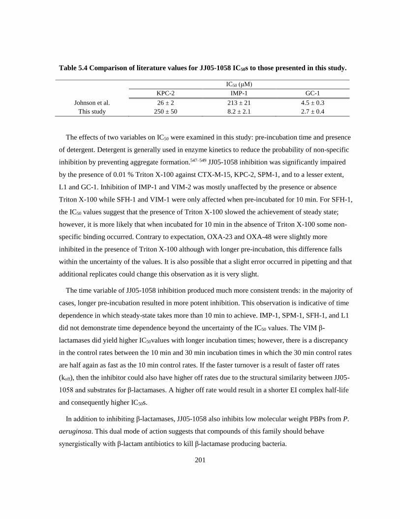
201
Table 5.4 Comparison of literature values for JJ05-1058 IC50s to those presented in this study.
IC50 (μM)
KPC-2 IMP-1 GC-1
Johnson et al. 26 ± 2 213 ± 21 4.5 ± 0.3
This study 250 ± 50 8.2 ± 2.1 2.7 ± 0.4
The effects of two variables on IC50 were examined in this study: pre-incubation time and presence
of detergent. Detergent is generally used in enzyme kinetics to reduce the probability of non-specific
inhibition by preventing aggregate formation.547–549 JJ05-1058 inhibition was significantly impaired
by the presence of 0.01 % Triton X-100 against CTX-M-15, KPC-2, SPM-1, and to a lesser extent,
L1 and GC-1. Inhibition of IMP-1 and VIM-2 was mostly unaffected by the presence or absence
Triton X-100 while SFH-1 and VIM-1 were only affected when pre-incubated for 10 min. For SFH-1,
the IC50 values suggest that the presence of Triton X-100 slowed the achievement of steady state;
however, it is more likely that when incubated for 10 min in the absence of Triton X-100 some non-
specific binding occurred. Contrary to expectation, OXA-23 and OXA-48 were slightly more
inhibited in the presence of Triton X-100 although with longer pre-incubation, this difference falls
within the uncertainty of the values. It is also possible that a slight error occurred in pipetting and that
additional replicates could change this observation as it is very slight.
The time variable of JJ05-1058 inhibition produced much more consistent trends: in the majority of
cases, longer pre-incubation resulted in more potent inhibition. This observation is indicative of time
dependence in which steady-state takes more than 10 min to achieve. IMP-1, SPM-1, SFH-1, and L1
did not demonstrate time dependence beyond the uncertainty of the IC50 values. The VIM β-
lactamases did yield higher IC50values with longer incubation times; however, there is a discrepancy
in the control rates between the 10 min and 30 min incubation times in which the 30 min control rates
are half again as fast as the 10 min control rates. If the faster turnover is a result of faster off rates
(koff), then the inhibitor could also have higher off rates due to the structural similarity between JJ05-
1058 and substrates for β-lactamases. A higher off rate would result in a shorter EI complex half-life
and consequently higher IC50s.
In addition to inhibiting β-lactamases, JJ05-1058 also inhibits low molecular weight PBPs from P.
aeruginosa. This dual mode of action suggests that compounds of this family should behave
synergistically with β-lactam antibiotics to kill β-lactamase producing bacteria.

202
5.5 Future Work
The cyclobutanone JJ05-1058 has been screened against 12 β-lactamases in this study; however,
the IC50 values presented here are from a single experiment due to time constraints. Additional
replicates of the experiments that demonstrated inhibition should be obtained to solidify trends and
get more accurate values. Since the data suggests the inhibition of cyclobutanones is time dependent,
it should be further characterized using methods similar to those described in 1113.2.1.3. The
hypothesis regarding higher off rates for VIM β-lactamases at longer pre-incubation times could be
tested by performing the experiment described in 3.2.1.3 using both the 10 min pre-heating described
as well as a 30 min pre-heating to ascertain whether or not a difference in koff and t1/2 is observed. To
date, only the only cyclobutanones that have been synthesized and tested are derived from the core
structure of a penicillin.388

203
Appendix A
Structures of β-Lactamase Inhibitors
Compound
Identifier Structure Laboratory Codes
Molecular
Weight
(g/mol)
DPA
Commercial: 2,6-
Pyridinedicarboxylic
acid
167.12
PA
Commercial: 2-
Pyridinecarboxylic
acid
123.11
6-MPA
Commercial: 6-
Methylpyridine-2-
carboxylic acid
137.14
PMPC-1
MB9801-60B
AK10025-027
AK12012-025
253.57
PMPC-2
MS-213
AK12012-115
297.58
PMPC-3
AK10025-089 269.57
PMPC-4
AK12012-023B 468.84
PMP
AK12012-162
AK12021-001
209.57
DPMP
AK12021-005 303.57
TBA
Commercial 138.18

204
TA-1
AD-4Br-TA 217.08
TA-2
AD-3OMe-TA 168.21
TA-3
AD-DCl-TA 207.07
TA-4
AD-TCl-TA 241.51
TA-5
AD-3NO2-TA 183.18
TA-6
AD-DN-TA 228.18
TA-7
AD-4NHAc-TA 195.24
TA-8
AD-4NHBoc-TA 253.32
PMTC
PMTA 139.17
PDTC
PDTA 199.24
I-PDTC
I-PDTA 198.25

205
TE-1
AD-4Br-TE 547.44
TE-2
AD-3OMe-TE 498.57
TE-3
AD-3F4Cl-TE 520.97
TE-4
AD-2,4DCl-TE 537.43
TE-5
AD-109,
AD-10012-109
788.58
UW-123
AD-TA-123,
WPI282
841.60
bis-PDTC-
ceph
859.96
JJ05-1058
JJ966, JJ930, JJ1058,
JJ958, unsat DCCB
239.07

206
Appendix B
Statistical Methods for IC50 Averages
Since we are working with values that have an associated uncertainty (the standard error), each
value should contribute differently to the mean such that values with lower uncertainties are more
strongly weighted. In order to do this, a weighted mean is used on the log (IC50) values so as to not
change the nature of the relationship between the IC50 and the error.*
�̅� =∑ 𝑝𝑖∗𝑤𝑖
𝑁𝑖=1
∑ 𝑤𝑖𝑁𝑖=1
Where pi is a property (in this case, each log(IC50) value) and wi is the weight as defined by 𝑤𝑖 =
1
𝑢𝑖2 where ui is the log(SE) associated with each log(IC50) value. This makes Eq. 1 the same as the
weighted mean equation in the Jones paper (except I used the log values of the IC50and SE).
In order to deal with the uncertainty of the weighted mean, the relative weights must be calculated
𝑤𝑖̅̅ ̅ =𝑤𝑖
∑ 𝑤𝑖𝑁𝑖=1
From this, the uncertainty of the weighted mean, 𝑢(�̅�) can be determined:
𝑢(�̅�) = √1
𝑁−1∑ (𝑝𝑖 − �̅�)2 ∗ 𝑤𝑖̅̅ ̅𝑁
𝑖=1
This gives the SE of the weighted mean, so we can convert this into the 95% confidence interval
using the t-table equation:
�̅� ± 𝑡 ∗ 𝑢(�̅�)
From this point the values for the mean and the 95% confidence interval that were calculated could
be converted back into the conventional form by taking the antilog of the values.550

207
Appendix C
Clinical Isolates and Control Strains
UWB Organism Isolate Identifier Known Resistance Source
11 K. pneumoniae KPC-3 Dr. Dylan Pillai
16 K. pneumoniae KPC-2 Dr. Dylan Pillai
24 P. putida C10 VIM-2 Dr. Johann Pitout
25 P. aeruginosa C7 VIM-2 Dr. Johann Pitout
26 S. maltophilia 5563 L1, L2 Dr. Dylan Pillai
27 P. aeruginosa 5564 Dr. Dylan Pillai
28 P. aeruginosa 5566 Dr. Dylan Pillai
29 S. maltophilia 5568 Dr. Dylan Pillai
31 P. aeruginosa 5785 IMP-5 like Dr. Dylan Pillai
33 S. maltophilia 6069 L1, L2 Dr. Dylan Pillai
34 S. maltophilia 6081 L1, L2 Dr. Dylan Pillai
35 P. aeruginosa 6225 Dr. Dylan Pillai
39 S. maltophilia 6487 L1, L2 Dr. Dylan Pillai
43 S. maltophilia 6773 L1, L2 Dr. Dylan Pillai
44 S. maltophilia 6776 L1, L2 Dr. Dylan Pillai
45 S. maltophilia 6779 L1, L2 Dr. Dylan Pillai
46 S. maltophilia 6781 L1, L2 Dr. Dylan Pillai
48 S. maltophilia 6924 L2 Dr. Dylan Pillai
49 S. maltophilia 6952 L2 Dr. Dylan Pillai
56 K. pneumoniae A7 AmpC ACC1 CMY-2 Dr. Johann Pitout
57 E. coli A9 AmpC CMY-2 Dr. Johann Pitout
59 E. coli 1175 Dr. Karen Pike
60 K. pneumoniae Dr. Karen Pike
62 P. aeruginosa 27853 ATCC551
75 E. coli MH1 NDM-1, CTX-M-15 Dr. Johann Pitout
78 P. aeruginosa Vim I-1 VIM-2 Dr. Johann Pitout
82 K. pneumoniae KpSA01 VIM-1 Dr. Dylan Pillai
83 K. pneumoniae Kp3VIM VIM-1 Dr. Dylan Pillai
86 K. pneumoniae Kp5OXA OXA-48 Dr. Dylan Pillai
87 K. pneumoniae Kp9OXA OXA-48, CTX-M-15 Dr. Dylan Pillai
88 K. pneumoniae KpCG02 KPC-2, SHV-12 Dr. Dylan Pillai

208
91 E. coli MH01 NDM-1, CTX-M type 1 Dr. Dylan Pillai
93 E. coli Ec7IMP IMP-1, CTX-M-15 Dr. Dylan Pillai
94 A. baumannii AB01 OXA-23 Dr. Dylan Pillai
95 A. baumannii AB02 OXA-23 Dr. Dylan Pillai
96 P. aeruginosa PA01 Dr. Stephen Seah
102 E. coli 25922 ATCC552
116 K. pneumoniae L3142005 NDM-1 Dr. Allison McGeer

209
Appendix D
KI Graphs of DPA, PMPC-3, and PMPC-4
0 2 4 6 8 10
0
1
2
3
4
0
100
75
50
25
15
10
5
Time (min)
[Pro
du
ct]
(µ
M)
0 2 4 6 8 10
0
2
4
0
250
150
100
75
50
25
10
Time (min)
[Pro
du
ct]
(µ
M)
0 50 100 1500.0
0.2
0.4
0.6
0.8
1.0
[I]
k o
bs
0 100 200 3000.0
0.5
1.0
1.5
[I]
k o
bs
0 2 4 6 8 10
0
1
2
3
0
100
75
50
25
10
5
1
Time (min)
[Pro
du
ct]
(µ
M)
0 2 4 6 8 10
0
1
2
3
4
0
25
15
10
8
6
4
2
Time (min)
[Pro
du
ct]
(µ
M)
0 50 100 1500.0
0.2
0.4
0.6
0.8
1.0
[I]
k o
bs
0 5 10 15 20 250.0
0.5
1.0
[I]
k o
bs
KI = 1.1 ± 0.3
R2 = 0.9895
KI = 2.8 ± 0.4
R2 = 0.9968
KI = 3.3 ± 0.6
R2 = 0.9620
KI = 0.8 ± 0.3
R2 = 0.8981
A B
C D
Figure 5.8 Time Dependent KI graphs for DPA against representative MBLs IMP-1 (A),
NDM-1 (B), VIM-2 (C), and L1 (D).

210
0 2 4 6 8 10
0
1
2
3
4
0
25
15
10
7.5
5
2.5
1
Time (min)
[Pro
du
ct]
(µ
M)
0 2 4 6 8 10
0
2
4 0
75
50
25
15
10
5
1
Time (min)
[Pro
du
ct]
(µ
M)
0 10 20 300.0
0.2
0.4
0.6
0.8
1.0
[I]
k o
bs
0 20 40 60 800.0
0.2
0.4
0.6
0.8
[I]
k o
bs
0 2 4 6 8 10
0
1
2
3
0
10
7.5
5
2.5
1
0.5
0.1
Time (min)
[Pro
du
ct]
(µ
M)
0 2 4 6 8 10
0
2
4
6
0
10
7.5
5
2.5
1
0.5
0.1
Time (min)
[Pro
du
ct]
(µ
M)
0 2 4 6 8 100
1
2
3
[I]
k o
bs
0 2 4 6 8 100.0
0.5
1.0
[I]
k o
bs
KI = 0.41 ± 0.10
R2 = 0.9789
KI = 1.5 ± 0.1
R2 = 0.9987KI = 0.074 ± 0.008
R2 = 0.9961
KI = 0.61 ± 0.04
R2 = 0.9947
A B
C D
Figure 5.9 Time Dependent KI graphs for PMPC-3 against representative MBLs IMP-1 (A),
NDM-1 (B), VIM-2 (C), and L1 (D).

211
0 2 4 6 8 10
0
1
2
3
4
0
25
20
15
10
5
2.5
1
Time (min)
[Pro
du
ct]
(µ
M)
0 2 4 6 8 10
0
2
40
25
20
15
10
5
2.5
1
Time (min)
[Pro
du
ct]
(µ
M)
0 10 20 300.0
0.5
1.0
1.5
[I]
k o
bs
0 10 20 300.0
0.2
0.4
0.6
0.8
[I]
k o
bs
0 2 4 6 8 10
0
1
2
3
0
2.5
1.75
1
0.5
0.25
0.1
0.05
Time (min)[P
rod
uct]
(µ
M)
0 2 4 6 8 10
0
2
4
0
2.5
2
1.5
1
0.5
0.25
0.1
Time (min)
[Pro
du
ct]
(µ
M)
0 1 2 30.0
0.5
1.0
1.5
[I]
k o
bs
0 1 2 30.0
0.5
1.0
1.5
[I]
k o
bs
KI = 0.40 ± 0.1
R2 = 0.9845
KI = 0.4 ± 0.2
R2 = 0.9463KI = 0.034 ± 0.006
R2 = 0.9944
KI = 0.038 ± 0.009
R2 = 0.9904
A B
C D
Figure 5.10 Time Dependent KI graphs for PMPC-4 against representative MBLs IMP-1 (A),
NDM-1 (B), VIM-2 (C), and L1 (D).

212
Appendix E
Protein Sequence Alignments
Alignment of NDM variants relative to NDM-1
10 20 30 40 50 60
....|....|....|....|....|....|....|....|....|....|....|....|
NDM-1 MELPNIMHPVAKLSTALAAALMLSGCMPGEIRPTIGQQMETGDQRFGD-----LVFRQLA
NDM-2 ...........................A....................-----.......
NDM-3 ................................................-----.......
NDM-4 ................................................-----.......
NDM-5 ................................................-----.......
NDM-6 ................................................-----.......
NDM-7 ................................................-----.......
NDM-8 ................................................-----.......
NDM-9 ................................................-----.......
NDM-10 ...............................S...D............-----.......
NDM-11 ................................................-----.......
NDM-12 ................................................-----.......
NDM-13 ................................................-----.......
NDM-14 ................................................-----.......
NDM-15 ................................................-----.......
NDM-16 ................................................-----.......
NDM-17 ................................................-----.......
NDM-18 ................................................QRFGD.......
NDM-19 ................................................-----.......
70 80 90 100 110 120
....|....|....|....|....|....|....|....|....|....|....|....|
NDM-1 PNVWQHTSYLDMPGFGAVASNGLIVRDGGRVLVVDTAWTDDQTAQILNWIKQEINLPVAL
NDM-2 ............................................................
NDM-3 .......................................N....................
NDM-4 ............................................................
NDM-5 ................................L...........................
213
NDM-6 ............................................................
NDM-7 ............................................................
NDM-8 ............................................................
NDM-9 ............................................................
NDM-10 .............S....T.........................................
NDM-11 ............................................................
NDM-12 ............................................................
NDM-13 .......................................N....................
NDM-14 ............................................................
NDM-15 ............................................................
NDM-16 ............................................................
NDM-17 ................................L...........................
NDM-18 ............................................................
NDM-19 ............................................................
130 140 150 160 170 180
....|....|....|....|....|....|....|....|....|....|....|....|
NDM-1 AVVTHAHQDKMGGMDALHAAGIATYANALSNQLAPQEGMVAAQHSLTFAANGWVEPATAP
NDM-2 ............................................................
NDM-3 ............................................................
NDM-4 ......................................L.....................
NDM-5 ......................................L.....................
NDM-6 ............................................................
NDM-7 ..............N.......................L.....................
NDM-8 ..............G.......................L.....................
NDM-9 ....................................K.......................
NDM-10 ............................................................
NDM-11 ......................................V.....................
NDM-12 ......................................L.....................
NDM-13 ......................................L.....................
NDM-14 ..............G.............................................
NDM-15 ......................................L.....................

214
NDM-16 ............................................................
NDM-17 ......................................L...............K.....
NDM-18 ............................................................
NDM-19 ..............N.......................L.....................
190 200 210 220 230 240
....|....|....|....|....|....|....|....|....|....|....|....|
NDM-1 NFGPLKVFYPGPGHTSDNITVGIDGTDIAFGGCLIKDSKAKSLGNLGDADTEHYAASARA
NDM-2 ............................................................
NDM-3 ............................................................
NDM-4 ............................................................
NDM-5 ............................................................
NDM-6 .........................................................V..
NDM-7 ............................................................
NDM-8 ............................................................
NDM-9 ............................................................
NDM-10 ........................R...................................
NDM-11 ............................................................
NDM-12 ..............................................D.............
NDM-13 ............................................................
NDM-14 ............................................................
NDM-15 .........................................................V..
NDM-16 ............................................................
NDM-17 ............................................................
NDM-18 ............................................................
NDM-19 .........................................................V..

215
250 260 270
....|....|....|....|....|....|....|
NDM-1 FGAAFPKASMIVMSHSAPDSRAAITHTARMADKLR
NDM-2 ...................................
NDM-3 ...................................
NDM-4 ...................................
NDM-5 ...................................
NDM-6 ...................................
NDM-7 ...................................
NDM-8 ...................................
NDM-9 ...................................
NDM-10 ...................................
NDM-11 ...................................
NDM-12 ...................................
NDM-13 ...................................
NDM-14 ...................................
NDM-15 ...................................
NDM-16 ............................H......
NDM-17 ...................................
NDM-18 ...................................
NDM-19 ...................................
Alignment of VIM Variants relative to VIM-2
10 20 30 40 50 60
....|....|....|....|....|....|....|....|....|....|....|....|
VIM-02 MFKLLSKLLVYLTASIMAIASPLAFSVDSSGEYPTVSEIPVGEVRLYQIADGVWSHIATQ
VIM-01 .L.VI.S....M...V..V.....H.GEP.......N.......................
VIM-03 ...........................................................K
VIM-04 .L.VI.S....M...V..V.....H.GEP.......N.......................
VIM-05 .L.VI.S....M...V..V.....H.GEP.......N.......................
VIM-06 ...........................................................R

216
VIM-07 ..Q-IRSF..GIS.FV..VLGSA.Y.AQPG......DD.........K.G..........
VIM-08 ............................................................
VIM-09 ............................................................
VIM-10 ............................................................
VIM-11 ............................................................
VIM-12 .L.VI.S....M...V..V.....H.GEP.......N.......................
VIM-13 .L.VI.S..F.M...L..V.....H.GE.R...................D.........H
VIM-14 .L.VI.S....M...V..V.....H.GEP.S.....N.......................
VIM-15 ............................................................
VIM-16 ......................................................L.....
VIM-17 ...............M............................................
VIM-18 ...........................................................R
VIM-19 .L.VI.S....M...V..V.....H.GEP.......N.......................
VIM-20 ............................................................
VIM-23 ............................................................
VIM-24 ............................................................
VIM-25 .L.VI.S....M...V..V.....H.GEP.......N.......................
VIM-26 .L.VI.S....M...V..V.....H.GEP.......N.......................
VIM-27 .L.VI.S....M...V..V.....H.GEP.......N....................S..
VIM-28 .L.VI.S....M...V..V.....H.GEP.......N.......................
VIM-29 .L.VI.S....M...V..V.....H.GEP.......N.......................
VIM-30 ....................................N.......................
VIM-31 ............................................................
VIM-32 .L.VI.S....M...V..V.....H.GEP.......N.......................
VIM-33 .L.VI.S....M...V..V.....H.GEP.......N.......................
VIM-34 .L.VI.S....M...V..V.....H.GEP.......N.......................
VIM-35 .L.VI.S....M...V..V.....H.GEP.......N.......................
VIM-36 ...........................................................R
VIM-37 .L.VI.S....M...V..V.....H.GEP.......N....................S..
VIM-38 .L.VI.S........V..V.....H.GEP.......N.......................
VIM-39 .L.VI.S....M...V..V.....H.GEP.....A.N.......................
VIM-40 .L.VI.S....M...V..V.....H.GEP.......N.......................
VIM-41 ............................................................
VIM-42 .L.VI.S....M...V..V.....H.GEP.......N.......................

217
VIM-43 .L.VI.S....M...V..V....VH.GEP.......N.......................
VIM-44 ............................................................
VIM-45 ..................................I.........................
VIM-46 ...F...............................A........................
VIM-47 ...VV.S..F.M...L..V.....H.GE.R...................D.........H
VIM-48 ............................................................
VIM-49 .L.VI.S....M...V..V.....H.GEP.......N....................S..
VIM-50 ............................................................
VIM-51 ............................................................
VIM-52 .L.VI.S....M...V..V.....H.GEP.......N.......................
VIM-54 .L.VI.S....M...V..V.....H.GEP.......N.......................
VIM-55 .L.VI.S....M...V..V.....H.GEP.......N.......................
70 80 90 100 110 120
....|....|....|....|....|....|....|....|....|....|....|....|
VIM-02 SFDGAVYPSNGLIVRDGDELLLIDTAWGAKNTAALLAEIEKQIGLPVTRAVSTHFHDDRV
VIM-01 ............................................................
VIM-03 ............................................................
VIM-04 ............................................................
VIM-05 ............................................................
VIM-06 ............................................................
VIM-07 KLGDT..S........A...............V................SI.........
VIM-08 ............................................................
VIM-09 ............................................................
VIM-10 ............................................................
VIM-11 ............................................................
VIM-12 ............................................................
VIM-13 T...V.......................T...V................S..........
VIM-14 ............................................................
VIM-15 ............................................................
VIM-16 ............................................................
VIM-17 ............................................................
VIM-18 ............................................................
VIM-19 ............................................................

218
VIM-20 ............................................................
VIM-23 ............................................................
VIM-24 ............................................................
VIM-25 ............................................................
VIM-26 ............................................................
VIM-27 ............................................................
VIM-28 ............................................................
VIM-29 ............................................................
VIM-30 ............................................................
VIM-31 ............................................................
VIM-32 ............................................................
VIM-33 ............................................................
VIM-34 ..................................................I.........
VIM-35 ............................................................
VIM-36 ............................................................
VIM-37 ............................................................
VIM-38 ............................................................
VIM-39 ............................................................
VIM-40 ............................................................
VIM-41 ............................................................
VIM-42 ............................................................
VIM-43 ............................................................
VIM-44 ............................................................
VIM-45 ............................................................
VIM-46 ............................................................
VIM-47 T...V.......................T...V................S..........
VIM-48 ............................................................
VIM-49 ............................................................
VIM-50 ............................................................
VIM-51 ............................................................
VIM-52 ............................................................
VIM-54 ............................................................
VIM-55 ............................................................

219
130 140 150 160 170 180
....|....|....|....|....|....|....|....|....|....|....|....|
VIM-02 GGVDVLRAAGVATYASPSTRRLAEVEGNEIPTHSLEGLSSSGDAVRFGPVELFYPGAAHS
VIM-01 ........................A...................................
VIM-03 ...........................S................................
VIM-04 ........................A...................................
VIM-05 .......K................A...................................
VIM-06 ...........................S................................
VIM-07 ..............T..L..Q...AA...V.A...KA......V.......V........
VIM-08 ..................A.........................................
VIM-09 ..................I.........................................
VIM-10 ............................................................
VIM-11 ...........................S................................
VIM-12 ........................A...................................
VIM-13 ....A...................A....V..............................
VIM-14 ........................A...................................
VIM-15 ............................................................
VIM-16 ............................................................
VIM-17 ............................................................
VIM-18 .......................----.................................
VIM-19 ........................A...................................
VIM-20 ............................................................
VIM-23 ............................................................
VIM-24 ............................................................
VIM-25 .......K................A...................................
VIM-26 ........................A...................................
VIM-27 ........................A...................................
VIM-28 ........................A...................................
VIM-29 ........................A...................................
VIM-30 ............................................................
VIM-31 ............................................................
VIM-32 ........................AA..................................
VIM-33 ........................A...................................
VIM-34 ........................A...................................

220
VIM-35 ........................A...................................
VIM-36 ............................................................
VIM-37 ........................A...................................
VIM-38 .......K................A...................................
VIM-39 ........................A...................................
VIM-40 ......................V.A...................................
VIM-41 ............................................................
VIM-42 ........................A...................................
VIM-43 ........................A...................................
VIM-44 ............................................................
VIM-45 ............................................................
VIM-46 ............................................................
VIM-47 ....A...................A....V..............................
VIM-48 ............................................................
VIM-49 .......K................A...................................
VIM-50 ...........................S................................
VIM-51 ............................................................
VIM-52 ........................A...................................
VIM-54 ........................A..S................................
VIM-55 ........................A...................................
190 200 210 220 230 240
....|....|....|....|....|....|....|....|....|....|....|....|
VIM-02 TDNLVVYVPSASVLYGGCAIYELSRTSAGNVADADLAEWPTSIERIQQHYPEAQFVIPGH
VIM-01 ...........N.......VH...S.................V....K.....EV.....
VIM-03 ............................................................
VIM-04 ...........N.......VH.....................V....K.....EV.....
VIM-05 ...........N.......VLA....................V....K.....EV.....
VIM-06 ............................................................
VIM-07 G........AVR..F....VH.A..E........N.....AT.K....R....EV.....
VIM-08 ............................................................
VIM-09 ............................................................
VIM-10 ......................................................Y.....
VIM-11 ............................................................

221
VIM-12 ...........N.......VH...S...................................
VIM-13 ...........N.......VL...................G.V..........EV.....
VIM-14 ...........N.......VH.....................V....K.....EV.....
VIM-15 ..............F.............................................
VIM-16 ............................................................
VIM-17 ............................................................
VIM-18 ............................................................
VIM-19 ...........K.......VH.....................V....K.....EV.....
VIM-20 ................................................R...........
VIM-23 ........................S...................................
VIM-24 ........................L...................................
VIM-25 ...........N.......VLA....................V....K............
VIM-26 ...........N.......VL...S.................V....K.....EV.....
VIM-27 ...........N.......VH...S.................V....K.....EV.....
VIM-28 ...........N.......VL.....................V....K.....EV.....
VIM-29 ...........K.......VH...S.................V....KR....EV.....
VIM-30 ............................................................
VIM-31 ....................H...........................R...........
VIM-32 ...........N.......VH...S.................V....K.....EV.....
VIM-33 ...........N..F....VH...S.................V....K.....EV.....
VIM-34 ...........N.......VH...S.................V....K.....EV.....
VIM-35 ...........N.......VH...S......T..........V....K.....EV.....
VIM-36 ............................................................
VIM-37 ...........N.......VH.....................V....K.....EV.....
VIM-38 ...........N.......VLA....................V....K.....EV.....
VIM-39 ...........N.......VL...S.................V....K.....EV.....
VIM-40 ...........N.......VH.....................V....K.....EV.....
VIM-41 ................................N...........................
VIM-42 ...........N.......VH...S......................K.....EV.....
VIM-43 ...........N.......VH.....................V....K.....EV.....
VIM-44 ............................................................
VIM-45 ............................................................
VIM-46 ............................................................
VIM-47 ...........N.......VL...................G.V..........EV.....

222
VIM-48 ............................................................
VIM-49 ...........N.......VLA....................V....K.....EV.....
VIM-50 ........................L...................................
VIM-51 ...........................V................................
VIM-52 ...........N.......VR...S.................V....K.....EV.....
VIM-54 ...........N.......VH.....................V....K.....EV.....
VIM-55 ...........K.......VH.....................V....KR....EV.....
250 260 270
....|....|....|....|....|....|..
VIM-02 GLPGGLDLLKHTTNVVKAHTNRSVVE------
VIM-01 .........Q..A......K....A.------
VIM-03 ..........................------
VIM-04 .........Q..A......K....A.------
VIM-05 .........Q..A...T..K....A.------
VIM-06 ..........................------
VIM-07 ......E..Q.......T.KV.P.A.------
VIM-08 ..........................------
VIM-09 ..........................------
VIM-10 ..........................------
VIM-11 ..........................------
VIM-12 ..........................------
VIM-13 .........Q..A...........A.------
VIM-14 .........Q..A......K....A.------
VIM-15 ..........................------
VIM-16 ..........................------
VIM-17 ..........................------
VIM-18 ..........................------
VIM-19 .........Q..A......K....A.------
VIM-20 ..........................------
VIM-23 ..........................------
VIM-24 ..........................------
VIM-25 ..........................------
VIM-26 .........Q..A......K....A.------

223
VIM-27 .........Q..A......K....A.------
VIM-28 .........Q..A......K....A.------
VIM-29 .........Q..A......K....A.------
VIM-30 ..........................------
VIM-31 ..........................------
VIM-32 .........Q..A......K....A.------
VIM-33 .........Q..A......K....A.------
VIM-34 .........Q..A......K....A.------
VIM-35 .........Q..A......K....A.------
VIM-36 ..........................------
VIM-37 .........Q..A......K....A.------
VIM-38 .........Q..A...T..K......------
VIM-39 .........Q..A......K....A.------
VIM-40 .........Q..A......K....A.------
VIM-41 ..........................------
VIM-42 .........Q..A......K....A.------
VIM-43 .........Q..A......K....A.------
VIM-44 ................N.........------
VIM-45 ..........................------
VIM-46 ..........................------
VIM-47 .........Q..A...........A.------
VIM-48 ..................---G..AKIGGSQR
VIM-49 .........Q..A...T..K....A.------
VIM-50 ..........................------
VIM-51 ..........................------
VIM-52 .........Q..A......K....A.------
VIM-54 .........Q..A......K....A.------
VIM-55 .........Q..A......K....A.------
VIM-1
F2L, L4V, L5I, K7S, L12M, I16V, I19V, F25H, V27G, D28E, S29P, S37N, V145A,
S192N, I200V, Y201H, R205S, I223V, Q228K, Q234E, F235V, K250Q, T253A, T260K,
V265A
224
VIM-3 Q60K, N148S
VIM-4
F2L, L4V, L5I, K7S, L12M, I16V, I19V, F25H, V27G, D28E, S29P, S37N, V145A,
S192N, I200V, Y201H, I223V, Q228K, Q234E, F235V, K250Q, T253A, T260K, V265A
VIM-5
F2L, L4V, L5I, K7S, L12M, I16V, I19V, F25H, V27G, D28E, S29P, S37N, A128K,
V145A, S192N, I200V, Y201L, E202A, I223V, Q228K, Q234E, F235V, K250Q, T253A,
K257T, T260K, V265A
VIM-6 Q60R, N148S
VIM-7
K3Q, L4del, L5I, S6R, K7S, L8F, Y11G, L12I, T13S, S15F, I16V, I19V, A20L, S21G,
P22S, L23A, F25Y, V27A, D28Q, S29P, S30G, S37D, E38D, Q48K, A50G, S61K, F62L,
D63G, G64D, A65T, P68S, G77A, A93V, A110S, V111I, A135T, S138L, R141Q, V145A,
E146A, I150V, T152A, E156K, G157A, A164V, L172V, T181G, S190A, A191V, S192R,
Y195F, I200V, Y201H, L203A, T206E, D215N, T221A, S222T, E224K, H229R, Q234E,
F235V, D247E, K250Q, A258T, T260K, N261V, S263P, V265A
VIM-8 T139A
VIM-9 T139I
VIM-10 F235Y
VIM-11 N148S
VIM-12
F2L, L4V, L5I, K7S, L12M, I16V, I19V, F25H, V27G, D28E, S29P, S37N, V145A,
S192N, I200V, Y201H, R205S
VIM-13
F2L, L4V, L5I, K7S, V10F, L12M, I16L, I19V, F25H, V27G, D28E, S30R, A50D, Q60H,
S61T, A65V, A89T, A93V, A110S, V125A, V145A, I150V, S192N, I200V, Y201L, T221G,
Q234E, F235V, K250Q, T253A, V265A
VIM-14
F2L, L4V, L5I, K7S, L12M, I16V, I19V, F25H, V27G, D28E, S29P, G31S, S37N, V145A,
S192N, I200V, Y201H, I223V, Q228K, Q234E, F235V, K250Q, T253A, T260K, V265A
VIM-15 Y195F
VIM-16 S55L
VIM-17 I16M
VIM-18 Q60R, E144_G147del
VIM-19
F2L, L4V, L5I, K7S, L12M, I16V, I19V, F25H, V27G, D28E, S29P, S37N, V145A,
S192K, I200V, Y201H, I223V, Q228K, Q234E, F235V, K250Q, T253A, T260K, V265A
VIM-20 H229R
VIM-23 R205S
VIM-24 R205L
VIM-25
F2L, L4V, L5I, K7S, L12M, I16V, I19V, F25H, V27G, D28E, S29P, S37N, A128K,
V145A, S192N, I200V, Y201L, E202A, I223V, Q228K

225
VIM-26
F2L, L4V, L5I, K7S, L12M, I16V, I19V, F25H, V27G, D28E, S29P, S37N, V145A,
S192N, I200V, Y201L, R205S, I223V, Q228K, Q234E, F235V, K250Q, T253A, T260K,
V265A
VIM-27
F2L, L4V, L5I, K7S, L12M, I16V, I19V, F25H, V27G, D28E, S29P, S37N, A58S, V145A,
S192N, I200V, Y201H, R205S, I223V, Q228K, Q234E, F235V, K250Q, T253A, T260K,
V265A
VIM-28
F2L, L4V, L5I, K7S, L12M, I16V, I19V, F25H, V27G, D28E, S29P, S37N, V145A,
S192N, I200V, Y201L, I223V, Q228K, Q234E, F235V, K250Q, T253A, T260K, V265A
VIM-29
F2L, L4V, L5I, K7S, L12M, I16V, I19V, F25H, V27G, D28E, S29P, S37N, V145A,
S192K, I200V, Y201H, R205S, I223V, Q228K, H229R, Q234E, F235V, K250Q, T253A,
T260K, V265A
VIM-30 S37N
VIM-31 Y201H, H229R
VIM-32
F2L, L4V, L5I, K7S, L12M, I16V, I19V, F25H, V27G, D28E, S29P, S37N, V145A,
E146A, S192N, I200V, Y201H, R205S, I223V, Q228K, Q234E, F235V, K250Q, T253A,
T260K, V265A
VIM-33
F2L, L4V, L5I, K7S, L12M, I16V, I19V, F25H, V27G, D28E, S29P, S37N, V145A,
S192N, Y195F, I200V, Y201H, R205S, I223V, Q288K, Q234E, F235V, K250Q, T253A,
T260K, V265A
VIM-34
F2L, L4V, L5I, K7S, L12M, I16V, I19V, F25H, V27G, D28E, S29P, S37N, V111I,
V145A, S192N, Y195F, I200V, Y201H, R205S, I223V, Q288K, Q234E, F235V, K250Q,
T253A, T260K, V265A
VIM-35
F2L, L4V, L5I, K7S, L12M, I16V, I19V, F25H, V27G, D28E, S29P, S37N, V145A,
S192N, Y195F, I200V, Y201H, R205S, A212T, I223V, Q228K, Q234E, F235V, K250Q,
T253A, T260K, V265A
VIM-36 Q60R
VIM-37
F2L, L4V, L5I, K7S, L12M, I16V, I19V, F25H, V27G, D28E, S29P, S37N, A58S, V145A,
S192N, I200V, Y201H, I223V, Q228K, Q234E, F235V, K250Q, T253A, T260K, V265A
VIM-38
F2L, L4V, L5I, K7S, I16V, I19V, F25H, V27G, D28E, S29P, S37N, A128K, V145A,
S192N, I200V, Y201L, E202A, I223V, Q228K, Q234E, F235V, K250Q, T253A, K257T,
T260K
VIM-39
F2L, L4V, L5I, K7S, L12M, I16V, I19V, F25H, V27G, D28E, S29P, T35A, S37N,
V145A, S192N, I200V, Y201L, R205S, I223V, Q228K, Q234E, F235V, K250Q, T253A,
T260K, V265A
VIM-40
F2L, L4V, L5I, K7S, I16V, I19V, F25H, V27G, D28E, S29P, S37N, A143V, V145A,
S192N, I200V, Y201H, I223V, Q228K, Q234E, F235V, K250W, T253A, T260K, V265A
VIM-41 D213N
VIM-42 F2L, L4V, L5I, K7S, I16V, I19V, F25H, V27G, D28E, S29P, S37N, V145A, S192N,

226
I200V, Y201H, R205S, Q228K, Q234E, F235V, K250Q, T253A, T260K, V265A
VIM-43
F2L, L4V, L5I, K7S, I16V, I19V, A24V, F25H, V27G, D28E, S29P, S37N, V145A,
S192N, I200V, Y201H, I223V, Q228K, Q234E, F235V, K250Q, T253A, T260K, V265A
VIM-44 K257N
VIM-45 T35I
VIM-46 L4F, V36A
VIM-47
L4V, L5V, K7S, V10F, L12M, I16L, I19V, F25H, V27G, D28E, S30R, A50D, Q60H,
S61T, A65V, A89T, A93V, A110S, V125A, V145A, I150V, S192N, I200V, Y201L, T221G,
I223V, Q234E, F235V, K250Q, T253A, V265A
VIM-48 H259_N261del, R262G, V265A, E266K, E266_insIGGSQR
VIM-49
F2L, L4V, L5I, K7S, L12M, I16V, I19V, F25H, V27G, D28E, S29P, S37N, A128K,
V145A, S192N, I200V, Y201L, E202A, I223V, Q228K, Q234E, F235V, K250Q, T253A,
K257T, T260K, V265A
VIM-50 N148S, R205L
VIM-51 A208V
VIM-52
F2L, L4V, L5I, K7S, L12M, I16V, I19V, F25H, V27G, D28E, S29P, S37N, V145A,
S192N, I200V, Y201R, R205S, I223V, Q228K, Q234E, F235V, K250Q, T253A, T260K,
V265A
VIM-54
F2L, L4V, L5I, K7S, L12M, I16V, I19V, F25H, V27G, D28E, S29P, S37N, V145A,
N148S, S192N, I200V, Y201H, I223V, Q228K, Q234E, F235V, K250Q, T253A, T260K,
V265A
VIM-55
F2L, L4V, L5I, K7S, L12M, I16V, I19V, F25H, V27G, D28E, S29P, S37N, V145A,
S192K, I200V, Y201H, I223V, Q228K, H229R, Q234E, F235V, K250Q, T253A, T260K,
V265A
IMP Variant Sequence Alignment
10 20 30 40 50 60
....|....|....|....|....|....|....|....|....|....|....|....|
IMP-01 MSKLSVFFIFLFCSIATAAESLPDLKIEKLDEGVYVHTSFEEVNGWGVVPKHGLVVLVNA
IMP-02 .K..F.LCVCFL...TA.GAR.........E..................S.........T
IMP-03 ............................................................
IMP-04 ....................P.....................................D.
IMP-05 ....F...M......TA..........................................T
IMP-06 ............................................................
IMP-07 .K......M.......ASG.A......................................T
IMP-08 .K..F.LCVCFL...TA.GAA.........E..................S.........T

227
IMP-09 ....F...M......TA.G.............................I..........T
IMP-10 ................................................F...........
IMP-11 .K..F.LC.......TA.GA..........E..................S.........T
IMP-12 .K..F.LC....L..TASG.V.........E....L.......S.....T.........N
IMP-13 .K..F.LCVCF....TA.GAA.........E...F..............T.........T
IMP-14 .K..F.LCV.F..N..V.E...........E............K..S..T........KN
IMP-15 .N......M.M....TA.G........................................T
IMP-16 .K..F.LC.......TA.G...........ED.................T.....F...T
IMP-17 .K..F.LCVCF....TA.GAA.........E...F..............T.........T
IMP-18 .K..F.LCV.FL.N..A.DD..........EK...........K.....T........KN
IMP-19 .K..F.LCVCFL...TA.GAA.........E..................S.........T
IMP-20 .K..F.LCVCFL...TA.GAA.........E.................FS.........T
IMP-21 .K..F.LC.......TA.GA..........E.................AS.........T
IMP-22 .K..F.LCV......TA.G...........E..................S.....I...T
IMP-23 .K..F.LCVCFL...TA.GAA.........E.................FS.........T
IMP-24 .K..F.LCVCFL...TA.GAA.........E..................S.........T
IMP-25 ............................................................
IMP-26 ....................P...........................F.........D.
IMP-27 .K..F.LCV.V....TV.G.T..N.RV...E........Y...K.....T.......IG.
IMP-28 ....F...M......TA...........R..............................T
IMP-29 ....F..L.......TA..........................................T
IMP-30 ........................................K...................
IMP-31 .K.IF.L.V......TA.G.....I.......D........KIT....IT.........T
IMP-32 .K..F.LCV.F..N..V.E...........E............K..S..T........KN
IMP-33 .K..F.LCVCF....TA.GS..........E...F..............T.........T
IMP-34 ............................................................
IMP-35 .K.IF.L.V......TA.G.....I.......D.........D.....IT.........T
IMP-37 .K..F.LCVCF....TA.GAA.........E...F..............T.........T
IMP-38 ....................P.....................................D.
IMP-40 ................................................F...........
IMP-41 .K..F.LC.......TA.GA..........E.................FS.........T
IMP-42 ............................................R...............
IMP-43 .K......M.......ASG.A...........................F..........T
IMP-44 .K..F.LC.......TA.GA..........E.................FS.........T

228
IMP-45 ....F...M......TA.G.............................I..........T
IMP-48 .K..F.LCV.F..N..V.E...........E............K..S..T........KN
IMP-49 .K..F.LCV.FL.N..A.DD..........EK...........K....FT........KN
IMP-51 .K......M.......ASG.A......................................T
IMP-52 ............................................................
IMP-53 ....F...M......TA.G.............................I..........T
IMP-54 .K..F.LCV.F..N..V.E...........E............K..S..T........KN
IMP-55 ...........I..................E.........K...................
IMP-56 .K..F.LCV.FL.N..A.DD..........EK...........K.....T........KN
IMP-58 .K..F.LCV......TA.G...........E.................FS.....I...T
IMP-59 ....................P.....................................D.
IMP-60 ............................................................
IMP-61 ............................................................
IMP-62 .N......M.M....TA.G........................................T
IMP-63 .K..F.LC....L..TASG.V.........E....L.......S.....T.........N
IMP-64 .K..F.LCV.V....TV...T..N.RV...E........Y...K.....T.......IG.
IMP-66 ...............................................F............
IMP-67 .K..F.LCV.V....TV.G.T..N.RV...E........Y...K.....T.......IG.
IMP-68 .K..F.LC.......TA.GA..........E..................S.........T
IMP-69 .K..F.LCVCFL...TA.GAT.........E..................S.........T
IMP-70 ............................................................
70 80 90 100 110 120
....|....|....|....|....|....|....|....|....|....|....|....|
IMP-01 EAYLIDTPFTAKDTEKLVTWFVERGYKIKGSISSHFHSDSTGGIEWLNSRSIPTYASELT
IMP-02 D..........T......N...........T..................Q..........
IMP-03 ............................................G...............
IMP-04 .................................................Q..........
IMP-05 .................................................Q..........
IMP-06 ............................................................
IMP-07 D................................................Q..........
IMP-08 D..........T......N...........T..................Q..........
IMP-09 D..............N..N.......R......................Q..........
IMP-10 ............................................................

229
IMP-11 D.................N..............................Q.......V..
IMP-12 D.........N.......A...G..FT....V.................Q..........
IMP-13 D..........T......N.......E...T..................Q..........
IMP-14 D.......I.........N..............T...G...A.......Q..........
IMP-15 .................................................Q..........
IMP-16 D........A........N.....................S........Q..........
IMP-17 D..........T......N.......E...T..................Q..........
IMP-18 D.......I.........N..I.H..R......T...G...A.......Q..S.......
IMP-19 D..........T......N...........T..................Q..........
IMP-20 D..........T......N...........T..................Q..........
IMP-21 D.................N..............................Q.......V..
IMP-22 D................................................Q..........
IMP-23 D..........T......N...........T..................Q..........
IMP-24 D..........T......N...........T..................Q..........
IMP-25 ............................................................
IMP-26 .................................................Q..........
IMP-27 D.................N...........TV.................Q..........
IMP-28 ......................G..........................Q..........
IMP-29 .................................................Q..........
IMP-30 ............................................................
IMP-31 D..I..............R...G.................A........Q.......K..
IMP-32 D.......I.........N..............T...G...A.......Q..........
IMP-33 D..........T......N...........T..................Q..........
IMP-34 ............................................G...............
IMP-35 D..I..............R...G.................A........Q.......K..
IMP-37 D..........T......N.......E...T..................Q..........
IMP-38 .................................................Q..........
IMP-40 ........S...................................................
IMP-41 D.................N..............................Q.......V..
IMP-42 ............................................................
IMP-43 D................................................Q..........
IMP-44 D.......S.........N..............................Q.......V..
IMP-45 D..............N..N.......R......................Q..........
IMP-48 D.......T.........N..............T...G...A.......Q..........

230
IMP-49 D.......I.........N..I.H..R......T...G...A.......Q..S.......
IMP-51 D................................................Q..........
IMP-52 ...I.............G..........................................
IMP-53 D..............N..N.......R......................Q..........
IMP-54 D.......V.........N..............T...G...A.......Q..........
IMP-55 ............................................................
IMP-56 D.......I.........N..I.H..R......T...G...A.......Q..S.......
IMP-58 D................................................Q..........
IMP-59 .................................................Q..........
IMP-60 ............................................................
IMP-61 ...............................................I............
IMP-62 .................................................Q..........
IMP-63 D.........N.......A...G..FT....V.................Q..........
IMP-64 D.................N...........TV.................Q..........
IMP-66 ............................................................
IMP-67 D.................N...........TV.................Q..........
IMP-68 D.................N..............................Q.......V..
IMP-69 D..........T......N...........T..................Q..........
IMP-70 ............................................................
130 140 150 160 170 180
....|....|....|....|....|....|....|....|....|....|....|....|
IMP-01 NELLKKDGKVQATNSFSGVNYWLVKNKIEVFYPGPGHTPDNVVVWLPERKILFGGCFIKP
IMP-02 ............K......S..................Q.........K........V..
IMP-03 ............................................................
IMP-04 ............K...G........................L..................
IMP-05 ............K.....AS.....K......................NRV......V..
IMP-06 ............................................................
IMP-07 ............K.....AS.....K...I..................HRV......V..
IMP-08 ............K......S..................Q.........K........V..
IMP-09 ............KY.....S.....K...........A..........NRV......V..
IMP-10 ............................................................
IMP-11 ............K......S..................Q........KN........V..
IMP-12 ......N............S.........I........Q.........N........V..

231
IMP-13 ......S.....KY...E.S..................Q..L......S...........
IMP-14 .......N....KH..N..S.S.I..............Q.........K........V..
IMP-15 ............K.....GS....N.......................NRV......V..
IMP-16 ......N.....K......S...L.....I........Q.........K........V..
IMP-17 ......S.....KY.....S..................Q..L......SE..........
IMP-18 .......N...........S.S.I..............Q.........K........V..
IMP-19 ............K......S..................Q.........K........V..
IMP-20 ............K......S..................Q.........K........V..
IMP-21 ............K......S..................Q........KN........V..
IMP-22 .D...QN.....K......S..................Q.........K........V..
IMP-23 ............K......S..................Q.........K........V..
IMP-24 ............K......S..................Q.........K........V..
IMP-25 ............................................................
IMP-26 ............K...G........................L..................
IMP-27 ............K...D..S...A.D............Q.........KE.......V..
IMP-28 ............K...G..S............................NRV......V..
IMP-29 ......G.....K......S.....K......................NRV......V..
IMP-30 ............................................................
IMP-31 ......N.NA..E......S.....H............Q.........K...........
IMP-32 .......N....KH..Y..S.S.I..............Q.........K........V..
IMP-33 ......S.....KY...E.S..................Q..L......S........V..
IMP-34 ............................................................
IMP-35 ......N.NA..E......S.....H............Q.........K...........
IMP-37 ......S.....KY...E.S..................Q..L......S...........
IMP-38 ............K...G........................L..................
IMP-40 ............................................................
IMP-41 ............K......S..................Q........KN........V..
IMP-42 ............................................................
IMP-43 ............K.....AS.....K...I..................HRV......V..
IMP-44 ............K......S..................Q........KN........V..
IMP-45 ............KY.....S.....K...........A..........NRV......V..
IMP-48 .......N....KH..N..S.S.I..............Q.........K........V..
IMP-49 .......N...........S.S.I..............Q.........K........V..
IMP-51 ............K.....AS.....K...I..................HRV......V..

232
IMP-52 ............................................................
IMP-53 ............KY.....S.....K........S..A..........NRV......V..
IMP-54 .......N....KH..N..S.S.I..............Q.........K........V..
IMP-55 ...................................................F........
IMP-56 .......N...........S.S.I..............Q.........K........V..
IMP-58 .D...QN.....K......S..................Q.........K........V..
IMP-59 ............K...G........................L..................
IMP-60 ...............................................K............
IMP-61 ............................................................
IMP-62 ............K.....GS....N.......................NRV......V..
IMP-63 ......N............S.........I........Q.........N........V..
IMP-64 ............K...D..S...A.D............Q.........KE.......V..
IMP-66 ............................................................
IMP-67 ............K...D..S...A.D............Q.........KE.......V..
IMP-68 ............K......S..................Q........KN........V..
IMP-69 ............K......S..................Q.........K........V..
IMP-70 ............................................................
190 200 210 220 230 240
....|....|....|....|....|....|....|....|....|....|....|....|
IMP-01 YGLGNLGDANIEAWPKSAKLLKSKYGKAKLVVPSHSEVGDASLLKLTLEQAVKGLNESKK
IMP-02 D.........L........I.M...V......S....I.......R.W............
IMP-03 .................................G..........................
IMP-04 ..........L..........I...............A......................
IMP-05 ..........V..........M.......................R..............
IMP-06 .................................G..........................
IMP-07 ..........L..........V.......................R..............
IMP-08 D.........L........I.M..........S....I.......R.W............
IMP-09 ..........L..........M...S..........DI..S......W..T...F.....
IMP-10 ............................................................
IMP-11 ......D...V....H..EK.I....N.........DI.........W............
IMP-12 D.....D...LK.......I.M..........SG...I.N.......W.......K....
IMP-13 H.........L........I.M..........S....K.....M.R.W...L...K....
IMP-14 D...Y.....L........I.M..........S...DI..V....R.W............

233
IMP-15 ..........L........I.M..........S....T.N.......W.......K....
IMP-16 ......D...V....H..EI.M.R..N.........D..........W.......K....
IMP-17 H.........L........I.M..........S....K.....M.R.W...L...K....
IMP-18 D.........L........I.M..........S....I.N....QR.W............
IMP-19 D.........L........I.M...V......S....I.......R.W............
IMP-20 D.........L........I.M...V......S....I.......R.W............
IMP-21 ......D...V....H..EK.I....N.........DI.........W............
IMP-22 ......D...VV...H..EI.M.R..N.........DI.........W.......K....
IMP-23 D.........L........I.M..........S....I.......R.W............
IMP-24 D.........L........I.M..........S....I.......R.W..........R.
IMP-25 ......S..........................G..........................
IMP-26 ..........L..........I...............A......................
IMP-27 H.........L....E...I.ME.........SG...T...TH..R.W.......K....
IMP-28 ..........L..........M.......................R...H..........
IMP-29 ......D...V....H..EI.M.R..N.........DI.N.......W.......K....
IMP-30 ............................................................
IMP-31 D...Y.....L.......ET.M....N.....S....I.G.....R.W.......K...N
IMP-32 D...Y.....L........I.M..........S...DI..V....R.W............
IMP-33 H.........L........I.M..........S....K.......R.W...L...K....
IMP-34 ............................................................
IMP-35 D...Y.....L.......ET.M....N.....S....I.G.....R.W.......K....
IMP-37 H.........L........I.M..........S....K.....M.R.W...L...K....
IMP-38 ..........L..........I...........G...A......................
IMP-40 ............................................................
IMP-41 ......D...V....H..EK.I....N.........DI.........W............
IMP-42 ............................................................
IMP-43 ..........L..........V.......................R..............
IMP-44 ......D...V....H..EK.I....N.........DI.........W............
IMP-45 ..........L..........M...S.......G..DI..S......W..T...F.....
IMP-48 D...Y.....L........I.M..........S...DI..V....R.W............
IMP-49 D.........L........I.M..........S....I.N....QR.W............
IMP-51 ..........L..........V...........G...........R..............
IMP-52 ............................................................
IMP-53 ..........L..........M...S.......G..DI..S......W..T...F.....

234
IMP-54 D...Y.....L........I.M..........S...DI..V....R.W............
IMP-55 ....K.......................................................
IMP-56 D.........L........I.M..........SG...I.N....QR.W............
IMP-58 ......D...VV...H..EI.M.R..N.........DI.........W.......K....
IMP-59 ....Y.....L..........I...............A......................
IMP-60 ............................................................
IMP-61 ............................................................
IMP-62 ..........L........I.M..........SG...T.N.......W.......K....
IMP-63 D.....D...LK.......I.M..........S....I.N.......W.......K....
IMP-64 H.........L....E...I.ME.........SG...T...TH..R.W.......K....
IMP-66 ............................................................
IMP-67 H.........L....E...I.ME.........SG...T..STH..R.W.......K....
IMP-68 ......D...V....H..EK.I....N......G..DI.........W............
IMP-69 D.........L........I.M..........S....I.......R.W............
IMP-70 ...............................................I............
....|...
IMP-01 PSKPS--N
IMP-02 ..Q..--.
IMP-03 .....--.
IMP-04 ...L.--.
IMP-05 .....--.
IMP-06 .....--.
IMP-07 L....--.
IMP-08 ..Q..--.
IMP-09 -.TTA--H
IMP-10 .....--.
IMP-11 -.NTV--H
IMP-12 .LL..--.
IMP-13 T.S..--.
IMP-14 S.Q..--D
IMP-15 ..L..--.
IMP-16 ..Q..--.

235
IMP-17 T.S..--.
IMP-18 .LQ..--S
IMP-19 ..Q..--.
IMP-20 ..Q..--.
IMP-21 -.NTV--H
IMP-22 ..E..--.
IMP-23 ..Q..--.
IMP-24 ..Q..--.
IMP-25 .....--.
IMP-26 ...L.--.
IMP-27 TLQ..--.
IMP-28 .....--.
IMP-29 ..Q..--.
IMP-30 .....--.
IMP-31 HHS.K---
IMP-32 S.Q..--D
IMP-33 T.S..--.
IMP-34 .....--.
IMP-35 ..Q.N--.
IMP-37 T.SQ.TAS
IMP-38 ...L.--.
IMP-40 .....--.
IMP-41 -.NTV--H
IMP-42 .....--.
IMP-43 L....--.
IMP-44 -.NTV--H
IMP-45 -.TTA--H
IMP-48 S.Q..--D
IMP-49 .LQ..--S
IMP-51 L....--.
IMP-52 .....--.
IMP-53 -.TTA--H
IMP-54 S.Q..--D
IMP-55 .....--.

236
IMP-56 .LQ..--S
IMP-58 ..E..--.
IMP-59 ...L.--.
IMP-60 .....--.
IMP-61 .....--.
IMP-62 ..L..--.
IMP-63 .LL..--.
IMP-64 TLQ..--.
IMP-66 .....--.
IMP-67 TLQ..--.
IMP-68 -.NTV--H
IMP-69 ..Q..--.
IMP-70 .....--.
Table of IMP variant amino acid differences relative to IMP-1
IMP-2 S2K, S5F, F7L, F8C, I9V, F10C, L11F, F12L, A16T, T17A, A19G, E20A, S21R, D31E,
P50S, A60T, E61D, K72T, T79N, S91T, R110Q, T133K, N140S, P159Q, R169K, I178V,
Y181D, I191L, L200I, K202M, G206V, P213S, V218I, L226R, L228W, K243Q
IMP-3 E105G, S214G
IMP-4 S21P, N59D, R110Q, T133K, S137G, V162L, I191L, K202I, V218A, P244L
IMP-5 S5F, I9M, A16T, T17A, A60T, R110Q, T133K, V139A, N140S, N146K, R169N,
K170R, I171V, I178V, I191V, K202M, L226R
IMP-6 S214G
IMP-7 S2K, I9M, T17A, A18S, A19G, S21A, A60T, E61D, R110Q, T133K, V139A, N140S,
N146K, V150I, R169H, K170R, I171V, I178V, I191L, K202V, L226R, P241L
IMP-8 S2K, S5F, F7L, F8C, I9V, F10C, L11F, F12L, A16T, T17A, A19G, E20A, S21A, D31E,
P50S, A60T, E61D, K72T, T79N, S91T, R110Q, T133K, N140S, P159Q, R169K, I178V,
Y181D, I191L, L200I, K202M, P213S, V218I, L226R, L228W, K243Q
IMP-9 S5F, I9M, A16T, T17A, A19G, V49I, A60T, E61D, K76N, T79N, K87R, R110Q,
T133K, N134Y, N140S, N146K, T158A, R169N, K170R, I171V, I178V, I191L, K202M,
G206S, E217D, V218I, A221S, L228W, A231T, L235F, P241del, K243T, P244T,
S245A, N246H
IMP-10 V49F
IMP-11 S2K, S5F, F7L, F8C, A16T, T17A, A19G, E20A, D31E, P50S, A60T, E61D, T79N,
R110Q, E118V, T133K, N140S, P159Q, E168K, R169N, I178V, G187D, I191V, K196H,
K199E, L200K, K202I, K207N, E217D, V218I, L228W, P241del, K243N, P244T,

237
S245V, N246H
IMP-12 S2K, S5F, F7L, F8C, C13L, A16T, T17A, A18S, A19G, S21V, D31E, V36L, N44S,
P50T, A60N, E61D, A71N, T79A, E83G, Y86F, K87T, I92V, R110Q, D127N, N140S,
V150I, P159Q, R169N, I178V, Y181D, G187D, I191L, E192K, L200I, L202M, P213S,
S214G, V218I, D220N, L228W, N236K, S242L, K243L
IMP-13 S2K, S5F, F7L, F8C, I9V, F10C, L11F, A16T, T17A, A19G, E20A, S21A, D31E, Y35F,
P50T, A60T, E61D, K72T, T79N, K87E, S91T, R110Q, D127S, T133K, N134Y, G138E,
N140S, P159Q, V162L, R169S, Y181H, I191L, L200I, K202M, P213S, V218K, L224M,
L226R, L228W, V232L, N236K, P241T, K243S
IMP-14 S2K, S5F, F7L, F8C, I9V, L11F, S14N, T17V, A19E, D31E, N44K, G47S, P50T, N59K,
A60N, E61D, F69I, T79N, S94T, S98G, G102A, R110Q, G128N, T133K, N134H,
S137N, N140S, W142S, V144I, P159Q, R169K, I178V, Y181D, N185Y, I191L, L200I,
K202M, P213S, E217D, V218I, A221V, L226R, L228W, P241S, K243Q, N246D
IMP-15 S2N, I9M, L11M, A16T, T17A, A19G, A60T, R110Q, T133K, V139G, N140S, K145N,
R169N, K170R, I171V, I178V, I191L, L200I, K202M, P213S, V218T, D220N, L228W,
N236K, K243L
IMP-16 S2K, S5F, F7L, F8C, A16T, T17A, A19G, D31E, E32D, P50T, V56F, A60T, E61D,
T70A, T79N, T101S, R110Q, D127N, T133K, N140S, V144L, V150I, P159Q, R169K,
I178V, G187D, I191V, K196H, K199E, L200I, K202M, K204R, K207N, E217D,
L228W, N236K, K243Q
IMP-17 S2K, S5F, F7L, F8C, T9V, F10C, L11F, A16T, T17A, A19G, E20A, S21A, D31E, Y35F,
P50T, A60T, E61D, K72T, T79N, K87E, S91T, R110Q, D127S, T133K, N134Y, N140S,
P159Q, V162L, R169S, K170E, Y181H, I191L, L200I, K202M, P213S, V218K, L224M,
L226R, L228W, V232L, N236K, P241T, K243S
IMP-18 S2K, S5F, F7L, F8C, I9V, L11F, F12L, S14N, T17A, A19D, E20D, D31E, E32K, N44K,
P50T, N59K, A60N, E61D, F69I, T79N, V82I, R84H, K87R, S94T, S98G, G102A,
R110Q, P113S, G128N, N140S, W142S, V144I, P159Q, R169K, I178V, Y181D, I191L,
L200I, K202M, P213S, V218I, D220N, K225Q, L226R, L228W, S242L, K243Q, N246S
IMP-19 S2K, S5F, F7L, F8C, I9V, F10C, L11F, F12L, A16T, T17A, A19G, E20A, S21A, D31E,
P50S, A60T, E61D, K72T, T79N, S91T, R110Q, T133K, N140S, P159Q, R169K, I178V,
Y181D, I191L, L200I, K202M, G206V, P213S, V218I, L226R, L228W, K243Q
IMP-20 S2K, S5F, F7L, F8C, I9V, F10C, L11F, F12L, A16T, T17A, A19G, E20A, S21A, D31E,
V49F, P50S, A60T, E61D, K72T, T79N, S91T, R110Q, T133K, N140S, P159Q, R169K,
I178V, Y181D, I191L, L200I, K202M, G206V, P213S, V218I, L226R, L228W, K243Q
IMP-21 S2K, S5F, F7L, F8C, A16T, T17A, A19G, E20A, D31E, V49A, P50S, A60T, E61D,
T79N, R110Q, E118V, T133K, N140S, P159Q, E168K, R169N, I178V, G187D, I191V,
K196H, K199E, L200K, K202I, K207N, E217D, V218I, L228W, P241del, K243N,
P244T, S245V, N246H
IMP-22 S2K, S5F, F7L, F8C, I9V, A16T, T17A, A19G, D31E, P50S, V56I, A60T, E61D,
R110Q, E122D, K126Q, D127N, T133K, N140S, P159Q, R169K, I178V, G187D, I191V,
E192V, K196H, K199E, L200I, K202M, K204R, K207N, E217D, V218I, L228W,

238
N236K, K243E
IMP-23 S2K, S5F, F7L, F8C, I9V, F10C, L11F, F12L, A16T, T17A, A19G, E20A, S21A, D31E,
V49F, P50S, A60T, E61D, K72T, T79N, S91T, R110Q, T133K, N140S, P159Q, R169K,
I178V, Y181D, I191L, L200I, K202M, P213S, V218I, L226R, L228W, K243Q
IMP-24 S2K, S5F, F7L, F8C, I9V, F10C, L11F, F12L, A16T, T17A, A19G, E20A, S21A, D31E,
P50S, A60T, E61D, K72T, T79N, S91T, R110Q, T133K, N140S, P159Q, R169K, I178V,
Y181D, I191L, L200I, K202M, P213S, V218I, L226R, L228W, K239R, K243Q
IMP-25 G187S, S214G
IMP-26 S21P, V49F, N59D, R110Q, T133K, S137G, V162L, I191L, K202I, V218A, P244L
IMP-27 S2K, S5F, F7L, F8C, I9V, L11V, A16T, T17V, A19G, S21T, D24N, K26R, I27V, D31E,
F40Y, N44K, P50T, V58I, N59G, E61D, T79N, S91T, I92V, R110Q, T133K, S137D,
N140S, V144A, N146D, D159Q, R169K, K170E, I178V, Y181H, I191L, K196E, L200I,
K202M, S203E, P213S, S214G, V218T, S222T, L223H, L226R, L228W, N236K, P241T,
S242L, K243Q
IMP-28 S5F, I9M, A16T, T17A, K29R, A60T, E83G, R110Q, T133K, S137G, N140S, R169N,
K170R, I171V, I178V, I191L, K202M, L226R, Q230H
IMP-29 S5F, F8L, A16T, T17A, A60T, R110Q, D127G, T133K, N140S, N146K, R169N, K170R,
I171V, I178V, G187D, I191V, K196H, K199E, L200I, K202M, K204R, K207N, E217D,
V218I, D220N, L228W, N236K, K243Q
IMP-30 E41K
IMP-31 S2K, L4I, S5F, F7L, I9V, A16T, T17A, A19G, L25I, G33D, E42K, V43I, N44T, V49I,
P50T, A60T, E61D, L64I, T79R, E83G, T101A, R110Q, E118K, D127N, K129N,
V130A, T133S, N140S, N146H, P159Q, R169K, Y181D, N185Y, I191L, K199E, L200T,
K202M, K207N, P213S, V218I, D220G, L226R, L228W, N236K, K240N, P241H,
S242H, K243S, S245K
IMP-32 S2K, S5F, F7L, F8C, I9V, L11F, S14N, T17V, A19E, D31E, N44K, G47S, P50T, N59K,
A60N, E61D, F69I, T79N, S94T, S98G, G102A, R110Q, G128N, T133K, N134H,
S137Y, N140S, W142S, V144I, P159Q, R169K, I178V, Y181D, N185Y, I191L, L200I,
K202M, P213S, E217D, V218I, A221V, L226R, L228W, P241S, K243Q, N246D
IMP-33 S2K, S5F, F7L, F8C, I9V, F10C, L11F, A16T, T17A, A19G, E20S, D31E, Y35F, P50T,
A60T, E61D, K72T, T79N, S91T, R110Q, D127S, T133K, N134Y, G138E, N140S,
P159Q, V162L, R169S, I178V, Y181H, I191L, L200I, K202M, P213S, V218K, L226R,
L228W, V232L, N236K, P241T, K243S
IMP-34 E105G
IMP-35 S2K, L4I, S5F, F7L, I9V, A16T, T17A, A19G, L25I, G33D, V43D, V49I, P50T, A60T,
E61D, L64I, T79R, E83G, T101A, R110Q, E118K, D127N, K129N, V130A, T133E,
N140S, N146H, P159Q, R169K, Y181D, N185Y, I191L, K199E, L200T, K202M,
K207N, P213S, V218I, D220G, L226R, L228W, N236K, K243Q, S245N
IMP-37 S2K, S5F, F7L, F8C, I9V, F10C, L11F, A16T, T17A, A19G, E20A, S21A, D31E, Y35F,
P50T, A60T, E61D, K72T, T79N, K87E, S91T, R110Q, D127S, T133K, N134Y, G138E,

239
N140S, P159Q, V162L, R169S, Y181H, I191L, L200I, K202M, P213S, V218K, L224M,
L226R, L228W, V232L, N236K, P241T, K243S, P244Q, S245_N246insTA, N246S
IMP-38 S21P, N59D, R110Q, T133K, S137G, V162L, I191L, K202I, S214G, V218A, P244L
IMP-40 V49F, F69S
IMP-41 S2K, S5F, F7L, F8C, A16T, T17A, A19G, E20A, D31E, V49F, P50S, A60T, E61D,
T79N, R110Q, E118V, T133K, N140S, P159Q, E168K, R169N, I178V, G187D, I191V,
K196H, K199E, L200K, K202I, K207N, E217D, V218I, L228W, P241del, K243N,
P244T, S245V, N246H
IMP-42 G45R
IMP-43 S2K, I9M, T17A, A18S, A19G, S21A, V49F, A60T, E61D, R110Q, T133K, V139A,
N140S, N146K, V150I, R169H, K170R, I171V, I178V, I191L, K202V, L226R, P241L
IMP-44 S2K, S5F, F7L, F8C, A16T, T17A, A19G, E20A, D31E, V49F, P50S, A60T, E61D,
F69S, T79N, R110Q, E118V, T133K, N140S, P159Q, E168K, R169N, I178V, G187D,
I191V, K196H, K199E, L200K, K202I, K207N, E217D, V218I, L228W, P241del,
K243N, P244T, S245V, N246H
IMP-45 S5F, I9M, A16T, T17A, A19G, V49I, A60T, E61D, K76N, T79N, K87R, R110Q,
T133K, N134Y, N140S, N146K, T158A, R169N, K170R, I171V, I178V, I191L, K202M,
G206S, S214G, E217D, V218I, A221S, L228W, A231T, L235F, P241del, K243T,
P244T, S245A, N246H
IMP-48 S2K, S5F, F7L, F8C, I9V, L11F, S14N, T17V, A19E, D31E, N44K, G47S, P50T, N59K,
A60N, E61D, F69T, T79N, S94T, S98G, G102A, R110Q, G118N, T133K, N134H,
S137N, N140S, W142S, V144I, P159Q, R169K, I178V, Y181D, N185Y, I191L, L200I,
K202M, P213S, E217D, V218I, A221V, L226R, L228W, P241S, K243Q, N246D
IMP-49 S2K, S5F, F7L, F8C, I9V, L11F, F12L, S14N, T17A, A19D, E20D, D31E, E32K, N44K,
V49F, P50T, N59K, A60N, E61D, F69I, T79N, V82I, R84H, K87R, S94T, S98G,
G102A, R110Q, P113S, G128N, N140S, W142S, V144I, P159Q, R169K, I178V, Y181D,
I191L, L200I, K202M, P213S, V218I, D220N, K225Q, L226R, L228W, S242L, K243Q,
N246S
IMP-51 S2K, I9M, T17A, A18S, A19G, S21A, A60T, E61D, R110Q, T133K, V139A, N140S,
N146K, V150I, R169H, K170R, I171V, I178V, I191L, K202V, S214G, L226R, P241L
IMP-52 L64I, V78G
IMP-53 S5F, I9M, A16T, T17A, A19G, V49I, A60T, E61D, K76N, T79N, K87R, R110Q,
T133K, N134Y, N140S, N146K, P155S, T158A, R169N, K170R, I171V, I178V, I191L,
K202M, G206S, S214G, E217D, V218I, A211S, L228W, A231T, L235F, P241del,
K243T, P244T, S245A, N246H
IMP-54 S2K, S5F, F7L, F8C, I9V, L11F, S14N, T17V, A19E, D31E, N44K, G47S, P50T, N59K,
A60N, E61D, F69V, T79N, S94T, S98G, G102A, R110Q, G128N, T133K, N134H,
S137N, N140S, W142S, V144I, P159Q, R169K, I178V, Y181D, N185Y, I191L, L200I,
K202M, P213S, E217D, V218I, A221V, L226R, L228W, P241S, K243Q, N246D
240
IMP-55 F12I, D31E, E41K, L172F, N185K
IMP-56 S2K, S5F, F7L, F8C, I9V, L11F, F12L, S14N, T17A, A19D, E20D, D31E, E32K, N44K,
P50T, N59K, A60N, E61D, F69I, T79N, V82I, R84H, K87R, S94T, S98G, G102A,
R110Q, P113S, G128N, N140S, W142S, V144I, P159Q, R169K, I178V, Y181D, I191L,
L200I, K202M, P213S, S214G, V218I, D220N, K225Q, L226R, L228W, S242L, K243Q,
N246S
IMP-58 S2K, S5F, F7L, F8C, I9V, A16T, T17A, A19G, D31E, V49F, P50S, V56I, A60T, E61D,
R110Q, E122D, K126Q, D127N, T133K, N140S, P159Q, R169K, I178V, G187D, I191V,
E192V, K196H, K199E, L200I, K202M, K204R, K207N, E217D, V218I, L228W,
N236K, K243E
IMP-59 S21P, N59D, R110Q, T133K, S137G, V162L, N185Y, I191L, K202I, V218A, P244L
IMP-60 E168K
IMP-61 N108I
IMP-62 S2N, I9M, L11M, A16T, T17A, A19G, A60T, R110Q, T133K, V139G, N140S, K145N,
R169N, K170R, I171V, I178V, I191L, L200I, K202M, P213S, S214G, V218T, D220N,
L228W, N236K, K243L
IMP-63 S2K, S5F, F7L, F8C, C13L, A16T, T17A, A18S, A19G, S21V, D31E, V36L, N44S,
P50T, A60N, E61D, A71N, T79A, E83G, Y86F, K87T, I92V, R110Q, D127N, N140S,
V150I, P159Q, R169N, I178V, Y181D, G187D, I191L, E192K, L200I, K202M, P213S,
V218I, D220N, L228W, N236K, S242L, K243L
IMP-64 S2K, S5F, F7L, F8C, T9V, L11V, A16T, T17V, S21T, D24N, K26R, I27V, D31E, F40Y,
N44K, P50T, V58I, N59G, E61D, T79N, S91T, I92V, R110Q, T133K, S137D, N140S,
V144A, N146D, P159Q, R169K, K170E, I178V, Y181H, I191L, K196E, L200I, K202M,
S203E, P213S, S214G, V218T, S222T, L223H, L226R, L228W, N236K, P241T, S242L,
K243Q
IMP-66 V48F
IMP-67 S2K, S5F, F7L, F8C, I9V, L11V, A16T, T17V, A19G, S21T, D24N, K26R, I27V, D31E,
F40Y, N44K, P50T, V58I, N59G, E61D, T79N, S91T, I92V, R110Q, T133K, S137D,
N140S, V144A, N146D, P159Q, R169K, K170E, I178V, Y181H, I191L, K196E, L200I,
K202M, S203E, P213S, S214G, V218T, A221S, S222T, L223H, L226R, L228W,
N236K, P241T, S242L, K243Q
IMP-68 S2K, S5F, F7L, F8C, A16T, T17A, A19G, E20A, D31E, P50S, A60T, E61D, T79N,
R110Q, E118V, T133K, N140S, P159Q, E168K, R169N, I178V, G187D, I191V, K196H,
K199E, L200K, K202I, K207N, S214G, E217D, V218I, L228W, P241del, K243N,
P244T, S245V, N246H
IMP-69 S2K, S5F, F7L, F8C, I9V, F10C, L11F, F12L, A16T, T17A, A19G, E20A, S21T, D31E,
P50S, A60T, E61D, K72T, T79N, S91T, R110Q, T133K, N140S, P159Q, R169K, I178V,
Y181D, I191L, L200I, K202M, P213S, V218I, L226R, L228W, K243Q
IMP-70 L228I

241
242
Bibliography
(1) Centers for Disease Control and Prevention. Achievements in Public Health, 1900-1999:
Control of Infectious Diseases. Morb. Mortal. Wkly. Rep. 1999, 48 (29), 621–629.
(2) Fleming, A. On the Antibacterial Action of Cultures of a Penicillium, with Special Reference
to Their Use in the Isolation of B. Influenzae. Br. J. Exp. Pathol. 1929, 10 (8), 226–236.
(3) Fletcher, C. First Clinical Use of Penicillin. Br. Med. J. 1984, 289 (December), 1721–1723.
(4) Chain, E.; Florey, H. W.; Gardner, A. D.; Heatley, N. G.; Jennings, M. A.; Orr-Ewing, J.;
Sanders, A. G. Penicillin as a Chemotherapeutic Agent. Lancet 1940, 236 (6104), 226–228.
(5) Aminov, R. I. A Brief History of the Antibiotic Era: Lessons Learned and Challenges for the
Future. Front. Microbiol. 2010, 1 (134), 1–7.
(6) Chopra, I.; Hesse, L.; O’Neill, A. J. Exploiting Current Understanding of Antibiotic Action for
Discovery of New Drugs. J. Appl. Microbiol. Symp. Suppl. 2002, 92, 4S–15S.
(7) Rice, L. B. Federal Funding for the Study of Antimicrobial Resistance in Nosocomial
Pathogens: No ESKAPE. J. Infect. Dis. 2008, 197 (8), 1079–1081.
(8) Lawe-Davies, O.; Bennett, S. WHO publishes list of bacteria for which new antibiotics are
urgently needed http://www.who.int/mediacentre/news/releases/2017/bacteria-antibiotics-
needed/en/ (accessed May 9, 2018).
(9) World Health Organization. Critically Important Antimicrobials for Human Medicine - 5th
Rev; Geneva, 2017.
(10) Kohanski, M. A.; Dwyer, D. J.; Collins, J. J. How Antibiotics Kill Bacteria: From Targets to
Networks. Nat. Rev. Microbiol. 2010, 8, 423–435.
(11) McCoy, L. S.; Xie, Y.; Tor, Y. Antibiotics That Target Protein Synthesis. Wiley Interdiscip.
Rev. RNA 2011, 2, 209–232.
(12) Schneider, T.; Sahl, H.-G. An Oldie but a Goodie - Cell Wall Biosynthesis as Antibiotic
Target Pathway. Int. J. Med. Microbiol. 2010, 300, 161–169.
(13) Epand, R. M.; Walker, C.; Epand, R. F.; Magarvey, N. A. Molecular Mechanisms of
Membrane Targeting Antibiotics. Biochim. Biophys. Acta 2016, 1858, 980–987.
(14) Falagas, M. E.; Kasiakou, S. K. Colistin: The Revival of Polymyxins for the Management of

243
Multidrug-Resistant Gram-Negative Bacterial Infections. Clin. Infect. Dis. 2005, 40, 1333–
1341.
(15) Wishart, D. S.; Feunang, Y. D.; Guo, A. C.; Lo, E. J.; Marcu, A.; Grant, J. R.; Sajed, T.;
Johnson, D.; Li, C.; Sayeeda, Z.; Assempour, N.; Iynkkaran, I.; Liu, Y.; Maciejewski, A.;
Gale, N.; Wilson, A.; Chin, L.; Cummings, R.; Le, D.; Pon, A.; Knox, C.; Wilson, M.
DrugBank 5.0: A Major Update to the DrugBank Database for 2018. Nucleic Acids Res. 2018,
46, D1074–D1082.
(16) Data Provided by Infectious Disease Prevention and Control Branch of the Public Health
Agency of Canada. Personal Communication. 2018.
(17) Canadian Antimicrobial Resistance Surveillance System 2017 Report; Ottawa, ON, 2018.
(18) Sauvage, E.; Kerff, F.; Terrak, M.; Ayala, J. A.; Charlier, P. The Penicillin-Binding Proteins:
Structure and Role in Peptidoglycan Biosynthesis. FEMS Microbiol. Rev. 2008, 32, 234–258.
(19) Munita, J. M.; Arias, C. A. Mechanisms of Antibiotic Resistance. Microbiol. Spectr. 2016, 4
(2), 1–24.
(20) Giedraitienė, A.; Vitkauskienė, A.; Naginienė, R.; Pavilonis, A. Antibiotic Resistance
Mechanisms of Clinically Important Bacteria. Medicina (Kaunas). 2011, 47 (3), 137–146.
(21) Santajit, S.; Indrawattana, N. Mechanisms of Antimicrobial Resistance in ESKAPE
Pathogens. Biomed Res. Int. 2016, 2016.
(22) Poole, K. Resistance to β-Lactam Antibiotics. Cell. Mol. Life Sci. 2004, 61 (17), 2200–2223.
(23) Drawz, S. M.; Bonomo, R. A. Three Decades of β-Lactamase Inhibitors. Clin. Microbiol. Rev.
2010, 23 (1), 160–201.
(24) Galleni, M.; Lamotte-Brasseur, J.; Raquet, X.; Dubus, A.; Monnaie, D.; Knox, J. R.; Frère, J.-
M. The Enigmatic Catalytic Mechanism of Active-Site Serine β-Lactamases. Biochem.
Pharmacol. 1995, 49 (9), 1171–1178.
(25) Page, M. I.; Badarau, A. The Mechanisms of Catalysis by Metallo β-Lactamases. Bioinorg.
Chem. Appl. 2008, 576297–576310.
(26) Palzkill, T. Metallo-β-Lactamase Structure and Function. Ann. N. Y. Acad. Sci. 2013, 1277,
91–104.

244
(27) Docquier, J.-D.; Mangani, S. An Update on β-Lactamase Inhibitor Discovery and
Development. Drug Resist. Updat. 2018, 36, 13–29.
(28) Bush, K.; Bradford, P. A. β-Lactams and β-Lactamase Inhibitors: An Overview. Cold Spring
Harb. Perspect. Med. 2016, 6, 1–22.
(29) Sanderson, J. B. Appendix 5 – Further Report of Researches Concerning the Intimate
Pathology of Contagion. The Origin and Distribution of Microzymes (Bacteria) in Water, and
the Circumstances Which Determine Their Existence in the Tissue and Liquids. In Thirteenth
Report of the Medical Officer of the Privy Council, with Appendix, 1870; Her Majesty’s
Stationery Office: London, 1871; pp 48–69.
(30) Selwyn, S. Pioneer Work on the “Penicillin Phenomenon”, 1870-1876. J. Antimicrob.
Chemother. 1979, 5, 249–255.
(31) Lister, J. A Contribution to the Germ Theory of Putrefaction and Other Fermentative Changes,
and to the Natural History of Torulæ and Bacteria. Trans. R. Soc. Edinburgh 1875, 27, 313–
348.
(32) Tyndall, J. Observations on the Optical Deportment of the Atmosphere in Reference to the
Phenomena of Putrefaction and Infection. Philos. Trans. R. Soc. London 1876, 166, 27–74.
(33) Friday, J. A Microscopic Incident in a Monumental Struggle: Huxley and Antibiosis in 1875.
Br. J. Hist. Sci. 1974, 7 (1), 61–71.
(34) Page, M. G. P. Beta-Lactam Antibiotics. In Antibiotic Discovery and Development;
Dougherty, T. J., Pucci, M. J., Eds.; Springer: Boston, Ma, 2012; pp 79–117.
(35) Fair, R. J.; Tor, Y. Antibiotics and Bacterial Resistance in the 21st Century. Perspect.
Medicin. Chem. 2014, 6, 25–64.
(36) Brotzu, G. Richerche Su Di Un Nuono Antibiotico (Research on a New Antibiotic). Lav.
dell’istituto d’Igiene di Cagliari 1948, 1–11.
(37) Abraham, E. P.; Newton, G. G. F. The Structure of Cephalosporin C. Biochem. J. 1961, 79,
377–393.
(38) Brown, A. G.; Butterworth, D.; Cole, M.; Hanscomb, G.; Hood, J. D.; Reading, C. Naturally-
Occurring β-Lactamase Inhibitors with Antibacterial Activity. J. Antibiot. (Tokyo). 1976, 29

245
(6), 668–669.
(39) Kahan, J. S.; Kahan, F. M.; Goegelman, R.; Currie, S. A.; Jackson, M.; Stapley, E. O.; Miller,
T. W.; Miller, A. K.; Hendlin, D.; Mochales, S.; Hernandez, S.; Woodruff, H. B.; Birnbaum, J.
Thienamycin, a New β-Lactam Antibiotic I. Discovery, Taxonomy, Isolation and Physical
Properties. J. Antibiot. (Tokyo). 1979, 32 (1), 1–12.
(40) Kahan, F. M.; Kropp, H.; Sundelof, J. G.; Birnbaum, J. Thienamycin: Development of
Imipenem-Cilastatin. J. Antimicrob. Chemother. 1983, 12 (Suppl. D), 1–35.
(41) Leanza, W. J.; Wildonger, K. J.; Miller, T. W.; Christensen, B. G. N-Acetimidoyl- and N-
Formimidoylthienamycin Derivatives: Antipseudomonal β-Lactam Antibiotics. J. Med. Chem.
1979, 22 (12), 22–23.
(42) Moellering, R. C.; Eliopoulos, G. M.; Sentochnik, D. E. The Carbapenems: New Broad
Spectrum β-Lactam Antibiotics. J. Antimicrob. Chemother. 1989, 24 (Suppl. A), 1–7.
(43) Johnson, J. W. Cyclobutanone Analogues of β-Lactam Antibiotics as Inhibitors of Serine- and
Metallo-β-Lactamases By, University of Waterloo, 2011.
(44) Centers for Disease Control and Prevention. Antibiotic resistance threats in the United States,
2013 https://www.cdc.gov/drugresistance/threat-report-2013/index.html (accessed May 2,
2018).
(45) Abraham, E. P.; Chain, E. An Enzyme from Bacteria Able to Destroy Penicillin. Nature 1940,
146, 837.
(46) Aminov, R. I. The Role of Antibiotics and Antibiotic Resistance. Environ. Microbiol. 2009, 11
(12), 2970–2988.
(47) Holmes, A. H.; Moore, L. S. P.; Sundsfjord, A.; Steinbakk, M.; Regmi, S.; Karkey, A.;
Guerin, P. J.; Piddock, L. J. V. Understanding the Mechanisms and Drivers of Antimicrobial
Resistance. Lancet 2016, 387, 176–187.
(48) Owens Jr., R. C.; Rice, L. Hospital-Based Strategies for Combating Resistance. Clin. Infect.
Dis. 2006, 42 (Suppl 4), S173–S181.
(49) Lim, D.; Strynadka, N. C. J. Structural Basis for the β-Lactam Resistance of PBP2a from
Methicillin-Resistant Staphylococcus Aureus. Nat. Struct. Biol. 2002, 9 (11), 870–876.

246
(50) Quinn, J. P.; Dudek, E. J.; DiVincenzo, C. A.; Lucks, D. A.; Lerner, S. A. Emergence of
Resistance to Imipenem during Therapy for Pseudomonas Aeruginosa Infections. J. Infect.
Dis. 1986, 154 (2), 289–294.
(51) Lynch, M. J.; Drusano, G. L.; Mobley, H. L. T. Emergence of Resistance to Imipenem in
Pseudomonas Aeruginosa. Antimicrob. Agents Chemother. 1987, 31 (12), 1892–1896.
(52) El Amin, N.; Giske, C. G.; Jalal, S.; Keijser, B.; Kronvall, G.; Wretlind, B. Carbapenem
Resistance Mechanisms in Pseudomonas Aeruginosa: Alterations of Porin OprD and Efflux
Proteins Do Not Fully Explain Resistance Patterns. Acta Pathol. Microbiol. Immunol. Scand.
2005, 113 (3), 187–196.
(53) Bush, K.; Palzkill, T.; Jacoby, G. β-Lactamase Classification and Amino Acid Sequences for
TEM, SHV, and OXA Extended-Spectrum and Inhibitor Resistant Enzymes
https://www.lahey.org/Studies/ (accessed Oct 18, 2018).
(54) Pleiss, J. The Metallo-β-Lactamase Engineering Database http://www.mbled.uni-stuttgart.de/
(accessed Oct 18, 2018).
(55) National Center for Biotechnology Information. Bacterial Antimicrobial Resistance Reference
Gene Database https://www.ncbi.nlm.nih.gov/bioproject/PRJNA313047 (accessed Dec 18,
2018).
(56) Johnson, A. P.; Woodford, N. Global Spread of Antibiotic Resistance : The Example of New
Delhi Metallo-β-Lactamase (NDM)-Mediated Carbapenem Resistance. J. Med. Microbiol.
2013, 62, 499–513.
(57) Miller, S. I.; Salama, N. R. The Gram-Negative Bacterial Periplasm: Size Matters. PLoS Biol.
2018, 16 (1), e2004935.
(58) Salton, M. R. J.; Kim, K. Structure. In Medical Microbiology; Baron, S., Ed.; Galveston, TX,
1996.
(59) Razin, S. The Cell Membrane of Mycoplasma. Ann. N. Y. Acad. Sci. 1967, 143 (1), 115–129.
(60) Razin, S. Mycoplasma Membranes as Models in Membrane Research. In Subcellular
Biochemistry v. 20 Mycoplasma Cell Membranes; Rotten, S., Kahane, I., Eds.; Springer
Science + Business Media: New York, New York, 1993; pp 1–28.

247
(61) Razin, S.; Yogev, D.; Naot, Y. Molecular Biology and Pathogenicity of Mycoplasmas.
Microbiol. Mol. Biol. Rev. 1998, 62 (4), 1094–1156.
(62) Llarrull, L. I.; Testero, S. A.; Fisher, J. F.; Mobashery, S. The Future of the β-Lactams. Curr.
Opin. Microbiol. 2010, 13, 551–557.
(63) Cabeen, M. T.; Jacobs-Wagner, C. Bacterial Cell Shape. Nat. Rev. Microbiol. 2005, 3, 601–
610.
(64) den Blaauwen, T.; de Pedro, M. A.; Nguyen-Distèche, M.; Ayala, J. A. Morphogenesis of
Rod-Shaped Sacculi. FEMS Microbiol. Rev. 2008, 32, 321–344.
(65) Korat, B.; Mottl, H.; Keck, W. Penicillin‐binding Protein 4 of Escherichia Coli: Molecular
Cloning of the DacB Gene, Controlled Overexpression, and Alterations in Murein
Composition. Mol. Microbiol. 1991, 5 (3), 675–684.
(66) Spratt, B. G.; Pardee, A. B. Penicillin-Binding Proteins and Cell Shape in E. Coli. Nature
1975, 254 (5500), 516–517.
(67) Vollmer, W.; Blanot, D.; de Pedro, M. A. Peptidoglycan Structure and Architecture. FEMS
Microbiol. Rev. 2008, 32 (2), 149–167.
(68) Goffin, C.; Ghuysen, J.-M. Multimodular Penicillin-Binding Proteins: An Enigmatic Family
of Orthologs and Paralogs. Microbiol. Mol. Biol. Rev. 1998, 62 (4), 1079–1093.
(69) Nelson, D. E.; Young, K. D. Penicillin Binding Protein 5 Affects Cell Diameter, Contour, and
Morphology of Escherichia Coli. J. Bacteriol. 2000, 182 (6), 1714–1721.
(70) Fisher, J. F.; Mobashery, S. The Sentinel Role of Peptidoglycan Recycling in the β-Lactam
Resistance of the Gram-Negative Enterobacteriaceae and Pseudomonas Aeruginosa. Bioorg.
Chem. 2014, 56, 41–48.
(71) Priyadarshini, R.; Popham, D. L.; Young, K. D. Daughter Cell Separation by Penicillin-
Binding Proteins and Peptidoglycan Amidases in Escherichia Coli. J. Bacteriol. 2006, 188
(15), 5345–5355.
(72) Vollmer, W.; Bertsche, U. Murein (Peptidoglycan) Structure, Architecture and Biosynthesis in
Escherichia Coli. Biochim. Biophys. Acta - Biomembr. 2008, 1778 (9), 1714–1734.
(73) Macheboeuf, P.; Contreras-Martel, C.; Job, V.; Dideberg, O.; Dessen, A. Penicillin Binding

248
Proteins: Key Players in Bacterial Cell Cycle and Drug Resistance Processes. FEMS
Microbiol. Rev. 2006, 30, 673–691.
(74) Litzinger, S.; Mayer, C. The Murein Sacculus. In Prokaryotic Cell Wall Compounds -
Structure and Biochemistry; König, H., Claus, H., Varma, A., Eds.; Springer-Verlag Berlin
Heidelberg: Mainz, Germany, 2010; pp 3–52.
(75) Lee, M.; Hesek, D.; Blázquez, B.; Lastochkin, E.; Boggess, B.; Fisher, J. F.; Mobashery, S.
Catalytic Spectrum of the Penicillin-Binding Protein 4 of Pseudomonas Aeruginosa, a Nexus
for the Induction of Β‑Lactam Antibiotic Resistance. J. Am. Chem. Soc. 2015, 137, 190–200.
(76) Dhar, S.; Kumari, H.; Balasubramanian, D.; Mathee, K. Cell-Wall Recycling and Synthesis in
Escherichia Coli and Pseudomonas Aeruginosa – Their Role in the Development of
Resistance. J. Med. Microbiol. 2018, 67, 1–21.
(77) Suzuki, H.; Van Heijenoort, Y.; Tamura, T.; Mizoguchi, J.; Hirota, Y.; Van Heijenoort, J. In
Vitro Peptidoglycan Polymerization Catalyzed by Penicillin Binding Protein 1b of Escherichia
Coli K-12. FEBS Lett. 1980, 110 (2), 245–249.
(78) Ishino, F.; Mitsui, K.; Tamaki, S.; Matsuhashi, M. Dual Enzyme Activities of Cell Wall
Peptidoglycan Synthesis, Peptidoglycan Transglycosylase and Penicillin-Sensitive
Transpeptidase, in Purified Preparations of Escherichia Coli Penicillin-Binding Protein 1A.
Biochem. Biophys. Res. Commun. 1980, 97 (1), 287–293.
(79) Tamura, T.; Suzuki, H.; Nishimura, Y.; Mizoguchi, J.; Hirota, Y. On the Process of Cellular
Division in Escherichia Coli: Isolation and Characterization of Penicillin-Binding Proteins 1a,
1b, and 3. Proc. Natl. Acad. Sci. USA 1980, 77 (8), 4499–4503.
(80) Terrak, M.; Ghosh, T. K.; Van Heijenoort, J.; Van Beeumen, J.; Lampilas, M.; Aszodi, J.;
Ayala, J. A.; Ghuysen, J.-M.; Nguyen-Distèche, M. The Catalytic, Glycosyl Transferase and
Acyl Transferase Modules of the Cell Wall Peptidoglycan-Polymerizing Penicillin-Binding
Protein 1b of Escherichia Coli. Mol. Microbiol. 1999, 34 (2), 350–364.
(81) Schiffer, G.; Hӧltje, J.-V. Cloning and Characterization of PBP 1C, a Third Member of the
Multimodular Class A Penicillin-Binding Proteins of Escherichia Coli. J. Biol. Chem. 1999,
274 (45), 32031–32039.
(82) Schwartz, U.; Seeger, K.; Wengenmayer, F.; Strecker, H. Penicillin-Binding Proteins of

249
Escherichia Coli Identified with a 125I-Derivative of Ampicillin. FEMS Microbiol. Lett. 1981,
10, 107–109.
(83) Hara, H.; Suzuki, H. A Novel Glycan Polymerase That Synthesizes Uncross-Linked
Peptidoglycan in Escherichia Coli. FEBS Lett. 1984, 168 (2), 155–160.
(84) Di Berardino, M.; Dijkstra, A.; Stüber, D.; Keck, W.; Gubler, M. The Monofunctional
Glycosyltransferase of Escherichia Coli Is a Member of a New Class of Peptidoglycan-
Synthesising Enzymes. FEBS Lett. 1996, 392, 184–188.
(85) Zapun, A.; Contreras-Martel, C.; Vernet, T. Penicillin-Binding Proteins and β-Lactam
Resistance. FEMS Microbiol. Rev. 2008, 32 (2), 361–385.
(86) Spratt, B. G. Distinct Penicillin Binding Proteins Involved in the Division, Elongation, and
Shape of Escherichia Coli K12. Proc. Natl. Acad. Sci. 1975, 72 (8), 2999–3003.
(87) Daniel, R. A.; Harry, E. J.; Errington, J. Role of Penicillin-Binding Protein PBP 2B in
Assembly and Functioning of the Division Machinery of Bacillus Subtilis. Mol. Microbiol.
2000, 35 (2), 299–311.
(88) Zapun, A.; Vernet, T.; Pinho, M. G. The Different Shapes of Cocci. FEMS Microbiol. Rev.
2008, 32 (2), 345–360.
(89) Pagliero, E.; Chesnel, L.; Hopkins, J.; Croizé, J.; Dideberg, O.; Vernet, T.; Di Guilmi, A. M.
Biochemical Characterization of Streptococcus Pneumoniae Penicillin-Binding Protein 2b and
Its Implication in β -Lactam Resistance Biochemical Characterization of Streptococcus
Pneumoniae Penicillin-Binding Protein 2b and Its Implication in  -Lactam Resi. Antimicrob.
Agents Chemother. 2004, 48 (5), 1848–1855.
(90) Sassine, J.; Xu, M.; Sidiq, K. R.; Emmins, R.; Errington, J.; Daniel, R. A. Functional
Redundancy of Division Specific Penicillin-Binding Proteins in Bacillus Subtilis. Mol.
Microbiol. 2017, 106 (2), 304–318.
(91) Wei, Y.; Havasy, T.; McPherson, D. C.; Popham, D. L. Rod Shape Determination by the
Bacillus Subtilis Class B Penicillin-Binding Proteins Encoded by PbpA and PbpH. J.
Bacteriol. 2003, 185 (16), 4717–4726.
(92) Meberg, B. M.; Paulson, A. L.; Priyadarshini, R.; Young, K. D. Endopeptidase Penicillin-

250
Binding Proteins 4 and 7 Play Auxiliary Roles in Determining Uniform Morphology of
Escherichia Coli. J. Bacteriol. 2004, 186 (24), 8326–8336.
(93) Clarke, T. B.; Kawai, F.; Park, S.-Y.; Tame, J. R. H.; Dowson, C. G.; Roper, D. I. Mutational
Analysis of the Substrate Specificity of Escherichia Coli Penicillin Binding Protein 4.
Biochemistry 2009, 48, 2675–2683.
(94) Gittins, J. R.; Phoenix, D. A.; Pratt, J. M. Multiple Mechanisms of Membrane Anchoring of
Escherichia Coli Penicillin-Binding Proteins. FEMS Microbiol. Rev. 1994, 13, 1–12.
(95) Harris, F.; Brandenburg, K.; Seydel, U.; Phoenix, D. Investigations into the Mechanisms Used
by the C-Terminal Anchors of Escherichia Coli Penicillin-Binding Proteins 4, 5, 6 and 6b for
Membrane Interaction. Eur. J. Biochem. 2002, 269, 5821–5829.
(96) Stefanova, M. E.; Tomberg, J.; Olesky, M.; Hӧltje, J.-V.; Gutheil, W. G.; Nicholas, R. A.
Neisseria Gonorrhoeae Penicillin-Binding Protein 3 Exhibits Exceptionally High
Carboxypeptidase and β-Lactam Binding Activities. Biochemistry 2003, 42, 14614–14625.
(97) Judd, R. C.; Strange, J. C.; Pettit, R. K.; Shafer, W. M. Identification and Characterization of a
Conserved Outer‐membrane Protein of Neisseria Gonorrhoeae. Mol. Microbiol. 1991, 5 (5),
1091–1096.
(98) Shafer, W. M.; Judd, R. C. Gonococcal Penicillin‐binding Protein 3 and the Surface‐exposed
44kDa Peptidoglycan‐binding Protein Appear to Be the Same Molecule. Mol. Microbiol.
1991, 5 (5), 1097–1103.
(99) Smith, J. D.; Kumarasiri, M.; Zhang, W.; Hesek, D.; Lee, M.; Toth, M.; Vakulenko, S.; Fisher,
J. F.; Mobashery, S.; Chen, Y. Structural Analysis of the Role of Pseudomonas Aeruginosa
Penicillin-Binding Protein 5 in β -Lactam Resistance. Antimicrob. Agents Chemother. 2013,
57 (7), 3137–3146.
(100) Nicholas, R. A.; Krings, S.; Tomberg, J.; Nicola, G.; Davies, C. Crystal Structure of Wild-
Type Penicillin-Binding Protein 5 from Escherichia Coli: Implications for Deacylation of the
Acyl-Enzyme Complex. J. Biol. Chem. 2003, 278 (52), 52826–52833.
(101) Vollmer, W.; Joris, B.; Charlier, P.; Foster, S. Bacterial Peptidoglycan (Murein) Hydrolases.
FEMS Microbiol. Rev. 2008, 32, 259–286.

251
(102) Guinane, C. M.; Cotter, P. D.; Ross, R. P.; Hill, C. Contribution of Penicillin-Binding Protein
Homologs to Antibiotic Resistance, Cell Morphology, and Virulence of Listeria
Monocytogenes EGDe. Antimicrob. Agents Chemother. 2006, 50 (8), 2824–2828.
(103) Morlot, C.; Noirclerc-Savoye, M.; Zapun, A.; Dideberg, O.; Vernet, T. The D,D-
Carboxypeptidase PBP3 Organizes the Division Process of Streptococcus Pneumoniae. Mol.
Microbiol. 2004, 51 (6), 1641–1648.
(104) Begg, K. J.; Dewar, S. J.; Donachie, W. D. A New Escherichia Coli Cell Division Gene ,
FtsK. J. Bacteriol. 1995, 177 (21), 6211–6222.
(105) van der Linden, M. P. G.; de Haan, L.; Hoyer, M. A.; Keck, W. Possible Role of Escherichia
Coli Penicillin-Binding Protein 6 in Stabilization of Stationary-Phase Peptidoglycan. J.
Bacteriol. 1992, 174 (23), 7572–7578.
(106) Romeis, T.; Höltje, J.-V. Penicillin-Binding Protein 7/8 of Escherichia Coli Is a DD-
Endopeptidase. Eur. J. Biochem. 1994, 224, 597–604.
(107) Henderson, T. A.; Templin, M.; Young, K. D. Identification and Cloning of the Gene
Encoding Penicillin-Binding Protein 7 of Escherichia Coli. J. Bacteriol. 1995, 177 (8), 2074–
2079.
(108) Noguchi, H.; Matsuhashi, M.; Mitsuhashi, S. Comparative Studies of Penicillin-Binding
Proteins in Pseudomonas Aeruginosa and Escherichia Coli. Eur. J. Biochem. 1979, 100, 41–
49.
(109) Handfield, J.; Gagnon, L.; Dargis, M.; Huletsky, A. Sequence of the PonA Gene and
Characterization of the Penicillin-Binding Protein 1A of Pseudomonas Aeruginosa PAO1.
Gene 1997, 199, 49–56.
(110) Legaree, B. A.; Daniels, K.; Weadge, J. T.; Cockburn, D.; Clarke, A. J. Function of Penicillin-
Binding Protein 2 in Viability and Morphology of Pseudomonas Aeruginosa. J. Antimicrob.
Chemother. 2007, 59, 411–424.
(111) Chen, W.; Zhang, Y.-M.; Davies, C. Penicillin-Binding Protein 3 Is Essential for Growth of
Pseudomoas Aeruginosa. Antimicrob. Agents Chemother. 2017, 61 (1), e01651-16.
(112) Rossi, C. G. A.; Gómez-Puertas, P.; Serrano, J. A. A. In Vivo Functional and Molecular
252
Characterization of the Penicillin-Binding Protein 4 (DacB) of Pseudomonas Aeruginosa.
BMC Microbiol. 2016, 16 (234), 1–14.
(113) Ropy, A.; Cabot, G.; Sánchez-Diener, I.; Aguilera, C.; Moya, B.; Ayala, J. A.; Oliver, A. Role
of Pseudomonas Aeruginosa Low-Molecular-Mass Penicillin-Binding Proteins in AmpC
Expression, β-Lactam Resistance, and Peptidoglycan Structure. Antimicrob. Agents
Chemother. 2015, 59, 3925–3934.
(114) Liao, X.; Hancock, R. E. W. Identification of a Penicillin-Binding Protein 3 Homolog , PBP3x
, in Pseudomonas Aeruginosa: Gene Cloning and Growth Phase-Dependent Expression. J.
Bacteriol. 1997, 179 (5), 1490–1496.
(115) Sainsbury, S.; Bird, L.; Rao, V.; Shepherd, S. M.; Stuart, D. I.; Hunter, W. N.; Owens, R. J.;
Ren, J. Crystal Structures of Penicillin-Binding Protein 3 from Pseudomonas Aeruginosa:
Comparison of Native and Antibiotic-Bound Forms. J. Mol. Biol. 2011, 405 (1), 173–184.
(116) Moyá, B.; Zamorano, L.; Juan, C.; Ge, Y.; Oliver, A. Affinity of the New Cephalosporin
CXA-101 to Penicillin-Binding Proteins of Pseudomonas Aeruginosa. Antimicrob. Agents
Chemother. 2010, 54 (9), 3933–3937.
(117) Curtis, N. A. C.; Orr, D.; Ross, G. W.; Boulton, M. G. Competition of β-Lactam Antibiotics
for the Penicillin-Binding Proteins of Pseudomonas Aeruginosa, Enterobacter Cloacae,
Klebsiella Aerogenes, Proteus Rettgeri, and Escherichia Coli: Comparison with Antibacterial
Activity and Effects upon Bacterial Morpho. Antimicrob. Agents Chemother. 1979, 16 (3),
325–328.
(118) Spratt, B. G.; Cromie, K. D. Penicillin-Binding Proteins of Gram-Negative Bacteria. Rev.
Infect. Dis. 1988, 10 (4), 699–711.
(119) Craig, W. A.; Andes, D. R. In Vivo Activities of Ceftolozane, a New Cephalosporin, with and
without Tazobactam against Pseudomonas Aeruginosa and Enterobacteriaceae, Including
Strains with Extended-Spectrum β-Lactamases, in the Thighs of Neutropenic Mice.
Antimicrob. Agents Chemother. 2013, 57 (4), 1577–1582.
(120) Giancola, S. E.; Mahoney, M. V.; Bias, T. E.; Hirsch, E. B. Critical Evaluation of
Ceftolozane–tazobactam for Complicated Urinary Tract and Intra-Abdominal Infections. Ther.
Clin. Risk Manag. 2016, 12, 787–797.

253
(121) Bassetti, M.; Vena, A.; Croxatto, A.; Righi, E.; Guery, B. How to Manage Pseudomonas
Aeruginosa Infections. Drugs Context 2018, 7, 1–18.
(122) Ceftolozane https://www.drugbank.ca/drugs/DB09050 (accessed Jul 3, 2018).
(123) Moyá, B.; Beceiro, A.; Cabot, G.; Juan, C.; Zamorano, L.; Alberti, S.; Oliver, A.; Oliver, A.
Pan-β-Lactam Resistance Development in Pseudomonas Aeruginosa Clinical Strains:
Molecular Mechanisms, Penicillin-Binding Protein Profiles, and Binding Affinities.
Antimicrob. Agents Chemother. 2012, 56 (9), 4771–4778.
(124) Winkler, M. L.; Papp-Wallace, K. M.; Hujer, A. M.; Domitrovic, T. N.; Hujer, K. M.; Hurless,
K. N.; Tuohy, M.; Hall, G.; Bonomo, R. A. Unexpected Challenges in Treating Multidrug-
Resistant Gram-Negative Bacteria: Resistance to Ceftazidime-Avibactam in Archived Isolates
of Pseudomonas Aeruginosa. Antimicrob. Agents Chemother. 2015, 59 (2), 1020–1029.
(125) Hartman, B. J.; Tomasz, A. Low-Affinity Penicillin-Binding Protein Associated with r-Lactam
Resistance in Staphylococcus Aureus. J. Bacteriol. 1984, 158 (2), 513–516.
(126) Utsui, Y.; Yokota, T. Role of an Altered Penicillin-Binding Protein in Methicillin- and
Cephem-Resistant Staphylococcus Aureus. Antimicrob. Agents Chemother. 1985, 28 (3), 397–
403.
(127) Ito, T.; Katayama, Y.; Asada, K.; Mori, N.; Tsutsumimoto, K.; Tiensasitorn, C.; Hiramatsu, K.
Structural Comparison of Three Types of Staphylococcal Cassette Chromosome Mec
Integrated in the Chromosome in Methicillin-Resistant Staphylococcus Aureus. Antimicrob.
Agents Chemother. 2001, 45 (5), 1323–1336.
(128) Tsubakishita, S.; Kuwahara-Arai, K.; Sasaki, T.; Hiramatsu, K. Origin and Molecular
Evolution of the Determinant of Methicillin Resistance in Staphylococci. Antimicrob. Agents
Chemother. 2010, 54 (10), 4352–4359.
(129) Chambers, H. F.; Sachdeva, M. J.; Hackbarth, C. J. Kinetics of Penicillin Binding to
Penicillin-Binding Proteins of Staphylococcus Aureus. Biochem. J. 1994, 301, 139–144.
(130) Graves-Woodward, K.; Pratt, R. F. Reaction of Soluble Penicillin-Binding Protein 2a of
Methicillin-Resistant Staphylococcus Aureus with β-Lactams and Acyclic Substrates: Kinetics
in Homogeneous Solution. Biochem. J. 1998, 332, 755–761.

254
(131) Lu, W.-P.; Sun, Y.; Bauer, M. D.; Paule, S.; Koenigs, P. M.; Kraft, W. G. Penicillin-Binding
Protein 2a from Methicillin-Resistant Staphylococcus Aureus: Kinetic Characterization of Its
Interactions with Beta-Lactams Using Electrospray Mass Spectrometry. Biochemistry 1999,
38 (20), 6537–6546.
(132) Pinho, M. G.; de Lencastre, H.; Tomasz, A. An Acquired and a Native Penicillin-Binding
Protein Cooperate in Building the Cell Wall of Drug-Resistant Staphylococci. Proc. Natl.
Acad. Sci. 2001, 98 (19), 10886–10891.
(133) Pinho, M. G.; Filipe, S. R.; De Lencastre, H.; Tomasz, A. Complementation of the Essential
Peptidoglycan Transpeptidase Function of Penicillin-Binding Protein 2 (PBP2) by He Drug
Resistance Protein PBP2A in Staphylococcus Aureus. J. Bacteriol. 2001, 183 (22), 6525–
6531.
(134) Pucci, M. J.; Dougherty, T. J. Direct Quantitation of the Numbers of Individual Penicillin-
Binding Proteins per Cell in Staphylococcus Aureus. J. Bacteriol. 2002, 184 (2), 588–591.
(135) Georgopapadakou, N. H. Penicillin-Binding Proteins and Bacterial Resistance to β-Lactams.
Antimicrob. Agents Chemother. 1993, 37 (10), 2045–2053.
(136) Grundstrӧm, T.; Jaurin, B. Overlap between AmpC and Frd Operons on the Escherichia Coli
Chromosome. Proc. Natl. Acad. Sci. 1982, 79, 1111–1115.
(137) Olsson, O.; Bergstrӧm, S.; Normark, S. Identification of a Novel AmpC β-Lactamase
Promoter in a Clinical Isolate of Escherichia Coli. EMBO J. 1982, 1 (11), 1411–1416.
(138) Bergstrӧm, S.; Olsson, O.; Normark, S. Common Evolutionary Origin of Chromosomal Beta-
Lactamase Genes in Enterobacteria. J. Bacteriol. 1982, 150 (2), 528–534.
(139) Honoré, N.; Nicolas, M. H.; Cole, S. T. Inducible Cephalosporinase Production in Clinical
Isolates of Enterobacter Cloacae Is Controlled by a Regulatory Gene That Has Been Deleted
from Escherichia Coli. EMBO J. 1986, 5 (13), 3709–3714.
(140) Zeng, X.; Lin, J. β-Lactamase Induction and Cell Wall Metabolism in Gram-Negative
Bacteria. Front. Microbiol. 2013, 4, 1–9.
(141) Templin, M. F.; Edwards, D. H.; Hӧltje, J.-V. A Murein Hydrolase Is the Specific Target of
Bulgecin in Escherichia Coli. J. Biol. Chem. 1992, 267 (28), 20039–20043.

255
(142) Park, J. T.; Uehara, T. How Bacteria Consume Their Own Exoskeletons (Turnover and
Recycling of Cell Wall Peptidoglycan). Microbiol. Mol. Biol. Rev. 2008, 72 (2), 211–227.
(143) Jacobs, C.; Frère, J.-M.; Normark, S. Cytosolic Intermediates for Cell Wall Biosynthesis and
Degradation Control Inducible β-Lactam Resistance in Gram-Negative Bacteria. Cell 1997,
88, 823–832.
(144) Moya, B.; Dötsch, A.; Juan, C.; Blázquez, J.; Zamorano, L.; Haussler, S.; Oliver, A. β-Lactam
Resistance Response Triggered by Inactivation of a Nonessential Penicillin-Binding Protein.
PLoS Pathog. 2009, 5 (3), e1000353.
(145) Cheng, Q.; Li, H.; Merdek, K.; Park, J. T. Molecular Characterization of the β-N-
Acetylglucosaminidase of Escherichia Coli and Its Role in Cell Wall Recycling. J. Bacteriol.
2000, 182 (17), 4836–4840.
(146) Höltje, J.-V.; Kopp, U.; Ursinus, A.; Wiedemann, B. The Negative Regulator of β-Lactamase
Induction AmpD Is a N-Acetyl-Anhydromuramyl-L-Alanine Amidase. FEMS Microbiol. Lett.
1994, 122, 159–164.
(147) Alksne, L. E.; Rasmussen, B. A. Expression of the AsbA1, OXA-12, and AsbM1 β-
Lactamases in Aeromonas Jandaei AER 14 Is Coordinated by a Two-Component Regulon. J.
Bacteriol. 1997, 179 (6), 2006–2013.
(148) Zamorano, L.; Moyà, B.; Juan, C.; Mulet, X.; Blázquez, J.; Oliver, A. The Pseudomonas
Aeruginosa CreBC Two-Component System Plays a Major Role in the Response to β-
Lactams, Fitness, Biofilm Growth, and Global Regulation. Antimicrob. Agents Chemother.
2014, 58 (9), 5084–5095.
(149) Yamamoto, K.; Hirao, K.; Oshima, T.; Aiba, H.; Utsumi, R.; Ishihama, A. Functional
Characterization in Vitro of All Two-Component Signal Transduction Systems from
Escherichia Coli. J. Biol. Chem. 2005, 280 (2), 1448–1456.
(150) Huang, H.-H.; Lin, Y.-T.; Chen, W.-C.; Huang, Y.-W.; Chen, S.-J.; Yang, T.-C. Expression
and Functions of CreD, an Inner Membrane Protein in Stenotrophomonas Maltophilia. PLoS
One 2015, 10 (12), 1–16.
(151) Hall, B. G.; Barlow, M. Evolution of the Serine β-Lactamases: Past , Present and Future. Drug
Resist. Updat. 2004, 7, 111–123.

256
(152) Medeiros, A. A. Evolution and Dissemination of β-Lactamases Accelerated by Generations of
β-Lactam Antibiotics. Clin. Infect. Dis. 1997, 24 (Suppl 1), S19-45.
(153) Bodey, G. P.; Whitecar Jr., J. P.; Middleman, E.; Rodriguez, V. Carbenicillin Therapy for
Pseudomonas Infections. J. Am. Med. Assoc. 1971, 218 (1), 62–66.
(154) Neu, H. C.; Labthavikul, P. Antibacterial Activity and , β-Lactamase Stability of Ceftazidime,
an Aminothiazolyl Cephalosporin Potentially Active Against Pseudomonas Aeruginosa.
Antimicrob. Agents Chemother. 1982, 21 (1), 11–18.
(155) Foord, R. D. Aspects of Clinical Trials with Ceftazidime Worldwide. Am. J. Med. 1985, 79
(Suppl 2A), 110–113.
(156) Philippon, A.; Labia, R.; Jacoby, G. Extended Spectrum β-Lactamases. Antimicrob. Agents
Chemother. 1989, 33 (8), 1131–1136.
(157) Richmond, M. H.; Sykes, R. B. β-Lactamases of Gram-Negative Bacteria and Their Possible
Physiological Role. Adv. Microb. Physiol. 1973, 9, 31–88.
(158) Paterson, D. L.; Bonomo, R. A. Extended-Spectrum β-Lactamases: A Clinical Update. Clin.
Microbiol. Rev. 2005, 18 (4), 657–686.
(159) Rodríguez-Baño, J.; Gutiérrez-Gutiérrez, B.; Machuca, I.; Pascual, A. Treatment of Infections
Caused by Extended-Spectrum-Beta-Lactamase-, AmpC-, and Carbapenemase-Producing
Enterobacteriaceae. Clin. Microbiol. Rev. 2018, 31 (2), e00079-17.
(160) Yong, D.; Toleman, M. A.; Giske, C. G.; Cho, H. S.; Sundman, K.; Lee, K.; Walsh, T. R.
Characterization of a New Metallo-β-Lactamase Gene, Bla(NDM-1), and a Novel
Erythromycin Esterase Gene Carried on a Unique Genetic Structure in Klebsiella Pneumoniae
Sequence Type 14 from India. Antimicrob. Agents Chemother. 2009, 53 (12), 5046–5054.
(161) Pagano, M.; Martins, A. F.; Barth, A. L. Mobile Genetic Elements Related to Carbapenem
Resistance in Acinetobacter Baumannii. Brazilian J. Microbiol. 2016, 47 (4), 785–792.
(162) Hudson, C. M.; Bent, Z. W.; Meagher, R. J.; Williams, K. P. Resistance Determinants and
Mobile Genetic Elements of an NDM-1-Encoding Klebsiella Pneumoniae Strain. PLoS One
2014, 9 (6), e99209.
(163) van Duin, D.; Paterson, D. Multidrug Resistant Bacteria in the Community: Trends and

257
Lessons Learned. Infect. Dis. Clin. North Am. 2016, 30 (2), 377–390.
(164) IDSA. Combating Antimicrobial Resistance: Policy Recommendations to Save Lives. Clin.
Infect. Dis. 2011, 52 (S5), S397–S428.
(165) Newton, G. G. F.; Abraham, E. P. Cephalosporin C, a New Antibiotic Containing Sulphur and
D-α-Aminoadipic Acid. Nature 1955, 175, 548.
(166) Newton, G. G. F.; Abraham, E. P. Isolation of Cephalosporin C , a Penicillin-like Antibiotic
Containing D-α-Aminoadipic Acid. Biochem. J. 1956, 62, 651–658.
(167) Ayliffe, G. A. J. Ampicillin Inactivation and Sensitivity of Coliform Bacilli. J. Gen.
Microbiol. 1963, 30, 339–348.
(168) Jack, G. W.; Richmond, M. H. A Comparative Study of Eight Distinct β-Lactamases
Synthesized by Gram-Negative Bacteria. J. Gen. Microbiol. 1970, 61, 43–61.
(169) Richmond, M. H.; Jack, G. W.; Sykes, R. B. The β-Lactamases of Gram-Negative Bacteria
Including Pseudomonads. Ann. N. Y. Acad. Sci. 1971, 182, 243–257.
(170) Ambler, R. P. The Structure of β-Lactamases. Philos. Trans. R. Soc. London. Ser. B Biol. Sci.
1980, 289 (1036), 321–331.
(171) Jaurin, B.; Grundström, T. AmpC Cephalosporinase of Escherichia Coli K-12 Has a Different
Evolutionary Origin from That of β-Lactamases of the Penicillinase Type. Proc. Natl. Acad.
Sci. USA 1981, 78 (8), 4897–4901.
(172) Ouellette, M.; Bissonnette, L.; Roy, P. H. Precise Insertion of Antibiotic Resistance
Determinants into Tn2l-like Transposons : Nucleotide Sequence of the OXA-1. Proc. Natl.
Acad. Sci. USA 1987, 84, 7378–7382.
(173) Bush, K.; Jacoby, G. A. Updated Functional Classification of β-Lactamases. Antimicrob.
Agents Chemother. 2010, 54 (3), 969–976.
(174) Bush, K.; Jacoby, G. A.; Medeiros, A. A. A Functional Classification Scheme for β-
Lactamases and Its Correlation with Molecular Structure. Antimicrob. Agents Chemother.
1995, 39 (6), 1211–1233.
(175) Bush, K. Proliferation and Significance of Clinically Relevant β-Lactamases. Ann. N. Y. Acad.
Sci. 2013, 1277, 84–90.

258
(176) Liakopoulos, A.; Mevius, D.; Ceccarelli, D. A Review of SHV Extended-Spectrum β-
Lactamases: Neglected Yet Ubiquitous. FEMS Microbiol. Rev. 2016, 7.
(177) Hennequin, C.; Ravet, V.; Robin, F. Plasmids Carrying DHA-1 β -Lactamases. Eur. J. Clin.
Microbiol. Infect. Dis. 2018, 37, 1197–1209.
(178) Galleni, M.; Lamotte-Brasseur, J.; Rossolini, G. M.; Spencer, J.; Dideberg, O.; Frère, J.-M.
Standard Numbering Scheme for Class B β-Lactamases. Antimicrob. Agents Chemother. 2001,
45 (3), 660–663.
(179) Hall, B. G.; Barlow, M. Revised Ambler Classification of β-Lactamases. J. Antimicrob.
Chemother. 2005, 55 (6), 1050–1051.
(180) Bush, K. Recent Developments in β-Lactamase Research and Their Implications for the
Future. Rev. Infect. Dis. 1988, 10 (4), 681–690.
(181) Bush, K. Classification of β-Lactamases: Groups 1, 2a, 2b, and 2b’. Antimicrob. Agents
Chemother. 1989, 33 (3), 264–270.
(182) Bush, K. Classification of β-Lactamases: Group 2c, 2d, 2e, 3, and 4. Antimicrob. Agents
Chemother. 1989, 33 (3), 271–276.
(183) Jacoby, G. A. β-Lactamase Nomenclature. Antimicrob. Agents Chemother. 2006, 50 (4),
1123–1129.
(184) Bradford, P. A. Extended-Spectrum β-Lactamases in the 21st Century: Characerization,
Epidemiology, and Detection of This Important Resistance Threat. Clin. Microbiol. Rev. 2001,
14 (4), 933–951.
(185) ur Rahman, S.; Ali, T.; Ali, I.; Khan, N. A.; Han, B.; Gao, J. The Growing Genetic and
Functional Diversity of Extended Spectrum Beta-Lactamases. Biomed Res. Int. 2018, 2018,
9519718.
(186) Fonzé, E.; Vanhove, M.; Dive, G.; Sauvage, E.; Frère, J.-M.; Charlier, P. Crystal Structures of
the Bacillus Licheniformis BS3 Class A β-Lactamase and of the Acyl-Enzyme Adduct Formed
with Cefoxitin. Biochemistry 2002, 41, 1877–1885.
(187) Díaz, N.; Sordo, T. L.; Merz, K. M.; Suárez, D. Insights into the Acylation Mechanism of
Class A β-Lactamases from Molecular Dynamics Simulations of the TEM-1 Enzyme

259
Complexed with Benzylpenicillin. J. Am. Chem. Soc. 2003, 125 (3), 672–684.
(188) Ke, W.; Bethel, C. R.; Papp-Wallace, K. M.; Pagadala, S. R. R.; Nottingham, M.; Fernandez,
D.; Buynak, J. D.; Bonomo, R. A.; van den Akker, F. Crystal Structures of KPC-2 β-
Lactamase in Complex with 3-Nitrophenyl Boronic Acid and the Penam Sulfone PSR-3-226.
Antimicrob. Agents Chemother. 2012, 56 (5), 2713–2718.
(189) Pratt, R. F.; Loosemore, M. J. 6-β-Bromopenicillanic Acid, a Potent β-Lactamase Inhibitor.
Proc. Natl. Acad. Sci. USA 1978, 75 (9), 4145–4149.
(190) Fisher, J.; Charnas, R. L.; Knowles, J. R. Kinetic Studies on the Inactivation of Escherichia
Coli RTEM β-Lactamase by Clavulanic Acid. Biochemistry 1978, 17 (11), 2180–2184.
(191) Charnas, R. L.; Fisher, J.; Knowles, J. R. Chemical Studies on the Inactivation of Escherichia
Coli RTEM β-Lactamase by Clavulanic Acid. Biochemistry 1978, 17 (11), 2185–2189.
(192) Knott-Hunziker, V.; Waley, S. G.; Orlek, B. S.; Sammes, P. G. Penicillinase Active Sites:
Labelling of Serine-44 in β-Lactamase I by 6β-Bromopenicillanic Acid. FEBS Lett. 1979, 99
(1), 59–61.
(193) Fisher, J.; Belasco, J. G.; Khosla, S.; Knowles, J. R. β-Lactamase Proceeds via an Acyl-
Enzyme Intermediate. Interaction of the Escherichia Coli Rtem Enzyme with Cefoxitin.
Biochemistry 1980, 19 (13), 2895–2901.
(194) Adachi, H.; Ohta, T.; Matsuzawa, H. Site-Directed Mutants, at Position 166, of RTEM-1 β-
Lactamase That Form a Stable Acyl-Enzyme Intermediate with Penicillin. J. Biol. Chem.
1991, 266 (5), 3186–3191.
(195) Strynadka, N. C. J.; Adachi, H.; Jensen, S. E.; Johns, K.; Sielecki, A.; Betzel, C.; Sutoh, K.;
James, M. N. G. Molecular Structure of the Acyl-Enzyme Intermediate in β-Lactam
Hydrolysis at 1.7 Å Resolution. Nature 1992, 359, 700–705.
(196) Chen, Y.; Bonnet, R.; Shoichet, B. K. The Acylation Mechanism of CTX-M β-Lactamase at
0.88 Å Resolution. J. Am. Chem. Soc. 2007, 129 (17), 5378–5380.
(197) Grigorenko, V. G.; Rubtsova, M. Y.; Uporov, I. V.; Ishtubaev, I. V.; Andreeva, I. P.;
Shcherbinin, D. S.; Veselovsky, A. V.; Egorov, A. M. Bacterial TEM-Type Serine Beta-
Lactamases: Structure and Analysis of Mutations. Biochem. (Moscow), Suppl. Ser. B Biomed.
260
Chem. 2018, 12 (2), 87–95.
(198) Page, M. G. P. Extended-Spectrum β-Lactamases: Structure and Kinetic Mechanism. Clin.
Microbiol. Infect. 2008, 14 (Suppl. 1), 63–74.
(199) Poirel, L.; Héritier, C.; Podglajen, I.; Sougakoff, W.; Gutmann, L.; Nordmann, P. Emergence
in Klebsiella Pneumoniae of a Chromosome-Encoded SHV β-Lactamase That Comprises the
Efficacy of Imipenem. Antimicrob. Agents Chemother. 2003, 47 (2), 755–758.
(200) Bauernfeind, A.; Grimm, H.; Schweighart, S. A New Plasmidic Cefotaximase in a Clinical
Isolate of Escherichia Coli. Infection 1990, 18, 294–298.
(201) Cantón, R.; González-Alba, J. M.; Galán, J. C. CTX-M Enzymes: Origin and Diffusion. Front.
Microbiol. 2012, 3 (110), 1–19.
(202) Shen, Z.; Ding, B.; Bi, Y.; Wu, S.; Xu, S.; Xu, X.; Guo, Q.; Wang, M. CTX-M-190, a Novel
β-Lactamase Resistant to Tazobactam and Sulbactam, Identified in an Escherichia Coli
Clinical Isolate. Antimicrob. Agents Chemother. 2017, 61 (1), e01848-16.
(203) Bevan, E. R.; Jones, A. M.; Hawkey, P. M. Global Epidemiology of CTX-M β-Lactamases:
Temporal and Geographical Shifts in Genotype. J. Antimicrob. Chemother. 2017, 72 (8),
2145–2155.
(204) Yigit, H.; Queenan, A. M.; Anderson, G. J.; Domenech-Sanchez, A.; Biddle, J. W.; Steward,
C. D.; Alberti, S.; Bush, K.; Tenover, F. C. Novel Carbapenem-Hydrolyzing β-Lactamase,
KPC-1, from a Carbapenem-Resistant Strain of Klebsiella Pneumoniae. Antimicrob. Agents
Chemother. 2001, 45 (4), 1151–1161.
(205) Pillai, D. R.; Melano, R.; Rawte, P.; Lo, S.; Tijet, N.; Fuksa, M.; Roda, N.; Farrell, D. J.;
Krajden, S. Klebsiella Pneumoniae Carbapenemase, Canada. Emerg. Infect. Dis. 2009, 15 (5),
827–829.
(206) Munoz-Price, L. S.; Poirel, L.; Bonomo, R. A.; Schwaber, M. J.; Daikos, G. L.; Cormican, M.;
Cornaglia, G.; Garau, J.; Giadkowski, M.; Hayden, M. K.; Kumarasamy, K.; Livermore, D.
M.; Maya, J. J.; Nordmann, P.; Patel, J. B.; Paterson, D. L.; Pitout, J.; Villegas, M. V.; Wang,
H.; Woodford, N.; Quinn, J. P. Clinical Epidemiology of the Global Expansion of Klebsiella
Pneumoniae Carbapenemases. Lancet Infect. Dis. 2013, 13 (9), 785–796.

261
(207) van Duin, D.; Doi, Y. The Global Epidemiology of Carbapenemase-Producing
Enterobacteriaceae. Virulence 2017, 8 (4), 460–469.
(208) Walther-Rasmussen, J.; Høiby, N. Class A Carbapenemases. J. Antimicrob. Chemother. 2007,
60 (3), 470–482.
(209) Dortet, L.; Poirel, L.; Abbas, S.; Oueslati, S.; Nordmann, P. Genetic and Biochemical
Characterization of FRI-1, a Carbapenem-Hydrolyzing Class A β-Lactamase from
Enterobacter Cloacae. Antimicrob. Agents Chemother. 2015, 59 (12), 7420–7425.
(210) Nicoletti, A. G.; Marcondes, M. F. M.; Martins, W. M. B. S.; Almeida, L. G. P.; Nicolás, M.
F.; Vasconcelos, A. T. R.; Oliveira, V.; Gales, A. C. Characterization of BKC-1 Class A
Carbapenemase from Klebsiella Pneumoniae Clinical Isolates in Brazil. Antimicrob. Agents
Chemother. 2015, 59 (9), 5159–5164.
(211) Toth, M.; Vakulenko, V.; Antunes, N. T.; Frase, H.; Vakulenko, S. B. Class A Carbapenemase
FPH-1 from Francisella Philomiragia. Antimicrob. Agents Chemother. 2012, 56 (6), 2852–
2857.
(212) Mangat, C. S.; Boyd, D.; Janecko, N.; Martz, S.-L.; Desruisseau, A.; Carpenter, M.; Reid-
Smith, R. J.; Mulvey, M. R. Characterization of VCC-1, a Novel Ambler Class A
Carbapenemase from Vibrio Cholerae Isolated from Imported Retail Shrimp Sold in Canada.
Antimicrob. Agents Chemother. 2016, 60 (3), 1819–1825.
(213) Girlich, D.; Poirel, L.; Nordmann, P. Novel Ambler Class A Carbapenem-Hydrolyzing β-
Lactamase from a Pseudomonas Fluorescens Isolate from the Seine River , Paris , France.
Antimicrob. Agents Chemother. 2010, 54 (1), 328–332.
(214) Ke, W.; Bethel, C. R.; Thomson, J. M.; Bonomo, R. A.; van den Akker, F. Crystal Structure of
KPC-2: Insights into Carbapenemase Activity in Class A β-Lactamases. Biochemistry 2008,
46 (19), 5732–5740.
(215) Jacoby, G. A. AmpC β-Lactamases. Clin. Microbiol. Rev. 2009, 22 (1), 161–182.
(216) Galleni, M.; Amicosante, G.; Frère, J.-M. A Survey of the Kinetic Parameters of Class C β-
Lactamases. Biochem. J. 1988, 255, 123–129.
(217) Philippon, A.; Arlet, G.; Jacoby, G. A. Plasmid-Determined AmpC-Type. Antimicrob. Agents
262
Chemother. 2002, 46 (1), 1–11.
(218) Conen, A.; Frei, R.; Adler, H.; Dangel, M.; Fux, C. A.; Widmer, A. F. Microbiological
Screening Is Necessary to Distinguish Carriers of Plasmid-Mediated AmpC Beta-Lactamase-
Producing Enterobacteriaceae and Extended-Spectrum Beta-Lactamase (ESBL)-Producing
Enterobacteriaceae Because of Clinical Similarity. PLoS One 2015, 10 (3), e0120688.
(219) Nukaga, M.; Haruta, S.; Tanimoto, K.; Kogure, K.; Taniguchi, K.; Tamaki, M.; Sawai, T.
Molecular Evolution of a Class C β-Lactamase Extending Its Substrate Specificity. J. Biol.
Chem. 1995, 270 (11), 5729–5735.
(220) Oefner, C.; D’Arcy, A.; Daly, J. J.; Gubernator, K.; Charnas, R. L.; Heinze, I.; Hubschwerlen,
C.; Winkler, F. K. Refined Crystal Structure of β-Lactamase from Citrobacter Freundii
Indicates a Mechanism for β-Lactam Hydrolysis. Lett. To Nat. 1990, 343, 284–288.
(221) Fisher, J. F.; Meroueh, S. O.; Mobashery, S. Bacterial Resistance to β-Lactam Antibiotics:
Compelling Opportunism, Compelling Opportunity. Chem. Rev. 2005, 105 (2), 395–424.
(222) Bauvois, C.; Ibuka, A. S.; Celso, A.; Alba, J.; Ishii, Y.; Frère, J.-M.; Galleni, M. Kinetic
Properties of Four Plasmid-Mediated AmpC β-Lactamases. Antimicrob. Agents Chemother.
2005, 49 (10), 4240–4246.
(223) Lobkovsky, E.; Moews, P. C.; Liu, H.; Zhao, H.; Frère, J.-M.; Knox, J. R. Evolution of an
Enzyme Activity: Crystallographic Structure at 2-Å Resolution of Cephalosporinase from the
AmpC Gene of Enterobacter Cloacae P99 and Comparison with a Class A Penicillinase. Proc.
Natl. Acad. Sci. USA 1993, 90, 11257–11261.
(224) Kato-Toma, Y.; Iwashita, T.; Masuda, K.; Oyama, Y.; Ishiguro, M. PKa Measurements from
Nuclear Magnetic Resonance of Tyrosine-150 in Class C β-Lactamase. Biochem. J. 2003, 371,
175–181.
(225) Dubus, A.; Wilkin, J.-M.; Raquet, X.; Normark, S.; Frère, J.-M. Catalytic Mechanism of
Active-Site Serine β-Lactamases: Role of the Conserved Hydroxy Group of the Lys-Thr(Ser)-
Gly Triad. Biochem. J. 1994, 301, 485–494.
(226) Tripathi, R.; Nair, N. N. Mechanism of Acyl−Enzyme Complex Formation from the
Henry−Michaelis Complex of Class C Β‑Lactamases with Β‑Lactam Antibiotics. J. Am.
Chem. Soc. 2013, 135 (39), 14679–14690.

263
(227) Tripathi, R.; Nair, N. N. Deacylation Mechanism and Kinetics of Acyl−Enzyme Complex of
Class C Β‑Lactamase and Cephalothin. J. Phys. Chem. B 2016, 120 (10), 2681–2690.
(228) Bulychev, A.; Massova, I.; Miyashita, K.; Mobashery, S. Nuances of Mechanisms and Their
Implications for Evolution of the Versatile β-Lactamase Activity: From Biosynthetic Enzymes
to Drug Resistance Factors. J. Am. Chem. Soc. 1997, 119 (33), 7619–7625.
(229) Patera, A.; Blaszczak, L. C.; Shoichet, B. K. Crystal Structures of Substrate and Inhibitor
Complexes with AmpC β-Lactamase: Possible Implications for Substrate-Assisted Catalysis.
J. Am. Chem. Soc. 2000, 122 (43), 10504–10512.
(230) Gherman, B. F.; Goldberg, S. D.; Cornish, V. W.; Friesner, R. A. Mixed Quantum
Mechanical/Molecular Mechanical (QM/MM) Study of the Deacylation Reaction in a
Penicillin Binding Protein (PBP) versus in a Class C β-Lactamase. J. Am. Chem. Soc. 2004,
126 (24), 7652–7664.
(231) Golemi, D.; Maveyraud, L.; Vakulenko, S.; Tranier, S.; Ishiwata, A.; Kotra, L. P.; Samama, J.-
P.; Mobashery, S. The First Structural and Mechanistic Insights for Class D β-Lactamases :
Evidence for a Novel Catalytic Process for Turnover of β-Lactam Antibiotics. J. Am. Chem.
Soc. 2000, 122 (7), 6132–6133.
(232) Leonard, D. A.; Bonomo, R. A.; Powers, R. A. Class D β-Lactamases: A Reappraisal after
Five Decades. Acc. Chem. Res. 2013, 46 (11), 2407–2415.
(233) Walther-Rasmussen, J.; Høiby, N. OXA-Type Carbapenemases. J. Antimicrob. Chemother.
2006, 57 (3), 373–383.
(234) Evans, B. A.; Amyes, S. G. B. OXA β-Lactamases. Clin. Microbiol. Rev. 2014, 27 (2), 241–
263.
(235) Poirel, L.; Naas, T.; Nordmann, P. Diversity, Epidemiology, and Genetics of Class D β-
Lactamases. Antimicrob. Agents Chemother. 2010, 54 (1), 24–38.
(236) Paetzel, M.; Danel, F.; de Castro, L.; Mosimann, S. C.; Page, M. G. P.; Strynadka, N. C. J.
Crystal Structure of the Class D β-Lactamase OXA-10. Nat. Struct. Biol. 2000, 7 (10), 918–
925.
(237) Maveyraud, L.; Golemi, D.; Kotra, L. P.; Tranier, S.; Vakulenko, S.; Mobashery, S.; Samama,
264
J.-P. Insights into Class D β-Lactamases Are Revealed by the Crystal Structure of the OXA10
Enzyme from Pseudomonas Aeruginosa. Structure 2000, 8 (12), 1289–1298.
(238) Che, T.; Bonomo, R. A.; Shanmugam, S.; Bethel, C. R.; Pusztai-Carey, M.; Buynak, J. D.;
Carey, P. R. Carboxylation and Decarboxylation of Active Site Lys 84 Controls the Activity
of OXA-24 β-Lactamase of Acinetobacter Baumannii : Raman Crystallographic and Solution
Evidence. J. Am. Chem. Soc. 2012, 134, 11206–11215.
(239) Golemi, D.; Maveyraud, L.; Vakulenko, S.; Samama, J.-P.; Mobashery, S. Critical
Involvement of a Carbamylated Lysine in Catalytic Function of Class D β-Lactamases. PNAS
2001, 98 (25), 14280–14285.
(240) Leonard, D. A.; Hujer, A. M.; Smith, B. A.; Schneider, K. D.; Bethel, C. R.; Hujer, K. M.;
Bonomo, R. A. The Role of OXA-1 β-Lactamase Asp66 in the Stabilization of the Active-Site
Carbamate Group and in Substrate Turnover. Biochem. J. 2008, 410 (3), 455–462.
(241) Docquier, J.-D.; Calderone, V.; De Luca, F.; Benvenuti, M.; Giuliani, F.; Bellucci, L.; Tafi,
A.; Nordmann, P.; Botta, M.; Rossolini, G. M.; Mangani, S. Crystal Structure of the OXA-48
β-Lactamase Reveals Mechanistic Diversity among Class D Carbapenemases. Chem. Biol.
2009, 16 (5), 540–547.
(242) Dale, J. W.; Smith, J. T. The Dimeric Nature of an R-Factor Mediated β-Lactamase. Biochem.
Biophys. Res. Commun. 1976, 68 (3), 1000–1005.
(243) Pernot, L.; Frénois, F.; Rybkine, T.; L’Hermite, G.; Petrella, S.; Delettré, J.; Jarlier, V.;
Collatz, E.; Sougakoff, W. Crystal Structures of the Class D β-Lactamase OXA-13 in the
Native Form and in Complex with Meropenem. J. Mol. Biol. 2001, 310, 859–874.
(244) Franceschini, N.; Boschi, L.; Pollini, S.; Herman, R.; Perilli, M.; Galleni, M.; Frère, J.-M.;
Amicosante, G.; Rossolini, G. M. Characterization of OXA-29 from Legionella (Fluoribacter)
Gormanii: Molecular Class D β-Lactamase with Unusual Properties. Antimicrob. Agents
Chemother. 2001, 45 (12), 3509–3516.
(245) Giuliani, F.; Docquier, J.-D.; Riccio, M. L.; Pagani, L.; Rossolini, G. M. OXA-46, a New
Class D β-Lactamase of Narrow Substrate Specificity Encoded by a BlaVIM-1-Containing
Integron from a Pseudomonas Aeruginosa Clinical Isolate. Antimicrob. Agents Chemother.
2005, 49 (5), 1973–1980.

265
(246) Sun, T.; Nukaga, M.; Mayama, K.; Braswell, E. H.; Knox, J. R. Comparison of β-Lactamases
of Classes A and D: 1.5-Å Crystallographic Structure of the Class D OXA-1 Oxacillinase.
Protein Sci. 2003, 12, 82–91.
(247) Santillana, E.; Beceiro, A.; Bou, G.; Romero, A. Crystal Structure of the Carbapenemase
OXA-24 Reveals Insights into the Mechanism of Carbapenem Hydrolysis. Proc. Natl. Acad.
Sci. 2007, 104 (13), 5354–5359.
(248) Voha, C.; Docquier, J.-D.; Rossolini, G. M.; Fosse, T. Genetic and Biochemical
Characterization of FUS-1 (OXA-85), a Narrow-Spectrum Class D β-Lactamase from
Fusobacterium Nucleatum Subsp. Polymorphum. Antimicrob. Agents Chemother. 2006, 50
(8), 2673–2679.
(249) Krasauskas, R.; Labeikytė, D.; Markuckas, A.; Povilonis, J.; Armalytė, J.; Plančiūnienė, R.;
Kavaliauskas, P.; Sužiedėlienė, E. Purification and Characterization of a New Β‑lactamase
OXA‑205 from Pseudomonas Aeruginosa. Ann. Clin. Microbiol. Antimicrob. 2015, 14 (52),
1–8.
(250) Fabiane, S. M.; Sohi, M. K.; Wan, T.; Payne, D. J.; Bateson, J. H.; Mitchell, T.; Sutton, B. J.
Crystal Structure of the Zinc-Dependent β-Lactamase from Bacillus Cereus at 1.9 Å
Resolution: Binuclear Active Site with Features of a Mononuclear Enzyme. Biochemistry
1998, 37, 12404–12411.
(251) Concha, N. O.; Janson, C. A.; Rowling, P.; Pearson, S.; Cheever, C. A.; Clarke, B. P.; Lewis,
C.; Galleni, M.; Frère, J.-M.; Payne, D. J.; Bateson, J. H.; Abdel-Meguid, S. S. Crystal
Structure of the IMP-1 Metallo-β-Lactamase from Pseudomonas Aeruginosa and Its Complex
with a Mecarptocarboxylate Inhibitor: Binding Determinants of a Potent, Broad-Spectrum
Inhibitor. Biochemistry 2000, 39, 4288–4298.
(252) Garcia-Saez, I.; Docquier, J.-D.; Rossolini, G. M.; Dideberg, O. The Three-Dimensional
Structure of VIM-2 , a Zn-β-Lactamase from Pseudomonas Aeruginosa in Its Reduced and
Oxidised Form. J. Mol. Biol. 2008, 375 (3), 604–611.
(253) Zhang, H.; Hao, Q. Crystal Structure of NDM-1 Reveals a Common β-Lactam Hydrolysis
Mechanism. FASEB J. 2011, 25 (8), 2574–2582.
(254) Palacios, A. R.; Mojica, M. F.; Giannini, E.; Taracila, M. A.; Bethel, C. R.; Alzari, P. M.;

266
Otero, L. H.; Klinke, S.; Llarrull, L. I.; Bonomo, R. A.; Vila, A. J. The Reaction Mechanism
of Metallo-β-Lactamases Is Tuned by the Conformation of an Active-Site Mobile Loop.
Antimicrob. Agents Chemother. 2019, 63, e01754-18.
(255) Nauton, L.; Kahn, R.; Garau, G.; Hernandez, J. F.; Dideberg, O. Structural Insights into the
Design of Inhibitors for the L1 Metallo-β-Lactamase from Stenotrophomonas Maltophilia. J.
Mol. Biol. 2008, 375 (1), 257–269.
(256) García-Sáez, I.; Mercuri, P. S.; Papamicael, C.; Kahn, R.; Frère, J. M.; Galleni, M.; Rossolini,
G. M.; Dideberg, O. Three-Dimensional Structure of FEZ-1, a Monomeric Subclass B3
Metallo-β-Lactamase from Fluoribacter Gormanii, in Native Form and in Complex with D-
Captopril. J. Mol. Biol. 2003, 325 (4), 651–660.
(257) Morán-Barrio, J.; Lisa, M.-N.; Larrieux, N.; Drusin, S. I.; Viale, A. M.; Moreno, D. M.;
Buschiazzo, A.; Vila, A. J. Crystal Structure of the Metallo-β-Lactamase GOB in the
Periplasmic Dizinc Form Reveals an Unusual Metal Site. Antimicrob. Agents Chemother.
2016, 60 (10), 6013–6022.
(258) Garau, G.; Bebrone, C.; Anne, C.; Galleni, M.; Frère, J.-M.; Dideberg, O. A Metallo-β-
Lactamase Enzyme in Action: Crystal Structures of the Monozinc Carbapenemase CphA and
Its Complex with Biapenem. J. Mol. Biol. 2005, 345 (4), 785–795.
(259) Fonseca, F.; Bromley, E. H. C.; Saavedra, M. J.; Correia, A.; Spencer, J. Crystal Structure of
Serratia Fonticola Sfh-I: Activation of the Nucleophile in Mono-Zinc Metallo-β-Lactamases.
J. Mol. Biol. 2011, 411 (5), 951–959.
(260) Garau, G.; García-Sáez, I.; Bebrone, C.; Anne, C.; Mercuri, P.; Galleni, M.; Frère, J.-M.;
Dideberg, O. Update of the Standard Numbering Scheme for Class B β-Lactamases.
Antimicrob. Agents Chemother. 2004, 48 (7), 2347–2349.
(261) Daiyasu, H.; Osaka, K.; Ishino, Y.; Toh, H. Expansion of the Zinc Metallo-Hydrolase Family
of the β-Lactamase Fold. FEBS Lett. 2001, 503, 1–6.
(262) Alderson, R. G.; Barker, D.; Mitchell, J. B. O. One Origin for Metallo-β-Lactamase Activity,
or Two? An Investigation Assessing a Diverse Set of Reconstructed Ancestral Sequences
Based on a Sample of Phylogenetic Trees. J. Mol. Evol. 2014, 79, 117–129.
(263) Abraham, E. P.; Newton, G. G. A Comparison of the Action of Penicillinase on

267
Benzylpenicillin and Cephalosporin N and the Competitive Inhibition of Penicillinase by
Cephalosporin C. Biochem. J. 1956, 63 (4), 628–634.
(264) Kuwabara, S.; Abraham, E. P. Some Properties of Two Extracellular β-Lactamases from
Bacillus Cereus 569/H. Biochem. J. 1967, 103, 859–861.
(265) Sabath, L. D.; Abraham, E. P. Zinc as a Cofactor for Cephalosporinase from Bacillus Cereus
569. Biochem. J. 1966, 98, 11c–13c.
(266) Saino, Y.; Kobayashi, F.; Inoue, M.; Mitsuhashi, S. Purification and Properties of Inducible
Penicillin β-Lactamase Isolated from Pseudomonas Maltophilia. Antimicrob. Agents
Chemother. 1982, 22 (4), 564–570.
(267) Cuchural, G. J.; Malamy, M. H.; Tally, F. P. β-Lactamase-Mediated Imipenem Resistance in
Bacteroides Fragilis. Antimicrob. Agents Chemother. 1986, 30 (5), 645–648.
(268) Shannon, K.; Phillips, I. The Effects on β-Lactam Susceptibility of Phenotypic Induction and
Genotypic Derepression of β-Lactamase Synthesis. J. Antimicrob. Chemother. 1986, 18
(Suppl E), 15–22.
(269) Chang, Y.-T.; Lin, C.-Y.; Chen, Y.-H.; Hsueh, P.-R. Update on Infections Caused by
Stenotrophomonas Maltophilia with Particular Attention to Resistance Mechanisms and
Therapeutic Options. Front. Microbiol. 2015, 6 (893), 1–20.
(270) Watanabe, M.; Iyobe, S.; Inoue, M.; Mitsuhashi, S. Transferable Imipenem Resistance in
Pseudomonas Aeruginosa. Antimicrob. Agents Chemother. 1991, 35 (1), 147–151.
(271) Osano, E.; Arakawa, Y.; Wacharotayankun, R.; Ohta, M.; Horii, T.; Ito, H.; Yoshimura, F.;
Kato, N. Molecular Characterization of an Enterobacterial Metallo β-Lactamase Found in a
Clinical Isolate of Serratia Marcescens That Shows Imipenem Resistance. Antimicrob. Agents
Chemother. 1994, 38 (1), 71–78.
(272) Walsh, T. R.; Toleman, M. A.; Poirel, L.; Nordmann, P. Metallo-β-Lactamases: The Quiet
before the Storm? Clin. Microbiol. Rev. 2005, 18 (2), 306–325.
(273) Walsh, T. R. The Emergence and Implications of Metallo-β-Lactamases in Gram-Negative
Bacteria. Clin. Microbiol. Infect. 2005, 11 (Suppl 6), 2–9.
(274) Queenan, A. M.; Bush, K. Carbapenemases: The Versatile Beta-Lactamases. Clin. Microbiol.

268
Rev. 2007, 20 (3), 440--58, table of contents.
(275) Oelschlaeger, P.; Ai, N.; DuPrez, K. T.; Welsh, W. J.; Toney, J. H. Evolving Carbapenemases:
Can Medicinal Chemists Advance One Step Ahead of the Coming Storm? J. Med. Chem.
2010, 53 (8), 3013–3027.
(276) Matsumura, Y.; Peirano, G.; Motyl, M. R.; Adams, M. D.; Chen, L.; Kreiswirth, B.;
DeVinney, R.; Pitout, J. D. D. Global Molecular Epidemiology of IMP-Producing
Enterobacteriaceae. Antimicrob. Agents Chemother. 2017, 61 (4), e02729-16.
(277) Lauretti, L.; Riccio, M. L.; Mazzariol, A.; Cornaglia, G.; Amicosante, G.; Fontana, R.;
Rossolini, G. M. Cloning and Characterization of Bla(VIM), a New Integron-Borne Metallo-
β-Lactamase Gene from a Pseudomonas Aeruginosa Clinical Isolate. Antimicrob. Agents
Chemother. 1999, 43 (7), 1584–1590.
(278) Matsumura, Y.; Peirano, G.; Devinney, R.; Bradford, P. A.; Motyl, M. R.; Adams, M. D.;
Chen, L.; Kreiswirth, B.; Pitout, J. D. D. Genomic Epidemiology of Global VIM-Producing
Enterobacteriaceae. J. Antimicrob. Chemother. 2017, 72 (8), 2249–2258.
(279) Chung, H.-S.; Hong, S. G.; Lee, Y.; Kim, M.; Yong, D.; Jeong, S. H.; Lee, K.; Chong, Y.
Antimicrobial Susceptibility of Stenotrophomonas Maltophilia Isolates from a Korean Tertiary
Care Hospital. Yonsei Med. J. 2012, 53 (2), 439–441.
(280) Shakil, S.; Azhar, E. I.; Tabrez, S.; Kamal, M. A.; Jabir, N. R.; Abuzenadah, A. M.;
Damanhouri, G. A.; Alam, Q. New Delhi Metallo-β-Lactamase (NDM-1): An Update. J.
Chemother. 2011, 23 (5), 35–38.
(281) Walsh, T. R.; Weeks, J.; Livermore, D. M.; Toleman, M. A. Dissemination of NDM-1
Positive Bacteria in the New Delhi Environment and Its Implications for Human Health: An
Environmental Point Prevalence Study. Lancet Infect. Dis. 2011, 11 (5), 355–362.
(282) Chowdhury, G.; Pazhani, G. P.; Sarkar, A.; Rajendran, K.; Mukhopadhyay, A. K.;
Bhattacharya, M. K.; Ghosh, A.; Ramamurthy, T. Carbapenem Resistance in Clonally Distinct
Clinical Strains of Vibrio Fluvialis Isolated from Diarrheal Samples. Emerg. Infect. Dis. 2016,
22 (10), 1754–1761.
(283) Huang, T.-W.; Wang, J.-T.; Lauderdale, T.-L.; Liao, T.-L.; Lai, J.-F.; Tan, M.-C.; Lin, A.-C.;
Chen, Y.-T.; Tsai, S.-F.; Chang, S.-C. Complete Sequences of Two Plasmids in a BlaNDM-1-

269
Positive Klebsiella Oxytoca Isolate from Taiwan. Antimicrob. Agents Chemother. 2013, 57
(8), 4072–4076.
(284) Boschi, L.; Mercuri, P. S.; Riccio, M. L.; Amicosante, G.; Galleni, M.; Frère, J.-M.; Rossolini,
G. M. The Legionella (Fluoribacter) Gormanii Metallo-β-Lactamase: A New Member of the
Highly Divergent Lineage of Molecular-Subclass B3 β-Lactamases. Antimicrob. Agents
Chemother. 2000, 44 (6), 1538–1543.
(285) Saavedra, M. J.; Peixe, L.; Sousa, J. C.; Henriques, I.; Alves, A.; Correia, A. Sfh-I, a Subclass
B2 Metallo-β-Lactamase from a Serratia Fonticola Environmental Isolate. Antimicrob. Agents
Chemother. 2003, 47 (7), 2330–2333.
(286) Toleman, M. A.; Simm, A. M.; Murphy, T. A.; Gales, A. C.; Biedenbach, D. J.; Jones, R. N.;
Walsh, T. R. Molecular Characterization of SPM-1, a Novel Metallo-β-Lactamase Isolated in
Latin America: Report from the SENTRY Antimicrobial Surveillance Programme. J.
Antimicrob. Chemother. 2002, 50 (5), 673–679.
(287) Murphy, T. A.; Catto, L. E.; Halford, S. E.; Hadfield, A. T.; Minor, W.; Walsh, T. R.; Spencer,
J. Crystal Structure of Pseudomonas Aeruginosa SPM-1 Provides Insights into Variable Zinc
Affinity of Metallo-β-Lactamases. J. Mol. Biol. 2006, 357 (3), 890–903.
(288) Castanheira, M.; Toleman, M. A.; Jones, R. N.; Schmidt, F. J.; Walsh, T. R. Molecular
Characterization of a β-Lactamase Gene, BlaGIM-1, Encoding a New Subclass of Metallo-β-
Lactamase. Antimicrob. Agents Chemother. 2004, 48 (12), 4654–4661.
(289) Lee, K.; Yum, J. H.; Yong, D.; Lee, H. M.; Kim, H. D.; Docquier, J.-D.; Rossolini, G. M.;
Chong, Y. Novel Acquired Metallo-β-Lactamase Gene, BlaSIM-1, in a Class 1 Integron from
Acinetobacter Baumannii Clinical Isolates from Korea. Antimicrob. Agents Chemother. 2005,
49 (11), 4485–4491.
(290) Sekiguchi, J.; Morita, K.; Kitao, T.; Watanabe, N.; Okazaki, M.; Miyoshi-Akiyama, T.;
Kanamori, M.; Kirikae, T. KHM-1, a Novel Plasmid-Mediated Metallo-β-Lactamase from a
Citrobacter Freundii Clinical Isolate. Antimicrob. Agents Chemother. 2008, 52 (11), 4194–
4197.
(291) Poirel, L.; Rodríguez-Martínez, J.-M.; Al Naiemi, N.; Debets-Ossenkopp, Y. J.; Nordmann, P.
Characterization of DIM-1, an Integron-Encoded Metallo-β-Lactamase from a Pseudomonas

270
Stutzeri Clinical Isolate in the Netherlands. Antimicrob. Agents Chemother. 2010, 54 (6),
2420–2424.
(292) Yong, D.; Toleman, M. A.; Bell, J.; Ritchie, B.; Pratt, R.; Ryley, H.; Walsh, T. R. Genetic and
Biochemical Characterization of an Acquired Subgroup B3 Metallo-β-Lactamase Gene,
BlaAIM-1, and Its Unique Genetic Context in Pseudomonas Aeruginosa from Australia.
Antimicrob. Agents Chemother. 2012, 56 (12), 6154–6159.
(293) Pollini, S.; Maradei, S.; Pecile, P.; Olivo, G.; Luzzaro, F.; Docquier, J.-D.; Rossolini, G. M.
FIM-1, a New Acquired Metallo-β-Lactamase from a Pseudomonas Aeruginosa Clinical
Isolate from Italy. Antimicrob. Agents Chemother. 2013, 57 (1), 410–416.
(294) Hong, D. J.; Bae, I. K.; Jang, I.-H.; Jeong, S. H.; Kang, H.-K.; Lee, K. Epidemiology and
Characteristics of Metallo-β-Lactamase-Producing Pseudomonas Aeruginosa. Infect.
Chemother. 2015, 47 (2), 81–97.
(295) Kazmierczak, K. M.; Rabine, S.; Hackel, M.; McLaughlin, R. E.; Biedenbach, D. J.;
Bouchillon, S. K.; Sahn, D. F.; Bradford, P. A. Multiyear, Multinational Survey of the
Incidence and Global Distribution of Metallo-β-Lactamase-Producing Enterobacteriaceae and
Pseudomonas Aeruginosa. Antimicrob. Agents Chemother. 2016, 60 (2), 1067–1078.
(296) Logan, L. K.; Weinstein, R. A. The Epidemiology of Carbapenem-Resistant
Enterobacteriaceae: The Impact and Evolution of a Global Menace. J. Infect. Dis. 2017, 215
(Suppl 1), S28–S36.
(297) Ghasemian, A.; Rizi, K. S.; Vardanjani, H. R.; Nojoomi, F. Prevalence of Clinically Isolated
Metallo-Beta-Lactamase-Producing Pseudomonas Aeruginosa, Coding Genes, and Possible
Risk Factors in Iran. Iran. J. Pathol. 2018, 13 (1), 1–9.
(298) Meini, M.-R.; Llarrull, L. I.; Vila, A. J. Overcoming Differences: The Catalytic Mechanism of
Metallo-β-Lactamases. FEBS Lett. 2015, 589, 3419–3432.
(299) Bounaga, S.; Laws, A. P.; Galleni, M.; Page, M. I. The Mechanism of Catalysis and the
Inhibition of the Bacillus Cereus Zinc-Dependent β-Lactamase. Biochem. J. 1998, 331, 703–
711.
(300) Wang, Z.; Fast, W.; Benkovic, S. J. On the Mechanism of the Metallo-β-Lactamase from
Bacteroides Fragilis. Biochemistry 1999, 38, 10013–10023.

271
(301) Garrity, J. D.; Carenbauer, A. L.; Herron, L. R.; Crowder, M. W. Metal Binding Asp-120 in
Metallo-β-Lactamase L1 from Stenotrophomonas Maltophilia Plays a Crucial Role in
Catalysis. J. Biol. Chem. 2004, 279 (2), 920–927.
(302) Yamaguchi, Y.; Kuroki, T.; Yasuzawa, H.; Higashi, T.; Jin, W.; Kawanami, A.; Yamagata, Y.;
Arakawa, Y.; Goto, M.; Kurosaki, H. Probing the Role of Asp-120(81) of Metallo-Beta-
Lactamase (IMP-1) by Site-Directed Mutagenesis, Kinetic Studies, and X-Ray
Crystallography. J. Biol. Chem. 2005, 280 (21), 20824–20832.
(303) Llarrull, L. I.; Fabiane, S. M.; Kowalski, J. M.; Bennett, B.; Sutton, B. J.; Vila, A. J. Asp-120
Locates Zn2 for Optimal Metallo-β-Lactamase Activity. J. Biol. Chem. 2007, 282 (25),
18276–18285.
(304) Saz, A. K.; Lowery, D. L.; Jackson, L. J. Staphylococcal Penicillinase. J. Bacteriol. 1961, 82,
298–304.
(305) Hou, J. P.; Poole, J. W. Measurement of β-Lactamase Activity and Rate of Inactivation of
Penicillins by a PH-Stat Alkalimetric Titration Method. J. Pharm. Sci. 1972, 61 (10), 1594–
1598.
(306) Perret, C. J. Iodometric Assay of Penicillinase. Nature 1954, 174, 1012–1013.
(307) Novick, R. P. Micro-Iodometric Assay for Penicillinase. Biochem. J. 1962, 83, 236–240.
(308) Sargent, M. G. Rapid Fixed-Time Assay for Penicillinase. J. Bacteriol. 1968, 95 (4), 1493–
1494.
(309) Henry, R. J.; Housewright, R. D. Studies on Penicillinase. II. Manometric Method of Assaying
Penicillinase and Penicillin, Kinetic of the Penicillin-Penicillinase Reaction, and the Effects of
Inhibitors of Penicillinase. J. Biol. Chem. 1947, 167, 559–571.
(310) O’Callaghan, C. H.; Morris, a; Kirby, S. M.; Shingler, a H. Novel Method for Detection of
Beta-Lactamases by Using a Chromogenic Cephalosporin Substrate. Antimicrob. Agents
Chemother. 1972, 1 (4), 283–288.
(311) Samuni, A. A Direct Spectrophotometric Assay and Determination of Michaelis Constants for
the β-Lactamase Reaction. Anal. Biochem. 1975, 63, 17–26.
(312) Schindler, P.; Huber, G. Use of PADAC, A Novel Chromogenic β-Lactamase Substrate, for

272
the Detection of β-Lactamase Producing Organisms and Assay of β-Lactamase
Inhibitors/Inactivators. In Enzyme Inhibitors, Verlag Chemie; Brodbeck, U., Ed.; Weinheim,
West Germany, 1980; pp 169–176.
(313) Jones, R. N.; Wilson, H. W.; Novick, W. J.; Barry, A. L.; Thornsberry, C. In Vitro Evaluation
of CENTA, a New Beta-Lactamase-Susceptible Chromogenic Cephalosporin Reagent. J. Clin.
Microbiol. 1982, 15 (5), 954–958.
(314) Jones, R. N.; Wilson, H. W.; Novick, W. J. In Vitro Evaluation of Pyridine-2-Azo-p-
Dimethylaniline Cephalosporin, a New Diagnostic Chromogenic Reagent, an Comparison
with Nitrocefin, Cephacetrile, and Other Beta-Lactam Compounds. J. Clin. Microbiol. 1982,
15 (4), 677–683.
(315) Yu, S.; Vosbeek, A.; Corbella, K.; Severson, J.; Schesser, J.; Sutton, L. D. A Chromogenic
Cephalosporin for β-Lactamase Inhibitor Screening Assays. Anal. Biochem. 2012, 428 (2), 96–
98.
(316) Gao, W.; Xing, B.; Tsien, R. Y.; Rao, J. Novel Fluorogenic Substrates for Imaging β-
Lactamase Gene Expression. J. Am. Chem. Soc. 2003, 125 (37), 11146–11147.
(317) van Berkel, S. S.; Brem, J.; Rydzik, A. M.; Salimraj, R.; Cain, R.; Verma, A.; Owens, R. J.;
Fishwick, C. W. G.; Spencer, J.; Schofield, C. J. Assay Platform for Clinically Relevant
Metallo-β-Lactamases. J. Med. Chem. 2013, 56 (17), 6945–6953.
(318) Xie, H.; Mire, J.; Kong, Y.; Chang, M.; Hassounah, H. A.; Thornton, C. N.; Sacchettini, J. C.;
Cirillo, J. D.; Rao, J. Rapid Point-of-Care Detection of the Tuberculosis Pathogen Using a
BlaC-Specific Fluorogenic Probe. Nat. Chem. 2012, 4 (10), 802–809.
(319) Rolinson, G. N.; Stevens, S.; Batchelor, F. R.; Wood, J. C.; Chain, E. B. Bacteriological
Studies on a New Penicillin-BRL.1241. Lancet 1960, 276 (7150), 564–567.
(320) Kiener, P. A.; Waley, S. G. Substrate-Induced Deactivation of Penicillinases Studies of β-
Lactamase I by Hydrogen Exchange. Biochem. J. 1977, 165, 279–285.
(321) Persaud, K. C.; Pain, R. H.; Virden, R. Reversible Deactivation of β-Lactamase by
Quinacillin. Extent of the Conformational Change in the Isolated Transitory Complex.
Biochem. J. 1986, 237, 723–730.

273
(322) Fink, A. L.; Behner, K. M.; Tan, A. K. Kinetic and Structural Characterization of Reversibly
Inactivated β-Lactamase. Biochemistry 1987, 26 (14), 4248–4258.
(323) Faraci, W. S.; Pratt, R. F. Mechanism of Inhibition of the PC1 β-Lactamase of Staphylococcus
Aureus by Cephalosporins: Importance of the 3’-Leaving Group. Biochemistry 1985, 24, 903–
910.
(324) Faraci, W. S.; Pratt, R. F. Mechanism of Inhibition of RTEM-2 β-Lactamase by Cephamycins:
Relative Importance of the 7α-Methoxy Group and the 3’ Leaving Group. Biochemistry 1986,
25, 2934–2941.
(325) Charnas, R. L.; Then, R. L. Mechanism of Inhibition of Chromosomal β-Lactamases by Third-
Generation Cephalosporins. Rev. Infect. Dis. 1988, 10 (4), 752–760.
(326) Mazzella, L. J.; Pratt, R. F. Effect of the 3’-Leaving Group on Turnover of Cephem
Antibiotics by a Class C β-Lactamase. Biochem. J. 1989, 259, 255–260.
(327) Powers, R. A.; Caselli, E.; Focia, P. J.; Prati, F.; Shoichet, B. K. Structures of Ceftazidime and
Its Transition-State Analogue in Complex with AmpC β-Lactamase: Implications for
Resistance Mutations and Inhibitor Design. Biochemistry 2001, 40 (31), 9207–9214.
(328) Mitchell, J. M.; Clasman, J. R.; June, C. M.; Kaitany, K.-C. J.; LaFleur, J. R.; Taracila, M. A.;
Klinger, N. V.; Bonomo, R. A.; Wymore, T.; Szarecka, A.; Powers, R. A.; Leonard, D. A.
Structural Basis of Activity against Aztreonam and Extended Spectrum Cephalosporins for
Two Carbapenem-Hydrolyzing Class D β-Lactamases from Acinetobacter Baumannii.
Biochemistry 2015, 54 (10), 1976–1987.
(329) Patel, M. P.; Hu, L.; Stojanoski, V.; Sankaran, B.; Prasad, B. V. V.; Palzkill, T. The Drug-
Resistant Variant P167S Expands the Substrate Profile of CTX-M β-Lactamases for
Oxyimino-Cephalosporin Antibiotics by Enlarging the Active Site upon Acylation.
Biochemistry 2017, 56 (27), 3443–3453.
(330) Charnas, R. L.; Knowles, J. R. Inhibition of the RTEM β-Lactamase from Escherichia Coli.
Interaction of the Enzyme with Derivatives of Olivanic Acid. Biochemistry 1981, 20, 2732–
2737.
(331) Easton, C. J.; Knowles, J. R. Inhibition of the RTEM β-Lactamase from Escherichia Coli.
Interaction of the Enzyme with Derivatives of Olivanic Acid. Biochemistry 1982, 21 (12),

274
2857–2862.
(332) Zafaralla, G.; Mobashery, S. Facilitation of the Δ2→ Δ1 Pyrroline Tautomerization of
Carbapenem Antibiotics by the Highly Conserved Arginine-244 of Class A β-Lactamases
during the Course of Turnover. J. Am. Chem. Soc. 1992, 114 (4), 1505–1506.
(333) Maveyraud, L.; Mourey, L.; Kotra, L. P.; Pedelacq, J.-D.; Guillet, V.; Mobashery, S.;
Samama, J.-P. Structural Basis for Clinical Longevity of Carbapenem Antibiotics in the Face
of Challenge by the Common Class A β-Lactamases from the Antibiotic-Resistant Bacteria. J.
Am. Chem. Soc. 1998, 120 (38), 9748–9752.
(334) Nukaga, M.; Bethel, C. R.; Thomson, J. M.; Hujer, A. M.; Distler, A.; Anderson, V. E.; Knox,
J. R.; Bonomo, R. A. Inhibition of Class A β-Lactamases by Carbapenems: Crystallographic
Observation of Two Conformations of Meropenem in SHV-1. J. Am. Chem. Soc. 2008, 130
(38), 12656–12662.
(335) Beadle, B. M.; Shoichet, B. K. Structural Basis for Imipenem Inhibition of Class C β-
Lactamases. Antimicrob. Agents Chemother. 2002, 46 (12), 3978–3980.
(336) Nagano, R.; Adachi, Y.; Imamura, H.; Yamada, K.; Hashizume, T.; Morishima, H.
Carbapenem Derivatives as Potential Inhibitors of Various β-Lactamases, Including Class B
Metallo-β-Lactamases. Antimicrob. Agents Chemother. 1999, 43 (10), 2497–2503.
(337) Bonnefoy, A.; Dupuis-Hamelin, C.; Steier, V.; Delachaume, C.; Seys, C.; Stachyra, T.;
Fairley, M.; Guitton, M.; Lampilas, M. In Vitro Activity of AVE1330A, an Innovative Broad-
Spectrum Non-β-Lactam β-Lactamase Inhibitor. J. Antimicrob. Chemother. 2004, 54 (2), 410–
417.
(338) U.S. Food and Drug Administration. Drug Trials Snapshot: AVYCAZ (cIAI)
https://www.fda.gov/Drugs/InformationOnDrugs/ucm441042.htm (accessed Dec 6, 2018).
(339) Howarth, T. T.; Brown, A. G.; King, T. J. Clavulanic Acid, a Novel β-Lactam Isolated from
Streptomyces Clavuligerus; X-Ray Crystal Structure Analysis. J. Chem. Soc. Chem. Commun.
1976, 266–267.
(340) Reading, C.; Cole, M. Clavulanic Acid: A Beta-Lactamase-Inhibiting Beta-Lactam from
Streptomyces Clavuligerus. Antimicrob. Agents Chemother. 1977, 11 (5), 852–857.

275
(341) English, A. R.; Retsema, J. A.; Girard, A. E.; Lynch, J. E.; Barth, W. E. CP-45,899, a Beta-
Lactamase Inhibitor That Extends the Antibacterial Spectrum of Beta-Lactams: Initial
Bacteriological Characterization. Antimicrob Agents Chemother 1978, 14 (3), 414–419.
(342) Bush, K.; Macalintal, C.; Rasmussen, B. A.; Lee, V. J.; Yang, Y. Kinetic Interactions of
Tazobactam with β-Lactamases from All Major Structural Classes. Antimicrob. Agents
Chemother. 1993, 37 (4), 851–858.
(343) Micetich, R. G.; Maiti, S. N.; Spevak, P.; Hall, T. W.; Yamabe, S.; Ishida, N.; Tanaka, M.;
Yamazaki, T.; Nakai, A.; Ogawa, K. Synthesis and β-Lactamase Inhibitory Properties of 2β-
[(l,2,3-Triazol-l-Yl)Methyl]-2α-Methylpenam-3α-Carboxylic Acid 1,1-Dioxide and Related
Triazolyl Derivatives. J. Med. Chem. 1987, 30 (8), 1469–1474.
(344) Brown, R. P. A.; Aplin, R. T.; Schofield, C. J. Inhibition of TEM-2 β-Lactamase from
Escherichia Coli by Clavulanic Acid: Observation of Intermediates by Electrospray Ionization
Mass Spectrometry. Biochemistry 1996, 35 (38), 12421–12432.
(345) Brown, R. P. A.; Aplin, R. T.; Schofield, C. J.; Frydrych, C. H. Mass Spectrometric Studies on
the Inhibition of TEM-2 β-Lactamase by Clavulanic Acid Derivatives. J. Antibiot. (Tokyo).
1997, 50 (2), 184–185.
(346) Kalp, M.; Buynak, J. D.; Carey, P. R. Role of E166 in the Imine to Enamine Tautomerization
of the Clinical β-Lactamase Inhibitor Sulbactam. Biochemistry 2009, 48, 10196–10198.
(347) Knowles, J. R. Penicillin Resistance: The Chemistry of β-Lactamase Inhibition. Acc. Chem.
Res. 1985, 18, 97–104.
(348) Therrien, C.; Levesque, R. C. Molecular Basis of Antibiotic Resistance and β-Lactamase
Inhibition by Mechanism-Based Inactivators: Perspectives and Future Directions. FEMS
Microbiol. Rev. 2000, 24 (3), 251–262.
(349) Buynak, J. D. Understanding the Longevity of the β-Lactam Antibiotics and of Antibiotic/β-
Lactamase Inhibitor Combinations. Biochem. Pharmacol. 2006, 71 (7), 930–940.
(350) Cartwright, S. J.; Coulson, A. F. W. A Semi-Synthetic Penicillinase Inactivator. Nature 1979,
278, 360–361.
(351) Richter, H. G. F.; Angehrn, P.; Hubschwerlen, C.; Kania, M.; Page, M. G. P.; Specklin, J.-L.;
276
Winkler, F. K. Design, Synthesis, and Evaluation of 2β-Alkenyl Penam Sulfone Acids as
Inhibitors of β-Lactamases. J. Med. Chem. 1996, 39, 3712–3722.
(352) Phillips, O. A.; Reddy, A. V. N.; Setti, E. L.; Spevak, P.; Czajkowski, D. P.; Atwal, H.;
Salama, S.; Micetich, R. G.; Maiti, S. N. Synthesis and Biological Evaluation of Penam
Sulfones as Inhibitors of β-Lactamases. Bioorganic Med. Chem. 2005, 13, 2847–2858.
(353) Loosemore, M. J.; Cohen, S. A.; Pratt, R. F. Inactivation of Bacillus Cereus β-Lactamase I by
6β-Bromopenicillanic Acid: Kinetics. Biochemistry 1980, 19, 3990–3995.
(354) Wise, R.; Andrews, J. M.; Patel, N. 6-β-Bromo- and 6-β-Iodo Penicillanic Acid, Two Novel β-
Lactamase Inhibitors. J. Antimicrob. Chemother. 1981, 7, 531–536.
(355) Sauvage, E.; Zervosen, A.; Dive, G.; Herman, R.; Amoroso, A.; Joris, B.; Fonzé, E.; Pratt, R.
F.; Luxen, A.; Charlier, P.; Kreff, F. Structural Basis of the Inhibition of Class A β-
Lactamases and Penicillin-Binding Proteins by 6-β-Iodopenicillanate. J. Am. Chem. Soc. 2009,
131 (16), 15262–15269.
(356) Arisawa, M.; Then, R. L. 6-Acetylmethylenepenicillanic Acid (Ro 15-1903), A Potent β-
Lactamase Inhibitor. I. Inhibition of Chromosomally and R-Factor-Mediated β-Lactamases. J.
Antibiot. (Tokyo). 1982, 35 (11), 1578–1583.
(357) Arisawa, M.; Then, R. Inactivation of TEM-1 β-Lactamase by 6-Acetylmethylenepenicillanic
Acid. Biochem. J. 1983, 209, 609–615.
(358) Brenner, D. G.; Knowles, J. R. 6-(Methoxylmethylene)Penicillanic Acid: Inactivator of RTEM
β-Lactamase from Escherichia Coli. Biochemistry 1984, 23, 5839–5846.
(359) Adam, S.; Then, R.; Angehrn, P. Potential Prodrugs of 6-Acetylmethylenepenicillanic Acid
(Ro 15-1903). J. Antibiot. (Tokyo). 1986, 39 (6), 833–838.
(360) Adam, S.; Then, R.; Angehrn, P. (6R)-6-(Substituted Methyl)Penicillanic Acid Sulfones: New
Potent β-Lactamase Inhibitors. J. Antibiot. (Tokyo). 1992, 46 (4), 641–646.
(361) Ehmann, D. E.; Jahić, H.; Ross, P. L.; Gu, R.-F.; Hu, J.; Durand-Réville, T. F.; Lahiri, S.;
Thresher, J.; Livchak, S.; Gao, N.; Palmer, T.; Walkup, G. K.; Fisher, S. L. Kinetics of
Avibactam Inhibition against Class A, C, and D β-Lactamases. J. Biol. Chem. 2013, 288 (39),
27960–27971.

277
(362) Shlaes, D. M. New β-Lactam-β-Lactamase Inhibitor Combinations in Clinical Development.
Ann. N. Y. Acad. Sci. 2013, 1277, 105–114.
(363) Merck & Co. Inc. Merck Pipeline as of November 1, 2018; 2018.
(364) Papp-Wallace, K. M.; Nguyen, N. Q.; Jacobs, M. R.; Bethel, C. R.; Barnes, M. D.; Kumar, V.;
Bajaksouzian, S.; Rudin, S. D.; Rather, P. N.; Bhavsar, S.; Ravikumar, T.; Deshpande, P. K.;
Patil, V.; Yeole, R.; Bhagwat, S. S.; Patel, M. V.; van den Akker, F.; Bonomo, R. A. Strategic
Approaches to Overcome Resistance against Gram-Negative Pathogens Using β-Lactamase
Inhibitors and β-Lactam Enhancers: Activity of Three Novel Diazabicyclooctanes WCK 5153,
Zidebactam (WCK 5107), and WCK 4234. J. Med. Chem. 2018, 61 (9), 4067–4086.
(365) Papp-Wallace, K. M.; Bonomo, R. A. New β-Lactamase Inhibitors in the Clinic. Infect. Dis.
Clin. North Am. 2016, 30 (2), 441–464.
(366) King, D. T.; King, A. M.; Lal, S. M.; Wright, G. D.; Strynadka, N. C. J. Molecular Mechanism
of Avibactam-Mediated Β‑Lactamase Inhibition. ACS Infect. Dis. 2015, 1 (175–184).
(367) Abboud, M. I.; Damblon, C.; Brem, J.; Smargiasso, N.; Mercuri, P.; Gilbert, B.; Rydzik, A.
M.; Claridge, T. D. W.; Schofield, C. J.; Frère, J.-M. Interaction of Avibactam with Class B
Metallo-β-Lactamases. Antimicrob. Agents Chemother. 2016, 60, 5655–5662.
(368) Kiener, P. A.; Waley, S. G. Reversible Inhibitors of Penicillinases. Biochem. J. 1978, 169,
197–204.
(369) Beesley, T.; Gascoyne, N.; Knott-Hunziker, V.; Petursson, S.; Waley, S. G.; Jaurin, B.;
Grundström, T. The Inhibition of Class C β-Lactamases by Boronic Acids. Biochem. J. 1983,
209, 229–233.
(370) Crompton, I. E.; Cuthbert, B. K.; Lowe, G.; Waley, S. G. β-Lactamase Inhibitors. The
Inhibition of Serine β-Lactamases by Specific Boronic Acids. Biochem. J. 1988, 251 (2), 453–
459.
(371) Weston, G. S.; Blázquez, J.; Baquero, F.; Shoichet, B. K. Structure-Based Enhancement of
Boronic Acid-Based Inhibitors of AmpC β-Lactamase. J. Med. Chem. 1998, 41 (23), 4577–
4586.
(372) Rojas, L. J.; Taracila, M. A.; Papp-Wallace, K. M.; Bethel, C. R.; Caselli, E.; Romagnoli, C.;

278
Winkler, M. L.; Spellberg, B.; Prati, F.; Bonomo, R. A. Boronic Acid Transition State
Inhibitors Active against KPC and Other Class a β-Lactamases: Structure-Activity
Relationships as a Guide to Inhibitor Design. Antimicrob. Agents Chemother. 2016, 60 (3),
1751–1759.
(373) Hecker, S. J.; Reddy, K. R.; Totrov, M.; Hirst, G. C.; Lomovskaya, O.; Griffith, D. C.; King,
P.; Tsivkovski, R.; Sun, D.; Sabet, M.; Tarazi, Z.; Clifton, M. C.; Atkins, K.; Raymond, A.;
Potts, K. T.; Abendroth, J.; Boyer, S. H.; Loutit, J. S.; Morgan, E. E.; Durso, S.; Dudley, M. N.
Discovery of a Cyclic Boronic Acid β-Lactamase Inhibitor (RPX7009) with Utility vs Class A
Serine Carbapenemases. J. Med. Chem. 2015, 58 (9), 3682–3692.
(374) Hirst, G.; Reddy, R.; Hecker, S.; Totrov, M.; Griffith, D. C.; Rodny, O.; Dudley, M. N.;
Boyer, S. Cyclic Boronic Acid Ester Derivatives and Therapeutic Uses Thereof.
US9694025B2, 2017.
(375) Lomovskaya, O.; Sun, D.; Rubio-Aparicio, D.; Nelson, K.; Tsivkovski, R.; Griffith, D. C.;
Dudley, M. N. Vaborbactam: Spectrum of Beta-Lactamase Inhibition and Impact of
Resistance Mechanisms on Activity in Enterobacteriaceae. Antimicrob. Agents Chemother.
2017, 61 (11), e01443-17.
(376) Werner, J. P.; Mitchell, J. M.; Taracila, M. A.; Bonomo, R. A.; Powers, R. A. Exploring the
Potential of Boronic Acids as Inhibitors of OXA-24/40 β-Lactamase. Protein Sci. 2017, 26
(3), 515–526.
(377) Rahil, J.; Pratt, R. F. Phosphonate Monoester Inhibitors of Class A β-Lactamases. Biochem. J.
1991, 275, 793–795.
(378) Rahil, J.; Pratt, R. F. Mechanism of Inhibition of the Class C β-Lactamase of Enterobacter
Cloacae P99 by Phosphonate Monoesters. Biochemistry 1992, 31 (25), 5869–5878.
(379) Chen, C. C. H.; Rahil, J.; Pratt, R. F.; Herzberg, O. Structure of a Phosphonate-Inhibited β-
Lactamase: An Analog of the Tetrahedral Transition State/Intermediate of β-Lactam
Hydrolysis. J. Mol. Biol. 1993, 234, 165–178.
(380) Li, N.; Rahil, J.; Wright, M. E.; Pratt, R. F. Structure-Activity Studies of the Inhibition of
Serine β-Lactamases by Phosphonate Monoesters. Bioorg. Med. Chem. 1997, 5 (9), 1783–
1788.
279
(381) Pratt, R. F.; Hammar, N. J. Salicyloyl Cyclic Phosphate, a “penicillin-like” Inhibitor of β-
Lactamases. J. Am. Chem. Soc. 1998, 120 (13), 3004–3006.
(382) Kaur, K.; Lan, M. J. K.; Pratt, R. F. Mechanism of Inhibition of the Class C β-Lactamase of
Enterobacter Cloacae P99 by Cyclic Acyl Phosph(on)Ates: Rescue by Return. J. Am. Chem.
Soc. 2001, 123 (43), 10436–10443.
(383) Kaur, K.; Adediran, S. A.; Lan, M. J. K.; Pratt, R. F. Inhibition of β-Lactamases by
Monocyclic Acyl Phosph(on)Ates. Biochemistry 2003, 42 (6), 1529–1536.
(384) Adediran, S. A.; Nukaga, M.; Baurin, S.; Frère, J.-M.; Pratt, R. F. Inhibition of Class D β-
Lactamases by Acyl Phosphates and Phosphonates. Antimicrob. Agents Chemother. 2005, 49
(10), 4410–4412.
(385) Majumdar, S.; Adediran, S. A.; Nukaga, M.; Pratt, R. F. Inhibition of Class D β-Lactamases
by Diaroyl Phosphates. Biochemistry 2005, 44 (49), 16121–16129.
(386) Majumdar, S.; Pratt, R. F. Inhibition of Class A and C β-Lactamases by Diaroyl Phosphates.
Biochemistry 2009, 48 (35), 8285–8292.
(387) Jungheim, L. N.; Ternansky, R. J. Non-β-Lactam Mimics of β-Lactam Antibiotics. In The
Chemistry of β-Lactams; Page, M. I., Ed.; Springer Science + Business Media: Bath, UK,
1992; pp 306–324.
(388) Devi, P.; Rutledge, P. J. Cyclobutanone Analogues of β-Lactam Antibiotics: β-Lactamase
Inhibitors with Untapped Potential? ChemBioChem 2017, 18 (4), 338–351.
(389) Gordon, E. M.; Pluščec, J.; Ondetti, M. A. Carbacyclic Isosteres of Penicillanic and
Carbapenemic Acids. Synthesis of Bicyclo[3.2.0]Heptan-6-Ones as Potential Enzyme
Inhibitors. Tetrahedron Lett. 1981, 22 (20), 1871–1874.
(390) Lowe, G.; Swain, S. Synthesis of a Cyclobutanone Analogue of a β-Lactam Antibiotic. J.
Chem. Soc. Perkin Trans. 1 1985, 391–398.
(391) Johnson, J. W.; Gretes, M.; Goodfellow, V. J.; Marrone, L.; Heynen, M. L.; Strynadka, N. C.
J.; Dmitrienko, G. I.; Goodfellow, Valerie, J.; Marrone, L.; Heynen, M. L.; Strynadka, N. C.
J.; Dmitrienko, G. I. Cyclobutanone Analogues of β-Lactams Revisited: Insights into
Conformational Requirements for Inhibition of Serine- and Metallo-β-Lactamases. J. Am.

280
Chem. Soc. 2010, 132 (8), 2558–2560.
(392) Abboud, M. I.; Kosmopoulou, M.; Krismanich, A. P.; Johnson, J. W.; Hinchliffe, P.; Claridge,
T. D. W.; Spencer, J.; Schofield, C. J.; Dmitrienko, G. I. Cyclobutanone Mimics of
Intermediates in Metallo-β-Lactamase Catalysis. Chem. A Eur. J. 2018, 24, 5734–5737.
(393) Pfizer. A Study to Determine the Efficacy, Safety and Tolerability of Aztreonam-Avibactam
(ATM-AVI) ± Metronidazole (MTZ) Versus Meropenem (MER) ± Colistin (COL) for the
Treatment of Serious Infections Due to Gram Negative Bacteria. (REVISIT) NCT03329092
https://clinicaltrials.gov/ct2/show/NCT03329092 (accessed Nov 21, 2018).
(394) Karlowsky, J. A.; Kazmieczak, K. M.; de Jonge, B. L. M.; Hackel, M. A.; Sahm, D. F.;
Bradford, P. A. In Vitro Activity of Aztreonam-Avibactam against Enterobacteriaceae and
Clinical Laboratories in 40 Countries from 2012 to 2015. Antimicrob. Agents Chemother.
2017, 61 (9), e00472-17.
(395) Fast, W.; Sutton, L. D. Metallo-β-Lactamase: Inhibitors and Reporter Substrates. Biochim.
Biophys. Acta - Proteins Proteomics 2013, 1834 (8), 1648–1659.
(396) Faridoon; Ul Islam, N. An Update on the Status of Potent Inhibitors of Metallo-β-Lactamases.
Sci. Pharm. 2013, 81 (2), 309–327.
(397) McGeary, R. P.; Tan, D. T.; Schenk, G. Progress toward Inhibitors of Metallo-β-Lactamases.
Future Med. Chem. 2017, 9 (7), 673–691.
(398) Rotondo, C. M.; Wright, G. D. Inhibitors of Metallo-β-Lactamases. Curr. Opin. Microbiol.
2017, 39 (November), 96–105.
(399) Bush, K. Characterization of β-Lactamases. Antimicrob. Agents Chemother. 1989, 33 (3),
259–263.
(400) Siemann, S.; Brewer, D.; Clarke, A. J.; Dmitrienko, G. I.; Lajoie, G.; Viswanatha, T. IMP-1
Metallo-β-Lactamase: Effect of Chelators and Assessment of Metal Requirement by
Electrospray Mass Spectrometry. Biochim. Biophys. Acta 2002, 1571 (3), 190–200.
(401) King, A. M.; Reid-Yu, S. a; Wang, W.; King, D. T.; De Pascale, G.; Strynadka, N. C.; Walsh,
T. R.; Coombes, B. K.; Wright, G. D. Aspergillomarasmine A Overcomes Metallo-β-
Lactamase Antibiotic Resistance. Nature 2014, 510, 503–506.
281
(402) Falconer, S. B.; Reid-Yu, S. A.; King, A. M.; Gehrke, S. S.; Wang, W.; Britten, J. F.;
Coombes, B. K.; Wright, G. D.; Brown, E. D. Zinc Chelation by a Small-Molecule Adjuvant
Potentiates Meropenem Activity in Vivo against NDM-1-Producing Klebsiella Pneumoniae.
ACS Infect. Dis. 2016, 1 (11), 533–543.
(403) Laraki, N.; Franceschini, N.; Rossolini, G. M.; Santucci, P.; Meunier, C.; de Pauw, E.;
Amicosante, G.; Frère, J.-M.; Galleni, M. Biochemical Characterization of the Pseudomonas
Aeruginosa 101/1477 Metallo-β-Lactamase IMP-1 Produced by Escherichia Coli. Antimicrob.
Agents Chemother. 1999, 43 (4), 902–906.
(404) Docquier, J.-D.; Lamotte-Brasseur, J.; Galleni, M.; Amicosante, G.; Frère, J.-M.; Rossolini, G.
M. On Functional and Structural Heterogeneity of VIM-Type Metallo-β-Lactamases. J.
Antimicrob. Chemother. 2003, 51 (2), 257–266.
(405) Thomas, P. W.; Zheng, M.; Wu, S.; Guo, H.; Liu, D.; Xu, D.; Fast, W. Characterization of
Purified New Delhi Metallo-β-Lactamase-1. Biochemistry 2011, 50 (46), 10102–10113.
(406) Toney, J. H.; Hammond, G. G.; Fitzgerald, P. M. D.; Sharma, N.; Balkovec, J. M.; Rouen, G.
P.; Olson, S. H.; Hammond, M. L.; Greenlee, M. L.; Gao, Y.-D. Succinic Acids as Potent
Inhibitors of Plasmid-Borne IMP-1 Metallo-β-Lactamase. J. Biol. Chem. 2001, 276 (34),
31913–31918.
(407) Balkovec, J. M.; Hammond, G.; Greenlee, M. L.; Olson, S. H.; Rouen, G. P.; Toney, J. H.
Novel Succinic Acid Metallo-β-Lactamase Inhibitors and Their Use in Treating Bacterial
Infections. WO2001030149, 2001.
(408) Moloughney, J. G.; Thomas, J. D.; Toney, J. H. Novel IMP-1 Metallo-β-Lactamase Inhibitors
Can Reverse Meropenem Resistance in Escherichia Coli Expressing IMP-1. FEMS Microbiol.
Lett. 2005, 243 (1), 65–71.
(409) Olsen, L.; Jost, S.; Adolph, H.-W.; Pettersson, I.; Hemmingsen, L.; Jørgensen, F. S. New
Leads of Metallo-β-Lactamase Inhibitors from Structure-Based Pharmacophore Design.
Bioorganic Med. Chem. 2006, 14 (8), 2627–2635.
(410) Horsfall, L. E.; Garau, G.; Liénard, B. M. R.; Dideberg, O.; Schofield, C. J.; Frère, J. M.;
Galleni, M. Competitive Inhibitors of the CphA Metallo-β-Lactamase from Aeromonas
Hydrophila. Antimicrob. Agents Chemother. 2007, 51 (6), 2136–2142.
282
(411) Hiraiwa, Y.; Morinaka, A.; Fukushima, T.; Kudo, T. Metallo-β-Lactamase Inhibitory Activity
of Phthalic Acid Derivatives. Bioorganic Med. Chem. Lett. 2009, 19, 5162–5165.
(412) Ishii, Y.; Eto, M.; Mano, Y.; Tateda, K.; Yamaguchi, K. In Vitro Potentiation of Carbapenems
with ME1071, a Novel Metallo-β-Lactamase Inhibitor, against Metallo-β-Lactamase-
Producing Pseudomonas Aeruginosa Clinical Isolates. Antimicrob. Agents Chemother. 2010,
54 (9), 3625–3629.
(413) Feng, L.; Yang, K.-W.; Zhou, L.-S.; Xiao, J.-M.; Yang, X.; Zhai, L.; Zhang, Y.-L.; Crowder,
M. W. N-Heterocyclic Dicarboxylic Acids: Broad-Spectrum Inhibitors of Metallo-β-
Lactamases with Co-Antibacterial Effect against Antibiotic-Resistant Bacteria. Bioorganic
Med. Chem. Lett. 2012, 22 (16), 5185–5189.
(414) Hiraiwa, Y.; Saito, J.; Watanabe, T.; Yamada, M.; Morinaka, A.; Fukushima, T.; Kudo, T. X-
Ray Crystallographic Analysis of IMP-1 Metallo-β-Lactamase Complexed with a 3-
Aminophthalic Acid Derivative, Structure-Based Drug Design, and Synthesis of 3,6-
Disubstituted Phthalic Acid Derivative Inhibitors. Bioorg. Med. Chem. Lett. 2014, 24 (20),
4891–4894.
(415) Hinchliffe, P.; Tanner, C. A.; Krismanich, A. P.; Labbé, G.; Goodfellow, V. J.; Marrone, L.;
Desoky, A. Y.; Calvopiña, K.; Whittle, E. E.; Zeng, F.; Avison, M. B.; Bols, N. C.; Siemann,
S.; Spencer, J.; Dmitrienko, G. I. Structural and Kinetic Studies of the Potent Inhibition of
Metallo-β-Lactamases by 6-Phosphonomethylpyridine-2-Carboxylates. Biochemistry 2018, 57
(12), 1880–1892.
(416) Hiraiwa, Y.; Morinaka, A.; Fukushima, T.; Kudo, T. Metallo-β-Lactamase Inhibitory Activity
of 3-Alkyloxy and 3-Amino Phthalic Acid Derivatives and Their Combination Effect with
Carbapenem. Bioorganic Med. Chem. 2013, 21 (18), 5841–5850.
(417) Livermore, D. M.; Mushtaq, S.; Morinaka, A.; Ida, T.; Maebashi, K.; Hope, R. Activity of
Carbapenems with ME1071 (Disodium 2,3-Diethylmaleate) against Enterobacteriaceae and
Acinetobacter Spp. With Carbapenemases, Including NDM Enzymes. J. Antimicrob.
Chemother. 2013, 68 (1), 153–158.
(418) Maret, W. Zinc and Sulfur: A Critical Biological Partnership. Biochemistry 2004, 43 (12),
3301–3309.
283
(419) Mollard, C.; Moali, C.; Papamicael, C.; Damblon, C.; Vessilier, S.; Amicosante, G.; Schofield,
C. J.; Galleni, M.; Frère, J.-M.; Roberts, G. C. K. Thiomandelic Acid, a Broad Spectrum
Inhibitor of Zinc β-Lactamases: Kinetic and Spectroscopic Studies. J. Biol. Chem. 2001, 276
(48), 45015–45023.
(420) Siemann, S.; Clarke, A. J.; Viswanatha, T.; Dmitrienko, G. I. Thiols as Classical and Slow-
Binding Inhibitors of IMP-1 and Other Binuclear Metallo-β-Lactamases. Biochemistry 2003,
42 (6), 1673–1683.
(421) Jin, W.; Arakawa, Y.; Yasuzawa, H.; Taki, T.; Hashiguchi, R.; Mitsutani, K.; Shoga, A.;
Yamaguchi, Y.; Kurosaki, H.; Shibata, N.; Ohta, M.; Goto, M. Comparative Study of the
Inhibition of Metallo-β-Lactamases (IMP-1 and VIM-2) by Thiol Compounds That Contain a
Hydrophobic Group. Biol. Pharm. Bull. 2004, 27 (6), 851–856.
(422) Antony, J.; Gresh, N.; Olsen, L.; Hemmingsen, L.; Schofield, C. J.; Bauer, R. Binding of D-
and L-Captopril Inhibitors to Metallo-β-Lactamase Studies by Polarizable Molecular
Mechanics and Quantum Mechanics. J. Comput. Chem. 2002, 23 (13), 1281–1296.
(423) Brem, J.; van Berkel, S. S.; Zollman, D.; Lee, S. Y.; Gileadi, O.; McHugh, P. J.; Walsh, T. R.;
McDonough, M. A.; Schofield, C. J. Structural Basis of Metallo-β-Lactamase Inhibition by
Captopril Stereoisomers. Antimicrob. Agents Chemother. 2016, 60, 142–150.
(424) Lassaux, P.; Hamel, M.; Gulea, M.; Delbrück, H.; Mercuri, P. S.; Horsfall, L.; Dehareng, D.;
Kupper, M.; Frère, J.-M.; Hoffmann, K.; Galleni, M.; Bebrone, C. Mercaptophosphonate
Compounds as Broad-Spectrum Inhibitors of the Metallo-β-Lactamases. J. Med. Chem. 2010,
53 (13), 4862–4876.
(425) Hinchliffe, P.; González, M. M.; Mojica, M. F.; González, J. M.; Castillo, V.; Saiz, C.;
Kosmopoulou, M.; Tooke, C. L.; Llarrull, L. I.; Mahler, G.; Bonomo, R. A.; Vila, A. J.;
Spencer, J. Cross-Class Metallo-β-Lactamase Inhibition by Bisthiazolidines Reveals Multiple
Binding Modes. PNAS 2016, No. August, 1–10.
(426) Atkinson, A. B.; Robertson, J. I. S. Captopril in the Treatment of Clinical Hypertension and
Cardiac Failure. Lancet 1979, 314 (8147), 836–839.
(427) Rydzik, A. M.; Brem, J.; Berkel, S. S. Van; Pfeffer, I.; Makena, A.; Claridge, T. D. W.;
Schofield, C. J. Monitoring Conformational Changes in the NDM-1 Metallo-β- Lactamase by

284
19 F NMR Spectroscopy**. Angew. Chem. Int. Ed. Engl. 2014, 126, 3193–3197.
(428) García-Sáez, I.; Hopkins, J.; Papamicael, C.; Franceschini, N.; Amicosante, G.; Rossolinill, G.
M.; Galleni, M.; Frère, J.-M.; Dideberg, O. The 1.5-Å Structure of Chryseobacterium
Meningosepticum Zinc β-Lactamase in Complex with the Inhibitor, D-Captopril. J. Biol.
Chem. 2003, 278 (26), 23868–23873.
(429) Liénard, B. M. R.; Garau, G.; Horsfall, L.; Karsisiotis, A. I.; Damblon, C.; Lassaux, P.;
Papamicael, C.; Roberts, G. C. K.; Galleni, M.; Dideberg, O.; Frère, J.-M.; Schofield, C. J.
Structural Basis for the Broad-Spectrum Inhibition of Metallo-β-Lactamases by Thiols. Org.
Biomol. Chem. 2008, 6 (13), 2282–2294.
(430) Brem, J.; van Berkel, S. S.; Aik, W.; Rydzik, A. M.; Avison, M. B.; Pettinati, I.; Umland, K.-
D.; Kawamura, A.; Spencer, J.; Claridge, T. D. W.; McDonough, M. A.; Schofield, C. J.
Rhodanine Hydrolysis Leads to Potent Thioenolate Mediated Metallo-β-Lactamase Inhibition.
Nat. Chem. 2014, 6 (12), 1084–1090.
(431) Brem, J.; Van Berkel, S. S.; McDonough, M. A.; Schofield, C. J. Beta Lactamase Inhibitors.
US2017/0369433 A1, 2017.
(432) Captopril https://www.drugbank.ca/drugs/DB01197 (accessed Nov 28, 2018).
(433) Munday, R. Toxicity of Thiols and Disulphides: Involvement of Free-Radical Species. Free
Radic. Biol. Med. 1989, 7 (6), 659–673.
(434) Mendgen, T.; Steuer, C.; Klein, C. D. Privileged Scaffolds or Promiscuous Binders: A
Comparative Study on Rhodanines and Related Heterocycles in Medicinal Chemistry. J. Med.
Chem. 2012, 55, 743–753.
(435) Payne, D. J.; Bateson, J. H.; Gasson, B. C.; Proctor, D.; Khushi, T.; Farmer, T. H.; Tolson, D.
A.; Bell, D.; Skett, P. W.; Marshall, A. C.; Reid, R.; Ghosez, L.; Combret, Y.; Marchand-
Brynaert, J. Inhibition of Metallo-β-Lactamases by a Series of Mercaptoacetic Acid Thiol
Ester Derivatives. Antimicrob. Agents Chemother. 1997, 41 (1), 135–140.
(436) Payne, D. J.; Bateson, J. H.; Gasson, B. C.; Khushi, T.; Proctor, D.; Pearson, S. C.; Reid, R.
Inhibition of Metallo-β-Lactamases by a Series of Thiol Ester Derivatives of
Mercaptophenylacetic Acid. FEMS Microbiol. Lett. 1997, 157, 171–175.

285
(437) Greenlee, M. L.; Laub, J. B.; Balkovec, J. M.; Hammond, M. L.; Hammond, G. G.;
Pompliano, D. L.; Epstein-Toney, J. H. Synthesis and SAR of Thioester and Thiol Inhibitors
of IMP-1 Metallo-β-Lactamase. Bioorganic Med. Chem. Lett. 1999, 9, 2549–2554.
(438) Hammond, G. G.; Huber, J. L.; Greenlee, M. L.; Laub, J. B.; Young, K.; Silver, L. L.;
Balkovec, J. M.; Pryor, K. D.; Wu, J. K.; Leiting, B.; Pompliano, D. L.; Toney, J. H. Inhibition
of IMP-1 Metallo-β-Lactamase and Sensitization of IMP-1-Producing Bacteria by Thioester
Derivatives. FEMS Microbiol. Lett. 1999, 459, 289–296.
(439) Roll, D. M.; Yang, Y.; Wildey, M. J.; Bush, K.; Lee, M. D. Inhibition of Metallo-β-
Lactamases by Pyridine Monothiocarboxylic Acid Analogs. J. Antibiot. (Tokyo). 2010, 63 (5),
255–257.
(440) Faridoon; Hussein, W. M.; Vella, P.; Ul Islam, N.; Ollis, D. L.; Schenk, G.; McGeary, R. P. 3-
Mercapto-1,2,4-Triazoles and N-Acylated Thiosemicarbazides as Metallo-β-Lactamase
Inhibitors. Bioorganic Med. Chem. Lett. 2012, 22 (1), 380–386.
(441) Siemann, S.; Evanoff, D. P.; Marrone, L.; Clarke, A. J.; Viswanatha, T.; Dmitrienko, G. I. N-
Arylsulfonyl Hydrazones as Inhibitors of IMP-1 Metallo-β-Lactamase. Antimicrob. Agents
Chemother. 2002, 46 (8), 2450–2457.
(442) Minond, D.; Saldanha, S. A.; Subramaniam, P.; Spaargaren, M.; Fotsing, J. R.; Weide, T.;
Fokin, V.; Sharpless, B.; Frère, J.; Hodder, P. Inhibitors of VIM-2 by Screening
Pharmacogically Active and Click-Chemistry Compound Librairies. Bioorg. Med. Chem. Lett.
2009, 17 (14), 5027–5037.
(443) Weide, T.; Saldanha, S. A.; Minond, D.; Spicer, T. P.; Fotsing, J. R.; Spaargaren, M.; Frère, J.-
M.; Bebrone, C.; Sharpless, K. B.; Hodder, P. S.; Fokin, V. V. NH-1,2,3-Triazole Inhibitors of
the VIM-2 Metallo-β-Lactamase. ACS Med. Chem. Lett. 2010, 1, 150–154.
(444) Payne, D. J.; Hueso-Rodríguez, J. A.; Boyd, H.; Concha, N. O.; Janson, C. A.; Gilpin, M.;
Bateson, J. H.; Cheever, C.; Niconovich, N. L.; Pearson, S.; Rittenhouse, S.; Tew, D.; Díez,
E.; Pérez, P.; de la Fuente, J.; Rees, M.; Rivera-Sagredo, A. Identification of a Series of
Tricyclic Natural Products as Potent Broad-Spectrum Inhibitors of Metallo-β-Lactamases.
Antimicrob. Agents Chemother. 2002, 46 (6), 1880–1886.
(445) Toney, J. H.; Fitzgerald, P. M. D.; Grover-Sharma, N.; Olson, S. H.; May, W. J.; Sundelof, J.

286
G.; Vanderwall, D. E.; Cleary, K. A.; Grant, S. K.; Wu, J. K.; Kozarich, J. W.; Pompliano, D.
L.; Hammond, G. G. Antibiotic Sensitization Using Biphenyl Tetrazoles as Potent Inhibitors
of Bacteroides Fragilis Metallo-β-Lactamase. Chem. Biol. 1998, 5 (4), 185–196.
(446) Bellosillo, B.; Colomer, D.; Pons, G.; Gil, J. Mitoxantrone, a Topoisomerase II Inhibitor,
Induces Apoptosis of B-Chronic Lymphocytic Leukaemia Cells. Br. J. Haematol. 1998, 100,
142–146.
(447) Fox, E. J. Mechanism of Action of Mitoxantrone. Neurology 2004, 63 (12 Suppl 6), S15–S18.
(448) Mitoxantrone https://www.drugbank.ca/drugs/DB01204 (accessed Apr 13, 2018).
(449) Hortobágyi, G. N. Anthracyclines in the Treatment of Cancer An Overview. Drugs 1997, 54
(Suppl. 4), 1–7.
(450) Cahill, S. T.; Cain, R.; Wang, D. Y.; Lohans, C. T.; Wareham, D. W.; Oswin, H. P.;
Mohammed, J.; Spencer, J.; Fishwick, C. W. G.; McDonough, M. A.; Schofield, C. J.; Brema,
J. Cyclic Boronates Inhibit All Classes of β-Lactamases. Antimicrob. Agents Chemother.
2017, 61 (4), 1–13.
(451) Brem, J.; Cain, R.; Cahill, S.; McDonough, M. A.; Clifton, I. J.; Jiménez-Castellanos, J.-C.;
Avison, M. B.; Spencer, J.; Fishwick, C. W. G.; Schofield, C. J. Structural Basis of Metallo-β-
Lactamase, Serine-β-Lactamase and Penicillin-Binding Protein Inhibition by Cyclic
Boronates. Nat. Commun. 2016, 7, 1–8.
(452) Bebrone, C.; Moali, C.; Mahy, F.; Rival, S.; Docquier, J. D.; Rossolini, G. M.; Fastrez, J.;
Pratt, R. F.; Frère, J.-M.; Galleni, M. CENTA as a Chromogenic Substrate for Studying β-
Lactamases. Antimicrob. Agents Chemother. 2001, 45 (6), 1868–1871.
(453) Ghavami, A.; Labbé, G.; Brem, J.; Goodfellow, V. J.; Marrone, L.; Tanner, C. A.; King, D. T.;
Lam, M.; Strynadka, N. C. J.; Pillai, D. R.; Siemann, S.; Spencer, J.; Schofield, C. J.;
Dmitrienko, G. I. Assay for Drug Discovery: Synthesis and Testing of Nitrocefin Analogues
for Use as β-Lactamase Substrates. Anal. Biochem. 2015, 486, 75–77.
(454) de las Heras, R.; Fry, S. R.; Li, J.; Arel, E.; Kachab, E. H.; Hazell, S. L.; Huang, C.-Y.
Development of Homogeneous Immunoassays Based on Protein Fragment Complementation.
Biochem. Biophys. Res. Commun. 2008, 370 (1), 164–168.
287
(455) Dereeper, A.; Audic, S.; Claverie, J. M.; Blanc, G. BLAST-EXPLORER Helps You Build
Datasets for Phylogenetic Analysis. Evol. Biol. 2010, 10 (8), 8–13.
(456) Dereeper, A.; Guignon, V.; Blanc, G.; Audic, S.; Buffet, S.; Chevenet, F.; Dufayard, J. F.;
Guindon, S.; Lefort, V.; Lescot, M.; Claverie, J. M.; Gascuel, O. Phylogeny.Fr: Robust
Phylogenetic Analysis for the Non-Specialist. Nucleic Acids Res. 2008, 36 (Web Server issue),
465–469.
(457) Edgar, R. C. MUSCLE: Multiple Sequence Alignment with High Accuracy and High
Throughput. Nucleic Acids Res. 2004, 32 (5), 1792–1797.
(458) Castresana, J. Selection of Conserved Blocks from Multiple Alignments for Their Use in
Phylogenetic Analysis. Mol. Biol. Evol. 2000, 17 (4), 540–552.
(459) Guindon, S.; Gascuel, O. A Simple, Fast, and Accurate Algorithm to Estimate Large
Phylogenies by Maximum Likelihood. Syst. Biol. 2003, 52 (5), 696–704.
(460) Anisimova, M.; Gascuel, O. Approximate Likelihood-Ratio Test for Branches: A Fast,
Accurate and Perwerful Alternative. Syst. Biol. 2006, 55 (4), 539–552.
(461) Chevenet, F.; Brun, C.; Bañuls, A. L.; Jacq, B.; Christen, R. TreeDyn: Towards Dynamic
Graphics and Annotations for Analyses of Trees. BMC Bioinformatics 2006, 7, 1–9.
(462) Hall, T. A. BioEdit: A User-Friendly Biological Sequence Alignment Editor and Analysis
Program for Windows 95/98/NT. Nucleic Acids Symp. Ser. 1999, 41, 95–98.
(463) Sambrook, J.; Fritsch, E. F.; Maniatis, T. Molecular Cloning, 2nd ed.; Nolan, C., Ed.; Cold
Spring Harbor Laboratory Press: Cold Spring Harbor, NY, 1989.
(464) Franceschini, N.; Caravelli, B.; Docquier, J.-D.; Galleni, M.; Frère, J.-M.; Amicosante, G.;
Rossolini, G. M. Purification and Biochemical Characterization of the VIM-1 Metallo-β-
Lactamase. Antimicrob. Agents Chemother. 2000, 44 (11), 3003–3007.
(465) Poirel, L.; Naas, T.; Nicolas, D.; Collet, L.; Bellais, S.; Cavallo, J.-D.; Nordmann, P.
Characterization of VIM-2, a Carbapenem-Hydrolyzing Metallo-β-Lactamase and Its Plasmid-
and Integron-Borne Gene from a Pseudomonas Aeruginosa Clinical Isolate in France.
Antimicrob. Agents Chemother. 2000, 44 (4), 891–897.
(466) Tsukamoto, K.; Tachibana, K.; Yamazaki, N.; Ishii, Y.; Ujiie, K.; Nishida, N.; Sawai, T. Role

288
of Lysine‐67 in the Active Site of Class C Β‐lactamase from Citrobacter Freundii GN346. Eur.
J. Biochem. 1990, 188 (1), 15–22.
(467) Copeland, R. A. Enzymes. A Practical Introduction to Structure, Mechanism, and Data
Analysis, 2nd Editio.; Wiley-VCH: New York, New York, 2000.
(468) Brem, J.; Struwe, W. B.; Rydzik, A. M.; Tarhonskaya, H.; Pfeffer, I.; Flashman, E.; van
Berkel, S. S.; Spencer, J.; Claridge, T. D. W.; Mcdonough, M. A.; Benesch, J. L. P.; Schofield,
C. J. Studying the Active-Site Loop Movement of the São Paolo Metallo-β-Lactamase-1.
Chem. Sci. 2015, 6, 956–963.
(469) Wang, X.; Li, H.; Zhao, C.; Chen, H.; Liu, J.; Wang, Z.; Wang, Q.; Zhang, Y.; He, W.; Zhang,
F.; Wang, H. Novel NDM-9 Metallo-β-Lactamase Identified from a ST107 Klebsiella
Pneumoniae Strain Isolated in China. Int. J. Antimicrob. Agents 2014, 44 (1), 90–91.
(470) Tada, T.; Shrestha, B.; Miyoshi-Akiyama, T.; Shimada, K.; Ohara, H.; Kirikae, T.; Pokhrel, B.
M. NDM-12, a Novel New Delhi Metallo-β-Lactamase Variant from a Carbapenem-Resistant
Escherichia Coli Clinical Isolate in Nepal. Antimicrob. Agents Chemother. 2014, 58 (10),
6302–6305.
(471) Makena, A.; Brem, J.; Pfeffer, I.; Geffen, R. E. J.; Wilkins, S. E.; Tarhonskaya, H.; Flashman,
E.; Phee, L. M.; Wareham, D. W.; Schofield, C. J. Biochemical Characterization of New Delhi
Metallo-β-Lactamase Variants Reveals Differences in Protein Stability. J. Antimicrob.
Chemother. 2015, 70 (2), 463–469.
(472) Makena, A.; Düzgün, A. Ӧ; Brem, J.; McDonough, M. A.; Rydzik, A. M.; Abboud, M. I.;
Saral, A.; Çiçek, A. Ç.; Sandalli, C.; Schofield, C. J. Comparison of Verona Integron-Borne
Metallo-β-Lactamase (VIM) Variants Reveals Differences in Stability and Inhibition Profiles.
Antimicrob. Agents Chemother. 2016, 60 (3), 1377–1384.
(473) Widmann, M.; Pleiss, J. Protein Variants Form a System of Networks: Microdiversity of IMP
Metallo-Beta-Lactamases. PLoS One 2014, 9 (7).
(474) Sancenon, V.; Goh, W. H.; Sundaram, A.; Er, K. S.; Johal, N.; Mukhina, S.; Carr, G.;
Dhakshinamoorthy, S. Development, Validation and Quantitative Assessment of an Enzymatic
Assay Suitable for Small Molecule Screening and Profiling: A Case-Study. Biomol. Detect.
Quantif. 2015, 4, 1–9.

289
(475) Henderson, P. J. F. Statistical Analysis of Enzyme Kinetic Data; Eisenthal, R., Danson, M. J.,
Eds.; Oxford, UK, 1993.
(476) Cleland, W. W. Statistical Analysis of Enzyme Kinetic Data; 1967; Vol. 29.
(477) Torol, S.; Kasap, M. Purification and Characterization of OXA-23 from Acinetobacter
Baumannii. J. Enzyme Inhib. Med. Chem. 2013, 28 (4), 836–842.
(478) Stojanoski, V.; Chow, D.-C.; Fryszczyn, B.; Hu, L.; Nordmann, P.; Poirel, L.; Sankaran, B.;
Prasad, B. V. V.; Palzkill, T. Structural Basis for Different Substrate Profiles of Two Closely
Related Class D β-Lactamases and Their Inhibition by Halogens. Biochemistry 2015,
150504140859003.
(479) Kumar, S.; Adediran, S. A.; Nukaga, M.; Pratt, R. F. Kinetics of Turnover of Cefotaxime by
the Enterobacter Cloacae P99 and GCl β-Lactamases: Two Free Enzyme Forms of the P99 β-
Lactamase Detected by a Combination of Pre- and Post-Steady State Kinetics. Biochemistry
2004, 43 (9), 2664–2672.
(480) Faheem, M.; Rehman, M. T.; Danishuddin, M.; Khan, A. U. Biochemical Characterization of
CTX-M-15 from Enterobacter Cloacae and Designing a Novel Non-β-Lactam-β-Lactamase
Inhibitor. PLoS One 2013, 8 (2), 1–10.
(481) Ourghanlian, C.; Soroka, D.; Arthur, M. Inhibition by Avibactam and Clavulanate of the β-
Lactamases KPC-2 and CTX-M-15 Harboring the Substitution N132G in the Conserved SDN
Motif. Antimicrob. Agents Chemother. 2017, 61 (3), e02510-16.
(482) Maryam, L.; Khan, A. U. A Mechanism of Synergistic Effect of Streptomycin and Cefotaxime
on CTX-M-15 Type β-Lactamase Producing Strain of E. Cloacae: A First Report. Front.
Microbiol. 2016, 7 (2007), 1–11.
(483) Papp-Wallace, K. M.; Bethel, C. R.; Distler, A. M.; Kasuboski, C.; Taracila, M.; Bonomo, R.
A. Inhibitor Resistance in the KPC-2 Beta-Lactamase, a Preeminent Property of This Class A
Beta-Lactamase. Antimicrob. Agents Chemother. 2010, 54 (2), 890–897.
(484) Papp-Wallace, K. M.; Taracila, M.; Hornick, J. M.; Hujer, A. M.; Hujer, K. M.; Distler, A. M.;
Endimiani, A.; Bonomo, R. A. Substrate Selectivity and a Novel Role in Inhibitor
Discrimination by Residue 237 in the KPC-2 β-Lactamase. Antimicrob. Agents Chemother.
2010, 54 (7), 2867–2877.

290
(485) Moali, C.; Anne, C.; Lamotte-Brasseur, J.; Groslambert, S.; Devreese, B.; Van Beeumen, J.;
Galleni, M.; Frère, J.-M. Analysis of the Importance of the Metallo-β-Lactamase Active Site
Loop in Substrate Binding and Catalysis. Chem. Biol. 2003, 10, 319–329.
(486) Kim, Y.; Tesar, C.; Mire, J.; Jedrzejczak, R.; Binkowski, A.; Babnigg, G.; Sacchettini, J.;
Joachimiak, A. Structure of Apo- and Monometalated Forms of NDM-1-A Highly Potent
Carbapenem-Hydrolyzing Metallo-β-Lactamase. PLoS One 2011, 6 (9), 1–12.
(487) Thomas, P. W.; Spicer, T.; Cammarata, M.; Brodbelt, J. S.; Hodder, P.; Fast, W. An Altered
Zinc-Binding Site Confers Resistance to a Covalent Inactivator of New Delhi Metallo-Beta-
Lactamase-1 (NDM-1) Discovered by High-Throughput Screening. Bioorg. Med. Chem. 2013,
21 (11), 3138–3146.
(488) Yang, H.; Aitha, M.; Hetrick, A. M.; Richmond, T. K.; Tierney, D. L.; Crowder, M. W.
Mechanistic and Spectroscopic Studies of Metallo- β -Lactamase NDM-1. Biochemistry 2012,
51, 3839–3847.
(489) Murphy, T. A.; Simm, A. M.; Toleman, M. A.; Jones, R. N.; Walsh, T. R. Biochemical
Characterization of the Acquired Metallo-β-Lactamase SPM-1 from Pseudomonas
Aeruginosa. Antimicrob. Agents Chemother. 2003, 47 (2), 582–587.
(490) Marchiaro, P.; Tomatis, P. E.; Mussi, M. A.; Pasteran, F.; Viale, A. M.; Limansky, A. S.; Vila,
A. J. Biochemical Characterization of Metallo-β-Lactamase VIM-11 from a Pseudomonas
Aeruginosa Clinical Strain. Antimicrob. Agents Chemother. 2008, 52 (6), 2250–2252.
(491) Fonseca, F.; Arthur, C. J.; Bromley, E. H. C.; Samyn, B.; Moerman, P.; Saavedra, M. J.;
Correia, A.; Spencer, J. Biochemical Characterization of Sfh-I, a Subclass B2 Metallo-β-
Lactamase from Serratia Fonticola UTAD54. Antimicrob. Agents Chemother. 2011, 55 (11),
5392–5395.
(492) Crowder, M. W.; Walsh, T. R.; Banovic, L.; Pettit, M.; Spencer, J. Overexpression,
Purification, and Characterization of the Cloned Metallo-Beta-Lactamase L1 from
Stenotrophomonas Maltophilia. Antimicrob. Agents Chemother. 1998, 42 (4), 921–926.
(493) Labbé, G.; de Groot, S.; Rasmusson, T.; Milojevic, G.; Dmitrienko, G. I.; Guillemette, J. G.
Evaluation of Four Microbial Class II Fructose 1,6-Bisphosphate Aldolase Enzymes for Use
as Biocatalysts. Protein Expr. Purif. 2011, 80 (2), 224–233.

291
(494) Kimura, S.; Ishii, Y.; Yamaguchi, K. Evaluation of Dipicolinic Acid for Detection of IMP- or
VIM- Type Metallo-β-Lactamase-Producing Pseudomonas Aeruginosa Clinical Isolates.
Diagn. Microbiol. Infect. Dis. 2005, 53 (3), 241–244.
(495) Chen, A. Y.; Thomas, P. W.; Stewart, A. C.; Bergstrom, A.; Cheng, Z.; Miller, C.; Bethel, C.
R.; Marshall, S. H.; Credille, C. V.; Riley, C. L.; Page, R. C.; Bonomo, R. A.; Crowder, M.
W.; Tierney, D. L.; Fast, W.; Cohen, S. M. Dipicolinic Acid Derivatives as Inhibitors of New
Delhi Metallo-β-Lactamase-1. J. Med. Chem. 2017, 60 (17), 7267–7283.
(496) Morrison, J. F.; Walsh, C. T. The Behavior and Significance of Slow-Binding Enzyme
Inhibitors; 1988; Vol. 61.
(497) Goličnik, M.; Stojan, J. Slow-Binding Inhibition. A Theoretical and Practical Course for
Students. Biochem. Mol. Biol. Educ. 2004, 32 (4), 228–235.
(498) Institute, C. and L. S. Methods for Dilution Antimicrobial Susceptibility Tests for Bacteria
That Grow Aerobically; Approved Standard-Tenth Edition M07-A10; Clincial and Laboratory
Standards Institute: Wayne, PA, USA, 2015.
(499) Copeland, R. A. Evaluation of Enzyme Inhibitors in Drug Discovery; John Wiley & Sons,
Inc.: Hoboken, New Jersey, 2005.
(500) Institute, C. and L. S. Performance Standards for Antimicrobial Susceptibility Testing;
Twenty-Fifth Informational Supplement M100-S25; Clincial and Laboratory Standards
Institute: Wayne, PA, USA, 2015.
(501) Labbé, G.; Krismanich, A. P.; de Groot, S.; Rasmusson, T.; Shang, M.; Brown, M. D. R.;
Dmitrienko, G. I.; Guillemette, J. G. Development of Metal-Chelating Inhibitors for the Class
II Fructose 1,6-Bisphosphate (FBP) Aldolase. J. Inorg. Biochem. 2012, 112, 49–58.
(502) Wommer, S.; Rival, S.; Heinz, U.; Galleni, M.; Frère, J.-M.; Franceschini, N.; Amicosante, G.;
Rasmussen, B.; Bauer, R.; Adolph, H.-W. Substrate-Activated Zinc Binding of Metallo-β-
Lactamases. J. Biol. Chem. 2002, 277 (27), 24142–24147.
(503) Kliebe, C.; Nies, B. A.; Meyer, J. F.; Tolxdorff-Neutzling, R. M.; Wiedemann, B. Evolution of
Plasmid-Coded Resistance to Broad-Spectrum Cephalosporins. Antimicrob. Agents
Chemother. 1985, 28 (2), 302–307.
292
(504) Jacoby, G. A.; Medeiros, A. A. More Extended-Spectrum β-Lactamases. Antimicrob. Agents
Chemother. 1991, 35 (9), 1697–1704.
(505) Mulvey, M. R.; Bryce, E.; Boyd, D.; Ofner-Agostini, M.; Christianson, S.; Simor, A. E.;
Paton, S. Ambler Class A Extended-Spectrum Beta-Lactamase-Producing Escherichia Coli
and Klebsiella Spp. in Canadian Hospitals. Antimicrob. Agents Chemother. 2004, 48 (4),
1204–1214.
(506) Pitout, J. D. D.; Hanson, N. D.; Church, D. L.; Laupland, K. B. Population-Based Laboratory
Surveillance for Escherichia Coli-Producing Extended-Spectrum Beta-Lactamases:
Importance of Community Isolates with BlaCTX-M Genes. Clin. Infect. Dis. 2004, 38, 1736–
1741.
(507) Castanheira, M.; Farrell, S. E.; Krause, K. M.; Jones, R. N.; Sader, H. S. Contemporary
Diversity of β-Lactamases among Enterobacteriaceae in the Nine U.S. Census Regions and
Ceftazidime-Avibactam Activity Tested against Isolates Producing the Most Prevalent β-
Lactamase Groups. Antimicrob. Agents Chemother. 2014, 58 (2), 833–838.
(508) Bush, K. A Resurgence of β-Lactamase Inhibitor Combinations Effective against Multidrug-
Resistant Gram-Negative Pathogens. Int. J. Antimicrob. Agents 2015, 46 (5), 483–493.
(509) Tsang, W. Y.; Dhanda, A.; Schofield, C. J.; Frère, J.-M.; Galleni, M.; Page, M. I. The
Inhibition of Metallo-β-Lactamase by Thioxo-Cephalosporin Derivatives. Bioorg. Med. Chem.
Lett. 2004, 14 (7), 1737–1739.
(510) Scorciapino, M. A.; Malloci, G.; Serra, I.; Milenkovic, S.; Moynié, L.; Naismith, J. H.;
Desarbre, E.; Page, M. G. P.; Ceccarelli, M. Complexes Formed by the Siderophore-Based
Monosulfactam Antibiotic BAL30072 and Their Interaction with the Outer Membrane
Receptor PiuA of P. Aeruginosa. Biometals 2019, 32, 155–170.
(511) Wilson, B. R.; Bogdan, A. R.; Miyazawa, M.; Hashimoto, K.; Tsuji, Y. Siderophores in Iron
Metabolism: From Mechanism to Therapy Potential. Trends Mol. Med. 2016, 22 (12), 1077–
1090.
(512) Nukaga, M.; Abe, T.; Venkatesan, A. M.; Mansour, T. S.; Bonomo, R. A.; Knox, J. R.
Inhibition of Class A and Class C β-Lactamases by Penems: Crystallographic Structures of a
Novel 1,4-Thiazepine Intermediate. Biochemistry 2003, 42, 13152–13159.

293
(513) Page, M. G. P.; Dantier, C.; Desarbre, E. In Vitro Properties of BAL30072, a Novel
Siderophore Sulfactam with Activity against Multiresistant Gram-Negative Bacilli.
Antimicrob. Agents Chemother. 2010, 54 (6), 2291–2302.
(514) Dmitrienko, G. I.; Ghavami, A.; Goodfellow, V. J.; Johnson, J. W.; Krismanich, A. P.;
Marrone, L.; Viswanatha, T.; Viswanatha, S. Cephalosporin Derivatives Useful as Beta-
Lactamase Inhibitors and Compositions and Methods of Use Thereof. US 2012/0329770 A1,
2012.
(515) Nicodemo, A. C.; Garcia Paez, J. I. Antimicrobial Therapy for Stenotrophomonas Maltophilia
Infections. Eur. J. Clin. Microbiol. Infect. Dis. 2007, 26 (4), 229–237.
(516) Falagas, M. E.; Valkimadi, P.-E.; Huang, Y.-T.; Matthaiou, D. K.; Hsueh, P.-R. Therapeutic
Options for Stenotrophomonas Maltophilia Infections beyond Co-Trimoxazole: A Systematic
Review. J. Antimicrob. Chemother. 2008, 62, 889–894.
(517) Adegoke, A. A.; Stenström, T. A.; Okoh, A. I. Stenotrophomonas Maltophilia as an Emerging
Ubiquitous Pathogen: Looking beyond Contemporary Antibiotic Therapy. Front. Microbiol.
2017, 8 (NOV), 1–18.
(518) The European Committee on Antimicrobial Susceptibility Testing. Breakpoint tables for
interpretation of MICs and zone diameters. Version 7.0 http://www.eucast.org.
(519) Papp-Wallace, K. M.; Winkler, M. L.; Gatta, J. A.; Taracila, M. A.; Chilakala, S.; Xu, Y.;
Johnson, J. K.; Bonomo, R. A. Reclaiming the Efficacy of β-Lactam-β-Lactamase Inhibitor
Combinations: Avibactam Restores the Susceptibility of CMY-2-Producing Escherichia Coli
to Ceftazidime. Antimicrob. Agents Chemother. 2014, 58 (8), 4290–4297.
(520) Masuda, G.; Nakamura, K.; Yajima, T.; Saku, K. Bacteriostatic and Bactericidal Activities of
β-Lactam Antibiotics Enhanced by the Addition of Low Concentrations of Gentamicin.
Antimicrob. Agents Chemother. 1980, 17 (3), 334–336.
(521) Satta, G.; Cornaglia, G.; Mazzariol, A.; Golini, G.; Valisena, S.; Fontana, R. Target for
Bacteriostatic and Bactericidal Activities of β-Lactam Antibiotics against Escherichia Coli
Resides in Different Penicillin-Binding Proteins. Antimicrob. Agents Chemother. 1995, 39 (4),
812–818.
(522) Singh, M. P.; Petersen, P. J.; Weiss, W. J.; Janso, J. E.; Luckman, S. W.; Lenoy, E. B.;

294
Bradford, P. A.; Testa, R. T.; Greenstein, M. Mannopeptimycins, New Cyclic Glycopeptide
Antibiotics Produced by Streptomyces Hygroscopicus LL-AC98: Antibacterial and
Mechanistic Activities. Antimicrob. Agents Chemother. 2003, 47 (1), 62–69.
(523) Calabrese, E. J.; Baldwin, L. A. U-Shaped Dose-Responses in Biology, Toxicology, and
Public Health. Annu. Rev. Public Health 2001, 22, 15–33.
(524) Owen, S. C.; Doak, A. K.; Ganesh, A. N.; Nedyalkova, L.; McLaughlin, C. K.; Shoichet, B.
K.; Shoichet, M. S. Colloidal Drug Formulations Can Explain “Bell-Shaped” Concentration-
Response Curves. ACS Chem. Biol. 2014, 9 (3), 777–784.
(525) Denton, M.; Kerr, K. G. Microbiological and Clinical Aspects of Infection Associated with
Stenotrophomonas Maltophilia. Clin. Microbiol. Rev. 1998, 11 (1), 57–80.
(526) Vila, J.; Marco, F. Lectura Interpretada Del Antibiograma de Bacilos Gramnegativos No
Fermentadores. Enferm. Infecc. Microbiol. Clin. 2002, 20 (6), 304–312.
(527) Samonis, G.; Karageorgopoulos, D. E.; Maraki, S.; Levis, P.; Dimopoulou, D.; Spernovasilis,
N. A.; Kofteridis, D. P.; Falagas, M. E. Stenotrophomonas Maltophilia Infections in a General
Hospital: Patient Characteristics, Antimicrobial Susceptibility, and Treatment Outcome. PLoS
One 2012, 7 (5).
(528) García-Rodríguez, J.; García Sánchez, J. E.; García García, M. I.; García Sánchez, E.; Muñoz
Bellido, J. L. Antibiotic Susceptibility Profile of Xanthomonas Maltophilia. Diagn. Microbiol.
Infect. Dis. 1991, 14, 239–243.
(529) Pankuch, G. A.; Jacobs, M. R.; Rittenhouse, S. F.; Appelbaum, P. C. Susceptibilities of 123
Strains of Xanthomonas Maltophilia to Eight β-Lactams (Including β-Lactam-β-Lactamase
Inhibitor Combinations) and Ciprofloxacin Tested by Five Methods. Antimicrob. Agents
Chemother. 1994, 38 (10), 2317–2322.
(530) Muñoz Bellido, J. L.; Muñoz Criado, S.; García García, I.; Alonso Manzanares, M. A.;
Gutiérrez Zufiaurre, M. N.; García-Rodríguez, J. A. In Vitro Activities of β-Lactam-β-
Lactamase Inhibitor Combinations against Stenotrophomonas Maltophilia: Correlation
between Methods for Testing Inhibitory Activity, Time-Kill Curves, and Bactericidal Activity.
Antimicrob. Agents Chemother. 1997, 41 (12), 2612–2615.
(531) Sader, H. S.; Jones, R. N. Antimicrobial Susceptibility of Uncommonly Isolated Non-Enteric

295
Gram-Negative Bacilli. Int. J. Antimicrob. Agents 2005, 25 (2), 95–109.
(532) Farrell, D. J.; Sader, H. S.; Jones, R. N. Antimicrobial Susceptibilities of a Worldwide
Collection of Stenotrophomonas Maltophilia Isolates Tested against Tigecycline and Agents
Commonly Used for S. Maltophilia Infections. Antimicrob. Agents Chemother. 2010, 54 (6),
2735–2737.
(533) Milne, K. E. N.; Gould, I. M. Combination Antimicrobial Susceptibility Testing of Multidrug-
Resistant Stenotrophomonas Maltophilia from Cystic Fibrosis Patients. Antimicrob. Agents
Chemother. 2012, 56 (8), 4071–4077.
(534) San Gabriel, P.; Zhou, J.; Tabibi, S.; Chen, Y.; Trauzzi, M.; Saiman, L. Antimicrobial
Susceptibility and Synergy Studies of Stenotrophomonas Maltophilia Isolates from Patients
with Cystic Fibrosis. Antimicrob. Agents Chemother. 2004, 48 (1), 168–171.
(535) Jackson, J. J.; Kropp, H. Differences in Mode of Action of β-Lactam Antibiotics Influence
Morphology , LPS Release and in Vivo Antibiotic Efficacy. J. Endotoxin Res. 1996, 3 (3),
201–218.
(536) Thompson, R. C. Use of Peptide Aldehydes to Generate Transition-State Analogs of Elastase.
Biochemistry 1973, 12 (1), 47–51.
(537) Gelb, M. H.; Svaren, J. P.; Abeles, R. H. Fluoro Ketone Inhibitors of Hydrolytic Enzymes.
Biochemistry 1985, 24 (8), 1813–1817.
(538) Imperiali, B.; Abeles, R. H. Inhibition of Serine Proteases by Peptidyl Fluoromethyl Ketones.
Biochemistry 1986, 25, 3760–3767.
(539) Wolfenden, R. Analog Approaches the Structure of the Transition State in Enzyme Reactions.
Acc. Chem. Res. 1972, 5 (1), 10–18.
(540) Schramm, V. L. Enzymatic Transition States and Transition State Analog Design. Annu. Rev.
Biochem. 1998, 67, 693–720.
(541) Schramm, V. L. Enzymatic Transition States, Transition-State Analogs, Dynamics,
Thermodynamics, and Lifetimes. Annu. Rev. Biochem. 2011, 80, 703–732.
(542) Lowe, G.; Swain, S. Synthesis of 7β-Phenylacetamido-6-Oxo-2-Oxabicyclo[3.2.0]Heptane-
4α-Carboxylic Acid, a Cyclobutanone Analogue of a β-Lactam Antibiotic. J. Chem. Soc.

296
Chem. Commun. 1983, 1279–1281.
(543) Lange, G.; Savard, M. E.; Viswanatha, T.; Dmitrienko, G. I. Synthesis of 4-Carboxy-2-
Thiabicyclo [3.2.0] Heptan-6-Ones via 3-Carboxy-2,3-Dihydrothiophenes: Potential β-
Lactamase Inhibitors. Tetrahedron Lett. 1985, 26 (15), 1791–1794.
(544) Meth-Cohn, O.; Reason, A. J.; Roberts, S. M. Carbocyclic Analogues of Penicillin. J. Chem.
Soc. Chem. Commun. 1982, 90–92.
(545) Cocuzza, A. J.; Boswell, G. A. Cyclobutanone Analogs of β-Lactam Antibiotics: Synthesis of
N-Acetyldeazathienamycin. Tetrahedron Lett. 1985, 26 (44), 5363–5366.
(546) Johnson, J. W.; Evanoff, D. P.; Savard, M. E.; Lange, G.; Ramadhar, T. R.; Assoud, A.;
Taylor, N. J.; Dmitrienko, G. I. Cyclobutanone Mimics of Penicillins: Effects of Substitution
on Conformation and Hemiketal Stability. J. Org. Chem. 2008, 73 (18), 6970–6982.
(547) Feng, B. Y.; Shoichet, B. K. A Detergent-Based Assay for the Detection of Promiscuous
Inhibitors. Nat. Protoc. 2006, 1 (2), 550–553.
(548) Irwin, J. J.; Duan, D.; Torosyan, H.; Doak, A. K.; Ziebart, K. T.; Sterling, T.; Tumanian, G.;
Shoichet, B. K. An Aggregation Advisor for Ligand Discovery. J. Med. Chem. 2015, 58,
7076–7087.
(549) Ehlert, F. G. R.; Linde, K.; Diederich, W. E. What Are We Missing? The Detergent Triton X-
100 Added to Avoid Compound Aggregation Can Affect Assay Results in an Unpredictable
Manner. ChemMedChem 2017, 12, 1419–1423.
(550) Deming, W. E. Statistical Adjustment of Data; Dover Publications, Inc.: New York, New
York, 1964.
(551) Medeiros, A. A.; O’Brien, T. F.; Wacker, W. E. C.; Yulug, N. F. Effect of Salt Concentration
on the Apparent In-Vitro Susceptibility of Pseudomonas and Other Gram-Negative Bacilli to
Gentamicin. J. Infect. Dis. 1971, 124, S59–S64.
(552) Boyle, V. J.; Fancher, M. E.; Ross, R. W. Rapid, Modified Kirby-Bauer Susceptibility Test
with Single, High-Concentration Antimicrobial Disks. Antimicrob. Agents Chemother. 1973, 3
(3), 418–424.
(553) Ducours, M.; Rispal, P.; Danjean, M. P.; Imbert, Y.; Dupont, E.; Traissac, E. M.; Grosleron, S.

297
Bordetella Bronchiseptica Infection. Med. Mal. Infect. 2017, 47 (7), 453–458.
(554) Henderson, D. W.; Gorer, P. A. The Treatment of Certain Experimental Anaerobic Infections
with Sulphapyridine and with Immune Sera and the Problem of Synergistic Action. J. Hyg.
(Lond). 1940, 40 (3), 345–364.
(555) Hoyt, A. Studies upon Growth Phases of Clostridium Septicum. J. Bacteriol. 1935, 30 (3),
243–251.
(556) Stephenson, D.; Ross, H. E. The Chemotherapy of Cl. Welchii Type A and Cl. Septique
Infections in Mice. Br. Med. J. 1940, 1 (4133), 471–475.
(557) Hawking, F. Prevention of Gas-Gangrene Infections in Experimental Woulds by Local
Application of Sulphonamide Compounds and by Sera. Br. Med. J. 1941, 1 (4181), 263–268.
(558) Dafaalla, E. N.; Soltys, M. A. Studies on Agglutination of Red Cells by Clostridia. Br. J. Exp.
Pathol. 1951, 32 (6), 510–515.
(559) Denisuik, A. J.; Lagacé-Wiens, P. R. S.; Pitout, J. D.; Mulvey, M. R.; Simner, P. J.; Tailor, F.;
Karlowsky, J. A.; Hoban, D. J.; Adam, H. J.; Zhanel, G. G. Molecular Epidemiology of
Extended-Spectrum β-Lactamase-, AmpC β-Lactamase- and Carbapenemase-Producing
Escherichia Coli and Klebsiella Pneumoniae Isolated from Canadian Hospitals over a 5 Year
Period: CANWARD 2007-11. J. Antimicrob. Chemother. 2013, 68 Suppl 1, 57–65.