CARCINOMA OF THE PENIS STRUCTURED REPORTING ...
79
1 CARCINOMA OF THE PENIS STRUCTURED REPORTING PROTOCOL (1st Edition 2018) Incorporating the: International Collaboration on Cancer Reporting (ICCR) Dataset for the reporting of Carcinoma of the Penis and Distal Urethra www.ICCR-Cancer.org
-
Upload
khangminh22 -
Category
Documents
-
view
0 -
download
0
Transcript of CARCINOMA OF THE PENIS STRUCTURED REPORTING ...
1
CARCINOMA OF THE PENIS STRUCTURED REPORTING
PROTOCOL
(1st Edition 2018)
Incorporating the:
International Collaboration on Cancer Reporting (ICCR)
Dataset for the reporting of Carcinoma of the Penis and Distal Urethra
www.ICCR-Cancer.org

2
Core Document versions:
1. ICCR Dataset for the Reporting of Carcinoma of the Penis and Distal Urethra
1st edition
2. AJCC Cancer Staging Manual 8th edition
3. World Health Organization (WHO). Classification of tumours. Pathology and
genetics of the urinary system and male genital organs. 4th edition.

3
ISBN: 978‐1‐76000‐923‐6
Publications number (SHPN): (CI) 180560.
Online copyright
© RCPA 2018
This work (Protocol) is copyright. You may download, display, print and
reproduce the Protocol for your personal, non-commercial use or use within your
organisation subject to the following terms and conditions:
1. The Protocol may not be copied, reproduced, communicated or displayed, in
whole or in part, for profit or commercial gain.
2. Any copy, reproduction or communication must include this RCPA copyright
notice in full.
3. With the exception of Chapter 6 - the checklist, no changes may be made
to the wording of the Protocol including any Standards, Guidelines,
commentary, tables or diagrams. Excerpts from the Protocol may be used
in support of the checklist. References and acknowledgments must be
maintained in any reproduction or copy in full or part of the Protocol.
4. In regard to Chapter 6 of the Protocol - the checklist:
o The wording of the Standards may not be altered in any way and must be
included as part of the checklist.
o Guidelines are optional and those which are deemed not applicable may be
removed.
o Numbering of Standards and Guidelines must be retained in the checklist,
but can be reduced in size, moved to the end of the checklist item or
greyed out or other means to minimise the visual impact.
o Additional items for local use may be added but must not be numbered as
a Standard or Guideline, in order to avoid confusion with the RCPA
checklist items.
o Formatting changes in regard to font, spacing, tabulation and sequencing
may be made.
o Commentary from the Protocol may be added or hyperlinked to the
relevant checklist item.
Apart from any use as permitted under the Copyright Act 1968 or as set out
above, all other rights are reserved. Requests and inquiries concerning
reproduction and rights should be addressed to RCPA, 207 Albion St, Surry Hills,
NSW 2010, Australia.
First published: July 2018,1st Edition (version 1.0)

4
Disclaimer
The Royal College of Pathologists of Australasia ("College") has developed these
protocols as an educational tool to assist pathologists in reporting of relevant
information for specific cancers. Each protocol includes “standards” and
“guidelines” which are indicators of ‘minimum requirements’ and
‘recommendations’, which reflect the opinion of the relevant expert authoring
groups. The use of these standards and guidelines is subject to the clinician’s
judgement in each individual case.
The College makes all reasonable efforts to ensure the quality and accuracy of the
protocols and to update the protocols regularly. However subject to any
warranties, terms or conditions which may be implied by law and which cannot be
excluded, the protocols are provided on an "as is" basis. The College does not
warrant or represent that the protocols are complete, accurate, error-free, or up
to date. The protocols do not constitute medical or professional advice. Users
should obtain appropriate medical or professional advice, or where appropriately
qualified, exercise their own professional judgement relevant to their own
particular circumstances. Users are responsible for evaluating the suitability,
accuracy, currency, completeness and fitness for purpose of the protocols.
Except as set out in this paragraph, the College excludes: (i) all warranties, terms
and conditions relating in any way to; and (ii) all liability (including for
negligence) in respect of any loss or damage (including direct, special, indirect or
consequential loss or damage, loss of revenue, loss of expectation, unavailability
of systems, loss of data, personal injury or property damage) arising in any way
from or in connection with; the protocols or any use thereof. Where any statute
implies any term, condition or warranty in connection with the provision or use of
the protocols, and that statute prohibits the exclusion of that term, condition or
warranty, then such term, condition or warranty is not excluded. To the extent
permitted by law, the College's liability under or for breach of any such term,
condition or warranty is limited to the resupply or replacement of services or
goods.

5
Contents
Scope ................................................................................................................. 6
Abbreviations ..................................................................................................... 7
Definitions .......................................................................................................... 8
Introduction ..................................................................................................... 11
Authority and development .............................................................................. 15
1 Pre-analytical ......................................................................................... 18
2 Specimen handling and macroscopic findings ........................................ 20
3 Microscopic findings ............................................................................... 24
4 Ancillary studies findings ....................................................................... 34
5 Synthesis and overview ......................................................................... 35
6 Structured checklist ............................................................................... 38
7 Formatting of pathology reports ............................................................ 60
Appendix 1 Pathology request information and surgical
handling procedures .............................................................................. 61
Appendix 2 Guidelines for formatting of a pathology report ................ 64
Appendix 3 Example of a pathology report for carcinoma of
the penis ........................................................................................... 65
Appendix 4 WHO classification of tumours of the penisa ..................... 68
Appendix 5 AJCC TNM staging .............................................................. 69
References ....................................................................................................... 72

6
Scope
This protocol contains standards and guidelines for the reporting of specimens
from patients with carcinoma of the penis, including resection, biopsy and
lymphadenectomy. The protocol applies to primary carcinoma of the penis, as
well as distal urethral squamous carcinomas. Proximal urethral tumours of the
prostatic and bulbar urethra, which are usually of urothelial origin, are covered in
the urethrectomy dataset.
Skin cancer of the penile shaft, appendage tumours, melanomas and
proximal/prostatic urethral carcinomas are not included in the scope of the
dataset – separate protocols are available and should be used for these
carcinomas.
Structured reporting aims to improve the completeness and usability of pathology
reports for clinicians, and improve decision support for cancer treatment. The
protocol provides the framework for the reporting of specimens of carcinoma of
the penis, whether as a minimum data set or fully comprehensive report.

7
Abbreviations
AJCC American Joint Committee on Cancer
CG Commentary for a guideline
CS Commentary for a standard
FISH Fluorescent in-situ hybridization
ICCR International Collaboration on Cancer Reporting
ISUP International Society of Urological Pathology
LIS laboratory information system
LVI lymphovascular invasion
PeIN Penile intraepithelial neoplasia
PBS Pharmaceutical Benefits Scheme
RCPA Royal College of Pathologists of Australasia
SCC Squamous cell carcinoma
TNM tumour-node-metastasis
UICC International Union Against Cancer
WHO World Health Organization

8
Definitions
The table below provides definitions for general or technical terms used in this
protocol. Readers should take particular note of the definitions for ‘standard’,
‘guideline’ and ‘commentary’, because these form the basis of the protocol.
Ancillary study An ancillary study is any pathology investigation that may form
part of a cancer pathology report but is not part of routine
histological assessment.
Clinical
information
Patient information required to inform pathological assessment,
usually provided with the specimen request form, also referred to
as “pre-test information”.
Commentary Commentary is text, diagrams or photographs that clarify the
standards (see below) and guidelines (see below), provide
examples and help with interpretation, where necessary (not every
standard or guideline has commentary).
Commentary is used to:
• define the way an item should be reported, to foster
reproducibility
• explain why an item is included (e.g. how does the item assist
with clinical management or prognosis of the specific cancer).
• cite published evidence in support of the standard or guideline
• state any exceptions to a standard or guideline.
In this document, commentary is prefixed with ‘CS’ (for
commentary on a standard) or ‘CG’ (for commentary on a
guideline), numbered to be consistent with the relevant standard
or guideline, and with sequential alphabetic lettering within each
set of commentaries (eg CS1.01a, CG2.05b).
General
commentary
General commentary is text that is not associated with a specific
standard or guideline. It is used:
• to provide a brief introduction to a chapter, if necessary
• for items that are not standards or guidelines but are included
in the protocol as items of potential importance, for which there
is currently insufficient evidence to recommend their inclusion.
(Note: in future reviews of protocols, such items may be
reclassified as either standards or guidelines, in line with
diagnostic and prognostic advances, following evidentiary
review).

9
Guideline Guidelines are recommendations; they are not mandatory, as
indicated by the use of the word ‘should’. Guidelines cover items
that are unanimously agreed should be included in the dataset but
are not supported by NHMRC level III-2 evidence.1 These
elements may be clinically important and recommended as good
practice but are not yet validated or regularly used in patient
management.
Guidelines include key information other than that which is
essential for clinical management, staging or prognosis of the
cancer such as macroscopic observations and interpretation, which
are fundamental to the histological diagnosis and conclusion eg
macroscopic tumour details, block identification key, may be
included as either required or recommended elements by
consensus of the expert committee. Such findings are essential
from a clinical governance perspective, because they provide a
clear, evidentiary decision-making trail.
Guidelines are not used for research items.
In this document, guidelines are prefixed with ‘G’ and numbered
consecutively within each chapter (eg G1.10).
Macroscopic
findings
Measurements, or assessment of a biopsy specimen, made by the
unaided eye.
Microscopic
findings
In this document, the term ‘microscopic findings’ refers to histo-
morphological assessment.
Predictive factor A predictive factor is a measurement that is associated with
response or lack of response to a particular therapy.
Prognostic
factor
A prognostic factor is a measurement that is associated with
clinical outcome in the absence of therapy or with the application
of a standard therapy. It can be thought of as a measure of the
natural history of the disease.
Standard Standards are mandatory, as indicated by the use of the term
‘must’. Standards are essential for the clinical management,
staging or prognosis of the cancer. These elements will either have
evidentiary support at Level III-2 or above (based on prognostic
factors in the NHMRC levels of evidence1 document). In rare
circumstances, where level III-2 evidence is not available an
element may be made a Standard where there is unanimous
agreement in the expert committee. An appropriate staging
system eg Pathological TNM staging would normally be included as
a required element.These elements must be recorded and at the
discretion of the pathologist included in the pathology report
according to the needs of the recipient of the report.
The summation of all standards represents the minimum dataset
for the cancer.
In this document, standards are prefixed with ‘S’ and numbered
consecutively within each chapter (eg S1.02).
10
Structured
report
A report format which utilises standard headings, definitions and
nomenclature with required information.
Synoptic report A structured report in condensed form (as a synopsis or precis).
Synthesis Synthesis is the process in which two or more pre-existing
elements are combined, resulting in the formation of something
new.
The Oxford dictionary defines synthesis as “the combination of
components or elements to form a connected whole”.
In the context of structured pathology reporting, synthesis
represents the integration and interpretation of information from
two or more modalities to derive new information.

11
Introduction
Cancer of the penis is uncommon, with an incidence of 1 in 100,000 men. It
typically occurs in older men and, although increasing in frequency, is still
uncommon in men less than 40 years of age.
These tumours are usually squamous cell carcinoma (95%), with sarcoma and
rarely melanoma, neuroendocrine carcinoma and basal cell carcinoma accounting
for the remaining 5% of malignant tumours. Urothelial carcinoma typically
occurs in the penile urethra, but can spread onto the glans penis
(“extramammary Paget’s disease”).
The risk factors for squamous cell carcinoma (SCC) of the penis include lack of
circumcision, poor hygiene and phimosis, suggesting that smegma plays a role in
carcinogenesis, and this may account for the protective effect of circumcision
when performed shortly after birth.2 A viral aetiology is also important,
particularly high risk HPV 16 which is present in approximately 50% of cases.3
Other factors include lichen sclerosus and smoking. There appear to be two
pathways of penile carcinogenesis, with an HPV related pathway in the basaloid
and warty penile SCC, and a non-HPV pathway in keratinizing and verrucous
forms of the disease.
SCC of the penis usually occurs on the mucosal squamous surfaces of the glans
penis, coronal sulcus and the foreskin, and is extremely rare on the cutaneous
surface of the foreskin and the shaft. These tumours have three major growth
patterns: (1) a superficial spreading growth pattern associated with a widely
spreading tumour of long duration and good prognosis; (2) a vertical growth
pattern in large, endophytic, deeply infiltrative tumours of poor prognosis; and
(3) a verruciform growth pattern of large exophytic, well differentiated tumours
with a very good prognosis.4 The histological types of SCC differ in their risk for
nodal metastases: low risk types include verrucous, papillary and warty SCC, the
intermediate risk tumours are of usual or mixed types, and the high risk tumours
are sarcomatoid, basaloid and adenosquamous carcinomas.5
Nodal status is the most important factor in predicting outcome. Pathologically
node negative patients have an 85% to 100% five year cancer specific survival.
While patients with a single positive superficial inguinal node may have a good
outcome, the survival of patients with multiple positive nodes is much less
favourable. The prognostic factors which must be assessed in surgical resection
specimens include tumour location, histological type (and growth pattern),
histological grade, depth of invasion, and the presence of lymphovascular
invasion and perineural invasion. The three most important factors predicting
nodal status and outcome are tumour grade, depth of invasion, and the presence
of lymphovascular invasion. Grade is important, with regional metastasis in 24%
of well differentiated carcinomas, 46% in moderately differentiated carcinomas
and 82% in poorly differentiated carcinomas.6 With respect to depth of invasion, a
depth of 5mm is an important cut point, with tumours <5 mm in thickness
considered to be low risk, and >5 mm at higher risk, especially with invasion into
the corpus cavernosum. Because the combination of tumour grade and depth of
invasion is better at predicting metastasis and mortality, a prognostic index has
been developed which assigns a numerical value to these factors.7

12
Importance of histopathological reporting
The information contained within a pathology report includes prognostic
information for the patient and treating clinical team. The content will assist in
subsequent management, whether this may be surveillance, further surgery,
radiotherapy or chemotherapy, or a combination of these modalities.
Benefits of structured reporting
The pathology report lays the foundation for a patient’s cancer journey and
conveys information which:
• Provides the definitive diagnosis
• Includes critical information for Tumour-Node-Metastasis (TNM) staging
• Evaluates the adequacy of the surgical excision
• Provides morphological and biological prognostic markers which determine
personalised cancer therapy
However, the rapid growth in ancillary testing such as immunohistochemistry,
flow cytometry, cytogenetics, and molecular studies, have made the task of
keeping abreast of advances on specific cancer investigations extremely difficult
for pathologists. The use of structured reporting checklists by pathologists
ensures that all key elements are included in the report specifically those which
have clinical management, staging or prognostic implications. Consequently
minimum or comprehensive datasets for the reporting of cancer have been
developed8,9 around the world. Both the United Kingdom,10 and United States11
have produced standardised cancer reporting protocols or “datasets” for national
use for many years.
The use of cancer reporting checklists improves completeness and quality of
cancer reporting and thereby ensures an improved outcome for cancer patients.
This has long term cost implications for public health by ensuring the most
effective and timely treatment based on accurate and complete information.
The use of a structured reporting format also facilitates easy extraction of the
necessary information by secondary users of the information ie cancer registries.
International Collaboration on Cancer Reporting
The International Collaboration on Cancer Reporting (ICCR), founded in 2011 by
the Australasian (RCPA), US (CAP) and UK (RCPath) Colleges of Pathology and
the Canadian Association of Pathology (CAP-ACP) in association with the Canadian
Partnership Against Cancer (CPAC), was established to explore the possibilities of
a collaborative approach to the development of common, internationally
standardised and evidence-based cancer reporting protocols for surgical
pathology specimens.
The ICCR, recognising that standardised cancer datasets have been shown to
provide significant benefits for patients and efficiencies for organisations through
the ease and completeness of data capture12-15 undertook to use the best
international approaches and the knowledge and experience of expert
pathologists, and produce cancer datasets which would ensure that cancer reports
13
across the world will be of the same high quality – ensuring completeness,
consistency, clarity, conciseness and above all, clinical utility.
Representatives from the four countries participating in the initial collaboration
undertook a pilot project in 2011 to develop four cancer datasets - Lung,
Melanoma, Prostate (Radical Prostatectomy), and Endometrium. Following on
from the success of this pilot project, the ICCR was joined by the European
Society of Pathology (ESP) in 2013 and in 2014 incorporated a not-for-profit
organisation focussed on the development of internationally agreed evidence-
based datasets developed by world leading experts. The ICCR Datasets are made
freely available from its website www.ICCR-Cancer.org
Design of this protocol
This structured reporting protocol has been developed using the ICCR dataset on
carcinoma of the penis and distal urethra as the foundation.
This protocol includes all of the ICCR cancer dataset elements as well as
additional information, elements and commentary as agreed by the RCPA expert
committee. It provides a comprehensive framework for the assessment and
documentation of pathological features of carcinoma of the penis and distal
urethra.
ICCR dataset elements for carcinoma of the penis are included verbatim. ICCR
required elements are mandatory and therefore represented as standards in this
document. ICCR Recommended elements, that is, those which are not
mandatory but are recommended, may be included as guidelines or upgraded to
a standard based on the consensus opinion of the local expert committee.
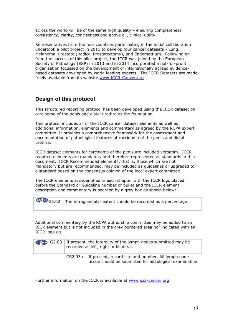
The ICCR elements are identified in each chapter with the ICCR logo placed
before the Standard or Guideline number or bullet and the ICCR element
description and commentary is boarded by a grey box as shown below:
G3.02 The intraglandular extent should be recorded as a percentage.
Additional commentary by the RCPA authorship committee may be added to an
ICCR element but is not included in the grey bordered area nor indicated with an
ICCR logo eg
G2.03 If present, the laterality of the lymph nodes submitted may be
recorded as left, right or bilateral.
CS2.03a If present, record site and number. All lymph node
tissue should be submitted for histological examination.
Further information on the ICCR is available at www.iccr-cancer.org

14
Checklist
Consistency and speed of reporting is improved by the use of discrete data
elements recorded from the checklist. Items suited to tick boxes are distinguished
from more complex elements requiring free text or narrative. A structured or
discrete approach to responses is favoured, however the pathologist is
encouraged to include free text or narrative where necessary to document any
other relevant issues, to give reasons for coming to a particular opinion and to
explain any points of uncertainty.
Report format
The structure provided by the following chapters, headings and subheadings
describes the elements of information and their groupings, but does not
necessarily represent the format of either a pathology report (Chapter 7) or
checklist (Chapter 6). These, and the structured pathology request form
(Appendix 1) are templates that represent information from this protocol,
organised and formatted differently to suit different purposes.
Key documentation
• Guidelines for Authors of Structured Cancer Pathology Reporting Protocols 22
• The Pathology Request-Test-Report Cycle — Guidelines for Requesters and
Pathology Providers23
• World Health Organization (WHO). Classification of tumours. Pathology and
genetics of the urinary system and male genital organs. Humphrey PA, Moch
H, Reuter VE, Ulbright TM editors. 4th edition. Lyon, France: IARC
Press;2016.16
• AJCC Cancer Staging Manual, 8th edition, American Joint Committee on
Cancer, 201617
Updates since last edition
Not applicable.

15
Authority and development
This section provides information about the process undertaken to develop this
protocol.
This 1st edition of the protocol is an amalgam of two separate processes:
1. This protocol is based on the ICCR Dataset for the reporting of carcinoma
of the penis and distal urethra 1st edition. All ICCR elements from this
dataset, both required (mandatory) and recommended (optional), are
included in this protocol, verbatim. (It should be noted that RCPA feedback
from all Anatomical Pathology fellows and specifically the local expert
committee was sought during the development process of the ICCR
dataset.) Details of the ICCR development process and the international
expert authoring committee responsible for the ICCR dataset are available
on the ICCR website: iccr-cancer.org.
2. Additional elements, values and commentary have been included as
deemed necessary by the local expert committee. In addition, the
standard inclusions of RCPA protocols eg example reports, request
information etc, have also been added.
Authorship
Dr David Clouston (Chair and Lead author), Pathologist
Professor Brett Delahunt, Pathologist
Professor Ian Davis, Medical Oncologist
Adjunct Professor Warick Delprado, Pathologist
Dr Andrew See, Radiation Oncologist
Clinical Professor James Kench, Pathologist
Associate Professor Nathan Lawrentschuk, Urologist
Professor Hemamali Samaratunga, Pathologist
Editorial manager
Meagan Judge, Royal College of Pathologists of Australasia.
Acknowledgements
The Penile cancer authorship committee wish to thank all the pathologists and
clinicians who contributed to the discussion around this document.

16
Stakeholders
ACT Health
ACT Cancer Registry
Australian Cancer Network
Australian Commission on Safety and Quality in Health Care
Australian Digital Health Agency
Australian Institute of Health and Welfare
Cancer Australia
Cancer Council ACT
Cancer Council Queensland
Cancer Council Victoria
Cancer Council Western Australia
Cancer Institute NSW
Cancer Services Advisory Committee (CanSAC)
Cancer Voices NSW
Clinical Oncology Society of Australia (COSA)
Department of Health, Australia
Department of Health, New Zealand
Faculty of Radiation Oncology Genito-Urinary Group (FROGG)
Health Informatics Society of Australia (HISA)
Independent Review Group of Pathologists
Medical Software Industry Association (MSIA)
National Pathology Accreditation Advisory Council (NPAAC)
New Zealand Cancer Registry
Northern Territory Cancer Registry
Pathology Australia
Public Pathology Australia
Queensland Cooperative Oncology Group (QCOG)
RCPA Anatomical Pathology Advisory Committee (APAC)
Representatives from laboratories specialising in anatomical pathology across
Australia
Royal Australasian College of Physicians (RACP)
South Australia Cancer Registry
Standards Australia
Tasmanian Cancer Registry
The Australian and New Zealand Urogenital and Prostate Cancer Trials Group
(ANZUP)
The Medical Oncology Group of Australia
The Prostate Cancer Foundation of Australia (PCFA)

17
The Prostate Cancer Foundation of New Zealand (PCFNZ)
The Royal Australasian College of Surgeons (RACS)
The Royal Australian and New Zealand College of Radiologists (RANZCR)
The Royal Australian College of General Practitioners (RACGP)
The Royal College of Pathologists of Australasia (RCPA)
The Urological Society of Australia And New Zealand (USANZ)
Western Australia Clinical Oncology Group (WACOG)
Development process
This protocol has been developed following the ten-step process set out in
Guidelines for Authors of Structured Cancer Pathology Reporting Protocols.18
Where no reference is provided, the authority is the consensus of the local expert
group for local inclusions and the ICCR Dataset Authoring Committee for ICCR
components denoted with the ICCR logo.

18
1 Pre-analytical
This chapter relates to information that should be recorded on receipt of the
specimen in the laboratory.
The pathologist is reliant on the quality of information received from the clinicians
or requestor. Some of this information may be received in generic pathology
request forms, however, the additional information required by the pathologist
specifically for the reporting of penile carcinoma is outlined in Appendix 1.
Appendix 1 also includes a standardised request information sheet that may be
useful in obtaining all relevant information from the requestor.
Surgical handling procedures affect the quality of the specimen and
recommendations for appropriate surgical handling are included in Appendix 1.
S1.01 All demographic information provided on the request form and
with the specimen must be recorded.
CS1.01a The Royal College of Pathologists of Australasia (RCPA) The
Pathology Request-Test-Report Cycle — Guidelines for
Requesters and Pathology Providers must be adhered to.19 This
document specifies the minimum information to be provided by
the requesting clinician for any pathology test.
CS1.01b Whether or not the patient identifies as Aboriginal and/ or
Torres Strait Islander. This is in support of a government
initiative to monitor the health of indigenous Australians
particularly in relation to cancer.
CS1.01c The patient’s health identifiers may include the patient’s
Medical Record Number as well as a national health number
such as a patient’s Individual Healthcare Identifier (IHI)
(Australia) or the National Healthcare Identifier (New Zealand).
S1.02 All clinical information as documented on the request form must
be recorded verbatim.
CS1.02a The request information may be recorded as a single text
(narrative) field or it may be recorded in a structured format.
CS1.02b The copy doctors requested on the request form must be
recorded.
S1.03 The pathology accession number of the specimen must be
recorded.
S1.04 The principal clinician involved in the patient’s care and
responsible for investigating the patient must be recorded.
CS1.04a The principle clinician should provide key information
regarding the clinical presentation of the patient. Follow up
may be required with the principle clinician for a number of
reasons:
• The clinical assessment and staging may be incomplete at

19
the time of biopsy.
• The pathology request is often authored by the clinician
performing the surgical excision/biopsy rather than the
clinician who is investigating and managing the patient.
• The identity of this clinician is often not indicated on the
pathology request form
In practice therefore, it is important in such cases that the
reporting pathologist should be able to communicate with the
managing clinician for clarification.
CS1.04b The Australian Healthcare identifiers i.e. Healthcare Provider
Identifier - Individual (HPI-I) and Healthcare Provider
Identifier - Organisation (HPI-O) should be included, where
possible, to identify the principal clinician involved in the
patient's care.
G1.01 Any clinical information received in other communications from the
requestor or other clinician should be recorded together with the source of
that information.

20
2 Specimen handling and macroscopic findings
This chapter relates to the procedures required after the information has been
handed over from the requesting clinician, and the specimen has been received in
the laboratory.
Specimen handling
➢ Detailed fixation and specimen handling instructions are available from the
RCPA online Cut-up Manual:
www.rcpa.edu.au/Library/Practising-Pathology/Macroscopic-Cut-Up
Macroscopic findings
S2.01 The labelling of the specimen(s) must be clearly recorded.
S2.02 The operative procedure20-22 should be recorded.
CS2.02a Treatment of penile carcinoma is primarily surgical. The
development of supranetworks in some countries has
made organ sparing techniques associated with
reconstruction widely available and radical or partial
penectomy is no longer the standard treatment for this
disease except in advanced cases.23,24
CS2.02b Most penile SCC arise from the distal mucosal surface and
may involve the glans, coronal sulcus and / or the
foreskin. When only the foreskin is involved, circumcision
alone is sufficient. For more extensive disease, a wider
excision is taken, with the operative specimen depending
on the tumour location. This can include a partial or total
penectomy with additional structures including scrotal
skin as required.
S2.03
The anatomical components submitted must be recorded and
measured.
CS2.03a The specimens may be circumcision only, partial or total
penectomy, with or without scrotal skin, testes and lymph
nodes.
CS2.03b For circumcision specimens, the foreskin should be
measured with mucosal and cutaneous margins inked in
different colours. After fixation, the foreskin can be sliced
vertically and clockwise from 1 to 12 o’clock.

21
CS2.03c For penectomy specimens, the length and diameter of the
specimen should be measured. After fixation, the
proximal margin of the specimen should be embedded
enface. The specimen should be sliced longitudinally
along the urethra from the meatus to the proximal
urethral margin and separated into left and right halves.
Each half can then be serially sliced.
G2.01 Whether or not the tumour is multifocal should be recorded.
CG2.01a Some types of penile squamous carcinoma may be
multifocal, particularly if associated with precancerous
changes (differentiated or undifferentiated penile
intraepithelial neoplasia (PeIN)). There are little data for
this in the literature but one text reports up to 5% of
tumours are multifocal.25
S2.04 The macroscopic tumour site(s)20,26-29 must be described.
CS2.04a The site(s) of primary penile and urethral tumours should
be noted macroscopically. The prognosis of equivalent
tumours of the foreskin may be better than that of the
glans. Tumours of the urethra have a worse prognosis
than those of the penis or foreskin. The presence or
absence of PeIN or urothelial carcinoma in situ can be
helpful in differentiating primary penile or urethral
squamous from urothelial carcinomas.
Penile and urethral melanomas and primary skin tumours
of the shaft should be handled and reported using
melanoma and skin tumour datasets respectively.
S2.05 The maximum macroscopic tumour dimensions7,30,31 must be
recorded.
CS2.05a Tumour dimensions have to be determined through a
combination of macroscopic and microscopic assessment,
particularly if tumours are very large.
G2.02 The tumour appearance should be recorded.
CG2.02a SCC may be exophytic and warty, have a broad spreading
growth pattern or have an endophytic, deeply invasive
growth pattern. As well as architecture, the colour,
borders, and presence of ulceration be recorded
G2.03 The macroscopic depth of invasion should be recorded.
CG2.03a SCC of the foreskin may spread vertically to the lamina
propria, dartos and into the outer skin. SCC of the glans
can spread into the corpus spongiosum and the corpus
cavernosum.
22
G2.04 Macroscopic distance of tumour from closest margin should be
recorded.
CG2.04a The important margins depend on the anatomical
specimen. For circumcision specimens, the coronal sulcus
mucosal margin is the most important margin. For
penectomy specimens, close attention needs to be made
to the proximal urethral margin, as the SCC may spread
horizontally along the urethra to this margin. The penile
fascial margin is also important. The penile fascia (Buck
fascia) lies superficial to the tunica albuginea which
surrounds the corpus cavernosum / corpus spongiosum,
and the SCC may spread along this fascia to the proximal
margin. The skin and corpus cavernosum margins must
also be assessed.
G2.05 The appearance of the glans penis should be recorded.
G2.06 If lymph nodes are submitted, the site(s) of the nodes and the
number of nodes per site should be recorded.
G2.07 The presence of any nodal metastases or mass seen macroscopically
should be recorded and the maximum size recorded (in mm).
CG2.07a If the nodal metastases are forming a confluent mass,
then the maximum overall dimension of the mass should
be given. This is important in determining “lymph node
mass” for accurate nodal staging.
S2.06 A block identification key20,32-35 listing the nature and origin
of all tissue blocks must be recorded.
CS2.06a The origin/designation of all tissue blocks should be
recorded and it is preferable to document this information
in the final pathology report. This is particularly important
should the need for internal or external review arise and
in larger more complex specimens and/or those with
orientation markings. The reviewer needs to be clear
about the origin of each block in order to provide an
informed specialist opinion including accurate staging. If
this information is not included in the final pathology
report, it should be available on the laboratory computer
system and relayed to the reviewing pathologist.
Specimen photographs and/or annotated diagrams may
be of assistance in clarification of block keys. These
documents should also be retrievable as part of the
pathology record.
Recording the origin/designation of tissue blocks also
facilitates retrieval of blocks, for example for further
immunohistochemical or molecular analysis, research
studies or clinical trials.

23
The availability of large block technology is strongly
recommended for larger specimens, such as
glansectomies and penectomies as it facilitates staging
with easier identification of deep structures, in particular
the urethra, corpus spongiosum and corpora cavernosa.
It is recommended that a record is kept of a good
representative paraffin block of tumour and if frozen
tissue is stored.
CS2.06b A minimum of three blocks of tumour should be taken
including the edge of the tumour and the maximum depth
of invasion. Also, the entire proximal margin of
penectomy specimens needs to be taken (embedded en
face), and blocks of glans, foreskin and urethra should be
included
CS2.06c For lymphadenectomy specimens, all lymph nodes need to
be submitted for histology. Small nodes up to 3 mm can
be submitted whole. Larger nodes should be bivalved or
sliced at 3 mm intervals, and at least one slice of every
node should be submitted. Ideally, the whole node will be
examined histologically.
G2.08
A descriptive or narrative field should be provided to record any
macroscopic information that is not recorded in the above standards
and guidelines, and that would normally form part of the
macroscopic description.
CG2.08a The traditional macroscopic narrative recorded at the
time of specimen dissection is often reported separately
from the cancer dataset. Although this remains an option,
it is recommended that macroscopic information be
recorded within the overall structure of this protocol.
CG2.08b Much of the information recorded in a traditional
macroscopic narrative is covered in the standards and
guidelines above and in many cases, no further
description is required.
CG2.08c A traditional macroscopic description may be required
when the Laboratory Information System (LIS) does not
allow a structured approach.
CG2.08d Where the LIS offers an electronic interface for structured
data entry the need for narrative can be significantly
reduced to describe only information not otherwise
captured.

24
3 Microscopic findings
Microscopic findings relates to purely histological (morphological) assessment.
Information derived from multiple investigational modalities, or from two or more
chapters, is described in Chapter 5.
S3.01 The histological tumour type4,36-41 must be recorded.
CS3.01a The most recent World Health Organisation (WHO) book
(2016) classifies and codes malignant squamous epithelial
tumours of the penis refer Appendix 4.
The tumours are further subclassified in the recent WHO
publication into non- HPV related and HPV related tumours.
However there is some group crossover particularly in
Usual type squamous cell carcinomas, a proportion of
which are HPV positive. Mixed carcinomas may also show
heterogeneity and sometimes include both HPV and non
HPV associated tumour types.
A. Non–HPV-related penile SCCs
1. SCC
Usual carcinoma
Pseudohyperplastic carcinoma
Pseudoglandular carcinoma
2. Verrucous carcinoma
Pure verrucous carcinoma
Carcinoma cuniculatum
3. Papillary carcinoma, NOS
4. Adenosquamous carcinoma
5. Sarcomatoid squamous carcinoma
6. Mixed carcinoma
B. HPV-related penile SCCs
7. Basaloid carcinoma
Papillary–basaloid carcinoma
8. Warty carcinoma
Warty–basaloid carcinoma

25
Clear cell carcinoma
9. Lymphoepithelioma-like carcinoma
C. Other rare carcinomas
CS3.01b Different subtypes of penile carcinomas have been defined,
which appear to be associated with different outcomes and
may also therefore justify the adoption of different
treatment strategies.
Over 95% of penile cancers are squamous cell carcinomas,
with rare instances of sarcomas, melanomas or
neuroendocrine carcinomas (including large cell and small
cell neuroendocrine carcinomas). In addition to the most
common, usual type of squamous carcinoma, subtypes
include papillary, basaloid, warty (condylomatous),
verrucous and sarcomatoid subtypes.
Subtyping is required as verruciform carcinomas (papillary,
warty or verrucous carcinomas) have better outcomes.
Basaloid, pseudoglandular/acantholytic and sarcomatoid
carcinomas are always high-grade with a worse prognosis
than the usual type of squamous carcinoma and may more
readily metastasise via the blood stream to distant sites
such as the lung. Mixed patterns are frequently present
and in these cases all subtypes identified should be
recorded.
Different patterns of growth can also be distinguished.
Vertical growth/endophytic carcinomas are associated with
a higher risk of metastases than superficial
spreading/exophytic carcinomas although it is not clear
whether this distinction offers superior prognostic power
over tumour stage.
p16 staining or assessment of HPV subtypes may also be
of help in subtyping squamous tumours but are not
mandatory.
Tumour subtypes of squamous cell carcinoma
• Squamous cell carcinoma of usual subtype
(NOS).25,42
• Basaloid squamous cell carcinoma.43
• Warty (condylomatous) squamous cell
carcinoma.44,45
• Verrucous squamous cell carcinoma.40
• Papillary squamous cell carcinoma.46
• Mixed squamous cell carcinomas (specify
subtypes).40

26
Other rare tumour subtypes
Squamous cell carcinoma variants
• Pseudohyperplastic squamous cell carcinoma.40,47,48
• Verrucous carcinoma variant
• Carcinoma cuniculatum.47,49
• Sarcomatoid (Spindle cell) squamous cell
carcinoma.50
• Pseudoglandular (Acantholytic adenoid) squamous
cell carcinoma.47,51
• Lymphoepithelioma like squamous cell carcinoma.52
• Warty carcinoma variants
• Clear cell carcinoma.47
• Warty basaloid squamous cell carcinoma.53
• Adenosquamous carcinoma.54
Non squamous tumours – not included in the scope of this
protocol
• High grade neuroendocrine carcinomas including
large cell neuroendocrine carcinoma and small cell
carcinoma.47,55,56
• Malignant melanoma.57
• Mesenchymal tumours.25
• Urothelial carcinoma of urethra.25
• Extramammary Paget’s disease.25
• Appendage tumours.25
• Metastatic tumours.58
• Lymphomas and haematological tumours.25
S3.02 The histological grade16,25,31,50,59,60 must be recorded (where
appropriate).
CS3.02a Accurate staging and grading of tumours are used to
determine subsequent clinical management and follow-up.
There is no consensus concerning grading, and the most
27
recent WHO classification (2016)58 recommends a three
step grading system based on degree of pleomorphism and
keratinisation with the overall grade determined by the
worst area no matter how small the percentage of the
tumour. The most recent College of American Pathologists
(CAP) guidelines61 offer some outline global guidance which
is applicable to usual type squamous carcinomas.
The “classical” method defines well-, moderately-well and
poorly-differentiated carcinomas on the basis of the degree
of cytological atypia, keratinisation, intercellular bridges
and mitotic activity. These criteria are difficult to apply to
some subtypes of penile carcinoma, for example verrucous
carcinomas which are well differentiated but often show
little or no keratinisation. Sarcomatoid change is a
separate category, which is often combined with other
tumour types and which conveys a very poor prognosis. All
tumours with sarcomatoid areas should be graded as
Grade 3 but this finding also needs to be noted separately
as tumours with sarcomatoid areas have a worse prognosis
than Grade 3 tumours generally.17
Tumours are generally graded on their worst component.
Although at one time a threshold of 50% of poorly-
differentiated cancer was suggested as the cut-off point
most predictive of nodal metastases, it has recently been
shown that any component of high-grade tumour conveys
a worse prognosis so should be included in the final
grade.60 Every effort should be made to assign a final
grade as this is an important prognostic factor and this
grade must be based on the most poorly-differentiated
component, no matter how small.
Refer Tables 1a and b.
S3.03 The maximum microscopic tumour dimensions must be
recorded.
CS3.03a Measurement of the depth of invasion, measured in
millimetres from the basement membrane of the adjacent
epithelium to the deepest point of invasion, or the
maximum thickness or size of the tumour may also give
prognostic information as seen in squamous tumours of
other sites such as skin. Minimal risk for metastasis is
reported for tumours measuring less than 5 mm in
thickness. Tumours invading deeper into penile anatomical
levels are usually associated with a higher risk of nodal
involvement. Thickness of penile tumours rather than
depth of invasion is more readily assessed, especially in
large tumours, because of the anatomical complexity of the
organ.
S3.04 The extent of invasion7,29,62,63 must be recorded.
CS3.04a Tumours invading deeper into penile anatomical levels are

28
usually associated with a higher risk of nodal involvement.
There is also a correlation between deeper infiltration and
higher histological grade, although some exceptions do
occur. Tumours invading corpus cavernosum are at higher
risk for presenting with nodal metastases than those
invading only corpus spongiosum and although these are
both staged as T2 in Union for International Cancer Control
(UICC)29 and American Joint Committee on Cancer
(AJCC)64 TNM7, TNM817 now stages corpus cavernosum
invasion as T3 irrespective of urethral involvement. The
tunica albuginea, which separates corpus spongiosum
from corpus cavernosum is considered part of the corpora
cavernosa.64
The anatomy of the penis is complex and difficulties often
arise in distinguishing levels of invasion. The distinction
between lamina propria and corpus spongiosum is made on
the basis of vascularity. Vessels within erectile tissue are
more angular and thin-walled with intervening
fibromuscular tissue than those within the lamina propria,
which are more variably sized and separated by loose
connective tissue.
S3.05 Evidence of lymphovascular invasion59,65 must be recorded.
CS3.05a Vascular invasion, lymphatic or venous, adversely affects
prognosis of penile cancer. The TNM staging classification
in the seventh edition of the AJCC Cancer Staging Manual64
subdivides T1 tumours into T1a and T1b based on the
absence or presence of lymphovascular invasion (LVI) or
poorly-differentiated tumours. This is also included in the
8th edition (TNM8)17 which also includes the additional
stratifier of perineural invasion (see PERINEURAL
INVASION below).
Embolic involvement of lymphatic vascular spaces occurs
usually near the invasive tumour front, but it may also be
found at a certain distance from the primary tumour in
anatomical areas such as the lamina propria, penile fascia,
and especially in the subepithelial connective tissues
surrounding the penile urethra. Venous invasion indicates a
more advanced stage of the disease and is related to the
compromise of the specialized erectile venous structures of
corpora spongiosa and cavernosa.
Vascular invasion may be difficult to assess particularly in
small biopsies and immunohistochemistry with vascular
markers may be of assistance in some cases.
S3.06 Evidence of perineural invasion7,31,63 must be recorded.
CS3.06a Risk groups stratification systems are available to predict
the likelihood of inguinal nodal involvement and
therapeutic planning and are based on a combination of
histological grade and pT stage. Strongest predictive power

29
is given by the combination of histological grade, deepest
anatomical level of infiltration, and presence of perineural
invasion. These factors are used for constructing the
Prognostic Index. TNM8 now includes perineural invasion
as a stratifier between T1a and T1b tumours in addition to
LVI.17
Perineural invasion may be difficult to assess, especially in
small and/or superficial biopsies. Immunohistochemistry
with neural markers may be helpful in some circumstances.
G3.01 Evidence of associated penile intraepithelial neoplasia (PeIN)28,59,66-70
should be recorded.
CG3.01a The pathological nomenclature and patterns of different
forms of preinvasive lesions of the penis has been radically
modified over the last few years with the abandonment of
clinical terms such as Erythroplasia of Queyrat and Bowen’s
disease and the adoption of the encompassing term Penile
Intraepithelial Neoplasia (PeIN) in pathological reports.
The new WHO classification of Penile Intraepithelial
Neoplasia distinguishes three groups: 1. Non HPV related
(differentiated or simplex), 2. HPV related
(undifferentiated) PeIN (basaloid, warty and warty-
basaloid) and 3. Others (pleomorphic, spindle, clear cell,
pagetoid).58 Undifferentiated HPV related PeIN shows full
thickness warty and/or basaloid features (previously
designated severe dysplasia/carcinoma in situ).
Differentiated PeIN usually involves only the basal layer
and is associated with architectural atypia and aberrant
keratinisation with features similar to that seen in
precancerous lesions of the vulva. Undifferentiated PeIN
is associated with p16 positivity and warty/basaloid
invasive tumours but differentiated PeIN is associated with
lichen sclerosis (balanitis xerotica obliterans), more
commonly seen with verrucous and pseudohyperplastic
tumours, and is usually p16 negative. It should also be
noted that PeIN of any type is often multifocal.
The presence and subtype of PeIN should be reported
together with its margin status independent of associated
invasive tumour. The splitting of PeIN into subgrades (for
example I-III or low-grade/high-grade) is not
recommended by the authors. Written reports should
indicate the subtype and extent of PeIN and whether or not
there is margin involvement.
Precancerous lesions identical to differentiated and
undifferentiated PeIN are seen in the distal penile urethra
but there is no guidance on how to report them. Rather
than designating these as carcinoma in situ or severe
dysplasia, it may be advisable to also use the term PeIN in
this context.
A potential problem arises when there are cytological

30
abnormalities not thought to be severe enough to be
designated as PeIN of either subtype. Then a category such
as ‘atypia falling short of PeIN’ with a recommendation for
follow up may be used, to avoid over treatment.
It is not necessary to report PeIN using the full dataset if it
is the only abnormality present without invasive
carcinoma.
Immunohistochemistry with p16 may be of help in
subclassifying PeIN but is not regarded as mandatory. It
may also be of use in identifying high-risk HPV in atypical
condylomas.
S3.07 Margin status71,72 must be recorded.
CS3.07a Penile preserving techniques have led to closer surgical
tumour resection margins and there is evidence that this
does not significantly compromise local recurrence rates if
tumour cells are not present at the margin itself. Positive
margins must be recorded by site and microscopic distance
of tumour from close margins (5 mm or less) recorded in
mm. Microscopic margin positivity may be identified
unexpectedly in tumours that infiltrate widely without
creating a mass effect. The presence of microscopic
involvement of surgical margins, however, has implications
for audit of pre-operative staging and/or surgical
technique. Actual measurement of linear extent of
individual involved margins is a non-core item but is valued
by surgeons in assessing their techniques.
Staging in the presence of positive margins needs to be
undertaken but made clear to clinicians. The term ‘at
least’, as in pT2 at least, may be used to indicate a positive
margin. It is not helpful to clinicians not to stage if
margins are positive.
The deep central soft tissue margin is defined as areas of
intervening tissue not identified as periurethral tissue,
corpus cavernosum or circumferential shaft margins or
may be used if the specific site of the deep margin is
indeterminate.
Margins of resection for penile specimens
• Urethral
• Periurethral tissues including lamina propria and
corpus spongiosum
• Corpus cavernosum
• Circumferential margins of bare penile shaft
• Peripheral skin
31
• Deep central soft tissue margin (other than
periurethral tissue, corpus cavernosum or
circumferential shaft)
Margins of resection of circumcision specimens
• Coronal sulcus/glans margin
• Peripheral cutaneous margin
• Deep central soft tissue margin
S3.08 Lymph node status21-23,28,29,73-75 must be recorded, recording
total number of nodes and the number of positive nodes, the
size of the largest metastasis, and the presence or absence of
extranodal spread.
CS3.08a The number of nodes found within an individual specimen
should be specified in the report. The size of the largest
nodal tumour deposit (not the lymph node size) together
with presence of extranodal spread must also be recorded
as there is evidence that this may affect prognosis. These
parameters must be reported separately for each individual
node site. Occasionally individual tumour cells are
identified in the peripheral sinus. The significance of these
is uncertain but they should be described within reports.
The size of the largest nodal tumour deposit must be
recorded as there is evidence that this may affect
prognosis in penile cancer. Both TNM7 and TNM8 classify
very small amounts of tumour as micrometastases (up to
0.2 mm)17,29,64,76 and isolated tumour cells as N0 (i+).11
However there is no evidence for a prognostic cut-off point
for lymph node metastasis size in penile cancer so it is
recommended that maximum dimension of largest tumour
deposit is recorded and tumour deposits over 0.2 mm
staged as N1.
CS3.08b Dynamic sentinel node biopsy, using either the blue dye
technique or lymphoscintigraphy, refers to the
intraoperative identification of the first node draining the
tumour. It relies on the assumption that lymphatic spread
is a stepwise process, so that, if the sentinel node is
negative, further nodal dissection would yield negative
results. This technique may be used in some centres for
patients with no clinical signs of nodal involvement.
Immunohistochemistry is essential for the assessment of
micrometastases in sentinel lymph nodes as small
metastases under 2 mm or single isolated tumour cells
may be easily missed.
CS3.08c Extent of inguinal node involvement and presence of ECS
also predicts pelvic node involvement.23,60,70,71 Therefore,
the extent of inguinal lymph node involvement including
32
number of nodes involved and presence or absence of ECS
is used to determine the need for pelvic node sampling or
excision.
CS3.08d Nodal involvement is a recognised predictor of poor
prognosis. In node positive disease, the number of positive
nodes, the presence of extracapsular spread (ECS) and the
level of nodal involvement (pelvic versus inguinal) have
been shown to influence survival by multivariate analysis
and this is reflected in both TNM723,60 and TNM811 which
classify any pelvic lymph node involvement or
extracapsular extension of any regional lymph node
(inguinal or pelvic) as pN3 in the penile but not in the
urethral TNM. However in penile TNM811 the number of
nodes which stratifies the staging between N1 and N2 is
two or more unilateral nodes rather than one or more in
TNM7.23,60
For urethral cancer in TNM723,60 the size of metastasis in a
single regional node, if greater than 2 cm, stratifies
between N1 and N2 nodes or if there are multiple nodes
involved, but in TNM811 there is no metastasis size
specified and the only stratifier is between single and
multiple regional nodes.
Although the N categories differ for P(p)enile and
U(u)rethral primary tumours it is recommended that data
items as specified in this section are recorded for tumours
of both these primary sites as tumours of the distal, as
opposed to proximal, urethra appear to spread in the same
way to local lymph nodes as do those of the penis.
G3.02 Any additional relevant comments should be recorded.

33
Table 1: Grading of penile squamous cell carcinoma
1a College of American Pathologists Protocol for the Examination of
Specimens From Patients With Carcinoma of the Penis Version: Penis
4.0.1.0 June 2017
Histological grade has been consistently reported as an influential predictive
factor of groin metastasis and dissemination of penile cancer. We recommend a
method to grade penile SCCs as follows:
Grade 1 Extremely well –differentiated carcinoma, with a minimal
deviation from the morphology of normal/hyperplastic
squamous epithelium.
Grade 2 Tumours show a more disorganized growth as compared to
grade 1 lesions, higher nuclear-to-cytoplasmic ratio, evident
mitoses, and, although present, less prominent keratinization.
Grade 3 Tumours showing any proportion of anaplastic cells, identified
as solid sheets or irregular small aggregates, cords or nests of
cells with little or no keratinization, high nuclear-to-
cytoplasmic ratio, thick nuclear membranes, nuclear
pleomorphism, clumped chromatin, prominent nucleoli, and
numerous mitoses.
A tumour should be graded according to the least differentiated component. Any
proportion of grade 3 should be noted in the report.
1b Modified from The Royal College of Pathologists (RCPath) Dataset for
penile and distal urethral cancer reports, 2nd Edition 2015
Feature Grade 1 Grade 2 Grade 3 Sarcomatoid
areas present
(Grade 3)
Cytological
atypia
Mild Moderate Anaplasia Sarcomatoid
Keratinisation Usually
abundant
Less
prominent
May be present Absent
Intercellular
bridges
Prominent Occasional Few Absent
Mitotic
activity
Rare Increased Abundant Abundant
Tumour
margin
Pushing/well
defined
Infiltrative/ill
defined
Infiltrative/ill
defined
Infiltrative/ill
defined

34
4 Ancillary studies findings
Ancillary studies may be used to determine lineage, clonality or disease
classification or subclassification; as prognostic biomarkers; or to indicate the
likelihood of patient response to specific biologic therapies.
Some studies, such as Her-2 testing, are required under the Pharmaceutical
Benefits Scheme, to enable certain specific therapies to be prescribed.
G4.01 Whether or not ancillary tests are performed should be recorded and the
results incorporated into the pathology report.
CG4.01a Immunohistochemistry can be performed and the results
incorporated into the pathology report. For example, p16 may
be helpful in the identification of HPV-related carcinomas. Also,
cytokeratin stains play a role in the sentinel node assessment
of penile carcinoma.

35
5 Synthesis and overview
Information that is synthesised from multiple modalities and therefore cannot
reside solely in any one of the preceding chapters is described here.
For example. tumour stage is synthesised from multiple classes of information –
clinical, macroscopic and microscopic.
By definition, synthetic elements are inferential rather than observational, often
representing high-level information that is likely to form part of the report
‘Summary’ or ‘Diagnosis’ section in the final formatted report.
Overarching case comment is synthesis in narrative format. Although it may not
necessarily be required in any given report, the provision of the facility for
overarching commentary in a cancer report is essential.
S5.01 The tumour stage must be recorded according to the AJCC TNM
system (8th edition).11(See Appendix 5) Used with the permission of
the American College of Surgeons, Chicago, Illinois. The original
source for this information is the AJCC Cancer Staging Manual, Eighth
Edition (2016) published by Springer Science+Business Media.
CS5.01a This dataset includes the AJCC TNM 8th edition11
definitions. Refer to Appendix 5.
Differences of the AJCC TNM 8th edition from the TNM 7th
edition should be noted:
1) Perineural invasion is now included as a stratifier
between T1a and T1b tumours of the penis in
addition to lymphovascular invasion and high
grade in TNM8.
2) The division between T2 and T3 in TNM8 of the
penis is entirely dependent on whether there
corpus spongiosum or corpus cavernosum invasion
irrespective of urethral involvement. This is the
most significant change between TNM7 and TNM8.
3) The number of unilateral nodes to indicate N2
rather than N1 of the penis has increased to 3
from 2.
4) The size of metastasis is no longer used as a
stratifier between N1 and N2 in unilateral regional
nodes in urethral cancer.
5) The use of TX is to be avoided if at all possible and
MX is not to be used.
6) Pathological staging should not be reported if the
specimen submitted is insufficient for definitive
staging. This may occur with biopsies or other
specimens where depth of invasion or the required

36
anatomical features cannot be discerned/assessed.
7) Staging in the presence of positive margins needs
to be undertaken but made clear to clinicians. The
term ‘at least’, as in pT2 at least, may be used to
indicate a positive margin. It is not helpful to
clinicians omit the stage if margins are positive.
By convention, the designation T refers to a primary
tumour that has not been previously treated. The symbol
p refers to the pathologic classification of the TNM, as
opposed to the clinical classification, and is based on
gross and microscopic examination. pT entails a resection
of the primary tumour or a biopsy adequate to evaluate
the highest pT category, pN entails removal of nodes
adequate to validate lymph node metastasis, and pM
implies microscopic examination of distant lesion.
Pathologic staging is usually performed after surgical
resection of the primary tumour.
Additional Descriptor
The m suffix indicates the presence of multiple primary
tumours and is recorded in parentheses, e.g. pTa(m)N0.
S5.02 The year of publication and/or edition of the cancer staging
system used in S5.01 must be included in the report.
G5.01 The “Diagnostic summary” section of the final formatted report should
include:
a. Operative procedure (S2.02)
b. Tumour site (S2.03)
c. Tumour type (S3.01)
d. Tumour grade (S3.02)
e. Maximum tumour dimensions ie depth of invasion (S3.03)
f. Margin status (S3.07)
g. Lymph node status (S3.08)
h. Tumour stage (S5.01)
S5.03 The reporting system must provide a field for free text or
narrative in which the reporting pathologist can give
overarching case comment, if required.
CS5.03a This field may be used, for example, to:
– document any noteworthy adverse gross and/or
histological features

37
– explain the decision-making pathway, or any
elements of clinicopathological ambiguity, or
factors affecting diagnostic certainty, thereby
allowing communication of diagnostic subtlety or
nuance that is beyond synoptic capture
– document further consultation or results still
pending.
CS5.03b Use of this field is at the discretion of the reporting
pathologist.
G5.02 The edition/version number of the RCPA protocol on which the report
is based should be included on the final report.
CG5.02a For example, the pathology report may include the
following wording at the end of the report: “the data fields
within this formatted report are aligned with the criteria as
set out in the RCPA document “ XXXXXXXXXX” XXXX
Edition dated XXXXXXX”.

38
6 Structured checklist
The following checklist includes the standards and guidelines for this protocol
which must be considered when reporting, in the simplest possible form. The
summation of all “Standards” is equivalent to the “Minimum Data Set” for penile
resections. For emphasis, standards (mandatory elements) are formatted in bold
font.
S6.01 The structured checklist provided may be modified as required
but with the following restrictions:
a. All standards and their respective naming conventions,
definitions and value lists must be adhered to.
b. Guidelines are not mandatory but are recommendations and
where used, must follow the naming conventions, definitions
and value lists given in the protocol.
G6.01 The order of information and design of the checklist may be varied
according to the laboratory information system (LIS) capabilities and as
described in Functional Requirements for Structured Pathology
Reporting of Cancer Protocols.73
CG6.01a Where the LIS allows dissociation between data entry and
report format, the structured checklist is usually best
formatted to follow pathologist workflow. In this situation,
the elements of synthesis or conclusions are necessarily at
the end. The report format is then optimised independently
by the LIS.
CG6.01b Where the LIS does not allow dissociation between data
entry and report format, (for example where only a single
text field is provided for the report), pathologists may elect
to create a checklist in the format of the final report. In this
situation, communication with the clinician takes precedence
and the checklist design is according to principles given in
Chapter 7.
G6.02 Where the checklist is used as a report template (see G6.01), the
principles in Chapter 7 and Appendix 2 apply.
CG6.02a All extraneous information, tick boxes and unused values
should be deleted.
G6.03 Additional comment may be added to an individual response where
necessary to describe any uncertainty or nuance in the selection of a
prescribed response in the checklist. Additional comment is not required
where the prescribed response is adequate.

39
Item descriptions in italics are conditional on previous responses.
Values in all caps are headings with sub values.
S/G Item description Response type Conditional
Pre-analytical
S1.01 Demographic information
provided
S1.02 Information provided on
request form
Not provided
OR
Text
OR
Structured entry as below:
Clinical information Text
Nature of operation Multi select value list (select all that apply):
• Partial penectomy
• Radical penectomy
• Glansectomy
• Circumcision
• Incisional/punch biopsy
• Excisional biopsy
• Urethrectomy

40
• Lymphadenectomy
o Sentinel
▪ Left, specify number of
site(s)
▪ Right, specify number of
site(s)
o Inguinal
▪ Left
▪ Right
o Pelvic
▪ Left, specify site(s)
▪ Right, specify site(s)
o Other, specify
▪ Left, specify site(s)
▪ Right, specify site(s)
o Not specified
• Other, specify laterality and site(s)
Operative findings Text
New primary lesion or
recurrence
Single selection value list:
• New primary
• Recurrence – regional, describe
• Recurrence – distant, describe
S1.03 Pathology accession number Alpha-numeric

41
S1.04 Principal clinician Text
G1.01 Comments Text
Macroscopic findings
S2.01 Specimen labelled as Text
S2.02 Operative procedure Text - Refer to S1.02
S2.03 ANATOMICAL COMPONENTS
SUBMITTED
Glans
Single selection value list:
• Absent
• Present
If present, consider recording
dimensions
Shaft
Single selection value list:
• Absent
• Present
If present, consider recording
dimensions
Foreskin
Single selection value list:
• Absent
• Present
If present, consider recording
dimensions
Coronal sulcus
Single selection value list:
• Absent
• Present
If present, consider recording
dimensions

42
Scrotal skin
Single selection value list:
• Absent
• Present
If present, consider recording
dimensions
Testis
Absent
OR
Multi select value list:
• Right present
• Left present
If present, consider recording
dimensions
Dimensions Numeric: __x__x__mm
G2.01 Tumour focality Single selection value list:
• Cannot be assessed
• Indeterminate
• Unifocal
• Multifocal, specify number of tumours in
specimen

43
S2.04 Macroscopic tumour site • No macroscopically visible tumour
• Indeterminate
OR
Multi selection value list (select all that
apply):
• Glans penis
• Sulcus
• Foreskin
• Distal penile urethra
S2.05 Macroscopic maximum
tumour dimensions
Single selection value list:
• Cannot be assessed
• Not applicable
OR
Complete the following
Width Numeric: __mm
Thickness Numeric: __mm
G2.02 Tumour appearance Normal
OR
Multi selection value list (select all that
apply):
• Nodule

44
• Endophytic
• Exophytic
• Polyp
• Papillary
• Verruciform
• Plaque
• Ulcer
• Pigmented macule, describe
G2.03 Macroscopic depth of invasion Numeric: __mm
G2.04 Macroscopic distance of tumour
from closest margin
Numeric: __mm
AND
Text (specify margin)
G2.05 Appearance of the glans penis Normal
OR
Text
G2.06 Lymph nodes Single selection value list:
• Submitted
• Not submitted
If submitted record, site(s) and
number of lymph nodes.
Site(s) and number of
lymph nodes
Text: Site
AND
Numeric: Number of LN’s (if possible)

45
Notes:
Note that the site and number of LN’s for that
site will need to be repeated for each site
received.
G2.07 Macroscopic nodal metastases or
mass
Single selection value list:
• Absent
• Present
If present, record the maximum
size
Size Numeric: __mm
S2.06 Block identification key Text
G2.08 Additional macroscopic
comments
Text
Microscopic findings
S3.01 Histological tumour type
Multi selection value list (select all that
apply):
• Squamous cell carcinoma of usual subtype
(NOS)
• Basaloid squamous cell carcinoma
• Warty (condylomatous) squamous cell
carcinoma
• Verrucous squamous cell carcinoma
• Papillary squamous cell carcinoma
• Mixed squamous cell carcinomas, specify
subtypes
• Other, specify*

46
Notes:
*refer to extended list in WHO Classification 2016
S3.02 Histological grade Single selection value list:
• Not applicable
• G1: Well differentiated
• G2: Moderately differentiated
• G3: Poorly differentiated
• Sarcomatoid areas present
S3.03 Microscopic maximum
tumour dimensions
Single selection value list:
• Cannot be assessed
• Not applicable
OR
Complete the following
Width Numeric: __mm
Thickness Numeric: __mm
S3.04 Extent of invasion Multi selection value list (select all that
apply):
• Subepithelial/lamina propria invasion
• Invasion of corpus spongiosum of glans
• Invasion of corpus cavernosum
• Invasion of the penile urethra
• Invasion of adjacent structures, specify

47
S3.05 Lymphovascular invasion Single selection value list:
• Not identified
• Present
• Indeterminate
S3.06 Perineural invasion Single selection value list:
• Not identified
• Present
• Indeterminate
G3.01 Associated penile intraepithelial
neoplasia (PeIN)
Single selection value list:
• Not identified
• Indeterminate
• Present
o Undifferentiated (Warty and/or
Basaloid)
o Differentiated
S3.07 MARGIN STATUS
Urethral margin (primary
tumours of the penis and
foreskin (resections and excision
biopsy only))
Single selection value list:
• Not applicable
• Cannot be assessed
• Involved by PeIN only
• Involved by invasive carcinoma
If not involved record the
distance to invasive tumour

48
• Not involved
Proximal urethral margin
(primary tumours of the urethra
only)
Single selection value list:
• Not applicable
• Cannot be assessed
• Involved by PeIN only
• Involved by invasive carcinoma
• Not involved
If not involved record the
distance to invasive tumour
Peri-urethral tissues Single selection value list:
• Not applicable
• Cannot be assessed
• Involved by invasive carcinoma
• Not involved
If not involved record the
distance to invasive tumour
Corpus cavernosum Single selection value list:
• Not applicable
• Cannot be assessed
• Involved by invasive carcinoma
• Not involved
If not involved record the
distance to invasive tumour
Circumferential shaft margin Single selection value list: If not involved record the
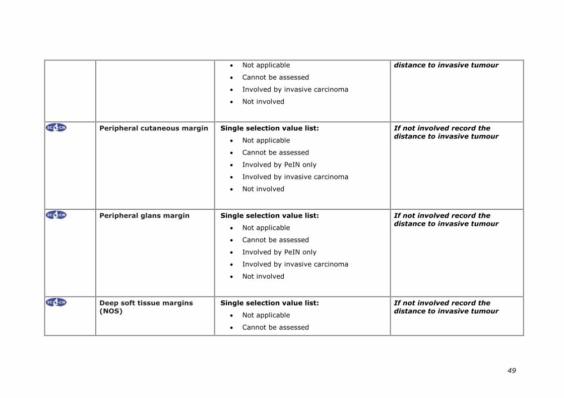
49
• Not applicable
• Cannot be assessed
• Involved by invasive carcinoma
• Not involved
distance to invasive tumour
Peripheral cutaneous margin Single selection value list:
• Not applicable
• Cannot be assessed
• Involved by PeIN only
• Involved by invasive carcinoma
• Not involved
If not involved record the
distance to invasive tumour
Peripheral glans margin Single selection value list:
• Not applicable
• Cannot be assessed
• Involved by PeIN only
• Involved by invasive carcinoma
• Not involved
If not involved record the
distance to invasive tumour
Deep soft tissue margins
(NOS)
Single selection value list:
• Not applicable
• Cannot be assessed
If not involved record the
distance to invasive tumour

50
• Involved by invasive carcinoma
• Not involved
Other margin Text (specify margin)
AND
Single selection value list:
• Cannot be assessed
• Involved by PeIN only
• Involved by invasive carcinoma
• Not involved
Complete only if required.
If not involved record the
distance to invasive tumour
Distance to invasive tumour Numeric: __mm
OR
>5 mm
S3.08 LYMPH NODE STATUS
INGUINAL NODES- SENTINEL
RIGHT Not submitted
OR
Complete the following:
Single selection value list:
• Not involved
• Isolated tumour cells only
If involved record the number of
positive nodes, max. dimension
of largest deposit and
extracapsular spread.

51
• Involved
Number of lymph nodes
examined
Numeric:____
Number of positive lymph
nodes
Number cannot be determined
OR
Numeric:____
Maximum dimension of
largest deposit
Numeric:___mm
Extracapsular spread Single selection value list:
• Not identified
• Present
LEFT Not submitted
OR
Complete the following:
Single selection value list:
• Not involved
• Isolated tumour cells only
• Involved
If involved record the number of
positive nodes, max. dimension
of largest deposit and
extracapsular spread.
Number of lymph nodes
examined
Numeric:____
Number of positive lymph
nodes
Number cannot be determined
OR

52
Numeric:____
Maximum dimension of
largest deposit
Numeric:___mm
Extracapsular spread Single selection value list:
• Not identified
• Present
INGUINAL NODES – NON
SENTINEL
RIGHT Not submitted
OR
Complete the following:
Single selection value list:
• Not involved
• Isolated tumour cells only
• Involved
If involved record the number of
positive nodes, max. dimension
of largest deposit and
extracapsular spread.
Number of lymph nodes
examined
Numeric:____
Number of positive lymph
nodes
Number cannot be determined
OR
Numeric:____
Maximum dimension of
largest deposit
Numeric:___mm

53
Extracapsular spread Single selection value list:
• Not identified
• Present
LEFT Not submitted
OR
Complete the following:
Single selection value list:
• Not involved
• Isolated tumour cells only
• Involved
If involved record the number of
positive nodes, max. dimension
of largest deposit and
extracapsular spread.
Number of lymph nodes
examined
Numeric:____
Number of positive lymph
nodes
Number cannot be determined
OR
Numeric:____
Maximum dimension of
largest deposit
Numeric:___mm
Extracapsular spread Single selection value list:
• Not identified
• Present
PELVIC NODES

54
RIGHT Not submitted
OR
Complete the following:
Single selection value list:
• Not involved
• Isolated tumour cells only
• Involved
If involved record the number of
positive nodes, max. dimension
of largest deposit and
extracapsular spread.
Number of lymph nodes
examined
Numeric:____
Number of positive lymph
nodes
Number cannot be determined
OR
Numeric:____
Maximum dimension of
largest deposit
Numeric:___mm
Extracapsular spread Single selection value list:
• Not identified
• Present
LEFT Not submitted
OR
Complete the following:
Single selection value list:
• Not involved
If involved record the number of
positive nodes, max. dimension
of largest deposit and
extracapsular spread.

55
• Isolated tumour cells only
• Involved
Number of lymph nodes
examined
Numeric:____
Number of positive lymph
nodes
Number cannot be determined
OR
Numeric:____
Maximum dimension of
largest deposit
Numeric:___mm
Extracapsular spread Single selection value list:
• Not identified
• Present
OTHER NODES Not submitted
OR
Complete the following:
Text (specify laterality and site)
AND
Single selection value list:
• Not involved
• Isolated tumour cells only
• Involved
Note: report only if required.
If involved record the number of
positive nodes, max. dimension
of largest deposit and
extracapsular spread.

56
Number of lymph nodes
examined
Numeric:____
Number of positive lymph
nodes
Number cannot be determined
OR
Numeric:____
Maximum dimension of
largest deposit
Numeric:___mm
Extracapsular spread Single selection value list:
• Not identified
• Present
G3.02 Additional microscopic comment Text
Ancillary findings
G4.01 Ancillary studies
Single selection value list:
• Not performed
• Performed, specify tests and results
Synthesis and overview
S5.01
PATHOLOGICAL STAGING
(AJCC 8TH EDITION)
Primary tumour (T) Single selection value list :
PENIS AND FORESKIN
Primary tumour (pT)

57
TX Primary tumour cannot be assessed
T0 No evidence of primary tumour
Tis Carcinoma in situ (Penile Intraepithelial
Neoplasia [PeIN])
Ta Non invasive localised squamous cell
carcinoma*
T1 Tumour invades subepithelial connective
tissue, dermis or lamina propria**
T1a Tumour is without lymphovascular
invasion or perineural invasion and is not
high grade
T1b Tumour exhibits lymphovascular invasion
and/or perineural invasion or is high
grade
T2 Tumour invades into corpus spongiosum
with or without urethral invasion
T3 Tumour invades into corpora cavernosum
with or without urethral invasion
T4 Tumour invades other adjacent structures
* The authors do not recommend the use of the
pTa category as it is not evidence based.

58
Regional lymph nodes (N) Single selection value list :
PENIS AND FORESKIN
Regional lymph nodes (pN)
NX Lymph node metastasis cannot be
established
N0 No lymph node metastasis
N1 ≤2 unilateral inguinal metastases, no ENE
N2 ≥3 unilateral inguinal metastases or
bilateral metastases
N3 Extranodal extension of lymph node
S5.02 Year and/or edition of
staging system
Numeric: year
AND/OR
Text: Edition eg 1st, 2nd etc
G5.01 Diagnostic summary
Include:
a. Operative procedure (S2.02)
b. Tumour site (S2.03)
c. Tumour type (S3.01)
d. Tumour grade (S3.02)
e. Maximum tumour
Text

59
dimensions (S3.03)
f. Margin status (S3.07)
g. Lymph node status (S3.08)
h. Tumour stage (S5.01)
S5.03 Overarching comment Text
G5.02 Edition/version number of the
RCPA protocol on which the
report is based
Text

60
7 Formatting of pathology reports
Good formatting of the pathology report is essential for optimising communication
with the clinician, and will be an important contributor to the success of cancer
reporting protocols. The report should be formatted to provide information clearly
and unambiguously to the treating doctors, and should be organised with their
use of the report in mind. In this sense, the report differs from the structured
checklist, which is organised with the pathologists’ workflow as a priority.
Uniformity in the format as well as in the data items of cancer reports between
laboratories makes it easier for treating doctors to understand the reports; it is
therefore seen as an important element of the systematic reporting of cancer. For
guidance on formatting pathology reports, please refer to Appendix 2.

61
Appendix 1 Pathology request information and surgical handling procedures
This appendix describes the information that should be collected before the
pathology test. Some of this information can be provided on generic pathology
request forms; any additional information required specifically for the reporting of
renal cancer may be provided by the clinician on a separate request information
sheet. An example request information sheet is included below. Elements which
are in bold text are those which pathologists consider to be required information.
Those in non-bold text are recommended.
Also included in this appendix are the procedures that are recommended before
handover of specimens to the laboratory.
Patient information
➢ Adequate demographic and request information should be
provided with the specimen.
• Items relevant to cancer reporting protocols include:
• patient name
• date of birth
• sex
• identification and contact details of requesting doctor
• date of request
• Whether or not the patient identifies as Aboriginal and/ or Torres
Strait Islander. This is in support of a government initiative to
monitor the health of indigenous Australians particularly in
relation to cancer.
➢ The patient’s health identifiers should be provided.
• The patient’s health identifiers may include the patient’s Medical
Record Number as well as a national health number such as a
patient’s Individual Healthcare Identifier (IHI) (Australia) or the
National Healthcare Identifier (New Zealand).
➢ The Australian Healthcare identifiers i.e. Healthcare Provider Identifier -
Individual (HPI-I) and Healthcare Provider Identifier - Organisation (HPI-
O) should be use, where possible, to identify the requesting doctor.
62
Clinical Information
➢ Any relevant clinical information should be provided.
• History of prior penile tumours and treatments, including topical
treatment, radiotherapy and chemotherapy should be given
particularly if the patient has been treated elsewhere.
It is good clinical practice to transcribe all clinical information from
the request form on to the pathology report.14 This is a
recommended rather than a required item as it is the responsibility
of the clinician requesting the pathological examination of a
specimen to provide information that will have an impact on the
diagnostic process or affect its interpretation.
➢ The nature of the operation must be recorded.
➢ The operative findings should be recorded.
➢ Record if this is a new primary cancer or a recurrence of a previous
cancer, if known.
• The term recurrence defines the return, reappearance or
metastasis of cancer (of the same histology) after a disease free
period.
Recurrence should be classified as distant metastases or regional
(local) recurrence.
Regional (local) recurrence refers to the recurrence of cancer cells
at the same site as the original (primary) tumour or the regional
lymph nodes.
Distant metastasis refers to the spread of cancer of the same
histologic type as the original (primary) tumour to distant organs or
distant lymph nodes.
• This information will provide an opportunity for previous reports to
be reviewed during the reporting process, which may provide
valuable information to the pathologist. This information also has
implications for recording cancer incidence and evidence based
research.

63
Example request information sheet
The above Request Information Sheet is published to the RCPA website.

64
Appendix 2 Guidelines for formatting of a pathology report
Layout
Headings and spaces should be used to indicate subsections of the report, and
heading hierarchies should be used where the LIS allows it. Heading hierarchies
may be defined by a combination of case, font size, style and, if necessary,
indentation.
• Grouping like data elements under headings and using ‘white space’ assists in
rapid transfer of information.74
Descriptive titles and headings should be consistent across the protocol, checklist
and report.
When reporting on different tumour types, similar layout of headings and blocks
of data should be used, and this layout should be maintained over time.
• Consistent positioning speeds data transfer and, over time, may reduce the
need for field descriptions or headings, thus reducing unnecessary information
or ‘clutter’.
Within any given subsection, information density should be optimised to assist in
data assimilation and recall.
• Configuring reports in such a way that they ‘chunk’ data elements into a
single unit will help to improve recall for the clinician. 74
• ‘Clutter’ should be reduced to a minimum.74 Thus, information that is not part
of the protocol (e.g. billing information, Snomed codes, etc) should not appear
on the reports or should be minimized.
• Injudicious use of formatting elements (e.g. too much bold, underlining or use
of footnotes) constitutes clutter and may distract the reader from the key
information.
Where a structured report checklist is used as a template for the actual report,
any values provided in the checklist but not applying to the case in question must
be deleted from the formatted report.
Reports should be formatted with an understanding of the potential for the
information to mutate or be degraded as the report is transferred from the LIS to
other health information systems.
As a report is transferred between systems:
• text characteristics such as font type, size, bold, italics and colour are often
lost
• tables are likely to be corrupted as vertical alignment of text is lost when fixed
font widths of the LIS are rendered as proportional fonts on screen or in print
• spaces, tabs and blank lines may be stripped from the report, disrupting the
formatting
• supplementary reports may merge into the initial report.
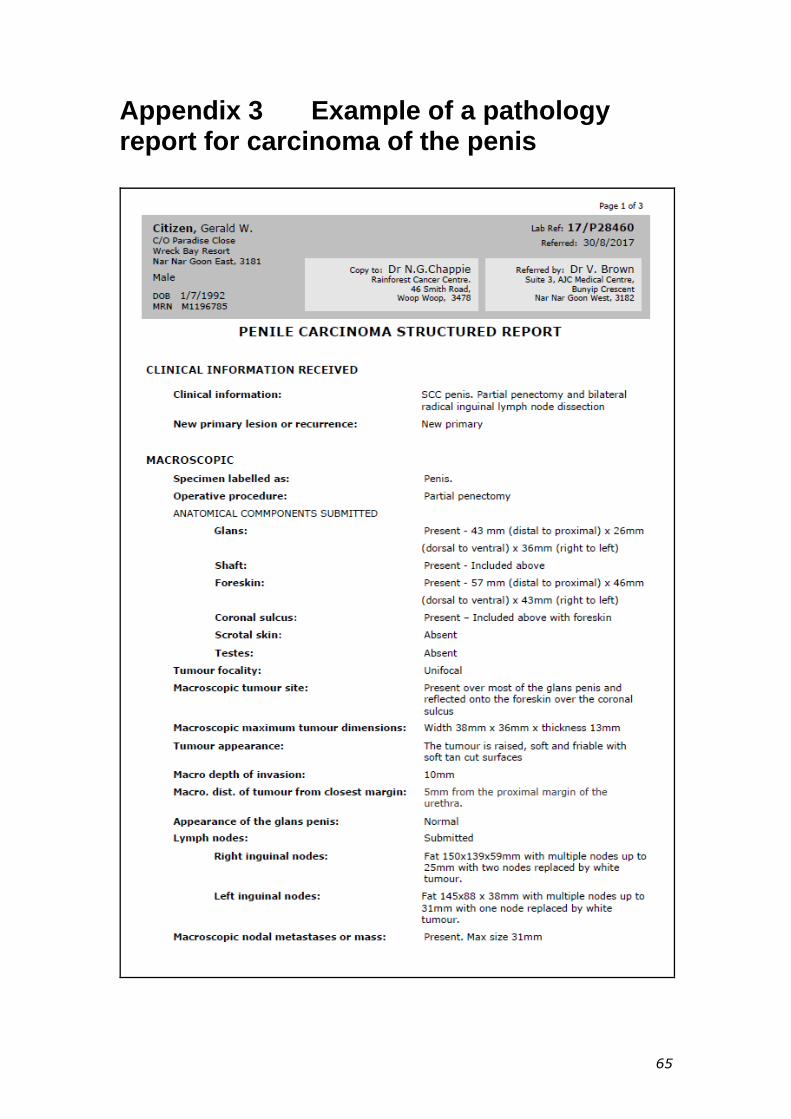
65
Appendix 3 Example of a pathology report for carcinoma of the penis

66

67

68
Appendix 4 WHO classification of tumours of the penisa
Descriptor ICD-O codes
Malignant epithelial tumours
Squamous cell carcinoma, NOS 8070/3
Verrucous carcinoma 8051/3
Adenosquamous carcinoma 8560/3
Sarcomatoid squamous carcinoma 8074/3
Mixed squamous cell carcinoma 8070/3
Basaloid squamous carcinoma 8083/3
Warty (condylomatous) carcinoma 8054/3
Papillary carcinoma (NOS) 8050/3
Lymphoepithelioma-like carcinoma 8082/2
Precursor lesions
Penile intraepithelial neoplasia
Low grade 8077/0
High grade 8077/2
Warty PeIN/Basaloid PeIN/Wart-basaloid PeIN
PeIN differentiated 8071/2
Paget disease 8542/3
a The morphology codes are from the International Classification of Diseases for Oncology (ICD-O). Behaviour is coded /0 for benign tumours; /1 for unspecified, borderline, or uncertain behaviour; /2 for carcinoma in situ and grade III intraepithelial neoplasia; and /3 for malignant tumours.
© World Health Organisation/International Agency for Research on Cancer (IARC). Reproduced with permission

69
Appendix 5 AJCC TNM staging
Tumours of the Penis and Foreskin (TNM 8)14,23,26,58,59
Primary Tumour (T)
TX Primary tumour cannot be assessed.
T0 No evidence of primary tumour.
Tis Carcinoma in situ (Penile intraepithelial neoplasia [PeIN]).
Ta Non invasive localised squamous cell carcinoma
T1 Glans: Tumour invades lamina propria
Foreskin: Tumour invades dermis, lamina propria or dartos fascia
Shaft: Tumour invades connective tissue between epidermis and corpora
regardless of location
All sites with or without LVI or perineural invasion and is or is not high grade
T1a Tumour invades lamina propria or subepithelial connective tissue and is
without lymphovascular or perineural invasion and is not high grade (i.e.
grade 3 or sarcomatoid)
T1b Tumour invades lamina propria or subepithelial connective tissue and
exhibits lymphovascular or perineural invasion and or is high grade (i.e.
grade 3 or sarcomatoid)
T2 Tumour invades into corpus spongiosum (either glans or ventral shaft) with
or without urethral invasion
T3 Tumour invades into corpora cavernosum (including tunica albuginea) with or
without urethral invasion
T4 Tumour invades other adjacent structures.

70
Regional Lymph Nodes (N)
pNX Lymph node metastasis cannot be established.
pN0 No lymph node metastasis.
pN1 Two or more inguinal metastases without extranodal extension (ENE)
pN2 Three or more unilateral inguinal metastases or bilateral metastases
pN3 ENE of lymph node metastases or pelvic lymph node metastases.
Distant Metastasis (M)
M0 No distant metastasis (clinical category only).
M1 Distant metastasis present.
M1 includes lymph node metastasis outside of the true pelvis in addition to
visceral or bone sites.
Accurate staging and grading of tumours are used to determine subsequent
clinical management and follow-up.
The anatomy of the penis is complex and difficulties often arise in distinguishing
levels of invasion. The distinction between lamina propria and corpus spongiosum
is made on the basis of vascularity. Vessels within erectile tissue are more
angular and thin-walled with intervening fibromuscular tissue than those within
the lamina propria which are more variably sized and separated by loose
connective tissue.
Although there is a category of non-invasive verrucous carcinoma in the primary
tumour classifications (Ta) in TNM7, the criteria for the diagnosis of this entity
and its distinction from verrucous hyperplasia are unclear to the authors of this
dataset and use of this category is not recommended. Although verrucous
carcinomas have a pushing rather than infiltrative margin, they are nevertheless
invasive. Invasion is often only superficial but more deeply invasive tumours may
be observed. Non invasive localised tumours of the penis of any subtype are
exceptionally rare in the authors’ experience.
Staging of pT1 is subdivided in TNM7 into pT1a for low-risk tumours and pT1b for
high-risk tumours depending on the absence or presence of high-grade tumour
and/or LVI. TNM8 also includes perineural invasion as a stratifier between T1a
and T1b. The number of unilateral nodes needed upstage from pN1 to N2 has
increased from two to three in TNM8. Metastatic tumour in regional lymph nodes
with extranodal spread is categorised as pN3.
It was initially proposed that the pT2 primary tumour classification be subdivided
to distinguish between invasion into the spongiosum and cavernosum, as some
reports show that risk of metastases in increased in patients with invasion of the
cavernosa. The RCPath dataset published in 2015 recommend substaging of T2
penile tumours into T2a (corpus spongiosum invasion) and T2b (corpus
cavernosum invasion) as this is evidence based.14 TNM8 now recommends that
71
involvement of the corpus spongiosum is classified as T2 and involvement of
corpora cavernosa is T3 irrespective of urethral involvement.
In the case of multiple tumours, the tumour with the highest T category should
be classified and the multiplicity or number of tumours should be indicated in
parentheses, e.g. pT2 (m) or pT2.
Use of the category TX is to be avoided and the designation e.g. ‘T (numerical
value) at least’ is preferable if full staging is not possible because of the nature of
the specimen (e.g. small incision biopsies) or the presence of positive margins.
If deep structures are not sampled and/or the invasive tumour extends to the
margins of excision staging should still be attempted but designated as ‘pT1 at
least’. The designation of pTX (unstageable) even in small biopsies should be
avoided as far as possible as it is clinically unhelpful.
The category M0 should not be used in pathological staging. The term MX is no
longer in use.
72
References
1 Merlin T, Weston A and Tooher R (2009). Extending an evidence hierarchy
to include topics other than treatment: revising the Australian 'levels of
evidence'. BMC Med Res Methodol 9:34.
2 Australian Cancer Network Colorectal Cancer Guidelines Revision
Committee (2005). Guidelines for the Prevention, Early Detection and
Management of Colorectal Cancer. The Cancer Council Australia and
Australian Cancer Network, Sydney.
3 Maughan NJ, Morris E, Forman D and Quirke P (2007). The validity of the
Royal College of Pathologists' colorectal cancer minimum dataset within a
population. British Journal of Cancer 97(10):1393–1398.
4 Royal College of Pathologists (2015). Cancer datasets and tissue
pathways. Available from:
https://www.rcpath.org/profession/publications/cancer-datasets.html.
http://www.rcpath.org/index.asp?PageID=254 (Accessed 19th Feb 2016).
5 College of American Pathologists (2015). Cancer protocol templates.
Available from:
http://www.cap.org/web/oracle/webcenter/portalapp/pagehierarchy/cance
r_protocol_templates.jspx?_adf.ctrl-
state=akc46lrl8_9&_afrLoop=6855098523798#! (Accessed 26th Sept 17).
6 Cross SS, Feeley KM and Angel CA (1998). The effect of four interventions
on the informational content of histopathology reports of resected
colorectal carcinomas. J Clin Oncol 51(6):481–482.
7 Mathers M, Shrimankar J, Scott D, Charlton F, Griffith C and Angus B
(2001). The use of a standard proforma in breast cancer reporting. J Clin
Pathol 54(10):809–811.
8 Srigley JR, McGowan T, MacLean A, Raby M, Ross J, Kramer S and Sawka
C (2009). Standardized synoptic cancer pathology reporting: A population-
based approach. J Surg Oncol 99(8):517–524.
9 Gill AJ, Johns AL, Eckstein R, Samra JS, Kaufman A, Chang DK, Merrett
ND, Cosman PH, Smith RC, Biankin AV and Kench JG (2009). Synoptic
reporting improves histopathological assessment of pancreatic resection
specimens. Pathology 41(2):161–167.
10 WHO (World Health Organization) (2016). Classification of tumours.
Pathology and genetics of the urinary system and male genital organs, 4th

73
edition. Humphrey PA, Moch H, Reuter VE and Ulbright TM. IARC Press,
Lyon, France.
11 Amin MB, Edge SB and Greene FL et al (eds) (2017). AJCC Cancer Staging
Manual. 8th ed., Springer, New York.
12 RCPA (Royal College of Pathologists of Australasia (2009 ). Guidelines for
Authors of Structured Cancer Pathology Reporting Protocols. RCPA, Surry
Hills NSW.
13 RCPA (Royal College of Pathologists of Australasia) (2004). Chain of
Information Custody for the Pathology Request-Test-Report Cycle —
Guidelines for Requesters and Pathology Providers. RCPA, Surry Hills,
NSW.
14 Royal College of Pathologists (2015). Dataset for penile and distal urethral
cancer histopathology reports. Available from:
https://www.rcpath.org/resourceLibrary/dataset-for-penile-and-distal-
urethral-cancer-histopathology-reports.html (Accessed 1st March 2016).
15 Horenblas S (2012). Sentinel lymph node biopsy in penile carcinoma.
Semin Diagn Pathol 29(2):90-95.
16 Lam W, Alnajjar HM, La-Touche S, Perry M, Sharma D, Corbishley C,
Pilcher J, Heenan S and Watkin N (2013). Dynamic sentinel lymph node
biopsy in patients with invasive squamous cell carcinoma of the penis: a
prospective study of the long-term outcome of 500 inguinal basins
assessed at a single institution. Eur Urol 63(4):657-663.
17 Hakenberg OW, Comperat EM, Minhas S, Necchi A, Protzel C and Watkin N
(2015). EAU guidelines on penile cancer: 2014 update. Eur Urol
67(1):142-150.
18 Lawindy SM, Rodriguez AR, Horenblas S and Spiess PE (2011). Current
and future strategies in the diagnosis and management of penile cancer.
Adv Urol 2011:593751.
19 Epstein JI CA, Humphrey PA (2011). Tumors of the prostate gland, seminal
vesicles, penis, and scrotum. AFIP Atlas of Tumor Pathology. American
Registry of Pathology, Washington DC, United States.
20 Oertell J, Caballero C, Iglesias M, Chaux A, Amat L, Ayala E, Rodriguez I,
Velazquez EF, Barreto JE, Ayala G and Cubilla AL (2011). Differentiated
precursor lesions and low-grade variants of squamous cell carcinomas are
frequent findings in foreskins of patients from a region of high penile
cancer incidence. Histopathology 58(6):925-933.

74
21 Tyson MD, Etzioni DA, Wisenbaugh ES, Andrews PE, Humphreys MR,
Ferrigni RG, Swanson SK and Castle EP (2012). Anatomic site-specific
disparities in survival outcomes for penile squamous cell carcinoma.
Urology 79(4):804-808.
22 Corbishley CM, Rajab RM and Watkin NA (2015). Clinicopathological
features of carcinoma of the distal penile urethra. Semin Diagn Pathol
32(3):238-244.
23 International Union against Cancer (UICC) (2009). TNM Classification of
Malignant Tumours. Sobin L, Gospodarowicz M and Wittekind C. Wiley-
Blackwell, Chichester, UK and Hoboken, New Jersey.
24 Emerson RE, Ulbright TM, Eble JN, Geary WA, Eckert GJ and Cheng L
(2001). Predicting cancer progression in patients with penile squamous cell
carcinoma: the importance of depth of invasion and vascular invasion. Mod
Pathol 14(10):963-968.
25 Velazquez EF, Ayala G, Liu H, Chaux A, Zanotti M, Torres J, Cho SI,
Barreto JE, Soares F and Cubilla AL (2008). Histologic grade and perineural
invasion are more important than tumor thickness as predictor of nodal
metastasis in penile squamous cell carcinoma invading 5 to 10 mm. Am J
Surg Pathol 32(7):974-979.
26 Chaux A, Caballero C, Soares F, Guimaraes GC, Cunha IW, Reuter V,
Barreto J, Rodriguez I and Cubilla AL (2009). The prognostic index: a
useful pathologic guide for prediction of nodal metastases and survival in
penile squamous cell carcinoma. Am J Surg Pathol 33(7):1049-1057.
27 Royal College of Pathologists (2015).
https://www.rcpath.org/resourceLibrary/dataset-for-penile-and-distal-
urethral-cancer-histopathology-reports.html (Accessed 16 February 2016).
28 Cubilla AL, Piris A, Pfannl R, Rodriguez I, Aguero F and Young RH (2001).
Anatomic levels: important landmarks in penectomy specimens: a detailed
anatomic and histologic study based on examination of 44 cases. Am J
Surg Pathol 25(8):1091-1094.
29 Tang V, Clarke L, Gall Z, Shanks JH, Nonaka D, Parr NJ, Elliott PA, Clarke
NW, Ramani V, Lau MW and Sangar VK (2014). Should centralized
histopathological review in penile cancer be the global standard? BJU Int
114(3):340-343.
30 Ebel JJ, Shabsigh A, Sharp DS and Zynger DL (2013). Whole-mount
evaluation of penectomies for penile cancer: feasibility, cost and
comparison to routine sectioning. Histopathology 63(1):64-73.
75
31 Cubilla AL VE, Ayala GE, Chaux A, Torres J, Reuter V (2005). Identification
of prognostic pathologic parameters in squamous cell carcinoma of the
penis: significance and difficulties. Pathol Case Rev 10:3-13.
32 Guimaraes GC, Cunha IW, Soares FA, Lopes A, Torres J, Chaux A,
Velazquez EF, Ayala G and Cubilla AL (2009). Penile squamous cell
carcinoma clinicopathological features, nodal metastasis and outcome in
333 cases. J Urol 182(2):528-534; discussion 534.
33 Chaux A, Reuter V, Lezcano C, Velazquez EF, Torres J and Cubilla AL
(2009). Comparison of morphologic features and outcome of resected
recurrent and nonrecurrent squamous cell carcinoma of the penis: a study
of 81 cases. Am J Surg Pathol 33(9):1299-1306.
34 Chaux A, Velazquez EF, Algaba F, Ayala G and Cubilla AL (2010).
Developments in the pathology of penile squamous cell carcinomas.
Urology 76(2 Suppl 1):S7-s14.
35 Cubilla AL (2009). The role of pathologic prognostic factors in squamous
cell carcinoma of the penis. World J Urol 27(2):169-177.
36 Chaux A and Cubilla AL (2012). Diagnostic problems in precancerous
lesions and invasive carcinomas of the penis. Semin Diagn Pathol
29(2):72-82.
37 Cubilla AL, Lloveras B, Alejo M, Clavero O, Chaux A, Kasamatsu E,
Monfulleda N, Tous S, Alemany L, Klaustermeier J, Munoz N, Quint W, de
Sanjose S and Bosch FX (2011). Value of p16(INK)(4)(a) in the pathology
of invasive penile squamous cell carcinomas: A report of 202 cases. Am J
Surg Pathol 35(2):253-261.
38 WHO (World Health Organization) (2004). World Health Organization
Classification of Tumours. Pathology and Genetics of Tumours of the
Urinary System and Male Genital Organ. Eble JN, Sauter G, Epstein JI and
Sesterhenn IA. IARC Press, Lyon, France.
39 Cubilla AL, Reuter VE, Gregoire L, Ayala G, Ocampos S, Lancaster WD and
Fair W (1998). Basaloid squamous cell carcinoma: a distinctive human
papilloma virus-related penile neoplasm: a report of 20 cases. Am J Surg
Pathol 22(6):755-761.
40 Cubilla AL, Velazques EF, Reuter VE, Oliva E, Mihm MC, Jr. and Young RH
(2000). Warty (condylomatous) squamous cell carcinoma of the penis: a
report of 11 cases and proposed classification of 'verruciform' penile
tumors. Am J Surg Pathol 24(4):505-512.

76
41 Chaux A, Tamboli P, Ayala A, Soares F, Rodriguez I, Barreto J and Cubilla
AL (2010). Warty-basaloid carcinoma: clinicopathological features of a
distinctive penile neoplasm. Report of 45 cases. Mod Pathol 23(6):896-
904.
42 Chaux A, Soares F, Rodriguez I, Barreto J, Lezcano C, Torres J, Velazquez
EF and Cubilla AL (2010). Papillary squamous cell carcinoma, not
otherwise specified (NOS) of the penis: clinicopathologic features,
differential diagnosis, and outcome of 35 cases. Am J Surg Pathol
34(2):223-230.
43 Chaux A, Velazquez EF, Barreto JE, Ayala E and Cubilla AL (2012). New
pathologic entities in penile carcinomas: an update of the 2004 world
health organization classification. Semin Diagn Pathol 29(2):59-66.
44 Cubilla AL, Velazquez EF and Young RH (2004). Pseudohyperplastic
squamous cell carcinoma of the penis associated with lichen sclerosus. An
extremely well-differentiated, nonverruciform neoplasm that preferentially
affects the foreskin and is frequently misdiagnosed: a report of 10 cases of
a distinctive clinicopathologic entity. Am J Surg Pathol 28(7):895-900.
45 Barreto JE, Velazquez EF, Ayala E, Torres J and Cubilla AL (2007).
Carcinoma cuniculatum: a distinctive variant of penile squamous cell
carcinoma: report of 7 cases. Am J Surg Pathol 31(1):71-75.
46 Velazquez EF, Melamed J, Barreto JE, Aguero F and Cubilla AL (2005).
Sarcomatoid carcinoma of the penis: a clinicopathologic study of 15 cases.
Am J Surg Pathol 29(9):1152-1158.
47 Cunha IW, Guimaraes GC, Soares F, Velazquez E, Torres JJ, Chaux A,
Ayala G and Cubilla AL (2009). Pseudoglandular (adenoid, acantholytic)
penile squamous cell carcinoma: a clinicopathologic and outcome study of
7 patients. Am J Surg Pathol 33(4):551-555.
48 Mentrikoski MJ, Frierson HF, Jr., Stelow EB and Cathro HP (2014).
Lymphoepithelioma-like carcinoma of the penis: association with human
papilloma virus infection. Histopathology 64(2):312-315.
49 Pfannl R HM, Velazquez EF, et al. (2008). Expression of p53 and p16 in
differentiated and warty/basaloid penile intraepithelial neoplasia (PeIN).
Lab Invest 88:807(A).
50 Cubilla AL, Ayala MT, Barreto JE, Bellasai JG and Noel JC (1996). Surface
adenosquamous carcinoma of the penis. A report of three cases. Am J
Surg Pathol 20(2):156-160.

77
51 Landeyro J, Garcia-Fontgivell JF, Condom E and Sirvent JJ (2012). Primary
large-cell neuroendocrine carcinoma of the penis: association with human
papilloma virus infection. Histopathology 61(2):319-320.
52 Vadmal MS, Steckel J, Teichberg S and Hajdu SI (1997). Primary
neuroendocrine carcinoma of the penile urethra. J Urol 157(3):956-957.
53 Oxley JD, Corbishley C, Down L, Watkin N, Dickerson D and Wong NA
(2012). Clinicopathological and molecular study of penile melanoma. J Clin
Pathol 65(3):228-231.
54 World Health Organization (2016). World Health Organization (WHO)
Classification of tumours. Pathology and genetics of the urinary system
and male genital organs. Moch H HP, Reuter VE, Ulbright TM. IARC Press,
Lyon, France.
55 Slaton JW, Morgenstern N, Levy DA, Santos MW, Jr., Tamboli P, Ro JY,
Ayala AG and Pettaway CA (2001). Tumor stage, vascular invasion and the
percentage of poorly differentiated cancer: independent prognosticators
for inguinal lymph node metastasis in penile squamous cancer. J Urol
165(4):1138-1142.
56 Chaux A, Torres J, Pfannl R, Barreto J, Rodriguez I, Velazquez EF and
Cubilla AL (2009). Histologic grade in penile squamous cell carcinoma:
visual estimation versus digital measurement of proportions of grades,
adverse prognosis with any proportion of grade 3 and correlation of a
Gleason-like system with nodal metastasis. Am J Surg Pathol 33(7):1042-
1048.
57 Colby TV, Koss MN and Travis WD (1995). Tumors of the Lower
Respiratory Tract. Armed Forces Institute of Pathology, Washington DC.
58 Leijte JA, Gallee M, Antonini N and Horenblas S (2008). Evaluation of
current TNM classification of penile carcinoma. J Urol 180(3):933-938;
discussion 938.
59 Chaux A and Cubilla AL (2012). Stratification systems as prognostic tools
for defining risk of lymph node metastasis in penile squamous cell
carcinomas. Semin Diagn Pathol 29(2):83-89.
60 Edge SE, Byrd DR, Compton CC, Fritz AG, Greene FL and Trotti A (eds)
(2010). AJCC Cancer Staging Manual 7th ed., New York, NY.: Springer.
61 Ficarra V, Zattoni F, Cunico SC, Galetti TP, Luciani L, Fandella A, Guazzieri
S, Maruzzi D, Sava T, Siracusano S, Pilloni S, Tasca A, Martignoni G,
Gardiman M, Tardanico R, Zambolin T, Cisternino A and Artibani W (2005).
Lymphatic and vascular embolizations are independent predictive variables

78
of inguinal lymph node involvement in patients with squamous cell
carcinoma of the penis: Gruppo Uro-Oncologico del Nord Est (Northeast
Uro-Oncological Group) Penile Cancer data base data. Cancer
103(12):2507-2516.
62 Velazquez EF, Chaux A and Cubilla AL (2012). Histologic classification of
penile intraepithelial neoplasia. Semin Diagn Pathol 29(2):96-102.
63 Chaux A, Velazquez EF, Amin A, Soskin A, Pfannl R, Rodriguez IM, Barreto
JE, Lezcano C, Ayala G, Netto GJ and Cubilla AL (2012). Distribution and
characterization of subtypes of penile intraepithelial neoplasia and their
association with invasive carcinomas: a pathological study of 139 lesions in
121 patients. Hum Pathol 43(7):1020-1027.
64 Velazquez EF, Soskin A, Bock A, Codas R, Cai G, Barreto JE and Cubilla AL
(2005). Epithelial abnormalities and precancerous lesions of anterior
urethra in patients with penile carcinoma: a report of 89 cases. Mod Pathol
18(7):917-923.
65 Chaux A, Pfannl R, Lloveras B, Alejo M, Clavero O, Lezcano C, Munoz N, de
Sanjose S, Bosch X, Hernandez-Perez M, Velazquez EF and Cubilla AL
(2010). Distinctive association of p16INK4a overexpression with penile
intraepithelial neoplasia depicting warty and/or basaloid features: a study
of 141 cases evaluating a new nomenclature. Am J Surg Pathol 34(3):385-
392.
66 Oparka R HC (2013). Pathology of the Vulva and Vagina. Precursors of
vulvovaginal squamous cell carcinoma. L B. Springer.
67 Minhas S, Kayes O, Hegarty P, Kumar P, Freeman A and Ralph D (2005).
What surgical resection margins are required to achieve oncological control
in men with primary penile cancer? BJU Int 96(7):1040-1043.
68 Velazquez EF, Soskin A, Bock A, Codas R, Barreto JE and Cubilla AL
(2004). Positive resection margins in partial penectomies: sites of
involvement and proposal of local routes of spread of penile squamous cell
carcinoma. Am J Surg Pathol 28(3):384-389.
69 Svatek RS, Munsell M, Kincaid JM, Hegarty P, Slaton JW, Busby JE, Gaston
KE, Spiess PE, Pagliaro LC, Tamboli P and Pettaway CA (2009). Association
between lymph node density and disease specific survival in patients with
penile cancer. J Urol 182(6):2721-2727.
70 Graafland NM, van Boven HH, van Werkhoven E, Moonen LM and
Horenblas S (2010). Prognostic significance of extranodal extension in
patients with pathological node positive penile carcinoma. J Urol
184(4):1347-1353.

79
71 Lughezzani G, Catanzaro M, Torelli T, Piva L, Biasoni D, Stagni S, Crestani
A, Guttilla A, Raggi D, Giannatempo P, Necchi A, Pizzocaro G, Colecchia M,
Salvioni R and Nicolai N (2014). The relationship between characteristics of
inguinal lymph nodes and pelvic lymph node involvement in penile
squamous cell carcinoma: a single institution experience. J Urol
191(4):977-982.
72 Brierley JD, Gospodarowicz MK and Wittekind C (eds) (2016). UICC TNM
Classification of Malignant Tumours, 8th Edition, Wiley-Blackwell.
73 Royal College of Pathologists of Australasia (2011). Functional
Requirements for Laboratory Information Systems to support Structured
Pathology Reporting of Cancer Protocols
https://www.rcpa.edu.au/Library/Practising-Pathology/Structured-
Pathology-Reporting-of-Cancer/Implementation.
74 Valenstein PN (2008). Formatting pathology reports: applying four design
principles to improve communication and patient safety. Arch Path Lab
Med. 132(1):84–94.