Fructooligosacharides reduce Pseudomonas aeruginosa PAO1 pathogenicity through distinct mechanisms
Upload
independentCategory
view
0download
0
Proc. Natl. Acad. Sci. USAVol. 93, pp. 14648–14653, December 1996Genetics
cag, a pathogenicity island of Helicobacter pylori, encodes typeI-specific and disease-associated virulence factors
(secretionyinsertion sequenceyinf lammationyevolution)
STEFANO CENSINI, CHRISTINA LANGE, ZHAOYING XIANG*, JEAN E. CRABTREE†, PAOLO GHIARA,MARK BORODOVSKY‡, RINO RAPPUOLI, AND ANTONELLO COVACCI§
Immunobiological Research Institute of Siena, Chiron Vaccines, Via Fiorentina 1, 53100 Siena, Italy
Communicated by Stanley Falkow, Stanford University School of Medicine, Stanford, CA, October 9, 1996 (received for review June 14, 1996)
ABSTRACT cagA, a gene that codes for an immunodom-inant antigen, is present only inHelicobacter pylori strains thatare associated with severe forms of gastroduodenal disease(type I strains). We found that the genetic locus that containscagA (cag) is part of a 40-kb DNA insertion that likely wasacquired horizontally and integrated into the chromosomalglutamate racemase gene. This pathogenicity island is f lankedby direct repeats of 31 bp. In some strains, cag is split into aright segment (cagI) and a left segment (cagII) by a novelinsertion sequence (IS605). In a minority of H. pylori strains,cagI and cagII are separated by an intervening chromosomalsequence. Nucleotide sequencing of the 23,508 base pairs thatform the cagI region and the extreme 3* end of the cagII regionreveals the presence of 19 ORFs that code for proteinspredicted to be mostly membrane associated with one gene(cagE), which is similar to the toxin-secretion gene of Borde-tella pertussis, ptlC, and the transport systems required forplasmid transfer, including the virB4 gene of Agrobacteriumtumefaciens. Transposon inactivation of several of the cagIgenes abolishes induction of IL-8 expression in gastric epi-thelial cell lines. Thus, we believe the cag region may encode anovel H. pylori secretion system for the export of virulencedeterminants.
Helicobacter pylori is a microaerophilic spiral-shaped lophotri-chous Gram-negative bacterium that colonizes the gastriclumen of primates, including humans (1). H. pylori was iden-tified as the cause of chronic active gastritis and peptic ulcerdisease in humans and is considered to be a risk factor for thedevelopment of gastric adenocarcinoma and MALT lym-phoma (2, 3). Strains of H. pylori are grouped into two broadfamilies tentatively named type I and type II, which are basedon whether they express or not the vacuolating cytotoxin(VacA) and the CagA antigen (cytotoxin-associated gene A)(4). An increasing body of evidence has shown that patientswith duodenitis, duodenal ulcers, and gastric tumors are mostoften infected by type I strains, which suggests that CagA andthe coexpressed cytotoxin play a role in its pathogenicity(4–6). These epidemiological observations are supported bystudies in the mouse animal model. Sonic extracts of type Istrains, but not those from type II strains, induce gastricdamage with histological lesions (epithelial vacuolation, mu-cosal erosion, necrosis, and ulceration) similar to what is foundin biopsies from patients infected by H. pylori (7). Further-more, while both type I and type II isolates of H. pylori cancolonize mice, only the type I strains promote gastric injuriessimilar to the ones observed in humans (8). In addition, onlytype I strains were able to induce epithelial cells to secreteInterleukin 8 (IL-8), a mediator of neutrophil migration, invitro (9, 10).
Genetic analysis shows that the cagA gene is present only intype I strains, while the vacA gene is present in both types (4).An active toxin is produced only by type I strains; however, thelinkage between CagA and VacA expression is not yet clearsince the cagA and vacA genes are located more than 300 kbapart on the chromosome of the H. pylori NCTC 11638 (11).Moreover, it has been shown that inactivation of the cagA genehas no consequence on expression of VacA or on the ability toinduce IL-8 (12–14). These findings suggest that CagA is amarker for the increased virulence observed in type I strains.To understand better the relationships between type I and
type II strains and to dissect some of the virulence traits of H.pylori, we have analyzed the region flanking the cagA gene. Wefound that type I strains contain an insertion of approximately40 kb of foreign DNA (the cag region) that has the typicalfeatures of a pathogenicity island (PAI) (15–23). This PAIencodes for virulence factors unique to H. pylori strains withenhanced virulence, which suggests that the acquisition of thisregion is an important event in the evolution of H. pylori andmarks the differentiation of a more virulent type of bacteriumwithin this genus.
MATERIALS AND METHODS
Bacterial Strains and Growth Media. The H. pylori typestrain CCUG 17874 [identical to NCTC 11638; Akopyants etal. (24)] was obtained from the Culture Collection of theUniversity of Gotheborg (Sweden). The H. pylori collectionwas established from clinical samples isolated at the Hospitalsof Grosseto and Siena, Italy (35 samples) and from bacterialstrains from France (2 samples), England (4 samples), andUnited States (3 samples) and has been described (4). Esch-erichia coli DH10B (BRL) and TG1 were used to preparegenetic libraries ofH. pylori. pBluescript SK1 (Stratagene) wasused as the cloning vector. H. pylori strains were recoveredfrom frozen stocks on Columbia agar plates containing 5%horse blood, 0.2% cyclodextrin, and Dent’s or Skirrow’s anti-biotic supplement under microaerophilic conditions (Oxoid,Basingstoke, U.K.) at 378C for 3 days. After passages ontofresh plates, the bacteria were subcultured in a 5% CO2y95%air atmosphere at 378C. E. coli strains were cultured inLuria–Bertani medium.
The publication costs of this article were defrayed in part by page chargepayment. This article must therefore be hereby marked ‘‘advertisement’’ inaccordance with 18 U.S.C. §1734 solely to indicate this fact.
Abbreviations: IL-8, interleukin 8; PAI, pathogenicity island; RDA,representational difference analysis; IS, insertion sequence.Data deposition: The sequences reported in this paper have beendeposited in the GenBank data base (accession nos. U60176, cag;U60177, IS605).*Present address: Institute for Vaccine Development, University ofMaryland, Baltimore, MD 21228-5394.†Present address: St. James’s University Hospital, Division of Medi-cine, Leeds LS9 7TF, England.‡Present address: Georgia Institute of Technology, Department ofBiology, Atlanta, GA 30332-0002.§To whom reprint requests should be addressed. e-mail: [email protected].
14648

Genomic Libraries. DNA preparations were performed asdescribed (4), except that minipreparations of DNA werefurther purified by an ethidium bromide–high salt extractionprocedure (25). Chromosomal DNA from H. pylori CCUG17874 (type I) or G50 and G21 (type II) strains were digestedto completion with either HindIII or EcoRI restriction en-zymes. Digested DNA were cloned in pBluescript SK1 (Strat-agene). DNA ligations, electroporations of DH10B, screen-ings, and amplifications were performed as described (26–28).Representational Difference Analysis (RDA) and PCR. The
RDA procedure was performed as described (29), usingHindIII restriction endonuclease (New England Biolabs) andDNA isolated from the CCUG 17874 (type I) and from theG50 and the G21 (type II) strains. The iterative hybridization–extension–amplification step was repeated three times. Theresulting material was digested with the same restrictionendonuclease, was ligated to a dephosphorylated pBluescriptSK1 vector, and was transformed into E. coli DH10B com-petent cells. PCR was performed on genomic DNA using aPerkin–Elmer model 9600 thermo cycler (Perkin–Elmer) andthe Expand Long Template PCR System (Boehringer). RDAand PCR products were used for sequencing reactions andSouthern blot hybridization with DNA isolated from a collec-tion of H. pylori strains and digested with the HindIII restric-tion enzyme. Southern blot hybridization was performed asdescribed (26–28).Molecular Cloning and Nucleotide Sequencing. General
molecular biology techniques were as described in Sambrooket al. (26). Ordered genomic clones that made up the cag regionwere used for generating unidirectional exonuclease III dele-tions (Exo-Size deletion kit, New England Biolabs). Nucleo-tide sequencing (six times, two-strand coverage) was per-formed by the dideoxynucleotide chain termination method(30) using a T7 sequencing kit (Pharmacia) and by primerwalking using an automatic sequencing machine (model 373Astretch, Applied Biosystems). Nucleotide and derived aminoacid sequences were assembled and initially analyzed with theGenetics Computer Group (GCG) package (version 8) fromthe University of Wisconsin (31). Further analyses were car-ried out using the GENEMARK (32) program and the World-Wide Web at National Center for Biotechnology Information,European Bioinformatics Institute (EBI), and EMBL serversfor BLASTA, BLITZ, and FASTA programs. Data were collectedand edited on a Power Macintosh 8500y120.Transposon Mutagenesis. The miniTn3-Km-cat delivery
system (kindly provided by A. Labigne, Institut Pasteur) wasused for random insertional mutagenesis of the cagI region asdescribed (33). Isogenic mutants were generated throughallelic replacement in the H. pylori G27 and G39.IL-8 Assay. Experiments were performed with KATO-III
gastric epithelial cells as described (9, 10). Cell lines routinelyweremaintained inRPMI1640medium, supplementedwith 10%fetal calf serum. Suspensions of approximately 5 3 105 cells perml were cultured for 24 h with 2.5 3 107 bacterial cells per ml inquadruplicate. The culture supernatants were assayed in dupli-cate for IL-8 by enzyme-linked immunosorbent assay (ELISA).The ELISA was carried as described (34) using a mouse mono-clonal antibody to IL-8 and a phosphatase-conjugated goatanti-IL-8 polyclonal antibody (Sandoz Pharmaceutical).
RESULTS
Molecular Tracking of cag. To define the chromosomalfragment present in type I and missing in type II strains, theregion around cagA in the strain CCUG 17874 was cloned andanalyzed using chromosome walking on HindIII and EcoRIbinary libraries and with RDA. A set of overlapping fragmentswas ordered and tested by Southern blot analysis on DNAisolated from both types of strains. We found homology toDNA from type II strains downstream from the cagA gene (6),
starting with an ORF with strong similarity to bacterialglutamate racemase (glr in Fig. 1). In marked contrast, wefound a long region of DNA unique to type I strains upstreamfrom cagA. This region is approximately 40 kb long and isinterrupted by an intervening chromosomal sequence that isalso present in the genome of type II strains. We defined cagIthe region up to the intervening sequence and we defined cagIIthe region after the intervening sequence (6). The names cagIIand cagI were originally proposed by D. E. Berg, who inde-pendently characterized the cagII region. We have named thecag ORFs in a sequential way using a one-letter code. Nucle-otide sequencing of the ordered clones of the cagI portionrevealed the organization shown in Fig. 1. Upstream from cagA,we found nine open-reading frames (B–L) with a transcriptionalpolarity opposed to that of cagA, followed by two ORFs (M andN) with the same transcriptional polarity of cagA. We found foursmallORFs upstream fromM, followed by two longerORFswithopposite polarity (tnpA and tnpB) adjacent to an interveningchromosomal sequence. In the cagII region (Fig. 1), sequences ofORFs S and T and a portion of the 59 end revealed that the cagIIregion (Fig. 1) was identical to a region previously sequenced byN. S. Akopyants (personal communication).Computer analysis of the ORFs sequenced showed that most
had weak similarities to proteins included in the data bank; themore significant are reported in Fig. 1. While at first glance,these proteins appear to have little in common, all share motifspresent within bacterial inner- and outer-membrane-associated proteins, including translocases, sensors, per-meases, and proteins required for pilus or flagellum assembly,which suggests they are part of complex membrane-associatedprotein structures. The most striking similarity was foundbetween cagE and the virB4 gene of Agrobacterium tumefaciens(35), the ptlC gene of Bordetella pertussis (36, 37), the traB geneof the IncN plasmid pKM101 (38), and the trbE gene of incPplasmid RP4 (39). These homologous molecules are compo-nents of a cellular engine responsible for the export of thetDNA (Agrobacterium) and of the pertussis toxin (Bordetella).These genes are thought to originate by an adaptation of aconjugation system present in several self-transmissible plas-mids (39). CagE, like VirB4, PtlC, TraB, and TrbE, containsa Walker box (type A nucleotide-binding site) that is requiredfor ATP hydrolysis. tnpA shows high scores when aligned to thetransposase gene of IS200 (40) from Escherichia coli, Salmo-nella enterica serovar Typhimurium, and Yersinia pestis, whiletnpB is similar to the transposase of the Thermophylic bacte-rium PS3 (41). Recently Blaser et al. (42) described two genes,picA and picB, that map within cagI. The picB gene is identicalto cagE, but we find two genes, cagC and cagD, rather than asingle picA gene. M. Tummuru (personal communication)agrees that as a result of a sequence error, picA is actuallycomposed of two ORFs identical to those described herein.The IS605, a New IS from H. pylori. On the basis of amino
acid sequence similarities, tnpA and tnpB could encode fortransposases. They are flanked by two short nucleotide se-quences with a dyadic symmetry and a common core sequenceCTTTAG (Fig. 2A). The left (L) and the right (R) ends are 41and 33 bp long, respectively, and were identified after align-ment of several independent insertions. This structure isrepeated twice within the cag region (Fig. 1) and at least fivemore times in the genome of strain CCUG 17874, whichsuggests that this may represent a novel IS, named IS605(independently identified by N.S. Akopyants, personal com-munication). Usually IS are characterized by the presence ofa single transposase flanked by inverted repeats. IS605 has anunusual structure that includes two coexisting transposases:one related to those found in the IS200 from Gram-negativebacteria and the other resembling the IS1341 from a Gram-positive thermophilic bacterium. Recently, an IS sequence,IS1253, with a structure close to the IS605, has been describedin the ovine pathogen Dichelobacter nodosus (43). Interest-
Genetics: Censini et al. Proc. Natl. Acad. Sci. USA 93 (1996) 14649

ingly, in this case the IS also was found to be associated witha virulence region responsible for chromosomal rearrange-ments, including deletions and transpositions. The two trans-posases of IS605 may form a heterodimeric complex acting onboth of the ends; alternatively, they are not associated and eachis highly specific for only one end. IS605 was found in morethan 56% of the 44 clinical isolates tested. Some strains had atleast seven copies per genome (data not shown). The maximalfrequency of insertions was in type I strains, which revealed aclose association between cag and IS605. Single copies of theIS605 also were detected in type I strains with an intermediatephenotype and a defective cagI (a partial deletion; see Fig. 4).Type II strains were negative for cag and also negative for theIS605. We have identified a variant of IS605 located at nt22476–22735 (Fig. 1). This element is composed of two armsof IS605 without any associated ORFs (Fig. 2A). This small
IS605 (is605) may represent a remnant of a transposition eventor a mobile element that depends on transposases supplied intrans. The cagI deletions seen in certain H. pylori strains mighthave been mediated by an IS.cag Represents a PAI of H. pylori. The genetic organization
of the cag region was studied in a collection of 44 clinicalisolates using Southern blot hybridization, long-distance PCR,RDA, and nucleotide sequencing. Clones isolated from DNAlibraries of the type II strains G21 and G50 were also se-quenced and mapped. In all type I strains analyzed, the cagregion was inserted at the 39 end of the glutamate racemasegene and flanked by a 31-bp direct repeat, presumably derivedfrom the duplication of the 39 end of the gene itself (Fig. 2A).Interestingly, the duplicated 31 bp had the same CTTTAGcore sequence identified in the right and left arms of the IS605.The presence of the direct repeats suggested that the cag region
FIG. 1. Schematic representation of the cag region as deduced from analysis of strain CCUG 17874. The cag integration site within the glutamateracemase gene (glr) is shaded (a). cag structure: the putative ORFs are represented by arrows (b). Flanking the intervening sequence (dots), theIS605 have a left and right end indicated with large solid and open arrowheads. ORFs with strong similarity are indicated. The cagII region is drawninterrupted but ordered clones and long distance PCR give an approximate indication of the total length, which is indicated by a continuous line.The results of amino acid database searches are included. The name of the genes are in boldface type. LLS, low level of similarity (e22 . e. e210);HLS, high level of similarity (.e250). GenBank, EBI, Swiss-Prot, and Protein Identification Resource releases for December 31, 1995 were used.cagT (LLS), Shigella flexnerii 42-kDa surface antigen IPAC_SHIFL; membrane protein P60 Mycoplasma hominis S42614; Plasmodium falciparum10b antigen (asparagine rich) J03986 (prokaryotic lipoprotein membrane attachment site). cagS (LLS), Erwinia chrysantemi IIABC component PTSsystem PTBA_ERWCH; Clostridium perfringens transposase for IS115 TRA1_CLOPEztnpA (HLS), IS200 from E. coli, S. typhimurium, and Yersiniapestis L25848, U22457, and L25845; S. typhimurium plasmid pSDL2yspvD26 (vsdA–F genes for virulence proteins) X56727. tnpB (HLS),thermophilic bacterium PS3 gene for transposase-like protein D38778; S. typhimurium virulence protein vsdF P24421. cagR (LLS), preproteintranslocase secY subunit SECY_STACA; polysialic acid transport protein KPSM_ECOLI; cagQ (LLS) Neisseria gonorrhoeae prepilin peptidaseP33566; glutamateyaspartate transporter E. coli GLTJ_ECOLI; 42-kDa membrane antigen precursor Shigella IPAC_SHIFL. cagP (LLS), fimbrialassembly protein (serogroup I) Fmbi_BACNO; type 4 prepilin-like protein-specific leader peptidase LEP3_PSEAE; hydrophobic membrane proteinHaemophilus U32720. cagO (LLS), transport system permease P69_Mychr; polysialic acid transporter E. coli KPSN_ECOLI; NADH-ubiquinoneoxidoreductase chain 5 (also chain 1, 2) NU1M_ASCSU. cagM (LLS), hook-associated protein type 3 U12817; merozoite surface antigenPFMEZSA1A_1. cagN (LLS), Lmp 1 M. hominis U21963; acidic basic repeat antigen P. falciparum A12521; a-cardiac myosin hc M76598. cagL(LLS), preprotein translocase SECA_ANTSP; proteases secretion protein PRTE_ERWCH. cagI (LLS), CFAyI fimbrial subunit D CFAD_ECOLI;FlaB C. coli A35146;Treponema membrane protein precursor P29721. cagH (LLS), extracellular protease precursor PROA_XANCP; GSP proteind precursor Erwinia chrisantemi GSQD_ERWCH;Treponema membrane protein precursor P29721. cagG (LLS), f lagellar motor switch proteinFLIM_CAUCR; toxin coregulated pilus biosynthesis protein D (TCP pilus biosynthesis protein TCPD)_VIBCH; GSP protein d precursor Erwiniachrisantemi GSPD_ERWCH. cagF (LLS), ToxB, ToxA_CLODI. cagE (HLS), VirB4 homolog_BPERT (ptlc); VirB4 homolog (plasmidpTiA6)_AGRT9; TraB of IncN plasmid pKM101; TrbE (Tra2 region) of IncP plasmid RP4 (Walker box). cagD (LLS), f lagellar hook-associatedprotein 2 (filament cap) FLID_SALTY; surface presentation SPAO_SHIFL. cagC (LLS), TRAH protein precursor TRH1_ECOLI; SECE_ECOLIprotein-export (preprotein translocase); 62-kDa membrane antigen IPAB_SHIDY; sensor protein ENVZ_SALTY. cagB (LLS), sensor proteinUHPB_ECOLI; hypothetical 16.6-kDa protein outside the virF region (ORF3)_AGRT9; BACN17G_7 hypothetical protein BACSU; transportsystem permease protein p69_MYCHR. glr (HLS), glutamate racemase LEPRAE_BREVIS_COLI. Coordinates, headers, and comments areincluded in the submitted sequence.
14650 Genetics: Censini et al. Proc. Natl. Acad. Sci. USA 93 (1996)

has been acquired by H. pylori through a recombination event.Analysis of the G1C content of the cag region revealed a G1Cabundance of approximately 35% compared with the 38–45%calculated from the chromosomal genes of H. pylori depositedinto the data banks. A similar G1C content is also present ina 8.1-kb plasmid that we have recently identified in 33% of H.pylori strains (S.C., S. Guidotti, R.R., and A.C., unpublishedresults). These observations suggest to us that the cag regionmay be derived from a plasmid or a phage, possibly throughhorizontal transmission (44–46). The structure of the cagregion is not identical in all type I strains, which indicates thatafter the initial integration event, this region has gone througha series of rearrangements in different strains. In the simplestorganization (Fig. 2B1), cag is found as a single uninterruptedunit (strains G11, G33, G46, G89, G103, G109, D932, Ba99,Ba137, and Ba158). We suggest that this structure is likely tobe the one initially acquired by H. pylori. In some strains, suchas G20 or 60190, the cag region is divided into the cagI andcagII regions by the insertion of one copy of the IS605 (Fig.2B2). In other strains, including the one from which wedetermined the map shown in Fig. 1 and the nucleotidesequence, cagI and cagII are separated by a large piece ofchromosomal DNA, flanked by two IS605 sequences (Fig.2B3). Finally, we have six strains where the recombination
events have deleted part or all of the cagI, cagII, or both regions(Fig. 4, genotypes 5–7). Insertion of IS elements in functionallysilent regions of PAIs has been reported (47). Additionally,models of prokaryotic and eukaryotic transposition mecha-nisms predict that a single IS inserted into a target maygenerate a second symmetric IS with an intervening sequencefollowing homologous recombination (48).The features described for the cag region, the presence only
in disease-associated strains, the G1C content different fromthe mean average of chromosomal sequences, the flankingdirect repeats, the presence of IS elements, and the genespacked at high density that encode a secretion system, lead usto conclude that cag is a PAI of H. pylori (15–23).The cag Region Is Required for H. pylori-Mediated IL-8
Induction in Gastric Epithelial Cells. The functional charac-terization of the cagI region was assessed by gene inactivation.Mutants in each of the major identified ORFs were generatedby random insertion of a mini-Tn3 transposon and null mu-tations were transferred into the recipient strains G27 andG39. The set of mutants were tested using KATO-III gastricepithelial cells for their ability to induce IL-8, a proinflam-matory chemokine believed to play a major role in thepathogenesis of gastritis and gastroduodenal ulceration. Asshown in Fig. 3, mutations affecting cagE, cagG, cagH, cagI,
FIG. 2. (A) Sequences of the left (L) and right (R) end of IS605 (1) and is605 (2) and the general structure of both elements. The core is formedby a conserved hexanucleotide, inscribed into a circle. The arrows indicate the dyadic structure of the ends. Astericks are nonconserved nucleotides.The sequences included in the boxes are specular. The last 31 bp of glutamate racemase gene, representing the duplicated sequence, are shown(3). The core is marked. Stop is the stop codon of glutamate racemase gene. Coordinates are relative to the cag nucleotide sequence. (B) Summarizesthe structure of cag in a collection of strains previously described (4). cag is represented by a horizontal white bar (1). IS605 (2 and 3) and theintervening sequence (3) are indicated as in Fig. 1.
Genetics: Censini et al. Proc. Natl. Acad. Sci. USA 93 (1996) 14651
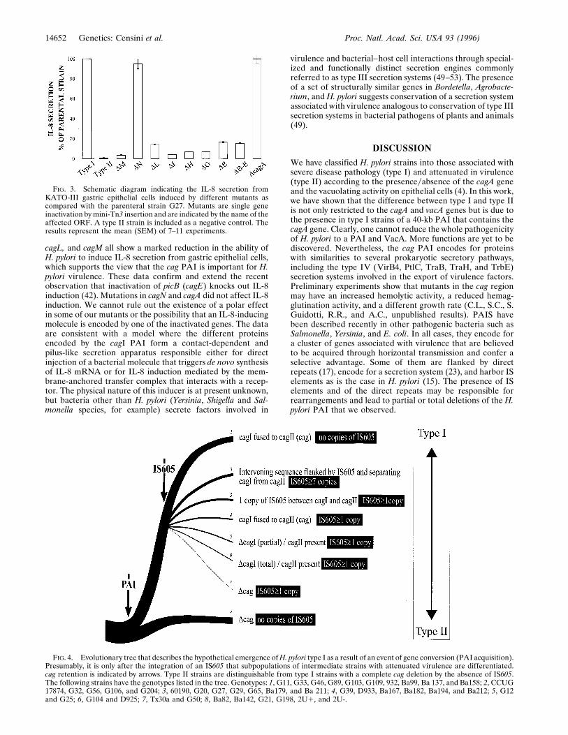
cagL, and cagM all show a marked reduction in the ability ofH. pylori to induce IL-8 secretion from gastric epithelial cells,which supports the view that the cag PAI is important for H.pylori virulence. These data confirm and extend the recentobservation that inactivation of picB (cagE) knocks out IL-8induction (42). Mutations in cagN and cagA did not affect IL-8induction. We cannot rule out the existence of a polar effectin some of our mutants or the possibility that an IL-8-inducingmolecule is encoded by one of the inactivated genes. The dataare consistent with a model where the different proteinsencoded by the cagI PAI form a contact-dependent andpilus-like secretion apparatus responsible either for directinjection of a bacterial molecule that triggers de novo synthesisof IL-8 mRNA or for IL-8 induction mediated by the mem-brane-anchored transfer complex that interacts with a recep-tor. The physical nature of this inducer is at present unknown,but bacteria other than H. pylori (Yersinia, Shigella and Sal-monella species, for example) secrete factors involved in
virulence and bacterial–host cell interactions through special-ized and functionally distinct secretion engines commonlyreferred to as type III secretion systems (49–53). The presenceof a set of structurally similar genes in Bordetella, Agrobacte-rium, and H. pylori suggests conservation of a secretion systemassociated with virulence analogous to conservation of type IIIsecretion systems in bacterial pathogens of plants and animals(49).
DISCUSSION
We have classified H. pylori strains into those associated withsevere disease pathology (type I) and attenuated in virulence(type II) according to the presenceyabsence of the cagA geneand the vacuolating activity on epithelial cells (4). In this work,we have shown that the difference between type I and type IIis not only restricted to the cagA and vacA genes but is due tothe presence in type I strains of a 40-kb PAI that contains thecagA gene. Clearly, one cannot reduce the whole pathogenicityof H. pylori to a PAI and VacA. More functions are yet to bediscovered. Nevertheless, the cag PAI encodes for proteinswith similarities to several prokaryotic secretory pathways,including the type IV (VirB4, PtlC, TraB, TraH, and TrbE)secretion systems involved in the export of virulence factors.Preliminary experiments show that mutants in the cag regionmay have an increased hemolytic activity, a reduced hemag-glutination activity, and a different growth rate (C.L., S.C., S.Guidotti, R.R., and A.C., unpublished results). PAIS havebeen described recently in other pathogenic bacteria such asSalmonella, Yersinia, and E. coli. In all cases, they encode fora cluster of genes associated with virulence that are believedto be acquired through horizontal transmission and confer aselective advantage. Some of them are flanked by directrepeats (17), encode for a secretion system (23), and harbor ISelements as is the case in H. pylori (15). The presence of ISelements and of the direct repeats may be responsible forrearrangements and lead to partial or total deletions of the H.pylori PAI that we observed.
FIG. 3. Schematic diagram indicating the IL-8 secretion fromKATO-III gastric epithelial cells induced by different mutants ascompared with the parenteral strain G27. Mutants are single geneinactivation by mini-Tn3 insertion and are indicated by the name of theaffected ORF. A type II strain is included as a negative control. Theresults represent the mean (SEM) of 7–11 experiments.
FIG. 4. Evolutionary tree that describes the hypothetical emergence ofH. pylori type I as a result of an event of gene conversion (PAI acquisition).Presumably, it is only after the integration of an IS605 that subpopulations of intermediate strains with attenuated virulence are differentiated.cag retention is indicated by arrows. Type II strains are distinguishable from type I strains with a complete cag deletion by the absence of IS605.The following strains have the genotypes listed in the tree. Genotypes: 1, G11, G33, G46, G89, G103, G109, 932, Ba99, Ba 137, and Ba158; 2, CCUG17874, G32, G56, G106, and G204; 3, 60190, G20, G27, G29, G65, Ba179, and Ba 211; 4, G39, D933, Ba167, Ba182, Ba194, and Ba212; 5, G12and G25; 6, G104 and D925; 7, Tx30a and G50; 8, Ba82, Ba142, G21, G198, 2U1, and 2U-.
14652 Genetics: Censini et al. Proc. Natl. Acad. Sci. USA 93 (1996)
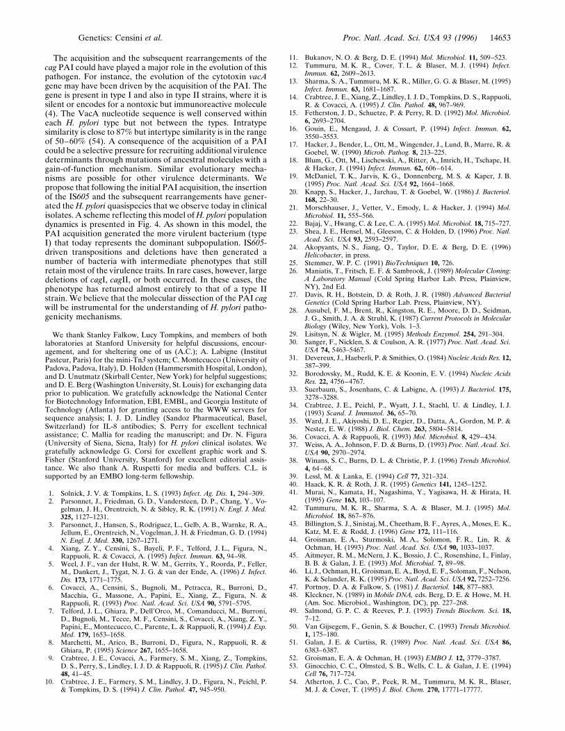
The acquisition and the subsequent rearrangements of thecag PAI could have played a major role in the evolution of thispathogen. For instance, the evolution of the cytotoxin vacAgene may have been driven by the acquisition of the PAI. Thegene is present in type I and also in type II strains, where it issilent or encodes for a nontoxic but immunoreactive molecule(4). The VacA nucleotide sequence is well conserved withineach H. pylori type but not between the types. Intratypesimilarity is close to 87% but intertype similarity is in the rangeof 50–60% (54). A consequence of the acquisition of a PAIcould be a selective pressure for recruiting additional virulencedeterminants through mutations of ancestral molecules with again-of-function mechanism. Similar evolutionary mecha-nisms are possible for other virulence determinants. Wepropose that following the initial PAI acquisition, the insertionof the IS605 and the subsequent rearrangements have gener-ated theH. pylori quasispecies that we observe today in clinicalisolates. A scheme reflecting this model ofH. pylori populationdynamics is presented in Fig. 4. As shown in this model, thePAI acquisition generated the more virulent bacterium (typeI) that today represents the dominant subpopulation. IS605-driven transpositions and deletions have then generated anumber of bacteria with intermediate phenotypes that stillretain most of the virulence traits. In rare cases, however, largedeletions of cagI, cagII, or both occurred. In these cases, thephenotype has returned almost entirely to that of a type IIstrain. We believe that the molecular dissection of the PAI cagwill be instrumental for the understanding of H. pylori patho-genicity mechanisms.
We thank Stanley Falkow, Lucy Tompkins, and members of bothlaboratories at Stanford University for helpful discussions, encour-agement, and for sheltering one of us (A.C.); A. Labigne (InstitutPasteur, Paris) for the mini-Tn3 system; C. Montecucco (University ofPadova, Padova, Italy), D. Holden (Hammersmith Hospital, London),and D. Unutmatz (Skirball Center, New York) for helpful suggestions;and D. E. Berg (Washington University, St. Louis) for exchanging dataprior to publication. We gratefully acknowledge the National Centerfor Biotechnology Information, EBI, EMBL, and Georgia Institute ofTechnology (Atlanta) for granting access to the WWW servers forsequence analysis; I. J. D. Lindley (Sandoz Pharmaceutical, Basel,Switzerland) for IL-8 antibodies; S. Perry for excellent technicalassistance; C. Mallia for reading the manuscript; and Dr. N. Figura(University of Siena, Siena, Italy) for H. pylori clinical isolates. Wegratefully acknowledge G. Corsi for excellent graphic work and S.Fisher (Stanford University, Stanford) for excellent editorial assis-tance. We also thank A. Ruspetti for media and buffers. C.L. issupported by an EMBO long-term fellowship.
1. Solnick, J. V. & Tompkins, L. S. (1993) Infect. Ag. Dis. 1, 294–309.2. Parsonnet, J., Friedman, G. D., Vandersteen, D. P., Chang, Y., Vo-
gelman, J. H., Orentreich, N. & Sibley, R. K. (1991) N. Engl. J. Med.325, 1127–1231.
3. Parsonnet, J., Hansen, S., Rodriguez, L., Gelb, A. B., Warnke, R. A.,Jellum, E., Orentreich, N., Vogelman, J. H. & Friedman, G. D. (1994)N. Engl. J. Med. 330, 1267–1271.
4. Xiang, Z. Y., Censini, S., Bayeli, P. F., Telford, J. L., Figura, N.,Rappuoli, R. & Covacci, A. (1995) Infect. Immun. 63, 94–98.
5. Weel, J. F., van der Hulst, R. W. M., Gerrits, Y., Roorda, P., Feller,M., Dankert, J., Tygat, N. J. G. & van der Ende, A. (1996) J. Infect.Dis. 173, 1771–1775.
6. Covacci, A., Censini, S., Bugnoli, M., Petracca, R., Burroni, D.,Macchia, G., Massone, A., Papini, E., Xiang, Z., Figura, N. &Rappuoli, R. (1993) Proc. Natl. Acad. Sci. USA 90, 5791–5795.
7. Telford, J. L., Ghiara, P., Dell’Orco, M., Comanducci, M., Burroni,D., Bugnoli, M., Tecce, M. F., Censini, S., Covacci, A., Xiang, Z. Y.,Papini, E., Montecucco, C., Parente, L. & Rappuoli, R. (1994) J. Exp.Med. 179, 1653–1658.
8. Marchetti, M., Arico, B., Burroni, D., Figura, N., Rappuoli, R. &Ghiara, P. (1995) Science 267, 1655–1658.
9. Crabtree, J. E., Covacci, A., Farmery, S. M., Xiang, Z., Tompkins,D. S., Perry, S., Lindley, I. J. D. & Rappuoli, R. (1995) J. Clin. Pathol.48, 41–45.
10. Crabtree, J. E., Farmery, S. M., Lindley, J. D., Figura, N., Peichl, P.& Tompkins, D. S. (1994) J. Clin. Pathol. 47, 945–950.
11. Bukanov, N. O. & Berg, D. E. (1994) Mol. Microbiol. 11, 509–523.12. Tummuru, M. K. R., Cover, T. L. & Blaser, M. J. (1994) Infect.
Immun. 62, 2609–2613.13. Sharma, S. A., Tummuru, M. K. R., Miller, G. G. & Blaser, M. (1995)
Infect. Immun. 63, 1681–1687.14. Crabtree, J. E., Xiang, Z., Lindley, I. J. D., Tompkins, D. S., Rappuoli,
R. & Covacci, A. (1995) J. Clin. Pathol. 48, 967–969.15. Fetherston, J. D., Schuetze, P. & Perry, R. D. (1992) Mol. Microbiol.
6, 2693–2704.16. Gouin, E., Mengaud, J. & Cossart, P. (1994) Infect. Immun. 62,
3550–3553.17. Hacker, J., Bender, L., Ott, M., Wingender, J., Lund, B., Marre, R. &
Goebel, W. (1990) Microb. Pathog. 8, 213–225.18. Blum, G., Ott, M., Lischewski, A., Ritter, A., Imrich, H., Tschape, H.
& Hacker, J. (1994) Infect. Immun. 62, 606–614.19. McDaniel, T. K., Jarvis, K. G., Donnenberg, M. S. & Kaper, J. B.
(1995) Proc. Natl. Acad. Sci. USA 92, 1664–1668.20. Knapp, S., Hacker, J., Jarchau, T. & Goebel, W. (1986) J. Bacteriol.
168, 22–30.21. Morschhauser, J., Vetter, V., Emody, L. & Hacker, J. (1994) Mol.
Microbiol. 11, 555–566.22. Bajaj, V., Hwang, C. & Lee, C. A. (1995)Mol. Microbiol. 18, 715–727.23. Shea, J. E., Hensel, M., Gleeson, C. & Holden, D. (1996) Proc. Natl.
Acad. Sci. USA 93, 2593–2597.24. Akopyants, N. S., Jiang, Q., Taylor, D. E. & Berg, D. E. (1996)
Helicobacter, in press.25. Stemmer, W. P. C. (1991) BioTechniques 10, 726.26. Maniatis, T., Fritsch, E. F. & Sambrook, J. (1989) Molecular Cloning:
A Laboratory Manual (Cold Spring Harbor Lab. Press, Plainview,NY), 2nd Ed.
27. Davis, R. H., Botstein, D. & Roth, J. R. (1980) Advanced BacterialGenetics (Cold Spring Harbor Lab. Press, Plainview, NY).
28. Ausubel, F. M., Brent, R., Kingston, R. E., Moore, D. D., Seidman,J. G., Smith, J. A. & Struhl, K. (1987) Current Protocols in MolecularBiology (Wiley, New York), Vols. 1–3.
29. Lisitsyn, N. & Wigler, M. (1995) Methods Enzymol. 254, 291–304.30. Sanger, F., Nicklen, S. & Coulson, A. R. (1977) Proc. Natl. Acad. Sci.
USA 74, 5463–5467.31. Devereux, J., Haeberli, P. & Smithies, O. (1984)Nucleic Acids Res. 12,
387–399.32. Borodovsky, M., Rudd, K. E. & Koonin, E. V. (1994) Nucleic Acids
Res. 22, 4756–4767.33. Suerbaum, S., Josenhans, C. & Labigne, A. (1993) J. Bacteriol. 175,
3278–3288.34. Crabtree, J. E., Peichl, P., Wyatt, J. I., Stachl, U. & Lindley, I. J.
(1993) Scand. J. Immunol. 36, 65–70.35. Ward, J. E., Akiyoshi, D. E., Regier, D., Datta, A., Gordon, M. P. &
Nester, E. W. (1988) J. Biol. Chem. 263, 5804–5814.36. Covacci, A. & Rappuoli, R. (1993) Mol. Microbiol. 8, 429–434.37. Weiss, A. A., Johnson, F. D. & Burns, D. (1993) Proc. Natl. Acad. Sci.
USA 90, 2970–2974.38. Winans, S. C., Burns, D. L. & Christie, P. J. (1996) Trends Microbiol.
4, 64–68.39. Lessl, M. & Lanka, E. (1994) Cell 77, 321–324.40. Haack, K. R. & Roth, J. R. (1995) Genetics 141, 1245–1252.41. Murai, N., Kamata, H., Nagashima, Y., Yagisawa, H. & Hirata, H.
(1995) Gene 163, 103–107.42. Tummuru, M. K. R., Sharma, S. A. & Blaser, M. J. (1995) Mol.
Microbiol. 18, 867–876.43. Billington, S. J., Sinistaj, M., Cheetham, B. F., Ayres, A., Moses, E. K.,
Katz, M. E. & Rodd, J. (1996) Gene 172, 111–116.44. Groisman, E. A., Sturmoski, M. A., Solomon, F. R., Lin, R. &
Ochman, H. (1993) Proc. Natl. Acad. Sci. USA 90, 1033–1037.45. Aitmeyer, R. M., McNern, J. K., Bossio, J. C., Rosenshine, I., Finlay,
B. B. & Galan, J. E. (1993) Mol. Microbial. 7, 89–98.46. Li, J., Ochman, H., Groisman, E. A., Boyd, E. F., Soloman, F., Nelson,
K. & Selander, R. K. (1995) Proc. Natl. Acad. Sci. USA 92, 7252–7256.47. Portnoy, D. A. & Falkow, S. (1981) J. Bacteriol. 148, 877–883.48. Kleckner, N. (1989) in Mobile DNA, eds. Berg, D. E. & Howe, M. H.
(Am. Soc. Microbiol., Washington, DC), pp. 227–268.49. Salmond, G. P. C. & Reeves, P. J. (1993) Trends Biochem. Sci. 18,
7–12.50. Van Gijsegem, F., Genin, S. & Boucher, C. (1993) Trends Microbiol.
1, 175–180.51. Galan, J. E. & Curtiss, R. (1989) Proc. Natl. Acad. Sci. USA 86,
6383–6387.52. Groisman, E. A. & Ochman, H. (1993) EMBO J. 12, 3779–3787.53. Ginocchio, C. C., Olmsted, S. B., Wells, C. L. & Galan, J. E. (1994)
Cell 76, 717–724.54. Atherton, J. C., Cao, P., Peek, R. M., Tummuru, M. K. R., Blaser,
M. J. & Cover, T. (1995) J. Biol. Chem. 270, 17771–17777.
Genetics: Censini et al. Proc. Natl. Acad. Sci. USA 93 (1996) 14653
Copyright © 2022 FDOKUMEN