list of probable entries identified to be deleted from electoral roll
Am. J. Hum. Genet. 73:261–270, 2003
261
BRD2 (RING3) Is a Probable Major Susceptibility Gene for CommonJuvenile Myoclonic EpilepsyDeb K. Pal,1,3,* Oleg V. Evgrafov,2,* Paula Tabares,2 Fengli Zhang,2 Martina Durner,1and David A. Greenberg1,2,3,4
1Division of Statistical Genetics, Department of Biostatistics, Mailman School of Public Health, 2Department of Psychiatry, and 3ColumbiaGenome Center, Columbia University, and 4Clinical and Genetic Epidemiology Unit, New York State Psychiatric Institute, New York
Juvenile myoclonic epilepsy (JME) is a common form of generalized epilepsy that starts in adolescence. A majorJME susceptibility locus (EJM1) was mapped to chromosomal region 6p21 in three independent linkage studies,and association was reported between JME and a microsatellite marker in the 6p21 region. The critical region forEJM1 is delimited by obligate recombinants at HLA-DQ and HLA-DP. In the present study, we found highlysignificant linkage disequilibrium (LD) between JME and a core haplotype of five single-nucleotide–polymorphism(SNP) and microsatellite markers in this critical region, with LD peaking in the BRD2 (RING3) gene (odds ratio6.45; 95% confidence interval 2.36–17.58). DNA sequencing revealed two JME-associated SNP variants in theBRD2 (RING3) promoter region but no other potentially causative coding mutations in 20 probands from familieswith positive LOD scores. BRD2 (RING3) is a putative nuclear transcriptional regulator from a family of genesthat are expressed during development. Our findings strongly suggest that BRD2 (RING3) is EJM1, the first geneidentified for a common idiopathic epilepsy. These findings also suggest that abnormalities of neural developmentmay be a cause of common idiopathic epilepsy, and the findings have implications for the generalizability of proposedpathogenetic mechanisms, derived from diseases that show Mendelian transmission, to their complex counterparts.
Introduction
The epilepsies comprise an enormous diversity of dis-orders of heterogeneous etiology, manifestation, andprognosis. Almost half of all epilepsies have some geneticbasis (Annegers et al. 1996), but only a small proportionappear to display Mendelian inheritance. Many of ourcurrent beliefs about the molecular and cellular mecha-nisms in epilepsy derive from these examples, which arebased on rare, large pedigrees. Such pedigrees received themost attention because they have been the simplest tostudy genetically. Although mutations in genes for ionchannels, neuroreceptors, and neurotransmitters havebeen demonstrated in such rare, densely affected epilepsypedigrees (Mulley et al. 2003), the forms of epilepsy com-monly seen in the clinic show neither the specific muta-tions nor any other mutations in genes identified in thosepedigrees (Harkin et al. 2002; Kananura et al. 2002; au-thors’ unpublished data). In contrast to the rare Men-delian pedigrees, the common forms of idiopathic gen-
Received February 26, 2003; accepted for publication May 7, 2003;electronically published June 25, 2003.
Address for correspondence and reprints: Dr. Deb K. Pal, Divisionof Statistical Genetics, Mailman School of Public Health, ColumbiaUniversity, 722 W. 168th Street, New York, New York 10032. E-mail:[email protected]
* The first two authors contributed equally to this work.� 2003 by The American Society of Human Genetics. All rights reserved.
0002-9297/2003/7302-0005$15.00
eralized epilepsy (IGE) have a complex inheritance, eventhough the IGEs are thought to have an exclusively geneticbasis (Greenberg et al. 1992). Not only do populationstudies (Tsuboi and Christian 1973; Beck-Mannagettaand Janz 1991) suggest an oligogenic mode of inheritancewith interaction between loci, but a genome scan of in-dividuals with adolescent-onset IGE demonstrated strongevidence of linkage to several loci, combinations of whichmay lead to specific epilepsy syndromes (Durner et al.2001). It seems unlikely that single gene mutations aresufficient to explain the molecular mechanisms for epi-lepsies with this model of complex inheritance.
Juvenile myoclonic epilepsy (JME [MIM 254770]) isone of the most easily recognized IGEs of adolescence,diagnosable by the occurrence of bilateral, upper-limbmyoclonic jerks on awakening (Janz and Christian 1957).Studies of three separate family collections have reportedsignificant evidence of linkage between JME and themajor susceptibility locus EJM1 at chromosome 6p21,designated “EJM1” (Greenberg et al. 1988b; Durner etal. 1991; Weissbecker et al. 1991; Sander et al. 1997;Greenberg et al. 2000). In addition to linkage, there isevidence of allelic association in this region, with a mi-crosatellite allele located in the HLA class II region. Thismicrosatellite is located in an intron of the BRD2(RING3) gene (Greenberg et al. 2000). Recombinationmapping in families with JME has delimited the bound-aries of EJM1 to a 1-cM region between the HLA-DQ
262 Am. J. Hum. Genet. 73:261–270, 2003
and HLA-DP (DQ-DP) loci (Sander et al. 1997; Green-berg et al. 2000). In the present study, we aimed toconfirm and further refine gene localization of EJM1and to search for molecular variants that might explainJME susceptibility in a complex genetic model.
Families and Methods
Study Design
Our earlier studies suggested that EJM1 was locatedbetween DQ and DP (Sander et al. 1997; Greenberg etal. 2000), so we first sought evidence of association be-tween JME and single SNP marker alleles in this region.Then, to increase informativeness of markers, we recon-structed two-locus haplotypes from data on consecutiveSNPs. We performed case-control analysis, using thesehaplotypes, and, to guard against possible populationstratification, we confirmed positive haplotype associa-tions, using intrafamilial controls (untransmitted alleles)in haplotype relative-risk analysis (Falk and Rubenstein1987). Next, we searched for a common risk haplotypein families with positive LOD scores in the EJM1 region.Finally, we searched for mutations by sequencing exonsand promoter sequences in BRD2 and adjacent genesthat showed significant linkage disequilibrium (LD) withJME.
Families
We collected probands with JME and their familiesfrom physicians’ practices, as described elsewhere (Green-berg et al. 2000). We selected probands with typical formsof JME, in accordance with international classificationguidelines (Commission on Classification and Terminol-ogy of the International League Against Epilepsy 1989).Twenty parent-offspring trios from families with positiveLOD scores (10.1) at both of two EJM1 microsatel-lite markers, DQB1 and DRB1, were designated as the“EJM1� set.” LOD scores for EJM1� families were0.15–1.50, with a mean of 0.38. Alleles and haplotypesof probands in the EJM1� set were used as case data inthe case-control analysis to find associations. Transmit-ted and untransmitted alleles and haplotypes in theEJM1� set were also used in transmission/disequilibriumtesting. Institutional review board approval for thisstudy was obtained from the appropriate institutions.All participating patients and family members gave theirinformed consent.
Controls
We used three control groups: The first consisted of53 JME parent-offspring trios with negative LOD scoresat EJM1 markers (EJM1�), collected at the same timeand from the same population as EJM1� families. The
second comprised laboratory controls, which consistedof 64 carriers of Wilson disease or spinal muscular at-rophy from the Columbia Genome Center. We used theepilepsy control group because it represented a popu-lation similar to that from which the EJM1� case setwas drawn, thus safeguarding against the possibility ofselection bias in the case-control analysis. One theoret-ical disadvantage of using the epilepsy control group isthe possibility of over-matching (i.e., an association can-not be demonstrated because case and control groupswith JME might share too many allelic similarities atadjacent markers). We therefore used the laboratory con-trols as a second control group to check for over-match-ing in case-control analysis. Finding significant LD usingeach of the two control groups would therefore dem-onstrate that associated alleles or haplotypes are neitherspecific to populations with epilepsy nor a result of se-lection bias. Third, we used untransmitted alleles in theEJM1� set as internal controls for EJM1� transmittedalleles in haplotype relative-risk analysis.
DNA Preparation
DNA was purified from blood or lymphoblastoid celllines, using the Puregene kit (Gentra Systems) accordingto the manufacturer’s protocols. PCR amplification forall fragments was performed on either an MJ ResearchTetrad thermal cycler or a Hybaid MultiBlock Systemunder the following conditions: 32 cycles at 94�C for 2min, with denaturation at 94�C for 30 s, annealing at56�C for 30 s, and an extension at 72�C for 45 s, fol-lowed by a final extension at 72�C for 4 min.
SNP Discovery
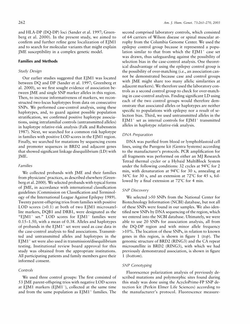
We selected 150 SNPs from the National Center forBiotechnology Information (NCBI) database, but not allof these SNPs were found in our samples. We also iden-tified new SNPs by DNA sequencing of the region, whichwe entered into the NCBI database. Ultimately, we wereable to use 20 SNPs for association analysis, all fromthe DQ-DP region and with minor allele frequency110%. The location of these SNPs, in relation to knowngenes in this region, is shown in figure 1 (top). Thegenomic structure of BRD2 (RING3) and the CA repeatmicrosatellite in BRD2 (RING3), with which we hadpreviously demonstrated association, is shown in figure1 (bottom).
SNP Genotyping
Fluorescence polarization analysis of previously de-scribed mutations and polymorphic sites found duringthis study was done using the AcycloPrime-FP SNP de-tection kit (Perkin Elmer Life Sciences) according tothe manufacturer’s protocol. Fluorescence measure-
Pal et al.: BRD2 and JME 263
Figure 1 DQ-DP region on human chromosome 6, SNP markers, and genomic structure of BRD2 (RING3). Top, arrows along thechromosome denote genes, which are labeled, and their direction of transcription; asterisks denote SNP markers. Bottom, BRD2 (RING3) geneexploded to show the relation of the exonic sequences with SNPs, the CA repeat microsatellite, and other DNA variations.
ments were performed on fluorescence microplate an-alyzer (LJL BioSystems).
Sequencing and Mutation Analysis
Sequences of coding and promoter regions of BRD2(RING3), PPP1R2P1, DMA, DMB, and DNA genes weredetermined on the ABI 310 automated sequencer usingthe ABI Taq2 sequencing kit according to the manufac-turer’s protocol. Presequencing PCR clean-up was done
by enzymatic degradation of primers and dNTPs, usingexonuclease I and shrimp alkaline phosphatase cocktail.
Statistical Analysis
We first performed unmatched case-control analyseson single SNPs, generating odds ratios (ORs) with95% CIs. To increase informativeness of the markers,we then generated consecutive two-locus haplotypes,reconstructing haplotypes from genotypic data, using
264 Am. J. Hum. Genet. 73:261–270, 2003
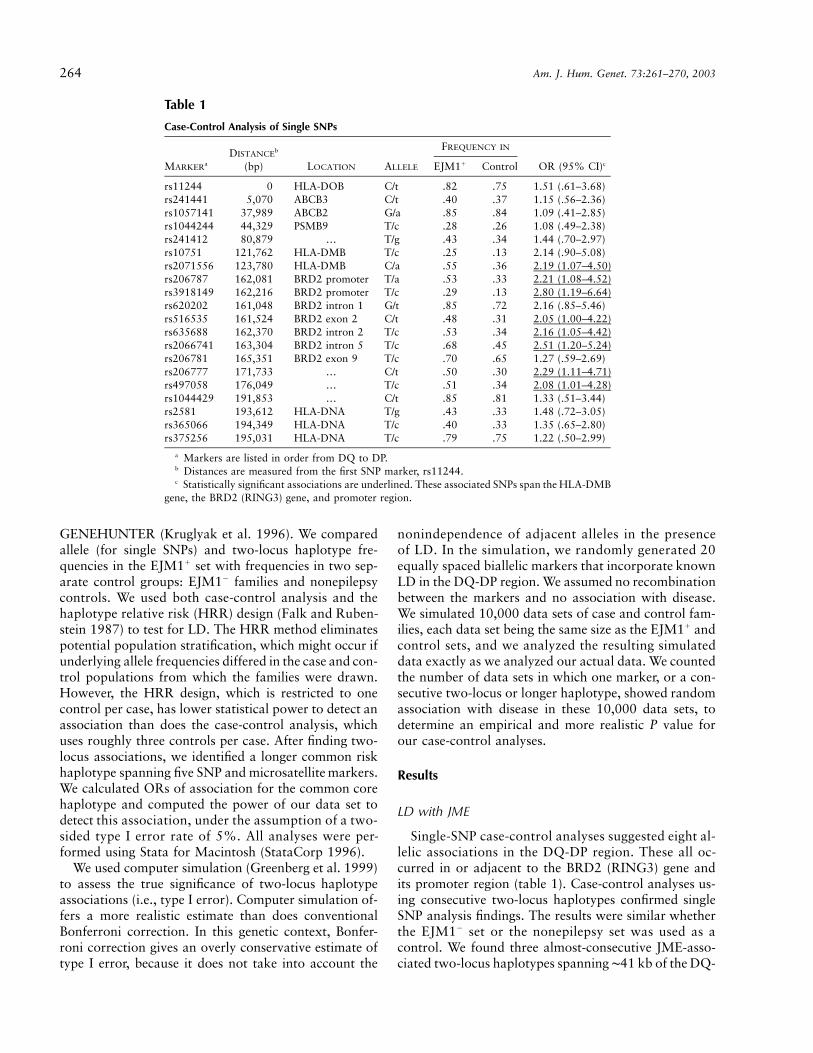
Table 1
Case-Control Analysis of Single SNPs
MARKERa
DISTANCEb
(bp) LOCATION ALLELE
FREQUENCY IN
OR (95% CI)cEJM1� Control
rs11244 0 HLA-DOB C/t .82 .75 1.51 (.61–3.68)rs241441 5,070 ABCB3 C/t .40 .37 1.15 (.56–2.36)rs1057141 37,989 ABCB2 G/a .85 .84 1.09 (.41–2.85)rs1044244 44,329 PSMB9 T/c .28 .26 1.08 (.49–2.38)rs241412 80,879 … T/g .43 .34 1.44 (.70–2.97)rs10751 121,762 HLA-DMB T/c .25 .13 2.14 (.90–5.08)rs2071556 123,780 HLA-DMB C/a .55 .36 2.19 (1.07–4.50)rs206787 162,081 BRD2 promoter T/a .53 .33 2.21 (1.08–4.52)rs3918149 162,216 BRD2 promoter T/c .29 .13 2.80 (1.19–6.64)rs620202 161,048 BRD2 intron 1 G/t .85 .72 2.16 (.85–5.46)rs516535 161,524 BRD2 exon 2 C/t .48 .31 2.05 (1.00–4.22)rs635688 162,370 BRD2 intron 2 T/c .53 .34 2.16 (1.05–4.42)rs2066741 163,304 BRD2 intron 5 T/c .68 .45 2.51 (1.20–5.24)rs206781 165,351 BRD2 exon 9 T/c .70 .65 1.27 (.59–2.69)rs206777 171,733 … C/t .50 .30 2.29 (1.11–4.71)rs497058 176,049 … T/c .51 .34 2.08 (1.01–4.28)rs1044429 191,853 … C/t .85 .81 1.33 (.51–3.44)rs2581 193,612 HLA-DNA T/g .43 .33 1.48 (.72–3.05)rs365066 194,349 HLA-DNA T/c .40 .33 1.35 (.65–2.80)rs375256 195,031 HLA-DNA T/c .79 .75 1.22 (.50–2.99)
a Markers are listed in order from DQ to DP.b Distances are measured from the first SNP marker, rs11244.c Statistically significant associations are underlined. These associated SNPs span the HLA-DMB
gene, the BRD2 (RING3) gene, and promoter region.
GENEHUNTER (Kruglyak et al. 1996). We comparedallele (for single SNPs) and two-locus haplotype fre-quencies in the EJM1� set with frequencies in two sep-arate control groups: EJM1� families and nonepilepsycontrols. We used both case-control analysis and thehaplotype relative risk (HRR) design (Falk and Ruben-stein 1987) to test for LD. The HRR method eliminatespotential population stratification, which might occur ifunderlying allele frequencies differed in the case and con-trol populations from which the families were drawn.However, the HRR design, which is restricted to onecontrol per case, has lower statistical power to detect anassociation than does the case-control analysis, whichuses roughly three controls per case. After finding two-locus associations, we identified a longer common riskhaplotype spanning five SNP and microsatellite markers.We calculated ORs of association for the common corehaplotype and computed the power of our data set todetect this association, under the assumption of a two-sided type I error rate of 5%. All analyses were per-formed using Stata for Macintosh (StataCorp 1996).
We used computer simulation (Greenberg et al. 1999)to assess the true significance of two-locus haplotypeassociations (i.e., type I error). Computer simulation of-fers a more realistic estimate than does conventionalBonferroni correction. In this genetic context, Bonfer-roni correction gives an overly conservative estimate oftype I error, because it does not take into account the
nonindependence of adjacent alleles in the presenceof LD. In the simulation, we randomly generated 20equally spaced biallelic markers that incorporate knownLD in the DQ-DP region. We assumed no recombinationbetween the markers and no association with disease.We simulated 10,000 data sets of case and control fam-ilies, each data set being the same size as the EJM1� andcontrol sets, and we analyzed the resulting simulateddata exactly as we analyzed our actual data. We countedthe number of data sets in which one marker, or a con-secutive two-locus or longer haplotype, showed randomassociation with disease in these 10,000 data sets, todetermine an empirical and more realistic P value forour case-control analyses.
Results
LD with JME
Single-SNP case-control analyses suggested eight al-lelic associations in the DQ-DP region. These all oc-curred in or adjacent to the BRD2 (RING3) gene andits promoter region (table 1). Case-control analyses us-ing consecutive two-locus haplotypes confirmed singleSNP analysis findings. The results were similar whetherthe EJM1� set or the nonepilepsy set was used as acontrol. We found three almost-consecutive JME-asso-ciated two-locus haplotypes spanning ∼41 kb of the DQ-
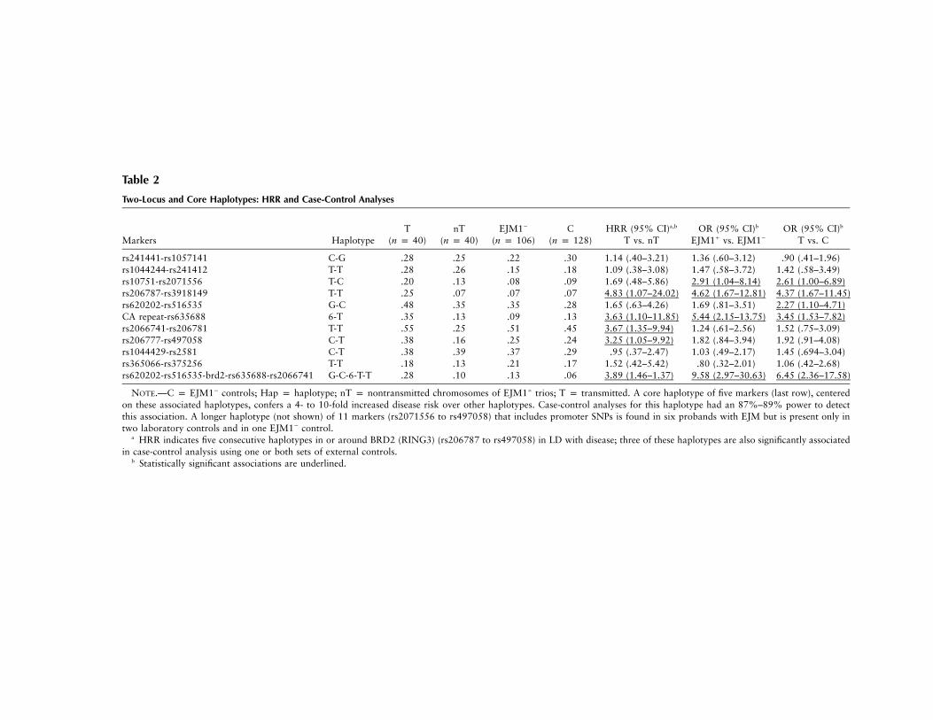
Table 2
Two-Locus and Core Haplotypes: HRR and Case-Control Analyses
Markers HaplotypeT
(n p 40)nT
(n p 40)EJM1�
(n p 106)C
(n p 128)HRR (95% CI)a,b
T vs. nTOR (95% CI)b
EJM1� vs. EJM1�
OR (95% CI)b
T vs. C
rs241441-rs1057141 C-G .28 .25 .22 .30 1.14 (.40–3.21) 1.36 (.60–3.12) .90 (.41–1.96)rs1044244-rs241412 T-T .28 .26 .15 .18 1.09 (.38–3.08) 1.47 (.58–3.72) 1.42 (.58–3.49)rs10751-rs2071556 T-C .20 .13 .08 .09 1.69 (.48–5.86) 2.91 (1.04–8.14) 2.61 (1.00–6.89)rs206787-rs3918149 T-T .25 .07 .07 .07 4.83 (1.07–24.02) 4.62 (1.67–12.81) 4.37 (1.67–11.45)rs620202-rs516535 G-C .48 .35 .35 .28 1.65 (.63–4.26) 1.69 (.81–3.51) 2.27 (1.10–4.71)CA repeat-rs635688 6-T .35 .13 .09 .13 3.63 (1.10–11.85) 5.44 (2.15–13.75) 3.45 (1.53–7.82)rs2066741-rs206781 T-T .55 .25 .51 .45 3.67 (1.35–9.94) 1.24 (.61–2.56) 1.52 (.75–3.09)rs206777-rs497058 C-T .38 .16 .25 .24 3.25 (1.05–9.92) 1.82 (.84–3.94) 1.92 (.91–4.08)rs1044429-rs2581 C-T .38 .39 .37 .29 .95 (.37–2.47) 1.03 (.49–2.17) 1.45 (.694–3.04)rs365066-rs375256 T-T .18 .13 .21 .17 1.52 (.42–5.42) .80 (.32–2.01) 1.06 (.42–2.68)rs620202-rs516535-brd2-rs635688-rs2066741 G-C-6-T-T .28 .10 .13 .06 3.89 (1.46–1.37) 9.58 (2.97–30.63) 6.45 (2.36–17.58)
NOTE.—C p EJM1� controls; Hap p haplotype; nT p nontransmitted chromosomes of EJM1� trios; T p transmitted. A core haplotype of five markers (last row), centeredon these associated haplotypes, confers a 4- to 10-fold increased disease risk over other haplotypes. Case-control analyses for this haplotype had an 87%–89% power to detectthis association. A longer haplotype (not shown) of 11 markers (rs2071556 to rs497058) that includes promoter SNPs is found in six probands with EJM but is present only intwo laboratory controls and in one EJM1� control.
a HRR indicates five consecutive haplotypes in or around BRD2 (RING3) (rs206787 to rs497058) in LD with disease; three of these haplotypes are also significantly associatedin case-control analysis using one or both sets of external controls.
b Statistically significant associations are underlined.
266 Am. J. Hum. Genet. 73:261–270, 2003
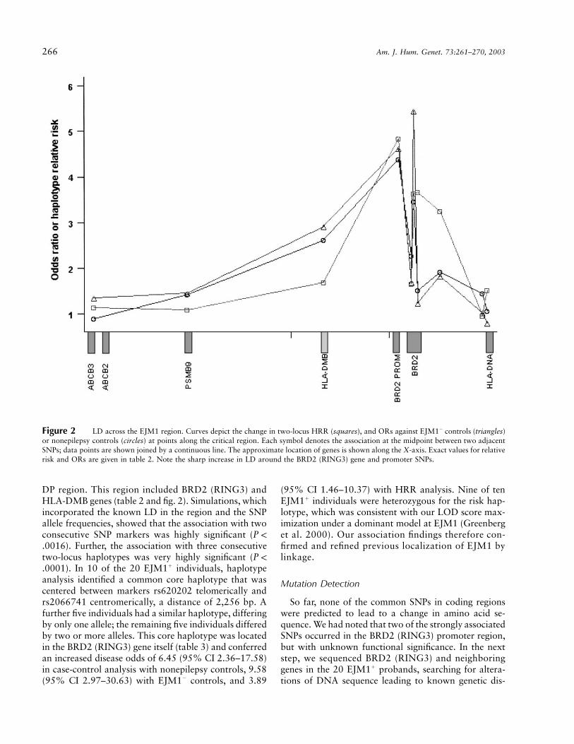
Figure 2 LD across the EJM1 region. Curves depict the change in two-locus HRR (squares), and ORs against EJM1� controls (triangles)or nonepilepsy controls (circles) at points along the critical region. Each symbol denotes the association at the midpoint between two adjacentSNPs; data points are shown joined by a continuous line. The approximate location of genes is shown along the X-axis. Exact values for relativerisk and ORs are given in table 2. Note the sharp increase in LD around the BRD2 (RING3) gene and promoter SNPs.
DP region. This region included BRD2 (RING3) andHLA-DMB genes (table 2 and fig. 2). Simulations, whichincorporated the known LD in the region and the SNPallele frequencies, showed that the association with twoconsecutive SNP markers was highly significant (P !
). Further, the association with three consecutive.0016two-locus haplotypes was very highly significant (P !
). In 10 of the 20 EJM1� individuals, haplotype.0001analysis identified a common core haplotype that wascentered between markers rs620202 telomerically andrs2066741 centromerically, a distance of 2,256 bp. Afurther five individuals had a similar haplotype, differingby only one allele; the remaining five individuals differedby two or more alleles. This core haplotype was locatedin the BRD2 (RING3) gene itself (table 3) and conferredan increased disease odds of 6.45 (95% CI 2.36–17.58)in case-control analysis with nonepilepsy controls, 9.58(95% CI 2.97–30.63) with EJM1� controls, and 3.89
(95% CI 1.46–10.37) with HRR analysis. Nine of tenEJM1� individuals were heterozygous for the risk hap-lotype, which was consistent with our LOD score max-imization under a dominant model at EJM1 (Greenberget al. 2000). Our association findings therefore con-firmed and refined previous localization of EJM1 bylinkage.
Mutation Detection
So far, none of the common SNPs in coding regionswere predicted to lead to a change in amino acid se-quence. We had noted that two of the strongly associatedSNPs occurred in the BRD2 (RING3) promoter region,but with unknown functional significance. In the nextstep, we sequenced BRD2 (RING3) and neighboringgenes in the 20 EJM1� probands, searching for altera-tions of DNA sequence leading to known genetic dis-
Pal et al.: BRD2 and JME 267
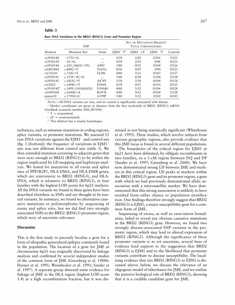
Table 3
Rare DNA Variations in the BRD2 (RING3) Gene and Promoter Region
MARKER
SNPNO. OF MUTATIONS PRESENT/
TOTAL CHROMOSOMES
Mutation Sitea Name EJM1� Tb EJM1� nTc EJM1� Tb Controls
rs3918148 �175CrG … 2/39 1/29 2/101 3/121rs3918150 �2CrG … 0/39 2/32 0/96 0/125rs3918144 c.145_146GCrTG A49C 1/40 0/32 2/104 2/126rs1803864 c.489CrT V163A 0/36 0/29 0/90 0/123rs176250 c.712CrT F238L 0/40 1/33 0/103 2/127rs3918141 c.1339�4CrG … 1/40 6/38 3/104 2/128rs3918143 c.1421CrT A474V 1/38 1/30 4/104 0/118rs12822 c.1498CrT E500X 0/39 0/31 0/103 0/125rs3918142d c.1499_1501delAGG E500del 4/40 1/32 0/104 0/128rs1049369 c.1640GrA R547K 0/40 0/32 0/104 1/128mutex10 c.1795GrC A599P 1/40 1/32 3/102 0/103
NOTE.—All DNA variants are rare, and no variant is significantly associated with disease.a Marker coordinates are given as distance from the first nucleotide of BRD2 (RING3) mRNA
(GenBank accession number NM_005104).b T p transmitted.c nT p nontransmitted.d This deletion has a murine homologue.
turbances, such as missense mutations in coding regions,splice variants, or promoter mutations. We assessed 11rare DNA variations against the EJM1� and control sets(fig. 1 [bottom]): the frequency of variations in EJM1�
sets was not different from control sets (table 3). Wethen extended mutation screening to adjacent genes thatwere near enough to BRD2 (RING3) to be within theregion implicated by LD mapping and haplotype anal-ysis. We tested for mutations in all exons and splicesites of PPP1R2P1, HLA-DMA, and HLA-DMB genes,which are centromeric to BRD2 (RING3), and HLA-DNA, which is telomeric to BRD2 (RING3), in fourfamilies with the highest LOD scores for 6p21 markers.All the DNA variants we found in these genes have beendescribed elsewhere as SNPs and are thought to be neu-tral variants. In summary, we found no alternative caus-ative mutations or polymorphisms by sequencing ofexons and splice sites, but we did find two stronglyassociated SNPs in the BRD2 (RING3) promoter region,which were of uncertain relevance.
Discussion
This is the first study to precisely localize a gene for aform of idiopathic generalized epilepsy commonly foundin the population. The location of a gene for JME atchromosome 6p21 was originally discovered by linkageanalysis and confirmed by several independent studiesof the common form of JME (Greenberg et al. 1988b;Durner et al. 1991; Weissbecker et al. 1991; Sander etal. 1997). A separate group detected some evidence forlinkage of JME in the HLA region (highest LOD score1.4) at a high recombination fraction, but it was dis-
missed as not being statistically significant (Whitehouseet al. 1993). These studies, which involve subjects fromvarious geographic regions, also provide evidence thatthis JME locus is found in several different populations.
The boundaries of the critical region for EJM1 at6p21 have been delimited, by obligate recombinants intwo families, to a 1-cM region between DQ and DP(Sander et al. 1997; Greenberg et al. 2000). We havenow demonstrated strong LD between JME and mark-ers in this critical region. LD peaks at markers withinthe BRD2 (RING3) gene and its promoter region, a genewith which we had previously demonstrated allelic as-sociation with a microsatellite marker. We have dem-onstrated that this strong association is unlikely to haveresulted from either chance or population stratifica-tion. Our findings therefore strongly suggest that BRD2(RING3) is EJM1, a major susceptibility gene for a com-mon form of JME.
Sequencing of exons, as well as exon-intron bound-aries, failed to reveal any obvious causative mutationsin the BRD2 (RING3) gene. However, we found twostrongly disease-associated SNP variants in the pro-moter region, which may lead to altered expression ofBRD2 (RING3). Although the significance of thesepromoter variants is as yet uncertain, several lines ofevidence lend support to the suggestion that BRD2(RING3) is EJM1 and to the likelihood that promotervariants contribute to disease susceptibility. The local-izing evidence that ties BRD2 (RING3) to EJM1 is dis-cussed above; below, we discuss the relevance of anoligogenic model of inheritance for JME, and we outlinethe putative biological role of BRD2 (RING3), showingthat it is a credible candidate gene for JME.
268 Am. J. Hum. Genet. 73:261–270, 2003
JME has an age-dependent, variable phenotype thatoverlaps with other common forms of adolescent-onsetIGEs. Specifically, JME is defined by the occurrence ofcharacteristic myoclonic jerks on awakening, but indi-viduals with JME may also have generalized tonic-clonicor absence seizure types. Our previous linkage findingsin the adolescent-onset IGEs (Durner et al. 2001) sup-ported the oligogenic model in which epistatic inter-actions between loci influenced the expression of theseindividual seizure types (Greenberg et al. 1988a). Strongevidence of linkage (LOD score 4.4 or 5.2 [multipointor two-point]) on chromosome 18 suggested that thislocus conferred susceptibility to all adolescent-onsetIGEs, possibly interacting with a modifying locus (EJM1)on chromosome 6 for myoclonic seizures and with locion chromosomes 5 and 8 for nonmyoclonic (generalizedtonic-clonic and absence) seizures (Durner et al. 2001).
Unlike the rare forms of IGE reported in denselyaffected pedigrees, the common form of JME is nota monogenic disorder in which single mutations cor-relate strongly with disease expression. It is apparentfrom the above oligogenic model that a single criticalmutation in BRD2 (RING3), sufficient by itself to causedisease manifestation, would preclude an interactive rolefor other loci and might not be compatible with the com-plex pattern of inheritance observed in typical familieswith JME. A hypothesis more consistent with the knownobservations is that a disturbance in the transcription ofBRD2 (RING3), which might not have severe conse-quences by itself, could lead to expression of seizures inconjunction with genetic variants at interacting loci. Con-tinuing from this genetic model, one of the interactinggenes is likely to be located within the major susceptibilitylocus that we have studied on chromosome 18 (Durneret al. 2001). To summarize, individual seizure types inIGE may result from the interaction of genetic variants.Separate loci are probably insufficient by themselves tolead to seizure expression, but each contributes to diseasesusceptibility. This model is consistent with the observedpattern of epilepsy and seizure distribution within fam-ilies of probands with IGE.
To date, assumptions about the function of genes in-volved in the pathogenesis of IGEs have largely beendrawn from the study of rare, densely affected Men-delian pedigrees (Mulley et al. 2003). Investigators havereported simple gene mutations associated with seriousdisruption of ion-channel and neuroreceptor function.So far, we have been unable to find mutations in eitherGABRG2 genes or KCNAB1 genes in probands withcommon forms of JME or IGE (Evgrafov et al. 2002;authors’ unpublished data). These negative findings arein agreement with the original reports of GABRG2 mu-tations in epilepsy: no additional mutations were foundin affected family members from 10 “GEFS-like” fam-ilies (Baulac et al. 2001), nor were GABRG2 mutations
found after screening 200 patients with IGE (Harkin etal. 2002) and 135 patients with idiopathic absence ep-ilepsy (Kananura et al. 2002). These findings in rarepedigrees reinforce our understanding of the role of ionchannels and neuroreceptors in normal signal trans-mission in the CNS. However, the known role of BRD2(RING3) and related genes suggests the involvement offar more complex and sophisticated mechanisms in thepathogenesis of common forms of IGE than those thatare suggested by reports of pedigrees with Mendeliantransmission of epilepsy.
BRD2 (RING3) belongs to a highly conserved sub-family of double bromodomain–containing proteins re-lated to the Drosophila female sterile homeotic (fsh)gene, which has an important function in developmentand appears to interact genetically with the trithoraxlocus (Gans et al. 1975, 1980; Digan et al. 1986). Thereare four members of the fsh subfamily in mice and hu-mans, all of which are characterized by the presence oftwo bromodomains and an extra-terminal (ET) domain.The mouse homologue of BRD2 (RING3), first desig-nated as “Fsrg1,” is expressed ubiquitously but occursat its highest levels in ovary, testis, placenta, and hor-monally modulated epithelia (Rhee et al. 1998). Fsrg1is also expressed in the mouse embryo, notably in thedeveloping brain and CNS (Rhee et al. 1998; T. Crow-ley, K. Rhee, M. Brunori, and D. Wolgemuth, personalcommunication). The Fsrg1 protein participates in nu-clear protein complexes that include E2 promoter–binding factor (E2F) proteins, transactivating the pro-moters of several important cell cycle genes that aredependent on E2F (Denis et al. 2000). The localizationof Fsrg1 protein on euchromatin is consistent with itshypothesized function as a transcriptional regulator(Crowley et al. 2002). A nuclear/cytoplasmic translo-cation of Fsrg1 protein has been observed, both in cul-tured mouse fibroblasts and in mammary epithelial cellsduring the reproductive cycle, correlating with both pro-liferation and apoptosis (Guo et al. 2000; Crowley etal. 2002). The rat homologue of RING3 is also inducedduring the early stages of programmed neuronal celldeath in experimental conditions (Wang et al. 1997),suggesting a role in the modeling of the developing ner-vous system. BRD2 (RING3) shares at least 95% ho-mology with murine Fsrg1 at the protein level and isexpressed in human brain.
Although it has not been extensively studied in hu-mans (Thorpe et al. 1997), a role for BRD2 (RING3)in regulating brain development is likely, and errors inregulation might explain the basis of this form of JME.The likelihood that BRD2 (RING3) plays a role in thebrain is especially interesting in light of evidence of ab-normal cerebral microanatomy in JME. Neuropatho-logical studies have shown a diffuse increase of singledystopic neurons in the stratum moleculare and in the
Pal et al.: BRD2 and JME 269
subcortical white matter in JME and other idiopathicgeneralized epilepsies (Meencke and Janz 1984). Quan-titative magnetic resonance imaging analysis suggestsan increase in cortical gray matter in the mesial frontallobes of living patients with JME, which lends furthersupport for a pathological mechanism resulting in subtlecerebral structural abnormality (Woermann et al. 1999).In the framework of an oligogenic model, we can pos-tulate that BRD2 (RING3) promoter variants may leadto abnormal structural and/or functional interaction withother proteins involved in controlling particular stages ofbrain development. The function of BRD2 (RING3) asa transcriptional regulator is consonant with an inter-active role in a more complex pathway. Abnormalitiesin a developmental pathway might result in neural cellovergrowth or lack of programmed cell death in specificregions of the brain. These abnormalities may result indisorganized neuronal connectivity and regions of neo-cortical hyperexcitability, leading to clinical seizures, amechanism of epileptogenesis already well established ingenetic cortical dysplasias (see Flint and Kriegstein [1997]for review). Persisting morphological and functional ab-normalities might also explain the poor prognosis forseizure remission in JME.
Taken together, the genetic evidence implicating BRD2(RING3) as EJM1, the oligogenic model of pathogen-esis for common IGE, and the putative role of BRD2(RING3) in the development of the CNS strongly sug-gest that BRD2 (RING3) is EJM1 and that variationsin the initiation of BRD2 (RING3) transcription maybe important in the molecular pathogenesis of JME.Although the SNP variants in the BRD2 (RING3) pro-moter do not appear to have a dramatic effect, findingsin other common complex diseases have suggested thatdramatic changes are not the rule. For example, in arecent study of the common forms of migraine, inves-tigators found five associated SNPs in the insulin re-ceptor gene (INSR), which had previously been localizedby linkage (McCarthy et al. 2001). None of the INSRSNPs affected transcription, translation, or protein ex-pression. Similarly, SNPs associated with Crohn diseasein the 5q cytokine cluster have not been shown to dis-rupt either amino acid sequence or the regulatory regionof a known gene (Rioux et al. 2001). Both these studies,like our own, offer persuasive localizing evidence, butwe must await investigation of interacting genes andbiological pathways to explain the pathogenetic role ofSNP associations.
Acknowledgments
The study was supported in part by National Institutes ofHealth grants NS27941, MH48858, DK31775 (to D.A.G.),and NS37466 (to M.D.) and by a Royal Society–FulbrightDistinguished Postdoctoral Scholarship, the Dunhill Medical
Trust, and the Epilepsy Foundation of America (to D.K.P.).We thank Debra Wolgemuth, Tom Crowley, Rhonda Trous-dale, and Enyuan Shang for helpful comments and discussion.Our thanks also to the families participating in the New YorkEpilepsy Project and to their referring clinicians: Shlomo Shin-nar, Stanley Resor, Jeffrey Cohen, Cynthia Harden, SolomonMoshe, David Rosenbaum, Harriet Kang, Karen Ballaban-Gill, Sharon Hertz, Douglas Labar, Daniel Luciano, SibylleWallace, and David Yohai.
Electronic-Database Information
URLs for data presented herein are as follows:
Cooperative Human Linkage Center (CHLC), http://www.chlc.org
GenBank, http://www.ncbi.nlm.nih.gov/Genbank/ (for BRD2[RING3] accession number NM_005104)
Online Mendelian Inheritance in Man (OMIM), http://www.ncbi.nlm.nih.gov/Omim/ (for JME)
References
Annegers JF, Rocca WA, Hauser WA (1996) Causes of epilepsy:contributions of the Rochester Epidemiology Project. MayoClin Proc 71:570–575
Baulac S, Huberfeld G, Gourfinkel-An I, Mitropoulou G, Ber-anger A, Prud’homme JF, Baulac M, Brice A, Bruzzone R,LeGuern E (2001) First genetic evidence of GABA(A) re-ceptor dysfunction in epilepsy: a mutation in the gamma2-subunit gene. Nat Genet 28:46–48
Beck-Mannagetta G, Janz D (1991) Syndrome-related geneticsin generalized epilepsy. Epilepsy Res Suppl 4:105–111
Commission on Classification and Terminology of the Inter-national League Against Epilepsy (1989) Proposal for re-vised clinical and electroencephalographic classification ofepileptic seizures. Epilepsia 22:489–501
Crowley TE, Kaine EM, Yoshida M, Nandi A, Wolgemuth DJ(2002) Reproductive cycle regulation of nuclear import, eu-chromatic localization, and association with components ofPol II mediator of a mammalian double-bromodomain pro-tein. Mol Endocrinol 16:1727–1737
Denis GV, Vaziri C, Guo N, Faller DV (2000) RING3 kinasetransactivates promoters of cell cycle regulatory genesthrough E2F. Cell Growth Differ 11:417–424
Digan ME, Haynes SR, Mozer BA, Dawid IB, Forquignon F,Gans M (1986) Genetic and molecular analysis of fs(1)h, amaternal effect homeotic gene in Drosophila. Dev Biol 114:161–169
Durner M, Keddache MA, Tomasini L, Shinnar S, Resor SR,Cohen J, Harden C, Moshe SL, Rosenbaum D, Kang H,Ballaban-Gill K, Hertz S, Labar DR, Luciano D, Wallace S,Yohai D, Klotz I, Dicker D, Greenberg DA (2001) Genomescan of idiopathic generalised epilepsy: evidence for majorsusceptibility gene and modifying genes influencing the sei-zure type. Ann Neurol 49:328–335
Durner M, Sander T, Greenberg DA, Johnson K, Beck-Man-nagetta G, Janz D (1991) Localization of idiopathic gen-eralized epilepsy on chromosome 6p in families of juvenilemyoclonic epilepsy patients. Neurology 41:1651–1655

270 Am. J. Hum. Genet. 73:261–270, 2003
Evgrafov OV, Zhang F-L, Tabares P, Durner M, Pal DK, Gil-liam TC, Greenberg DA (2002) KCNAB1 is not responsiblefor predisposition to a subgroup of juvenile myoclonic ep-ilepsy. Am J Hum Genet 71:S481
Falk CT, Rubenstein P (1987) Haplotype relative risks: an easyreliable way to construct a proper control sample for riskcalculations. Ann Hum Genet 51:227–233
Flint AC, Kriegstein AR (1997) Mechanisms underlying neu-ronal migration disorders and epilepsy. Curr Opin Neurol10:92–97
Gans M, Audit C, Masson M (1975) Isolation and character-ization of sex-linked female-sterile mutants in Drosophilamelanogaster. Genetics 81:683–704
Gans M, Forquignon F, Masson M (1980) The role of dosageof the region 7D1-7D5-6 of the X chromosome in the pro-duction of homeotic transformations in Drosophila melan-ogaster. Genetics 96:887–902
Greenberg DA, Delgado-Escueta AV, Maldonado HM,Widelitz H (1988a) Segregation analysis of juvenile my-oclonic epilepsy. Genet Epidemiol 5:81–94
Greenberg DA, Delgado-Escueta AV, Widelitz H (1988b) Ju-venile myoclonic epilepsy may be linked to the BF and HLAloci on human chromosome 6. Am J Med Genet 31:185–192
Greenberg DA, Durner M, Delgado-Escueta AV (1992) Evi-dence for multiple gene loci in the expression of the commongeneralized epilepsies. Neurology 42:56–62
Greenberg DA, Durner M, Keddache M, Shinnar S, Resor SR,Moshe SL, Rosenbaum D, Cohen J, Harden C, Kang H,Wallace S, Luciano D, Ballaban-Gil K, Tomasini L, ZhouG, Klotz I, Dicker E (2000) Reproducibility and complica-tions in gene searches: linkage on chromosome 6, hetero-geneity, association, and maternal inheritance in juvenilemyoclonic epilepsy. Am J Hum Genet 66:508–516
Greenberg DA, MacCluer JW, Spence MA, Falk CT, HodgeSE (1999) Simulated data for a complex genetic trait (prob-lem 2 for GAW11): how the model was developed, and why.Genet Epidemiol 17:S449–S459
Guo N, Faller DV, Denis GV (2000) Activation-induced nu-clear translocation of RING3. J Cell Sci 113:3085–3091
Harkin LA, Bowser DN, Dibbens LM, Singh R, Phillips F,Wallace RH, Richards MC, Williams DA, Mulley JC,Berkovic SF, Scheffer IE, Petrou S (2002) Truncation of theGABAA-receptor g2 subunit in a family with generalizedepilepsy with febrile seizures plus. Am J Hum Genet 70:530–536
Janz D, Christian W (1957) Impulsiv-petit mal. Dtsch ZNervenheilk 176:348–386
Kananura C, Haug K, Sander T, Runge U, Gu W, Hallmann K,Rebstock J, Heils A, Steinlein OK (2002) A splice-site mu-tation in GABRG2 associated with childhood absence epilepsyand febrile convulsions. Arch Neurol 59:1137–1141
Kruglyak L, Daly MJ, Reeve-Daly MP, Lander ES (1996) Par-
ametric and nonparametric linkage analysis: a unified mul-tipoint approach. Am J Hum Genet 58:1347–1363
McCarthy LC, Hosford DA, Riley JH, Bird MI, White NJ,Hewett DR, Peroutka SJ, et al (2001) Single-nucleotide poly-morphism alleles in the insulin receptor gene are associatedwith typical migraine. Genomics 78:135–149
Meencke H-J, Janz D (1984) Neuropathological findings inprimary generalized epilepsy: a study of eight cases. Epilep-sia 25:8–21
Mulley JC, Scheffer IE, Petrou S, Berkovic SF (2003) Chan-nelopathies as a genetic cause of epilepsy. Curr Opin Neurol16:171–176
Rhee K, Brunori M, Besset V, Trousdale R, Wolgemuth DJ(1998) Expression and potential role of Fsrg1, a murinebromodomain-containing homologue of the Drosophilagene female sterile homeotic. J Cell Sci 111:3541–3550
Rioux JD, Daly MJ, Silverberg MS, Lindblad K, Steinhart H,Cohen Z, Delmonte T, et al (2001) Genetic variation in the5q31 cytokine gene cluster confers susceptibility to Crohndisease. Nat Genet 29:223–228
Sander T, Bockenkamp B, Hildmann T, Blasczyk R, Kretz R,Wienker TF, Volz A, Schmitz B, Beck-Mannagetta G, RiessO, Epplen JT, Janz D, Ziegler A (1997) Refined mapping ofthe epilepsy susceptibility locus EJM1 on chromosome 6.Neurology 49:842–847
StataCorp (1996) Stata for Macintosh, version 5.0. StataCorp,College Station, TX
Thorpe KL Gorman P, Thomas C, Sheer D, Trowsdale J, BeckS (1997) Chromosomal localization, gene structure and tran-scription pattern of the ORFX gene, a homologue of theMHC-linked RING3 gene. Gene 200:177–183
Tsuboi T, Christian W (1973) On the genetics of primary gen-eralized epilepsy with sporadic myoclonias of impulsive petitmal: a clinical and electroencephalographic study of 399probands. Hum Genet 19:155–182
Wang S, Dibenedetto AJ, Pittman RN (1997) Genes inducedin programmed cell death of neuronal PC12 cells and de-veloping sympathetic neurons in vivo. Dev Biol 188:322–336
Weissbecker KA, Durner M, Janz D, Scaramelli A, Sparkes RS,Spence MA (1991) Confirmation of linkage between juvenilemyoclonic epilepsy locus and the HLA region of chromo-some 6. Am J Med Genet 38:32–36
Whitehouse WP, Rees M, Curtis D, Sundqvist A, Parker K,Chung E, Baralle D, Gardiner RM (1993) Linkage analysisof idiopathic generalized epilepsy (IGE) and marker loci onchromosome 6p in families of patients with juvenile my-oclonic epilepsy: no evidence for an epilepsy locus in theHLA region. Am J Hum Genet 53:652–662
Woermann FG, Free SL, Koepp MJ, Sisodiya SM, Duncan JS(1999) Abnormal cerebral structure in juvenile myoclonicepilepsy demonstrated with voxel-based analysis of MRI.Brain 122:2101–2108
Copyright © 2022 FDOKUMEN