Brain Metabolism during Hallucination-Like Auditory Stimulation in Schizophrenia
9
Brain Metabolism during Hallucination-Like Auditory Stimulation in Schizophrenia Guillermo Horga 1,2,3 *, Emilio Ferna ´ ndez-Egea 4,5 , Anna Mane ´ 5,6 , Mireia Font 2 , Kelly C. Schatz 1 , Carles Falcon 7 , Francisco Lomen ˜a 2,5,8 , Miguel Bernardo 2,3,5,8 , Eduard Parellada 2,3,5,8 1 Department of Psychiatry, New York State Psychiatric Institute, Columbia University Medical Center, New York, New York, United States of America, 2 Schizophrenia Unit, Neuroscience Institute, Hospital Clinic of Barcelona, Barcelona, Spain, 3 Department of Psychiatry and Clinical Psychobiology, University of Barcelona, Barcelona, Spain, 4 Department of Psychiatry, University of Cambridge, Cambridge, and the Cambridgeshire and Peterborough NHS Foundation Trust, Huntingdon, United Kingdom, 5 Centro de Investigacio ´ n Biome ´ dica en Red de Salud Mental (CIBERSAM), Madrid, Spain, 6 Centre Forum, Barcelona, Spain, 7 GIB-UB. CIBER-BBN, Barcelona, Spain, 8 Institut d9Investigacions Biome ` diques August Pi i Sunyer (IDIBAPS), Barcelona, Spain Abstract Auditory verbal hallucinations (AVH) in schizophrenia are typically characterized by rich emotional content. Despite the prominent role of emotion in regulating normal perception, the neural interface between emotion-processing regions such as the amygdala and auditory regions involved in perception remains relatively unexplored in AVH. Here, we studied brain metabolism using FDG-PET in 9 remitted patients with schizophrenia that previously reported severe AVH during an acute psychotic episode and 8 matched healthy controls. Participants were scanned twice: (1) at rest and (2) during the perception of aversive auditory stimuli mimicking the content of AVH. Compared to controls, remitted patients showed an exaggerated response to the AVH-like stimuli in limbic and paralimbic regions, including the left amygdala. Furthermore, patients displayed abnormally strong connections between the amygdala and auditory regions of the cortex and thalamus, along with abnormally weak connections between the amygdala and medial prefrontal cortex. These results suggest that abnormal modulation of the auditory cortex by limbic-thalamic structures might be involved in the pathophysiology of AVH and may potentially account for the emotional features that characterize hallucinatory percepts in schizophrenia. Citation: Horga G, Ferna ´ ndez-Egea E, Mane ´ A, Font M, Schatz KC, et al. (2014) Brain Metabolism during Hallucination-Like Auditory Stimulation in Schizophrenia. PLoS ONE 9(1): e84987. doi:10.1371/journal.pone.0084987 Editor: Emmanuel Andreas Stamatakis, University Of Cambridge, United Kingdom Received March 14, 2013; Accepted November 25, 2013; Published January 8, 2014 Copyright: ß 2014 Horga et al. This is an open-access article distributed under the terms of the Creative Commons Attribution License, which permits unrestricted use, distribution, and reproduction in any medium, provided the original author and source are credited. Funding: This work was supported by the Fundacio ´ Marato ´ of TV3 (registered number 012410), Janssen-Cilag; Ministerio de Ciencia y Tecnologı ´a (grant number SAF-2002–04270-C02–02); the Fondo de Investigacio ´ n Sanitaria (grant numbers G03/185, C03/06); DIUE (Government of Catalonia, Comissionat per Universitats I Recerca del Departament d9Innovacio ´ , Universitats I Empresa, grant number 2009SGR1295); and the Instituto de Salud Carlos III, Centro de Investigacio ´n Biome ´ dica en Red de Salud Mental (CIBERSAM). The funders had no role in study design, data collection and analysis, decision to publish, or preparation of the manuscript. Competing Interests: Dr. Parellada declares having received grant support and lecture fees from Janssen- Cilag and GlaxoSmithKline, and grant support from Lilly. Dr. Lomen ˜a6a declares having received lecture fees from GE Healthcare. Dr. Font declares having received grant support from Maraton TV3 and Janssen- Cilag and travel support from Janssen-Cilag. Dr. Bernardo declares having received grant support from Bristol-Myers Squibb, CIBERSAM, Eli Lilly, FIS-Institute of Health Carlos III, the Government of Catalonia, Janssen-Cilag, Marato ´ TV3, Organon and Pfizer, and declares board membership with and lecture fees from Bristol- Myers Squibb, Eli Lilly, Janssen-Cilag, Mylan and Pfizer. This does not alter the authors’ adherence to all the PLOS ONE policies on sharing data and materials. * E-mail: [email protected] Introduction The pathophysiology of positive symptoms in schizophrenia remains a critical and poorly understood area of psychiatry. Auditory verbal hallucinations (AVH), a core positive symptom of schizophrenia, often manifest in the form of emotionally distress- ing voices. While several studies have successfully corroborated AVH-related activity in brain regions responsible for normal speech perception [1], little is known about the neural interface between emotion and perception as a potential mechanism for AVH, despite the rich emotional phenomenology that character- izes AVH. Converging evidence indicates that the mesolimbic dopaminer- gic system, normally involved in reward prediction and assignment of salience to environmental stimuli, may be critically altered in individuals with schizophrenia [2]. Indeed, positive symptoms have been conceptualized as an usurpation of the normal process of salience attribution caused by dysregulation of mesolimbic dopamine, potentially leading to misattributed salience to internal representations and external stimuli [2]. Within the mesolimbic system, the amygdala plays a central role in recognizing emotionally meaningful events and their associa- tions with sensory inputs, including fear conditioning [3]. Some amygdala nuclei interact with an array of cortical and subcortical structures including the striatum, midbrain dopaminergic system, and the prefrontal cortex (particularly its medial and orbital parts) to modulate reward processing by dynamically updating stimulus- value associations [4]. This physiological update of associations permits accurate predictions about the environment, and its malfunction has been proposed to lead to delusions and hallucinations [5]. Neuroimaging and molecular studies in schizophrenia have shown anomalies in constituents of the mesolimbic system. Meta-analyses have confirmed volume reduc- tions in the amygdala and other limbic structures, some of which are already present at the onset of the disorder [6,7]. Postmortem studies, in turn, indicate disturbances in amygdalocortical circuitry PLOS ONE | www.plosone.org 1 January 2014 | Volume 9 | Issue 1 | e84987
Transcript of Brain Metabolism during Hallucination-Like Auditory Stimulation in Schizophrenia
Brain Metabolism during Hallucination-Like AuditoryStimulation in SchizophreniaGuillermo Horga1,2,3*, Emilio Fernandez-Egea4,5, Anna Mane5,6, Mireia Font2, Kelly C. Schatz1,
Carles Falcon7, Francisco Lomena2,5,8, Miguel Bernardo2,3,5,8, Eduard Parellada2,3,5,8
1 Department of Psychiatry, New York State Psychiatric Institute, Columbia University Medical Center, New York, New York, United States of America, 2 Schizophrenia Unit,
Neuroscience Institute, Hospital Clinic of Barcelona, Barcelona, Spain, 3 Department of Psychiatry and Clinical Psychobiology, University of Barcelona, Barcelona, Spain,
4 Department of Psychiatry, University of Cambridge, Cambridge, and the Cambridgeshire and Peterborough NHS Foundation Trust, Huntingdon, United Kingdom,
5 Centro de Investigacion Biomedica en Red de Salud Mental (CIBERSAM), Madrid, Spain, 6 Centre Forum, Barcelona, Spain, 7 GIB-UB. CIBER-BBN, Barcelona, Spain,
8 Institut d9Investigacions Biomediques August Pi i Sunyer (IDIBAPS), Barcelona, Spain
Abstract
Auditory verbal hallucinations (AVH) in schizophrenia are typically characterized by rich emotional content. Despite theprominent role of emotion in regulating normal perception, the neural interface between emotion-processing regions suchas the amygdala and auditory regions involved in perception remains relatively unexplored in AVH. Here, we studied brainmetabolism using FDG-PET in 9 remitted patients with schizophrenia that previously reported severe AVH during an acutepsychotic episode and 8 matched healthy controls. Participants were scanned twice: (1) at rest and (2) during the perceptionof aversive auditory stimuli mimicking the content of AVH. Compared to controls, remitted patients showed an exaggeratedresponse to the AVH-like stimuli in limbic and paralimbic regions, including the left amygdala. Furthermore, patientsdisplayed abnormally strong connections between the amygdala and auditory regions of the cortex and thalamus, alongwith abnormally weak connections between the amygdala and medial prefrontal cortex. These results suggest thatabnormal modulation of the auditory cortex by limbic-thalamic structures might be involved in the pathophysiology of AVHand may potentially account for the emotional features that characterize hallucinatory percepts in schizophrenia.
Citation: Horga G, Fernandez-Egea E, Mane A, Font M, Schatz KC, et al. (2014) Brain Metabolism during Hallucination-Like Auditory Stimulation inSchizophrenia. PLoS ONE 9(1): e84987. doi:10.1371/journal.pone.0084987
Editor: Emmanuel Andreas Stamatakis, University Of Cambridge, United Kingdom
Received March 14, 2013; Accepted November 25, 2013; Published January 8, 2014
Copyright: � 2014 Horga et al. This is an open-access article distributed under the terms of the Creative Commons Attribution License, which permitsunrestricted use, distribution, and reproduction in any medium, provided the original author and source are credited.
Funding: This work was supported by the Fundacio Marato of TV3 (registered number 012410), Janssen-Cilag; Ministerio de Ciencia y Tecnologıa (grant numberSAF-2002–04270-C02–02); the Fondo de Investigacion Sanitaria (grant numbers G03/185, C03/06); DIUE (Government of Catalonia, Comissionat per Universitats IRecerca del Departament d9Innovacio, Universitats I Empresa, grant number 2009SGR1295); and the Instituto de Salud Carlos III, Centro de InvestigacionBiomedica en Red de Salud Mental (CIBERSAM). The funders had no role in study design, data collection and analysis, decision to publish, or preparation of themanuscript.
Competing Interests: Dr. Parellada declares having received grant support and lecture fees from Janssen- Cilag and GlaxoSmithKline, and grant support fromLilly. Dr. Lomena6a declares having received lecture fees from GE Healthcare. Dr. Font declares having received grant support from Maraton TV3 and Janssen-Cilag and travel support from Janssen-Cilag. Dr. Bernardo declares having received grant support from Bristol-Myers Squibb, CIBERSAM, Eli Lilly, FIS-Institute ofHealth Carlos III, the Government of Catalonia, Janssen-Cilag, Marato TV3, Organon and Pfizer, and declares board membership with and lecture fees from Bristol-Myers Squibb, Eli Lilly, Janssen-Cilag, Mylan and Pfizer. This does not alter the authors’ adherence to all the PLOS ONE policies on sharing data and materials.
* E-mail: [email protected]
Introduction
The pathophysiology of positive symptoms in schizophrenia
remains a critical and poorly understood area of psychiatry.
Auditory verbal hallucinations (AVH), a core positive symptom of
schizophrenia, often manifest in the form of emotionally distress-
ing voices. While several studies have successfully corroborated
AVH-related activity in brain regions responsible for normal
speech perception [1], little is known about the neural interface
between emotion and perception as a potential mechanism for
AVH, despite the rich emotional phenomenology that character-
izes AVH.
Converging evidence indicates that the mesolimbic dopaminer-
gic system, normally involved in reward prediction and assignment
of salience to environmental stimuli, may be critically altered in
individuals with schizophrenia [2]. Indeed, positive symptoms
have been conceptualized as an usurpation of the normal process
of salience attribution caused by dysregulation of mesolimbic
dopamine, potentially leading to misattributed salience to internal
representations and external stimuli [2].
Within the mesolimbic system, the amygdala plays a central role
in recognizing emotionally meaningful events and their associa-
tions with sensory inputs, including fear conditioning [3]. Some
amygdala nuclei interact with an array of cortical and subcortical
structures including the striatum, midbrain dopaminergic system,
and the prefrontal cortex (particularly its medial and orbital parts)
to modulate reward processing by dynamically updating stimulus-
value associations [4]. This physiological update of associations
permits accurate predictions about the environment, and its
malfunction has been proposed to lead to delusions and
hallucinations [5]. Neuroimaging and molecular studies in
schizophrenia have shown anomalies in constituents of the
mesolimbic system. Meta-analyses have confirmed volume reduc-
tions in the amygdala and other limbic structures, some of which
are already present at the onset of the disorder [6,7]. Postmortem
studies, in turn, indicate disturbances in amygdalocortical circuitry
PLOS ONE | www.plosone.org 1 January 2014 | Volume 9 | Issue 1 | e84987
that are suggestive of increased excitatory afferents in the
amygdalar projections to the neocortex [8].
Schizophrenia research has long used emotionally salient stimuli
to probe for limbic dysfunction in schizophrenia during emotional
challenge. Functional imaging studies using diverse visual para-
digms of emotional challenge have revealed abnormal evoked
responses in the amygdala of patients [9,10,11] as well as deficits in
the normal influence of emotional salience on the visual cortex
[12]. The influence of emotion on perception, specifically
enhanced perception of aversive verbal material dependent on
an intact left amygdala [13], is an intriguing mechanism with
potential links to AVH generation. Auditory-emotional paradigms
mimicking the aversive content of AVH are particularly appealing
to evaluate the interface between perceptive and emotional facets
of positive symptoms in schizophrenia. Studies using this type of
auditory paradigm have reported increased hemodynamic re-
sponse to emotional words in limbic and paralimbic regions,
namely the amygdala, orbitofrontal cortex and cingulate gyrus, in
chronic and remitted hallucinators [14,15]. In addition to these
abnormalities in evoked responses to emotional stimuli, patients
with schizophrenia exhibit abnormalities in sustained activation
and response habituation in mesolimbic structures such as the
amygdala, hippocampus, and ventral striatum [16,17,18], al-
though the relationship of abnormalities in this sustained, tonic
activation and AVH is unclear. Tonic hyperactivity in mesolimbic
structures could represent the stimulus-independent engagement
of limbic circuitry proposed to be a crucial factor in the
development of positive symptoms [2] and might account for
modulatory failures in response to emotional cues [17]. To the best
of our knowledge, the present study is the first to use
fluorodeoxyglucose positron emission tomography (FDG-PET),
which provides an index of sustained neural activity, during
an emotional challenge in hallucination-prone patients with
schizophrenia.
We aimed to investigate emotional and linguistic processing
during hallucination-like auditory stimulation in remitted schizo-
phrenia patients with AVH compared to healthy controls. FDG-
PET measured accumulated neural activity at baseline and during
the perception of aversive verbal stimuli mimicking AVH. We
hypothesized that patients with schizophrenia would show an
exaggerated sustained response to aversive verbal stimuli in limbic
and auditory sensory regions (hypothesis one) as well as abnormal
interactions between the amygdala and auditory cortex represent-
ing alterations in the neural interface between emotion and
perception systems (hypothesis two).
Methods
SubjectsNine patients meeting DSM-IV criteria for schizophrenia and
eight matched healthy controls were recruited at the Hospital
Clinic of Barcelona. All participants were right-handed. Only
patients who reported prominent, commenting AVH during their
first psychotic episode were prospectively included in this study.
Additionally, psychotic symptoms remitted (PANSS-Positive score
reduction .50% and complete remission of AVH, based on the
PANSS item hallucinatory behaviour) after 4 weeks of treatment with
risperidone 4–9 mg/day in all patients. Prior to this treatment,
patients were antipsychotic-naıve. The current report focuses on
the remission phase of AVH; FDG-PET findings during the acute
psychotic episode have been previously reported for the same
patients included here [19]. Diagnoses were established using the
Structured Clinical Interview for DSM (SCIDDSM)-IV version
and structured assessment with the Positive and Negative
Syndrome Scale (PANSS) [20], and Comprehensive Assessment
of Symptoms and History (CASH) [21]. Frequency and formal
features of AVH during the acute psychotic episode were rated
using the Psychotic Symptom Rating Scale (PSYRATS) [22] and
patients were asked to describe content and relevant linguistic
features of recently experienced AVH in detail. All participants
were screened with medical history, physical examination,
structural magnetic resonance imaging, and laboratory testing.
Exclusion criteria were history of head trauma, neurological
disorders, DSM-IV criteria for alcohol dependence, or positive
urine test for illicit drugs on the day of admission to the hospital. A
negative pregnancy test was obtained in all women before PET
acquisition. All subjects provided written informed consent to
participate in the study as approved by the ethics committee
(CEIC) of the Hospital Clinic of Barcelona. Healthy volunteers
were recruited through word of mouth; patients were invited to
participate upon admission to the hospital.
Auditory stimulation taskWe collected self-reports on content and linguistic features of
AVH (including language, type, number, prosody, distortion,
echo, intelligibility, age and gender of the hallucinatory voices) for
each patient. We asked patients to describe as many examples of
AVHs as they could remember and transcribed the whole set of
example AVHs (mostly short sentences and single words) for each
patient, as they were described. Nine unique 30-minute recordings
were created, each mimicking individual AVH features for one
patient, with the participation of several male and female speakers
who read statements (from single words to complex discourse)
following each patient’s descriptions. Specifically, each recording
included all example AVHs, each separated by a silent inter-
stimulus interval of 3 s, arranged in a pseudorandom order. Each
example was repeated at least twice so as to create recordings of
the intended length. AVH content, and therefore the content of
the recordings, was largely aversive and involved in all cases
critical/derogatory comments of high linguistic complexity in the
3rd person. All recordings were edited so their volume had equal
intensity (,60 dB) and was clearly audible. The auditory
stimulation condition consisted of binaural reproduction of the
30-minute recordings through headphones. Patients were stimu-
lated using the recording based on their own report; control
participants were randomly assigned to listen to one of the
recordings.
PET imaging protocol and data reconstructionWe acquired FDG-PET scans at rest (scan 1) and during
auditory stimulation (scan 2) for both study groups. The two
scanning sessions took place within a week apart (562 days). All
subjects fasted for at least four hours before intravenous injection
of 18F-FDG (4.7 MBq/kg of body weight). At the moment of the
injection, they all had a blood glucose concentration below
130 mg/dl. From at least 10 minutes before FDG administration
to 30 minutes after, all subjects were silent and rested in a dark
room to avoid visual stimulation. For the auditory stimulation
condition, the reproduction started five minutes before FDG
injection. Image acquisition began 35 minutes after FDG
injection, using a PET scanner (Advance Nxi, GE Healthcare).
To minimize motion during image acquisition, the subject’s head
was strapped to a cushioned head rest. A 1-minute emission scan
was performed to verify the correct brain position in the field of
view (14.5 cm). A 20-minute emission scan and a 7-minute
transmission data scan were then acquired using a two-
dimensional mode and 2836336 matrix. Slice thickness
was 5 mm. Image reconstruction was performed on a SUN
Aversive Auditory Stimulation in Schizophrenia
PLOS ONE | www.plosone.org 2 January 2014 | Volume 9 | Issue 1 | e84987
workstation (SUN Microsystems Mountain View, California,
USA), using a 1286128635 matrix, with a voxel size of
4.2964.2964.25 mm3. The ordered-subsets expectation maximi-
zation algorithm with 28 subsets and two iterations was used for
reconstruction. Attenuation correction was performed using the
acquired transmission data. The standardized uptake value was
calculated voxelwise for each session from tissue counts on static
images, injected dose, and body weight [23].
Data processing and statistical analysisPreprocessing and statistical analysis of the PET images were
performed in SPM5 (Statistical Parametric Mapping 5, Wellcome
Department of Neurology, London, UK; http://www.fil.ion.ucl.
ac.uk/spm). Images were visually inspected for motion artifacts by
an expert in quality assurance of PET images. Preprocessing
included manual reorientation, affine co-registration of scan 2 to
scan 1, joint spatial normalization into MNI (Montreal Neurolog-
ical Institute) space of both scans and smoothing with a Gaussian
kernel (full-width-at-half-maximum: 12 mm).
Hypothesis one testing. The primary aim was to establish
differences in relative glucose metabolic rate (rGMR) between
hallucination-prone schizophrenia patients and healthy controls by
means of a voxelwise, full-factorial analysis. We generated a 2
(Group: patients, controls) 62 (Condition: stimulation, baseline)
linear model in SPM (hypothesis one model). We accounted for global
nuisance and gain effects by including the global covariate (in an
AnCova) as one regressor per group (AnCova by Group factor)
and using proportional scaling by group [24]. We assumed
unequal variance of measurements and chose a relative propor-
tional threshold masking of 0.8. The planned contrasts
for this analysis were [PatientStimulation - ControlStimulation] and
[PatientStimulation-Baseline-ControlStimulation-Baseline].
Hypothesis two testing. To assess group differences in
amygdalar correlations with other brain regions, we generated a
second factorial model in SPM (hypothesis two model) with the same
design described for the hypothesis one model except for the addition
of one regressor per group corresponding to rGMR extracted from
a left amygdala region-of-interest (ROI). We followed a previously
described method for assessing ‘‘metabolic connectivity’’ using
resting FDG-PET, where mean counts extracted from an ROI are
used as a covariate in a regression model to find voxels where
activity correlates with the ROI activity across subjects [25,26].
This and similar methods provide information about functional
organization of the brain by detecting ‘‘functional covariance
networks’’ [27,28], which are partially converging with functional
networks detected via within-subject correlations across time
points within a time-series of BOLD-fMRI signal [29]. Our focus
here was on within-group patterns of amygdalar correlations and
between-group differences in amygdalar correlation with auditory
regions. MarsBaR toolbox version 0.42 was used to extract
subject-scaled rGMR values for the left amygdala (center
coordinates (mm): 225 23 211; radius: 10 mm) and the left
auditory cortex (cluster centered at coordinates (mm): 264 216 0)
ROIs. The left amygdala ROI was defined based on group
differences during stimulation (thresholded at p = 0.005, uncor-
rected) that fell within an anatomically predefined mask of
bilateral amygdalae (see Results). This mask was created using
WFU PickAtlas Tool Version 2.4 [30]. The auditory cortex ROI
was functionally defined by using the left temporal cortex cluster
resulting from the conjunction analysis of both groups’
[Stimulation-Baseline] contrasts in the hypothesis one model (thre-
sholded at p = 0.005, uncorrected). A bilateral Heschl gyri ROI
was generated with WFU PickAtlas to evaluate direct effects
concerning the primary auditory cortex. An exploratory aim was
to assess connections between amygdala and prefrontal cortex,
given importance of prefrontal-limbic connections in the patho-
physiology of schizophrenia [31].
Control analysis of medication effects. To assess whether
results from hypothesis two were a consequence of treatment with
antipsychotic medication, we performed a control analysis within
patients using their pre-treatment, acute-psychotic data [19] in
addition to their post-treatment, remission data (at rest and during
auditory stimulation). We used a one-way AnCova design with a
state factor comprising three levels (acute AVH pre-treatment,
post-treatment rest, post-treatment auditory stimulation) and one
covariate corresponding to rGMR in the left amygdala ROI. The
critical test here was the interaction between state and amygdalar
correlations. The absence of a significant interaction would
represent a failure to detect state-dependent changes in amygdalar
connectivity and would thus support the notion that amygdalar
connectivity does not change with treatment (or cognitive state).
Statistical parametric t maps resulting from contrasts in the
linear regression models were thresholded at a height of
puncorrected,0.005 and extent of 10 adjacent voxels. Of the
surviving findings, only clusters significant at pFWE,0.05 (family-
wise error-corrected) according to Gaussian Random Field Theory
or voxels significant at pFDR,0.05 (false discovery rate-corrected)
are reported. Reported effects for findings related to the primary
auditory cortex in which we applied small-volume correction
survived a more stringent cutoff of pFWE,0.05 at the voxel level.
Local-maxima coordinates were transformed into Talairach space
using nonlinear registration [32] and anatomically labeled based
on the nearest gray-matter point using the Talairach Daemon atlas
(http://www.talairach.org/). All coordinates are presented in
MNI space and images are presented in neurological convention
(left is left).
Results
There were no significant differences in sociodemographical
features between the study groups (mean age 6 s.d., 25.3365.5 in
patients, 28.1464.6 in controls; t15 = 0.46, p = 0.69; % women,
44% in patients, 50% in controls, p = 1, Fisher’s exact test). All
participants were Caucasian. Patients were maintained on the
same dose of risperidone (range: 429 mg/day) throughout the
current experiment. After a minimum of one-year follow-up in our
outpatient clinic, schizophrenia diagnosis was confirmed in all
patients. Before symptom remission, patients reported marked
(‘‘clear evidence of voices that occur frequently’’) to severe (‘‘clear
evidence of voices that occur almost daily’’) AVH. All patients
reported commenting hallucinatory voices with critical/derogato-
ry content. Table 1 summarizes the clinical characteristics of
patients during their acute psychotic episode, prior to treatment
and remission. None reported hallucinations during FDG uptake
on scans 1 or 2 of the current study.
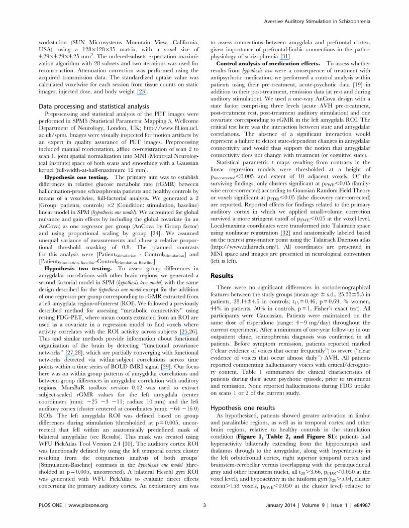
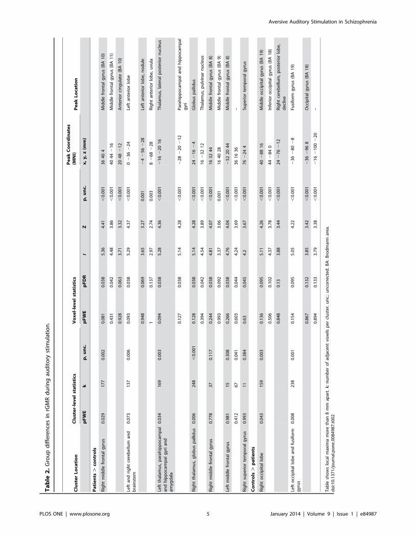
Hypothesis one resultsAs hypothesized, patients showed greater activation in limbic
and paralimbic regions, as well as in temporal cortex and other
brain regions, relative to healthy controls in the stimulation
condition (Figure 1, Table 2, and Figure S1): patients had
hyperactivity bilaterally extending from the hippocampus and
thalamus through to the amygdalae, along with hyperactivity in
the left orbitofrontal cortex, right superior temporal cortex and
brainstem-cerebellar vermis (overlapping with the periaqueductal
gray and other brainstem nuclei, all t28.3.66, pFDR,0.050 at the
voxel level), and hypoactivity in the fusiform gyri (t28.5.04, cluster
extent.158 voxels, pFWE,0.050 at the cluster level) relative to
Aversive Auditory Stimulation in Schizophrenia
PLOS ONE | www.plosone.org 3 January 2014 | Volume 9 | Issue 1 | e84987
controls. In addition, we observed a substantial overlap in the regions
of the temporal cortex that responded to stimulation across the study
groups (right superior temporal gyrus, conjunction analysis of
Stimulation-Baseline in both groups at p = 0.005, uncorrected; and
bilateral superior temporal gyri, average effect of Stimulation-
Baseline across all participants, Figure 1). The main effect of
condition in both groups was detected at trend level in the left
temporal cortex (t28 = 5.10, cluster extent = 143 voxels, pFWE = 0.063
at the cluster level, local maxima xyz coordinates (mm): 264 24 24).
The PatientStimulation-Baseline-ControlStimulation-Baseline contrast did not
yield significant findings, nor did the contrast between the groups at
rest ([PatientBaseline-ControlBaseline]).
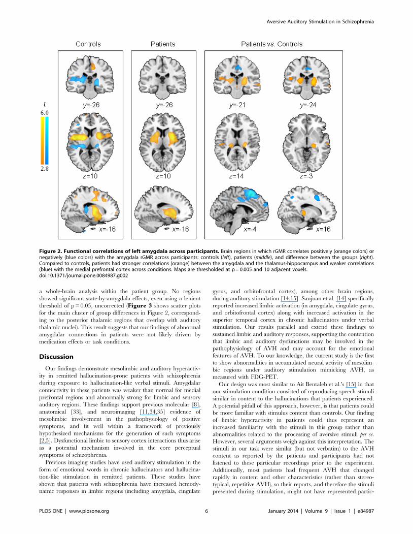
Hypothesis two resultsBoth groups shared local correlations of the left amygdala
bilaterally with the hippocampus, striatum (mostly putamen),
globus pallidus, and orbitofrontal cortex, as well as connections
with the contralateral amygdala (conjunction analysis of amygdala
correlations for both groups; t.6.90, cluster extent: 392 (left) and
273 (right), cluster-level pFWE#0.002). However, differences
between the two groups were also prominent. Group-by-amygdala
interaction effects were most prominent in the medial wall of the
brain, encompassing bilateral supplementary motor, medial
prefrontal, and middle cingulate cortices and precuneus, but were
also present in bilateral hippocampi, posterior thalami, right
inferior frontal, and right middle posterior temporal cortices
(F1,26.20.02, voxel-level pFDR,0.050; Figure 2, Table 3 andFigure S2). These interactions were explained by weaker
correlations in patients relative to controls of the left amygdala
with medial prefrontal, precuneus and right middle temporal
cortex (all t26.3.90, pFDR,0.050 at the voxel level) and stronger
correlations in patients relative to controls of the left amygdala
with posterior regions of the thalamus (overlapping with the
medial geniculate nucleus, part of the auditory thalamus, FigureS3) and hippocampus (t26 = 5.25, cluster extent = 144,
pFWE = 0.047 at cluster-level, local maxima xyz coordinates
(mm): 220 228 4). Moreover, stronger amygdala-auditory cortex
interaction was detected in patients with schizophrenia relative to
controls (small-volume-corrected to the Heschl gyri ROI;
t26 = 3.97, voxel-level pFWE = 0.038, pFDR = 0.051, and
t26 = 3.41, voxel-level pFWE = 0.112, pFDR = 0.056, for left and
right Heschl’s gyri, respectively).
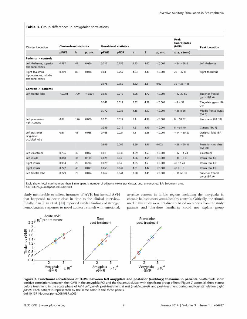
Control analysis of medication effectsFinally, we performed a post-hoc analysis of state-dependent
effects using pre-treatment data from the same AVH patients
during acute psychosis [19] to assess whether our results
concerning hypothesis two were driven by medication (see Methods).
We tested interactions between state and amygdalar correlations in
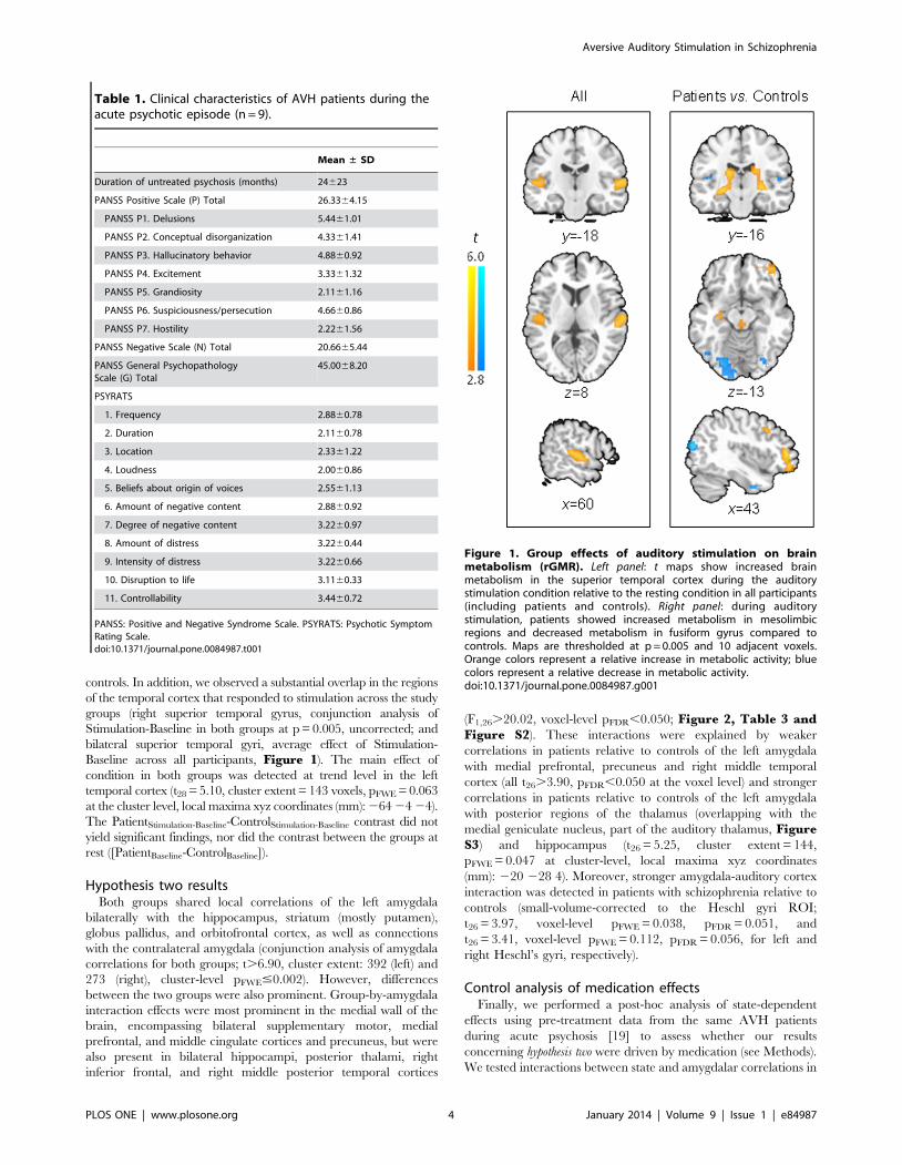
Table 1. Clinical characteristics of AVH patients during theacute psychotic episode (n = 9).
Mean ± SD
Duration of untreated psychosis (months) 24623
PANSS Positive Scale (P) Total 26.3364.15
PANSS P1. Delusions 5.4461.01
PANSS P2. Conceptual disorganization 4.3361.41
PANSS P3. Hallucinatory behavior 4.8860.92
PANSS P4. Excitement 3.3361.32
PANSS P5. Grandiosity 2.1161.16
PANSS P6. Suspiciousness/persecution 4.6660.86
PANSS P7. Hostility 2.2261.56
PANSS Negative Scale (N) Total 20.6665.44
PANSS General PsychopathologyScale (G) Total
45.0068.20
PSYRATS
1. Frequency 2.8860.78
2. Duration 2.1160.78
3. Location 2.3361.22
4. Loudness 2.0060.86
5. Beliefs about origin of voices 2.5561.13
6. Amount of negative content 2.8860.92
7. Degree of negative content 3.2260.97
8. Amount of distress 3.2260.44
9. Intensity of distress 3.2260.66
10. Disruption to life 3.1160.33
11. Controllability 3.4460.72
PANSS: Positive and Negative Syndrome Scale. PSYRATS: Psychotic SymptomRating Scale.doi:10.1371/journal.pone.0084987.t001
Figure 1. Group effects of auditory stimulation on brainmetabolism (rGMR). Left panel: t maps show increased brainmetabolism in the superior temporal cortex during the auditorystimulation condition relative to the resting condition in all participants(including patients and controls). Right panel: during auditorystimulation, patients showed increased metabolism in mesolimbicregions and decreased metabolism in fusiform gyrus compared tocontrols. Maps are thresholded at p = 0.005 and 10 adjacent voxels.Orange colors represent a relative increase in metabolic activity; bluecolors represent a relative decrease in metabolic activity.doi:10.1371/journal.pone.0084987.g001
Aversive Auditory Stimulation in Schizophrenia
PLOS ONE | www.plosone.org 4 January 2014 | Volume 9 | Issue 1 | e84987
Ta
ble
2.
Gro
up
dif
fere
nce
sin
rGM
Rd
uri
ng
aud
ito
ryst
imu
lati
on
.
Clu
ste
rL
oca
tio
nC
lust
er-
lev
el
sta
tist
ics
Vo
xe
l-le
ve
lst
ati
stic
sP
ea
kC
oo
rdin
ate
s(M
NI)
Pe
ak
Lo
cati
on
pF
WE
kp
,u
nc.
pF
WE
pF
DR
tZ
p,
un
c.x
,y
,z
(mm
)
Pa
tie
nts
.co
ntr
ols
Rig
ht
mid
dle
fro
nta
lg
yru
s0
.02
91
77
0.0
02
0.0
81
0.0
38
5.3
64
.41
,0
.00
13
64
04
Mid
dle
fro
nta
lg
yru
s(B
A1
0)
0.4
31
0.0
42
4.4
83
.86
,0
.00
14
04
42
16
Mid
dle
fro
nta
lg
yru
s(B
A1
1)
0.9
28
0.0
63
3.7
13
.32
,0
.00
12
04
82
12
An
teri
or
cin
gu
late
(BA
10
)
Left
and
rig
ht
cere
be
llum
and
bra
inst
em
0.0
73
13
70
.00
60
.09
30
.03
85
.29
4.3
7,
0.0
01
02
36
22
4Le
ftan
teri
or
lob
e
0.9
48
0.0
69
3.6
53
.27
0.0
01
24
25
62
28
Left
ante
rio
rlo
be
,n
od
ule
10
.13
72
.97
2.7
40
.00
38
26
82
28
Rig
ht
ante
rio
rlo
be
,u
vula
Left
thal
amu
s,p
arah
ipp
oca
mp
alan
dh
ipp
oca
mp
alg
yri
and
amyg
dal
a
0.0
34
16
90
.00
30
.09
40
.03
85
.28
4.3
6,
0.0
01
21
62
20
16
Th
alam
us,
late
ral
po
ste
rio
rn
ucl
eu
s
0.1
27
0.0
38
5.1
44
.28
,0
.00
12
28
22
02
12
Par
ahip
po
cam
pal
and
hip
po
cam
pal
gyr
i
Rig
ht
thal
amu
s,g
lob
us
pal
lidu
s0
.00
62
48
,0
.00
10
.12
80
.03
85
.14
4.2
8,
0.0
01
24
21
62
4G
lob
us
pal
lidu
s
0.3
94
0.0
42
4.5
43
.89
,0
.00
11
62
32
12
Th
alam
us,
pu
lvin
arn
ucl
eu
s
Rig
ht
mid
dle
fro
nta
lg
yru
s0
.77
83
70
.11
70
.24
40
.03
84
.81
4.0
7,
0.0
01
16
32
44
Mid
dle
fro
nta
lg
yru
s(B
A8
)
0.9
93
0.0
92
3.3
73
.06
0.0
01
16
40
28
Mid
dle
fro
nta
lg
yru
s(B
A9
)
Left
mid
dle
fro
nta
lg
yru
s0
.98
11
50
.30
80
.26
60
.03
84
.76
4.0
4,
0.0
01
25
22
04
4M
idd
lefr
on
tal
gyr
us
(BA
8)
0.4
12
67
0.0
41
0.6
03
0.0
44
4.2
43
.69
,0
.00
13
61
63
6–
Rig
ht
sup
eri
or
tem
po
ral
gyr
us
0.9
93
11
0.3
84
0.6
30
.04
54
.23
.67
,0
.00
17
62
24
4Su
pe
rio
rte
mp
ora
lg
yru
s
Co
ntr
ols
.p
ati
en
ts
Rig
ht
occ
ipit
allo
be
0.0
43
15
90
.00
30
.13
60
.09
55
.11
4.2
6,
0.0
01
40
28
81
6M
idd
leo
ccip
ital
gyr
us
(BA
19
)
0.5
06
0.1
02
4.3
73
.78
,0
.00
14
42
84
0In
feri
or
occ
ipit
alg
yru
s(B
A1
8)
0.8
48
0.1
33
.88
3.4
4,
0.0
01
24
27
62
12
Rig
ht
cere
be
llum
,p
ost
eri
or
lob
e,
de
cliv
e
Left
occ
ipit
allo
be
and
fusi
form
gyr
us
0.0
08
23
80
.00
10
.15
40
.09
55
.05
4.2
2,
0.0
01
23
62
80
28
Fusi
form
gyr
us
(BA
19
)
0.8
67
0.1
32
3.8
53
.42
,0
.00
12
36
29
68
Occ
ipit
alg
yru
s(B
A1
8)
0.8
94
0.1
33
3.7
93
.38
,0
.00
12
16
21
00
22
0–
Tab
lesh
ow
slo
cal
max
ima
mo
reth
an8
mm
apar
t.k:
nu
mb
er
of
adja
cen
tvo
xels
pe
rcl
ust
er.
un
c.:
un
corr
ect
ed
.B
A:
Bro
dm
ann
are
a.d
oi:1
0.1
37
1/j
ou
rnal
.po
ne
.00
84
98
7.t
00
2
Aversive Auditory Stimulation in Schizophrenia
PLOS ONE | www.plosone.org 5 January 2014 | Volume 9 | Issue 1 | e84987
a whole-brain analysis within the patient group. No regions
showed significant state-by-amygdala effects, even using a lenient
threshold of p = 0.05, uncorrected (Figure 3 shows scatter plots
for the main cluster of group differences in Figure 2, correspond-
ing to the posterior thalamic regions that overlap with auditory
thalamic nuclei). This result suggests that our findings of abnormal
amygdalar connections in patients were not likely driven by
medication effects or task conditions.
Discussion
Our findings demonstrate mesolimbic and auditory hyperactiv-
ity in remitted hallucination-prone patients with schizophrenia
during exposure to hallucination-like verbal stimuli. Amygdalar
connectivity in these patients was weaker than normal for medial
prefrontal regions and abnormally strong for limbic and sensory
auditory regions. These findings support previous molecular [8],
anatomical [33], and neuroimaging [11,34,35] evidence of
mesolimbic involvement in the pathophysiology of positive
symptoms, and fit well within a framework of previously
hypothesized mechanisms for the generation of such symptoms
[2,5]. Dysfunctional limbic to sensory cortex interactions thus arise
as a potential mechanism involved in the core perceptual
symptoms of schizophrenia.
Previous imaging studies have used auditory stimulation in the
form of emotional words in chronic hallucinators and hallucina-
tion-like stimulation in remitted patients. These studies have
shown that patients with schizophrenia have increased hemody-
namic responses in limbic regions (including amygdala, cingulate
gyrus, and orbitofrontal cortex), among other brain regions,
during auditory stimulation [14,15]. Sanjuan et al. [14] specifically
reported increased limbic activation (in amygdala, cingulate gyrus,
and orbitofrontal cortex) along with increased activation in the
superior temporal cortex in chronic hallucinators under verbal
stimulation. Our results parallel and extend these findings to
sustained limbic and auditory responses, supporting the contention
that limbic and auditory dysfunctions may be involved in the
pathophysiology of AVH and may account for the emotional
features of AVH. To our knowledge, the current study is the first
to show abnormalities in accumulated neural activity of mesolim-
bic regions under auditory stimulation mimicking AVH, as
measured with FDG-PET.
Our design was most similar to Ait Bentaleb et al.’s [15] in that
our stimulation condition consisted of reproducing speech stimuli
similar in content to the hallucinations that patients experienced.
A potential pitfall of this approach, however, is that patients could
be more familiar with stimulus content than controls. Our finding
of limbic hyperactivity in patients could thus represent an
increased familiarity with the stimuli in this group rather than
abnormalities related to the processing of aversive stimuli per se.
However, several arguments weigh against this interpretation. The
stimuli in our task were similar (but not verbatim) to the AVH
content as reported by the patients and participants had not
listened to these particular recordings prior to the experiment.
Additionally, most patients had frequent AVH that changed
rapidly in content and other characteristics (rather than stereo-
typical, repetitive AVH), so their reports, and therefore the stimuli
presented during stimulation, might not have represented partic-
Figure 2. Functional correlations of left amygdala across participants. Brain regions in which rGMR correlates positively (orange colors) ornegatively (blue colors) with the amygdala rGMR across participants: controls (left), patients (middle), and difference between the groups (right).Compared to controls, patients had stronger correlations (orange) between the amygdala and the thalamus-hippocampus and weaker correlations(blue) with the medial prefrontal cortex across conditions. Maps are thresholded at p = 0.005 and 10 adjacent voxels.doi:10.1371/journal.pone.0084987.g002
Aversive Auditory Stimulation in Schizophrenia
PLOS ONE | www.plosone.org 6 January 2014 | Volume 9 | Issue 1 | e84987
ularly memorable or salient instances of AVH but instead AVH
that happened to occur close in time to the clinical interview.
Finally, San Juan et al. [14] reported similar findings of stronger
hemodynamic responses to novel auditory stimuli with emotional,
aversive content in limbic regions including the amygdala in
chronic hallucinators versus healthy controls. Critically, the stimuli
used in this study were not directly based on reports from the study
patients and therefore familiarity could not explain group
Table 3. Group differences in amygdalar correlations.
Cluster Location Cluster-level statistics Voxel-level statistics
PeakCoordinates(MNI) Peak Location
pFWE k p, unc. pFWE pFDR t Z p, unc. x, y, z (mm)
Patients . controls
Left thalamus, superiortemporal cortex
0.597 49 0.066 0.717 0.752 4.23 3.62 ,0.001 224 228 4 Left thalamus
Right thalamus,hippocampus, middletemporal cortex
0.219 88 0.018 0.84 0.752 4.03 3.49 ,0.001 20 232 8 Right thalamus
0.978 0.752 3.62 3.2 0.001 32 236 216
Controls . patients
Left frontal lobe ,0.001 709 ,0.001 0.023 0.012 6.26 4.77 ,0.001 212 20 60 Superior frontalgyrus (BA 6)
0.141 0.017 5.32 4.28 ,0.001 28 4 52 Cingulate gyrus (BA24)
0.772 0.036 4.15 3.57 ,0.001 236 8 56 Middle frontal gyrus(BA 6)
Left precuneus,right cuneus
0.08 126 0.006 0.123 0.017 5.4 4.32 ,0.001 0 268 32 Precuneus (BA 31)
0.339 0.019 4.81 3.99 ,0.001 8 264 40 Cuneus (BA 7)
Left posteriorcingulate,occipital lobe
0.61 48 0.068 0.468 0.024 4.6 3.85 ,0.001 244 260 20 Occipital lobe (BA19)
0.999 0.082 3.29 2.96 0.002 228 260 16 Posterior cingulate(BA 30)
Left claustrum 0.736 39 0.097 0.81 0.038 4.09 3.53 ,0.001 232 24 24 Claustrum
Left insula 0.818 33 0.124 0.824 0.04 4.06 3.51 ,0.001 248 28 4 Insula (BA 13)
Right insula 0.954 20 0.224 0.829 0.04 4.05 3.5 ,0.001 48 12 24 Insula (BA 13)
Right insula 0.722 40 0.093 0.853 0.042 4.01 3.47 ,0.001 48 4 28 Insula (BA 13)
Left frontal lobe 0.279 79 0.024 0.867 0.044 3.98 3.45 ,0.001 216 60 32 Superior frontalgyrus (BA 9)
Table shows local maxima more than 8 mm apart. k: number of adjacent voxels per cluster. unc.: uncorrected. BA: Brodmann area.doi:10.1371/journal.pone.0084987.t003
Figure 3. Functional correlations of rGMR between left amygdala and posterior (auditory) thalamus in patients. Scatterplots showpositive correlations between the rGMR in the amygdala ROI and the thalamus cluster with significant group effects (Figure 2) across all three states:before treatment, in the acute phase of AVH (left panel), post-treatment at rest (middle panel), and post-treatment during auditory stimulation (rightpanel). Each patient is represented by the same color in the three panels.doi:10.1371/journal.pone.0084987.g003
Aversive Auditory Stimulation in Schizophrenia
PLOS ONE | www.plosone.org 7 January 2014 | Volume 9 | Issue 1 | e84987

differences in their fMRI measures. These arguments thus provide
some support for the argument that limbic hyperactivity represents
an abnormal processing of aversive stimuli in patients with AVH
rather than an effect of familiarity. Nonetheless, we do acknowl-
edge the possibility of a differential effect of familiarity with the
recordings content in our group findings as part of the
unbridgeable experiential differences between patients and con-
trols. Although we believe that our current focus on individual
characteristics of AVHs represents a particular strength of this
study, future research on phenomenology should strive to
circumvent such experiential differences across groups while
keeping a focus on individual experiences.
Our analyses revealed abnormal functional connections of the
amygdala in patients with AVH. Specifically, the left amygdala
displayed abnormally strong connections to the auditory thalamus
and the auditory cortex in patients compared to controls. The left
amygdala is central to enhancing the perception of aversive verbal
material in healthy individuals [13], likely through its interactions
with the auditory system [36]. Thus, increased interactions
between the amygdala and the auditory system might render
aversive stimuli particularly salient or make the perception of
neutral stimuli acquire emotional characteristics or salience
normally associated with aversive stimuli. Another possible
account relates to our finding that patients had abnormally weak
connections between amygdala and areas of the prefrontal cortex
involved in self-referential processing [37,38] and top-down
control of emotions [39]. In line with studies showing a prefrontal
modulation of emotions via prefrontal-limbic interations [39,40],
increased amygdalo-auditory connectivity in patients could reflect
a failure in top-down regulation of amygdala function by the
prefrontal cortex. This latter interpretation is also consistent with
the extensive literature emphasizing prefrontal abnormalities
[41,42] and abnormal prefrontal-amygdala interactions [31] in
schizophrenia.
In addition to these notable differences between patients with
schizophrenia and healthy controls, we also found meaningful
similarities between the groups. First, both patients and controls
engaged a similar region within the temporal cortex in response to
auditory stimulation. This common response may imply that the
functional organization of the auditory cortex is preserved at least
to some extent in patients. Second, in addition to the excess of
short-range intra-limbic connections found in patients, the two
groups exhibited overlapping connectivity between the amygdala
and limbic-paralimbic regions. This relatively preserved function-
ality of the auditory cortex in conjunction with the abnormal
connectivity between the amygdala and auditory regions, may
suggest that the key disturbance underlying AVH lies in amygdalo-
auditory circuits that modulate activity in the temporal cortex
rather than in the auditory cortex itself, consistent with current
views of hallucination pathophysiology [43]. Dysfunctions in a
modulatory circuit of the auditory cortex involving limbic regions
and the thalamus have been recently linked to tinnitus [44,45,46],
a positive symptom accompanied by some of the emotional
features of AVH. Our results suggest that auditory positive
symptoms in a broad sense, including tinnitus and AVH, might
share dysfunctions in a common modulatory circuit involving
limbic regions that are normally responsible for noise attenuation
in the auditory system. Because we showed that the abnormal
interactions between the amygdala and auditory regions were
present before and after treatment and during rest and stimula-
tion, these interactions do not seem to be induced by treatment.
Although there is no direct histopathological evidence confirming
such altered connectivity in patients with AVH, growing molecular
evidence points to aberrant excitatory outputs from the amygdala
in persons with schizophrenia [8].
There are some relevant limitations to the current study. The
small sample size could make our analyses susceptible to a type II
error, thus underestimating true differences between groups.
While FDG-PET imaging was suitable to achieve our goal of
investigating sustained neural activation, its low temporal resolu-
tion prevented an assessment of the effects of linguistic and other
time-varying features of the verbal stimuli. Additionally, our study
did not include a comparison group of schizophrenic patients
without AVH, and this prevents us from drawing conclusions
specific to hallucination-prone patients. With regard to hypothesis
two, our analyses assume fixed group effects of amygdala
connectivity and therefore do not account for individual variabil-
ity. Future studies should seek to infer causality regarding the
relationship between dysfunctional limbic connectivity and AVH,
and aim to overcome these methodological drawbacks to better
understand the amygdalar influence on auditory-cortex function in
schizophrenia.
In sum, our study on AVH replicates previous reports of
abnormally elevated neural activity in limbic regions to aversive
speech stimuli. In addition, we uncovered abnormal interactions
between the amygdala and auditory regions of the cortex and
thalamus that persisted after symptom remission. We suggest that
an abnormal modulation of the auditory cortex by a limbic-
thalamic circuit might underlie the generation of emotional AVH.
Further research on limbic-auditory interactions and their role in
AVH is warranted.
Supporting Information
Figure S1 Axial view of group effects of auditorystimulation on brain metabolism (rGMR). The t-statistic
map is thresholded at p = 0.005 and 10 adjacent voxels and
overlaid onto a single-subject T1 scan (MNI Colin brain). Hot
colors represent increased rGMR in patients relative to controls.
Cold colors represent increased rGMR in controls relative to
patients.
(TIF)
Figure S2 Axial view of group differences in functionalcorrelations of left amygdala. The t-statistic map is
thresholded at p = 0.005 and 10 adjacent voxels and overlaid
onto a single-subject T1 scan (MNI Colin brain). Hot colors
represent regions of increased connectivity with the amygdala in
patients relative to controls. Cold colors represent regions of
increased connectivity with the amygdala in controls relative to
patients.
(TIF)
Figure S3 Orthogonal view of group differences infunctional correlations of left amygdala. Compared to
controls, patients had stronger correlations (orange) between the
amygdala and the thalamus-hippocampus (same notation as in
Figure 2 of the main text). The white circles correspond to the
medial geniculate nucleus (MGN) of the auditory thalamus. Note
the overlap of the significant cluster of increased amygdalar
connectivity for patients (orange) with the MGN. Maps are
thresholded at p = 0.005 and 10 adjacent voxels.
(TIF)
Author Contributions
Conceived and designed the experiments: GH EP FL EF-E MF MB.
Performed the experiments: GH AM EF-E MF. Analyzed the data: GH
AM KS CF. Wrote the paper: GH.
Aversive Auditory Stimulation in Schizophrenia
PLOS ONE | www.plosone.org 8 January 2014 | Volume 9 | Issue 1 | e84987
References
1. Allen P, Laroi F, McGuire PK, Aleman A (2008) The hallucinating brain: a
review of structural and functional neuroimaging studies of hallucinations.Neurosci Biobehav Rev 32: 175–191.
2. Kapur S (2003) Psychosis as a state of aberrant salience: a framework linkingbiology, phenomenology, and pharmacology in schizophrenia. Am J Psychiatry
160: 13–23.
3. Calder AJ, Lawrence AD, Young AW (2001) Neuropsychology of fear andloathing. Nat Rev Neurosci 2: 352–363.
4. Baxter MG, Murray EA (2002) The amygdala and reward. Nat Rev Neurosci 3:563–573.
5. Fletcher PC, Frith CD (2009) Perceiving is believing: a Bayesian approach to
explaining the positive symptoms of schizophrenia. Nat Rev Neurosci 10: 48–58.6. Ellison-Wright I, Glahn DC, Laird AR, Thelen SM, Bullmore E (2008) The
anatomy of first-episode and chronic schizophrenia: an anatomical likelihoodestimation meta-analysis. Am J Psychiatry 165: 1015–1023.
7. Wright IC, Rabe-Hesketh S, Woodruff PW, David AS, Murray RM, et al.(2000) Meta-analysis of regional brain volumes in schizophrenia. Am J
Psychiatry 157: 16–25.
8. Benes FM (2010) Amygdalocortical circuitry in schizophrenia: from circuits tomolecules. Neuropsychopharmacology 35: 239–257.
9. Schneider F, Weiss U, Kessler C, Salloum JB, Posse S, et al. (1998) Differentialamygdala activation in schizophrenia during sadness. Schizophr Res 34: 133–
142.
10. Holt DJ, Kunkel L, Weiss AP, Goff DC, Wright CI, et al. (2006) Increasedmedial temporal lobe activation during the passive viewing of emotional and
neutral facial expressions in schizophrenia. Schizophr Res 82: 153–162.11. Fernandez-Egea E, Parellada E, Lomena F, Falcon C, Pavia J, et al. (2010)
18FDG PET study of amygdalar activity during facial emotion recognition inschizophrenia. Eur Arch Psychiatry Clin Neurosci 260: 69–76.
12. Taylor SF, Liberzon I, Decker LR, Koeppe RA (2002) A functional anatomic
study of emotion in schizophrenia. Schizophr Res 58: 159–172.13. Anderson AK, Phelps EA (2001) Lesions of the human amygdala impair
enhanced perception of emotionally salient events. Nature 411: 305–309.14. Sanjuan J, Lull JJ, Aguilar EJ, Marti-Bonmati L, Moratal D, et al. (2007)
Emotional words induce enhanced brain activity in schizophrenic patients with
auditory hallucinations. Psychiatry Res 154: 21–29.15. Ait Bentaleb L, Stip E, Mendrek A, Mensour B, Beauregard M (2006) [Effects of
listening to previously hallucinated words by schizophrenia patients in remission:a functional magnetic resonance imaging study of six cases]. Encephale 32: 27–
40.16. Holt DJ, Weiss AP, Rauch SL, Wright CI, Zalesak M, et al. (2005) Sustained
activation of the hippocampus in response to fearful faces in schizophrenia. Biol
Psychiatry 57: 1011–1019.17. Taylor SF, Phan KL, Britton JC, Liberzon I (2005) Neural response to
emotional salience in schizophrenia. Neuropsychopharmacology 30: 984–995.18. Salgado-Pineda P, Fakra E, Delaveau P, Hariri AR, Blin O (2010) Differential
patterns of initial and sustained responses in amygdala and cortical regions to
emotional stimuli in schizophrenia patients and healthy participants. J PsychiatryNeurosci 35: 41–48.
19. Horga G, Parellada E, Lomena F, Fernandez-Egea E, Mane A, et al. (2011)Differential brain glucose metabolic patterns in antipsychotic-naive first-episode
schizophrenia with and without auditory verbal hallucinations. J PsychiatryNeurosci 36: 312–321.
20. Kay SR, Fiszbein A, Opler LA (1987) The positive and negative syndrome scale
(PANSS) for schizophrenia. Schizophr Bull 13: 261–276.21. Andreasen NC, Flaum M, Arndt S (1992) The Comprehensive Assessment of
Symptoms and History (CASH). An instrument for assessing diagnosis andpsychopathology. Arch Gen Psychiatry 49: 615–623.
22. Haddock G, McCarron J, Tarrier N, Faragher EB (1999) Scales to measure
dimensions of hallucinations and delusions: the psychotic symptom rating scales(PSYRATS). Psychol Med 29: 879–889.
23. Weber WA, Schwaiger M, Avril N (2000) Quantitative assessment of tumormetabolism using FDG-PET imaging. Nucl Med Biol 27: 683–687.
24. Grunder G (2009) "Absolute" or "relative": choosing the right outcome measure
in neuroimaging. Neuroimage 45: 258–259.
25. Lee DS, Kang H, Kim H, Park H, Oh JS, et al. (2008) Metabolic connectivity by
interregional correlation analysis using statistical parametric mapping (SPM) and
FDG brain PET; methodological development and patterns of metabolicconnectivity in adults. Eur J Nucl Med Mol Imaging 35: 1681–1691.
26. Morbelli S, Drzezga A, Perneczky R, Frisoni GB, Caroli A, et al. (2012) Resting
metabolic connectivity in prodromal Alzheimer’s disease. A EuropeanAlzheimer Disease Consortium (EADC) project. Neurobiol Aging 33: 2533–
2550.
27. Taylor PA, Gohel S, Di X, Walter M, Biswal BB (2012) Functional covariance
networks: obtaining resting-state networks from intersubject variability. BrainConnect 2: 203–217.
28. Zhang Z, Liao W, Zuo XN, Wang Z, Yuan C, et al. (2011) Resting-state brain
organization revealed by functional covariance networks. PLoS One 6: e28817.
29. Di X, Biswal BB (2012) Metabolic brain covariant networks as revealed by FDG-
PET with reference to resting-state fMRI networks. Brain Connect 2: 275–283.
30. Lancaster JL, Woldorff MG, Parsons LM, Liotti M, Freitas CS, et al. (2000)Automated Talairach atlas labels for functional brain mapping. Hum Brain
Mapp 10: 120–131.
31. Anticevic A, Repovs G, Barch DM (2012) Emotion effects on attention,
amygdala activation, and functional connectivity in schizophrenia. SchizophrBull 38: 967–980.
32. Lacadie CM, Fulbright RK, Rajeevan N, Constable RT, Papademetris X (2008)
More accurate Talairach coordinates for neuroimaging using non-linear
registration. Neuroimage 42: 717–725.
33. Davis KL, Kahn RS, Ko G, Davidson M (1991) Dopamine in schizophrenia: areview and reconceptualization. Am J Psychiatry 148: 1474–1486.
34. Anticevic A, Van Snellenberg JX, Cohen RE, Repovs G, Dowd EC, et al. (2012)
Amygdala recruitment in schizophrenia in response to aversive emotionalmaterial: a meta-analysis of neuroimaging studies. Schizophr Bull 38: 608–621.
35. Diederen KM, Neggers SF, Daalman K, Blom JD, Goekoop R, et al. (2010)Deactivation of the parahippocampal gyrus preceding auditory hallucinations in
schizophrenia. Am J Psychiatry 167: 427–435.
36. Kumar S, von Kriegstein K, Friston K, Griffiths TD (2012) Features versusfeelings: dissociable representations of the acoustic features and valence of
aversive sounds. J Neurosci 32: 14184–14192.
37. Gusnard DA, Akbudak E, Shulman GL, Raichle ME (2001) Medial prefrontal
cortex and self-referential mental activity: relation to a default mode of brainfunction. Proc Natl Acad Sci U S A 98: 4259–4264.
38. Ochsner KN, Knierim K, Ludlow DH, Hanelin J, Ramachandran T, et al.
(2004) Reflecting upon feelings: an fMRI study of neural systems supporting the
attribution of emotion to self and other. J Cogn Neurosci 16: 1746–1772.
39. Ochsner KN, Bunge SA, Gross JJ, Gabrieli JD (2002) Rethinking feelings: anFMRI study of the cognitive regulation of emotion. J Cogn Neurosci 14: 1215–
1229.
40. Wager TD, Davidson ML, Hughes BL, Lindquist MA, Ochsner KN (2008)
Prefrontal-subcortical pathways mediating successful emotion regulation.Neuron 59: 1037–1050.
41. Minzenberg MJ, Laird AR, Thelen S, Carter CS, Glahn DC (2009) Meta-
analysis of 41 functional neuroimaging studies of executive function inschizophrenia. Arch Gen Psychiatry 66: 811–822.
42. Pomarol-Clotet E, Canales-Rodriguez EJ, Salvador R, Sarro S, Gomar JJ, et al.(2010) Medial prefrontal cortex pathology in schizophrenia as revealed by
convergent findings from multimodal imaging. Mol Psychiatry 15: 823–830.
43. Waters F, Allen P, Aleman A, Fernyhough C, Woodward TS, et al. (2012)Auditory hallucinations in schizophrenia and nonschizophrenia populations: a
review and integrated model of cognitive mechanisms. Schizophr Bull 38: 683–
693.
44. Leaver AM, Renier L, Chevillet MA, Morgan S, Kim HJ, et al. (2011)Dysregulation of limbic and auditory networks in tinnitus. Neuron 69: 33–43.
45. Maudoux A, Lefebvre P, Cabay JE, Demertzi A, Vanhaudenhuyse A, et al.
(2012) Auditory resting-state network connectivity in tinnitus: a functional MRI
study. PLoS One 7: e36222.
46. Rauschecker JP, Leaver AM, Muhlau M (2010) Tuning out the noise: limbic-auditory interactions in tinnitus. Neuron 66: 819–826.
Aversive Auditory Stimulation in Schizophrenia
PLOS ONE | www.plosone.org 9 January 2014 | Volume 9 | Issue 1 | e84987