Use and valuation of native and introduced medicinal plant ...
Upload
khangminh22Category
view
0download
0
PlantaMedica

PlantaMedica

Plenary Lectures
PL 001Discovery of natural products by chemical andpharmacological profilingHamburger MDepartment of Pharmaceutical Sciences, Institute of Pharmaceutical Biology,University of Basel, CH-4056 Basel, Switzerland
Over the past decade, a number of new technologies and tools havebecome available in the biosciences and in analytical chemistry.They enable new approaches in the discovery of bioactive naturalproducts which can be summarized with a few keywords such asminiaturization, on-line analysis of complex samples, study of mo-lecular modes of action, and systems oriented approaches towardsthe characterization of drug effects in vitro and in vivo. Some of thetechnologies which are useful in the context of natural productsdiscovery will be discussed and illustrated with examples fromour lab. The efficient tracking of bioactivity in an extract remains amajor challenge. We have replaced preparative activity-directed iso-lation by HPLC-based activity profiling at analytical scale. In thesearch for natural products leads, we prefer assays with high infor-mation content and complex endpoints, such as phenotypicalscreens, over biochemical assays. The molecular targets for theseleads are subsequently studied with the tools of molecular and cellbiology. HPLC-based activity and metabolite profiling will be illu-strated with the example of the anti-inflammatory plant Isatis tinc-toria, while phenotypical screening and subsequent characterizationof signaling pathways will be discussed with the example of fungalpyridone alkaloids from Paecilomyces militaris. The postgenomicera offers a range of new tools and approaches for an essentiallyunbiased and global investigation which does not need to be hy-pothesis-driven. The application of genome-wide expression profil-ing in the characterization of extracts will be described with ourongoing studies on Cimicifuga racemosa (L.) Nutt. and Leuzeacarthamoides DC. Findings from array experiments are confirmedby quantitative PCR and functional assays, followed by HPLC-basedactivity profiling, eg. for AhR-agonistic activity, and by structuredetermination with LC-PDA-MS and microprobe NMR in HPLC frac-tions. Reference: 1. Potterat, O., Hamburger, M. (2006), Curr. Org.Chem., in press.
PL 002Advances in stationary phase development for the analysisof target compounds in proteomics, phytomics andmetabolomicsBonn GK, Stecher G, Huck CW, Bakry R, Feuerstein IInstitute of Analytical Chemistry and Radiochemistry, Leopold-FranzensUniversity, Innrain 52a, 6020-Innsbruck, Austria
Extraction, purification, preconcentration and separation are theclassical steps for the analysis of plant materials. Although a hugenumber of different techniques are available, the design of novelmaterials and stationary phases is still needed. In fact, selectiveextraction, preconcentration and purification prior to analysis isoften necessary owing to the complexity of samples. Additionally,analytes are often present in low concentrations, what makes suc-cessful analysis a challenge. Within this talk we present differentstrategies for the synthesis and the modification of stationaryphases to produce tailored solutions for the analytical questions.In fact, different possibilities concerning extraction, purificationand separation will be presented, e. g. a multidimensional approachfor the simultaneous preconcentration and separation of biomole-cules and flavonoids. Further on, focus will be placed on open tub-ular capillaries with special surface modifications, as these systemsallow the selective extraction of target compounds and the elutionof the sample within a high concentrated fraction. For the separa-
tion of analytes newly synthesized materials on the basis of methyl-styrene (MS) and 1,2-bis(p-vinylphenyl)ethane (BVPE) will be intro-duced. The polymer was build in the confines of fused silica capil-laries (200mL I.D.) and was successfully employed for the fractiona-tion of peptides, b-blocker drugs (Pindolol, Metopolol, Alprenolol,Propranolol) as well as flavonoids and stilbenes (epicatechin, epi-gallocatechin gallate, epicatechin gallate, resveratrol). Next to this,several approaches concerning the pharmacological investigation ofanalytes will be shown, e. g. the analysis of stilbenemetabolites inhuman urine using silica C-18 stationary phases or the analysis ofsalix ingredients using encapsulated silica-C18 poly-(styrene/divi-nylbenzene) capillaries. Finally within the talk some new instru-mental developments and applications in phytomics will be pres-ented, e. g. the use of a contactless conductivity detector in capillaryelectrophoresis for the detection of flavonoids. At last also the use ofmatrix assisted laser desorption ionization mass spectrometry(MALDI-MS) for the detection of small molecules such as sugars,glucuronic acid derivatives and glycerol will be showen, accentingits potential for metabolomic investigations.
PL 003Plant Metabolomics: Small Molecules Take Center StageTrethewey Rmetanomics GmbH, Tegeler Weg 33, 10589 Berlin, Germany
The advancement of genomics technologies in the last decade hasbeen extremely rapid and the opportunity for novel experimenta-tion profound. However, whilst there has been much focus on largemolecules (DNA, RNA and protein), small molecules have beensomewhat neglected in international efforts. This is odd given theessential importance of small molecules in determining functionalperformance and phenotype and our emerging understanding oftheir role as signals that interplay with and regulate gene expres-sion and protein activity in biological networks. In this presentationthe importance of the analysis of small molecules via metaboliteprofiling will be introduced and illustrated with examples fromthe work of metanomics, a company which has pioneered industrialmetabolomics. Today laboratories are operated with some 60 massspectrometers allowing a throughput of > 100,000 samples per year.This capability has been deployed in plant functional genomics: thecompany has generated large, unique, populations of Arabidopsisand crop plants where genes have been systematically overex-pressed or knocked out at a genome scale. Screening the metaboliteprofiles of these transgenic lines enables genes to be rapidly se-lected which influence and control commercially important areasof metabolism e. g. oils, amino acids, vitamins or sugars. Furtherthe linking of metabolic data to genetic and phenotypic data hasbeen demonstrated to be of particular importance and the status ofsuch system biology approaches based on metabolite profiling datawill be reviewed.
PL 004Clinical trials and systematic reviews of herbal medicineErnst EDirector, Complementary Medicine, Peninsula Medical School, Universities ofExeter & Plymouth, 25 Victoria Park Road, Exeter, EX2 4NT, UK
The popularity of herbal medicines begs the question whether agiven herbal remedy is safe and efficacious in treating a given con-dition. The latter question is best answered on the basis of rando-mised (preferably placebo-controlled, double-blind) clinical trials.Several hundred clinical trials of variable methodological rigourhave been published. The emerging evidence is often contradictory.In this situation the best evidence is provided by a systematic re-view or meta-analysis, i. e. an evaluation of the totality of all theavailable studies on a specific topic. This approach is aimed at mini-mising both random and selection biases. Today well over 100 sys-tematic reviews relating to a wide range of herbal medicines have
Planta Med 2006; 72: 961–1089 Georg Thieme Verlag KG Stuttgart · New York · ISSN 0032-0943
Abstracts
961
been published. Examples of some of these systematic reviews willbe discussed. They leave little doubt that some herbal medicines areefficacious in treating some clinical conditions. To date the evidencerelating to safety is largely anecdotal, i. e. based on case reports. Theleast biased and most informative tool for summarising it is againthe systematic review. Several systematic reviews of safety data willbe provided. Their results vary but, by and large, suggest that ad-verse effects are rare. In conclusion, more systematic research isrequired to evaluate the balance between risk and benefit for com-monly used herbal medicines.
PL 005Absorption and metabolism of dietary phenolicsCrozier AInstitute of Biomedical and Life Sciences, University of Glasgow, Glasgow G128QQ, UK
Since the early 1990 s there has been growing interest in the pro-tective effects of dietary phenolics and flavonoids. In order to assessthe potential health benefits of these compounds information isrequired on the sites of absorption, the metabolic forms in whichthey are absorbed and their concentrations in the circulatory sys-tems and body tissues. After consumption of onions, which containflavonol glucosides, the main components being quercetin-4’-O-glu-coside (143 mmoles) and quercetin-3,4’-O-diglucoside (107mmoles), the flavonols undergo rapid metabolism in the small in-testine followed by absorption of glucuronidated, sulfated andmethylated quercetin metabolites. These metabolites are detectedin the bloodstream reaching a Cmax after 1.0 –1.5 h. Excretion ofmetabolites in urine over a 24h period indicates that absorption is~4% of intake. In subjects with an ileostomy, the major componentsin ileal fluid after ingestion of onions are quercetin-3-glucuronide,quercetin-3’-sulfate and quercetin in quantities corresponding to ca.20% of intake, suggesting that absorption is substantially higherthan 4%. A comparative study on the absorption an metabolism of164 mmoles of quercetin-3-rhamnosylglucoside (rutin) in tomatojuice showed trace levels of quercetin and methylquercetin glucur-onides, but no sulfated metabolites, in plasma with a Tmax of ca.5 h. There was an 85+% recovery of the ingested ruin in ileal fluidand no quercetin metabolites were detected in plasma collectedfrom ileal volunteers These observations indicate that absorptionof rutin is more limited than that of quercetin glucosides and thatin healthy subjects it takes place on the large intestine. Other stud-ies have demonstrated that colonic bacteria hydrolyse and break-down rutin to phenolic acids which are excreted in urine in amountscorresponding to 25% of intake.
PL 006Bioassay development in natural product drug discoveryVuorela PDepartment of Biochemistry and Pharmacy, Faculty of Mathematics andNatural Sciences, bo Akademi University, FI-20520 Turku, Finland
Bioactivity screening is an integral part of the natural product drugdiscovery process [1]. The bioactive compounds in the natural pro-duct extracts are screened utilizing e. g. whole cells, cell fractions,recombinant enzymes or biochemicals as targets. The screening ofnatural products provides a complementary structural diversity tosynthetic chemistry and offers new low molecular weight lead com-pounds. Our work involves generating and screening of extract/com-pound libraries of biogenic origin for pharmaceutical purposes. Wehave used microfractionation of plant and microbial extracts onHPLC combined with design and development of new bioactivityscreening assays as an approach to find bioactive principals. UsingHPLC microfractionation, components of crude extracts can bedivided into fractions collected into microwell plates and subse-quently subjected to diverse bioassays. Miniaturized screening as-says have been developed with special emphasis on quality of the
bioassays for e. g. susceptibility testing of Chlamydia pneumoniaeutilizing time-resolved fluorometric immunoassay [2, 3]. The cou-pling of automated bioassay to analytical HPLC microfractionationgreatly facilitated the classical process leading from a plant to phar-macologically active compound [4]. References: 1. Vuorela, P., Lei-nonen, M., Saikku, P., Tammela, P., Rauha, J.-P., Wennberg, T., Vuor-ela, H. (2004), Curr. Med. Chem. 11: 1375–1389. 2. Tammela, P. et al.(2004), Anal. Biochem. 333: 39–48. 3. Alvesalo, J. et al. (2006), J.Med. Chem. 49: 2353–2356. 4. Tammela, P. et al. (2004), Anal.Bioanal. Chem. 380: 614–618.
Keynote Lectures
K 001Merits and limits of computational methods for thediscovery of natural acetylcholinesterase inhibitorsRollinger JM1, Schuster D2, Langer T2, Stuppner H1
1Institute of Pharmacy / Pharmacognosy; 2Institute of Pharmacy / CAMD-Group;Center for Molecular Biosciences Innsbruck, Leopold-Franzens University ofInnsbruck, 6020 Innsbruck, Austria
Bioactive natural products and drug substances in general exhibittheir pharmacological activity by binding as ligands to biomoleculartargets. Functions and 3D-structures of an increasing number oftarget macromolecules are becoming available. On the other hand,a wealth of potent ligands from both synthetic and natural originprovides a rich pool of structural and biological information. In thislight, computational methods contribute to (i) a rapid identificationof novel lead compounds and (ii) an improved molecular insight ofligand-target interactions. This study deals with the application ofdiverse integrated in silico tools to increase the efficiency in thesearch for natural acetylcholinesterase (AChE) inhibitors. In contrastto previous screening results, where we have been able to correctlypredict novel bioactive natural products from in house molecular 3Dlibraries [1, 2], we report here on the limitations of pharmacophorebased virtual screening. A highly potent anticholinesterase alkaloid,taspine (IC50 333€70 nM), was isolated by bio-guided fractionationfrom Magnolia x soulangiana Soul.-Bod. However, none of the 3Dconformers was able to fit into the elaborated pharmacophore mod-el [1]. Extensive docking studies on human- and Torpedo californica-AChE strongly suggest a binding mode of taspine, which is differentto that of known ligands in the active binding site (e. g. galantha-mine; [3]) and in the peripheral anionic binding site [4]. It may beassumed that taspine does not occupy the catalytic center itself butprevents acetylcholine from accurately being positioned in the bind-ing pocket for cleavage. Concluding, molecular docking studieshelped to explore the possible binding mode of taspine as “hydro-phobic plug“ in the aromatic gorge of AChE. Acknowledgements: Thiswork was granted by the FWF Austria (P18379) References: 1. Roll-inger, J.M. et al. (2004), J. Med. Chem. 47: 6248–6254. 2. Rollinger,J.M. et al. (2005), Curr. Drug Disc. Techn. 2: 185–193. 3. Greenblatt,H.M. et al. (2004), J. Am. Chem. Soc. 126: 15405–15411. 4. Kryger, G.et al. (2000), Acta Crystallogr. D 56: 1385–1394.
Planta Med 2006; 72: 961–1089 Georg Thieme Verlag KG Stuttgart · New York · ISSN 0032-0943
Abstracts
962
K 002Flavonoids Target Multiple Enzymes from the Type II FattyAcid Pathway of Plasmodium falciparum and Do Not InvokeDelayed Death PhenomenonTasdemir D1, Lack G2, Brun R3, Kaiser M3, R edi P4, Perozzo R2
1Centre for Pharmacognosy and Phytotherapy, University of London, 29 – 39Brunswick Square, London WC1N 1AX, UK; 2School of PharmaceuticalSciences, University of Geneva, Quai Ernest-Ansermet 30, CH-1211 Geneva 4,Switzerland; 3Department of Medical Parasitology, Swiss Tropical Institute,Socinstrasse 57, CH-4002 Basel, Switzerland; 4University of Zurich, Instituteof Organic Chemistry, Winterthurerstrasse 190, CH-8057 Zurich, Switzerland
The deciphering of the complete genome of Plasmodium falciparum[1] has uncovered a number of biochemical pathways, including thetype II fatty acid biosynthesis (FAS-II) that occurs in a recently dis-covered plastid-like organelle (apicoplast) in Plasmodium [2]. Theorganizational and structural differences between the fatty acidbiosynthesis in the malaria parasite and in humans make FAS-IIand its enzymes attractive targets for the design of antimalarialagents. The unique enzyme enoyl-ACP reductase (PfFabI) of P. falci-parum commits the final reduction step during the fatty acid elon-gation. We recently identified luteolin-7-O-glucoside as the firstnatural product targeting PfFabI [3]. This prompted us to evaluatethe inhibitory activity of a large flavonoid library against FabI, aswell as two other crucial enzymes (FabG and FabZ) of the FAS-IIsystem of P. falciparum. Several compounds, e. g. (-)-catechin gallate,luteolin, fisetin inhibited all three enzymes in the low ng/mL tosubmg/mL range and were further investigated kinetically to shedlight on the inhibitory mechanism. The ability of a single compoundto inhibit three enzymes from the same pathway is a very importantaspect, as it is unlikely for the parasite to develop resistance to thedrug by introducing mutations at all three enzymes at the sametime. Previous studies indicate that the inhibition of some otherapicoplast functions, e. g. replication, translation may not result inimmediate parasite death. Rather than inhibiting growth in the firstgeneration (48h), some antimalarial agents kill the parasites later inthe second generation (96h) [4]. Therefore, we investigated wheth-er the FAS inhibitory flavonoids invoke this so-called delayed deathphenomenon. Fortunately, none of the tested flavonoids elicited thedelayed death response and demonstrated rapid antiparasitic ef-fects. References: 1. Gardner, M.J. et al. (2002), Nature 419: 498–511. 2. Fadden, G.I. et al. (1996), Nature 381: 482. 3. Kirmizibekmez,H. et al. (2004), Planta Med. 70: 711–717. 4. Surolia, A. et al. (2004),Biochem. J. 383: 401–412.
K 003Plant secondary metabolism in the post-genomic eraOksman-Caldentey KM1, H kkinen ST1, Rischer H1, Ritala A1, Ma R1,Sepp nen-Laakso T1, Goossens A2, OreÐic M1, Inz D2
1VTT Technical Research Centre of Finland, Tietotie 2, Espoo, FIN-02044 VTT,Finland; 2Department of Plant Systems Biology, Flanders InteruniversityInstitute for Biotechnology, Ghent University, B-9052 Gent, Belgium
The biotechnological production of high-value plant secondary me-tabolites in cultivated cells is an attractive alternative to isolationprocesses from the intact plants or to the total chemical synthesis.However, plant metabolic engineering has met only limited success,in sharp contrast to microorganisms, since our knowledge on bio-synthesis of secondary metabolites is still very limited. Despite ofthe rapid development of not only plant genomics but also of ana-lytical tools genetic maps of biosynthetic pathways are far fromcomplete. Furthermore, regulation of the individual steps leadingto the desired end-product is poorly understood. We have devel-oped a SoluCel technology platform based on genome-wide iden-tification and functional analysis of genes involved in the produc-tion of plant-derived small molecules. It allows the exploitation ofthese genes in order to produce already existing secondary metabo-lites at higher levels in cell and tissue cultures through metabolicengineering. Moreover our combinatorial biochemistry approach al-
lows to increase the chemical diversity of plant-based moleculesthus offering novel molecules for the industry. A proof-of-concepthas first been gained using tobacco cells as a model system. Thetechnology was further applied to several medicinal plants. UsingcDNA-AFLP based transcript profiling linked to our UPLC-MS or GC-MS metabololite profiling platform, an inventory of hundreds ofgenes, potentially involved in secondary metabolism, has been built.The functional analysis of these genes alone or in combination hasshown clearly enhanced or altered metabolite accumulation pat-terns both in tobacco and in other plants. With this technology weare able to offer new opportunities to exploit the entire metabolicrepertoire of a plant cell, and to create higher quatities of knownmetabolites or novel compounds that may find applications not onlyin pharmaceutical but also in chemical or biotechnological indus-tries.
K 004Capability of Prenylflavanones present in Hops to InduceApoptosis in a Human Burkitt Lymphoma Cell LineRiepl HM1, Diller RA1, Rose O2, Frias C2, Henze G2, Prokop A2
1Institute of Technology for Biogenic Resources, Technical University ofMunich, Petersgasse 18, 94315 Straubing, Germany; 2Department ofPediatric Oncology/Hematology, University Medical Center Charit , CampusVirchow, Augustenburger Platz 1, 13353 Berlin, Germany
8-Prenylnaringenin (8-PN), a flavanone present in the female flowerof hops (Humulus lupulus L.) and in some other plants (e. g. in Ana-xagorea luzonensis A.Gray.) [1, 2] is known as being a very potentphytoestrogen [3]. As such it may accelerate proliferation analogousto estradiol in sensitive cell lines. The question was to be clarifiedwhether it may contribute to growth of hormone dependent neo-plasms when present in herbal preparations. We found instead anti-proliferative and apoptosis inducing effects of 8 –PN. We comparedsome side chain variants of 8-prenylnaringenin e. g. 8–geranylnar-ingenin, isolated also from hops and the synthetic variations 8–furanylmethylnaringenin, 8 –cinnamylinaringenin. These weresynthesized by a Mitsunobu reaction and Claisen rearrangement[4]. When applied to BJAB cells, grown in RPMI 1640 medium, theseflavanones showed improved cytotoxic and apoptotic activities –only 8-furanylmethylnaringenin is not active. 8-Geranylnaringenindisplayed noticeably improved apoptotic effects when compared to8-PN. 8-Cinnamylnaringenin significantly induced apoptosis in BJABcells at a concentration of 50mM. (Fig. 4). The apoptotic effect of 8-cinnamylnaringenin exceeded those of all other naringenins testedin this study. The induction of apoptosis is concentration dependent(11% apoptotic cells at 50mM and 38% at 100mM). Apoptosis wasinduced in a mitochondrial dependent manner. Despite low capacityto induce apoptosis, 8-PN induced a decrease of the mitochondrialmembrane potential, too. However, 8-geranylnaringenin caused achange in the membrane potential at much lower concentration.At 100mM we noticed a saturation effect in decrease of mitochon-drial membrane potential. But the greatest effect was demonstratedwith 8-cinnamylnaringenin. Even at a concentration of 50mM, it isobserved a transition in 77% of the BJAB cells. The potential of 8 –PNis shown in an ex vivo experiment of a multi-drug resistant leukae-mia blast. References: 1. Zierau, O., Hauswald, S., Schwab, P., Metz,P., Vollmer, G. (2004), J. Steroid Biochem. Mol. Biol. 92: 107–110. 2.Kitaoka, M., Kadokawa, H., Sugano, M., Ichikawa, K., Taki, M., Taka-hashi, S., Iijima, Y., Tsutsumi, S., Boriboon, M., Akiyama, T., (1998),Planta Med. 64: 511–515. 3. Milligan, S.R., Kalita, J.C., Pocock, V.,Van de Kauter, V., Stevens, J.F., Deinzer, M.L., Rong, H., De Keukeleire,D. (2000), J. Clin. Endocrin. Metab. 85: 4912–4915, 4. Gester, S.,Metz, P., Zierau, O., Vollmer, G. (2001), Tetrahedron 57: 1015.
Planta Med 2006; 72: 961–1089 Georg Thieme Verlag KG Stuttgart · New York · ISSN 0032-0943
Abstracts
963
K 005Biobehavioural effects of herbal extractsScholey AB, Kennedy DOHuman Cognitive Neuroscience Unit, Northumbria University, Newcastleupon Tyne, NE1 8ST UK
Mainstream pharmaceuticals have largely been developed from theisolation and/or synthesis of active agents with specific targets. Onthe other hand plant medicines may contain dozens of activeswhich exert multiple and often subtle effects upon target systems.Individually these components may act either positively or nega-tively, and together may affect multiple neuronal, metabolic andhormonal systems. Since mental processes are themselves modu-lated by such systems, the behavioural effects of plant extracts in-volve complex interactions both within and between physiologicalsystems. Additionally such interactions may be synergistic, resultingin complex dose- and time dependent effects [1]. Over the last fewyears work in our laboratory has aimed to systematically assess theeffects of plant extracts on human functions which are relevant toageing and dementia. Extracts include those used in traditionalmedicine systems. Thus we have built up a portfolio of researchdocumenting the biobehavioural effects of Ginkgo biloba L., Panaxginseng C.A. Meyer, species of Salvia L., Melissa officinalis L., Valeri-ana officinalis L. and Paullinia cupana Kunth ex H.B.K. amongstothers. This talk examines methodology for capturing such effectsand presents data suggesting that standardised extracts are capableof differentially affecting aspects of memory and mood. The poten-tial for such agents to act as cognition enhancing, anti-stress andanxiogenic treatments is considered. Reference: 1. Scholey, A. et al.(2005), Psychopharmacology 179: 705–707.
K 006Salicylate: a phytochemist’s headacheVerpoorte R, Verberne M, Budi Muljono RA, Mustafa NKDepartment of Pharmacognosy, Section Metabolomics, IBL, Leiden University
Acetylsalicylate is one of the most successful drugs ever made, withstill novel indications being discovered. It was developed on thebasis of the use of Salix bark, which contains salicin which onemay consider as a pro-drug for salicylate (SA). Interestingly it wasfound that SA acts as signal compound in plants, particularly insystemic acquired resistance (SAR) observed after infection withfor example a virus. Despite extensive studies in the past 20 yearsthe biosynthesis still poses many questions [1]. Most work has beenon the phenylalanine pathway leading to SA. Several enzymes havebeen proposed to be involved, but the step(s) between the putativeintermediate benzoic acid and phenylalanine remain uncertain. Mi-croorganisms produce SA in two steps via the isochorismate path-way. Verberne et al. (2000) proposed that this pathway might alsofunction in plants, and showed that by introduction of microbialgenes this pathway can be introduced in tobacco, making the plantmore resistant against viral and fungal infections. The effect of theconstitutive expression of salicylate and TMV infection in tobaccowas studied by means of NMR-metabolomics. This metabolomicsapproach showed clear differences for the production of phenylpro-panoids. In case of TMV infection clear differences between infectedleaves and SAR leaves could be detected. In Arabidopsis it wasshown that a gene encoding isochorismate synthase is correlatedwith the formation of SA and SAR. But still the direct chemicalevidence is missing that SA is derived from isochorismate and notfrom phenylalanine. Catharanthus roseus (L.) G. Don. cell culturesproduce both SA and the closely related 2,3-dihydroxybenzoic acid(DHBA) upon elicitation. Feeding the cultures with 1-13C-glucose wefound by means of 13C-NMR-spectrometry that DHBA has a labelingpattern as expected for the isochorismate pathway [3]. However, incase of SA the labeling in the aromatic ring was such that it mightbe a mixture of both pathways. References: 1. Verberne, M. Ver-poorte, R. et al. (2000), Nature Biotechnology 18: 779–783. 2. Ver-berne, M.C. et al. (1999), Salicylic acid biosynthesis. In: Biochemistry
and Molecular Biology of Plant Hormones. New Comprehensive Bio-chemistry. Vol. 33. P.J.J. Hooikaas, M.A. Hall, and K.R. Libbenga,(Eds.) Elsevier, Amsterdam, 1999, pp. 295–312. 3. Budi Muljono,R.A. et al. (2002), Plant Physiol. Biochem. 40: 231–234.
Short Lectures
1. Drug Discovery from Natural Products
S 001Evaluation of reversible antiandrogenic andantispermatogenic activities of Annona squamosa (Linn)stem bark methanol extract in male albino ratsGupta RS, Sharma A, Rehwani HCenter for Advanced Studies, Reproduction Physiology Section, Departmentof Zoology, University of Rajasthan, Jaipur-302004, India
The present study was undertaken to evaluate antiandrogenic activ-ities of (methanol stem bark extract) Annona squamosa L. (Annona-ceae) with their respective reversibility in male albino rats. Adultmale albino rats were gavaged with 100% methanol extract of An-nona squamosa stem bark at the dose level of 50, 100 and 200 mg/rat/day for 60 days. Fertility test was performed before and after55th day of treatment. Sperm dynamics in cauda epididymides andtestis were assessed. Biochemical and histological analysis were alsodone in blood, serum and in reproductive organs. Recovery of ferti-lity was followed to evaluate the reversibility of drug nature. Anno-na squamosa stem bark extract brought about a significant decreasein the weights of testes and accessory reproductive organs. Spermmotility and density was also reduced significantly. Significant re-duction was seen in protein, sialic acid and glycogen content oftestis as well as fructose content of seminal vesicle. An increasedlevel of cholesterol was seen in testis. The blood and serum param-eters were found to be within the normal range whereas the serumtestosterone levels decreased significantly. The stem bark extractfeeding caused a marked reduction in the number of spermatocytesand spermatids in the testis. The diameter of the seminiferous tu-bules and the numbers of mature Leydig cells were also decreasedwhereas, number of degenerating cells increased proportionately. Inconclusion Annona squamosa stem bark extract have an antiandro-genic and antispermatogenic activity, which were reversible afterwithdrawal of drug. Acknowledgment: Authors are thankful to theHead, Department of Zoology, Prof. N.K. Lohiya Coordinator CAS,Department of Zoology for providing the neccessory facilities andUGC, Regional Office, Bhopal, INDIA for financial support.
S 002Phytochemical investigation of Noni fruit (Morinda citrifolia)and Noni-derived commercial productsPotterat O1, Dalsgaard P1, Dieterle F1, Paululat T2, K hn T3, Hamburger M1
1Institut f r Pharmazeutische Biologie, Universit t Basel, CH-4056 Basel,Switzerland; 2University of Siegen D-57068 Siegen, Germany; 3Bruker BiospinAG, NMR Division, Industriestrasse 26, CH-8117 F llanden, Switzerland
In recent years, the fruit of the noni tree (Morinda citrifolia L., Ru-biaceae), a plant used in the Polynesian traditional medicine, hasbecome increasingly popular as a food supplement. Since its ap-proval in 2003 by the European Commission as a novel food, numer-ous noni products have become available in Europe and are mostlydistributed via the internet market. Products are promoted withnumerous health claims. However, information about the constitu-ents of the fruit remains scarce [1]. In a phytochemical re-investiga-tion, we identified several new di- and trisaccharide fatty acid esterssuch as 1 from a fruit extract. Isolation was achieved by a combina-tion of Sephadex LH20, HSCCC and HPLC-ELSD. The composition ofvarious commercial noni capsules and juices was analyzed by LC-
Planta Med 2006; 72: 961–1089 Georg Thieme Verlag KG Stuttgart · New York · ISSN 0032-0943
Abstracts
964
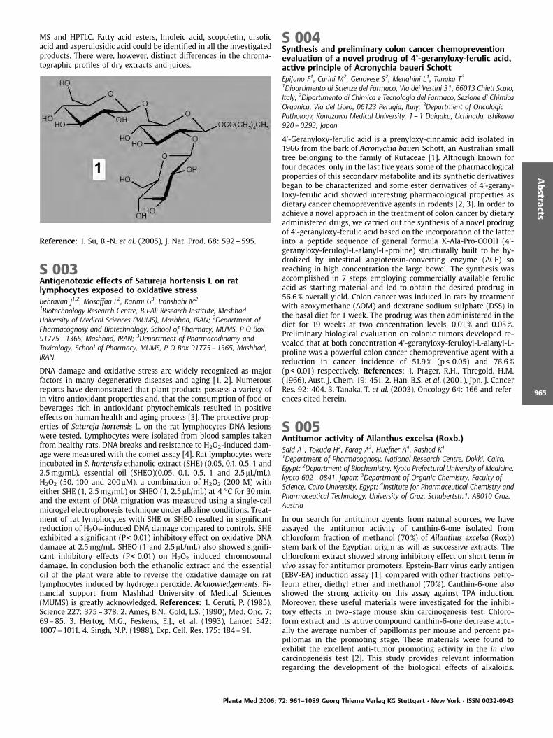
MS and HPTLC. Fatty acid esters, linoleic acid, scopoletin, ursolicacid and asperulosidic acid could be identified in all the investigatedproducts. There were, however, distinct differences in the chroma-tographic profiles of dry extracts and juices.
Reference: 1. Su, B.-N. et al. (2005), J. Nat. Prod. 68: 592–595.
S 003Antigenotoxic effects of Satureja hortensis L on ratlymphocytes exposed to oxidative stressBehravan J1,2, Mosaffaa F2, Karimi G3, Iranshahi M2
1Biotechnology Research Centre, Bu-Ali Research Institute, MashhadUniversity of Medical Sciences (MUMS), Mashhad, IRAN; 2Department ofPharmacognosy and Biotechnology, School of Pharmacy, MUMS, P O Box91775 – 1365, Mashhad, IRAN; 3Department of Pharmacodinamy andToxicology, School of Pharmacy, MUMS, P O Box 91775 –1365, Mashhad,IRAN
DNA damage and oxidative stress are widely recognized as majorfactors in many degenerative diseases and aging [1, 2]. Numerousreports have demonstrated that plant products possess a variety ofin vitro antioxidant properties and, that the consumption of food orbeverages rich in antioxidant phytochemicals resulted in positiveeffects on human health and aging process [3]. The protective prop-erties of Satureja hortensis L. on the rat lymphocytes DNA lesionswere tested. Lymphocytes were isolated from blood samples takenfrom healthy rats. DNA breaks and resistance to H2O2-induced dam-age were measured with the comet assay [4]. Rat lymphocytes wereincubated in S. hortensis ethanolic extract (SHE) (0.05, 0.1, 0.5, 1 and2.5mg/mL), essential oil (SHEO)(0.05, 0.1, 0.5, 1 and 2.5mL/mL),H2O2 (50, 100 and 200mM), a combination of H2O2 (200 M) witheither SHE (1, 2.5mg/mL) or SHEO (1, 2.5mL/mL) at 4 oC for 30min,and the extent of DNA migration was measured using a single-cellmicrogel electrophoresis technique under alkaline conditions. Treat-ment of rat lymphocytes with SHE or SHEO resulted in significantreduction of H2O2-induced DNA damage compared to controls. SHEexhibited a significant (P< 0.01) inhibitory effect on oxidative DNAdamage at 2.5mg/mL. SHEO (1 and 2.5mL/mL) also showed signifi-cant inhibitory effects (P < 0.01) on H2O2 induced chromosomaldamage. In conclusion both the ethanolic extract and the essentialoil of the plant were able to reverse the oxidative damage on ratlymphocytes induced by hydrogen peroxide. Acknowledgements: Fi-nancial support from Mashhad University of Medical Sciences(MUMS) is greatly acknowledged. References: 1. Ceruti, P. (1985),Science 227: 375–378. 2. Ames, B.N., Gold, L.S. (1990), Med. Onc. 7:69–85. 3. Hertog, M.G., Feskens, E.J., et al. (1993), Lancet 342:1007–1011. 4. Singh, N.P. (1988), Exp. Cell. Res. 175: 184–91.
S 004Synthesis and preliminary colon cancer chemopreventionevaluation of a novel prodrug of 4’-geranyloxy-ferulic acid,active principle of Acronychia baueri SchottEpifano F1, Curini M2, Genovese S2, Menghini L1, Tanaka T31Dipartimento di Scienze del Farmaco, Via dei Vestini 31, 66013 Chieti Scalo,Italy; 2Dipartimento di Chimica e Tecnologia del Farmaco, Sezione di ChimicaOrganica, Via del Liceo, 06123 Perugia, Italy; 3Department of OncologicPathology, Kanazawa Medical University, 1 – 1 Daigaku, Uchinada, Ishikawa920 – 0293, Japan
4’-Geranyloxy-ferulic acid is a prenyloxy-cinnamic acid isolated in1966 from the bark of Acronychia baueri Schott, an Australian smalltree belonging to the family of Rutaceae [1]. Although known forfour decades, only in the last five years some of the pharmacologicalproperties of this secondary metabolite and its synthetic derivativesbegan to be characterized and some ester derivatives of 4’-gerany-loxy-ferulic acid showed interesting pharmacological properties asdietary cancer chemopreventive agents in rodents [2, 3]. In order toachieve a novel approach in the treatment of colon cancer by dietaryadministered drugs, we carried out the synthesis of a novel prodrugof 4’-geranyloxy-ferulic acid based on the incorporation of the latterinto a peptide sequence of general formula X-Ala-Pro-COOH (4’-geranyloxy-feruloyl-L-alanyl-L-proline) structurally built to be hy-drolized by intestinal angiotensin-converting enzyme (ACE) soreaching in high concentration the large bowel. The synthesis wasaccomplished in 7 steps employing commercially available ferulicacid as starting material and led to obtain the desired prodrug in56.6% overall yield. Colon cancer was induced in rats by treatmentwith azoxymethane (AOM) and dextrane sodium sulphate (DSS) inthe basal diet for 1 week. The prodrug was then administered in thediet for 19 weeks at two concentration levels, 0.01% and 0.05%.Preliminary biological evaluation on colonic tumors developed re-vealed that at both concentration 4’-geranyloxy-feruloyl-L-alanyl-L-proline was a powerful colon cancer chemopreventive agent with areduction in cancer incidence of 51.9% (p < 0.05) and 76.6%(p < 0.01) respectively. References: 1. Prager, R.H., Thregold, H.M.(1966), Aust. J. Chem. 19: 451. 2. Han, B.S. et al. (2001), Jpn. J. CancerRes. 92: 404. 3. Tanaka, T. et al. (2003), Oncology 64: 166 and refer-ences cited herein.
S 005Antitumor activity of Ailanthus excelsa (Roxb.)Said A1, Tokuda H2, Farag A3, Huefner A4, Rashed K1
1Department of Pharmacognosy, National Research Centre, Dokki, Cairo,Egypt; 2Department of Biochemistry, Kyoto Prefectural University of Medicine,kyoto 602– 0841, Japan; 3Department of Organic Chemistry, Faculty ofScience, Cairo University, Egypt; 4Institute for Pharmaceutical Chemistry andPharmaceutical Technology, University of Graz, Schubertstr.1, A8010 Graz,Austria
In our search for antitumor agents from natural sources, we haveassayed the antitumor activity of canthin-6-one isolated fromchloroform fraction of methanol (70%) of Ailanthus excelsa (Roxb)stem bark of the Egyptian origin as will as successive extracts. Thechloroform extract showed strong inhibitory effect on short term invivo assay for antitumor promoters, Epstein-Barr virus early antigen(EBV-EA) induction assay [1], compared with other fractions petro-leum ether, diethyl ether and methanol (70%). Canthin-6-one alsoshowed the strong activity on this assay against TPA induction.Moreover, these useful materials were investigated for the inhibi-tory effects in two–stage mouse skin carcinogenesis test. Chloro-form extract and its active compound canthin-6-one decrease actu-ally the average number of papillomas per mouse and percent pa-pillomas in the promoting stage. These materials were found toexhibit the excellent anti-tumor promoting activity in the in vivocarcinogenesis test [2]. This study provides relevant informationregarding the development of the biological effects of alkaloids.
Planta Med 2006; 72: 961–1089 Georg Thieme Verlag KG Stuttgart · New York · ISSN 0032-0943
Abstracts
965
References: 1. Kubota et al. (1997), Cancer Lett. 113: 165–168. 2.Henle, G., Henle, W. (1966), J. Bacteriol. 91: 1248–1256.
S 006Active anti-head lice component from custard apple seedGritsanapan W1, Intaranongpai J1, Chavasiri W2
1Department of Pharmacognosy, Faculty of Pharmacy Mahidol University,447 Sri-Ayudthaya Rd, Ratchatevi, Bangkok 10400, Thailand; 2Department ofChemistry, Faculty of Science, Chulalongkorn University, Bangkok, Thailand
Seeds of custard apple (Annona squamosa Linn., Annonaceae) havebeen used for anti-head lice for a long time. In Thailand, the petro-leum ether seed extract prepared as a cream preparation was re-ported to kill 93% of head lice within 3 hours [1]. Twenty grams ofthe 20% w/w freshly prepared cream can kill 94.5 € 9.1% of head licewithin 3 hours when applied to school girls and the cream is biolo-gically stable for at least 12 months [2, 3]. There have been noreports on chemically active anti-head lice component of this plant.The present study is focused on the separation and identification ofthe active compound from the hexane extract of the seeds of custardapple. Chromatographic and spectroscopic techniques revealed thata major component of the hexane seed extract was a triglyceridewith one oleate ester (with 2 unknown acyl moieties). The separat-ed pure triglyceride and the crude hexane extract which were sep-arately diluted with coconut oil (1:1), contained 22.25 and 11.49mgof the triglyceride, respectively were tested in vitro for anti-head liceactivity and found that they could kill all tested head lice within 11and 30 minutes, respectively. The triglyceride with one oleate esterwas the active compound against human head lice. It could be usedas a marker for quality control and standardization of custard appleseeds, the extracts and anti-head lice preparations from the seeds ofthis plant. References: 1. Areekul, M., Chaikledkaew, U. (1944), Anti-parasitic cream from Annona squamosa Linn. A special project sub-mitted in partial fulfillment of the requirement for the degree ofBachelor of Science in Pharmacy. Mahidol University. Bangkok. 2.Gritsanapan, W. et al. (1998), Studies of stability and effectivenessof intensive hair masks from Annona squamosa seed extract. 50th IPCand 17th FAPA Congress, Mumbai, India. 3. Tiangda, CH. et al. (2000),Southeast Asian J. Trop. Med. Public Health 31 (Suppl 1): 174–7.
S 007Antibacterial proanthocyanidins isolated from the Australianmedicinal plant, Planchonia careya (F. Muell.) R. Knuth(Lecythidaceae)McRae J1, Yang Q2, Crawford R1, Palombo E11Environment and Biotechnology Centre, Faculty of Life and Social Science,Swinburne University, P.O. Box 218, Hawthorn, 3122 Australia; 2CSIROMolecular and Health Technologies Division, Bag 10, Clayton South, 3169,Australia
One of the many plants traditionally used for wound healing by theindigenous peoples of northeastern Australia is Planchonia careya (F.Muell) R. Knuth. Based on this knowledge, investigation was carriedout into the antibacterial activity of the leaf extracts of this species.The chemical constituents responsible for the observed activitywere then isolated. The plate-hole diffusion method was used toevaluate the antibacterial activity of the crude aqueous and metha-nol extracts against a range of Gram positive and Gram negativebacteria. Based on these assays, HPLC-piloted activity-guided frac-tionation was carried out to isolate the active compounds from thecrude aqueous extract. Separation was performed using XAD-16media, followed by a 20mm grade Chromatorex C18 column witha 10% methanol/ water mobile phase. The active fractions fromthese columns were separated further with Sephadex LH-20 gel inmethanol, and final isolation was attained using an Alltima Prepara-tive C18 (5mm) column in a 5% methanol/ water mobile phase.Elucidation of the isolated active compounds was achieved by UV,1-D NMR (1H, 13C), and 2-D NMR (COSY, HSQC, HMBC) techniques.
This analysis yielded (+)-gallocatechin and the prodelphinidin, gal-locatechin-(4a-8)-gallocatechin. The structures were confirmed byreference to previously reported NMR spectra of these compounds(1, 2). Further examination of the UV profiles of other active frac-tions and of the crude methanol extract suggests that the minoractive constituents of P. careya are also of the flavonoid class. Theisolation of these known antibacterial compounds confirms the tra-ditional use of P. careya in wound healing. References: 1. Sun, D., etal. (1987), Phytochem. 26: 1825–1829. 2. Cai, Y., et al. (1991), Phy-tochem. 30: 2033–2040.
S 008Antidermatophytic prenylated coumarins from asafetidaHoughton PJ1, Ismail KM1, Maxia L2, Appendino G2
1Pharmacognosy Research, Pharmaceutical Sciences Research Division, KCL,150 Stamford St, SE1 9NH London, UK; 2DISCAFF, 28100 Novara, Italy
Asafoetida is a resinous substances with a smell similar to garlic,which is obtained by drying the exudates from various species ofFerula growing in northern Iraq and Iran and surrounding countries.It is widely used in cooking in India and is used medicinally forgastro-intestinal complaints and for treating skin diseases. The bo-tanical source of commercial samples of asafoetida is not easy todetermine since several species of Ferula exist. Samples of asafetidawith proven source were obtained from the pharmacognosy mu-seum of King’s College London and compared with some commer-cial samples obtained from Asian shops in the UK, India and Syria.Samples were examined by TLC and for antifungal activity usingserial dilution assay in microtitre plates with two dermatophytes,Microsporeum gypseum and Trichophyton interdigitale [1]. The mostactive sample was obtained from India and on TLC conformed mostclosely with a museum sample from F. foetida Regel. From this sam-ple nine prenylated coumarins were isolated and were testedagainst the two fungal species. Four of the compounds exhibitedstrong antifungal activity against the dermatophytes with 5,8 dihy-droxyumbelliprenin 1 being most active with MIC of 10mM, thepositive control miconazole having MIC of 0.5mM. No compoundsof this type have previously shown antifungal activity.
1
Reference: 1. Mensah, A.Y. et al. (2000), J. Nat. Prod. 63:1210–1213.
S 009Antiviral compounds from Icelandic lichensOmarsdottir S1, lad ttir AK1, rnad ttir T2, Ing lfsd ttir K1
1Faculty of Pharmacy, University of Iceland, Hagi, Hofsvallagata 53, IS-107Reykjavik, Iceland; 2Department of Virology, Landspitali-University Hospital,IS-101 Reykjavik, Iceland
Although one third of prescription drugs are derived from naturalsources [1], lichens have only been investigated to a limited extentfrom a pharmacological perspective. The aim of the study was toinvestigate whether antiviral compounds could be found in Icelan-dic lichens, to isolate active compounds in a purified form and elu-cidate their chemical structure and to confirm antiviral activity ofisolated compounds and compare it with that of marketed antiviraldrugs. Extracts were made from ten lichen species and screened forantiviral activity in vitro against three different viruses by using theplaque reduction assay (PRA). Two compounds exhibiting potentantiviral activity against respiratory syncytial (RS) virus were isolat-ed and purified and their activity confirmed using both the PRAmethod and ELISA. The compounds were the depsidone salazinic
Planta Med 2006; 72: 961–1089 Georg Thieme Verlag KG Stuttgart · New York · ISSN 0032-0943
Abstracts
966
acid from Parmelia saxatilis (L.) Ach. and the benzyl depside alector-ialic acid from Alectoria nigricans (Ach.) Nyl. The activity of bothlichen compounds was more potent than that of the marketed drugribavirin, which is used to treat serious respiratory conditions re-sulting from RS infection. The IC50 value for salazinic acid as deter-mined by ELISA was 11.9mg/mL, for alectorialic acid 17.0mg/mL andfor ribavirin 22.9mg/mL. The lichen compounds were no cytotoxic atantiviral concentrations. Activity against herpes viruses I and II wasless potent than activity against RS virus. Acknowledgements: Icelan-dic Council of Science, University of Iceland Research Fund, TheIcelandic Research Fund for Graduate Students. Reference: 1. King-horn, A.D. (2001), J. Pharm. Pharmacol. 53: 135–148.
S 010Inhibitory effects of cucurbitacin R on lymphocyteproliferation and cytokine productionEscandell JM1, Recio MC1, Gil R2, Merfort I3, R os JL11Departament de Farmacologia, Facultat de Farmacia, Universitat deVal ncia, Av. Vicent Andr s Estell s s/n, 46100 Burjassot, Spain;2Departament de Patologia, Facultat de Medicina, Universitat de Val ncia,Av. Blasco Ib ez 15, 46010 Valencia, Spain; 3Department of PharmaceuticalBiology and Biotechnology, University Freiburg, Freiburg, Germany
Cucurbitacin R (CCR) isolated from tayuya roots reduced both theacute and subchronic inflammation in different experimental mod-els [1]. In addition, its acetyl-derivative showed inhibitory effects ina model of adjuvant-induced arthritis [2]. In order to gain insightinto the mechanism of action of CCR, we studied its effect not onlyon lymphocyte proliferation induced by phytohemagglutinin (PHA),but also on the lymphocyte cell cycle. In addition, we examined itsinfluence on the production of cytokines, and the effects on cyclinsA1, B1, D2 and E2 and the transcription factors involved in inflamma-tion. CCR strongly inhibited lymphoproliferation with an IC50 valueof 16mM, arresting the cell cycle in the G0 phase. Inhibition of lym-phoproliferation and on cell cycle disappeared with time. Westernblot analysis was used to show CCR’s effects on assayed cyclins. Theproduction of mediators such as IL-2, IL-4, IL-10, TNF-a and IFN-g byhuman lymphocytes was also significantly inhibited by CCR, withIC50 values of 18mM for interleukins, 12mM for IFN-g, and 15mM forTNF-a. The PCR analysis showed a clear inhibition of all these cyto-kines. In Jurkat cells, a total inhibition of the nuclear factor of Tactivated cells (NF-AT) was observed at a 50mM concentration ofCCR. AP-1 remained unaffected. These results indicate that lympho-cyte proliferation is inhibited by CCR through NF-AT blocking, whichreduces cytokine production. Acknowledgements: J.M.E. is recipientof a grant from the Generalitat Valenciana. This work was supportedby the Spanish Government (SAF2002–00723) References: 1. Recio,M.C. et al. (2004), Planta Med. 70: 414–420. 2. Escandell, J. et al.(2006), Eur. J. Pharmacol. 532:145–154.
S 011Four New Natural Products from Mongolian Medicinal PlantsScorzonera divaricata and Scorzonera pseudodivaricata(Asteraceae)Edrada RA1, Tsevegsuren N1,2, Lin W3, Ebel R1, Torre C1, Ortlepp S1,Wray V4, Proksch P11Institut f r Pharmazeutische Biologie und Biotechnologie, Heinrich-Heine-Universit t D sseldorf, Universit tsstr. 1, Geb. 26.23, 40225 D sseldorf,Germany; 2Department of Organic and Food Chemistry, Faculty of Chemistry,National University of Mongolia, Ulaanbaatar, Mongolia; 3State KeyLaboratory of Natural and Biomimetic Drugs, Peking University, Beijing, P. R.China; 4Gesellschaft f r Biotechnologische Forschung, Braunschweig
Eleven Scorzonera species are found in Mongolia, one species isendemic, four of which are sub-endemic [1]. Two of these, Scorzo-nera pseudodivaricata Lipsch., a sub-endemic perennial species, andS. divaricata Turcz. are used in the Mongolian traditional medicine[2]. Since only a few papers have been published on this genus and
no previous chemical work has been recorded on S. divaricata and S.pseudodivaricata, this arose our interest to do further phytochemicalwork on these plants. Investigation of the (diphenylpicrylhydrazyl)DPPH-active EtOAc extract of aerial parts of S. divaricata, whichshowed radical scavenging activity, yielded two new 1-O-caffeoyl-quinic acid derivatives. From the cytotoxic EtOAc extract of aerialparts of S. pseudodivaricata, a novel phenolic glucoside and an un-sual terpene lactone were isolated. The structures of the new com-pounds were unambiguously established based on NMR spectro-scopic (1H, 13C, COSY, HMBC) and mass spectrometric (ESIMS) data.References: 1. Grubov, V. I. (1982), Key to the Vascular Plants ofMongolia, Leningrad, Nauka, pp. 263–264. 2. Ligaa, U. (1996), Med-icinal Plants from Mongolia Used in Mongolian Traditional Medi-cine, KSA Press, p. 337.
S 012Secondary metabolites of Globularia species from the Floraof TurkeyKirmizibekmez H2,1, Calis I11Department of Pharmacognosy, Faculty of Pharmacy, Hacettepe University,TR-06100, Ankara, Turkey; 2Department of Pharmacognosy, Faculty ofPharmacy, Yeditepe University, TR-34755, Erenkoy, Istanbul, Turkey
The genus Globularia (formerly Globulariaceae, now “new“ Planta-ginaceae) is represented in the flora of Turkey by nine species, threeof which are endemic [1, 2]. Some of these species are used asdiuretic, laxative, stomachic, tonic and for the treatment of haemor-rhoids in Anatolian folk medicine [3, 4]. Among these species, G.alypum is widely used in indigenous systems of medicine in someMediterrean countries, especially in Morocco as a hypoglycaemicagent, laxative, cholagogue, stomachic and purgative [5]. As a partof our interest on Turkish medicinal plants we have investigated thesecondary metabolites of seven Globularia species, G. trichosanthaFosch. Et Mey., G. orientalis L., G. cordifolia L., G. dumulosa O.Schwarz, G. davisiana O. Schwarz, G. sintenisii Hausskn. et Wettst.and G. alypum L.. Various chromatographic studies (VLC, MPLC, OCC)on the MeOH (or EtOH) extracts of the aerial or underground partsof the species resulted in the isolation of 58 different compounds,which can be categorized under eight chief groups; 27 iridoids, 14phenylethanoid glycosides, 6 flavone glycosides, 4 lignan glycosides,3 sugar esters, 2 sterols, an acetophenone glycoside, and a phenyl-propanoid glycoside. The structures of the isolates were elucidatedby 1D and 2D NMR and MS experiments as well as chemical meth-ods. 15 of the isolated compounds were new for nature, while manyof them were new to the genus Globularia. The occurrence of suchdiverse compounds in Globularia might be of great chemotaxono-mical importance at both the genus and family level. Recent study(6) based on the DNA sequence of the genus Globularia placed thisgenus under the “new“ Plantaginaceae family, which was in goodaccordance with our results. Acknowledgement: Hacettepe Univer-sity, Scientific Research Unit (Project number: 02 G 076) Refer-ences: 1. Edmondson, J.R. (1982), Globularia L., in Flora of Turkeyand the East Aegean Islands. Vol. 7 (Ed. Davis P.H.), University Press,Edinburgh. 2. Duman, H. (2001), Bot. J. Linn. Soc. 137: 425–428. 3.Baytop, T. (1999), Therapy with Medicinal Plants in Turkey (Past andPresent), Nobel Tip Kitapevleri, Istanbul, p. 371. 4. Sezik, E. et al.(1991), J. Ethnopharmacol. 35: 191–196. 5. Bellakhdar, J. et al.(1991), J. Ethnopharmacol, 35: 123–143. 6. Albach, D.C. et al.(2005), Am. J. Bot. 92: 297–315.
Planta Med 2006; 72: 961–1089 Georg Thieme Verlag KG Stuttgart · New York · ISSN 0032-0943
Abstracts
967

S 013Adaptogens modify stress response by suppressing theincrease of p-SAPK, nitric oxide and cortisone in the blood ofrabbitsPanossian A1, Hambartsumyan M2, Hovhanissyan A2, Wikman G1
1Swedish Herbal Institute Research and Development, Prinsgatan 12, SE-41305 G teborg, Sweden; 2ExLab“ Expert Analytical Laboratory of Armenia DrugAgency, Komitas Ave. 49/4, 375051 Yerevan, Armenia
Adaptogens possess anti-fatigue and anti-stress activities that canincrease mental and physical working performance against a back-ground of fatigue or stress. The aim of the present study was toascertain which mediators of stress response are significantly in-volved in the mechanisms of action of adaptogens, and to determinetheir relevance as biochemical markers for evaluating anti-stresseffects in laboratory animals subjected to immobilisation stress.Basal blood levels of cortisone, testosterone, nitric oxide, prosta-glandin E2, thromboxane B2, leukotriene B4, stress-activated proteinkinase (SAPK), and phosphorylated-SAPK (p-SAPK/p-JNK) were de-termined in three groups of rabbits. Group A and B animals weretreated orally for 7 days with extracts of Eleutherococcus senticosus(Rupr. & Maxim.) Maxim., Schizandra chinensis (Turcz.) Baill., Panaxginseng C.A. Meyer, Bryonia alba L., Rhodiola rosea L. and activecomponent rhodioloside; group C received only placebo. Ten min-utes after the final treatment, group A and C animals were immobi-lized for 2 hours, and blood levels of markers in rabbits of all groupsre-determined. Only p-SAPK, cortisone and nitric oxide increasedsignificantly (200–300% > basal levels) following immobilizationstress (group C). However, following repeated administration ofadaptogens, basal levels of these markers remained practically un-changed during acute stress (group A). S. chinensis, R. rosea andrhodioloside were the most active inhibitors of p-SAPK formation(group B). It is speculated that the positive effects of adaptogens onmental performance in stress may be associated with the inhibitionof p-SARK formation, and that such activity might be beneficial inneurodegenerative disorders associated with loss of neurons inbrain regions involved in learning and memory.
S 014Antiviral Terpenoid Constituents of Ganoderma pfeifferi BresLalk M, Niedermeyer THJ, Mentel R, Lindequist UInstitute of Pharmacy, Ernst-Moritz-Arndt-University, Friedrich-Ludwig-Jahn-Str. 17, 17487 Greifswald, Germany
Ganoderma pfeifferi Bres., a weak parasitic and later saprophyticbasidiomycete, is a fungus only found in Europe. It is living prefer-entially on Fagus L. and some other deciduous trees such as AesculusL., Acer L., Fraxinus L. and Quercus L.. In contrast to G. lucidum (Fr.)Karst. and G. applanatum (Persoon) Patouillard, from which a num-ber of biologically and pharmacologically interesting triterpenes,steroids and polysaccharides have been isolated [1], G. pfeifferi isone of the phytochemically poorer examined species of the familyGanodermataceae [2]. As part of our continuing interest in com-pounds from G. pfeifferi, four sterols and ten triterpenes were isolat-ed from a DCM-extract of the fruiting bodies of this mushroom. Inaddition to compounds common in mushrooms and other Ganoder-mataceae, we isolated the previously unknown triterpenoid consti-tuents 3,7,11-trioxo-5a-lanosta-8,24-diene-26-al 1 (LucialdehydeD), 5a-lanosta-8,24-diene-26-hydroxy-3,7-dione 2 (Ganoderone A),and 5a-lanosta-8-ene-24,25-epoxy-26-hydroxy-3,7-dione 3 (Gano-derone C). The evaluation of the antiviral properties of the isolatedcompounds showed strong inhibition of the growth of Herpes sim-
plex and influenza viruses with IC50 values between 0.01 and 3.0mg/mL [3].
1
2
3
References: 1. Gao, Y.; Zhou, S.; Huang, M.; Xu, A. (2003), Int. J. Med.Mushrooms 5: 235. 2. (a) Mothana, R.A.A., Jansen, R., J lich, W.-D.,Lindequist, U. (200), J. Nat. Prod. 63: 416, (b) Mothana, R.A.A., Ali,N.A.A., Jansen, R., Wegner, U., Mentel, R., Lindequist, U. (2003), Fito-terapia 74: 177. 3. Niedermeyer, T.H.J; Lindequist, U.; Mentel, R.;G rdes, D.; Schmidt, E.; Thurow, K.; Lalk, M. (2005), J. Nat. Prod.68: 1728.
S 015In search of promising antimalarial drugs: Detection ofheme-based adducts induced in complex matrixes fromBrazilian plants using HPLC-DADCastro-Gamboa I1, Pauletti PM1, Cavalheiro AJ1, Siqueira DHS1,da S. Bolzani V1
1Nfflcleo de Bioensaios, Bioss ntese e Ecofisiologia de Produtos Naturais –NuBBE, Universidade Estadual Paulista – UNESP – Instituto de Qu mica,Departamento de Qu mica Org nica, Rua Prof. Francisco Degni s/n –14.800 – 900 – Araraquara – S¼o Paulo – Brasil
The development of fast and efficient detection and HPLC separationmethods on a bioprospection program is crucial for speeding-up theselection of natural matrixes. Based on this goal, the induction of anin situ heme adduct in crude plant extracts from Brazilian Cerradoand Atlantic Forest turned out to be a powerful tool, foretelling if acrude extract contains promising molecules that may have antima-
Planta Med 2006; 72: 961–1089 Georg Thieme Verlag KG Stuttgart · New York · ISSN 0032-0943
Abstracts
968
larial or antileishmanial properties. Studies of heme adducts werereported using known drugs such as quinine and artemisinin. Weinitially selected 75 plants from our bank of extracts based on che-mosystematics, reported antimalarial activities as well as ethno-pharmacological data. Species such as Arrabidaea samydoides(Cham.) Sandwith (Bignoniaceae), Strychnos pseudoquina St. Hil. (Lo-ganiaceae), Garcinia gardineriana Miers ex Planch. Et Triana (Clusia-ceae), Zanthoxyllum rhoifolium Lam. (Rutaceae), Sorocea bonplandiiBaill. Burg. (Moraceae) and Bidens segetum Mart. ex Colla (Astera-ceae) were some of the matrixes that showed adduct formationwhen incubated with hemine. Through the observation of retentiontime shifts and comparison of UV spectra after adduct induction, wewere able to pin-point the responsible molecules and thus selectplant extracts for further specific studies. C-glucosylxanthonesisolated from A. samydoides were the metabolites responsible foradduct formation. Acknowledgements: To Funda ¼o de AmparoPesquisa do Estado de S¼o Paulo (FAPESP), CAPES and CNPq forresearch funding.
S 016Inhibition potential of natural based products againstChlamydia pneumoniae infectionAlvesalo J1,2, Vuorela HJ2, Tammela P1,2, Leinonen M3, Saikku P4,Vuorela PM5
1Drug Discovery and Development Technology Center, Faculty of Pharmacy,P.O. Box 56, FI-00014 University of Helsinki, Finland; 2Division ofPharmaceutical Biology, Faculty of Pharmacy, P.O. Box 56, FI-00014University of Helsinki, Finland; 3National Public Health Institute, PO Box 310,FI-90101, Oulu, Finland; 4Department of Microbiology, PO Box 5000, FI-90014 Oulu University, Finland; 5Department of Biochemistry and Pharmacy,bo Akademi University, Tykist katu 6A, FI-20520 Turku, Finland
A large number of antimicrobial substances, phytoalexins, are foundin nature and they form a variable group of compounds playingimportant role in the natural defence of living organism. This studywas carried out to evaluate whether several groups of natural, nat-ural derived synthetic compounds or natural extracts have an im-pact on C. pneumoniae infection, in vitro. 37% (21/57) of the testedcompounds were highly active; 28% (16/57) active; 11% (6/57) mod-erately active; 24% (14/57) inactive. Highly active compounds werefound in many compound groups, but the most active group wasthat of gallates. Inactive compounds could also be found in manycompound classes, but among synthetic coumarins, many com-pounds had 0% inhibition. Chlamydia pneumoniae is a commoncause of acute upper and lower respiratory tract infections, includ-ing pharyngitis, sinusitis and pneumonia, but it also has a tendencyto cause chronic infections. There is augmenting evidence on theinvolvement of chronic C. pneumoniae infection in the atherosclero-tic diseases like coronary heart disease. Even though the acute in-fections can be successfully treated with several antibiotics, theeradication of chronic C. pneumoniae infection seems to be exceed-ingly difficult. High doses and prolonged treatment is often neededto achieve clinical cure and there is still a risk of the persistence of C.pneumoniae in the tissues after treatment. Thus, it is extremelyimportant to find new compounds that can be used in the treatmentor prophylaxis of C. pneumoniae infections.
S 017Two new isomeric tropane alkaloids from Schizanthustricolor identified by capillary NMRHumam M1, Kehrli T1, Jeannerat D2, Mu oz O3, Christen P1,Hostettmann K1
1Laboratory of Pharmacognosy and Phytochemistry, School ofPharmaceutical Sciences, University of Geneva, University of Lausanne, QuaiErnest-Ansermet 30, CH-1211 Geneva 4, Switzerland; 2Department ofOrganic Chemistry, University of Geneva, Quai Ernest-Ansermet 30, CH-1211Geneva 4, Switzerland; 3Departamento de Qu mica, Facultad de Ciencias,Universidad de Chile, Casilla 653, Santiago, Chile
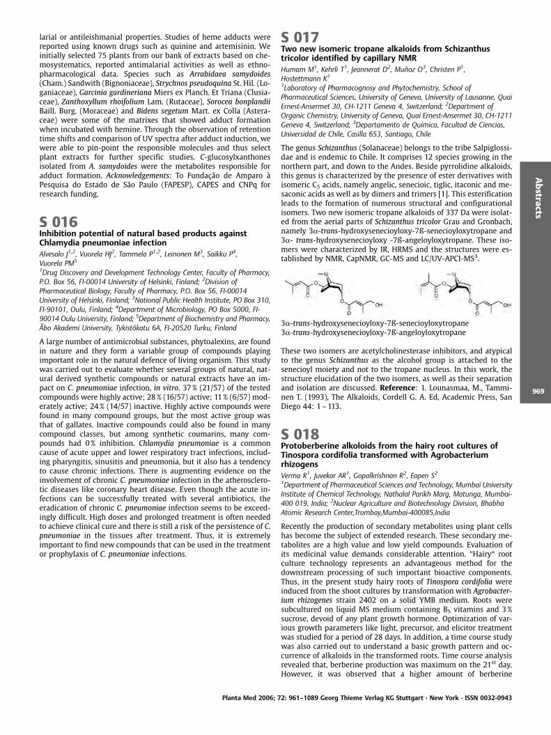
The genus Schizanthus (Solanaceae) belongs to the tribe Salpiglossi-dae and is endemic to Chile. It comprises 12 species growing in thenorthern part, and down to the Andes. Beside pyrrolidine alkaloids,this genus is characterized by the presence of ester derivatives withisomeric C5 acids, namely angelic, senecioic, tiglic, itaconic and me-saconic acids as well as by dimers and trimers [1]. This esterificationleads to the formation of numerous structural and configurationalisomers. Two new isomeric tropane alkaloids of 337 Da were isolat-ed from the aerial parts of Schizanthus tricolor Grau and Gronbach,namely 3a-trans-hydroxysenecioyloxy-7ß-senecioyloxytropane and3a- trans-hydroxysenecioyloxy -7ß-angeloyloxytropane. These iso-mers were characterized by IR, HRMS and the structures were es-tablished by NMR, CapNMR, GC-MS and LC/UV-APCI-MS3.
3a-trans-hydroxysenecioyloxy-7ß-senecioyloxytropane3a-trans-hydroxysenecioyloxy-7ß-angeloyloxytropane
These two isomers are acetylcholinesterase inhibitors, and atypicalto the genus Schizanthus as the alcohol group is attached to thesenecioyl moiety and not to the tropane nucleus. In this work, thestructure elucidation of the two isomers, as well as their separationand isolation are discussed. Reference: 1. Lounasmaa, M., Tammi-nen T. (1993), The Alkaloids, Cordell G. A. Ed, Academic Press, SanDiego 44: 1– 113.
S 018Protoberberine alkoloids from the hairy root cultures ofTinospora cordifolia transformed with AgrobacteriumrhizogensVerma R1, Juvekar AR1, Gopalkrishnan R2, Eapen S21Department of Pharmaceutical Sciences and Technology, Mumbai UniversityInstitute of Chemical Technology, Nathalal Parikh Marg, Matunga, Mumbai-400 019, India; 2Nuclear Agriculture and Biotechnology Division, BhabhaAtomic Research Center,Trombay,Mumbai-400085,India
Recently the production of secondary metabolites using plant cellshas become the subject of extended research. These secondary me-tabolites are a high value and low yield compounds. Evaluation ofits medicinal value demands considerable attention. “Hairy“ rootculture technology represents an advantageous method for thedownstream processing of such important bioactive components.Thus, in the present study hairy roots of Tinospora cordifolia wereinduced from the shoot cultures by transformation with Agrobacter-ium rhizogenes strain 2402 on a solid YMB medium. Roots weresubcultured on liquid MS medium containing B5 vitamins and 3%sucrose, devoid of any plant growth hormone. Optimization of var-ious growth parameters like light, precursor, and elicitor treatmentwas studied for a period of 28 days. In addition, a time course studywas also carried out to understand a basic growth pattern and oc-currence of alkaloids in the transformed roots. Time course analysisrevealed that, berberine production was maximum on the 21st day.However, it was observed that a higher amount of berberine
Planta Med 2006; 72: 961–1089 Georg Thieme Verlag KG Stuttgart · New York · ISSN 0032-0943
Abstracts
969
(0.034%) was produced in the cultures treated with 500mg/L of L-Tyrosine as precursor, than the control. Jasmonate elicitation wasfound best at the concentration of 200mmoles/mL (0.047%). More-over the berberine content in hairy roots was comparable to thatproduced by the roots of parent plant. HPLC and HPTLC results showthe presence of jatrorrhizhine, in trace amounts. Thus it can beconcluded that, the hairy root cultures form a promising sourcefor the production of berberine and related compounds. Refer-ences: 1. Hyeon-J., Jack, M.,(2002), Plant Cell Tissue Organ Culture69:259–269. 2. Kamada, H. et al, (1996), Plant Cell Rep. 5: 239–242. 3. Ravishankar, G.A., Venkatraman, L.V. (1997), BiotechnologicalApplication of Plant Tissue and Cell Culture, Oxford and IBH pub-lishings Co, New Delhi, pp. 74 –90.
S 019Anti-stress anxiolytic and nootropic activity of Nyctanthesarbour tritis leavesDeshmukh VS, Juvekar ARDepartment of Pharmaceutical Sciences and Technology, Mumbai UniversityInstitute of Chemical Technology, Nathalal Parikh Marg, Matunga, Mumbai-400 019, India
Reports suggest that stress is the most common etiological factor inCNS disorders like anxiety, Schizophrenia, Parkinson’s disease andAlzheimer disease for which effective treatment strategies are in-adequate due to complexities of the ailment and the limitations ofallopathic medications. There are scanty reports1 on the putativeneuro pharmacological effects of the leaves of Nyctanthes arbortristis Linn. (Family: Oleaceae) [NAT]; hence the present work in-vestigated gamut of its neuro-pharmacological effects. The metha-nolic extract was evaluated for anxiolytic activity using plus mazemodel, open field test and light dark model. Further, the nootropicpotential2 of extract was evaluated using Morris water maze testand plus maze model. Antistress potential3 was evaluated in Wistarrats by subjecting the animals to chronic cold restraint stress fol-lowed by biochemical estimation of plasma corticosterone, glucose,triglycerides; dopamine, 5-Hydroxy Tryptamine and nor epine-phrine from brain. Diazepam 1mg/kg was used as a positive control.One-way ANOVA followed by Dunnett’s test was applied for statis-tical significance. Pretreatment with NAT extract resulted in prefer-ence to open arm in plus maze test, increased exploratory behaviorin open field test and increased number of crossings in light darkmodel. Further it improved cognitive function with respect to spa-tial and working memory processes. The treatment with NAT extractameliorated the stress-induced variations in the biochemical levelsof corticosterone, glucose, triglycerides; dopamine, 5-HT and norepinephrine. In conclusion, the NAT extract exhibited anxiolytic,antistress and nootropic activity with utility in oxidative cognitiveimpairment due to its antioxidant potential. References: 1. SaxenaR.S., Gupta B. (2002), J. Ethnopharmacol. 81: 321-/325. 2. Vogel,G.H., Vogel, W.H. (Eds) (2005), Drug Discovery and Evaluation- Phar-macological assays, pp. 435. 3. Nachankar, R.S., Juvekar, A.R.A.(2005), Acta Hort. (ISHS) 680:101–107.
S 020New Norterpene Cyclic Peroxides from the SpongeDiacarnus megaspinorhabdosaEdrada RA1, Ibrahim S1, Ebel R1, Wray V2, M ller WEG3, Proksch P11Institut f r Pharmazeutische Biologie und Biotechnologie, Heinrich-Heine-Universit t D sseldorf, Universit tsstr. 1, Geb. 26.23, 40225 D sseldorf,Germany; 2Gesellschaft f r Biotechnologische Forschung, Braunschweig; 3Institut f r Physiologische Chemie und Pathobiochemie, Johannes-Gutenberg-Universit t, Mainz
Chemical investigation of the n-hexane extract of the sponge Dia-carnus megaspinorhabdosa has provided a series of norterpenes, in-cluding three new norditerpene cyclic peroxides and five new nor-sesterterpene peroxides together with four known norterpene per-
oxides: nuapapuin A methyl ester, epimuqubilin B, methyl-2-epi-nuapapuinoate and methyl diacarnoate A. The structures of thenew compounds were established on the basis of one and two di-mensional NMR spectroscopic studies (1H, 13C, COSY, HMQC, HMBCand ROESY) as well as on mass spectral analysis. The isolated com-pounds exhibited moderate (2 –5mg) to strong toxicity (0.01 –0.10mg) toward L5178Y (mouse lymphoma) and HeLa (human cervixcarcinoma) while the same congeners showed weaker activity onthe PC-12 (rat pheochromocytoma) cell line. Capon’s empiricalrules1 were extensively used in this study to derive the relativestereochemistry at C-2, C-3 and C-6. Following Horeau’s procedure,the peroxide ring was cleaved to yield its diol congener onto whichthe advanced Mosher method was utilized to confirm the stereo-chemistry obtained from Capon’s empirical rules. References: 1.Ca-pon, R.J., MacLeod, J.K. (1985), Tetrahedron 41: 3391–3404. 2. Hor-eau, A. (1977), Determination of the configuration of secondary al-cohol by partial resolution, in Stereochemistry, Fundamentals andMethods, Kagan, H.B ed., Vol. 3., Georg Thieme, Stuttgart, p. 51.
S 021New materials for extraction, separation and massspectrometric investigations in phytochemistryStecher G, Hashir MA, Bonn GKInstitute of Analytical Chemistry and Radiochemistry, University of Innsbruck,Innrain 52a, 6020, Innsbruck, Austria
The design of novel materials and stationary phases for the selectiveextraction and fast separation of analytes from plant materials is animportant part in phytomics [1]. In fact, preconcentration and pur-ification prior to analysis is necessary owing to the complexity ofsamples. Add to this, analytes are often present in low concentra-tions, what means that sample extraction, purification and precon-centration are the starting points to successful analyses. Within thispresentation we present different strategies for the synthesis andthe modification of stationary phases to produce tailored solutionsfor the analytical questions. In fact, sample preparation proceduresshould be shortened as much as possible to save time and consum-ables and to prevent degradation of target compounds. As an exam-ple the combination of extraction and preconcentration or/and se-paration within one step using selective materials will be presented.In this coherence not only multidimensional chromatography playsa central role, but also the use of new stationary phases withindifferent formats such as columns, capillaries and discs. Further onfocus will be placed on the separation of analytes and on the detec-tion. Especially the use of matrix assisted laser desorption ioniza-tion time of flight mass spectrometry (MALDI-ToF-MS) for the anal-ysis of small molecules using a newly synthesized material as ma-trix free system will be presented [2]. In this coherence examples ofdifferent classes of plant ingredients will be shown. Performance ofthe introduced material will be compared with different systemsdescribed in literature accenting its effectiveness and power forscreening plant systems and metabolites. References: 1. Stecher,G., Huck, C.W., St ggl, W.M., Bonn, G.K. (2003), TrAC, 22: 1–14. 2.Bonn, G.; Hashir, M.A.; Stecher, G., Bakry, R., (2006), Patent pending.
S 022Exploration of natural alkylamides and synthetic analogs assource for new ligands for the cannabinoid type-2 receptorGertsch J1, Raduner S1, Feyen F1, Altmann KH1
1Institute of Pharmaceutical Sciences, ETH Zurich, 8093-Z rich, Switzerland
The cannabinoid type-2 (CB2) receptor is an attractive target for thedevelopment of drugs against inflammatory disease, atherosclerosis,and osteoporosis. Based on the discovery that alkylamides (alka-mides) from Echinacea constitute a new class of CB2-specific canna-binomimetics1, we have screened a series of synthetic analogs ofEchinacea alkylamides as well as other plant-derived natural N-alkylamides for competitive binding to the CB2-receptor. Because dode-
Planta Med 2006; 72: 961–1089 Georg Thieme Verlag KG Stuttgart · New York · ISSN 0032-0943
Abstracts
970
ca-2E,4E-dienoic acid isobutylamide from Echinacea has a high affi-nity to the human CB2-receptor we synthesized analogs with mod-ified head groups, as reported for the endogenous cannabinoid ana-ndamide2. The resulting preliminary structure-activity relationshipclearly shows that the CB2 receptor binding mode of Echinacea al-kylamides is different from anandamide. To further explore the po-tential of natural N-alkyl amides as a new general class of CB2 li-gands we have initiated a screening of plant extracts. The hexaneextracts of the medicinal plants Spilanthes oleracea L. and Lepidiummeyenii Walper. which are known to contain distinct types of alky-lamides, were tested in receptor binding assays. The alkylamide-fraction of the L. meyenii extract showed significant receptor bindingand five isolated benzylated alkylamides (macamides) were as-sessed for both their CB2-receptor affinity as well as CB2-mediatedfunctional effects. Our data show that natural alkylamides fromEchinacea and Lepidium are promising candidates for the develop-ment of novel CB2-receptor ligands. Acknowldgments: Prof. Ikhlas A.Khan, School of Pharmacy, University of Mississippi for providingthe macamide references References: 1. Raduner, S. et al. (2006), J.Biol. Chem. 281: 14192–14206. 2. Khanolkar, A.D. et al. (1996), J.Med. Chem. 39: 4515–4519.
S 023Selected phototoxicological assays used for plantmetabolites screeningChobot V1,2, Vytlacilov J1, Kubicov L11Faculty of Pharmacy, Charles University, Heyrovskeho 1203,500 05 HradecKr love, Czech Republic; 2Present address: Faculty of Life Sciences, UniversityofVienna, Althanstral!,e 14, A.1090 Vienna, Austria
In the last decade, phototoxins especially attracted the attention ofpharmacists, toxicologists, cosmetologists, and food specialists [1].Progress in phototoxicologal research relies on efforts to developreliable screening methods. This issue will be discussed with theaid of the thiophene polyacetylene (E)-1-[5-(hept-5-en-1,3-diynyl)-2-thienyl]ethan-1,2-diol. Together with the furocoumarin xantho-toxin as positive control phototoxicity in conjunction with UV Aradiation was assessed by histidine photo-oxidation assay and Arle-mia and Tubifex bioassays [2]. The determined activities were sta-tistically explored by probit-log calculations of EC50 and LC50 valuesand evaluated by comparison of effective and lethal concentrationsof dark controls to the irradiated sets. The thiophene polyacetyleneshowed strong phototoxicity in the histidine photo-oxidation assayand in both organismic bioassays. Xanthotoxin demonstrated highereffects independent of UV radiation in the Artemia assay. The differ-ences in the phototoxicity of both photosensitizers may be causedby their variable absorption of the test compound and differentmechanisms of activity. The results demonstrate the different sen-sitivity of the applied assays and suggest combining various photo-toxicological assessment methods. Acknowledgements: This workwas supported by Project MSM 0021620822 of the Czech Ministryof Education. References: 1. Chobot, V. et al. (2004), Cent. Eur. J.Publ. Health 12: S31.S33. 2. Chobot, V. et al. (2006), Fitoterapia 77:194–198.
S 024Antifouling and Anti-Aggregatory Effects of Bastadins fromthe Marine Sponge Ianthella bastaOrtlepp S1, Edrada-Ebel RA1, Ebel R1, Hohlfeld T2, Bohlin L3, Proksch P11Pharmaceutical Biology and Biotechnology, Heinrich-Heine University,Universit tstrasse 1, Geb. 26.23, 40225 D sseldorf, Germany;2Pharmacology and Clinical Pharmacology, Heinrich-Heine University,Universit tstrasse 1, Geb. 22.21, 40225 D sseldorf, Germany;3Pharmacognosy, Medicinal Chemistry, Box 574, 751 23 Uppsala, Sweden
Marine sponges are sessile, soft bodied invertebrates that relymainly on the accumulation of toxic and/or deterrent natural pro-ducts as a chemical defence against predators (fishes) and other
biotic stress factors such as fouling by epibionts. Overgrowth byfouling organisms can be detrimental to filter feeders like spongesas it will block pores that are needed for inhaling seawater followedby phagocytosis of suspended particles. As marine sponges areusually free of overgrowth a suppression of epibionts by spongederived natural products is usually assumed. In this study we em-ployed a settling bioassay using barnacle cyprids (Balanus improvi-sus Darwin) in order to investigate sponge compounds for possibleanti-fouling activity. The compounds studied are complex bromi-nated tyrosine derived substances named bastadins. The substanceswere isolated from the marine sponge Ianthella basta collected inIndonesia and included a new bastadin congener along with theknown compounds bastadin 3, 4, 9 and 16. All bastadins showedpronounced inhibition of cyprid settlement and are suggested beinvolved in the chemical defence of the sponge against fouling or-ganisms. Additionally, the bastadins were also tested for humanplatelet aggregation inhibition and gave likewise positive results.Preliminary results suggest that the presence of the oxime groupaccounts for the antifouling and anti-aggreagtory effects of the bas-tadin derivatives. Acknowledgements: Dr. Mia Dahlstr m, Dr. MartinSj gren, Dr. Victor Wray.
S 025Biological activity of a putative 50-kDa protein purified fromTinospora rumphii BoerlCharmain J, Bonifacio O1, Matias RR2, Natividad FF21Philippine Institute of Traditional and Alternative Health Care, AtlantaCentre, 31 Annapolis St, San Juan, Metro Manila, Philippinesb Research and Biotechnology Division-St. Luke’s Medical Center, E.Rodriguez Blvd., Quezon City, Philippines Tinospora rumphii Boerl.locally known in the Philippines as makabuhay is one of the mostcommon plants being used to treat various ailments. Aqueous plantextracts are prescribed in the treatment of indigestion, diarrhea,scabies and topical ulcers. A 50-kDa protein purified from the stemwas assayed in vitro for its cytotoxic activity in five human cell lines(HeLa, LIM 1215, HT-29, Jurkat and a normal cell line – fetal skinfibroblast). The apoptosis-inducing activity was likewise investi-gated by flow cytometry, DNA staining and DNA fragmentationusing the same set of cell lines as the target cells. Genes that areupregulated in the cells treated with the purified 50-kDa proteinwere identified by differential display reverse transcription poly-merase chain reaction (DDRT-PCR). Five clones from each samplewere sequenced and analyzed. In all cell lines studied, the 50-kDaprotein demonstrated strong cytotoxicity (IC50 from 4 to 6.5 ng/ml)and induced cell death in a dose-dependent manner. Typical mor-phological and biochemical features of apoptosis including cellshrinkage, chromatin condensation, DNA ladder formation, phos-phatidylserine expression using Annexin V were observed in all celllines used in the study. From DDRT-PCR, a total of 176 genes weredifferentially expressed, 65 of which were upregulated and 111 weredown regulated. Four cDNAs were successfully cloned and se-quenced. The sequences showed homology to transcription factors,a chemokine receptor and a voltage-dependent anion channel. Theidentification of these genes may lead to the elucidation of themolecular mechanism of action of the cytotoxic activity of the pro-tein. Acknowledgments: Philippine Council for Health Research andDevelopment – Department of Science and Technology Researchand Biotechnology Division – St. Luke’s Medical Center Institute ofBiology – University of the Philippines Diliman References: 1. Mos-mann, T. (1983), J. Immunol. Methods 65: 55–63. 2. Liang, P., Par-dee, A. (1992), Science 257: 967–971.
Planta Med 2006; 72: 961–1089 Georg Thieme Verlag KG Stuttgart · New York · ISSN 0032-0943
Abstracts
971
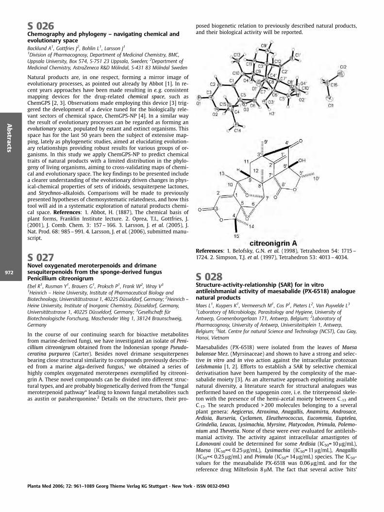
S 026Chemography and phylogeny – navigating chemical andevolutionary spaceBacklund A1, Gottfries J2, Bohlin L1, Larsson J11Division of Pharmacognosy, Department of Medicinal Chemistry, BMC,Uppsala University, Box 574, S-751 23 Uppsala, Sweden; 2Department ofMedicinal Chemistry, AstraZeneca R&D M lndal, S-431 83 M lndal Sweden
Natural products are, in one respect, forming a mirror image ofevolutionary processes, as pointed out already by Abbot [1]. In re-cent years approaches have been made resulting in e. g. consistentmapping devices for the drug-related chemical space, such asChemGPS [2, 3]. Observations made employing this device [3] trig-gered the development of a device tuned for the biologically rele-vant sectors of chemical space, ChemGPS-NP [4]. In a similar waythe result of evolutionary processes can be regarded as forming anevolutionary space, populated by extant and extinct organisms. Thisspace has for the last 50 years been the subject of extensive map-ping, lately as phylogenetic studies, aimed at elucidating evolution-ary relationships providing robust results for various groups of or-ganisms. In this study we apply ChemGPS-NP to predict chemicaltraits of natural products with a limited distribution in the phylo-geny of living organisms, aiming to cross-validating maps of chemi-cal and evolutionary space. The key findings to be presented includea clearer understanding of the evolutionary driven changes in phys-ical-chemical properties of sets of iridoids, sesquiterpene lactones,and Strychnos-alkaloids. Comparisons will be made to previouslypresented hypotheses of chemosystematic relatedness, and how thistool will aid in a systematic exploration of natural products chemi-cal space. References: 1. Abbot, H. (1887), The chemical basis ofplant forms, Franklin Institute lecture. 2. Oprea, T.I., Gottfries, J.(2001), J. Comb. Chem. 3: 157–166. 3. Larsson, J. et al. (2005), J.Nat. Prod. 68: 985–991. 4. Larsson, J. et al. (2006), submitted manu-script.
S 027Novel oxygenated meroterpenoids and drimanesesquiterpenoids from the sponge-derived fungusPenicillium citreonigrumEbel R1, Rusman Y1, Brauers G1, Proksch P1, Frank W2, Wray V3
1Heinrich – Heine University, Institute of Pharmaceutical Biology andBiotechnology, Universit tsstrasse 1, 40225 D sseldorf, Germany; 2Heinrich –Heine University, Institute of Inorganic Chemistry, D sseldorf, Germany,Universit tsstrasse 1, 40225 D sseldorf, Germany; 3Gesellschaft f rBiotechnologische Forschung, Mascheroder Weg 1, 38124 Braunschweig,Germany
In the course of our continuing search for bioactive metabolitesfrom marine-derived fungi, we have investigated an isolate of Peni-cillium citreonigrum obtained from the Indonesian sponge Pseudo-ceratina purpurea (Carter). Besides novel drimane sesquiterpenesbearing close structural similarity to compounds previously describ-ed from a marine alga-derived fungus,1 we obtained a series ofhighly complex oxygenated meroterpenes exemplified by citreoni-grin A. These novel compounds can be divided into different struc-tural types, and are probably biogenetically derived from the “fungalmeroterpenoid pathway“ leading to known fungal metabolites suchas austin or paraherquonine.2 Details on the structures, their pro-
posed biogenetic relation to previously described natural products,and their biological activity will be reported.
References: 1. Belofsky, G.N. et al. (1998), Tetrahedron 54: 1715–1724. 2. Simpson, T.J. et al. (1997), Tetrahedron 53: 4013–4034.
S 028Structure-activity-relationship (SAR) for in vitroantileishmanial activity of maesabalide (PX-6518) analoguenatural productsMaes L1, Kuypers K1, Vermeersch M1, Cos P1, Pieters L2, Van Puyvelde L31Laboratory of Microbiology, Parasitology and Hygiene, University ofAntwerp, Groenenborgerlaan 171, Antwerp, Belgium; 2Laboratory ofPharmacognosy, University of Antwerp, Universiteitsplein 1, Antwerp,Belgium; 3Nat. Centre for natural Science and Technology (NCST), Cau Giay,Hanoi, Vietnam
Maesabalides (PX-6518) were isolated from the leaves of Maesabalansae Mez. (Myrsinaceae) and shown to have a strong and selec-tive in vitro and in vivo action against the intracellular protozoanLeishmania [1, 2]. Efforts to establish a SAR by selective chemicalderivatisation have been hampered by the complexity of the mae-sabalide moiety [3]. As an alternative approach exploiting availablenatural diversity, a literature search for structural analogues wasperformed based on the sapogenin core, i. e. the triterpenoid skele-ton with the presence of the hemi-acetal moiety between C-13 andC-17. The search produced >200 molecules belonging to a severalplant genera: Aegicerus, Atroxima, Anagallis, Anamirta, Androsace,Ardisia, Burseria, Cyclamen, Eleutherococcus, Eucommia, Euptelea,Grindelia, Leucas, Lysimachia, Myrsine, Platycodon, Primula, Polemo-nium and Thevetia. None of these were ever evaluated for antileish-manial activity. The activity against intracellular amastigotes ofL.donovani could be determined for some Ardisia (IC50=10mg/mL),Maesa (IC50=< 0.25mg/mL), Lysimachia (IC50=11mg/mL), Anagallis(IC50=< 0.25mg/mL) and Primula (IC50=14mg/mL) species. The IC50-values for the measabalide PX-6518 was 0.06mg/mL and for thereference drug Miltefosin 8mM. The fact that several active ’hits’
Planta Med 2006; 72: 961–1089 Georg Thieme Verlag KG Stuttgart · New York · ISSN 0032-0943
Abstracts
972
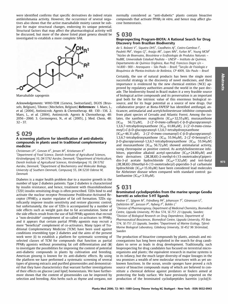
were identified confirms that specific derivatives do indeed retainantileishmania activity. However, the occurrence of several nega-tives also shows that the active maesabalide moiety cannot be sub-ject for major structural changes, endorsing its unique potential.Structural factors that may affect the pharmacological activity willbe discussed, but more of the above listed plant genera should beinvestigated to establish a more complete SAR.
Maesabalide sapogenin
Acknowledgements: WHO-TDR (Geneva, Switserland), DGOS (Brus-sels, Belgium), Tibotec (Mechelen, Belgium) References: 1. Maes, L.,et al. (2004), Antimicrob. Agents & Chemotherap. 48: 130–136. 2.Maes, L., et al. (2004), Antimicrob. Agents & Chemotherap. 48:2056–2060. 3. Germonprez, N., et al. (2005), J. Med. Chem, 48:32–37.
S 029A screening platform for identification of anti-diabeticcompounds in plants used in traditional complementarymedicineChristensen LP1, Grevsen K2, Jensen M2, Kristiansen K3
1Department of Food Science, Danish Institute of Agricultural Sciences,Kirstinebjergvej 10, DK-5792 Aarslev, Denmark; 2Department of Horticulture,Danish Institute of Agricultural Sciences, Kirstinebjergvej 10, DK-5792Aarslev, Denmark; 3Department of Biochemistry and Molecular Biology,University of Southern Denmark, Campusvej 55, DK-5230 Odense M,Denmark
Diabetes is a major health problem due to a massive growth in thenumber of type 2 diabetes patients. Type 2 diabetes is characterizedby insulin resistance, and hence, treatment with thiazolidinedione(TZD) insulin sensitizing drugs is often prescribed. TZDs bind to andactivate the nuclear receptor Peroxisome Proliferator-Activated Re-ceptor (PPAR)g, a master regulator of fat cell formation. TZDs sig-nificantly improve insulin sensitivity and restore glycemic control,but unfortunately, the use of TZDs is accompanied by a number ofside effects such as weight gain due to fat accumulation. Some ofthe side effects result from the use of full PPARg agonists that recruita “non-desirable“ complement of so-called co-activators to PPARg,and it appears that certain partial PPARg agonists may recruit amore “beneficial“ complement of co-activators. Many types of Tra-ditional Complementary Medicine (TCM) have been used againstconditions resembling type 2 diabetes and the aims of the presentwork were (i) to establish a platform for systematic screening ofselected classes of TCM for compounds that function as partialPPARg agonists without promoting fat cell differentiation and (ii)to investigate the possibilities for improving the content of potentialanti-diabetic compounds in plants by cultivation and breeding.American ginseng is known for its anti-diabetic effects. By usingthe platform we have performed a systematic screening of severaltypes of ginseng extracts and purified ginsenosides and demonstrat-ed that they have characteristics that warrant further investigationsof their effects on glucose (and lipid) homeostasis. We have further-more shown that the content of ginsenosides can be improved byselection and breeding. Also herbs such as thyme and oregano, not
normally considered as “anti-diabetic“ plants contain bioactivecompounds that activate PPARg in vitro, and hence may affect glu-cose homeostasis.
S 030Bioprospecting Program-BIOTA: A Rational Search for DrugDiscovery from Brazilian Biodiversityda S. Bolzani V1, Siqueira DHS1, Cavalheiro AJ1, Castro-Gamboa I1,Pauletti PM1, Viegas CJ1, Araffljo AR1, Lopes MN1, Furlan M1, Young MCM2
1Nfflcleo de Bioensaios, Bioss ntese e Ecofisiologia de Produtos Naturais –NuBBE, Universidade Estadual Paulista – UNESP – Instituto de Qu mica,Departamento de Qu mica Org nica, Rua Prof. Francisco Degni s/n –14.800 – 900 – Araraquara – S¼o Paulo – Brazil; 2Sec ¼o de Fisiologia eBioqu mica de Plantas-Instituto de Bot nica, CP 4009, S¼o Paulo – Brazil
Certainly, the use of natural products has been the single mostsuccessful strategy in the discovery of novel medicines, and theirimportance is evidenced by the new chemical entities (NCE) ap-proved by regulatory authorities around the world in the past dec-ade. The biodiversity found in Brazil makes it a very feasible sourceof biological active compounds and its preservation is an importantgoal both for the intrinsic value of this enormous biological re-source, and for its huge potential as a source of new drugs. Ourcollaborative project at Biota-FAPESP has identified antifungal, an-ticancer, antimalarial and acetylcholinesterase inhibitor compoundsfrom plant species of Cerrado and Atlantic Forest. Among the iso-lates, the xanthones mangiferin (IC50=32,55mM), muraxanthone(IC50 56.72mM), 2-(2’-O-trans-caffeoyl)-C-b-D-glucopyranosyl-1,3,6,7-tetrahydroxyxanthone (IC50 61.88mM), 2-(2’-O-trans-cinna-moyl)-C-b-D-glucopyranosyl-1,3,6,7-tetrahydroxyxanthone(IC50=46.31mM), 2-(2’-O-trans-coumaroyl)-C-b-D-glucopyranosyl-1,3,6,7-tetrahydroxyxanthone (IC50 55.94mM), 2-(2’-O-benzoyl)-C-b-D-glucopyranosyl-1,3,6,7-tetrahydroxyxanthone (IC50 55.94mM)and muraxanthone (IC50 56.72mM) showed antimalarial activity,using chroroquine as positive control. As acetylcholinesterase inhi-bitors, piperidine alkaloid acetyl-spectaline (IC50=24.80mM) andtheir derivatives (2R,3R,6S)-2-methyl-6-(13-oxotetradecyl)piperi-din-3-yl acetate hydrochloride (IC50=7.32mM) and tert-butyl(2R,3R,6S)-20methyl-6-(13-oxotetradecyl)-piperidin-3-yl carbamatehydrochlride (IC50=15.10mM) have been considered lead moleculesfor Alzheimer disease when compared with standard control: ga-lanthamine (IC50=3.10mM).
S 031Brominated cyclodipeptides from the marine sponge Geodiabarretti as selective 5-HT ligandsHedner E1, Sj gren M1, Fr ndberg PA2, Johansson T2, G ransson U1,Dahlstr m M3, Jonsson P3, Nyberg F2, Bohlin L11Division of Pharmacognosy, Department of Medicinal Chemistry, BiomedicalCentre, Uppsala University, PO Box 574, SE-751 23 Uppsala, Sweden;2Division of Biological Research on Drug Dependence, Department ofPharmaceutical Biosciences, Biomedical Centre, Uppsala University, PO Box574, SE-751 23 Uppsala, Sweden; 3Department of Marine Ecology, Tj rnMarine Biological Laboratory, G teborg University, SE-452 96 Str mstad,Sweden
The production of bioactive compounds by plants, animals and mi-croorganisms has long been exploited in the search for drug candi-dates to serve as leads in drug development. Traditionally, suchbioprospecting for drug candidates has focused on terrestrial micro-organisms and plants; the equivalent research in marine systems isin its infancy, but the much larger diversity of major lineages in thesea promises a wealth of new molecular structures with as yet un-known functions. In the ocean, sessile sponges have proved a richsource of bioactive compounds many of which are believed to con-stitute a chemical defense against predators or foulers aimed atprotecting the body surface. We have previously reported on theproduction of the brominated cyclodipeptides barettin (cyclo[(6-
Planta Med 2006; 72: 961–1089 Georg Thieme Verlag KG Stuttgart · New York · ISSN 0032-0943
Abstracts
973
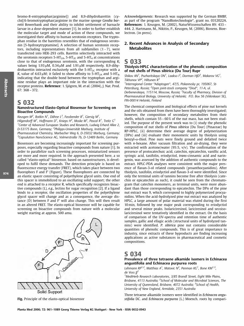
bromo-8-entryptophan)arginine]) and 8,9-dihydrobarettin (cy-clo[(6-bromotryptophan)arginine in the marine sponge Geodia bar-retti Bowerbank and their ability to inhibit settlement of barnaclelarvae in a dose-dependent manner [1]. In order to further establishthe molecular target and mode of action of these compounds, weinvestigated their affinity to human serotonin receptors. The trypto-phan residue in the barettins resembles that of endogenous seroto-nin [5-hydroxytryptamine]. A selection of human serotonin recep-tors, including representatives from all subfamilies (1 –7), weretransfected into HEK-293 cells. Barettin selectively interacted withthe serotonin receptors 5-HT2A, 5-HT2C and 5-HT4 at concentrationsclose to that of endogenous serotonin, with the corresponding Kivalues being 1.93mM, 0.34mM and 1.91mM respectively. 8,9-dihy-drobarettin interacted exclusively with the 5-HT2C receptor with aKi value of 4.63mM; it failed to show affinity to 5-HT2A and 5-HT4,indicating that the double bond between the tryptophan and argi-nine residue plays an important role in the interaction with thereceptor proteins. Reference: 1. Sj gren, M. et al. (2004), J. Nat. Prod.67: 368–372.
S 032Nanostructured Elasto-Optical Biosensor for Screening onBioactive CompoundsKeusgen M2, Botkin N1, D hne L3, Fassbender B1, Giersig M1,Hilgendorff M1, Hoffmann D1, Knieps H2, Moske M1, Pascal R1, Treitz G1
1Center of Advanced European Studies and Research, Ludwig Erhard Allee 2,D-53175 Bonn, Germany; 2Philipps-Universit t Marburg, Institute ofPharmaceutical Chemistry, Marbacher Weg 6, D-35032 Marburg, Germany;3Capsulution NanoScience AG, Volmerstraße 7b, D-12489 Berlin, Germany
Biosensors are becoming increasingly important for screening pur-poses, especially regarding bioactive compounds from nature [1]. Inorder to parallelize such screening processes, miniaturized sensorsare more and more required. In the approach presented here, a socalled “elasto-optical“ biosensor, based on nanostructures, is devel-oped to fulfil these demands. The detection principle is based onfluorescence energy transfer (FRET), which takes place between twofluorophors F and F’ (Figure). These fluorophores are connected byan elastic spacer consisting of polyethylene glycol units. One end ofthis spacer is immobilized to an oscillating solid support; the otherend is attached to a receptor R, which specifically recognizes bioac-tive compounds (L), e.g., lectins for sugar recognition [2]. If a ligandbinds to a receptor, the oscillation properties of the polyethyleneglycol spacer will change and as a consequence, the average dis-tance (D) between F and F’ will also change. This will then resultin an altered FRET. The elasto-optical biosensor will be capable forscreening on bioactive compounds from nature with a molecularweight starting at approx. 500 amu.
Fig. Principle of the elasto-optical biosensor
Acknowledgements: Research was supported by the German BMBF,as part of the program “NanoBiotechnologie“, grant no. 0312022A.References: 1. Keusgen, M. (2002), NaturWissenschaften 89: 433–444. 2. Hartmann, M., Nikitin, P., Keusgen, M. (2006), Biosens. Bioe-lectron. (in press).
2. Recent Advances in Analysis of SecondaryMetabolites
S 033Thiolysis-HPLC characterization of the phenolic compositionof nut shells of Pinus sibirica (Du Tour) RuprShikov AN1, Pozharitskaya ON1, Laakso I3, Dorman HJD3, Makarov VG1,Tikhonov VP2, Hiltunen R31Interregional Center “Adaptogen“, 47/5, Piskarevsky pr, 195067, St-Petersburg, Russia; 2Open joint-stock company “Diod“, 11-A, ul.Derbenevskaya, 115114, Moscow, Russia; 3Faculty of Pharmacy, Division ofPharmaceutical Biology, University of Helsinki; P.O. Box 56 (Viikinkaari 5E),FIN-00014 Helsinki, Finland
The chemical composition and biological effects of pine nut kernelsand the oils obtained from them have been thoroughly investigated;however, the composition of secondary metabolites from theirshells, which contain 55–60% of the nut mass, has not been stud-ied. The purpose of the present work was to (i) study the phenoliccomposition of nut shells of Pinus sibirica (Du Tour) (Pinaceae) byRP-HPLC, (ii) determine their average degree of polymerization(DPn) and (iii) evaluate their monomeric units by thiolysis usingbenzyl-a-thiol. Pine nuts were finely ground and defatted twicewith n-hexane. After vacuum filtration and air-drying, they wereextracted with acetone/water (95:5, v/v). The confirmation of thepresence of protocatechuic acid, catechin, epicatechin, vanillic acid,syringic acid, taxifolin, eriodyctiol, trans-cinnamic acid and narin-genin, was assessed by the addition of authentic compounds to theextract. HPLC-PDA analyses were consistent with the major pres-ence of flavan-3-ol related compounds (proanthocyanidins). Afterthiolysis, taxifolin, eriodyctiol and flavan-3-ol were identified. Sinceonly the terminal units of tannins become free after thiolysis (cate-chin or epicatechin as such), it could be seen from the chromato-gram that catechin monomers, as terminal units, were more abun-dant than those corresponding to epicatechin. The DPn of the pinenut extract was 9, which correspond to highly polymerized procya-nidins. When the acid-hydrolyzed pine nut extract was analyzed byHPLC, a large amount of polar material was eluted during the first10min, followed by one major peak corresponding to eriodyctioland several minor peaks. Isolariciresinol, lariciresinol and secoiso-lariciresinol were tentatively identified in the extract. On the basisof comparison of the UV-spectra and retention time of authenticsamples, gallic and ellagic acids (structural units of hydrolyzed tan-nins) were identified. P. sibirica pine nut contains considerablequantities of phenolic compounds. This is of great importance toindustry, since extracts of these byproducts are finding increasingapplications as active substances in pharmaceutical and cosmeticcompositions.
S 034Prevalence of three tetraene alkamide isomers in Echinaceaangustifolia and Echinacea purpurea rootsLehmann RP1,2, Matthias A1, Matovic N2, Penman KG1, Bone KM1,3,de Voss JJ21MediHerb Research Laboratories, 3/85 Brandl Street, Eight Mile Plains,Brisbane, 4113 Australia; 2School of Molecular and Microbial Sciences, TheUniversity of Queensland, Brisbane, 4072 Australia; 3School of Health,University of New England, Armidale, 2351 Australia
Three tetraene alkamide isomers were identified in Echinacea angu-stifolia DC. and Echinacea purpurea (L.) Moench. roots by compari-
Planta Med 2006; 72: 961–1089 Georg Thieme Verlag KG Stuttgart · New York · ISSN 0032-0943
Abstracts
974
son with their synthetic cis-trans 8,10 counterparts which weresynthesised using novel pathways. The three tetraenes were: (2E,4E, 8Z, 10Z)-isobutyldodeca-2, 4, 8, 10-tetraenamide, the ZZ isomer,(2E, 4E, 8Z, 10E)-isobutyldodeca-2, 4, 8, 10-tetraenamide, the ZEisomer and (2E, 4E, 8E, 10Z)-isobutyldodeca-2, 4, 8, 10-tetraenamide,the EZ isomer. The relative concentration of each tetraene was ex-amined in several commercially available samples by GCMS. Theamount of each tetraene as a percentage of the total differed be-tween the two species, with 10% and 29% of the ZZ isomer, 80% and63%of the ZE isomer and 10% and 8% of the EZ isomer in E. angu-stifolia and E. purpurea respectively. These species differences be-tween E. angustifolia and E. purpurea roots may help to explainexperimental differences in the activity of preparations from eitherspecies as well as the variations in their efficacy noted in clinicaltrials.
3. Genomics, Proteomics and Metabolomics inMedicinal Plant Research
S 035Molecular cloning and characterization of a novelS-adenosyl-L-methionine: coniferyl alcoholO-methyltransferase from suspension cultures of Linumnodiflorum LBerim A, Petersen MInstitut f r Pharmazeutische Biologie, Philipps Universit t Marburg,Deutschhausstr. 17A, D-35037 Marburg, Germany
Several methylation reactions occur in the course of aryl tetralinlignan biosynthesis in L. nodiflorum L., explaining our interest in thisenzyme class. Using a homology-based RT-PCR strategy [1], we havecloned and functionally expressed in E. coli a novel 41 kDa methyl-transferase displaying high regiospecificity towards the allylic OH-group of coniferyl alcohol (CA). The apparent Km for CA was deter-mined to be 6.77mM with Vmax of 621.19mkat/kg protein at 30 C,whereas the Km for the co-substrate S-adenosyl-L-methionine is18.93mM Structure-activity relationship studies proved the doublebond of the side-chain to be important, as the enzyme activity withdihydroconiferyl alcohol amounted to about 22.95% as compared tothe best substrate (CA). The substitution pattern of the phenol ringis also essential, for both sinapyl and cinnamic alcohols were poorlyaccepted (7.86 and 15.69% activity of that with CA, resp.), whereascrotonyl and allyl alcohols are no substrates (< 0.7% activity), con-firming the aromatic ring itself is indispensable. The WU-BLAST2(EMBL, Heidelberg) search revealed only low homology (< 45%) toenzymes listed hitherto. The transcription levels, determined bysemi-quantitative RT-PCR, were highest between day 2 and 6 ofthe suspension culture period, whilst the corresponding enzymeactivity declined for the first 4 days and rose from day 5 onwards,reaching its maximum of 612.31 nkat/kg on day 7. By identifying aso far undescribed substrate preference of an enzyme with littlehomology to the already known, function attribution to newly dis-covered and/or yet unassigned genes might now be facilitated. Thephysiological role of this side-chain methylation of coniferyl alcohol,a precursor of both lignin and lignan biosyntheses, remains to beassessed yet. Reference: 1. Ibrahim, R.K. et al. (1998), Plant Mol.Biol. 36: 1 –10.
S 036Different strategies for discriminator identification in a NMRbased metabonomics matrix of the genus Leontopodiumusing LC-SPE-NMR, 1H-NMR-guided isolation and classicalphytochemistrySchwaiger S1, Seger C1, Godejohann M2, Humpfer E2, Hehenberger S1,Tseng LH2, Ellmerer EP3, Spraul M2, Stuppner H1
1Institut f r Pharmazie/Pharmakognosie, Leopold-Franzens-Universit tInnsbruck, Innrain 52c, 6020 Innsbruck, Austria; 2Bruker-Biospin GmbH,Silberstreifen 4, 76287 Rheinstetten, Germany; 3Institut f r OrganischeChemie, Leopold-Franzens-Universit t Innsbruck, Innrain 52a, 6020Innsbruck, Austria
The secondary metabolite profile of Edelweiss (Leontopodium alpi-num Cass.; Asteraceae), is highly complex – more than 50 analyteshave been characterized until now [1–4]. Thus, the chemotaxono-mical assessment of the genus Leontopodium comprising more thanthirty mostly Asian species seems a major undertaking not subsum-able with a single analytical technique. NMR based metabolic pro-filing can be envisioned as alternative analytical approach. Thistechnique, combining 1H-NMR spectroscopy of extracts with multi-variate statistical data interpretation, allows an unbiased selectionof spectral regions responsible for sample discrimination. Profilingof CDCl3-extracts of the roots of twelve Leontopodium species re-sulted in clear species discriminations. The three species with thehighest variation compared to L. alpinum, the European Edelweiss,were identified as L. souliei Beauverd, L. franchetii Beauverd and L.subulatum Beauverd. Due to varying amounts of available plant ma-terial and differences in the obtained extract matrices, identificationof the discriminating metabolites was carried out by three differentstrategies: LC-SPE-NMR experiments for L. souliei, 1H-NMR guidedisolation for L. franchetii and classic phytochemical techniques (e. g.silica gel CC, Sephadex LH 20 CC and preparative TLC) for L. subula-tum. These three distinctly different strategies allowed in each casethe identification and structure elucidation of the discriminatingconstituents. Differences of the applied methods in time consume,amount of the required plant material, yield of the discriminatingcompounds and costs will be discussed. References: 1. Schwaiger, S.et al. (2006), Phytochem. Anal., accepted. 2. Schwaiger, S. et al.(2005), Tetrahedron 61: 4621–4630. 3. Schwaiger, S. et al. (2004),Planta Med. 70: 978–785. 4. Dobner, M.J. et al. (2003), Helv. Chim.Acta; 86: 733–738.
4. Health Beneficial Effects of Plant Phenolics
S 037Inhibition of interleukin-8 secretion by a green tea specialextract in the intestinal cell line Caco-2Netsch M1, Gutmann H2, Schmidt A1, Drewe J21Frutarom Switzerland Ltd, Ruetiwisstrasse 7, 8820 Waedenswil, Switzerland;2Dept. of Research and Clinical Pharmacology, University Hospital(Kantonsspital), 4031 Basel, Switzerland
The intestinal mucosa represents a site of active immunologic activ-ity. Accordingly, intestinal epithelial cells, e. g. Caco-2, secrete a widearray of inflammatory mediators including chemokines, e. g. IL-8,that are able to induce an inflammatory state in intestinal cells orto attract inflammatory immune cells [1, 2]. The chemokine inter-leukin (IL)-8 is involved in neutrophil attraction and activation andelevated levels have been observed in intestinal inflammation. Nat-ural compounds including green tea have been shown to modulateinflammation in vitro and in vivo [3, 4]. We investigated the influ-ence of the green tea extract EFLA 942 (GTE) on the secretion of IL-8 protein and on the mRNA expression levels of IL-8 in the humangastrointestinal cell line Caco-2 in an inflammatory state. Therefore,extracellular IL-8 concentrations were determined by ELISA andmRNA expression levels of IL-8 were analyzed by quantitative RT-PCR. Characteristic components in 0.01mg/mL GTE are 4.02mM
Planta Med 2006; 72: 961–1089 Georg Thieme Verlag KG Stuttgart · New York · ISSN 0032-0943
Abstracts
975

EGCG, 2.27mM EGC, 4.36mM caffeine, and 0.99mM theanine. GTE didsignificantly inhibit the IL-1b-induced IL-8 secretion in a dose-de-pendent manner. At highest concentration the GTE-mediated inhi-bition was comparable to brefeldin A, a fungal inhibitor of vesiculartransport. This was not related to a significant down-regulation ofIL-1b-induced IL-8 mRNA expression by GTE. These results suggestthat GTE may exert an anti-inflammatory activity in enterocytes,which may be useful for the treatment of intestinal inflammation.References: 1. Yang, S.K. et al. (1997), Gastroenterology 113: 1214–1223. 2. Eckmann, L. et al. (1993), Gastroenterology 105: 1689–1697. 3. Porath, D. et al. (2005), J. Pharmacol. Exp. Ther. 315:1172–1180. 4. Varilek, G.W. et al. (2001), J. Nutr. 131: 2034–2039.
S 038Plant Phenolics as Antibiotic Resistance Modifying AgentsSmith E1, Williamson E2, Gibbons S11Centre for Pharmacognosy and Phytotherapy, The School of Pharmacy,University of London, 29 – 39 Brunswick Square, London WC1N 1AX, UK;2Department of Pharmacy, University of Reading, Whiteknights, Reading,Berks RG6 6AJ, UK
There has recently been renewed interest in plant phenolics, longconsidered to be toxic and non-specific in their activity. Many activ-ities have been reported for phenolic plant compounds, and ofparticular interest is their activity as antibiotic resistance modifyingagents against Gram-positive bacteria and mycobacteria. Multidrug-resistance (MDR) exhibited by many bacterial species is a majorproblem in treating both hospital and community acquired infec-tions. A modifying agent is a compound which reduces the mini-mum inhibitory concentration (MIC) for an antibiotic. This could beof great benefit in combinatory therapy, perhaps facilitating thereintroduction of antibiotics that are no longer effective due to re-sistance. An example in clinical use is Unasyn comprising the b-lactamase inhibitor sulbactam in combination with ampicillin. Poly-phenols from green tea, epicatechin gallate [1] and epigallocatechingallate have been shown to potentiate oxacillin activity againstmethicillin-resistant Staphylococcus aureus (MRSA) [1] and to havemodest activity in reducing the MIC of some standard antibioticsagainst effluxing strains of S. aureus [2]. Resistance modifying activ-ity has also been reported for the phenolic abietanes, totarol andferruginol [2] isolated from conifer species [3; 4], for carnosic acidfrom Rosmarinus officinalis L. [5], and anacardic acid from the cash-ew Anacardium occidentale L. [6]. Small differences in structure suchas the position of a hydroxyl group can have a considerable effect onmodulatory activity [3; 4] which suggests a more subtle mode ofaction for these phenolics than general membrane perturbation.Recent reports have indicated a potentially useful separation be-tween the concentrations required for biological activity and cyto-toxicity for both ferruginol and totarol [7].
Acknowledgements: We thank Stiefel International R & D Ltd forfunding this study. References: 1. Shiota, S. et al. (1999), Biol.Pharm. Bull. 22: 1388–1390. 2. Gibbons, S. et al. (2004), PlantaMed. 70: 1240–1242. 3. Nicolson, K. et al. (1999), FEMS Microbiol.Lett. 179: 233–239. 4. Smith, E. et al. (2006), in preparation. 5.Oluwatuyi, M. et al. (2004), Phytochemistry 65: 3249–3254. 6.Kubo, I. et al. (1992), J. Nat. Prod. 55: 1436–1440. 7. Clarkson C. etal. (2003), Planta Med. 69: 720–724.
S 039Flavonoids from Vigna angularis – composition andantioxidative effectsWeber N1, W tjen W2, Edrada RA1, Wray V3, Lou Y4, Wang ZQ4, Proksch P11Heinrich – Heine University, Institute of Pharmaceutical Biology andBiotechnology (a), Universit tsstrasse 1, 40225 D sseldorf, Germany;2Heinrich – Heine University, Institute of Toxicology, D sseldorf, Germany;3Gesellschaft f r Biotechnologische Forschung, Braunschweig, Germany;4Zhejiang University, Department of Pharmacology and Toxicology,Hangzhou,China
In the last decades interest in the group of flavonoids has markedlyincreased due to their presumed beneficial effects in humans. Manyflavonoids act as radical scavengers and are suggested to preventchronic diseases such as atherosclerosis or even cancer [1]. For acritical evaluation of the effects of flavonoids with regard to humanhealth, quantitative (daily intake as part of diet or as food supple-ments) as well as qualitative (structural) aspects have to be takeninto consideration [2]. As part of a broader Sino-German project onflavonoids from plants used for human consumption, we analyzedfour different cultivars of Vigna angularis (Willd.) Ohwi & H. Ohashi,two of them growing in China (Hangzhou) and the other two culti-vated in Germany (D sseldorf). All analyzed plants yielded catechin,myricetin–3-O–rutinoside, and rutin as main compounds but dif-fered with regard to their total flavonoid concentrations which werelower in the Chinese cultivars. We analyzed pharmacological effectsof these compounds focussing mainly on their antioxidative proper-ties. All flavonoids exhibited a good radical scavenger activity in acell free system (DPPH assay). In different cellular systems (ratH4IIE hepatoma and C6 glioma cells) we further analyzed protectiveeffects of these substances against oxidative stress using the fluor-escent probe DCF. Beside these antioxidative effects, cytotoxic andpro-apoptotic activities of these compounds were also determined.In consideration of these data it can be emphasized that a knowncomposition of flavonoids is necessary in nutrient uptake. Acknowl-edgements: We thank DFG for financial support and Dr. Ulrike Loh-wasser (IPK Gatersleben) for taxonomic identification. References:1. Gordon, M.H. (1996), Nat. Prod. Rep. 13: 265–273. 2. Rice – Evans,C. et al. (1996), Free Radical Biology & Medicine 20: 933–956.
Planta Med 2006; 72: 961–1089 Georg Thieme Verlag KG Stuttgart · New York · ISSN 0032-0943
Abstracts
976
S 040Composition and antioxidant activities of Salvia halophilaand S. virgata from TurkeyKosar M, Goger F, Husnu K, Baser CAnadolu University, Faculty of Pharmacy, Department of Pharmocognosy26470 Eskisehir, TURKEY
Several biochemical reactions in our body generate reactive oxygenspecies and these are capable of damaging crucial bio-molecules [1].Free radicals are also very important in food products, because oxi-dative degradation of lipids is one of the main factors limiting theirshelf–life [2]. In recent years, natural antioxidants have been fo-cused on because of the harmful effects of synthetic antioxidantsin living systems [3] Salvia is one of the wide spread members of thefamily Labiatae (Lamiaceae). Salvia species, especially S. officinalis L.,are an important source of antioxidants used in food and have widerimplications for the dietary intake of natural antioxidants [3]. Tur-key is an important country for Salvia species in the world. The floraof Turkey includes 88 species of the genus Salvia. The aerial parts ofS. halophila Hedge and S. virgata L. were used in Soxhlet extractionwith different solvents such as n-hexane, ethylacetate, methanoland aqueous methanol (50%). Plants were also extracted with waterunder reflux. All the extracts were analyzed by HPLC for their phe-nolic composition and in in vitro antioxidant assays for their effectson oxidation. The free radical scavenging activity of the extractswere investigated using 1,1-diphenylpicrylhydrazin (DPPH·) radical.Linoleic acid was also used to determine the effects of lipid perox-idation of the extracts. Total phenols, flavonoids and flavonols, andreductive activity of the extracts were also analyzed. Phenolics richextracts of ethylacetate, methanol and aqueous methanol (50%)showed scavenging activity on DPPH· whereas non-polar extracts(n-hexane and ethylacetate) inhibited the peroxidation of linoleicacid. The aqueous methanol and ethylacetate extracts reduced theferric(III) to ferro(II) in a certain proportion. Rosmarinic acid wasfound as the main component and caffeic acid, ferulic acid andluteolin-7-O-glycoside were identified in the extracts. References:1. Kumaran, A., Joel karunakaran, R. (2006), Food Chemistry 97:109–114. 2. Pizzalle, L. et al. (2002), J.Sci. Food Agric. 82: 1645–1651. 3. Kintzios, S.E. (2000), Sage The Genus Salvia. Harwood aca-demic publishers,pp. 27–53 and 185–192.
S 041Effects of acute and repeated administration of Hypericumperforatum extract (WS 5572 ) and its main constituents onextracellular levels of serotonin, noradrenaline anddopamine in the rat brain: A microdialysis studyKehr J1,2, N ldner M3, Yoshitake T1,41Dept. of Physiology and Pharmacology, Karolinska Institutet, Nanna Svartzv g 2, 171 77 Stockholm, Sweden; 2Pronexus Analytical AB, KarolinskaScience Park, Fogdevreten 2a, 171 77 Stockholm, Sweden; 3PreclinicalResearch, Dr. Willmar Schwabe GmbH & Co. KG, Willmar-Schwabe-Str. 4,76227 Karlsruhe, Germany; 4Dept. of Pharmaceutical Sciences, InternationalUniversity of Health and Welfare, 2600 – 1, Kitakanemaru, Ohtawara-shi,Tochigi 324 – 8501, Japan
The effects of acute and repeated administration of WS 5572 , ahydro-alcoholic extract of Hypericum perforatum L. (St. John’s wort)on extracellular levels of dopamine (DA), serotonin (5-HT), noradre-naline (NA), and the metabolites 5-HIAA, DOPAC and HVA wereexamined by use of in vivo microdialysis in the prefrontal cortex(PFC), ventral hippocampus or striatum of awake rats. A single oral(p.o.) dose of WS 5572 (300mg/kg) caused a dramatic reduction ofmetabolites DOPAC and HVA to about 15% and 53% of controls,respectively at 180min after the drug. Extracellular DA levels in-creased only modestly to about 141% and there was no effect on5-HT and NA levels [1]. Hypericum given after sub-chronic pretreat-ment (300mg/kg, p. o. daily for 14 days), increased most markedlythe DA levels to about 260% and decreased DOPAC, HVA and 5-HIAA, while the extracellular 5-HT and NA increased only moder-
ately to 138% and 125%, respectively. In addition, a single dose ofthe constituents rutin (18mg/kg, p. o.) and hyperforin (8mg/kg,p. o.) increased 5-HT levels in the PFC of awake rats to 118% and125% of control values, respectively, where as following sub-chronic14-days administration rutin, hyperforin and isorhamnetin (0. 3mg/kg, p.o.) increased extracellular 5-HT levels to 142%, 131% and 120%of control values, respectively, and the concentrations of 5-HIAAdecreased only to about 90% of control values for all three treat-ments. These data suggest that rutin and hyperforin, given at dosescorresponding to their content in WS 5572 , were about equallypotent to increase 5-HT levels in the PFC of awake rats. In conclu-sion, the data suggest that WS 5572 inhibits preferentially themetabolism and reuptake of DA, and to a lesser extent affects theextracellular levels of 5-HT and NA, emphasizing the role of dopa-minergic system in the mechanisms underlying potential antide-pressive effects of WS 5572 . Reference: 1. Yoshitake, T. et al.(2004), Br. J. Pharmacol. 142: 414–418.
5. Clinical Studies with Herbal Medicinal Products
S 042Effects of hops on clinical efficacy of a valerian-hops-extractcombination (Ze 91019) in patients suffering fromnon-organic sleep disorderKoetter U1, Schrader E2, Brattstr m A1
1 Max Zeller S hne AG, CH.8590 Romanshorn, Switzerland; 2Clinical DrugResearch, D-35415 Pohlheim,Germany
The fixed valerian-hops extract combination Ze 91019 is used as asleep aid. For this combination pharmacodynamic actions [1, 2] aswell as efficacy has been demonstrated in patients suffering fromsleep problems [3]. While the main focus of research has been onvalerian or the extract combination as a single active entity less isknown about the contributions of hops to the sleep inducing effectof the combination. In this report, sleep onset latency (SL2) of thecombination Ze 91019 was tested in comparison to valerian extractusing a mobile device to allow for recording of sleep parameters athome while participating in the trial. Patients suffering from sleepdisorders (ICD10: F 51.0, F 51.2) were enrolled when SL2 was pro-longed at baseline (‡30min, inclusion criteria). The hypnograms tocalculate the objective sleep parameters were recorded with a mo-bile device (QUISI) twice before including the patient into the study.Treatment period lasted for 4 weeks with placebo, valerian extract(500mg) or the valerian-hops-extract combination (500mg plus120mg). Both of the extracts were prepared with 45% methanolm/m with a DER for valerian: 5–8:1 and for hops 7–10:1. Theresults of this trial clearly indicate that hops added to the clinicalefficacy as Ze 91019 was superior to placebo in reducing the origi-nally prolonged sleep onset latency. References: 1. Vonderheid-Guth, B. et al. (2000), Eur. J. Med. Res. 5: 139–144. 2. Schellenberg,R. et al. (2004), Planta Med. 70: 595–597. 3. F ssel, A. et al. (2000),Eur. J. Med. Res. 5: 385–389.
S 043Efficacy Of A Plant Based Formulation In The Treatment OfRecurrent Airway Obstruction In HorsesLarkins NJ1, Deaton CM2, Jones K1
1Nutritional Laboratories,Monmouth, Monmouthshire, NP15 2DJ, UnitedKingdom; 2Animal Health Trust, Newmarket, Suffolk, CB8 7UU, UnitedKingdom
The purpose of this double blinded placebo controlled cross-overclinical study was to assess the ability of a plant based formulation(Ginkgo biloba L., Zingiber officinale Roxburgh, Chlorella pyrenosa) toprevent or delay the onset, decrease the magnitude of response and/or speed the recovery of lung dysfunction, clinical signs of disease,airway inflammation, and pulmonary oxidative stress in horses with
Planta Med 2006; 72: 961–1089 Georg Thieme Verlag KG Stuttgart · New York · ISSN 0032-0943
Abstracts
977

recurrent airway obstruction (RAO) on exposure to organic dust. Theperformance of the active supplement was judged on the basis ofresponses in lung function, clinical examination, airway inflamma-tion and pulmonary oxidative stress following organic dust chal-lenge compared to responses on the placebo diet. Lung dysfunctionwas assessed by measuring airway reactance and airway respon-siveness to histamine by forced oscillation mechanics. Clinical signswere assessed by assigning scores for respiratory rate, nasal dis-charge, abdominal lift/expiratory effort, nasal flaring, lung soundsand cough. Airway inflammation was determined by cytologicalanalyses of tracheal wash and bronchoalveolar lavage fluid (BALF)samples, and by measuring the concentration of hydrogen peroxidein exhaled breath condensate (EBC). Oxidative stress was assessedby measuring the concentrations of reduced ascorbic acid, dehy-droascorbate (DHA, oxidised ascorbic acid), reduced glutathioneand oxidised glutathione in tracheal wash and BALF. Results of sta-tistical analyses demonstrated that BALF ascorbic acid concentra-tions were higher after challenge in horses when fed the activeformulation compared to the placebo and that BALF, DHA and ARR(ratio of DHA to total ascorbic acid), were lower after challenge inthe active supplement horses irrespective of the order of treatmentallocation. References: 1. Deaton, C.M., Marlin, D.J. (2004), Am. J.Vet. Res. 65: 80–87. 2. Larkins, N.J. (1999), J. Equine Vet. Sci. 19:84–89.
S 044Strix Forte , an antioxidant mixture with bilberryanthocyanosides, reduces oxidative stress and immuneactivation in exfoliation syndrome and exfoliative glaucomaBlomster H1, Greilberger J2, ttl K2, Fuchs D3, Winkler C3, Hiltunen R4,Juan H5, Psilander N6
1Institution of Deacony, Eye Policlinic, Sibeliuksenkatu 6 C, 15110 Lahti,Finland; 2Medical University Graz, Institute of Physiological Chemistry, Graz,Austria; 3Innsbruck Medical University, Biocentre, Division of BiologicalChemistry, Innsbruck, Austria; 4University of Helsinki, Division ofPharmaceutical Biology, Helsinki, Finland; 5Medical University Graz, Instituteof Experimental and Clinical Pharmacology, Graz, Austria; 6Ferrosan A/G,Sjøborg, Denmark
Background: To evaluate the quantity of oxidative stress (OS), im-mune activation (IA) and exfoliation material before and after fourmonths daily ingestion of Strix Forte in exfoliation syndrome (XFS)and exfoliative glaucoma (XFG). Methods: 15 XFS, 15 XFG and 15control patients ingested one Strix Forte tablet twice daily duringfour months containing 74mg bilberry (Vaccinium myrtillus L.) an-thocyanosides, 6mg lutein, 15mg zinc sulphate, 10mg dl-a-toco-pheryl acetate, 800mg retinyl acetate and 50mg selenomethionine.Exfoliation material quantity (EmQ), scale 0– 3, was also examined.Plasma concentrations of isoprostanes, malondialdehyde, carbonylproteins and oxidized albumin were measured as markers for OS.Neopterin and kynurenine/tryptophan ratio were measured as mar-kers for IA. Wilcoxon Signed Ranks Tests were used for statisticalanalyses. Results: EmQ was significantly reduced after four monthsin both XFS and XFG group (XFS: P=0.007 for right and 0.003 for lefteyes, XFG: P = 014 for right and 0.005 for left eyes. Isoprostanes werereduced in XFS group (P = 0.001) and in controls (P=0.009), but notin XFG group (P=0.191). Malondialdehyde levels were not dimin-ished in any groups. Carbonyl proteins were reduced in all groups:[XFS (P=0.002), XFG (P=0.001), controls (P=0.015)]. Oxidized albu-min diminished in all groups [XFS (P=0.003), XFG (P=0.005), con-trols (P=0.001)]. Neopterin levels were slightly increased in the XFGgroup (P=0.020) and tryptophan levels were slightly elevated in theXFS group (P=0.047). Kynurenine levels decreased in the XFG group(P=0.012) and in controls (P=0.008). Kynurenine/tryptophan levelsdecreased in all three groups [XFS (P=0.006), XFG (P=0.003), con-trols (P=0.006)]. Conclusion: Regular Strix Forte ingestion reducedbesides OS and IA also EmQ in XFS and XFG patients which may beexplained by the observed beneficial alterations in the OS and IAmarkers. Acknowledgements: Research supported by Ferrosan A/S,
Sjøborg, Denmark References: 1. Koliakos, G.G. et al. (2003), Br. J.Ophthalmol. 87: 353–356. 2. Laganovska, G. et al. (2003), Adv ExpMed Biol 527: 367–74. 3. Viljanen, K. et al. (2004), J. Agric. FoodChem. 52: 7419–7424. 4. Youdim, K.A. et al. (2002), J. Nutr. Bio-chem. 13: 282–288. 5. Kang, J.H. et al. (2003), Am. J. Epidemiol.158: 337–346. 6. Noaman, E. et al. (2002), Biol. Trace Elem. Res.86: 55–64. 7. Gherghel, D. et al. (2005), Invest. Ophthalmol. Vis. Sci.46: 877–883. 8. De la Fuente, M. (2002), Eur. J. Clin. Nutr. 56 Suppl3: S5–8.
6. Other related topic
S 045Monographic profile of Guiera, leaves a West African herbaldrugSilva O, Gomes ETPharmaceutical Sciences Research Centre, Faculty of Pharmacy, University ofLisbon, Lisbon, Portugal
Guiera senegalensis Lam (Combretaceae) is a major West Africanmedicinal plant, often employed to treat venereal, diarrhoeal, re-spiratory and fungal diseases. Previous work confirmed G. senega-lensis leaves antimicrobial activity against Neisseria gonorrhoeae (in-cluding resistant strains), Shigella dysenteriae, Vibrio cholerae, Giar-dia lamblia and Cladosporium cucumerinum, corroborant it’s tradi-tional uses [1 –3]. Bioguided phytochemical studies permitted theidentification of flavonoids, gallic tannins, naphthalene derivatives(naphthyl butenone and naphthopyrans) and terpenoids, among G.senegalensis compounds [4]. In sequence, the significant and usefulmarkers to G. senegalensis leaves diagnosis were also determined[5]. Hereby we present the botanical and chemical characteristicsthat should be included under Characteristics, Identification andDosage parts of a future herbal drug monograph. References: 1Silva, O. et al. (1997), Int. J. Pharmacogn. 35: 323–328. 2 Silva, O.et al. (1996), J. Etnopharm. 50: 55–59. 3. Silva, O. (2004), PhDThesis, Lisboa, Universidade de Lisboa. 4. Silva, O., Gomes, E.T.(2003), J. Nat. Prod. 66: 447–449. 5. Silva, O., Serrano, R., Gomes,E.T. (2005), 53rd Annual Congress of the Society for Medicinal PlantResearch, Florence.
S 046Isolation of E & Z guggulsterones from young aerial stems ofCommiphora wightii without destruction of plantsSoni VMahatma Gandhi Institute of Applied Sceinces, Shri Ram Ki Nangal, Via:Vatika, EPIP Gate, Sitapura, Jaipur-303905, Rajasthan, INDIA
Commiphora wightii (Arn.) Bhandari is an important traditionallyused plant in India. It provides oleo gum resin mentioned by Sus-hruta (3000 year ago) as being a valuable drug. Clinical analysisrevealed that the isomers E- and Z-guggulsterone are responsiblefor the hypolipidemic activity of oleo gum resin [1]. Unscientifictapping methods to increase yield of oleo gum resin causes mortal-ity of plants and danger of extinction of the species. Therefore,guggulsterones were isolated from the aerial stems of C. wightiiplants. Dried powder of aerial stems was subjected in soxhlet appa-ratus followed by purification by column chromatography andquantification for E- and Z- guggulsterones by high performanceliquid chromatography (HPLC).Highest extraction of guggulsterone(0.35%) was observed when ethyl acetate was used as solvent, whilethe lowest in chloroform (0.06%). Through this procedure guggul-sterones can be isolated from the aerial stems thereby saving theentire plant. Though the amount of guggulsterone contents record-ed was very low in the aerial stems as compared to the oleo gumresin, the yield can be enhanced using elite plant propagation meth-od coupled with improvised extraction technique. Reference: 1.Urizar, N.L., Moore, D.D. (2003), Ann. Rev. Nutr. 23: 303–313.
Planta Med 2006; 72: 961–1089 Georg Thieme Verlag KG Stuttgart · New York · ISSN 0032-0943
Abstracts
978

S 047Turkish Oregano: Chemistry & Biological ActivitiesBaser KHC, Demirci FDepartment of Pharmacognosy, Faculty of Pharmacy, Anadolu University,26470-Eskisehir/Turkey
“Kekik“ is a collective term given in Turkey to plants which smelllike oregano or thyme. Several taxa belonging to the genera Origa-num L., Thymus L., Thymbra L., Coridothymus L. and Satureja L. arevariously called, used and traded as kekik in Turkey [1]. Among all,five Origanum species are widely traded and Turkey is the biggestexporter of Oregano to the world markets, with over 10500 tons inthe year 2005 for a return of US$ 18 million. The main characteristicof Turkish Oregano is high yielding essential oil containing carva-crol, 2-methyl-5-isopropylphenol – isomer of thymol, as the mainconstituent. Carvacrol is a simple monoterpene phenol responsiblefor most bioactivities of Oregano. Biological activities of Oreganoherb, Oregano essential oil, Oregano water (hydrosol – aromaticwater), and carvacrol range from antimicrobial and antioxidant ac-tivities to the treatment of gastrointestinal disorders and even can-cer. Studies using both in vitro and in vivo classical and,molecularbiology techniques have provided proof to most biological activities.Other uses of Oregano and carvacrol include antiparasitic, insectici-dal, herbicidal, food preservative, and as feed additive especially inpoultry, etc. The paper will review recent progress in the Science ofOregano with special reference to Turkish Oregano. Reference: 1.Baser, K.H.C. (2002), The Turkish Origanum Species, In:Oregano,The Genera Origanum and Lippia, Kintzios, S.E. (Ed.) Taylor andFrancis, UK.
S 048Chemical ecology and screening for bioactivity: commonand contrasting issuesHadacek FDepartment of Chemical Ecology and Ecosystem Research Faculty of LifeSciences, University of Vienna, Althanstraße 14, A-1090 Vienna, Austria
Chemical ecology deals with the elucidation of low molecular mol-ecules in biotic interactions, such as those between plant and mi-crobes as well as plants and herbivores. Today, industry favours highthroughput screening of extracts obtained from organisms of allkinds. Dereplication in active compounds and the accession as wellas false positives represent major factors affecting the success ofthis methodology. Conversely, many natural products may be deci-sive in determining the outcome of many biotic interactions. Here,the constraints in selecting for biological activity are especially high.However, many compounds may be often produced only as conse-quence of the stress caused by interactions with predators (mi-crobes, plants, and herbivores) or in nutrient-limiting situations(microbes). Decomposition of tissues is accomplished by specificmicrobial communities that are intrinsically affected by the qualityof the resource. The facts may somehow act as a constraint on theaccessibility to this natural products but the quality of the recoveredmetabolites may justify the effort. From the pharmaceutical view-point result comparability and reproducibility of assay results re-present central issues. In this aspect, this awareness is much lessdeveloped among ecologist utilizing biological activities in the elu-cidation of ecological phenomena. As example the novel weaponshypothesis will be introduced that predicts phytotoxic natural pro-ducts as cause for plant invasiveness. Various assay techniques toassess phytotoxic activities of candidate natural products are com-pared and discussed in terms of published procedures and conclu-sions based on these results.
S 049Behavioural improvements following acute guaranadministrationHaskell CF1, Kennedy DO1, Milne AL1, Wesnes KA1,2, Scholey AB11Human Cognitive Neuroscience Unit, Northumbria University, Newcastleupon Tyne, NE1 8ST, UK; 2CDR Ltd, Goring-on-Thames, UK
Extracts from the plant guaran (Paullinia cupana Kunth ex H.B.K.)have been largely ignored in the literature. Despite this they areadded to a number of beverages with claims that they possess sti-mulant properties. These stimulant properties are often attributedto its caffeine content, although extracts also contain potentiallypsychoactive levels of tannins and saponins. A series of randomised,placebo-controlled, double-blind, balanced crossover studies as-sessed: effects of multi-doses of guaran (PC-102 Pharmaton ex-tract); effects of a single dose of guaran compared with a matchedcaffeine dose (9mg) and; effects of single doses of guaran andPanax ginseng C. A. Meyer (G115), and their combination. Cognitiveperformance and mood were assessed pre-dose and at differenttimes up to 6 hours post-dose. Compared with placebo, all dosesof guaran resulted in improved task performance and moodthroughout the day. Comparison of guaran with a matched caffeinedose suggests some similar effects but also some different. Combin-ing guaran with ginseng produced elements of each active compo-nent but provided little evidence of a synergistic effect. These stud-ies provide the first demonstration of behavioural effects of guaranin humans. Comparison of effects with a matched caffeine dosesuggests that effects are unlikely to be attributable to caffeine con-tent alone. No specific advantage was found for combining guaranwith ginseng.
S 050Hydroquinone- and cinnamate-based plant phenolics inexperimental contact hypersensitivityM ez S, Olmos A, Giner RMDepartament de Farmacologia, Facultat de Farmacia, Universitat de Val ncia,Av. Vicent Andr s Estell s s/n, 46100 Burjassot, Spain
Contact hypersensitivity (CHS) is characterized by the percutaneouspenetration of a low molecular weight hapten and the cross-talkbetween lymphocytes and antigen-presenting cells. Following ourinvestigations into the role of reactive nitrogen species (RNS) in thisprocess, we have now studied the effect of three phenolics obtainedfrom Phagnalon rupestre (Asteraceae) on CHS. The three compounds,which have previously been described as inhibitors of peroxynitritereactivity, are 2-isoprenylhydroquinone-1-glucoside (IHG), 3,5-di-caffeoylquinic acid (DCA) and 3,5-dicaffeoylquinic acid methyl ester(DCE) [1]. The experimental design begins with the sensitizationwith oxazolone on abdominal mouse skin followed by the subse-quent elicitation with the same agent on the ear five days later [2].Inflammation end points include histological analyses and determi-nation of the presence of cytokines, 3-nitrotyrosine and inducibleNO synthase (iNOS). Dunnett’s t values measures statistical signifi-cance. The most active compound was DCE, which inhibited earswelling by 54% 24h after the challenge with oxazolone. Its freeacid form (DCA) produced a 40% inhibition. The levels of IL-1b werealways parallel to the time course observed for swelling. IL-4evolved similarly in a lower range. Both caffeoyl esters significantlyaffected the liberation of interleukins, with DCE reducing the IL-4levels at both 24 and 96h by 78 and 87%, respectively. Of all the testcompounds, only IGH was able to reduce iNOS expression. Takentogether with our previous results, these findings suggest that theefficacy on CHS is associated with antioxidant potency rather thanwith the ability to inhibit RNS production. Acknowledgements: AnaOlmos is recipient of a grant from Generalitat Valenciana. This workwas supported by the Spanish Ministry of Science and Technology(SAF 2002–00723). References: 1. Olmos, A. et al. (2005), NitricOxide 12: 54–60. 2. Wang, B. et al. (2000), J. Immunol. 165:6783–6790.
Planta Med 2006; 72: 961–1089 Georg Thieme Verlag KG Stuttgart · New York · ISSN 0032-0943
Abstracts
979
S 051Phytochemical-dependent modulation of endocytictrafficking-novel screening strategies for drug discoveryfrom natural productsVieira A1,2, Chen S1, Luong T1, Kuo J11Laboratory for Nutrition & Metabolic Research, Kines-9600 Applied Sciences;2Institute for Health Research & Education, Simon Fraser University, Burnaby,BC, Canada, V5A 1S6
Receptor-mediated internalization and endocytic trafficking path-ways are attractive targets for new drug development. Althoughimportant for delivery of therapeutics into the cell, the endocyticroute has not been extensively explored as a pharmacological targetper se. Endocytic trafficking has been implicated in some patholo-gies and therapies, e. g., HIV infection of cells, amyloid/precursoruptake or membrane disruption, G-protein-receptors and analgesia,glutamate I receptors and neuroprotection, and generally in thecontrol of cell signaling-mediated proliferation, death, differentia-tion. We report on experimental strategies for screening potentialtherapeutic activities of phytochemicals in this context. The recep-tor and transport assays considered are aimed at detecting modula-tors of (i) cell surface receptor (R) levels, (ii) early internalizationrates of R/ligand (L), and (iii) recycling of internalized R/L. In partic-ular, we are developing a cellular screen based on the use of L-enzyme conjugates for analysis of (ii) and (iii). The method is cur-rently being tested with a human keratinocyte line. We are begin-ning to screen plant aq. and alc. extracts, and purified phytochem-icals; in most experiments L is biotinylated-transferrin. To date, wehave observed the greatest % inhibition of (ii) with Capsicum an-nuum (5 microL of 0.4 g/mL aq. extract): 54.2 € 12.3 (p < 0.05, n=4).The main phytochemical(s) responsible for this activity remain(s) tobe identified. Ascorbate is one candidate; at 40 microM it exhibitsstrong inhibition, 49.1 €7.3% (p < 0.05, n=8). Moreover, the ascor-bate level likely reaches 30–60 microM with Capsicum treatment.In conclusion, we have developed a high-throughput cellular screenfor modulation of endocytic trafficking by natural products; stronginhibitors or stimulators are candidates for further analyses (e. g., incontext of above pathologies).
S 052Altitudinal variation in plant secondary metabolites, recentresults from the Asteraceae familyZidorn C1
1Institut f r Pharmazie, Universit t Innsbruck, Innrain 52, A-6020, Innsbruck,Austria
Various factors, such as age of the plant, season, microbial attack,grazing, radiation, competition, and nutritional status, have an im-pact on the secondary metabolite profile in higher plants [1]. Afactor rarely assessed is the altitude of the growing site. Many en-vironmental parameters like precipitation, mean temperature, soil,wind speed, low and high temperature extremes, duration of snow-cover, length of the vegetation period, and the intensity of radiationunder clear sky conditions differ between low and high altitudesites in temperate zones [2]. The increased solar radiation at higheraltitudes and the enhanced UV-B radiation in particular are as-sumed to have a negative impact on plant life. Moreover, an increaseof the contents of phenolic compounds and carotenoids with risingaltitude has been postulated as a response to increasing UV radia-tion [2]. In particular, phenolic acids and flavonoids are consideredto possess UV-B protective properties, because they are UV-B-ab-sorbing compounds and free radical scavengers. The induction ofenzymes involved in the biosynthesis of flavonoids under experi-mentally enhanced UV radiation is well established [3]. However, itremains unclear whether the environmental factors correlated withaltitude have an effect on plant secondary metabolism under natur-al conditions. Recent studies on various wild (Leontodon helveticusM rat, [4]), introduced (Crepis capillaries (L.) Wallr., Hieracium pilo-sella L., Hypochaeris radicata L., [5]), and cultivated taxa (Arnica
Montana L., [6]; Matricaria chamomilla L., [7]) from the Asteraceaefamily indicated that the factor altitude indeed has a pronouncedeffect on the composition of secondary metabolite profiles in flow-ering heads. Moreover, new data also prove that the antioxidantpotential of plant extracts derived from high altitude samples ishigher than that from lowland samples [7]. Implications of thesefindings for chemical ecology and for the cultivation of high qualitymedicinal plants will be discussed. References: 1. Harborne, G.(1982), Introduction to Ecological Biochemistry. Academic Press.London. 2. K rner, C. (1999), Alpine Plant Life. Functional Plant Ecol-ogy of High Mountain Ecosystems. Springer. Berlin. 3. Jaakola, L.,M tt -Riihinen, K. (2004), Planta 218: 721–728. 4. Zidorn, C.,Stuppner, H. (2001), Taxon 50: 115–133. 5. Zidorn, C. et al. (2005),Biochem. Syst. Ecol. 33: 855–872. 6. Spitaler et al. (2006), Phyto-chemistry 67: 409–417. 7. Zidorn et al. (2006), unpublished data.
S 053Evaluation of the effect of grapefruit juice and itscomponents on P-glycoprotein activityButterweck V1, De Castro WV1, Mertens-Talcott S1, Derendorf H1
1College of Pharmacy, Department of Pharmaceutics, University of Florida,Gainesville, POBox 100494, 32610, USA
Grapefruit (Citrus paradisi Macfad.) juice (GFJ) has been demon-strated to interact with a variety of prescription medications in-creasing their plasma concentrations [1]. The major mechanism forGFJ-drug interaction is the inhibition of the drug-metabolizing en-zyme cytochrome P-450 3A4 (CYP450 3A4) in the small intestine[2]. GFJ also interacts with intestinal P-glycoprotein (P-gp), an en-ergy-dependent membrane efflux-transporter which restricts theabsorption of a wide range of substrates [3]. However, the modula-tion of P-gp activity by GFJ and its clinical relevance is still unclear.The objective of this study was to compare the contents of thespecific flavonoids (naringin and naringenin) and furanocoumarins(bergamottin and 6’,7’-dihydroxybergamottin) in commerciallyavailable and fresh squeezed GFJ and to assess their in vitro effecton P-gp activity using Caco-2 cells and talinolol (a P-gp but a non-CYP450 3A4 substrate) as P-gp substrate. From the tested com-pounds the furanocoumarins 6’,7’-epoxybergamottin and 6’,7’-dihy-droxybergamottin showed the highest inhibitory effect with IC50values of about 1mmol/L and 33mmol/L, respectively. Although notdetected in any of the tested juices, naringenin showed to be threefold more potent than its glycoside naringin with IC50 values ofabout 411 and 1250mmol/L, respectively. The in vitro data demon-strated that compounds present in grapefruit juice are able to in-hibit the P-gp activity modifying the disposition of drugs that are P-gp substrates. References: 1. Bailey, D.G. et al. (1998), Br. J. Clin.Pharmacol. 46: 101–10; 2. Schmiedlin-Ren, P. et al. (1997), Drugmetab. Dispos. 25: 1228–33; 3. Spahn-Langguth, H., (2001), Eur. J.Pharm. Sci. 12: 361–367.
S 054Effect of Rubia cordifolia on blood glucose level and glucoseutilization by isolated rat hemidiaphragmSomani R1, Vadnere G2, Jain K1, Singhai AK3
1Sinhgad College of Pharmacy, Pune-411 041 (MS), India; 2Smt. S.S. PatilCollege of Pharmacy, Chopda-425 127 (MS), India; 3Dept. of Pharm. Sci, Dr.HS Gour University, Sagar-470 003 (MP), India
The present study aims to investigate the effect of ethyl acetatefraction of roots of Rubia cordifolia (RCEAF) on blood glucose leveland glucose utilization study to find out the mechanism of action ofthe extract. Recently we have reported hypoglycaemic effect of etha-nolic extract of roots of R. cordifolia L. (RCAE). RCEAF was fractio-nated from RCAE by column chromatography. Single dose study ofRCEAF (50,100 and 200mg/kg, p. o.) was carried out in i) normalfasted ii) oral glucose tolerance test (OGTT) iii) alloxan (120mg/kg,s.c.)- induced diabetic rats. Repeated dose study of RCEAF (100 and
Planta Med 2006; 72: 961–1089 Georg Thieme Verlag KG Stuttgart · New York · ISSN 0032-0943
Abstracts
980

200mg/kg, p. o.) was carried out for two weeks. The blood glucoselevels were estimated by glucose oxidase- peroxidase reactive strips(One Touch, Johnson and Johnson, India). We found that, oral pre-treatment with RCEAF induced a significant (P < 0.05) decrease inblood glucose level in i) normoglycaemic rats at 6h ii) OGTT at hcompared to control glucose fed rats iii) alloxan- induced diabeticrats at 6h. After two weeks daily administration of RCEAF, diabetictreated rats showed significant (P< 0.05) reduction in blood glucoselevel as compared to diabetic control rats. In vitro experimentshowed that insulin (0.05 IU/mL) increased glucose utilization byan isolated rat diaphragm. Alone RCEAF (25mg/mL) as well as com-bination of RCEAF (25mg/mL) and insulin (0.05 IU/mL) showed amarked increase (P < 0.05) of glucose uptake. This exhibited theextra- pancreatic effect of the RCEAF. Further studies with estima-tion of insulin and insulin receptor may give more insight into themechanism of the antidiabetic activity of the R. cordifolia.
S 055Hydroxylation of selected sesquiterpenes by the fungusNeurospora crassaDemirci F1, Akar T2, Demir TA2, Kiran I2, Kirimer N1, Baser KHC1
1Department of Pharmacognosy, Faculty of Pharmacy, Anadolu University,26470, Eskisehir, Turkey; 2Department of Chemistry, Faculty of Sciences,Eskisehir Osmangazi University, 26480 Eskisehir, Turkey
Sesquiterpenes are natural products mainly obtained from essentialoils with a vast spectrum of bioactivities [1]. In recent years the useof sesquiterpenes as starting material in the microbial biotransfor-mation of new compounds has been of great interest to chemical,cosmetic and pharmaceutical industries [1 –3]. Within this scope,sesquiterpenes such as caryophylleneoxide, alpha-cedrol, patchouloland diisophorone [4] were selected for the microbial transformationby the plant pathogenic fungus Neurospora crassa. Metabolites werescreened and detected both by TLC and GC-MS. Further NMR, UV, IRand mass spectroscopic analyses showed the transformation of avariety of hydroxylated new metabolites. In addition, antimicrobialactivities of each metabolite were evaluated against human patho-genic bacteria and the yeast Candida albicans using the broth micro-dilution technique. References: 1. Fraga, B.M. (2005), Nat. Prod. Rep.22: 465–486. 2. Garcia-Granados, A. et al. (2003), Org. Biomol.Chem. 1: 2314–2320. 3. Ishida, T. (2005), Chemistry & Biodiversity2: 569–590. 4. Kiran, I. et al. (2005), Biotechnol. Lett. 27: 1007–1010.
S 056Tissue culture and genetic engineering of an importantanticancer compound producing plant Veratrumcalifornicum DuranRitala A, Ma R, Nohynek L, Suortti T, Rischer H, Oksman-Caldentey KMVTT Technical Research Centre of Finland, P.O.Box 1000, Tietotie 2, Espoo, FI-02044 VTT, Finland
Veratrum californicum Duran (Liliaceae) is an important monocoty-ledonous medicinal plant which is the only source of the anticancercompound cyclopamine. The in vitro platform is needed for utiliza-tion of Veratrum cells in the production of the important secondarymetabolites. Tissue culture, green plant regeneration and geneticengineering of V. californicum were developed. Fine suspension celllines were established from germinated mature embryos by em-bloying friable embryogenic calli, AA- and L2-medium as culturemedia. The suspension cells were cryopreserved successfully andrecovered at a high rate. Green plants were regenerated from em-bryogenic calli maintained on solid medium with 73% regenerationability (green plants/100 calli) in 27 months old culture. The in vitroplantlets contained the steroid alkaloids cyclopamine and veratra-mine. In addition, Agrobacterium-mediated and protoplast-basedtransformation methods were developed. For the first time, the ba-
sic tools for the metabolic engineering and biotechnological produc-tion of secondary metabolites of V. californicum are now available.
S 057Integrated transcript and metabolite profiling of themedicinal plant Catharanthus roseusRischer H1, Goossens A2, OreÐic M1, Inz D2, Oksman-Caldentey KM1
1VTT Technical Research Centre of Finland, Tietotie 2, Espoo, FIN-02044 VTT,Finland; 2Department of Plant Systems Biology, Flanders InteruniversityInstitute for Biotechnology, Ghent University, B-9052 Gent, Belgium
Plant-derived secondary metabolites still constitute important re-sources for currently prescribed drugs and for the discovery of ac-tive principles which are effective in new indication areas. The med-icinal plant Catharanthus roseus (L.) G. Don has been extensivelyinvestigated during the last decades in order to utilize cell culturesystems for the biotechnological production of important anticancercompounds e. g. vinblastine and vincristine. However, rational engi-neering of complicated metabolic networks such as the pathwaysleading to terpenoid indole alkaloids (TIAs) has been greatly im-peded by our poor understanding of the regulation and structuralorganization underlying the biosynthesis. We have applied a com-prehensive profiling approach based on functional genomics, whichis independent of prior sequence knowledge, to monitor jasmonate-induced changes on the transcript and metabolite profiles of cellcultures. The behaviour of most of the currently known genes andmetabolites involved in TIA biosynthesis plus hundreds of unknownelements could be observed in a single experiment. The integrationof the expression profiles of 417 gene tags and the accumulationprofiles of 178 metabolite peaks through correlation network anal-ysis resulted in novel gene-to-metabolite networks revealing thatthe different branches of TIA biosynthesis as well as various othermetabolic pathways are subject to differing hormonal regulation.These networks served also to identify a select number of genesand metabolites likely to be involved in the biosynthesis of TIAs.This study sets the base for a better understanding of periwinklesecondary metabolism and increases the practical potential of me-tabolic engineering of this important medicinal plant.
S 058Pro-secretory effects in the human small and large intestineas a mechanism of action of STW 5 (Iberogast ) in irritablebowel syndrome (IBS)Kelber O3, Kr ger D1, Zeller F2, Okpanyi SN3, Frieling T4, Schemann M1
1Human Biology, TU Munich, Hochfeldweg 2, 85350 Freising, Germany;2Department Surgery, Clinic Freising, Mainburger Straße 29, 85356 Freising,Germany; 3Scientific Department, Steigerwald Arzneimittelwerk GmbH,Havelstr. 5, 64295 Darmstadt, Germany; 4Medical Clinic II, Clinic Krefeld,Lutherplatz 40, 47805 Krefeld, Germany
Phytotherapy is a successful approach to treat functional gastro-intestinal diseases. The indications of the fixed herbal combinationSTW 5, consisting of hydroethanolic extracts from Iberis amara,chamomile flower, peppermint leaves, caraway fruit, liquorice root,melissa leaves, angelica root, greater celandine herbs, and milk this-tle fruit, include functional dyspepsia and irritable bowel syndrome(IBS). Clinical data show the efficacy in these indications [1 –3]. Wetherefore investigated the effect of STW 5 on secretory activity ofmucosa/submucosa preparations from human ileum and colonusing the Using chamber technique. Experiments were performedon normal tissue from surgical specimens (59 preparations from 29patients, age: 69.8 € 11.1), using an ethanol-free lyphilisate of thedrug. Serosal application of STW 5 (256mg/mL-1024mg/mL) concen-tration dependently increased the short circuit current. Mucosalapplication had no effect. The response was similar in small andlarge intestine and the data were therefore pooled. The increasewas 9.7 € 2.9mA/cm2 for 256mg/mL, 22€7.9mA/cm2 for 512mg/mLand 29€8.1mA/cm2 for 1024mg/mL (p < 0.05 at all concentrations).
Planta Med 2006; 72: 961–1089 Georg Thieme Verlag KG Stuttgart · New York · ISSN 0032-0943
Abstracts
981
The STW 5 evoked secretory effect was bumetanide (100mM) sensi-tive and therefore due to increased chloride secretion. Blockade ofnerves by tetrodotoxin (1mM) and electrical field stimulation ofnerves did not influence the effect, indicating a direct epithelialaction of the drug. Our results indicate that STW 5 has a significantpro-secretory effect in the human intestine in vitro. It does notinterfere with neurally mediated secretion but appears to stimulatechloride secretion at the level of the epithelial cell. Decreased secre-tion is discussed as a relevant factor in the aetiology of IBS, inparticular in its constipation-predominant form. So this mechanismof action may be of special relevance in the clinical effect of STW 5(Iberogast ) in patients with IBS. References: 1. Gundermann, K.J. etal. (2003), Advances in Therapy 20: 2–7. 2. Von Arnim, U. et al.(2004), Gut 53: A284. 3. Madisch, A. et al. (2004), Aliment. Pharma-col. Ther. 19: 271–279.
S 059Evaluation of EndoTrap blue for removing endotoxincontamination from Echinacea extractsGusenleitner S1, Woelkart K1, Barth S2, Bauer R11Institute of Pharmaceutical Sciences, Department of Pharmacognosy, Karl-Franzens-University Graz, Universit tsplatz 4/I, A-8010 Graz, Austria;2Institute of Hygiene, Medical University of Graz, Universit tsplatz 4, A-8010Graz, Austria
Endotoxins (lipopolysaccharides, LPS) are part of the outer mem-brane of the cell wall of Gram-negative bacteria. It is known thatLPS causes cytokine production in immune cells. Therefore contam-ination of plant extracts with traces of LPS severely influences meas-urements of immune reactions in cell based assays. For that reasonefficient endotoxin removal is necessary before studying immunmo-dulating actions of plant extracts. We determined the ability of En-doTrap blue (Profos) to remove LPS from an extract of Echinaceaangustifolia roots (60% ethanol), and checked whether the contentof active plant constituents like alkamides, caffeic acid-derivativesand polysaccharides is influenced. EndoTrap blue is an endotoxinremoval system based on high affinity chromatographic separation.The content of active constituents was determined before and afterendotoxin removal: alkamides and caffeic acid-derivatives by aHPLC method [1] and polysaccharides with a colorimetric method[2]. The efficiency of endotoxin removal was measured by the Limu-lus-Amebocyte-Lysat Test (Charles River Endosafe ). EndoTrapblue removed 76% of the LPS. From the active compounds only thepolysaccharides could be recovered by 99.0%. Alkamides and caffeicacid-derivatives were retrieved only by 7.4% and 13.6% respectively.According to these results EndoTrap blue seems to be an inap-propriate tool for endotoxin removal from Echinacea extracts. Re-ferences: 1. Turner, R.B., et al. (2005), NEJM 353: 341–348 (Supp.Materials). 2. Dubois, M. et al. (1956), Anal. Chem. 28: 350–356.
S 060A plant antifungal product from Melianthus comosus(Melianthaceae) leaf extractsEloff JN, Angeh I, McGaw LPhytomedicine Programme, University of Pretoria, Private Bag X04,Onderstepoort, 0110 South Africa
Melianthus comosus Vahl growing widely in southern Africa is usedtraditionally to treat bacterial infections. A company was interestedin developing an antibacterial product for the veterinary market.The antibacterial activity of extracts was not high enough to pursueany further. It had excellent antifungal activity against animalpathogens, but the toxicity would have complicated the develop-ment of a product. Extracts had an excellent activity against 10 plantfungal pathogens investigated (Rhizoctonia solani, Fusarium oxyspor-um, Penicillium janthinelum, Penicillium expansum, Colletotrichumglocosponicales, Trichoderma harzianum, Pythium ultimum, Phy-
tophthora nicotiana, Aspergillus niger, and Aspergillus parasiticus).The extract contained one major antifungal compound and thiscompound was isolated and characterized as 3-hydroxy-12-olea-nen-30-oic acid. By selective extraction and solvent fractionationan extract with an average MIC of 0.066mg/mL against all ten fun-gal pathogens was obtained. Ignoring MIC values of 0.16mg/mLagainst Penicillium expansum and Aspergillus niger, the average MICfor the other fungi was 0.04mg/mL. The acetone extract did not loseactivity at room temperature for a month. The dried extract wasslightly soluble in water and ethanol, reasonably soluble in ethylacetate and highly soluble in acetone. The potentised extract had ahigher antifungal activity than six commercially used fungicidesagainst some important plant fungal pathogens. In a limited fieldtrial it gave a much better result than a commercial fungicide eventhough it was used at a quarter of the dose of the commercialfungicide. The results have been patented and a product is underdevelopment. Acknowledgements: Healthtechlaboratories and THRIPprovided funding.
S 061Characterization and partial purification of cystatins fromMalian medicinal plantsBah S1,2, Diallo D2, Paulsen BS1, Johansen HT31University of Oslo, School of Pharmacy, Department of PharmaceuticalChemistry, PO Box 1068 Blindern, N-0316 Oslo, Norway; 2Institut Nationalde Recherche en Sant Publique, D partement de M decine Traditionnelle, BP1746, Bamako, Mali; 3University of Oslo, School of Pharmacy, Department ofPharmaceutical Biosciences, PO Box 1068 Blindern, N-0316 Oslo, Norway
Cysteine proteases (CPs) expressed by Schistosoma mansoni (Sm)participate in the hydrolysis of host hemoglobin [1]. Cystatins areproteinaceous CP inhibitors (CPIs) present in humans as well as inplants. Two Malian medicinal plants used against schistosomiasis,Securidaca longepedunculata Fres (root: SR and leaf: SL) and Stylo-santhes erecta Beauv. (aerial part: SE), were investigated for pres-ence of cystatins. Cystatins were purified by extraction of powderedplant material with Tris-HCl, followed by affinity chromatography,gel filtration (GF) and anion exchange chromatography (AEC). S.mansoni CPs activity and protease inhibitory assays were performedusing fluorogenic substrates according to [2]. High papain inhibitionobserved in all crude extracts indicated presence of CPIs. The papaininhibitory activity in the three extracts eluted into one single peakeach, after affinity chromatography. These fractions were resolvedby GF into papain inhibitory activities consistent with the presenceof cystatins. SmCP activities were also inhibited by these cystatins.The strongest papain and SmCPs inhibitory activities were observedin SR. After AEC, one papain inhibitory peak which weakly inhibitedSmCP was obtained from SR. On SDS-PAGE (under reduction), thispeak appeared as a single 88kDa- band. The purified cystatins werecharacterized with respect to their papain and SmCPs inhibitoryactivities and by Mr. Cystatins with strong papain and moderateSmCPs inhibitory activity are isolated from the extracts and couldparticipate into the antischistosomal activity of the studied plants.References: 1. McKerrow, J.H., Engel, J.C., Caffrey, C.R. (1999), Bioorg.Med. Chem. 7: 639–644. 2. Bah, S., Paulsen, B.S., Diallo, D., Johan-sen, H.T. (2006), Characterization of cysteine proteases in Malianmedicinal plants. J. Ethnopharmacol., in press.
S 062New Insights in the bioavailability and molecular mode ofaction of Echinacea preparationsWoelkart K, Bauer RInstitute of Pharmaceutical Sciences, Department of Pharmacognosy, Karl-Franzens-University Graz, Universit tsplatz 4/I, A-8010 Graz, Austria
Echinacea is a widely used herbal remedy for prevention and treat-ment of the common cold. Recently a lot of new insights concerningthe molecular mode of action of the main lipophilic constituents,
Planta Med 2006; 72: 961–1089 Georg Thieme Verlag KG Stuttgart · New York · ISSN 0032-0943
Abstracts
982
the alkamides, have renewed interest in this plant [1 –3]. Alkamideshave recently been shown to be quite fast absorbed and nanomolarquantities have been detected by LC-MS/MS in the blood after oralapplication of different Echinacea preparations in randomized open,single-dose, crossover studies. Due to the structural similarity of thealkamides with anandamide, an endocannabinoid, we have evaluat-ed their ability to bind to cannabinoid receptors CB1 and CB2. Eachof the alkamides was recognized by both the CB1 and CB2 receptorsand can therefore be considered as a new class of cannabinomi-metics. There is also evidence that alkamide containing Echinaceapreparations trigger effect on the pro-inflammatory cytokine TNF-aand chemokine IL-8 in an ex vivo study and therefore not only bindbut also activate these CB2 receptors. However, due to a lot of newcognitions the effects are not exclusively related to CB binding. Re-ferences: 1. Woelkart, K. et al. (2005), Planta Med. 71:701–705. 2.Gertsch J. et al. (2004), FEBS Lett. 577:563–569. 3. Raduner, S. et al.(2006), J. Biol. Chem. 281:14192–14206.
Workshops
WS 001Workshop of the Permanent Committees (PCs) on HerbalMedicinal Products (HMPs)Chair: Prof. Winterhoff H1
Panellists: Prof. Pelkonen O2, Prof. Schrenk D3, Dr. Abeld G4
1Institute for Pharmacology and Toxicology University of M nster, Germany;2University of Oulu, Finland; 3Department of food chemistry andenvironmental toxicology, University of Kaiserslautern, Germany; 4Bionorika,Germany
Discussion on the Guideline on non-clinical documentation for her-bal medicinal products in applications for marketing authorisation(bibliographic and mixed applications) and in applications for sim-plified registration.A synopsis of the different proposals worked out in a presession willbe presented as a modified form of the Guideline. These commentsshall serve as a basis for the comments at the workshop and a finalproposal.
WS 002Plant Intellectual Property Rights (Workshop Perm. Comm.Breeding and Cultivation)Franz C1, Kr ck C2
1Institute for Applied Botany and Pharmacognosy, VU-Wien, Veterinaerpl. 1,A-1210 Vienna; 2ProBenefit, Graf-Recke-Str. 84, D-40002 Duesseldorf
Medicinal Plants are either systematically cultivated, or collectedfrom the wild. In both cases there is a large discussion on intellec-tual property of the starting material since neither “natural nature“nor “essentially biological processes“ are patentable. But investmentsin research and development of new pharmaceutical drugs derivedfrom natural products or especially for herbal medicinal productsdepend heavily on distinguishing features and on the possibility oflegal product protection. This Workshop will therefore deal on onehand with the Convention on Biological Diversity (CBD) regulatingthe legal access to genetic resources as well as benefit-sharing andon the other hand with plant variety rights (PVR) versus plant pa-tents. References: Ten Kate, K., Laird, S.A. (2000), The commercialuse of biodiversity. Earthscan Publ., London. Llewelyn, M., AdcockM., Goode, M-J. (ed.) (2001), PIPWEG 2001: Proceedings of the Con-ference on Plant Intellectual Property within Europe and the WiderGlobal Community. Sheffield Academic Press.
WS 003Workshop of the Permanent GA-committee ofManufacturing and Quality Control of Herbal MedicinalProducts – Reference CompoundsChair: Prof. Dr. Meier B1
Panelists: Dr. Reif K2, Dr. Rose U3, Prof. Dr. Verpoorte R4
1University of Applied Sciences, Gr ental, CH-8820 W denswil, Switzerland;2PhytoLab GmbH & Co. KG, Dutendorferstrasse 5 – 7, D-91487Vestenbergsgreuth, Germany; 3European Directorate for the Quality ofMedicines EDQM, Council of Europe, 225, Avenue de Colmar, F-67029Strasbourg, France; 4Division of Pharmacognosy, Institute of Biology, LeidenUniversity, PO Box 9502, NL- 2300 RA Leiden, The Netherlands
Topics: Reference compounds and Herbal Medicinal Products – howmuch of identity and quality do we need? European PharmacopeiaReference Standards – the new policy of the European Directoratefor Herbal Medicinal Products. Is there a near future without refer-ence compounds? Metabolomic type of approaches in quality con-trol. The quality criteria for reference standards used in the qualitycontrol of pharmaceutical products are described in several docu-ments like in NOTE FOR GUIDANCE ON GOOD MANUFACTURINGPRACTICE FOR ACTIVE PHARMACEUTICAL INGREDIENTS (CPMP/ICH/4106/00, Glossary: Reference Standard), NOTE FOR GUIDANCEON QUALITY OF HERBAL MEDICINAL PRODUCTS / TRADITIONALHERBAL MEDICINAL PRODUCTS (CPMP/QWP/2819/00 Rev 1) andothers. Also the Federal Institute for Medicinal Products and MedicalDevices in Berlin (BfArM) released requirements on the quality ofreference standards back in 1996. In practice QC labs in pharmaceu-tical companies try to follow these guidelines. For herbal referencestandards it is not always possible to adopt these guidelines due toavailability of the compounds, price and reasonableness of the testprocedures. In the workshop we will discuss how to combine officialrequirements and the practicability of the test procedures on thebasis of practical examples. The quality requirements for many her-bal drugs and drug preparations are described in a growing numberof monographs of the European Pharmacopoeia. Increasingly, phy-sico-chemical methods are described therein to enable a more ob-jective and reproducible control of the products concerned. For in-stance, HPLC-assays are often used to quantify a constituent withknown therapeutic activity or an inactive „ marker “ for whichthere is a minimum content given in the definition section of themonograph. The former policy using reagents of a defined mini-mum purity as „ reference standards “ in these HPLC-assays wasnot always satisfactory for different reasons. With the intention ofhaving a harmonised approach for monographs for synthetic com-pounds and herbal products, the European Pharmacopoeia Commis-sion decided to introduce the use of chemical reference substanceswith assigned content into the monographs for herbal drugs andtheir preparations. This was also in agreement with recent guide-lines of the EMEA (CPMP/QWP/2820/00). Depending on the charac-teristics of the individual herbal product, different types of referencestandards may be chosen, such as the „ active constituent “, a mar-ker substance or an extract with a defined content of the constitu-ent. This presentation describes the strategy for the choice of asuitable material and the ways of establishment and value assign-ment for this type of reference standards. A totally different ap-proach to total quality control of botanicals is the metabolomicsapproach. In this approach the aim is to determine all metabolitesin a biological sample both qualitatively and quantitatively. LC-MS,GC-MS, MS(-MS) and NMR could be used for this purpose. The latterhas the great advantage that signal intensities of all kind of com-pounds are only dependent on molar concentrations. This allows thequantitation without the need of standards and calibration curves.By combining such a holistic approach with chemometric methodssuch as multivariate and principle component analysis, it is possibleto define a quality profile for a botanical, without the need of anychemical standard. The fact that depending on the type of equip-ment an 1HNMR spectra can be obtained in less than 1 minute to 10minutes makes it also suitable for a high throughput method. Thehigh degree of reproducibility (independent of chromatographic col-
Planta Med 2006; 72: 961–1089 Georg Thieme Verlag KG Stuttgart · New York · ISSN 0032-0943
Abstracts
983
umns of variable quality, or matrix effects in ionization in case ofMS) is a further major advantage of an NMR-metabolomics basedapproach for quality control of herbal products. Acknowledgements:The Workshop is sponsored by Zeller AG, Herbal Medicinal Pro-ducts, CH-8590 Romanshorn und by PhytoLab GmbH, D-914867Vestenbergsgreuth.
WS 004Implementation of the Guideline on non-clinicaldocumentation for HMPs in applications for marketingauthorisation and simplified registrationChair: Vlietinck AJ1
Panellists: Pelkonen O2, Claeson P3, Abel G4
1Department of Pharmaceutical Sciences, University of Antwerp (UA),Antwerp, Belgium, Member HMPC, EMEA, London; 2University of Oulu,Finland, Member HMPC, EMEA, London; 3Swedish Medical Products Agency,Uppsala, Sweden, Member HMPC, EMEA; London; 4Bionorika, Germany
Half April 2006 the deadline for comments on the guideline on non-clinical documentation for herbal medicinal products in applica-tions for marketing authorisation (bibliographic and mixed applica-tions) and in applications for simplified registration (Doc. Ref.EMEA/HMPC/321 16/2005) expired. The rapporteur will addressthe many comments which were sent to the Herbal Medicinal Pro-ducts Committee of the EMEA and it is expected that the guidelinewill be finalised in one of next meetings of that Committee. It istherefore appropiate in this workshop to explain the scope, legalbasis and the different aspects of this guideline and to discuss itsimplementation at the level of the national authorities and themanufacturers of herbal medicinal products, especially in terms oflegal basis, rational and feasibility.
WS 005Rethinking the new role of nasal epithel – more than asimple barrier?Chair: Stierna P1
Speakers: Maune S2, Neher A3, Pahl A4, Stecher G5, Szelenyi I41Karolinska Intitute, E.N.T. Research Laboratory, Huddinge UniversityHospital, Huddinge, Sweden; 2E.N.T Hospital, University Hospital Schleswig-Holstein, Kiel, Germany; 3University Hospital for E.N.T, Innsbruck, Austria;4Institute of Experimental and Clinical Pharmacology and Toxicology,Friedrich-Alexander-University of Erlangen, Germany; 5Leopold-Franzens-University, Innsbruck, Austria
It is important that the cell is able to transport molecules in and outof itself. The transport mechanisms can be divided in simple diffu-sion, facilitated diffusion, and active transport. Simple diffusionmeans that the molecules can pass directly through the membranecorresponding to a concentration gradient. Facilitated diffusionutilizes membrane protein channels to allow charged molecules tofreely diffuse in and out of the cell. Active transport requires energyto transport the molecule from one side of the membrane to theother, but active transport is the only type of transport that can takemolecules up their concentration gradient as well as down. Simi-larly to facilitated transport, active transport is limited by the num-ber of protein transporters. Drug transport through the nasal epithe-lium can be classified as either paracellular or transcellular. In theabsence of active transport components, most drugs cross the nasalepithelium by the paracellular route, driven by passive diffusion.The transcellular route is relevant for carrier or receptor mediatedtransport processes or for transcytosis. Both transcellular routes areenergy-dependent and are therefore designated as active transportprocesses. Additionally, the nasal epithelium is also rich in manycell-surface located enzymes. Apart from the “transport-barrier“we have to consider the “metabolic barrier“. The “mucus barrier“in the nasal epithelium is important for hydrophobic drugs, butrelatively permeable to hydrophilic compounds. Influence of variousdrugs on the ciliary beat activity has been investigated and the
results will be presented. A separate lecture will deal with thehighly important role of the immunologically active cells in theepithelium. An additional defence mechanism of the nasal mucosawill be discussed. The effects of herbal drugs on the immunologicalfunction of the epithelial cells will be revealed. Special interest hasbeen focussed on the compartmentalisation of a herbal drug usinghigh-sophisticated analytical methods. Szelenyi I.: Transport mecha-nisms and function of nasal epithelial cells. Stecher G.: What is to befound in different parts of the nasal epithelium? – Analytical pro-files of a herbal drug. Maune S.: Antibacterial activity of the nasalepithelium. Neher A.: Influence of ciliary beating activity by differ-ent compounds. Pahl A.: Is the nasal epithelium a simple barrier oran immunologically important organ?
Posters
1. Drug Discovery from Natural Products
P 001Blood Pressure Lowering Action of Active Principle fromOcimum basilicumAftab KHamdard Institute of Pharmaceutical Sciences, Hamdard University,Islamabad Campus & H.E.J. Research Institute of Chemistry, University ofKarachi, Karachi, Pakistan
Ocimum basilicum (L.) belongs to the family Labiaceae and com-monly has known as Basil (Tulsi). It is a widespread plant cultivatedin the world. In Indo-China, the ashes of the roots are suggested as aremedy for skin disease. The plant is used as aromatic, anti-micro-bial, astringent in dysentery, while the leaves are antipyretic. Theseeds are laxative, particularly in case of habitual constipation. Thejuice of the leaves and flowers are a treatment of cough. A decoctionmay be given after parturition as emmenagogue and febrifuge. Theleaves are carminative, antispasmodic and sedative. Preparations ofbasil are used fir supportive therapy for feeling of fullness andflatulence, for the stimulation of appetite and digestion, and asdiuretic. In anaesthetized rats, methanolic extract, fractions, andpure compound eugenol (0.3 –3.0mg/kg) produced dose-dependentfall in blood pressure and heart rate. These effects were not blockedby atropine (1mg/kg) and eugenol did not modify presser responseof norepinephrine which rules out the possibility of cholinergicstimulation or a-adrenergic blockade. In spontaneously beating at-ria, Eugenol caused decrease in force and rate of contractions. Theseeffects remain unaltered in presence of atropine. In rabbit aorta,eugenol caused relaxation of norepinephrine and potassium in-duced contractions in a concentration-dependent manner. Theseresults suggest that the direct relaxant action of Eugenol on myo-cardium and on blood vessels may be responsible for its hypoten-sive and bradycardiac effects observed in the in vivo studies.
P 002Bioassay guided purification of an immunomodulatorypolysaccharide from roots of Tinospora cordifoliaVerma R1, Juvekar AR1, Gopalkrishnan R2
1Department of Pharmaceutical Sciences and Technology, Mumbai UniversityInstitute of Chemical Technology, Nathalal Parikh Marg, Matunga, Mumbai-400 019, India; 2Nuclear Agriculture and Biotechnology Division, BhabhaAtomic Research Center,Trombay,Mumbai-400085,India
The interest of researchers in medicinal plants as natural sourceshas noticeably increased in the past 20 years. Further particularattention has been given to substances, which are used as folkloremedicines. Plant polysaccharides have been extensively studied fortheir antitumor, antibacterial and antifungal properties. Certainpolysaccharides from herbs possess immuno-enhancing effects,
Planta Med 2006; 72: 961–1089 Georg Thieme Verlag KG Stuttgart · New York · ISSN 0032-0943
Abstracts
984

such as the augmentation of spleen lymphocyte functions. Hencethe present investigation was carried out towards isolation and ac-tivity based purification of one such immunologically active poly-saccharide (arabinogalactan) from the roots of Tinospora cordifolia(Menispermaceae). Fractionation of crude isolated polysaccharidewas carried out using sephacryl S-400 GPC column followed by invitro mitogenic stimulation of mice spleen lymphocytes. The cellswere pulsed with 3H Thymidine (1mCi/well) and the amount ofradioactivity incorporated into DNAwas measured using liquid scin-tillation counter. Results revealed that fraction-II exhibited a higherstimulation index (S.I.= 17) as compared to that of both fraction-I(S.I.= 12) and crude (S.I.= 11). Further, on structure elucidation offraction-II by qualitative sugar analysis, 1HNMR-spectroscopy andpartial acid hydrolysis revealed predominantly, the presence of ara-binogalactan in the pure fraction. References: 1. Kapil A., Sharma S.(1997), J. Ethnopharmacol., 50: 89–95. 2. Vogel G. H., Vogel W. H.(1997), Drug Discovery and Evaluation: Pharmacological assays,Springer – Verlag, Berlin. 3. Sendl, A. et al.,(1993), Phytochemistry,4: 1357–1362.
P 003Antiparasitic Activity of Some Xanthones and Biflavonoidsand Identification of a New Biflavanoid from the Root Bark ofGarcinia livingstoneiPieters L2, Mbwambo ZH1, Kapingu MC1, Moshi MJ1, Machumi F1, Apers S2,Cos P3, Ferreira D4, Marais JPJ4, Vanden Berghe D3, Maes L3, Vlietinck A2
1Institute of Traditional Medicine, Muhimbili University College of HealthSciences, P.O. Box 65001, Dar es Salaam, Tanzania; 2Laboratory ofPharmacognosy and Phytochemistry, Department of PharmaceuticalSciences, University of Antwerp, Universiteitsplein 1, B-2610 Antwerp,Belgium; 3Laboratory of Microbiology, Parasitology and Hygiene,Departments of Pharmaceutical and Biomedical Sciences, University ofAntwerp, Groenenborgerlaan 171, B-2020 Antwerp, Belgium; 4Departmentof Pharmacognosy and National Center for Natural Products Research,School of Pharmacy, The University of Mississippi, University, MS 38677, USA
A new biflavanoid, ent-naringeninyl-(I-3,II-8)-4’-O-methylnaringen-in (1), along with five known xanthones and two known biflavo-noids, (+)-volkensiflavone and (+)-morelloflavone, was isolated fromthe root bark of Garcinia livingstonei (Clusiaceae) T. Anders., col-lected in Tanzania. The absolute configuration of 1 was establishedby CD spectroscopy. This compound showed a moderate activityagainst P. falciparum (IC50 6.0 € 1.7mM). Antitrypanosomal activity(IC50 0.87 €0.23mM) was observed for 1,4,5-trihydroxy-3-(3-methyl-but-2-enyl)-9H-xanthen-9-one. The dimeric xanthone garcilivin Ashowed a higher and non-selective antiparasitic activity and cyto-toxicity (IC50 2.0 € 0.1mM against MRC-5 cells) than its diastereoi-somer garcilivin C (IC50 52.3 € 5.5mM).
P 004Flavonoids from Acacia saligna leaves and Evaluation ofAntihyperglycaemic Effect of Aqueous ExtractEl-Toumy SAAChemistry of Tannins Department, National Research Center, Cairo, Egypt
Diabetes mellitus is a chronic disease caused by inherited and/oracquired deficiency in production of insulin by pancreas, or by theineffectiveness of the insulin produced. The present study dealswith the isolation and identification of flavonoids from Acacia sal-igna (Labil L.) H.L. Wendel. leaves and evaluation of antihypergly-caemic effect of aqueous alcoholic extract. The aqueous alcoholicextract (MeOH: H2O, 7: 3) of Acacia saligna leaves was subjectedto extensive repeated Column chromatography on polyamide, cellu-lose and Sephadex LH-20 resulted in myricetin 3-O-b-arabinopyra-noside, quercetin 3-O-b-arabinopyranoside, luteolin 7-O-b-arabino-pyranoside, myricetin 3-O-a-L-rhamnopyranoside, quercetin 3-O-a-L- rhamnopyranoside, myricetin 3-O-b-glucopyranoside, quercetin3-O-b-glucopyranoside, luteolin 7-O-b-glucopyranoside, luteolin,quercetin and myricetin. The structure of the isolated compoundswas elucidated on the basis of spectral analysis. The effect of theoral treatment with dry aqueous alcoholic extract of Acacia salignaleaves (30mg/kg for 21 days) on serum glucose in normal and allox-an-induced diabetic rats is reported. Fasting blood glucose levels ofdiabetic rats were significantly (P < 0.01) higher than those in nor-mal rats. A significant decrease in blood glucose level was observedin diabetic rats treated with the extract of Acacia saligna leaves froman initial level of (255.6€ 20.8) to (117.8 € 10mg/dL). The extractfailed to produce hyperglycemic activity in normal treated rats.The chemical constituents of plant especially flavonoids and othercompounds present in the plant may be involved in the observedhypoglycemic effect of the plants extract [1]. The results show thatthe oral administration of Acacia saligna leaves extract on the dia-betic state reducing hyperglycemia. Reference: 1. Resurreccion-Mago, M.H., Villasenor I.M. et al. (2005), Phytother. Res. 19: 246–251.
P 005Isolation and Structure Elucidation of Anthraquinones fromBarleria eranthemoides (Acanthaceae)Pieters L, Maregesi S, Apers S, Vlietinck ALaboratory of Pharmacognosy and Phytochemistry, Department ofPharmaceutical Sciences, University of Antwerp, Universiteitsplein 1, B-2610Antwerp, Belgium
Barleria eranthemoides R. Br. (Acanthaceae) is used in traditionalmedicine in the Bunda district, Tanzania. Root decoctions or infu-sions of pounded leaves are drunk for treatment of dysentery andagainst infectious diseases. Whereas phytochemical and biologicalinvestigations on the 80% methanolic extract of roots of B. eranthe-moides are still in progress, a series of anthraquinone derivativeshave been obtained from the n-hexane extract, which was investi-gated for its antiprotozoal activity. In addition to barleriaquinone(1), reported before from Barleria buxifolia, and the other knownanthraquinones chrysophanol (2), isochrysophanol (3), digitopur-pone (4), and 2-methoxy-7-methylanthraquinone (5), a new unu-sual anthraquinone derivative containing an epoxide moiety (6) was
Planta Med 2006; 72: 961–1089 Georg Thieme Verlag KG Stuttgart · New York · ISSN 0032-0943
Abstracts
985

obtained. Structures were elucidated by means of 1D (1H, 13C, DEPT)and 2D (COSY, HSQC and HMBC) NMR and mass spectroscopy.
R1 R2 R3 R4 R5 R6 6
1 OH H H H Me H
2 OH H Me H H OH
3 OH Me H H H OH
4 OH Me H OH H OH
5 H OMe H H Me H
P 006Evaluation of hepatoprotective activity of the Acacia nilotica(L.) Wild. ex Delile. leaves on carbon tetrachloride-inducedliver damage in ratsAbdel-Razik HF1, Enayat AO1, El-Toumy SAA2, Wafaa EAA1
1Pathology Department, National Research Center, 33 El-Bohouth Street,Dokki, PO: 12622- Cairo, Egypt; 2Chemistry of Tannins and LeatherTechnology Department, National Research Center, 33 El-Bohouth Street,Dokki, PO: 12622- Cairo, Egypt
In this study, the hepatoprotective effect of the methanolic extractof Acacia nilotica leaves was investigated against CCl4-induced liverdamage in rats. The extract was tested in two different treatments(15 and 30mg/kg/b.w) and three different durations (1, 2 and 3weeks). Serum samples were taken to determine the levels of as-partate aminotransferase (AST), alanine aminotransferase (ALT) andalkaline phosphatase (ALP). The histopathological and histochem-ical effects on the liver tissue were also investigated to support theabove parameters. The results of the present study indicated thatthe levels of serum AST, ALT and ALP were significantly (P < 0.05)elevated by CCl4 administration as compared with the control groupand significantly reduced at P< 0.05 by the treatment with the plantextract (15 and 30mg/kg/b.w for 1, 2, or 3 weeks) in the CCl4-in-toxicated rats. Microscopic examination of liver of CCl4 treated ani-mals revealed focal necrosis and lymphocytic infiltration in theperiportal areas with massive fatty infiltration. The histopathologi-cal examination also showed clearly that the extract of Acacia nilo-tica leaves reduced the alterations that induced in liver by CCl4. Themaximum protection against CCl4-induced hepatic aberrations wasachieved with the optimum dose (30mg/kg b. wt.) of the extractand the effect of Acacia nilotica seems dose- and time-dependant. In
conclusion, the results suggest that Acacia nilotica exerts hepatopro-tective effects against CCl4-induced liver injury.
P 007Antiprotozoal activity of saponins from Anogeissusleiocarpus (Combretaceae)Chaabi M1, Benayache S2, Vonthron-S n cheau C1,3, Weniger B1, Anton R1,Lobstein A1
1Laboratoire de Pharmacognosie, LC1 UMR 7175, Facult de Pharmacie deStrasbourg,78 route du Rhin, 67401 Illkirch, France; 2Laboratoire deValorisation des Ressources Naturelles, Route Ain El-Bey. 25000 Constantine.Alg rie; 3Laboratoire de Biologie et Biotechnologies Marines, Ifremer UMR100, Universit de Caen Basse Normandie, Esplanade de la Paix, 14032 Caen,France
In the frame work of our research on African species presentingantiparasitic activities, we reported previously the good antiproto-zoal activity of Anogeissus leiocarpus (DC.) Guill. & Perr. (Combreta-ceae) [1]. In continuation of our work, we examined the constitu-ents of the bark of this species. Fractionation of the ethanolic barkcrude extract was carried out by combination of gel filtration onSephadex LH-20 and preparative TLC. Two saponins of oleane typewere isolated for the first time from this genus and their structureswere established by spectroscopic methods, including 2D-NMR het-eronuclear correlation experiments. They were identified as olean-12-en-28-oic acid 2a,3b,19a,23,24-pentahydroxy-b-D-glucopyrano-syl ester (trachelosperoside E1) (1) and olean-12-en-28-oic acid2a,3b,19a,23-tetrahydroxy-b-D-glucopyranosyl ester (arjungluco-side I) (2), both previously isolated from Rudgea viburnioides [2]. Acomparison of their antiprotozoal activity shows that (1) has a goodantitrypanosomal activity (IC50=1.24mM), without significant cyto-toxicity (SI > 100). Structure elucidation of three other saponins andthree ellagic acid derivatives are currently under progress.
Samples In vitro antiprotozoal activity IC50 (mM) Cytotoxicitye
Antiplasmo-dialactivity a
Leishmanici-dalactivity b
Antitrypanoso-malactivity c
Antitrypanoso-malactivity d
IC50 (mM) SI f
1 > 5 > 5 >5 1.24 > 150 > 100
2 > 5 > 5 >5 > 5 > 150 > 5
Chloroquine 0.19 - - - - -
Artemisinin 0.007 - - - - -
Miltefosin - 0.47 - - - -
Benznidazole - - 1.69 - - -
Melarsoprol - - - 0.005 - -
Podophyllotoxin - - - - 0.048 -
Data shown are values of duplicatea Plasmodium falciparum K1 resistant strain; b Leishmania donovani amastigotes; c Trypanosomacruzi Talahuen strain trypomastigotes; d Trypanosoma brucei rhodensiense STIB 900 strain trypo-mastigotes; e L6 cells, f SI: selectivity index, ratio of cytotoxic activity on L6 cells to antitrypa-nosomal activity against STIB 900 strain of T. brucei rhodensiense trypomastigotes.
References: 1. Vonthron-S n cheau, C. et al. (2003), J. Ethnophar-macol. 87: 221–225. 2. Young, M.C. et al. (1998), J. Nat. Prod. 617:936–938.
P 008Antimalarial and antitrypanosomal activities of WestCameroon medicinal plantsNdjakou BL1, Weniger B2, Tantangmo F3, Chaabi M2, Ngouela S3, Tsamo E3,Anton R2
1Department of Chemistry, Higher Teacher’s Training College, University ofYaounde 1, BP 47, Yaounde, Cameroon; 2Laboratoire de Pharmacognosie,LC1 UMR 7175, Facult de Pharmacie de Strasbourg,78 route du Rhin, 67401Illkirch, France; 3Department of Organic Chemistry, Faculty of Science,University of Yaounde 1, BP 812, Yaounde, Cameroon
Fourteen extracts from seven Cameroon medicinal plants [1, 2], tra-ditionally used to treat malaria and other parasitic diseases werescreened for their antiprotozoal activities against Plasmodium falci-parum K1 chloroquine resistant strain and Trypanosoma brucei rho-desiense, protozoa responsible for malaria and trypanosomiasis, re-spectively. The most active extract against P. falciparum was the
Planta Med 2006; 72: 961–1089 Georg Thieme Verlag KG Stuttgart · New York · ISSN 0032-0943
Abstracts
986
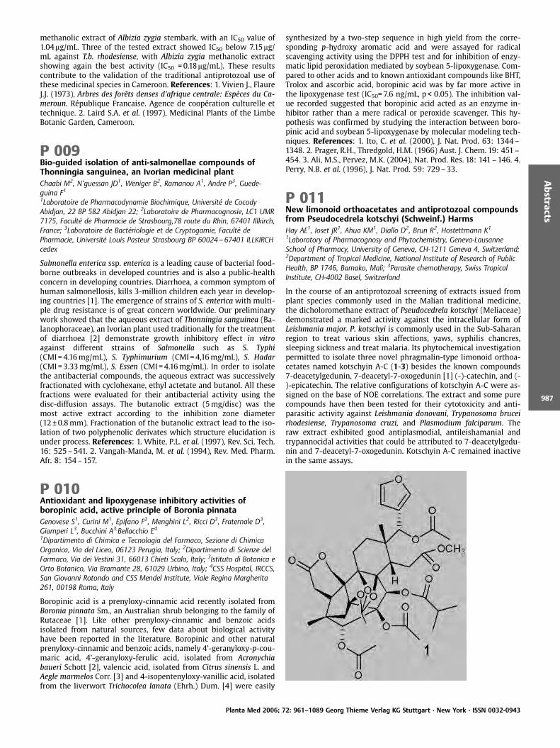
methanolic extract of Albizia zygia stembark, with an IC50 value of1.04 mg/mL. Three of the tested extract showed IC50 below 7.15mg/mL against T.b. rhodesiense, with Albizia zygia methanolic extractshowing again the best activity (IC50 =0.18mg/mL). These resultscontribute to the validation of the traditional antiprotozoal use ofthese medicinal species in Cameroon. References: 1. Vivien J., FlaureJ.J. (1973), Arbres des forÞts denses d’afrique centrale: Esp ces du Ca-meroun. R publique Francaise. Agence de coop ration culturelle ettechnique. 2. Laird S.A. et al. (1997), Medicinal Plants of the LimbeBotanic Garden, Cameroon.
P 009Bio-guided isolation of anti-salmonellae compounds ofThonningia sanguinea, an Ivorian medicinal plantChaabi M2, N’guessan JD1, Weniger B2, Ramanou A1, Andre P3, Guede-guina F11Laboratoire de Pharmacodynamie Biochimique, Universit de CocodyAbidjan, 22 BP 582 Abidjan 22; 2Laboratoire de Pharmacognosie, LC1 UMR7175, Facult de Pharmacie de Strasbourg,78 route du Rhin, 67401 Illkirch,France; 3Laboratoire de Bact riologie et de Cryptogamie, Facult dePharmacie, Universit Louis Pasteur Strasbourg BP 60024– 67401 ILLKIRCHcedex
Salmonella enterica ssp. enterica is a leading cause of bacterial food-borne outbreaks in developed countries and is also a public-healthconcern in developing countries. Diarrhoea, a common symptom ofhuman salmonellosis, kills 3-million children each year in develop-ing countries [1]. The emergence of strains of S. entericawith multi-ple drug resistance is of great concern worldwide. Our preliminarywork showed that the aqueous extract of Thonningia sanguinea (Ba-lanophoraceae), an Ivorian plant used traditionally for the treatmentof diarrhoea [2] demonstrate growth inhibitory effect in vitroagainst different strains of Salmonella such as S. Typhi(CMI =4.16mg/mL), S. Typhimurium (CMI =4,16mg/mL), S. Hadar(CMI =3.33mg/mL), S. Essen (CMI =4.16mg/mL). In order to isolatethe antibacterial compounds, the aqueous extract was successivelyfractionated with cyclohexane, ethyl actetate and butanol. All thesefractions were evaluated for their antibacterial activity using thedisc-diffusion assays. The butanolic extract (5mg/disc) was themost active extract according to the inhibition zone diameter(12€0.8mm). Fractionation of the butanolic extract lead to the iso-lation of two polyphenolic derivates which structure elucidation isunder process. References: 1. White, P.L. et al. (1997), Rev. Sci. Tech.16: 525–541. 2. Vangah-Manda, M. et al. (1994), Rev. Med. Pharm.Afr. 8: 154–157.
P 010Antioxidant and lipoxygenase inhibitory activities ofboropinic acid, active principle of Boronia pinnataGenovese S1, Curini M1, Epifano F2, Menghini L2, Ricci D3, Fraternale D3,Giamperi L3, Bucchini A3,Bellacchio E41Dipartimento di Chimica e Tecnologia del Farmaco, Sezione di ChimicaOrganica, Via del Liceo, 06123 Perugia, Italy; 2Dipartimento di Scienze delFarmaco, Via dei Vestini 31, 66013 Chieti Scalo, Italy; 3Istituto di Botanica eOrto Botanico, Via Bramante 28, 61029 Urbino, Italy; 4CSS Hospital, IRCCS,San Giovanni Rotondo and CSS Mendel Institute, Viale Regina Margherita261, 00198 Roma, Italy
Boropinic acid is a prenyloxy-cinnamic acid recently isolated fromBoronia pinnata Sm., an Australian shrub belonging to the family ofRutaceae [1]. Like other prenyloxy-cinnamic and benzoic acidsisolated from natural sources, few data about biological activityhave been reported in the literature. Boropinic and other naturalprenyloxy-cinnamic and benzoic acids, namely 4’-geranyloxy-p-cou-maric acid, 4’-geranyloxy-ferulic acid, isolated from Acronychiabaueri Schott [2], valencic acid, isolated from Citrus sinensis L. andAegle marmelos Corr. [3] and 4-isopentenyloxy-vanillic acid, isolatedfrom the liverwort Trichocolea lanata (Ehrh.) Dum. [4] were easily
synthesized by a two-step sequence in high yield from the corre-sponding p-hydroxy aromatic acid and were assayed for radicalscavenging activity using the DPPH test and for inhibition of enzy-matic lipid peroxidation mediated by soybean 5-lipoxygenase. Com-pared to other acids and to known antioxidant compounds like BHT,Trolox and ascorbic acid, boropinic acid was by far more active inthe lipoxygenase test (IC50=7.6 ng/mL, p < 0.05). The inhibition val-ue recorded suggested that boropinic acid acted as an enzyme in-hibitor rather than a mere radical or peroxide scavenger. This hy-pothesis was confirmed by studying the interaction between boro-pinic acid and soybean 5-lipoxygenase by molecular modeling tech-niques. References: 1. Ito, C. et al. (2000), J. Nat. Prod. 63: 1344–1348. 2. Prager, R.H., Thredgold, H.M. (1966) Aust. J. Chem. 19: 451–454. 3. Ali, M.S., Pervez, M.K. (2004), Nat. Prod. Res. 18: 141–146. 4.Perry, N.B. et al. (1996), J. Nat. Prod. 59: 729–33.
P 011New limonoid orthoacetates and antiprotozoal compoundsfrom Pseudocedrela kotschyi (Schweinf.) HarmsHay AE1, Ioset JR1, Ahua KM1, Diallo D2, Brun R2, Hostettmann K1
1Laboratory of Pharmacognosy and Phytochemistry, Geneva-LausanneSchool of Pharmacy, University of Geneva, CH-1211 Geneva 4, Switzerland;2Department of Tropical Medicine, National Institute of Research of PublicHealth, BP 1746, Bamako, Mali; 3Parasite chemotherapy, Swiss TropicalInstitute, CH-4002 Basel, Switzerland
In the course of an antiprotozoal screening of extracts issued fromplant species commonly used in the Malian traditional medicine,the dicholoromethane extract of Pseudocedrela kotschyi (Meliaceae)demonstrated a marked activity against the intracellular form ofLeishmania major. P. kotschyi is commonly used in the Sub-Saharanregion to treat various skin affections, yaws, syphilis chancres,sleeping sickness and treat malaria. Its phytochemical investigationpermitted to isolate three novel phragmalin-type limonoid orthoa-cetates named kotschyin A-C (1-3) besides the known compounds7-deacetylgedunin, 7-deacetyl-7-oxogedunin [1] (-)-catechin, and (-)-epicatechin. The relative configurations of kotschyin A-C were as-signed on the base of NOE correlations. The extract and some purecompounds have then been tested for their cytotoxicity and anti-parasitic activity against Leishmania donovani, Trypanosoma bruceirhodesiense, Trypanosoma cruzi, and Plasmodium falciparum. Theraw extract exhibited good antiplasmodial, antileishamanial andtrypannocidal activities that could be attributed to 7-deacetylgedu-nin and 7-deacetyl-7-oxogedunin. Kotschyin A-C remained inactivein the same assays.
Planta Med 2006; 72: 961–1089 Georg Thieme Verlag KG Stuttgart · New York · ISSN 0032-0943
Abstracts
987

Reference: 1. MacKinnon, S., Durst, T. et al. (1997), J. Nat. Prod. 60:336–341.
P 012Flavonoids and insecticidal activity of Teucrium zanoniiAbdelshafeek KA1, Ismail IA2, Alwahsh MA1
1ALTahady University, Faculty of Science, Chemistry dept, Sirt, Libya p. o.674;2National Research Center, pests and plant protection dept, Dokki, Cairo,Egypt
In frame of our investigation for medicinal Libyan plants we chooseTeucrium zanonii Pamp. (Family Labiatae) which is an endemic plantgrowing in Abofakhra region (25 Km from Benghazi City) [1]. Theplant was used in folk medicine for gastrointestinal troubles, tonic,renal inflammatory and antidiabetic [2]. Investigation of the flavo-noidal constituents lead to the identification of ciriliol, luteolin,chrysoeriol, and xanthomicrol from the ethyl acetate fraction whileapigenin –6,8 – di-O-glucoside and Luteolin –7– O – rutinosidefrom the butanol fraction. All structures were established usingdifferent chromatographic and spectroscopic (UV,MS, FAB-MS,1H,13C-nmr) measurements [3]. The insecticidal activity measure-ments of different extracts against Phloeotribus oleae on olive treesshowed that, the aqueous extract exhibit the highest mortality inthe lab. and field experiments (86.67% and 70.82) respectively [4].References: 1. Siddiqi, M.A. (1985), Flora of Libya, Vol. 118: Lamia-ceae. Revolution printing press, Tripoli, Libya. 2. Assem, M.E., Karam,T.H. (2004), Biochem. Syst. Ecol. 32: 665–674. 3. Savona, G., Pater-nostro, M.P, et al. (1979), An. Quim. 75: 433–436. 4. Bruno, M.,Piozzi, F. et al. (2002) Biochem. Syst. Ecol. 30: 595–599.
P 013Brocchiana carboxylic acid; the analogue of brevifolincarboxylic acid, Isolation and identification fromChrozophora brocchianaHawas UWPhytochemistry and Plant Systematic Dept, National Research Centre, Dokki,Cairo, Egypt
The Euphorbiaceae is a large family with close to 300 genera and5000–7500 species. In Egypt, this family is represented by onlyseven genera [1]. The chemosystematics of Euphorbia species inEgypt have recently been investigated and the current study ofChrozophora species is a continuation of our research on the phe-nolic constituents of this family. Previous phytochemical investiga-tion of the genus Chrozophora resulted in the isolation of severaltypes of chemical constituents including essential oils, terpenes,sterols, phenylpropanoid glycosides, xanthones, chromone and fla-vonoids [2]. The present study deals with the isolation and identi-fication of phenolic constituents from the aerial parts of Chrozo-phora brocchiana Vis. Brocchiana carboxylic acid, the analogue ofbrevifolin carboxylic acid [3] was isolated and identified from theaqueous methanol extract of the aerial parts of Chrozophora brocchi-ana in addition to eight known compounds identified as gallic acid,methyl gallate, ethyl gallate, ellagic acid, mono- and di-methoxyellagic acid, apigenin and luteolin 7-O-glucoside. The structureswere determined primarily by ESI-MS spectrometry and NMR spec-troscopy. The assignment of NMR signals was performed by meansof 1H–1H COSY, HMQC and HMBC experiments.
Brocchiana carboxylic acidBrevifolin carboxylic acid
References: 1. Taeckholm, V. (1974), Student’s Flora of Egypt, 2nd
ed., Cairo University Press, Cairo. 2. Hashim, O.K, Abou–Zaid, M.M. etal. (1990), Biochem. Syst. Ecol. 18: 151–152. 3. Nawwar, M.A.M. etal. (1994), Phytochemistry 36: 793–798.
P 014Helichrysums: antibacterial and monoamine oxidaseinhibitory activity of South African summer-rainfall speciesvan Staden J1, Stafford GI1, Pedersen PD2, Chukwujekwu JC1, J ger AK2
1Research Centre for Plant Growth and Development, School of Biologicaland Conservation Sciences, University of KwaZulu-Natal Pietermaritzburg,Private Bag X01, Scottsville 3209, South Africa; 2Department of MedicinalChemistry, Pharmacognosy, The Danish University of PharmaceuticalSciences, Universitetsparken 2, DK-2100 Copenhagen, Denmark
A large number of Helichrysum spp. (Asteraceae) is used traditionallyin southern Africa to treat a variety of ailments. It is a popularingredient in wound dressings [1] and is an important plant cultu-rally as it is burned at almost all traditional gatherings. Severalspecies have been shown to have a sedative effect [2] and antibac-terial activity both against Gram-positive and Gram-negative bac-teria [3]. Antibacterial activity was detected using MIC values ofcrude extracts ranging from 6.25 to 0.049mg/mL. Of the 9 Helichry-sum species assayed for activity against Staphylococcus aureus, Es-cherichia coli, Klebsiella pneumoniae, and Bacillus subtilis, 2 species,H. ruderale and H. argyolepis showed broad spectrum activity. Be-sides H. hesbaceum and H. adenocarpum that did not display anti-bacterial activity, the rest were active either against one or twobacteria. MAO-B inhibitors are known to increase the basal dopa-
Planta Med 2006; 72: 961–1089 Georg Thieme Verlag KG Stuttgart · New York · ISSN 0032-0943
Abstracts
988
mine levels in the nigrostriatal dopaminergic input pathway, a factthat is utilised in the symptomatic therapy for Parkinson’s disease[4].Seven species were assayed for MAO-B inhibitory activity using aperoxidase-linked photometric assay. H. agyrolepis (IC50=0.1mg/mL),H. umbraculigerum (IC50=4.3mg/mL) and H. ruderale (IC50=3.3mg/mL) were the most active species, although all species tested exhib-ited some MAO-B inhibition activity. References: 1. Watt, J.M.,Breyer-Brandwijk, M.G. (1962), The Medicinal and Poisonous Plantsof Southern and Eastern Africa. Livingston. London. 2. Stafford, G.I. etal. (2005), J. Ethnopharmacol. 100: 210–215. 3. Afolayan, A.J., Meyer,J. J. M. (1997), J. Ethnopharmacol. 57: 177–181. 4. Cesura, A.M.,Pletscher A. (1992), Prog. Drug. Res. 38: 171–297.
P 015In vitro antiplasmodial activity and cytotoxicity ofethnobotanically selected East African plants used for thetreatment of malariaFroelich S1, Onegi B2, Kakooko A2, Schubert C1, Jenett-Siems* K1
1Institut f r Pharmazie (Pharmazeutische Biologie), Freie Universit t Berlin,K nigin-Luise-Str.2 – 4, D-14195 Berlin, Germany. Fax: +49 – 30 – 83853729,E-mail: [email protected]; 2Department of Pharmacy(Pharmacognosy Unit), Makere University, Kampala, Uganda
In Uganda, a variety of plant remedies is used by traditional healersto treat symptoms of malaria. Due to oral interviews, Vernoniaamygdalina (Asteraceae), Aspilia africana (Asteraeae) and Momordi-ca foetida (Cucurbitaceae) were selected for further evaluation. Ex-tracts were obtained by macerating air dried plant material col-lected near Kampala, Uganda, in equal volumes of petroleumether/ethyl acetate and methanol (herb-solvent ratio 1:3, repeatedtwice), respectively. The crude extracts from roots and leaves, char-acterized by HPLC fingerprint chromatograms, were tested in vitroagainst the chloroquine-sensitive strain PoW (IC50 value for chlor-oquine= 0.011mM) and a chloroquine-resistant strain Dd2 (IC50 val-ue for chloroquine= 0.12mM) of Plasmodium falciparum. The anti-plasmodial activity was determined according to Desjardins et al.[1]. M. foetida showed significant antimalarial activity (IC50 valuesranging from 7.3 to 13.0mg/mL), whereas the root extract of V. amyg-dalina and the leaf extract of A. africana displayed lower activities(IC50 values: 19.0 and 30.3mg/mL [PoW]). Cytotoxicities of all ex-tracts were determined against human hepatocellar carcinoma(HepG2) and human urinary bladder carcinoma (ECV-304) (deriva-tive of T-24) cells [2]. The petroleum ether/ethyl acetate leaf extractof Vernonia amygdalina showed the highest cytotoxicity. Acknowl-edgements: Mrs. Ursula Friedrich (Institut f r Pharmazie) for tech-nical assistance in the cell-laboratory. References: 1. Desjardins, R.E.et al. (1979), Antimicrobial Agents Chemother. 16: 710–718. 2. Mos-mann, T. et al. (1983), J. Immunol. Methods 65: 55–63.
P 016Cytotoxic stilbenes from Cajanus cajan (L.) Millsp. leavesAshidi JS, Houghton PJ, Hylands PJPharmacognosy Research Laboratories, Pharmaceutical Sciences ResearchDivision, King’s College London, Franklin-Wilkins Building, 150 StamfordStreet, London SE1 9NH, United Kingdom
Cancer remains one of the leading causes of death in the worldespecially in the developed countries [1]. We now report a biologi-cally monitored phytochemical separation of the leaves of Cajanuscajan against a panel of human cancer and non- cancer cell lines invitro. Cajanus cajan, commonly called Pigeon pea, is grown for foodand medicinal purposes in the tropics, especially Nigeria. The airdried leaves were extracted with methanol continuously for fivedays. The extract was then concentrated under reduced pressure.60 g of the extract was adsorbed on Silica Gel GF254 and separatedinto fractions by vacuum liquid chromatography (VLC). The SRBassay was used to evaluate the cytotoxicity of the extracts and theisolated compounds. The dichloromethane (DCM) fraction of the
leaves exhibited modest cytotoxicity against human amelanoticmelanoma – C32, human breast adenocarcinoma – MCF-7 and hu-man large cell lung carcinoma cell lines – COR-L23 and human fetallung fibroblast – MRC-5 (IC50 =12.0, 10.0, 10.0 and 15.0mg/mL, re-spectively). This finding prompted further activity-guided fractiona-tion of the DCM fraction by flash chromatography and subsequentpurification on preparative thin-layer chromatography which led tothe identification of two prenylated stilbenes, longistylin A and C.These compounds have previously been reported to have antiplas-modial activity [2]. This is also the first time that these prenylatedstilbenes are shown to exhibit in vitro cytotoxic activity against hu-man amelanotic melanoma, C32, human breast adenocarcinoma,MCF-7, and human large cell lung carcinoma, COR-L23, cell lines.The IC50 of the compounds ranges between 20 and 35mM. Thesecompounds could explain the rational inclusion of Cajanus cajan intraditional herbal medicines used for the treatment of cancer insouth- western Nigeria. Further study to establish the mechanismof action of these two stilbenes is in progress. Acknowledgement: JSAthanks the Association of Commonwealth Universities, UK, for fi-nancial support. References: 1. Anonymous, http://health.yahoo.-com/news/58569. 2. Duker-Eshun, G, et al. (2004), PhytotherapyResearch, 18:128–130.
P 017Antibacterial and resistance –modifying effects ofMezoneuron benthamianumDickson RA, Houghton PJ, Hylands PJPharmacognosy Research Laboratories, Pharmaceutical Sciences ResearchDivision, King’s College London, 150 Stamford Street, London SE1 9NN, UK
The ever increasing resistance of human pathogens to current anti-microbial agents is a serious medical problem, and has resulted inthe need for novel antibiotic prototypes. The root bark of Mezoneur-on benthamianum Baill.(Caesalpinaceae) is used in Ghanaian tradi-tional medicine for the treatment of wounds and other dermal in-fections. Bioactivity-guided fractionation of the pet. spirit extractled to the isolation of cassane-type diterpenes active against variousbacteria and two strains of Staphylococcus aureus possessing themultidrug efflux pumps NorA and TetK (SA1199B and XU212) [1].Addition of R2 and R3 in the growth medium at 10mg/mL resulted ina 16-fold and 8-fold (Norfloxacin) and 8-fold and 4-fold (Tetracy-cline) potentiation of activities in both compounds respectively (Ta-ble 1). The other three compounds however were not active againstthe efflux pumps but showed various degrees of activities againstother bacteria.Table1 Antibacterial susceptibility of test strains in the absence andpresence of 10mg/mL of R2 and R3 and 20mg/mL reserpine, a natu-rally occurring MDR efflux inhibitor serving as a standard modula-tor, n = 3.
Antibacterial agent MIC of test strain expressing the indicated efflux proteinSA 1199B(NorA) XU 212(TetK)
Norfloxacin+R2+R3+ReserpineTetracycline+R2+R3+Reserpine
322432NT
NT
128163232
Ackowledgement: Rita Akosua Dickson is funded by the Common-wealth Scholarship Commission, UK. Reference: 1. Gibbons, S. et al.(2004), Phytochem. 65: 3249 –3254.
Planta Med 2006; 72: 961–1089 Georg Thieme Verlag KG Stuttgart · New York · ISSN 0032-0943
Abstracts
989
P 018Chemical composition and cytotoxic activities of essentialoils of leaves and berries of Juniperus phoenicea L grown inEgyptEl-Sawi S, Motawae HNational Research Centre, Dokki, 12622,Cairo, Egypt
Hydrodistillation of berries and leaves of Juniperus phoeniceagrown in Sinai yielded volatile oils in the yield of 0.36 and 1.96%,respectively. Using gas chromatography/mass spectrometry techni-que, fifty eight compounds were identified in berry oil representing99.2% of the oil composition. Alpha-pinene was the major com-pound in berry oil (39.30%) followed by sabinene (24.29%). Berryoil composed mainly of monoterpenoids which amounted to90.58%, of which 72.85% was monoterpene hydrocarbons. The ses-quiterpenoids accounted for about 8% of the total oil composition.Leaf oil was composed of about 66 compounds representing 99.16%of the total composition of the oil. Alpha-pinene was the majorconstituent of leaf oil at concentration of 38.22%, followed by al-pha-cedrol (31.23%). The monoterpene hydrocarbon was the predo-minant chemical group (41.29%) followed by the oxygenated sesqui-terpenes (32.21%). Both oils showed very high cytotoxic activitiesagainst all cell line tested. Both oils showed equal activities againstbrain (0.6 microgram/mL) and cervix (5.0 microgram/mL) humancell lines, while berry oil was slightly more active than leaf oilagainst lung (0.6 and 0.7 microgram/mL, respectively), liver (0.7and 0.9 microgram/mL, respectively) and breast human cell lines(0.8 and 1.0 microgram/mL, respectively). References: 1, Adams, R.(1995), Identification of Essential Oils Components by Gas Chroma-tography/Mass Spectrometry. Allured, Carol Stream, Illinois. 2, Afifi,M. et al. (1992), Mans. J. Pharm. Sci. 8: 37–46.
P 019Effects of different carbon sources on production ofpolysaccharides by Agaricus blazeiVahidi H, Hamedi ASchool of Pharmacy Shaheed Beheshti University of Med. Sci. Tehran, Iran
Mushroom polysaccharides offer a lot of hope for cancer patientsand sufferers of many devastating diseases. A variety of polysacchar-ides from a number of mushroom varieties have been demonstratedto enhance the immune system. Yield and functionally of polysac-charides by fermentation are highly dependent on their culture con-ditions, such as different microorganisms, medium compositionsand environmental parameter. Carbon source is one of the mostimportant parameter affecting polysaccharide fermentations. [1].In this study the effects of different carbon sources including glu-cose, lactose, sucrose, manitol, starch, galactose, maltose and fruc-tose in two different media (Complex and synthetic) were investi-gated. For the determination of polysaccharides produced by thefungus the total polysaccharides which precipitated by absolutealcohol were weighed. The experiments showed that the highestgrowth and polysaccharide production were obtained when galac-tose and starch were used as carbon source. The concentration ofpolysaccharide in both complex and synthetic media were similar.The lowest growth and productivity were also seen in medium con-taining sucrose. Reference: 1. Chin–Hang, S. et al. (2004), J. Chem.Technol. Biotechnol. 79: 998–1002.
P 020Activity of a compound isolated from Senna villosa againstTrypanosoma cruziP rez MS1, Guzm n E2, Gonz lez MR2, P rez C1, Zavala MA1, Acosta K2
1Universidad Aut noma Metropolitana-Xochimilco, Calz. del Hueso 1100 CP04960 M xico D.F. A.P. 23 – 181 M xico; 2Centro de InvestigacionesRegionales “Dr. Hideyo Noguchi“, Universidad Aut noma de Yucat n, MeridaYucat n, M xico
Previously we reported that chloroform extract of S. villosa (Miller)Irwin & Barneby possess activity against epimastigotes and trypo-mastigotes of T. cruzi [1]. From this extract was isolated a com-pound, which was identified as 4-hydroxymethylen-2-pentaeicona-none mp 76–77 C, (KBr/cm): 3451, 2919, 2850, 1735. 1HNMR(400MHz CDCl3) d (ppm): 0.88(t,3H), 1.25(m,38H), 1.57(s,3H),1.61(m,3H), 2.29(m,2H), 4.05(m,2H). 13CNMR: 14.0(CH3), 22.7(CH2),25.0(CH2), 25.9(CH2), 28.6(CH2), 29.4(CH), 29.5(CH3), 29.6(CH2),31.0(CH2), 34.3(CH2), 64.3(CH2), 173.6(C =O), EIMS (m/z): 396(1),97(54), 83(60), 57(100). Analysis combustion calcd for C26, H52O2,C 78.35%, H 13.44%, O 8.21%. Activity was assayed on epimatigotesand trypomastigotes of T. cruzi strain Y isolated from human, cul-tured in liver infusion tryptose medium supplemented with 10% ofheat-inactived fetal calf serum and cultured at 28 C. 1.65, 3.3 and6.6mg/mL concentration of this compound was tested. Gentian vio-let (17.5mg/mL) was used as positive control. Parasites were count-ed daily for 10 days. At concentration of 1.65mg/mL the growthinhibition of trypomastigotes was 60%, with 3.3mg/mL 72% andwith 6.6mg/mL was 82%, similar results were obtained with epima-tigotes. The activity was doses-dependent in both forms of parasite.The susceptibility of parasite to the compound is similar to thatobserved with gentian violet. Reference: 1. Guzm n, E. et al.(2004), Pharm. Biol. 42: 1 –4.
P 021Partial chemical structure and immunomodulating activitiesof RGAP (Red Ginseng acidic Polysaccharide) from Koreanred ginseng (Panax ginseng C. A. Meyer)Park JD1, Shin HJ1, Kwak YS1, Wee JJ1, Song YB1, Kyung JS1, Kiyohara H2,Yamada H2
1Div. Of Ginseng Efficacy, KT&G Central Research Institute, Daeduk ScienceTown, 305 – 345, KOREA; 2Laboratory of Biochemical Pharmacology forPhytomedicine, Kitasato Institute for Life Science, Kitasato University, Tokyo108 – 8642, JAPAN
A red ginseng acidic polysaccharide (RGAP) with immunomodulat-ing antitumor activities was isolated from Korean red ginseng,steamed and dried ginseng (Panax ginseng C. A. Meyer). The mole-cular weight of RGAP was estimated to be 12–450 kDa by gel filtra-tion chromatography. RGAP has been found to increase survival rateand to inhibit of tumor growth significantly in a dose dependentmanner in mice transplanted with tumor cells. RGAP significantlypromoted nitric oxide (NO) production from peritoneal macro-phages both in vivo and in vitro. Western blot analysis exhibited anewly synthesized inducible nitric oxide synthase (iNOS) proteinband in the RGAP treated group. It seems likely that immunomodu-lating antitumor activities of RGAP are mainly mediated by NO pro-duction of macrophage. RGAP was further purified by ultrafiltrationand anion exchange chromatography on DEAE-sepharose, followedby gel filtration on Sephacryl S-300 to give an active fraction (GFP)with stronger NO production in murine macrophages. GFP increasedsurvival rate ten times compared to RGAP in male ICR mice trans-planted with sarcoma 180 and also showed more potent tumoricidalactivities of natural killer cells than RGAP. Sugar composition(mol %)of GFP was found to be arabinose: rhamnose: xylose: galacturonicacid: mannose: galactose: glucose (10:9:1:25:8:20:27) by GC/MS.Partial acid hydrolysis of GFP resulted in acidic oligosaccharides,and a combination of enzymatic digestion(Endo-a-D-(1?4)-polyga-lacturonase) and methylation analysis, MALDI-TOF-MS, PSD analysisof the acidic oligosaccharides suggested that GFP comprised a ga-
Planta Med 2006; 72: 961–1089 Georg Thieme Verlag KG Stuttgart · New York · ISSN 0032-0943
Abstracts
990
lactogalacturonan core such as –a-D-GalA-(1?4)-a-D-GalA-(1?4)-a-D-GalA-(1?6)-a-D-Gal as the acidic moiety. The results suggestthat certain galactogalacturonan core chains are responsible for im-munomodulating antitumor activities as well as nitric oxide (NO)production. Acknowledgement: This work was supported by agrant(GP-303) from Korea Ginseng Corporation.
P 022Effects of QKL on the level of TNF-a, IL-1b, ICAM-1 afterintracerebral hemorrhageYi L1,2, Ming L1, Pengtao L, Qingguo W1
1School of preclinical Medicine, Bei San Huan Dong Lu 12, Beijing Universityof Chinese Medicine, 100029, Beijing, China; 2Institut f r Pharmazie(Pharmazeutische Biologie), FU Berlin, K nigin-Luise-Str. 2 – 4. 14195 Berlin,Germany
Aims: Inflammatory cytokines release and action are central in thepathogenesis of the inflammatory response that occurs after intra-cerebral hemorrhage (ICH). Among the inflammatory cytokines,TNF-a, IL-1b, intercellular adhesion molecule (ICAM)-1 is the keyfactors which lead to secondary brain damages e. g. hydrocephalusand neuron death. QKL is an injection of natural products (consist-ing of radix scutellariae, fructus gardeniae, cholic acid, radix noto-ginseng, concha margaritifera). The aim here was to detect the func-tion of anti-inflammation of QKL after the ICH. Method: We estab-lished a rat model with collagenase-induced intracerebral hemor-rhage. The QKL was injected from the tail vein 3mL every day. TheELISA and RIA were used to detect the level of TNF-a, IL-1b, ICAM-1in the cerebral homogenate in different periods after ICH. Result:The level of the TNF-a, IL-1b, ICAM-1 is markedly decreased withtreating in the period of 48 hours after occurrence of the ICH. But inthe period of 24 hours and 72 hours it shows no visible effect.Therefore, QKL has a good effect to inhibit the inflammatory cyto-kines TNF-a, IL-1b, ICAM-1 in the period of 48 hours after the ICHand consequently protects the neurons from secondary damages.
P 023Indirubin-3’-monoxime inhibits rat vascular smooth musclecell proliferation induced by plateled-derived growth factorvia the Jak/STAT-pathwaySchwaiberger AV, Heiss E, Dirsch VMDepartment of Pharmacognosy, University of Vienna, Althanstraße 14, 1090Vienna, Austria
Indirubin, a constituent identified in the traditional Chinese antil-eukaemic recipe Danggui Longhui Wan, and its derivatives havebeen shown to be potent cyclin-dependent kinase (CDK)-inhibitorsin vitro [1]. CDKs as key regulators of cell cycle progression arepromising targets in the treatment of vasculoproliferative disorders,for instance atherosclerosis and restenosis [2]. The aim of the studywas therefore to investigate the antiproliferative effect of indirubin-3’-monoxime (I3MO) on plateled-derived growth factor (PDGF-BB)-induced rat vascular smooth muscle cell (RVSMC) growth. Effects onDNA-synthesis were assessed via BrdU-incorporation after 23h oftreatment with PDGF-BB (20 ng/mL) and increasing concentrationsof I3MO (0.1 –10mM). At a concentration of 3mM, the BrdU-positivelabeling index was reduced to control level. Cell cycle analysis in thepresence of I3MO showed a significant arrest of RVSMCs in the G0/G1 phase after 16h of stimulation with PDGF-BB. Ongoing treat-ment over 48h led to DNA strand breaks at high concentrations(10mM), as shown by the detection of propidium-iodide stainednuclei with a sub-diploid DNA-content by flow cytometry. Focusingon the involved signaling pathways for these effects, activation ofmolecules evidentially participating in proliferation was examined.Western blot analysis revealed that the kinases Akt, Erk1/2 and p38were activated after 10 minutes of stimulation with PDGF-BB, butthe effects were not blocked by I3MO at concentrations of 3 and5mM. Further investigation of influence on the Jak/STAT pathway,
however, indicated a significant inhibition of STAT1 andSTAT3(Y705) phosphorylation. These results demonstrate thatI3MO inhibits STAT1 and STAT3(Y705) phosphorylation, suggestingthat blocking of the Jak/STAT pathway is at least partially responsi-ble for the antiproliferative activity of the compound. Acknowlegde-ments: CNRS, Station Biologique, Amylo ds and Cell Division Cycle,Meijer L. References: 1. Hoessel, R. et al. (1999), Nat. Cell Biol. 1:60–67. 2. Dzau, V.J. (2002), Nat. Med. 8: 1249 –1256.
P 024Wound healing activity of Alocasia odora (Roxb.) KochViet LD, Houghton PJ, Forbes B, Corcoran O, Hylands PJPharmacognosy Research Laboratories, Pharmaceutical Sciences ResearchDivision, King’s College London, 150 Stamford Street, London SE1 9NH,United Kingdom
Introduction: During the twenty years of the Vietnam war, the stemof Alocasia odora has been used in Vietnam for the treatment ofwounds. However, there is little literature about this activity of theplant or its chemical composition. The present study investigatessome possible modes of action of the plant in wound healing pro-cess and attempts to identify the constituents responsible. Meth-ods: Skin fibroblast proliferation assay-guided fractionation andHPLC isolation were used to study bioactivity in extracts of the stemand to locate the active moieties. In addition, an assay measuringprotection against H2O2-induced damage to skin fibroblasts (142-BR) [1] and the DPPH radical scavenging assay were used to supple-ment the results of the proliferation assay [2]. Results: A total of 10compounds (1 triterpenoid glycoside, 2 flavonoid C-glycosides, 5lignan glycosides, 1 lignan and 1 alkaloid) were isolated from theactive fractions and tested for bioactivities. The structures of theisolated compounds were determined by the joint application ofspectroscopic methods. The lignans, the triterpenoid and the alka-loid showed a slight stimulation of cell proliferation. No compoundwas shown to possess a protective effect against H2O2-induceddamage on fibroblasts. Only lignans were shown to have DPPH ra-dical scavenging activity. Conclusions: Extracts and some isolatedcompounds from Alocasia odora stems were shown to possess mod-est activity in the bioassays. It is possible that proliferation of skinfibroblasts and antioxidant activities partly contribute to woundhealing activity of Alocasia odora. Acknowledgements: VietnameseMinistry of Education and Training, Prof. P K Man, Dr. P V HienReferences: 1. Tran, V.H. et al. (1997), Wound Rep. Reg. 5: 159–167. 2. Kyong Soon, K. et al. (2003), J. Ethnopharmacol. 85: 69–73.
P 025Evaluation of Picralima nitida hypoglycemic activity, toxicityand analytical standardsInya-Agha SI1, Ezea SC1, Odukoya OA2
1Department of Pharmacognosy, Faculty of Pharmaceutical Sciences,University of Nigeria, Nsukka, Nigeria; 2Department of Pharmacognosy,Faculty of Pharmacy University of Lagos, Nigeria
WHO Expert Committee on diabetes encourages further investiga-tion into traditional methods of treatment and also emphasizes theneed to ensure safety and quality control of ingredients used. Picra-lima nitida Stapf (Apocynaceae) seeds used in the treatment of he-patitis, worms, sleeping sickness and malaria in Ihiala town, Anam-bra state of Nigeria, have been shown to have hypoglycemic effects[1]. Hypoglycemic activity was confirmed, recorded as an index ofblood glucose with a glucometer in normal and intraperitoniallyinduced alloxan diabetic albino rats with glibenclamide as referencestandard and normal saline as control. Toxicity studies included theevaluation of acute and sub-acute (15 days) tests. The animals wereobserved for toxic signs and symptoms, body weight changes re-corded and LD50 calculated. Analytical standards were moisturecontent, ash and extractive values for quality assurance. 100mg/kg,300mg/kg and 900mg/kg of the extracts to normal rats resulted in
Planta Med 2006; 72: 961–1089 Georg Thieme Verlag KG Stuttgart · New York · ISSN 0032-0943
Abstracts
991
significant (P< 0.01) lowering of fasting blood sugar after eighthours. Extract maintained hypoglycemic action throughout the24hours of study indicating a long duration of action. In normalrats, pulp extract (100mg/kg) produced a maximum percentage re-duction of 38.35%, rind extract (900mg/kg) 46.19% and seed extract(100mg/kg) 36.81%. Alloxan induced rats were pulp 85.85%(300mg/kg), seed 83.26% (300mg/kg) and rind 80.25% (900mg/kg) respectively. Order of activity recorded as pulp > seed > rind.Acute toxicities (LD50) of pulp, seed and rind were 7071.06mg/kg,948.68mg/kg and 1364.91mg/kg respectively. Reference: 1. Aguwa,C.N. et al. (2001), J. Natural Remed. 1: 135–139.
P 026Effects of Leuzea carthamoides DC. on human breast cancerMCF-7 cells detected by gene expression profilingHamburger M1,5, Gaube F1, W lfl S2,3, Pusch L2, Kroll T2, Riese U1,Schrenk D4
1Institute of Pharmacy, Department of Pharmaceutical Biology, Friedrich-Schiller-University Jena, D-07743 Jena, Germany; 2Clinic of Internal MedicineII, Friedrich-Schiller-University Jena, D- 07740 Jena, Germany; 3Institute ofPharmacy and Molecular Biotechnology, Ruprecht-Karls-UniversityHeidelberg, D-69120 Heidelberg, Germany; 4Department of Food Chemistryand Environmental Toxicology, University of Kaiserslautern, D-67663Kaiserslautern, Germany; 5Department of Pharmaceutical Sciences, Instituteof Pharmaceutical Biology, University of Basel, CH-4056 Basel, Switzerland
Products derived from roots of Leuzea carthamoides DC. (Maral root)are being promoted as anti-aging and adaptogenic. The phytoecdys-teroids are considered as active principles with numerous beneficialeffects [1], but little is known about the pharmacological propertiesof Leuzea extracts. We, therefore, investigated the effects of a lipo-philic Leuzea root extract on the human breast cancer cell line MCF-7. Cell proliferation was inhibited by the extract (IC50=28mg/mL) butnot by the major phytoecdysteroid, 20-hydroxyecdysone. Genome-wide expression profiling using Affymetrix HG U133 Plus 2.0 micro-arrays was carried out to analyze effects at the mRNA level. 241genes appeared to be significantly regulated. Transcripts of geneproducts involved with cell cycle progression and DNA replicationwere decreased, while mRNAs coding for inhibitory products wereincreased. This was in agreement with the antiproliferative activityof the extract. Upregulation of several pro-apoptotic genes provideevidence that the extract may sensitize the cells for apoptoticevents. Downregulation of estrogen receptor a could be confirmedby real-time RT-PCR and Western blot. Additionally, expression lev-els of several transcripts of enzymes with oxidoreductase activitywere induced, including a strong increase of CYP1A1 transcriptwhich is known to be regulated via the aryl hydrocarbon receptor(AhR). AhR-agonistic activity of the Leuzea root extract, but not 20-hydroxyecdysone was confirmed by a XRE-dependent reporter geneassay. This suggests that at least a part of the effects could be due toAhR activation. However, the phytoecdysteroids are not active prin-ciples in Leuzea root. Reference: 1. Sl ma, K., Lafont, R. (1995), Eur. J.Entomol. 92: 355–377.
P 027Antibacterial activity of the essential oils of catnip (Nepetacataria L.) and lemon balm (Melissa officinalis L.) againstclinical isolates from the respiratory tractSuschke U1, Geiss HK2, Reichling J11Institute of Pharmacy and Molecular Biotechnology, Dept. Biology,University of Heidelberg, INF 364, 69120 Heidelberg, Germany; 2HygieneInstitute, Dept. Medical Microbiology, University Hospital Heidelberg,INF 324, 69120 Heidelberg, Germany
Minimum inhibitory concentration (MIC) and minimum bactericidalconcentration (MBC) of the essential oils of catnip (Nepeta cataria),lemon catnip (N. cataria var. citriodora) and lemon balm (Melissaofficinalis), whose composition has been analyzed by GC-MS, were
determined in vitro by a modified broth microdilution method ac-cording to the German DIN-regulation 58940–8 [1] against clinicalisolates of Staphylococcus aureus, Streptococcus pyogenes, Streptococ-cus pneumoniae, Haemophilus influenzae and Moraxella catarrhalis.These bacteria are frequently involved in respiratory tract infectionslike sinusitis, tonsillitis, otitis media, bronchitis and pneumonia. Ofeach strain twelve patient isolates from different culture materialswere chosen, which had been identified and characterized by anantibiogram in the routine laboratory of the Hygiene Institute, Uni-versity Hospital Heidelberg. In spite of their different origin andlevel of resistance to standard antibiotics, all isolates were suscep-tible to catnip and lemon balm oils with MIC values ranging from0.25 % to 0.008 % (v/v). MIC values within the groups of isolatesdid not differ from those obtained with essential oil sensitive refer-ence strains by more than one dilution step, indicating that naturalresistance to these essential oils and cross resistance to standardantibiotics are unlikely to occur in these bacteria. Lemon balm oil,whose main components were geranial (23 %) and neral (17 %),exhibited the highest antibacterial activity, followed by lemon cat-nip oil, whose main components were the monoterpene alcoholsnerol, citronellol, and geraniol (» 50 %), and catnip oil which con-tained mainly nepetalactones (77 %) and only small amounts ofmonoterpene aldehydes. Reference: 1. Harkenthal, M. et al. (1999),Pharmazie 54: 460–463.
P 028Screening of medical plants from Mali for antitrypanosomalactivityAderbauer B1, Clausen PH2, Melzig MF11Department of Pharmacy, Freie Universitaet Berlin, Koenigin-Luise-Str. 2 + 4,D-14195 Berlin, Germany; 2Institute for Parasitology and Tropical VeterinaryMedicine, Freie Universitaet Berlin, Koenigsweg 67, D-14163 Berlin, Germany
Dichlormethane extracts of 50 plant parts collected in Mali, tradi-tionally used against trypanosomes were investigated for their invitro and in vivo activity against Trypanosoma brucei brucei. [1]. Sixextracts showed high efficacy in vitro, using the Long-term ViabilityAssay (LtVA) [2] with MIC-values of 50mg/mL. Four extracts showedlow cytotoxicity and good tolerance in mice and were tested in vivousing the standard mouse test by Eisler et al. (2001), [3]. The ex-tracts of Guiera senegalensis J.F. leaves and of Securidaca longepen-dunculata Fres. roots were able to reduce parasitaemia in mice treat-ed at a dose of 150mg/kg b.w. (i.p., two times daily for three days)with a reduction in parasitaemia of 42 and 48.5% compared to theuntreated control group. These extracts are now phytochemicalllyanalysed for active principles. Acknowledgements: The authors thankDr. Bizimana for providing the plant material and for technical sup-port. References: 1. Bizimana, N. et al. (2006), J. Ethnopharmacol.103: 350–356. 2. Kaminsky, R. et al. (1989), Exp. Parasitol. 69: 281–289. 3. Eisler, M.C. et al. (2001), Vet. Parasitol. 97: 171–182.
P 029Methanolic extracts of two Betulaceae species from Spain,induced apoptosis in tumoral cell linesAcero N, Mu oz-Mingarro D, Dom nguez MT, Llinares FFacultad de Farmacia Universidad San Pablo CEU. Urb. Montepr ncipe. 28660Boadilla del Monte MADRID. SPAIN
The citotoxic activity of methanolic extracts of leaves and barksfrom Alnus glutinosa L. and Betula alba L. were studied over threetumoral cell lines (PC-3, HeLa and HT-29). The LDH test allows thedetermination of the necrotic effect of the extracts. Apoptosis wasmicroscopically visualized after staining [1]. The extracts capacity toregulate the Tumoral Necrosis Factor-a (TNF- a) and the VascularEndothelial Growth Factor (VEGF) synthesis, was also analyzed bySigma ELISA kits. The plants antioxidant capacity was estimated bythe DPPH assay [2]. The method evaluates antioxidation as the abil-ity of the extracts to scavenge DPPH radical. All the extracts show
Planta Med 2006; 72: 961–1089 Georg Thieme Verlag KG Stuttgart · New York · ISSN 0032-0943
Abstracts
992
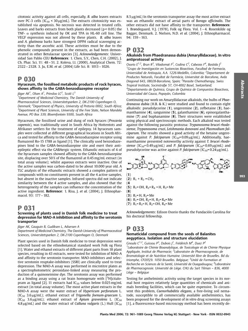
citotoxic activity against all cells, especially, B. alba leaves extractsover PC-3 cells (IC50 < 30mg/mL). The extracts citotoxicity was es-tablished via apoptosis. No necrosis was detected in treated cells.Leaves and barks extracts from both plants decreased (p < 0.05) theTNF- a synthesis induced by OK and TPA in HL-60 cell line. TheVEGF expression was not altered by these plants. B. alba leavesand A. glutinosa barks have strongest DPPH radical scavenging ac-tivity than the ascorbic acid. These activities must be due to thephenolic compounds present in the extracts, as had been demon-strated in other Betulaceae species [3]. Acknowledgements: Univer-sidad San Pablo CEU References: 1. Chen, S.Y., Chen, C.H. (2002), J.Ch. Phar. Sci. 11: 48 –51. 2. Koleva, I.I. (2000), Analytical Chem. 72:2322–2328. 3. Ju, E.M. et al. (2004) Life Sci. 8: 1013–1026.
P 030Hyraceum, the fossilised metabolic products of rock hyraces,shows affinity to the GABA-benzodiazepine receptorJ ger AK1, Olsen A1, Prinsloo LC2, Scott L31Department of Medicinal Chemistry, The Danish University ofPharmaceutical Sciences, Universitetsparken 2, DK-2100 Copenhagen O,Denmark; 2Department of Physics, University of Pretoria 0002, South Africa;3Department of Plant Sciences, University of the Free State, Nelson MandelaAvenue, PO Box 339; Bloemfontein 9300, South Africa
Hyraceum, the fossilised urine and dung of rock hyraces (Procaviacapensis), was traditionally used in South Africa by Hottentots andAfrikaner settlers for the treatment of epilepsy. 14 hyraceum sam-ples were collected at different geographical locations in South Afri-ca and tested for affinity to the GABA-benzodiazepine receptor usingflumazenil Ro-15 1788 as ligand [1]. The clinically used benzodiaze-pines bind to the GABA-benzodiazepine site and exert their anti-epileptic effect via the GABAergic system. Ethanolic extracts of 4 ofthe hyraceum samples showed affinity to the GABA-benzodiazepinesite, displacing over 50% of the flumazenil at 0.45mg/mL extract (intotal assay volume); whilst aqueous extracts were inactive. One ofthe active samples was carbon-dated to be about 10.000 year old. ATLC analysis of the ethanolic extracts showed a complex pattern ofcompounds with no constituents present in all the 4 active samples,but absent in the inactive samples. Infrared spectra did not indicatesimilarity between the 4 active samples, and also showed that theheterogeneity of the samples can influence the concentration of theactive ingredient. Reference: 1. Risa, J. et al. (2004), J. Ethnophar-macol. 93: 177–182.
P 031Screening of plants used in Danish folk medicine to treatdepression for MAO-A inhibition and affinity to the serotonintransporterJ ger AK, Gauguin B, Gudiksen L, Adsersen ADepartment of Medicinal Chemistry, The Danish University of PharmaceuticalSciences, Universitetsparken 2, DK-2100 Copenhagen O, Denmark
Plant species used in Danish folk medicine to treat depression wereselected based on the ethnobotanical standard work Folk og Flora[1]. Water and ethanol extracts of different plant parts from 18 plantspecies, resulting in 43 extracts, were tested for inhibition of MAO-Aand affinity to the serotonin transporter. MAO-inhibitors and selec-tive serotonin reuptake-inhibitors (SSRI) are clinically used to treatdepression. The MAO-A assay was performed in microtitre plates asa spectrophotometric peroxidase-linked assay measuring the pro-duction of a quinoneimine dye. The serotonin assay was performedas a binding assay using a rat brain homogenate with [3H]citalo-pram as ligand [2]. 11 extracts had IC50 values below 0.025mg/mLextract (in total assay volume). The most active plant extracts in theMAO-A assay were the water extract of Hypericum perforatum L.(IC50 3.6mg/mL); ethanol extract of Trigonella foenum-graecum L.(IC50 3.6mg/mL); ethanol extract of Apium graveolens L. (IC504.8mg/mL) and the water extract of Calluna vulgaris (L.) Hull (IC50
8.5mg/mL) In the serotonin transporter assay the most active extractwas an ethanolic extract of aerial parts of Borago officinalis. Theother extracts did not have affinity to the transporter. References:1. Brøndegaard, V.J. (1978), Folk og Flora. Vol. 1 –4. Rosenkilde ogBagger, Denmark. 2. Nielsen, N.D. et al. (2004), J. Ethnopharmacol.94: 159–163.
P 032Alkaloids from Phaedranassa dubia (Amaryllidaceae). In vitroantiprotozoal activityOsorio E1,2, Brun R3, Viladomat F2, Codina C2, Cabezas F4, Bastida J21Grupo de Investigaci n en Sustancias Bioactivas, Facultad de Farmacia,Universidad de Antioquia, A.A. 1226-Medell n, Colombia; 2Departament deProductes Naturals, Facultat de Farm cia, Universitat de Barcelona, AvdaDiagonal 643, 08028-Barcelona, Spain; 3Parasite Chemotherapy, SwissTropical Institute, Socinstrabe 57, CH-4002 Basel, Switzerland;4Departamento de Qu mica, Grupo de Qu mica de Compuestos Bioactivos,Universidad del Cauca, Popay n, Colombia
Continuing our work on Amaryllidaceae alkaloids, the bulbs of Phae-dranassa dubia (H.B. & K.) were studied and found to contain eightalkaloids: pseudolycorine (1), ungeremine (2), zefbetaine (3), hae-manthamine (4), sanguinine (5), galanthamine (6), epinorgalantha-mine (7) and buphanamine (8). Their structures were establishedusing physical and spectroscopic methods. Each alkaloid was testedagainst the following parasitic protozoa: Trypanosoma brucei rhode-siense, Trypanosoma cruzi, Leishmania donovani and Plasmodium fal-ciparum. The results showed a good activity of the betaine ungere-mine against P. falciparum (IC50=0.09mg/mL). Additionally, hae-manthamine presented noteworthy activity against T. brucei rhode-siense (IC50=0.49mg/mL) and P. falciparum (IC50=0.69mg/mL) andpseudolycorine was active against P. falciparum (IC50=0.24mg/mL).
(1)(2): R1 + R2 =CH2
(4(5): R1=OH, R2=R4 =H, R3=Me(8)(3): R1=H, R2=Me(6): R1=OH, R2=H, R3=R4=Me(7): R1=R3=H, R2=OH, R4=Me
Acknowledgements: Edison Osorio thanks the Fundaci n Carolina forhis doctoral fellowship.
P 033Nematicidal compound from the seeds of Balanitesaegyptiaca. Isolation and structure elucidationGnoula C1,2, Guissou P3, Dubois J1, Fr d rich M4, Duez P21Laboratoire de Chimie Bioanalytique, de Toxicologie et de Chimie PhysiqueAppliqu e, Institut de Pharmacie; 2Laboratoire de Pharmacognosie, deBromatologie et de Nutrition Humaine. Universit libre de Bruxelles. Bd dutriomphe, CP205/9. 1050 Bruxelles. Belgique; 3Unit de Formation etRecherche en Sciences de la Sant , Universit de Ouagadougou; 4Laboratoirede Pharmacognosie. Universit de Li ge. CHU du Sart Tilman – B36, 4000Li ge – Belgique
Testing for anthelmintic activity using the target species in its nor-mal host requires relatively large quantities of chemicals and ani-mals breeding facilities, which can be quite expensive. To circum-vent this problem, Caenorhabditis elegans, a free-living soil nema-tode, susceptible to all commercially available anthelmintics, hasbeen proposed for the development of in vitro drug screening assays[1], a fluorescence-based microscopy method has been recently de-
Planta Med 2006; 72: 961–1089 Georg Thieme Verlag KG Stuttgart · New York · ISSN 0032-0943
Abstracts
993
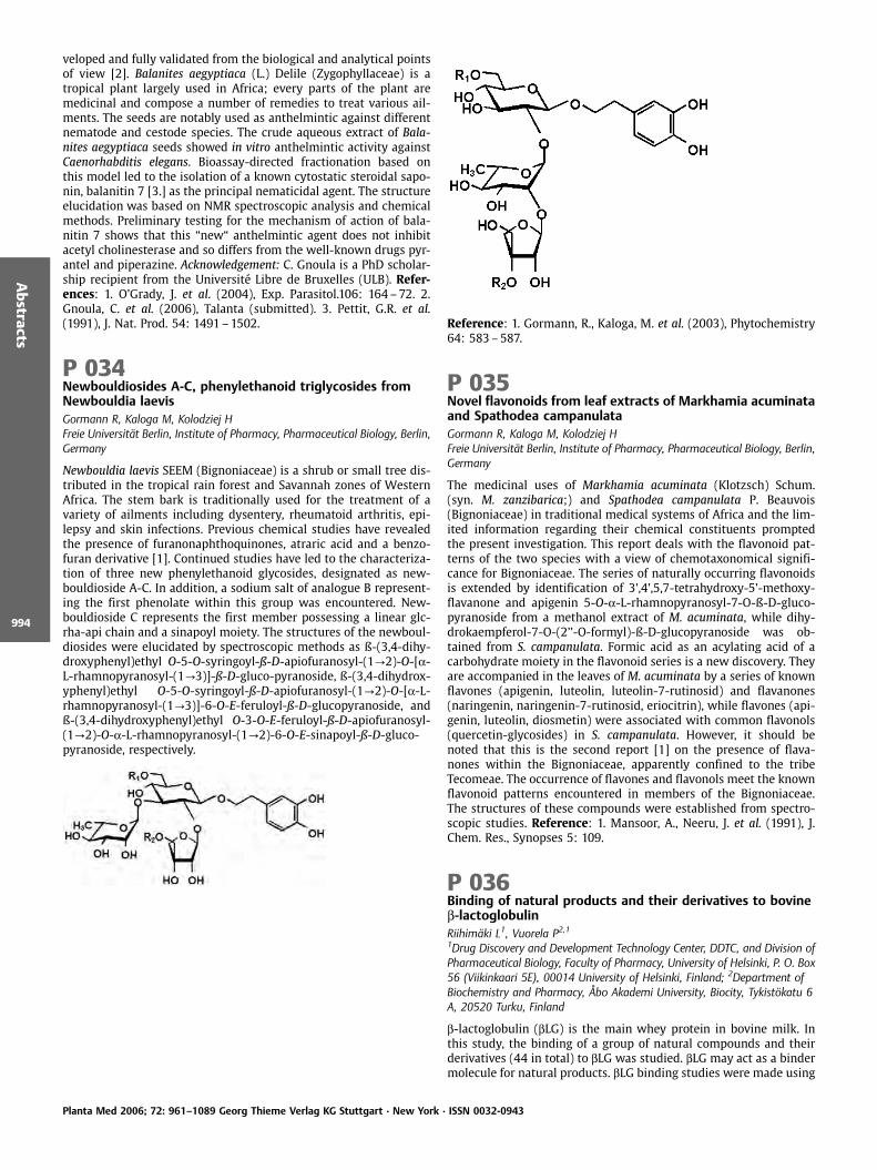
veloped and fully validated from the biological and analytical pointsof view [2]. Balanites aegyptiaca (L.) Delile (Zygophyllaceae) is atropical plant largely used in Africa; every parts of the plant aremedicinal and compose a number of remedies to treat various ail-ments. The seeds are notably used as anthelmintic against differentnematode and cestode species. The crude aqueous extract of Bala-nites aegyptiaca seeds showed in vitro anthelmintic activity againstCaenorhabditis elegans. Bioassay-directed fractionation based onthis model led to the isolation of a known cytostatic steroidal sapo-nin, balanitin 7 [3.] as the principal nematicidal agent. The structureelucidation was based on NMR spectroscopic analysis and chemicalmethods. Preliminary testing for the mechanism of action of bala-nitin 7 shows that this “new“ anthelmintic agent does not inhibitacetyl cholinesterase and so differs from the well-known drugs pyr-antel and piperazine. Acknowledgement: C. Gnoula is a PhD scholar-ship recipient from the Universit Libre de Bruxelles (ULB). Refer-ences: 1. O’Grady, J. et al. (2004), Exp. Parasitol.106: 164–72. 2.Gnoula, C. et al. (2006), Talanta (submitted). 3. Pettit, G.R. et al.(1991), J. Nat. Prod. 54: 1491–1502.
P 034Newbouldiosides A-C, phenylethanoid triglycosides fromNewbouldia laevisGormann R, Kaloga M, Kolodziej HFreie Universit t Berlin, Institute of Pharmacy, Pharmaceutical Biology, Berlin,Germany
Newbouldia laevis SEEM (Bignoniaceae) is a shrub or small tree dis-tributed in the tropical rain forest and Savannah zones of WesternAfrica. The stem bark is traditionally used for the treatment of avariety of ailments including dysentery, rheumatoid arthritis, epi-lepsy and skin infections. Previous chemical studies have revealedthe presence of furanonaphthoquinones, atraric acid and a benzo-furan derivative [1]. Continued studies have led to the characteriza-tion of three new phenylethanoid glycosides, designated as new-bouldioside A-C. In addition, a sodium salt of analogue B represent-ing the first phenolate within this group was encountered. New-bouldioside C represents the first member possessing a linear glc-rha-api chain and a sinapoyl moiety. The structures of the newboul-diosides were elucidated by spectroscopic methods as ß-(3,4-dihy-droxyphenyl)ethyl O-5-O-syringoyl-ß-D-apiofuranosyl-(1?2)-O-[a-L-rhamnopyranosyl-(1?3)]-ß-D-gluco-pyranoside, ß-(3,4-dihydrox-yphenyl)ethyl O-5-O-syringoyl-ß-D-apiofuranosyl-(1?2)-O-[a-L-rhamnopyranosyl-(1?3)]-6-O-E-feruloyl-ß-D-glucopyranoside, andß-(3,4-dihydroxyphenyl)ethyl O-3-O-E-feruloyl-ß-D-apiofuranosyl-(1?2)-O-a-L-rhamnopyranosyl-(1?2)-6-O-E-sinapoyl-ß-D-gluco-pyranoside, respectively.
Reference: 1. Gormann, R., Kaloga, M. et al. (2003), Phytochemistry64: 583–587.
P 035Novel flavonoids from leaf extracts of Markhamia acuminataand Spathodea campanulataGormann R, Kaloga M, Kolodziej HFreie Universit t Berlin, Institute of Pharmacy, Pharmaceutical Biology, Berlin,Germany
The medicinal uses of Markhamia acuminata (Klotzsch) Schum.(syn. M. zanzibarica;) and Spathodea campanulata P. Beauvois(Bignoniaceae) in traditional medical systems of Africa and the lim-ited information regarding their chemical constituents promptedthe present investigation. This report deals with the flavonoid pat-terns of the two species with a view of chemotaxonomical signifi-cance for Bignoniaceae. The series of naturally occurring flavonoidsis extended by identification of 3’,4’,5,7-tetrahydroxy-5’-methoxy-flavanone and apigenin 5-O-a-L-rhamnopyranosyl-7-O-ß-D-gluco-pyranoside from a methanol extract of M. acuminata, while dihy-drokaempferol-7-O-(2’’-O-formyl)-ß-D-glucopyranoside was ob-tained from S. campanulata. Formic acid as an acylating acid of acarbohydrate moiety in the flavonoid series is a new discovery. Theyare accompanied in the leaves of M. acuminata by a series of knownflavones (apigenin, luteolin, luteolin-7-rutinosid) and flavanones(naringenin, naringenin-7-rutinosid, eriocitrin), while flavones (api-genin, luteolin, diosmetin) were associated with common flavonols(quercetin-glycosides) in S. campanulata. However, it should benoted that this is the second report [1] on the presence of flava-nones within the Bignoniaceae, apparently confined to the tribeTecomeae. The occurrence of flavones and flavonols meet the knownflavonoid patterns encountered in members of the Bignoniaceae.The structures of these compounds were established from spectro-scopic studies. Reference: 1. Mansoor, A., Neeru, J. et al. (1991), J.Chem. Res., Synopses 5: 109.
P 036Binding of natural products and their derivatives to bovineb-lactoglobulinRiihim ki L1, Vuorela P2,11Drug Discovery and Development Technology Center, DDTC, and Division ofPharmaceutical Biology, Faculty of Pharmacy, University of Helsinki, P. O. Box56 (Viikinkaari 5E), 00014 University of Helsinki, Finland; 2Department ofBiochemistry and Pharmacy, bo Akademi University, Biocity, Tykist katu 6A, 20520 Turku, Finland
b-lactoglobulin (bLG) is the main whey protein in bovine milk. Inthis study, the binding of a group of natural compounds and theirderivatives (44 in total) to bLG was studied. bLG may act as a bindermolecule for natural products. bLG binding studies were made using
Planta Med 2006; 72: 961–1089 Georg Thieme Verlag KG Stuttgart · New York · ISSN 0032-0943
Abstracts
994

our earlier miniaturised microplate screening assay based on fluor-escence quenching [1], where the apparent dissociation constant(Kd) and the number of independent ligand binding sites (n) weredetermined. Of the compounds investigated the major of flavones,flavonols, flavanones and isoflavones were bound to bLG with highaffinity, Kd between 0.203–0.633mM. In addition some compoundfrom the group of catechins and derivatives, coumarins and pheno-lic acids and derivatives were slightly bound to bLG, Kd >0.7mM.These studies showed that bLG could act as a binder for phenolicnatural products and in this way enhance their health benefit char-acters when fortified in health promoting products. Reference: 1.Riihim ki, L. et al. (2006), J. Biochem. Biophys. Methods. Accepted.
P 037Miniaturisation and automatisation of Caco-2 permeabilitystudies for screening of natural and synthetic ligandsGalkin A1, Pakkanen J2, Vuorela P31Drug Discovery and Development Technology Center (DDTC) and Division ofPharmaceutical biology, Faculty of Pharmacy, P.O. Box 56, 00014, Universityof Helsinki, Finland; 2Division of Pharmacology, Faculty of Pharmacy, P.O. Box56, 00014, University of Helsinki, Finland; 3Department of Biochemistry andPharmacy, bo Akademi University, BioCity, Tykist katu 6 A, 20520, Turku,Finland
Caco-2 cell monolayers have been widely accepted by pharmaceu-tical companies and by regulatory authorities as a standard in vitro–model system to predict permeability of compounds in human. Toobtain these monolayers Caco-2 cells are traditionally grown on 12and 24 wells for 21 to 28 days which is time consuming, laboriousand expensive. To adapt the model to the needs of modern high-throughput screening, we replaced the 12- and 24-well plates with96-well plates and reduced the growing time to 7 days. A set ofstandard compounds with different permeabilities, various perme-ability markers and confocal microscopy were used to assess theutility of our new method on Biomek FX automation. The perme-ability results obtained from standard compounds used were com-parable to those obtained from traditionally performed experi-ments. These results indicate that we managed to build up a fastminiaturized and automated protocol to make a first evaluation ofpermeation of new compounds in the drug discovery process. Ac-knowledgements: The European Commission 6th framework pro-gram Pro-Kinase Research project no. 503467
P 038Biological activity of Gunnera tinctoria, an invasive plant inthe island of S. Miguel (Azores)Barreto MC1,2, Baptista J1,2, Teixeira DM2, Monteiro M1,2
1CIRN, Azores University, 9500 Ponta Delgada, Portugal2DCTD, University of Azores, 9501 – 801 Ponta Delgada, Portugal
Gunnera tinctoria (Mol.) Mirb. is a Halogaracea originary from SouthAmerica, which was introduced in the island of S. Miguel (Azores).An ornamental plant, it escaped from the Furnas botanical garden inthe 1960 s and has been slowly invading the central area of theisland [1]. This plant has been the object of preliminary work inour laboratory [2], and there are reports about biological activitiesdetected in another species belonging to the same genus [3]. Di-chloromethane and methanol leaf extracts of G. tinctoria were pre-pared either by soxhlet extraction (hot – CHCl2 and hot – MeOH,respectively), or at room temperature (cold – CHCl2 and cold –MeOH). The extracts were screened both for antibacterial activityagainst Pseudomonas aeruginosa, Bacillus subtilis, Escherichia coliandMicrococcus luteus, and for antitumour activity against HeLa cellline. The only antibacterial activity detected was from the hot –CHCl2 extract against Micrococcus luteus (EC50 =169.15mg/mL). Allextracts were active against HeLa tumour cell line, both in antipro-liferative and in cytotoxycity assays. The extract which exhibitedhigher activity was hot – MeOH, with EC50 values of 25.3 and
49.5mg/mL for antiproliferative and cytotoxicity assays, respectively.Soxhlet extraction yielded better results than cold extraction, sug-gesting that the active compound(s) involved are not particularlyheat labile. The results obtained are promising and further workwill be carried out to identify the molecules responsible for theeffects detected. References: 1. Sj gren, E. (1984), Azores Flowers.Direc ¼o Regional do Turismo, Horta. 2. Medeiros, J., Macedo, F.W. etal. (1999), A oreana 9: 55–61. 3. Drewes, S.E., Khan, F., et al. (2005),Phytochemistry 66: 1812–1816.
P 039Proanthocyanidins and phenolglycosides from Rumexacetosa LAnke J1, Petereit F1, Hensel A1
1University of M nster, Institute of Pharmaceutical Biology andPhytochemistry, Hittorfstr. 56, D-48149 M nster, Germany
Rumex acetosa L. (Polygonaceae), also known as “sauerampfer“ or“sorrel“, is a perennial herb, which grows abundantly in most partsof Europe and North America. It is a traditional ingredient in salador soup due to its vitamin C content. Additionally, preparations ofRumex species were used for constipation or for treatment ofchronic skin diseases [1]. This claimed therapeutic use and the closerelationship to rhubarb (Rheum officinale) indicate a high content oftannins, especially proanthocyanidins. In the literature, the amountof tannins found in different related Rumex species varies fromabout 2 to 15% [2, 3]. However, only few investigations concerningtannin composition in Rumex have been carried out so far. In orderto isolate and elucidate new proanthocyanidins from Rumex acetosawe purified an acetone/water extract via elution on Sephadex LH20,MLCCC, MCI-Gel and RP-18 material. Different proanthocyanidins, apolymer fraction and the new phenolglycoside 1-O-b-D – (2,4-Dihy-droxy-6-methoxyphenyl)-6-O-(4-hydroxy-3,5-dimethoxybenzoyl)-glucopyranoside were obtained. The isolated proanthocyanidin frac-tions contain mono-, di-, tri-, and tetramers consisting of catechin,epicatechin and epiafzelechin as flavan-3-ol components. A- and B-type interflavan-linkage was found as well as substitution with gal-lic acid. The polymer fraction was characterized by NMR and MStechniques.
References: 1. Williamson, E.M. (2003), Potter’s Herbal Cyclopaedia.C.W. Daniel Company Limited. Essex. 2. McGuffin M. (1997) Amer-ican Herbal Products Assosiation’s Botanical Safety Handbook. CRCPress. Boca Raton. 3. Demirezer, L. . et al. (1997), FABAD 22: 153–158.
Planta Med 2006; 72: 961–1089 Georg Thieme Verlag KG Stuttgart · New York · ISSN 0032-0943
Abstracts
995
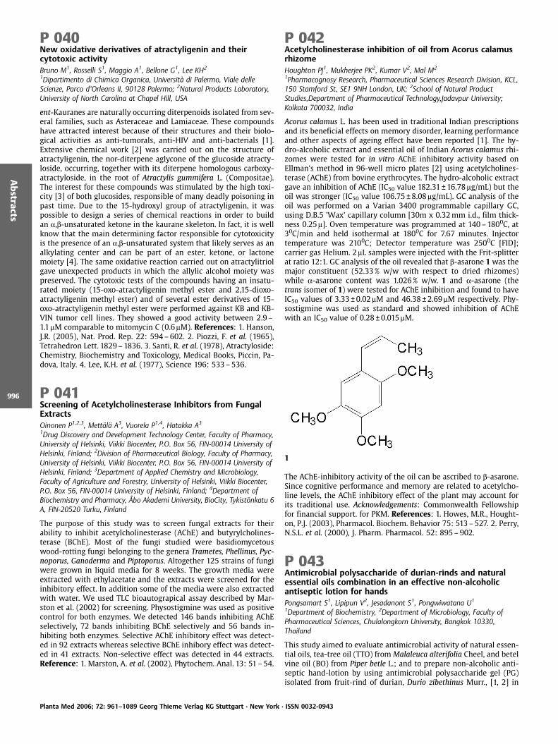
P 040New oxidative derivatives of atractyligenin and theircytotoxic activityBruno M1, Rosselli S1, Maggio A1, Bellone G1, Lee KH2
1Dipartimento di Chimica Organica, Universit di Palermo, Viale delleScienze, Parco d’Orleans II, 90128 Palermo; 2Natural Products Laboratory,University of North Carolina at Chapel Hill, USA
ent-Kauranes are naturally occurring diterpenoids isolated from sev-eral families, such as Asteraceae and Lamiaceae. These compoundshave attracted interest because of their structures and their biolo-gical activities as anti-tumorals, anti-HIV and anti-bacterials [1].Extensive chemical work [2] was carried out on the structure ofatractyligenin, the nor-diterpene aglycone of the glucoside atracty-loside, occurring, together with its diterpene homologous carboxy-atractyloside, in the root of Atractylis gummifera L. (Compositae).The interest for these compounds was stimulated by the high toxi-city [3] of both glucosides, responsible of many deadly poisoning inpast time. Due to the 15-hydroxyl group of atractyligenin, it waspossible to design a series of chemical reactions in order to buildan a,b-unsaturated ketone in the kaurane skeleton. In fact, it is wellknow that the main determining factor responsible for cytotoxicityis the presence of an a,b-unsaturated system that likely serves as analkylating center and can be part of an ester, ketone, or lactonemoiety [4]. The same oxidative reaction carried out on atractylitriolgave unexpected products in which the allylic alcohol moiety waspreserved. The cytotoxic tests of the compounds having an insatu-rated moiety (15-oxo-atractyligenin methyl ester and 2,15-dioxo-atractyligenin methyl ester) and of several ester derivatives of 15-oxo-atractyligenin methyl ester were performed against KB and KB-VIN tumor cell lines. They showed a good activity between 2.9 –1.1mM comparable to mitomycin C (0.6mM). References: 1. Hanson,J.R. (2005), Nat. Prod. Rep. 22: 594–602. 2. Piozzi, F. et al. (1965),Tetrahedron Lett. 1829–1836. 3. Santi, R. et al. (1978), Atractyloside:Chemistry, Biochemistry and Toxicology, Medical Books, Piccin, Pa-dova, Italy. 4. Lee, K.H. et al. (1977), Science 196: 533–536.
P 041Screening of Acetylcholinesterase Inhibitors from FungalExtractsOinonen P1,2,3, Mett l A3, Vuorela P1,4, Hatakka A3
1Drug Discovery and Development Technology Center, Faculty of Pharmacy,University of Helsinki, Viikki Biocenter, P.O. Box 56, FIN-00014 University ofHelsinki, Finland; 2Division of Pharmaceutical Biology, Faculty of Pharmacy,University of Helsinki, Viikki Biocenter, P.O. Box 56, FIN-00014 University ofHelsinki, Finland; 3Department of Applied Chemistry and Microbiology,Faculty of Agriculture and Forestry, University of Helsinki, Viikki Biocenter,P.O. Box 56, FIN-00014 University of Helsinki, Finland; 4Department ofBiochemistry and Pharmacy, bo Akademi University, BioCity, Tykist nkatu 6A, FIN-20520 Turku, Finland
The purpose of this study was to screen fungal extracts for theirability to inhibit acetylcholinesterase (AChE) and butyrylcholines-terase (BChE). Most of the fungi studied were basidiomycetouswood-rotting fungi belonging to the genera Trametes, Phellinus, Pyc-noporus, Ganoderma and Piptoporus. Altogether 125 strains of fungiwere grown in liquid media for 8 weeks. The growth media wereextracted with ethylacetate and the extracts were screened for theinhibitory effect. In addition some of the media were also extractedwith water. We used TLC bioautograpical assay described by Mar-ston et al. (2002) for screening. Physostigmine was used as positivecontrol for both enzymes. We detected 146 bands inhibiting AChEselectively, 72 bands inhibiting BChE selectively and 56 bands in-hibiting both enzymes. Selective AChE inhibitory effect was detect-ed in 92 extracts whereas selective BChE inhibory effect was detect-ed in 41 extracts. Non-selective effect was detected in 44 extracts.Reference: 1. Marston, A. et al. (2002), Phytochem. Anal. 13: 51–54.
P 042Acetylcholinesterase inhibition of oil from Acorus calamusrhizomeHoughton PJ1, Mukherjee PK2, Kumar V2, Mal M2
1Pharmacognosy Research, Pharmaceutical Sciences Research Division, KCL,150 Stamford St, SE1 9NH London, UK; 2School of Natural ProductStudies,Department of Pharmaceutical Technology,Jadavpur University;Kolkata 700032, India
Acorus calamus L. has been used in traditional Indian prescriptionsand its beneficial effects on memory disorder, learning performanceand other aspects of ageing effect have been reported [1]. The hy-dro-alcoholic extract and essential oil of Indian Acorus calamus rhi-zomes were tested for in vitro AChE inhibitory activity based onEllman’s method in 96-well micro plates [2] using acetylcholines-terase (AChE) from bovine erythrocytes. The hydro-alcoholic extractgave an inhibition of AChE (IC50 value 182.31 €16.78mg/mL) but theoil was stronger (IC50 value 106.75 €8.08mg/mL). GC analysis of theoil was performed on a Varian 3400 programmable capillary GC,using D.B.5 ’Wax’ capillary column [30m x 0.32mm i.d., film thick-ness 0.25m]. Oven temperature was programmed at 140–1800C, at30C/min and held isothermal at 1800C for 7.67 minutes. Injectortemperature was 2100C; Detector temperature was 2500C [FID];carrier gas Helium. 2mL samples were injected with the Frit-splitterat ratio 12:1. GC analysis of the oil revealed that b-asarone 1 was themajor constituent (52.33% w/w with respect to dried rhizomes)while a-asarone content was 1.026% w/w. 1 and a-asarone (thetrans isomer of 1) were tested for AChE inhibition and found to haveIC50 values of 3.33€0.02mM and 46.38€2.69mM respectively. Phy-sostigmine was used as standard and showed inhibition of AChEwith an IC50 value of 0.28 €0.015mM.
1
The AChE-inhibitory activity of the oil can be ascribed to b-asarone.Since cognitive performance and memory are related to acetylcho-line levels, the AChE inhibitory effect of the plant may account forits traditional use. Acknowledgements: Commonwealth Fellowshipfor financial support. for PKM. References: 1. Howes, M.R., Hought-on, P.J. (2003), Pharmacol. Biochem. Behavior 75: 513–527. 2. Perry,N.S.L. et al. (2000), J. Pharm. Pharmacol. 52: 895–902.
P 043Antimicrobial polysaccharide of durian-rinds and naturalessential oils combination in an effective non-alcoholicantiseptic lotion for handsPongsamart S1, Lipipun V2, Jesadanont S1, Pongwiwatana U1
1Department of Biochemistry, 2Department of Microbiology, Faculty ofPharmaceutical Sciences, Chulalongkorn University, Bangkok 10330,Thailand
This study aimed to evaluate antimicrobial activity of natural essen-tial oils, tea-tree oil (TTO) fromMalaleuca alterifolia Cheel, and betelvine oil (BO) from Piper betle L.; and to prepare non-alcoholic anti-septic hand-lotion by using antimicrobial polysaccharide gel (PG)isolated from fruit-rind of durian, Durio zibethinus Murr., [1, 2] in
Planta Med 2006; 72: 961–1089 Georg Thieme Verlag KG Stuttgart · New York · ISSN 0032-0943
Abstracts
996

combination with essential oils. PG, TTO or BO each showed goodinhibitory effect against Staphylococcus aureus, S. epidermidis, icro-coccus luteus, Bacillus subtilis, Escherichia coli, Proteus vulgaris andPropionibacterium acnes by agar diffusion and broth macrodilutionmethods [3]. Minimum bactericidal concentrations (MBCs) of eachsingle component, i.e. PG, TTO and BO against the tested bacteriawere 2.5%, 0.156–0.625% and 0.020–0.156% w/v, respectively. TTOor BO each by itself also killed other bacteria and fungi includingStreptococcus typhimurium, Klebsiella pneumoniae, Pseudomonasaeruginosa and Candida albicans. PG at 2.5% w/w was used in com-bination with 1% w/w TTO and 0.5% w/w BO to prepare hand-lo-tion. Antimicrobial activity of the hand-lotion prepared was eval-uated where clear inhibition zone against all tested microorganismsby using agar diffusion test was observed. For in vitro Time-killanalysis, the hand-lotion prepared killed all the tested bacteria, S.aureus and E. coli, within 15min. Upon hand-washing test [4], thetreated hands exposed to the antiseptic hand-lotion preparedshowed significant reduction of hand normal flora compared to apositive control exposed to a commercially available alcoholic anti-septic hand gel. Thus, PG in combination with TTO together with BOis of good potential to be used for preparing a non-alcoholic anti-septic lotion used topically on hands or skin. Acknowledgements:This work was supported by Annual Research Budget of Faculty ofPharmaceutical Sci., Chulalongkorn University. References: 1. Pong-samart, S. et al. (2005), Acta Hortic. 678: 65–73. 2. Hokputsa, S. etal. (2004), Carbohydrate Polymers 56: 471–481. 3. Lorian, V. (1996),Antibiotics in laboratory medicine. 4th ed. Williams&Wilkins. Balti-more. 4. Messager, S. et al. (2004), J. Hosp. Infect. 59: 220–228.
P 044Oral hypoglycemic activity of water extract from Ya-Tevada,Malvastrum Coromandelianum Garcke, equivalent to insulininjectionJesadanont S, Sitthiwej C, Poungshompoo S, Pongsamart SChulalongkorn University, Bangkok 10330, THAILAND
Water extract from whole plant of Ya-Tevada, Malvastrum coroman-delianum Garcke; Malvaceae, showed strong hypoglycemic activitywhen given orally. Feeding the spray-dried crude water extract aslow as 50 and 100mg/kg body weight (bw) to streptozotocin-in-duced male Wistar Rats, reduced significantly non-fasting bloodglucose from approx. 350–450mg/dL within one hour after admin-istration to a level of blood glucose in normal non-diabetic ratswhich is about 150mg/dL, equivalent to i.p. injection of Insulin (Hu-mulin R ) 5U/kg bw. The hypoglycemic effect lasted for at least 5hours. This strong hypoglycemic activity of this plant extract wouldfulfill the search for an oral hypoglycemic agent equivalent to insu-lin injection. Feeding the extract at repeated doses of 50, 100 and500mg/kg bw/day for 30 days suppressed only fasting blood glu-cose to a level equivalent to a level shown in the group of diabeticrats injected with insulin 5 U/kg bw/day and the group of normalrats fed water. Previous report showed no chronic toxic effect uponfeeding this water extract at daily doses of 0.2, 2.0 and 20g/kg bwfor 6 months in male and female Wistar rats [1]. This largest dose is400 times the effective hypoglycemic dose of 50mg. This agent alsoshowed antibacterial activity against Staphylococcus aureus bothmethicillin-sensitive and methicillin-resistant and good woundhealing effect. Thus, water extract of this plant showed great poten-tial as oral hypoglycemic agent to be used in diabetic condition.Acknowledgements: Fac Pharm Sci, and Fac Vet Sci, ChulalongkornU., Bangkok 10900, Thailand. Reference: 1. Attawish, A, et al. (1998),Bull. Dept. Med. Sci. 40: 261–271.
P 045Screening of Antibacterial, Antifungal and AntiviralProperties of the Selected Turkish Helichrysum SpeciesAslana M, z elik B, Orhana I, Karaoglu T, Sezika E1Department of Pharmacognosy, Faculty of Pharmacy, Gazi University,06330 Ankara, Turkey; 2Department of Pharmaceutical Microbiology, Facultyof Pharmacy, Gazi University, 06330 Ankara, Turkey; 3Department ofVirology, Faculty of Veterinary, University of Ankara, 06100 Ankara, Turkey
Various Helichrysum species have been widely used as folk remedyin Turkish folk medicine for diuretic, and anti-asthmatic propertiesas well as against kidney stones, stomache as decoction. Besides, thepowder of capitulums have been also used like pomade, prepared bymixing with barley flour, for wound healing (1 –3). In the presentstudy, Fourteen extracts prepared with hot water and ethanol (80%)from the capitulums obtained from seven Helichrysum species in-cluding H. armenium ssp. araxinum (Takht. ex Kirp.) Tahkt, H. arme-nium ssp. armenium DC, H. arenarium (L.) DC, H. pallasii (Spreng.)Ledeb., H. stoechas (L.) Moench, H. sanguineum, and H. graveolens(Bieb.) Sweet (Compositae) growing in Turkey were screened fortheir antibacterial, antifungal and antiviral activity against bothstandard and the isolated strains of Escherichia coli, Pseudomonasaeruginosa, Proteus mirabilis, Klebsiella pneumoniae, Acinetobacerbaumannii, Staphylococcus aureus, Enterococcus faecalis, Candida al-bicans and C. parapsilosis by microdilution method. Both Herpessimplex (HSV) (DNA) and Para-influenza-3 (PI-3) viruses (RNA) wereused for the determination of antiviral activity of the water andethanol extracts of Helichrysum species by using MDBK and Verocell line. Ampicilline, levofloxacin, ofloxocin, ketoconazole, flucona-zole, acyclovir and oseltamivir were used as the control agents. Thesame degree of inhibitory effect has been observed on Gram (-)bacteria. All the extracts have shown more potent effect againstGram (+) bacteria than Gram (-) ones. Moreover, all the extractsscreened have exerted a better inhibitory effect towards ATCCstrains than the isolates. In particular, all of the ethanolic extractswere found to have a very significant inhibition against ATCC strainof S.aureus with MIC value of 8mg/mL. Ethanolic extracts of H. are-narium and H. armenium ssp. armenium showed antiviral actionagainst both HSV and PI-3, whereas the rest was completely inactivewith the related activity. References: 1.Sezik, E., Tabata, M. et al.(1991), J. Ethnopharmacol. 35: 191–196. 2. Fujita, T., Sezik, E., etal. (1995), Econ. Bot. 49: 406–422. 3. Sezik, E., Yesilada, E. et al.(2001), J. Ethnopharmacol. 75: 95–115.
P 046Antimutagenic effects of ethanolic extracts from threePalestinian medicinal plantsKhader M, Eckl PM, Bresgen NDepartment of Cell Biology, University of Salzburg, Hellbrunnerstrasse 34,Salzburg, A-5020, Austria
Eryngium creticum L., Nigella sativa L., and Teucrium polium L. havebeen traditionally used for the treatment of inflammations, liverdisorders, and arthritis. Several studies on these plants revealedantioxidant, anti-inflammatory, hepatoprotective, antimutagenicand antiulcerogenic activities. In this study the antimutagenic activ-ity of these plant species was tested in rat hepatocyte primary cul-tures by treatment with N-methyl-N’-nitro-N-nitrosoguanidine(MNNG), a directly acting mutagen, which was shown to inducemassive chromosomal damage in hepatocytes [1]. Since it cannotbe excluded that the active constituents of the plant extracts requirebiotransformation or induce metabolic enzymes, causing antimuta-genic or detoxifying effects, the present investigation was carriedout with metabolically competent primary cultures of rat hepato-cytes. Rat hepatocytes were isolated as described by Michalopouloset al. [2]. Establishment of primary cultures and cytogenetic studieswere performed according to Eckl et al. [1]. Plant extracts wereprepared by Soxhlet continuous extraction method, 6 gm of groundplant materials were extracted in 50 mL of absolute ethanol at 85 C
Planta Med 2006; 72: 961–1089 Georg Thieme Verlag KG Stuttgart · New York · ISSN 0032-0943
Abstracts
997
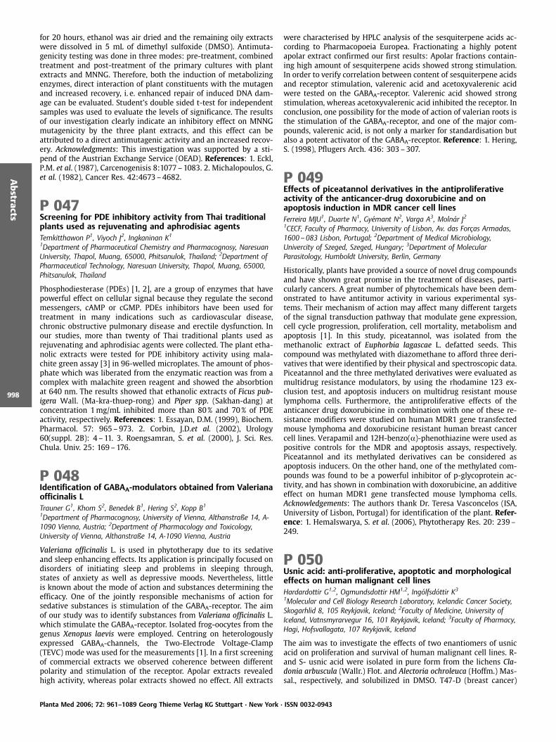
for 20 hours, ethanol was air dried and the remaining oily extractswere dissolved in 5 mL of dimethyl sulfoxide (DMSO). Antimuta-genicity testing was done in three modes: pre-treatment, combinedtreatment and post-treatment of the primary cultures with plantextracts and MNNG. Therefore, both the induction of metabolizingenzymes, direct interaction of plant constituents with the mutagenand increased recovery, i. e. enhanced repair of induced DNA dam-age can be evaluated. Student’s double sided t-test for independentsamples was used to evaluate the levels of significance. The resultsof our investigation clearly indicate an inhibitory effect on MNNGmutagenicity by the three plant extracts, and this effect can beattributed to a direct antimutagenic activity and an increased recov-ery. Acknowledgments: This investigation was supported by a sti-pend of the Austrian Exchange Service (OEAD). References: 1. Eckl,P.M. et al. (1987), Carcenogenisis 8:1077–1083. 2. Michalopoulos, G.et al. (1982), Cancer Res. 42:4673–4682.
P 047Screening for PDE inhibitory activity from Thai traditionalplants used as rejuvenating and aphrodisiac agentsTemkitthawon P1, Viyoch J2, Ingkaninan K1
1Department of Pharmaceutical Chemistry and Pharmacognosy, NaresuanUniversity, Thapol, Muang, 65000, Phitsanulok, Thailand; 2Department ofPharmaceutical Technology, Naresuan University, Thapol, Muang, 65000,Phitsanulok, Thailand
Phosphodiesterase (PDEs) [1, 2], are a group of enzymes that havepowerful effect on cellular signal because they regulate the secondmessengers, cAMP or cGMP. PDEs inhibitors have been used fortreatment in many indications such as cardiovascular disease,chronic obstructive pulmonary disease and erectile dysfunction. Inour studies, more than twenty of Thai traditional plants used asrejuvenating and aphrodisiac agents were collected. The plant etha-nolic extracts were tested for PDE inhibitory activity using mala-chite green assay [3] in 96-welled microplates. The amount of phos-phate which was liberated from the enzymatic reaction was from acomplex with malachite green reagent and showed the absorbtionat 640 nm. The results showed that ethanolic extracts of Ficus pub-igera Wall. (Ma-kra-thuep-rong) and Piper spp. (Sakhan-dang) atconcentration 1mg/mL inhibited more than 80% and 70% of PDEactivity, respectively. References: 1. Essayan, D.M. (1999), Biochem.Pharmacol. 57: 965–973. 2. Corbin, J.D.et al. (2002), Urology60(suppl. 2B): 4 –11. 3. Roengsamran, S. et al. (2000), J. Sci. Res.Chula. Univ. 25: 169–176.
P 048Identification of GABAA-modulators obtained from Valerianaofficinalis LTrauner G1, Khom S2, Benedek B1, Hering S2, Kopp B11Department of Pharmacognosy, University of Vienna, Althanstraße 14, A-1090 Vienna, Austria; 2Department of Pharmacology and Toxicology,University of Vienna, Althanstraße 14, A-1090 Vienna, Austria
Valeriana officinalis L. is used in phytotherapy due to its sedativeand sleep enhancing effects. Its application is principally focused ondisorders of initiating sleep and problems in sleeping through,states of anxiety as well as depressive moods. Nevertheless, littleis known about the mode of action and substances determining theefficacy. One of the jointly responsible mechanisms of action forsedative substances is stimulation of the GABAA-receptor. The aimof our study was to identify substances from Valeriana officinalis L.which stimulate the GABAA-receptor. Isolated frog-oocytes from thegenus Xenopus laevis were employed. Centring on heterologouslyexpressed GABAA-channels, the Two-Electrode Voltage-Clamp(TEVC) mode was used for the measurements [1]. In a first screeningof commercial extracts we observed coherence between differentpolarity and stimulation of the receptor. Apolar extracts revealedhigh activity, whereas polar extracts showed no effect. All extracts
were characterised by HPLC analysis of the sesquiterpene acids ac-cording to Pharmacopoeia Europea. Fractionating a highly potentapolar extract confirmed our first results: Apolar fractions contain-ing high amount of sesquiterpene acids showed strong stimulation.In order to verify correlation between content of sesquiterpene acidsand receptor stimulation, valerenic acid and acetoxyvalerenic acidwere tested on the GABAA-receptor. Valerenic acid showed strongstimulation, whereas acetoxyvalerenic acid inhibited the receptor. Inconclusion, one possibility for the mode of action of valerian roots isthe stimulation of the GABAA-receptor, and one of the major com-pounds, valerenic acid, is not only a marker for standardisation butalso a potent activator of the GABAA-receptor. Reference: 1. Hering,S. (1998), Pflugers Arch. 436: 303–307.
P 049Effects of piceatannol derivatives in the antiproliferativeactivity of the anticancer-drug doxorubicine and onapoptosis induction in MDR cancer cell linesFerreira MJU1, Duarte N1, Gy mant N2, Varga A3, Moln r J21CECF, Faculty of Pharmacy, University of Lisbon, Av. das For as Armadas,1600 – 083 Lisbon, Portugal; 2Department of Medical Microbiology,Univercity of Szeged, Szeged, Hungary; 3Department of MolecularParasitology, Humboldt University, Berlin, Germany
Historically, plants have provided a source of novel drug compoundsand have shown great promise in the treatment of diseases, parti-cularly cancers. A great number of phytochemicals have been dem-onstrated to have antitumor activity in various experimental sys-tems. Their mechanism of action may affect many different targetsof the signal transduction pathway that modulate gene expression,cell cycle progression, proliferation, cell mortality, metabolism andapoptosis [1]. In this study, piceatannol, was isolated from themethanolic extract of Euphorbia lagascae L. defatted seeds. Thiscompound was methylated with diazomethane to afford three deri-vatives that were identified by their physical and spectroscopic data.Piceatannol and the three methylated derivatives were evaluated asmultidrug resistance modulators, by using the rhodamine 123 ex-clusion test, and apoptosis inducers on multidrug resistant mouselymphoma cells. Furthermore, the antiproliferative effects of theanticancer drug doxorubicine in combination with one of these re-sistance modifiers were studied on human MDR1 gene transfectedmouse lymphoma and doxorubicine resistant human breast cancercell lines. Verapamil and 12H-benzo(a)-phenothiazine were used aspositive controls for the MDR and apoptosis assays, respectively.Piceatannol and its methylated derivatives can be considered asapoptosis inducers. On the other hand, one of the methylated com-pounds was found to be a powerful inhibitor of p-glycoprotein ac-tivity, and has shown in combination with doxorubicine, an additiveeffect on human MDR1 gene transfected mouse lymphoma cells.Acknowledgements: The authors thank Dr. Teresa Vasconcelos (ISA,University of Lisbon, Portugal) for identification of the plant. Refer-ence: 1. HemaIswarya, S. et al. (2006), Phytotherapy Res. 20: 239–249.
P 050Usnic acid: anti-proliferative, apoptotic and morphologicaleffects on human malignant cell linesHardardottir G1,2, Ogmundsdottir HM1,2, Ing lfsd ttir K3
1Molecular and Cell Biology Research Laboratory, Icelandic Cancer Society,Skogarhlid 8, 105 Reykjavik, Iceland; 2Faculty of Medicine, University ofIceland, Vatnsmyrarvegur 16, 101 Reykjavik, Iceland; 3Faculty of Pharmacy,Hagi, Hofsvallagata, 107 Reykjavik, Iceland
The aim was to investigate the effects of two enantiomers of usnicacid on proliferation and survival of human malignant cell lines. R-and S- usnic acid were isolated in pure form from the lichens Cla-donia arbuscula (Wallr.) Flot. and Alectoria ochroleuca (Hoffm.) Mas-sal., respectively, and solubilized in DMSO. T47-D (breast cancer)
Planta Med 2006; 72: 961–1089 Georg Thieme Verlag KG Stuttgart · New York · ISSN 0032-0943
Abstracts
998
and Capan-2 (pancreatic cancer) were from ATCC and the myelomacell lines RPMI-8226*, U266–84* and LP-1*. Anti-proliferative ef-fects were tested by thymidine-uptake, results expressed as IC50. Totest for apoptosis cells were exposed to four times this concentra-tion for 24 hours; a commercial TUNEL assay was used. The mor-phology of MG-G-stained cells was investigated after 2, 6 and 24hour-exposure to usnic acid at the four times IC50 concentration.Usnic acid had anti-proliferative effects against T47-D(IC50=4.2mg/mL) and Capan-2 (IC50=5.3mg/mL). No difference wasfound between the enantiomers and only (+) usnic acid was used forfurther testing. None of the myeloma cell lines was significantlyaffected. Usnic acid did not induce apoptosis in any of the cell lines.T47-D cells, but not Capan-2 cells, showed morphological changesindicative of necrosis in a small proportion of cells after 6 hoursexposure to usnic acid. In conclusion, both enantiomers of usnicacid have significant anti-proliferative effects against two humancarcinoma cell lines but not against myeloma cell lines. Usnic aciddid not induce apoptosis, which is in line with its non-genotoxicmode of action (1), but mild signs of necrosis were seen. *A kindgift from Kenneth Nilsson.
P 051Diterpenic compounds as antineoplasic agents in classicaland atypical multidrug resistant cancer cellsDuarte N1, Lage H2, Ferreira MJU1
1CECF, Faculty of Pharmacy, University of Lisbon, Av. das For as Armadas,1600 – 083 Lisbon, Portugal; 2Charit Campus Mitte, Institute ofPathology,Schumannstr. 20/21, D-10117 Berlin, Germany
The successful chemotherapy of neoplasic diseases requires that theantitumor combination of drugs employed displays a sustained ac-tivity against malignant cells. Simultaneous resistance of cancercells to multiple classes of structurally and mechanistically unre-lated antitumor drugs can be defined as multidrug resistance(MDR), and it is one of the main causes of chemotherapy failure[1]. In addition with the classical multidrug phenotype mediatedby increased activity of the ATP-binding cassette transporters P-gp/ MDR1, that were responsible for the efflux of drugs out of the cells,there are other multidrug resistant tumors with resistance causedby different mechanisms. This is called atypical resistance and couldbe due to the decreased DNA topoisomerase (Topo II)-like activity.DNA topoisomerases are nuclear enzymes that are essential for DNAreplication, transcription, chromosomal aggregation and DNA re-combination. For these reasons, Topo II is one of the possible targetsfor the commonly used anticancer drugs [2]. The aim of this work isto study the antiproliferative effect of diterpenic compounds isolat-ed from Euphorbia species in various human drug sensitive cancercell lines (gastric, pancreatic and colon carcinomas) and in classicaland atypical multidrug resistant sublines of these cell models. Eto-poside was used as positive control. It could be demonstrated, thatmost of the drug resistant cell variants decreased the expressionlevels of both Topo II isoforms on mRNA level as well as on proteinlevel. None of the tested compounds were as effective as etoposide,but some of them are much more effective in drug resistant cellsthat in drug sensitive cell lines. In conclusion, some of these com-pounds may be considered as potential new drugs for the treatmentof drug-resistant human cancer cells. Acknowledgements: Theauthors thank Dr. Teresa Vasconcelos (ISA, University of Lisbon, Por-tugal) for identification of the plant. References: 1. Avenda o, C. etal. (2002), Curr. Med. Chem. 9: 159–193. 2. Lage, H. et al. (2000),Lancet Oncol. 1: 169–175.
P 052Search for P-glycoprotein modulators and apoptosisinducers on cancer cells among ergostane and stigmastanesteroidsDuarte N1, Ramalhete C1, Gy mant N2, Varga A3, Moln r J2, Ferreira MJU1
1CECF, Faculty of Pharmacy, University of Lisbon, Av. das For as Armadas,1600 – 083 Lisbon, Portugal; 2Department of Medical Microbiology,University of Szeged, Szeged, Hungary; 3Department of MolecularParasitology, Humboldt University, Berlin, Germany
One of the most effective strategies in biological systems to demon-strate resistance to cytotoxic drugs is the efflux of these compoundsout of the cell, via membrane transporter proteins. This phenomen-on is called multidrug resistance (MDR) and is a mediator of drugresistance observed in tumor cells and in microorganisms (in vivoand in vitro) [1]. There are numerous resistance mechanisms andthe MDR phenotype alone, can not completely explain this occur-rence. In fact, several other related proteins are also overexpressedin resistant tumors. Furthermore apoptosis also play a vital role inresistance [2]. Because apoptosis is a major modality by which dif-ferent tumor cell types can be eliminated, the identification of newdrugs able to induce the programmed cell death is an importantgoal in cancer therapy, and may provide new useful tools for thetreatment of patients with drug resistance malignancies. In thiscontext, plants could be potential sources for the isolation of newmetabolites that could be used as lead molecules on the treatmentof neoplasic diseases. In our search for biologically active com-pounds from Euphorbia species, several ergostane and stigmastanesteroids were isolated from the methanolic extract of Euphorbialagascae, and its structures were deduced by the combination ofphysical and spectroscopic data (IR, MS, 1D and 2D NMR). The abil-ity of some of the isolated steroids to modulate MDR reversion wasinvestigated using mdr-1 (L5178) mouse lymphoma cell line. In ad-dition, the apoptosis induction on the same cell line was also stud-ied. 12H-benzophenothiazine was used as a positive control forapoptosis induction and verapamil for resistance reversal. The test-ed compounds were weak inducers of apoptosis in the studiedmouse lymphoma cell line, but two of them were found effectiveon MDR reversion in a concentration dependent manner. Acknowl-edgements: The authors thank Dr. Teresa Vasconcelos (ISA, Univer-sity of Lisbon, Portugal) for identification of the plant. References:1. Borges-Walmsley, M. et al. (2003), Biochem. J. 376: 313–338. 2.Volm, M. (1998), Anticancer Res. 18: 2905–2917.
P 053Antibacterial compounds from Vaccinium myrtillus(bilberry)Bessad ttir M1, J nsd ttir 1, Omarsdottir S1, Erlendsd ttir H2,Ing lfsd ttir K1
1Faculty of Pharmacy, University of Iceland, Hagi, Hofsvallagata 53, IS-107Reykjavik, Iceland; 2Department of Bacteriology, Landspitali-UniversityHospital, IS-101 Reykjavik, Iceland
Bilberries have been used in herbal medicine against various dis-eases [1]. The aim of the study was to determine the antibacterialactivity of three different (petroleum ether (PB), acetone (A) andmethanol (M)) extracts of Icelandic bilberries, using broth microdi-lution assay. Bioguided fractionation was used to isolate active com-pounds. The PB extract exhibited potent antibacterial activityagainst antibiotic sensitive and resistant Gram-positive bacteria(Staphylococcus aureus, Enterococcus faecalis and E. faecium) whereminimum inhibitory concentration (MIC) was shown to be from 8to125mg/mL. The extracts showed no significant activity againstGram-negative bacteria and C. albicans. A fraction composed ofthree pentacyclic triterpenoids was isolated from the PB extractusing LC and HPLC. These triterpenoids were tested against sevenresistant Gram-positive bacteria. The MIC value for the triterpenoidswas determined to be: 8mg/mL against coagulase-negative staphy-lococci and ampicillin resistant E. faecium; 16mg/mL against ampi-
Planta Med 2006; 72: 961–1089 Georg Thieme Verlag KG Stuttgart · New York · ISSN 0032-0943
Abstracts
999

cillin-and vancomycin resistant E. faecium and 31mg/mL againstmethicillin-resistant S. aureus, gentamicin resistant E. faecalis andvancomycin resistant E.faecalis, respectively. Aqueous extract of bil-berries has previously been shown to exhibit some antibacterialactivity (2). However, the MIC value was ranging from 15–31mg/mL [2]. Acknowledgements: Icelandic Council of Science, Universityof Iceland Research Fund References: 1. Morazzoni, P., Bombardelli,E. (1996), Fitoterapia 66: 3–29. 2. Brantner, A., Grein, E. (1994), J.Ethnophamacol. 44: 35–40.
P 054Cucurbitacin R reduces delayed-type hypersensitivityreaction induced by dinitrofluorobenzene and sheep redblood cells in miceR os JL, Escandell JM, Cerd -Nicol s M1, Recio MCDepartament de Farmacologia, Facultat de Farmacia, Universitat de Val ncia,Av. Vicent Andr s Estell s s/n, 46100 Burjassot, Spain; Departament dePatologia, Facultat de Medicina, Universitat de Val ncia, Av. Blasco Ib ez 15,46010 Valencia, Spain
The roots of Cayaponia tayuya (Vell.) Cogn. (Cucurbitaceae) are usedin folk medicine as an anti-inflammatory and anti-allergic crudedrug [1]. We previously reported its anti-inflammatory effect [2]and the anti-arthritic properties [3] of two cucurbitacins isolatedfrom the chloroform extract. Now we tested the effects of cucurbi-tacin R (CCR) on different experimental models of systemic delayed-type hypersensitivity (DTH) in mice [3, 4]. CCR showed anti-allergiceffects in two of the three models assayed. In the dinitrofluoroben-zene (DNFB) model inhibited the ear oedema with a DI50 of 0.56mg/ear at 48h. In the sheep red blood cells (SRBC) model, the inhibitionreached 64% (18h), 58% (24h) and 62% (48h). In the oxazolone-induced DTH there was not significant effect. In the histologicalstudies of the DNFB-induced contact dermatitis, the CCR-treatedgroup inhibited the oedema formation and the inflammatory cellinfiltration, along with a reduction of the tissue damage. In theDTH-induced by SRBC, when compared with the non-treated group,the paws of the CCR-treated group (12.5mg/kg) present a mild in-flammatory lesion and scarce mixture of inflammatory cells, and areduction of tissue damage. In addition, CCR abolished the produc-tion of TNF-a and IL-1b in the paws of the CCR-treated group givinga 100% (TNF-a) and 90% (IL-1b) of inhibition. Acknowledgements:J.M.E. is recipient of a grant from the Generalitat Valenciana. Thiswork was supported by the Spanish Government (SAF2002–00723)References: 1. R os, J.L. et al. (1990), Fitoterapia 61: 275–278. 2.Recio, M.C. et al. (2004), Planta Med. 70: 414–420. 3. Escandell, J.et al. (2006), Eur. J. Pharmacol. 532: 145–154. 4. G ngora, L. et al.(2000), Life Sci. 66: PL183.
P 055Evaluating the effects of phenolics from Phagnalon rupestre(L.) DC on cellular nitration and oxidationGiner RM, Olmos A, M ez SDepartament de Farmacologia, Facultat de Farmacia, Universitat de Val ncia,Av. Vicent Andr s Estell s s/n, 46100 Burjassot, Spain
Not only do reactive nitrogen species modify proteins and nucleo-tides by means of oxidative or nitrating reactions, but they can alsocause other structural alterations which have a physiopathologicalsignificance in a number of human diseases. Given that some natu-rally occurring phenolics, such as hydroxycinnamate and hydroxy-benzoate derivatives, are particularly effective at preventing thedegradation of biomolecules, such compounds should actually pro-tect against these unfavourable conditions. The present communi-cation describes our ongoing investigations of three phenolicsisolated from Phagnalon rupestre (Asteraceae): 2-isoprenylhydroqui-none-1-glucoside (IHG), 3,5-dicaffeoylquinic acid (DCA) and itsmethyl ester (DCE) [1], previously described as peroxynitrite-sca-vengers for free tyrosine [2], on nitrating and oxidative reactions
in two different cellular systems. We examined the effect of thesecompounds on bovine seroalbumin nitration by human neutrophils[3] and on dihydrorhodamine 123 (DHR 123) oxidation in macro-phages [4], both stimulated with tetradecanoylphorbol acetate. Allthe compounds tested significantly reduced protein-bound tyrosinenitration in neutrophils with IC50 values of 27.9, 10.5, 11.0 and 20.7microM for IHG, DCA, DCE and the reference epigallocatechin gallate(EGCG), respectively. DCE was the most active compound in pre-venting DHR 123 oxidation with 46 and 61% inhibition at 50 and100 microM, respectively. Since caffeoylquinic derivatives have pre-viously been described as human leukocyte myeloperoxidase (MPO)inhibitors [5], this may have an influence on their ability to impairthe formation of the nitrating agent NO2, which is generated byMPO in the neutrophils. Because of their phenolic nature, such com-pounds should manifest noteworthy antioxidant activity; however,they do not behave uniformly in preventing DHR 123 oxidation.Acknowledgements: A.O. is recipient of a grant from Generalitat Va-lenciana. This work was supported by the Spanish Ministry ofScience and Technology (SAF 2002–00723). References: 1. G n-gora, L. et al. (2002), Planta Med. 68: 561–564. 2. Olmos, A. et al.(2005), Nitric Oxide 12: 54–60. 3. Eiserich, J.P. et al. (1998), Nature391: 393–397. 4. Walrand, S. et al. (2003), Clin. Chim. Acta 331:103–110. 5. G ngora, L. et al. (2002), Life Sci. 71: 2995–3004.
P 056Phytochemical investigation of the Mongolian medicinalplant Saussurea amara (L.) DC (Asteraceae)Glasl S1, Mayr K1, Daariimaa K2, Narantuya S2, Kletter C1
1Department of Pharmacognosy, University of Vienna, PharmaCenter Vienna,Althanstrasse 14, A-1090 Vienna, Austria; 2Health Sciences University ofMongolia, Choidog 3, Ulaanbaatar, Mongolia
In Mongolia, the genus Saussurea is represented by 42 differentspecies [1]. Among them, some species such as S. amara (L.) DC, S.involucrata (TCSaul) Kar. & Kir. and S. salicifolia (L.) DC are frequentlyused as medicinal plants. In traditional Mongolian medicine theherbal parts of Saussurea species are considered to be useful to treatfever, infectious diseases, rheumatism, indigestion and haemor-rhages [2, 3]. S. amara is added to medical preparations which areapplied to treat hepato-biliary disorders. To determine the effect ofthis plant on liver functions, four different extracts (crude water –extract 1, ethyl acetate – extract 2, methanol – extract 3, water –extract 4) were investigated for their potential to stimulate bilesecretion (choleresis). Extract 3 exerted a dose-dependent increasein bile flow (16%, 37%, 53%, 61%) in the applied isolated rat liverperfusion system in concentrations of 50mg/L, 100mg/L, 250mg/Land 500mg/L. Extracts 1 and 2 also showed a dose-dependent in-crease, but at the highest concentrations (1000mg/L and 100mg/L,respectively) a continuous decrease in bile flow could be observed.However, in order to trace the active, liver-affecting principles in S.amara the respective extracts were investigated phytochemically.We identified the flavonoids apigenin, luteolin, genkwanin, querci-trin and apigenin-7-O-glucoside besides the terpenoids taraxasterol,taraxasterol-acetate, cynaropicrin and desacylcynaropicrin. Ac-knowledgements:We thank Dr. Enebishiin Ganbold, Institute of Biol-ogy, Mongolian Academy of Sciences, Ulaanbaatar, Mongolia, for hisparticipation in the expeditions and for the identification of theplant species. We are greatly thankful to Peter Wyskovsky, Centrefor Physiology and Pathophysiology, Medical University Vienna, forhis technical assistance in the liver perfusion tests. References: 1.Grubov, V.I. (1982), Key to the vascular plants of Mongolia. Lenin-grad. 2. Ligaa, U. (1996), Medicinal plants of Mongolia used in Mon-golian traditional medicine. KCA Press Korea. 3. Khaidav, T., Altan-chimeg, B., Varlamova T.S. (1985), Medicinal plants in Mongolianmedicine, State Printing House, Ulaanbaatar.
Planta Med 2006; 72: 961–1089 Georg Thieme Verlag KG Stuttgart · New York · ISSN 0032-0943
Abstracts
1000

P 057Antioxidant activity of water and ethanol extracts fromroots of Cassine transvaalensis Burtt-Davy from BotswanaMotlhanka DMT1, Miljkovic-Brake A2, Houghton PJ2, Habtemarium S3,Hylands PJ21Medicinal Plant Research Laboratory, Basic Sciences Department, BotswanaCollege of Agriculture, Bag 0027, Gaborone, Botswana; 2PharmacognosyResearch Laboratories, Pharmaceutical Sciences Research Division, King’sCollege University of London, 150 Stamford Street, SE1 9NH, London, UK;3Pharmacognosy & Phytotherapy Research Laboratory, School of Chemicaland Life Sciences, The University of Greenwich at Medway, Central Avenue,Chatham Maritime, Kent, ME4 4TB, KENT, UK
Several studies have described the antioxidant properties of medic-inal plants rich in phenolic compounds. Botswana, a country with astrong history of traditional healing, hosts a variety of plant specieswith therapeutic reputation. There is however, very little or scantyinformation regarding the phytochemical composition of medicinalplants of Botswana. In this work, water and ethanol extracts of rootsfrom Cassine transvaalensis Burtt-Davy (Celastraceae) were assessedfor in vitro antioxidant activity using 1,1-diphenyl-2-picrylhydrazyl(DPPH) scavenging assay. The ethanolic extract exhibited higher freeradical scavenging effect than the water extract at all tested con-centrations. Above 100mg/mL, the ethanolic extract showed 80%scavenging activity, similar to control antioxidant compounds quer-cetin, rutin and L-ascorbic acid. The water extract reached a similarlevel of activity (80%) at 200mg/mL. Between 25–50mg/mL, 4’-O-methyl-epigallocatechin isolated by bioassay directed fractionationexhibited (65%) scavenging activity greater than that of either theethanolic or aqueous extract. However, at concentrations above50mg/mL, the scavenging activity of the ethanolic extract exceededthat of 4’-O-methyl-epigallocatechin. This shows that there are ad-ditional active compounds than the isolate. The results suggest thatextracts from the roots of Cassine transvaalensis have strong antiox-idant activity. These findings support the ethnomedical use of thisplant to promote good health. Acknowledgements: Botswana Collegeof Agriculture for funding. King’s College, London PharmacognosyResearch Laboratory.
P 058Antifungal and multidrug resistance modulatory effects ofditerpenic and phenolic compoundsFerreira MJU1, Duarte N1, Kołaczkowski M2, Michalak K2
1CECF, Faculty of Pharmacy, University of Lisbon, Av. das For as Armadas,1600 – 083 Lisbon, Portugal; 2Department of Biophysics, Wrocław MedicalUniversity, Chałubinskiego 10, PL-50 – 368 Wrocław, Poland
Azoles (fluconazole, itraconazole and ketoconazole) are among thefew classes of antifungals available for the treatment of systemicyeast infections. Nowadays, due to the global AIDS pandemic andthe use of immunosuppressive drugs in anticancer chemotherapy,the incidence of fungal infection as increased [1]. For these reasons,the resistance of yeasts to treatment is very common and the effec-tiveness of antifungal drugs is reduced by the activity of multidrugtransporters from the ATP-binding cassette superfamily, such asCdr1p and Cdr2p of the major human fungal pathogen Candidaalbicans. These proteins reduce intracellular drug concentration byactively extruding them out of cells. One of the strategies employedto overcome this type of resistance is the combination treatmentwith efflux pump inhibitors. The aim of this work is to study theantifungal and multidrug resistance modulatory effect of diterpenicand phenolic compounds isolated from Euhorbia species in themodel eucaryote Saccharomyces cerevisiae. Yeast strains either de-leted in major endogenous multidrug ATP-binding cassette trans-porters PDR5, SNQ2 and YOR1 or specifically overproducing eachpump separately were used. The effect of analysed compounds onthe inhibition of heterologously overproduced Cdr1p of Candida al-bicanswas also verified. Trifluoperazine was used as positive controlexerting both growth inhibitory and modulatory activity. The ana-
lysed compounds exert weak antifungal activity and modulate to adifferent extent azole antifungal resistance mediated by Pdr5p,Snq2p, and Cdr1p. Acknowledgements: The authors thank Dr. TeresaVasconcelos (ISA, University of Lisbon, Portugal) for identification ofthe plant. Reference: 1. Kolaczkowski, M. et al. (2003), Int. J. Anti-microbial Agents 22: 279–283.
P 059Vasorelaxant effect of ethanolic extract from Cecropiaobtusifolia in Guinea pigs aortic ringsCassani J1, Luna HM1, Magos GA2, Tato P3, Jim nez-Estrada M4
1Departamento de Sistemas Biol gicos, UA M- Xochimilco, Calz. del Hueso1100, Col. Villa Quietud, Coyoac n 04960 M xico, D.F; 2Facultad deMedicina. Departamento de Farmacolog a, UNAM, Coyoac n 04510, M xico,D.F; 3Departamento de Microbiolog a y Parasitolog a, Facultad de Medicina,UNAM, Coyoac n 04510, M xico, D.F; 4Instituto de Qu mica, UNAM,Coyoac n 04510, M xico, D.F
Cecropia obtusifolia (Moraceae) is a 20–25 m tall tree, which growsin tropical rain forest. It is commonly known as “guarumbo“, “chan-carro“ or “trumpet“. It is widespread in M xico, especially in Vera-cruz, Oaxaca, Hidalgo and from Sinaloa to Chiapas [1]. Several spe-cies of Cecropia had been described for its different biological activ-ities like cardiovascular, anxiolytic, and mainly hypoglycemic effect[4, 5]. However, the vascular effect of C. obtusifolia has been poorlyinvestigated. The aim of this work was to study the effects of C.obtusifolia on the Guinea pigs aortic model in vitro. The activity ofthe ethanolic extract was assayed, showing efficient relaxing activ-ity; 3mg/mL produced 50% of relaxation in aortic rings pre-treatedwith norepinephrine (1 X 10⁻⁷ M). After the pigments removal, theresidue was taken in water and dialyzed using molecular porousmembrane with a cutoff of 3.500 Da. We then assayed again theactivity in vitro, the effect was better than the crude ethanolic ex-tract, because 3mg/mL produced 98% relaxation over pre-treatedwith norepinephrine (1 X 10⁻⁷ M). Further purification can be doneby precipitation by salting out using ammonium sulfate and desalt-ing by dialysis, conducing to a potential vasorelaxant agent. Refer-ences: 1. Consolini, A.E., Migliori, G.N. (2005), J. Ethnopharmacol.96: 417–422. 2. Rocha, F.F. et al. (2002), Pharmacol. Biochem.Behavior 71:183–190. 3. Andrade-Cetto, A., Wiedenfeld. H.(2001), J. Ethnopharmacol. 78:145–149. 4. P rez, G. et al. (1984), J.Ethnopharmacol. 12: 253–262. 5. P rez-Guerrero, C. et al. (2001), J.Ethnopharmacol. 76:279–284.
P 060Some cardiovascular effects of the aqueous extract of theleaves of Starchytarpheta jamaicensis L. (Vahl)Idu M1, Omogbai EKI2, Amaechina F2, Ataman JE31Department of Botany, University of Benin, PMB 1154, Benin City, Nigeria;2Department of Pharmacology and Toxicology, University of Benin, BeninCity, Nigeria; 3Department of Anatomy, University of Benin, PMB 1154, BeninCity, Nigeria
Traditionally, many herbal doctors claim, some plants are known fortheir anti-hypertensive effects. The high patronage of sellers of suchherbs may be an indication of the plants efficacy. However, theirmechanisms of action as well as the active constituents may nothave been documented. The efficacy of powdered Stachytarphetajamaicensis (L.) Vahl. leaves, known for treating hypertension insome Nigerian communities, was investigated in anaesthetized nor-motensive male rabbits. The extract was administered intravenouslyat doses ranging from 2.5 –80mg/kg. The extract caused a dose-dependent fall in blood pressure and heart rate. 2.5mg/kg of theextract reduced the mean arterial pressure (MAP) from the initial102.8 € 4.2mmHg to 96.6 €7.3mmHg and the heart rate (HR) from398.3 €8.3 beats/min to 373.1 € 9.7 beats/min. 80mg/kg reducedMAP and HR to 38.9 €3.1mmHg and178.3 €83.7beats/min respec-tively. The results showed that the water extract has a significant
Planta Med 2006; 72: 961–1089 Georg Thieme Verlag KG Stuttgart · New York · ISSN 0032-0943
Abstracts
1001

dose-dependent hypotensive effect. It has been shown in this studythat the extract may not be acting through histamine release orthrough the stimulation of muscarinic receptors. Neither atropinenor promethazine inhibited the hypotensive effect of the extract.The acute hypotensive effect of the extract may be partly due tothe negative chronotropic effect or to a direct effect on vascularsmooth muscle.
P 061Effect of masticadienonic acid on the induction ofmicronuclei in polychromatic erythrocytes in mouseperipheral bloodHern ndez MS1, Acevedo HR1, Rojas MD2, Barriga SD2
1Colegio de Postgraduados, Km 35 – carretera M xico-Texcoco, Montecillo,Texcoco, C.P. 56230, Estado de M xico, M xico; 2Facultad de EstudiosSuperiores Cuautitl n, Universidad Nacional Aut noma de M xico, Av.Primero de Mayo s/n, Cuautitl n, Izcalli, C.P. 54740, Estado de M xico,M xico
The bark of Amphipterygium adstringens Schiede ex Schltdl. iswidely used in the traditional Mexican medicine for treating ail-ments such as gastric ulcers, gastritis and stomach cancer. The mas-ticadienonic acid was isolated from the bark of this species. In pre-vious papers have been informed that this compound possess anti-inflammatory and cytotoxic activities [1, 2]. Now we describe cyto-toxic and genotoxic effects of this compound. The cytotoxic andgenotoxic effects of masticadienonic acid on CD1 male mice weredetermined with micronucleus assay at 24, 48 and 72h after oraladministration of doses of 250, 500 and 1000mg/kg. Peripheralblood samples were drawn from the caudal vein and analyzed byGiemsa-stained technique. The results showed that the ratios ofpolychromatic erythrocytes (PCE) to normochromatic erythrocytes(NCE) in mice treated with 250, 500 and 1000mg/kg were not sta-tistically different compared with their negative control animals at0, 24 and 72h. PCE/NCE ratios were increased at 48h at all doses.The masticadienonic acid showed cytotoxic activity at 48h afteradministered at all doses. The test compound do not increased thefrequency of micronucleated polychromatic erythrocytes; this com-pound may not lead to chromosome damage at the evaluated doses.References: 1. Oviedo-Chavez, I. et al. (2004), Phytomed. 11: 436–445. 2. Giner-Larza, E.M. et al. (2002), Planta Med. 68: 311–315.
P 062Anti fungal activity of alkaloid extract of Erythrinacoralloides A. DC. against five phyto pathogen fungiSoto-Hern ndez M, San Miguel-Ch vez RPostgrado en Bot nica. Campus Montecillo. Colegio de Postgraduados. Carr.M xico-Texcoco km. 36.5 CP 56230, Montecillo, M xico
The genus Erythrina (Leguminoseae) produces a high variety of sec-ondary metabolites, such as flavonoids, isoflavonoids and alkaloidswith high pharmacological, antimicrobial and anti-inflammatoryactivities [1]. The African species of Erythrina have been investi-gated in connection with the anti microbial properties of the iso-flavonoids but in the American species there is not informationabout the antimicrobial activity of their constituents The aim of thiswork was to assess the antifungal activity of the alkaloid extract ofE. coralloides. The alkaloid extract was obtained from one month ageseedlings grew in a controlled environment chamber at 25oC, rela-tive humidity of 80% and 12h of light. The seedlings were lyophi-lised, and ground to a fine powder and then mixed with 0.1% tri-fluoroacetic acid. The mixture was filtered and the pH was adjustedto 8 with NH4OH. The filtrate was extracted with dichloromethane(three times) and the solvent was evaporated under vacuum [3]. Theantifungal activity evaluation of the crude extract showed that Pe-nicillium sp. had a minimum inhibitory concentration (MIC) of6000mg/L, Alternaria solani, Botrytis cinerea and Fusarium oxyspor-um of 4000mg/L and Monilia fructicola of 2000mg/L. It is proposed
that the interaction of the various alkaloids detected in the extractcould cause the growth inhibition of the fungi Acknowledgements:To the mycology laboratory of the Phytopathology Department ofthe Colegio de Postgraduados by the donation of the fungi. Refer-ences: 1. Garc a-Mateos, R. et al. (2001), Econ. Bot. 55: 391–400. 2.Majinda, R.T. et al. (2001), Pure Appl. Chem. 73: 1191–1208. 3. SanMiguel-Ch vez, R. et al. (2003), Biotech. Lett. 25 (13): 1055–1059.
P 063Acetylcholinesterase inhibitors from Huperzia selagoBorloz A, Marston A, Hostettmann KLaboratory of Pharmacognosy and Phytochemistry, School of PharmaceuticalSciences, University of Geneva, University of Lausanne, Quai Ernest-Ansermet30, CH-1211 Geneva 4, Switzerland
At the present time, most comercially available drugs to slow downthe progression of Alzheimer’s disease (AD) are acetylcholinesterase(AChE) inhibitors. In an effort to find new active substances, differ-ent Lycopodiaceae species were screened by TLC bioautography [1].The alkaloid extract of the whole plant of Huperzia selago (L.) Bernh.ex Schrank et Martius (Lycopodiaceae) was the only one found tocontain huperzine A, in addition to other AChE inhibitors. Activeconstituents were isolated by bio-guided fractionation, using differ-ent chromatographic methods such as centrifugal partition chroma-tography, low-pressure chromatography and semi-preparativeHPLC. Three active compounds were isolated with minimal inhibi-tory quantities (MIQ) as low as 0.01 ug (MIQ (€)-huperzine A=0.2ng). Acknowledgements: The Swiss National Science Foundation(grant no. 2153–066874.01/1 to K. Hostettmann) is gratefully ac-knowledged for financial support. Reference: 1. Marston, A. et al.(2002), Phytochem. Anal. 13: 51–54.
P 064b-Secretase (BACE1) Inhibitors from Pomegranate (Punicagranatum) L. HuskKwak HM1, Choi SH1, Yang EJ1, Kwon SH1, Jeong HH1, Bae KH2, Seong YH3,Song KS11College of Agriculture and Life Sciences, Kyungpook National University,Daegu 702– 701, Korea; 2College of Pharmacy, Chungnam NationalUniversity, Yusung, Daejon305 – 764, Korea; CCollege of Veterinary Medicine,Chungbuk National University, Cheongju, Chungbuk361 – 763, Korea
Alzheimer’s disease (AD) is a neurodegenerative disorder clinicallycharacterized by progressive dementia that inevitably leads to in-capacitation and death. Two characteristic brain lesions define AD atthe microscopic level: (1) amyloid plaques, extracellular depositsprimarily composed of 4 kDa, 40–42 amino acid Ab peptide, aproduct of APP proteolysis, and (2) neurofibrillary tangles, and in-tracellular aggregates of the microtubule associated protein tau. Therelationships between amyloid plaques, neurofibrillary tangles, andthe pathogenic mechanisms of AD are controversial. Evidence, how-ever, suggests that Ab is critically involved at an early stage in ADpathology. Two proteolytic cleavage events are required to generateAb from its precursor, one at the N-terminus by an enzyme termedb-secretase and one at the C-terminus by an enzyme termed g-se-cretase. Among the secretases, a novel transmembrane aspartic pro-tease BACE1 (for b-site APP, cleaving enzyme 1), also known as Asp2(for novel aspartic protease 2) and memapsin 2 (for membraneaspartic protease/pepsin 2), is at present the most attractive targetfor the inhibition of amyloid production. In the course of screeninganti-dementia agents from natural products, two b-secretase(BACE1) inhibitors were isolated from the ethyl acetate soluble frac-tion of pomegranate husk. Chromatographic separation includingsilica gel, Sephadex LH-20, and RP-HPLC afforded two active princi-ples. They were identified as ellagic acid (1) and punicalagin (2) andwere shown to non-competitively inhibit b-secretase (BACE1) withthe IC50 values of 3.9 x10⁻⁶ M and 4.1 x10⁻⁷ M, respectively. The Kivalues of 1 and 2 were 2.4 x10⁻⁵ M and 5.9 x10⁻⁷ M. They were less
Planta Med 2006; 72: 961–1089 Georg Thieme Verlag KG Stuttgart · New York · ISSN 0032-0943
Abstracts
1002
inhibitory to a-secretase (TACE) and other serine proteases such aschymotrypsin, trypsin, and elastase, suggesting that they were rela-tively specific inhibitors of BACE1.
P 065Inhibitory Effects of the Constituents of Prunus mume onBradykinin and Prostaglandin E2 Production in AbdominalCavities of MiceIna H1, Yamada K1, Matsumoto K2, Miyazaki T11School of Pharmacy, Tokyo University of Pharmacy & Life Science, 1432 – 1Horinouchi, Hachioji, Tokyo 192 – 0392, Japan; 2Umekenkyuukai Foundation,1 – 1-26 Gakuenchou, Settsu, Osaka 566 – 8566, Japan
The fruits of Prunus mume Sieb. et Zucc. (Rosaceae) have been tra-ditionally used as medicinal food in Japan. In regard to the chemicalconstituents, we previously reported on isolation of benzyl b-D-glu-copyranoside (BG) and chlorogenic acid (CA) [1]. In the course of ourresearch on the pharmacologically active constituents of the fruitsof P. mume, we recently found that BG and CA inhibited acetic acid-induced writhing behavior of mice. To elucidate the analgesicmechanisms of BG and CA, we compared the inhibitory effects ofBG and CA on bradykinin and prostaglandin E2 production [2, 3] inabdominal cavities of mice with those of aspirin (Asp). In this sym-posium we report the effects of BG, CA and ASP on acetic acid-induced writhing behavior of mice, and on bradykinin and prosta-glandin E2 production in abdominal cavities of mice. BG, CA and Aspequally inhibited bradykinin production. Though the inhibitory ef-fects of BG and CA were smaller than that of Asp in the case ofprostaglandin E2 production, BG and CA meaningfully inhibited it.These results indicate that the inhibitory effects of BG and CA onacetic acid-induced writhing behavior were brought about by inhi-biting both bradykinin and prostagrandin E2 production in abdom-inal cavities of mice. References: 1. Ina, H. et al. (1999), Nat. Med.53: 109–110. 2. Ando, T. et al. (1982), Recent Progress on Kinins, ed.by Fritz, H., Dietze, G. et al., Birkhauser Verlag, Stuttgart, pp. 222–232. 3. Kawano, K. (1987), Enshou, 7: 511–515.
P 066Anticancer properties of brassinosteroidsSwaczynov J1, Mal kov J2, Hoffmannov L1, Kohout L3, Strnad M1
1Laboratory of Growth Regulators, Institute of Experimental Botany ASCR &Palacky University, lechtitelu 11, 78371 Olomouc, Czech Republic;2Laboratory of Molecular Pathology, Institute of Pathology, Faculty ofMedicine, Palacky University, Hn vot nsk 3, 775 15 Olomouc, CzechRepublic; 3Institute of Organic Chemistry and Biochemistry ASCR, Flemingovon m st 2, 16610 Praha 6, Czech Republic
Brassinosteroids (BRs) represent a large group of plant steroidswhich include more than 70 congerners distributed from lower tohigher plants. BRs have been detected and isolated from seeds,fruits, leaves, galls and pollen. Physiological functions proposed forBRs include plant cell elongation, cell division and modulation ofstress responses when applied at very low concentrations [1]. Somemedically oriented applications of BRs have also been already re-ported [2–4]. Wachsman et al.[2, 3] showed that some natural BRs(28-homocastasterone, 28-homobrassinolide) and their syntheticanalogues have in vitro antiviral activity against several pathogenviruses, like herpes simplex virus type 1 (HSV-1), arenaviruses andmeasles virus (MV). The aim of our study was to determine whethernatural types of BRs can affect the viability, proliferation, differen-tiation, apoptosis and expression of some cell cycle related proteinsin cancer cell lines. Cytotoxic activity of BRs were tested in vitro byCalcein AM assay. IC50 values were estimated for human breastadenocarcinoma cell lines (MCF- 7 – estrogen-sensitive, MDA-MB-468– estrogen-insensitive), human acute lymphoblastic leukemiacell line (CEM) and human myeloma cell line (RPMI 8226)[5]. TU-NEL, DNA ladder assay, and immunoblotting were used for analysisof changes of cell viability, proliferation, differentiation and apopto-
sis. 28-Homocastasterone inhibited the viability of cancer cell linesand significantly reduced or induced the expression of p21, p27, p53,cyclins, proteins of Bcl-2 family, and ER-alpha. The antiproliferativeproperties can be usable for development of new brassinosteroid-derived generation of anticancer drugs. Acknowledgements: Thiswork was supported by the grant MSM 6198959216 of the Ministryof Education of Czech Republic. References: 1. Clouse, S.D. (2002),Brassinosteroids. In: The Arabidopsis book. American Society ofPlant Biologists, Washington D.C., pp. 1 –23. 2. Wachsman, M.B. etal. (2002), Antivir. Chem. Chemother. 13: 61 –66. 3. Wachsman, M.B.et al. (2000), Antivir. Chem. Chemother. 11: 71 –77. 4. Michelini, F.M.et al. (2004), Steroids 69: 713–720. 5. Swaczynov , J. et al. (2006),Polish J. Chem. 80: 629–635.
P 067Structure elucidation of bioactive pectins from Opiliaceltidifolia (Guill. & Perr.) Endl. Ex Walp. (Opiliaceae)Togola A1, Diallo D2, Michaelsen TE3, Paulsen BS11Section of Pharmacognosy, Department of Pharmaceutical Chemist,University of Oslo PO box 1068 Binder, 0316 Oslo, Norway; 2Department ofTraditional Medicine Bamako Mali; 3National Institute of Public Health Oslo,Norway
Many plants contain polysaccharides that exhibit biological activ-ities of different kinds. Immunostimulatory, antitumor, antiviral,antibacterial and anti-inflammatory activities are among the nu-merous demonstrated biological proprieties [1]. Polysaccharidesfrom the leaves of Opilia celtidifolia (Oc), a medicinal plant used inwound healing processes in traditional medicine in Mali (West Afri-ca), possesses a potent activity in the complement system. The ac-tivity was tested using the inhibition of haemolysis of human sen-sitized erythrocytes [2]. A pure pectin fraction PMII, with Rhamno-galacturonan (RG) type I structure, from the leaves of Plantago ma-jor L. was used as positive control. Oc polysaccharides were moreactive than PMII. The ICH 50 of the most active fraction was 0.6mg/mL while that of PMII was 8.6mg/mL. We elucidated the structure ofthese active polysaccharides. They were water soluble; they werepurified by gel filtration and anion exchange chromatography. Thestructure was determined by enzymatic degradation followed bymethylation using GC-MS. The monosaccharide composition deter-mined by gas chromatography and a positive reaction with the Yar-iv-b-glucosyl reagent coupled with the GC-MS results indicated thatthe active polysaccharides are pectic type and contain some struc-tural elements associated with arabinogalactans type II. Refer-ences: 1. Paulsen, B.S., and Barset, H. (2005), Adv. Polym. Sci. 186:69–101. 2. Michaelsen, T.E. et al. (2000), Scand. J. Imm. 52: 483–490.
P 068Functionality of oligo- and polysaccharides againstgastrointestinal epithelial membranes: bioadhesive andmucin-stimulating carbohydratesLengsfeld C1, Schmidgall J1, Hensel A1
1University of M nster, Institute of Pharmaceutical Biology andPhytochemistry, Hittorfstr. 56, D-48149 M nster, Germany
Within a screening on bioadhesive carbohydrates against gastroin-testinal tissues rhamnogalacturonans with a low degree of esterifi-cation and linear oligogalacturonids derived from pectin as well aspolygalacturonic acid showed significant bioadhesion against intes-tinal mucous membranes. Bioadhesion was based on the occurrenceof linear, strongly acidic homogalacturonide parts in the polymers.Esterification, branching or non-linear backbone structures reducedthe adhesive properties. The bioadhesive effects were concentra-tion-dependent and due to exclusive localization of the polysacchar-ides on the apical membrane surface of the membranes. The bioad-hesive effects were tissue specific: no adhesion occurred on porcinesmall intestine, while porcine stomach and porcine and human co-
Planta Med 2006; 72: 961–1089 Georg Thieme Verlag KG Stuttgart · New York · ISSN 0032-0943
Abstracts
1003

lonic membranes strongly bound the carbohydrates. The bioadhe-sion was due to an interaction of the acidic polymers with the en-dogenous mucin via bivalent positive cations as shown by in situinvestigations on tissue and by a rheological mucin-galacturonidesynergism. The artificial mucin layers provided protective effects oncolonic mucous membranes against exogenous toxic agents. Chito-oligosaccharides (N-Acetyl-D-glucosamintetramer and -pentamer)were found to stimulate the endogenous mucin synthesis fromintestinal membranes. This stimulating effect was deduced fromcolorimetric methods and Western blots of mucins with lectinstaining. While colonic and stomach tissue were susceptible for thismucin stimulation, membranes from ileum did not show these ef-fects.
P 069Okra polysaccharides inhibit adhesion of Campylobacterjejuni to mucosa from poultry in situ but not in vivo withininfection studyHensel A1, Lengsfeld C1, Faller G2
1University of M nster, Institute of Pharmaceutical Biology andPhytochemistry, Hittorfstr. 56, D-48149 M nster, Germany; 2University ofErlangen-N rnberg, Pathology, Krankenhausstr. 8 – 10, D-91054 Erlangen,Germany
With a potential use of prophylactic functional food additives toanimal feed, the application of antiadhesive compounds againstthe gastrointestinal docking of pathogenic microorganisms is in-creasingly under discussion. High-molecular glycosylated com-pounds (polysaccharides and glycoproteins) isolated from the im-mature fruits of the okra plant, Abelmoschus esculentus (L.)MOENCH, were shown to have a strong antiadhesive activity againstHelicobacter pylori, leading to an inhibition of binding to mucosalepithelia from human stomach in situ. In order to evaluate a furtherpotential use these polymers were additionally tested concerningthe adhesion of Campylobacter jejuni towards intestinal epitheliaderived from chicken, the transducer of this germ to humans. Usingan in situ adhesion model with fluorescent-labelled Campylobactercells, the bacterial adhesion was shown to occur predominantlywithin jejunum and colon sections of the GUT of these animals,but only to a low extend on stomach, ileum and caecum tissue.Under these conditions, isolated okra polysaccharides strongly in-hibited the microbial adhesion to colonic tissue. Within a controlledin vivo infection study over 42 days with chicken broilers infectedwith Campylobacter and fed with okra aqueous extract (5 and 10%)no significant reduction in Campylobacter excretion was observed,indicating that intestinal decontamination is not possible by theoral application of these compounds.
P 070Antiplasmodial, GABAA-benzodiazepine receptor bindingand acetylcholinesterase inhibitory activities of plants usedin traditional medicine in Mali, West AfricaBah S1,3, J ger AK2, Adsersen A2, Diallo D3, Paulsen BS11University of Oslo, School of Pharmacy, Department of PharmaceuticalChemistry, PO Box 1068 Blindern, N-0316 Oslo, Norway; 2The DanishUniversity of Pharmaceutical Sciences, Department of Medicinal Chemistry, 2Universitetsparken, 2100, Copenhagen O, Denmark; 3Institut National deRecherche en Sant Publique, D partement de M decine Traditionnelle, BP1746, Bamako, Mali
The recourse to traditional medicine and medicinal plants could bean alternative to expensive synthetic drugs for developing countries.Malaria is the leading cause of morbidity and mortality in Mali.Plants used in traditional medicine for treatment of epilepsy andconvulsions are potential sources to look into in order to find sub-stances that enhance GABA’s affinity to the GABAA-receptor. An im-portant approach in the symptomatic treatment of Alzheimer’s dis-ease (AD) involves the inhibition of acetylcholinesterase. Five med-
icinal plants: Boscia angustifolia A. Rich, Cissus quadrangularis L.,Securidaca longepedunculata Fers, Stylosanthes erecta P. Beauv. andTrichilia emetica Vahl., used traditionally in Mali to treat malaria, oldage-related memory loss, epilepsy and convulsion have been eval-uated for their antiplasmodial activities, their ability to bind to theGABAA-benzodiazepine receptor [1] and acetylcholinesterase inhibi-tory activity on the TLC assay [2]. The strongest antiplasmodialactivity was observed with dichloromethane extracts of leaf of S.longepedunculata with IC50 of 7mg/mL (95% CI: 5 – 9) and leaf of T.emetica IC50: 12mg/mL (95% CI: 12–14). The strongest binding toGABAA-receptor was obtained with the methanol extract of aerialpart of S. erecta. No acetylcholinesterase inhibitory activity in theTLC assay was observed with any of the tested extracts. The GABAA-benzodiazepine receptor assay results suggest that the active com-pounds are of apolar nature. The results of this study justify some ofthe traditional indications of the plants investigated. References: 1.Kahnberg, P., Howard M.H. et al.(2004), J. Mol. Graph. Model. 23:253–261. 2. Rhee, I. K., van Rijn, R.M., Verpoorte R. (2003), Phyto-chem. Analysis 14: 145–149.
P 071Antiviral and antimicrobial activities of three sesquiterpenelactones from Centaurea solstitialis L. ssp. solstitialisG rb z I1, z elik B2, Karaaoglu T3, Yesilada E41Department of Pharmacognosy, Faculty of Pharmacy, Gazi University, Etiler06330, Ankara- Turkey; 2Department of Pharmaceutical Microbiology,Faculty of Pharmacy, Gazi University, Etiler 06330, Ankara-Turkey;3Department of Virology, Faculty of Veterinary, Ankara University, Diskapi06100, Ankara-Turkey; 4Department of Pharmacognosy, Faculty ofPharmacy, Yeditepe University, Kayisdagi, Kadik y 34755 -Istanbul, Turkey
Three sesquiterpene lactones (centaurepensin = chlorohyssopifolinA, chlorojanerin and 13-acetyl solstitialin A) were isolated fromCentaurea solstitialis L. ssp. solstitialis (Asteraceae). Antimicrobialand antiviral properties of centaurepensin, chlorojanerin and 13-acetyl solstitialin A were screened against both standard and theisolated strains of Escherichia coli, Pseudomonas aeruginosa, Entero-coccus faecalis, Staphylococcus aureus, Candida albicans and C. para-psilosis by microdilution method. Herpes simplex type-1, as repre-sentative of DNA viruses and Parainfluenza as representative of theRNA viruses were employed for the determination of antiviral activ-ity of this three sesquiterpene lactones by using Vero cell line. Am-picilline, ofloxacine, ketoconazole, fluconazole, aciclovir and oselta-mivir were used as the control agents. 13-acetyl solstitialin A dis-played remarkable antibacterial activity against isolated strains of E.faecalis at 1mg/mL concentration, which is close to the effectiveconcentrations of the ampicillin. The data obtained from antiviralactivity screening showed that 13-acetyl solstitialin A had signifi-cant activity against DNA virus same as the aciclovir at maximumand minimum concentration of 16 and 0.00006mg/mL
P 072Antiprotozoal activity of Xanthium brasilicum andbioactivity-guided isolation of its active constituentsNour AMM1, Khalid SA2, Abdallah WE3, Kaiser M4, Brun R4, Schmidt TJ11Westf lische Wilhelms-Universit t, Institut f r Pharmazeutische Biologieund Phytochemie, Hittorfstraße 56, D-48149 M nster, Germany; 2Universityof Khartoum, Department of Pharmacognosy, Faculty of Pharmacy, P.O. box1996, Khartoum, Sudan; 3Medicinal and Aromatic Plants Research Institute,P.O. box 2404-Khartoum, Sudan; 4Swiss Tropical Institute, Socinstraße 57,CH-4002 Basel, Switzerland
In the course of a screening of Sudanese Asteraceae against protozoacausing tropical diseases, a crude DCM extract of Xanthium brasili-cum Vell. was found highly active against Trypanosoma brucei rho-desiense (East African Sleeping Sickness, IC50 =0.1mg/mL). Bioactiv-ity-guided fractionation yielded two monomeric and two dimericxanthanolide sesquiterpene lactones (STL 1-4), all highly active.
Planta Med 2006; 72: 961–1089 Georg Thieme Verlag KG Stuttgart · New York · ISSN 0032-0943
Abstracts
1004

The unusual compound 5 was almost inactive as a pure compound.Compound 1 (8-epixanthatin) showed the lowest IC50 values againstT. brucei and Leishmania donovani (visceral Leishmaniasis) of0.34mM and 0.60mM, respectively. All compounds were reportedas constituents of X. pungens [1] but no information on their bioac-tivity existed. Structure-activity studies with a variety of STL [2] arein progress.
Acknowledgements: A. M. M. Nour acknowledges a scholarship fromthe German Academic Exchange Service (DAAD). References: 1.Ahmed, A. A. et al. (1990), Phytochemistry 29: 2211–2215. 2.Schmidt, T. J. et al. (2002), Planta Med. 68: 750–751.
P 073Trypanocidal Flavonoids from Ageratum conyzoidesNour AMM1, AKhalid S2, Abdallah WE3, Kaiser M4, Brun R4, Schmidt TJ11Westf lische Wilhelms-Universit t, Institut f r Pharmazeutische Biologieund Phytochemie, Hittorfstraße 56, D-48149 M nster, Germany; 2Universityof Khartoum, Department of Pharmacognosy, Faculty of Pharmacy, P.O. box1996, Khartoum, Sudan; 3Medicinal and Aromatic Plants Research Institute,P.O. box 2404-Khartoum, Sudan; 4Swiss Tropical Institute, Socinstraße 57,CH-4002 Basel,Switzerland
The crude extract (MeOH sol. part of DCM extract) of Ageratumconyzoides L. (Asteraceae) was found to be active against the trypo-mastigote forms of Trypanosoma brucei rhodesiense (IC50=0.78mg/mL), the protozoan that causes East African sleeping sickness. Bioas-say-guided fractionation of this extract so far afforded four polyox-ygenated flavonoids (1-4) and a chromene (5). Compounds 1–4were active with IC50 values of 16mM, 18mM, 21mM and 11mM,respectively, whereas compound 5 was almost inactive with anIC50 of 316mM. The cytotoxicity level of all compounds against mur-ine L6 cells was negligibly low. Although polyoxygenated flavonoidshave previously been reported from A. conyzoides [1] no informationwas hitherto available on the trypanocidal activity of the crude ex-tract or pure compounds. These results suggest that the polyoxyge-nated flavone skeleton deserves further investigation as a templatefor novel trypanocidal compounds. Structure-activity studies in re-
lation with other flavonoids, see, e. g. [2], are under way. Search forfurther active constituents from this species is still in progress.
Acknowledgements: Amal Nour acknowledges a scholarship from theGerman Academic Exchange Service (DAAD). References: 1. Vyas,A.V. et al. (1986), Phytochemistry 25: 2625–27. 2. Tasdemir, D. et al.(2006), Antimicr. Agents Chemother. 50: 1352–64.
P 074Analytical and functional aspects on Saffron from Crocussativus L.: development of quality control methods, speciesassortment and affinity to sigma-1 and NMDA receptorsHensel A1, Niehues M1, Lechtenberg M1, Quandt B1, Schepmann D2,W nsch B21University of M nster, Institute of Pharmaceutical Biology andPhytochemistry, Hittorfstr. 56, D-48149 M nster, Germany; 2University ofM nster, Institute of Pharmaceutical and Medicinal Chemistry, Hittorfstr.58 – 62, D-49149 M nster, Germany
Saffron from Crocus sativus L. (Iridaceae) is getting into focus ofmedicinal development because of antidepressant and anticanceractivities. Within the present investigation a potential influence oncentral receptor systems of ethanolic saffron extract and the crocinswas investigated. While the extract showed no affinity to NMDAreceptor binding of the crocin was found with IC50 10mM. Usingsigma-1 receptors a IC50 of 30mM was determined for both, theextract and crocins. No affinity was found against sigma-2 receptors.Using saffron for product development a strong need for validatedcontrol methods and use of reference standards is obvious. For thatreason methods for isolation of reference material are described.Crocin-1,-2,-4, cis-crocin-1 and picrocrocin were isolated in goodyields and high purity; identity was proven by NMR and MS. Fordetermination of crocin distribution analytical RP-18 HPLC methodwith acetonitrile-TFA as mobile phase was developed. Using thismethod the different crocins can be quantified beside degradationproducts. Using an RP18 column picrocrocin was quantified fromextracts effectively. Volatile compounds were analyzed by GC-MS,using isophoron, ketosiophoron and safranal for calibration. For in-vestigation of drying conditions residual enzyme activity was inves-tigated. All methods were validated. Short time stability was inves-tigated using crocin-1, indicating sufficient stability of test solutionsat RT; degradation was observed at higher temperatures. A broadinvestigation using 21 saffron samples indicated that products fromcertain proveniences (Spain, Iran, Greece, others) are not superior toothers.
Planta Med 2006; 72: 961–1089 Georg Thieme Verlag KG Stuttgart · New York · ISSN 0032-0943
Abstracts
1005
P 075A novel Aryldihydronaphthalene Lignan from Linumperenne LSchmidt TJ, V ßing SWestf lische Wilhelms-Universit t M nster, Institut f r PharmazeutischeBiologie und Phyochemie, Hittorfstraße 56, D-48149 M nster, Germany
In the course of our study on the chemical diversity of lignans in thegenus Linum, we investigated the aerial parts of L. perenne L. ByHPLC-MS, the cytotoxic, antiviral and anti-inflammatory aryl-naphthalene lignan justicidin B, previously reported by us as a con-stituent of L. austriacum [1] was identified. Further peaks corre-sponded to lignans hitherto unreported for Linum species. Com-pound 1 was found to be an aryldihydronaphthalene lignan with ahitherto unreported additional oxepine ring connecting the two aro-matic moieties. Its structure was unambiguously proven by hetero-nuclear 2D-NMR (HSQC, HMBC). Compound 2 was previously ob-tained synthetically [2] but not reported as a natural product up topresent. The structure of 3 has not been reported before, to the bestof our knowledge. Compound 4 has previously been found as anatural product in Cleistanthus collinus (Euphorbiaceae) [3]. It isnoteworthy that such presumable biogenetic intermediates be-tween aryltetralin-type lignans (e. g. podophyllotoxin) and aryl-naphthalenes (e. g. justicidin B), found in other members of thegenus, were now found in a Linum species together with an aryl-naphthalene for the first time. Compound 1, moreover, deservesspecial mentioning since its novel ring system including a dibenzo[-b,e]oxepine structure has not been reported as part of a lignan ornatural product so far, to the best of our knowledge. Isolation offurther lignans from L. perenne and establishment of the absolutestereochemistry of 1-4, as well as evaluation of their biological ac-tivity, is in progress.
Acknowledgements: TJS acknowledges DFG grant Schm 1166/2– 2.We thank Dr. H. Luftmann, Organic Chemistry, M nster, for HR MSspectra. References: 1. Mohagheghzadeh, A. et al. (2002), J. Nat.Prod. 65: 69–71. 2. Stevenson, R. et al. (1971), J. Org. Chem. 36:3453–5. 3. Ajaneyulu, A. S. R. et al. (1981), Tetrahedron 37: 3641–52.
P 076FabI, FabZ and FabG, Three Key Enzymes from the Type IIFatty Acid System of Plasmodium falciparum, as PossibleDrug Targets of Polymethoxyflavones of Artemisia annuaBilia AR1, Vincieri FF1, Perozzo R2, Tasdemir D3
1Department of Pharmaceutical Sciences, University of Florence, 50019 SestoFiorentino, Florence, Italy; 2School of Pharmaceutical Sciences, University ofGeneva, Quai Ernest-Ansermet 30, CH-1211 Geneva 4, Switzerland; 3Centrefor Pharmacognosy and Phytotherapy, University of London, 29 – 39Brunswick Square, London WC1N 1AX, United Kingdom
The sesquiterpene lactone artemisinin is the antimalarial principleof Artemisia annua L. (Asteraceae). The antimalarial activity of arte-misinin and its derivatives has been postulated to result from theinhibition of SERCA, a P. falciparum Ca2+-ATPase [1]. The plant con-tains also some polymethoxyflavones, which were shown to po-tentiate the antimalarial activity of artemisinin [2]. However, no
data concerning the mechanism related to their synergistic effecthas been reported. Recent discoveries reveal that Plasmodium is ableto synthesize its own fatty acids in the apicoplast [3]. Plasmodiumfatty acid synthase (FAS) is a type II multienzyme complex, as foundin plants and bacteria, and as such, differs markedly from humantype I FAS. FabG, FabI and FabZ represent three key enzymes of theFAS-II system and are ideal targets for malaria drug discovery. Afterdiscovering a flavonoid as the first natural product inhibiting theplasmodial FabI enzyme [4], we suspected the A. annua poly-methoxyflavones to have similar effects. Thus, three flavonoids, cry-sophenol D, crysyoplenitin and artemetin isolated from A. annuawere tested against purified FabG, FabI and FabZ. Indeed, crysophe-nol D and artemetin inhibited all three enzymes (IC50s 15 –50mg/mL). Crysoplenitin inhibits both FabI and FabZ (IC50s 20 and 40mg/mL), but is inactive against FabG. These findings identify the FAS-IIenzymes as possible targets of A. annua flavonoids and providelogical explanations for their synergistic activity when combinedwith artemisinin. The inhibition of multiple enzymes from the samepathway is very useful, as it increases the efficacy of the drug andreduces the risk of resistance. Consequently, the combinations ofpolymethoxyflavonoids with artemisinin(s) might be a promisingoption for treating drug-resistant malaria. Acknowledgements: Thefinancial support of MIUR (PRIN 2004) and Ente Cassa di Risparmiodi Firenze are acknowledged. References: 1. Eckstein-Ludwig, U. etal. (2003), Nature 21: 957–961. 2. Elford, B.C. et al. (1987), Trans. R.Soc. Trop. Med. Hyg. 8: 434–436. 3. Waller, R.F. et al. (1998), Proc.Natl. Acad. Sci. USA 95: 12352–12357. 4. Kirmizibekmez, H. et al.(2004), Planta Med. 70: 711–717.
P 077Essential oil of Turkish Origanum onites L. and its maincomponents, carvacrol and thymol show potentantiprotozoal activity without cytotoxicityTasdemir D1, Kaiser M2, Demirci F3, Baser KHC3
1Centre for Pharmacognosy and Phytotherapy, University of London, 29 – 39Brunswick Square, London WC1N 1AX, United Kingdom; 2Department ofMedical Parasitology, Swiss Tropical Institute, Socinstrasse 57, CH-4002Basel, Switzerland; 3Department of Pharmacognosy, Faculty of Pharmacy,Anadolu University, TR-26470, Eskisehir, Turkey
Origanum species (Lamiaceae) are natural floristic elements of Tur-key and widely used as traditional medicines and flavor enhancersin foods [1]. Oregano herb is also an important commercial productof Turkey, where Origanum onites accounts for 80% of all the orega-no exports of the country. The plant is characterized by high yield ofessential oil that contains very high amounts (up to 80%) of carva-crol as major constituent [2]. In the continuation of our search fornew antiparasitic agents from Turkish plants [3], we have investi-gated the in vitro activity of the essential oil of Origanum onites L.against several parasitic protozoa, namely Trypanosoma brucei rho-desiense, Leishmania donovani and Plasmodium falciparum. The es-sential oil was obtained via hydrodistillation from the dried herbalparts of Origanum onites and the analyses were performed on a GCand GC-MS system simultaneously. Carvacrol (70%), linalool (9.7%),p-cymene (7%), gamma-terpinene (2%), and thymol (1.7 %) wereidentified as main components. The oil showed very significant ac-tivity against T. b. rhodesiense (IC50 186 ng/mL), and moderate antil-eishmanial and antiplasmodial effects (IC50 values 17.8 and 7.9mg/mL, respectively). As a next step, the main constituent, carvacrol,but also its position isomer, thymol, were tested. Interestingly, bothcompounds have retained the same activities as observed for the oil.Furthermore, their trypanocidal activity was even stronger (IC50value for thymol: 114 ng/mL, for carvacrol: 149 ng/mL). Since boththe oil and its two components are devoid of cytotoxicity on mam-malian L6 cells (IC50s > 50mg/mL), they are currently being evaluat-ed on animal models for in vivo trypanocidal activity. References: 1.Baser, K.H.C. (2002), Oregano. Taylor & Francis. London. 2. Demirci,F. et al. (2004), J. Agric. Food. Chem. 52: 251–254. 3. Tasdemir, D. etal. (2005), Phytochemistry 66: 355–362.
Planta Med 2006; 72: 961–1089 Georg Thieme Verlag KG Stuttgart · New York · ISSN 0032-0943
Abstracts
1006

P 078Antinociceptive profile of ethyl acetate extract of Hyptisfruticosa Salzm. ex Benth. (Labiatae)Mar al RM1, Ptak DM1, Krempser RR1, Krempser MR1, Floresta SV1,Moreno MPN2, Cavalcanti SCH1, Fernandes JB21Physiology Department, Federal University of Sergipe, Av. Marechal Rondon,s/n; CEP 49.100 – 000, S¼o Crist v¼o, SE, Brazil; 2Chemistry Department;Federal University of S¼o Carlos, Rodovia Washington Luis, KM 235, CEP13.0565 – 905, S¼o Carlos, SP, Brazil
Hyptis fruticosa, an aromatic medicinal shrub, is commonly used inBrazilian folk medicine to soothe pain. In this work, the antinoci-ceptive profile of the ethyl acetate extract of H. fruticosa (EA) wasinvestigated on three different pain models: the acetic acid-inducedwrithing reaction [1], the hot-plate test [2], and the formalin test[3]. H. fruticosa leaves, collected in S¼o Crist v¼o (Brazil), weredried, pulverized and macerated (r.t.) in ethyl acetate (17.8% yield).The exploratory HPLC show compounds at A (236; 276 nm), B(242nm; 275 nm), C (196; 202; 236; 276 nm), D (198; 214; 229;262; 273 nm) and E (198; 214; 229; 262; 273 nm). Indomethacine(7.5mg/kg; p < 0.001) and morphine (2.5mg/kg; p < 0.001) wereused as positive control in the writhing and the hot-plate tests,respectively. The EA (25–400mg/kg) dose-dependently reducedwrithing induced by acetic acid (p > 0.05– p < 0.001) and increasedthe latency time in the hot-plate test (p > 0.05– p < 0.001). The an-algesic effect of EA was reversed by naloxone (3mg/kg) in the hot-plate model. Naloxone (3.0mg/kg) was ineffective in the hot-platemethod. Morphine (7.5mg/kg) reduced the pain reaction in both theearly (p < 0.001) and the late (p < 0.001) phases of the formalinmodel. Ethyl acetate extract (100mg/kg, p < 0.05 and 200mg/kg;p < 0.05), like morphine, showed analgesic effects in both phasesof the formalin test. In conclusion, the ethyl acetate extract of Hyptisfruticosa showed an opioid-like antinociceptive effect. Acknowledg-ments: CNPq. References: 1. Koster, R. et al (1959), Fed. Proc. 18:412–418. 2. Ankier, S.I. (1974), Eur. J. Pharmacol. 27:1– 4. 3. Huns-kaar, S., Hole, K. (1987), Pain 30:103–114.
P 079Antinociceptive profile of (+)-citronellol in experimentalanimalsMar al RM1, Salinas RAM1, Paula SPS1, Santos AB1, Souza DP21Physiology Department, Federal University of Sergipe, Av. Marechal Rondon,s/n; CEP 49.100 – 000, S¼o Crist v¼o, SE, Brazil; 2Laborat rio de TecnologiaFarmacÞutica, Universidade Federal da Para ba, Cidade Universit ria, CampusI, CEP 58 059 – 900, Jo¼o Pessoa, PB, Brazil
(+)-Citronellol, the natural occurring enantiomer monoterpene com-pound, is commonly found as a component of aromatic plants es-sential oil. Considering that several monotepenes have showed an-algesic properties [1, 2], the aim of this study was to investigate thepossible antinociceptive effect of (+)-citronellol on three differentpain models, namely the acetic acid-induced writhing reaction [3],the hot-plate test [4], and the tail-flick model [5]. Indomethacine(7.5mg/kg; i.p.; p < 0.001) and morphine (2.5mg/kg; i.p.; p < 0.001)were used as positive control in the writhing and the hot-plate tests,respectively. (+)-Citronellol, at doses of 10, 25, and 50mg/kg (s.c.),dose-dependently reduced the writhing induced by acetic acid(p > 0.05, p < 0.01 and p < 0.001, respectively) and increased the la-tency in the hot-plate test (p > 0.05, p < 0.05 and p < 0.01, respec-tively). The (+)-citronellol-induced antinociception (50mg/kg; s.c.)in the hot-plate model was reversed by the opiate antagonist nalox-one (3mg/kg; i.p.). Naloxone (3.0mg/kg; i.p.) was ineffective in thehot-plate method. Morphine (12mg/kg; i.p.; p < 0.001) increased thereaction time in the tail-flick test. However, unlike morphine, (+)-citronellol did not manifest a significant effect in the tail-flick model(10–75mg/kg; s.c.). In conclusion, (+)-citronellol showed analgesicproperties, involving the opioid system activation in a supra-spinal,but not a spinal, site of action. Since it was effective in the aceticacid-induced writhing model, a peripheral antinociceptive effect can
not be disregarded. Acknowledgments: CNPq. References: 1. Peana,A.T. et al.(2003), Eur. J. Pharmacol. 460: 37 –41. 2. Santos, F.A., Rao,V.S.N. (2000), Phytother, Res, 14: 240–244. 3. Koster, R. et al. (1959),Fed. Proc. 18: 412–418. 4. Ankier, S.I. (1974) Eur. J. Pharmacol. 27:1–4. 5. Langerman, L. et al. (1995), Pharmacol. Toxicol. 34: 23–27.
P 080Effects of constituents from the fruits of Idesia polycarpa onlipopolysaccharide-induced nitric oxide production in BV2microglial cellsKim SH, Jeong EJ, Ha NR, Yang H, Hyun S, Young S, Kim CCollege of Pharmacy, Seoul National University (a), San 56 – 1, Shillim-Dong,Kwanak-Gu, 151 – 742, Seoul, Korea
In our search for anti-inflammatory substances from natural pro-ducts, we found that the total methanol extract of the fruits of Idesiapolycarpa effectively inhibited nitric oxide (NO) production inducedby lipopolysaccharide (LPS) in BV2 microglial cells [1]. Through thebioactivity-guided fractionation of this extract, four compounds(1~4), idescorpin (1), idesin (2), 1-hydroxy-6-oxocyclohex-2-enoicacid methyl ester (3) and salirepin (4), were isolated from the ethy-lacetate fraction. These compounds significantly inhibited LPS-in-duced NO production in BV2 microglial cells despite of their weakNO radical scavenging activity. Moreover, they showed significantinhibitory effect on inducible NO synthase (iNOS) activity withoutaffecting iNOS expression as demonstrated by Western blot analysis.
Acknowledgements: Brain Research Center of the 21st Century Fron-tier Research Program funded by the Ministry of Science and Tech-nology, the Republic of Korea Reference: 1. Kim, S.H. et al. (2005),Org. Lett. 7: 3275–3277.
P 081Investigation of anti-inflammatory activity of complexherbal oil extract in vitro and in vivoTikhonov VP2, Rydlovskaya A1, Makarov VG1, Makarova MN1,Pozharitskaya ON1
1Interregional Center “Adaptogen“ Pyskarevsky pr.47/5, 195067, St.-Petersburg, Russia [email protected]; 2Open joint-stock company “Diod“, 11-A, ul. Derbenevaskaya, 115114, Moscow, Russia
Known from ancient China and India by their therapeutic qualitiesBoswellia serrata Roxb. and Curcuma longa L. now is an object ofstudy of many scientists. Turmeric extract shows strong antioxidantactivity and can inhibit COX enzymatic activity [1]. In one’s turn,boswellic components inhibit 5-LOX activity [2] and NF-kB tran-scription factor [3]. As a result two perspective therapeutic naturesubstances work by different mechanisms of action and in sum canshow synergistic effect. So we pooled dry extract of B. serrata resign(contented 85% of boswelic acids in sum) with oil extract of C. longaroots in ratio 1/9. This product was named as BsCl. Using of mono-nuclear cells of healthy donor’s blood we showed that BsCl (125mkg/mL) had an ability to inhibit LPS-inducible production of pro-inflammatory cytokines (TNFa, IL-1b) on 40–50%. BsCl showedsome antiradical activity: it had 0.15 €0.004 of trolox equivalentantioxidant capacity in relation to HO. More effective BsCl was inrelation to LOO : 2.6 mkg/mL of BsCl decreased MDA concentrationin blood plasma in 2.5 times. Using model of adjuvant arthritis onWistar rats we showed high anti-inflammatory action of BsCl invivo. 300mg/kg of BsCl effectively and comparable by force withvoltaren (8mg/kg) and prednisolon (10mg/kg) decreased edema,
Planta Med 2006; 72: 961–1089 Georg Thieme Verlag KG Stuttgart · New York · ISSN 0032-0943
Abstracts
1007
temperature and ulcer formation on infected rat paws, normalizedbiochemical indexes (ESR, level of fibrinogen, sialic acids and leuko-cytes in blood) and decreased TNFa level in blood plasma to zero.Thus we determined high anti-inflammatory action of BsCl not onlyin vitro but in vivo too. References: 1. Strasser, E.M., Wessner, B.(2005), Biochem, Pharmacol. 70: 552–559. 2. Sailer, E., Schweizer,S. (1998), Eur. J. Biochem. 256: 364–368. 3. Syrovets, T., Buchele, B.(2005), J. Immunol. 174: 498–506.
P 082Cytotoxic and apoptosis–inducing activity of ramentaceone– a naphthoquinone from Drosera spKawiak A1, Wasilewska A1, Stasilojc G2, Stobiecki M3, Bigda J2, Lojkowska E11Department of Plant Protection and Biotechnology, Intercollegiate Faculty ofBiotechnology, University of Gdansk & Medical University of Gdansk, Kladki24, 80 – 822, Gdansk, Poland; 2Department of Cell Biology, IntercollegiateFaculty of Biotechnology, University of Gdansk & Medical University ofGdansk, Debinki 1, 80 – 211, Gdansk, Poland; 3Institute of BioorganicChemistry PAS, Noskowskiego 12/14, 61 – 704, Poznan, Poland
Naphthoquinones represent a group of compounds, which exhibitvarious biological activities including anticancer properties. The ob-jective of this research was to evaluate the cytotoxic activity oframentaceone (7-methyljugone) isolated from Drosera sp. and de-termine whether cell death induced by this compound is mediatedthrough the induction of apoptosis. Ramentaceone exhibited highcytotoxic activity against various human tumor cell lines, with thehighest activity observed against leukemic lines HL-60 and U937(IC50 1.5mg/mL).The mode of cell death induced by ramentaceonewas evaluated using the HL-60 cell line. Typical morphological fea-tures of cells undergoing apoptosis were examined, such as cellshrinkage, nuclear condensation and DNA fragmentation. The treat-ment of HL-60 cells with ramentaceone induced an increase in thesub-diploid DNA content. A loss in membrane phospholipids asym-metry determined by the externalization of phosphatidylserine aswell as a loss in mitochondrial membrane potential (Dym) wereobserved upon the treatment of cells with ramentaceone. Naphtho-quinones are known redox cycling agents, therefore the generationof reactive oxygen species by ramentaceone was evaluated in HL-60cells. To determine whether the induction of cell death by ramenta-ceone is mediated through the generation of ROS, cells were pre-treated with a free radical scavenger N-acetylcysteine (NAC). NACreversed the toxicity of ramentaceone as well as prevented the in-duction of DNA fragmentation in ramentaceone-treated cells point-ing out to the involvement of ROS generation in the mechanism ofcell death induced by ramentaceone. Acknowledgements: Fundingfrom Grant No. BW/B051–5-00620–6, The Foundation for the De-velopment of Gdansk University and The Integrated Regional Opera-tional Programme (IROP).
P 083Antibacterial activity of Nigella sativa seed essential oil andeffect of different extraction methods on content of itsactive principle, thymoquinoneKokoÐka L1, Havlik J1, Valterova I2, Sovova H3, Sajfrtova M3, Marsik P21Institute of Tropics and Subtropics, Czech University of Agriculture Prague,Kamycka 129, 165 21 Prague 6, Czech Republic; 2Institute of OrganicChemistry and Biochemistry, Academy of Sciences of the Czech Republic,Flemingovo nam. 2, 166 10, Prague 6, Czech Republic; 3Institute of ChemicalProcess Fundamentals, Academy of Sciences of the Czech Republic, Rozvojova2, 165 02 Prague 6-Suchdol, Czech Republic
Nigella sativa L. is used in folk medicine all over the world for thetreatment of a number of diseases. Its seed essential oil (EO) haspreviously demonstrated a wide range of biological activities, in-cluding antimicrobial effect [1]. In our study, we aimed to deter-mine the influence of four different extraction methods on the che-mical composition and antimicrobial activity of N. sativa seed EO, as
well as on the content of its active principle, thymoquinone. EOsextracted by hydrodistillation (HD), dry steam distillation (SD),steam distillation of crude oils obtained by solvent extraction (SE/SD), and supercritical fluid extraction (SFE/SD) were tested for theirantibacterial activities using broth microdilution method [2] againstBacillus cereus, B. subtilis, Bacteroides fragilis, Enterococcus faecalis,Escherichia coli, Pseudomonas aeruginosa, Staphylococcus aureus, S.epidermidis, and Streptococcus pyogenes. All samples were subse-quently analyzed by gas chromatography and gas chromatography-mass spectrometry. The results showed that the EOs obtained by HDand SD were dominated by p-cymene, whereas the major constitu-ent identified in both volatile fractions obtained by SD of extractedoils was thymoquinone (ranging between 0.36 and 0.38mg/mL,whereas in oils obtained by HD and SD it constituted only 0.03and 0.05mg/mL). Both oils distilled directly from seeds showed low-er antimicrobial activity (MICs ‡512 and 256 mg/mL for HD and SD,respectively) than those obtained by SE/SD and SFE/SD (MICs ‡4 mg/mL). Thymoquinone exhibited potent growth-inhibiting activityagainst Gram-positive bacteria with MICs ranging from 8 to 512mg/mL. Acknowledgements: This research was supported by projectsGACR 104/06/1174 and GA AV Z4 055 0506. References: 1. Ali, B.H.,Blunden G. (2003), Phytother. Res. 17: 299–305. 2. Jorgensen, J.H. etal. (1999), Manual of Clinical Microbiology, Murray, P.R. ed., ASMPress, Washington, DC.
P 084Pharmacophore modelling on the apoptosis regulatingtarget XIAP-Bir3Bliem CB1, Schyschka L2, Rollinger JM1, Langer T1, Vollmar AM2,Stuppner H1
1Institute of Pharmacy / Pharmacognosy / CAMD; CMBI, Leopold-FranzensUniversity, 6020 Innsbruck, Austria; 2Department of Pharmacy, Center ofdrug research pharmaceutical biology, Ludwig-Maximilians University, 81377Munich, Germany
XIAP (X-linked inhibitor of apoptosis protein) has been identified tobe an endogenous protein that regulates the activity of both initia-tor (caspase-9) and effector caspases (caspase-3 and-7) and hastherefore emerged as promising therapeutic target in cancer therapy[1]. The aim of our study was to generate a reliable pharmacophoremodel for small drug-like molecules binding at the Bir3 domain ofXIAP at the same groove where endogenous Smac (second mito-chondria-derived activator of caspases) and caspase-9 are binding.A GRID based pharmacophore model was recently published byOrtuso et. al. [2]. Since this model showed only poor selectivity itwas further optimized using the ligand target interactions of thenatural ligand Smac and its analogue (PDB entries 1G3F, 1TFQ, resp.;[3]). The refined model was validated by means of a small databasecontaining 30 compounds with known inhibitory effects on the Bir3domain; 29 out of 30 structures could be found by our hypothesis.Subsequently a virtual screening filtering experiment of commercialdatabases was performed revealing hit rates from 0.17 to 2.07%depending on the used libraries. Fit values and docking experimentsaided in the final selection of promising test candidates which weresuccessfully evaluated for their ability to enhance apoptosis of Jur-kat and XIAP overexpressing Jurkat cells affording the proof of con-cept. Evidence is given that natural products can be found by thepharmacophore model because embelin, a known natural productinhibitor of XIAP [4], could be retrieved from commercial databases.Our intention is to apply this validated in silico tool for the virtualscreening of our in house natural product database. Thus, we hopeto identify new lead structures from natural sources in a target-oriented way able to interact with XIAP in the apoptotic pathway.References: 1. Liston, P. et al. (2003), Oncogene 22: 8568–8580. 2.Ortuso, F. et al. (2006), Bioinformatics, doi: 10.1093/bioinformatics/btl115. 3. Berman, H. et al. (2000), Nucleic Acids Res. 28: 235–242.4. Nikolovska-Coleska, Z. et al. (2004), J. Med. Chem. 47: 2430–2440.
Planta Med 2006; 72: 961–1089 Georg Thieme Verlag KG Stuttgart · New York · ISSN 0032-0943
Abstracts
1008

P 085Antimutagenic effects of ethanolic extracts from threePalestinian medicinal plantsKhader M, Eckl PM, Bresgen NDepartment of Cell Biology, University of Salzburg, Hellbrunnerstrasse 34,Salzburg, A-5020, Austria
Eryngium creticum L., Nigella sativa L., and Teucrium polium L. havebeen traditionally used for the treatment of inflammations, liverdisorders, and arthritis. Several studies on N. sativa revealed anti-oxidant, anti-inflammatory, hepatoprotective and antimutagenic ac-tivities. T. polium is reported to have antioxidant, anti-inflammatory,and antiulcerogenic activities, while Eryngium species are consid-ered to have antioxidant and anti-inflammatory properties. In thisstudy the antimutagenic activity of these plant species was tested inrat hepatocyte primary cultures by treatment with N-methyl-N’-ni-tro-N-nitrosoguanidine (MNNG), a directly acting mutagen, whichmethylates DNA and was shown to induce massive chromosomaldamage in hepatocytes [1]. Since it cannot be excluded that theactive constituents of the plant extracts require biotransformationor induce metabolic enzymes, causing antimutagenic or detoxifyingeffects, the present investigation was carried out with metabolicallycompetent primary cultures of rat hepatocytes. Rat hepatocyteswere isolated as described by Michalopoulos et al. [2]. Establish-ment of primary cultures and cytogenetic studies were performedaccording to Eckl et al. [1]. Plant ethanolic extracts were dissolved indimethyl sulfoxide (DMSO). Antimutagenicity testing was done inthree modes: pre-treatment, combined treatment and post-treat-ment of the primary cultures with plant extracts and MNNG. There-fore, both the induction of metabolizing enzymes, direct interactionof plant constituents with the mutagen and increased recovery, i. e.enhanced repair of induced DNA damage can be evaluated. Stu-dent’s double sided t-test for independent samples was used toevaluate the levels of significance. The results of our investigationclearly indicate an inhibitory effect on MNNG mutagenicity by thethree plant extracts, and this effect can be attributed to a directantimutagenic activity and an increased recovery. Acknowledgments:This investigation was supported by a stipend of the Austrian Ex-change Service (OEAD) References: 1. Eckl, P.M. et al. (1987), Carce-nogenisis 8 (8):1077–1083. 2. Michalopoulos, G. et al. (1982), Can-cer Res. 42:4673–4682.
P 086Secondary metabolites from Drimiopsis baterriiNgamga D1, Tane P1, Bezabih M2, Abegaz BM2
1Chemistry Department, University of Dschang, Box 67, Dschang, Cameroon;2Department of Chemistry, Faculty of Science, University of Botswana, P.O.Box UB00704, Gaborone, Botswana
The genus Drimiopsis Lindl. (Hyacinthaceae) is endemic to sub-Sa-haran Africa, where it is represented by approximately 20 species[1]. Drimiopsis barterii Bak is the only specie of the genus whichoccurs in Cameroon. The plant is used by the Bamileke people of theWestern province of Cameroon to treat fever. In a continuation ofour search for bioactive compounds from natural source [2], amethylene chloride-methanol (1:1) extract of Drimiopsis barterii(whole plant) was investigated. Two new alkaloids (Drimiopsine A(1) and B(2)) and nine homoisoflavonids with two new structures(3, 4) were isolated.The structure of the compounds were estab-
lished by MS, 1D and 2D spectroscopy including DEPT, COSY, HMQCand HMBC experiments.
Acknowledgements: The authors gratefully acknowledge financialsupport from the African Network of Scientific and TechnologicalInstitutions (ANSTI) and the international program in the chemicalSciences (IPICS) through the Network for Analytical and BioassayServices in Africa (NABSA). Reference: 1. Ngamga, D. et al. (2005),Zeitschrift f r Naturforschung B 60b: 973–977.
P 087In vitro and in vivo immunomodulatory activity of aqueousextract of Clerodendrum serratum L. rootsJuvekar AR, Nachankar RS, Hole RC, Wakade AS, Kulkarni MP, Ambaye RYDepartment of Pharmaceutical Sciences and Technology, Mumbai UniversityInstitute of Chemical Technology, Nathalal Parikh Marg, Matunga, Mumbai-400 019, India
The aqueous extract of Clerodendrum serratum Linn (CSAQ) roots hasbeen investigated for its immunomodulatory activity. The phyto-chemical screening revealed presence of D-mannitol, stigma sterols,three triterpenoids/ oleionolic acid, queretaric acid and cerratagenicacid. The CSAQ was examined for the ability to induce secretory andcellular responses in murine peritoneal macrophages. Macrophagestreated with extract exhibited increased acid phosphatase and mye-loperoxidase activity as well as significant increase in the produc-tion of nitric oxide (NO), hydrogen peroxide (H2O2), and O2. Hence,in vivo studies were carried to confirm the immunomodulatory ac-tivity of CSAQ in mice. Administration of CSAQ at doses of 100 and200mg/kg p. o. significantly increased in total leukocyte count andin weight of spleen indicating an uplift of innate immunity. It hassignificantly increased carbon clearance index and ovalbumin in-duced delayed type hypersensitivity (DTH) reactions. It also pro-duced a significant increase serum globulin content and specificantibody titer against ovalbumin. Treatment with CSAQ increasedthe number of bone marrow cells positive for nonspecific esteraseand peroxidase activity In conclusion CSAQ has shown to stimulateboth innate and adaptive immune response either through stimula-tion of macrophages or through stimulating the release of factorsthat are involved in proliferation of bone marrow cells. References:1. Choi, E.M., Hwang, J.K. (2002), Fitoterapia 73: 629–637. 2. Kim,K.I., Shin K.S. et al. (2001), Biosci. Biotechnol. Biochem. 65: 2369–2377.
Planta Med 2006; 72: 961–1089 Georg Thieme Verlag KG Stuttgart · New York · ISSN 0032-0943
Abstracts
1009
P 088Carvacrol as the inhibitor of cyclooxygenase-1 and -2, thekey enzymes of prostaglandin biosynthesis: in vitro assaysMarsik P1, Landa P1, Pribylova M1, Vanek T1, KokoÐka L21Department of Plant Tissue Cultures, Institute of Organic Chemistry andBiochemistry AS CR, Flemingovo n. 2, 166 10 Prague 6, Czech Republic;2Department of Crop Sciences and Agroforestry, Institute of Tropics andSubtropics, Czech University of Agriculture Prague, Prague, Czech Republic
Carvacrol is a phenolic monoterpene which is one of the most abun-dant constituents in essential oil of many aromatic plants such asoregano (Origanum vulgare L.), savory (Satureja thymbra L.), thyme(Thymus vulgaris L.), rosemary (Rosmarinus officinalis L.), fennel(Foeniculum vulgare L.) and black cumin (Nigella sativa L.). Thesespecies are widely used in traditional medicine against various mi-crobial diseases and gastrointestinal and inflammatory disorders.Antimicrobial, antiangiogenic, antioxidative and analgesic activityof carvacrol was also confirmed by recent studies [1, 2]. Carvacrolis closely related to other phenolic monoterpenes as thymol or eu-genol, which anti-inflammatory effects have been published [3]. Theinhibition of cyclooxygenase-1 (COX-1) and –2 (COX-2) enzymaticactivities by carvacrol is reported here. The anti-inflammatory assaywas based on inhibition of activity of COX-1 and –2, which cata-lyzes prostaglandin biosynthesis from [14C] radioactive arachidonicacid. The inhibition was monitored as concentration of prostaglan-din E2 and D2, the biosynthetic products of the COX reaction. Theidentification and quantification of the metabolites were performedby HPLC on C18 reversed phase columnwith an on-line radioactivityflow detector. IC50 values and percentage inhibition of different car-vacrol concentrations were compared with COX inhibitors indo-methacin and NS-398 as control samples. Student’s two tailed t-testwas employed for calculation of statistical significance and IC50values were determined by regression analysis. Carvacrol as wellas other inhibitors showed similar inhibition activity against COX-1 and -2. IC50 were almost identical for all tested substances. Inhi-bition effect of carvacrol (IC50 =0.7) as well as indomethacin (IC50=0.6) on COX-1 was stronger in comparison with COX-2 (IC50 =0.9for both inhibitors). These results probably indicate non-specific butrelatively strong inhibition of COX activity by carvacrol. Acknowl-edgements: This work was supported by 1P04OC926.001 researchproject and Z4055 0506 project References: 1. Faleiro, L. et al.(2005), J. Agr. Food Chem. 53: 8162–8168. 2. Aydin, S. et al.(1996), Phytoter. Res. 10: 342–344. 3. Marsik, et al. (2005) PlantaMed. 71: 739–742.
P 089COX-1 and COX-2 inhibitory activity of extracts producedfrom organic waste materialsWenzig E1, Erler J2, Bauer R11Institute of Pharmaceutical Sciences, Dept. Pharmacognosy, University ofGraz, Universitaetsplatz 4, 8010 Graz, Austria; 2Bionorica AG,Kerschensteinerstrasse 11 – 15, 92318 Neumarkt, Germany
The aim of the European project SAFEWASTES is the processing oforganic waste materials from the food, feed and pharmaceuticalindustry to high added value products of preventive or therapeuticpotential for livestock or humans. As a part of the in vitro testingsperformed within this project, we screened aqueous, hydroethanolicand lipophilic extracts from 12 different waste materials for theirinhibitory activity against the enzymes COX-1 and COX-2, whichcatalyze the first two steps of prostaglandin formation in the ara-chidonic acid cascade. From the 30 extracts tested up to now, alipophilic extract from preextracted willow bark (Salix sp.) showedthe highest inhibitory activity against the two isoenzymes, with IC50values of 4.72mg/mL against COX-1 and 1.86mg/mL against COX-2.The aqueous extract of this material was inactive, and the ethanolicextract only showed moderate activity at the screening concentra-tion of 20mg/mL. In order to find out, whether the high activity ofthe extract is caused by the presence of high amounts of free fatty
acids, which are known to possess a certain COX-1- and a quitestrong COX-2 inhibitory activity in vitro, the free fatty acids in theextract were quantified by GC-FID. 3.02% linoleic acid, 0.95% oleicacid, 2.08% palmitic acid and 0.12% stearic acid were found in theextract. Comparison of the fatty acid concentration present in theassay mixture at the extract’s IC50 with the IC50 values of the purefatty acids [1] led to the conclusion that the free fatty acids do notsignificantly contribute to the COX-1 and COX-2 inhibitory activityof this extract. The bioassay guided fractionation of the extract inorder to isolate the active principle is under progress. Acknowledge-ments: This project is supported by funding under the Sixth Re-search Framework Programme of the European Union. Reference:1. Reininger, E.A., Bauer, R. (2006), Phytomedicine 13: 164–169.
P 090Evaluation of the antiherpetical activities of Sideritisperfoliata L. subsp. perfoliata (Lamiaceae)Lazari DM, Sylignaki GI, Matta MK, Panagiotidis CADepartment of Pharmaceutical Sciences, Aristotle University of Thessaloniki,GR 54 124 Thessaloniki Greece
Herpes simplex viruses (HSV) are ubiquitous pathogens which causea variety of diseases ranging in severity from mild to severe, and incertain cases, they can even become life threatening, especially inimmunocompromized patients. HSV becomes latent mainly in tri-geminal ganglia, after primary infection, and persists for the lifetimeof the host with periodic reactivations. Nucleoside analogues, suchas aciclovir (ACV), are the only approved drugs for the treatment ofHSV infections. However, the widespread use of nucleoside-baseddrugs has led to the emergence of resistance in HSV. Medicinalplants have been traditionally used for different kinds of ailmentsincluding infectious diseases. Continuing our chemotaxonomic ex-aminations of the Greek flora belonging to Lamiaceae and oursearch for new compounds of pharmacological interest, we evaluatethe aerial parts of Sideritis perfoliata subsp. perfoliata (a plant widelyused in folk medicine in Greece since antiquity because of its anti-bacterial, anti-inflammatory, antirheumatic, anti-ulcer, digestive,and vaso-protective properties), for their virucidal activity or theirabilities to inhibit HSV-1 propagation. Air-dried and powdered aer-ial parts of the above mentioned plant were extracted at room tem-perature with a series of solvents of increasing polarity, petroleumether, CH2Cl2, MeOH, mixture of MeOH-H2O 1:1 and H2O. The driedextracts were dissolved in DMSO and tested for their ability to in-hibit infection or delay the virus lytic cycle. Anti-HSV activities werefound in dichloromethane extract. Bioguided fractionation of thisextract led to the isolation of three active substances, which themechanism of action is being evaluated.
P 091Synthesis and biological activity of a new benzothiazolderivative of curcuminLozada MC1, Avila EV2, Montiel JL3, Villarreal ML3, Enr quez RG4, Gnecco D5
1Departamento de Sistemas Biol gicos, UAM-Xochimilco, Calzada del Hueso1100, Villa Quietud, Coyoac n, 04960, D. F, M xico; 2Departamento deCiencias de la Salud, UAM-Iztapalapa, Av. San Rafael Atlixco 186, Vicentina,Iztapalapa, 09340, D. F, M xico; 3Universidad Aut noma del Estado deMorelos Av. Universidad 1001, 62210, Morelos, M xico; 4Instituto deQu mica, UNAM, Cd. Universitaria, Coyoac n, 04510, D. F, M xico; 5Centro deQu mica, Instituto de Ciencias, BUAP, 72000, Puebla, M xico
Recently much attention has been focused in the research of curcu-min, a secondary metabolite isolated from Curcuma longa L. andfrom other species of Curcuma. Current investigation of curcuminis fastly increasing due to its biological activities (anti-inflamma-tory, [1] antioxidant, anti-HIV, [2] including cytotoxic effects onseveral cancer line cells [3, 4] and upon cystic fibrosis [5]). A ben-zothiazol derivative of curcumin was obtained by systematic struc-tural modification of curcumin (Scheme). We have described pre-
Planta Med 2006; 72: 961–1089 Georg Thieme Verlag KG Stuttgart · New York · ISSN 0032-0943
Abstracts
1010
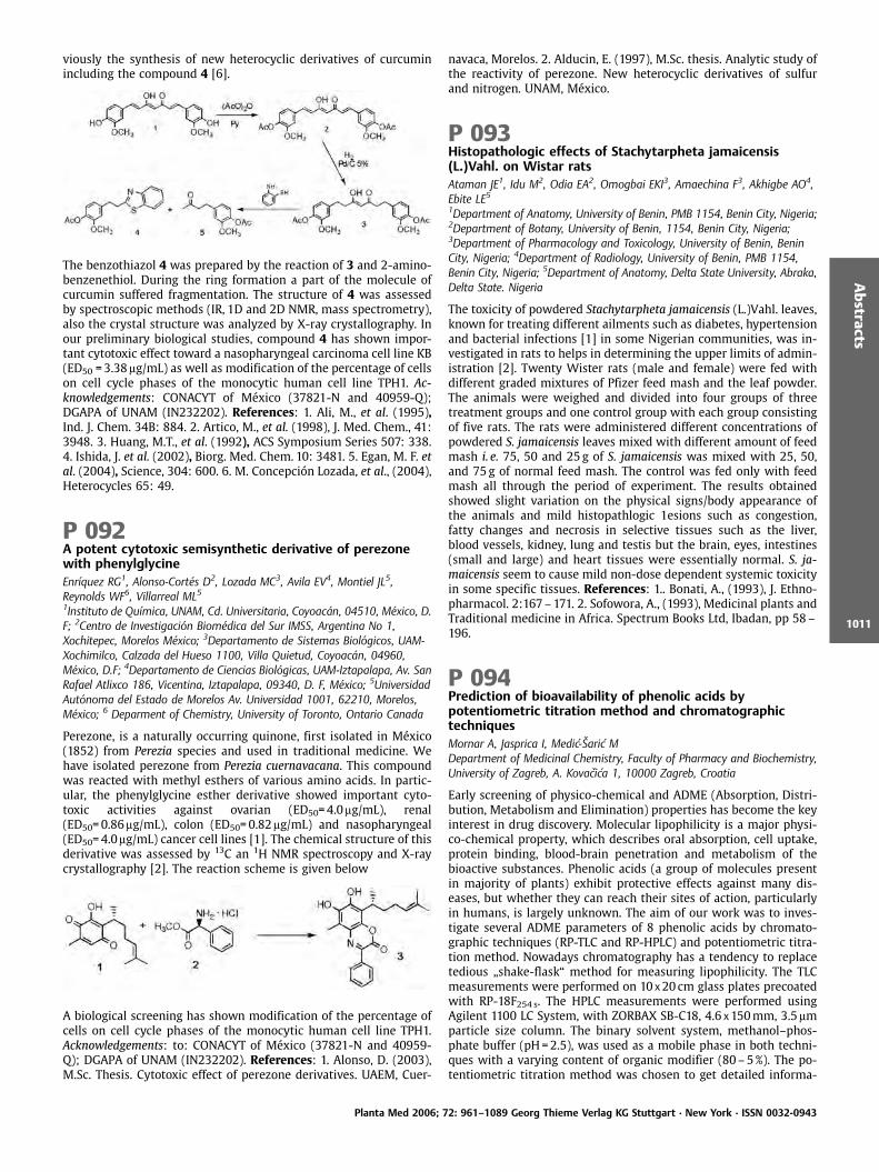
viously the synthesis of new heterocyclic derivatives of curcuminincluding the compound 4 [6].
The benzothiazol 4 was prepared by the reaction of 3 and 2-amino-benzenethiol. During the ring formation a part of the molecule ofcurcumin suffered fragmentation. The structure of 4 was assessedby spectroscopic methods (IR, 1D and 2D NMR, mass spectrometry),also the crystal structure was analyzed by X-ray crystallography. Inour preliminary biological studies, compound 4 has shown impor-tant cytotoxic effect toward a nasopharyngeal carcinoma cell line KB(ED50 =3.38mg/mL) as well as modification of the percentage of cellson cell cycle phases of the monocytic human cell line TPH1. Ac-knowledgements: CONACYT of M xico (37821-N and 40959-Q);DGAPA of UNAM (IN232202). References: 1. Ali, M., et al. (1995),Ind. J. Chem. 34B: 884. 2. Artico, M., et al. (1998), J. Med. Chem., 41:3948. 3. Huang, M.T., et al. (1992), ACS Symposium Series 507: 338.4. Ishida, J. et al. (2002), Biorg. Med. Chem. 10: 3481. 5. Egan, M. F. etal. (2004), Science, 304: 600. 6. M. Concepci n Lozada, et al., (2004),Heterocycles 65: 49.
P 092A potent cytotoxic semisynthetic derivative of perezonewith phenylglycineEnr quez RG1, Alonso-Cort s D2, Lozada MC3, Avila EV4, Montiel JL5,Reynolds WF6, Villarreal ML51Instituto de Qu mica, UNAM, Cd. Universitaria, Coyoac n, 04510, M xico, D.F; 2Centro de Investigaci n Biom dica del Sur IMSS, Argentina No 1,Xochitepec, Morelos M xico; 3Departamento de Sistemas Biol gicos, UAM-Xochimilco, Calzada del Hueso 1100, Villa Quietud, Coyoac n, 04960,M xico, D.F; 4Departamento de Ciencias Biol gicas, UAM-Iztapalapa, Av. SanRafael Atlixco 186, Vicentina, Iztapalapa, 09340, D. F, M xico; 5UniversidadAut noma del Estado de Morelos Av. Universidad 1001, 62210, Morelos,M xico; 6 Deparment of Chemistry, University of Toronto, Ontario Canada
Perezone, is a naturally occurring quinone, first isolated in M xico(1852) from Perezia species and used in traditional medicine. Wehave isolated perezone from Perezia cuernavacana. This compoundwas reacted with methyl esthers of various amino acids. In partic-ular, the phenylglycine esther derivative showed important cyto-toxic activities against ovarian (ED50=4.0mg/mL), renal(ED50=0.86mg/mL), colon (ED50=0.82mg/mL) and nasopharyngeal(ED50=4.0mg/mL) cancer cell lines [1]. The chemical structure of thisderivative was assessed by 13C an 1H NMR spectroscopy and X-raycrystallography [2]. The reaction scheme is given below
A biological screening has shown modification of the percentage ofcells on cell cycle phases of the monocytic human cell line TPH1.Acknowledgements: to: CONACYT of M xico (37821-N and 40959-Q); DGAPA of UNAM (IN232202). References: 1. Alonso, D. (2003),M.Sc. Thesis. Cytotoxic effect of perezone derivatives. UAEM, Cuer-
navaca, Morelos. 2. Alducin, E. (1997), M.Sc. thesis. Analytic study ofthe reactivity of perezone. New heterocyclic derivatives of sulfurand nitrogen. UNAM, M xico.
P 093Histopathologic effects of Stachytarpheta jamaicensis(L.)Vahl. on Wistar ratsAtaman JE1, Idu M2, Odia EA2, Omogbai EKI3, Amaechina F3, Akhigbe AO4,Ebite LE51Department of Anatomy, University of Benin, PMB 1154, Benin City, Nigeria;2Department of Botany, University of Benin, 1154, Benin City, Nigeria;3Department of Pharmacology and Toxicology, University of Benin, BeninCity, Nigeria; 4Department of Radiology, University of Benin, PMB 1154,Benin City, Nigeria; 5Department of Anatomy, Delta State University, Abraka,Delta State. Nigeria
The toxicity of powdered Stachytarpheta jamaicensis (L.)Vahl. leaves,known for treating different ailments such as diabetes, hypertensionand bacterial infections [1] in some Nigerian communities, was in-vestigated in rats to helps in determining the upper limits of admin-istration [2]. Twenty Wister rats (male and female) were fed withdifferent graded mixtures of Pfizer feed mash and the leaf powder.The animals were weighed and divided into four groups of threetreatment groups and one control group with each group consistingof five rats. The rats were administered different concentrations ofpowdered S. jamaicensis leaves mixed with different amount of feedmash i. e. 75, 50 and 25g of S. jamaicensis was mixed with 25, 50,and 75g of normal feed mash. The control was fed only with feedmash all through the period of experiment. The results obtainedshowed slight variation on the physical signs/body appearance ofthe animals and mild histopathlogic 1esions such as congestion,fatty changes and necrosis in selective tissues such as the liver,blood vessels, kidney, lung and testis but the brain, eyes, intestines(small and large) and heart tissues were essentially normal. S. ja-maicensis seem to cause mild non-dose dependent systemic toxicityin some specific tissues. References: 1.. Bonati, A., (1993), J. Ethno-pharmacol. 2:167–171. 2. Sofowora, A., (1993), Medicinal plants andTraditional medicine in Africa. Spectrum Books Ltd, Ibadan, pp 58–196.
P 094Prediction of bioavailability of phenolic acids bypotentiometric titration method and chromatographictechniquesMornar A, Jasprica I, Medic- aric MDepartment of Medicinal Chemistry, Faculty of Pharmacy and Biochemistry,University of Zagreb, A. Kovacica 1, 10000 Zagreb, Croatia
Early screening of physico-chemical and ADME (Absorption, Distri-bution, Metabolism and Elimination) properties has become the keyinterest in drug discovery. Molecular lipophilicity is a major physi-co-chemical property, which describes oral absorption, cell uptake,protein binding, blood-brain penetration and metabolism of thebioactive substances. Phenolic acids (a group of molecules presentin majority of plants) exhibit protective effects against many dis-eases, but whether they can reach their sites of action, particularlyin humans, is largely unknown. The aim of our work was to inves-tigate several ADME parameters of 8 phenolic acids by chromato-graphic techniques (RP-TLC and RP-HPLC) and potentiometric titra-tion method. Nowadays chromatography has a tendency to replacetedious „shake-flask“ method for measuring lipophilicity. The TLCmeasurements were performed on 10 x20 cm glass plates precoatedwith RP-18F254 s. The HPLC measurements were performed usingAgilent 1100 LC System, with ZORBAX SB-C18, 4.6 x150mm, 3.5mmparticle size column. The binary solvent system, methanol–phos-phate buffer (pH=2.5), was used as a mobile phase in both techni-ques with a varying content of organic modifier (80–5%). The po-tentiometric titration method was chosen to get detailed informa-
Planta Med 2006; 72: 961–1089 Georg Thieme Verlag KG Stuttgart · New York · ISSN 0032-0943
Abstracts
1011
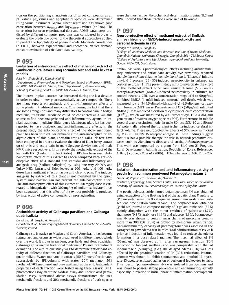
tion on the partitioning characteristics of target compounds at allpH values. pKa values and lipophilic pH-profiles were determinedusing Sirius instrument GLpKa. Linear regression has shown goodcorrelation between RM(TLC) and logkw(HPLC) values (r = 0.96). Thecorrelation between experimental data and ADME parameters pre-dicted by different computer programs was considered in order toevaluate the predictive power of the theoretical approaches appliedto predict the lipophilicity of phenolic acids. Moderate correlations(r < 0.90) between experimental and theoretical values demandconstant evaluation of calculated data validity.
P 095Evaluation of anti-nociceptive effect of methanolic extract ofSambucus nigra leaves using Formalin test and Tail-Flick testmodelsFaizi M1, Shafaghi B1, Kamalinejad M2
1Department of Pharmacology and Toxicology, School of Pharmacy, SBMU,P.O.BOX: 14155 – 6153, Tehran, Iran; 2Department of Pharmacognosy,School of Pharmacy, SBMU, P.O.BOX:14155 –6153, Tehran, Iran
The interest in plant sources has increased during the last decadesin order to obtain new pharmaceutically active compounds. Thereare many reports on analgesic and anti-inflammatory effects ofsome plants in traditional medicine. Considering the fact that thereare some ambiguities and many difficulties to control pain in todaysmedicine, traditional medicine could be considered as a valuablesource to find new analgesic and anti-inflammatory agents. In Ira-nian traditional medicine, Elder berry (Sambucus nigra L.) has beenreported to have analgesic and anti-inflammatory effects. In thepresent study the anti-nociceptive effect of the above mentionedplant has been studied. For evaluating the anti-nociceptive or an-algesic effect of this plant, Formalin test and Tail-Flick test havebeen employed in order to evaluate the effect of Elder berry extracton chronic and acute pain in male Sprague-dawley rats and maleNMRI mice respectively. In this study the methanolic extract of theleaf with HRT (Herbal to Extract Ratio) of 10% has been used. Anti-nociceptive effect of this extract has been compared with anti-no-ciceptive effect of a standard non-steroidal anti-inflamatory andanalgesic drug (Sodium salicylate) by using one-way ANOVA andTukey HSD. Extracts of Elder leaves at 200mg/kg and 400mg/kgdoses has significant effect on acute and chronic pain. The inducedanalgesia by extract of this plant is not mediated by the opioidsystem since naloxon can not prevent the anti-nociceptive effect.The anti-nociceptive effect of this extract at 200mg/kg dose is esti-mated to bioequivalent with 300mg/kg of sodium salicylate. It hasbeen suggested that this effect of the extract probably is producedby interaction of active components on prostaglandins.
P 096Antioxidant activity of Galinsoga parviflora and GalinsogaquadriradiataDerwinska M, Bazylko A, Kowalski JDepartament of Pharmacognosy,Medical University,1 Banacha St, 02 – 097Warsaw, Poland
Galinsoga sp. is native to Mexico and South America. It has becomenaturalized and occurs as widespread weed in different areas wholeover the world. It grows in gardens, crop fields and along roadsides.Galinsoga sp. is used in traditional medicine in Poland for treatmentdermatitis. The aim of our study was to determine antioxidant ac-tivity of different fractions of Galinsoga parviflora and Galinsogaquadriradiata. Water-methanolic extracts (50:50) were fractionatedsuccessively by SPE-columns with water, 20% methanol, 50%methanol, 70% methanol and pure methanol at the end. Antioxidantproperties of each fraction were tested using three assays: DPPHphotometric assay, xanthine oxidase assay and linoleic acid perox-idation assay. Mentioned above assays demonstrated the 50%methanolic fractions and 20% methanolic fractions of both species
were the most active. Phytochemical determinations using TLC andHPLC showed that those fractions were rich of flavonoids.
P 097Neuroprotective effect of methanol extract of Smilacischinae rhizome on NMDA-induced neurotoxicity andcerebral ischemia in ratsSeonga YH, Bana JY, Songb KS1College of Veterinary Medicine and Research Institute of Herbal Medicine,Chungbuk National University, Cheongju, Chungbuk 361 – 763,South Korea;2College of Agriculture and Life-Sciences, Kyungpook National University,Daegu, 702 – 701, South Korea
Smilax has various pharmacological effects including antiiflamma-tory, anticancer and antioxidant activity. We previously reportedthat Smilacis chinae rhizome from Smilax china L. (Liliaceae) inhibitsamyloid b protein (25–35)-induced neurotoxicity in cultured ratcortical neurons [1]. The present study aims to investigate the effectof the methanol extract of Smilacis chinae rhizome (SCR) on N-methyl-D-aspartate (NMDA)-induced neurotoxicity in cultured ratcortical neurons. CSR, over a concentration range of 5 to 50mg/mL,inhibited NMDA (1 mM)-induced neuronal cell death, which wasmeasured by a 3-[4,5-dimethylthiazol-2-yl]-2,5-diphenyl-tetrazo-lium bromide (MTT) assay. Pretreatment of CSR (50mg/mL) inhibitedNMDA (1 mM)-induced elevation of cytosolic calcium concentration([Ca2+]c), which was measured by a fluorescent dye, Fluo 4-AM, andgeneration of reactive oxygen species (ROS). Furthermore, in middlecerebral artery occlusion model in male SD rats, SCR (30 and 50mg/kg) potentially reduced the transient ischemia-induced cerebral in-farct volume. These neuroprotective effects of SCR were mimickedby MK-801, an NMDA receptor antagonist. These findings suggestthat SCR has a possible therapeutic role in neurodegenerative dis-eases such as Alzheimer’s disease and stroke. Acknowledgements:This work was supported by a grant from BioGreen 21 Program,Rural Development Administration, Republic of Korea. Reference:1. Ban, J.Y., Cho, S.O. et al. (2006), J. Ethnopharmacol. 106: 230–237.
P 098Isolation, characterization and anti-inflammatory activity ofpectin from common pondweed Potamogeton natans LPopov SV, Popova GY, Ovodova RG, Ovodov YSInstitute of Physiology, Komi Science Centre, The Urals Branch of the RussianAcademy of Sciences, 50, Pervomaiskaya str, 167982 Syktyvkar, Russia
The pectic polysaccharide named potamogetonan PN was obtainedusing extraction of the floating leaf of the aquatic plant P. natans L.(Potamogetanaceae) by 0.7% aqueous ammonium oxalate and sub-sequent precipitation with ethanol. The polysaccharide obtained(yield 4%) proved to compose mainly of D-galacturonic acid (82%)mainly altogether with the minor residues of galactose (1.7 %),rhamnose (0.8%), arabinose (1.4 %) and glucose (1.1 %). Potamogeto-nan PN was shown to contain sugar chains of molecular weightsmore than 300 kDa (78%) as proved by membrane ultrafiltration.Anti-inflammatory capacity of potamogetonan was assessed in thecarrageenan paw edema test in mice. Oral administration of PN 24hprior to induction of inflammation was found to reduce the edemaformation in a dose-related manner. The maximal effect of PN(50mg/kg) was observed at 1h after carrageenan injection (80%reduction of footpad swelling) and was comparable with that ofindomethacin (50mg/kg, p. o.) The delayed edema (5h) was lessaffected by the preadministration of PN (33% reduction). Potamo-getonan was shown to inhibit spontaneous and phorbol-12-myris-tate-13-acetate-activated adhesion of peritoneal leukocytes in vitro.Thus, pectin (potamogetonan PN) was isolated from P.natans andwas found to possess strong preventive anti-inflammatory activityespecially in relation to initial phase of inflammation development.
Planta Med 2006; 72: 961–1089 Georg Thieme Verlag KG Stuttgart · New York · ISSN 0032-0943
Abstracts
1012
P 099Formation of supramolecular structures of alkylamides fromEchinacea – implications for cannabinoid type-2 receptor(CB2) interactions in vitroRaduner S1, Bisson W2, Altmann KH1, Gertsch J11Institute of Pharmaceutical Sciences, ETH Zurich, 8093-Z rich, Switzerland;2The Scripps Research Institute, Molecular Biology, La Jolla, CA92037, USA
Various N-alkyl amides (alkylamides) from the medicinal plant Echi-nacea are cannabinoid type-2 receptor (CB2)-specific cannabinomi-metics. Based on biphasic effects observed in radioligand-based re-ceptor binding assays it was postulated that Echinacea alkylamidesmay form aggregates. In this study we show that dodeca-2E,4E,8Z,10Z-tetraenoic acid isobutylamide (A1) and dodeca-2E,4E-dienoic acid isobutylamide (A2) assemble into micelles, whereas nomicelle formation occurs for undeca-2E-ene,8,10-diynoic acid isobu-tylamide (A3) or the structurally related endogenous cannabinoidsarachidonoyl ethanolamide (anandamide) and 2-arachidonoyl gly-cerol (2-AG). Light scattering spectroscopy was used to determinethe size of premicelle aggregates, micelles and supermicelles. Thecritical micelle forming concentrations (CMC) of A1 (7.4 nmol/L) andA2 (150 pmol/mL) were determined by fluorescence spectroscopy.The experimental data were complemented by molecular dynamics(MD) simulations of the aggregation phenomenon based on Monte-carlo calculations. The results of these studies suggest that both A1and A2 readily aggregate into premicelles, whereas A2 forms morecompact aggregates due to a better alignment of hydrophobic chainsand higher curvature. This is in line with microscopic analyses,which show that A2 spontaneously forms both globular and rod-like surfactant micellar superstructures. The data on the self-assem-bly of A1 and A2 may provide a rationale for the concentration-dependent effects of alkylamides in the radioligand binding assayand suggest a partition between the receptor-bound, monomeric,premicellar, and micellar states.
P 100Analysis of the Physiological Activity from Kalopanaxseptemlobus Koidz. Extracts in KoreaKim SH, Chung HG, Han J, Kim SCKorea Forest Research Institute, 44 – 3 Omokcheon, 441 – 350, Suwon,Republic of Korea
Aiming to find new uses and to select clones that contain highphysiological active materials, stem and root barks of Kalopanaxseptemlobus Koidz., were analyzed to measure cell cytotoxicity(NRassay, MTT assay), anti-lipid peroxidation(TBA method), anti-freeradical activity(DPPH test, NBT test) and oxidative stress(DCFH-DAmethod). NR50 and MTT50 values were 0.002~0.33mg/mL,0.003~0.80mg/mL, respectively, and MeOH treatment of stem barksshowed lowest toxicity. TBA analysis showed many differences byextraction methods and sampling parts. MeOH treatment of stembarks showed the best result, 98% and that was better than control,vitamin C. Extracts of Kalopanax septemlobus showed superior anti-free radical activity on the DPPH and NBT tests. MeOH 0.1mg/mLtreatment of stem bark showed the best, 96% on the DPPH test andthat was better than control, vitamin E. Also, hot-water extractssoluble treatment of stem barks showed the best, 95%. Hot-waterextracts insoluble treatment of root barks showed the best in DCFH-DA analysis. The stem bark of Kalopanax septemlobus has been usedin traditional Korean medicine for anti-inflammatory, expectorant,tranquilizer and effective on anti-rheumatic. Thirteen kinds of che-mical components were isolated from the extracts of the dried stembark of K. septemlobus. On the basis of physico-chemical, spectro-scopic data and in comparison with those of authentic samples orvalues reported in the literatures, they were identified as b-sitoster-ol, oleanolic acid, caffeic acid, kalopanaxsaponin A, chlorogenic acid,protocatechuic acid, 3, 3’-bis(3,4-dihydro-4-hydroxy-6-methoxy-2H-1-benzopyran), (-)-balanophonin, liriodendrin, syringin, kalopa-
naxsaponin B, kalopanaxsaponin I and kalopanxsaponin H, respec-tively.
P 101Development of antifungal agents from essential oilcompounds in Ostericum koreanumShin S, Sim Y, Lim S, Byun Y, Choi SCollege of Pharmacy, Duksung Women’s University, Seoul, 132 – 714, Korea
Essential oils from plants are a promising source for novel naturalantifungal drugs, though their activity against human pathogenicfungi is generally milder than commercial synthetic antifungaldrugs. Ostericum koreanum (Max.) Kitagawa (Umbelliferae) is a per-ennial herb used in traditional Korean medicines for treatment ofthe common cold and for relief of rheumatic pains or headaches; theherb imparts a pungent and warm sensation. A rich source of essen-tial oil, Ostericum koreanum is widely distributed in the wild andcultivated in Korea. In this study we analyzed the essential oil fromO. koreanum and evaluated its antifungal activity by the broth dilu-tion method and disk diffusion test against various pathogenic fun-gal species. On the basis of these results, checkerboard micro titertests were performed and isobolograms were constructed to deter-mine the combined effect of the essential oils and ketoconazole inorder to develop more effective and safer anti-catharsis therapy. Asthe results, the essential oil of O. koreanum and its main compo-nents showed high susceptibility against the tested fungi. The anti-fungal activities were dose dependent. It exhibited significant syner-gism in combination with ketoconazole. Reference: Shin, S., Lim, S.(2004), J. Appl. Microbiol. 97: 1289–1296.
P 102Antifertility activities of Acanthus montanus and its newsulphate ester on female rats with possible mechanism(s) ofactionAsongalem EA1, Nana P2, Kamtchouing P21Pharmacology and Toxicology Unit, Department of Physiological Sciences,Faculty of Medicine & Biomedical Sciences, University of Yaounde 1, Yaounde,Cameroon, P.O. Box 8283, Yaounde Email: [email protected], Fax: 2372221873; 2Department of Animal Biology & Physiology, Faculty of Science,University of Yaounde 1, Yaounde, Cameroon
This study centred on assessing the effects of aqueous extract (AE)of Acanthus montanus (Acanthaceae) and its new compound –Acanthus sulphate ester (ASE) on oestrous cycle, implantationsand possible mode(s) of action. Oestrous cycles of Wistar rats(150–212g) were monitored before, during and after oral adminis-tration of distilled water (control), AE (250, 500, 1000mg/kg/day)and ASE (0.25, 0.5, 1.0mg/kg/day; intravenous) for 6 consecutivedays. Concerning implantations, pregnant rats received above dosesof AE and ASE from days 1–6 (pre-implantation) or 6–15 (post-implantation) of gestation and sacrificed on day 8 or 20 of preg-nancy(1). AE (1000mg/kg/day) and ASE (2mg/kg/day) were given tooveriectomised rats in the presence and absence of exogenouslyadministered oestrogen and or progesterone with uterine weightand deciduoma count assessed. PGF2a was evaluated on pre-implan-tation in the presence and absence of AE and ASE. One-way ANOVAat P < 0.05 was used. AE and ASE dose independently prolongedmetoestrous and dioestrous stages of the oestrous cycle which re-versed at least 10 days post-dosing. On pre-implantation, the AE(1000mg/kg/day) and ASE (0.5mg/kg/day) caused appreciable pre-implantation losses of 36.8 € 6.5%, P < 0.05 and 42.5 € 11.5%, P < 0.01respectively whereas AE (1000mg/kg/day) and ASE (1.0mg/kg/day)insignificantly caused post-implantation losses. AE and ASE did notalter the uterine weights or deciduoma counts (in the presence ofprogesterone) but reduced (P < 0.05) the number of implants ofPGF2a-administered rats. AE and ASE caused infertility by prolong-ing oestrous cycle and promoting pre-implantation loss; abolisheddeciduoma formation and prostaglandin inhibition was implicated
Planta Med 2006; 72: 961–1089 Georg Thieme Verlag KG Stuttgart · New York · ISSN 0032-0943
Abstracts
1013

but not sex hormones. Acknowledgement: This research was sup-ported by the International Foundation for Science, Stockholm, Swe-den and United Nations University (UNU), Tokyo, Japan, through agrant to Dr Emmanuel Acha ASONGALEM. Reference: 1. Asongalem,E.A., Akintonwa, A., (1997), Bull. Environ. Contam. Toxicol. 58: 184–189.
P 103Antiinflammatory activity of the aqueous leaf extract ofManihot esculenta CrantzYemitan OK2, Afolabi L1, Adeyemi OO1
1Department of Pharmacology, College of Medicine of the University ofLagos, Idi-Araba, P.M.B. 12003 Lagos, Lagos, Nigeria; 2Department ofPharmacology, Lagos State University College of Medicine, P.M.B. 21266,Ikeja, Lagos, Nigeria
The aqueous leaf extract of Manihot esculenta Crantz (MELE) hasbeen used in traditional African medicine for the treatment of in-flammation. The anti-inflammatory effects of MELE given throughoral and topical routes, were tested in rodents. MELE (100–400mg/kg, p.o) was given to rats and 30min. later, 0.9 % carrageenan wasinjected into the right hind paw [1]. In another set, MELE (1– 4% w/w in petroleum jelly) was applied topically to either the paws or toshaved back portion of rats before carrageenan. Paw diameter wasmeasured between 0–24h post- carrageenan injection. In anotherexperiment, MELE (100–400mg/kg, p. o.) or (1 –4% applied tomouse abdomen) was administered and 30min. later, 0.03 mL ofxylene was applied to the right ear of mice; then sections of earremoved and weighed for oedema [2]. MELE (100–400mg/kg, p.o)produced significant (P < 0.001) inhibition of carrageenan and xy-lene-induced oedema in rats and mice respectively. The percentageinhibition at 4% w/w in petroleum jelly (52.3 € 2.0%) is comparableto those produced by acetylsalicylic acid (50.0 €2.6%). At 1–4% w/w, topically, MELE produced significant (P < 0.01) inhibition of car-rageenan- induced rat paw oedema (68.0 € 2.1%) and xylene-in-duced ear swelling in mice (76.6 € 2.2%). Effects are significantlyhigher than those produced by indomethacin (74.0 € 3.1%,47.0 € 2.1%, respectively). Based on the results, the extract may con-tain orally safe, anti-inflammatory principles, justifying its use infolklore medicine. References: 1. Winter, C.A. et al. (1962), Proc.Soc. Exp. Biol. 11: 533–547. 2. Tang, Xi Can, et al. (1984), ActaPharmacol. Sinica 5: 85–89.
P 104Analgesic activity of aqueous leaf extract of Manihotesculenta CrantzYemitan OK2, Afolabi L1, Adeyemi OO1
1Department of Pharmacology, College of Medicine of the University ofLagos, Idi-Araba, P.M.B. 12003 Lagos, Lagos, Nigeria; 2Department ofPharmacology, Lagos State University College of Medicine, P.M.B. 21266,Ikeja, Lagos, Nigeria
The aqueous leaf extract of Manihot esculenta Crantz (MELE) hasbeen used in traditional African medicine for the treatment of acuteand chronic pain [1] and is claimed to be safe. The analgesic andacute toxicity effects of the extract, given through oral and topicalroutes, were tested in rodents. MELE (100–400mg/kg, orally) wasadministered to mice 30min before injection of 10mL/kg, acetic acid(0.6 % v/v in normal saline) intraperitoneally [2] or acetylcholine(8.3mg/kg) [3]. In another set, 0.5mL MELE was rubbed onto thedepilated abdomen of mice, and at 4h and 8h post-treatment, eachmouse was challenged with 0.6%, 10mL/kg, acetic acid or acetylcho-line (8.3mg/kg) intraperitoneally. The number of writhes wascounted for 15 and 30min., respectively in both experiments. MELE(100–400mg/kg, orally) and (1–4% w/w, topically), like aspirinexhibited significant (P < 0.05) inhibition of acetic acid (Orally:MELE: 61.3 €3.5%, Aspirin: 70.4 € 4.8%; Topically: MELE:47.4 €4.5%; Aspirin: 54.9 € 5.5%) and acetylcholine (Orally: MELE:
54.7 € 7.5%; Aspirin: 70.1 € 4.5%; Topically: MELE: 68.0 € 3.8%; As-pirin: 57.8 € 4.5%) – induced mouse writhing tests, compared tountreated control. Acute oral administration up to 10 g/kg did notcause death within 14 days, but produced mortalities in i.p. admi-nistered extract with LD50 of 2.5 g/kg. Based on these, the extractmay contain orally safe, analgesic principles, justifying its use intraditional African medicine. References: 1. Anderson, G.J., Coee,F.G. (1996), Econ. Bot. 50, 1:71 –107. 2. Koster, R. et al. (1959), Fed.Proc. 18: 418–420. 3. Sancilio L.F. et al. (1977), Agents and Actions 7:133–144.
P 105Ultra low concentrations of sophoraflavanone G fromSophora pachycarpa C. Meyer enhanced the antibacterialactivity of gentamycin aginst Staphylococcus aureusShahverdi AR1, Iranshahi M2
1Department of Pharmaceutical Biotechnology, Faculty of Pharmacy, TehranUniversity of Medical Sciences, Tehran, Iran; 2Department of Pharmacognosy,Mashhad University of Medical Sciences, Mashhad, Iran
In this study the enhancement effect of Sophora pachycarpa roots’acetone extract on the antibacterial activity of gentamycin was eval-uated against Staphylococcus aureus. Disk diffusion and broth dilu-tion methods were used to determine the antibacterial activity ofgentamycin in the absence and presence of plant extract and itsvarious fractions separated by TLC. Clinical isolate of S. aureus wasused as test strain. The active component of plant extract involvedin enhancement of gentamycin’s activity had Rf =0.72 on TLC. Thespectral data (1H NMR, 13C NMR) of this compound revealed thatthis compound was 5,7,2’,4’-tetrahydroxy-8-lavandulylflavanone(sophoraflavanone G), previously isolated from Sophora exigua. Inthe presence of 0.03 mcg/mL of sophoraflavanone G the MIC ofgentamycin for S. aureus decreased from 32 to 8 mcg/mL (a fourfolddecrease). These results signify that the ultra low concentration ofsophoraflavanone G potentiates the antimicrobial action of genta-mycin suggestion a possible utilization of this compound in combi-nation therapy against S. aureus. Reference: Tsuchiya, H., Iinuma M.(2000), Phytomedicine 7:161–165.
P 106Alkaloids from the club moss Lycopodium annotinum L. –acetylcholinesterase inhibitory activity in vitroHalldorsdottir ES1, Olafsdottir ES11University of Iceland, Faculty of Pharmacy, Hagi, Hofsvallagata 53, IS-107Reykjavik, Iceland
Plant species belonging to the genera Lycopodium are known toproduce so-called Lycopodium alkaloids. The club moss Huperziaserrata (Thumb.) Trev., which has long been used traditionallyagainst Alzheimer’s disease in China, has been shown to containLycopodium alkaloids which are acetylcholinesterase inhibitors [1].Five species of club mosses are found in Iceland. Huperzia selago L.Bernh. ex Schrank & C. Martius has previously been studied andshown to contain a new alkaloid, selagoline, in addition to theknown huperzine A and serratidine. The inhibitory activity on acet-ylcholinesterase was not investigated [2]. The aim of this study wasto examine the alkaloid content of the Icelandic Lycopodium anno-tinum and to determine their ability to inhibit the enzyme acetyl-cholinesterase in vitro. The plant extract was fractionated and thealkaloids purified using liquid chromatography methods (VLC, SPE,HPLC) and 1H- and 13C-NMR spectroscopy was used for the struc-ture determination. The inhibitory activity of the alkaloids towardsacetylcholinesterase was determined by an in vitro TLC acetylcholi-nesterase assay [3]. Three Lycopodium alkaloids from L. annotinumwere found to inhibit acetylcholinesterase activity in vitro includingthe known annotine. Another known alkaloid from this plant, anno-tinine, did not show inhibition. Annotine and annotinine have onlybeen found in L. annotinum and their effect on acetylcholinesterase
Planta Med 2006; 72: 961–1089 Georg Thieme Verlag KG Stuttgart · New York · ISSN 0032-0943
Abstracts
1014

has not been described before. References: 1. Ma, X.Q. et al. (2004),Nat. Prod. Rep. 21: 752–772. 2. Staerk, D. et al. (2004), Nat. Prod.Res. 18: 197–203. 3. Rhee I.K. et al. (2001), J. Chromatogr. A 915:217–223.
P 107Anticonvulsant activities of the methanol extracts (leaf,root), saponins and n-butanol-insoluble fraction ofCalliandria portoricensis Jacq (Benth) (Family: Mimosaceae)Agunu A1, Abdurahman EM1, Musa KY1, Zezi UA2
1Department of Pharmacognosy and Drug Development, Ahmadu BelloUniversity, Zaria, Nigeria; 2Department of Pharmacology and ClinicalPharmacy, Ahmadu Bello University, Zaria, Nigeria
Calliandra portoricensis Jacq (Benth) (family: Mimosaceae) is a plantused in Nigeria as an analgesic, anticonvulsant, antihelminthic, anti-depressant and for treatment of skin rashes [1]. The methanol ex-tracts (leaf, root), saponins and n-butanol-insoluble fraction [2, 3]were used in the investigation. The LD50 of the extracts (root, leaf)carried out in mice were 1292mg/kg and 7041mg/kg respectively.The anticonvulsant evaluation was carried out in mice using pente-lyenetetrazole (PTZ) and electroshock (ES) by determining both an-tagonistic and potentiation properties [4] of the methanol extractsat 100mg/kg, 200mg/kg and 300mg/kg, saponins and n-butanol-insoluble fraction at 50mg/kg and 100mg/kg. There were no antag-onistic action to PTZ induced clonic convulsion by extracts, saponinsand n-butanol-insoluble fraction. The root extract showed signifi-cant (P < 0.05) protection when subtreshold of PTZ was adminis-tered compared to leaf extract. The root extract, saponins and n-butanol-insoluble fraction showed higher protection against ES in-duced tonic seizures compared to the leaf. The root extract and n-butanol-insoluble fraction also increased pentobarbital-inducedhypnosis compared with the leaf. This investigation provides scien-tific explanation for the anticonvulsant activities in the roots andleaves of Calliandra portoricensis. References: 1. Dalziel, J.M. (1937),Useful plants of West Tropical Africa pp. 303–304. 2. Cannell, R.J.P(1998), Natural product isolation pp. 343–348, Humana Press,U.S.A.. 3. Williamson, M.E; Okpako, T.D, et al. (1996), Selection, pre-paration and pharmacological evaluation of plant material pp 5 JohnWiley and Sons Ltd, West Sussex. 4. Amagaya, S; Lizuka, A, et al.(2001), Phytomedicine 8: 338–347.
P 108Reversible antifertility activity of methanol extract ofMomordica dioica Roxb. in male albino ratsKachhawa JBS, Sharma A, Gupta RSCenter for Advanced Studies, Reproduction Physiology Section, Departmentof Zoology, University of Rajasthan, Jaipur-302004, India
In the present era the major problem for developing countries is theincreasing population. To fight this, various hormonal, chemical andsurgical methods are developed for male but they have undesirableside effects and irreversibility. The present research work is going onthe development of fertility regulating drug from plant organs.Therefore the present work was done to evaluate the antifertilityactivity of Momordica dioica Roxb. (Cucurbitaceae) in male albinorats. For this adult proven fertile male rats were gavaged 100%methanol extract of Momordica dioica root at the dose level of5mg/rat/day for 60 days. Momordica dioica reduced the fertility ofmale rats by 100%. A reduction was seen in the cauda epididymalsperm motility and density. Marked decline were also found in thetesticular germ cell population, Leydig cell nuclear area and thenumber of mature Leydig cells, However no morphological changeswere observed in Sertoli cells as well as in their counts. Serumtestosterone level was also reduced after Momordica dioica treat-ment. The protein glycogen, sialic acid, acid phosphatase and alka-line phosphatase content of testes, protein and sialic acid in caudaepididymis and fructose in seminal vesicle was decreased signifi-
cantly, whereas cholesterol content of testes increased significantly.Momordica dioica extract did not alter the blood & serum param-eters, which shows the non-toxic nature. All the parameters werereversible after withdrawal of the drug. In conclusion methanol ex-tract of Momordica dioica root have antifertility activity with theirreversible nature. Acknowledgment: Authors are thankful to theHead, Department of Zoology, Prof. N.K. Lohiya Coordinator CAS,Department of Zoology for providing the necessary facilities andUGC, Regional Office, Bhopal, INDIA for financial support.
P 109Evaluation of Narcissus tazetta L. under different habitatsHassan NM, Habib AA, Abdel-Azim NS, Shams KA, Hammouda FMPhytochemistry Dept, National Research Centre, 12311 Dokki, Cairo, Egypt
Narcissus tazetta belongs to the family Amaryllidaceae. In Egypt, it iscommercially grown outdoors for cut flowers and essential oil ex-traction that used in perfume industry. In this work, the bulbs werecultivated in three different localities under different habitats repre-senting the loamy soil, the new reclaimed sandy soil and the sandysoil; which were differentiated by their soil and water analysis.Evaluation of the cultivated plant samples from each locality; in-cluding essential oil content and the total alkaloids; was done. Theloamy soil showed to be the most suitable habitat for the cultivationof the plant. Investigation of the essential oil revealed that the high-est oil content was recorded in the plants grown in the loamy soil(0.13%). The plants cultivated at sandy soil gave the lowest yield ofthe oil (0.10%). GC/MS of the essential oil for all samples showedthat the main constituents of the oil are the same, but only differ intheir percentages. The maximum terpenoid percentage (65.1 %) wasdetermined in the plants cultivated in the loamy soil followed bysandy soil samples (52%), and then new reclaimed sandy soil sam-ples (47.65%). The main constituents were a-pinene, limonine, line-lool, methyl and ethyl cinnamate in all samples.The total alkaloidswere extracted from the collected leaves and bulbs of the cultivatedsamples. The highest alkaloid percent was found in the loamy soilleave samples (0.28%) while the lowest one was found at the sandysoil samples (0.12). Also, the average of the total alkaloidal contentof the collected bulbs showed the same tendency as the leaves. Thehighest percent was found in the loamy soil samples (0.13%) fol-lowed by the new reclaimed sandy soil samples (0.11%) and thelowest percent was found in the bulb samples collected from sandysoil (0.07). TLC-densitometric analysis indicated that the main spotof alkaloids might be identified as narcessine. Also, in vitro propa-gation by tissue culture technique was successful from callus for-mation from meristematic tips. References: 1. Guenther, E. (1952),The essential oils. Vol.5. Individual essential oils of the plant fa-milies, D. Van Nostrand Company, Inc. New York, pp. 343–351. 2.Furusawa, E., Furusawa, S. (1985), J. Ethnopharmacol. 16: 299. 3. El-Moghazy, A.M., Gomaa, G.S. et al. (1978), Egyptian J. Pharm. Sci. 17:273–281.
P 110Modification of the polarity of an anthocyanin pigment.Structure determination and antioxidant activityPal E1, Duez P2, Luhmer M3, Nacro M1
1Laboratoire de Chimie Organique Appliqu e, D partement de Chimie, UFR-SEA, Universit de Ouagadougou, 03 BP 7021 Ouagadougou 03, BurkinaFaso; 2Laboratoire de Pharmacognosie, de Bromatologie et de NutritionHumaine, Institut de Pharmacie, Universit Libre de Bruxelles, Campus de laPlaine CP 205 – 4, Bld Triomphe, B-1050 Bruxelles, Belgique; CLaboratoire deRMN haute r solution, D partement de Chimie Universit Libre de Bruxelles,CP 160 – 08, 50 avenue Franklin Roosevelt, B-1050 Bruxelles, Belgique
In the aim to develop antioxidant compounds with cellular mem-brane anchoring potential, Cyanidin (3-O-(2-O-b-D-glucopyrano-syl)-b-D-glucopyranosyl -5- O-b-D-glucopyranoside), an anthocya-nin extracted from Ipomoea asarifolia (convolvulaceae), was acy-
Planta Med 2006; 72: 961–1089 Georg Thieme Verlag KG Stuttgart · New York · ISSN 0032-0943
Abstracts
1015
lated with a palmityl group to yield two new acylated anthocyanins.Their structures were elucidated using chemical, mass and NMRspectroscopy methods (1H and 13C, TOCSY-1D, DQF-COSY andHMBC). Theses new pigments were found to consist of cyanidin 3-O-(2-O-b-D-glucopyranosyl)-b-D-glucopyranosyl-5-O-b-D-gluco-pyranosyl-7-O-palmitoyl and cyanidine 3-O-(2-O-b-D-glucopyrano-syl)-b-D-glucopyranosyl -5- O-b-D-glucopyranosyl-7,3’-O-dipalmi-toyl. Using MTT and DPPH assays for measuring the cytotoxic andantioxidant properties of these compounds, it was found that theacylated anthocyanins retain the non-toxicity and antioxidant prop-erties of the parent cyanidin. Work is in progress to further charac-terize the antioxidant profile of these interesting compounds.
P 111Pyridine Alkaloids of Senna multijulga (Cesalpiniaceae) asAcetylcholinesterase Inhibitorsda S. Bolzani V1, Serrano MAR1, Lopes MN1, Young MCM2, Torres LB2,Cardoso EM2
1Nfflcleo de Bioensaios, Bioss ntese e Ecofisiologia de Produtos Naturais –NuBBE, Universidade Estadual Paulista – UNESP – Instituto de Qu mica,Departamento de Qu mica Org nica, Rua Prof. Francisco Degni s/n –14.800 – 900 – Araraquara – S¼o Paulo – Brazil; 2Instituto de Bot nica,Avenida Miguel St fano 3687 – 04301– 902 – gua Funda – S¼o Paulo –Brazil
As part of a search for naturally occurring acetylcholinesterase in-hibitors, two new 2-methyl-3-hydroxyl-6-n-alquil-pyridine alka-loids 1– 2 and the known flavonoid quercetin-3-O-glicopyranosyl-(1,6)-rhamnopyranoside, were isolated by bioassay-guided fractio-nation from the leaves of Senna multijulga (Rich.) (Cesalpiniaceae),an ornamental plant species, popularly named “canafistula“, col-lected in Atlantic Forest, S¼o Paulo State. Compounds 1 and 2 weresubmitted to preliminary TLC screening for selecting potential AchEinhibitors, in which both alkaloids 1 and 2 inhibited the enzyme at0.1 and 0.5 mM concentrations. These compounds were also eval-uated for their efficacy in an in vitro rat brain assay, to measure itsAchE inhibitory potential, and additional results will be presented.(Supported by Program project Biota-FAPESP grant no 03/02176–7).Acknowledgements: To Funda ¼o de Amparo Pesquisa do Estado deS¼o Paulo (FAPESP), CAPES and CNPq for research funding.
P 112Phytochemical and Antimicrobial investigation of Taleghanplants speciesMehrabadi Yari L, Hosein M, Surmaghi S, Amin G, Badami N, Emami M,Asgari TFaculty of Pharmacy, Tehran University of Medical Sciences, Pursina Ave,14155 – 3451; and faculty of pharmacy, Azad university, Tehran, Iran
Taleghan a highly beautiful area located in 80 km.Nw of Tehran, hasa suitable ecology for growth of diverse plants. There are manyspecies that are used in folk medicine by inhabitants for treatmentof infection disease. In this study 158 species were collected, iden-tified and deposited in the herbarium of faculty of pharmacy, TehranUniversity of medical sciences. Prephytochemical, antibacterial andanti fungal effects of plant extracts considered and for this proposeshydroalchoholic extracts were prepared using suxhelt apparatus,then dried over vacuum system. Qualitative tests for assessment offour components: alkaloids, flavonoides, saponins, and tannins weredone via laboratory tests. Each extract was used due to find anti-fungal and antimicrobial properties against:Candida, Aspergillus,Nocardia, Microspom, Trychophyton, Streptococcus, Proteus, Es-cherchia, Salmonella, Staphylococcus and Pseudomonas. Blood agarand muller hinton were used as cultures media. Results of pre-phy-tochemical tests were as fallows: 84% had saponin, 42% tannin, 45%flavonoids, 13% alkaloids.69 species had attractive antimicrobial ef-fect and three species including Epilobium hirsutum L., Centaureabrugeriana and Centaurea virgata were the best. Antifungal and
antibacterial tests showed that the effective species were Achilleamicrantha Wild., Matricaria disciformis C.A. Meyers, Mentha longifo-liaL., Hypericum hyssopifolium Chaix. and Ducrosia anethifolia(DC)Boiss. in conclusion the species of containing saponin were moreeffective.
P 113Chemical composition, antiviral and antimicrobial activitiesof the essential oils of Ferula hormonis, Plectranthuscoleoides and Magnolia grandifloraMohamed SM1, Ibrahim NA2, Ali MA3, Faraid MA4
1Medicinal and Aromatic Plants; 2Pharmacognosy; 3Water Pollution; 4NaturalProducts Department, National Research Center, Tahrir St. Dokki, 12311. Fax3370931, Cairo, Egypt
Essential oils were obtained by hydrodistillation from the freshleaves of Magnolia grandiflora L., aerial part of Plectranthus coleoidesBenth. and dried roots of Ferula hormonis and were analyzed by GC/MS. The main constituents of the oil of Ferula were levomenol(23.42%), alpha-humulene (13.80%), cycloisolongifolene (8.35%),spathulenol (7.89%) and beta–oplopenone (7.76%) while the oil ofPlectranthus was characterized by thymol (57.57%), gamma-terpi-nene (15.37%), p-cymene (9.07%) and trans-caryophyllene (5.81%).The major component of Magnolia essential oil were trans-caryo-phyllene (9.70%), caryophyllene oxide (9.55%), dendrolasin (9.48%),butanoic acid 2-methyl-1-methylpropyl ester (7.38%) and spathule-nol (7.23%). Anti herpes simplex virus (HSV-1) was performed usingPlaque inhibition assay [1]. The activity was calculated by percen-tage of viral plaque inhibition at a non-cytotoxic dilution of the oil(1:320 v/v). The most potent oil was obtained from ferula essentialoil (81.4%) against HSV-1. Antimicrobial screening was conductedusing the disc diffusion assay against Staphylococcus aureus, Escher-ichia coli, Bacillus subtilis, Aspergillus niger, and the human patho-genic yeast, Candida albicans. The minimum inhibitory concentra-tions (MIC) were determined [2]. The essential oil of Plectranthuswas the most active as antimicrobial. At dilution of 1:100 v/v essen-tial oil of Plectranthus inhibited the growth of S. aureus, B. subtilis, A.niger and C. albicans while the dilution of 1:75 v/v inhibited thegrowth of E. coli. This chemical and biological investigation havenot studied previously. References: 1. Bermejo, P. et al. (2002),Planta Med. 68: 106–110. 2. Gabraith, H. et al. (1971), J. Appl. Bact.34: 803–813.
P 114Investigation of free radical scavenging activity by ESR forcoumarins isolated from Tecoma radicansHashem FAPharmacognosy depart. National Research Centre, Tahrir Street, Dokki, Cairo,Egypt
Tecoma radicans (Campsis radicans (L.) Seem.), F. Bignoniaceae is aspecies that belongs to a tropical family but has been introduced inmany countries as ornamental. Phytochemical investigation of theaerial parts of Tecoma radicans (L.) DC, indicates the presence of fourcoumarins, 2’, 3’-epoxide alloimperatorin (I), pabulenone (II), pere-florin B (III) and 17–methylbothrioclinin (IV). One chromone wasalso isolated peucenin–7-methyl ether (V), and showed the violetcolour with FeCl3 directly while the four coumarins showed thisviolet after alkalinization with ammonia solution. They were isolat-ed, purified and identified from their spectroscopic analysis andcomparing with the published data [1]. When successive extractsof T. radicans and coumarin fraction were screened for cytotoxicactivity in concentration of 100mg/0.1 mL DMSO, in vitro using asingle tumour (Ehrlich ascites carcinoma cells) [2], they showedno cytotoxic activity. When the coumarin fraction and isolated com-pounds were examined for free radical scavenging activity, using thestable DPPH free radical [3] and recorded by ESR, using vitamin C ascontrol, it was found that the whole coumarin fraction was the most
Planta Med 2006; 72: 961–1089 Georg Thieme Verlag KG Stuttgart · New York · ISSN 0032-0943
Abstracts
1016

active, Fig.1, (89. 06% inhibition of DPPH free radical), and the isolat-ed components are 0, 25.9, 8.45, 13.85 and 50% respectively, Table 1.Fig.1. ESR of control (DPPH) and coumarin extract of T.radicans
Table 1: Inhibition of DPPH radical by coumarins isolated from T. ra-dicans.
Compound Double integrationarea Percentage inhibition
DPPH 638
Vitamin C 29.0 95.45
Coumarin ext. 69.0 89.2
Compound I 638 -
Compound II 472.7 25.9
Compound III 584 8.45
Compound IV 549.6 13.85
Compound V 319.0 50.0
References: 1. Murray, R.D.H., Mendez, J., Brown, S.A.. (1982), TheNatural Coumarins, John Wiley and Sons Ltd., New York. 2. El-Hos-sary, G.A., Fathy, M.M. (2000), Bull. Fac. Pharm. Cairo Univ. 38: 87–97. 3. Acqua, S.D., Innocenti, G. (2004), Fitoterapia, 75, 592–595.
P 115Establishment of a cell-based screening system for NK-1antagonists using SP-conjugated fluorescence; effects ofplant extracts on the NK-1 receptor binding in U-373MGculturesKoo KA3, You JM1, Xu P3, Lee EJ4, Youn HJ2, Lee BJ11Biohealth Products Research Center and Department of Chemistry;2Biohealth Products Research Center and School of Biotechnology &Biomedical Science; 3Biohealth Products Research Center, Inje University,Gimhae 621 – 749, Gyungnam, Republic of Korea; 4Life Science R&D Center,SK Chemicals, Suwon 440 – 745, Kyungki-do, Republic of Korea
Substance P (SP) is a peptide neurotransmitter binding to neuroki-nin-1 (NK-1) receptors which are common in the central nervoussystems. The interaction between SP and NK-1 receptor has beenassociated with a number of diseases, including inflammatory bo-wel disease, liver diseases, asthma, diabetes, migraine, emesis, de-pression and pain [2]. There is no such study for the discovery of anew agent from plant products even though their action could pres-ent value as a target in the treatment of many diseases. In this study,we have established a cell-based system for screening NK-1 receptorantagonists from plant extracts using a fluorescence probe, SP con-jugated Oregon Green 488 (Molecular Probes, Eugene, OR), and U-
373MG human malignant glioma cells, which dominantly expressesNK receptors [3 –5]. The treatment of 10 nM SP-Oregon Green 488to the -373MG culture resulted in the significant increase of thefluorescence intensity, which was selectively inhibited by L-733,060, a selective synthetic NK-1 antagonist having high affinityon human NK-1 receptor (6). L-733,060 blocked the binding of theSP fluorescence probe in a dose-dependent manner at the concen-trations ranging from 0.1 to 100 nM (IC50=1.85 nM). We were ableto investigate some prospective plant extracts with the NK-1 recep-tor antagonist activity using the assay system. Acknowledgement:This study was supported by a grant from the Ministry of Com-merce, Industry and Energy (MOCIE) and the Korea Institute of In-dustrial Technology Evaluation & Planning (ITEP) through the Bio-health Products Research Center of Inje University. References: 1.Harrison, S. et al. (2001), Int. J. Biochem. Cell. Biol. 33: 555–576. 2.Duffy, R.A. (2004), Expert Opinion on Emerging Drugs 9: 9–21. 3.Bennett, V.J. et al. (2001), BMC Chem. Biol. 1: 1. 4. Rasley, A. et al.(2002), Glia 37: 258–267. 5. Walpole, C.S.J. et al. (1998), Br. J. Phar-macol. 124: 83–92. 6. Seabrook, G.R. et al. (1996), Eur. J. Pharmcol.317: 129–135.
P 116Anti-inflammatory effects of water-soluble fractions fromArtemisia species using the LPS-induced inflammatoryresponse in primary rat astrocyte culturesKoo KA3, Lee BJ2, Youn HJ11Biohealth Products Research Center and School of Biotechnology &Biomedical Science; 2Biohealth Products Research Center and Department ofChemistry; 3Biohealth Products Research Cente, Inje University, Gimhae621 – 749, Gyungnam, Republic of Korea
Water-soluble fractions have been purified from the crude aqueousextracts of Artemisia species, especially from Artemisia folum (AVF3)and Artemisia iwayomogi Kitam. (AIP1). The herbs are traditionallyused as the medicinal plant to prevent or treat a number of liverdiseases in Asia [1]. The AIP1 fraction has been shown to havediverse immuno-modulating activities including, anti-apoptosisand anti-cancer effects in our previous works [2, 3]. In this study,we have investigated the anti-inflammatory activity of the fractionsusing the LPS-induced inflammation model with primary rat astro-cyte cultures. The treatment of the astrocyte culture either withAVF3 or AIP1 resulted in the suppression of the NO production bythe LPS treatment, which was comparable to that of the dexametha-sone treatment. Quantitative real-time PCR analysis revealed thatthe expression of iNOS gene was significantly suppressed by thesamples, indicating that the inhibition of NO production could re-sult from an inhibitory effect on iNOS gene transcription. The treat-ment also suppressed the up-regulation of the pro-inflammatory IL-6 and MIP-1b genes as the dexamethasone treatment. These resultsclearly demonstrate that the water-soluble fractions from Artemisiaspecies might modulate the inflammatory response in brain astro-cytes. Since NO production in brain astrocytes is important in thepathogenesis of a number of brain inflammatory diseases such asmultiple sclerosis and Alzheimer’s disease, the anti-inflammatoryeffect of the fraction could have considerable value for the protec-tion or the treatment of the neurodegenerative diseases. Acknowl-edgement: This study was supported by a grant from the Ministry ofCommerce, Industry and Energy (MOCIE) and the Korea Institute ofIndustrial Technology Evaluation & Planning (ITEP) through the Bio-health Products Research Center of Inje University. References: 1.Bensky, D. et al. (2004), Chinese Herbal Medicine; Materia Medica.Eastland Press Inc. Seattle, USA. 2. Koo, K.A. et al. (1994), Arch.Pharm. Res. 17: 371–374. 3. Hwang, J.S. et al. (2005), Biol. Pharm.Bull. 28: 921–924.
Planta Med 2006; 72: 961–1089 Georg Thieme Verlag KG Stuttgart · New York · ISSN 0032-0943
Abstracts
1017
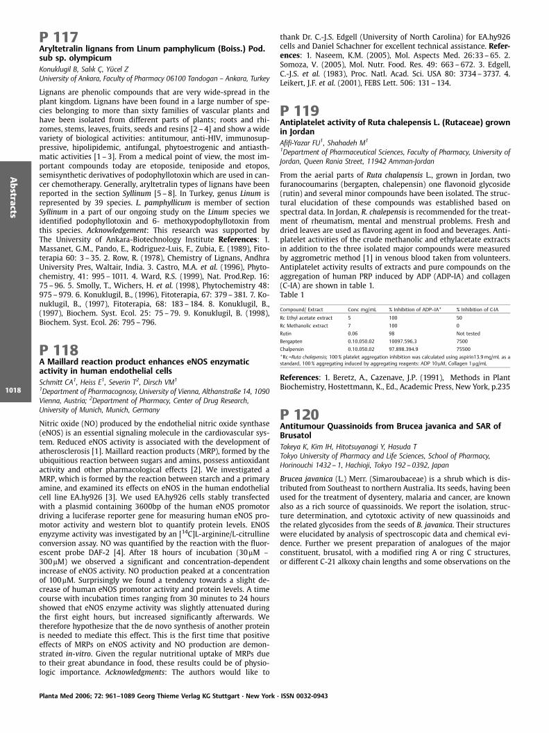
P 117Aryltetralin lignans from Linum pamphylicum (Boiss.) Pod.sub sp. olympicumKonuklugil B, Salık , Y cel ZUniversity of Ankara, Faculty of Pharmacy 06100 Tandogan – Ankara, Turkey
Lignans are phenolic compounds that are very wide-spread in theplant kingdom. Lignans have been found in a large number of spe-cies belonging to more than sixty families of vascular plants andhave been isolated from different parts of plants; roots and rhi-zomes, stems, leaves, fruits, seeds and resins [2 –4] and show a widevariety of biological activities: antitumour, anti-HIV, immunosup-pressive, hipolipidemic, antifungal, phytoestrogenic and antiasth-matic activities [1 – 3]. From a medical point of view, the most im-portant compounds today are etoposide, teniposide and etopos,semisynthetic derivatives of podophyllotoxin which are used in can-cer chemotherapy. Generally, aryltetralin types of lignans have beenreported in the section Syllinum [5 –8]. In Turkey, genus Linum isrepresented by 39 species. L. pamphyllicum is member of sectionSyllinum in a part of our ongoing study on the Linum species weidentified podophyllotoxin and 6- methoxypodophyllotoxin fromthis species. Acknowledgement: This research was supported byThe University of Ankara-Biotechnology Institute References: 1.Massanet, G.M., Pando, E., Rodriguez-Luis, F., Zubia, E. (1989), Fito-terapia 60: 3–35. 2. Row, R. (1978), Chemistry of Lignans, AndhraUniversity Pres, Waltair, India. 3. Castro, M.A. et al. (1996), Phyto-chemistry, 41: 995–1011. 4. Ward, R.S. (1999), Nat. Prod.Rep. 16:75–96. 5. Smolly, T., Wichers, H. et al. (1998), Phytochemistry 48:975–979. 6. Konuklugil, B., (1996), Fitoterapia, 67: 379–381. 7. Ko-nuklugil, B., (1997), Fitoterapia, 68: 183–184. 8. Konuklugil, B.,(1997), Biochem. Syst. Ecol. 25: 75–79. 9. Konuklugil, B. (1998),Biochem. Syst. Ecol. 26: 795–796.
P 118A Maillard reaction product enhances eNOS enzymaticactivity in human endothelial cellsSchmitt CA1, Heiss E1, Severin T2, Dirsch VM1
1Department of Pharmacognosy, University of Vienna, Althanstraße 14, 1090Vienna, Austria; 2Department of Pharmacy, Center of Drug Research,University of Munich, Munich, Germany
Nitric oxide (NO) produced by the endothelial nitric oxide synthase(eNOS) is an essential signaling molecule in the cardiovascular sys-tem. Reduced eNOS activity is associated with the development ofatherosclerosis [1]. Maillard reaction products (MRP), formed by theubiquitious reaction between sugars and amins, possess antioxidantactivity and other pharmacological effects [2]. We investigated aMRP, which is formed by the reaction between starch and a primaryamine, and examined its effects on eNOS in the human endothelialcell line EA.hy926 [3]. We used EA.hy926 cells stably transfectedwith a plasmid containing 3600bp of the human eNOS promotordriving a luciferase reporter gene for measuring human eNOS pro-motor activity and western blot to quantify protein levels. ENOSenyzyme activity was investigated by an [14C]L-arginine/L-citrullineconversion assay. NO was quantified by the reaction with the fluor-escent probe DAF-2 [4]. After 18 hours of incubation (30mM –300mM) we observed a significant and concentration-dependentincrease of eNOS activity. NO production peaked at a concentrationof 100mM. Surprisingly we found a tendency towards a slight de-crease of human eNOS promotor activity and protein levels. A timecourse with incubation times ranging from 30 minutes to 24 hoursshowed that eNOS enzyme activity was slightly attenuated duringthe first eight hours, but increased significantly afterwards. Wetherefore hypothesize that the de novo synthesis of another proteinis needed to mediate this effect. This is the first time that positiveeffects of MRPs on eNOS activity and NO production are demon-strated in-vitro. Given the regular nutritional uptake of MRPs dueto their great abundance in food, these results could be of physio-logic importance. Acknowledgments: The authors would like to
thank Dr. C.-J.S. Edgell (University of North Carolina) for EA.hy926cells and Daniel Schachner for excellent technical assistance. Refer-ences: 1. Naseem, K.M. (2005), Mol. Aspects Med. 26:33–65. 2.Somoza, V. (2005), Mol. Nutr. Food. Res. 49: 663–672. 3. Edgell,C.-J.S. et al. (1983), Proc. Natl. Acad. Sci. USA 80: 3734–3737. 4.Leikert, J.F. et al. (2001), FEBS Lett. 506: 131–134.
P 119Antiplatelet activity of Ruta chalepensis L. (Rutaceae) grownin JordanAfifi-Yazar FU1, Shahadeh M1
1Department of Pharmaceutical Sciences, Faculty of Pharmacy, University ofJordan, Queen Rania Street, 11942 Amman-Jordan
From the aerial parts of Ruta chalapensis L., grown in Jordan, twofuranocoumarins (bergapten, chalepensin) one flavonoid glycoside(rutin) and several minor compounds have been isolated. The struc-tural elucidation of these compounds was established based onspectral data. In Jordan, R. chalepensis is recommended for the treat-ment of rheumatism, mental and menstrual problems. Fresh anddried leaves are used as flavoring agent in food and beverages. Anti-platelet activities of the crude methanolic and ethylacetate extractsin addition to the three isolated major compounds were measuredby aggrometric method [1] in venous blood taken from volunteers.Antiplatelet activity results of extracts and pure compounds on theaggregation of human PRP induced by ADP (ADP-IA) and collagen(C-IA) are shown in table 1.Table 1
Compound/ Extract Conc mg/mL % Inhibition of ADP–IA* % Inhibition of C-IA
Rc Ethyl acetate extract 5 100 50
Rc Methanolic extract 7 100 0
Rutin 0.06 98 Not tested
Bergapten 0.10.050.02 10097.596.3 7500
Chalpensin 0.10.050.02 97.898.394.9 75500
*Rc=Ruta chalepensis; 100% platelet aggregation inhibition was calculated using aspirin13.9mg/mL as astandard, 100% aggregating induced by aggregating reagents: ADP 10mM, Collagen 1mg/mL
References: 1. Beretz, A., Cazenave, J.P. (1991), Methods in PlantBiochemistry, Hostettmann, K., Ed., Academic Press, New York, p.235
P 120Antitumour Quassinoids from Brucea javanica and SAR ofBrusatolTakeya K, Kim IH, Hitotsuyanagi Y, Hasuda TTokyo University of Pharmacy and Life Sciences, School of Pharmacy,Horinouchi 1432 – 1, Hachioji, Tokyo 192– 0392, Japan
Brucea javanica (L.) Merr. (Simaroubaceae) is a shrub which is dis-tributed from Southeast to northern Australia. Its seeds, having beenused for the treatment of dysentery, malaria and cancer, are knownalso as a rich source of quassinoids. We report the isolation, struc-ture determination, and cytotoxic activity of new quassinoids andthe related glycosides from the seeds of B. javanica. Their structureswere elucidated by analysis of spectroscopic data and chemical evi-dence. Further we present preparation of analogues of the majorconstituent, brusatol, with a modified ring A or ring C structures,or different C-21 alkoxy chain lengths and some observations on the
Planta Med 2006; 72: 961–1089 Georg Thieme Verlag KG Stuttgart · New York · ISSN 0032-0943
Abstracts
1018
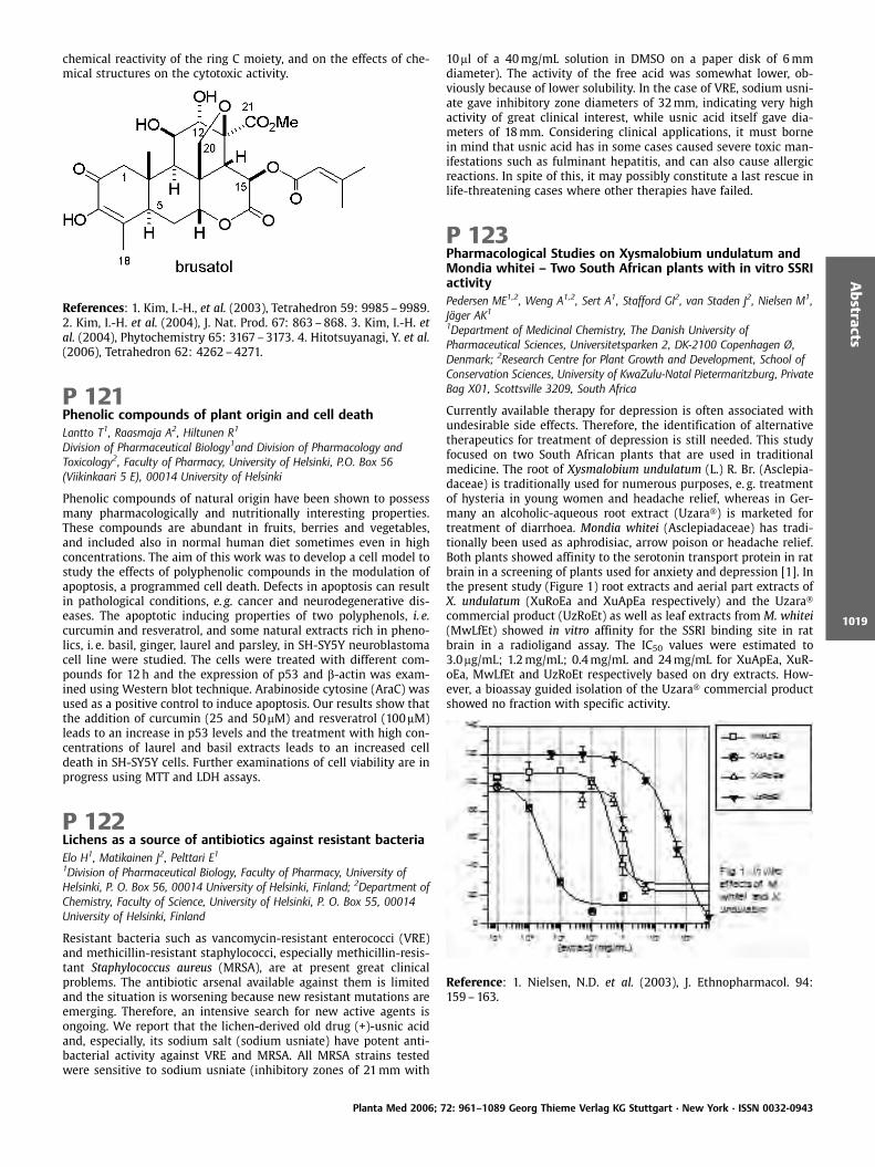
chemical reactivity of the ring C moiety, and on the effects of che-mical structures on the cytotoxic activity.
References: 1. Kim, I.-H., et al. (2003), Tetrahedron 59: 9985–9989.2. Kim, I.-H. et al. (2004), J. Nat. Prod. 67: 863–868. 3. Kim, I.-H. etal. (2004), Phytochemistry 65: 3167–3173. 4. Hitotsuyanagi, Y. et al.(2006), Tetrahedron 62: 4262–4271.
P 121Phenolic compounds of plant origin and cell deathLantto T1, Raasmaja A2, Hiltunen R1
Division of Pharmaceutical Biology1and Division of Pharmacology andToxicology2, Faculty of Pharmacy, University of Helsinki, P.O. Box 56(Viikinkaari 5 E), 00014 University of Helsinki
Phenolic compounds of natural origin have been shown to possessmany pharmacologically and nutritionally interesting properties.These compounds are abundant in fruits, berries and vegetables,and included also in normal human diet sometimes even in highconcentrations. The aim of this work was to develop a cell model tostudy the effects of polyphenolic compounds in the modulation ofapoptosis, a programmed cell death. Defects in apoptosis can resultin pathological conditions, e. g. cancer and neurodegenerative dis-eases. The apoptotic inducing properties of two polyphenols, i. e.curcumin and resveratrol, and some natural extracts rich in pheno-lics, i. e. basil, ginger, laurel and parsley, in SH-SY5Y neuroblastomacell line were studied. The cells were treated with different com-pounds for 12h and the expression of p53 and b-actin was exam-ined using Western blot technique. Arabinoside cytosine (AraC) wasused as a positive control to induce apoptosis. Our results show thatthe addition of curcumin (25 and 50mM) and resveratrol (100mM)leads to an increase in p53 levels and the treatment with high con-centrations of laurel and basil extracts leads to an increased celldeath in SH-SY5Y cells. Further examinations of cell viability are inprogress using MTT and LDH assays.
P 122Lichens as a source of antibiotics against resistant bacteriaElo H1, Matikainen J2, Pelttari E11Division of Pharmaceutical Biology, Faculty of Pharmacy, University ofHelsinki, P. O. Box 56, 00014 University of Helsinki, Finland; 2Department ofChemistry, Faculty of Science, University of Helsinki, P. O. Box 55, 00014University of Helsinki, Finland
Resistant bacteria such as vancomycin-resistant enterococci (VRE)and methicillin-resistant staphylococci, especially methicillin-resis-tant Staphylococcus aureus (MRSA), are at present great clinicalproblems. The antibiotic arsenal available against them is limitedand the situation is worsening because new resistant mutations areemerging. Therefore, an intensive search for new active agents isongoing. We report that the lichen-derived old drug (+)-usnic acidand, especially, its sodium salt (sodium usniate) have potent anti-bacterial activity against VRE and MRSA. All MRSA strains testedwere sensitive to sodium usniate (inhibitory zones of 21mm with
10ml of a 40mg/mL solution in DMSO on a paper disk of 6mmdiameter). The activity of the free acid was somewhat lower, ob-viously because of lower solubility. In the case of VRE, sodium usni-ate gave inhibitory zone diameters of 32mm, indicating very highactivity of great clinical interest, while usnic acid itself gave dia-meters of 18mm. Considering clinical applications, it must bornein mind that usnic acid has in some cases caused severe toxic man-ifestations such as fulminant hepatitis, and can also cause allergicreactions. In spite of this, it may possibly constitute a last rescue inlife-threatening cases where other therapies have failed.
P 123Pharmacological Studies on Xysmalobium undulatum andMondia whitei – Two South African plants with in vitro SSRIactivityPedersen ME1,2, Weng A1,2, Sert A1, Stafford GI2, van Staden J2, Nielsen M1,J ger AK1
1Department of Medicinal Chemistry, The Danish University ofPharmaceutical Sciences, Universitetsparken 2, DK-2100 Copenhagen Ø,Denmark; 2Research Centre for Plant Growth and Development, School ofConservation Sciences, University of KwaZulu-Natal Pietermaritzburg, PrivateBag X01, Scottsville 3209, South Africa
Currently available therapy for depression is often associated withundesirable side effects. Therefore, the identification of alternativetherapeutics for treatment of depression is still needed. This studyfocused on two South African plants that are used in traditionalmedicine. The root of Xysmalobium undulatum (L.) R. Br. (Asclepia-daceae) is traditionally used for numerous purposes, e. g. treatmentof hysteria in young women and headache relief, whereas in Ger-many an alcoholic-aqueous root extract (Uzara ) is marketed fortreatment of diarrhoea. Mondia whitei (Asclepiadaceae) has tradi-tionally been used as aphrodisiac, arrow poison or headache relief.Both plants showed affinity to the serotonin transport protein in ratbrain in a screening of plants used for anxiety and depression [1]. Inthe present study (Figure 1) root extracts and aerial part extracts ofX. undulatum (XuRoEa and XuApEa respectively) and the Uzaracommercial product (UzRoEt) as well as leaf extracts from M. whitei(MwLfEt) showed in vitro affinity for the SSRI binding site in ratbrain in a radioligand assay. The IC50 values were estimated to3.0mg/mL; 1.2mg/mL; 0.4mg/mL and 24mg/mL for XuApEa, XuR-oEa, MwLfEt and UzRoEt respectively based on dry extracts. How-ever, a bioassay guided isolation of the Uzara commercial productshowed no fraction with specific activity.
Reference: 1. Nielsen, N.D. et al. (2003), J. Ethnopharmacol. 94:159–163.
Planta Med 2006; 72: 961–1089 Georg Thieme Verlag KG Stuttgart · New York · ISSN 0032-0943
Abstracts
1019
P 124Two new triterpene saponins from Nylandtia spinosaLacaille-Dubois MA1, Mitaine-Offer AC1, Diome C1, Miyamoto T2,Delaude C3
1Laboratoire de Pharmacognosie, Unit de Mol cules d’Int rÞt Biologique,UMIB, UPRES EA-3660, Facult de Pharmacie, Universit de Bourgogne 7, Bd.Jeanne D’Arc, BP 87900, 21079 Dijon Cedex, France; 2Graduate School ofPharmaceutical Sciences, Kyushu University, Fukuoka, Japan; 3Centre deRecherche Phytochimique, Universit de Li ge, Institut de Chimie-B6, SartTilman, B-4000-Li ge, Belgique
In a continuation of our study on saponin constituents of medicinalplants of the Polygalaceae family [1 –3], we have examined thesaponin fraction of the roots of Nylandtia spinosa (L.) Dum.. Twonew triterpene saponins were isolated by successive MPLC oversilica gel. Their structures were established mainly by 600MHz 2DNMR techniques (COSY, TOCSY, NOESY, HSQC, HMBC) and massspectrometry as 3-O-ß-D-glucopyranosyl-presenegenin-28-O-ß-D-galactopyranosyl-(1?4)-[a-L-arabinopyranosyl-(1?3)]-ß-D-xylo-pyranosyl-(1?4)-[ß-D-apiofuranosyl-(1?3)]-a-L-rhamnopyrano-syl-(1?2)-ß-D-fucopyranosyl ester and 3-O-ß-D-glucopyranosyl-presenegenin-28-O-ß-D-galactopyranosyl-(1?4)-[a-L-arabinopyra-nosyl-(1?3)]-ß-D-xylopyranosyl-(1?4)-[a-L-rhamnopyranosyl-(1?2)-ß-D-fucopyranosyl ester. References: 1. Mitaine-Offer, A.-C.et al. (2005), Helv. Chim. Acta 88: 2986–2995. 2. Mitaine-Offer, A.-C. et al. (2003), Helv. Chim. Acta 86: 2404–2413. 3. Mitaine-Offer,A.-C. et al. (2002), J. Nat. Prod. 65: 553–557.
P 125New steroidal saponins from Asparagus acutifoliusLacaille-Dubois MA1, Sautour M1, Miyamoto T21Laboratoire de Pharmacognosie, Unit de Mol cules d’Int rÞt Biologique,UMIB, UPRES-EA 3660, Facult de Pharmacie, Universit de Bourgogne, 7, Bd.Jeanne d’Arc, BP 87900, 21079 Dijon Cedex, France; 2Graduate School ofPharmaceutical Sciences, Kyushu University, Fukuoka, Japan
Asparagus acutifolius L. belongs to the Liliaceae family in which thesteroidal saponins are fairly widespread. A survey of the literatureshowed that several Asparagus species have already been chemicallystudied and found to contain bioactive steroidal saponins. As part ofour ongoing search for biologically active steroid saponins as potentantifungal agents [1 –3] a phytochemical investigation of the rootsof A. acutifolius has led to the isolation by several chromatographicsteps on normal and reversed phase silica gel of three new steroidalglycosides. Their structures were determined by spectroscopicmethods including 1D- and 2D-NMR (COSY, TOCSY, HSQC andHMBC) and FAB-MS as (25S)-3b,5b,22a-22-methoxyfurostan-3,26-diol 3-O-b-D-xylopyranosyl-(1?2)-[b-D-xylopyranosyl-(1?4)]-b-D-glucopyranosyl 26-O-b-D-glucopyranoside [1], (25S)-5b-spiro-stan-3b-ol 3-O-b-D-xylopyranosyl-(1?2)-[b-D-xylopyranosyl-(1?4)]-b-D-glucopyranoside [2] and (25S)-5b-spirostan-3b-17a-diol 3-O-b-D-xylopyranosyl-(1?2)-[b-D-xylopyranosyl-(1?4)]-b-D-glucopyranoside [3]. In addition, the antifungal activity of thesecompounds was tested against three human pathogenic yeasts(Candida albicans, C. glabrata and C. tropicalis). Spirostanol saponinspresented antifungal activity with MICs values between 12.5 and50mg/mL whereas furostanol compounds were inactive. Refer-ences: 1. Sautour, M. et al. (2004), Planta Med. 70: 90–92. 2. Sau-tour, M. et al. (2004), Chem. Pharm. Bull. 52: 1353–1355. 3. Sautour,M. et al. (2005), J. Nat. Prod. 68: 1489–1493.
P 126Hypolipidemic and antioxidant effect of Ajuga iva in rats feda high-cholesterol dietLacaille-Dubois MA3, Chenni A1, Yahia DA1, Boukortt FO1, Prost J2,Bouchenak M1
1Laboratoire de Nutrition Clinique et M tabolique, Facult des Sciences,Universit d’Oran Es S nia, 31000, Alg rie; 2UPRES Lipides- Nutrition EA2422. 6, Bd Gabriel, Universit de Bourgogne, 21000 Dijon, France;3Laboratoire de Pharmacognosie, Unit de Mol cules d’Int rÞt Biologique,UMIB UPRES EA 3660, Facult de Pharmacie, Universit de Bourgogne, BP87900, 21079 Dijon Cedex, France
Ajuga iva (L.) Schreb. has been reported to have a variety of biolo-gical effects including hypoglycemic, vasorelaxant, hypolipidemic,antiinflammatory, antifungal, antimicrobial and anthelmintic activ-ity [1, 2]. The present study explores the possible antioxidant andhypolipidemic effects of the aqueous extract of Ajuga iva (Ai 0.5% inthe diet) in rats fed a high-cholesterol (1%) diet (HCD). The resultsindicated that the HCD-Ai vs. HCD treatment led to many changes inbiochemical parameters. They showed a decrease by 18% of plasmatotal cholesterol (TC) and by 29% VLDL-cholesterol but an increaseby about 35% of HDL2-cholesterol. The triacylglycerol contents werereduced by 31% in plasma and 74% in VLDL. The lipid peroxidationdetermined by TBARS was decreased by 75% in plasma. TBARS inliver, heart and kidneys were highly reduced excepted in the adiposetissue. A.i. treatment enhanced superoxide dismutase activity inliver and kidney. Glutathione reductase activity was lowered in adi-pose tissue but increased in liver and in kidney. A significant in-crease was noted in glutathione peroxidase activity in liver, heartand kidney but a low value in adipose tissue was observed. In con-clusion, this study demonstrates that in addition to its potent TGand TC-lowering effects, Ai is effective in improving the antioxidantstatus by reducing lipid peroxidation in plasma and tissues andenhancing the antioxidant enzymes in HCD fed rats. A phytochem-ical screening indicated the presence of flavonoids and terpenoids.Their isolation and characterization are currently in progress. Refer-ences: 1. El Hilaly, J. et al. (2006), J. Ethnopharmacol. 105: 441–448.2. Bondi, M.L. et al. (2000) Biochem. Syst. Ecol. 28: 1023–1025.
P 127ChemGPS-NP – tuned for navigation in biologically relevantchemical spaceLarsson J1, Gottfries J2, Muresan S3, Bohlin L1, Backlund A1
1Division of Pharmacognosy, Department of Medicinal Chemistry, BMC,Uppsala University, Box 574, S-751 23 Uppsala, Sweden; 2Department ofMedicinal Chemistry, AstraZeneca R&D M lndal, S-431 83 M lndal Sweden;3GDECS Computational Chemistry, AstraZeneca R&D M lndal, S-431 83M lndal, Sweden
Natural compounds have a unique chemical diversity occupying adifferent and larger space than that normally dealt with in medic-inal chemistry [1, 2]. They are evolutionary selected and pre-vali-dated by Nature with explicit biological activities, making themhighly interesting for development of new drug lead candidates.Chemical space is a multi-dimensional region defined by the de-scriptors chosen to describe a set of chemical compounds [3]. Ofutmost importance for discovery of new active compounds for fu-ture therapies is the identification and charting of a biologicallyrelevant chemical space and a primary key to this is the coverageof the diverse natural product chemical space [1, 2, 4]. A map ofchemical space can be constructed by applying the same principlesas the Mercator convention in geography. Rules are correspondingto dimensions (e. g. longitude and latitude), and structures are cor-responding to objects (e. g. cities and countries) [5]. The rules andobjects together present the chemical space map, where the coordi-nates are t-scores from principal component analysis [6]. Here wepresent ChemGPS-NP, a new tool tuned for exploration of biologi-cally relevant natural products chemical space, forming a frame-work for making compound comparison and selection more effec-
Planta Med 2006; 72: 961–1089 Georg Thieme Verlag KG Stuttgart · New York · ISSN 0032-0943
Abstracts
1020
tive and increasing probability of hit generation when screening thevast diversity of natural products in the search for novel bioactivemolecules. References: 1. Larsson J. et al. (2005), J. Nat. Prod. 68:985–991. 2. Feher, M., Schmidt, J.M. (2003), J. Chem. Inf. Comput.Sci. 43: 218–227. 3. Dobson, C.M. (2004), Nature 432: 824–828. 4.Koch, M.A. et al. (2005), Proc. Natl. Acad. Sci. USA 102: 17272–17277. 5. Oprea, T.I., Gottfries, J. (2001), J. Comb. Chem. 3: 157–166. 6. Jackson, J.E. (1991), A users guide to principal components,Wiley, New York.
P 128Sustainable use of the Brazilian biodiversity: Chemical andpharmacological prospection on higher plantsWagner V1, Monteiro Souza Brito AR2 et al1S¼o Paulo State University -Instituto de Qu mica de Araraquara. CP 355, CEP14801 – 970, Araraquara, S¼o Paulo, Brazil; 2Campinas State University –Instituto de Biologia, Campinas, S¼o Paulo, Brazil
Our research deals with the investigation of plant species with anti-ulcer, anti-oxidant, analgesic, immuno-stimulating, genotoxic, anti-inflammatory and antimicrobial activities. We have investigatedseveral plant species, like Davilla elliptica, Strychnos pseudoquinaand Byrsonima fagifolia. We performed the aforementionated biolo-gical assays and then we fractionate the active extracts in order torecognize their chemical composition, using chromatographic andspectrometric techniques. D. elliptica presented activity against My-cobacterium tuberculosis (MIC 62.5 ug/mL); S. pseudoquina present-ed moderate activity against gastric ulcers, which might be due tothe presence of an indole alkaloid whose structure roughly resem-bles that of omeprezole; B. fagifolia presented significant activityagainst gastric ulcers. Therefore, Brazilian plants may be used as apotential source for compounds with biological activity. Acknowl-edgements: Biota-Fapesp Program, CNPq. Acknowledgements: Biota-Fapesp Program, CNPq
P 129Characterization, design and synthesis of potential COX-2inhibitors based on natural productsJachak SMDepartment of Natural Products, NIPER, SAS Nagar 160 062, Punjab
Synthetic COX-2 inhibitors (such as rofecoxib and other coxibs) havehigh selectivity but at the same time they are associated withthrombotic cardiovascular problems [1]. Thus, the use of COX-2inhibitors still remains controversial and it represents challengefor the pharmaceutical industry to develop improved anti-inflam-matory drugs devoid of severe side effects. Natural product-derivedcompounds are better candidates for lead identification and optimi-zation in drug discovery process due to their great structural diver-sity which is not commonly seen in synthetic compounds. We areengaged in the characterization of natural COX-2/COX-1 inhibitorsthrough bioassay-directed fractionations from medicinal plants [2,3] and design, synthesis of natural product-derived analogues aspotential COX-2 inhibitors e. g. curcumin and chalcone [4, 5]. Incontinuation of our efforts to discover COX inhibitors, design andsynthesis of isoflavone analogues based on naturally-derived isofla-vones are described herein. References: 1 Schror, K., Mehta, P.,Mehta, J. L. (2005), Cardiovasc. Pharmacol. Ther. 10: 95–101. 2.Selvam, C., Jachak, S. M. et al. (2004), Tet. Lett. 45: 4311–4314. 3.Selvam, C., Jachak, S. M. J. (2004), J. Ethnopharmacol. 95: 209–212.4. Selvam, C., Jachak, S. M. et al. (2005), Bioorg. Med. Chem. Lett. 15:1793–1797. 5. Jachak, S. M. (2006), Curr. Med. Chem., 13, 659–678.
P 130Extracts of Salvia officinalis from different growing areasand their antiviral effect against enveloped andnon-enveloped virusesNolkemper S1,2, Schnitzler P2, Keppler OT2, Reichling J11Institute for Pharmacy and Molecular Biotechnology, Department of Biology,University of Heidelberg, ImNeuenheimer Feld 364, 69120 Heidelberg,Germany; 2Hygiene Institute, Department of Virology, University ofHeidelberg, Im Neuenheimer Feld 324, 69120 Heidelberg, Germany
In our study we compared extracts from Salvia officinalis L. (sage),which was grown at two different areas in Germany, for their anti-viral effect. Firstly it was cultivated in an dry and hot area (area 1)and secondly in an area with a cooler climate and more rain (area 2).Different extracts were made from both samples with water/etha-nol- mixtures in the ratio of 0, 20, 40, 60 and 80% ethanol, respec-tively. A HPLC analysis of the extracts showed as major componentsapigenin-7-glucuronide, luteolin- 3-glucuronide and rosmarinicacid. The antiviral effects of these extracts were tested against theenveloped herpes simplex virus type 1 (HSV-1) and the non-envel-oped adenovirus type 5. In order to determine the mode of antiviralaction, the extracts were added to the cells or viruses at differenttimes during infection. The inhibitory activity against HSV-1 wastested using a plaque reduction assay. Adenoviral GFP reporter geneexpression allowed for a flow cytometry-based quantification of in-fection levels. When HSV-1 was pretreated with the extracts prior toadsorption, plaque formation was reduced by >90%. The best resultswere shown by the extracts from area 1. In time-response studiesover a period of 2 hours, a clearly time-dependent activity for allextracts was demonstrated. Already after 20 minutes of incubationof HSV-1 with the extracts, an antiviral activity of about 70–80%was shown. Pretreatment of adenovirus with the extracts for 1 hourshowed an inhibitory effect on adenoviral infection from 20% (80%ethanol) up to 70% (20% ethanol). Therefore the extracts show anti-viral activity independent of an envelope.
P 131Characterisation of in vitro antioxidative properties ofaqueous ethanolic (45%v/v) extract of Lemon Balm (Melissaofficinalis L.)Dastmalchi K1, Dorman HJD1, Darwis Y2, Hiltunen R1
1Faculty of Pharmacy, Division of Pharmaceutical Biology, University ofHelsinki, P.O. Box 56 (Viikinkaari 5E), FIN-00014, Finland; 2School ofPharmaceutical Sciences, Universiti Sains Malaysia, Minden, 11800 Penang,Malaysia
The plant Melissa officinalis (L.) has been used in European tradi-tional system of medicine for its cognitive enhancing properties.Based on its traditional use and cholinergic properties, this plantwas recently assessed for its clinical efficacy in Alzheimer’s Disease(AD) in a trial conducted by the Institute of Medicinal Plant (IMP),Tehran, Iran [1]. The plant was found to be effective in the manage-ment of mild to moderate AD patients. Since it has been proposedthat oxidative stress plays a cardinal role in the pathogenesis of thedisease [2], we wanted to investigate if the efficacy of the plantextract in the clinical trial is due to its antioxidative properties.Therefore based on the IMP research M. officinalis was extractedby a similar procedure using the same solvent (45% v/v ethanol)and plant material (cultivar). The method of extraction was MediumPressure Solid Liquid Extraction. The total phenol content was esti-mated as gallic acid acid equivalents using Folin-Ciocalteu reagentmethod and the in vitro antioxidant activities assayed were iron (III)reduction, iron (II) chelation, 1,1-diphenyl-2-picrylhydrazyl, 2,2’-azi-nobis(3-ethylbenzothiazoline-6-sulphonate) free radical scavengingactivities, inhibiton of b-carotene-linoleic acid bleaching. The ex-tract demonstrated activity in all the assays, however it was not aspotent as the positive controls except in the b-carotene-linoleic acidbleaching assay where its antioxidant activity was superior to thatof gallic and caffeic acid and statistically indistinguishable from
Planta Med 2006; 72: 961–1089 Georg Thieme Verlag KG Stuttgart · New York · ISSN 0032-0943
Abstracts
1021

quercetin and butylated hydroxyanisole. Acknowledgements: Insti-tute of Medicinal Plants, Iranian Academic Center for Education,Culture and Research, Tehran, Iran References: 1. Akhondzadeh, S.et al. (2003), J. Neurol. Neurosurg. Psychiatry 74: 863–866. 2. Var-adarajan, S. et al. (1999), J. Struct. Biol. 309: 746–768.
2. Recent Advances in Analysis of SecondaryMetabolites
P 132New Apigenin Triosides From The Seeds of SyzygiumaromaticumNassar MIANatural Compounds Chemistry Department, National Research Centre, Dokki12622, Cairo, Egypt
Syzygium aromaticum (L.) Merr. & Perry belongs to the family Myr-taceae. S. aromaticum buds (clove) are used in folk medicine asdiuretic, odontalgic, stomachic, tonicardiac, aromatic condimentand condiment with carminative and stimulant properties [1]. Com-pounds isolated from S. aromaticum have been found to possessgrowth inhibitory activity against oral pathogens [2]. The aim of thiswork is the isolation and structural elucidation of flavonoid glyco-sides from the seeds of S. aromaticum. The crushed seeds were sub-jected to successive extraction using n-hexane, dichloromethaneand ethanol (70%). The aqueous ethanol extract was subjected tocellulose column chromatography. The eluted polar fractions werefurther chromatographed on Sephadex LH-20 columns to give twonew apigenin triglycosides, apigenin 6-C-[ b-D-xylopyranosyl-(1?2)- b-D- galactopyranoside]-7-O-D-glucopyranoside (1) and api-genin 6-C-[b-D-xylopyranosyl-(1?2)-D-galactopyranoside]-7-O- b-D-(6-O-p-oumarylglucopyranoside) (2). The structures of the newcompounds were elucidated by chemical and spectral analysis in-cluding UV, FABMS, 1H, 13C NMR, DEPT, HMQC, HMBC and NOESY.
References: 1. Boulos, L. (1983), Medicinal Plants of North Africa.Ref. Publications Inc., Michigan. 2. Cai, L. et al. (1996), J. Nat. Prod.59: 987–990.
P 133Quantitative method development for measurement ofMaesa lanceolata saponins by LC-MSPieters L, Theunis M, Apers S, Vlietinck ALaboratory of Pharmacognosy and Phytochemistry, Department ofPharmaceutical Sciences, University of Antwerp, Universiteitsplein 1; B-2610Antwerp, Belgium
Triterpene saponins are a class of plant natural products with a widerange of bioactivities, which make them an interesting researchsubject. The small tree Maesa lanceolata, growing in African coun-tries, is used in traditional medicine against various diseases. Inprevious work a triterpenoid saponin mixture was isolated fromthe leaves of Maesa lanceolata and the compounds were identified[1, 2]. The compounds showed virucidal, haemolytic, molluscicidaland anti-angiogenic activity [3, 4]. Here we report the developmentof a quantitative LC-MS method to analyse saponin compounds incrude root and leaf extracts of Maesa plants. The crude extract isfirst purified on a C18ec SPE column. Then the compounds are sep-arated on a reversed phase C18 column (Grace Vydac, 3.2mm x250mm –300 ) with a H2O/ACN (0.06% formic acid) gradient be-fore analysis with mass spectrometry (Bruker Daltonics Esquire3000 plus). This method will be used to screen plants whether ornot induced by methyl-jasmonate, or plants such as Medicago trun-catula transformed with genes involved in saponin biosynthesis, forthe presence of Maesa saponins. References: 1. Apers, S. et al.(1998), J. Pharm. Biomed. Anal. 18: 737. 2. Apers, S. et al. (1999),Phytochemistry 52: 1121. 3. Apers, S. et al. (2001), Planta Med. 67:528. 4. Apers, S. et al. (2002), J. Pharm. Belg. 57, Hors-s rie 1: 47.
P 134Proanthocyanidins from the herb of Myrothamnusflabellifolia WelwPetereit F, Anke J, Engelhardt C, Hensel AUniversity of M nster, Institute of Pharmaceutical Biology andPhytochemistry, Hittorfstr. 56, D-48149 M nster, Germany
Myrothamnus flabellifoliaWelw. (Myrothamnaceae), a species grow-ing in arid areas of Southeastern Africa, has the ability to surviveregular periods of extreme dehydration to an air-dry-state (resurrec-tion plant). In continuation of our previous results [1] we investigatethe proanthocyanidin pattern from an aqueous acetone extract inmore detail. Chemical investigation of the ethylacetate soluble frac-tion has led to the isolation and characterization of epicatechin,epigallocatechin, epicatechin-3-O-gallate and epigallocatechin-3-O-gallate as flavan-3-ol precursors. The dimeric proanthocyanidinfraction consists exclusively of procyanidins, partly substituted withgallic acid or p-hydroxybenzoic acid. A range of ten different pro-cyanidins were identified by extensive 2D NMR studies of the per-acetylated derivatives beside the known 3,4,5-tri-O-galloylquinicacid [2]. In addition, the more abundant polymeric proanthocyani-din fraction was also isolated and its chemical constitution charac-terized by 13C NMR and optical rotation. The results are discussedbriefly. Acknowledgement: Myro AG, CH-8606 Greifensee, switzer-land, for financial support. References: 1. Deters, A. et al. (2005),53rd Annual Congress of the Society for Medicinal Plant Research,Florence, P 130 (poster abstract). 2. Moore, J. et al. (2005), Biochem.J. 385: 301–308.
Planta Med 2006; 72: 961–1089 Georg Thieme Verlag KG Stuttgart · New York · ISSN 0032-0943
Abstracts
1022
P 135Air-transport alters the composition of essential oils inaromatic plantsJ ger AK1, Rasmussen HB1, van Staden J21Department of Medicinal Chemistry, The Danish University ofPharmaceutical Sciences, Universitetsparken 2, DK-2100 Copenhagen O,Denmark; 2Research Centre for Plant Growth and Development, University ofKwaZulu-Natal, P/Bag X01, Scottsville 3209, South Africa
A previous study has shown that aromatic plant material trans-ported on a truck could lose all the essential oil during a half hourdrive [1]. In our collaborative work, plant material is often air-freighted between continents, and it is suspected that the qualityof the material is altered due to the reduced pressure in the cargohold of an aircraft. Aerial parts of the aromatic plants Artemisia afraJacq. ex Wild., Mentha longifolia (L.) Huds., Ocimum basilicum L. andSalvia africana L. were harvested in South Africa. Essential oils wereprepared in South Africa from the fresh plant material and frommaterial dried at 40¤C for 2 days. The oils were sealed in ampulesand sent by air-freight to Denmark, along with portions of the driedplant material. In Denmark, essential oils were distilled from the air-freighted material and all the oil samples analysed by GC-MS. Themost pronounced changes occurred in S. africana where 60–75% ofthe monoterpenes were lost from the air-freighted material. In com-parison, the sesquiterpene content did not change much for S. afri-cana. Overall, there seemed to be a quantitative loss of 10–25% ofthe monoterpenes in all the oils. Consequently, it is necessary to becareful about the interpretation of work, both chemical and biolo-gical, done on essential oils from aromatic plants that have not beenprocessed locally before shipping. Reference: 1. Webber, L.N. et al.(2000), S. Afr. J. Plant Soil 17: 10–14.
P 136Phytochemical Study of Artemisia persica Boiss. andEvaluation of its Antiplasmodial ActivitySadeghpour O1, Asghari G2, Ardekani MRS3, Jaroszewski JW4
1Herbal Medicine Dept.Research institute of Medical History,Islamic andComplementary Medicine Pirnia, Lalezar now Jomhoori Tehran IRAN;2Pharmacognosy Dept.Faculty of Pharmacy Isfahan Univrsity of MedicalSciences Hezarjerib St. Isfahan IRAN; 3Pharmacognosy Dept.Faculty ofPharmacy Tehran Univrsity of Medical Sciences Enghelab St. TehranIRAN.4Medicinal Chemistry Dept.Pharmaceutical University of DenmarkUniversitetsparken 2 DK-2100 Copenhagen Denmark
Introduction: The genus Artemisia is one of the largest and mostwidely distributed of the nearly 100 genera in the tribe Anthemi-deae of the Asteraceae. Considering to different compounds exist inArtemisia spp. They have a wide variety of biological activity. Theoutstanding antimalarial compound, artemisinin, isolated from Ar-temisia annua L.. The aim of this study was phytochemical investi-gation of “Artemisia persica“ in order to identify bioactive com-pounds, which may have antiplasmodial effect [1, 2]. Methods:The Flowering aerial parts of plant were collected in November2002 from Isfahan province (IRAN), and undergoes to chemical iso-lation of compounds by VLC, CC and HPLC, mainly using bioactivity-guided fractionation approach, and the structures of isolated com-pounds were determined using modern homo-and heteronucleartwo-dimentional NMR experiments. The essential oil of A. persicawas obtained by hydrodistillation and analyzed by GC/MS. Results:Methanolic extract of A. persica showed a moderate effect againstthe Plasmodium parasite compared Chloroquine. The yield of essen-tial oil was 0.40% Davanone, as the major constituent. Phytochem-ical investigation of the extract led to isolation of Friedelin, Ascar-idol, Scopoletin, Scopolin and two other unknown compounds. Dis-cussion: Davanone was the major constituent of A. persica while theother studies indicated that the major constituent of volatile oil is1,8-Cineol. The results showed that although isolated compoundslike Friedelin, Ascaridol, Scopoletin and etc. might have differentbiological effect but not high antiplasmodial. However comparing
with artemisinin the compounds Ascaridol and Friedelin (speciallyits derivative, ß-Amyrin) showed week antiplasmodial effect. Inconclusion there is improbable to find major compounds with highantimalarial effect in Artemisia persica or other species of Artemisia.Acknowledgement: I would like to give my best gratefulness to MajidSairafianpour for all his efforts and kindnesses in Copenhagen. Re-ferences 1.Marco, J.A., Barbera, O., (1990), Natural Products fromGenus Artemisia L., in Studies in Natural Products Chemistry, Atta-ur-Rahman ed., London, Elsevier, Vol.7, 201–264. 2. Wright, C.W.(2002), Artemisia, New York: Taylor & Francis Inc..
P 137Induction of naphthoquinone and flavonoid production inDionaea muscipula and Drosera capensisKrolicka A1, Szpitter A1, Gilgenast E2, Romanik G2, Kaminski M2,Lojkowska E11Department of Biotechnology UG & AMG, Kladki 24, 80 – 822 Gdansk,Poland; 2Technical University of Gdansk, Chemical Faculty, AnalyticalChemistry Department, Narutowicza 11/12, 80 – 952 Gdansk, Poland
The secondary metabolites (naphthoquinones: plumbagin and ra-mantaceone, flavonoids: myricetinand quercetin) from Droseraceaeplants: Dionaea muscipula and Drosera capensis ’Broadleaf’ are usedas anticancer drugs and antispasmodic agents. The aim of the studywas to check the ability of the biotic elicitors to induce the produc-tion of secondary metabolites in in vitro grown D. muscipula and D.capensis ’Broadleaf’. The optimal conditions for micropropagation ofboth species were described as: 0.75% agar solidified MS mediumwith 25mg/l ascorbic acid and 2% sucrose, pH 5.6. Autoclaved over-night suspension of Agrobacterium rhizogenes and a crude elicitorfrom Verticillium dahliae Kleb. were added to MS medium aselicitors, to the final concentration of 2.5% and 0.2 –0.4mg%, re-spectively. A 4–6-week-old plantlets were planted on these mediaand after 30 days of growth they were collected and the extractionof naphthoquinones and flavonoids was performed. The extractionwas carried out in an ultrasonic bath Sonic-5. Quantitative and qua-litative determination of naphthoqinones and flavonoids in chloro-form and methanol extracts was performed by using NP – HPLC/UV-DAD. Dihydroksypropyl stationary phases, hexane and tetrahydro-furan mixture as eluent and gradient elution were used. HPLC anal-ysis of chloroform extracts indicated 3 times higher accumulation ofplumbagin in D. muscipula plants elicited with A. rhizogenes than inthe control ones. In D. capensis elicited with A. rhizogenes about100% increase of ramantaceone was determined. Also increase ofmyricetin and quercetin content was observed after biotic elicita-tion. Chloroform and methanol extracts obtained from A. rhizogeneselicited D. muscipula and D. capensis plants show antimicrobial ac-tivity. Methanol extracts of D. muscipula exhibit the strongest anti-microbial activity against a broad spectrum of tested human patho-genic bacteria: Enterococcus faecalis, Staphylococcus aureus, Klebsiel-la pneumoniae and Pseudomonas aeruginosa; MBC from 25 to 75mgfw/mL. Acknowledgements: State Committee for Scientific Research,Grant No KBN 0430/P04/2004/26.
P 138Exploring the structural diversity of myxobacterialsecondary metabolismOhlendorf B, Erol , Krick A, Kehraus S, K nig GMInstitute for Pharmaceutical Biology, University of Bonn, Nussallee 6, D-53115 Bonn, Germany, [email protected]
Myxobacteria are gram-negative bacteria commonly found in soil.They form swarm-like colonies which spread over surfaces and feedon other microorganisms. The most outstanding feature of myxo-bacteria is their ability to form fruiting bodies by which the differ-ent genera can be distinguished. In the last years myxobacteria havebeen in the focus of natural products research and have becomeknown as potent producers of secondary metabolites. To date more
Planta Med 2006; 72: 961–1089 Georg Thieme Verlag KG Stuttgart · New York · ISSN 0032-0943
Abstracts
1023
than fifty unique structural types have been isolated, among themcompounds with promising biological activities, e. g. the epothilones[1] which are now in clinical studies as anticancer drugs. In ourscreening program the myxobacterial strain 150 (morphologicallycharacterized as a Polyangium or Nannocystis sp.) was singled outdue to the results of TLC, LC-MS and NMR analyses. The next stepwas the cultivation of the strain in a large scale and subsequentextraction and fractionation. HPLC separation eventually yieldedcompound 150E, a new metabolite with an unusual ethyl residueconnected to a polyunsaturated carbon chain. The structure waselucidated by applying mass spectrometry and different 1D- and2D- NMR techniques. Future work will address the biosynthesisand evaluation of the bioactivity of the compound.
Reference: 1. Gerth, K. et al. (1996), J. Antibiot. 49: 560–563
P 139New cyclic peptides from the cyanobacterium Tychonema spMehner C, Kehraus S, Krick A, M ller D, K nig GMInstitute for Pharmaceutical Biology, University of Bonn, Nußallee 6, D-53115Bonn, Germany, [email protected]
Cyanobacteria are an amazing source of new compounds of phar-maceutical interest. A specific feature of their secondary metabolitespectrum is the occurrence of cyclic peptides containing unusualamino acids. Our project focuses on a group of bioactive cyclic pep-tides, which are present in the methanolic extract of the cyanobac-terium Tychonema sp. This microorganism was isolated from a fieldsample collected from a pond of a sugar factory near Braunschweig(Germany). To date the new cyclic hexapeptides brunsvicamides A-Cwere identified. Brunsvicamide C contains a unique N-methylated-N’-formyl-kynurenine moiety 1 [1]. The brunsvicamides are relatedto the sponge-derived mozamides.
1
Furthermore two cyclic peptides with [MH+]= 1486 and [MH+]= 1456were isolated. The structure elucidation of these peptides is stillongoing using a combination of NMR methods and MS/MS spectro-scopy. Reference: 1. K nig, G.M., Kehraus, S. et al. (2006), ChemBio-Chem 7: 229–238.
P 140Safety Assessment and Metabolic Fingerprinting of GMOGerberasPohjala L1,2, Tammela P1,2, Ainasoja M3, Somervuo P3, Teeri T3, Vuorela P41Drug Discovery and Development Technology Center, Faculty of Pharmacy,P.O. Box 56, 00014 University of Helsinki, Finland; 2Division ofPharmaceutical Biology, Faculty of Pharmacy, University of Helsinki, Finland;3Department of Applied Biology, Faculty of Agriculture and Forestry,University of Helsinki, Finland; 4Pharmacy, Faculty of Mathematics andNatural Sciences, bo Akademi University, Turku, Finland
Genetically modified organisms (GMOs) have raised concerns ingeneral public and involve also subjects of scientific interest, as itis still not exactly known how single genes brought about by meansof genetic engineering affect other genes and thus the metabolismof the plant. In this work, effects of 228 GMO Gerbera hybrida linesand 42 traditional gerbera varieties on human gastrointestinalepithelium Caco-2, bronchial epithelium Calu-3 and hepatocellularHuh-7 cell lines were assessed using WST-1 cell viability assay [1].After collection, the inflorescence gerbera samples were freeze-dried, ground and extracted with methanol. The cell viability assayswere performed in automated environment by exposing the cellcultures to 40mg/mL and 100mg/mL extracts for 24 hours. < 80%and >120% threshold values were set to mark significant effectson cell viability. The statistical analysis of the frequencies of hitextracts found no differences between GMO and non-GMO lines inany of the cell lines used. In addition to the cell viability testing, themetabolic fingerprinting of the extracts was performed with TLC.The principal component analysis did not separate GMO lines fromnon-GMO lines, whereas the nearest neighbour classifier methodfound the right neighbour in 46.3% of samples when 42 differenttransgenic groups were formed. These results indicate that eventhough some metabolic differences between traditional and GMOgerbera lines may exist, these differences seem not to affect humancell viability in vitro. Reference: 1. Ishiyama, M. (1995), In VitroToxicol. 8:187–190.
P 141Soyabean Lipoxygenase Inhibitory Activity of Flavonoids,Phenylethanoid glycosides and phenolic acids fromMarrubium velutinum and M. cylleneumKarioti A1, Hadjipavlou-Litina D2, Skaltsa H1
1Department of Pharmacognosy & Chemistry of Natural Products, School ofPharmacy, Panepistimiopolis-Zografou, 15771 Athens, Greece; 2Departmentof Pharmaceutical Chemistry, School of Pharmacy, Aristotelian University ofThessaloniki, Thessaloniki, 54124, Greece
Marrubium sp. are widely used in traditional medicine in Greece incases of inflammatory, bronchial, stomach diseases, coughs and cat-arrhs of the respiratory tract. In previous papers, we reported theisolation and identification of secondary metabolites from themethanol extracts from the aerial parts of M. velutinum Sm. andM. cylleneum Boiss. et Heldr. [1, 2]. In a continuation of our chemicaland biological investigations on Marrubium species of the Greekflora, we studied the isolated phenolic compounds in vitro for theirinteraction with 1,1-diphenyl-2-picryl-hydrazyl (DPPH) stable freeradical and for their inhibitory activity against soybean lipoxygen-ase. The conversion of sodium linoleate to 13-hydroperoxylinoleicacid at 234 nm was recorded and compared with the appropriatestandard inhibitor. The initial methanol extracts and 25 secondarymetabolites have been tested. Phenylethanoid glycosides werefound to be active. References: 1 Michelis, F., Tiligada, E. et al.(2002), Pharm. Biol. 40: 245–248. 2. Karioti, A. et al. (2003), Phy-tochemistry 64: 655–660.
Planta Med 2006; 72: 961–1089 Georg Thieme Verlag KG Stuttgart · New York · ISSN 0032-0943
Abstracts
1024
P 142Novel cytotoxic labdane diterpenes from MarrubiumcylleneumKarioti A1, Skopeliti M2, Heilmann J3, Tsitsilonis O2, Skaltsa H1
1Department of Pharmacognosy & Chemistry of Natural Products, School ofPharmacy, Panepistimiopolis-Zografou, 15771 Athens, Greece; 2Departmentof Animal and Human Physiology, School of Biology, Panepistimiopolis-Zografou, 15771 Athens, Greece; 3Institute of Pharmacy, Department ofPharmaceutical Biology, University of Regensburg, Universit tsstrasse 31, D-93040, Regensburg, Germany
In continuation of our phytochemical investigations into Marrubiumspecies of the Greek flora [1], we report on the isolation and identi-fication of further novel secondary metabolites from the dichloro-methane extract from the aerial parts ofMarrubium cylleneum Boiss.& Heldr.. One labdane diterpene (1) and one labdane nor-diterpene(2) have been isolated along with b-sitosterol and palmitic acid. Thestructures of the isolated compounds were established by means of1D & 2D NMR and MS spectral analyses. Both new and previouslyisolated diterpenes were tested for their immunomodulating poten-tial in standard in vitro cytotoxicity assays [2]. Peripheral bloodmononuclear cells (PBMC) from normal donors and cancer patientswere isolated and subsequently incubated with low concentrationsof each compound for 1–3 days. Effectors PBMC were further as-sayed for enhancement of their lytic ability against 51Cr-labeledtarget cells (K562, Daudi and Jurkat) at effect or to target ratiosvarying between 10–80:1. Some of these compounds could be po-tentially used to enhance PBMC anticancer activity, as they enhanceimmune responses of human lymphocytes, inducing cell prolifera-tion and augmenting their cytotoxicity against tumor targets,whereas at the same time they present significant antitumor activ-ity, efficiently lysing leukemic cells.
1
2
References: 1. Karioti, A. et al. (2005), Phytochemistry 66: 1060–1066. 2. Tsavaris, N.B. et al. (2004), Oncology 67: 403–410.
P 143Extraction and chromatographic analysis of ginsenosidesoccurring in roots and leaves of Panax quinquefolium grownin PolandLudwiczuk A1, Wolski T1,2, Głowniak K1
1Department of Pharmacognosy with Medicinal Plant Laboratory,Skubiszewski Medical University, 1 Chodzki Street, 20 – 093 Lublin, Poland;2Department of Vegetable and Medicinal Plant, University of Agriculture, 58Leszczynski Street, 20 – 068 Lublin, Poland
A wide variety of extraction methods has been employed for theisolation of ginsenosides from plant material. They are e. g.: extrac-tion in Soxhlet apparatus [1], ultrasound-assisted extraction (UAE)[2], microwave-assisted extraction (MAE) [3], pressurized liquid ex-traction (PLE, ASE) [4], and supercritical fluid extraction (SFE) [5]. Inpresented studies, for isolation of ginsenosides from ginseng culti-vated in Poland, three different extraction methods such as: ASE,UAE and mechanical shaking assisted solvent extraction was ap-plied. The separation of compounds was achieved with water-acet-onitrile gradient system using a C18 reversed-phase column. Thehighest extraction efficiency of ginsenosides in roots and leaves ofPanax quinquefolium L. was observed during mechanical shakingwith 50% aqueous methanol (total concentration for roots –4.3%and for leaves –9.1%). Extraction efficiency of ginsenosides by ASE iscomparable to that obtained by sonication. The main compound ofP. quinquefolium roots is ginsenoside Rb1. The amount of this com-pound in ginseng roots was ranged from 1.36% to 3% according toextraction method and extraction solvent used. Ginsenosides Rd,Rg2 and Rb2 are the main compounds occurring in American gin-seng leaves. They content in raw material after mechanical shakingwas 3.8%, 2.4% and 1.4% respectively. Ginseng leaves, in comparisonto ginseng roots are characterized by higher concentration of ginse-nosides. Therefore, based on the concentration of major saponins,leaves can by alternative to root source of ginsenosides used inherbal preparations. References: 1. Chuang, W.C., Sheu, S.J. (1994),J. Chrom. A 685: 243–251. 2. Wu, J. et al. (2001), Ultrason. Sono-chem. 8: 347–352. 3. Kwon, J.-H. et al. (2003), Food Research Inter-national 36: 491–498. 4. Choi, M.P.K. et al. (2003), J. Chrom. A 983:153–162. 5. Wang, H.-C. et al. (2001), Food Chemistry 72: 505–509.
P 144Aromatic Polyketides from the Marine Algicolous FungusNodulisporium spPontius A, Kehraus S, Krick A, K nig GMInstitute for Pharmaceutical Biology, University of Bonn, Nußallee 6, 53115Bonn, Germany, [email protected]
Marine microbes, especially those living in close association withmacroorganisms represent an important source of pharmacologi-cally active natural products1. Investigation of the marine-derivedfungus Nodulisporium sp. located in the inner tissue of a marinealgal species led to the isolation of the new natural product (3R)-7-hydroxy-5-methylmellein (1) and a new polyketide with a di-meric xanthone structure (2). The new compound 1 is distinguishedfrom other natural melleins2 by its rare 7,8-ortho hydroxy substitu-tion. The new dimeric xanthone 2, presumably related to anthraqui-nones concerning its biosynthesis3, consists of two subunits similarto ergochrome F. In one of these the lactone ring is cleaved to give a4-hydroxy-butyric acid methyl ester moiety. The monomeric sub-structures are connected asymmetrically via carbon 2 and 4’. Thestereochemistry of the four chiral centres and the chiral axis is stillunder investigation.
Planta Med 2006; 72: 961–1089 Georg Thieme Verlag KG Stuttgart · New York · ISSN 0032-0943
Abstracts
1025
1
2
References: 1. K nig, G.M. et al. (2006), ChemBioChem. 7: 229–238. 2. Krohn, K. et al. (1997), Phytochem. 45: 313–320. 3.Tabata,N. et al. (1996), J. Antibiot. 49: 267–271.
P 145Prevalence of three tetraene alkamide isomers in Echinaceaangustifolia and Echinacea purpurea rootsLehmann RP1,2, Matthias A1, Matovic N2, Penman KG1, Bone KM1,3,de Voss JJ21MediHerb Research Laboratories, 3/85 Brandl Street, Eight Mile Plains,Brisbane, 4113 Australia; 2School of Molecular and Microbial Sciences, TheUniversity of Queensland, Brisbane, 4072 Australia; 3School of Health,University of New England, Armidale, 2351 Australia
Three tetraene alkamide isomers were identified in Echinacea angu-stifolia DC. and Echinacea purpurea (L.) Moench roots by comparisonwith their synthetic cis-trans 8,10 counterparts which were synthe-sised using novel pathways. The three tetraenes were: (2E, 4E, 8Z,10Z)-isobutyldodeca-2, 4, 8, 10-tetraenamide, the ZZ isomer, (2E,4E, 8Z, 10E)-isobutyldodeca-2, 4, 8, 10-tetraenamide, the ZE isomer,and (2E, 4E, 8E, 10Z)-isobutyldodeca-2, 4, 8, 10-tetraenamide, the EZisomer. The EZ isomer has not been previously reported to be pres-ent in Echinacea species. The relative concentration of each tetraenewas examined in several commercially available samples by GCMS.The amount of each tetraene as a percentage of the total differedbetween the two species, with 10% and 29% of the ZZ isomer, 80%and 63%of the ZE isomer and 10% and 8% of the EZ isomer inEchinacea angustifolia and Echinacea purpurea respectively. Thesespecies differences between Echinacea angustifolia and Echinaceapurpurea roots may help to explain experimental differences in theactivity of preparations from either species as well as the variationsin their efficacy noted in clinical trials.
P 146Identification of spiroketal polyacetylenes as the maincomponents of an oil extract of chamomile (Chamomillarecutita L. Rausch.) flowersShikov AN1, Laakso I3, Pozharitskaya ON1, Dorman HJD3, Makarov VG1,Tikhonov VP2, Hiltunen R31Interregional Center “Adaptogen“, 47/5, Piskarevsky pr, 195067, St-Petersburg, Russia; 2Open joint-stock company “Diod“, 11-A, ul.Derbenevskaya, 115114, Moscow, Russia; 3Faculty of Pharmacy, Division ofPharmaceutical Biology, University of Helsinki, P.O. Box 56 (Viikinkaari 5E),FIN-00014 Helsinki, Finland
The composition of Chamomilla recutita L. Raush. (family Astera-ceae) flowers extract obtained by polar solvents (water, ethanol) iswell investigated. They contain flavonoids, coumarins, arene car-boxylic acids, sesquiterpene lactones as precursors of chamazulene,etc. However, the extracts of this plant obtained by non-polar, nat-ural plant fixed oils (soybean, olive, etc.) are significantly enrichedby more hydrophobic compounds. This report is devoted to theidentification of main compounds of an oil extract from the flowersof C. recutita. Chamomile oil extract have been obtained by originaltechnology at the flowers/oil ratio 1:10 [1]. In our previous studies,it has been shown that the oil extract of the flowers contains flavo-noids and coumarins and two major compounds. For concentrationof these compounds, flowers were ground and extracted withdiethyl ether/saturated sodium hydrogen carbonate solution (1:1,v/v). The non-silylated and silylated samples were injected into aGC-MS system consisting of an HP 5980 gas chromatograph coupledwith an HP 5790A quadrupole mass selective detector operating atEI mode with electron energy of 70 eV and mass range of m/z 40–400. The injector and detector were set at 250 C. The analyses wereperformed on an NB-54 capillary column (15 m x 0.25mm i.d.,Nordion, Finland) with split injection mode. Identification of com-pounds was done by comparing the retention times and spectraldata obtained from GC-MS library and literature. Total ion analysesof the sample showed excellent resolution between the two com-pounds. Retention times were 17.8 € 0.1min and 18.0 € 0.1min, andtheir molecular weights were 200. Both compounds had molecularions at m/z 200 and other fragments at m/z 185, 170, 157, 144, 141,128, 115, 102, 76. Based upon the library and literature data, thecompounds were identified as en-yn-dicycloethers (spiroketal poly-acetylenes) (E)-2-[2,4-hexadiyniliden]-1,6-dioxaspiro[4,4]-non-3-ene and (Z)-2-[2,4-hexadiyniliden]-1,6-dioxaspiro[4,4]-non-3-ene.Spiroketal polyacetylenes are powerful inhibitors of NFkB activity,and they occur in chamomile [2], where they might synergize theactivity of other inflammatory principles, like the bisabolane-typesesquiterpenoids and chamazulene. References: 1. Shikov, A.N. et al.(2004), Plants oil and oily extracts: technology, standardization,properties. M, Russky vrach. 2. Redaelli, C., Formentini, L. (1981), J.Chrom. A 209: 110–112.
P 147Biologically active compounds from grated cocoa and cocoabutter samplesKosman V1, Stanckevich N1, Makarov VG1, Tichonov V2
1Interregional center Adaptogen, Puskarevsky prospect, 47/5, 195067, St-Petersburg, Russia; 2Diod- factory of ecological technique and econutrition,Derbenevskaya str. 11-a, 115114, Moscow, Russia
Cocoa-products are source of biologically active substances and ren-der the expressed and versatile pharmacological action – raise ser-viceability, regulate inflammatory mediators, normalize blood pres-sure, influence on immune system, inhibits growth of human breastcancer cells, etc. The results of five samples of grated cocoa and fivesamples of cocoa butter analysis under contents of the main biolo-gically active components such as phenolic compounds – tannins,monomeric and polymeric proanthocyanidins, individual catechins;amino acids and their biochemical derivatives – purin alkaloids andbiogenic amines; lipophylic compounds – fatty acids formed trigly-
Planta Med 2006; 72: 961–1089 Georg Thieme Verlag KG Stuttgart · New York · ISSN 0032-0943
Abstracts
1026
cerides and phytosterines are presented. The investigation was doneby modern instrumental methods such as HPLC, GC, UV-VIS-spec-troscopy, and also with application of titrimetric and gravimetricmethods. Grated cocoa samples were richer then cocoa butter forcontent of biologically active components. In the analyzed samplescontents of total phenolics changes in an interval 1.0 –3.2%, includ-ing monomeric proantocyanidins 0.6 –1.35%; pyrroloquinoline qui-nine (PQQ) 0.34–0.76mg/g; phenyl ethylamine from 2.79 to14.97mg/g, tyramine from 9.56 to 71.68mg/g, dopamine from 5.3 to25.85mg/g; theobromine from 3.3 to 8%, caffeine from 0.49 to0.70%; among the amino acids at the greatest quantities were pres-ented glutaminic and asparaginic acids, arginin and leucin; threemain fatty acids were determined – palmitinic (31 €2% rel.), oleinic(35€2% rel.) and stearinic (35€2% rel.); the main phytosterins weresytosterin (up to 192mg%) and obtusifoliol (up to 198.5mg%). Anti-radical and protective effects of cocoa can be connected withproanthocyanidins and pyrroloquinoline quinone presence, softstimulating action on the central nervous system and spasmolythiceffect are caused by alkaloids theobromine and coffeine, emotionaland angioprotective effects, increase of serviceability and sexualpotention – by presence of biogenic amines (phenyl ethylamine,tyramine, dopamine). Acknowledgements: Prof. I.G. Zenkevich (St.-Petersburg State University, Russia), Dr. I.K. Zurkovich (Institute ofToxicology, St.-Petersburg, Russia), Dr. E.A. Protasov (Institute ofhigh clear substances, St.-Petersburg, Russia)
P 148Isoflavonoids in the Cannabaceae familyKoblovsk R1, KokoÐka L2, Klejdus B3, Lapc k O1
1Department of Chemistry of Natural Compounds, Institute of ChemicalTechnology, Technick 5, 166 28 Praha 6, Czech Republic; 2Institute ofTropical and Subtropical Agriculture, Czech University of Agriculture inPrague, Kamyck 129, 165 21 Praha 6, Czech Republic; 3Department ofChemistry and Biochemistry, Mendlova University of Agriculture and Forestry,Zem d lsk 1, 613 00 Brno, Czech Republic
Isoflavones (3-phenyl chromones) are biologically active secondarymetabolites found in a limited number of taxa with unclear phylo-genetic relationships. They are abundant in the Fabaceae family(about 1000 known structures) and a few others, e. g. Iridaceaeand Moraceae. In recent study, we have tested two representativesof the Cannabaceae family, i. e. Humulus lupulus L. cultivars Orionand Magnum and Cannabis sativa L. cultivars Manitoba poison andDuke foot, for the presence of ten metabolites synthesized at theearly steps of the isoflavonoid biosynthetic pathway. Six compoundsof interest were aglycones (i. e. daidzein, genistein, formononetin,isoformononetin, biochanin A, prunetin) and four were glycosides(i. e. daidzin, genistin, ononin, sissotrin). Leaves and hops were lyo-philized, pulverized and extracted with a mixture methanol/water.The extracts were analyzed by HPLC-MS-SIM and by specific ELISAs.Both approaches revealed a spectrum of isoflavonoids, aglycones aswell as glycosides, in all plants under study. The concentrations ofindividual compounds ranged from units up to hundreds of micro-grams per kg (dry weight). Methoxy isoflavones prevailed to non-methylated ones. Previously small amounts of isoflavonoids havebeen found in beer, but their origin in this foodstuff was unclear.Mazur [1] detected daidzein and genistein in several samples ofbarley. Our data indicate that hops may represent an additionalsource of isoflavonoids in beer. Moreover, this is the first report onisoflavonoids in the Cannabaceae family. Acknowledgment: Thisstudy was supported by the grant GACR 525/06/0864. Reference:1. Mazur, W., Adlercreutz, H. (1998), Pure & Appl. Chem. 70: 1759–1776.
P 149Detection of isoflavonoids in selected representatives of theSolanaceae familyKoblovsk R1, Klejdus B2, KokoÐka L3, Lapc k O1
1Department of Chemistry of Natural Compounds, Institute of ChemicalTechnology, Technick 5, 166 28 Praha 6, Czech Republic; 2Department ofChemistry and Biochemistry, Mendl University of Agriculture and Forestry,Zem d lsk 1, 613 00 Brno, Czech Republic; 3Institute of Tropical andSubtropical Agriculture, Czech University of Agriculture in Prague, Kamyck129, 165 21 Praha 6, Czech Republic
Selected representatives of the Solanaceae family have been testedfor the presence of ten isoflavonoids, namely daidzein, genistein,formononetin, isoformononetin, biochanin A, prunetin, daidzin,genistin, ononin and sissotrin. Plant material was obtained in theBotany Garden of Charles University and in Czech University ofAgriculture. Following species have been tested: Nicotiana tabacum,N. alata., N. sanderae Hort., N. glauca Graham, N. silvestris, Lycoper-sicon esculentum Mill., Solanum dulcamara L.. Leaves and inflores-cence stalks were freeze-died, pulverized and extracted with metha-nol/water. Extracts were analyzed by HPLC-MS-SIM and by specificimmunoassays. Both approaches revealed a spectrum of isoflavo-noids, aglycones as well as glycosides, in all Solanaceae plants understudy. The concentrations of individual compounds ranged fromunits of micrograms up to two milligrams per kg (dry weight).Methoxy isoflavones (both types, i. e. 4’-methoxy as well as 7-meth-oxy) prevailed to non-methylated ones. Prunetin was the mostabundant aglycone, followed by formononetin and biochanin A, sis-sotrin was the most abundant glycoside. Isoflavones were moreabundant in the inflorescence than in leaves, the content in stalkswas negligible. Traces of individual isoflavones were detected also intomato jujice. Up to now, the only isoflavonoid described in theSolanaceae was torvanol from Solanum torvum (1). Our data indi-cate, that isoflavonoid metabolism may occur generally in the Sola-naceae. Acknowledgment: This study was supported by the grantsMSM6046137305 and GACR 525/06/0864. Reference: 1. Arthan, D.et al. (2002), Phytochemistry 59: 459.
P 150Qualitative and quantitative analysis of grape seeds byHPLC-MSMayer R1, Stecher G1, Sultana T1, Trojer L1, Abel G2, Popp M2, Bonn GK1
1Institute of Analytical Chemistry and Radiochemistry, University ofInnsbruck, Innrain 52a, 6020, Innsbruck, Austria; 2Bionorica AG;Kerschensteinerstr. 11 – 15, 92318 Neumarkt, Germany
Seeds mainly consist of 35% crude fibres, 29% nitrogen free com-pounds (e. g. polyphenols), 15% crude lipids, 11% crude proteins, 3 %ash and 7% water [1]. Especially the class of polyphenolic com-pounds presents an interesting source for pharmacologically activeplant ingredients. The heterogeneity of phenolic compounds in thesample as well as the similarity of some compounds poses a chal-lenge to every analyst. In literature catechin and epicatechin as wellas their oligomers already are described in grape seeds [2, 3]. Theaim of this work was extraction, separation and qualitative as wellas quantitative analysis of phenolic compounds of grape seeds. Ex-traction was optimized on basis of different extraction methodolo-gies as well as on the basis of diverse solvents. Variable forms ofstationary phases were tested for their separation efficiency. There-fore not only conventional carrier material but also monolithic sys-tems in capillaries and columns were evaluated. The results of theseexperiments clearly show that microwave extraction results in high-est yields of phenolic compounds in connection with short durationtime possible. A mixture of methanol water (50/50 v/v) was chosenas extraction solvent. Monolithic carrier systems show higher se-paration efficiency in comparison to other stationary phases. Theseparation efficiency of a new carrier material on basis of mono-lithic poly(p-methylstyrene-co-1,2-bis(p-vinylphenyl)ethane [4] incapillaries can be impressively demonstrated by separation of small
Planta Med 2006; 72: 961–1089 Georg Thieme Verlag KG Stuttgart · New York · ISSN 0032-0943
Abstracts
1027
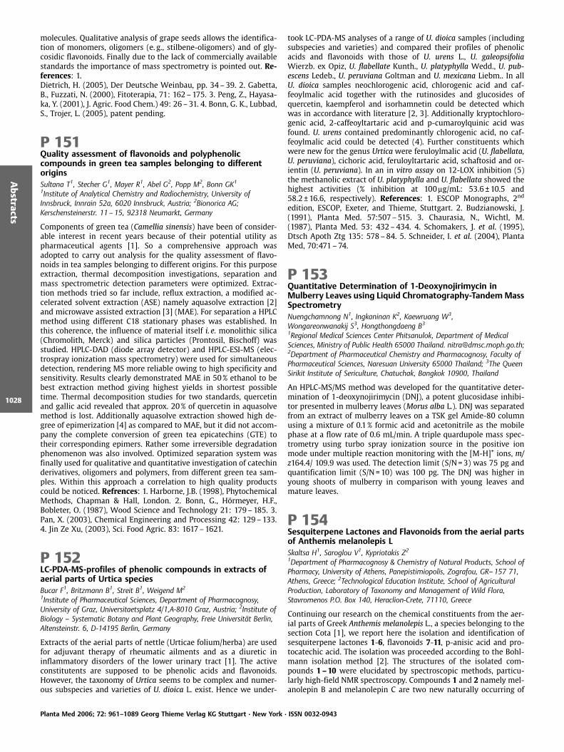
molecules. Qualitative analysis of grape seeds allows the identifica-tion of monomers, oligomers (e. g., stilbene-oligomers) and of gly-cosidic flavonoids. Finally due to the lack of commercially availablestandards the importance of mass spectrometry is pointed out. Re-ferences: 1.Dietrich, H. (2005), Der Deutsche Weinbau, pp. 34–39. 2. Gabetta,B., Fuzzati, N. (2000), Fitoterapia, 71: 162–175. 3. Peng, Z., Hayasa-ka, Y. (2001), J. Agric. Food Chem.) 49: 26–31. 4. Bonn, G. K., Lubbad,S., Trojer, L. (2005), patent pending.
P 151Quality assessment of flavonoids and polyphenoliccompounds in green tea samples belonging to differentoriginsSultana T1, Stecher G1, Mayer R1, Abel G2, Popp M2, Bonn GK1
1Institute of Analytical Chemistry and Radiochemistry, University ofInnsbruck, Innrain 52a, 6020 Innsbruck, Austria; 2Bionorica AG;Kerschensteinerstr. 11 – 15, 92318 Neumarkt, Germany
Components of green tea (Camellia sinensis) have been of consider-able interest in recent years because of their potential utility aspharmaceutical agents [1]. So a comprehensive approach wasadopted to carry out analysis for the quality assessment of flavo-noids in tea samples belonging to different origins. For this purposeextraction, thermal decomposition investigations, separation andmass spectrometric detection parameters were optimized. Extrac-tion methods tried so far include, reflux extraction, a modified ac-celerated solvent extraction (ASE) namely aquasolve extraction [2]and microwave assisted extraction [3] (MAE). For separation a HPLCmethod using different C18 stationary phases was established. Inthis coherence, the influence of material itself i. e. monolithic silica(Chromolith, Merck) and silica particles (Prontosil, Bischoff) wasstudied. HPLC-DAD (diode array detector) and HPLC-ESI-MS (elec-trospray ionization mass spectrometry) were used for simultaneousdetection, rendering MS more reliable owing to high specificity andsensitivity. Results clearly demonstrated MAE in 50% ethanol to bebest extraction method giving highest yields in shortest possibletime. Thermal decomposition studies for two standards, quercetinand gallic acid revealed that approx. 20% of quercetin in aquasolvemethod is lost. Additionally aquasolve extraction showed high de-gree of epimerization [4] as compared to MAE, but it did not accom-pany the complete conversion of green tea epicatechins (GTE) totheir corresponding epimers. Rather some irreversible degradationphenomenon was also involved. Optimized separation system wasfinally used for qualitative and quantitative investigation of catechinderivatives, oligomers and polymers, from different green tea sam-ples. Within this approach a correlation to high quality productscould be noticed. Refrences: 1. Harborne, J.B. (1998), PhytochemicalMethods, Chapman & Hall, London. 2. Bonn, G., H rmeyer, H.F.,Bobleter, O. (1987), Wood Science and Technology 21: 179–185. 3.Pan, X. (2003), Chemical Engineering and Processing 42: 129–133.4. Jin Ze Xu, (2003), Sci. Food Agric. 83: 1617–1621.
P 152LC-PDA-MS-profiles of phenolic compounds in extracts ofaerial parts of Urtica speciesBucar F1, Britzmann B1, Streit B1, Weigend M2
1Institute of Pharmaceutical Sciences, Department of Pharmacognosy,University of Graz, Universitaetsplatz 4/1,A-8010 Graz, Austria; 2Institute ofBiology – Systematic Botany and Plant Geography, Freie Universit t Berlin,Altensteinstr. 6, D-14195 Berlin, Germany
Extracts of the aerial parts of nettle (Urticae folium/herba) are usedfor adjuvant therapy of rheumatic ailments and as a diuretic ininflammatory disorders of the lower urinary tract [1]. The activeconstitutents are supposed to be phenolic acids and flavonoids.However, the taxonomy of Urtica seems to be complex and numer-ous subspecies and varieties of U. dioica L. exist. Hence we under-
took LC-PDA-MS analyses of a range of U. dioica samples (includingsubspecies and varieties) and compared their profiles of phenolicacids and flavonoids with those of U. urens L., U. galeopsifoliaWierzb. ex Opiz, U. flabellate Kunth., U. platyphylla Wedd., U. pub-escens Ledeb., U. peruviana Goltman and U. mexicana Liebm.. In allU. dioica samples neochlorogenic acid, chlorogenic acid and caf-feoylmalic acid together with the rutinosides and glucosides ofquercetin, kaempferol and isorhamnetin could be detected whichwas in accordance with literature [2, 3]. Additionally kryptochloro-genic acid, 2-caffeoyltartaric acid and p-cumaroylquinic acid wasfound. U. urens contained predominantly chlorogenic acid, no caf-feoylmalic acid could be detected (4). Further constituents whichwere new for the genus Urtica were feruloylmalic acid (U. flabellata,U. peruviana), cichoric acid, feruloyltartaric acid, schaftosid and or-ientin (U. peruviana). In an in vitro assay on 12-LOX inhibition (5)the methanolic extract of U. platyphylla and U. flabellata showed thehighest activities (% inhibition at 100mg/mL: 53.6 € 10.5 and58.2 € 16.6, respectively). References: 1. ESCOP Monographs, 2nd
edition, ESCOP, Exeter, and Thieme, Stuttgart. 2. Budzianowski, J.(1991), Planta Med. 57:507–515. 3. Chaurasia, N., Wichtl, M.(1987), Planta Med. 53: 432–434. 4. Schomakers, J. et al. (1995),Dtsch Apoth Ztg 135: 578–84. 5. Schneider, I. et al. (2004), PlantaMed, 70:471–74.
P 153Quantitative Determination of 1-Deoxynojirimycin inMulberry Leaves using Liquid Chromatography-TandemMassSpectrometryNuengchamnong N1, Ingkaninan K2, Kaewruang W3,Wongareonwanakij S3, Hongthongdaeng B31Regional Medical Sciences Center Phitsanulok, Department of MedicalSciences, Ministry of Public Health 65000 Thailand. [email protected];2Department of Pharmaceutical Chemistry and Pharmacognosy, Faculty ofPharmaceutical Sciences, Naresuan University 65000 Thailand; 3The QueenSirikit Institute of Sericulture, Chatuchak, Bangkok 10900, Thailand
An HPLC-MS/MS method was developed for the quantitative deter-mination of 1-deoxynojirimycin (DNJ), a potent glucosidase inhibi-tor presented in mulberry leaves (Morus alba L.). DNJ was separatedfrom an extract of mulberry leaves on a TSK gel Amide-80 columnusing a mixture of 0.1% formic acid and acetonitrile as the mobilephase at a flow rate of 0.6 mL/min. A triple quardupole mass spec-trometry using turbo spray ionization source in the positive ionmode under multiple reaction monitoring with the [M-H]+ ions, m/z164.4/ 109.9 was used. The detection limit (S/N=3) was 75 pg andquantification limit (S/N=10) was 100 pg. The DNJ was higher inyoung shoots of mulberry in comparison with young leaves andmature leaves.
P 154Sesquiterpene Lactones and Flavonoids from the aerial partsof Anthemis melanolepis LSkaltsa H1, Saroglou V1, Kypriotakis Z2
1Department of Pharmacognosy & Chemistry of Natural Products, School ofPharmacy, University of Athens, Panepistimiopolis, Zografou, GR–157 71,Athens, Greece; 2Technological Education Institute, School of AgriculturalProduction, Laboratory of Taxonomy and Management of Wild Flora,Stavromenos P.O. Box 140, Heraclion-Crete, 71110, Greece
Continuing our research on the chemical constituents from the aer-ial parts of Greek Anthemis melanolepis L., a species belonging to thesection Cota [1], we report here the isolation and identification ofsesquiterpene lactones 1-6, flavonoids 7-11, p-anisic acid and pro-tocatechic acid. The isolation was proceeded according to the Bohl-mann isolation method [2]. The structures of the isolated com-pounds 1–10 were elucidated by spectroscopic methods, particu-larly high-field NMR spectroscopy. Compounds 1 and 2 namely mel-anolepin B and melanolepin C are two new naturally occurring of
Planta Med 2006; 72: 961–1089 Georg Thieme Verlag KG Stuttgart · New York · ISSN 0032-0943
Abstracts
1028
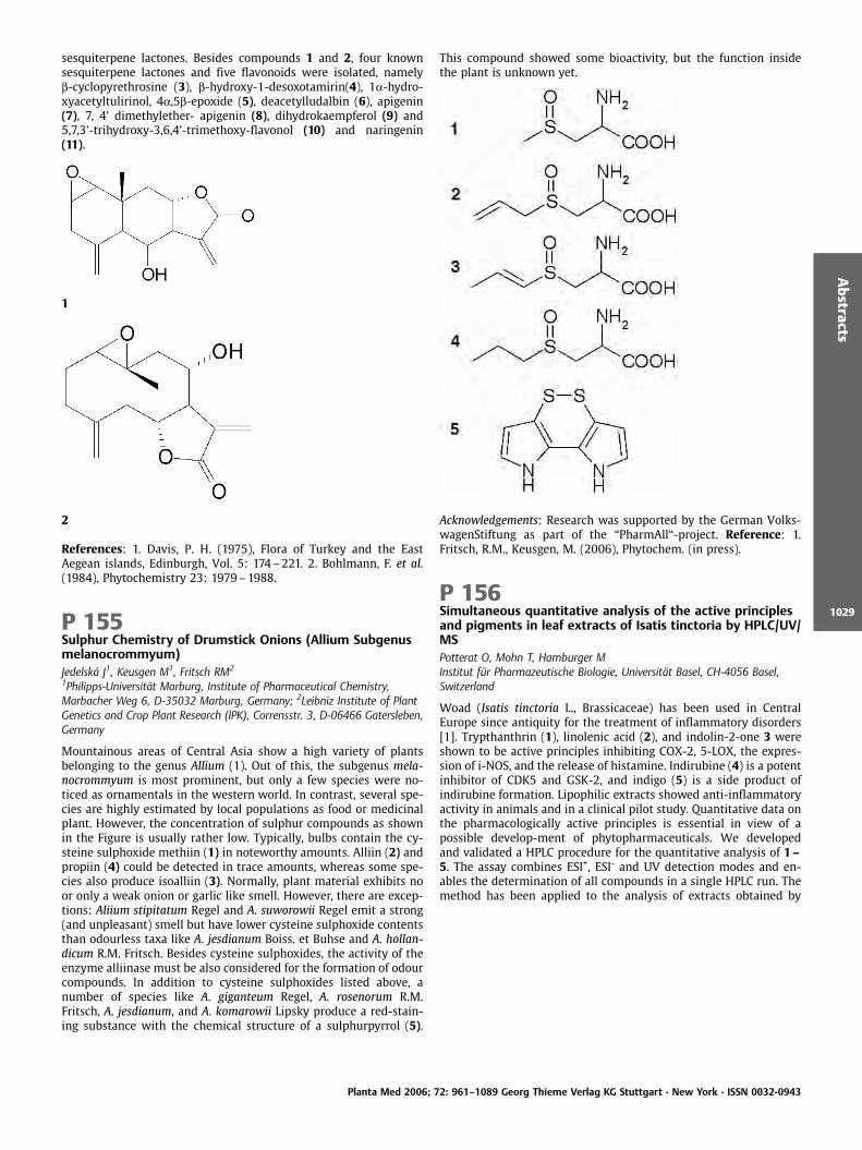
sesquiterpene lactones. Besides compounds 1 and 2, four knownsesquiterpene lactones and five flavonoids were isolated, namelyb-cyclopyrethrosine (3), b-hydroxy-1-desoxotamirin(4), 1a-hydro-xyacetyltulirinol, 4a,5b-epoxide (5), deacetylludalbin (6), apigenin(7), 7, 4’ dimethylether- apigenin (8), dihydrokaempferol (9) and5,7,3’-trihydroxy-3,6,4’-trimethoxy-flavonol (10) and naringenin(11).
1
2
References: 1. Davis, P. H. (1975), Flora of Turkey and the EastAegean islands, Edinburgh, Vol. 5: 174–221. 2. Bohlmann, F. et al.(1984), Phytochemistry 23: 1979–1988.
P 155Sulphur Chemistry of Drumstick Onions (Allium Subgenusmelanocrommyum)Jedelsk J1, Keusgen M1, Fritsch RM2
1Philipps-Universit t Marburg, Institute of Pharmaceutical Chemistry,Marbacher Weg 6, D-35032 Marburg, Germany; 2Leibniz Institute of PlantGenetics and Crop Plant Research (IPK), Corrensstr. 3, D-06466 Gatersleben,Germany
Mountainous areas of Central Asia show a high variety of plantsbelonging to the genus Allium (1). Out of this, the subgenus mela-nocrommyum is most prominent, but only a few species were no-ticed as ornamentals in the western world. In contrast, several spe-cies are highly estimated by local populations as food or medicinalplant. However, the concentration of sulphur compounds as shownin the Figure is usually rather low. Typically, bulbs contain the cy-steine sulphoxide methiin (1) in noteworthy amounts. Alliin (2) andpropiin (4) could be detected in trace amounts, whereas some spe-cies also produce isoalliin (3). Normally, plant material exhibits noor only a weak onion or garlic like smell. However, there are excep-tions: Aliium stipitatum Regel and A. suworowii Regel emit a strong(and unpleasant) smell but have lower cysteine sulphoxide contentsthan odourless taxa like A. jesdianum Boiss. et Buhse and A. hollan-dicum R.M. Fritsch. Besides cysteine sulphoxides, the activity of theenzyme alliinase must be also considered for the formation of odourcompounds. In addition to cysteine sulphoxides listed above, anumber of species like A. giganteum Regel, A. rosenorum R.M.Fritsch, A. jesdianum, and A. komarowii Lipsky produce a red-stain-ing substance with the chemical structure of a sulphurpyrrol (5).
This compound showed some bioactivity, but the function insidethe plant is unknown yet.
Acknowledgements: Research was supported by the German Volks-wagenStiftung as part of the “PharmAll“-project. Reference: 1.Fritsch, R.M., Keusgen, M. (2006), Phytochem. (in press).
P 156Simultaneous quantitative analysis of the active principlesand pigments in leaf extracts of Isatis tinctoria by HPLC/UV/MSPotterat O, Mohn T, Hamburger MInstitut f r Pharmazeutische Biologie, Universit t Basel, CH-4056 Basel,Switzerland
Woad (Isatis tinctoria L., Brassicaceae) has been used in CentralEurope since antiquity for the treatment of inflammatory disorders[1]. Trypthanthrin (1), linolenic acid (2), and indolin-2-one 3 wereshown to be active principles inhibiting COX-2, 5-LOX, the expres-sion of i-NOS, and the release of histamine. Indirubine (4) is a potentinhibitor of CDK5 and GSK-2, and indigo (5) is a side product ofindirubine formation. Lipophilic extracts showed anti-inflammatoryactivity in animals and in a clinical pilot study. Quantitative data onthe pharmacologically active principles is essential in view of apossible develop-ment of phytopharmaceuticals. We developedand validated a HPLC procedure for the quantitative analysis of 1–5. The assay combines ESI+, ESI- and UV detection modes and en-ables the determination of all compounds in a single HPLC run. Themethod has been applied to the analysis of extracts obtained by
Planta Med 2006; 72: 961–1089 Georg Thieme Verlag KG Stuttgart · New York · ISSN 0032-0943
Abstracts
1029

accelerated solvent extraction (ASE) and supercritical fluidextraction.
Reference: 1. Hamburger, M. (2002), Phytochem. Rev. 1: 333–344.
P 157An UPLC-MSTOF investigation of the leaves of ArrabidaeapatelliferaMartin F1, Hay AE1, Gupta MP2, Hostettmann K1
1Laboratory of Pharmacognosy and Phytochemistry, School ofPharmaceutical Sciences, University of Geneva, University of Lausanne, QuaiErnest-Ansermet 30, CH-1211 Geneva 4, Switzerland; 2Center forPharmacognostic Research on Panamanian Flora (CIFLORPAN), College ofPharmacy, University of Panama, Panama City, Panama
As a part of our ongoing investigations on Panamean Bignoniaceae,several species were submitted to rapid TLC autobiographical tests,against Cladosporium cucumerinum and radical scavenging activity.Arrabidaea patellifera (Schlecht) Sandw, a liana which grows fromlowlands to mountains forest, has been selected due to the goodactivity observed against Cladosporium, and moreover this planthas never been investigated.The methanol extract has been prepurified by SPE, separated andanalyzed by Ultra-Performance Liquid Chromatography coupledwith High-Resolution Time of Flight Mass Spectroscopy (UPLC-TOF). Comparing datas (UV, exact mass) with those of Arrabidaeasamydoides (1), several compounds with radical scavenging activityhave been identified such as mangiferin, muraxanthone and otherC-glucosylxanthones. The dichloromethane extract of A. patelliferashas been fractionated by centrifugal partition chromatography(CPC), medium pressure chromatography (MPLC) and semi-prepara-tive HPLC. Several products have been isolated, such as a flavonoid(chrysin), an antocyanidine and triterpens (like ursolic acid).Reference: 1. Pauletti, P. et al. (2003), J. Nat. Prod. 66: 1384–1387.
P 158Genetic polymorphism, antitumour and antioxidantpotential of Todelia asiatica on in vitro mice modelsRamaswamy VInternational Centre for Bioresources Management, Malankara CatholicCollege Mariagiri, Kaliakkavilai. PIN 629 153. Tamil Nadu. India
The aim of the present study is to evaluate the antitumor effect andantioxidant role of Todelia asiatica against EAC bearing Swiss albinomice. The effect of methanol extract of T. asiatica on tumor growthand host’s survival time was studied by the following parameters:tumor volume, packed cell volume, viable and non-viable cell countand life span of the host. Methanol extract was administered at a125 and 250mg/kg b.w. once a day for 14 days, after 24h of tumorinoculation. Decrease in tumor volume, packed cell volume, andviable cell count were observed in Methanol extract treated animalswhen compared to EAC animals. Treatment with Methanol extractat a dose of 125 and 250mg/kg increased the mean survival time to29.5 € 0.55 and 34€0.2 days respectively. The extract also decreasedthe body weight of the EAC tumor bearing mice. Hematological
studies reveal that the Hb content was decreased in EAC treatedmouse, whereas restoration to near normal levels was observed inextract treated animals. There was a significant decrease in RBCcount and increase in WBC counts in extract treated animals whencompared to EAC treated animals. The study was also extended toestimate the liver biochemical parameters such as LPO, GSH, andantioxidant enzymes like SOD, CAT etc. Treatment with Methanolextract decreased the levels of lipid peroxidation and increased thelevels of glutathione (GSH), superoxide dismutase (SOD) and cata-lase (CAT). The results suggest that the methanol extract exhibitedsignificant antitumor and antioxidant effects in EAC bearing mice.Manganese superoxide dismutase (MnSOD) is a major enzyme thatis responsible for the detoxification of reactive oxygen species in themitochondria. A T? C substitution in theMnSOD gene resulting in aVal ? Ala change at the -9 position of the mitochondrial targetingsequence (Val-9Ala), which alters the protein secondary structureand thus affects transport of MnSOD into the mitochondria was alsoanalysed in the present study.
3. Genomics, Proteomics and Metabolomics inMedicinal Plant Research
P 159Study of metabolites in benzothiadiazole treatedArabidopsis using nuclear magnetic resonance spectroscopyand principal component analysisThanh HDT1,2, Puig RC1, Kim HK1, Choi YH1, Verpoorte R11Pharmacognosy Department, Metabolomic Section, IBL, Leiden University,PO Box 9502, 2300RA Leiden, The Netherlands; 2Traditional PharmacyDepartment, Hanoi Pharmacy University, 23 Le Thanh Tong, Hanoi, Vietnam;*Email address: [email protected]
The resistance against pathogen infection in Arabidopsis is associat-ed with systemic acquired resistance (SAR) gene induction. Afterinfection, mRNAs of PR1, PR2 and PR5 genes accumulate in a coor-dinate manner in tissues that become resistant to subsequentpathogen infection [1]. The SAR pathway also can be activated bybenzothiadiazole (BTH) [2, 3]. BTH has been shown to activate SARin tobacco [4], wheat and Arabidopsis [5]. A powerful tool for plantmetabolite analysis is high-resolution nuclear magnetic resonancespectroscopy (NMR) combined with principal component analysis(PCA). We used NMR and PCA to study the changes in metaboliteprofile of Arabidopsis after treatment with BTH. The results shownclear changes in the metabolite profile in the polar fraction whichcontain compounds such as flavonoids and phenylpropanoids... butthe metabolite profile in non-polar fraction did not show any differ-ence between BTH treated and non-treated Arabidopsis. Acknowl-edgements: Vietnamese overseas scholarship fund. References: 1.Uknes, S. et al. (1993), Mol. Plant-Microbe Interact. 6: 692–698. 2.Sticher, L. et al. (1997), Ann. Rev. Phytopathol. 35: 235–270. 3. Ryals,J. A. et al. (1996), Plant Cell 8: 1809–1819. 4. Friedrich, L. et al.(1996), Plant J. 10: 61–70. 5. Lawton, M.A. et al. (1996), Plant J.10: 71 –82.
P 160Effects of Echinaforce on differentiation and activity ofantigen-presenting cells in vitroSchoop R, Suter AA. Vogel Bioforce AG, 9325-Roggwil, Switzerland
Extracts from different species of Echinacea are used today for treat-ment and prevention of upper respiratory tract diseases. No definitemolecular way of action could be attributed to the observed clinicalefficacy so far. Here we investigated the effects of endotoxin-freealcoholic fresh plant extract from Echinacea purpurea L. Moench(Echinaforce ) on different antigen-processing cells. Blood-isolatedmonocytes were differentiated into macrophages (MDM), into im-
Planta Med 2006; 72: 961–1089 Georg Thieme Verlag KG Stuttgart · New York · ISSN 0032-0943
Abstracts
1030

mature (DDC) or mature dendritic cells (MDC). Phagocytosis ofFITC-labelled bacteria was not affected in any of the above celllineages upon incubation with Echinaforce . Also the process ofmaturation from DDC into MDC was not influenced by Echinaforceas shown by typical markers of differentiation. However Echina-force modulated LPS-induced cytokine expression in blood-derivedmonocytes. At concentrations of 1 ul/mL Echinaforce (45mg/mLdry mass) potently inhibited Interleukin-1 and Interleukin-6 expres-sion after 12h (p < 0.05). Minor and no inhibition were seen for TNF-alpha and Interleukin-8 respectively. The observed effects partlywere amplified or reversed after 24h of incubation indicating com-plex feedback regulation. As demonstrated earlier Echinaforce didnot significantly induce cytokine production even at high concen-trations (50ml/mL). LPS-induced expression of metalloproteinase(MMP-1 and MMP-9) also was reversed at 1ml/mL of Echinaforceafter 24h (p < 0.01). We postulate that Echinaforce has no effect ondifferentiation- and maturation-process of various antigen-present-ing cells. We further demonstrated that pyrogenic and inflamma-tion-inducing mediators (IL-1, IL-6) in a first phase are potentlydown regulated by Echinacea extract. Although many cytokinesshow biphasic expression profiles a clear anti-inflammatory effectwas shown.
P 161Isolation of microsatellite markers in Hieracium pilosella LZini E1, Komjanc M2
1Consiglio per la Ricerca e la sperimentazione in Agricoltura (CRA), Istitutosperimentale per l’Assestamento Forestale e per l’Alpicoltura (ISAFA), PiazzaNicolini, 6,38050, Villazzano (Trento), Italy; 2Istituto Agrario di San Micheleall’Adige, Via E.Mach 1, 38010, San Michele all’Adige (Trento), Italy
Hieracium pilosella L. (mouse-ear hawkweed) is a perennial, herbac-eous weed belonging to the Asteraceae family. It grows in grassy anddry sites, on the edge of the fields and along the roads [1]. H.pilosellaL. is mostly apomictic, but it is also known to have the potential toreproduce sexually under field conditions [2]. Additionally, popula-tions possess high levels of genotypic variation, almost similar tooutcrossing species [3]. The herb is mildly astringent, cholagogue,diaphoretic, strongly diuretic, expectorant and tonic [4]. A directstudy of representative DNA would be most suitable for elucidatingthe genetic variability of species and establishing the genetic asso-ciations. Microsatellite or SSR (Simple Sequence Repeats) markersare co-dominant, multiallelic, highly polymorphic genetic markersand they are considered the most appropriate for genetic diversitystudies. Microsatellite markers were developed in Hieracium pilosel-la using the SSR enrichment procedure (5). Three biotinylatedprobes (CAA)10, (CT)15 and (GT)15, which were reported being richin other plant species, were separately used to construct an en-riched genomic library. Primer pairs for SSR analysis were designedon 34 different microsatellite regions detected. Eight developedSSRs were applied for genotyping 130 plants collected in 10 differ-ent locations in the Trentino region (Italy) and five of them showedpolymorphism among the genotypes studied. The aim of this re-search was to evaluate the genetic diversity within different acces-sions of Hieracium pilosella. This is the first reported attempt ofsuccessfully SSRs isolation and characterization in this species. Ac-knowledgements: PARMA project financed by Autonomous Provinceof Trento, Claudio Varotto (CSBT) References: 1. Dalla Fior, G.(1969), La nostra flora. Monauni. Trento. 2. Houliston, G., Chapman,H. (2001), Am. J. Bot. 91: 37–44. 3. Chapman, H. et al. (2000),Heredity 84: 401–409. 4. Chiej, R. (1984), Encyclopaedia of Medic-inal Plants. MacDonald. London. 5. He, G. et al. (2003), BMC PlantBiol. 3: 3.
P 162DNA fingerprinting of medicinally used Derris species byRAPD molecular markersSukrong S, Phadungcharoen T, Ruangrungsi NDepartment of Pharmacognosy, Faculty of Pharmaceutical Sciences,Chulalongkorn University, 10330, Bangkok, Thailand
DNA fingerprinting of five medicinal used Derris species, D. scandens(Roxb.) Benth, D. elliptica (Wallich.) Benth., D. malaccensis (Benth.),D. trifoliate Lour., and D. reticulate Benth., was studied using randomamplified polymorphic DNA (RAPD) technique. Herbal drugs, Derris,were often sold in medicinal plant market as processed plant partswhich often lose their original features, making them difficult to bedifferentiated morphologically. Accurate identification is needed inorder to ensure their efficacy, thus RAPD was exploited as a mole-cular method. The screening of twenty deca-oligonucleotide pri-mers allowed the selection of nine primers, OPS-03, OPS-05, OPS-07, OPS-08, OPS-12, OPS-14, OPS-16, OPS-17, and OPS-19, whichrevealed polymorphism. The results were reproducible. RAPD bandswere scored for the presence and the absence from the photo-graphic results and grouped by distance analysis using a pair-wisegenetic similarity according to the index of Nei and Li. Dendrogramwas generated by the unweighted pair-group method using arith-metic averages (UPGMA). Two clusters were revealed: the first con-sisted of D. scandens, D. elliptica, D. malaccensis, and D. reticulataand the second only D. trifoliata. Variation in DNA fingerprint de-tected among selected medicinal Derris species indicates the effi-ciency of RAPD molecular markers for the identification and con-struction of genetic relationship. Acknowledgements: Young ScientistScholar, Ratchadapiseksompoach Research Funds, ChulalongkornUniversity, Bangkok, Thailand. Reference: 1. Sukrong, S. et al.(2006), Thai J. Pharm. Sci. 29: 155–163.
P 163Metabolic engineering of plant cell cultures – towards thenew resources of alkaloidsH kkinen ST1, Rischer H1, Goossens A2, Ritala A1, Sepp nen-Laakso T1,Inz D2, Oksman-Caldentey KM1
1VTT Technical Research Centre of Finland, P.O. Box 1000, FIN-02044 VTT,Finland; 2VIB Plant Systems Biology, University of Ghent, B-9000 Ghent,Belgium
Plants produce a wide range of secondary compounds, which haveimportant functions for plants in survival and competing in theecosystem. One of the major bottlenecks connected with the exploi-tation of plant cell cultures is that the biosynthetic pathways ofsecondary compounds are still poorly understood. We have de-signed a novel technology for unravelling the genes involved inthe plant secondary metabolism. This technology called SoluCelis based on the genome wide identification and functional analysisof genes involved in the production of phytopharmaceuticals inplant cell cultures. The advantage in this technology is that it isapplicable to any plant, and no prior knowledge of the gene se-quences is required, the fact which very often is encountered whenit comes to exotic medicinal plants. As a model system we usedNicotiana tabacum L. (BY-2) cell culture to unravel the unknownsteps involved in nicotine alkaloid biosynthesis. From altogether591 differentially regulated genes discovered by cDNA-AFLP, 38were chosen for further functional studies. Of particular interestwere the genes encoding for protein kinases, signal transductionproteins, transcription factors and other master regulators. Fulllength cDNAs were constructed from cDNA-AFLP tags and weresubsequently delivered to Agrobacterium for the establishment oftransgenic cell suspension cultures as well as hairy root cultures.The transformed cell lines were subjected to metabolite analysis inorder to determine the functional properties of the inserted geneconstruct. Two genes have resulted in an altered secondary meta-bolite profile in tobacco hairy roots. Furthermore, the genes derivedfrom tobacco showed potential in altering the secondary metabolite
Planta Med 2006; 72: 961–1089 Georg Thieme Verlag KG Stuttgart · New York · ISSN 0032-0943
Abstracts
1031
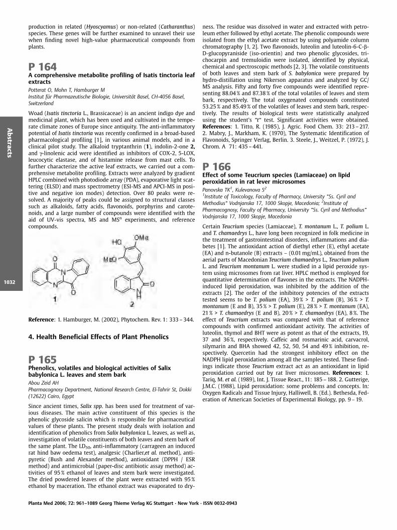
production in related (Hyoscyamus) or non-related (Catharanthus)species. These genes will be further examined to unravel their usewhen finding novel high-value pharmaceutical compounds fromplants.
P 164A comprehensive metabolite profiling of Isatis tinctoria leafextractsPotterat O, Mohn T, Hamburger MInstitut f r Pharmazeutische Biologie, Universit t Basel, CH-4056 Basel,Switzerland
Woad (Isatis tinctoria L., Brassicaceae) is an ancient indigo dye andmedicinal plant, which has been used and cultivated in the tempe-rate climate zones of Europe since antiquity. The anti-inflammatorypotential of Isatis tinctoria was recently confirmed in a broad-basedpharmacological profiling [1], in various animal models, and in aclinical pilot study. The alkaloid tryptanthrin (1), indolin-2-one 2,and g-linolenic acid were identified as inhibitors of COX-2, 5-LOX,leucocytic elastase, and of histamine release from mast cells. Tofurther characterize the active leaf extracts, we carried out a com-prehensive metabolite profiling. Extracts were analyzed by gradientHPLC combined with photodiode array (PDA), evaporative light scat-tering (ELSD) and mass spectrometry (ESI-MS and APCI-MS in posi-tive and negative ion modes) detection. Over 80 peaks were re-solved. A majority of peaks could be assigned to structural classessuch as alkaloids, fatty acids, flavonoids, porphyrins and carote-noids, and a large number of compounds were identified with theaid of UV-vis spectra, MS and MSn experiments, and referencecompounds.
Reference: 1. Hamburger, M. (2002), Phytochem. Rev. 1: 333–344.
4. Health Beneficial Effects of Plant Phenolics
P 165Phenolics, volatiles and biological activities of Salixbabylonica L. leaves and stem barkAbou Zeid AHPharmacognosy Department, National Research Centre, El-Tahrir St, Dokki(12622) Cairo, Egypt
Since ancient times, Salix spp. has been used for treatment of var-ious diseases. The main active constituent of this species is thephenolic glycoside salicin which is responsible for pharmaceuticalvalues of these plants. The present study deals with isolation andidentification of phenolics from Salix babylonica L. leaves, as well as,investigation of volatile constituents of both leaves and stem bark ofthe same plant. The LD50, anti-inflammatory (carrageen an inducedrat hind baw oedema test), analgesic (Charlier,et al. method), anti-pyretic (Bush and Alexander method), antioxidant (DPPH / ESRmethod) and antimicrobial (paper-disc antibiotic assay method) ac-tivities of 95% ethanol of leaves and stem bark were investigated.The dried powdered leaves of the plant were extracted with 95%ethanol by maceration. The ethanol extract was evaporated to dry-
ness. The residue was dissolved in water and extracted with petro-leum ether followed by ethyl acetate. The phenolic compounds wereisolated from the ethyl acetate extract by using polyamide columnchromatography [1, 2]. Two flavonoids, luteolin and luteolin-6-C-b-D-glucopyraniside (iso-orientin) and two phenolic glycosides, tri-chocarpin and tremuloidin were isolated, identified by physical,chemical and spectroscopic methods [2, 3]. The volatile constituentsof both leaves and stem bark of S. babylonica were prepared byhydro-distillation using Nikerson apparatus and analyzed by GC/MS analysis. Fifty and forty five compounds were identified repre-senting 88.04% and 87.38% of the total volatiles of leaves and stembark, respectively. The total oxygenated compounds constituted53.25% and 85.49% of the volatiles of leaves and stem bark, respec-tively. The results of biological tests were statistically analyzedusing the student’s “t“ test. Significant activities were obtained.References: 1. Titto, R. (1985), J. Agric. Food Chem. 33: 213–217.2. Mabry, J., Markham, K. (1970), The Systematic Identification ofFlavonoids, Springer Verlag, Berlin. 3. Steele, J., Weitzel, P. (1972), J.Chrom. A 71: 435–441.
P 166Effect of some Teucrium species (Lamiaceae) on lipidperoxidation in rat lever microsomesPanovska TK1, Kulevanova S21Institute of Toxicology, Faculty of Pharmacy, University “Ss. Cyril andMethodius“ Vodnjanska 17, 1000 Skopje, Macedonia; 2Institute ofPharmacognosy, Faculty of Pharmacy, University “Ss. Cyril and Methodius“Vodnjanska 17, 1000 Skopje, Macedonia
Certain Teucrium species (Lamiaceae), T. montanum L., T. polium L.and T. chamaedrys L., have long been recognized in folk medicine inthe treatment of gastrointestinal disorders, inflammations and dia-betes [1]. The antioxidant action of diethyl ether (E), ethyl acetate(EA) and n-butanole (B) extracts – (0.01mg/mL), obtained from theaerial parts of Macedonian Teucrium chamaedrys L., Teucrium poliumL. and Teucrium montanum L. were studied in a lipid peroxide sys-tem using microsomes from rat liver. HPLC method is employed forquantitative determination of flavones in the extracts. The NADPH-induced lipid peroxidation, was inhibited by the addition of theextracts [2]. The order of the inhibitory potencies of the extractstested seems to be T. polium (EA), 39% > T. polium (B), 36% > T.montanum (E and B), 35% > T. polium (E), 28% > T. montanum (EA),21% > T. chamaedrys (E and B), 20% > T. chamaedrys (EA), 8 %. Theeffect of Teucrium extracts was compared with that of referencecompounds with confirmed antioxidant activity. The activities ofluteolin, thymol and BHT were as potent as that of the extracts, 19,37 and 36%, respectively. Caffeic and rosmarinic acid, carvacrol,silymarin and BHA showed 42, 52, 50, 54 and 49% inhibition, re-spectively. Quercetin had the strongest inhibitory effect on theNADPH lipid peroxidation among all the samples tested. These find-ings indicate those Teucrium extract act as an antioxidant in lipidperoxidation carried out by rat liver microsomes. References: 1.Tariq, M. et al. (1989), Int. J. Tissue React., 11: 185–188. 2. Gutterige,J.M.C. (1988), Lipid peroxidation: some problems and concepts. In:Oxygen Radicals and Tissue Injury, Halliwell, B. (Ed.). Bethesda, Fed-eration of American Societies of Experimental Biology, pp. 9 –19.
Planta Med 2006; 72: 961–1089 Georg Thieme Verlag KG Stuttgart · New York · ISSN 0032-0943
Abstracts
1032

P 167Hepatoprotective activity of the ethyl acetate extract ofTeucrium polium L. against carbon tetrachloride inducedhepatic injury in ratsKulevanova S1, Panovska TK2, Stefkov G1, Gjorgoski I3, Bogdanova M4,Petrushevska G5
1Institute of Pharmacognosy, Faculty of Pharmacy, Vodnjanska 17, 1000Skopje, Macedonia; 2Institute of Toxicology, Faculty of Pharmacy, Vodnjanska17, 1000 Skopje, Macedonia; 3Institute of Biology, Faculty of Natural Sciencesand Mathematics, P.B. 126, 1000 Skopje, Macedonia; 4Institute of ClinicalBiochemistry, Clinical Center, Vodnjanska 17, 1000 Skopje, Macedonia;5Institute of Pathology, Faculty of Medicine, University “Ss. Cyril andMethodius“ Vodnjanska 17, 1000 Skopje, Macedonia
The hepatoprotective activity of ethyl acetate extract of Teucriumpolium (L.) has been investigated using CCl4-induced liver damagein rats. Specific biochemical parameters (glutathione peroxidase –GPx, superoxide dismutase – SOD, reduced glutathione – GSH andtotal antioxidative status – TAS) were estimated in blood and liverhomogenate [1]. Lipid peroxidation (LP) in CCl4-intoxicated rats wasevidenced by a marked increment in the levels of thiobarbituric acidreactive substances (TBARS) [2]. Histopatological examination of theliver were undertaken to monitor the status of the liver. Silymarinhas been used as a standard to compare the activity of the extract[3]. The influence of T. polium extract at a dose of 25mg/Kg, on thebiochemical parameters was significant different (p < 0.05) withthat of the CCl4-treated group. The liver biopsy of experimental ratsshowed significant restoration of normal histomorfological patternof liver cells. The study substantiates the hepatoprotective potentialof ethyl acetate extract of T. polium which may be due to the pres-ence of luteolin, apigenin, cirsimaritin and cirsiliol, determined byHPLC. References: 1. Ellenhorn, M.J. (1997), Ellenhorn’s MedicalToxicology: Diagnosis and Treatment of human Poisoning. Williamsand Wilkins publication, Los Angeles. 2. Ohkawa, H. et al. (1979),Anal. Biochem. 95: 351–358. 3. Valenzuela, A. et al. (1985), Bio-chem. Pharmacol. 3: 2209–2212.
P 168Antioxidant and anti-inflammatory phenolics fromPedilanthus tithymaloidesAbreu P1, Matthew S1, Gonz lez T1,2, Costa D3, Segundo M3, Fernandes E31CQFB/REQUIMTE, Faculdade de CiÞncias e Tecnologia da Universidade Novade Lisboa, 2829 – 516 Caparica, Portugal; 2Departamento de Farmacia,Facultad de Ciencias Naturales, Universidad de Oriente, Santiago de Cuba;Cuba; 3REQUIMTE, Departamento de Qu mica-F sica, Faculdade de Farm ciada Universidade do Porto, Rua Anibal Cunha 164, 4090 – 030, Porto,Portugal
Pedilanthus tithymaloids (L.) Poit. (Euphorbiaceae) is a low tropicalAmerican shrub with a wide range of healing properties such asemetic, anti-inflammatory, antibiotic, antiseptic, antihemorrhagic,antiviral, antitumoral, and abortive [1]. In Cuban traditional medi-cine, a tincture of P. tithymaloides is used as an anti-inflammatoryremedy in the treatment of stomatological affections. Following amethodology of bioassay-guided fractionation using ROO, ONOO ,NO, and DPPH, the flavonoids kaempferol 7-O-b-d-glucopyranoside-6’’-(3’’’-hydroxy-3’’’-methylglutarate) (1), quercitrin (2), isoquerci-trin (3), and the coumarin scopoletin were isolated from the etha-nolic extract of P. tithymaloides stems and leaves. The results herepresented indicate that these phenolic compounds constitute part of
the active principles of the plant extract responsible for its anti-inflammatory and antioxidant activities [2].
Acknowledgements: REQUIMTE, Funda ¼o para a CiÞncia e Tecnolo-gia, and Programme Alban. References: 1. Roig, J.T. (1974), PlantasMedicinales e Venenosas de Cuba, ed. Editorial Cient fico Tecnico, LaHabana. 2. Abreu, P. et al. (2006), Life Sciences 78: 1578–1585.
P 169Antioxidant activity of selected Nigerian green leafyvegetablesOdukoya OA1, Inya-Agha SI2, Segun FI1, Sofidiya MO1, Ilori OO1
1Department of Pharmacognosy, Faculty of Pharmacy, University of Lagos,Nigeria; 2Department of Pharmacognosy, Faculty of Pharmaceutical Sciences,University of Nigeria, Nsukka, Nigeria
Green leafy vegetables (GLV) offer a cheap but rich source of micro-nutrients and other phytochemicals having antioxidant propertiesand essential for good health. The potential of 21 GLV in the cookedform as natural antioxidant supplements diets was assessed. Theantioxidant activity of hot water extracts of the GLV of Amaranthushybridus Linn. (Amaranthaceae), Amaranthus caudatus (Amarantha-ceae), Beilschmedia manni (Meisn.) Benth. Et Hook.f. (Lauraceae),Celosia argentea var argentea (L.)O.Kuntze (Amarantheceae) Celosiaargentea var cristata Linn. (Amarantheceae), Corchorus olitorius L.(Tiliaceae), Crassocephalum crepidioides (Benth). S.Moore (Astera-ceae), Gnetum bucholzianum Welw. (Gnetaceae), Gongronema latifo-lium Benth. (Asclepiadaceae), Heinsia crinita (Afz.) G. Taylor (Rubia-ceae), Hibiscus callyphyllus Cav. (Malvaceae), Lasianthera africana P.Beauv (Icacinaceae), Myrianthus arboreus P. Beauv. (Urticaceae),Pterocarpus mildbraedii Harms (Papilionaceae), Pterocarpus santali-noides DC. (Papilionaceae), Solanum macrocarpon L. (Solanaceae),Solanum melongena Linn. (Solanaceae), Struchium sparganophora(Linn.) O. Ktze (Asteraceae), Talinum triangulare (Jacq.) Wild. Portu-lacaceae, Telferia occidetalis Hook (Curcurbitaceae), Vernonia amyg-dalina Del. (Asteraceae) were investigated.Potential free radicalscanvenging activity of these vegetables was confirmed by sprayingspots of the extracts with DPPH(yellow color on purple background).Antioxidant activity was assayed in linoleic acid model system. Totalpolyphenols as Tannic Acid Equivalent (TAE) and ascorbic acid wereevaluated spectrophotometrically.The activity of each extract wascalculated as %inhibition of lipid peroxidation. The extracts showedmarked antioxidant activity in linoleic acid model systems. Antiox-idant values ranged from as low as 3.67% in A. hybridus to as high as68.41% in C. argentea var cristata. Phenol content varied from21.83mg/100 g dry weight in T. triangulare to 546.97mg/100 g dryweight in G. bucholzianum. Ascorbic acid content was from13.41mg/100 g dry weight in V. amygdalina to 187.11mg/100g dry
Planta Med 2006; 72: 961–1089 Georg Thieme Verlag KG Stuttgart · New York · ISSN 0032-0943
Abstracts
1033
weight in G. latifolium. There was no correlation (R2=–0.432) be-tween antioxidant activity, total phenols and ascorbic acid content.
P 170Astringency as antisensitivity marker of some Nigerianchewing sticksSegun FI1, Odukoya OA1, Inya-Agha SI2, Agbelusi GA3, Sofidiya MO1
1Department of Pharmacognosy, Faculty of Pharmacy, 3Department ofPreventive Dentistry, College of Medicine, University of Lagos, Nigeria.2Department of Pharmacognosy, University of Nigeria, Nsukka, Nigeria
Astringents contract the tissues and canals of the body. Chewingsticks are used for oral hygiene both as an antibacterial and desen-sitizing agent. Astringency of cold water extracts of Afzelia africanaSm. ex Pers. (Caesalpiniaceae), Dialium guineense Willd. (Fabaceae),Masularia acuminata (G.Don) Bullock ex. Hoyle, Rauwolfia vomitoriaAfz. (Apocynaceae), Terminalia glauscens Planch. (Combretaceae),Vernonia amygdalina Del. (Asteraceae) and Zanthoxylum zanthoxy-loides (Lam.) Waterman (Rubiaceae) was measured by precipitatingextracts with hemoglobin, centrifugation and loss of absorbancemeasured spectrophotometrically at 578nm relative to tannic acid.Tannic Acid Equivalents (TAE) were determined from standard curveobtained. Total Tannin (TT) was determined using the protein tanninprecipitation method. Relative Astringency (RA) was astringency oftannin present relative to tannic acid. Activity was in the order ofAfzelia > Terminalia > Zanthoxylum >Masularia > Vernonia > Rauwol-fia > Dialium. TT ranged from 106.92€0.03mg/100g dry plant inDialium to 632.86€0.42mg/100 g dry plant in Afzelia. TAE was27.37 €0.07mg/100 g dry plant in Dialium to 148.11 €0.07mg/100 gdry plant in Afzelia. RA correlated positively with TAE (R2=0.8763);TT (R2 =0.9493). It is proposed that the desensitization may be dueto the astringent activity; as these extracts will form a protectivelayer on the exposed dentine; contract / block the tube like channelsthat pass through teeth and connect to nerves thereby reduce theability of the nerves to transmit pain.
P 171Free radical scavenging activity of some Nigerian medicinalplantsSofidiya MO1, Odukoya OA1, Familoni OB2, Inya-Agha SI31Department of Pharmacognosy, Faculty of Pharmacy, 2Department ofChemistry, Faculty of Science, University of Lagos, Nigeria. 3Department ofPharmacognosy, Faculty of Pharmaceutical Sciences, University of Nigeria,Nsukka, Nigeria
Antioxidant properties are among the first links between chemicalreactions and biological activity. Phenolic natural products are ofparticular interest because of their antioxidant activity throughscavenging oxygen radicals and inhibiting peroxidation. The cureall activity of Nigerian medicinal plant extracts used in traditionalmedicine practice has been associated with the antioxidant poten-tial of their phenolic content in our laboratory [1]. The present workevaluated the DPPH radical scavenging, total antioxidant activities,reducing power, and total contents of phenolic compounds inmethanolic leaf extracts of five Nigerian medicinal plants Dalbergiasaxatilis Hook.f. (Papilionaceae), Ekebergia senegalensis A.Juss.(Me-liaceae), Hymenocardia acida Tul. (Hymenocardiaceae), Icacina tri-cantha Oliv.(Icacinaceae) and Salacia pallescens Oliv.(Celastraceae) ].Free radical scavenging activity was measured spectrophotometri-cally as maximum fading power of DPPH at 517nm (DPPH is reducedto DPPH-H, with colour change from violet to yellow)at 0.025, 0.05,0.1, 0.2mg/mL concentration of extracts. Reducing power was deter-mined using Ferricyanide Trichloroacetic acid method and totalphenolic content, according to the Folin-Ciocalteu assay. Antioxi-dant activity of the plant extracts with the DPPH radical scavengingand reducing power method, were in the order Hymenocardia >Ekebergia > Salacia > Icacina > Dalbergia. H. acida and E. senegalensispossess very high radical scavenging activity in both assays. Potency
of H. acida extract (97.4 % inhibition) was of the same magnitude asthat of reference a – tocopherol. Total phenols in all the samplesexpressed as GAE (Gallic Acid Equivalent) varied from 1.83 to15.47mg/g of dry plant material. Free radical scavenging activitycorrelated with reducing power (R2 =0.9564) and total phenols R2
=0.6640 (y =1.2281 x -103.11) respectively. This suggests that 66%of the antioxidant capacity of these extracts result from contribu-tion of phenolic compounds. Reference: 1. Odukoya, O.A. et al.(2005), Antioxidant activity of Nigerian Dietary Spices. Electronic J.Environ. Agricult. Food Chem. 4:1086–1093.
P 172Effects of grape consumption on plasma and erythrocyteantioxidant parameters in elderly subjectsAvci A1, Atli T2, Erg der IB1, Varli M2, Devrim E1, Demir O2, Durak I1, Aras S21Department of Geriatric Medicine, Ankara University School of Medicine06110, Ankara; 2Department of Biochemistry, Ankara University School ofMedicine 06100, Ankara
Aim: Effects of ingesting Fructus vitis minuta (black grape) on plas-ma and erythrocyte antioxidant parameters of elderly subjects wereinvestigated in this study. Dried black grape (Maras uzumu for Turk-ish name) contain water (approx. 80% v/w), sugars (glucose andfructose) (appprox. 15% w/w) lipids, proteins, phenolic compounds,some minerals and vitamins.Methods: Thirteen subjects (mean age74.67 €0.58) participated in the study. They ingested grape at thedaily dose of 1 g/kg body weight for 1 month. Before and after theseperiods, fasting blood samples were obtained, and oxidant (malon-dialdehyde, MDA (nmol/mg protein), and xanthine oxidase, XO(-mIU/mL)) and antioxidant (superoxide dismutase, SOD (U/mg pro-tein) and glutathione peroxidase, GSH-Px (IU/mg protein)and cata-lase, CAT (IU/mg protein)) parameters were studied in erythrocytes,and MDA levels were studied in plasma samples obtained from thesubjects. SOD, GSH-Px and CAT activities were measured in erythro-cyte hemolysate fraction. Results: In the erythrocyte hemolysate,MDA levels and SOD activities were found to be lower (P < 0.01) inthe second samples relative to the first ones. Xanthine oxidase ac-tivity was found to be lower in the second samples, but this de-crease was not statistically meaningful. Because of free radical pro-duction was decreased, SOD activities were not increased. Our re-sults show that ingestion of grape consumption leads to signifi-cantly lowered erythrocyte MDA levels, which indicate that con-sumption of grape decreases oxidation reactions. It is quite possiblethat reduced peroxidation processes due to grape consumption mayplay a part in some of the beneficial effects of grape in elderlysubjects.
P 173Variation of polyphenols and antioxidant activity inmulberry leavesIngkaninan K1, Kaewruang W2, Wongthai J1, Wongareonwanakij S2,Hongthongdaeng B21Department of Pharmaceutical Chemistry and Pharmacognosy, Faculty ofPharmaceutical Sciences, Naresuan University 65000 Thailand; 2The QueenSirikit Institute of Sericulture, Chatuchak, Bangkok 10900, Thailand
Mulberry (Morus alba L., Moraceae) tea is considered as healthybeverage in Thailand. In order to identify the good source of Mul-berry tea, seven cultivars grown in Thailand were studied for theirtotal polyphenolic content and antioxidant activity using Follin-Cio-calteu and DPPH methods, respectively. The results showed thatKhunphai, the native cultivar, had the highest total polyphenols(41.88 €0.45 gGA/kg) and antioxidant activity (with IC50 value of93.42€13.17mg/L). The young mulberry leaves contained 2–3 foldhigher level of total polyphenols as well as antioxidant activitycomparing to older leaves. Seasonal variations of total polyphenoliccontent in mulberry leaves were also observed. The leaves werecollected in three commercial harvest seasons i. e. summer (April-
Planta Med 2006; 72: 961–1089 Georg Thieme Verlag KG Stuttgart · New York · ISSN 0032-0943
Abstracts
1034
July 2005), rainy season (July-September 2005) and winter (Octo-ber-December 2005). The total polyphenols and antioxidant activitywere not different in dry periods (winter and summer) while sig-nificantly decreased in rainy period.
P 174Efficacy of (€)-taxifolin from Larix sibirica (M nchh.) Ledeb.on blood pressure in experiments in vivoTikhonov VP2, Makarova MN1, Zajtseva MA1, Makarov VG1
1Interregional Center “Adaptogen“, Piskarevsky pr. 45/5, 195067, St.-Petersburg, Russia; 2Open joint-stock company “Diod“, 11-A, ul.Derbenevskaya, 115114, Moscow, Russia
The cardiovascular pathology is one of principal causes of deatharound the world. P-vitamin and antioxidant effects of flavonoidsare well-known, however the mechanism of their action is not clear.The aim of the present work was to investigate the efficacy of taxi-folin on a vascular tone booth blood pressure (BP) in the model ofspontaneously hypertensive rats (SHR). (€)-taxifolin was isolatedfrom Larix sibirica wood (OJSC “Diod). Throughout the experimentmale SHR rats (180–230 g; age of 3 months breeder: Rappolovo, St-Petersburg, Russia) were used. All animals were kept in a roommaintained under environmentally controlled conditions of 20–23 C, relative humidity –50–70%, and 12h light–12h dark cycle.All animals had free access to water and standard food. Taxifolin (instarch suspension) was administered intragastrically daily in 14days, in doses 10, 20, 50 and 150mg/kg. Enalapril maleate (KRKA;2mg/kg) and atenolol (AO “Pliva“; 10mg/kg) were used as referencepreparations. The control group received vehicle only. Systolic anddiastolic blood pressure, diuresis, electrocardiogram (II standardleads) were monitored in rats. It was established, that taxifolin indoses 10, 20, 50 and 150mg/kg has no diuretic effect. Taxifolincaused a manifest decrease of blood pressure. The most positiveresults were observed at use of a dose of 20mg/kg. Taxifolin hadmore expressed influence on the level of systolic BP. The decrease ofmyocardium stress was observed at the dose of taxifolin over 20mg/kg that was shown in normalization of R wave amplitude on anelectrocardiogram. The effect of taxifolin was similar to referencepreparations.
P 175In vitro antiviral assessment against DNA and RNA viruses aswell as antibacterial and antifungal profiles of selectedTurkish species of the Salvia genusOrhan I2, z elik B1, Kartal M3, Karaoglu T4, Yilmaz G5, Kan Y6, Sener B21Department of Pharmaceutical Microbiology, Faculty of Pharmacy, GaziUniversity, 06330 Ankara, Turkey; 2Department of Pharmacognosy, Faculty ofPharmacy, Gazi University, 06330 Ankara, Turkey; 3Department ofPharmacognosy, Faculty of Pharmacy, Ankara University, 06330 Ankara,Turkey; 4Department of Virology, Faculty of Veterinary, Ankara University,Ankara, Turkey; 5Department of Pharmaceutical Botany, Faculty of Pharmacy,Ankara University, 06330 Ankara, Turkey; 6Department of Field Crops, Facultyof Agriculture, Selcuk University, 42070 Konya, Turkey
The present study was undertaken to evaluate antibacterial, anti-fungal, and antiviral properties of the chloroform and methanolextracts from the aerial parts of fourteen Turkish Salvia species(Lamiaceae) including S. albimaculata Hedge & Hub., S. aucheri varcanescens Boiss. et Heldr., S. candidissima ssp. occidentalis Vahl., S.ceratophylla L., S. cryptantha Montbret et Aucher ex. Benth., S. cya-nescens Boiss. et Bal., S. frigida Boiss., S. forskahlei L., S. halophilaHedge, S. microstegia Boiss. & Bal., S. multicaulis Vahl., S. sclarea L.,S. syriaca L., and S. verticillata L. ssp. amasiaca (Freyn, & Bornm.)against Escherichia coli, Pseudomonas aeruginosa, Proteus mirabilis,Klebsiella pneumoniae, Acinetobacer baumannii, Staphylococcus aur-eus, Bacillus subtilis, Enterococcus faecalis, and Candida albicans bymicrodilution method. Both Herpes simplex (DNA) and Parainfluen-za-3 viruses (RNA) were used for the determination of antiviral
activity of the abovementioned Salvia extracts by using Madin-Dar-by bovine kidney (MDBK) and Vero cell lines. The methanol extractswere found to be quite active against S. aureus and E. faecalis (2 and4mg/mL, respectively), while the chloroform extracts were moreactive against S. aureus, B. subtilis, and E. faecalis (1, 2, and 2mg/mL, respectively). All of the extracts displayed the antifungal activityhaving the MIC value at 8mg/mL. Maximum cytopathogenic effect(CPE) concentrations as the indicator of antiviral activities of theextracts were determined and the chloroform extracts belongingto S. albimaculata, S. cyanescens, and S. microstegia (1–64mg/mL)along with the methanol extracts of S. ceratophylla, S. halophila,and S. sclarea (16–32mg/mL) showed reasonable antiviral effect.The cytotoxicity of the extracts was also expressed as the maximumnon-toxic concentrations (MNTC), ranging between 16–128mg/mL.
P 176Antioxidant and antimicrobial activity of lichenPseudevernia furfuracea (L.) ZopfZovko M1, Kosalec I1, Pepeljnjak S1, Kalo era Z1, Partl A2, Bakmaz M3
1Faculty of Pharmacy and Biochemistry, University of Zagreb, A. Kovacica 1,HR-10000 Zagreb, Croatia; 2State Institute for Nature Protection, Savska 41,PO Box 50, HR-10144 Zagreb, Croatia; 3Zagreb City Pharmacy, Karlovackacesta 108, HR-10436 Rakov Potok, Croatia
Lichen Pseudevernia furfuracea (L.) Zopf. (Parmeliaceae) grows acrossEurope and northern Africa. Antioxidant and antimicrobial activity,as well as phenolic content of chloroformic (PFC), ethanolic (PFE)and water (PFW) extracts of P. furfuracea collected on Velebit moun-tain were investigated. Antioxidant activity of extracts was evaluat-ed in three manners: b-carotene-linoleic acid model system, iron(III) to iron (II) reducing activity and DPPH radical-scavenging activ-ity. PFC and PFE extracts demonstrated significant antioxidant activ-ity. As it can be expected from the higher phenolic content, ethano-lic extract exhibited higher antioxidant activity than the chlorofor-mic extract in each of our three assays, especially in b-carotene-linoleic acid assay. Antimicrobial activity of PFE and PFW extractswas determinated using cylinder diffusion method, and macro-broth dilution method against standard and clinical isolates of 19bacterial, 7 yeast and 4 dermatophyte species. Gram-positive bac-terial species were highly susceptible to the PFE with inhibitionzones (ZI) from 19 to 33mm, and MIC values between 0.15 and3.1mg/mL (MMC under 4.8mg/mL), with the Bacillus spp. mostresistant (MIC 9.95–114.6mg/mL). Dermatophytes showed ZI 13–17mm and MICs between 1.19 and 2.39mg/mL (MMC 2.39–4.78mg/mL). Susceptibility of Gram-negative bacterial species, aswell as antifungal activity against yeasts, was specie-dependent.On the other hand, PFW did not show noticeable antimicrobial ac-tivity. Using bioactivity guided “bioassay in situ“ against Bacillussubtilis NCTC 8236, fraction rich in antimicrobial phenolic acidswas isolated.
P 177Evaluation of cytotoxic and antioxidant activity ofRhaponticum carthamoides (Willd.) Iljin extractsBiskup E, ðojkowska EDepartment of Plant Protection and Biotechnology, Intercollegiate Faculty ofBiotechnology, University of Gdansk and Medical University of Gdansk ul.Kladki 24, 80 – 822 Gdansk, Poland
Rhaponticum carthamoides (Willjd) Iljin is a Siberian plant used forcenturies in the folk medicine. Its extracts possess immunomodu-lating activity and may stimulate the protein synthesis in musclesand kidneys. Their activity is mainly attributed to ecdysteroids –steroid compounds occurring in plants and in insects. This studywas focused on antioxidant and cytotoxic activity of R. carthamoidesextracts. Plant material (dry leaves) was obtained from FITOSTAR .Secondary metabolites were extracted via sonication (30min.),using three solvents, differning in polarity: chloroform, methanol
Planta Med 2006; 72: 961–1089 Georg Thieme Verlag KG Stuttgart · New York · ISSN 0032-0943
Abstracts
1035
and water. Radical scavenging activity (RSA) of the extracts wasmeasured in the DPPH (2,2-diphenyl-1-pichydrazyl free radical) as-say and was compared with the activity of known antioxidants:ascorbic acid, a-tocopherol and butylated hydroxytoluene (BHT).Aqueous and methanolic extracts exhibited radical scavenging ac-tivity towards DPPH (IC50=25 and 45mg/mL, respectively) andturned to be more effective than BHT (IC50=190mg/mL), but lessthan ascorbic acid and alpha-tocopherol (IC50=4 and 12mg/mL, re-spectively). In the same time chloroformic extract was not capableof scavenging DPPH (IC50 >1mg/mL). Cytotoxic activity of all threeextracts and pure 20-hydroxyecdysone (20E) was examined againstHeLa (cervical carcinoma) and HL-60 (leukemia) cell lines, usingMTT assay. Aqueous extract as well as pure 20E did not influencethe cell lines viability, whereas chloroformic and methanolic ex-tracts exhibited mild cytotoxic activity. However according to theNCI recommendation (IC50 < 4mg/mL) neither of them can be re-garded as a potential cytotoxic agent. Acknowledgements: StateCommittee for Scientific Research Nr PBZ-KBN-092/P05/2003
P 178Changes in the phenolic compounds composition of virginolive Oil due to different storage conditions underaccelerated ageingKrieg C1, Stecher G1, Abel G2, Popp M2, Bonn GK1
1Institute of Analytical Chemistry and Radiochemistry, Leopold FranzensUniversity, Innrain 52a, 6020 Innsbruck, Austria; 2Bionorica AG;Kerschensteinerstr. 11 – 15, 92318 Neumarkt, Germany
Virgin olive oil is well known for its high amount of antioxidantphenolic compounds, such as hydroxytyrosol or oleocanthal [1],and their protective effects against cancer, coronary heart diseasesand ageing by inhibiting oxidative stress [2]. The composition ofthese compounds depends on various parameters, the variety, en-vironmental conditions, ripeness degree and the extraction type andstorage [3]. We have special interest in the long term stability ofthese compounds and their concentration changes during storageafter bottling. For this purpose the oil is bottled in different kind ofglasses and exposed to light under monitored and recorded circum-stances, to induce accelerated aging. After defined periods olive oilsamples are taken and their phenolic compositions are analyzed.Next to the phenolic compounds, the free fatty acids, the peroxidevalues and the aliphatic alcohol contents are determined accordingto Pharmacopeia Europea, in order to classify the oil quality. Withinthis approach, different types of solid phase extraction (SPE) mate-rials are compared to each other. The focus lays on the differentproperties of silica based and polymeric based materials in regardto their applications for phenolic compounds. These materials arepartly commercial available and partly self synthesised and functio-nalised [4]. Due to this work, a greater insight is gained for theadulteration of olive oils and its health supporting components.Furthermore, a good way for the optimisations of phenolic analysesin complex biological matrices is shown by synthesising and func-tionalising of SPE materials. References: 1. Beauchamp, G. et al.(2005), Nature 437: 45–46. 2. Owen, R. et al. (2000), Eur. J. Cancer36: 1235–1247. 3. Owen, R. et al. (2000), Food Chem. Tox. 38: 647–659. 4. Sultan, M., Stecher, G. et al. (2005), Curr. Med. Chem. 12:573–588.
P 179Immunomodulatory effects of flavonoids in vitroJasprica I1, Dumic J2, Mornar A1, Medic- aric M1
1Department of Medicinal Chemistry, 2Department of Biochemistry andMolecular Biology, Faculty of Pharmacy and Biochemistry, University ofZagreb, A. Kovacica 1, 10000 Zagreb, Croatia
Flavonoids, a group of very popular phytochemicals, exhibit numer-ous biological activities. We investigated immunomodulatory ef-fects of 16 flavonoids (eriodictyol, rhamnetin, isorhamnetin, sakur-
anetin, isosakuranetin, pinocembrin-7-methylether, kaempferide,tamarixetin, tectochrysin, flavone, flavanone, quercetin, myricetin,morin, kaempferol and apigenin) in vitro (using cell culture models)by measuring the production of cytokines (TNFa, IL-1b, IL-2) thatplay crucial roles in innate and adaptive immune responses. Theinhibitory effects of chosen flavonoids (30, 10 and 3mM) on IL-2production by Jurkat cells (a cell line derived from human T-cellleukemia) stimulated with polymyristate-acetate and phytohema-glutinine was determined after 24 hrs using ELISA. The level of IL-2produced by stimulated cells was considered as a maximal (100%),while un-stimulated cells do not produce IL-2. TNFa and IL-1b pro-duction by differentiated THP-1 cells (human macrophage-likecells) was measured using ELISA 24 hrs after the addition of flavo-noids (E. coli lipopolysacharide was used as a positive control; allother reagents were LPS-free). To investigate the effects of testedcompounds on cell proliferation, [6-H3] thymidine incorporationwas determined. Flavonoids that significantly (> 50%) inhibited IL-2 production suppressed the proliferation of Jurkat cells as well,except sakuranetin (SKN) and pinocembrin-7-methylether (P7ME).When applied even in the highest concentration these compoundsdid not affect Jurkat cell proliferation, but inhibited IL-2 productionfor 68.3% and 76.5%. All flavonoids that induced TNFa and IL-1bproduction for ‡50% of the positive control significantly inhibitedproliferation of differentiated THP-1 cells. Interestingly, SKN andP7ME did not stimulate cytokine production, but suppressed thecell proliferation. Although all tested flavonoids exhibited immuno-modulatory effects the most intriguing compounds for further in-vestigation are SKN and P7ME.
P 180Protective effects of catechin and epicatechin from Smilaxchina rhizome on amyloid b protein (25 – 35)-inducedneurotoxicity in cultured neuronsBana JY1, Songb KS1, Seonga YH2
1College of Veterinary Medicine and Research Institute of Herbal Medicine,Chungbuk National University, Cheongju, Chungbuk 361 – 763,South Korea;2College of Agriculture and Life-Sciences, Kyungpook National University,Daegu, 702 – 701, South Korea
We previously reported that Smilax china L. rhizome inhibits amy-loid b protein (25–35) (Ab (25–35))-induced neurotoxicity in cul-tured rat cortical neurons [1]. Here, we isolated catechin and epica-techin from S. china rhizome and also studied their neuroprotectiveeffects on Ab (25–35)-induced neurotoxicity in cultured rat corticalneurons. Catechin and epicatechin inhibited 10mM Ab (25–35)-in-duced neuronal cell death at a concentration of 10mM, which wasmeasured by a 3-[4,5-dimethylthiazol-2-yl]-2,5-diphenyl-tetrazo-lium bromide (MTT) assay and Hoechst 33342 staining. Catechinand epicatechin inhibited 10mM Ab (25–35)-induced elevation ofcytosolic calcium concentration ([Ca2+]c), which was measured by afluorescent dye, Fluo-4 AM. Catechin and epicatechin also inhibitedglutamate release into medium induced by 10mM Ab (25–35),which was measured by HPLC, generation of reactive oxygen species(ROS) and activation of caspase-3. These results suggest that cate-chin and epicatechin prevent Ab (25–35)-induced neuronal celldamage by interfering with the increase of [Ca2+]c, and then byinhibiting glutamate release, generation of ROS and caspase-3 activ-ity. Furthermore, these effects of catechin and epicatechin may beassociated with the neuroprotective effect of S. china rhizome. Ac-knowledgements: This work was supported by a grant from BioGreen21 Program, Rural Development Administration, Republic of Korea.Reference: 1. Ban, J.Y., Cho, S.O. et al. (2006), J. Ehthnopharmcol. (inpress).
Planta Med 2006; 72: 961–1089 Georg Thieme Verlag KG Stuttgart · New York · ISSN 0032-0943
Abstracts
1036

P 181Organic extract of flowers from a chamomile specieseliminates complaints resulted from hemorrhoid diseaseKavutcu M1, Arhan M2, Ayta B3, etin R4, Durak I31Gazi University Medical Faculty, Department of Biochemistry, BesevlerAnkara-Turkey; 2Ankara Oncology Teaching and Research Hospital,Department 2Gastroenterology & 4General Surgery, Ankara-Turkey; 3AnkaraUniversity School of Medicine, Department of Biochemistry, Sihhiye Ankara-Turkey
Molecular mechanisms leading to hemorrhoid mainly consist ofdegeneration of connective tissue, stagnation and stasis of blood inthe vascular plexus of the anal cushions, activation of white cells,release of inflammatory substances and toxic free radicals, whichare followed by oxidation reactions and tissue damage [1]. Purifiedflavonoid fraction has been used to prevent inflammatory reactionsresulting from the leukocyte-endothelium interaction [2]. We aimedto establish possible therapeutic effect(s) of the chamomile flowerwhich contains several types of flavonoids and other flavones in thepatients with hemorrhoid. A trial of 24 patients with acute hemor-rhoid bleeding was performed by using organic extract of flowersfrom a chamomile species. Patients consumed extract fraction for 2months at the daily dose of 200ml/kg body weight. It has beenobserved that the frequency and severity of the hemorrhoid attacksare significantly reduced in 21 of 24 patients during the study peri-od and, that all the complaints are almost eliminated after use of amonth period. In this regard, significant reductions were observedin the prelopsus by the rectal touche inspection. Bleeding was alsolessened (Mean€ SD, 2.2 €0.4 before and 0.2 €0.01 after), pain anditching frequencies decreased after extract use (Mean€SD, 5.22 €0.6before and 1.6 €0.3 after for pain and, 9.6 € 3.1 before and 2.4 €0.6after for itching). It seems possible that constituents in the extractfraction of chamomile flowers have significant potential to inhibitinflammation and, to increase micro circulation in the bowel, there-by ameliorating hemorrhoid and bleeding. References: 1. Haas, P.A.et al. (1984), Dis Colon Rectum 27: 442–450. 2. Ho, Y.H. et al.(2000), Dis Colon Rectum 43: 66 –69.
P 182Polyphenols are of special relevance for the multiplemechanisms of action of the willow bark extract STW 33-I(Proaktiv )Kelber O6, Abdel-Aziz H1, Elstner EF2, Fiebich BL3, Heinle H4, Jaeggi R5,Khayyal MT1, Metz J7, M ller J6, Okpanyi SN6, Weiser D6
1Department of Pharmacology, Faculty of Pharmacy, Cairo University, Kasr el-Aini Str, 11562 Cairo, Egypt; 2Phytopathology, Laboratory for AppliedBiochemistry, TU Munich, Am Hochanger 2, 85350 Freising, Germany; 3
VivaCell Biotechnology GmbH, Ferdinand-Porsche-Str. 5, 79211 Denzlingen,Germany; 4Institute for Physiology, University of T bingen, Gmelinstr. 5,72076 T bingen, Germany; 5Department Bioassays, Vitaplant AG,Benkenstrasse 254, 4108 Witterswil, Switzerland; 6Scientific Department,Steigerwald Arzneimittelwerk GmbH, Havelstr. 5, 64295 Darmstadt,Germany; 7Institute of Anatomy and Cell Biology III, University of Heidelberg,Im Neuenheimer Feld 307, 69120 Heidelberg, Germany
The pharmacological profile of the willow bark extract STW 33-I(water extract, 16–23:1) and the contribution of its fractions to itwere studied in a number of pharmacological models in vitro and invivo for elucidating its clinical effects. In Interferon-g/LPS treatedmonocytes, STW 33-I reduced expression of iNOS, COX-2, the anti-apoptotic protein Bcl2, Il-1b, Il-6 and TNF-a, measured by real timePCR, with IC50 between 10 and 200mg/mL. It inhibited PGE2, Il-6 andMMP-3 in chondrocytes. Activities of 5-LOX, hyaluronidase, elastase(HLE), COX-1 and -2 and oxidation in AAPH and XOD reactions wereinhibited. Five fractions of the extract, obtained by sequential ex-traction with solvents of increasing polarity and analytically char-acterized by HPLC, were tested as well, showing that the fractionscontaining the different groups of polyphenols were responsible forthe main part of the effect, while the fraction containing the main
part of the salicylates showed only a minor contribution. In vivo,STW 33-I (50 to 150mg/kg b.w.) was effective in writhing test inmice, Randall-Sellito model, brewers yeast model, paw edema, ad-juvant arthritis and air pouch model in rats. In the latter, PGE2 andLTB4, Il-1b, Il-6, TNF- a, TxB4, COX-2 and the antioxidative param-eters MDH were decreased, GSH increased. These studies show mul-tiple mechanisms of the willow bark extract, including anti-inflam-matory, -oxidative, -pyretic, joint protecting, and analgesic actions.These were mainly not due to salicylates, but to polyphenols, whichtherefore seem to be the group most relevant for the therapeuticefficacy of STW 33-I (Proaktiv ) in back pain.
P 183Role of endogenous SHs and NO on Vernonia ferruginea Lessinduced gastroprotectionBarbastefano V1, Cola-Miranda M1, Luiz-Ferreira A1, Farias-Silva E1,de Paula Michelatto D1, Camargo EES2, Hiruma-Lima CA3, Vilegas W2,Souza Brito ARM1
1Departamento de Fisiologia e Biof sica, IB, UNICAMP, Campinas, SP, Brazil;2Departamento de Qu mica Org nica, IQ, UNESP, Araraquara, SP, Brazil;3Departamento de Fisiologia, IB, UNESP, Botucatu, SP, Brazil
The methanolic crude extract (MeOH) obtained from aerial parts ofVernonia ferruginea Less. (Asteraceae), a Brazilian savannah plantpopularly known as “assa-peixe“, was investigated for its antiulcero-genic proprieties and mechanisms employing three experimentalmodels. Preliminar phytochemical screening showed that glycosideflavonoids are the major compounds present in this extract. To ob-tain MeOH, aerial parts of V. ferruginea were air dried (7 days at40oC) and powdered. The powder (100 g) were exhaustively ex-tracted with methanol at room temperature (3 times for 72h) andthen concentrated in a vacuum rotator evaporator. The previousadministration of MeOH (50, 100, 250 and 500mg/kg) significantlyinhibited the gastric mucosa damage, from the 100mg/kg dose (91%of inhibition), caused by absolute ethanol oral administration inrats. This antiulcerogenic propriety of MeOH (100mg/kg) dependsin part on endogenous NO, once its gastroprotection was lightlyattenuated by pretreatment with NG-nitro-L-arginine methyl-ester,a NO-Synthase inhibitor. Besides, when animals were pretreatedwith N-ethylmaleimide, a thiol blocker, including mucosal nonpro-tein sulfhydryl groups (SHs), the gastroprotective effects of MeOHhave been extinguished. These results show that MeOH gastropro-tective proprieties are due to the cytoprotective proprieties of NOand mainly of endogenous sulfhydryls compounds. Acknowledge-ments: FAPESP, CNPq and CAPES.
P 184Hypotensive and vasorelaxant effect of the procyanidinscomplex from Guazuma ulmifolia bark, in normotensive andhypertensive ratMagos GA1, Mateos JC2, P ez E1, Fern ndez G1, M rquez C2, Lobato CE2,Enr quez RG2
1Departamento de Farmacolog a, Escuela de Medicina, Universidad NacionalAut noma de M xico, 04510 M xico D.F, M xico, D. F; 2Instituto de Qu mica,Universidad Nacional Aut noma de M xico, Circuito Exterior, CiudadUniversitaria, 04100 M xico D.F, M xico, D. F
Guazuma ulmifolia Lam. bark is used in Panama traditional medicinefor the treatment of arterial hypertension [1]. The phytochemicalstudies carried on with G. ulmifolia bark, have led to the isolationof procyanidins. These compounds have also been related to anti-hypertensive activities. The hypotensive and vasorelaxant effect of aprocyanidin complex (PACC) prepared from G. ulmifolia bark wasinvestigated in normotensive and hypertensive rats, and in rat aorticrings. The oral administration of PACC (10mg/kg) to conscious nor-motensive or hypertensive rats decreases the systolic arterial pres-sure (SAP) and the heart rate (HR). The intravenous administrationof same fraction (10mg/kg) in anesthetized rats induced arterial
Planta Med 2006; 72: 961–1089 Georg Thieme Verlag KG Stuttgart · New York · ISSN 0032-0943
Abstracts
1037

hypotension that was attenuated by pre-treatment with NG-nitro-L-arginine-methylester (L-NAME 31mg/kg). In isolated aortic rings ofnormotensive and hypertensive rats, the PACC reduced the contrac-tion induced by norepinephrine (1X10⁻⁷ M). The IC50 doses was35.3 € 12 ng/mL and 101€57 ng/mL in isolated aortas of normoten-sive and hypertensive rats, respectively. This relaxant activity wasinhibited by either removal of vascular endothelium or pre-treat-ment with L-NAME (30mM), while indomethacin (10mM) or atro-pine (10mM) had no effect. Preliminary analysis of the PACC byHPLC-PAD-/MS and FAB+ mass spectrometry allowed detection ofthe main components as a complex of procyanidin oligomers con-sisting mainly of type B procyanidins. These findings suggest thatcardiovascular effects from G. ulmifolia are due to the procyanidincomplex, while the antihypertensive mechanism was found to belinked to endothelium related factors, where nitric oxide is know tobe involved. Acknowledgements: Consejo Nacional de Ciencias y Tec-nolog a, CONACYT, M xico for financial support through projectsMO292 and Q42096. Reference: 1. Caballero-George, C. et al.(2001), Phytomedicine 8: 59–70.
P 185Antioxidant activity of an aqueous fraction obtained fromIndigofera truxillensis against ischemia-reperfusion-inducedgastric lesionsBarbastefano V1, Farias-Silva E1, Cola-Miranda M1, Calvo T2, Luiz-Ferreira A1,Pimentel FO1, de Paula Michelatto D1, Hiruma-Lima CA3, Vilegas W2,Souza Brito ARM1
1Departamento de Fisiologia e Biof sica, IB, UNICAMP, Campinas, SP, Brazil;2Departamento de Qu mica Org nica, IQ, UNESP, Araraquara, SP, Brazil;3Departamento de Fisiologia, IB, UNESP, Botucatu, SP, Brazil
The aqueous fraction (FAq) from the methanolic crude extract ob-tained from aerial parts of Indigofera truxillensis Kunth. (Fabaceae), aBrazilian savannah plant popularly known as “Indigo“, was investi-gated for its antioxidative proprieties in gastric lesions induced byischemia-reperfusion (IR) in rats. Preliminar phytochemical screen-ing showed that flavonoid glycosides are the major compoundspresent in this fraction. male Wistar rats (180–220 g, n > 4) werefasted during 24h, orally treated with FAq (100mg/kg), and sub-mitted to gastric lesions by IR of the celiac artery. After, the stomachwas removed, lesion areas were counted, gastric mucosa wasscraped, homogenized, and frozen to posterior analysis of: a) DNAfragmentation, b) LPO and c) GSH content. FAq protected the gastricmucosa in 91% in IR model, but presented no significant changesover LPO and GSH content. Indeed, one of the antioxidative mecha-nisms observed in FAq was its capacity to protect cells from IRinduced DNA fragmentation. Flavonoids should be responsible forthis antiulcerogenic activity of FAq. Acknowledgements: FAPESP,CNPq and CAPES.
P 186Antioxidant activities of Punica granatum as determined byFRAP assay methodHajimahmoodi M1, Oveisi MR1, Sadeghi N1, Jannat B1, Davoodabadi E1
Drug and food control department, faculty of pharmacy, Tehran university ofmedical science, 14155, Tehran, Iran
The fruits are rich sources of various vitamins, minerals and fibersrequired by human body for optimal health. In the recent years,more attention has been paid to the antioxidants contained in fruits.Epidemiological studies revealed that high fruit intake was associat-ed with reduced mortality and morbidity of cardiovascular diseaseand some types of cancer and one of possible mechanisms wasattributed to the antioxidant activity presented by the fruits. Pome-granate (Punica granatum L.) is native to the Mediterranean regionand has been used extensively in the folk medicine of many coun-tries. The presence of antioxidants has been reported from pome-granate in juice, peel, pulp and seed fractions [1–3], however, no
literature was found reporting the antioxidant activity in the pome-granate of Iran. In the present study, the ferric reducing ntioxidantpower assay (FRAP assay) was employed and the FRAP value of peeland pulp fractions 10 cultivar of pomegranate produced in Savehwas determined in an attempt to make a systematic comparisonamong their antioxidant activities and identify the fractions withhigh antioxidant power for further studies. On the other hand thestability of antioxidant power was determined for three weeks. Theresults showed that sweet white peel and black peel cultivars washigh in antioxidant power in peel and pulp fractions respectivelyand both peel and pulp fractions are quite stable in antioxidantactivity for three week in refrigerator. References: 1. Navindra, P.et al. (2005), J. Nut. Biochem. 16: 360–367. 2. Negi, P. S. et al. (2003),Food Chemistry 80: 393–397. 3. Yunfeng, L. et al. (2006), FoodChemistry 96: 254–260.
P 187Antioxidants from fruits and leaves of Eugenia jambolana, anedible Myrtaceae species from Atlantic ForestSilva DHS, Plaza CV, da S. Bolzani V, Cavalheiro AJ, Castro-Gamboa IInstituto de Qu mica, Universidade Estadual Paulista CP 355 CEP 14800 – 900Araraquara, SP, Brazil
Eugenia jambolana Lam. belongs to Myrtaceae, a plant family wide-spread in Brazil, known for its edible fruits as guava (Psidium gua-java L.), jambo (Syzygium jambos (L.) Alston), ara -rosa (Psidiumcattleianum Sabine), pineapple guava (Feijoa sellowiana Berg.), pi-tanga (Eugenia uniflora L.), grumixama (Eugenia brasiliensis Lam.),scrub cherry (Syzygium australe (Link.) B. Hyland), most of whichpresent astringent properties due to their tannin content. There arefew phytochemical studies on such species, although preliminaryevaluation of some Myrtaceae edible fruits has evidenced high con-tents in vitamin C and phenolic compounds. Additionally, the ex-tract from seeds of E. jambolana has been used as traditional med-icine in India, Brazil and other tropical countries for its hypoglyce-mic and antidiabetic properties, which have been associated with itsantioxidant and anti-inflammatory activities. As part of our studieson species from Tropical Rain Forest (Atlantic Forest), fruits andleaves of Eugenia jambolana were collected and their ethanol ex-tracts showed positive results when screened for antioxidant(bleaching of beta-carotene TLC autographic assay [1]) and cytotoxic(MTT assay for MDA/MB-435, SF-295 and HCT-8 cell lines [2]) activ-ities. Phytochemical work on the EtOH fraction led to the isolationof polyphenols, including flavonoid glycosides and phenolic acidsfrom the leaves, and anthocyanins cyanidin and delphinidin glyco-sides from the fruits extract. The isolates were submitted to evalua-tion of their antioxidant properties and showed strong free radicalscavenging activity towards DPPH. These results support the use ofextracts of E. jambolana in traditional medicine and suggest theiractions as possible chemopreventive agents or phytoceuticals. Ac-knowledgements: This work was sponsored by the program BIOTA-FAPESP, BioProspecTa, CAPES and CNPq. References: 1. Pratt, D. E.,Miller, E. E. (1984), J. Am. Oil Chem. Soc. 61: 1064–1067. 2. Skehan,P., Storeng, R. et al. (1990), J. Natl. Cancer Inst. 82: 1107–1112.
P 188Aromatic plants from Valsesia (Italy): bioassay-guidedisolation of flavonoids with antioxidant activity fromAchillea speciesTom F1, Fico G1, Visioli F2, Iorizzi M3, Vitalini S11Dipartimento di Biologia, via Celoria 26, 20133, Universit degli Studi diMilano, Italy; 2Dipartimento di Scienze Farmacologiche, via Balzaretti 9,20133, Universit degli Studi di Milano, Italy; 3Dipartimento di Scienze eTecnologie per l’Ambiente e il Territorio, Via Mazzini 8, Universit degli Studidel Molise, Isernia, Italy
Valsesia is an alpine area of Northern Italy whose autochthonousflora has been maintained over the years due to the almost com-
Planta Med 2006; 72: 961–1089 Georg Thieme Verlag KG Stuttgart · New York · ISSN 0032-0943
Abstracts
1038
plete absence of imported varieties. This area maintains many of itstraditional habits, including the use of wild plants for culinary andpharmaceutical purposes. In a previous work we have investigatedsome methanolic extracts of different species collected in Valsesia[1]. Among the analysed plants, two species belonging to the Achil-lea genus (Achillea distans, Achillea moschata) have shown signifi-cant antioxidant activity. Aim of this work is therefore the identifi-cation of pure compounds responsible of the activity of crude ex-tracts. The MeOH extracts have been separated with Sephadex LH-20; 22 and 25 fractions have been obtained from A. distans and A.moschata respectively. The fractions with different TLC profiles weretested for the antioxidant activity, evaluated as scavenging activityof the stable radical DPPH, antioxidant capacity (Cu++ to Cu+ reduc-tion) and LDL oxidation. Subsequently we have separated the se-lected fractions with HPLC: 5 active compounds were isolated. Ac-knowledgements: supported by MIUR grant PRIN 2004038183/004,programma 2004 Reference: 1. Vitalini, S. et al. (2006), Phytoter.Research, in press.
P 189Structure and biological activities of antimicrobialcompounds recently isolated from southern AfricanCombretum and Terminalia speciesEloff JNPhytomedicine Programme, University of Pretoria, Private Bag X04,Onderstepoort, 0110 South Africa
Combretum and Terminalia species have recently been identified asamong the 50 most important medicinal plant species in Africa.These widely distributed species are traditionally used for a widerange of indications in Africa and Asia. By antibacterial and antifun-gal bioassay guided fractionation more than 20 terpenoids, flavo-noids and bibenzyls have been isolated by our group during the lastdecade [MIC 16–50mg/mL] Several biological activities of thesecompounds and of crude plant extracts against several bacteria,fungi, parasites and cell lines were determined in vitro [MIC someextracts against fungi 20mg/mL] and also in vivo in animal experi-ments. Toxicity and the application of some of these compounds orextracts in animal production systems were also evaluated. Theresults support many of the ethnomedicinal uses of these genera,but there is a poor correlation between activities of extracts ofdifferent species and use against microbial infections. The reasonis probably the relatively non-polar character of the antimicrobialcompounds, making it difficult for poor rural people to extract.Many species with excellent antimicrobial activities growing widelyare not used. It may be possible to use these species in primaryhealth care. For example, leaf extracts of C. erythrophyllum growingwidely along river banks have good activity against Vibrio cholera[MIC 25mg/mL] and may be used during cholera outbreaks. Thereappears to be no correlation between sections of Combretum andchemical composition or biological activity. Some of the results in-dicate that at least in some of the species active speciation is stilltaking place.
P 190Phenolic extracts of strawberry fruits, leaves and cellcultures – analysis and biological activitiesNohynek L, Sepp nen-Laakso T, J ger K, Oksman-Caldentey KM,Puupponen-Pimi RVTT Biotechnology, Tietotie 2, FI-02044 VTT, Finland
Berry fruits are rich in phenolic compounds, such as flavonoids,phenolic acids, lignans and complex polymeric tannins, with widevariation in their contents depending on the plant species and en-vironmental factors. The main phenolics in strawberry fruits areanthocyanins and ellagitannins. We have shown earlier that ellagi-tannins possess strong antimicrobial properties against human gas-trointestinal pathogens. As an alternative to use of strawberry fruits,
plant leaves and cell cultures are interesting choice for productionof strawberry phenolics, as well as other, potentially novel second-ary metabolites with interesting biological effects. As far as weknow, strawberry cell cultures have not been used for productionof berry and plant phenolics earlier. In this study, sterile in vitrostrawberry plants were germinated from sterilized seeds. Cuts ofthe in vitro leaves were treated with plant growth hormones forproduction of callus, and the most productive calli were used forestablishment of strawberry cell cultures. Analysis of phenolic ex-tracts of strawberry fruits, leaves and cell cultures were performedusing HPLC-DAD and LC-MS, and comparison of phenolic compoundpatterns of in vivo strawberry fruits and leaves to those of in vitroleaves and cell cultures revealed interesting differences. Also anti-microbial activities against selected human pathogenic bacteria andanti-oxidative activities, measured as radical scavenging activity,varied depending on the origin of the material of phenolic extract.These results showed that strawberry leaves are interesting and richsources of bioactive phenolic compounds.
P 191Are cinnamic acids responsible for in vitro neuroprotectionexerted by Bryothamnion triquetrum (S.G.Gmelin) Howeaqueous extract?Fallarero A1,4, Tammela P2, Loikkanen J3, Vidal A4, Vuorela P11- Department of Biochemistry and Pharmacy, bo Akademi University, FIN-20520 bo, Finland; 2- Viikki Biocenter, Faculty of Pharmacy, University ofHelsinki, FIN-00014, Helsinki, Finland; 3- Department of Pharmacology andToxicology, University of Kuopio, FIN-7021, Kuopio, Finland; 4- Department ofBiochemistry, Faculty of Biology, University of Havana, CP 10400, CiudadHabana, Cuba
Three cinnamic acids: ferulic (FA), p-coumaric (p-CA) and trans-cin-namic acids (t-CA) have been identified as constituents ofBryothamnion triquetrum (S. G. Gmelin) Howe aqueous extract, aproduct that has been reported to exert different in vitro neuropro-tective properties [1, 2, 3]. In current study, it was analyzed theeffect of these three cinnamic acids in different models of oxidativestress, with the purpose of elucidating their contribution to the invitro neuroprotective properties of B. triquetrum extract. GT1–7cells were exposed to chemical agents that induce oxidative neuro-nal death: H2O2; H2O2 + FeSO4; 3-morpholinosydnonimine hydro-chloride (SIN-1) and methyl mercury (MeHg) and the protectiveeffect of cinnamic acids, when added immediately before toxic com-pounds, was assessed. At the end of the insulting period, GT1–7cells viability was measured by using propidium iodide fluorometricassay [2]. Treatments were compared using ANOVA and Tukey’sMultiple Comparison tests. The protective effect of FA was provedto occur in all the 4 examined cytotoxicity models. Results showedthat FA can at least partially mimic the neuroprotective effect of B.triquetrum extract, although some other antioxidant compounds arestill required to reach the extract maximal protective effect. How-ever, no protection was observed after exposure to p-CA or t-CA, andno increase in FA protection was registered when adding p-CA and t-CA to FA, as they naturally occurs in the extract. In summary, thisinvestigation showed evidences of the contribution exerted by FA tothe in vitro neuroprotective effect of B. triquetrum aqueous extract,in the models of neuronal cell death induced by H2O2; H2O2 +FeSO4; SIN-1 and MeHg. References: 1. Vidal, A., Motidome, M. etal. (2001), Braz. J. Pharm. Sci. 37: 373–382. 2. Fallarero, A., Loikka-nen, J.J. et al. (2003), Phytomedicine. 10: 39–47. 3. Fallarero, A.,Peltoketo, A. et al. (2006). Phytomedicine. 13: 240–245.
Planta Med 2006; 72: 961–1089 Georg Thieme Verlag KG Stuttgart · New York · ISSN 0032-0943
Abstracts
1039

P 192Effects Of Carvacrol Upon The Liver Of Rats UndergoingPartial HepatectomyCanbek M1, Uyanolu M1, Aral E2, Baser KHC3
1Eskisehir Osmangazi University, Faculty of Science, Biology Department,26480, Eskisehir, Turkey; 2Eskisehir Osmangazi University, Medical Faculty,Histology Department, 26480, Eskisehir, Turkey; 3Anadolu University, Facultyof Pharmacy, 26470, Eskisehir, Turkey
There are several studies reporting effects of volatile oils upon hu-man health extracted from origanum (kekik). The present studyaims to investigate the possible effects of purified carvacrol obtain-ed from origanum and silymarin upon the regenerative feature ofthe liver subsequent to partial hepatectomy in rats. The carvacrolwas tested in comparison with silymarin ale Wistar Albino rats,weighting 230 €30 g, were divided into 3 experiment groups. Firstgroup rats, called Group 1 (n =8) were used as control group. Rats inGroup 2 (n=8) were applied carvacrol and hepatectomy (73mg/kg).Silymarin and hepatectomy (100mg/kg) were applied to the lastgroup of the rats, Group 3 (n =8). One dose of test materials wasinjected to Groups 2 and 3 one hour before 68 % partial hepatect-omy. At the end of the experiments, blood and organs were removedintra cortically. The liver regeneration ratio of the rats was calculat-ed measuring the half weights of their liver before and after thehepatectomy. H&E, IL-6 and PCNA treatments were applied to liversections. AST, ALT, TNF-a and IL-6 levels were determined in serumsamples. In AST, ALT, TNF-a and IL-6 levels, there were no statisti-cally significant difference. Mitotic index and PCNA index compar-isons were displayed significant differences; between Group 1 and2, p < 0.001 between Group 1 and 2, p < 0.05 between Group 2 and3. Histological evaluations were also similar with these results ofPCNA and Mitotic indexes. According to these results, it is concludedthat carvacrol increases the liver regeneration ratio.
P 193Black Grape Extract Protects Against Cyclosporine ANephrotoxicityDurak I1, etin R2, andır 3, Devrim E1, Kılı olu B4, Avci A1
1Ankara University School of Medicine, Department of Biochemistry, Ankara-Turkey; 2Ankara Oncology Education and Search Hospital, Department ofGeneral Surgery, Ankara-Turkey; 3S leyman Demirel University School ofMedicine, Department of Pathology, Isparta-Turkey; 4Ankara Education andSearch Hospital, Department of General Surgery, Ankara-Turkey
The aim of this study was to determine if dried black grape protectagainst cyclosporine nephrotoxicity. Twenty eight Sprague-Dawleyrats were given Cyclosporine A (CsA) orally for 10 days, with theblack grape (Kalecik karasi, total phenolic content of the grape wasapprox. 96.25€2.03mg gallic acid equivalent/gr) supplementationbegun three days before CsA treatment and continued during thestudy period (totally 13 days). In each group (control, CsA alone, CsAplus black grape, and black grape alone), there were 7 animals. Atthe end of the study period, the animals were sacrificed; their kid-neys were removed and prepared for biochemical investigations.Oxidant (xanthine oxidase enzyme and malondialdehyde) and anti-oxidant (superoxide dismutase, glutathione peroxidase and catalaseenzymes) parameters were measured in the kidney tissues of thegroups. It has been found that CsA creates oxidant load to the kid-neys through both xanthine oxidase activation and impaired anti-oxidant defense system, which accelerates oxidation reactions inthe kidney tissue. Supplementation dried black grape led to reducedmalondialdehyde level in the kidney tissue possibly, by preventingoxidant reactions. In conclusion, the results suggest that impairedoxidant/antioxidant balance may play part in the CsA-induced ne-phrotoxicity, and black grape may ameliorate this toxicity, in agree-ment with studies with antioxidant vitamins.
5. Clinical Studies with Herbal Medicinal Products
P 194The flavonoidal constituents of Limoniastrum monopetatumand their biological activityRadwan HM, Hassan RAChemistry of Medicinal Plants Dept, National Research Centre, Dokki, Cairo,Egypt, 12311
Investigation of Limoniastrum species (Family Plumbaginaceae) re-vealed the presence of flavonoids, coumarins, terpenes and alkaloids[1, 2]. Limoniastrum species are represented in Egypt by only onespecies [3]. The present work deals the study of the flavonoidalconstituents of L.monopetatum and evaluation their hepatoprotec-tive and the anti- oxidant activity of both the total extracts and theisolated compounds, using DPPH free radical. The aerial parts of L.monopetatum was dried, powdered and extracted with pet.etherand then with 80% ethyl alcohol. The alcoholic extract after parti-tioner with chloroform, ethyl acetare and n-butanol yielded crudeextracts containing flavonoids [4]. The previous extracts were sub-jected separately to preparative PC (3MM, 20% acetic acid) and themain flavonoidal bands were cut and eluted separately with 90%methanol. The eluted fractions were further purified by using Se-phadex LH-20 column. The isolated flavonoid compounds wereidentified as myricetin, rutin, kaempferol-7-O-glucoside and myri-cetin-3-O-glucoside. Their identities were verified by TLC, PC, m.p,UV, 1-H-NMR, MS and FAB mass spectrum. Acute toxicity studies ofpet. ether and alcoholic extracts of the aerial parts of the plant,showed that the alcoholic is more safe than that of pet.ether extractand both extracts have a hepatoprotective effect on the hepatocytesagainst CCl4 cytotoxicity at concentration of 40mg/mL and 50mg/mL,respectively. On the other hand, all the isolated flavonoid com-pounds showed significant antioxidant activity compared to Trolox.References: 1. Rizk, A.M. (1986), The Phytochemistry of the flora ofQatar, Scientific and Applied Research Centre, University of Qatar. 2.Rizk, A.M. (1982), Fitoterapia 52: 35. 3. Tackholm, V. (1974), Stu-dents Flora of Egypt, 2nd ed., Published by Cairo Univer., CooperativePrinting Co., Beirut. 4. Radwan, H.M., Shams, K.A. (2005), J. Egypt.Pharmac. 4 (2).
P 195Effect of garlic during and before administration of leadacetate on lead content of some tissues in mousePourjafar M1, Karimi I1, Kojouri G1, Kheiri S2, Aghbolaghi PA3
1School of veterinary medicine, Shahrekord University, Shahrekord, Iran;2School of medical science, Shahrekord University of medical science,Shahrekord, Iran; 3Educated of Shahrekord University, Shahrekord, Iran
Garlic (Allium sativum L.) has been found to possess heavy metalchelator activity. Prophylactic and therapeutic effects of garlic andgarlic tablets on chronic administration of lead in mouse were in-vestigated. Eighty mature mice (body weight of 35–40g) weredivided into eight groups and each group was made up of ten mice.Group D as a negative control group received placebo garlic tablet.Groups A1, A2 and A3 respectively received 500,250 and 125mg/Kg/day garlic in first four weeks, and in second four weeks they receiv-ed 5mg/kg/day lead acetate and 500,250 and 125mg/kg/day garlicrespectively. Groups B1, B2 and B3 respectively received 1/4, 1/8 and1/16 garlic tablet/kg/day (equal to 500mg of fresh garlic) in firstfour weeks and in second four weeks received 5mg/kg/day leadacetate and also respectively 1/4, 1/8 and 1/16 garlet tablet/kg/day(equal to 500mg of fresh garlic). Group C as a positive control groupreceived a quarter of a placebo garlic tablet/kg/day in first fourweeks and in second four weeks they received 5-mg/kg/day leadacetate and a quarter of a placebo garlic tablet/kg/day. Reductionin lead content of kidney, liver and bone as a result of administra-tion of garlic or garlic tablet in studied groups was significant com-pared with group C (p < 0.05) and reduction in lead content of blood
Planta Med 2006; 72: 961–1089 Georg Thieme Verlag KG Stuttgart · New York · ISSN 0032-0943
Abstracts
1040
in all groups was significant except group A3. Results of comparisonof lead content between different groups showed that fresh garlicand garlic tablet had the same effects on lead intoxication and leaddeposition in tissues.
P 196Cardioprotective effect of Thevetia neriifolia Jusss glycosidein male white ratSaikia GDepartment of Zoology, Gauhati University, Guwahati. India
The plant Thevetia neriifolia Jussex Steud. (TN, yellow oleander) iswidely distributed in Assam, India either in wild or in garden. Theroot has been in used against heart heart trouble [1]. The seedkernels were used as common position for suicidal purpose. Thedefatted seed kernel contains glycoside [2] and the basic carbonskeleton is cyclopentanophenanthrene nucleus is a reduced formwith an unsaturated gamma lactone ring attached to the 3–17 po-sition. It has been aimed to identify the glycoside present in theseed kernel extracted in ether (EE) and its possible effect on bloodglucose and blood cholesterol. The melting point of the extract wasdetermined at 210 C and contain C, H and O in the percentageproportion of 65.89, 9.01 and 25.10 respectively. The mass spectralanalysis suggests the molecular formula as C22H36O6. It has beenascertained that the EE fraction is the aglycone part of the glycoly-side. The EE extract (2mg) was dissolved in 0.1 mL absolute alcoholand was made 1.00mL in redistilled water injected intramuscular-ly(per animal) into the male Sprague Dawley rat (b.w. 120 g) and theblood was collected after 60 minutes following the animal ethicalprotocol. A control group receiving 0.1mL alcohol as in above wasstandard procedure and the chemicals used were either from Sigmaor Hi Media, India Ltd. The blood glucose was reduced by 30% after60 minutes in the treated group. The serum lipid profile viz totalcholesterol, triglycerides LDL and HDL were estimated following theCHOD – PAP, GPO and Friedwads method. The LDL and TC werereduced by 30% and 80% compared to hyperlipidaemic rats main-taine separetly on 14th day. The HDL was recorded as higher 25%compared to control and the results were compared with Gamfibra-zil on 21st day of treatment. The fall of blood glucose might be for itsrapid utilization while the cholesterol might be degraded rapidlyunder the influence of glycoside. The reduction of LDL and the riseof HDL is a positive character. Therefore, it is suggested that theaglycone, present in the TN may be a potential cardiac glycoside ascholesterol controlling agent, however further characterization isnecessary. References: 1. Mazumdar, R. et al. (1978), J. Crude Res.16: 185. 2. Rangaswami, S. et al. (1959), J. Indust. Res. 18B: 443.
P 197Hypolipidaemic effect of Clerodendron colebrookianumWalp glycosides in C3H miceSharma DK, Bhuyan SKDepartment of Zoology, Gauhati University, Guwahati – 781 014, Asom:India
Clerodendron colebrookianum Walp leaves (Verbenaceae; CC) aretraditionally used in the folklore medicine of the Mizo people ofIndia. The CC leaf possesses antihypertensive property [1, 2]. Mag-nesium is related to aldosteronism and hypertensive. The nexusbetween deficiency and disturbances in plasma lipid profile favorsatherosclerosis. Therefore, it is aimed to quantify the leaf Mg and toevaluate the effect of methanolic extract and isolated glycosides(sitosterol) [3] of CC in the lipid profile of rats. 30 male C3H micewere used and grouped into 1)with only basal diet as control; 2)with high fat (11%) diet; 3) with basal diet and oral dose of metha-nol extract (40mg’kg b.w.); 4) high fat diet and CC glycoside (20mg/kg/ b.w.) and 5) with high fat diet and Gemfibrozil (20mg/kg b.w).Dried finely powered leaves of CC were extracted in Methanol for18h and the presence of glycosides was qualitatively tested. 500mg
of weighed liver tissue was added with 10mL of 1:2 mixtures ofHNO3 and HClO3 and digested to get a clear solution for Mg analysis.The cholesterol (TC) and HDL was assayed by the enzymatic CHOD –PAP method while the triglyceride (TG) was obtained by the GPOmethod. LDL was noted to be 40% and 90% respectively, after 7 daysof introduced rats on and after 28th day. Elevation of Mg was notedin serum as well as in liver in the treated group. CC glycoside andhigh Mg extends antihyperlipidaemic activity. References: 1. DeviRajlakshmi, Sharma, D.K. (2004), J. Ethnopharmacol. 90: 63–68. 2.Hui Yang et al. (2000), Fitopteria 71: 641–648. 3. Goswami, P. et al.(1995), Phytochemistry 41: 279–281.
P 198Anti-dog tick herbal shampoo from Thai medicinal plantGritsanapan W1, Ratanasak W2, Ratanatham T1, Carithape A1
1Department of Pharmacognosy, Faculty of Pharmacy, Mahidol University,Bangkok, Thailand; 2Faculty of Veterinary Science, Mahidol University, Salaya,Nakornpratom, Thailand
Dogs are always infested with ticks and flea which cause itching,skin irritation, inflammation and skin diseases. Some chemicals anddrugs used for killing dog parasites cause irritation to dogs and dogkeepers. Medicinal plants such as Custard apple, Stemona, Pyre-thrum and Neem tree have been used to kill insects, head-lice anddog ticks [1, 2]. This study is aimed to investigate antiparasitic ac-tivity of Thai herbal shampoo preparations against dog ticks andfind out the satisfaction of the dog keepers after using the herbalshampoo for cleaning their dogs. Anti-dog tick activity of the rootextract of Stemona tuberosa; the seed extracts of Siamese neem treeand custard apple; and citronella oil was investigated in vitro. The5–15%w/w Stemona extract in polyethylene glycol gave the bestkilling effect within 30–40 minutes and was further incorporatedwith a shampoo base. The herbal shampoo with different concen-trations of the Stemona extract (2, 4, 6, 8, 10% w/w) were tested invitro for the killing effect against dog ticks by direct contaction. Theherbal shampoos with selected concentrations were used for sham-pooing 30 dogs infested with brown dog ticks. After 4 treatments,once a week, continuing for 4 weeks, the satisfaction of the dogkeepers was investigated by answering questionnaires concerningdecreasing of the dog ticks, the shampoo appearances and odor,cleaning and softness of dog hairs, skin irritation, etc. The shampooswith 2–5% w/w of Stemona extract were found to be the preferredpreparations. References: 1. Muanwongyart, P. (1994), Samunpraikaw mai. T.P. Print. Bangkok. 2. Gritsanapan, W. et al. (1998), Studiesof stability and effectiveness of intensive hair masks from Annonasquamosa seed extract. 50th IPC and 17th FAPA Congress, Mumbai,India.
P 199Comparison of alkamide pharmacokinetics betweenequivalent liquid and tablet echinacea preparationsLehmann RP1, Matthias A1, Watson K2, Bone KM1,3
1MediHerb Research Laboratories, 3/85 Brandl Street, Eight Mile Plains,Brisbane, 4113 Australia; 2School of Biological, Biomedical and MolecularSciences, University of New England, Armidale, 2351 Australia; 3School ofHealth, University of New England, Armidale, 2351 Australia
The traditional liquid preparations of echinacea suffer poor compli-ance due to the strong, characteristic tingling sensation associatedwith the alkamides. Tablet preparations are a dose form which of-fers much better compliance, but there have been concerns thatalkamides from these are not as well absorbed. To investigate thisconcern, the relative oral bioavailability of alkamides from two dif-ferent echinacea formulations (liquid and tablet) were compared ina small two way crossover study in humans (n=3) and their phar-macokinetics parameters compared. The liquid preparation was amixture of Echinacea purpurea L. (Moench) root (300mg/mL) andEchinacea angustifolia DC. root (200mg/mL) extracted in 60% etha-
Planta Med 2006; 72: 961–1089 Georg Thieme Verlag KG Stuttgart · New York · ISSN 0032-0943
Abstracts
1041
nol. The tablet preparation was also a mixture of Echinacea purpurearoot (675mg/tablet) and Echinacea angustifolia root (600mg/tablet)but was prepared from the dried ethanolic extracts of the two Echi-nacea species. 9 mL of the liquid, containing 9.1mg of the tetraenealkamide, was diluted to 25 mL with water and then swallowedimmediately. 3 tablets, containing 7.8mg of the tetraene alkamidewere swallowed with 25 mL of water. Alkamides were rapidly ab-sorbed and measurable in plasma from both preparations. No signi-ficant differences in the tetraene alkamide pharmacokinetic param-eters for T1/2, AUCt-lin and Cmax in the two different preparationswere found. Tmax increased from 20 minutes with the liquid to 30minutes with the tablet which is not unexpected as the tablet re-quired time for disintegration before absorption could occur. Byswallowing the liquid product immediately, the normal method ofdosing for this product, any buccal absorption should have beenminimised. These results suggest that there is no significant differ-ence in the bioavailability of alkamides from liquid and tablet echi-nacea formulations and that both any alkamide loss due to digestiveprocesses and the absorption site is similar in both preparations.
P 200Positive influence of a Harpagophytum procumbenspreparation on different rheumatic complaints – resultsfrom clinical trialSuter A1, Whittaker P2, Dickson S2, McIntyre L2, Tan J21A. Vogel Bioforce AG, 9325-Roggwil, Switzerland; 2Clinical Trial Department,Bioforce UK, Irvine, United Kingdom
Preparations of Devil’s claw (Harpagophytum procumbens Burch.(DC.)) are nowadays widely used in the treatment of rheumaticdiseases. Several clinical trials carried out in recent years showharpagophytum may improve symptoms of back pain and of os-teoarthritis. Nevertheless, it is not known if there are differencesin the efficacy of Devil’s Claw in small or large joints affected byosteoarthritis. We carried out an open clinical trial with patientswho reported a mild to moderate rheumatic disorder in at least onejoint or body area. The main goal was to investigate if in the endthere were differences in pain relief in the assessed joints and bodyareas. For 8 weeks, patients took 2 x1 harpagophytum tablet daily(A. Vogel Rheuma Tabletten ), one tablet contained 480mg harpa-gophytum extract (DER 1.5 –3:1; extractant 60% V/V ethanol). Foreach affected joint pain was assessed with a 10 point rating scale,additionally for knee and hip osteoarthritis (OA) the WOMAC score,for hand OA the hand algofunctional index and for patients sufferingfrom back pain, the finger-floor distance were evaluated togetherwith global assessments of safety and efficacy and of quality of life(SF-12). A total of 259 patients were treated and 222 analyzed in theintention-to-treat population. Osteoarthitis was the most commonarthritic condition amongst patients. From 154 patients taking painmedication for their rheumatic disorder, at the end of the treatment44.8% could decrease their dosage, 16.9 % remained the same and26% could stop pain medication completely. Global pain, stiffnessand function improved significantly. Pain reduction was similar inall joints. Pain decreased on average -35% and statistically signifi-cant (p < 0.05). Treatment was well tolerated and 92% of all patientswould take the treatment again.
P 201Effects of Garlic Consumption on Plasma and ErythrocyteAntioxidant Parameters in Elderly SubjectsErg der IB1, Avci A1, Atli T2, Varli M2, Devrim E1, Aras S2, Durak I11Department of Biochemistry, Ankara University School of Medicine 06100,Ankara; 2Department of Geriatric Medicine, Ankara University School ofMedicine 06110, Ankara
Aim: Effects of ingesting garlic on plasma and erythrocyte antiox-idant parameters of elderly subjects were investigated in this study.Methods: Thirteen subjects (mean age 70.69 €4.23) participated in
the study. They ingested garlic at the daily dose of 0.1 g/kg bodyweight for 1 month. Before and after this period, fasting blood sam-ples were obtained, and oxidant (malondialdehyde, MDA andxanthine oxidase, XO) and antioxidant (superoxide dismutase, SODand glutathione peroxidase, GSH-Px and catalase, CAT) parameterswere studied in erythrocytes, and MDA levels were studied in plas-ma samples obtained from the subjects. Results: In the plasmafraction and erythrocyte hemolysate, MDA levels significantly werefound to be lower, but erythrocyte GSH-Px and SOD activities sig-nificantly higher in the second samples relative to the first ones.Xanthine oxidase activity was found to be lower in the second sam-ples, but this decrease was not statistically meaningful. Our resultsshow that ingestion of garlic consumption leads to significantlylowered plasma and erythrocyte MDA levels, and to increased activ-ities of some antioxidant enzymes, which indicate that consumptionof garlic decreases oxidation reactions. It is quite possible that re-duced peroxidation processes due to garlic consumption may play apart in some of the beneficial effects of garlic in elderly subjects.Table 1: Oxidant and antioxidant parameters in erythrocytes (RBC)and plasma from elderly subjects who consumed garlic (Mean€ SD;n=13).
Groups G SH-Px(RBC)IU/mL
CAT(RBC)IU/mL
SOD(RBC)U/mL
MDA(RB)nmol/mL
XO(RBC)IU/mL
MDA(Plasma)nmol/mL
Beforegarlic
8.34 €1.07 57462 €17533 1668€ 495 381.7 € 39.8 2.21 €0.86 2.2 € 1.4
After garlic 9.32 €0.78* 565787€17904 2065€ 298* 352.2 € 38.4* 1.76 €0.93 1.2 € 0.8*
* p < 0.05; Paired t-test
P 202Destenotil – a combination of troxerutin and aescin to treatinner ear perfusion disturbancesSiegers CP1, Schulze J21Institute of Experimental and Clinical Pharmacology and Toxicology,University of L beck, Ratzeburger Allee 160 D-23538 L beck; 2Office of theDean, Johann Wolfgang Goethe-University Frankfurt/Main, Theodor Stern Kai7, D-60590 Frankfurt/Main, GERMANY
Destenotil is a fixed combination of troxerutin (450mg) and aescin(25mg) per capsule. Indications for this combination are inner earperfusion problems of different aetiology. The efficiency of desteno-til (5 x450mg troxerutin plus 25mg aescin; n =34 patients) versuspentoxyfyllin (600mg/die; n =34 patients) was tested in a random-ized clinical study in a randomized group comparison design; endpoint was hearing improvement after 40–44 day treatment as com-pared to pretreatment values. The study included patients withhypacusis for ore than 6 month; patients with sound transmissiondisturbances were excluded (difference between bone and air con-duction < 10 dB). Hearing was measured by threshold audiometry at1000, 2000 and 4000Hz before and after treatment; a difference of10 dB or more was judged as significant improvement. After deste-notil treatment hearing was significantly improved, in 23 of 34patients the threshold was changed by more than 10 dB (signifi-cantly different). With the comparison treatment pentoxyfyllinhearing was also improved, albeit to a lesser degree. Both drugswere well tolerated, major adverse drug effects were not observedwith either treatment.
Planta Med 2006; 72: 961–1089 Georg Thieme Verlag KG Stuttgart · New York · ISSN 0032-0943
Abstracts
1042
P 203Antibacterial and anti-inflammatory activity of Byrsocarpuscoccineus and its constituentsAhmadu AA1, Haruna AK1, Sule MI1, Pateh UU1, Akpulu IN2
1Department of Pharmaceutical and Medicinal Chemistry, Ahmadu BelloUniversity, Zaria-Nigeria; 2Department of Pharmaceutics and Pharmaceuticalmicrobiology, Ahmadu Bello University, Zaria-Nigeria
In our continuous search to find bioactive compounds from Nigeriamedicinal plant Byrsocarpus coccineus Schumach. & Thonn. wasstudied. The plant is used locally to treat diarrhea, venereal disease,inflammation and wound healing [1], but there is no hitherto reporton the chemical constituents. The ethanol leaf extract, n-butanoland aqueous fraction of the leaf extracts were investigated for in-vitro antibacterial activities by agar diffusion technique [2]. The n-butanol extract inhibited the growth of standard and local strain ofbacteria, including Pseunomonas aeruginosa, Staphylococcus aureus,Escherichia coli and Bacillus subtilis. The minimum inhibitory con-centration of the extract range 1.25mg/mL to 5mg/mL was studied.Fractionation of this extract over silica gel G and repeated purifica-tion over sephadex LH-20 led to the isolation of flavonoid: quercitin,quercetin 3-O-a-arabinoside, and quercetin 3-O-b-D-glucoside.Anti-inflammatory activity of the n-butanol extract by egg albumininduced edema in rats was assayed [3]. The extract at 100mg/kgand 200mg/kg i.p showed significant activity (p < 0.05) in compar-ism to the standard acetysalicyclic acid. The observed activity sup-ports the traditional uses. References: 1. Dalziel, J.M., Hutchinson, J.(1955), Useful plants of West Africa. Crown Agents for Oversea Pub-lication, London pp. 88–90. 2. Perez,C, et al. (1990), Acta Biol. Med.Exp. 15: 113–115. 3. Akah, P.A., Nwambie, A.I. (1994), J. Ethnophar-macol. 42. 179–182.
6. Other related topic
P 204In vitro anti-fungal activity of a plant-based ear gel and itsessential oilsKetzis JK3, Nolard N1, Ryder NS21Scientific Institute of Public Health, Mycology Section, 14 rue JulietteWytsman, 1050 Brussels, Belgium; 2Infectious Diseases, Novartis Institutesfor Biomedical Research, Inc, 100 Technology Square, Cambridge, MA 02139,USA; 3Novartis Animal Health, Inc, Basel, Switzerland (currently at CharlesRiver Laboratories Biolabs Europe Ltd, Carrentrila, Ballina, County Mayo,Ireland)
The dog and cat ear gel (PID 02027020, produced by Oystershell NV,Belgium) used to maintain auricular health, contains a mixture ofessential oils (11% v/w) in a non-natural base. While the ear gelcontains oils from 15 plants, five of the essential oils make 9% v/wof the product. These are listed in order of highest to lowest con-centration in the ear gel with those with equal quantities listedtogether: Calophyllum inophylum L. and Hypericum perforatum L.,Calendula officinalis L., Melaleuca alternifolia (Maiden & Betche)Cheel, and Origanum compactum. Two in vitro studies were conduct-ed to determine the anti-fungal activity of the gel. In Trial 1, thefungal inhibition properties of the formulated gel (rates of 100mgand 250mg) were tested against 7 fungi using agar diffusion. Theminimum inhibition concentration (MIC) also was tested with 6fungi using a broth macrodilution assay. Given difficulties of usingthe gel, a second trial was done with 2 fungi (Candida albicans andMalassezia pachydermatis; 3 strains of each) using the mixture ofessential oils in the gel. In Trial 2, a broth microdilution method wasused and the MIC (read visually and with a spectrometer) and mini-mum fungicidal concentration (MFC) was determined. In Trial 1, theformulated gel, at 100mg, showed 80% (*100%) inhibition against C.albicans (*against one strain), Saccharomyces cerevisiae*, Cryptococ-cus neoformans*, Aspergillus fumigatus, Trichophyton mentagro-phytes*, Trichophyton rubrum*. It was not effective against Malasse-
zia furfur andM. sympodialis and the MIC in the macrodilution assaywas >1280mg. In Trial 2, the MIC 80% values ranged from 0.25 to>2% concentration (visual) and 0.5 to 1% concentration (spectro-meter), with more activity against C. albicans. The MFC valuesranged from 0.5 to >2% concentration with more inhibition againstM. pachydermatis. Acknowledgements: Novartis Animal Health Inc.,Switzerland, Oystershell NV, Belgium References: 1. Timoney, J.F. etal. (eds) (1998), Hagan & Bruner’s Microbiology and Infectious Dis-eases of Domestic Animals, 8th Ed. Comstock Publishing Associa-tion. Ithaca, NY. 2. Griffin, S.G. et al. (2000), J. Essent. Oil Res. 12:249–255. 3. Jansen, A.M. et al. (1987), Planta Medica 53: 395–398.
P 205In Vivo Testing of the Wound-healing Activity of aNatural-based Skin Cream for Dogs and CatsJia S1, Mustoe T1, Ketzis JK2
1Wound healing research laboratory, Northwestern University, 303 E.Chicago Ave, Tarry 4 – 723, Chicago, IL 60611; 2Novartis Animal Health, Inc,Basel, Switzerland (currently at Charles River Laboratories Biolabs Europe Ltd,Carrentrila, Ballina, County Mayo, Ireland)
PID 02027030 (Oystershell NV, Belgium), a skin cream for minorabrasions for dog and cats, was tested in a rat incision and rabbitexcision model to determine wound-healing activity. The productcontains plant extracts (Arnica Montana L., Calendula officinalis L.,Echinacea purpurea (L.) Moench, Hamamelis virginiana L.) and es-sential oils (Lavandula officinalis Chaix, Melaleuca quinquenervia(Cav.) S.T. Blake, Salvia lavendulifolia Vahl and Thuja occidentalis L.)(8.25% v/w) in a non-natural excipient. The extracts and oils con-form with the EU or French pharmacopeia (when available) and/orare standardized using marker compounds. In the incision model, 10Sprague-Dawley rats were used to determine the effect of the pro-duct on wound breaking strength. Each rat served as its own controland had a total of 6 incisions. Peak breaking strength was measuredusing a tensometer and 50 N load cells. Six New Zealand Whiterabbits were used in the excision model to determine histologicchanges. Each rabbit served as its own control and had a total offour 6-mm full-thickness dermal punches on the inner surface ofthe ear down to bare cartilage. Any wound with evidence of infec-tion, desiccation, or necrosis was excluded from the study. UsingMasson’s trichrome stain, histologic measurement of hypertrophicscar was determined by light microscopy using two blinded obser-vers. In both models, the treated wounds had the test product ap-plied at a rate sufficient to cover the area, while the control woundswere left untreated. Skin was harvested 7 days post-application ofproduct. All wounds were created and harvested in a matched fash-ion, and the data collected in a manner to allow paired analysis.Statistical analysis using a paired two-tailed Student’s t-test wasperformed (significance set at p < 0.05). Histological data also wereanalyzed using Chi-square. In the rat model, the product signifi-cantly increased tensile strength. In the rabbit model, the productsignificantly increased granulation, but decreased epithelialisation.Acknowledgements: Novartis Animal Health Inc., Switzerland, Oys-tershell NV, Belgium
P 206In vitro anti-microbial activity of the essential oils andextracts in a plant-based skin creamKetzis JK, Laux MT121Department of Clinical Sciences, College of Veterinary Medicine, CornellUniversity, Ithaca, New York, 14853 USA; 2Novartis Animal Health, Inc, Basel,Switzerland (currently at Charles River Laboratories Biolabs Europe Ltd,Carrentrila, Ballina, County Mayo, Ireland)
The investigational dermal cream (PID 02027030) is designed tosoothe skin lesions of dogs, cats, and horses. The cream contains amixture of plant extracts (Arnica Montana L., Calendula officinalis L.,Echinacea purpurea (L.) Moench, Hamamelis virginiana L.) and es-
Planta Med 2006; 72: 961–1089 Georg Thieme Verlag KG Stuttgart · New York · ISSN 0032-0943
Abstracts
1043
sential oils (Lavandula officinalis Chaix ex Villars, Melaleuca quin-quenervia (Cav.) S.T. Blake, Salvia lavendulifolia Vahl. and Thuja occi-dentalis L.) (8.25% v/w) in a non-natural excipient. The extracts andoils conform with the EU or French pharmacopeia (when available)and/or are standardized using marker compounds. In order to de-termine the anti-microbial activity of the cream, the mixture ofplant-based oils and extracts (not in the excipients) was tested invitro against 12 microorganisms (Candida albicans (ATCC#90028),Epidermophytum floccosum, Malassezia pachydermatis, Microsporumcanis, Psuedomonas aeruginosa (ATCC#27858), Saccharomyces cere-visiae (ATCC#2601), Trichophyton mentagrophytes, Trichophyton ru-brum, Bacillus cereus (ATCC#11778), Escherichia coli (ATCC#25922),Helicobacter pylori and Staphylococcus aureus (ATCC#25923); Or-ganisms without ATCC numbers were isolated from infected ani-mals presented at the Cornell University Veterinary Clinics [1]). Amodified National Committee for Clinical Laboratory Standards(NCCLS) method was used. To increase solubility of the oils, thegrowth media was prepared with 0.5% Tween 20 [2, 3]. Six mmdiameter filter-paper discs were treated with 20ml of the mixture.Ethanol and chloramphenicol were used as controls. Plates of allbacteria were incubated for 24h at 37 C. Plates with fungi wereincubated for 48h at 35 C. Microsporum canis was incubated for72h at 35 C. The mixture was active against all test organisms at20ml. It was more active against the fungi than the bacteria, withrings of growth inhibition ranging from irregular (Malassezi pachy-dermatis) to 1.5 cm for the fungi and 0.8 for all of the bacteria. Basedon these results, the dermal cream could be useful in preventingsecondary infections in lesions. Acknowledgements: Novartis AnimalHealth Inc., Switzerland, Oystershell NV, Belgium References: 1.Timoney, J.F. et al. (eds) (1998), Hagan & Bruner’s Microbiologyand Infectious Diseases of Domestic Animals, 8th Ed. Comstock Pub-lishing Association. Ithaca, NY. 2. Griffin, S.G. et al. (2000), J. Essent.Oil Res. 12: 249–255. 3. Jansen, A.M. et al. (1987), Planta Medica 53:395–398.
P 207Evening primrose oil (EPO) quality changes dependent onstorage temperature and storage timeGhasemnezhad A, Cergel S, Honermeier B
Evening primrose (Oenothera biennis L.) is a seed drug plant and arich source of g-linolenic acid (GLA), an unusual fatty acid withproven value as a nutrient and prescription pharmaceutical. EPOcontain high amount of unsaturated fatty acids, which makes itmore susceptible to the oxidative deterioration. Therefore a suitablestorage condition can improve the seed oil quality and delay it’sspoliation (rancidity). Present trail was executed to clarify the ef-fects of different storage temperatures (4 C, 21 C, 35 C) and sto-rage time (0–4 months) on Oenothera biennis extracted crude oiland unextracted oil (seed) fatty acid composition and their stabilityduring storage period. The results showed that the oil and proteincontent in the whole seed (unextracted oil) were significantly influ-enced by storage time. The lowest amount of seed oil content wasobserved at the last storage month (24,3 % in compare to 26,0 % incontrol). In contrast to that, the highest amount of protein contentwas measured in the last two months (15,4%) of storage. g-linoleicacid and linolenic acid content were influenced by storage time bothin unextracted oil and extracted crude oil. The amount of these twofatty acids shows a linear decrease during storage time. Free fattyacid was influenced by all treatment factors used for both oil sam-ples. The highest amount of free fatty acid was observed when seedswere stored under high temperature during four months (3,4% FFA).The lowest free fatty acid content was observed in the low tempera-ture. The content of oleic acid, linoleic acid and g-linolenic acid ofextracted oil did not change under different combination of lightand temperature. According to the obtained results it can be con-cluded that extracted and unextracted oil compositions are more
stable under cold temperature during a short time and extractedcrude oil has more stability than unextracted oil.
P 208Development of skin whitening preparations from kaffirlime oil (Citrus hystrix)Hongratanaworakit T1, Tapaneeyasin P1, Nuamlert J1, Chansiri A1,Hongrattanavorakit N2
1Faculty of Pharmacy, Srinakharinwirot University, Nakhon-nayok 26120,Thailand; 2Department of Cosmetic Sciences, Fairleigh Dickinson University,Teaneck, NJ, USA
Kaffir lime oil (Citrus hystrix L., Rutaceae) has been used in medica-tions and cosmetics. The oil showed antioxidant and skin whiteningeffects [1]. The main objective of this study was to develop topicalwhitening formulations from essential oil of C. hystrix grown inThailand. Tyrosinase inhibition was used as an indicator of thewhitening effect. The dopachrome microplate assay was used fordetermination of enzyme activity [2, 3]. The percentage of tyrosi-nase inhibition of fruit peel oil and leaf oil (0.25% w/v) was 54 and55, respectively. The oil was extracted and analyzed by GC-FID andGC-MS. Topical formulations from the essential oil of C. hystrix weredeveloped into two different dosage forms, i. e. cream and gel. Theformulations were developed in terms of physicochemical proper-ties and organoleptic feature. Skin irritation test in rabbits and hu-man volunteers were performed. The results showed that both to-pical cream and gel formulations had no irritation to either rabbitsor human volunteers. In addition, stability studies of the formula-tions were evaluated and the formulations were found to be physi-cally and chemically stable over a period of 1 year at 4 and 25 C. Inconclusion, this study demonstrated the strong potential of healthproducts containing essential oil of C. hystrix in cosmetic applica-tion. References: 1.Manosroi, A. et al. (2003), Proceedings in the 3rd
World Congress on Medical and Aromatic Plants for Human Wel-fare. 2. Isao, K. et al. (1999), Planta Med. 65: 19 –22. 3. Koichi, I. et al.(1995), Planta Med. 61: 425–428.
P 209Effects of nutrient elements on the production of bioactivevolatile compounds from Citrus oilsHongratanaworakit T1, Kanna C1, Mulsri N1
1Faculty of Pharmacy, Srinakharinwirot University, Nakhon-nayok 26120,Thailand
The kaffir lime tree (Citrus hystrix L., Rutaceae) is a native plant inAsia and grows best in the tropical area, especially in Thailand. It is asmall and upright tree. The kaffir lime leaf is dark green color andglossy. The kaffir lime fruit is nearly spherical or lime-shape with arough surface. The main aim of this study is to investigate theeffects of nutrient elements on the production of bioactive volatilecompounds from kaffir lime oils. This study was performed to ob-tain the highest quality of the oils by using ten different nutrientformulas. The treatments were NPK (nitrogen-phosphorous-potas-sium), NP, NK, PK, 2P, 2N, 2K, N, P, and K. The quality of the oil wasindicated in terms of the amount of the main bioactive componentof the oil. The kaffir lime oils were extracted from leaf and fruit peelof C. hystrix by hydrodistillation in a Clevenger apparatus. The dis-tillation time was 2 hours. All samples were analyzed by GC-FID andGC-MS. Essential oil yield varied between 0.90 –1.80% w/w. Themost abundant component of leaf oil was beta-citronellal [1, 2].The results showed that the amount of beta-citronellal in leaf oilsignificantly increased in all nutrient formulas as compared to thecontrol formula. Four main components, i. e. beta-pinene, limonene,beta-phellandrene and beta-citronellal, were prominent in kaffirlime fruit peel oil [1, 2]. The results revealed that the amount ofbeta-pinene in fruit peel oil significantly increased in all nutrientformulas as compared to the control formula. The amount of limo-nene in fruit peel oil significantly increased in N, K, and NK formulas
Planta Med 2006; 72: 961–1089 Georg Thieme Verlag KG Stuttgart · New York · ISSN 0032-0943
Abstracts
1044
as compared to the control formula. Also, the amount of beta-citro-nellal in fruit peel oil significantly increased in all nutrient formulasexcept NK and N formulas as compared to the control formula. Incontrast, the amount of beta-phellandrene in fruit peel oil signifi-cantly decreased in all nutrient formulas as compared to the controlformula. Acknowledgements: Srinakharinwirot University, ThailandReference: 1. Akiyoshi, S. et al. (1990), J. Essential Oil Res. 2: 179–183. 2. Ibrahim, J. et al. (1996), J. Essential Oil Res. 8: 627–632.
P 210Antibacterial activity evaluation of Tunisian Thymuscapitatus essential oilsBounatirou S1, Smiti S1, Rejeb MN2, Neffati M3, Costa MM4, Faleiro ML5,Miguel MG5, Figueiredo AC4, Barroso JG4, Pedro LG4
1Facult des Sciences de Tunis, Universit Tunis el Manar, CampusUniversitaire, 2092 Tunis, Tunisia; 2Institut National de Recherche en G nieRural, Eaux et ForÞts, 2080 Tunis, Tunisia; 3Institut des R gions Arides, 4119Mednine, Tunisia; 4Universidade de Lisboa, Faculdade de CiÞncias de Lisboa,DBV, Centro de Biotecnologia Vegetal, C2, Campo Grande, 1749 – 016Lisbon,Portugal; 5Faculdade de Engenharia de Recursos Naturais, Universidade doAlgarve, Campus de Gambelas, 8005 – 139 Faro, Portugal
Thymus capitatus L. (Hoffm.) commonly used in Tunisia for culinarypurposes and locally known as “za tar“ is a perennial, herbaceousshrub (Lamiaceae). Th. capitatus essential oils were isolated by hy-drodistillation from the aerial parts of plants collected during thedifferent phases of the plant development at different locations(Jendouba, A n Tounine and Haouaria) in a total of 16 oils. The maincomponents of the essential oils were carvacrol (62–83%), p-cym-ene (5 –17%) and g-terpinene (2 –14%). In this work, we evaluatedusing the disc agar diffusion technique the antibacterial ability of allTh. capitatus essential oils against: 1) Bacillus cereus (C1060), Salmo-nella sp. and Listeria innocua [0.8mL/disc] 2) Three different strainsof Staphylococus aureus (C15, ATCC 6538 and ATCC 25923) [0.4mL/disc] The most effective oils (8) were assayed against: 3) S. aureusC15, CFSA-2 and ATCC 25923 and one multi-resistant form of S.aureus (MRSA-2) [0.8mL/disc]. Listeria innocuawas the most suscep-tible of the three tested bacteria in assay (1). Of the 16 oils assayed,the flower buds and flower oils from Jendouba had a similar effectto that of the antibiotic. Of the three Staphylococus strains studied inassay (2), ATCC 6538 was more susceptible than C15 andATCC25923. Of the 16 oils, 12 were the most effective, all from theflowering and the fructification phases, showing a diameter of inhi-bition zone 1.3 times higher than that of the antibiotic. Multi-resis-tant form of S. aureus studied in assay (3) was most vulnerable toTC11 (Jendouba, flowering buds oil). Th. capitatus essential oils seemto constitute an effective biocide to either combat foodborne patho-gens or serious clinical pathogens such as MRSA.
P 211Composition and Antimicrobial Activity of the Essential OilFrom Satureja edmondiMasumeh KDepartment of Chemistry, Kermanshah ACECR, Iran
The genus Satureja belongs to the family Apiaceae [1]. Since ancienttimes satureja species have been used as spice and flavoring. Litera-ture search showed wildly researches about this genus. The anti-bacterial antiviral, antimicrobial impacts of some type were Signifi-cant. [2, 3]. Satureja.edmondi Briquet as a member of this genusbelongs to west part of Iran. In the folk medicines of the west ofIran, besides it’s used in food as spice and flavoring, it’s used asdigestive, carminative, stimulant. To date we know of no publishedreport concerning the volatile constituents and antimicrobial prop-erty of S.edmondi the present work was under taken to study thechemical composition and antimicrobial screening of this plant.Aerial parts of S.edmondi were collected from Dalahoo, province ofKermanshah, Iran, in Jun 2005.the yield of the yellow oil that was
obtained by Hydrodistilation in a Clevenger-type apparatus was5.26 (w/w). The essential oil was analyzed using a GC-MS Analysisto determine main constituents and micro-dilution broth suscept-ibility assay [4] was used for the antibacterial evaluation of the oils.9 bacteria were used as test microorganism. 40 components wereidentified constituting 91.21% of oil P-cymene (15.09%), g–terpinen(16.24%) and a–terpinen (16.24%) were the major components ofthe oil. Others are in small amounts and evaluated for their anti-microbial properties against the standard antimicrobial agent cho-loramphenicol. Results showed that the essential oil of S.edmondihas a minimal inhibitory concentration (MIC) value of 62.5mg/mLagainst the pathogenic yeast Candida albicans. Eshericia coli andPseudomonas aeruginosa were best inhibited by the S.edmondi oilwith MIC value of 31.22–125mg/mL stronger than the standardChloramphenicol. Enterobacter aerogenes, Salmonella typhimuriumand Aspergillus flavus were inhibited as good as the standard anti-microbial agent. The results of the present study support the folk-loric usage of the studied plant. In conclusion furthermore, antibac-terial activities, especially against various plant pathogenic micro-organisms, and essential components of this plant, have also beenreported here for the first time. References: 1. Rechinger, K.H.(1999), Flora Iranika. Pp. 162. 2. Saadat, M., Pournourmohammadi,SH. et al. (2004), J. Pharm. Pharmaceut. Sci. 7: 327–331. 3. Suarez,A., Echandi. M.M., et al. (2003), Rev. Biol. Trop. 51. 247–252. 4.Koneman, E.W. et al. (1997), Color Atlas and Texbook of DiagnosticMicrobiology. Lippincott-Raven Publ., Philadelphia, pp. 785–856.
P 212Antioxidant Evaluation and Stability of Guava (Psidiumguajava Linn.) Dried ExtractIngkatawornwong S, Pinsuwan S, Itharat A, Sukying S, Puripattanavong JFaculty of Pharmaceutical Sciences, Prince of Songkla University, Hat-YaiCampus, Songkhla 90112, THAILAND
Thai guava leaves were extracted, using 50% ethanol in water. TheDPPH radical scavenging activity of the extract was determinedalong with its total phenolic content using caffeic acid as a standard.It has been found that the extract of Thai guava leaves possessedhigh antioxidant activity with EC50 values of 3.31 (r2=1) mcg /mLand total phenolic contents of 35.39€2.63%w/w. Since the driedextract was hygroscopic under high humidity, its preparation wasformulated by combining 3.35% w/w Aerosil with the extract be-fore drying. This provided a more physically stable extract product.The stability evaluation of the extract product was carried out underaccelerated conditions (45, 60 and 70oC, 75% RH). Quercetin in theextract was determined as a marker using HPLC. Under acceleratedconditions, the overall loss of quercetin in the extract product dis-played a non-linear profile. The initial phase of degradation exhib-ited an apparent second-order kinetic behavior where the half-lifewas dependent on the initial concentration and the degradation rateconstant at each condition. The initial lag time was also observed inthe degradation profile, suggesting the protective effect of the ad-ditive in formulation. No degradation was observed in the productheated at 100oC for 3 minutes. Although the EC50 of the extractproduct increased to about 10 mcg/mL under accelerated condi-tions, it is still better than the activity of the positive control, BHT(EC50 19.92 mcg/mL). Acknowledgements: Thailand Research Fund,Thailand References: 1. Qian, H., Nihorumbere, V. (2004), J. ZhejiangUniversity Science 5: 676–683. 2. Yamasaki, K. et al. (1994), Chem.-Pharm. Bull. 42: 1663–1665.
Planta Med 2006; 72: 961–1089 Georg Thieme Verlag KG Stuttgart · New York · ISSN 0032-0943
Abstracts
1045
P 213Bactericidal and fungicidal activity of plant extracts fromendemic plants of the Chihuahuan Desert of northernMexicoLira RH1, Hern ndez M1, Pineda G1
1Centro de Investigaci n en Qu mica Aplicada (CIQA), Blvd. Enrique Reyna140, CP 25100, Saltillo, Coah, M xico
A large number of compounds are produced by plants endemic tothe arid zones of Mexico. These compounds, which are stored in theroots and aerial parts, include phenolics, terpenoids, flavonoids, al-kaloids and amino acids with pharmacological potential. In thiswork we determined the microbicidal effect of three plants fromthe Chihuahuan Desert. Methanol and ethanol extracts from theaerial parts of Larrea tridentata (Ses. et Moc ex DC.) Felger & Lowe(Zygophyllaceae), Flourensia cernua DC. (Asteraceae) and Lippiagraveolens Kunth. (Verbenaceae) yielded nordihydroguiaretic acid,dehydroflourensic acid [1] and timol. The MeOH and EtOH extractsshowed bactericidal and fungicidal activity in a variety of in vitroassays. L. tridentata EtOH extracts were more active against sevenbacteria and fourteen fungi that cause infection in humans, plantsand their products. The ANOVA showed highly significant differ-ences (P £0.01) within the extracts, doses, and the interaction ex-tract x dose. At the low concentration of 125mL/L the antibacterialactivity of L. tridentata EtOH extract was evident against Escherichiacoli; at 1000mL/L all other six bacteria were inhibited by the sameextract. On the other hand, aflatoxins produced by the fungi Asper-gillus flavus, A. parasiticus and A. niger have received great attentionbecause of their potent and acute toxicological effects in humans.Our results showed that at 1000mL/L A. niger totally inhibited theirmycelia growth. For the same effect, A. flavus and A. parasiticusrequired 4000mL/L. These results provide evidence that L. tridentataextracts may offer a promising option to antibiotics and syntheticpesticides. Acknowledgements: This work was partially supported bythe Government of Campeche State, Mexico and CONACYT throughthe project CAMP-2005-C01–045. Reference: 1. Jasso de Rodr guez,et al. (2006), J. Ind. Crops (In press).
P 214Composition and nutritive value of protein in someMacedonian edible wild Russulaceae mushroomsBauer Petrovska B, Kulevanova SInstitute of Pharmacognosy, Faculty of Pharmacy, Vodnjanska 17, 1000Skopje, Republic of Macedonia
Increased protein requirements stimulated the interest in the intro-duction and use of new protein sources, which could compete as asubstitute and addition to the ordinary protein food items of animalorigin. Most literature data consider edible mushrooms as proteinfood products of the future, because the protein content of most ofthe species is higher than in many other natural products, the culti-vation takes a short time and is inexpensive [1]. Nutritional qualityof the mushroom protein varies and is strongly affected from therelative proportion of each amino acid. Thus, the purpose of thisstudy was to estimate the concentration of the amino acid levelpresent in the mushroom proteins, in order to evaluate the proteinnutritional value of four mushroom samples of Russulaceae familycollected in Macedonia. After acid hydrolysis and pre-column deri-vatization with phenyl isothiocyanate (PITC) determination of se-venteen amino acids was carried out by HPLC method [2]. In thealkaline hydrolisates tryptophan was determined spectrophotome-trically [3]. Evaluation of the protein quality was achivied by com-parison of the essential amino acid content with the reference FAO/WHO pattern [4]. Essential amino acids made up 49–73% of alldetermined amino acids depending on the origin and the speciesof the fruit body. Lysine was the most often found limiting aminoacid. The nutritional value of proteins calculated by biological value,protein ratio, chemical score and essential amino acid index wasvery high. The biological value of the mushroom proteins varied
from 55.31 to 82.87%. Russula xerampelina could serve as a sourceof high quality proteins (PER=2.8) with high biological value(BV=82.87%) similar to beefmeat (BV=85%; PER=2.9). Lactariusdeliciosus samples contains medium-quality proteins (PER=1.54)with lower biological value (BV=57.94%) similar to soybean(BV=59.9%; PER=1.6). References: 1. Friedman, M. (1996), J. Agric.Food Chem. 44, 6 –29. 2. Bidlingmeyer, B. (1984), J. Chrom. A 336:93–104. 3. Shamanthaka, M.C. Sastry, D.R. (1986), J. Sci. Food Agric.37: 535–538. 4. FAO/WHO Protein quality evaluation, Rome (1991).
P 215Assessment of antileishmanial and cytotoxic activities ofsome phenolic acids using the MTT assay – a criticalevaluationKram D1, Th le C2, Kiderlen AF2, Kolodziej H1
1Freie Universit t Berlin, Institute of Pharmacy, Pharmaceutical Biology,Germany; 2Robert Koch-Institut, Cellular Immunology Unit P22, Berlin,Germany
The MTT assay, based on the reduction of a tetrazolium salt into ablue formazan product by dehydrogenase activity in viable cells, iswidely used for measuring cell viability. Problems with MTT in thepresence of plant phenolic acids and related reports [1] promptedthe critical re-evaluation of a method that we routinely use forscreening antileishmanial compounds and evaluating their potentialtoxicity for host cells. For this, we used caffeic, benzoic, p-hydroxy-benzoic, and gallic acid, and the methyl and ethyl esters of gallicacid. We show that gallic acid and its tested esters could reduceMTT in the absence of living cells. In a cell viability assay, thisapparent metabolic effect would lead to false positive results. Also,serum proteins interfered with the samples in a time-dependentmanner. For example, prolonged pre-incubation of gallic acid inmedium reduced its cytotoxicity (RAW 264.7 cells; IC50 of 230mMat 30min ? 380mM at 7h). Further, when a standardized cell sus-pension was added to the sample, the IC50 values of some phenolicswere conspicuously smaller compared to when the samples weregiven to an existing cell monolayer. Parallel investigations by FACSanalysis confirmed these findings. For testing antileishmanial activ-ity with the MTT assay, a starting concentration of 2 x10⁵ parasites/well was found useful. No significant differences in sensitivity wereseen with L. donovani and L. major. We conclude that cell-free con-trols are essential when cell viability is to be tested. On the otherhand, in our assay for antileishmanial activity against intracellularamastigotes [2], samples are fully removed before host cell lysis andaddition of MTT, thus rendering this assay much less prone to irre-gular results. References: 1. Rollino, C. et al. (1995), J. Immunol.Methods 185: 141–143. 2. Kiderlen, A.F., Kaye, P.M. (1990), J. Im-munol Methods 127: 11–18.
P 216The in Vitro Antibacterial Activity of a Multiherbal Formulaused in Yemeni Traditional Medicine for Topical Treatment ofImpetigoAlasbahi R1, Al-Helali MF21Department of Pharmacognosy, Faculty of Pharmacy, Sana’a University. P.O.Box 19065, Sana’a, Yemen; 2Department of Biology, Faculty of Science,Sana’a University, P.O. Box 13499, Sana’a, Yemen
Impetigo – a contagious superficial pyogenic infection caused byspecific strains of Staphylococcus aureus and Streptococcus pyo-genes– is spreading among preschool and young school age childrenin Yemen especially in late summer. A number of indigenous multi-herbal formulas have been appreciated in Yemeni traditional med-icine and claimed to be useful for the treatment of impetigo. Thepresent study was aimed at evaluating the antibacterial activity ofan empirically applied indigenous aqueous multiherbal formula forthe treatment of impetigo. Different amounts (25mL 50mL, and100mL equivalent to 5mg, 10mg, and 20mg of the dried extract)
Planta Med 2006; 72: 961–1089 Georg Thieme Verlag KG Stuttgart · New York · ISSN 0032-0943
Abstracts
1046
of the aqueous extracts of an indigenous multiherbal formula -com-posed of a mixture of equal quantities of leaves from Meriandrabenghalensis Benth., Ruta chalepensis L., and Thymus laevigatus L.and of these individual components, as well as of 80% ethanol ex-tracts of the aforementioned plant materials individually and as amixture of them, designated as alcoholic multiherbal formula weretested for antibacterial activity by using a modified agar diffusionassay [1, 2] against the pathogenic Staphylococcus aureus strains (1),(2), (3), and Streptococcus pyogenes strains (1) & (2) isolated frompatients with impetigo. Indigenous aqueous multiherbal formulawas found ineffective against all tested bacteria. On the other handonly the high concentration (100mL equivalent to 20mg of the driedextract) of the aqueous extracts of R. chalepensis, and T. laevigatusshowed antibacterial activity approaching as well as exceedingthose exhibited by the positive controls against pathogenic Strepto-coccus pyogenes strain 1. One or more of the tested concentrations(equivalent to 5. 10, and 20mg extracts) of the 80% ethanol extractsof the alcoholic multiherbal formula as well as of two of its individ-ual components (R. chalepensis, and T. laevigatus) demonstratedantibacterial effect similar or higher than those produced by thepositive controls against the tested pathogenic bacteria. Conse-quently the antibacterial activity demonstrated by the 80% ethanolextracts of Ruta chalepensis leaves, and Thymus laevigatus leavessuggest these components could be utilized as an alcoholic multi-herbal formula or individually as alcoholic extracts for the treat-ment of impetigo instead of the ineffective indigenous aqueousmultiherbal formula. Acknowledgements: the authors thank Dr.Saeed Shibani, Director of the Central Laboratory- Sana’a, Yemen-for providing laboratory facilities. The authors also wish to thank Dr.Huda Al-Shami for isolating pathogenic bacteria from the patientsand her persistent help. References: 1. Bauer, A.W. et al. (1996), Am.J. Clin. Path. 45: 493–496. 2. Wilkins, T.D. et al. (1972), Chemother-apy 1:451–496.
P 217Degradation of Amyloid b-peptide (Ab) by NEP-induction isincreased by selected natural productsAyoub S, Melzig MFFreie Universit t Berlin, Institute f r Pharmazie, K nigin-Luise-Str. 2+4, D-14195 Berlin, Germany
Neutral endopeptidase (EC 3.4.24.11, NEP) contributes to the degra-dation of amyloid beta-peptide in the brain [1]. Amyloid beta-pep-tide can be deposited as senile plaques in the brain leading to Alz-heimer’s disease (AD) [2]. The up-regulation of NEP in the brain mayprevent AD development by increasing Amyloid beta-peptide clear-ance, resulting in a decrease of amyloid beta-peptide levels [3]. Theaim of the present study was to investigate the cellular regulation ofNEP expression in human neuroblastoma cell line SK-N-SH, focusingon the role of cyclic nucleotides. We studied the changes in the NEPactivity after long-term treatment with substances, which increasethe level of cyclic adenosine monophosphate (cAMP). The assay ofNEP activity was determined according to Bormann and Melzig [4].We determined the influence of some flavonoids as apigenin andluteolin, which are able to inhibit phosphodiesterase enzyme (PDE)[5], dibutylryl-cAMP (as protein kinase A activator [6]), forskolin (anadenylate cyclase activator [7]), and rolipram (a specific inhibitor ofthe phosphodiesterase type 4 isoform [8]) on the NEP activity. Wecould show that apigenin and luteolin induced NEP activity (up to580%) with inhibition of cell proliferation. Whereas dibutylryl-cAMP, forskolin and rolipram induced the cellular NEP activity (upto 150%) and did not influence the proliferation. It is suggested thatthe enhancement of the cellular NEP activity might be correlatedwith an elevated level of cyclic adenosine monophosphate (cAMP)[9]. The results indicate, that the enhancement of the cellular NEPactivity by apigenin and luteolin not only depends on the differen-tiation improvement but also on the direct influence on NEP geneexpression via elevated level of intracellular cyclic Adenosine mono-phosphate (cAMP). The present data provide evidence for a cAMP-
mediated increase of NEP activity in human neuroblastoma cells[10]. References: 1. Iwate, et al. (2001), Science 292: 1550–1552.2. Selkoe, et al. (1998), Trends. Cell. Biol. 8: 447–453. 3. Iwate, et al.(2005), Pharmacol. Ther. 108: 129–148. 4. Bormann, H., Melzig M.F.(2000), Pharmazie 55: 129–132. 5. Ko, et al. (2004), Biochem. Phar-macol. 68: 2087–2094. 6.Graf, et al. (1995), Peptides 16: 1273–1278. 7. Wan Kim, et al. (2004), J. Am. Soci. Nephrol. 15: 2998–3005. 8. Vitolo, et al. (2002), Proc. Natl. Acad. Sci. USA 99: 13217–13221. 9. Ajiro, et al. (1990), J. Biol. Chem. 265: 6494–6500. 10.Ayoub, S., Melzig M.F. (2006), J. Pharm. Pharmacol. 58: 495–501.
P 218Analgesic and topical anti-inflammatory activity ofterpenoids and flavonoids from species of the genusTeucrium and Salvia in miceBonkanka CX, Rabanal RM, S nchez-Mateo CCDepartamento de Farmacolog a, Facultad de Farmacia, Universidad de LaLaguna, c/ Astrof sico Francisco S nchez s/n, 38071, La Laguna, Tenerife,Spain
It is known that terpenoids and flavonoids exhibit interesting bio-logical activities, including anti-nociceptive and anti-inflammatoryproperties [1, 2]. Nevertheless, up to date no reports have beenfound concerning the in vivo pharmacological action of the diter-penes Teucrin A, 19-Acetylgnaphalin, Eriocephalin, Teucvin and Teu-flin isolated from different species of the genus Teucrium [3 –5] aswell as the hydroxymethoxyflavone Salvigenin isolated from variousspecies of the genus Salvia, where it is a usual component [6, 7].Therefore, the present study was undertaken to evaluate their po-tential analgesic and anti-inflammatory activities, using acetic acid-induced writhing test, tail-flick test (both of them at a dose of10mg/kg i.p.), and the tetradecanoylphorbol acetate (TPA)-inducedear inflammation model in mice (at a dose of 1mg/ear). Statisticalanalysis was performed with the Student’s t-test. Our findingsshowed that all products under study significantly inhibited aceticacid-induced writhing with values ranging from 28.23 to 51.24%.The most significant activity was observed with 19-Acetylgnaphalinand Teucrin A, which showed inhibition values (51.24 and 48.75%,respectively) close to that of Indomethacin (64.51%), the referencedrug. Only Teucrin A was significantly active in the tail-flick assay,suggesting that it may have central analgesic properties. Moreover,the topical treatment of all products tested significantly reduced theTPA-induced ear oedema, with values ranging from 37.41 to 61.43%,being Salvigenin the best one (61.43%) with activity values similarto those found for Indomethacin (63.66%). In conclusion, the resultsdemonstrate that all the products studied show analgesic and topi-cal anti-inflammatory activities in mice. Acknowledgements: CajaCanarias- University of La Laguna, Dr. D. Benjam n Rodr guez Refer-ences: 1. Ghisalberti, E.L. (1997), Fitoterapia 68: 303–325. 2. DiCarlo, G. et al. (1999), Life Sci. 65: 337–353. 3. Fayos, J. et al.(1979), J. Org. Chem. 44: 4992–4994. 4. Savona, G. et al. (1979),Tetrahedron Lett. 20: 379–382. 5. Savona, G. et al. (1982), Phyto-chemistry 21: 721–723. 6. Miana, G.A. et al. (1985), J. Chem. Soc.Pak. 7: 67–68. 7. Topcu, G. et al. (1995), Phytochemistry 40: 501–504.
P 219Radical scavenging activity of ethanolic extracts from sixspecies from genus AchilleaNickavar B, Haj Yahya M, Kamalinejad MDepartment of Pharmacognosy, School of Pharmacy, Shaheed BeheshtiMedical University, P. O. Box: 14155 – 6153, Tehran, Iran
Antioxidants and free radical scavengers are of great importance interms of preventing oxidative stress that may cause several degen-erative diseases. Due to the toxic effects of some synthetic com-pounds, natural-plant derived antioxidants have received growingattention. They are known to function as chemopreventive agents
Planta Med 2006; 72: 961–1089 Georg Thieme Verlag KG Stuttgart · New York · ISSN 0032-0943
Abstracts
1047

against oxidative damage. The aim of the present study was to ex-amine the radical scavenging capacity of the ethanolic extracts ofsix Achillea species (A. micrantha Willd., A. filipendula Lam., A. mill-efolium L. subsp. millefolium, A. tenuifolia Lam., A. vermicularis Trin.and A. wilhelmsii C. Koch) found in Iran. Achillea species are wellknown in Iranian traditional medicine as highly effective medicinalplants for various purposes such as the treatment of abdominalpain, inflammation, hemorrhage, rheumatic pain, menstrual disor-ders, etc. The in vitro radical scavenging property of the ethanolicextracts was examined using the DPPH test. The total flavonoidcontent (TFC) in the extracts were determined by AlCl3 methodand calculated as rutin. All botanical extract exhibited significantradical scavenging activityies. A. micrantha showed the greatest ca-pacity (IC50 =58.17mg/mL), whereas the lowest IC50 value of118.90mg/mL was detected in A. wilhelmsii. The herb of A. vermicu-laris contained the highest content of flavonoid (58.17mg/mg).
P 220Free radical scavenging activity of ethanolic extracts fromsome Apiacean speciesNickavar B, Abolhasani FS, Kamalinejad M, Reza Khani MR, Mojab FDepartment of Pharmacognosy, School of Pharmacy, Shaheed BeheshtiMedical University, P. O. Box: 14155 – 6153, Tehran, Iran
It is well known that naturally occurring substances in higher plantshave antioxidant activity. Recently there has been increased interestin free radicals in biological systems and their roles as causativeagents in a variety of chronic disorders. Accordingly, attention isbeing focused on the protective biochemical functions of naturallyoccurring antioxidants in the cells of the organisms containingthem. The aim of this work was investigation of free radical scaven-ging activity (FRSA) of seven seeds from Apiacean plants (Carumcarvi L., Coriandrum sativum L., Cuminum cyminum L., FoeniculumvulgareMill., Heracleum persicum Desf. ex Fischer, Pimpinella anisumL. and Trachyspermum copticum (L.) Link). All of these plants areused in food industry and Iranian’s traditional Medicine. FRS activ-ities of the ethanolic extracts are evaluated by DPPH method. Thetotal flavonoid content (TFC) in the extracts was determined byAlCl3 method and calculated as rutin. The results showed that allexamined extracts have FRSA activity. The highest scavenging activ-ity was obtained with the extract of P. anisum (IC50 =96.46mg/mL).The largest quantity of the TFC was determined in the extract C.cyminum (FC=56.92mg/mg). However, a favorable correlation wasnot found between the FRSA and TFC of the extracts. Besides, fourdifferent fractions of P. anisum seed (as the most active radicalscavenger) were studied for their FRSA. The ethyl acetate fractionexhibited the strongest activity with inhibition percentage value(IP =93.39%).
P 221The Effects of Ginger Oils on Rat Uterine ContractionKupittayanant S, Buddhakala NInstitute of Science, Suranaree University of Technology, Muang District,Nakhon Ratchasima 30000, Thailand
Ginger, the rhizome of Zingiber officinale Roscoe (Zingiberaceae), hasbeen extensively studied for its pharmacological activities [1]. Itproduced inhibitory effects on vascular and gut contractility [2, 3].However, its effects on uterine contractility have not been eluci-dated. The aims of the study were to investigate the effects of gingeron rat uterine contraction. We examined the effects of ginger oils onphasic contractions arising either spontaneously or PGF2a stimula-tion and the mode of action. Ginger oils were obtained by waterdistillation. Rats were killed by asphyxiation with CO2 and long-itudinal uterine smooth muscles isolated. Isometric force was meas-ured and the effects of ginger oils studied. The results showed thatat each concentration (10–100 microL/100mL) ginger oils reducedspontaneous contractions, and that the effect was dose dependent.
The PGF2a-induced contractions were significantly reduced by gin-ger oils. Increases in external calcium concentration completely re-versed the relaxant effects of ginger oils. In conclusion, ginger oilsare potent inhibitors of phasic activity in rat uterus, irrespective ofhow it is produced, and our data suggest their effects lie at thesurface membrane. References: 1. Langner, E. et al. (1998), Adv.Ther. 15: 25–44. 2. Hashimoto, K. et al. (2002), Planta Med. 68:936–939. 3. Borrellia, F. et al. (2004), Life Sci. 74: 2889–2896.
P 222Two New Cyclic Amino Acids from the seeds and AntiviralActivity of methanolic extract of the roots of ZizyphusspinachristiSaid A1, Huefner A2, Tabl ESAA3, Fawzy G1
1Department of Pharmacognosy, National Research Centre, Dokki, Cairo,Egypt; 2Institute for Pharmaceutical Chemistry and PharmaceuticalTechnology, University of Graz, Schubertstr.1, A8010 Graz, Austria;3Department of Pharmacognosy, Faculty of Pharmacy, Cairo University, Egypt
Increased attention has been paid to Genus Zizyphus Family Rham-naceae due to its significant medicinal uses viz hypoglycemic, hy-potensive, anti-inflammatory, antimicrobial, antioxidant, antitumor,liver protective and improves the immune function. In this study weisolate two new cyclic amino acids from the seeds of Zizyphus spi-nachristi (L.) Willd. 70% methanolic extract. The two compoundswere identified by means of 1H-NMR, 13C-NMR, HSQC, HMBC andGC-MS as 4-hydroxymethyl-1-methyl pyrrolidine-2-carboxylic acid(less polar and major compound) and 4-hydroxy-4-hydroxymethyl-1-methyl pyrrolidine-2-carboxylic acid (more polar and minor com-pound) of the ratio 4:3. Antiviral activity was shown to be 94%, 99%with the concentration of 20, 50mg of the 70% methanolic extract ofZizyphus spinachristi roots respectively. This was done using Plaquereduction assay against Herpes Simplex virus (HSV)[1]. Reference: 1.Farag, R.S. et al. (2004), Phytother. Res. 18: 30–35.
P 223Valerian extract prepared with methanol but not withethanol or ethyl acetat inhibits the postsynaptic potentialsin rat cortical neurons indicating an adenosine like actionBrattstr m A2, Nieber K1, Sichardt K1, Vissiennon Z1, Koetter U2
1Inst. Pharmacy, Univ. Leipzig, D-04103 Leipzig, Germany; 2)ax Zeller S hneAG, CH- 8590 Romanshorn, Switzerland
Valerian binds to adenosine receptors and acts as a partial agonistthereon. Investigation on brain slices revealed an agonistic activityon the central adenosine A1 receptors which can be blocked by anadenosine A1 receptors antagonist. Manufacturing the extract bymeans of different solvents might influence the extract compositionand alter its biological action. Therefore, the aim of the presentexperiments was to compare the central action of an ethanolic(EtOH 63%) and a methanolic extract (MeOH 45%, Ze 911) preparedfrom the identical starting material. For that purpose the brain slicetechnique was used again. Coronal slices were cut with a vibratomefrom a block of rat brain including the cingulated cortex. Intracel-lular recordings were obtained from pyramidal cells of the cingu-lated cortex in layer V. Post-synaptical potentials (PSP) were evokedby electrical stimulation (0.2Hz, 1 –2 ms, 20–120 V) with a con-centric bipolar tungsten electrode placed in layer I. The stimulationvoltage was adjusted individually for each slice to yield PSP ampli-tudes which were approximately 80% of maximum. The ethanolicextract did not modulate the PSP. However, the methanolic extractclearly inhibited the PSP in a dose related manner (range: 0.1 –15mg) with an IC50 value of 0.8mg/mL. The maximal inhibitioninduced by 10mg Ze911/mL was completely antagonized by 0.1mMof 1,3-dipropyl-8-cyclopentylxanthine (DPCPX), an adenosine A1blocker indicating that adenosine A1 receptors mediate the pharma-cological action of the methanolic valerian extract Ze911. The ex-traction solvent is important for extract composition and influences
Planta Med 2006; 72: 961–1089 Georg Thieme Verlag KG Stuttgart · New York · ISSN 0032-0943
Abstracts
1048
the pharmacological action. Exclusively, the methanolic extract Ze911 acts at central adenosine A1 receptors whilst an ethanolic ex-tract failed in this respect.
P 224Valerian extract modulates the GABAA-action on itsreceptorsBrattstr m A2, Baburin I1, Khom S1, Hering S1, Koetter U2
1Dept Pharmacol Toxicol, Univ. Vienna, A-1010 Vienna, Austria; 2Max ZellerS hne AG, CH- 8590 Romanshorn, Switzerland
Valerian act at the central GABA system which mediates inhibitoryactions. A modulation of the GABA induced chloride channels con-ductance (IGABA) has been tested for different valerian extracts man-ufactured with either ethanol (C1), methanol (C2) or ethyl acetate(C3) to obtain extracts with different constituents, and in additionan ethyl acetate extract from C2 residue (C4) as well as the combi-nation of C2 + C4 (C5). From anaesthetized female Xenopus laevisparts of the ovaries were removed. Follicel membranes from isolatedoocytes were enzymatically digested with collagenase. Chloridechannels conductance were studied 1 to 5 days after microinjectionof approximately equimolar cRNA mixtures of a1-, b2- and g2S- sub-units of the rat GABAA receptors in a ratio 1:1:10. Experiments werecarried out at room temperature in bath solution containing (mM):90 NaCl, 1 KCl, 1 MgCl2, 5 Hepes, 1CaCl2, adjusted to pH 7.4 withNaOH. Ionic influx was measured by means of the conventionaltwo-microelectrode-voltage-clamp technique using a Turbo Tec01C Amplifier (NPI Electronic, Germany). Voltage-recording and cur-rent-injecting microelectrodes were filled with 2 M KCl and had aresistance of 1 –5 MW. GABA was solved freshly in bath solutionevery day immediately before the experiments. Valerian extracts(C1– C5) were solved in DMSO (20mg/mL) and the stock solutiondiluted to 50mg/mL and 100mg/mL, respectively. The extracts wereeither co-applied with GABA (EC3–10) or applied alone. Almost nostimulatory effects were observed if the extracts were applied alone.An enhancement of IGABA (EC3–10) of 134% and 123% respectively,could be observed for extracts C3 and C4. The following order of theactivity was obtained: C4=C3 > C1=C5 > C2, indicating that thedifferent active components by the solvents used are distinguish-able solved.
P 225Effects of some Hypericum reflexum L. fil. extracts in theforced swimming test in miceS nchez-Mateo CC, Bonkanka CX, Prado B, Rabanal RMDpto. de Farmacolog a, F. Farmacia, Universidad de La Laguna, c/ Astrof sicoFrancisco S nchez s/n, 38071, La Laguna, Tenerife, Spain
We previously reported that the infusion and the methanol extractfrom the aerial parts of Hypericum reflexum L. fil., an endemic spe-cies of the Canary Islands, showed antidepressant activity in mice [1,2]. Preliminary phytochemical analysis carried out with the metha-nol extract of this species reveals the presence of flavonoids, tan-nins, saponins and anthraquinones. The presence of hypericin couldnot be detected in the crude methanol extract. On the basis of theseresults, the present study was undertaken to evaluate the antide-pressant activity of the aqueous, butanol and chloroform fractionsobtained from the methanol extract of this plant on the forcedswimming test in mice. Also the effects of these fractions on loco-motor activity, body temperature and sleep potentiation were eval-uated. Student’s t-test was used to verify the statistical significance.The fractions under study (500mg/kg p. o.) did not have a significanteffect on the spontaneous motor activity, with the exception of thebutanol fraction which significantly reduced this activity by 32.81%at the first hour after administration. Furthermore, only the chloro-form fraction produced a slight but significant hypothermia, beingmaintained up to the second hour after administration. Moreover,none of the different fractions assayed significantly prolonged pen-
tobarbital induced sleeping time. In the forced swimming test, itwas found that the butanol and chloroform fractions (500mg/kgp. o.) significantly shortened the immobility time of mice by 17.05and 22.83%, respectively. It could be concluded that the butanol andchloroform fractions assayed have a certain antidepressant activityin the forced swimming test. Acknowledgements: Consejer a de Edu-caci n, Cultura y Deportes del Gobierno de Canarias (PI2000/105)project, Caja Canarias- University of La Laguna. References: 1.S nchez-Mateo, C.C. et al. (2002), J. Ethnopharmacol. 79: 119–127.2. Prado, B. et al. (2002), Phytother. Res. 16: 740–744.
P 226Volatile constituents of Scutellaria rubicunda Hornem subsp.linnaeana (Caruel) Rech. (Lamiaceae) endemic in SicilyFormisano C1, Rigano D1, Senatore F1, Bruno M2, Rosselli S21Dipartimento Chimica Sostanze Naturali, Universit di Napoli “Federico II“,Via D. Montesano, 49, I-80131 Napoli, Italy; 2Dipartimento di ChimicaOrganica, Universit degli Studi di Palermo, Parco d’Orleans II, I-90128Palermo, Italy
The genus Scutellaria comprises about 350 species of herbs or sub-shrubs and rarely shrubs, some of which are used for their antitu-mor [1] or anti-feedant properties [2]. The essential oils of someScutellaria species were also investigated and showed antimicrobialand antifungal activities [3, 4]. Scutellaria rubicunda Hornem. subsp.linnaeana (Caruel) Rech. is a herbaceous, endemic species growingwild in the central part of Sicily in the Parco delle Madonie. Anearlier phytochemical study revealed that aerial parts containedScutecyprol B and Scutalbin C, active against five species of lepidop-teran larvae [5]. No reports on the essential oil of S. rubicunda subsp.linnaeana has been found in the literature so far. Therefore we re-port on the chemical composition of the essential oil of this plant.Flowering aerial parts were collected at Piano Battaglia, 1600 m s/l,80 km south of Palermo (Italy) in July 2005. The oil was isolated byhydrodistillation [6]. The GC and GC/MS analyses evidenced 31compounds, accounting for 92.7% of the oil that consisted mainlyof terpenoids. The oxygenated monoterpenes (44.5 %) representedthe main fraction with linalool (27.8%) as dominant component.Other representative compounds were a-terpineol (6.7%) and nerol(4.2%). Sesquiterpenes (39.0%) were composed of caryophyllene(28.7%), caryophyllene oxide (4.2%) and a -cedrol (2.3%) as maincomponents. References: 1. Chan, J. et al. (2006), Planta Med. 72:28–33. 2. Bruno, M. et al. (2002), Biochem. Syst. & Ecol. 30: 793–799. 3. Skaltsa, H.D. et al. (2000), Planta Med. 66: 672–674. 4. Yu, J.et al. (2004), Phytochemistry 65: 881–884. 5. Bruno, M. et al. (1999)Phytochemistry 50: 973–976. 6. European Pharmacopoeia 4th ed.(2002), Council of Europe.
P 227Evaluation of analytical markers characterising differentdrying methods of parsley leaves (Petroselinum crispum L.)Lechtenberg M1, Zumdick S1, Engelshowe R1, Gerhards C2, Schmidt TJ1,Hensel A1
1University of M nster, Institute of Pharmaceutical Biology andPhytochemistry, Hittorfstraße 56, 48149 M nster, Germany; 2University ofApplied Science – Hochschule W denswil, Department of Food Technology,Gruental, CH-8820 W denswil, Switzerland
Parsley (Petroselinum crispum L.) is a typical spice native to Medi-terranean countries. It is widely used to enhance the flavor of dif-ferent foods. On the other hand there is evidence for the assumeddiuretic effect of parsley in folk medicine [1, 2]. In literature effectsof different drying methods are described mainly on the volatilecomponents [3, 4]. The aim of our study was to include furtheranalytical parameters in order to determine the used drying methodof unknown parsley samples. At best we should be able to identifyblends of air-dried parsley in an excess of freeze-dried material. Ourstudy included a variety of different analytical methods: microscopy
Planta Med 2006; 72: 961–1089 Georg Thieme Verlag KG Stuttgart · New York · ISSN 0032-0943
Abstracts
1049
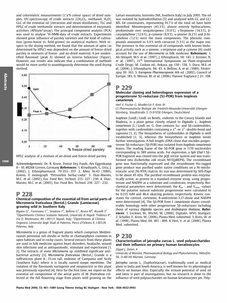
and colorimetric measurements (L*a*b colour space) of dried sam-ples, UV-spectroscopy of crude extracts (CH2Cl2, methanol, H2O),GLC of the essential oil (extraction and steam distillation), TLC andHPLC of crude methanolic extracts and determination of enzymaticactivities (APIzym assay). The principal component analysis (PCA)was used to analyse 1H-NMR-data of crude extracts. Experimentsshowed great influence of parsley varieties and the kind of cultiva-tion (green-house vs. field-grown) on analytical markers. With re-spect to the drying method, we found that the amount of apiin (asdetermined by HPLC) was dependent on the amount of freeze-driedparsley in mixtures of freeze- and air-dried material. A yet uniden-tified flavonoid (peak A) showed an inverse behaviour (Figure).However, our results also indicate that a combination of methodswould be more useful to unambiguously determine the used dryingmethod.
HPLC analysis of a mixture of air-dried and freeze-dried parsley
Acknowledgements: Dr. R. Kruse, Freeze Dry Foods, Am Eggenkamp8–10, 48268 Greven, Germany References: 1. Kreydiyyeh, S., Usta, J.(2002), J. Ethnopharmacol. 79:353–357. 2. BAnz Nr.43 (1989),Komm. E monograph “Petroselini herba/-radix“ 3. Diaz-Maroto,M.C. et al. (2002), Eur. Food Res. Technol. 215: 227–230. 4. Diaz-Maroto, M.C. et al. (2003), Eur. Food Res. Technol. 216: 227–232.
P 228Chemical composition of the essential oil from aerial parts ofMicromeria fruticulosa (Bertol.) Grande (Lamiaceae)growing wild in Southern ItalyRigano D1, Formisano C1, Senatore F1, Bellone G2, Bruno M2, Rosselli S21Dipartimento Chimica Sostanze Naturali, Universit di Napoli “Federico II“,Via D. Montesano, 49, I-80131 Napoli, Italy; 2Dipartimento di ChimicaOrganica, Universit degli Studi di Palermo, Parco d’Orleans II, I-90128Palermo, Italy
Micromeria is a genus of fragrant plants which comprises Mediter-ranean perennial sub shrubs or herbs or chamaephytes common inopen habitats and in rocky coasts. Several extracts from these plantsare used in folk medicine against heart disorders, headache, woundskin infections and as antispasmodic, stimulant and expectorant [1,2]. The extracts of some Micromeria sp. exhibited significant anti-bacterial activity [3]. Micromeria fruticulosa (Bertol.) Grande is asuffruticous plant 8– 15 cm tall, endemic of Campania and Sicily(Southern Italy) where it is locally named issopo marittimo. Theisolation of the flavonoids naringenin and neoponcirin in this plantwas previously reported [4]. Here for the first time, we report on theessential oil composition of the aerial parts of M. fruticulosa col-lected at the full flowering stage from plants wild growing on the
Lattari mountains, Sorrento (NA, Southern Italy) in July 2005. The oilwas isolated by hydrodistillation [5] and analyzed with GC and GC/MS. 64 constituents, representing 91.7% of the total oil have beenidentified. Monoterpenes (43.3%), almost entirely hydrocarbons,predominate over sesquiterpenes (31.0 %). g-Terpinene (14.5%), b-caryophyllene (12.6%), p-cymene (8.9 %), a-pinene (8.2%) and b-bi-sabolene (7.2 %) were the main components. The phenolic com-pounds amounted to 5.9% with carvacrol (5.3%) as the major one.The presence in this essential oil of compounds with known biolo-gical activity such as a-pinene, g-terpinene and p-cymene [6] couldaccount for the use of Micromeria in folk medicine. References: 1.Ali-Shtayeh, M.S. et al. (1997), J. Ethnopharm. 58: 143. 2. Kirimer, N.et al. (1997), 11th International Symposium on Plant-originatedCrude Drugs. M. Coskun ed., Ankara, pp. 130–138. 3. Duru, M.E. etal. (2004), J. Ethnopharm. 94: 43. 4. Bellino, A. et al. (1980), Fitoter-apia 10: 163. 5. European Pharmacopoeia 4th ed. (2002), Council ofEurope, 183. 6. Mitsuo, M. et al. (2006), Flavour fragrance J 21: 198.
P 229Molecular cloning and heterologous expression of aprogesterone 5b-reductase (5b-POR) from IsoplexiscanariensisHerl V, Fischer G, Mueller-Uri F, Kreis WLS Pharmazeutische Biologie der Friedrich-Alexander-Universit t Erlangen-N rnberg, Staudtstraße 5, D-91058 Erlangen, Deutschland
Isoplexis (Lindl.) Lindl. ex Benth., endemic to the Canary Islands andMadeira, is a plant genus closely related to Digitalis L.. Isoplexiscanariensis (L.) Lindl. ex. G. Don contains 5a- and 5b-cardenolides,together with cardenolides containing a D4 -or D5 -double-bond andsaponins [1, 2]. The biosynthesis of cardenolides in Digitalis is wellestablished [1, 3], whereas the biosynthesis in Isoplexis needsfurther investigation. A full-length cDNA clone that encodes proges-terone 5b-reductase (5b-POR) was isolated from Isoplexis canariensisleaves. The reading frame of the 5b-POR gene is 1170 nucleotidescorresponding to 389 amino acids. For expression, a Sph I/Sal I 5b–POR fragment was cloned into the pQE vector system and was trans-formed into Escherichia coli strain M15[pREP4]. The recombinantgene was functionally expressed and the recombinant His-taggedgene product was purified under native conditions on a Ni-nitrilo-triacetic acid (Ni-NTA) matrix. Its size was determined by SDS-Pageto be about 45 kDa. The purified recombinant protein was enzyma-tically active, as proven in a standard enzyme assay, using proges-terone and NADPH as a substrate and cosubstrate, respectively. Bio-chemical parameters were determined; the Km – and Vmax -valuesfor the putative natural substrate progesterone were calculated tobe 0.215 mM and 46.4 nkat/mg protein, respectively. Kinetic con-stants for cortisol, cortexone, 4-androstene-3,17-dione and NADPHwere determined [4]. The 5b-POR from I. canariensis shares consid-erable homology with other progesterone 5b-reductases includingthose of various Digitalis species and Arabidopsis thaliana. Refer-ences: 1. Luckner, M., Wichtl, M. (2000), Digitalis, WVG Stuttgart.2. Schaller, F., Kreis, W. (2006), Planta Med. submitted. 3. Kreis, W. etal. (1998), Planta Med. 64: 491–499. 4. Herl, V. et al. (2006), PlantaMed. submitted.
P 230Characterization of Jatropha curcas L. seed polysaccharidesand their influence on primary human keratinocytesZippel J, Deters AUniversity of M nster, Pharmazeutical Biology and Phytochemistry, Hittorfstr.56, D-48149 M nster, Germany
Jatropha curcas L. (Euphorbiaceae), traditionally used as medicalplant in India and South America, is well known for its physiologicaleffects on human skin. Especially the irritant potential of seed oiland latex is part of investigations, but no research is done to theinfluence of seed polysaccharides on human keratinocytes yet. Poly-
Planta Med 2006; 72: 961–1089 Georg Thieme Verlag KG Stuttgart · New York · ISSN 0032-0943
Abstracts
1050
saccharides were isolated by water extraction from the seed endo-sperm and the monosaccharide composition was elucidated by GCFID. Galactose, Rhamnose, Arabinose, Mannose and Glucose werethe main components, in a ratio of 1: 1,2: 1,7: 1,8: 1,6. Additionallyminor amounts of Fucose, Ribose and Xylose were found. Galacturo-nic acid was detected by Dionex HPLC and TLC. Protein content(10%) of the crude extract was determined by Bradford Test [1].Treatment of primary human keratinocytes with the crude extract(10mg/mL) showed a strong enhancement of proliferation deter-mined by BrdU- incorporation ELISA. Cell viability, measured bythe MTT-Test, was improved and no necrotic cytotoxicity could beobserved. Immuno blotting showed an increased production of Ker-atin and Involucrin, which characterizes an initiated differentiationas consequence of the intensified proliferation. Reference: 1. Brad-ford, M.M. (1976), Anal. Biochem. 72: 248–254
P 231Antioxidants from Xylocarpus granatumWangensteen H1, Duong GM1, Alamgir M2, Sarder M2, Samuelsen AB1,Malterud KE11Dept. of Pharmaceutical Chemistry, School of Pharmacy, University of Oslo,P. O. Box 1068 Blindern, N-0316 Oslo, Norway; 2Pharmacy Discipline, KhulnaUniversity, Khulna-9208, Bangladesh
Xylocarpus granatum Koeg. (Meliaceae) is growing in mangrove for-ests in Southeast Asia, tropical Australia and East Africa The barkhas been used in traditional medicine to treat diarrhea, cholera,fever and abdominal troubles, and it is well known for its astringentproperties. Chemically, the tree contains a number of xyloccensins(triterpenoids of limonoid type). Content of tannins has also beenreported, but their chemical structures appear to be unknown. Theaim of this study was to isolate and identify chemical substancesfrom X. granatum and to evaluate the DPPH radical scavenging ac-tivity and the inhibitory effect towards 15-lipoxygenase (15-LO).Total phenolic content was quantified by the Folin Ciocalteu meth-od. X. granatumwas collected in the Sundarbans mangrove forest inBangladesh. The 80% methanol extract of the stem bark was sus-pended in distilled water and successively extracted with chloro-form, ethyl acetate and n-butanol. The extracts were fractionatedby different chromatographic techniques, compounds were identi-fied by NMR and by degradation with phloroglucinol/HCl. Highamounts of procyanidins in addition to the monomeric compoundscatechin and epicatechin were isolated. The structures of the pro-cyanidins were procyanidin B1 (epicatechin (4b?8) catechin), epi-catechin (4b?8) epicatechin (4b?8) catechin and epicatechin(4b?8) epicatechin (4b?8) epicatechin (4b?8) epicatechin(4b?8) catechin. The limonoids gedunin, xyloccensin O, xyloccensinP and xyloccensin Q were isolated, as well. The procyanidinsshowed high DPPH radical scavenging and 15-LO inhibitory activ-ities, whereas the limonoids were inactive as radical scavengers andshowed only weak inhibition of 15-LO.
P 232Direct and indirect antimicrobial activity of Cordia gilletiiextractsOkusa PN1,2, Penge O2, Devleeschouwer M3, Duez P11Universit Libre de Bruxelles, Institut de Pharmacie, Laboratoire dePharmacognosie, de Bromatologie et de Nutrition Humaine, CP 205/9, 1050Brussels, Belgium; 2Universit de Kinshasa, Facult de Pharmacie, Laboratoirede Pharmacognosie, BP 212 Kin XI, Dem. Rep. Of Congo; 3Universit Libre deBruxelles, Institut de Pharmacie, Laboratoire de MicrobiologiePharmaceutique et Hygi ne, CP 205/2, 1050 Brussels, Belgium
The alarming incidence of antibiotic resistance causes an increasingneed for new products that can act either by a direct antimicrobialactivity or by inhibiting resistance mechanisms of germs of medicalimportance. Plants represent a potential source for this kind ofcompounds [1, 2]. Root barks of Cordia gilletii De Wild (Boragina-
ceae), a Congolese plant traditionally used for antimicrobial proper-ties, were extracted successively by n-hexane, dichloromethane,ethyl acetate, methanol and water. These extracts were tested fordirect antimicrobial activity against eight microbial species (Staphy-lococcus aureus, Escherichia coli, Pseudomonas aeruginosa, Klebsiellapneumoniae, Enterobacter cloacae, Proteus mirabilis, Serratia marces-cens and Candida albicans) and for effect on antibiotic resistance bybroth microdilution methods [3, 4]. The methanol extract showeddirect antimicrobial activity against all the strains with MIC valuesranging between 125mg/mL and 1000mg/mL, whereas the ethylacetate and dichloromethane extracts showed activity on two (Sta-phylococcus aureus and Escherichia coli) and three (Klebsiella pneu-moniae, Enterobacter cloacae and Serratia marcescens) microbial spe-cies respectively. 200mg/mL of the n-hexane and dichloromethaneextracts decreased the MIC of penicillin, amoxicillin, ampicillin andstreptomycin 4–64 fold for S. aureus methicillino-resistant. Ac-knowledgement: Dr Lerson (CHU Charleroi, Belgium), Belgian Tech-nical Cooperation. References: 1. Chariandy, C.M. et al. (2000), J.Ethnopharmacol. 64: 265–270. 2. Hatano, T. et al. (2005), Phyto-chemistry 66: 2047. 3. NCCLS (2003), Approved Standard, 6th edi-tion. 4. NCCLS (2002), Approved Standard, 2th edition.
P 233Stimulation of LAL-test by LPS-free arabinogalactan-proteinpreparations from Echinacea purpureaBlaschek W1, Zager A1, Classen B1, Ulmer AJ21University of Kiel, Institute of Pharmacy, Gutenbergstr. 76, 24118 Kiel,Germany; 2Research Center Borstel, Parkallee 22, 23845 Borstel, Germany
While investigating the immunological activities of Echinacea pre-parations, the question of microbiological contamination arises, be-cause microorganisms or parts and products of them may influencethe test results. Especially LPS from gram-negative bacteria andlipopeptides from gram-negative and gram-positive bacteria maystimulate the immune-system already at very low concentrations.Arabinogalactan-proteins (AGPs) from Echinacea purpurea [1] weretested in the LAL-test, using the gel-clotting-method, before andafter LPS-removal. Since this test responses not only to LPS, but alsoto b-1,3-glucans [2], it seemed possible that there could also be aresponse to other polysaccharide containing polymers such as AGPs.After removal of LPS by two different methods (affinity chromato-graphy and treatment with sodium hydroxide), absence of LPS andbacterial lipopeptides was proven by testing the LPS-free AGP pre-paration for interaction with toll-like receptors (TLR) 2- or TLR4-transfected HEK293 cells. There was no reactivity of AGP prepara-tions with these receptors, indicating that LPS and lipopeptides havebeen successfully removed. After blocking of possible glucan-activ-ity in the LAL-test by a special buffer, the LPS-free AGP preparationsstill led to coagulation. The remaining activity in the test thereforehas to be considered as the activity of AGPs. Thus, AGPs are able toactivate the LAL-test in a mg-concentration range. This is lower, butconsiderable activity compared to the activity of glucans (ng-con-centration range) or LPS (pg-concentration range). Acknowledge-ment: The authors thank the Madaus AG, K ln, for financial supportof this work. References: 1. Classen, B. et al. (2000), Carbohydr. Res.327: 497–504. 2. Blaschek, W. et al. (1992), Pharm. Pharmacol. Lett.1: 118–122.
P 234Phytoestrogenic Activity of Morinda citrifolia L. FruitsBasar S, Iznaguen H, Zeglin A, Westendorf JInstitute of Experimental and Clinical Pharmacology and Toxicology,University Clinic Hamburg Eppendorf, Vogt-K lln-Straße 30, D-22527Hamburg, Germany
It is well known that certain plant metabolites can exert estrogenicactivity. There are an increasing number of studies concerning theeffects of phytoestrogen-rich diets which demonstrate that these
Planta Med 2006; 72: 961–1089 Georg Thieme Verlag KG Stuttgart · New York · ISSN 0032-0943
Abstracts
1051
plant metabolites have protective effects on estrogen-related condi-tions, such as menopausal symptoms, and estrogen-related diseases,such as prostate and breast cancers, osteoporosis and cardiovasculardiseases [1]. Because beneficial effects on menopausal symptomsare often reported by women drinking regularly Noni fruit juice(Morinda citrifolia L.), we investigated the estrogenic capacity ofNoni fruits in two in vitro assays, the estrogen receptor bindingassay with both estrogen receptors, ER-a and ER-b, and the estro-gen-receptor dependent induction of alkaline phosphatase in Ishi-kawa cells. Hexane extracts prepared from Noni fruit puree exhib-ited high activity in both systems. A preferential binding for ER-bwas observed (ED50(ER-a)/ED50(ER-b)= 2.33). Further analysis andfractionation of these extracts by HPLC showed that one compoundis responsible for almost all the activity. The isolation of this con-stituent was performed by HPLC and will be followed by its char-acterisation with 1- and 2-dimensional NMR techniques and Massspectrometry. Reference: 1. Cos, P. et al. (2003), Planta Med. 69:589–599.
P 235Cytotoxicity of b-aescin/agrostin mixtures in different celllines depends on their growth characteristicsWeng A, Melzig MFFreie Universit t Berlin, Institut f r Pharmazie, K nigin-Luise-Str. 2+4, D-14195 Berlin
Agrostin (Mr: 27kDa), a ribosome-inactivating- protein (RIPs) fromAgrostemma githago L., cleaves an essential adenin residue from therRNA, leading to inhibition of protein syntheses [1]. In previousstudies it was shown, that only the combination of specific triterpe-noid saponins such as Saponinum album L. with a formyl functionattached to position C4 together with agrostin was cytotoxic in ECV-304 cells [2]. This enhancement in cytotoxicity by Saponinum albumL is due to an enhanced penetration of agrostin through the cellmembrane, indicating the induction of endocytosis, because thetreatment with latranculin A und bafilomycin A 1 inhibited the cy-totoxicity in ECV-304 cells [3]. b-Aescin is also a triterpenoid sapo-nin and the major compound of aescin, a mixture of glycosidssynthesized by Aesculus hippocastanum L.. In this study we investi-gated the cytotoxic effect of b-aescin (10–2.5mg/mL) /agrostin (150ng/mL) mixtures in ECV-304, Hep-G2, SK-N-SH, U-937 and H-2171cells. In contrast to the non-adherent U-937 and H 2171 cells theproliferation of adherent growing cell lines like ECV-304, Hep-G2and SK-N-SH was significantly reduced by the mixture of b-aescinand agrostin. It is therefore concluded that the stimulation of en-docytosis of the cytotoxic agrostin by b-aescin depends on specificmembrane structures present on the cell surface of adherent grow-ing cells. Especially the difference in the caveolin-1 expression be-tween adherent growing (high) and in suspension growing cells(low) was demonstrated [4]. Caveolin-1 is an integral membraneprotein and correlates with the number of caveolae in the cell mem-brane. The caveolae, flask shaped invaginations in the cell mem-brane, are necessary for internalization of endocytotic markers andby that strongly involved in endocytosis. References: 1. Stirpe, F.(2004), Toxicon 44: 371–83. 2. Melzig, M.F. et al. (2005), PlantaMed. 77: 1088–1090. 3. Hebestreit, P. et al. (2006), Toxicon 47:330–35. 4. Sunaga, N. et al. (2004), Canc. Res. 64: 4277–4285. 5.Kiss, L.A. et al. (2002), Micron 33: 75–93.
P 236An assessment of the use of an in vitro cell based model ofthe intestinal tract to investigate the bioavailability of theChinese herbal remedy Oldenlandia diffusaPanagiotou E, Jones LA, Opara EIBiomedical and Pharmaceutical Sciences Research Group, School of LifeSciences, Kingston University, Penryhn Road, Kingston upon Thames, Surrey,KT1 2EE, UK
Certain Chinese herbal remedies (CHRs) are used to treat cancer.However, their mode of action has yet to be fully elucidated. Anearlier study of the anti-cancer activity of one such CHR, Oldenlan-dia diffusa (Willd.) Roxb (OD) indicated that its cell cytotoxicity:may occur via apoptosis; be cancer cell specific; and involve morethan one constituent [1, 2]. However, as this study was carried out invitro, no information about the impact of the intestinal tract on theanti-cancer activity of this remedy, which is commonly ingested inthe form of a tea, was obtained. Thus the aim of this study was toassess the use of a 21 day in vitro model of the intestinal tract toinvestigate the bioavailability of this CHR. The colon adenocarcino-ma cell line Caco-2, in the form of a monolayer, was used to mimicthe intestinal tract. The integrity of this model was assessed usingthe trypan blue exclusion test, transepithelial electrical resistance(TEER), phenol red exclusion and scanning electron microscopy(SEM). Cytotoxicity assessment using trypan blue showed that themodel was not adversely affected by OD (ranging in concentrationfrom 15–70%). Phenol red exclusion and TEER measurementsshowed that the model’s integrity was maintained before and afterexposure to OD. Furthermore, SEM showed that the monolayer re-tained its microvilli expression and tight junctions formation beforeand after exposure to OD. These results demonstrate that this modelcan be used to study the bioavailability of this CHR. References: 1.Willimott, S. et al. (2005), Isolation of tumour modulatory com-pounds in Chinese herbal remedies through activity guided fractio-nation. Poster at the International Conference and 53rd AnnualMeeting of the Society for Medicinal Plant Research; Florence Au-gust 2005. 2. Willimot, S., Barker J., Jones L., and Opara EI. TheChinese herbal medicine Oldenlandia diffusa induces cell-cycle-in-dependent apoptosis in the leukaemic HL60 cell line and growtharrest in PHA-stimulated blood lymphocytes. (in preparation).
P 237Variations in extraction protocol lead to differences inmonosaccharide composition and bioactivity on humankeratinocytes as shown by polysaccharides from banana andplum fruitsDeters A, Maas M, Kemper M, Lamerding F, Klenke AInstitute for Pharmaceutical Biology and Phytochemistry, WestfaelischeWilhelms University Muenster, Hittorftsr. 56, 48149 Muenster, Germany
In precede investigations polysaccharides have been shown to ex-hibit different bioactivities on the cell physiology of primary kera-tinocytes dependent on their monosaccharid composition related tothe plant, which was used for extraction. The intention of the pres-ent study was that the way of polysaccharide precipitation leads toa varition of monosaccharide composition and different bioactiv-ities, too. For the investigations water extracts from banana (Musaparadisica var. sapientum, Musaceae) and plum (Prunus domestica L.,Rosaceae) fruits were splitted and carbohydrates were obtained bytwo ways of precipitation: 1. the water extracts were dropped intoethanol and 2. ethanol was dropped into the extracts to a finalethanol concentration of 80%. Subsequently polysaccharides wereexamined by chemical and chromatographic methods and incu-bated with human keratinocytes. The analysis of monosaccharidesby Dionex HPLC, GC/FID revealed that the polysaccharides obtainedafter dropping of ethanol in the extracts contained up to 20% moreuronic acid determined as galacturonic acid by TLC than the poly-saccharides obtained by method 1. Further the amounts of fructose,galactose and arabinose rose up to 26% (Fruc), 20% (Gal) and 10%
Planta Med 2006; 72: 961–1089 Georg Thieme Verlag KG Stuttgart · New York · ISSN 0032-0943
Abstracts
1052

(Ara). Differences in the protein content were not observed. Inves-tigation of keratinocyte cell physiology after incubation with thepolysacchrides exhibited that the polysaccharides obtained afterdropping of ethanol in the water extract triggered the cell prolifera-tion and cell viability to a greater extend than the others, which hadno or only minor effects. Further polysacchrides aquired by method2 reduced the amount of necrotic cells. The obtained data show thatslight variations in extrac
P 238Polysaccharides from Glycyrrhiza glabra L. exert significantanti-adhesive effects against Helicobacter pylori andPorphyromonas gingivalisWittschier N1, Faller G2, Beikler T3, Stratmann U4, Hensel A1
1Institute of Pharmaceutical Biology and Phytochemistry, University ofM nster, Hittorfstr. 56, D-48149 M nster, Germany; 2Institute of Pathology,University of Erlangen-N rnberg, Krankenhausstrasse 8 – 10, D-91054Erlangen, Germany; 3Department of Periodontology, University ofWashington, Seattle, USA; 4Institute of Anatomy, University of M nster,Vesaliusweg 2 – 4, D-48149 M nster, Germany
Glycyrrhiza glabra L. (Fabaceae), one of the oldest medicinal plantsof the world, is a ligneous perennial shrub growing in Mediterra-nean region and Asia. Because of the expectorant, anti-spasmodicand anti-inflammatory effects extracts from roots are used thera-peutically against bronchitis and gastric ulcer. In order to investigatenew modes of action we investigated the influence of isolated poly-saccharides from Liquorice roots with regard to their capacity toreduce bacterial binding to host cells. In the present study an in-situadhesion model with Helicobacter pylori and Porphyromonas gingi-valis on sections of human gastric mucosa resp. rat esophagus mu-cosa was used as screening model for anti-adhesive activity. Prein-cubation of Helicobacter-suspensions with a solution of the rawpolysaccharide resulted in a significant decrease in the bacterialadhesion to gastric mucosa of 40% compared with the non-treatedcontrol. Fractionation of the raw polysaccharide via anion ion-ex-change chromatography (AEX) yielded 5 subfractions. The strongestreduction of adhesion exhibited the 0.25 molar fraction (60% inhi-bition) while the other fractions were inactive. The AEX-fractionswere further separated by gel permeation chromatography. Respec-tive polysaccharide structures were elucidated. Considerable anti-adhesive effects of liquorice root polysaccharides were also ob-served after pre-treatment of P. gingivalis. Furthermore the agardiffusion-test revealed absence of any cytotoxicity of the raw poly-saccharides against H.pylori and P.gingivalis. Thus, data show thatpolysaccharides from Glycyrrhiza glabra L. are a potent agent againstbacterial adhesion and are able to block the initial step of an infec-tion.
P 239Pelargonium sidoides extract EPs 7630 inhibits adhesion ofHelicobacter pylori to human gastric mucosaWittschier N1, Faller G2, Hensel A1
1University of M nster, Institute of Pharmaceutical Biology andPhotochemistry, Hittorfstr. 56, D-48149 M nster, Germany; 2University ofErlangen-N rnberg, Institute of Pathology, Krankenhausstrasse 8 – 10,D-91054 Erlangen, Germany
Pelargonium sidoides (DC.), belonging to the family of Geraniaceae,originates from the southern parts of Africa. In traditional medicineextracts from roots are used in diseases of the respiratory systemand gastrointestinal complaints. Nowadays a root extract (Umcka-loabo ) is used therapeutically as antimicrobial agent against infec-tions of the respiratory system. In order to elucidate possible modesof actions we investigated the influence of the extract EPs7630 con-cerning its influence on microbial adhesion. As model microorgan-ism Helicobacter pylori was used, a germ with a strong adherence tohuman stomach tissue via its highly specific surface adhesions. In
an in-situ anti-adhesion assay intact human stomach tissue frompatient resectates was incubated with fluorescent-labelled bacteria.Epithelial adhesion occurred in untreated samples and was quanti-fied by fluorescent microscopy. Pre-treatment of the bacteria withEPs 7630 showed good anti-adhesive activity, being less than thatobtained by the positive control blocker sialyllactose. The antiadhe-sive effect was clearly dose-dependent in a range from 0.001 to10mg/mL. Using an agar diffusion-test it was shown that EPs7630had no direct cytotoxicity against Helicobacter pylori over the con-centration range used in the adhesion assays. The results show thatthe extract from Pelargonium sidoides is a potent anti-adhesiveagent against Helicobacter pylori and could therefore be a usefulchoice to avoid the first step of a bacterial infection.
P 240Impact of fertilization on the accumulation of leaf salicylatesin four field-grown dark-leaved willow (Salix myrsinifoliaSalisb.) clonesPaunonen R1, Julkunen-Tiitto R1, Tegelberg R1, Rousi M2
1University of Joensuu, Department of Biology, PO Box 111, FIN-80101,Joensuu, Finland; 2The Finnish Forest Research Institute, Finlandiantie 18, FIN-58450, Punkaharju, Finland
Due to their anti-inflammatory and analgesic properties, salicylatesare medically interesting phenolic compounds [1]. In some patients,asperin, a synthetic derivative of salicin (b-D-glucoside of 2-hydro-xybenzyl alcohol), induces more side effects than salicin [1]. Thus,herbal drugs could be excellent alternatives to asperin. The leaves ofdark-leaved willow (Salix myrsinifolia) contain salicylates (salicinand its derivatives) [2], and are therefore a promising source ofherbal drugs. The aim of the present study was to clarify the impactof fertilization on leaf biomass and salicylates in four dark-leavedwillow clones. Willows were established from cuttings in May 2002and were grown on plastic mulch in field trials in Eastern Finland.Two fertilization treatments were used: 0 and 150kg (N)/ha. InAugust 2004, leaves were collected and air-dried. Soluble phenolicswere extracted with methanol and quantified using HPLC/DAD [3].Results showed that fertilization increased significantly leaf bio-mass (g plant-1, dw) (F =6.458, DF=1, P < 0.05) and concentrations(mg/g, dw) of leaf salicortin (a derivative of salicin) (F =6.098, DF =1,P < 0.05) and total salicylates (F =4.424, DF =1, P < 0.05). The in-creased production of other phenolics (chlorogenic acid and quer-cetin-3-galactoside) did not limit the salicylate accumulation. How-ever, most salicylates varied quantitatively among clones, andclones also responded differently to fertilization (P < 0.05). Thus,the selection of clones for cultivation should be made with care.Acknowledgements: The Academy of Finland (project no. 64308),University of Joensuu. References: 1. Pierpoint, W.S. (1994), Adv.Bot. Res. 20: 163–235. 2. Julkunen-Tiitto, R., Meier, B. (1992), PlantaMed, 58: 77–80. 3. Julkunen-Tiitto, R. et al. (1996), Trees-Struct.Funct 11: 16–22.
P 241Pharmacological in vivo test to evaluate the bioavailability ofsome St. John’s wort innovative oral preparationsBilia AR1, Bergonzi MC1, Isacchi B1, Galeotti N2, Ghelardini C2, Vincieri FF11Department of Pharmaceutical Sceinces, University of Florence, 50019 SestoFiorentino, Florence, Italy; 2Department of Preclinical & ClinicalPharmacology, University of Florence, 50139 Florence, Italy
Preparations based on extracts of St. John’s wort are widely mar-keted for treating mild to moderately severe depressive disordersand other health conditions such as anxiety and sleep disorders [1].Active principles are not yet discovered and flavonols, based onquercetin aglycone, naphthodianthrones (hypericin and pseudohy-pericin) and phloroglucinols such as hyperforin, adhyperforin seemsto be related to this action. Thus, flavonols and naphthodianthronesare polyphenols, quite polar derivatives but their water solubility is
Planta Med 2006; 72: 961–1089 Georg Thieme Verlag KG Stuttgart · New York · ISSN 0032-0943
Abstracts
1053

very scarce; phloroglucinols are lipophilic and completely notwater-soluble constituents. In addition, hypericins and hyperforinsare not stable with regard to heat and light [2]. In this study theoptimisation of technological and pharmaceutical aspects of driedcommercial extract of St. John’s wort were evaluated by the in vivo“Porsolt test“. Solid dosage forms containing b-cyclodextrin and mi-cellear systems (SDS, ASC-8) were compared in the “Porsolt test“with the extract alone. The extract showed the antidepressant ac-tivity in the mice after 60 minutes and with the dosage of 100mg/kg. The same antidepressant activity appeared in 30min with amicellar solution of SDS 40mM containing the same quantity ofextract (100mg/kg), while with micelles of ASC-8 40 mM the effectappeared at 15min and with a dosage of 30mg/kg. In the case ofcolyophilized with b-cyclodextrin the best results were obtained at30min, administering 60mg/kg of the extract. Acknowledgements:The financial support of MIUR (PRIN 2004) and Ente Cassa di Ris-parmio di Firenze is gratefully acknowledged for financial support.References: 1. Chatterjee, S.S. et al. (1998), Pharmacopsychiatry 31:7–15. 2 Bilia A.R. et al. (2001), Int. J. Pharm. 213: 199–208.
P 242Composition of the Essential Oils from Three Species fromLabiatae from IranMojab F, Nickavar BDepartment of Pharmacognosy, Faculty of Pharmacy and PharmaceuticalSciences Research Center, Shahid Beheshty University of Medical Sciences,Tehran, P.O. Box 14155 – 6153, Iran
The aerial parts of three species from Labiatae (Thymus carmanicusJalas, Salvia hypoleuca Benth. and Teucrium Stocksianum Boiss.) fromIran were obtained by hydrodistillation in a clevenger-type appara-tus for 3h (2%, 0.3% and 0.5%, respectively). The species were col-lected from Karkas-Kuh (Natanz area, Province Isfahan), Ab-ali(North of Tehran) and Siahoo, North of Bandar-Abbas, Province Hor-mozegan), respectively. This oil has been examined by GC and GC/MS. The components of the oil were identified by comparison oftheir fragmentation patterns of mass spectra and retention indiceswith those published in the literature and presented in the MScomputer library. In the oil of T. carmanicus, monoterpenes predo-minated over sesquiterpenes. Thymol and carvacrol were the majorcomponents of the oil, 20.8 and 52.8%, respectively. Other compo-nents were h-terpinene (5.4%), p-cymene (4.1%) and borneol (1.5 %).In the oil of T. Stocksianum a-pinene (24.5%) and a-copaene (3.4%)were major compounds; and in the oil of S. hypoleuca, bicycloger-macrene (15.3%), b-caryophyllene (14.6%), viridiflorol (13.3%),spathulenol (12.5%) d-elemene (7.7 %), b-pinene (7.2 %) and a-pinene (5.9%) were major compounds.
P 243Artemisinin and flavonoids yield from aqueous extracts andtinctures of Artemisia annua LBilia AR1, Gabriele C1, Bergonzi MC1, Melillo de Malgalhaes P2, Vincieri FF11Department of Pharmaceutical Sceinces, University of Florence, 50019 SestoFiorentino, Florence, Italy; 2Divis¼o de Agrotecnologia, CPQBA-UNICAMP, C.P.6171, 13.081.970 Campinas, SP, Brasil
Malaria morbidity and mortality continue to increase across theentire world [1]. This is largely as a result of the continued use ofchloroquine and sulfadoxine-pyrimethamine, despite widespreadresistance. Artemisinin is an interesting molecule to treat multi-drug-resistant Plasmodium falciparum malaria. It is extracted fromthe plant qinghao (Artemisia annua L. or sweet wormwood) [2].After the discovery of the active principle artemisinin almost allthe clinical evaluations have focused on pure, isolated artemisininand its derivatives. Clinical trials with patients using teas or decoc-tions have appeared in the most recent literature [3 –5], after devel-opment of high artemisinin-yielding plants (> 0.5% per driedweight). The principal aim of such investigations is related to the
possibility for populations in endemic areas to cultivate selectedbreedings of A. annua and prepare teas or decoctions with a positiveeffect in the treatment of malaria. In this study the qualitative pro-file and content of artemisinin and polymethoxyflavones is investi-gated on infusions and decoctions prepared with different methodsand on 40% w/v and 60% w/v tinctures using a hybrid form of A.annua successfully cultivated in Brasil. The aerial parts of the plantcontained 0.52% artemisinin per dry weight, and approximately27–40% of this artemisinin could be extracted by simple tea pre-paration methods or decoctions and the best extraction is obtainedwith a short decoction (5min) followed by an infusion of 9 g herbaldrug in 1 L water. Tinctures 40% w/v extracted about 26% artemi-sinin while tincture 60% w/v about 40%. The content of total poly-methoxylated flavonoids in the plant was about 2.6 % mainly repre-sented by chrisoplenetin plus casticin (1.4 %), eupatin (0.8 %) andartemetin (0.4 %). The total flavonoid content extracted in the infu-sions ranged 30–60% and that of tinctures was less than 40%.Tinctures showed also the presence of chrisoplenol. Acknowledge-ments: The financial support of MIUR (PRIN 2004) and Ente Cassa diRisparmio di Firenze is gratefully acknowledged for financial sup-port. References: 1. World Health Organization, (2000), Trans. R.Soc. Trop. Med. Hyg. 94: 1–90. 2. O’Neill, P. et al. (2004), J. Med.Chem. 47: 2945–2964. 3 Mueller, M.S. et al. (2000), J. Ethnophar-macol. 73: 487–493. 4 Mueller, M.S. et al. (2004), Trans. Royal Soc.Trop. Med. Hyg. 98: 318–321. 5 R th, K. et al. (2004), Am. J. TropicalMed. Hyg. 70: 128–132.
P 244Yields in phenylpropanoids and antioxidant properties ofdifferent aqueous extracts of lemon verbena (Lippiacitriodora K.)Bilia AR, Giomi M, Innocenti M, Vincieri FFDepartment of Pharmaceutical Sciences, Via Ugo Schiff,6 50019, SestoFiorentino, Italy
Lemon verbena (Lippia citriodora K.) contains several flavonoids andphenylpropanoids mainly represented by verbascoside [1]. In orderto develop a rich phytocomplex of such constituents’ different aqu-eous extracts (i. e. a decoction obtained 5min. boiling, one obtainedafter 20min. boiling and a tea) were prepared from the dried plantand liophylised. Quantification of constituents in the dried extractswas carried out by HPLC-DAD-MS and the chromatographic condi-tions were the following: a binary system H2O (pH 3.2 by HCOOH)and CH3CN using a multi-step linear solvent gradient elution meth-od. Total time of analysis was 28min and flow rate was 0.8 mL/min.The column was a Varian Polaris TM C18-E (250 x4.6mm i.d., 5mm)mantained at 26 C with a pre-column of the same phase. By UV andMS spectral data were identified the constituents of the 3 extracts.The qualitative profiles of the three extracts was quite similar, 9constituents were identified, mostly of them represented by verbas-coside and its analogues, besides luteolin and apigenin derivativesand one iridoid, verbenalin. However, the yield of such constituentsand in particular phenylpropanoids was very different being in thelyophilized infusion 8.3% with respect to the decoctions, 1.7 or 4.4 %.The lowest content of phenylpropanoids in the liophilised decoc-tions was probably due to the heat instability of verbascosides andanalogs. In view of the pharmacological interest of verbascoside andanalogs as antioxidants, the DPPH test was also carried out accord-ing to Son and coworkers [2]. Acknowledgments: The financial sup-port of MIUR (PRIN 2004) is gratefully acknowledged. References: 1.Valentao, P. et al. (2002), Biol. Pharm. Bull. 2: 1324–1327. 2. Son, S.,Lewis, B.A. (2002), J. Agric. Food Chem. 50: 468 –472.
Planta Med 2006; 72: 961–1089 Georg Thieme Verlag KG Stuttgart · New York · ISSN 0032-0943
Abstracts
1054

P 245Fatty Acid Patterns of the Various Parts of Turkish Pistaciavera L. TreeAslan M, Orhan IDepartment of Pharmacognosy, Faculty of Pharmacy, Gazi University, 06330Ankara, Turkey
Pistacia vera L. (Anacardiaceae) is a small tree grown in southernEurope and Asia minor, being the only species within the 11 speciesbelonging to the genus Pistacia that produces edible nuts. Pistachiois a nut of the tree having an edible green kernel enclosed in awoody shell. P. vera is widely cultivated in southern Anatolia forits nuts and has a significant contribution to the major agriculturalexports of Turkey. Extensive researches have exerted that pistachionuts are a rich source of fatty acids [1 – 6]. A survey for fatty acidcomposition was made for the waste products of P. vera (pistachiotree) grown in Turkey. The waste products and various parts of thetree were classified as fresh leaves (FL), dried leaves (DL), stem (ST),branches (BR), fresh skin of natural-woody shell (non-processed)(FSN), fresh kernel (FK), and skin of processed-woody shell (SP). Inthis study, gas chromatography-mass spectrometry data revealedthat FSN, FL, DL, and, ST could be evaluated to be rich sources forfatty acids. In particular, FL contains a remarkable amount of lino-lenic acid (30.4 €3.28%). References: 1. Agar, It., Sarmiento, C. et al.(1995), Acta Horticult. 419: 405–410. 2. Agar, It., Kaska, N., Kafkas, S.(1995), Acta Horticult. 419: 417–422. 3. Aslan, M., Orhan, I., Sener, B.(2002), Int. J. Food Sci. Technol. 37: 333–335. 4. Garcia, Jm., Agar, It.,Streif, J. (1992), Gartenbauwissenschaft 57: 130–133. 5. Kucukoner,E., Yurt, B. (2003), Eur. Food Res. Technol. 217: 308–310. 6. Satil, F.,Azcan, N., Baser, K.H.C. (2003), Chem. Nat. Compds. 39: 322–325.
P 246Induction of Cytoprotective Mechanisms by theChemopreventive Isothiocyanate Sulforaphane in Rat andMurine Hepatoma Cell LinesHamed A, Fry JSchool of Biomedical Sciences, Medical School, Queen’s Medical Centre,Nottingham NG7 2UH, UK
Sulforaphane [1-isothiocyanato-4(methylsulfinyl) butane, SN] is awell known cruciferous chemopreventive phytochemical that in-duces the anticarcinogenic enzyme quinone reductase (QR, NQO1,EC 1.6.99.2) and other phase II detoxification enzymes [1]. The pres-ent study aimed to investigate the induction of QR activity and heatshock protein 70 (HSP70) expression in the rat hepatoma (FGC4)and murine hepatoma (hepa1c1c7) cells by SN.
(A)
(B)Figure (1): Responses of FGC4 cells to SN treatment; induction of QR(A) and HSP70 (B).
Following 24 hours exposure to non-toxic concentrations of SN (asassessed by neutral red uptake), FGC4 cells were less sensitive to QRactivity induction (Figure 1A) as compared to hepa1c1c7 (significantinduction of 150%€16 vs. 440%€44, respectively at 6.25mM SN,p < 0.01) which may be due to the high basal QR activity of FGC4cells. In a preliminary experiment, SN had no effect on HSP70 ex-pression in hepa1c1c7 (data not shown). Interestingly, SN signifi-cantly induced HSP70 expression in FGC4 (Figure 1B) as revealed bywestern blotting and densitometric analysis (250%€49 at 25mM SN,p < 0.01). These data represent the first report of the induction ofHSP70 expression in vitro by SN as a cytoprotective mechanisminduced by this promising chemopreventive compound. In supportof these data, Hu et al. [2] have very recently reported the inductionof HSP70 genes in livers of SN- treated mice. Acknowledgements:Egyptian Government, School of Biomedical Sciences (Nottingham,UK). References: 1. Zhang, Y. et al. (1994), PNAS 91: 3147–3150. 2.Hu, R. et al. (2006), Cancer Lett. In press.
P 247Essential oils from leaves, stems and ripened seed capsulesof Hypericum undulatumGuedes AP, Fernandes-Ferreira MDepartament of Biology, University of Minho, Campus de Gualtar, 4710 – 057BRAGA, Portugal
Hypericum undulatum Willd. (Guttiferae) is a common herb in Por-tugal, growing in wet places and in the riverside edges. The phenolicextracts of leaves and aerial parts of this species have already beenstudied, showing the presence of hypericin, quercetin, quercetinsulphate, rutin, mangiferin, chlorogenic acid [1; 2]. However studiesof its volatile component are scarce. The essential oils were obtainedby hydrodistillation of a small amount of fresh leaves, stems andripened seed capsules harvested in September. More than 40 com-pounds were detected in the leaves and ripened seed capsules,while in the stems 20 compounds were detected. The compoundswere identified by GC-MS and quantified by GC. Excepting for thestems, in which there wasn’t any oxygenated compound, the identi-fied compounds in the essential oils from all samples distributed bymonoterpene hydrocarbons (MH), oxygenated monoterpenes (MO),sesquiterpene hydrocarbons (SH), oxygenated sesquiterpenes (SO)and alkanes. The most complex essential oils were those obtainedfrom leaves. In both leaves and ripened seed capsules the majorcompound group was the sesquiterpene hydrocarbons. However,in both essential oils, the major compound was an n-alkane (n-nonane). Caryophyllene oxide and globulol were the two major oxy-genated-sesquiterpenes in those two samples. n-Nonane was alsothe most represented in the stem essential oils, the major group ofcompounds being n-alkanes. b-Pinene, a monoterpene hydrocarbon,was also well represented in the three samples. References: 1. Sea-bra, R.M. et al. (1991), Rev. Port. Farm. 12: 16–18. 2. Seabra, R.M. etal. (1992), Fitoterapia 68: 473–474.
Planta Med 2006; 72: 961–1089 Georg Thieme Verlag KG Stuttgart · New York · ISSN 0032-0943
Abstracts
1055
P 248Analysis of Phenolic Acids from Actaea spec. by CapillaryElectrophoresisPrinza S, Singhubera J, Zhub M, Koppa B1Department of Pharmacognosy, University of Vienna, Althanstraße 14, A-1090 Vienna, Austria; 2Medical Institute POB 76, Beijing University of ChineseMedicine and Pharmacology, Bei San Huan Dong Lu No. 11, Beijing 100029,PR China
The rhizome of Actaea racemosa L. (syn. Cimicifuga racemosa L.),Ranunculaceae, is used for the treatment of menopausal disorders.Regarded as well accepted alternative to standard hormone therapy,the increasing demand of black cohosh leads to overharvesting ofthe wildcrafted plant in the US. In Asia related Actaea spec. arecultivated and pharmaceutically used in TCM (e. g. A. dahurica, A.foetida, A. heracleifolia, A. simplex). Problems in sourcing of A. race-mosa plant material and adulterations with those Actaea spec. usedin TCM are the consequence, requiring a sound analytical system forquality control. Whereas the pattern of triterpene glycosides is noteligible for a qualitative fingerprint, the phenolic acids (caffeic acid,ferulic acid, isoferulic acid, fukinolic acid and the cimicifugic acidsA, B, D, E and F) provide a convincing tool in sample identification todistinguish between different Actaea spec. [1]. The latter cimicifugicacids from the rhizomes of A. racemosa were fractionated and iden-tified according to [2] and [3]. A rapid method for the qualitativeanalysis of the phenolic acid fingerprint of methanolic extracts ofthe above mentioned Actaea spec. has been established on capillaryelectrophoresis (CE). Baseline separation of all phenolic acids wasachieved on a 50mm capillary (70 cm, 60 cm to detector) with a25mM borate buffer (pH 9.0) at 25 kV within 25 minutes, represent-ing a time and solvent saving method for quality control and asound alternative to HPLC. References: 1. Kusano, G. (2001), Yaku-gaku Zasshi 121: 497–521. 2. Stromeier, S. et al. (2005), Planta Med.71: 495–500. 3. Kruse, S.O. et al. (1999), Planta Med. 65: 763–764.
P 249Sesquiterpenoids and phenolics from roots of Cichoriumendivia var. crispumKisiel W, Michalska KDepartment of Phytochemistry, Institute of Pharmacology, Polish Academy ofSciences, 12 Smetna Str, Pl-31 – 343 Krakow, Poland
Endive (Cichorium endivia L.) and chicory (C.intybus L.) are the mostpopular species of the genus Cichorium (Asteraceae, Lactuceae).Leaves of commercial varieties are used as a salad additive or avegetable and roots are considered wastes. Chicory is also knownas a traditional herbal remedy which improve digestive and meta-bolic functions. A single report [1] on sesquiterpene lactones of C.endivia roots revealed the presence of germacranolides, eudesma-nolides and lactucin-like guaianolides. We have undertaken an in-vestigation of roots of C. endivia L. var. crispum Lam. The dried rootswere extracted with ethanol and the extract, after sequential frac-tionation on silica gel followed by semipreparative RP HPLC, gave 13sesquiterpene lactones, including 11 guaianolides, and three pheno-lics. The guaianolides 8-deoxylactucin and lactucopicrin appeared tobe major sesquiterpene lactone constituents, and the guaianolideshieracin II and macrocliniside G were found in Cichorium species forthe first time. Moreover, a new natural product was isolated andcharacterized as 10b-methoxy-1a (10), 11b(13)-tetrahydrolactucin.In addition, the roots yielded methyl- and ethyl p-hydroxyphenyla-cetates, and ethyl trans-caffeate, the latter in a relatively highamount. The formation of the ethyl esters is likely to occur duringthe extraction procedure. All the compounds were characterized byspectral methods. 8-Deoxylactucin, also known as the major sesqui-terpenoid of chicory roots [2], was reported to inhibit DNA bindingof the transcription factor NFkB [3] and cyclooxygenase-2 proteinexpression [4]. References: 1. Seto, M. et al. (1988), Chem. Pharm.Bull. 36: 2423. 2. Kisiel, W., Zielinska, K. (2001), Phytochemistry 57:
523. 3. Siedle, B. et al. (2004), J. Med. Chem. 47: 6042. 4. Cavin, C. etal. (2005), Biochem. Biophys. Res. Commun. 327: 742.
P 250A convenient TLC method for the quality control of turmericSriphong L1, Phattanawasin P1, Sotanaphun U2
1Department of Pharmaceutical Chemistry, Faculty of Pharmacy, SilpakornUniversity; Nakhon Pathom, 73000 Thailand; 2Department ofPharmacognosy, Faculty of Pharmacy, Silpakorn University, Nakhon Pathom,73000 Thailand
Turmeric, the dried rhizome of Curcuma longa Linn., is well docu-mented for its medicinal properties and widely used for the treat-ment of several diseases. The biological effects of turmeric havebeen attributed to its constituent curcumin that has been studiedfor its anti-inflammatory, anti-angiogenic, antioxidant, wound heal-ing and anticancer effects [1, 2]. For quality control, the content ofcurcuminoids in turmeric was to be determined by the spectro-metric method at 420 nm according to Thai Herbal Pharmacopoeia(THP) Vol. 1 [3]. In this study, a simple and rapid thin layer chro-matography (TLC) method for the determination of three curcumi-noids, curcumin (CUR), desmethoxycurcumin (DES) and bisdes-methoxycurcumin (BIS) in turmeric was developed and validated.The method was performed on pre-coated silica gel TLC plates andthe mobile phase consisting of chloroform-hexane-methanol(1:1:0.1, v/v/v). Quantification of each curcuminoid was carried outby image analysis technique using Photoshop software. The amountof curcuminoids in ten turmeric samples assayed by the proposedmethod was compared to those assayed by the official method inTHP vol. 1. The statistical test showed no significant differences(p > 0.05) between the methods, indicating that this TLC method isacceptable and convenient to assess the quality of turmeric as analternative method for routine analysis. Acknowledgements: Theauthors are thankful to Institute of Research and Development, Sil-pakorn University for providing funds for the research project. Re-ferences: 1. WHO Monographs on Selected Medicinal Plants Part 1Vol. 1 (1999), World Health Organization, Geneva, Switzerland. 2.Radha, K. et al. (2006), Life Sciences, 78: 2081–2087. 3. Thai HerbalPharmacopoeia Vol. 1 (1995), Prachachon Co., Ltd., Bangkok.
P 251Achillea millefolium L. s.l. – is the antiphlogistic activitymediated by protease inhibition?Benedek B1, Melzig MF2, Kopp B11Department of Pharmacognosy, University of Vienna, Althanstraße 14, A-1090 Vienna, Austria; 2Institut f r Pharmazie, Freie Universit t Berlin,K nigin-Luise-Straße 2+4, D-14195 Berlin, Germany
Achillea millefolium L. s.l. is traditionally used not only in the treat-ment of gastro-intestinal and hepato-biliary disorders, but also asan antiphlogistic drug. As various proteases, for instance humanneutrophil elastase (HNE) and matrix metalloproteinases (MMP-2and -9), are associated with the inflammatory process, the aim ofthis study was to test a crude plant extract in different proteaseinhibition assays for understanding the mechanisms of antiinflam-matory action. Furthermore, two fractions enriched in phenoliccompounds – flavonoids and dicaffeoylquinic acids (DCCAs), respec-tively – were also tested in order to evaluate the contribution ofthose substances to the antiphlogistic activity of the drug. Briefly,various concentrations of the extract and the two fractions wereincubated with the respective proteases and a chromogenous sub-strate. After measuring the absorbance against a blank sample, thepercentage of protease inhibition was determined and allowed cal-culation of the IC50 values. The extract and the flavonoid fractioninhibited HNE showing IC50 values of 20mg/mL, whereas the DCCAfraction was less active (IC50=65mg/mL). The inhibitory activity onMMP-2 and -9 was observed at IC50 values from 600 to 800mg/mL,whereas the DCCA fraction showed stronger effects than the flavo-
Planta Med 2006; 72: 961–1089 Georg Thieme Verlag KG Stuttgart · New York · ISSN 0032-0943
Abstracts
1056
noid fraction and the crude extract. In conclusion, the antiphlogisticactivity of Achillea millefolium L. s.l. is at least partly mediated byinhibition of human neutrophil elastase and matrix metalloprotei-nase-2 and -9, whereas the extract was equally or even strongereffective than the two fractions which is consistent with the holisticapproach of phytotherapy.
P 252Antiproliferative and apoptotic effects of garlic on chronicmyeloid leukemia cell lineSunguroglu A1, Akay GG1, Ozkal P1, Varol N1, Akcora D1, Altinok B2,Gokmen D3, Avci A4, Erg der IB4, Devrim E4, Durak I41Department of Medical Biology, Ankara University School of Medicine,ANKARA; 2Institute of Biotechnology, Ankara University, ANKARA;3Department of Medical Statistics, Ankara University School of Medicine,ANKARA; 4Department of Biochemistry, Ankara University School of Medicine,ANKARA
INTRODUCTION: Garlic is a plant commonly used for seasoningfood in many different cultures of the world, and its medicinalproperties have been known since ancient times. Epidemiologicalstudies have shown that enhanced garlic consumption is closelyrelated with reduced cancer incidence. In vitro studies indicate thatgarlic has antiproliferative and apoptotic effects on different cancercell lines including HL-60 (human acute myeloid leukemia cell line).However, there are no reports on whether or not it affects CML(chronic myeloid leukemia) cell lines in vitro. CML is a myeloproli-ferative disorder that is characterized by Philedelphia (Ph) chromo-some. This chromosome is caused by resiprocal translocationt(9;22)(q34;q11.2) which results in BCR-ABL fusion gene producesa fusion tyrosine kinase (FTKs). The fusion tyrosine kinases createbipartite proteins in which the kinase is hyperactivated by an ad-joining oligomerization domain. Oncogenic tyrosine kinases arethought to induce either directly or indirectly a critical repertoireof transforming events, namely uncontrolled cell growth, genomicinstability and protection of DNA–damaged cells from apoptosis. Wehypothesized that garlic could cause apoptosis in CML cells. There-fore, in this study, it is aimed to investigate possible antiprolifera-tive and apoptotic effects of garlic on 32Dp210 (BCR-ABL fusiongene (+) mouse CML cell line) and 32D (wild type mouse myeloidcell line) cell lines.MATERIALS and METHODS: Cells were grown at370C under a humidified, 5% CO2 atmosphere in RPMI 1640 mediumsupplemented with 20% fetal calf serum. Cells were incubated withgarlic extract at final concentrations of 1% (w/v) and 0.4% (w/v) for0, 24, 48 and 72 hours. Cell viability was detected by MTT assay andapoptosis was determined morphologically. RESULTS: It is demon-strated that garlic has antiproliferative and apoptotic effects on bothof the cell lines. All of the concentrations were found to be statisti-cally different (p < 0.001) in respect to their antiproliferative andapoptotic effects. The most effective apoptotic and antiproliferativeconcentration was found 0.4% (w/v). It has been calculated that atthis concentration the death risk of 32Dp210 was 2.08 times higherthan 32D. Our results indicate that garlic could be used as a poten-tial chemopreventive agent in CML.
P 253Anxiolytic effects of Lavender (Lavandula angustifolia) odouron the mongolian gerbil (Meriones unguiculatus) elevatedplus-mazeHornby BF1, Starkey NJ2, Brown SL1, Lea RW3
1Department of Psychology, University of Central Lancashire, Preston.Lancashire. UK. PR1 2HE. 2Department of Psychology University ofWaikato.Private Bag 3105. Hamilton. New Zealand. 3Department ofBiological Sciences, University of Central Lancashire PR1 2HE
The prolonged effects of (Lavandula angustifolia L.) lavender odourinhalation were examined in gerbils on the elevated plus maze.Mature male and female gerbils were exposed to lavender odour
over two week or 24 hour periods, and compared to a no-lavendercondition. This pattern of results was compared with the effects ofdiazepam (1mg/kg) i.p. after two week administration. The Jonc-kheere- Terpstra test for ordered alternatives was used, with theMann Whitney U test to examine group differences within signifi-cant trends. Traditional measures of open entries showed an in-creasing trend over the two weeks exposure, (chronic lavenderodour vs. no odour control U=166, p < 0.05). Whereas, stretch-at-tend frequency, an ethological measure indicative of anxiety, de-creased after exposure to lavender odour (acute vs. control U=71,p < 0.001 and chronic vs. control U=25, p < 0.001). Likewise, ex-ploratory behaviour, total head-dip frequency, increased after laven-der exposure (acute U=35, p < 0.001 and chronic exposure U=28, P,0.001). These results are comparable with chronic diazepam admin-istration. There were sex differences in protected head-dip, an etho-logical indicator of anxiety: after two weeks exposure femalesshowed a significant decrease in protected head-dips compared toboth males (U=39, p < 0.05) and to female controls (U=10,p < 0.01). In conclusion, exposure to lavender odour may have ananxiolytic profile in gerbils similar to that of the anxiolytic diaze-pam. In addition, prolonged, two week lavender odour exposureincreased exploratory behaviour in females indicating a further de-crease in anxiety in this sex.
P 254Structural characterization of two galactofuranomannanisolated from the lichen Thamnolia vermicularis var.subuliformisOmarsdottir S1, Petersen BO2, Paulsen BS3, Togola A3, Duus JØ2,Olafsdottir ES11University of Iceland, Faculty of Pharmacy, Hagi, Hofsvallagata 53, IS-107Reykjavik, Iceland; 2Carlsberg Laboratory, Gamle Carlsberg Vej 10, DK-2500Valby, Denmark; 3University of Oslo, Institute of Pharmacy, Department ofPharmacognosy, P. B. box 1068, N-0316 Oslo, Norway
Lichens are symbiotic organisms consisting of a fungus and an algaeand/or cyanobacterium. Of 13, 500 lichen species growing world-wide, less than 100 species have been investigated for polysacchar-ide content. Lichen polysaccharides are mainly of three differentstructural types: b-glucans, a-glucans and galactomannans [1]. Theaim of the study was to isolate and structurally characterize twogalactofuranomannans, Ths-4 and Ths-5 from the lichen Thamnoliavermicularis (Sw.) Schaer. var. subuliformis (Ehrh.) Schaer. usingethanol fractionation, anion-exchange and size exclusion chromato-graphy. The average molecular weight of Ths-4 and Ths-5 was esti-mated to be 19 and 200 kDa, respectively. Structural characteriza-tion of Ths-4 and Ths-5 and their partially hydrolysed derivativeswas performed by methanolysis and methylation analysis. The in-tact and partially hydrolysed Ths-4, was further analysed usingNMR-spectroscopy (1D, COSY, NOESY, TOCSY, HSQC and HMBC). Ac-cording to the data obtained, the heteroglycans Ths-4 and Ths-5have similar structures, but have large difference in molecularweight. The structure is composed of 3-O-linked and 5-O-linkedgalactofuranosyl-chains linked to a mannan core. The mannan coreconsists of a main chain of a-(1?6)-linked mannopyranosyl resi-dues, substituted at O-2 with either a single a-mannopyranosyl unitor an a-Manp-(1?2)-a-Manp-(1?2)-a-Manp group in the ratio ofapproximately 1:3, respectively. Acknowledgements: Danish Instru-ment Center for NMR Spectroscopy of Biological Macromolecules,Icelandic Council of Science, University of Iceland Research Fund,The Icelandic Research Fund for Graduate Students, Nordic Councilof Ministers, The Bergthoru and Thorsteins Scheving Thorsteinsson,Finn Tønnesen, NUFU project PRO22/2002. Reference: 1. Olafsdottir,E.S., Ing lfsd ttir, K. (2001), Planta Med 67: 199–208.
Planta Med 2006; 72: 961–1089 Georg Thieme Verlag KG Stuttgart · New York · ISSN 0032-0943
Abstracts
1057
P 255Aqueous Rooibos extract: development of a new functionalfood ingredient based on a botanical extractVillar A, Alaouia S, Buchwald-Werner SCognis Iberia s.l, Pol. Ind. San Vicente, E-08755, Castellbisbal, Barcelona,Spain; Cognis Deutschland GmbH & Co KG, Rheinpromenade 1, D-40789,Monheim am Rhein, Germany
Functional food can be defined as food consumed as part of thenormal diet that provides additional health benefits beyond thetraditional nutrients it contains; that has demonstrated physiologic-al benefits; and/or that reduces the risk of nutrition-related diseases[1]. This idea that food can be health-promoting beyond its tradi-tional nutritional value is gaining acceptance among consumers andhealth professionals who see functional foods as an attractive, con-venient and tasty way of receiving health benefits via whole food.Within this new trend, botanical extracts are attracting increasinginterest as ingredients which can confer functionality to traditionalfoods and beverages. This new use of botanical extracts has createdthe need for product development so that products are able to sa-tisfy the various requirements of the food industry. This poster out-lines the procedure followed in developing a functional food ingre-dient based on an extract of the plant Aspalathus linearis (Bum.f.) R.Dahlgren (Rooibos). Rooibos is a South African plant traditionallyassociated with health benefits and appreciated for its mild, sweettaste and lack of caffeine [2, 3]. The different issues involved in thedevelopment of this functional ingredient are described – from theinitial study defining market requirements to delivery of the finalproduct to the food manufacturer. The development stages include:studying the traditional use of the plant, selecting the right rawmaterial, designing a production process that meets food industryrequirements, developing analytical methods to determine quality,stability and specifications, establishing food regulatory status, pro-viding scientific evidence that supports the product’s activity, defin-ing the product application in food and beverage matrices, develop-ing analytical methods to detect the ingredient in the food productand initiating marketing tools and claims that respond to food mar-ket trends. References: 1. Position of the American Dietetic Associa-tion: functional foods (1999), J. Am. Diet. Assoc. 99(10): 1278–1285.2. Erickson, L. (2003), Herbalgram 59: 34–45. 3. Joubert, E. et al.(1995), Proceedings of Recent Development of Technologies on Fun-damental Foods for Health. Korean Society of Food, Science andTechnology. Seoul, Korea.
P 256Effects of aqueous garlic extract on oxidant/antioxidantstatus in 32 D and 32 Dp cell linesDurak I1, Sunguroglu A2, Avci A1, Devrim E1, Erg der IB1, Akay GG2,Ozkal P2, Varol N2, Akcora D2, Altinok B3, Gokmen D4
1Ankara University School of Medicine, Department of Biochemistry, Sihhiye06100 Ankara – Turkey; 2Ankara University School of Medicine, Departmentof Medical Biology, Sihhiye 06100 Ankara – Turkey; 3Ankara UniversityInstitute of Biotechnology, Ankara – Turkey; 4Ankara University School ofMedicine, Department of Medical Statistics, Sihhiye 06100 Ankara – Turkey
It was aimed to investigate possible effects of aqueous garlic extracton oxidant/antioxidant status in 32 D (wild type mouse myeloidcell) and 32 Dp210 (BCR-ABL fusion gene (+) mouse myeloid cell)cell lines. Chronic myeloid leukemia (CML) is a myeloproliferativedisorder that is characterized by Philedelphia (Ph) chromosome.This chromosome is caused by resiprocal translocationt(9;22)(q34;q11.2) which results in BCR-ABL fusion gene. We hy-pothesized that garlic could cause apoptosis in CML cells. Therefore,in this study, it is aimed to investigate possible antiproliferative andapoptotic effects of garlic on 32Dp210 (BCR-ABL fusion gene (+)mouse CML cell line) and 32D (wild type mouse myeloid cell line)cell lines. For this aim, aqueous garlic extract (10% w/v) was addedinto the cell line media at 2 different final concentrations (0.4 and1%). At the 0 time and 24, 48 and 72 hours later, oxidant (malon-
dialdehyde-MDA level and xanthine oxidase-XO activity) and anti-oxidant (superoxide dismutase-SOD, glutathione peroxidase-GSH-Px and catalase-CAT activities) parameters were measured in thecell lines. It was observed that the garlic extract caused no changein XO and antioxidant enzyme activities but increased MDA level inthe 32 D cell line. However, in the 32 Dp210 cell line treated by thegarlic extract, significant increases in MDA level (1.63 nmol/millioncells at 0 time vs. 4.05 nmol/million cells at the 72nd hour), XO andantioxidant enzyme activities were found. In conclusion, it has beensuggested that garlic directly causes oxidant stress in 32 D cell lineowing to its own oxidant ingredients and, that the oxidant stresscreated in 32 Dp210 cell line owing to garlic treatment might occurthrough increased XO activity and/or its own oxidant ingredients.Although antioxidant enzyme activities were found to increase inthe 32 Dp210 cell line, it seemed that this compensatory changecould not prevent the oxidant stress created. The oxidant potentialof garlic extract might play part in the possible anticancer propertyof the garlic which was supposed by several investigators.
P 257Immunomodulating effects of lichen-derivedpolysaccharides on monocyte-derived dendritic cellsOmarsdottir S1, Olafsdottir ES1, Freysdottir J21Faculty of Pharmacy, University of Iceland, Hagi, Hofsvallagata 53, IS-107Reykjavik, Iceland; 2NaturImm Ltd/Centre for Rheumatology Research,Landspitali-University Hospital, IS-101 Reykjavik, Iceland
Dendritic cells (DCs) belong to the innate immune system and playan important role as a bridge between the innate and the adaptiveimmune response. In this study the effects of eleven different chro-matographically purified and well-characterised lichen polysacchar-ides on the maturation of DCs were tested by analysing the secre-tion of IL-12p40 and IL-10 by human monocyte-derived dendriticcells in vitro. Eight of the polysaccharides upregulated IL-10 secre-tion by the DCs, as compared with unstimulated cells, with the IL-10secretion induced by the b-glucans lichenan and Ths-2 (1) and theheteroglycans Pc-4 (2) and thamnolan (3) reaching significant lev-els. IL-12p40 secretion was significantly upregulated by the b-glu-can lichenan and the heteroglcyans Pc-2 (2), Pc-4, thamnolan andThs-4 (4), while the mature dendritic cells stimulated with the het-eroglycan Pc-1 secreted significantly less IL-12p40 than the unsti-mulated cells. Proportional index (PI) was used to determine therelationship between the IL-12p40 and IL-10 secretion. The PI ofall the b-glucans, i. e. lichenan, pustulan and Ths-2, and the hetero-glycan thamnolan, was significantly lower than the PI observed forthe unstimulated cells, which was mainly due to increased IL-10secretion. Therefore, these polysaccharides could be consideredsuitable candidates in tolerance and anti-inflammatory studies, asIL-10 is one of the major cytokines involved in tolerance and anti-inflammatory responses. Acknowledgements: Icelandic Council ofScience References: 1. Olafsdottir, E.S. et al. (2003), Phytomedicine10: 318–324. 2 Omarsdottir, S. et al. (2005), Phytomedicine 12:461–467. 3. Olafsdottir, E.S. et al. (1999), Phytomedicine 6: 273–279 4. Omarsdottir, S. unpublished results.
Planta Med 2006; 72: 961–1089 Georg Thieme Verlag KG Stuttgart · New York · ISSN 0032-0943
Abstracts
1058

P 258Analgesic and anti-inflammatory activities of the aqueousextracts of Maytenus senegalensis, Stereospermumkunthianum and Trichilia emetica used in the treatment ofdysmenorrhoea in MaliSanogo R1,2, Diallo D1,2, Maiga A1,2, De Tommasi N3, De Pasquale R41Faculty of Medicine, Pharmacy, Odontostomatology, University of Bamako –Mali; 2D partement m decine Traditionnelle, B.P. 1746 Bamako – Mali;3Dipartimento di Scienze Farmaceutiche, Universit di Salerno, Via Ponte DonMelillo, 84084 Fisciano (SA), Italy; 4Dipartimento Farmaco-Biologico,Universit di Messina, Vill. SS. Annunziata, 98168, Messina, Italy
The use of traditional herbal remedies is commonly encountered inthe rural and urban areas in Mali. Traditional medicine is one of thesurest means to achieve total health care coverage of the Africa’spopulation. In Mali, more than 80 percent of the population de-pends upon traditional medicine and medicinal plants for primaryhealth care. Our project is a contribution to the pharmacological andtoxicological studies of three medicinal plants used in the treatmentof dysmenorrhoea in Mali. The principal goal of the project is topropose improved traditional prescription developed with extractsof the three plants for the health care of the Malian population,especially women. Our previous report presented the ethnobotani-cal information on the three plants: Maytenus senegalensis Lam.(Celastraceae), Stereospermum kunthianum Cham. (Bignoniaceae)and Trichilia emetica Vahl. (Meliaceae) [1]. Preliminary phytochem-ical analysis of the aqueous extracts revealed the presence of cou-marins, tannins, polysaccharides, leucoanthocyans, saponins glyco-sides etc. Here we studied the analgesic and anti-inflammatory ac-tivities of aqueous extracts of leaves, bark and roots of these plants.Investigations were carried out on acetic acid-induced writhing(pain) and hind paw oedema in mice. Results showed the decoctions10% to possess significant anti-nociceptive and anti-inflammatoryactivities at the dose of 25mL/kg administrated orally in mice com-pared to control group (P < 0.05, test t-Student). The best analgesicactivity was found with the leaves of M. senegalensis, S. kunthianumand T. emetica, respectively 72, 85 and 75% of protection againstpain. These data corroborate the traditional use of these three plantsin the treatment of dysmenorrhoea. Acknowledgements. This projectis supported by grants International Foundation for Science (IFS) NF/3771–1 (Dr. Rokia Sanogo) Reference: 1. Sanogo, R., Diallo, D.(2005), Study of three plants traditionally used in Mali in the treat-ment of dysmenorrhoea (I): Ethnobotanical information on Mayte-nus senegalensis, Stereospermum kunthianum and Trichilia emetica(Poster N 437, GA conference, Florence, August 2005).
P 259Low Molecular Weight Polyphenols in insect infected leavesof Quercus ilex L. (Fagaceae)Skaltsa H1, Karioti A1, Karabourniotis G2, Bilia AR31Department of Pharmacognosy & Chemistry of Natural Products, School ofPharmacy, Panepistimiopolis-Zografou, 15771 Athens, Greece; 2Laboratoryof Plant Physiology and Morphology, Department of Agricultural University ofAthens, Iera Odos 75, 11855 Botanikos, Athens, Greece; 3Department ofPharmaceutical Sciences, University of Florence, Ugo Schiff 6, Polo Scientifico,Sesto Fiorentino, 50019, Florence, Italy
Plant-insect interactions in Mediterranean ecosystems have nothitherto sufficiently studied in terms of Chemical Ecology. Quercusilex is an excellent model for this type of study, as its leaves presentimpressive alterations after insect attacks. In this study, low mole-cular weight polyphenols were studied by HPLC in samples of insectinfected leaves. The leaves were extracted with solvents of increas-ing polarity and the methanol extract was further submitted tochromatographic separations, mainly column chromatography andHPLC and afforded gallic, protocatechuic, vanillic, caffeic, ferulic, andellagic acids. Comparative HPLC study of healthy and infected leaves
showed an increase in the phenolic content. The structures of theisolated compounds were established by means of 1D & 2D NMR.
P 260The Use of Near Infrared Spectroscopy to discriminatebetween THC-rich and hemp forms of CannabisWilson N, Heinrich MCentre for Pharmacognosy and Phytotherapy, The School of Pharmacy,University of London, 29 – 39 Brunswick Square, London, WC1N 1AX, UK
The main psychoactive component of ’drug type’ cannabis, Tetrahy-drocannabinol (THC), is present at only very low levels in hemp,which is often used in the food and textile industry. Near Infrared(NIR) spectroscopy is ideally suited to the identification and qualitycontrol of plant material. This work illustrates of potential of NIRspectroscopy to differentiate between ’drug type’ cannabis andhemp. The different plant materials were scanned on a FOSS NIR-Systems 6500 spectrophotometer with the Rapid Content Samplermodule and Vision software. A spectral library containing samplesof THC-rich cannabis or hemp in the form of dried flowering tops orleaf was constructed and samples were assigned as either ’high THC’or ’low THC’. The use of spectral correlation methods allowed for thecorrect identification of all samples in the library. Principal Compo-nent Analysis (PCA) (The Unscrambler software) was also carriedout on the spectral library and the scores plot discriminated be-tween the ’high’ and the ’low’ THC content samples. The first Prin-cipal Component loading correlated with the NIR spectrum of THC,further supporting the evidence that the differences seen betweenthe two sets of samples were due to the THC content. The librarywas ’interrogated’ with further samples, which included materialwith the cannabinoids removed by solvent extraction and old sam-ples of ’drug type’ cannabis. The scores plots obtained were consis-tent with their THC content. This demonstrated the robustness ofthe analytical models used to discriminate between the THC-richand hemp forms of Cannabis.
P 261The allergenic potential of sesquiterpene lactones inphytomedicines from Arnica – an immunologic revisionLass C1,2, Vocanson M3, Schempp C1, Nicolas JF3, Martin SF1#, Merfort I2#*1Clinical Research Group Allergology, Department of Dermatology;2Department of Pharmaceutical Biology and Biotechnology, University ofFreiburg, Stefan-Meier-Str. 19 D-79104 Freiburg, Germany; 3Institut Nationalde la Sante et de la Recherche Medicale (INSERM) U503, Lyon Cedex, France.#joint senior authors
Preparations of Arnica montana L. flowers have been used in tradi-tional medicine since a long time to treat a variety of inflammatorydiseases. The secondary metabolites that mediate the anti-inflam-matory effects are sesquiterpene lactones (SLs) of the 10a-methylp-seudoguaianolide type like helenalin and 11a,13-dihyrohelenalin,and their ester derivatives. Several studies have shown that SLs
Planta Med 2006; 72: 961–1089 Georg Thieme Verlag KG Stuttgart · New York · ISSN 0032-0943
Abstracts
1059
exert this effect in part by inhibiting activation of the transcriptionfactor NF-kB. Despite the proven anti-inflammatory effects, Arnicapreparations are often considered as strong contact sensitizers andinducers of allergic contact dermatitis. This bad reputation is basedon results from a guinea pig model where different preparationsfrom Arnica montana and their isolated SLs turned out to be stronginducers of skin erythema and on case reports in the literature. Incontrast to these findings, we had no success in causing contacthypersensitivity (CHS) to the same SLs and preparations in a wellaccepted mouse model, on the contrary, we observed an anti-in-flammatory effect of Arnica tinctures in an allergic ear swellingreaction caused by the strong contact sensitizer TNCB. Further stud-ies were undertaken to find out if CD4+CD25+ regulatory T cells(Treg) actively prevent CHS to Arnica tinctures. Although we failedcausing CHS in CD4+CD25+ T cell-depleted mice, our preliminarystudies using MHC II0/0 mice indicate that CHS to Arnica can beinduced. As in CHS to TNCB and other allergens, CD8+ T cells arethe effector cells. Our results show that immunosuppressive mecha-nisms such as the action of Treg cells prevent CHS to Arnica. Accord-ing to these findings, SLs and tinctures from Arnica have to beclassified as weak contact sensitizers. Acknowlegement: We grate-fully acknowledge financial support from Kneipp company.
P 262Composition and antimicrobial activity of the essential oil ofsix Hypericum species from SerbiaSkaltsa H1, Saroglou V1, Marin PD2, Rancic A3, Veljic M2
1Department of Pharmacognosy and Chemistry of Natural Products, Schoolof Pharmacy, University of Athens, Panepistimiopolis, Zografou, 157 71,Athens, Greece; 2Institute of Botany and Botanical Garden “Jevremovac“,Faculty of Biology, University of Belgrade, Studentski trg 16, 11000 Belgrade,Serbia; 3Institute for Biological Research “SiniÐa Stankovic“, Despota Stefana142, 11000 Belgrade, Serbia & Montenegro
The essential oils of six Hypericum sp. growing in Serbia were ana-lyzed by GC and GC-MS [1]. The main constituents were revealed asfollows: H. alpinum: Waldst. et Kit. non Vill. (-)-b-pinene, g-terpi-nene, (-)-(E)-caryophyllene; H barbatum: Jacq. (-)-a-pinene, (-)-b-pinene, (-)-limonene, (-)-(E)-caryophyllene, (-)-caryophyllene oxide;H. rumeliacum: Boiss. (-)-a-pinene, (-)-b-pinene, (-)-limonene, H.hirsutum L.: nonane, undecane, (-)-(E)-caryophyllene, (-)-caryophyl-lene oxide; H. maculatum L.: spathulenol, globulol; H. perforatum L.:(-)-a-pinene, (Z)-b-farnesene, germacrene D; Monoterpene hydro-carbons were shown to be the main group of the taxa belonging tothe section Drosocarpium, while the taxa of section Hypericumwere more rich in sesquiterpene hydrocarbons. The essential oilswere screened for their antimicrobial activity [Table 1], using themicrodilution method [2]. H. barbatum essential oil was proven themost active against all tested bacteria.Table 1. Minimum Inhibitory Concentrations (MICs) of essential oils(mg/mL).
MIC alpi barb rume mac perf hirs Control*
Bacillus cereus 12.5 6.25 12.5 12.5 12.5 12.5 50
Micrococcus luteus 12.5 6.25 12.5 12.5 12.5 25 50
Sarcina lutea 12.5 6.25 6.25 12.5 12.5 12.5 50
Staphylococcus aureus 12.5 6.25 6.25 12.5 12.5 25 50
Agrobacterium tumefaciens 25 25 25 25 25 50 100
Escherichia coli 50 25 25 25 25 50 100
Proteus mirabilis - 50 50 50 50 - 200
Pseudomonas aeruginosa - 50 25 25 50 - -
Pseudomonas tolaasii 50 25 25 25 25 50 200
Salmonella enteritidis 50 25 25 25 25 50 200
Candida albicans - 25 25 50 50 - 200
* Control: Streptomycin for bacteria; Bifonazole for Candida albicans
References: 1. Adams, R. (2001), Identification of Essential oil com-ponents by Gas Chromatography/Quadrupole Mass Spectroscopy,Allured Publishing Corporation, Carol Stream, Illinois, USA. 2. Daouk,K.D. et al. (1995), J. Food Prot. 58: 1147–1149.
P 263Phytochemical and Biopharmaceutical Analysis of WillowBarkKrauze-Baranowska M1, Sznitowska M2, Pobłocka-Olech L1, Gł d D1
1Department of Pharmacognosy and 2Department of PharmaceuticalTechnology, Medical University of Gdansk, Gen. J. Hallera 107, 80 – 416Gdansk, Poland
Willow bark is the herbal remedy used for centuries as antiphlogis-tic and analgesic. The 2 D HPLC system was developed to determinethe chemical composition of S. purpurea L., S. daphnoidesWill. and S.acutifolia Willd. bark and dried extracts obtained from this plantmaterial. In all analysed species the presence of salicin, salicortin,naringenin 5-O- and 7-O-glucosides, naringenin and isosalipurpo-side, 6’’-p-coumarylisosalipurposide, catechin and pyrocatechin wasconfirmed. Pyrocatechin was isolated from the bark of S. purpureafor the first time (the structure elucided by NMR and MS). Thecontent of pyrocatechin varied from 0.94% in dried extract of S.purpurea to 0.05% in the bark of S. daphnoides. The biopharmaceu-tical evaluation of the material was performed by a pharmacopoeialdissolution test. Ph.Eur. The test was carried out for the extract(1500mg) or pulverised Salix bark (3000mg). Fast (within 30min)and practically complete dissolution of salicin, salicortin, pyrocate-chin, naringenin 5-O and 7-O glucoside was observed from the ex-tract. For isosalipurposide and p-coumarylisosalipurposide dissolu-tion process was slower. Unlike for the extract dissolution of theactive compounds from the pulverised cortex was incomplete –irrespective of the compound, with the exception of p-coumaryliso-salipurposide (13–20% dissolved), 60–83% of the substance wasdissolved after 30min and up to 4h maximum 10% was additionallyreleased. Practically no difference was noticed between dissolutionrate in water and in HCl solution. Such profiles indicate that due tothe pulverisation of the bark (passed through 0.315 sieve) a largeportion of the active substances is easily available for dissolutionand absorption. Acknowledgments: The work was financially sup-ported by the Polish State Committee for Scientific Research (KBN)Grant No PBZ-KBN-092
P 264Effect of calcium on enzyme activities and phenolicaccumulation in Hypericum androsaemum cell culturesParanhos AFaculty of Pharmacy, University of Coimbra, Rua do Norte, 3000 – 295Coimbra, Portugal
The aerial parts of Hypericum androsaemum L. have been used infolk medicine for its diuretic and hepatoprotective properties [1],which are attributed to the several flavonoids and phenolic acidsfound in the plant. Suspension cultures were established from hy-pocotyl-derived callus using MS medium supplemented with 2,4-D(1mg/L) and BA (0.5mg/L). The total flavonoid and total hydroxy-cinnamic acid contents of cells were evaluted according to [2] and[3], respectively. Levels of these compounds exhibited a similar pat-tern of changes over the cell cycle, reaching a minimum on day 7and a maximum during the stationary phase (day 14). Culture ofcells for 7 and 14 days in nutrient media containing high concentra-tions of CaCl2 (15 or 18 mM) induced a substantial increase in theaccumulation of flavonoids (up to 2-fold) and a small raise (20–30%) in the levels of hydroxycinnamic acids, with the most pro-nounced effects being observed at the longer incubation period.Catalase activity in 7-day treated cultures was 2 to 3-fold higherthan in control cultures, while that of superoxide dismutase was30% lower. By contrast, the levels of both enzyme activities showedno significant alterations in 14-day treated cells possessing thehighest amount of phenols. These results are in agreement with anincreased production of H2O2 in treated cultures and suggest thatphenolic compounds may play a role in protecting cultured cellsagainst oxidative stress. Acknowledgements: Center of Pharmaceuti-cal Studies References: 1. Novais, M. et al. (2004), J. Ethnopharma-
Planta Med 2006; 72: 961–1089 Georg Thieme Verlag KG Stuttgart · New York · ISSN 0032-0943
Abstracts
1060
col. 93: 183–195. 2. Lamaison, J., Carnat, A. (1990), Pharm. ActaHelv. 65: 315–320. 3. Lamaison, J. et al. (1991), Pharm. Acta Helv.66: 185–188.
P 265Oral treatment with the Crataegus special extract WS 1442inhibits cardiac hypertrophy in rats with DOCA-salt or aorticbanding induced hypertensionKoch E, Sp rl-Aich GPreclinical Reseach, Dr. Willmar Schwabe GmbH & Co. KG, Willmar-Schwabe-Str. 4, 76227 Karlsruhe, Germany
Cardiac hypertrophy (CH) is an adaptive enlargement of the myo-cardium in response to diverse pathophysiological stimuli. Whereasthis process is generally a beneficial response that temporarily aug-ments cardiac output, sustained hypertrophy often becomes mala-daptive and is a leading cause for the development of heart failure.Activation of the protein phosphatase calcineurin is discussed as amajor intracellular signaling pathway that contributes to the growthof cardiomyocytes. We have previously observed that WS 1442, aspecial extract from leaves with flowers of Crataegus ssp., inhibitsthe enzymatic activity of calcineurin. Thus, it was the aim of thepresent study to evaluate if WS 1442 affects the development ofCH in animal models of hypertension. Hypertension and subsequentCH was induced in rats by aortic bending (AB) or administration ofdeoxycorticosterone (DOCA) in combination with NaCl/KCl-substi-tuted drinking water. Animals were treated orally for a period of 14(AB) or 28 days (DOCA-salt) with vehicle (0.2 % agar suspension) orWS 1442 (100 and 300mg/kg/day). On the final day, animals wereanaesthetized and blood pressure (BP) and heart rate were measur-ed following cannulation of the carotic artery. After euthanization,the heart was removed and the weights of the entire heart and theleft ventricle were obtained. In both experimental models a markedincrease of BP as well as enlargement of the heart and the leftventricle were observed. Treatment with WS 1442 dose-depen-dently lowered the pathologically increased BP but had no effecton the BP in normal control animals. In parallel with the reductionof the BP development of cardiac hypertrophy was inhibited. Thepresent study demonstrates that oral treatment of rats with WS1442 prevents development of CH induced by primary or secondaryhypertension and thus supports its therapeutic use in the treatmentof mild forms of heart failure.
P 266Absolute configuration and conformation of 3 tetralonederivatives from Ammannia bacciferaTechatanawat I1,2,Houghton PJ2, Hylands PJ21Pharmacognosy Research Laboratories, King’s College London, 150Stamford Street, London SE1 9NH, United Kingdom; 2GovernmentPharmaceutical Organisation, 75/1 Rama VI Road, Ratchatewi, Bangkok10400, Thailand
The ethanol extract of Ammannia baccifera L. (Lythraceae) was sub-jected to vacuum liquid column chromatography over silica geleluted with n-hexane: dichloromethane: methanol (step gradient)followed by column chromatography (silica gel eluted with dichlor-omethane: methanol) and multi-preparative thin-layer chromato-graphy. (-)-(4R)-Hydroxy-1-tetralone, (-)-(4S)-acetoxy-1-tetralone,(-)-(4S)-hydroxy-1-tetralone-4-O-b-D-glucoside, b-sitosterol and b-sitosterol-b-D-glucoside have been purified and identified by thejoint application of UV spectroscopy, mass spectrometry, NMR spec-troscopy, specific rotation and circular dichroism spectroscopy.Further light on the precise nature of the shape of the moleculewas provided by a more detailed analysis of the 1H NMR spectrum.Since the cyclohexanone ring was flexible, the conformation of thesubstituted group at C-4 of 3 tetralone derivatives was an average ofthe pseudoaxial and pseudoequatorial orientation [1]. The absoluteconfiguration and conformation responsible for the NMR measure-
ment of (-)-(4R)-hydroxy-1-tetralone, (-)-(4S)-acetoxy-1-tetraloneand (-)-(4S)-hydroxy-1-tetralone-4-O-b-D-glucoside were b-equa-torial, a-axial and a-axial orientation respectively. This is the firstreport of the presence of (-)-(4R)-hydroxy-1-tetralone, (-)-(4S)-acet-oxy-1-tetralone and (-)-(4S)-hydroxy-1-tetralone-4-O-b-D-gluco-side in the ethanol extract of A. baccifera. Acknowledgements: I.Techatanawat thanks the Thai Government Pharmaceutical Organi-sation for financial support Reference: 1. Talapatra, S. K. et al.(1988), Phytochemistry 27: 3929–3932.
P 267Effect of an extract from red grapes and perilla oil (TUIMarteria) on experimental atherosclerosis in miceVeveris M1, Koch E21Department of Medicinal Chemistry, Latvian Institute of Organic Synthesis,Riga, Latvia; 2Preclinical Reseach, Dr. Willmar Schwabe GmbH & Co. KG,Willmar-Schwabe-Str. 4, 76227 Karlsruhe, Germany
A number of epidemiological studies have demonstrated that mod-erate consumption of red wine is associated with a reduced mortal-ity from cardiovascular diseases. Although ethanol may contributeto the health benefits of red wine, there is strong evidence thatthese effects are mainly due to the anti-oxidative action of polyphe-nols. Similarly, it is increasingly recognized that (n-3) polyunsatu-rated fatty acids [(n-3)-PUFA] have positive effects on risk factors forcoronary heart disease. A reason for the insufficient intake of (n-3)-PUFA is low consumption of oily fish, the richest source of these FA.An alternative source of n3-PUFA is a-linolenic acid which is con-tained in high concentrations in Perilla oil derived from the seeds ofthe plant Perilla frutescens (L.) Britt.. Based on these observations,TUIM arteria, a combination of an extract from red grapes (100mg/capsule) and perilla oil (450mg/capsule) has been developed as adietary food for special medical purposes, i. e. for patients with me-tabolic disorders such as hypercholesterolemia or diabetes. It wasthe aim of the present study to examine if the proposed beneficialeffects can be demonstrated in animal models of atherosclerosis: 1)experimental atherosclerosis in genetically susceptible C57BL/6 Jmice and 2) mice with experimental endothelial dysfunction andfed an atherogenic diet. Animals were fed daily for up to 6 monthswith 250 or 750mg/kg TUIM arteria by gavage which is equivalentto the consumption recommendation in humans. Administration ofTUIM arteria significantly decreased the serum concentrations ofmalondialdehyde as well as total and LDL-cholesterol in both animalmodels. In addition, the development of atherosclerotic lesions inthe aorta was reduced. The results demonstrate that consumption ofTUIM arteria induces favorable changes in the lipoprotein profileand exerts antioxidative activity which correlate with risk reductionfor the development of fibro-fatty atherosclerotic lesions in the aor-ta.
P 268Triterpene saponins from Calendula arvensisKirmizibekmez H1,2, Bassarello C3, Pizza C3, Calis I21Department of Pharmacognosy, Faculty of Pharmacy, Yeditepe University,TR-34755 Erenkoy, Istanbul, Turkey; 2Department of Pharmacognosy, Facultyof Pharmacy, Hacettepe University, TR-06100 Ankara, Turkey; 3Departmentof Pharmaceutical Sciences, University of Salerno, Via Ponte Don Melillo84084 Fisciano-Salerno, Italy
Calendula species especially C. officinalis L. (Marigold) are widelyused in European and western Asian traditional medicines for skincomplaints, wounds, burn, dysmenorrhoea and duodenal ulcers [1].As a part of our studies on the Turkish medicinal plants, we inves-tigated the secondary metabolites of C. arvensis L., which is used assudorific and for the treatment of menstrual irregularities in Anato-lian folk medicine [2]. The aerial parts were extracted with MeOH.The MeOH extract was suspended in water and partitioned succes-sively with n-hexane, CH2Cl2, EtOAc and n-BuOH. Extensive chro-
Planta Med 2006; 72: 961–1089 Georg Thieme Verlag KG Stuttgart · New York · ISSN 0032-0943
Abstracts
1061

matographic studies on the n-BuOH soluble fraction led to the iso-lation of a new triterpene saponin, arvensoside C (1) in addition tofour known saponins, arvensosides A and B, glycoside C and calen-duloside D. Three known flavonol glycosides, isorhamnetin 3-O-b-D-glucopyranoside, quercetin 3-O-b-D-glucopyranoside and querce-tin 3-O-b-D-galactopyranoside were also obtained and character-ized from the EtOAc fraction. The structures of the isolates wereelucidated by 1D and 2D NMR and MS experiments.
References: 1. Yoshikawa, M. et al. (2001), Chem. Pharm. Bull. 49:863–870. 2. Baytop, T. (1999), Therapy with Medicinal Plants inTurkey (Past and Present), Nobel Tip Kitapevleri. Istanbul, p. 371.
P 269Increase of Aurantio-Obtusin Content in Cassiae Semen bythe Treatment of Crude Enzyme Extract from AspergilluskawachiiKwon SH1, So JH1, Yang EJ1, Choi SH1, Jeong HH1, Hur JM1, Jeon M1,Lee YY2, Suh DY2, Rhee IK1, Song KS11College of Agriculture and Life Sciences, Kyungpook National University,Daegu 702– 701, Korea; 2Yeongnam Agricultural Research Institute, NICS,RDA, Milyang, Kyungnam 627 – 803, Korea
“Suchi“, a kind of processing technique, have been used for reducingtoxicity or changing medicinal efficacy of oriental crude drugs. Thetechniques, however, are rarely used nowadays due to their com-plexity and lack of scientific backgrounds. On the other hand, foodprocessing techniques such as heating, extrusion, and enzyme treat-ment (fermentation) might be applied to oriental crude drugs inorder to increase the contents and/or to change the chemical struc-ture of biologically active compounds.
The effect of processing on chemical compositions of fifty com-monly used oriental crude drugs was investigated. As a result, an
increased peak was found in HPLC analysis of Cassiae Semen whichwas treated with enzyme solution from Aspergillus kawachii. Theincreased peak was isolated by column chromatography and identi-fied by spectroscopic analysis as an anthraquinone, aurantio-obtu-sin. At 37 , the obtusin reached its maximal level at 50min after onset of the crude enzyme treatment (before treatment:24.55 €2.06mg/g, after treatment: 72.31 €1.58mg/g). The crude en-zyme extract from A. kawachii has been known to have a strongglycosidase activity, therefore aurantio-obtusin appeared to be pro-duced by the cleavage of its corresponding glycosides. Obtusin hasbeen known to have ant-mutagenic, anti-phytopathogenic activity.Above results suggested that the simple processing might be usefulfor increasing the contents of biologically active substances in or-iental drugs.
P 270Alkamides from Echinacea angustifolia roots inhibitCyclooxygenase-2-dependent Prostaglandin synthesis inHuman Neuroglioma CellsWoelkart K1, Bauer R1, Hinz B21Institute of Pharmaceutical Sciences, Department of Pharmacognosy, Karl-Franzens-University Graz, Universit tsplatz 4/I, A-8010 Graz, Austria;2Department of Experimental and Clinical Pharmacology and Toxicology,Friedrich Alexander University Erlangen-N rnberg, Fahrstrasse 17, 91054Erlangen, Germany
During past years inhibition of the cyclooxygenase-2 (COX-2) en-zyme has been proven as an effective strategy to suppress pain andinflammation. Based on this and other mechanistic findings, interesthas also renewed in the molecular pathways underlying the anti-inflammatory effects of herbal drugs. The present study addressedthis issue and investigated the impact of several polyunsaturatedalkamides isolated from a CO2 extract of the roots of Echinaceaangustifolia DC. with both activity and expression of COX-2. Experi-ments were performed using the human neuroglioma cell line H4,which has been established as a suitable model for studying mole-cular mechanisms and pathways involved in COX-2 expression [1,2]. A 48-h treatment of H4 human neuroglioma cells with the CO2
extract led to a significant downregulation of prostaglandin E2 for-mation. Analysis of 8 different alkamides revealed a contribution ofundeca 2Z-ene-8,10-diynoic acid isobutylamide (A5), dodeca-2E-ene-8,10-diynoic acid isobutylamide (A7) and dodeca-2E,4Z-diene-8,10-diynoic acid 2-methylbutylamide (A8) to this response. Usingan established short-term COX-2 activity assay all three alkamideswere shown to interfere with COX-2 activity. In contrast, none of theCOX-2-suppressing nor any other tested alkamide was found to in-hibit COX-2 expression at the transcriptional and translational level.Overall, our results suggest that certain alkamides derived fromEchinacea angustifolia roots may contribute to the pharmacologicalaction of the herbal extract by inhibiting COX-2-dependent prosta-glandin E2 formation at sites of inflammation. References: 1. Ramer,et al. (2003), Mol. Pharmacol. 324: 621–626. 2. Hinz, B. et al. (2004),Mol. Pharmacol. 64:1189–1198.
P 271The identification of new aromatic cytokinins in Arabidopsisthaliana by hybrid Q-Tof mass spectrometryHauserova E, Dolezal K, Novak O, Strnad MLaboratory of Growth Regulators, Palacky University and Institute ofExperimental Botany ASCR, Slechtitelu 11, 783 71, Olomouc, Czech Republic
Cytokinins [1] are plant hormones that affect a wide array of biolo-gical processes. They are involved in the growth and development ofplants. Some of the cytokinin-derived compounds are also specificinhibitors of cyclin-dependent kinases and exhibit an interestingtherapeutical effect against various types of diseases. This work isfocused on the isolation of new di- and tri-substituted aromaticcytokinins in Arabidopsis thaliana (L.) Heynh. and their identifica-
Planta Med 2006; 72: 961–1089 Georg Thieme Verlag KG Stuttgart · New York · ISSN 0032-0943
Abstracts
1062
tion by mass spectrometry. In general, preparation of cytokinin sam-ples constitutes of 3 individual steps – extraction of target analytes,solid phase extraction and immunoaffinity purification step. An ef-ficient batch immunoaffinity extraction (IAE) method [2] was devel-oped and optimized for the purification of new cytokinins and theircorresponding ribosides. The combination of simple C18 solid phaseextraction with batch IAE provides fast, easy to use and cost-effec-tive technique for routine samples processing. A general screeningfor new cytokinins was performed on Acquity Ultra PerformanceLiquid Chromatography (UPLC) linked to a Quattro micro API massspectrometer equipped with an electrospray interface and photo-diode array detector (Waters). The daughter ion spectra in positiveion mode were obtained for all tested standard compounds andtheir monitoring was based on multiple reactions monitoring(MRM). The high-resolution measurement was done on a hybridmass analyser Q-Tof micro with electrospray ionisation technique,connected to capLC instrument (Waters). The accurate masses of theparent ions and its fragments were calculated and used for thedetermination of the elementary composition and the structureconfirmation. Using this technique we were able to identify newaromatic cytokinin ribosides. References: 1. Mok, D.W.S., Mok,M.C. (2001), Ann. Rev. Plant Physiol. Plant Mol. Biol. 52: 89–118.2. Hauserova, E. et al. (2005), J. Chromatography A 1100: 116–125.
P 272Flavan-3-ols and procyanidins from the bark of SalixpurpureaNahrstedt A2, J rgenliemk G1, Petereit F21Institute of Pharmaceutical Biology, Universit tsstr. 31, D-93053Regensburg, Germany; 2Institute of Pharmaceutical Biology andPhytochemistry, Hittorfstr. 56, D-48149 M nster, Germany
From a hydroethanolic extract obtained from the bark of Salix pur-purea L. (Salicaceae) the flavan-3-ols catechin, epicatechin, galloca-techin, catechin-3-O-(1-hydroxy-6-oxo-2-cyclohexene-1-carboxylicacid)-ester [1], the dimeric procyanidins B1, B3 [2] and the trimericprocyanidins epicatechin-(4ß8)-catechin-(48)-catechin [3] and epi-catechin-(4ß8)-epicatechin-(4ß8)-catechin [4] were isolated. Theirstructures were elucidated by 1H-, 13C-NMR inlcuding COSY, HSCQand HMBC methods and CD. The 13C-NMR spectral data of a frac-tion containing higher oligomeric procyanidins indicate an averagedegree of oligomerization of 4 to 5 flavan-3-ol units with dihydroxy-lated B-rings and predominance of the relative 2,3-cis-stereochem-istry; B-ring trihydroxylated units were not detected. Acknowledge-ments: Bionorica AG, Neumarkt (Germany), for financial support andthe extract material. References: 1. Hsu, F.L. et al. (1985), Phyto-chemistry 24: 2089–2092. 2. Kolodziej, H. (1986), Phytochemistry25: 1209–1215. 3. Foo, L.Y., Karchesy, J.J. (1989), Phytochemistry 28:1743–1747. 4. Shoji, T. et al. (2003), J. Agric. Food Chem. 51: 3806–3813.
P 273Development of an HPLC – method for the analysis ofmixture of natural ingredientsAlaoui S1, Gomeza A1, Buchwald-Werner S2, Fornera P1, Villar A1
1Cognis Iberia s.l, Pol. Ind. San Vicente, E-08755, Castellbisbal, Barcelona,Spain; 2Cognis Deutschland GmbH & Co KG, Rheinpromenade 1, D-40789Monheim am Rhein, Germany
Herbal ingredients are increasingly used in nutritional supplementsand functional foods, products which can often claim health bene-fits in terms of reducing risk of disease and which are part of theoverall well-being trend. Analytically, these natural ingredients aresometimes very complex due to their unknown matrix effect. Insome cases, a mixture of different herbal extracts is needed in orderto achieve an adequate level of activity either by a synergistic effector by acting at different areas in the body to ensure a satisfactoryeffect. In these cases, the analytical complexity increases with the
number of ingredients, their concentration, their polarity, their dif-ferent responses and their level of interaction. To ensure the qualityof any complex formulation and to fulfill legal requirements, com-plete analytical control is necessary. High performance chromato-graphy is the most popular method for carrying out analysis of non-volatile natural ingredients. A suitable sample preparation is impor-tant to reduce the complexity and ensure a reliable result. An ana-lytical method for a mixture containing five different natural ingre-dients – lutein esters, natural vitamin E, bilberry extract, passionflower extract and green tea extract – was successfully developed.The recovery of each ingredient is more than 90% except for passionflower that is lower (more than 70%). This method is suitable forroutine quality control of the final product to ensure that the activeingredients are used at the right dosage, and for product stabilitytests and bioavailability studies.
P 274Antimicrobial, antioxidant and cytotoxic activities ofselected medicinal plants from YemenAl-Fatimi M1, Wurster M3, Schr der G2, Lindequist U3
1Faculty of Medicine and Health Sciences, Pharmacy Section, Department ofPharmacognosy, Aden University Aden,Yemen; 2Institute of MedicalMicrobiology, Ernst-Moritz-Arndt-University Greifswald, Germany; 3Instituteof Pharmacy, Dep. Pharmaceutical Biology, Ernst-Moritz-Arndt-UniversityGreifswald, Germany
Ninety crude extracts, including dichloromethanic, methanolic andaqueous extracts from 30 plants used in Yemeni ethnomedicine totreat common infections, were screened in vitro for antibacterial,antifungal, antioxidant and cytotoxic activities. Three Gram-positivebacteria and two Gram-negative bacteria, Candida maltosa and 5opportunistic human pathogenic fungi (2 yeasts, 3 hyphomycetes)have been used as test organisms. Extracts of Acacia nilotica (L.) Del.,A. tortilis (Forsk.) Hayne, Commiphora foliacea Dhotar W, Ficus vastaForsk., Ocimum forskolei Benth., Plicosephalus curviflorus, Salvadorapersica, Sansevieria aff. ehrenbergii, Solanum nigrum L. and Tamarin-dus indica L. showed antibacterial activities against at least fourbacterial strains with methanolic extract of Tamarindus indica flow-ers being the most active, followed by the methanolic extract of thefruits from Ficus vasta. Of the 30 plants tested, thirteen showedantifungal activity. Methanolic extracts of Azima tetracantha Lam.and Solanum incanum L. inhibited the growth of all tested patho-genic fungi. In the DPPH assay extracts from 10 plants showed ac-tivities comparable to those of ascorbic acid. The highest antioxida-tive activities could be found in the methanolic extracts of Acacianilotica leaves and Tamarindus indica fruits. The extracts of 5 plants,e. g. Plicosephalus curviflorus and Commiphora kua, exhibited re-markable cytotoxic activities against cultivated FL cells. The resultsconfirm the great potential of ethnomedicinal plants from the Ara-bian region and are useful for rationalizing the use of medicinalplants in primary health care in Yemen. Acknowledgements: Theauthors would like to thank Deutscher Akademischer Austausch-dienst (DAAD) for a grant enabling the stay of Dr. Al-Fatimi atErnst-Moritz–Arndt University Greifswald, that was used to carryout this research.
P 275Quality caracterisation of propolis tinctures bypharmacopoeial parameters and wax contentCvek J1, Zubcic S1, Vitali D2, Vedrina-Dragojevic I2, Tomic S1, Medic- aric M2
1Agency for medicinal products and medical devices, Ksaverska cesta 4,10000 Zagreb, Croatia; 2Faculty of Pharmacy and Biochemistry, University ofZagreb, A. Kovacica 1, 10000 Zagreb, Croatia
The increasing use of propolis preparations nowadays requires set-ting of clear criteria for their quality control. For that purpose, tentinctures of propolis samples from different Croatian regions weresubjected to analysis of general pharmacopoeial parameters which
Planta Med 2006; 72: 961–1089 Georg Thieme Verlag KG Stuttgart · New York · ISSN 0032-0943
Abstracts
1063
are fundamental for the creation of quality specification. These arerelative density (determined using instrument Mettler Toledo DE40Density Meter), dry residue of extract (determined according to Ph.Eur. 5.0 method), and content of ethanol and its possible impurities– methanol and isopropanol (developed and validated gas chroma-tography method for their simultaneous analysis was applied). Ad-ditionally, by the method of Woisky and Salatino we determined thecontent of waxes as the main inactive constituents in order to de-termine the level of their migration from crude propolis samples tothe prepared tinctures (extraction solvent: 80% V/V ethanol; drug –preparation ratio = 1:10) [1]. Relative density values increased alongwith the increase of dry residue of extract (e. g. the lowest valueswere determined in propolis tincture from South Dalmatian Islands:d20 =0.8688, RSD(n = 2)=0.42% and dry residue =4.40% w/w,RSD(n =2)=0.80% while the highest values were obtained for propolistincture from central Croatia: d20 =0.8841, RSD(n =2)=0.01% and dryresidue =7.62% w/w, RSD(n =2)=0.28%). Investigated validationparameters for GC method met the acceptance criteria (correlationcoefficient ‡0.999; precision: RSD(n = 21)=0.60%; average recover-y(n = 21): 99.08%; DL=0.001% for methanol and 0.002% for isopropa-nol; QL =0.003% for methanol and 0.006% for isopropanol). Ethanolcontent was in range from 73.98 to 77.74% V/V (RSD(n = 3)=0.06–2.43%) which is in accordance with USP 29 requirement for alcoholcontent in herbal extracts (90–110% of declared amount). Contentsof methanol and isopropanol were below detection limits. Presenceof waxes was not observed in propolis tinctures indicating the suit-ability of applied extraction method. Reference: 1. Woiski, R.G.,Salatino, A. (1998), J. Apic. Res. 37(2): 99–105.
P 276Pharmacological Effects of a Decolorised St. Johns WortExtract Designed for Topical ApplicationErdelmeier CAJ, Koch EPreclinical Research Department, Dr. Willmar Schwabe Pharmaceuticals,Willmar-Schwabe-Str. 4, 76227, Karlsruhe, Germany
St. John’s Wort extract (=Hypericum perforatum L. extract) is a verywell-established phytotherapeutic drug for the treatment of ner-vous disorders. Its beneficial effects against mild to moderate de-pression have been proven in several clinical studies. On the otherhand, in the form of its oil, St.John’s Wort was also externally usedfor the treatment of sores, ulcers, burns, myalgia, and bruises [1].More recently, beneficial dermatological effects were demonstratedfor a lipophilic preparation of St. Johns Wort [2]. As for severalingredients of St. John’s Wort, anti-viral, anti-bacterial, anti-prolif-erative and anti-inflammatory properties have been reported, itsextracts may be effective against respective diseases as well, whenapplied topically. Normally, St. Johns Wort extract are deeply co-loured due to presence of chlorophyll and proanthocyanidin pig-ments. In order to provide for an extract, acceptable for use in atopical preparation, we have developed a procedure to remove pig-ments from a crude extract. The method employed a selective solidphase extraction with Diaion HP-20 (polystyrene resin) to obtain anextract largely free from green (chlorophylls) and brownish(proanthocyanidins) pigments. This extract and its individual com-ponents were tested for activity against Herpes simplex Virus (HSV-1) and gram-positive bacteria including methicillin-resistent Sta-phylococcus aureus (MRSA). Antiphlogistic properties were exam-ined using the croton oil ear-oedema model. Furthermore, the ex-tract was tested for inhibition of human keratinocyte proliferation(psoriasis), and for inhibition of 5a-reductase (acne). The extractdisplayed significant activity in these test models, with a variablecontribution of the individual ingredients. References: 1. Maisenba-cher, P. (1991), Ph.D. Thesis, T bingen. 2. Schempp, Ch. et al. (2003),Phytomedicine 10: 31–37.
P 277Apoptosis inducing activity of an extract from saw palmetto(Serenoa repens) berries towards human cancer cellsHostanska K1, Suter A2, Melzer J1, Saller R11University Hospital Z rich, Dept. of Internal Medicine, Institute forcomplementary medicine; R mistrasse 100, 8091 Z rich, Switzerland; 2A.Vogel Bioforce AG, 9325 Roggwil, Switzerland
Phytotherapeutic formulations based on Saw palmetto (Serenoa re-pens (Bartr.) Small) berry extract (SRE) have tradionally been usedfor treating prostate-related problems. This study was aimed toevaluate the cytotoxicity and mode of cell death caused by com-monly used ethanolic SRE (Prostasan ; DER 9–12:1; 96% v/v etha-nol) on some human cancer cells. We investigated the antiprolifera-tive and apoptosis inducing activity of SRE on breast MCF-7 (ER+)and MDA MB231 (ER-), prostate LNCaP (AR+) and DU 145 (AR-),colon HT29, lung A549, renal Caki-1 and bladder J82 cells. Thegrowth of all 8 human cancer cells after 48h established by WST-1assay was inhibited by SRE dose dependently with GI50 values be-tween 107 and 327ug/mL. ER+ MCF-7 and AR+ LNCaP cells re-sponded with highest sensitivity to SRE (GI50: 107 and 127.7 ug/mL. The viability of cells was higher as 80%.Vehicle treated cells(0.5 % v/v ethanol) were always included. This concentration didnot affect the viability, proliferation or apoptosis of cells. The killingand growth inhibiton of 7 cell lines were partially apoptosis-related.Apoptosis induction was confirmed by Annexin V adherence usingflow cytometry in all cell lines at GI50 which exerted low toxicity.The amount of apoptotic cells at their GI50 concentrations lay be-tween 22.5 –36.3%. Apoptosis induction was comparable to genis-tein and quercetin (5 x10⁻⁵M) used as controls. SRE did not induceapoptosis in only A549 cells. Results of this study provide evidencethat SRE exerted antiproliferative effect is triggered by induction ofapoptosis. The results also suggest that patients taking SRE on long-er term may profit also from a chemopreventive effect.
P 278Effects of extracts from Valeriana officinalis L. inpharmacological studiesHattesohl M1, Feistel B2, Sievers H3, Lehnfeld R3, Winterhoff H1
1Institute of Pharmacology and Toxicology, Domagkstraße 12, 48149Muenster, Germany; 2Finzelberg GmbH & Co. KG, Koblenzer Straße 48 – 56,56626 Andernach, Germany; 3PhytoLab GmbH & Co. KG, Dutendorfer Straße5 – 7, 91487 Vestenbergsgreuth, Germany
Though several clinical studies revealed sleep improving propertiesof aqueous and ethanolic extracts from Valeriana officinalis L.,neither the mode of action is known nor the active constituentsare identified. A sedative effect, which predominantly is assumedto be responsible for efficacy, could not be demonstrated in mostclinical studies or pharmacological investigations. The therapeuticindication given in the ESCOP monograph [1] “relief of temporarymild nervous tension and/or difficulty in falling asleep“ may notonly indicate a sedative but rather a tranquillizing effect of suchextracts. Thus it was the aim of this study to test extracts and frac-tions from Valeriana officinalis L. for sedation (narcotica inducedsleeping time, locomotor activity) and anxiolysis (elevated plusmaze) following acute administration to female NMRI mice. Do-sages up to 1000mg/kg bw no sedation was observed, neither mo-tility was reduced nor sleeping time increased by aqueous as well asby alcoholic extracts. On the contrary a methanolic, an ethanolicextract and a fraction derived from the latter extract clearly in-creased the test parameters of the elevated plus maze (percentageof time on the open arms, percentage of open arm entries) indicat-ing an anxiolytic effect. These results suggest that the efficacy ofextracts from Valeriana officinalis L. can be ascribed to their anxio-lytic activity, an effect which is in accordance with the indication ofthe monograph. Reference: 1. ESCOP (2003) ESCOP Monographs –The Scientific Foundation for Herbal Medicinal Products, ESCOP,Exter.
Planta Med 2006; 72: 961–1089 Georg Thieme Verlag KG Stuttgart · New York · ISSN 0032-0943
Abstracts
1064
P 279Essential oils from Anethum graveolens, Levisticumofficinale and Pimpinella anisum hairy root cultures:composition, antibacterial and antioxidant activitiesCosta MM1, Bounatirou S2, Miguel MG3, Faleiro ML3, Figueiredo AC1,Barroso JG1, Pedro LG1, Deans SG4, Scheffer JJC5
1Universidade de Lisboa, Faculdade de CiÞncias de Lisboa, DBV, Centro deBiotecnologia Vegetal, C2, Campo Grande, 1749 – 016Lisbon, Portugal;2Facult des Sciences de Tunis, Universit Tunis el Manar, CampusUniversitaire, 2092 Tunis, Tunisia; 3Faculdade de Engenharia de RecursosNaturais, Universidade do Algarve, Campus de Gambelas, 8005 – 139 Faro,Portugal; 4Dept. of Pharmaceutical Sciences, University of Strathclyde,Glasgow G4 0NR, Scotland, UK; 5LACDR, Leiden University, GorlaeusLaboratories, PO Box 9502, 2300 RA Leiden, The Netherlands
Anethum graveolens L., Levisticum officinale D.W.J.Koch and Pimpi-nella anisum L. hairy root cultures were maintained at 24 C and80r.p.m. in five different culture media, under darkness or 16h lightphotoperiod conditions. The essential oils isolated by distillation-extraction from these hairy root cultures were studied for theirchemical composition, and their antibacterial and antioxidant activ-ities. The oil composition was analysed by GC and GC-MS; the anti-bacterial activity was tested by the disc diffusion technique-againsta Salmonella spp., Bacillus cereus (C1060), Staphylococcus aureus(ATCC6538) and Listeria innocua (CLF2/95). The antibiotic chloram-phenicol was used as control. The antioxidant activity was deter-mined by TBARS, scavenging capacity (DPPH) and reductive poten-tial. Results were submitted to analysis of variance by ANOVA pro-cedures (SPSS12.0 for Windows). Significant differences betweenmeans were determined by Tukey’s Post Hoc tests; p £0.05 wasregarded as significant. Phenylpropanoids were the major compo-nents of the oil isolated from the A. graveolens hairy roots, whilesesquiterpenes dominated the P. anisum hairy root oils. Dependingon the light conditions and culture media tested, L. officinale hairyroot oils were dominated by either monoterpenes, polyacetylenes orother components. L.officinale and P.anisum hairy root oils showedantibacterial activity against some of the tested strains, with inhi-bition zones smaller than that of the antibiotic. A. graveolens hairyroot oils showed antioxidant capacity similar to BHT with the TBARSassay. Although lower than BHA, P. anisum hairy root oils showedbest results with the reductive potential evaluation and DPPH test.Acknowledgments: This study was partially funded by FCT, underresearch contract POCTI/AGG/42961/2001
P 280Effects of STW 5 (Iberogast ) on prostaglandinF2a –inducedcontractions of ileum of mice in vitroHagelauer D1, Kelber O2, Weiser D2, Heinle H1
1Institute of Physiology, University of Tuebingen, Gmelinstrasse 5, 72076Tuebingen, Germany; 2Steigerwald Arzneimittelwerk GmbH, ScientificDepartment, Havelstr. 5, 64295 Darmstadt, Germany
STW 5 (Iberogast ) is a phytotherapeutic combination of nine her-bal extracts and used in the treatment of functional gastrointestinaldiseases i. e. motility disturbances. ProstaglandinF2a (PGF2a), a pro-inflammatory mediator, has been shown to play an important rolein modulating intestinal motility under physiological as well as un-der pathophysiological conditions [1]. The aim of this study is todetermine the effects of STW 5 and its herbal components on thePGF2a-induced contractile activity in mouse ileum. Smooth musclerings of the ileum are mounted in a perfused organ bath and long-itudinal spontaneous peristaltic activity and tonus are recorded.First the effect of PGF2a [10-6 M] on the ileum is measured thusobtaining the control response, then the application is repeated tothe same sample pretreated with different herbal extracts and STW5 (diluted 1:100), respectively. Each tissue specimen is used for onlyone extract. The preparations show a very stable spontaneous peri-staltic contractility (mean amplitude 5.8 € 2 mN; mean frequency25€5/minute). PGF2a induces a transient tonic contraction with a
low increase of frequency of the spontaneous peristaltic activity.The plant extracts influence PGF2a-induced contractility in differentways: peppermint, chamomile, angelica root and milk thistle inhibitthe PGF2a-induced tone as well as the amplitude of spontaneouscontractility, whereas liquorice root and melissa show a minor effecton these measurement parameter. The combination STW 5 inhibitsmainly the PGF2a-induced tonic contraction. STW 5 (Iberogast ) andits components can inhibit the PGF2a-induced contraction. Thesefindings suggest a spasmolytic activity of STW 5 in irritable bowelsyndrome and inflammation induced increased motility. Acknowl-edgement: Supported by Alfred Teufel-Stiftung, Nagold, GermanyReferences: 1. Frantzides, C.T. et al. (1992), Am. J. Physiol. 262(3 Pt1): G488–97.
P 281Hypothermic effects of hops could be antagonized with thecompetitive melatonin receptor antagonist luzindoleGrundmann O1, Brattstr m A2, Koetter U2, Butterweck V1
1College of Pharmacy, Department of Pharmaceutics, University of Florida,Gainesville, POBox 100494, 32610, USA; 2Zeller Medical AG, 8590Romanshorn, Switzerland
Flowers of Humulus lupulus L. (Cannabinaceae), commonly knownas hops, are traditionally used to relief insomnia, anxiety, excitabil-ity and restlessness associated with tension headache and gastroin-testinal spasms. However, little information is available about theunderlying sleep inducing mechanism of hops. It has been shownpreviously that a combination of valerian and hops interacts withserotoninergic 5-HT6 and melatoninergic ML1 receptors [1]. Melato-nin is known to have both hypnotic and hypothermic effects atphysiological levels. Indeed, the hypnotic effect may be mediatedvia the hypothermic action [2]. The above considerations and thetraditional use of hops as a sleep inducer prompted us to evaluatethe hypothermic activity of hops extract (HE) in mice. In a dosage of250mg/kg HE significantly decreased body temperature in maleBL6/C57 J mice (DT -1.1 C) 2h after oral administration. The effectsof the plant extract were comparable to melatonin (50mg/kg; DT -0.8 C; 2h after i.p. injection). The hypothermic effects of both, mel-atonin and HE could be antagonized with the competitive melatoninreceptor antagonist luzindole. Thus, these data suggest that thehypothermic effects of HE are mediated through activation of mel-atonin receptors. References: 1. Abourashed, E.A. et al. (2004), Phy-tomedicine 11: 633–638. 2. Zemlan, F.P. (2005), J. Clin. Psychiatry66: 384–390.
P 282Improvement of learning in rats by desoxypeganineJalali MS1, Moormann J2, Winterhoff H1
1Institute of Pharmacology and Toxicology, Domagkstr. 12, 48149 Muenster,Germany; 2HF Arzneimittelforschung GmbH, St. Johannes 5, 59355 Werne,Germany
Desoxypeganine (DOP), an alkaloid from Peganum harmala L., inhi-bits acetyl- and buturylcholinesterase as well as monoaminooxidaseA in vitro. This drug was tested for an improvement of learning inrats. Shuttle box apparatus (active avoidance) was used to assesscognitive performance. Unimpaired rats proved to learn rather quickand showed an enormous long-term memory. Such animals are notsuited for such tests as an improvement of learning by a cholines-terase-inhibitor can be only shown in animals with a memory def-icit or under impaired conditions for learning. Therefore test condi-tions were modified in order to affect learning efficiency: In contrastto previous studies animals were not habituated to the shuttle boxand training was performed only on separate test days not continu-ously. DOP was investigated in three different shuttle box tasks: 1)with young male Wistar rats, 2) with young rats which were treatedwith ethanol i.p. 46 days before testing, 3) with old AA rats, sincemore than one year on 10% alcohol in a two bottle free choice
Planta Med 2006; 72: 961–1089 Georg Thieme Verlag KG Stuttgart · New York · ISSN 0032-0943
Abstracts
1065
paradigm. No improvement by DOP was seen in condition 1, only aslight effect in condition 2, whereas a pronounced and long lastingeffect could be shown under condition 3. These animals showed avery bad learning under control conditions. Thus these results un-derline the importance of a learning deficit for detecting effects ofcholinesterase inhibitors on learning.
P 283Inhibitory effects of Willow bark extracts onproinflammatory processes in LPS activated humanmonocytesKelber O2, Bonaterra GA1, Kinscherf R1, Weiser D2, Metz J11Institute of Anatomy and Cell Biology III, University of Heidelberg, ImNeuenheimer Feld 307, 69120 Heidelberg, Germany; 2SteigerwaldArzneimittelwerk GmbH, Havelstr. 5, 64295, Darmstadt, Germany
Willow bark (Salix daphnoides, purpurea, and fragilis) is successfullyapplied in the treatment of painful and pro-inflammatory diseases.Aim of the present study was to study the effects of five fractions(A-E) of a standardized willow bark extract, differing in polyphenoland salicylate content, on the regulation of inflammatory processesin activated human monocytes. Monocytes from buffy coats ofhealthy subjects were isolated by Histopaque-density gradient cen-trifugation and adhesion. The monocytes were pre-incubated for90min with 5–30mg/mL willow bark extract and their fractions A-E, 30mg/mL diclofenac or aspirin. Thereafter they were incubated inserum-free RPMI 1640 medium with interferon-gamma (INF-g; 50U/mL; 45min) and lipopolysaccharide (LPS; 1mg/mL) for 5 to 48hours. Apoptosis of monocytes (YO-PRO-1 -staining), intracellularNO-concentration (DAF-FM-diacetate), gene (real-time PCR) andprotein (Cell Elisa) expressions of caspase-3, cyclooxygenase-2(COX-2) and tumor necrosis factor-alpha, and nitrite concentrationin the supernatant (Griess–reagens) were analyzed. The willow barkextract and all fractions (A-E) inhibited the pro-inflammatory in-crease of survival rate of monocytes by IFN-gamma/LPS signifi-cantly. The increased gene and protein expressions of COX-2 andtumor necrosis factor-alpha, and the inhibitory effects on nitriterelase and NO-concentration of LPS activated monocytes were sig-nificantly reduced by willow bark extract and to varying extents byits fractions. The anti-inflammatory effects of the plant extractswere compared to the NSAID diclofenac and to aspirin, which wereused as reference. Fractions of a standardized willow bark extractdiffer in their inhibitory effects on inflammatory processes suggest apredominant role of the polyphenol content.
P 284Flavonoid comparative analysis of GM/wt wheatIoset JR1, Urbaniak B2, Ndjoko K1, Martin F1, Gruissem W2, Sautter C2,Hostettmann K1
1Laboratoire de Pharmacognosie et Phytochimie, Ecole de Pharmacie Gen ve-Lausanne, Universit de Gen ve; Quai Ansermet 30, 1211 Gen ve 4,Switzerland; 2Institute of Plant Sciences, Swiss Federal Institute ofTechnology, Universitaetstrasse 2, CH-8092 Z rich, Switzerland
A rapid, non-sophisticated and reproducible analytical HPLC/UVmethod was developed to qualitatively and quantitatively comparethe flavonoid content of wild and transgenic type Swiss springwheat plants. Well characterized homozygotic, T4 generations oftransgenic Frisal, Golin and Greina wheat varieties were obtainedby transformation with antifungal genes of broad spectrum effectlike chitinase and glucanase, ribosome-inactivating protein (RIP), aswell as with a transgene of a specific effect – KP4 against smuts andbunts. HPLC/UV profiles of wheat GM varieties were compared withthe respective wild type plant. This comparative profiling wascoupled with computerized tools enabling a statistical processingof the data including cluster analyses. The structures of the majordetected flavonoid derivatives were elucidated on-line including LC/DAD-UV analysis with post-column addition of UV shift reagents
and LC/MSn experiments. Different lines of transgenic wheat withincreased antifungal resistance showed only small differences totheir respective wild type, whereas differences between varietieswere remarkable. These results indicated that the insertion of thosespecific resistance genes did not interfere with the biosyntheticpathways of flavonoids and provide a simple, robust and reliablemethodology for the comparative profiling of flavonoid-containingplants. Reference: Clausen, M. et al. (2000), Nat. Biotechnol. 18:446–449.
P 285Effects of Saw palmetto extract in vitro on receptors andenzymes which are relevant in incontinenceSuter A, Bommer SA. Vogel Bioforce AG, 9325-Roggwil, Switzerland
Preparations of saw palmetto (Sabal serrulata Roem. & Schult., Ser-enoa repens (Bartn) Small.) are widely used in the treatment ofbenign prostate hyperplasia BPH. Clinical trials have shown thatthe efficacy of saw palmetto is superior to placebo and as good as5-alpha-reductase-inhibitors and alpha-blockers. So far severalmechanism of action has been postulated which lead to the de-crease of symptoms of BPH. This includes inhibition of 5-alpha-re-ductase, cyclooxygenase, lipoxygenase and alpha1-receptors. One ofthe main symptoms connected to BPH is incontinence. At the timeonly scarce data is available how saw palmetto extract may affectreceptors and enzymes which are known to play an important rolein incontinence. Thus we investigated with radioligand binding theinfluence of an ethanolic saw palmetto extract (Prostasan ; DER 9–12:1; 96% V/V ethanol) on the alpha-1-adrenoreceptor, the muscar-nic receptors M1, M2 and M3, the nicotinic acetylcholine receptor,and the serotonin and norepinephrine transporter. The extractshowed only a marginal binding affinity to the alpha-1-adrenore-ceptor and to the muscarinic receptors M1 and M2 and almost noinhibition of the nicotinic acetylcholine receptor. Interestingly, theextract seemed to inhibit at pharmacologically relevant concentra-tions the muscarinic receptor M3 and had a high binding affinity tothe serotonin transporter. Further experiments are warranted toprove these findings which may contribute to a broader understand-ing of the mechanism of action of saw palmetto in the treatment ofBPH.
P 286Natural COX-2 inhibitors and effects on colon cancer cellsPettersson J1, Huss U1, Karlsson PC2, Choi YH3, Verpoorte R3, Rafter JJ2,Bohlin L11Division of Pharmacognosy, Department of Medicinal Chemistry, UppsalaUniversity, BMC Box 574, SE-751 23 Uppsala, Sweden; 2Division of MedicalNutrition, Department of Biosciences and nutrition, Karolinska Institute,Novum, SE-141 57 Huddinge, Sweden; 3Division of Pharmacognosy, Sectionmetabolomics, Institute of Biology, Leiden University, 2300 RA Leiden, TheNetherlands
The cyclooxygenase-2 enzyme (COX-2) is involved in prostaglandinbiosynthesis and thus plays a significant role in the process of in-flammation. Different types of natural compounds can affect theCOX-2 enzymatic activity [1]. In recent years it has also been sug-gested that COX-2 is involved in cancer development [2]. As manynatural compounds are present in the human diet this study hasfocused on discovery of natural COX-2 inhibitors present in food. Anumber of human fecal water samples (the aqueous phase of feces)from vegetarians were collected and assessed for effects on COX-2enzymatic activity and also for effects on protein expression level inhuman colon cancer cells (HT-29). Of 14 samples analyzed, 13 de-creased COX-2 protein levels in the cells (19–63% inhibition) [3].Most samples also significantly decreased prostaglandin (PGE2)production in HT-29 cells [3]. Solid phase extraction was performedto trace the activity of the fecal water samples. The resulting water
Planta Med 2006; 72: 961–1089 Georg Thieme Verlag KG Stuttgart · New York · ISSN 0032-0943
Abstracts
1066
fraction was found to be responsible for the inhibitory activity of thesamples, suggesting that the active compounds are most likely polarin nature. The chemical content of fecal water was analyzed usingchromatographic techniques. Gas chromatography-mass spectrome-try was used to study the content of the fecal water, with an em-phasis on finding phenolic compounds, whereas nuclear magneticresonance (NMR) was used to obtain an overview of the total fecalwater content. The NMR metabolite profiles of vegetarian fecalwater samples were very similar, suggesting that the colonic con-tent of the individuals in the study population were similar in com-position. A variety of compounds were identified including severalphenolic compounds, amino acids and fatty acids. To the best of ourknowledge, the use of NMR as a tool in studying colonic contentsrepresents a new approach that would potentially be very useful incolon cancer research. References: 1. Perera, P. et al. (2000), Bioac-tive compounds from natural sources, Tringali, C., Ed., Taylor & Fran-cis: London, pp. 433–472. 2. Soslow, R.A. et al. (2000), Cancer 89:2637–2645. 3. Karlsson, P.C. et al. (2005), J. Nutr. 135: 2343–2349.
P 287The drug-extract-ratio of aqueous/ethanolic Harpagophytiradix extracts has to be revisedSpriano D1, Krasniqi B2, Strompen T2, Tobler M2, Meier B11University of Applied Sciences, Gr ental, CH-8820 W denswil, Switzerland;2Bioforce AG, CH- Roggwil, Switzerland
The ratio of the herbal substance to the herbal preparation (drug-extract ratio =DER) is one of the most important criteria to approvethe therapeutic equivalence of different herbal preparations. Theapproval itself is very important in simplified registration proce-dures. The herbal guideline of SWISSMEDIC [1] is an example there-of: DER is placed on second position of totally nine criteria. Dinger-mann [2] called for a DER-declaration in its natural range. Actually,there is a discrepancy between the published DER and the DER in itsnatural range of ethanolic Devil’s Claw [Harpagophytum progumbens(Burch.) DC ex Meissn.] root extracts. A DER of 4.4 – 5:1 is declaredfor most of the products in Germany. This is not the DER of thenative extract in its natural range. A study with ethanol/water mix-tures of 0 to 95% of ethanol showed a low DER for all mixtures up to70%. The results are reproducible. DER has therefore to be reducedto 1.5 –2.5:1 as it is correctly done for water extracts. A nativeethanolic (60%) extract has been analytically compared with a com-mercially available extract of a declared DER 4.4– 5:1. No phyto-chemical differences could be detected: Iridoid glycosides, sacchar-ides, phenylethyl-derivatives [3, 4] and plant acids have been ana-lysed by HPLC quantitatively; TLC-fingerprint has been adapted ad-ditionally to the lipophilic fraction. A percolation of the cutted, driedroot showed no differences in the chemical composition of the threemain fractions. In all the extracts and fractions, a high amount ofstachyose (ca 45%) has been detected. We conclude that the de-clared DER of most of the ethanolic Devil’s Claw products is not inaccordance with the natural range. A special extraction procedurecan be excluded due to phytochemical equivalence. A revision isnecessary. As a consequence, the ESCOP [5] (and other) recommen-dations for the dosage of Devil’s Claw roots have to be correctedfrom 2–5g of the drug per day down to 1.5 –3g/day for painfulosteoarthritis or to 1000mg/day for extracts. References: 1. Swiss-medic (2004), Anleitung zum Einreichen von Zulassungsgutachtenf r pflanzliche Arzneimittel der Humanmedizin (Phyto-Anleitung),Paragraph IV A1). 2. Dingermann, T. (2000), Transparenzkriterien f rpflanzliche, hom opathische und anthroposophische Arzneimittel.Karger-Verlag, Freiburg und Basel. ISBN 3–8055–7045–7. 3. Boje,K. et al. (2003), Planta Med. 69: 820–825. 4. Munkombwe, N.M.(2003), Phytochem. 62: 1231–1234. 5. The Scientific Foundationfor Herbal Medicinal Products (2003), ESCOP-Monographs, SecondEdition, Harpagophyti radix, Thieme, Stuttgart, New York, pp. 233–240.
P 288Effects of various flavonoids on xanthine oxidase activities invitro and on plasma uric acid levels in oxonate-induced ratsSarawek S1, Butterweck V1
1College of Pharmacy, Department of Pharmaceutics, University of Florida,Gainesville, POBox 100494, 32610, USA
Xanthine oxidase (XO) is the key enzyme that catalyzes the oxida-tion of xanthine and hypoxanthine into uric acid, and plays an im-portant role in producing hyperuricemia and gout [1]. It has beenshown in the literature that flavonoids showed potent XO inhibitoryactivities in vitro [2]. However, less information about the in vivohypouricemic activities of flavonoids are available. In the presentstudy it was therefore of interest to investigate if the in vitro XOinhibitory activities of various flavonoids can be correlated withhypouricemic effects in vivo. Eight flavonoids including flavones,flavonols, and flavanones were used to inhibit the XO activity invitro. The aglycones luteolin, apigenin, kaempferol and quercetinwere shown to have potent xanthine oxidase inhibitory activitieswith IC50 values less than 5mM. Glycosides such as luteolin-7-O-glucoside and rutin showed weaker activities with IC50 value greaterthan 20mM. Eriodictyol and naringenin showed low activities withIC50 value greater than 50mM. All compounds were administeredorally to oxonate-induced hyperuricemic rats at doses of 50 and100mg/kg. None of the tested flavonoids elicited any hypouricemiain vivo after oral administration. This lack of effect might be due tothe low intestinal absorption of the flavonoids or the first pass effectthrough the liver. References: 1. Donald voet JGV. (2004), Biochem-istry. 3 ed., Von Hoffmann Corporation. p 1096. 2. Nguyen, M.T.T. etal. (2006), Planta Med. 72: 46–51.
P 289Anti diarrheal activity of root extracts of Elephantopusscaber LVrushabendra Swamy BM1, Jayaveera KN2, Kumar GS1, Ashok Kumar CK3,Sreedhar C1
1Rural college of Pharmacy, P.B No 10, D.S Road, Devanahalli 562110,Bangalore, Karnataka, India; 2JNTU, 515 001, Anatapur, Andhra Pradesh,India; 3Sri Lakshmi Narasimha college of Pharmacy, sf.316, Chittor, 517132,Andhra Pradesh, India
Elephantopus scaber L. (Asteraceae) is a small herb, which grows inthe wild throughout the hotter parts of India. The plant has beenused in the Indian System of Medicine as an analgesic, diuretic,astringent, antidiarrhea and anti-emetic [1]. In the present study,we evaluated the antidiarrheal activity of roots of E.scaber againstseveral experimental models of diarrhea in rats. The antidiarrhealactivity of ethanol and aqueous extracts of E. scaber was evaluatedusing castor oil-induced diarrheal model in rats [2]. Further, weevaluated the effect of ethanol and aqueous extracts on gastrointes-tinal tract motility after charcoal meal administration [2], and PGE2induced intestinal fluid accumulation (enteropooling) [3]. The plantextracts showed significant (p < 0.05) inhibitory activity against cas-tor oil induced diarrhea (Table 1) and PGE2 induced enteropooling inrats when tested at 200mg/kg (Table 2). Both extracts also showedsignificant (p < 0.001) reduction in gastrointestinal motility in char-coal meal test in rats (Table 3). The results point out the possibleantidiarrheal effect of the plant extracts and substantiate the use ofthis herbal remedy as a non – specific treatment for diarrhea in folkmedicine. Tables edited. Acknowledgements: All authors are gratefulto our honorable secretary Mr. C. Basavaraj. Rural college of Phar-macy, Devanahalli, Bangalore, Karnataka, India for financial assist-ance. References: 1. Avani, K., Neeta, S. (2005), Ind. J. Pharmacol.37:126–127. 2. Venkatesan, N. et al. (2005), J. Pharm. Pharmaceut.Sci. 8: 39–45. 2. Gunakkurna, A. et al. (2005), J. Ethnopharmacol.98: 241–244.
Planta Med 2006; 72: 961–1089 Georg Thieme Verlag KG Stuttgart · New York · ISSN 0032-0943
Abstracts
1067
P 290Echinacea and its alkamides – an assessment of potentialCYP-P450 enzyme inhibition?Modarai M1, Gertsch J2, Suter A3, Kortenkamp A1, Heinrich M1
1The School of Pharmacy University of London29/39 Brunswick SquareLondon WC1N 1AX United Kingdom; 2Department of Chemistry and AppliedBiosciences, Office HCI H494.4, Wolfgang-Pauli-Str. 10, ETH H nggerbergCH-8093 Z rich Switzerland; 3Bioforce AG, 9325 Roggwil, Switzerland
Echinacea is a top selling HMP, yet there is still limited and unclearinformation regarding possible interactions between Echinacea andother concurrent medicines. The objective was to analyse the inhi-bitory potential of the standardised Echinacea extract (Echina-force ) and two alkamides: dodeca 2E,4E,8Z,10E/Z tetraenoic acidisobutylamide (TAI) and dodeca 2E,4E-dienoic acid isobutylamide(DAI) on single baculovirus expressed Cytochrome P450 isoforms –CYP1A2, CYP2C19, CYP2D6 and CYP3A4 as stipulated by the Germanregulatory authority BfArM (Bundesinstitut fur Arzneimittel undMedizinprodukte) [1]. In a modified fluorometric 96-well plate as-say enzyme activity was measured by detecting the fluorescent me-tabolite produced from the reaction of the substrate with the CYPs[2]. The substrates used were 7-BFC (CYP3A4), CEC (CYP1A2,CYP2C19) and AMMC (CYP2D6). Control reactions were also set upto account for intrinsic fluorescence of the extract and the effect ofethanol on the enzyme. The extract and its alkamides showed mod-erate inhibitory activity against CYP enzymes, but these effects areunlikely at the doses of Echinaforce normally encountered in clin-ical setting (Table). The lowest IC50 value recorded in our study was1.96mg/mL for TAI. Based upon a recent bioavailability study, thesevalues would be 4900 folds higher than the anticipated maximalconcentration in hepatocytes, assuming that there are no potentiallosses of the alkamide via distribution, uptake etc [3]. With theseIC50 values it is unlikely that inhibitory concentrations will bereached within the liver.Table: Median inhibitory concentrations (IC50) and the upper andlower 95% confidence limits (depicted in brackets) for Echinaforceand alkamides against CYP isoforms.
CYP1A2 CYP2C19 CYP2D6 CYP3A4
Echinaforce (mg/mL) 26.54 (23.81 –29.57) 53.47 (32.30– 88.79) 60.97 (50.29– 73.91) 19.49 (18.80– 20.20)
DAI (mg/mL) No inhibition 23.35 (17.37– 31.40) 10.10 (7.209– 14.151) 5.17 (4.11 – 6.52)
TAI (mg/mL) No inhibition 18.91 (14.86– 24.05) 6.76 (5.21 – 8.77) 1.91 (1.74 – 2.09)
Acknowledgements: Bioforce UK for funding this project. Refer-ences: 1. http://www.bfarm.de/cln_042/nn_424630/DE/Arzneimit-tel/besTherap/amPflanz/ ampflanz-node.html. 2. Crespi, C.L. et al.(1997) Analytical Biochemistry 248: 188–190. 3. Bauer, R. et al.(2005) Presentation, GA conference, Florence.
P 291Antinociceptive effect of the essential oil of Lippia sidoideson miceMar al RM1, Ptak DM1, Krempser RR1, Krempser MR1, Cardoso GC2,Santos RB3, Blank AF3, Alves PB21Physiology Department, 2Chemistry Department; 3Agronomy EngeneeringDepartment, Federal University of Sergipe, Av. Marechal Rondon, s/n; CEP49.100 – 000, S¼o Crist v¼o, SE, Brazil
Lippia sidoides Cham. (Verbenaceae), an aromatic medicinal shrub,is used in Brazilian folk medicine to combat bacterial infections,inflammation and pain [1]. The aim of this study was to investigatethe possible analgesic effect of Lippia sidoides essential oil on twopain models in mice, namely acetic acid-induced writhing [2] andhot-plate [3] tests. Leaves of L. sidoides were collected in S¼o Cris-t v¼o county (10¤56’S, 37¤05’W), Brazil. Essential oil was obtainedby steam distillation in a Clevenger-type apparatus (4.1 % yield) andanalyzed by gas chromatography/mass spectroscopy. p-Cymene(26.8%), thymol (21.9%) e myrcene (12.8%) are identified as themajor constituents. A dose-related antinociceptive effect was ob-tained in the acetic acid-induced writhing test at doses of 100,
200, and 400mg/kg (s.c.; p > 0.5; p < 0.05; p < 0.01, respectively). Inthe hot-plate test, the essential oil (25–200mg/kg; s.c.) signifi-cantly increased the latency at doses of 100 (p < 0.05) and 200mg/kg (p < 0.01). The essential oil-induced antinociception in hot-platetest (200mg/kg; s.c.) was antagonized by naloxone (3mg/kg; i.p.). Inconclusion, the essential oil of Lippia sidoides showed antinocicep-tive effect in chemical and thermal models of nociception in mice.The activation of opioidergic system appears to play a crucial role inthe observed antinociceptive effect. Acknowledgments: CNPq. Refer-ences: 1. Gir¼o, V.C.C. et al. (2003), Preventive Veterinary Medicine59: 95. 2. Koster, R. et al. (1959), Fed. Proc. 18: 412. 3. Ankier, S.I.(1974), Eur. J. Pharmacol. 27: 1.
P 292Possible involvement of muscarinic mechanisms incontractile response of guinea pig ileum by ErythrinavelutinaMar al RM1, Carvalho ACS1, Almeida DS1, Mello ICM1, Antoniolli AR11Physiology Department, Federal University of Sergipe, Av. Marechal Rondon,s/n; CEP 49.100 – 000, SE, Brazil
The plant Erythrina velutina Willd (Fabaceae) is popularly used tocombat pain and anxiety in Brazil [1]. We have recently reportedthat E. velutina (AEEV) produces an opioid-like antinociceptive ef-fect in mice [2]. In the present study, we investigated the mecha-nism of action of AEEV in the guinea pig ileum. E. velutina leaves,collected in Brazil (10¤56’S, 37¤05’W), were infused and lyophilized(8.32%). Terminal segments of guinea pig ileum (n=5–8) weremounted in an organ bath and isotonic contractions were recorded.AEEV (0.05– 2.5mg/mL) contracted the guinea pig ileum(0.86€0.31 g –1.45 €0.16 g) and increased neurogenic contractions(0.1Hz; 0.5 ms; 40 V) by a maximum of 57.7 € 9.1 % (p < 0.01). Te-trodotoxine (1mM; p < 0.01; 38.2 € 2.5%), a neuronal sodium chan-nel blocker, and the muscarinic receptor antagonist atropine (10mM;p < 0.01; 38.7 € 13.2%) reduced contractile response induced by aqu-eous extract (1.5mg/mL). Verapamil (10 nM; 52.4 € 9.8%; p < 0.01),an L-type Ca+2 channel blocker, or low Ca+2 concentration(76.0 €6.1 %; p < 0.05) also reduced the contractile response to AEEV.Atropine (10mM) along with verapamil (10 nM) abolished AEEV-induced contractile response (p < 0.001; 98.1 €1.4%). In conclusion,an opioid-like response could not be detected in the guinea pigileum. Indeed, these results suggest that the contractile responseof the aqueous extract of Erythrina velutina involves a neurotrans-mitter release, possibly acetylcholine, muscarinic receptor activa-tion, augmentation of Ca+2 entry through L-type calcium channelsand calcium release from the intracellular stores. Acknowledgments:CNPq. References: 1. Dantas, M.C. et al. (2004), J. Ethnopharmacol.94: 12. 2. Marchioro, M. et al. (2005), Fitoterapia 76: 637.
P 293Growth Inhibitor of insect larvae from Paulownia tomentosaKunugi A, Yun YS, Kobayashi ASchool of Life Science, Tokyo University of Pharmacy and Life Sciences,1432 – 1 Horinouchi, Hachioji, 192 – 0392, Tokyo, Japan
Paulownia (Paulownia tomentosa (Thumb.) Sieb. & Zucc. ex Steud.)has been used as a traditional medicine for stomach disease inChina. In Japan, it is called ’Kiri’, which is often used as the materialof chests of drawers. When paulownia is used industrially, a lot ofsawdust is generated. From sawdust of paulownia, we isolated thepaulownin as a major compound and some paulownin derivatives,and examined the growth inhibition of insect larvae with MeOHextract and paulownin. First, one with MeOH extract treatment(2.5, 5.0, and 10.0mg/mL), one with paulownin treatment (2.5, 5.0,and 10.0mg/mL), and a control group were prepared. We measuredthe instars of larvae every week for 3 weeks after MeOH extract andpaulownin were added in nutrient medium, and the head width ofeach larva was measured after three weeks. The results showed
Planta Med 2006; 72: 961–1089 Georg Thieme Verlag KG Stuttgart · New York · ISSN 0032-0943
Abstracts
1068

instars of larvae were delayed and head diameters of larvae weresmaller for MeOH extract and paulownin treatment groups in com-parison with control. In conclusion, we proved that sawdust of pau-lownia has a protecting effect against insects.
P 294Monitoring of distributed Schizandra chinensis (Turcz.) Baillin KoreaJun KH, Yeong LA, Won LH, Byungkil C, JinMi C, JiHyun C, Seol J, Kyoung KHQuality Control of Herbal Medicine Department, Korea Institute of OrientalMedicine, 461 – 24, JeonminDong, YuseongGu, 305 – 811, Daejeon, Korea
This study was investigated that quality inspection of distributedSchizandra chinensis (Turcz.) Baill in Korea. To evaluate the quality ofthese herbal medicines, we carried out TLC pattern analysis, foreignmatter in purity, loss on drying, ash, acid-insoluble ash, oil content,dilute ethanol-soluble, water-soluble, ether-soluble extracts andHPLC analysis. As a result, TLC pattern analysis of gomisin A andshizandrin was showed Rf value 0.64 and 0.74, respectively. Foreignmatter in purity, loss on drying, ash and acid-insoluble ash weremeasured by average 1.30% (€1.08), 12.59% (€1.65), 4.08% (€0.67)and 0.53% (€0.15), respectively. Average oil content, dilute ethanol-soluble extract, water-soluble extract and ether-soluble extract wereobserved by 0.70(€ 0.07)%, 38.53(€5.92)%, 39.72(€ 4.91)% and12.00(€ 1.65)%, respectively. To measured contents of schizandrinand gomisin A, we were quantitatively analyzed using HPLC. Theaverage contents of schizandrin and gomisin A were detected by0.60% (€ 0.02) and 0.12% (€0.004), respectively. As a result of thisstudy, we could suggest quality standard of each item of Schizandrachinensis (Turcz.) Baill.
P 295Separation and quantitative analysis of anthraquinones inMorinda officinalis How. by HPLCKyoung KH, Byungkil C, Mi CJ, Yeong LA, Won LHQuality Control of Herbal Medicine Department, Korea Institute of OrientalMedicine, 461 – 24, JeonminDong, YuseongGu, 305 – 811, Daejeon, Korea
Morinda officinalis How.(Rubiaceae) is used in folk medicine as atonic, warming, sex impulse and anti-inflammatory agent in Asia.From n-hexane and ethylacetate extracts of the root of Morindaofficinalis How., we have isolated two known anthraquinones com-pounds and their structures were identified rubiadin(I) and rubia-din-1-methyl ether(II) by NMR. Our object was to determine therubiadin-1-methyl ether content in root of M. officinalis by HPLC-PDA. Chromatography was performed using a reversed-phase sys-tem with Luna C18 column, flow rate 1.0 mL/min, UV 280nm andacetonitrile-water (50:50, v/v), as the mobile phase. The rubiadin-1-methyl ether content of M. officinalis from four different districts inKorea were determined to be 0.013%.
P 296Preharvest Combined Application of Triacontanol andKinetin Could Ameliorate the Growth, Yield and CurcuminContent of Turmeric (Curcuma longa L.)Masroor M, Khan A, Singh M, Naeem M, Nasir SPlant Physiology Laboratory, Botany Department, Aligarh Muslim University,Aligarh-202002. India
A simple randomized pot experiment was conducted at the Depart-ment of Botany, A.M.U., Aligarh during the year 2004–2005 to studythe influence of foliar application of two potent plant growth reg-ulators (PGRs), namely, Triacontanol (TRIA) and Kinetin (KN) onturmeric (Curcuma longa L.). The five treatments applied four timesat fortnightly intervals comprised Control (T0), 10-6 M TRIA +1.0 x10⁻⁶ M KN (T1), 10-6 M TRIA + 5.0 x10⁻⁶ M KN (T2), 10-6 M TRIA+ 1.0 x10⁻⁵ M KN (T3) and 10-6 M TRIA + 5.0 x10⁻⁵ M KN (T4). Various
selected attributes viz. plant height, fresh mass, number of leaves,number of tillers per plant, total carbohydrate, protein, nitrogen,phosphorus, potassium and curcumin contents in the rhizome andtotal chlorophyll content in leaf were recorded. T3 was found to bemost effective for most parameters. Correlation analysis revealedthat rhizome yield was contributed by increased plant height(R = 0.985), fresh weight of shoot (R = 0.997), number of leaves(R = 0.983), leaf-N content (R = 0.999) and total chlorophyll content(R = 0.997). Curcumin yield was significantly contributepd due toleaf-N content (R = 0.999) and total chlorophyll content (R = 0.992).T3 enhanced the rhizome yield by 25.7% and curcumin by 39.9%over their respective controls. Thus, the combined application of thetwo PGRs in the form of foliar spray may be successfully adopted forimproved productivity and quality of turmeric.
Triacontanol
Curcumin
Kinetin
P 297Antiasthmatic potential of aqueous extract of CassiaoccidentalisVadnere G1, Somani R2, Singhai AK3
1Smt.S. S.Patil College of Pharmacy, Chopda-425 127 (MS), India; 2SinhgadCollege of Pharmacy, Pune-411 041(MS), India; 3Dept. of Pharm. Sci, Dr. HSGour University, Sagar-470 003 (MP), India
Asthma is a chronic inflammatory disease of the airways character-ized by the fibrosis of the airways, hyperplasia and hypertrophy ofsmooth muscle cells and mucus secretary cells due to infiltration ofactivated eosinophils and activation of mast cells and lymphocytes.Various traditional medicinal plants have been used in folk medi-cine to treat a wide range of physical ailments such as asthma andbronchitis. Cassia occidentalis L. (Caesalpiniaceae) leaf juice is tradi-tionally used for the treatment of the respiratory tract ailments andis well-known drug in Ayurvedic and Unani system of medicine.This plant is abundant in the region of North Maharashtra com-monly known as Kasmardan. Since no scientific studies have beencarried out on the leaf the present study evaluates the antiasthmaticactivity an aqueous extract of C. occidentalis (COAE) on in vitro andin vivo animal models. In vitro studies carried out on histamine-induced contraction in isolated goat tracheal chain and in vivo stud-ies on milk- induced eosinophilia, mast cell degranulation and ca-
Planta Med 2006; 72: 961–1089 Georg Thieme Verlag KG Stuttgart · New York · ISSN 0032-0943
Abstracts
1069

pillary permeability in mice (n=5). The results showed that aqueousextract of C. occidentalis inhibited the contractile effect of histamine(P < 0.05). A dose dependent contraction of goat tracheal chain isobserved. Treatment with COAE (200mg/ kg, i.p.) decreased eosino-philia by 71% while mast cells were protected 66% from degranula-tion as compared to control group. Also, COAE decreased capillarypermeability by 69% in mice was evident from its effect on opticaldensity of the dye. Thus, COAE showed antihistaminic, mast cellstabilizing and decreasing capillary permeability effect and hencepossesses potential role in the treatment of asthma.
P 298Ruscus aculeatus Trade in Turkey: Is It Sustainable?Coskun M, G ven A, Kılı CS, Arıhan OAnkara University, Faculty of Pharmacy, Department of PharmaceuticalBotany, 06100 Tandoan, ANKARA, TURKEY
Due to Turkey’s rich floral diversity, plants in Turkey have beensubjected to studies for many years. In addition, it has been knownthat trade in medicinal plants mainly exist by harvesting directlyfrom nature. If this is done in great quantities, then it will surelydisturb the balance of the nature. Ruscus species belong to the fam-ily Liliaceae. They are rhizomatous evergreen shrubs or perennialherbs, usually dioecious. They have small scale-like stem leaves;leaf-like cladodes, unisexual flowers opening one at a time in theaxil of a membranous or leaf-like scale on the adaxial (upper) orabaxial (lower) surface of cladodes. Fruit is a large berry with 1–4seeds. Four species and 2 varieties, a total of 5 taxa grow naturally inTurkey (R. aculeatus L. var. aculeatus, R. aculeatus L. var. angustifoliusBoiss., R. hypoglossum L. R. colchicus P. F. Yeo and R. hypophyllum L.)1.Underground parts of R. aculeatus are used in the treatment ofdiseases due to its ruscogenin content which is used as a startingcompound in hormone synthesis. Roots of R. aculeatus have beenexported for the last 3 decades. Although quantities vary from yearto year, an annual of 900 tones of dried and cleaned roots are ex-ported. This equals 4500 tones of fresh roots. Collection of such highquantities will effect the natural population negatively. In Terme,Ruscus population is destroyed due to extensive collection between1982 and 1990. A similar case is observed in Adapazarı, Karasutown. Since harvesting was done via tractors, its population in san-dy areas is destroyed and can not be regenerated. In other areassuch as Balıkesir, anakkale, Bursa, Aydın, Osmaniye, Hatay, Hendek,D zce and Bafra, harvesting was done by collecting by hand withpick axes and hoes so a decrease in the population was observed butit was not so severe and a partial regeneration has occurred. Cur-rently, no big destruction in these areas is observed. Reference:Davis, P.H. (1984), Flora of Turkey and East Aegean Islands, Volume8, Edinburgh University Press, Edinburgh, UK.
P 299Destenotil – a combination of Troxerutin and Aescin to treatinner ear perfusion disturbancesSiegers CP1, Tegtmeier M2
1Institute of Experimental and Clinical Pharmacology and Toxicology,University of Luebeck, Ratzeburger Allee 160,D-23538 Luebeck; 2Schaper &Bruemmer GmbH&Co.KG, Bahnhofstr. 35, D-38259 Salzgitter, Germany
Destenotil is a fixed combination of troxerutin (450mg) and aescin(25mg) per capsule. Indications for this combination are inner earperfusion problems of different aetiology. The efficacy of Destenotil(n =34) versus pentoxyfyllin (n =34 patients) was tested in a ran-domized clinical study as group comparison; end point was theimprovement of hearing after 40–44 days of treatment. Hearingwas measured by threshold audiometry; a difference of > 10dB wasjudged as a significant improvement. Results: After Destenotil treat-ment hearing was significantly improved, in 23 of 34 patients thethreshold was changed more than 10dB (sign-test, p < 0.05). Withpentoxyfyllin hearing was also improved, albeit to a lesser degree.
Both drugs were well tolerated, major adverse drug effects were notobserved with either treatment.
P 300Unusual chemical transformations of natural flavonoidsZenkevich IG1, Eschenko AY2, Makarova SV3, Makarov VG2
1St. Petersburg State University, Chemical Research Institute, Universitetskypr, 26, St. Petersburg 109504, Russia; 2Interregional Center “Adaptation“,Piskarevsky pr, 47/5, St. Petersburg 333333, Russia; 3St. Petersburg ChemicalPharmaceutical Academy, Prof. Popov str. 14, St. Petersburg 444444, Russia
Any new data on chemical properties of natural flavonoids seem tobe interesting in many aspects. They are, for instance, i. activity ofcarbonyl group in flavanonole molecules (like taxifolin) in reactionsof nucleophylic substitution, ii. the tautomerization of flavanonolesthat leads to endiol forms, and iii. the sensitivity of flavonoles (likequercetin) to nucleophylic attack on C2-atom of ring B, includingbases catalyzed hydrolysis. First group of reactions (i) includes theformation of MO-TMS derivatives prior their GC analysis that wasrecommended for some dihydroflavones (e. g., naringenin [1]), thatmeans the possibility of similar reactions with other NH2-com-pounds. The tautomerization (ii) was proposed first for the explana-tion of formation of alphitonin from taxifolin at hydrothermolysis[2]. However, no direct evidences of endiol existence are known upto present, namely its fixing in the form of TMS-derivatives like it isknown for isoflavones [3]. The irreversible instability of quercetin inbasic media is confirmed by the results of its stepwise UV-spectro-photometric titration. The registration of UV spectra of this com-pounds in the pH range 4?10 indicates the natural changes; a partof them can be explained by ionization ArOH ArO- [pKa » 7.1 (7-OH)]. Surprisingly, the following restoration of initial pH-value 4gives not H-form of quercetin, but another compound (lmax 294nm, no absorbance at 350–400 nm). Possible explanations of thisphenomenon are discussed. Acknowledgements: DIOD Co., Moscow,Russia. References: 1. http://www.upsc.se/RTI.pdf. 2. Ohmura, W.O.et al. (2002), Holzforshung 56:493–497. 3. Joannou, G.E. (2000),Tetrahedron Lett. 41: 7925–7928.
P 301Improvement of Culture of Black caraway (Nigella sativa L.)in Kermanshah (Iran)Abdolhamid P1, Sohbat B21Azad Islamic University, 6,Kermanshah, Iran;and Raziuniversity,67155,Kermanshah, Iran; 2Razi University,67155 Kermanshah, Iran
Black caraway (Nigella sativa) from the Ranunculaceae family, is oneof the most useful medicinal plants which grows wild in someregions of Iran, such as Kermanshah. In India and Middle East coun-tries, the seeds of Black caraway, are used as spice and seasoning,and also used as an additive in bread. N. sativa contains linoleic,oleic, and palmetic acid [1]. Because of substances such as thymo-quinone and di-thymoquinone in their seeds [2], it is also used as ananticancer, antidiabetic, antimicrobial, and antialergic agent. Re-garding the importance of this medicinal plant, a research studywas conducted in determining to phenological stages, and to findthe best density culture, in the Agricultural College of Kermanshah(Iran) at Razi University, in 2000. In this study, four row spacing i. e.40, 50, 60 and 70cm were studied by using randomized completeblock design (RCBD) with four replications. Various characteristicsincluding height of plant, the number of follicles per plant, thenumber of seeds in each follicle, weight of one thousand seeds,biological yield, grain yield, harvest index, percentage of oil, andessential oil were used. According to experiment results, the com-parison of mean grain yield, by using Duncan test, revealed thatwhen the seeds were planted in 40 centimeters spaced rows, thegrain yield was highest, and equal to 660 kg per hectare. Havingcompared the means with this treatment and other treatmentsshowed a significant difference between the grain yields. The oil
Planta Med 2006; 72: 961–1089 Georg Thieme Verlag KG Stuttgart · New York · ISSN 0032-0943
Abstracts
1070
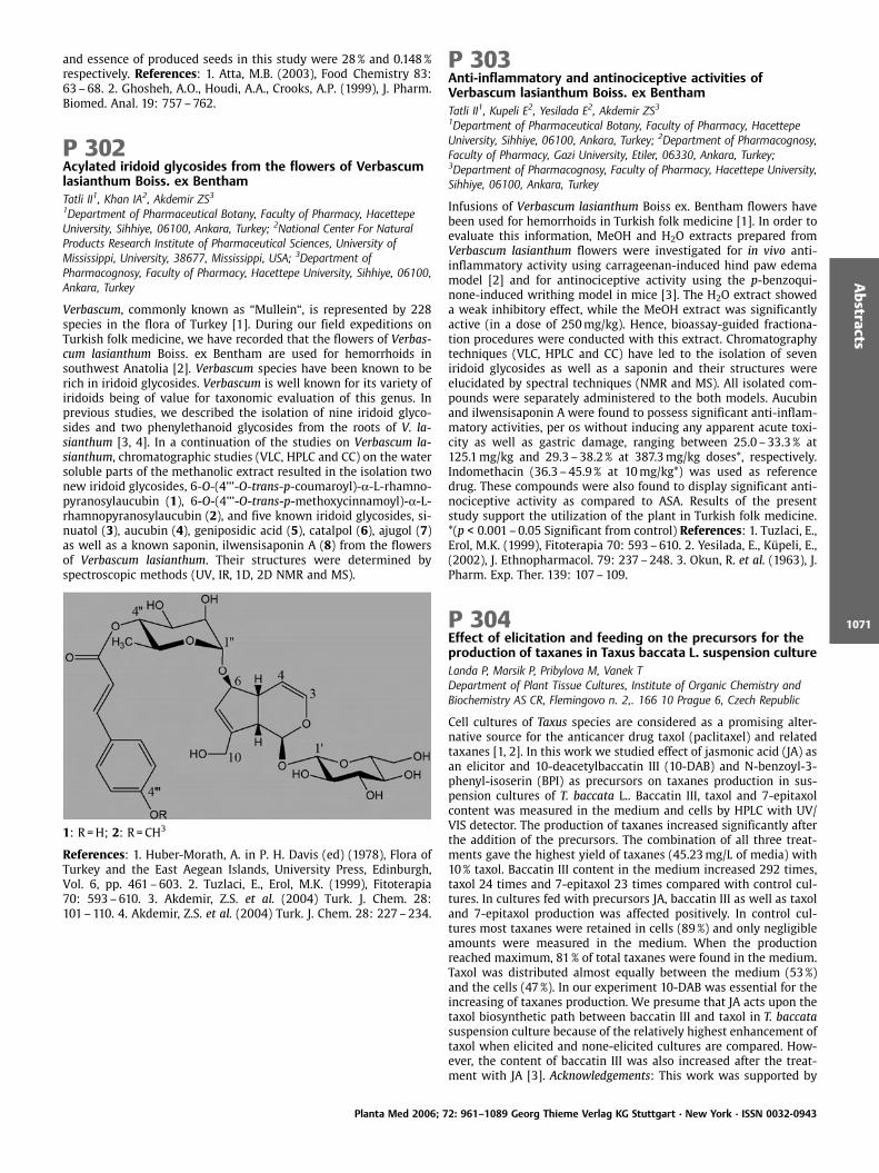
and essence of produced seeds in this study were 28% and 0.148%respectively. References: 1. Atta, M.B. (2003), Food Chemistry 83:63–68. 2. Ghosheh, A.O., Houdi, A.A., Crooks, A.P. (1999), J. Pharm.Biomed. Anal. 19: 757–762.
P 302Acylated iridoid glycosides from the flowers of Verbascumlasianthum Boiss. ex BenthamTatli II1, Khan IA2, Akdemir ZS31Department of Pharmaceutical Botany, Faculty of Pharmacy, HacettepeUniversity, Sihhiye, 06100, Ankara, Turkey; 2National Center For NaturalProducts Research Institute of Pharmaceutical Sciences, University ofMississippi, University, 38677, Mississippi, USA; 3Department ofPharmacognosy, Faculty of Pharmacy, Hacettepe University, Sihhiye, 06100,Ankara, Turkey
Verbascum, commonly known as “Mullein“, is represented by 228species in the flora of Turkey [1]. During our field expeditions onTurkish folk medicine, we have recorded that the flowers of Verbas-cum lasianthum Boiss. ex Bentham are used for hemorrhoids insouthwest Anatolia [2]. Verbascum species have been known to berich in iridoid glycosides. Verbascum is well known for its variety ofiridoids being of value for taxonomic evaluation of this genus. Inprevious studies, we described the isolation of nine iridoid glyco-sides and two phenylethanoid glycosides from the roots of V. la-sianthum [3, 4]. In a continuation of the studies on Verbascum la-sianthum, chromatographic studies (VLC, HPLC and CC) on the watersoluble parts of the methanolic extract resulted in the isolation twonew iridoid glycosides, 6-O-(4’’’-O-trans-p-coumaroyl)-a-L-rhamno-pyranosylaucubin (1), 6-O-(4’’’-O-trans-p-methoxycinnamoyl)-a-L-rhamnopyranosylaucubin (2), and five known iridoid glycosides, si-nuatol (3), aucubin (4), geniposidic acid (5), catalpol (6), ajugol (7)as well as a known saponin, ilwensisaponin A (8) from the flowersof Verbascum lasianthum. Their structures were determined byspectroscopic methods (UV, IR, 1D, 2D NMR and MS).
1: R =H; 2: R =CH3
References: 1. Huber-Morath, A. in P. H. Davis (ed) (1978), Flora ofTurkey and the East Aegean Islands, University Press, Edinburgh,Vol. 6, pp. 461–603. 2. Tuzlaci, E., Erol, M.K. (1999), Fitoterapia70: 593–610. 3. Akdemir, Z.S. et al. (2004) Turk. J. Chem. 28:101–110. 4. Akdemir, Z.S. et al. (2004) Turk. J. Chem. 28: 227–234.
P 303Anti-inflammatory and antinociceptive activities ofVerbascum lasianthum Boiss. ex BenthamTatli II1, Kupeli E2, Yesilada E2, Akdemir ZS31Department of Pharmaceutical Botany, Faculty of Pharmacy, HacettepeUniversity, Sihhiye, 06100, Ankara, Turkey; 2Department of Pharmacognosy,Faculty of Pharmacy, Gazi University, Etiler, 06330, Ankara, Turkey;3Department of Pharmacognosy, Faculty of Pharmacy, Hacettepe University,Sihhiye, 06100, Ankara, Turkey
Infusions of Verbascum lasianthum Boiss ex. Bentham flowers havebeen used for hemorrhoids in Turkish folk medicine [1]. In order toevaluate this information, MeOH and H2O extracts prepared fromVerbascum lasianthum flowers were investigated for in vivo anti-inflammatory activity using carrageenan-induced hind paw edemamodel [2] and for antinociceptive activity using the p-benzoqui-none-induced writhing model in mice [3]. The H2O extract showeda weak inhibitory effect, while the MeOH extract was significantlyactive (in a dose of 250mg/kg). Hence, bioassay-guided fractiona-tion procedures were conducted with this extract. Chromatographytechniques (VLC, HPLC and CC) have led to the isolation of seveniridoid glycosides as well as a saponin and their structures wereelucidated by spectral techniques (NMR and MS). All isolated com-pounds were separately administered to the both models. Aucubinand ilwensisaponin A were found to possess significant anti-inflam-matory activities, per os without inducing any apparent acute toxi-city as well as gastric damage, ranging between 25.0 –33.3% at125.1mg/kg and 29.3 –38.2% at 387.3mg/kg doses*, respectively.Indomethacin (36.3 –45.9% at 10mg/kg*) was used as referencedrug. These compounds were also found to display significant anti-nociceptive activity as compared to ASA. Results of the presentstudy support the utilization of the plant in Turkish folk medicine.*(p < 0.001–0.05 Significant from control) References: 1. Tuzlaci, E.,Erol, M.K. (1999), Fitoterapia 70: 593–610. 2. Yesilada, E., K peli, E.,(2002), J. Ethnopharmacol. 79: 237–248. 3. Okun, R. et al. (1963), J.Pharm. Exp. Ther. 139: 107–109.
P 304Effect of elicitation and feeding on the precursors for theproduction of taxanes in Taxus baccata L. suspension cultureLanda P, Marsik P, Pribylova M, Vanek TDepartment of Plant Tissue Cultures, Institute of Organic Chemistry andBiochemistry AS CR, Flemingovo n. 2,. 166 10 Prague 6, Czech Republic
Cell cultures of Taxus species are considered as a promising alter-native source for the anticancer drug taxol (paclitaxel) and relatedtaxanes [1, 2]. In this work we studied effect of jasmonic acid (JA) asan elicitor and 10-deacetylbaccatin III (10-DAB) and N-benzoyl-3-phenyl-isoserin (BPI) as precursors on taxanes production in sus-pension cultures of T. baccata L.. Baccatin III, taxol and 7-epitaxolcontent was measured in the medium and cells by HPLC with UV/VIS detector. The production of taxanes increased significantly afterthe addition of the precursors. The combination of all three treat-ments gave the highest yield of taxanes (45.23mg/L of media) with10% taxol. Baccatin III content in the medium increased 292 times,taxol 24 times and 7-epitaxol 23 times compared with control cul-tures. In cultures fed with precursors JA, baccatin III as well as taxoland 7-epitaxol production was affected positively. In control cul-tures most taxanes were retained in cells (89%) and only negligibleamounts were measured in the medium. When the productionreached maximum, 81% of total taxanes were found in the medium.Taxol was distributed almost equally between the medium (53%)and the cells (47%). In our experiment 10-DAB was essential for theincreasing of taxanes production. We presume that JA acts upon thetaxol biosynthetic path between baccatin III and taxol in T. baccatasuspension culture because of the relatively highest enhancement oftaxol when elicited and none-elicited cultures are compared. How-ever, the content of baccatin III was also increased after the treat-ment with JA [3]. Acknowledgements: This work was supported by
Planta Med 2006; 72: 961–1089 Georg Thieme Verlag KG Stuttgart · New York · ISSN 0032-0943
Abstracts
1071

S4055301 project of ASCR. References: 1.Yukimune, Y. et al. (1996),Nat. Biotechnol. 14: 1129–1132. 2. Srinivasan, V. et al. (1997), Plant.Cell Rep. 16: 600–604. 3. Czech Patent 19–05.
P 305Medicinal plants and conservation efforts in the buffer zoneof Kure Mountains National Park (Bartin – Turkey),particularly in Ulus regionOzkan AMAnkara University, Faculty of Pharmacy, Department of PharmaceuticalBotany, 06100, Tandogan-Ankara,Turkey
The Kure Mountains National Park lies between Kastamonu andBartin, in the Western Black Sea Region of Turkey. It is a place ofbeauty and magnificent wildlife and has a particular importancerelated to the age and size of its forests, biodiversity, and varietyof its endemic wildlife. Hence, The World Wide Fund for Nature(WWF) has listed the area as one of a hundred forest hot spots inEurope deserving priority conservation. It was declared a NationalPark in 2000 due to its natural and cultural richness. The core areawhich covers an area of over 50 000 hectares is surrounded with abuffer zone, in which local inhabitants, mostly living with low in-come will be able to play an active role in the conservation of thearea. The aim of the present study is to understand the overallpicture of medicinal plant knowledge, use and commercializationin the Ulus region of the buffer zone. Additionally, to develop apublic education program to encourage the preservation of localknowledge of medicinal plants, forest viability, plant survival andhealthy communities. The main results demonstrate that; medicinalplants play a major role in local people’s health care; certain speciesof medicinal plants are commercialized at a large scale and becomelocally rare and sustainable, controlled harvesting may be beneficialboth for the local economy and the habitat conservation. The resultsalso reveal that; a successful approach recognizes the power of localcommunities and conservation of medicinal plants cannot be effec-tive without the support and involvement of local people.
P 306Rapid TLC analysis of Ranunculus bulbosus L. homeopathictinctureGehrmann B1, Melzig MF21Einhorn-Rats-Apotheke, Markt 10 – 12, 25813 Husum, Germany; 2Instituteof Pharmacy, Freie Universit t Berlin, K nigin-Luise-Str. 2+4, 14195 Berlin,Germany
Ranunculus bulbosus L., Bulbous Buttercup or Crowfoot (Ranuncula-cea) is a perennial plant native to the Northern parts of Europe andto the North eastern parts of the United States. Ethanolic tinctures ofRanunculi bulbosi herba cum radice are used in homoeopathy forskin diseases, rheumatism, gout, (zoster) neuralgia, influenza, andmeningitis [1, 2]. Ranunculus bulbosus L. is monographed in the HAB2005 [3]; however, the described TLC procedure is a general com-parative method. Thus, we propose a rapid and simple TLC analysisusing the DESAGA H-separating chamber and different solvent sys-tems containing e. g. ethyl acetate, methanol, water at different pro-portions as mobile phase and diverse silica gel plates (e. g. Si 60,HPTLC-, RP-material) as stationary phase. The optimised TLC condi-tions are performed on different samples of homoeopathic BulbousButtercup tinctures and provide chromatograms showing satisfyingdistributions of characteristic zones in the range of Rf values fromabout 0.2 to 0.8. The described TLC methods are time saving (run-ning time ca. 3min) and only need a small amount of solvents(mobile phase 1–2 mL/performance), sorbens (plates 5 x5 cm),and homoeopathic tincture (application of 10–20mL/performance).The TLC method may be proposed for an improved homoeopathicpharmacopoeia monograph of Ranunculus bulbosus L. References: 1.Brendler, Th., Gr nwald, J., J nicke, Chr., Editors. (2003), Herbal Re-medies, CD-ROM, medpharm, Scientific Publishers, Stuttgart. 2.
Hom opathisches Repetitorium (2003), Deutsche Hom opathie-Un-ion, Karlsruhe. 3. Deutsches Hom opathisches Arzneibuch (2005),Monograph Ranunculus bulbosus (Edition 2000).
P 307Antipyretic activity of the aqueous leaf extract ofByrsocarpus coccineusAkindele AJ, Adeyemi OODepartment of Pharmacology, College of Medicine, University of Lagos,P.M.B. 12003, Lagos, Nigeria
The aqueous leaf extract of Byrsocarpus coccineus Schum and Thonn.(Connaraceae), ABC, was investigated for antipyretic activity in ratsand rabbits using yeast [1], amphetamine [2] and lipopolysacchar-ide [3] induced pyrexia models. In control rats, yeast (10 mL/kg, s.c.)caused elevation of rectal temperature of 1.7oC 19h after adminis-tration. The extract (100, 200 and 400mg/kg, p. o.) produced a sig-nificant (p < 0.05) dose dependent inhibition of temperature eleva-tion. Peak inhibitory effect was observed at 1h post therapy (42.1,47.2 and 63.6% inhibition, respectively for ABC at 100, 200 and400mg/kg). The effect at 400mg/kg was greater than that of acet-ylsalicylic acid, ASA (100mg/kg, p. o.; 43.2%). An elevation in rectaltemperature of 1.9oC was provoked in control rats by amphetamine(10mg/kg, i.p.) 0.5 h after administration while in control rabbits,lipopolysaccharide from E. coli (0.2mg/kg, i. v.) elicited an elevationof 1.1oC, 1.5h post challenge. In both models, ABC produced a signi-ficant (p < 0.05) dose and time dependent direct reduction of elevat-ed temperature with peak effect observed at 3.5 h post therapy.Percent reduction of fever values were 50.2, 61.1 and 84.3, respec-tively for ABC at 100, 200 and 400mg/kg (p. o.) in respect of theamphetamine test. The effect at 400mg/kg was about the same asthat of ASA (85.3%) in this case, but it was lower (44.9 %) comparedto the standard drug (96.6%) in the lipopolysaccharide test. Theresults obtained in this study suggest that the extract possessesantipyretic activity. References. 1 Mukherjee, K. et al. (2002), Phyt-other. Res. 16: 686–688. 2 Berkan, T. et al. (1991), Planta Med. 57:34–37. 3 Vogel, H.G., Vogel, W.H. (1997), Drug Discovery and Eva-luation. Springer–Verlag Berlin Heidelberg, New York.
P 308Antioxidant oligomeric proanthocyanidins from CistussalvifoliusQa’dan F1, Petereit F2, Mansour K3, Nahrstedt A2
1Faculty of Pharmacy, The University of Petra, P.O.Box: 961343 Amman-Jordan; 2Institut fuer Pharmazeutische Biologie und Phytochemie,Hittorfstrasse 56, 48149 Muenster, Germany; 3School of Pharmacy andPharmaceutical Sciences, The University of Manchester, Oxford Road,Manchester M13 9PL, UK
The purified proanthocyanidin oligomers of Cistus salvifolius herbextract accounted for 78% of the total proanthocyanidins and 73%of the total antioxidant activity of this extract [1]. To elucidate thestructure of the oligomer, it was depolymerized by acid catalysis inthe presence of phloroglucinol [2]. The structures of the resultingflavan-3-ols and phloroglucinol adducts were determined on thebasis of 1D-and reverse 2D-NMR (HSQC, HMBC) experiments oftheir peracetylated derivatives, MALDI-TOF-MS and CD- spectro-scopy [3]. These observations resulting from the degradation withphloroglucinol were confirmed by 13C-NMR spectroscopy of the oli-gomer (4). The mean molecular weight of the higher oligomericfraction was estimated to be 5–6 flavan-3-ol-units. Acknowledg-ments: F Qa’dan would like to acknowedge gratefully the DAADand the Deanship of Research at the University of Petra for fundsand grants (Grant No.1/5/2002). We wish to acknowlege also thehelp of Dr.H.Lahl, Ms.M.Heim (Inst.f. Pharmazeutische Chemie,M nster) and Dr. Brian Lockwood (School of Pharmacy, Manchester)for the NMR-spectra. References: 1. Al-Khalil, S. (1995), Int. J. Phar-macognosy 33(4): 317–323. 2. Kennedy, J.A., Graham, P.J. (2001), J.
Planta Med 2006; 72: 961–1089 Georg Thieme Verlag KG Stuttgart · New York · ISSN 0032-0943
Abstracts
1072
Agric. Food Chem. 49:1740–1746. 3. Balas, L., Vercauteren, J. (1994),Magn. Reson. Chem. 32: 386–393. 4. Eberhardt, T., Young, R.A.(1994), J. Agric. Food Chem. 42: 1704–1708.
P 309Analgesic, Antipyretic and Anti-Inflammatory Properties ofMezoneuron Benthamianum Baill (Caesalpiniaceae)Mbagwu HOC1,2, Anene RA1, Adeyemi OO1
1Department of Pharmacology, College of Medicine, University of Lagos, Idi-Araba, P. M. B. 12003, Lagos Nigeria; 2Department of Pharmacology andToxicology, Faculty of Pharmacy, University of Uyo, P. M. B. 1017, Uyo,Nigeria
The analgesic, antipyretic and anti-inflammatory effects of the aqu-eous extract of Mezoneuron benthamianum (MB) were evaluated inmice, rats and rabbits using the mouse writhing, tail flick, hot plateand formalin-induced pain tests; 2,4-Di-nitrophenol (DNP), D-Am-phetamine and Escherichia coli Lipopolysaccharide-induced pyrexiaand carrageenan, egg albumin and xylene- induced oedema [1,2].The extract (400–1600mg/kg) and acetylsalicylic acid (ASA),(100mg/kg) produced a significant (P < 0.05) inhibition of the sec-ond phase response in the formalin pain model, while only thehighest dose (1600mg/kg) of the extract showed a comparable anti-nociceptive effect in the first phase. The extract also showed a dose-dependent inhibition of acetic acid induced abdominal writhing.The tail flick latency and the hot plate pain threshold were dosedependently enhanced by the extract but these were significantlylower than that produced by morphine (2mg/kg). The 2,4-DNP andD-Amphetamine (10 and 5mg/kg, i.p.respectively) increased therectal temperatures of rats within 30 minutes of their administra-tion. The extract at doses of 400, 800 and 1600mg/kg producedsignificant lowering of the elevated body temperature in rats. Theextract (800mg/kg) administered orally to rabbits passages with E.coli lipopolysacharride was able to relieve the pyrogen induced fe-ver. The antipyretic effect produced by the extract was comparableto a standard antipyretic drug, ASA. The extract (400–1600mg/kg)administered 1h after carrageenan-induced paw swelling did notinhibit the oedema. No inhibitions were observed with the egg al-bumin and xylene induced oedema models. Phytochemical analysisrevealed the presence of flavonoids, tannins, cardiac glycosides, an-thraquinones, and saponins in the extract. Administration of theextract up to 2 g/kg (orally) did not produce any toxic effect in theacute toxicity studies in mice. The LD50 of the extract when admi-nistered intraperitoneally was 1021.31mg/kg. The data obtainedshow that MB extract possesses analgesic and antipyretic activitiesbut lacks an anti-inflammatory property. References: 1. Koster, R. etal. (1959), Fed. Proc. 18: 418–420. 2. Winter, C. et al. (1962), Fed.Proc. 46: 118–126.
P 310Effects of Phenolic Compounds from Hypericum perforatumL. on the solubility and permeation of Hypericin in vitroSieger R, Nahrstedt AUniversity of M nster. Institute of Pharmaceutical Biologie andPhytochemistry, Hittorfstraße 56, D-48149 M nster, Germany
The naphthodianthrone hypericin is one of the active constituents inextracts of Hypericum perforatum L., widely used in the therapy ofmild to moderate depression. Former studies have shown that phe-nolic compounds such as procyanidin B1 and some flavonoids areessential for hypericin’s activity in the forced swimming test [1] inthat they improve hypericin’s low water solubility [1, 2] and in-crease its plasma levels in rats [3]. First part of our studies was toinvestigate the influence of Hypericum phenolic compounds on theoctanol/buffer partition coefficient of hypericin. Addition of poly-phenols (mainly flavonoids) to the system resulted in a decreasedoctanol/buffer partition coefficient (LogD) of hypericin and a con-siderably higher concentration of it in the aqueous phase: Without
such coeffector, hypericin’s LogD-value was 4.73 €0.05; but whenthe most active quercetin-3-O-glucuronide was present the LogD-value was 2.52 €0.001. We then undertook an in vitro pharmacoki-netic study, using the Caco2 cell line as a model for intestinal ab-sorption of hypericin without and in the presence of coeffectors. Theapparent permeability (Papp) was measured in both directions, api-cal to basolateral and basolateral to apical. The results for hypericinwithout coeffector showed a 300-fold higher Peff-value from baso-lateral to apical (Peff (baso. to api.) 12.89·10-6 cm/s) than in the reversedirection (Peff (api. to baso.) 0.36·10-6 cm/s). In the presence of Hyper-icum polyphenols the Peff-values for both directions became nearlyequal; this effect was comparable to that of the selective syntheticMRP1/2-inhibitor MK-571. Thus certain polyphenols not only causehigher plasma levels of hypericin by its increased solubility, but alsoby its decreased apical efflux by ABC transporters. References: 1.Butterweck, V. et al. (1998), Planta Med. 64:291–294. 2. J rgen-liemk, G., Nahrstedt, A. (2003), Pharmazie 58:200–203. 3. Butter-weck, V. et al. (2003), Planta Med. 69:189–192.
P 311Effect of development stage at harvest on the content offlavonoids and phenolic acids in aerial parts of Greekoregano (Origanum vulgare L. ssp. hirtum (Link.))Grevsen K1, Frett XC2, Nørbæk R2, Christensen LP21Department of Horticulture, Danish Institute of Agricultural Sciences,Kirstinebjergvej 10, DK-5792 Aarslev, Denmark; 2Department of FoodScience, Danish Institute of Agricultural Sciences, Kirstinebjergvej 10, DK-5792 Aarslev, Denmark
Aromatic plants are used for their culinary qualities as well as fortheir preservative and medicinal properties. Greek oregano isknown for its flavour, which is mainly due to volatile terpenes. Inaddition, oregano and related herbs possess anti-microbial, anti-vir-al and antioxidant activities [1]. The antioxidant properties of ore-gano are mainly due to flavonoids and phenolic acids. The content ofthese compounds in plants depend on a number of factors, such asgrowing conditions, development stage at harvest and genotype.The aim of the present study was to investigate how the concentra-tion of flavonoids and phenolic acids in oregano, depends on thedevelopment stage of the plant during the growing season.Plants were cultivated in 2003 and 2004 and the aerial parts wereharvested at five different development stages during the growingseason (from beginning of July to late August) and samples stored at-24 ¤C until analysis. Several flavonoids and phenolic acids wereidentified from methanol extracts by LC-MS and NMR spectroscopy,and quantified in extracts by RP-HPLC. The major phenolics wereapigenin 6,8-di-C-glucopyranosyl, luteolin 7,4’-di-O-glucuronide, lu-teolin 7-O-glucuronide, lithospermic acid B and rosmarinic acid.Flavonoids varied from 2.8 –5.2mg/g dry matter (DM) (2003) and5.3– 6.8mg/g DM (2004), and phenolic acids from 1.4 –9.9mg/g DM(2003) and 24–53mg/g DM (2004). The highest content of flavo-noids were obtained at the 3rd harvest late in July (near full flower-ing stage) in both years and for phenolic acids the highest contentwere obtained at the 2nd harvest (early flowering stage) in the be-ginning of July also in both years. The conclusion of the presentstudy is that the development stage has a significant impact onthe content of flavonoids and phenolic acids in Greek oregano, andthat an optimal harvest time of this type of oregano depends on theflavonoids or phenolic acids of interest. Reference: 1. Dorman, H.J.D.et al. (2004), J. Agric. Food Chem. 48: 2576–2581.
Planta Med 2006; 72: 961–1089 Georg Thieme Verlag KG Stuttgart · New York · ISSN 0032-0943
Abstracts
1073
P 312Effect of essential oil of Citrus cinensis cv new hall – Citrusaurantium (indigenous in Greece) upon growth of YarrowialipolyticaGortzi O1, Papanikolaou S2, Lalas S1, Galiotou-Panayotou M2, Mitliaga P31Department of Food Technology, Technological Educational Institute ofLarissa (Karditsa Branch), Terma N. Temponera str, GR-43100, Karditsa,Greece; 2Laboratory of Food Microbiology and Biotechnology, Department ofFood Science and Technology, Agricultural University of Athens 75 Iera Odosstr, GR-11855, Athens, Greece; 3Department of Agricultural ProductsMarketing and Quality Control, Technological Educational Institute ofWestern Macedonia (Florina Branch), Terma Kontopoulou str, GR-53100,Florina, Greece
The effect of essential oil from Citrus cinensis cv new hall – Citrusaurantium (indigenous in Greece) upon growth of the dimorphicnon-conventional yeast Yarrowia lipolytica strain ACA-DC 50109was studied. The microorganism was aerobically cultivated in batchmode in carbon-limited media. The essential oil was added into theculture medium in different quantities while the control experimentwas carried out without addition. The essential oil caused a rela-tively important decrease of the highest concentration of biomassproduced. Additionally, biomass yield on glucose consumed wassignificantly decreased with the addition of the oil on the cultiva-tion medium. Moreover, the addition of the essential oil consider-ably increased the lag time of the culture. In all trials, a remarkabledrop the pH value of the mediumwas observed due to the biosynth-esis of small amounts of organic acids. Given that one principalcomponent of this membrane is the one of cellular lipids, it wasassumed that the extraction and the analysis of cellular lipids couldprovide information about the microbial behaviour. Total lipidswere extracted, methanolyzed and analyzed with the aid of G.L.C.In the control experiment, the culture conditions did not favouraccumulation of storage lipid inside the yeast cells and, hence, lipidproduced corresponded to 5–9% (wt/wt) in dry cell mass. Similarconcentrations of cellular lipids were produced when essential oilwas added in various amounts. When essential oils were added, anincrease of lower aliphatic chain saturated fatty acids was observed,suggesting an alteration in the membrane function. Acknowledge-ments: This study has been co-funded by 75% from E.E. and 25%from the Greek Government under the framework of the Educationand Initial Vocational Training Program – Archimedes II. Refer-ences: 1. Aggelis, G., Komaitis, M. (1999), Biotechnol. Lett. 21:747–749 2. Aggelis, G. et al. (1998), Anton. Leeuw. Int. J. G. 73:195–198.
P 313Simultaneous determination of ginsenosides andpolyacetylenes in American ginseng (Panax quinquefoliumL.) using high-performance liquid chromatographyChristensen LP1, Jensen M2, Kidmose U1
1Department of Food Science, Danish Institute of Agricultural Sciences,Kirstinebjergvej 10, DK-5792 Aarslev, Denmark; 2Department of Horticulture,Danish Institute of Agricultural Sciences, Kirstinebjergvej 10, DK-5792Aarslev, Denmark
Panax quinquefolium L. (American ginseng) is native to North Amer-ica, and is one of the most widely used medicinal herbs in the worldtogether with other ginseng species. The alcoholic extract of ginsengroots has been widely used as a tonic against cancer, diabetes, car-diovascular disorders, and immune functions [1]. The active princi-ples of ginseng roots appear to be polyacetylenes and dammaranesaponins (ginsenosides), which are normally determined by differ-ent analytical methods. The aim of this study was to develop amethod for simultaneous determination of both ginsenosides andpolyacetylenes of P. quinquefolium roots. A high-performance liquidchromatography (HPLC) method was developed for simultaneousdetermination of ginsenosides and polyacetylenes from ginseng ex-tracts. Polyacetylenes and ginsenosides were extracted from fresh
ginseng roots with 100% methanol followed by extraction with 80%methanol, which ensured a complete extraction of both types ofbioactive compounds. The combined methanol extracts were sub-jected to HPLC analysis on a reversed-phase (RP) C18 column usinga gradient consisting of acetonitrile and water. The major polyace-tylenes were identified as falcarinol and panaxydol by 1D- and 2D-NMR spectroscopy and the major ginsenosides as Rb1, Rb2, Rc, Rd, Reand Rg1 by comparison with authentic standards on HPLC. The HPLCmethod was validated and used to quantify the content of polyace-tylenes and ginsenosides in root hairs, lateral roots and main rootsof 5-year old American ginseng. The total mean concentration ofpolyacetylenes and ginsenosides was approximately 4.5 and 2 timeshigher in root hairs, respectively, compared to the main roots, in-dicating possibilities for production of differentiated ginseng pre-parations. The developed HPLC method can also be used as a qualitycontrol of fresh ginseng roots as well as dried root material andvarious ginseng preparations. Reference: 1. Sticher, O. (1998),Chemtech. 28: 26–32.
P 314Determination of flavonoids in extracts of Epilobiiangustifolii herba by HPTLC-densitometryBazylko A, Kiss AK, Kowalski JDepartment of Pharmacognosy, Faculty of Pharmacy, Medical University ofWarsaw 1 Banach St, 02 – 097 Warsaw, Poland
Epilobium angustifolium L., Oenotheraceae is used in folk medicine.The herb is rich in polyphenolic compounds such as flavonol-3-O-glycosides, phenolic acids and tannins [1]. Flavonoids analyses haveshown that quercetin glycosides are predominant in Epilobium an-gustifolium [2]. Several studies suggest that at least flavonoids arepartly responsible for the biological action of the herb [3, 4]. Theseparation and quantitative determination of quercetin glycosidesin methanolic and aqueous extracts of Epilobii angustifolii herba byHPTLC-densitometry was established. Ethyl acetate/formic acid/water 68:2.5:03 was used as a mobile phase and silica gel as astationary phase. The flavonoids were more abundant in the aqu-eous extract than in the methanolic one. In both extracts quercetinglucuronide was the dominating compound, 2.12% and 1.78% re-spectively. Our method is fast, easy and selective particularly forquercetin glucuronide determination in Epilobium extracts. Refer-ences: 1. PDR for Herbal Medicines (1998), Medical Economics Com-pany, Montvale, New Jersey 2. Ducrey, B. et al. (1995), Phytochem-istry 38: 129. 3. Tita, B. et al. (2001), Farmaco 56: 341. 4. Kiss, A. et al.(2006), Pharmazie 61: 66.
P 315Effect of herbal formula tonics for reinforcement of yin oryang deficiency on the inhibition of whole bloodaggregationKo BS, Jeon WK, Lee JHQuality Control of Herbal Medicine Department, Korea Institute of OrientalMedicine, 461 – 24 Jeonmin-dong, Yuseong-gu, 305 – 811, Daejeon, Korea
Herbal formula tonics for reinforcement of yin or yang deficiencyare commonly used in Korea traditional medicine [1, 2]. This studyexamined the possible inhibitory effects of 30 kinds of these herbalformula tonics on platelet aggregation induced by collagen in hu-man whole blood using the impedance method of aggregometry [3].Among them, 4 kinds of yin-tonic and 3 kinds of yin-yang-tonicwater extracts were selected to be the most effective candidates(p < 0.001). Also, through in vivo study, the anti-thrombotic effectsof Igyeongtang-, Gamisipjeondaebotang-, and Gamisoyosan-treatedgroups, with recovery rate of 60%, 50%, 45.45%, respectively, wereobserved to be higher than the control group (36.8%) in a mouseacute thrombosis. The results from this experiment provide phar-macological evidence for the traditional use of tonics with yin-yangtheory of traditional medicine, suggesting that yin-tonics could be
Planta Med 2006; 72: 961–1089 Georg Thieme Verlag KG Stuttgart · New York · ISSN 0032-0943
Abstracts
1074
used to help problems of blood circulation more than yang-tonics.Acknowledgements: This study supported in part by the Inter-Insti-tutional Collaboration Research Program under the Korea ResearchCouncil for Industrial Science & Technology (KOCI), Korea. Refer-ences: 1. Liu, Y., Dong, L. (2002), Basic Theories of Traditional Chi-nese Medicine, 2nd ed., Academy Press, Beijing. 2. Heo, J. (1610),translated by Yoon, S. H. et al. (2005), Donguibogam. DonguibogamPress. Hadong. Korea. 3. Armida, P. T. et al. (1995) Thromb Res 78:107–115.
P 316Inhibitory effect of compounds from Rhus chinensis ongeneration of oxygen species generation in YPEN-1 cellsKim GS1, Chung HY2, Kim JM2, Lee SE1, Seong NS1, Song KS3, Baek NI41National Institute of Crop Science, Rural Development Administration, 441 –857, Suwon, South Korea; 2College of Pharmacy, Pusan National University,609 – 735, Busan, South Korea; 3 Division of Applied Biology & Chemistry,College of Agriculture & Life Sciences, Kyungpook National University, 702 –701, Daegu, South Korea; 4Graduate School of Biotechnology, Kyung HeeUniversity, 449 – 701, Suwon, South Korea
Rhus chinensis Mill. (Anacardiaceae) is a broad leaf tree, which iswidely distributed in Korea, Japan and China. Its barks and gall havelong been used traditionally for remedies of dysentery and diarrhea.This study has been carried out as a part of our research on anti-oxidative and antidemential compounds from R. chinensis. Ethylace-tate extract from the stems of R. chinensis was chromatographed onsilica gel glass column to yield dammarane triterpene compounds1-4. Their structures were identified by spectral techniques as 20-hydroxy-24-dammaren-3-one (1), 3-oxodammar-20,24E-dien-26-oic acid, (2), semialatone, (3) and semialatic acid (4), respectively.Compound 2 was previously isolated and identified as a new dam-marane triterpene and compound 1 was first isolated in R. chinensisby us. The other compounds were previously reported in R. chinen-sis. Compound 5 was isolated as a major compound from the leavesof R. chinensis and identified as methyl gallate (5). Compounds 2 and5 strongly inhibited the oxidation of 2,7-di-chlorodihydrofluores-cein diacetate (H2DCFDA) to 2,7-dihydrofluorescein (DCF) withIC50 values of 26.8 and 12.7mM, respectively. Compounds 2 and 5also showed the inhibitory effect on intracellular ROS (reactive oxy-gen species) generation induced by 20mM t-BHP (tert-butyl hydro-peroxide) in YPEN-1 cells. Compared to untreated control, t-BHPtreated cell increased ROS generation to 135.7%. Pretreatment withcompounds 2 and 5 decreased the ROS generation to 86.7 and 60.2%at the concentration of 10mM, respectively. In conclusion, com-pounds 2 and 5 maybe potent ROS scavengers. References: 1. Kim,A. R. et al. (2005), J. Pharm. Pharmacol. 57: 475–481. 2. Kim, J. Y. etal. (2004), Free Radical Res. 38 (7): 761–769. 3. Kim, A. R. et al.(2002), J. Pharm. Pharmacol. 54: 1385–1392. 4. Hsieh, T. J. et al.(2004), Food Chem. Toxicol. 42: 843–850.
P 317Effects of Apple Consumption on Plasma and ErythrocyteAntioxidant Parameters in Elderly Subjectszt rk HS1, Avci A1, Erg der IB1, Atli T2, Varli M2, Devrim E1, Aras S2,
Turgay M2, Durak I11Department of Biochemistry, Ankara University School of Medicine, Ankara,Turkey; 2Department of Geriatric Medicine, Ankara University School ofMedicine, Ankara, Turkey
Aim: Effects of apple consumption on plasma and erythrocyte anti-oxidant parameters of elderly subjects were investigated in thisstudy. Methods: Fifteen elderly subjects (mean age 71.86€4.17)participated in the study. They consumed an apple a day for 1month. Before and after this period, fasting blood samples wereobtained, and oxidant (malondialdehyde, (MDA)) and antioxidant(superoxide dismutase (SOD), glutathione peroxidase (GSH-Px), cat-alase (CAT) and antioxidant potential (AOP)) parameters were stud-
ied. MDA and AOP levels were studied in plasma, and SOD, GSH-Px,CAT activities and MDA levels were measured in the erythrocytes.Results: In the erythrocytes, GSH-Px and SOD activities were foundto be higher (p < 0.001), but MDA levels lower in the second sam-ples relative to the first ones. In the plasma, AOP value was found tobe higher in the second samples relative to first ones (p < 0.001).Conclusion: Our results suggest that consumption of apple leads toa significant increase in the activities of some antioxidant enzymesand in the antioxidant potential values of the blood, does decreasingoxidation reactions in the body in significant amount. It is quitepossible that reduced peroxidation processes owing to consumptionof this fruit may play a part in some of their beneficial effects in theelderly subjects.Table: Oxidant and antioxidant parameters in erythrocytes (RBC)and plasma from elderly subjects who consumed apple (Mean€ SD;n=15).
Groups GSH-Px(RBC)IU/mL
CAT(RBC)IU/mL
SOD(RBC)U/mL
MDA(RBC)nmol/mL
AOP(Plasma)(nmol/mL.h)-1
MDA(Plasma)nmol/mL
Before apple consumption 6.24 € 1.23 58877€ 8588 7337€ 1623 394.3 €72.3 3.43 € 2.13 2,6 € 1.0
After apple consumption 7.65 € 1.29* 62220€ 11224 9299€ 1015* 380.9 €60.6 8.11 € 2.29* 3.3 € 1.0
* p< 0.05; paired t-test
P 318Analysis of essential oils in Chrysanthemum zawadskii inKoreaShin S, Byun YCollege of Pharmacy, Duksung Women’s University, Seoul, 132 – 714, Korea
The essential oils of Chrysanthemum zawadskii Herbich, C. zawadskiivar. latilobum (Maxim.) Kitam, C. zawadskii Herb. ssp. Naktongense(Nakai) T. Lee, C. zawadskii var. leiophyllum (Nakai) T. Lee, and C.zawadskii var. tenuisectum Kitaqawa were obtained by steam distil-lation, using a simultaneous steam distillation-extraction apparatus,from the above ground parts of plants cultivated in the herbal gar-den of Duksung Women’s University. The compositions of the oilswere analyzed by gas chromatography-mass spectrometry and com-pared. Additionally, their inhibiting activities were investigated bybroth dilution method against antibiotic-susceptible and resistantstrains of Streptococcus pneumoniae. Analyses resulted in tremen-dous diversity in composition of essential oils among the speciesand the varieties. The predominantly contained compound of essen-tial oils from Chrysanthemum species were camphor (13.00%), myr-tenol (11.97%) and germacrene D (17.02%). Additionally, all of thetested Chrysanthemum zawadskii essential oils significantly inhib-ited growth of S. pneumoniae with MICs ranging from 0.5mg/mL to1mg/mL. Reference: 1. Shin, S., Lim, S. (2005), Arch. Pharm. Res. 7:765–769.
P 319Preparation of official reference standards from herbalmedicinesKim EK, Ze KR, Seong RS, Lee JP, Park JY, Kang IH, Kim JH, Lee HY, Kim JS,Heo MH, Chang SYHerbal Medicine Standardization Team, Korea Food and Drug Administration,231 Jinheungno, Eunpyung-gu, 122 – 704, Seoul, Republic of Korea
The reference standards are very important and essential for qualitycontrol of herbal medicines. They are required for identification,assay and purity test etc. Unlike general chemical references, whi-ch are used for conventional pharmaceutical products, referencestandards for herbal medicines are difficult to obtain or have rela-tively higher price. We have prepared botanical samples of herbalmaterials and marker substances for reference standards and in-tended to promote the quality control of herbal medicines. By bota-nical scientists, herbal materials are collected, identified its originand then evaluated its quality and specification. Marker substances
Planta Med 2006; 72: 961–1089 Georg Thieme Verlag KG Stuttgart · New York · ISSN 0032-0943
Abstracts
1075

are prepared from medicinal plant materials through extraction,separation and purification. All materials are tested and character-ized [1, 2]. We review and reevaluate the herbal materials and mar-ker substances and then establish them as official reference stand-ards. Particularly, several herbal materials and marker substancessuch as gardenia fruit and saikosaponin A etc., were reviewed andreevaluated, and we will describe the establishment process ofthese reference standards. Acknowledgements: Chung-Ang Univer-sity1, WonKwang University, Wann Kyunn Whang1, Youn ChulKim. References: 1. (2002), The Korean Pharmacopoeia 8th edition,KFDA 2. (2005), The Korean Herbal Pharmacopoeia, KFDA.
P 320Toxicity assessment of the aqueous root extract ofSanseviera Liberica (Agavaceae)Adeyemi OO1, Amida MB1, Yemitan OK1,2
1Department of Pharmacology, College of Medicine of the University ofLagos, Idi-Araba, P.M.B. 12003 Lagos, Lagos, Nigeria; 2Department ofPharmacology, Lagos State University College of Medicine, P.M.B. 21266Ikeja, Lagos, Nigeria
The aqueous root extract of Sanseviera liberica Ger, & Labr. (SL) isused in African folklore medicine for ailments including chronicpain and inflammatory conditions, and convulsive disorders [1].Because of the potential for long term uses, the study investigatedthe acute and subchronic toxicity patterns of the plant. Acute oral &i.p. toxicity tests were carried out in mice; and the median lethaldose estimated [2]. Subchronic (52 days) studies were conducted inrats with oral daily doses of 80, 400 & 2000mg/kg, which repre-sented 1/ 5 [3], active dose and 5x the pharmacologically active dose[4], respectively. Parameters observed at the end of chronic testsincluded changes in body & vital organ weights, mortality, haema-tological, biochemical, hepatic and male reproductive effects [3, 4].SL did not produce any visible toxicities or mortality with oral dosesup to 20g/kg within 14 days of single treatment, but i.p. adminis-tration caused mortalities with LD50 of 668.3mg/kg. In the chronictests, neither mortality nor visible signs of lethality was seen in rats.No significant change in the weight of the kidney, liver, heart andspleen, but at 400mg/kg, a significant reduction in weight of thelungs. Significant increases in the weight of testes, sperm count andmotility was produced. There were no changes in the sperm headand tail abnormalities, but significant increases in the % normalsperm cells. Biochemical parameters like the AST, ALT and uric acidwere not affected, but significant increase in ALP level at 2 g/kg wasproduced. Significant increase in RBC was recorded, but no changesin levels of PCV and Hb. Results indicate that oral doses of SL aresafe, but assessment of hepato-biliary function be done duringchronic uses. References: 1. Watt, J.M., Breyer-Brandwiijk, M.J.(1962), Medicinal and Poisonous Plants of South and Eastern Africa2nd ed. pp 197–198. 2. Miller, L.C., Tainter, M.L. (1944), Proc. Soc. 24:839–840. 3. Tanira, M.O.M. et al. (1988), Int. J. Crude Drug Res. 26:56–60. 4. Yemitan, O.K., Adeyemi, O.O. (2004), Nigerian J. Hlth.Biomed Sci. 3 (1): 20–23.
P 321Seasonal variation in the essential oil composition of Salviafruticosa Mill. cultivated in PortugalBraga PSC1, Vicente AM2, Fernandes-Ferreira M1
1Department of Biology, University of Minho, Campus de Gualtar, 4710 – 057Braga, Portugal; 2Esta ¼o Regional de Hortofloricultura, Quinta S. Jos – S.Pedro de Merelim, 4700 – 856 BRAGA, Portugal
Salvia fruticosa Miller (Greek sage) is a well-known medicinal plantendemic of the Eastern Mediterranean region. Also named three-lobed sage (Salvia triloba L.) due to its leaf morphology, Greek sagehas been commercialized since ancient times for use in therapy butalso as a spice to flavour meats such as pork, sausage and poultry[1]. Its essential oil has shown antimicrobial, cytotoxic, antiviral and
anti-tumor properties [2]. However, the composition of the essentialoil varies geographically and seasonally and these variations seemto affect its properties [3, 4]. Our group has already studied theseasonal and geographical variation of the essential oil compositionof S. officinalis plants grown in Portugal [5]. We now report on theseasonal variation of S. fruticosa essential oil composition. Aerialparts of S. fruticosa plants (~30 cm) were collected every twomonths, during a year period, from an experimental field fromDRAEDM located in Merelim, Braga (Portugal). The aerial parts werethen divided in an upper (UAP) and lower segment (LAP) at ~10 cmfrom the top and approximately 25g of each sample were hydro-distilled in a Clevenger type apparatus. The resulting essential oilswere analyzed by GC and GC/MS. They were rich in oxygenatedmonoterpenes and monoterpene hydrocarbons and, despite somevariation, 1,8-cineole was the major compound in all samples, re-presenting 29% to 46% of the oil. Myrcene was present in highpercentages throughout the year. However, its levels fell duringflowering, especially in the UAP’s. The levels of cis- and trans-thu-jone were always low (< 3%). Camphor percentages were also lowbut rose in August and October, being higher in the LAP’s. Acknowl-edgements: This work was sponsored by EU (FSE/FEDER) and Portu-guese Republic Government (FCT) through the Grant SFRH/BD/18908/2004 and the Project SageBiotech (POCTI/AGR/62040/2004).References: 1. Gali-Muhtasib, H. et al. (2000), J. Ethnopharmacol.71: 513–520. 2. Gali-Muhtasib, H.U., Affara, N. I. (2000), Phytome-dicine 7: 129–136. 3. Skoula M. et al. (2000), Biochem. Syst. Ecol.28: 551–561. 4. Farhat, G.N. et al. (2001), Toxicon 39: 1601–1605. 5.Santos-Gomes, P.C., Fernandes-Ferreira, M. (2001), J. Agric. FoodChem. 49: 2908–2916.
P 322Effects of Chelidonium majus extracts in human hepatocytesin vitroAdler M1, Appel K2, Canal T3, Corvi Mora P3, Delfino R4, Gennaro R4,Gritzko K5, Pascolo L4, Ruzzier F3, Tiribelli C4, Wallner B51Sohlbacher Str. 20, 57078 Siegen, Germany; 2VivaCell Biotechnology GmbH,Ferdinand-Porsche-Str. 5, 79211 Denzlingen, Germany; 3Actimex SrL, Ed. Q –Area Science Park, S.S. 14, Km 163.5, 34012 Basovizza, Trieste, Italy; 4Centerfor Liver Studies (CSF) and Dept. BBCM, University of Trieste, Via L. Giorgieri1, 34127 Trieste, Italy; 5GenPharmTox BioTech AG, Fraunhoferstr. 9, 82152Planegg, Germany
In recent years there has been a discussion whether reversible he-patitis may be a rare potential side effect in Chelidonium majus L.extracts used in the therapy of biliary and hepatic dysfunctions [1].Therefore, earlier in vitro studies of cytotoxic effects of these ex-tracts in rat hepatocytes have found increased attention of regula-tory agencies. With the aim to establish a better database, twostudies in human hepatocytes have been conducted. A study inprimary human hepatocytes was conducted under GMP/GLP condi-tions with C. majus extract (extraction medium 30% ethanol (v/v),1:2.5 –3.5, total alkaloids 5.9mg/g). For indication of cytotoxicity,MTT and neutral red assay were used. EC50 over 24h was calculatedas 0.83 €1.69mg/mL and 0.82 €2.49mg/mL, respectively, which wasequivalent to 4.9mg/mL total alkaloids. In a concentration of0.74mg/mL 86% of the cells survived (MTT). The EC50 of ascorbicacid in this assay is in a similar range (0.8–3.5mg/mL). A secondstudy was conducted in the human-derived Chang liver cell line,testing C. majus extract with 6.2mg/g total alkaloids. The vitalityof the cells was determined from MTT assay and morphologicalappearance. EC50 (24h) was 0.96 €0.49mg/mL, equivalent to5.9mg/mL total alkaloids. The EC50 of Ginkgo biloba extract and para-cetamol, assayed for comparison, was 0.31 and 2.49mg/mL. Thesedata show a clear dose dependency of all observed effects and donot point to special hepatotoxicity of Chelidonium majus extracts,compared to other drugs with established therapeutic safety. Theyare in accordance with available toxicity data for oral application,which show no hepatotoxic effects and give no indication for ex-
Planta Med 2006; 72: 961–1089 Georg Thieme Verlag KG Stuttgart · New York · ISSN 0032-0943
Abstracts
1076

tended pharmacovigilance risk limitation. Reference: 1. Nahrstedt,A., Weber, C. (2005), Deutsche Apotheker-Zeitung 145:3890–3892.
P 323Topical anti-inflammatory activity of Plantago major L.leavesDella Loggia R1, Sosa S1, Cateni F2, Zacchigna M2, Tossi A3, Tubaro A1
1Dipartimento dei Materiali e delle Risorse Naturali, Universit di Trieste, ViaA. Valerio 6, 34127, Trieste, Italia; 2Dipartimento di Scienze Farmaceutiche,Universit di Trieste, P.le Europa 1, 34127, Trieste, Italia; 3BBCM, Universit diTrieste, Via L. Giorgieri 1, 34127, Trieste, Italia
The leaves of Plantago major L. (Plantaginaceae) are traditionallyused for the topical treatment of skin inflammations, infectionsand wounds [1]. Their constituents, such as terpenoids, phenolsand iridoids, possess immunostimulant activity and/or inhibit en-zymes involved in inflammation [2, 3]. Although the main thera-peutic use of P. major is in cutaneous inflammatory diseases, theirtopical antiphlogistic properties were not yet investigated. There-fore, P. major leaves were studied for their topical anti-inflamma-tory activity. P. major leaves were sequentially extracted with n-hexane, chloroform and methanol and the relevant extracts wereevaluated for their ability to inhibit the Croton oil-induced ear der-matitis in mice [4]. Each extract (300mg/cm2) provoked a significantedema reduction, the chloroform one being the most active. Itspotency was only two fold lower than that of the reference drugindomethacin: their ID50 (dose giving 50% edema inhibition) valueswere 177 and 93mg/cm2, respectively. By column chromatography,the extract was separated in four fractions (I-IV), concentrating itsactivity into fraction III, a mixture of ursolic acid and oleanolic acid,in the ratio 1:2. Each compound induced a dose-dependent edemainhibition, being ursolic acid (ID50=56mg/cm2) more active thanoleanolic acid (ID50=132mg/cm2) and indomethacin. The antimicro-bial activity of the plant is under investigation, in order to identifypossible active compounds, different from the well known aucubin.References: 1. Samuelsen, A.B. (2000), J. Ethnopharmacol. 71: 1–21.2. Chiang, L.C. et al. (2003), Planta Med. 69: 600–604. 3. Ringbom T.et al. (1998), J. Nat. Prod. 61: 1212–1215. 4. Tubaro, A. et al. (1985)Agents Actions 17: 347–349.
P 324Release of soy isoflavones from commercial capsulepreparationCvejic J1, Stepanov I1, Pocuca M2
1Laboratory for Analysis of Natural and Pharmaceutical Products,Department of Pharmacy, Faculty of Medicine, University of Novi Sad, Serbiaand Montenegro; 2Laboratory for Biopharmacy, Department of Pharmacy,Faculty of Medicine, University of Novi Sad, Serbia and Montenegro
Herbal medicinal products can be biopharmaceuticaly characterizedby dissolution tests, provided that there was identification of theactive pharmaceutical ingredient. This study describes the determi-nation of the concentration of released soy isoflavones (dissolutiontest) for commercial capsules containing soybean extract using anHPLC method. In analysed preparation four different isoflavoneswere detected and total of their concentrations was evaluated.Two major compounds were daidzein and genistein glucosides,most probably malonyl-glucosides. Aglycones daidzein and genis-tein were also present, but in much smaller amounts. Release ofisoflavones from commercial capsules (Menosoya, Belgrade) wasmeasured with in 90 minutes (six time-points). The dissolutiontests were performed in a Erweka DT800 multi-bath (n =6) dissolu-tion test system, in accordance with the United States Pharmacopeia(USP) general methods. Conditions applied to carry out the dissolu-tion tests were 900 mL of 0.05M potassium dihydrogen phosphatebuffer pH4.5 (KH2PO4) as dissolution medium, basket at 100 rota-tions per minute (rpm) stirring speed and bath temperature of 37 C.Quantification of isoflavones was performed using gradient reversed
phase (150 x4mm, 5mm LiChrospher RP-18) high-performance li-quid chromatography and detection at 270 nm. All dissolution pro-files obtained for six capsules were similar. Values of standard de-viation for all measurements (10, 20, 30, 40, 60 and 90min) werebetween 3.0 10-4 and 5.3 10-4. Analysis of samples showed thatmore than 50% of isoflavones were dissolved from capsules after40 minutes. Approximately 30% of the total is released in the first10min and 65% after 90 minutes. Individual isoflavones showedslight differences between their dissolution curves. In the first20min release of glucosides from formulation was more importantthan of aglycones.
P 325Evaluation of the hepatoprotective effect of Ocimumlamiifolium methanolic extract on acetaminophen-inducedhepatotoxicity in rats – precision cut liver slicesMukazayire MJ1, Allaeys V2, Buc Calderone P2, Duez P1
1. Universit Libre de Bruxelles (ULB), Laboratory of Pharmacognosy,Bromatology and Human Nutrition, Institute of Pharmacy, CP 205/9, Bd duTriomphe, 1050 Brussels, Belgium; 2. Universit Catholique de Louvain(UCL), Unit de Pharmacocin tique, M tabolisme, Nutrition et Toxicologie,Ecole de Pharmacie, Woluwe, Brussels, Belgium
Numerous plant species are used to treat hepatitis in the indigenoushealth care system of Rwanda. Notable among these is Ocimumlamiifolium Hoechst. ex Benth. in DC. The present study aims toevaluate the hepatoprotective effects of the methanolic extract ofleaves of O. lamiifolium against hepatotoxicity induced by acetami-nophen in rats – precision cut liver slices (PCLS). The fresh rats-PCLSwere incubated in Williams medium E for 24h with acetaminophen(hepatotoxicant) concomitantly with the plant extract or N-acetyl-cysteine (NAC) (reference antihepatotoxicant). The measurement ofATP level and CYP2E1 activity were used as endpoints to assess livertoxicity and activity. The O. lamiifolium methanolic extract wasfound to be free of hepatotoxic effects in concentrations up to10mg/mL. The severe depletion of intracellular ATP by acetamino-phen (10mM) was prevented by treatment with the extract at adosage of 1mg/mL, to the same extent as with NAC treatment(20mM). Reference: 1. Evdokimova, E. et al. (2001), Toxicology inVitro 15: 683–690.
P 326Modulation of the peristaltic reflex of rat ileum segments bySTW 5 (Iberogast )Kelber O2, Yuece B1, Wallbach J1, Sibaev A1, Weiser D2, Goeke B1, Storr M1
1Medizinische Klinik II, Klinikum Grosshadern, Ludwig-Maximilian University,Marchioninistr. 15, 81377 Munich, Germany; 2Scientific Department,Steigerwald Arzneimittelwerk GmbH, Havelstr. 5, 64295 Darmstadt,Germany
Dyspepsia and motility related disorders of the gastrointestinal tractare increasingly treated with herbal drugs. This especially applies toSTW 5 (Iberogast ), a fixed combination of standardized plant ex-tracts, for which clinical efficacy has been proven in several modernrandomized controlled double blind studies [1, 2]. But there stillremain unanswered questions about the possible mechanisms ofaction by which the plant extracts induce the beneficial effects.Aim of the present study was to investigate whether plant extractsinfluence the reflex pathways underlying the peristaltic reflex of ratsmall intestine. The myenteric pathways of the peristaltic reflexwere studied in 10 cm ileum segments and peristaltic activity wasstimulated by electrical stimulation in the middle of the segments.Ascending and descending reflex responses were recorded by usingforce transducers. Areas under the contraction curves were evaluat-ed. Drugs were added in a cumulative manner. Concentrations aregiven as dilution of the standardized fluid plant extract in organbath medium. The extracts had individual, reproducible effects onthe elicited reflex responses, showing a clear concentration depen-
Planta Med 2006; 72: 961–1089 Georg Thieme Verlag KG Stuttgart · New York · ISSN 0032-0943
Abstracts
1077
dency (e. g. peppermint leaf extract on ascending contraction:1:1000: -10.1 €4.0%; 1:500: -8.8 € 6.6%; 1:250: –17.3 € 4.7%;1:167: – 39.5 € 13.5%; 1:83: -55.4 € 15.5; 1:50: -58.9 € 15.6). All ob-served effects were fully reversible after washout and were blockedby tetrodotoxin proving the underlying neuronal mechanisms. Con-tractile responses were abolished by atropine proving that they aremediated by cholinergic mechanisms. The observed effects of STW 5and its components on gastrointestinal motility are a clue for under-standing the pharmacological mechanisms underlying the relief inpatient symptoms. References: 1. Holtmann, G. et al. (2004), WienMed Wochenschr 154:21–22. 2. Madisch, A. et al. (2004), Aliment.Pharmacol. Ther. 19:271–279.
P 327Concentration of Grapefruit Essential oil by FractionalDistillationMonsef-Esfahani HR1, Amanzade Y1, Hajiaghaee R1, Mahdavi F11Department of Pharmacognosy, Faculty of Pharmacy, Tehran University ofMedical Sciences, P. O. Box 14155 –6451, Tehran, Iran
Grapefruit, Citrus paradisi M. (rutaceae) the tree grows in north andsouth part of Iran with a height about 3–5 m, it’s fruit is globular,with nipple at apex, mostly big and bright yellow or lemon coloredwith a mild acid or slightly bitter taste. Fruit of grapefruit of north ofIran were purchased during winter 2003. Grapefruit oil was obtain-ed from peel of the fruits by hydro distillation method. GC and GC/Mass analysis performed on the sample and computer library andkovates index used to identify the compounds. Limonene (96.11%),beta-Myrcene (1.89%), alfa-Pinene (0.58%) detected as the majorcomponents in the essential oil. 20 fold concentrate was preparesby a fractional distillation process from hydro distilled oil and ana-lyzed quantitivly by GC and GC/Mass. major and minor constituentswere identified by computer library and kovats index. The influenceof the concentration process on oxygenated flavor compounds, pri-marily aldehydes and alcohols and monoterpene hydrocarbons wasevaluated by comparing the results. Concentration of Limonen in 20fold concentrate decreased 21.86%, whereas alfa-Pinen, Sabineneand beta-Myrcene were completely removed. Concentration of De-canal and Linalool in the 20 fold oil increased 41.96 and 11.94 timesrespectively.
P 328Improved isolation of a-mangostin from the fruit hull ofGarcinia mangostana and its antioxidant and antifungalactivityPuripattanavong J, Khajorndetkun W, Chansathirapanich WFaculty of Pharmaceutical Sciences, Prince of Songkla University, Hat-YaiCampus, Songkhla 90112, THAILAND
The fruit hull of mangosteen, Garcinia mangostana L. (Family Clu-siaceae), has been used for many years as a folk medicine for treat-ment of skin infection, wounds and diarrhea [1]. In this study, theHPLC analysis for lpha;-mangostin was developed and validated interms of resolution (Rs), capacity factor (k’), selectivity factor (a),and tailing factor (Tf). In addition, the effect of solvents on quantityof a-mangostin, total phenolic content [2], antioxidative [3] andanti-tinea activities were examined. Seven types of solvent (hexane,dichloromethane, chloroform, acetone, ethyl acetate, methanol andethanol) were used for extraction giving % yields of 0.03, 1.87, 1.39,3.00, 0.90, 7.74 and 6.07, respectively. The EtOAc extract was foundto possess the highest content of a-mangostin (91.92%). The acetoneextract showed the highest phenolic content of 0.5713mg/g as gallicacid equivalent (GAE). The best antioxidant activity (DPPH-assay)was found in the chloroform extract with the EC50 of 3.44mg/mL.The study of antifungal activity showed that extracts (ethanol, acet-one and methanol) provided antifungal activity against three spe-cies of tinea: Trichophyton rubrum, T. mentagrophyte and Microspor-um gypseum. Acknowledgements: Thailand Research Fund (TRF),
Thailand., Sirirat Pinsuwan References: 1. Chomnawang, M.T. et al.(2005), J. Ethnopharmacol. 101: 330–333. 2. Folin, O., Ciocalteu, V.(1927), J. Biol. Chem. 27: 627–650. 3. Yamasaki, K. et al. (1994),Chem. Pharm. Bull. 42: 1663–1665.
P 329Secondary Metabolites from Hypoglycaemic fraction ofTreculia africana Decne (Moraceae)Moody JO1, Oyelola OO1, Fukushi Y2, Tahara S2, Hashidoko Y2, Sakihama Y21Department of Pharmacognosy, University of Ibadan, Ibadan, NIGERIA;2Laboratory of Ecological Chemistry, Hokkaido University, Sapporo, JAPAN
Treculia africana Decne (Moraceae) commonly known as Africanbreadfruit is a plant food native to tropical West and parts of EastAfrica. Ethnomedicinally, it is used as a vermifuge, febrifuge, galac-togogue and laxative (Irvine, 1961). The plant is also an importantcomponent of some ancient anti-diabetic recipe used in Westernand Middle Belt areas of Nigeria as shown by our survey amongherbalists and a number of patients attending diabetic clinics inthe University College Hospital, Ibadan. Bioactivity –monitored frac-tionation of the hydroacetone extract of the bark of T. africana re-vealed that ’the non-aqueous soluble fraction (10mg/kg) exhibitedthe highest activity by giving a significant reduction in blood sugarlevel (69.4% at 240 hours, p < 0.05) which was in comparable rangewith reference standard glibenclamide (65.8% reduction at 0.5mg/kg dose level). Column Chromatographic separation (Silica gel, hex-ane:ethyl acetate mixtures) and reverse-phase preparative thin-layer chromatography of the ethyl-acetate fraction resulted in theisolation, for the first time from T. africana, of secondary metabolitescharacterized as 3-prenyl – 2’-4,4’- trihydroxy- chalcone and ber-gapten (IR, NMR, MS). Reference: 1. Irvine, F.R. (1961), WoodyPlants of Ghana, Oxford Univ. Press.
P 330Volatile Composition and Cyclooxygenase (COX) inhibitoryeffect of Stachys setifera C. A. MeyKhanavi M1, Hadjiakhoondi A1, Sharifzadeh M2, Shafiee A3, Rustaiyan A4
1Department of Pharmacognosy, Medicinal Plant Research Center andPharmaceutical Sciences Research Center (a), Faculty of Pharmacy, TehranUniversity of Medical Sciences, P.O.Box: 14155/6451, Tehran, Iran;2Department of Toxicology and Pharmacology, Medicinal Plants andPharmaceutical Sciences Research Centers (b), Faculty of Pharmacy, TehranUniversity of Medical Sciences, P.O.Box: 14155/6451, Tehran, Iran;3Department of Chemistry, Medicinal Plant and Pharmaceutical SciencesResearch Centers (c), Faculty of Pharmacy, Tehran University of MedicalSciences, P.O.Box: 14155/6451, Tehran, Iran; 4Department of Chemistry (d),Sciences and Research campus, Islamic Azad University, P.O.Box: 14515 –775, Tehran, Iran
The genus Stachys L. from lamiaceae family comprises about 300species in the world [1]. Iran is an area particularly rich in taxa withmore than 34 species including Stachys setifera C.A.Mey [2]. Thechemical composition of the essential oil and COX inhibitory effectof aerial parts of S. setifera has not yet been described. The oilobtained by steam distillation of the aerial parts of the plant wasanalyzed by GC and GC/MS. It was rich in eugenol (21.1%), hexade-canoic acid (12.5%) and linoleic acid (11.0 %). Because of some pre-vious reports about modulatory effect of eugenol on cyclooxygenasepathway [3], anti-inflammatory activity of methanolic extract ofaerial parts of S. setifera was investigated in this study. For assess-ment of anti-inflammatory properties, two well-characterized in-flammatory models, formalin test and carrageenan-induced pawedema were used. Interaperitoneal injections of methanolic extract(50,100, 200mg/kg), 30min before formalin injection, had no effectsagainst the first phase of the formalin- induced pain, but all threedoses caused a significant blockade on the second phase (P < 0.01,< 0.001). In the carrageenan-induced paw edema, each extract re-vealed dose-related inhibitory effects over the dose range 50–
Planta Med 2006; 72: 961–1089 Georg Thieme Verlag KG Stuttgart · New York · ISSN 0032-0943
Abstracts
1078

200mg/kg. The anti-inflammatory activity of S. setiferawas compar-able with high dose of indomethacin (5mg/kg). In conclusion, thepresent findings provide further evidences for inhibitory effects ofthese extracts in inflammatory processes via possible interactionswith cyclooxygenase (COX). References: 1. Rechinger, K.H. (1982),Flora Iranica. Akademiche Druck-u. Verlagsanstalt, Graz-Austria. 2.Mozaffarian, V. (1996), A Dictionary of Iranian Plant Names. FarhangMoaser, Tehran-Iran. 3. Kelm, M.A. et al. (2000), Phytomedicine7:7 –13.
P 331Discrimination of Piper longum and Piper retrofractum fruitsby chromatographic fingerprint analysisBauer R1, St hr J21Institute of Pharmaceutical Sciences, Department of Pharmacognosy, Karl-Franzens-University Graz, Universit tsplatz 4/I, A-8010 Graz, Austria;2Institute of Pharmaceutical Biology, Heinrich-Heine-Universit t, 40225D sseldorf, Germany
In the Pharmacopoeia of the Peoples Republic of China the fruits ofPiper longum L. are considered as “Bibo“. According to our investiga-tions with a series of commercial batches of bibo, most of the sam-ples were derived from Piper retrofractum Vahl. although this is notan accepted substitute. Several books dealing with the Chinese Ma-teria Medica clearly describe properties of Piper retrofractum underthe chapter “Piper longum L.“ and even illustrate them with picturesof this allied species. The macroscopic description of the drug in thepharmacopoeia does not unambiguously allow the destinction ofboth species. Therefore chromatographic methods have been devel-oped for a clear identification. Peaks have been identified by LC-MS[1]. The HPLC-fingerprint of P. retrofractum only shows one predo-minant peak (piperine) and a very small one of retrofractamide A. Incontrast to the chromatogram of P. longum retrofractamides B andD, and N-isobutyl-2E,4E-octadecadienamide can be detected. TheHPLC-chromatogram of Piper longum shows a homogeneous distri-bution of numerous peaks, with piperine and pellitorine as the pre-dominant compounds. Piper retrofractum and Piper longum can alsoeasily be distinguished by TLC under UV254nm and UV365nm [2].References: 1. St hr, J. et al. (2001), J. Ethnopharmacol. 75:133–139. 2. Wagner, H. et al. (2006), Chinese Drug Monographs andAnalysis, in press.
P 332In vitro effect of BA and NAA on growth and development ofPueraria candollei Grah. ex. Benth. var. mirificaChawapun A, Dheeranupattana S, Jatisatienr A, Jatisatienr CDepartment of Biology, Faculty of Science, Chiang Mai University, Chiang Mai50200, Thailand
Shoot of Pueraria candollei were cultured on MS medium with 0,0.25, 0.5, 1.0 and 2.0mg/L BA found that medium supplementedwith 0.5 and 1.0mg/L BA induced the greatest number of multipleshoots formation at 5.3 and 5.2 shoots/explant respectively for 12weeks. Shoot of Pueraria candollei from natural were cultured on MSmedium supplemented with 1 and 2mg/L BA plus 1mg/L NAA. Itwas found that MS medium with 2mg/L BA plus 1mg/L NAA in-duced the greatest diameter of callus (2.0 cm) for 4 weeks andshoots formation were induced the greatest number of 4.4 shoots/explant and root formation were induced the highest at 60% on MSmedium with 1mg/L BA plus 1mg/L NAA for 10 weeks. Acknowl-edgements: Chiang Mai University, Plant Tissue Culture Unit Refer-ences: 1. Dougall, O.K. (1981), Tissue culture and the study of sec-ondary (nature) product, In Conn, E.E. (ed.), The biochemistry ofPlant. Vol. 4 New York: Academic Press. 2. Kashemsanta, L., Subata-bandhu, K. and Bartlett, S. (1963), Estrogenis substance Zmiroestrolfrom the tuberous roots of Pueraria mirifica. Proc. Pacific Sci Assoc9th Bangkok Thailand. 5: 37–40. 3. Matkowski, A. (2004), J. PlantPhys. 161: 3. 343–346. 4. Rao, A.N., Lee, S.K. (1986), An overview of
the in vitro propagation of woody plants and plantation crops. InWither, L.A. and Aldersan, P.C. (eds.), Plant cell tissue and its agri-cultural application. Singapore: Butterworths. 5. Thiem, B. (2003),Plant Science. 165 (5): 1123–1128. 6. Yu, S., Li, L. (1999), PlantResoures and Environment 8: 63–64.
P 333In vitro secondary compound production from roots ofStemona curtisiiPalee J1, Dheeranupattana S1, Jatisatienr A1, Jatisatienr C1
1Department of Biology, Faculty of Science, Chiang Mai University, 50200,Chiang Mai, Thailand
Stem explants of Stemona curtisii Hook. f. plantlets from in vitroplantlets were cultured on MS agar media supplemented with 11concentrations of NAA (0 –6.0mg/L) and in liquid media supple-mented with 6 concentrations of NAA (0–6.0mg/L) for 12 weeks.It was found that the MS agar medium containing 1.0mg/L NAA andthe MS liquid medium containing 3.0mg/L NAA induced 100% rootsformation with the highest average number of 21.53 and 17.44 rootsper explant, respectively. Apical roots were then cultivated in theMS liquid media supplemented with 10 concentrations of NAA (0–3.0mg/L) for 8 weeks. It was found that the MS medium containing0.3mg/l NAA induced 60% lateral roots with the highest averagenumber of 8 lateral roots per explant. Secondary compounds wereextracted with 95% ethanol from S. curtisii roots cultured on theagar and in liquid MS media supplemented with 7 concentrationsof NAA (0–1.0mg/L). The crude extracts was then separated by TLCusing dichloromethane: methanol: ammonia (95: 5: 1) as the mo-bile phase. In the initial experiment, stemocurtisine was not foundin the tissue cultured roots. However, the unknown nitrogenoussecondary compound was detected. Acknowledgements: We aregrateful to the Ministry of National Research and Environment forsupporting this project. Reference: 1.Mungkornasawakul, P. et al.(2003), J. Natural Pros. 66(7): 980–982. 2.Babu, K.N. et al. (1993),Hort. Abstr. 63(7): 5386.
P 334Ascorbic acid in Iranian Rose species (2)Asgari T, Amin G, Hamidian Jahromi AFaculty of Pharmacy, Tehran University of Medical Sciences, Pursina Ave,14155 – 3451, and Faculty of Pharmacy, Azad university, Tehran, Iran
Fruits of Roses specially Rosa canina L. are one of the richest sourceof Ascorbic acid (AA) and have the best effect on human body. Iran isthe main habitat of Roses in the world, and there are many nativeand endemic species. Previously three species (R. foetid J. Herrm., R.boissierii Crep., R. hemisphaerica J. Herrm.) were investigated and AAwas (1000–4000)mg/100 g. In the recent study two other species(R. canina, R. beggeriana Schrenk.) were collected from Taleghan inN.W of Tehran, identified and their voucher specimen were depos-ited in the Herbarium of faculty of pharmacy, Tehran university ofmedical sciences. Their pulps were dried and extracted via decoc-tion method. AA were assayed via two methods of titrimetric (by2,6-dichlorophenolindophenol) and spectrometric (by 2, 6-dinitro-phenylhydrazine) with providing of calibration curve. Range of AAin these two species was 2200 and 4658mg/100g, respectively.While there is reported that the range of AA or Rosa species is1095–6694mg/100g. In conclusion the range of AA and diversityof R. canina and R. beggeriana in Iran is very noticeable and will agood source of natural products. Acknowledgments: Safar Ali asgari,Lina mehrabadi-e-yari. Refrence: Joublan, J.P., M. Berti, H. et al.(1996), Wild rose germplasm evaluation in Chile. p. 584–588. In:J. Janick (ed.), Progress in new crops. ASHS Press, Arlington, VA.
Planta Med 2006; 72: 961–1089 Georg Thieme Verlag KG Stuttgart · New York · ISSN 0032-0943
Abstracts
1079

P 335Flavonoids, volatiles and biological activities of the aerialparts of Calliandra haematocephala HasskZeid AA1, Hifnawy M2, Saleh M1, Sleem A3, Mohamed R11Pharmacognosy Dept., National Research Centre, El-Tahrir St, Dokki(12622), Cairo, Egypt; 2Pharmacognosy Dept, Faculty of Pharmacy, CairoUniv, Kasr El-Einy, Cairo, Egypt; 3Pharmacology Dept, National ResearchCentre, El-Tahrir St, Dokki (12622), Cairo, Egypt
The genus Calliandra (Fabaceae) contains 132 species. Most of themare native of America, but few of Asia and Africa. The current studydeals with isolation and identification of flavonoids, as well as, in-vestigation of volatile constituents and biological activity of extractsof the aerial parts of Calliandra haematocephala. The total ethanolextract, as well as, successive extracts (petroleum ether, chloroform,ethyl acetate and methanol) were prepared from the dried pow-dered plant. The flavonoids were isolated from the ethyl acetateextract by using Silica gel column chromatography. The isolatedcompounds were finally purified by Sephadex LH –20 column [1].Three flavonoid aglycones, quercetin, kaempferol, myricetin, as wellas, three flavonoid glycosides, quercetin-3-O- rhmnopyranoside,keampferol-3-O-(2“-O-galloyl)- rhamnopyranoside and myricetin-3-O-(2“,3“-di-O-galloyl)-rhamnopyranoside were identified by de-termination of UV, 1HNMR, 13CNMR spectra and hydrolytic products[1, 2]. The volatile constituents of fresh aerial parts were preparedby hydro-distillation using Nikerson’s apparatus and analyzed byGC/MS analysis. Sixty four compounds were identified representing93.32% of the total volatiles of the plant. The oxygenated and non-oxygenated compounds constituted 72.34% and 20.98%, respec-tively. The LD50, analgesic, antipyretic, anticonvulsant, anti-ulceractivity of the successive extracts, as well as, the antioxidant activityof the isolated compounds were investigated. The results of thebiological activity tests were statistically analyzed using the stu-dent’s “t“ test [3]. Most of the tested extracts were found to besignificantly active. References: 1. Mabry, J., Markham, K. (1970),The Systematic Identification of Flavonoids, Springer Verlag, Berlin.2. Markham, K. (1982), Techniques of Flavonoid Identification, Aca-demic Press, London. 3. Snedecor, W., Cochran, G. (1982), Statisticalmethods 10th ed, lowa State, University Press, USA.
P 336Efficient production of Sundew (Drosera rotundifolia L.) invitro using a temporary immersion systemKopp B, Wawrosch C, Buol I, Dorfer TDepartment of Pharmacognosy, University of Vienna, Althanstr. 14, A-1090Vienna, Austria
The carnivorous plant Drosera rotundifolia has been used since cen-turies against affections of the respiratory tract. Its spasmolytic,antibacterial and antiinflammatory properties are attributed tonaphthoquinones and flavonoids [1]. In the last decades this speciesbecame very rare due to degradation of the natural habitats. Thecrude drug obtained from various other species is of poor quality,and adequate quantities are difficult to obtain [1]. In vitro-culturecan be an alternative in that uniform plants for further field culturecan be produced [2]. In addition, the contents of active compoundsin material obtained in vitro can be higher than under field condi-tions [3]. Thus, a biotechnological approach to the production offresh plant material (e. g. for homeopathic use) offers interestingperspectives. Frequently, a major obstacle to a more widespreadapplication of tissue culture for plant production is elevated costsresulting from labour [4] and expensive nutrient media. The use oftemporary immersions systems with liquid nutrient medium can behighly efficient in reducing production costs. Furthermore, the over-all efficiency of this micropropagation technique concerning multi-plication, biomass yields, and plant quality is substantially higherthan in conventional systems using semisolid media [4]. In thiscontribution an in vitro-culture system based on temporary immer-sion is presented. By adjusting frequency and duration of the im-
mersion multiplication and biomass yield could be improved whencompared to the control in submerged culture. The results indicatethat this system would allow for the efficient production of plantsfor field culture as well as crude drug material. References: 1.Krenn, L., Kartnig, T. (2005), Z. Phytotherapie 26: 197–202. 2. Wa-wrosch, C. et al. (1996), Sci. Pharm. 64: 709–717. 3. Wawrosch, C. etal. (2005), Sci. Pharm. 74: 251–262. 4. Etienne, H., Berthouly, M.(2002), Plant Cell Tissue Organ Cult. 69: 215–231.
P 337Is the Alkaloid Pipermethystin Connected with Liver Toxicityof Kava Products?Lechtenberg M1, Quandt B1, Schmidt M2, Nahrstedt A1
1Institute of Pharmaceutical Biology and Phytochemistry, Univ. of M nster,Hittorfstr. 56, D-48149 M nster, Germany; 2Herbresearch Germany,Wartbergweg 15, D-86874 Tussenhausen-Mattsies, Germany
Recently, a worldwide discussion on the potential liver toxicity ofextracts obtained from Kava (Piperis methystici rhizoma) was in-itiated by a series of reports resulting in a ban by the GermanFederal Institute for Drugs and Medicinal Products (BfArM) thatwas followed by other countries [1]. However, most cases wereevaluated as “doubtful“ on causality assessment [2]. Several theoriesevolved as to why liver failure may have occurred [1]. Dragull et al.[3] suggested the alkaloid pipermethystin being responsible for he-patotoxicity. We therefore investigated various kava preparationsincluding a series of retain samples of kava extract containing pro-ducts from the German market, self-produced extracts from rootand stem material obtained from two identified kava cultivars (“no-ble kava“ Ava La’au from Samoa, “Tudei kava“ Palisi from Vanuatu;extracted with ethanol 96% respectively acetone 75% or 100%), andan extract from the leaves of Piper methysticum G. Forst. (as a posi-tive control). Samples were analyzed for their content of piper-methystin by GC-ESI-MS using total ion currency (TIC) and selectiveion monitoring (SIM) detection. Limit of detection (LOD) was about0.009%. As a result, pipermethystin was detected in the leaves(0.2 %), but no pipermethystin above LOD was detected in all othersamples except one where a peak below 0.02% was found at theposition corresponding to pipermethystin. Thus, if there is hepato-toxicity, it should not be connected to the alkaloid pipermethystin.Acknowledgemenst: We thank Dr. K. Dragull, Univ. of Hawaii-Manoain Honolulu, for a sample of pure pipermethystin. References: 1.Anke, J., Ramzan, I. (2004), Planta Med. 70: 193–196. 2. Schmidt,M. et al. (2002), Wien. Med. Wochenschr (WMW) 152: 382–388. 3.Dragull, K. et al. (2003), Phytochemistry 63: 193–198.
P 338Effect of drying methods on essential oil content andcomposition of wormwood (Artemisia absinthium L.)Khangholi S1, Rezaeinodehi A1, Sefidkon F2
Department of Horticulture, Faculty of Agricultural Sciences, ShahedUniversity, Tehran, Iran
The aerial parts of wormwood (Artemisia absinthium L.) were har-vested in full blooming stage in September 2005 from an area be-tween Deylaman and Asiabar villages, around the Siahkal city inGilan province in north of Iran. In order to complete drying, a sam-ple of aerial parts was placed at shade (room temperature) for sev-eral days and a sample placed in ventilated oven at 35oC tempera-ture for 24h. The aerial parts essential oil was extracted by hydro-distillation in a Clevenger apparatus and analyzed by GC/MS. Re-sults showed that essential oil yields in shade condition and ovencondition were 1.3 and 1.1 percent respectively. Also number ofchemical components of the essential oil in shade drying and ovendrying methods were 28 and 33 components respectively, whichwere mostly monoterpenes. b- pinene and b- thujone were maincomponents of the both drying methods, which their contents inshade drying and oven drying methods were (23.8 and 18.6%) and
Planta Med 2006; 72: 961–1089 Georg Thieme Verlag KG Stuttgart · New York · ISSN 0032-0943
Abstracts
1080

(18.7 and 27.9%) respectively. Hydrocarbon monoterpenes contentin shade drying method were higher than the other method but incase of oxygenated monoterpenes vice versa. There was not signifi-cant difference with respect to sesquiterpenes content between thedrying methods. The results proved that chemo type of the studiedwormwood essential oil was specific and different from otherwormwood essential oil chemotypes, which have been reported.
P 339Prediction of microbial metabolism of phytochemicals usingan in vitro colon modelAura AM1, Sepp nen-Laakso T1, Bounsaythip C1, Oresic M1, Oksman-Caldentey KM1
1VTT Technical Research Centre of Finland, Tietotie 2, P.O.Box 1000, FIN-02044 VTT, Finland
Human colon contains 1.5 kg of microbiota, which actively takespart in the degradation and decomposition of the non-absorbableintake. Colonic microbiota changes by age, diet, intestinal diseasesand medication causing intra-individual variation in the metabolitepool in addition to the inter-individual variation between subjects.However, a good correlation has been found for dietary phenolicmicrobial metabolites between the in vitro colon model1 and corre-sponding metabolite profiles from human body fluids. Traditionalprediction of drug metabolism includes several in vitro test systemsand animal trials. However, microbial metabolism in the colon is notgenerally used in the pre-clinical stage. To address these challenges,the developed batch in vitro colon mode can be coupled with anadvanced metabolomics and bioinformatics platform that providecomplementary pre-clinical data on the metabolites circulating inthe human body. The model can be used for: Identification of toxicmetabolites of new phytochemicals Comparison of metabolite pro-files of phytochemicals Comparison of the microbial metabolite pro-file with those from body fluids of man: in vitro-in vivo correlationReference: 1. Aura A.M. et al. (2002), J. Agric.Food Chem. 50: 1725–1730.
P 340GC-MS Analysis of Eryngium maritumum L. Volatile OilAslan S, Kartal MAnkara University, Faculty of Pharmacy, Department of Pharmacognosy,06100 Tandogan-Ankara, Turkey
The genus Eryngium (Umbelliferae family; Saniculoideas subfamily)is known to contain acetylenes, flavonoids, coumarins and terpeniccompounds [1]. Eryngium species are represented by 317 species,subspecies and varieties [2]. Eryngium species, generally E. campes-tre L. and E. maritimum L. are known in Turkish folk-medicine as“Bogadikeni“ and widely distributed in all parts of Turkey. Infusionsof aerial and underground parts of this species are used in folkremedies as antitussive, diuretic, appetizer, stimulant and aphrodi-siac [3]. The genus Eryngium is represented by 23 species (24 taxa)in the Flora of Turkey and East Aegean Islands which ten of them areendemic [4– 6]. In the present study, the volatile oil composition ofthe aerial part of E. maritumum was investigated by capillary gaschromatography-mass spectrometry (GC-MS). The oil of was foundto be remarkably rich in spathulenol, 1,5-epoxysalvial-4(14)-ene, a-amorphene and caryophellene oxide. References: 1. Erdelmeier,C.A.J., Sticher, O. (1986), Phytochemistry 25(3): 741–743. 2. W rz,A. (1999), Stuttgarter Beitr. Naturk. Ser. A, Nr. 596: 1–48.3. Baytop,T. (1999), Turkiye’de Bitkilerle Tedavi-Gecmisten Bugune (Therapywith Medicinal Plants in Turkey-Past and Present), 2nd edn. Pp. 169,Nobel Tıp Basimevi, Istanbul, Turkey. 4. Davis, P.H. (1972), Flora ofTurkey and The East Aegean Islands. University Press, Edinburgh,Vol. 4: 292–304. 5. Davis P.H. et. Al (1988), Flora of Turkey andThe East Aegean Islands (Supplement), University Press, Edinburgh,Vol.10: 145. 6. G ner, A. et. al. (2000), Flora of Turkey and the East
Aegean Islands, Edinburgh University Press, Edinburgh, vol.11: 136–138.
P 341Onions of the Allium Subgenus Melanocrommyum – theBetter Garlic?Keusgen M1, Fritsch RM2
1Philipps-Universit t Marburg, Institute of Pharmaceutical Chemistry,Marbacher Weg 6, D-35032 Marburg, Germany; 2Leibniz Institute of PlantGenetics and Crop Plant Research (IPK), Corrensstr. 3, D-06466 Gatersleben,Germany
About 200 Allium species occur in the mountainous regions of Cen-tral and South-West Asia [1]. The use of especially tasteful andcurative members of this family has a long tradition with apparentlydeep historical roots in several Asian countries. Besides cultivatedonion and garlic, also a number of wild species are collected andeaten by local populations of the above mentioned regions. In thisarea, especially members of the subgenus Melanocrommyum arewidely distributed and highly estimated as vegetable and medicinalplant. In Tajikistan and in some parts of neighbouring countrieswhere related tribes settled, leaves of A. rosenbachianum auct. – thisname is used in some scientific literature for A. rosenbachianumRegel in a strict sense as well as for A. rosenorum R.M. Fritsch –are extensively used. Leaves are often collected and eaten in formof traditional dishes because consumption “refreshes the body afterthe winter period“ [2]. Allium komarowii Lipsky owns obviously arather strong medical activity, because it is used as anabolic drug forhorses [2]. Also this kind of activity could not be correlated to highcysteine sulphoxide contents, but these species contain a conspic-uous red dye, which is chemically a sulphurpyrrol. Allium motorKamelin et Levichev is often used in a similar manner as A. rosen-bachianum in parts of Uzbekistan. The term ‘motor’ means ‘health’,but application may cause problems for people suffering from highblood pressure [29. The above mentioned examples demonstratethat members of the subgenus Melanocrommyum are used as med-icinal plants with huge variety of applications and are even higherestimated as garlic. However, the active principle of these plants isstill unknown and needs further investigation. Acknowledgements:Research was supported by the German VolkswagenStiftung as partof the “PharmAll“-project. References: 1. Khassanov, F.O. (1996),Plant life in Southwest and Central Asia, EGE University Press. Iz-mir.141–159. 2. Keusgen, M., Fritsch, R.M. et al. (2006), J. Ethnobiol.Ethnomed. 2: 18.
P 342Matrix free MALDI mass spectrometry for phytochemicalinvestigationsHashir MA1, Stecher G1, Abel G2, Popp M2, Bonn GK1
1Institute of Analytical Chemistry and Radiochemistry, University ofInnsbruck, Innrain 52a, 6020, Innsbruck, Austria; 2Bionorica AG;Kerschensteinerstr. 11 – 15, 92318 Neumarkt, Germany
Matrix assisted laser desorption ionisation time of flight mass spec-trometry (MALDI-TOF-MS) is a very sensitive mass spectrometrictechnique which utilizes acidic materials as matrices for laser en-ergy absorption, desorption and ionisation of bio-molecules such aspeptides and proteins. These matrix materials produce backgroundpeaks particularly in the low mass range and make the detectionand identification of small molecules difficult. Therefore, some ef-forts have been made to develop matrix free MALDI for the analysisof small molecules. For this purpose a neutral substance was im-mobilized on a carrier system in order to enable absorption of laserenergy sufficient for successful desorption and ionization of ana-lytes of low mass without interference and fragmentation [1]. Forcomparison some already existing other systems [2–5] were repro-duced and compared with the new system, resulting in spectra ofhighest quality by means of pure spectra with high signal intensity
Planta Med 2006; 72: 961–1089 Georg Thieme Verlag KG Stuttgart · New York · ISSN 0032-0943
Abstracts
1081
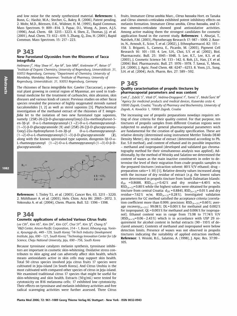
and low noise for the newly synthesized material. References: 1.Bonn, G.; Hashir, M.A.; Stecher, G., Bakry, R. (2006), Patent pending.2. Mohr, M.D., B rnsen, O.K., Widmer, H. M. (1995), Rapid Commun.Mass Spectrom. 9: 809–814. 3. Papac, D.I., Wong, A., Jones, A.J.S.(1996), Anal. Chem. 68: 3215–3223. 4. Shen, Z., Thomas, J.J. et al.(2001), Anal. Chem. 73: 612–619. 5. Zhang, Q., Zou, H. (2001), RapidCommun. Mass Spectrom. 15: 217–223.
P 343New Furostanol Glycosides from the Rhizomes of TaccaintegrifoliaHeilmann J3, Htay Shwe H1, Aye M2, Sein MM2, Kreitmeier P1, Reiser O1
1Institute of Organic Chemistry, University of Regensburg, Universit tsstr. 31,93053 Regensburg, Germany; 2Department of Chemistry, University ofMandalay, Mandalay; Myanmar; 3Institute of Pharmacy, University ofRegensburg, Universit tsstr. 31, 93053 Regensburg, Germany
The rhizomes of Tacca integrifolia Ker. Gawler (Taccaceae), a peren-nial plant growing in central region of Myanmar, are used in tradi-tional medicine for the treatment of carbuncles, skin abrasion, skindiseases and various kinds of cancer. Previous studies on other Taccaspecies revealed the presence of highly oxygenated steroids namedtaccalonolides [1, 2] as well as sterol saponins [3]. Phytochemicalinvestigation of the methanol extract of the rhizomes of T. integri-folia let to the isolation of two new furostanol type saponins,namely (25R)-26-[(b-D-glucopyranosyl)oxy]-22a-methoxyfurost-5-en-3b-yl O-a-L-rhamnopyranosyl-(1?2)-O-a-L-rhamnopyranosyl-(1?3)-b-D-glucopyranoside (1) and (25R)-26-[(b-D-glucopyranosy-l)oxy]-22a-hydroxyfurost-5-en-3b-yl O-a-L-rhamnopyranosyl-(1?2)-O-a-L-rhamnopyranosyl-(1?3)-b-D-glucopyranoside (2),along with the known spirostanol type saponin, diosgenin-3b-O-a-L-rhamnopyranosyl (1?2)-O-a-L-rhamnopyranosyl-(1?3)-O-b-D-glucopyranoside.
References: 1. Tinley T.L. et al. (2003), Cancer Res. 63, 3211–3220.2. M hlbauer A. et al. (2003), Helv. Chim. Acta 86: 2065–2072. 3.Yokosuka A. et al. (2004), Chem. Pharm. Bull. 52: 1396–1398.
P 344Cosmetic applications of selected Various Citrus fruitsLee CW1, Kim HS1, Kim DH1, Kim GO2, Choi SY3, Kim SJ4, Chang IS11R&D Center, Amore-Pacific Corporation, 314 – 1, Borari, Kiheung-eup, Yonin-si, Kyounggi-do, 449 – 729, South Korea; 2Hi-Tech Industry DevelopmentInstitute, Jeju, 690 – 121, South Korea; 3Technology Innovation Center for LifeScience, Cheju National University, Jeju, 690 – 756, South Korea
Because tyrosinase catalyzes melanin synthesis, tyrosinase inhibi-tors are important in cosmetic skin-whitening. Oxidative stress con-tributes to skin aging and can adversely affect skin health, whichmeans antioxidants active in skin cells may support skin health.Total 50 citrus species involved jeju citrus fruits 17 species werecultivated in Jeju-island (in South Korea). And Citrus Unshiu is themost cultivated with compared other species of citrus in Jeju-island.We examined traditional citrus 17 species that might be useful forskin-whitening and skin health. Extracts (50 g/mL) were tested forcytotoxicity on B16 melanoma cells; 17 exhibited low cytotoxicity.Their effects on tyrosinase and melanin inhibitory activities and freeradical scavenging activities were further assessed. Three Citrus
fruits, Immature Citrus unshiu Marc., Citrus hassaku Hort. ex Tanakaand Citrus sinensis x reticulata exhibited potent inhibitory effects onmelanin formation. Immature Citrus unshiu, Citrus hassaku, and Ci-trus sinensis x reticulata showed good antioxidative activities.Among active making them the strongest candidates for cosmeticapplication found in the current study. References: 1. Aburjai, T.,Natsheh, F.M. (2003), Phytotherapy Research 17: 987–1000. 2. Baur-in, N., Arnoult, E., Scior, T. et al. (2002), J. Ethnopharmacol. 82: 155–158. 3. Briganti, S., Camera, E., Picardo, M. (2003), Pigment CellResearch 16: 101–110. 4. Lee, S.H., Choi, S.Y. et al. (2002), Biol.Pharmaceutic. Bull. 25: 1045–1048. 5. Lee, K.T., Lee, K.S. et al.(2003), J. Cosmetic Science 54: 133–142. 6. Roh, J.S., Han, J.Y. et al.(2004) Biol. Pharmaceutic. Bull. 27: 1976–1978. 7. Sawai, Y., Moon,J.H. (2000), J. Agric. Food Chem. 48: 6247–6253. 8. Yoon, J.S., Sung,S.H. et al. (2004), Arch. Pharm. Res. 27: 589–592.
P 345Quality caracterisation of propolis tinctures bypharmacopoeial parameters and wax contentCvek J1, Zubcic S1, Vitali D2, Vedrina-Dragojevic I2, Tomic S1, Medic- aric M2
1Agency for medicinal products and medical devices, Ksaverska cesta 4,10000 Zagreb, Croatia; 2Faculty of Pharmacy and Biochemistry, University ofZagreb, A. Kovacica 1, 10000 Zagreb, Croatia
The increasing use of propolis preparations nowdays requires set-ting of clear criteria for their quality control. For that purpose, tentinctures of propolis samples from different Croatian regions weresubjected to analysis of general pharmacopoeial parameters wichare fundamental for the creation of quality specification. These arerelative density (determined using instrument Mettler Toledo DE40Density Meter), dry residue of extract (determined according to Ph.Eur. 5.0 method), and content of ethanol and its possible impurities– methanol and isopropanol (developed and validated gas chroma-tography method for their simultaneous analysis was applied). Ad-ditionally, by the method of Woisky and Salatino we determined thecontent of waxes as the main inactive constituents in order to de-termine the level of their migration from crude propolis samples tothe prepared tinctures (extraction solvent: 80% V/V ethanol; drug –preparation ratio = 1:10) [1]. Relative density values increased alongwith the increase of dry residue of extract (e. g. the lowest valueswere determined in propolis tincture from South Dalmatian Islands:d20 =0.8688, RSD(n = 2)=0.42% and dry residue =4.40% w/w,RSD(n =2)=0.80% while the highest values were obtained for propolistincture from central Croatia: d20 =0.8841, RSD(n =2)=0.01% and dryresidue =7.62% w/w, RSD(n =2)=0.28%). Investigated validationparameters for GC method satisfied the acceptance criteria (correla-tion coefficient more than 0.999; precision: RSD(n =21)=0.60%; aver-age recovery(n =21): 99.08%; DL =0.001% for methanol and 0.002%for isopropanol; QL=0.003% for methanol and 0.006% for isopropa-nol). Ethanol content was in range from 73.98 to 77.74% V/V(RSD(n = 3)=0.06–2.43%) which is in accordance with USP 29 re-quirement for alcohol content in herbal extracts (90–110% of de-clared amount). Contents of methanol and isopropanol were belowdetection limits. Presence of waxes was not observed in propolistinctures indicating the suitability of applied extraction method.Reference: 1. Woiski, R.G., Salatino, A. (1998), J. Apic. Res. 37:99–105.
Planta Med 2006; 72: 961–1089 Georg Thieme Verlag KG Stuttgart · New York · ISSN 0032-0943
Abstracts
1082
P 346Impact of kava cultivar, plant part and extraction mediumon in-vitro cytotoxicity of kava (Piper methysticum) inHepG2 and Hep3B cellsSchmidt M2, Gebhardt R1
1Institut f r Biochemie, Universit t Leipzig, Liebigstr. 16, D-04103 Leipzig,Germany; 2Herbresearch Germany, Wartbergweg 15, D-86874Tussenhausen-Mattsies, Germany
Preparations from kava (Piper methysticum G. Forst.) have beenbanned based on the suspicion of adverse liver effects. To date, noconvincing proof has been given to substantiate the danger of arelevant toxicity. We systematically tested kava extracts preparedwith acetone or ethanol from two different cultivars, both used forkava extract production: Ava Laau from Samoa, a “noble kava“, andPalisi from Vanuatu, a “Tudei kava“ (“two-day“ lasting effect). Wealso tested the influence of aerial parts (stem peelings) on toxicity.Methods: Extracts were prepared and characterized by the workinggroup of Prof. Nahrstedt at the University of M nster (Germany).Kava plant material was obtained from cultivations. Extracts weretested in HepG2 and Hep3B liver cells, using the MTT test, theRezasurin blue assay, quantification of LDH leakage and measure-ments of intracellular ATP and GSH contents. Results: Only gradualdifferences in cytotoxicity were found. The sequence of toxicity forethanolic extracts was roots (noble) < peelings (noble) £ roots (Tu-dei) < peelings (Tudei). In the case of extracts prepared with acet-one the toxicity of the Tudei-material was partly reversed: peelings(Tudei) < roots (Tudei). In no case were the EC50 values in a relevantdosage range (1250 to >5000mg/mL for roots, 800 to >5000mg/mLfor peelings in the MTT test and rezasurin blue assay, with the high-est toxicity found with Tudei peelings in the rezasurin blue test inHep 3B cells). Conclusions: No hint on relevant liver cell toxicitywas found in this battery of in vitro models.
P 347Effects of chronic administration of Ginkgo biloba extract(EGb 761 ) on levels of dopamine, noradrenaline andserotonin in the prefrontal cortex of the awake ratKehr J1,2, N ldner M3, Yoshitake T1,41Dept. of Physiology and Pharmacology, Karolinska Institutet, Nanna Svartzv g 2, 171 77 Stockholm, Sweden; 2Pronexus Analytical AB, KarolinskaScience Park, Fogdevreten 2a, 171 77 Stockholm, Sweden; 3PreclinicalReseach, Dr. Willmar Schwabe GmbH & Co. KG, Willmar-Schwabe-Str. 4,76227 Karlsruhe, Germany; 4Dept. of Pharmaceutical Sciences, InternationalUniversity of Health and Welfare, 2600 – 1, Kitakanemaru, Ohtawara-shi,Tochigi 324 – 8501, Japan
The special Ginkgo biloba extract (EGb 761 ) has been shown toexert beneficial effects in the therapy of age-related neurologicaldisorders such as Parkinson’s and Alzheimer’s disease [1]. Besides
neuroprotective effects, EGb 761 has been demonstrated to im-prove cognitive functions in animal models [2] and in clinical stud-ies [3]. Thus, it was the aim of the present study to investigatewhether EGb 761 influences monoamine levels in brain areas im-plicated in cognitive function, motivation and mood behaviour. Forthis purpose, the effect of sub-chronic (14 days) daily administrationof EGb 761 on basal extracellular levels of dopamine (DA), nora-drenaline (NA) and serotonin (5-HT) were monitored by microdia-lysis in the prefrontal cortex of awake rats. Administration of EGb761 at a dose of 100mg/kg p. o., caused a significant increase in DAlevels during 40–180min, reaching a maximum level of 163% of thecontrol group. The extracellular levels of NA increased only by about120% and the concentrations of 5-HT were not changed from thepre-drug levels. These findings supports previous observations sug-gesting that Ginkgo biloba extracts could increase monoaminergicfunction via inhibition of MAO activity. However, the minimal ef-fects on NA and 5-HT concentrations indicate that EGb 761 mayaffect brain monoaminergic system also through other mechanismsthan direct inhibition of MAO activity. These results suggest thattreatment with EGb 761 can lead to increased dopaminergic func-tion in the prefrontal cortex which may be an underlying factor toclinically observed effects on improved cognitive function. Refer-ences: 1. Andrieu, S. et al. (2003), J. Gerontol. A Biol. Sci. Med. Sci.58: 372–377. 2. M ller, W.E., Chatterjee, S.S. (2003), Pharmacopsy-chiatry 36 (Suppl.1): S24. 3. Mix, J.A., Crews, W.D. (2002), Hum.Psychopharmacol. Clin. Exp. 17: 267–277.
Planta Med 2006; 72: 961–1089 Georg Thieme Verlag KG Stuttgart · New York · ISSN 0032-0943
Abstracts
1083

Author’s Index Planta Med 2006; 72: 961–1089 Georg Thieme Verlag KG Stuttgart · New York · ISSN 0032-0943
Author’s
Index
1084
AAbdallah WE 1004, 1005Abdel-Azim NS 1015Abdel-Aziz H 1037Abdel-Razik HF 986Abdelshafeek KA 988Abdolhamid P 1070Abdurahman EM 1015Abegaz BM 1009Abel G 984, 1027, 1028, 1036, 1081Abeld G 983Abolhasani FS 1048Abou Zeid AH 1032Abreu P 1033Acero N 992Acevedo HR 1002Acosta K 990Aderbauer B 992Adeyemi OO 1014, 1072, 1073, 1076Adler M 1076Adsersen A 993, 1004Afifi-Yazar FU 1018Afolabi L 1014Aftab K 984Agbelusi GA 1034Aghbolaghi PA 1040Agunu A 1015Ahmadu AA 1043Ahua KM 987Ainasoja M 1024Akar T 981Akay GG 1057, 1058Akcora D 1057, 1058Akdemir ZS 1071AKhalid S 1005Akhigbe AO 1011Akindele AJ 1072Akpulu IN 1043Alamgir M 1051Alaoui S 1063Alaouia S 1058Alasbahi R 1046Al-Fatimi M 1063Al-Helali MF 1046Ali MA 1016Allaeys V 1077Almeida DS 1068Alonso-Cort s D 1011Altinok B 1057, 1058Altmann KH 970, 1013Alves PB 1068Alvesalo J 969Alwahsh MA 988Amaechina F 1001, 1011Amanzade Y 1078Ambaye RY 1009
Amida MB 1076Amin G 1016, 1079Andre P 987Anene RA 1073Angeh I 982Anke J 995, 1022Anton R 986Antoniolli AR 1068Apers S 985, 1022Appel K 1076Appendino G 966Aral E 1040Aras S 1034, 1042, 1075Araffljo AR 973Ardekani MRS 1023Arhan M 1037Arıhan O 1070rnad ttir T 966
Asgari T 1016, 1079Asghari G 1023Ashidi JS 989Ashok Kumar CK 1067Aslan M 1055Aslan S 1081Aslana M 997Asongalem EA 1013Ataman JE 1001, 1011Atli T 1034, 1042, 1075Aura AM 1081Avci A 1034, 1040, 1042, 1057, 1058, 1075Avila EV 1010, 1011Aye M 1082Ayoub S 1047Ayta B 1037
BBaburin I 1049Backlund A 972, 1020Badami N 1016Bae KH 1002Baek NI 1075Bah S 982, 1004Bakmaz M 1035Bakry R 961Bana JY 1012, 1036Baptista J 995Barbastefano V 1037, 1038Barreto MC 995Barriga SD 1002Barroso JG 1045, 1065Barth S 982Basar S 1051Baser C 977Baser KHC 979, 981, 1006, 1040Bassarello C 1061Bastida J 993Bauer R 982, 1010, 1062, 1079
Bauer Petrovska B 1046Bazylko A 1012, 1074Behravan J 965Beikler T 1053Bellacchio E 987Bellone G 996, 1050Benayache S 986Benedek B 998, 1056Bergonzi MC 1053, 1054Berim A 975Bessad ttir M 999Bezabih M 1009Bhuyan SK 1041Bigda J 1008Bilia AR 1006, 1053, 1054, 1059Biskup E 1035Bisson W 1013Blank AF 1068Blaschek W 1051Bliem CB 1008Blomster H 978Bogdanova M 1033Bohlin L 971, 972, 973, 1020, 1066Bommer S 1066Bonaterra GA 1066Bone KM 974, 1026, 1041Bonifacio O 971Bonkanka CX 1047, 1049Bonn GK 961, 970, 1027, 1028, 1036, 1081Borloz A 1002Botkin N 974Bouchenak M 1020Boukortt FO 1020Bounatirou S 1045, 1065Bounsaythip C 1081Braga PSC 1076Brattstr m A 977, 1048, 1049, 1065Brauers G 972Bresgen N 997, 1009Britzmann B 1028Brown SL 1057Brun R 963, 987, 993, 1004, 1005Bruno M 996, 1049, 1050Bucar F 1028Buc Calderone P 1077Bucchini A 987Buchwald-Werner S 1058, 1063Buddhakala N 1048Budi Muljono RA 964Buol I 1080Butterweck V 980, 1065, 1067Byun Y 1013, 1075Byungkil C 1069
CCabezas F 993Calis I 967, 1061
Author’s IndexThe numbers refer to the pagination
Author’s Index Planta Med 2006; 72: 961–1089 Georg Thieme Verlag KG Stuttgart · New York · ISSN 0032-0943
Author’s
Index
1085
Calvo T 1038Camargo EES 1037Canal T 1076Canbek M 1040andır 1040
Cardoso EM 1016Cardoso GC 1068Carithape A 1041Carvalho ACS 1068Cassani J 1001Castro-Gamboa I 968, 973, 1038Cateni F 1077Cavalcanti SCH 1007Cavalheiro AJ 968, 973, 1038Cerd -Nicol s M 1000Cergel S 1044etin R 1037, 1040
Chaabi M 986, 987Chang IS 1082Chang SY 1075Chansathirapanich W 1078Chansiri A 1044Charmain J 971Chavasiri W 966Chawapun A 1079Chen S 980Chenni A 1020Chobot V 971Choi S 1013Choi SH 1002, 1062Choi SY 1082Choi YH 1030, 1066Christen P 969Christensen LP 973, 1073, 1074Chukwujekwu JC 988Chung HG 1013Chung HY 1075Claeson P 984Classen B 1051Clausen PH 992Codina C 993Cola-Miranda M 1037, 1038Corcoran O 991Corvi Mora P 1076Cos P 972, 985Coskun M 1070Costa D 1033Costa MM 1045, 1065Crawford R 966Crozier A 962Curini M 965, 987Cvejic J 1077Cvek J 1063, 1082
DDaariimaa K 1000Dahlstr m M 973D hne L 974Dalsgaard P 964Darwis Y 1021da S. Bolzani V 968, 973, 1016, 1038Dastmalchi K 1021Davoodabadi E 1038Deans SG 1065Deaton CM 977De Castro WV 980Delaude C 1020Delfino R 1076Della Loggia R 1077Demir O 1034Demir TA 981Demirci F 979, 981, 1006De Pasquale R 1059de Paula Michelatto D 1037, 1038Derendorf H 980Derwinska M 1012Deshmukh VS 970Deters A 1050, 1052De Tommasi N 1059Devleeschouwer M 1051de Voss JJ 974, 1026Devrim E 1034, 1040, 1042, 1057, 1058, 1075Dheeranupattana S 1079
Diallo D 982, 987, 1003, 1004, 1059Dickson RA 989Dickson S 1042Dieterle F 964Diller RA 963Diome C 1020Dirsch VM 991, 1018Dolezal K 1062Dom nguez MT 992Dorfer T 1080Dorman HJD 974, 1021, 1026Drewe J 975Duarte N 998, 999, 1001Dubois J 993Duez P 993, 1015, 1051, 1077Dumic J 1036Duong GM 1051Durak I 1034, 1037, 1040, 1042, 1057, 1058,
1075Duus JØ 1057
EEapen S 969Ebel R 967, 970, 971, 972Ebite LE 1011Eckl PM 997, 1009Edrada RA 967, 970, 976Edrada-Ebel RA 971Ellmerer EP 975Elo H 1019Eloff JN 982, 1039El-Sawi S 990Elstner EF 1037El-Toumy SAA 985, 986Emami M 1016Enayat AO 986Engelhardt C 1022Engelshowe R 1049Enr quez RG 1010, 1011, 1037ðojkowska E 1035Epifano F 965, 987Erdelmeier CAJ 1064Erg der IB 1034, 1042, 1057, 1058, 1075Erlendsd ttir H 999Erler J 1010Ernst E 961Erol 1023Escandell JM 967, 1000Eschenko AY 1070Ezea SC 991
FFaizi M 1012Faleiro ML 1045, 1065Fallarero A 1039Faller G 1004, 1053Familoni OB 1034Farag A 965Faraid MA 1016Farias-Silva E 1037, 1038Fassbender B 974Fawzy G 1048Feistel B 1064Fernandes E 1033Fernandes JB 1007Fernandes-Ferreira M 1055, 1076Fern ndez G 1037Ferreira D 985Ferreira MJU 998, 999, 1001Feuerstein I 961Feyen F 970Fico G 1038Fiebich BL 1037Figueiredo AC 1045, 1065Fischer G 1050Floresta SV 1007Forbes B 991Formisano C 1049, 1050Fornera P 1063Fr ndberg PA 973Frank W 972Franz C 983Fraternale D 987
Fr d rich M 993Frett XC 1073Freysdottir J 1058Frias C 963Frieling T 981Fritsch RM 1029, 1081Froelich S 989Fry J 1055Fuchs D 978Fukushi Y 1078Furlan M 973
GGabriele C 1054Galeotti N 1053Galiotou-Panayotou M 1074Galkin A 995Gaube F 992Gauguin B 993Gebhardt R 1083Gehrmann B 1072Geiss HK 992Gennaro R 1076Genovese S 965, 987Gerhards C 1049Gertsch J 970, 1013, 1068Ghasemnezhad A 1044Ghelardini C 1053Giamperi L 987Gibbons S 976Giersig M 974Gil R 967Gilgenast E 1023Giner RM 979, 1000Giomi M 1054Gjorgoski I 1033Glasl S 1000Gł d D 1060Głowniak K 1025Gnecco D 1010Gnoula C 993Godejohann M 975Goeke B 1077Goger F 977Gokmen D 1057, 1058Gomes ET 978Gomeza A 1063Gonz lez MR 990Gonz lez T 1033Goossens A 963, 981, 1031Gopalkrishnan R 969, 984G ransson U 973Gormann R 994Gortzi O 1074Gottfries J 972, 1020Greilberger J 978Grevsen K 973, 1073Gritsanapan W 966, 1041Gritzko K 1076Gruissem W 1066Grundmann O 1065Gudiksen L 993Guede-guina F 987Guedes AP 1055Guissou P 993Gupta MP 1030Gupta RS 964, 1015G rb z I 1004Gusenleitner S 982Gutmann H 975G ven A 1070Guzm n E 990Gy mant N 998, 999
HHa NR 1007Habib AA 1015Habtemarium S 1001Hadacek F 979Hadjiakhoondi A 1078Hadjipavlou-Litina D 1024Hagelauer D 1065Hajiaghaee R 1078
Author’s Index Planta Med 2006; 72: 961–1089 Georg Thieme Verlag KG Stuttgart · New York · ISSN 0032-0943
Author’s
Index
1086
Hajimahmoodi M 1038Haj Yahya M 1047H kkinen ST 963, 1031Halldorsdottir ES 1014Hambartsumyan M 968Hamburger M 961, 964, 992, 1029, 1032Hamed A 1055Hamedi A 990Hamidian Jahromi A 1079Hammouda FM 1015Han J 1013Hardardottir G 998Haruna AK 1043Hashem FA 1016Hashidoko Y 1078Hashir MA 970, 1081Haskell CF 979Hassan NM 1015Hassan RA 1040Hasuda T 1018Hatakka A 996Hattesohl M 1064Hauserova E 1062Havlik J 1008Hawas UW 988Hay AE 987, 1030Hedner E 973Hehenberger S 975Heilmann J 1025, 1082Heinle H 1037, 1065Heinrich M 1059, 1068Heiss E 991, 1018Hensel A 995, 1003, 1004, 1005, 1022, 1049,
1053Henze G 963Heo MH 1075Hering S 998, 1049Herl V 1050Hern ndez M 1046Hern ndez MS 1002Hifnawy M 1080Hilgendorff M 974Hiltunen R 974, 978, 1019, 1021, 1026Hinz B 1062Hiruma-Lima CA 1037, 1038Hitotsuyanagi Y 1018Hoffmann D 974Hoffmannov L 1003Hohlfeld T 971Hole RC 1009Honermeier B 1044Hongratanaworakit T 1044Hongrattanavorakit N 1044Hongthongdaeng B 1028, 1034Hornby BF 1057Hosein M 1016Hostanska K 1064Hostettmann K 969, 987, 1002, 1030, 1066Houghton PJ 966, 989, 991, 996, 1001, 1061Hovhanissyan A 968Htay Shwe H 1082Huck CW 961Huefner A 965, 1048Humam M 969Humpfer E 975Hur JM 1062Husnu K 977Huss U 1066Hylands PJ 989, 991, 1001, 1061Hyun S 1007
IIbrahim NA 1016Ibrahim S 970Idu M 1001, 1011Ilori OO 1033Ina H 1003Ingkaninan K 998, 1028, 1034Ingkatawornwong S 1045Ing lfsd ttir K 966, 998, 999Innocenti M 1054Intaranongpai J 966Inya-Agha SI 991, 1033, 1034
Inz D 963, 981, 1031Iorizzi M 1038Ioset JR 987, 1066Iranshahi M 965, 1014Isacchi B 1053Ismail IA 988Ismail KM 966Itharat A 1045Iznaguen H 1051
JJ ger K 1039Jachak SM 1021Jaeggi R 1037J ger AK 988, 993, 1004, 1019, 1023Jain K 980Jalali MS 1065Jannat B 1038Jaroszewski JW 1023Jasprica I 1011, 1036Jatisatienr A 1079Jatisatienr C 1079Jayaveera KN 1067Jeannerat D 969Jedelsk J 1029Jenett-Siems* K 989Jensen M 973, 1074Jeon M 1062Jeon WK 1074Jeong EJ 1007Jeong HH 1002, 1062Jesadanont S 996, 997Jia S 1043JiHyun C 1069Jim nez-Estrada M 1001JinMi C 1069Johansen HT 982Johansson T 973Jones K 977Jones LA 1052J nsd ttir 999Jonsson P 973Juan H 978Julkunen-Tiitto R 1053Jun KH 1069J rgenliemk G 1063Juvekar AR 969, 970, 984, 1009
KKachhawa JBS 1015Kaewruang W 1028, 1034Kaiser M 963, 1004, 1005, 1006Kakooko A 989Kaloga M 994Kalo era Z 1035Kamalinejad M 1012, 1047, 1048Kaminski M 1023Kamtchouing P 1013Kan Y 1035Kang IH 1075Kanna C 1044Kapingu MC 985Karaaoglu T 1004Karabourniotis G 1059Karaoglu T 997, 1035Karimi G 965Karimi I 1040Karioti A 1024, 1025, 1059Karlsson PC 1066Kartal M 1035, 1081Kavutcu M 1037Kawiak A 1008Kehr J 977, 1083Kehraus S 1023, 1024, 1025Kehrli T 969Kelber O 981, 1037, 1065, 1066, 1077Kemper M 1052Kennedy DO 964, 979Keppler OT 1021Ketzis JK 1043Keusgen M 974, 1029, 1081Khader M 997, 1009Khajorndetkun W 1078
Khalid SA 1004Khan A 1069Khan IA 1071Khanavi M 1078Khangholi S 1080Khayyal MT 1037Kheiri S 1040Khom S 998, 1049Kiderlen AF 1046Kidmose U 1074Kılı CS 1070Kılı olu B 1040Kim C 1007Kim DH 1082Kim EK 1075Kim GO 1082Kim GS 1075Kim HK 1030Kim HS 1082Kim IH 1018Kim JH 1075Kim JM 1075Kim JS 1075Kim SC 1013Kim SH 1007, 1013Kim SJ 1082Kinscherf R 1066Kiran I 981Kirimer N 981Kirmizibekmez H 967, 1061Kisiel W 1056Kiss AK 1074Kiyohara H 990Klejdus B 1027Klenke A 1052Kletter C 1000Knieps H 974Ko BS 1074Kobayashi A 1068Koblovsk R 1027Koch E 1061, 1064Koetter U 977, 1048, 1049, 1065Kohout L 1003Kojouri G 1040KokoÐka L 1008, 1010, 1027Kołaczkowski M 1001Kolodziej H 994, 1046Komjanc M 1031K nig GM 1023, 1024, 1025Konuklugil B 1018Koo KA 1017Kopp B 998, 1056, 1080Koppa B 1056Kortenkamp A 1068Kosalec I 1035Kosar M 977Kosman V 1026Kowalski J 1012, 1074Kram D 1046Krasniqi B 1067Krauze-Baranowska M 1060Kreis W 1050Kreitmeier P 1082Krempser MR 1007, 1068Krempser RR 1007, 1068Krick A 1023, 1024, 1025Krieg C 1036Kristiansen K 973Krolicka A 1023Kroll T 992Kr ck C 983Kr ger D 981Kubicov L 971K hn T 964Kulevanova S 1032, 1033, 1046Kulkarni MP 1009Kumar GS 1067Kumar V 996Kunugi A 1068Kuo J 980Kupeli E 1071Kupittayanant S 1048Kuypers K 972

Author’s Index Planta Med 2006; 72: 961–1089 Georg Thieme Verlag KG Stuttgart · New York · ISSN 0032-0943
Author’s
Index
1087
Kwak HM 1002Kwak YS 990Kwon SH 1002, 1062Kyoung KH 1069Kypriotakis Z 1028Kyung JS 990
LLaakso I 974, 1026Lacaille-Dubois MA 1020Lack G 963Lage H 999Lalas S 1074Lalk M 968Lamerding F 1052Landa P 1010, 1071Langer T 962, 1008Lantto T 1019Lapc k O 1027Larkins NJ 977Larsson J 972, 1020Lass C 1059Laux MT 1043Lazari DM 1010Lea RW 1057Lechtenberg M 1005, 1049, 1080Lee BJ 1017Lee CW 1082Lee EJ 1017Lee HY 1075Lee JH 1074Lee JP 1075Lee KH 996Lee SE 1075Lee YY 1062Lehmann RP 974, 1026, 1041Lehnfeld R 1064Leinonen M 969Lengsfeld C 1003, 1004Lim S 1013Lin W 967Lindequist U 968, 1063Lipipun V 996Lira RH 1046Llinares F 992Lobato CE 1037Lobstein A 986Loikkanen J 1039Lojkowska E 1008, 1023Lopes MN 973, 1016Lou Y 976Lozada MC 1010, 1011Ludwiczuk A 1025Luhmer M 1015Luiz-Ferreira A 1037, 1038Luna HM 1001Luong T 980
MMa R 963, 981Maas M 1052Machumi F 985Maes L 972, 985Maggio A 996Magos GA 1001, 1037Mahdavi F 1078Maiga A 1059Makarov VG 974, 1007, 1026, 1035, 1070Makarova MN 1007, 1035Makarova SV 1070Mal M 996Mal kov J 1003Malterud KE 1051M ez S 979, 1000Mansour K 1072Marais JPJ 985Mar al RM 1007, 1068Maregesi S 985Marin PD 1060M rquez C 1037Marsik P 1008, 1010, 1071Marston A 1002Martin F 1030, 1066
Martin SF 1059Masroor M 1069Masumeh K 1045Mateos JC 1037Matias RR 971Matikainen J 1019Matovic N 974, 1026Matsumoto K 1003Matta MK 1010Matthew S 1033Matthias A 974, 1026, 1041Maune S 984Maxia L 966Mayer R 1027, 1028Mayr K 1000Mbagwu HOC 1073Mbwambo ZH 985McGaw L 982McIntyre L 1042McRae J 966Medic- aric M 1011, 1036, 1063, 1082Mehner C 1024Mehrabadi Yari L 1016Meier B 983, 1067Melillo de Malgalhaes P 1054Mello ICM 1068Melzer J 1064Melzig MF 992, 1047, 1052, 1056, 1072Menghini L 965, 987Mentel R 968Merfort I 967, 1059Mertens-Talcott S 980Mett l A 996Metz J 1037, 1066Mi CJ 1069Michaelsen TE 1003Michalak K 1001Michalska K 1056Miguel MG 1045, 1065Miljkovic-Brake A 1001Milne AL 979Ming L 991Mitaine-Offer AC 1020Mitliaga P 1074Miyamoto T 1020Miyazaki T 1003Modarai M 1068Mohamed R 1080Mohamed SM 1016Mohn T 1029, 1032Mojab F 1048, 1054Moln r J 998, 999Monsef-Esfahani HR 1078Monteiro M 995Monteiro Souza Brito AR 1021Montiel JL 1010, 1011Moody JO 1078Moormann J 1065Moreno MPN 1007Mornar A 1011, 1036Mosaffaa F 965Moshi MJ 985Moske M 974Motawae H 990Motlhanka DMT 1001Mueller-Uri F 1050Mukazayire MJ 1077Mukherjee PK 996M ller D 1024M ller J 1037M ller WEG 970Mulsri N 1044Mu oz O 969Mu oz-Mingarro D 992Muresan S 1020Musa KY 1015Mustafa NK 964Mustoe T 1043
NNachankar RS 1009Nacro M 1015Naeem M 1069
Nahrstedt A 1063, 1072, 1073, 1080Nana P 1013Narantuya S 1000Nasir S 1069Nassar MIA 1022Natividad FF 971Ndjakou BL 986Ndjoko K 1066Neffati M 1045Neher A 984Netsch M 975Ngamga D 1009Ngouela S 986N’guessan JD 987Nickavar B 1047, 1048, 1054Nicolas JF 1059Nieber K 1048Niedermeyer THJ 968Niehues M 1005Nielsen M 1019Nohynek L 981, 1039Nolard N 1043N ldner M 977, 1083Nolkemper S 1021Nørbæk R 1073Nour AMM 1004, 1005Novak O 1062Nuamlert J 1044Nuengchamnong N 1028Nyberg F 973
OOdia EA 1011Odukoya OA 991, 1033, 1034Ogmundsdottir HM 998Ohlendorf B 1023Oinonen P 996Okpanyi SN 981, 1037Oksman-Caldentey KM 963, 981, 1031, 1039,
1081Okusa PN 1051lad ttir AK 966
Olafsdottir ES 1014, 1057, 1058Olmos A 979, 1000Olsen A 993Omarsdottir S 966, 999, 1057, 1058Omogbai EKI 1001, 1011Onegi B 989Opara EI 1052Oresic M 1081OreÐic M 963, 981Orhan I 1035, 1055Orhana I 997Ortlepp S 967, 971Osorio E 993ttl K 978
Oveisi MR 1038Ovodov YS 1012Ovodova RG 1012Oyelola OO 1078z elik B 997, 1004, 1035
Ozkal P 1057, 1058Ozkan AM 1072zt rk HS 1075
PP ez E 1037Pahl A 984Pakkanen J 995Pal E 1015Palee J 1079Palombo E 966Panagiotidis CA 1010Panagiotou E 1052Panossian A 968Panovska TK 1032, 1033Papanikolaou S 1074Paranhos A 1060Park JD 990Park JY 1075Partl A 1035Pascal R 974Pascolo L 1076
Author’s Index Planta Med 2006; 72: 961–1089 Georg Thieme Verlag KG Stuttgart · New York · ISSN 0032-0943
Author’s
Index
1088
Pateh UU 1043Paula SPS 1007Pauletti PM 968, 973Paulsen BS 982, 1003, 1004, 1057Paululat T 964Paunonen R 1053Pedersen ME 1019Pedersen PD 988Pedro LG 1045, 1065Pelkonen O 983, 984Pelttari E 1019Penge O 1051Pengtao L 991Penman KG 974, 1026Pepeljnjak S 1035P rez C 990P rez MS 990Perozzo R 963, 1006Petereit F 995, 1022, 1063, 1072Petersen BO 1057Petersen M 975Petrushevska G 1033Pettersson J 1066Phadungcharoen T 1031Phattanawasin P 1056Pieters L 972, 985, 1022Pimentel FO 1038Pineda G 1046Pinsuwan S 1045Pizza C 1061Plaza CV 1038Pobłocka-Olech L 1060Pocuca M 1077Pohjala L 1024Pongsamart S 996, 997Pongwiwatana U 996Pontius A 1025Popov SV 1012Popova GY 1012Popp M 1027, 1028, 1036, 1081Potterat O 964, 1029, 1032Poungshompoo S 997Pourjafar M 1040Pozharitskaya ON 974, 1007, 1026Prado B 1049Pribylova M 1010, 1071Prinsloo LC 993Prinza S 1056Prokop A 963Proksch P 967, 970, 971, 972, 976Prost J 1020Psilander N 978Ptak DM 1007, 1068Puig RC 1030Puripattanavong J 1045, 1078Pusch L 992Puupponen-Pimi R 1039
QQa’dan F 1072Qingguo W 991Quandt B 1005, 1080
RRaasmaja A 1019Rabanal RM 1047, 1049Raduner S 970, 1013Radwan HM 1040Rafter JJ 1066Ramalhete C 999Ramanou A 987Ramaswamy V 1030Rancic A 1060Rashed K 965Rasmussen HB 1023Ratanasak W 1041Ratanatham T 1041Recio MC 967, 1000Rehwani H 964Reichling J 992, 1021Reif K 983Reiser O 1082Rejeb MN 1045
Reynolds WF 1011Rezaeinodehi A 1080Reza Khani MR 1048Rhee IK 1062Ricci D 987Riepl HM 963Riese U 992Rigano D 1049, 1050Riihim ki L 994R os JL 967, 1000Rischer H 963, 981, 1031Ritala A 963, 981, 1031Rojas MD 1002Rollinger JM 962, 1008Romanik G 1023Rose O 963Rose U 983Rosselli S 996, 1049, 1050Rousi M 1053Ruangrungsi N 1031R edi P 963Rusman Y 972Rustaiyan A 1078Ruzzier F 1076Ryder NS 1043Rydlovskaya A 1007
SSadeghi N 1038Sadeghpour O 1023Salık 1018Said A 965, 1048Saikia G 1041Saikku P 969Sajfrtova M 1008Sakihama Y 1078Saleh M 1080Salinas RAM 1007Saller R 1064Samuelsen AB 1051San Miguel-Ch vez R 1002S nchez-Mateo CC 1047, 1049Sanogo R 1059Santos AB 1007Santos RB 1068Sarawek S 1067Sarder M 1051Saroglou V 1028, 1060Sautour M 1020Sautter C 1066Scheffer JJC 1065Schemann M 981Schempp C 1059Schepmann D 1005Schmidgall J 1003Schmidt A 975Schmidt M 1080, 1083Schmidt TJ 1004, 1005, 1006, 1049Schmitt CA 1018Schnitzler P 1021Scholey AB 964, 979Schoop R 1030Schrader E 977Schrenk D 983, 992Schr der G 1063Schubert C 989Schulze J 1042Schuster D 962Schwaiberger AV 991Schwaiger S 975Schyschka L 1008Scott L 993Sefidkon F 1080Seger C 975Segun FI 1033, 1034Segundo M 1033Sein MM 1082Senatore F 1049, 1050Sener B 1035Seol J 1069Seong NS 1075Seong RS 1075Seong YH 1002
Seonga YH 1012, 1036Sepp nen-Laakso T 963, 1031, 1039, 1081Serrano MAR 1016Sert A 1019Severin T 1018Sezika E 997Shafaghi B 1012Shafiee A 1078Shahadeh M 1018Shahverdi AR 1014Shams KA 1015Sharifzadeh M 1078Sharma A 964, 1015Sharma DK 1041Shikov AN 974, 1026Shin HJ 990Shin S 1013, 1075Sibaev A 1077Sichardt K 1048Sieger R 1073Siegers CP 1042, 1070Sievers H 1064Silva DHS 1038Silva O 978Sim Y 1013Singh M 1069Singhai AK 980, 1069Singhubera J 1056Siqueira DHS 968, 973Sitthiwej C 997Sj gren M 973Skaltsa H 1024, 1025, 1028, 1059, 1060Skopeliti M 1025Sleem A 1080Smith E 976Smiti S 1045So JH 1062Sofidiya MO 1033, 1034Sohbat B 1070Somani R 980, 1069Somervuo P 1024Song KS 1002, 1062, 1075Song YB 990Songb KS 1012, 1036Soni V 978Sosa S 1077Sotanaphun U 1056Soto-Hern ndez M 1002Souza DP 1007Souza Brito ARM 1037, 1038Sovova H 1008Sp rl-Aich G 1061Spraul M 975Spriano D 1067Sreedhar C 1067Sriphong L 1056Stafford GI 988, 1019Stanckevich N 1026Starkey NJ 1057Stasilojc G 1008Stecher G 961, 970, 984, 1027, 1028, 1036,
1081Stefkov G 1033Stepanov I 1077Stierna P 984Stobiecki M 1008St hr J 1079Storr M 1077Stratmann U 1053Streit B 1028Strnad M 1003, 1062Strompen T 1067Stuppner H 962, 975, 1008Suh DY 1062Sukrong S 1031Sukying S 1045Sule MI 1043Sultana T 1027, 1028Sunguroglu A 1057, 1058Suortti T 981Surmaghi S 1016Suschke U 992Suter A 1030, 1042, 1064, 1066, 1068
Author’s Index Planta Med 2006; 72: 961–1089 Georg Thieme Verlag KG Stuttgart · New York · ISSN 0032-0943
Author’s
Index
1089
Swaczynov J 1003Sylignaki GI 1010Szelenyi I 984Sznitowska M 1060Szpitter A 1023
TTabl ESAA 1048Tahara S 1078Takeya K 1018Tammela P 969, 1024, 1039Tan J 1042Tanaka T 965Tane P 1009Tantangmo F 986Tapaneeyasin P 1044Tasdemir D 963, 1006Tatli II 1071Tato P 1001Techatanawat I 1061Teeri T 1024Tegelberg R 1053Tegtmeier M 1070Teixeira DM 995Temkitthawon P 998Th le C 1046Thanh HDT 1030Theunis M 1022Tichonov V 1026Tikhonov VP 974, 1007, 1026, 1035Tiribelli C 1076Tobler M 1067Togola A 1003, 1057Tokuda H 965Tom F 1038Tomic S 1063, 1082Torre C 967Torres LB 1016Tossi A 1077Trauner G 998Treitz G 974Trethewey R 961Trojer L 1027Tsamo E 986Tseng LH 975Tsevegsuren N 967Tsitsilonis O 1025Tubaro A 1077Turgay M 1075
UUlmer AJ 1051Urbaniak B 1066Uyanolu M 1040
VVadnere G 980, 1069Vahidi H 990Valterova I 1008Vanden Berghe D 985
Vanek T 1010, 1071Van Puyvelde L 972van Staden J 988, 1019, 1023Varga A 998, 999Varli M 1034, 1042, 1075Varol N 1057, 1058Vedrina-Dragojevic I 1063, 1082Veljic M 1060Verberne M 964Verma R 969, 984Vermeersch M 972Verpoorte R 964, 983, 1030, 1066Veveris M 1061Vicente AM 1076Vidal A 1039Viegas CJ 973Vieira A 980Viet LD 991Viladomat F 993Vilegas W 1037, 1038Villar A 1058, 1063Villarreal ML 1010, 1011Vincieri FF 1006, 1053, 1054Visioli F 1038Vissiennon Z 1048Vitali D 1063, 1082Vitalini S 1038Viyoch J 998Vlietinck A 985, 1022Vlietinck AJ 984Vocanson M 1059Vollmar AM 1008Vonthron-S n cheau C 986V ßing S 1006Vrushabendra Swamy BM 1067Vuorela HJ 969Vuorela P 962, 994, 995, 996, 1024, 1039Vuorela PM 969Vytlacilov J 971
WWafaa EAA 986Wagner V 1021Wakade AS 1009Wallbach J 1077Wallner B 1076Wang ZQ 976Wangensteen H 1051Wasilewska A 1008W tjen W 976Watson K 1041Wawrosch C 1080Weber N 976Wee JJ 990Weigend M 1028Weiser D 1037, 1065, 1066, 1077Weng A 1019, 1052Weniger B 986, 987Wenzig E 1010Wesnes KA 979
Westendorf J 1051Whittaker P 1042Wikman G 968Williamson E 976Wilson N 1059Winkler C 978Winterhoff H 983, 1064, 1065Wittschier N 1053Woelkart K 982, 1062W lfl S 992Wolski T 1025Won LH 1069Wongareonwanakij S 1028, 1034Wongthai J 1034Wray V 967, 970, 972, 976W nsch B 1005Wurster M 1063
XXu P 1017
YYahia DA 1020Yamada H 990Yamada K 1003Yang EJ 1002, 1062Yang H 1007Yang Q 966Yemitan OK 1014, 1076Yeong LA 1069Yesilada E 1004, 1071Yi L 991Yilmaz G 1035Yoshitake T 977, 1083You JM 1017Youn HJ 1017Young MCM 973, 1016Young S 1007Y cel Z 1018Yuece B 1077Yun YS 1068
ZZacchigna M 1077Zager A 1051Zajtseva MA 1035Zavala MA 990Ze KR 1075Zeglin A 1051Zeid AA 1080Zeller F 981Zenkevich IG 1070Zezi UA 1015Zhub M 1056Zidorn C 980Zini E 1031Zippel J 1050Zovko M 1035Zubcic S 1063, 1082Zumdick S 1049
Copyright © 2022 FDOKUMEN