Blockade of Gap Junction Hemichannel Suppresses Disease Progression in Mouse Models of Amyotrophic...
13
Blockade of Gap Junction Hemichannel Suppresses Disease Progression in Mouse Models of Amyotrophic Lateral Sclerosis and Alzheimer’s Disease Hideyuki Takeuchi 1 *, Hiroyuki Mizoguchi 2 , Yukiko Doi 1 , Shijie Jin 1 , Mariko Noda 1 , Jianfeng Liang 1 , Hua Li 1 , Yan Zhou 1 , Rarami Mori 2 , Satoko Yasuoka 1 , Endong Li 1 , Bijay Parajuli 1 , Jun Kawanokuchi 1 , Yoshifumi Sonobe 1 , Jun Sato 2 , Koji Yamanaka 3 , Gen Sobue 4 , Tetsuya Mizuno 1 , Akio Suzumura 1 1 Department of Neuroimmunology, Research Institute of Environmental Medicine, Nagoya University, Furo-cho, Chikusa-ku, Nagoya, Japan, 2 Futuristic Environmental Simulation Center, Research Institute of Environmental Medicine, Nagoya University, Furo-cho, Chikusa-ku, Nagoya, Japan, 3 Laboratory for Motor Neuron Disease, RIKEN Brain Science Institute, Hirosawa, Wako, Saitama, Japan, 4 Department of Neurology, Nagoya University Graduate School of Medicine, Tsurumai-cho, Showa-ku, Nagoya, Japan Abstract Background: Glutamate released by activated microglia induces excitotoxic neuronal death, which likely contributes to non-cell autonomous neuronal death in neurodegenerative diseases, including amyotrophic lateral sclerosis and Alzheimer’s disease. Although both blockade of glutamate receptors and inhibition of microglial activation are the therapeutic candidates for these neurodegenerative diseases, glutamate receptor blockers also perturbed physiological and essential glutamate signals, and inhibitors of microglial activation suppressed both neurotoxic/neuroprotective roles of microglia and hardly affected disease progression. We previously demonstrated that activated microglia release a large amount of glutamate specifically through gap junction hemichannel. Hence, blockade of gap junction hemichannel may be potentially beneficial in treatment of neurodegenerative diseases. Methods and Findings: In this study, we generated a novel blood-brain barrier permeable gap junction hemichannel blocker based on glycyrrhetinic acid. We found that pharmacologic blockade of gap junction hemichannel inhibited excessive glutamate release from activated microglia in vitro and in vivo without producing notable toxicity. Blocking gap junction hemichannel significantly suppressed neuronal loss of the spinal cord and extended survival in transgenic mice carrying human superoxide dismutase 1 with G93A or G37R mutation as an amyotrophic lateral sclerosis mouse model. Moreover, blockade of gap junction hemichannel also significantly improved memory impairments without altering amyloid b deposition in double transgenic mice expressing human amyloid precursor protein with K595N and M596L mutations and presenilin 1 with A264E mutation as an Alzheimer’s disease mouse model. Conclusions: Our results suggest that gap junction hemichannel blockers may represent a new therapeutic strategy to target neurotoxic microglia specifically and prevent microglia-mediated neuronal death in various neurodegenerative diseases. Citation: Takeuchi H, Mizoguchi H, Doi Y, Jin S, Noda M, et al. (2011) Blockade of Gap Junction Hemichannel Suppresses Disease Progression in Mouse Models of Amyotrophic Lateral Sclerosis and Alzheimer’s Disease. PLoS ONE 6(6): e21108. doi:10.1371/journal.pone.0021108 Editor: Koichi M. Iijima, Thomas Jefferson University, United States of America Received March 21, 2011; Accepted May 18, 2011; Published June 21, 2011 Copyright: ß 2011 Takeuchi et al. This is an open-access article distributed under the terms of the Creative Commons Attribution License, which permits unrestricted use, distribution, and reproduction in any medium, provided the original author and source are credited. Funding: This work was supported by the Program for Promotion of Fundamental Studies in Health Sciences from the National Institute of Biomedical Innovation; a grant from the Uehara Memorial Foundation; a grant from the Japan Research Foundation for Clinical Pharmacology; a Grant-in-Aid for the global Center of Excellence program from the Ministry of Education, Culture, Sports, Science and Technology of Japan; grants from the Ministry of Health, Labour and Welfare of Japan; and a grant from New Energy and Industrial Technology Development Organization. The funders had no role in study design, data collection and analysis, decision to publish, or preparation of the manuscript. Competing Interests: The authors have declared that no competing interests exist. * E-mail: [email protected] Introduction Microglia are macrophage-like resident immune cells of the central nervous system (CNS). They function as not only antigen- presenting cells but also effector cells that have been shown to damage neural cells directly in vitro and in vivo [1]. Microgliosis— i.e., the accumulation of activated microglia—is a pathologic hallmark of various neurologic disorders, including trauma, ischemia, inflammation, epilepsy, and such neurodegenerative diseases as multiple sclerosis, Huntington’s disease, Parkinson’s disease, amyotrophic lateral sclerosis (ALS), and Alzheimer’s disease (AD) [2–4]. Activated microglia release large amounts of glutamate and induce excitotoxicity via N-methyl-D-aspartate (NMDA) receptor signaling [5–8]. NMDA receptor signaling increases Ca 2+ influx, resulting in Ca 2+ /calmodulin-dependent protein kinase (CaMK) activation. CaMK augments nitric oxide (NO) production by activating neuronal NO synthase. NO inhibits mitochondrial respiratory chain complex IV activity and induces a rapid drop in intracellular ATP levels, which negatively affects dendritic and axonal transport. Impaired transport causes PLoS ONE | www.plosone.org 1 June 2011 | Volume 6 | Issue 6 | e21108
-
Upload
independent -
Category
Documents
-
view
0 -
download
0
Transcript of Blockade of Gap Junction Hemichannel Suppresses Disease Progression in Mouse Models of Amyotrophic...
Blockade of Gap Junction Hemichannel SuppressesDisease Progression in Mouse Models of AmyotrophicLateral Sclerosis and Alzheimer’s DiseaseHideyuki Takeuchi1*, Hiroyuki Mizoguchi2, Yukiko Doi1, Shijie Jin1, Mariko Noda1, Jianfeng Liang1, Hua
Li1, Yan Zhou1, Rarami Mori2, Satoko Yasuoka1, Endong Li1, Bijay Parajuli1, Jun Kawanokuchi1,
Yoshifumi Sonobe1, Jun Sato2, Koji Yamanaka3, Gen Sobue4, Tetsuya Mizuno1, Akio Suzumura1
1 Department of Neuroimmunology, Research Institute of Environmental Medicine, Nagoya University, Furo-cho, Chikusa-ku, Nagoya, Japan, 2 Futuristic Environmental
Simulation Center, Research Institute of Environmental Medicine, Nagoya University, Furo-cho, Chikusa-ku, Nagoya, Japan, 3 Laboratory for Motor Neuron Disease, RIKEN
Brain Science Institute, Hirosawa, Wako, Saitama, Japan, 4 Department of Neurology, Nagoya University Graduate School of Medicine, Tsurumai-cho, Showa-ku, Nagoya,
Japan
Abstract
Background: Glutamate released by activated microglia induces excitotoxic neuronal death, which likely contributes tonon-cell autonomous neuronal death in neurodegenerative diseases, including amyotrophic lateral sclerosis andAlzheimer’s disease. Although both blockade of glutamate receptors and inhibition of microglial activation are thetherapeutic candidates for these neurodegenerative diseases, glutamate receptor blockers also perturbed physiological andessential glutamate signals, and inhibitors of microglial activation suppressed both neurotoxic/neuroprotective roles ofmicroglia and hardly affected disease progression. We previously demonstrated that activated microglia release a largeamount of glutamate specifically through gap junction hemichannel. Hence, blockade of gap junction hemichannel may bepotentially beneficial in treatment of neurodegenerative diseases.
Methods and Findings: In this study, we generated a novel blood-brain barrier permeable gap junction hemichannelblocker based on glycyrrhetinic acid. We found that pharmacologic blockade of gap junction hemichannel inhibitedexcessive glutamate release from activated microglia in vitro and in vivo without producing notable toxicity. Blocking gapjunction hemichannel significantly suppressed neuronal loss of the spinal cord and extended survival in transgenic micecarrying human superoxide dismutase 1 with G93A or G37R mutation as an amyotrophic lateral sclerosis mouse model.Moreover, blockade of gap junction hemichannel also significantly improved memory impairments without altering amyloidb deposition in double transgenic mice expressing human amyloid precursor protein with K595N and M596L mutations andpresenilin 1 with A264E mutation as an Alzheimer’s disease mouse model.
Conclusions: Our results suggest that gap junction hemichannel blockers may represent a new therapeutic strategy totarget neurotoxic microglia specifically and prevent microglia-mediated neuronal death in various neurodegenerativediseases.
Citation: Takeuchi H, Mizoguchi H, Doi Y, Jin S, Noda M, et al. (2011) Blockade of Gap Junction Hemichannel Suppresses Disease Progression in Mouse Models ofAmyotrophic Lateral Sclerosis and Alzheimer’s Disease. PLoS ONE 6(6): e21108. doi:10.1371/journal.pone.0021108
Editor: Koichi M. Iijima, Thomas Jefferson University, United States of America
Received March 21, 2011; Accepted May 18, 2011; Published June 21, 2011
Copyright: � 2011 Takeuchi et al. This is an open-access article distributed under the terms of the Creative Commons Attribution License, which permitsunrestricted use, distribution, and reproduction in any medium, provided the original author and source are credited.
Funding: This work was supported by the Program for Promotion of Fundamental Studies in Health Sciences from the National Institute of BiomedicalInnovation; a grant from the Uehara Memorial Foundation; a grant from the Japan Research Foundation for Clinical Pharmacology; a Grant-in-Aid for the globalCenter of Excellence program from the Ministry of Education, Culture, Sports, Science and Technology of Japan; grants from the Ministry of Health, Labour andWelfare of Japan; and a grant from New Energy and Industrial Technology Development Organization. The funders had no role in study design, data collectionand analysis, decision to publish, or preparation of the manuscript.
Competing Interests: The authors have declared that no competing interests exist.
* E-mail: [email protected]
Introduction
Microglia are macrophage-like resident immune cells of the
central nervous system (CNS). They function as not only antigen-
presenting cells but also effector cells that have been shown to
damage neural cells directly in vitro and in vivo [1]. Microgliosis—
i.e., the accumulation of activated microglia—is a pathologic
hallmark of various neurologic disorders, including trauma,
ischemia, inflammation, epilepsy, and such neurodegenerative
diseases as multiple sclerosis, Huntington’s disease, Parkinson’s
disease, amyotrophic lateral sclerosis (ALS), and Alzheimer’s
disease (AD) [2–4]. Activated microglia release large amounts of
glutamate and induce excitotoxicity via N-methyl-D-aspartate
(NMDA) receptor signaling [5–8]. NMDA receptor signaling
increases Ca2+ influx, resulting in Ca2+/calmodulin-dependent
protein kinase (CaMK) activation. CaMK augments nitric oxide
(NO) production by activating neuronal NO synthase. NO inhibits
mitochondrial respiratory chain complex IV activity and induces a
rapid drop in intracellular ATP levels, which negatively affects
dendritic and axonal transport. Impaired transport causes
PLoS ONE | www.plosone.org 1 June 2011 | Volume 6 | Issue 6 | e21108

cytoskeletal and motor protein accumulation at sites of neuritic
beading [6]. This energy-starved condition represents neuronal
dysfunction state and eventually leads to neuronal death (recently
termed non-cell-autonomous neuronal death [9]). This process has
been postulated as a major cause of neuronal damage in many
neurologic diseases [10,11]. Thus, blocking glutamate receptors
has emerged as a potential therapeutic approach for several
neurodegenerative diseases, however, associated perturbations in
physiologic glutamate signaling lead to severe adverse effects [12].
Whereas inhibition of microglial activation has also been
considered as a therapeutic strategy, these cells play neuroprotec-
tive roles that are mediated by release of neurotrophic factors,
glutamate uptake, and sequestering of neurotoxic substances
[2,13–16]. After all, these clinical trials have largely failed in the
last two decades [17–19]. Therefore, efforts are underway to target
neurotoxic microglia specifically.
Gap junctions are composed of two adjacent hemichannels,
which directly connect the cytoplasmic compartments of adjacent
cells and allow small molecules (,1 kD) and ions to pass freely
between cells [20]. Recent evidence suggests that ‘‘free’’
hemichannels on the cell surface play an important role as
communication channels between the cytosol and extracellular
milieu [20]. We recently showed that gap junction hemichannels
are the main avenue of excessive glutamate release from
neurotoxic activated microglia [5]. Moreover, we demonstrated
that blockade of gap junction hemichannels by glycyrrhetinic acid
derivatives significantly prevented activated microglia/macro-
phage-mediated neuronal death in vitro [5,21,22] and in vivo using
rodent models of transient ischemic brain injury [23] and
experimental autoimmune encephalomyelitis [24], which are
associated with the blood-brain barrier (BBB) damage. In the
present study, we investigated whether a gap junction hemi-
channel blocker alleviates neurodegeneration in mouse models of
ALS and AD, two representative neurodegenerative diseases that
are thought to involve pathologic microglial responses. Unfortu-
nately, glycyrrhetinic acid and its derivatives including carbenox-
olone hardly penetrate intact BBB observed in ALS and AD.
Therefore we generated a novel BBB permeable gap junction
hemichannel blocker based on glycyrrhetinic acid. Our findings
suggest that blockade of gap junction hemichannels may be a
potent therapeutic strategy to counteract microglia-induced
excitotoxicity in neurodegenerative diseases.
Results
Effects of INI-0602 on microglial glutamate release invitro and in vivo
Glycyrrhetinic acid derivatives, such as 18a- or 18b-glycyr-
rhetinic acid and carbenoxolone disodium (CBX), are known to
block gap junction hemichannels [25]. These compounds are
primary components of licorice extract and are traditionally used
as anti-inflammatory drugs. But these glycyrrhetinic acid
derivatives hardly penetrate intact BBB. Moreover, long-term
treatment with these compounds induces pseudoaldosteronism
with hypertension, hypokalemia, and eventual suppression of
renin and aldosterone production. To enhance drug penetration
into the CNS and circumvent pseudoaldosteronism, we used a
previously published method to synthesize various analog drugs
of CBX with dihydropyridine conjugates [26]. This approach
resulted in a novel gap junction hemichannel blocker, which we
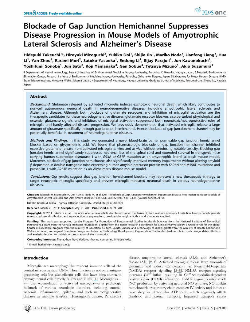
named INI-0602 (Figure 1A–B). Although treatment with INI-
0602 did not affect morphological change (Figure S1), major
cytokine/chemokine expression and NO production (Figure 1C–
D and Figure S2) in activated microglia, INI-0602 significantly
suppressed lipopolysaccharide (LPS)-induced glutamate release
from microglia and subsequent excitotoxic neuronal death in a
dose-dependent manner (Figure 1E–G). Since activated microg-
lia release glutamate from Cx32 hemichannels (Figure 2A), INI-
0602 was considered to block the hemichannel directly. INI-
0602, as well as CBX, was effective to inhibit microglial
glutamate release but the apparent toxicity of the compound
was much lower than that observed with CBX (Figure S3). We
also confirmed that INI-0602 penetrated the CNS more
effectively than CBX (Table S1).
We next evaluated the inhibitory effect of INI-0602 on
glutamate release in vivo. Among the CNS cells, LPS induced
glutamate release only from microglia (Figure 2B). INI-0602
significantly ameliorated the elevated glutamate levels in the brains
of LPS-treated mice, whereas CBX did not (Figure 2C). These
findings agree with the drug distribution data. To identify
potential adverse effects of long-term INI-0602 administration,
we intraperitoneally injected wild-type C57BL/6J mice with PBS
or INI-0602 (5, 10, 20, or 40 mg/kg) every other day for five
months. Histological analysis did not detect any obvious
differences between the PBS-treated and INI-0602-treated mice
(Figure S4). Serologic and urologic examinations also showed no
significant abnormalities in these mice (Table S2 and S3).
Assessment of 24-h cage activity revealed no significant behavioral
changes in these mice (Figure S5). These data suggest that INI-
0602 can be used for long-term treatment without precipitating
serious adverse effects.
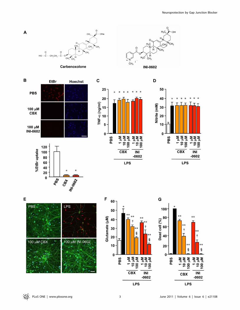
INI-0602 ameliorates symptoms in two ALS mousemodels
Next, we administered INI-0602 (5, 10, 20, or 40 mg/kg) to
transgenic (Tg) mice carrying a high copy number of a transgene
encoding a variant of human superoxide dismutase 1 with a G93A
mutation (SOD1 G93A Tg mice), which results in rapid progression
of an ALS-like disorder. At 20 weeks old, PBS-treated and INI-
0602-treated SOD1 G93A Tg mice showed obvious differences in
body size, muscular atrophy, and kyphosis (Figure 3A, Video S1 and
S2). Treatment with INI-0602 significantly prolonged the lifespans
of the SOD1 G93A Tg mice (5 mg/kg, P,0.05; 10 mg/kg,
P,0.00001; 20 mg/kg, P,0.00001; 40 mg/kg, P,0.05), whereas
all PBS-treated SOD1 G93A Tg mice died by week 25 (Figure 3B).
Moreover, INI-0602 treatment prevented axonal loss in the L5 root
of the SOD1 G93A Tg mice, whereas PBS-treated SOD1 G93A Tg
mice showed markedly fewer axons (Figure 3C–G). PBS-treated
SOD1 G93A Tg mice lost significantly more body weight at an
earlier age than INI-0602-treated SOD1 G93A Tg mice (P,0.05;
Figure 3H). Treatment with 10 mg/kg INI-0602 was most effective
for all parameters tested.
We also examined the efficacy of INI-0602 in a more slowly
progressing model of ALS, in which Tg mice carrying a low copy
number of a transgene encoding a G37R mutant variant of human
SOD1 (SOD1 G37R Tg mice) [27]. Treatment with INI-0602
significantly prolonged lifespans and prevented body weight loss in
SOD1 G37R Tg mice (10 and 20 mg/kg, P,0.05; Figure 3I–J).
Next, we histologically examined lumber spinal cords from
SOD1 G93A Tg mice. Whereas 20-week-old PBS-treated SOD1
G93A Tg mice showed severe atrophy in the anterior horns
(Figure 4A) with marked neuronal loss (Figure 4C) and activated
microglia (Figure 4E) and astrocytes (Figure 4G), INI-0602
treatment significantly prevented these effects (Figure 4B, D, F,
H, J).
Neuritic bead formation is focal swelling in dendrites and axons.
This is an early pathologic sign of neuronal damage that precedes
neuronal death in a variety of physiologic and pathologic
Neuroprotection by Gap Junction Blocker
PLoS ONE | www.plosone.org 2 June 2011 | Volume 6 | Issue 6 | e21108
Neuroprotection by Gap Junction Blocker
PLoS ONE | www.plosone.org 3 June 2011 | Volume 6 | Issue 6 | e21108

conditions, including ischemia [28], aging [29], and such
neurodegenerative diseases as ALS [30,31] and AD [32]. Neuritic
beading reflects impaired dendritic and axonal transport due to
intracellular energy loss in neurons [6]. PBS-treated SOD1 G93A
Tg mice at 14 weeks old (early–moderate phase of disease)
exhibited profound neuritic beading in the anterior horns of their
spinal cords (Figure 4K, arrows), which was morphologically
similar to spheroid structures observed in ALS patients [30,31].
Interestingly, INI-0602 treatment markedly reduced this neuritic
beading in the anterior horns (Figure 4L).
Figure 2. INI-0602 effectively inhibits microglial glutamate release in vivo. (A) Glutamate concentrations in microglia conditioned media.Cx32 mimetic peptide reduced microglial glutamate release, which revealed that activated microglia release glutamate from Cx32 hemichannels.Data represent the means 6 SD (n = 6 per group). *, P,0.05 vs. PBS. (B) Glutamate concentrations in media from primary cell cultures of mouse brain.Only the microglia released a significant amount of glutamate after stimulation with 1 mg/ml LPS for 24 h. Neu, neuron; Ast, astrocyte; Mi, microglia.Data represent the means 6 SD (n = 6 per group). *, P,0.05 vs. fresh medium. (C) Real-time glutamate concentration monitoring in the hippocampiof mice. All mice were injected LPS at time zero except PBS-treated mice. Blue, PBS-treated mice; black, LPS-treated mice; orange, LPS plus 20 mg/kgCBX-treated mice; red, LPS plus 20 mg/kg INI-0602-treated mice. Data represent the means 6 SE (n = 8 per group). *, P,0.05 vs. LPS.doi:10.1371/journal.pone.0021108.g002
Figure 1. A novel gap junction hemichannel blocker INI-0602 effectively inhibits neurotoxic microglial glutamate release. (A)Structure of carbenoxolone (CBX) and INI-0602. (B) Assessment of microglial hemichannel inhibition by EtBr dye uptake. Scale bar, 100 mm. Datarepresent the means 6 SD (n = 6 per group). *, P,0.05 vs. PBS. (C–D) TNF-a production (C) and NO production (D) by microglia treated with 1 mg/mlLPS for 24 h. Data represent the means 6 SD (n = 3 per group). *, P,0.05 vs. PBS. (E) Fluorescent microscopic images of live/dead staining of mouseprimary cortical neuron cultures in microglia-conditioned media. Neurons in microglia-conditioned media containing 1 mg/ml LPS (LPS) underwentcell death with neuritic beading, whereas neurons in microglia-conditioned media containing PBS (PBS), 1 mg/ml LPS plus 100 mM CBX (100 mM CBX),or 1 mg/ml LPS plus 100 mM INI-0602 (100 mM INI-0602) appeared healthy. Green, neuron (MAP2); red, dead cell (PI); blue, nucleus (Hoechst). Scalebar, 20 mm. (F) Glutamate concentrations in media. (G) The percentages of dead neurons. Data represent the means 6 SD (n = 6 per group). *, P,0.05vs. PBS; **, P,0.05 vs. LPS; {, P,0.05 vs. 1 mM; 1, P,0.05 vs. 10 mM.doi:10.1371/journal.pone.0021108.g001
Neuroprotection by Gap Junction Blocker
PLoS ONE | www.plosone.org 4 June 2011 | Volume 6 | Issue 6 | e21108
INI-0602 alleviates symptoms in a mouse model of ADWe also examined whether blocking microglial glutamate
release improved cognitive dysfunction in an AD mouse model
using transgenic mice expressing mutant variants of human
amyloid precursor protein (APP) and presenilin 1 (PS1) (APP/
PS1 Tg mice). First, we investigated recognition memory in 9-
month-old mice using a novel-object recognition test. Compared
with wild-type mice, PBS-treated APP/PS1 Tg mice displayed
reduced exploratory preference for the novel object in the
retention session (P,0.05; Figure 5A), an indication of impaired
recognition memory. In contrast, INI-0602 treatment (10 or
20 mg/kg) significantly reversed this impairment in the APP/PS1
Tg mice (P,0.05; Figure 5A). Next, we evaluated associative
learning in 10-month-old mice using a conditioned fear learning
Figure 3. INI-0602 ameliorates disease symptoms in both SOD1 G93A Tg mice and SOD1 G37R Tg mice. (A) A representativephotograph of 20-week-old SOD1 G93A Tg mice treated with PBS (left) or 10 mg/kg INI-0602 (right). (B) Survival rates of SOD1 G93A Tg mice (n = 24per group). Black, PBS; light blue, 5 mg/kg (P,0.05); red, 10 mg/kg (P,0.00001); green, 20 mg/kg (P,0.00001); purple, 40 mg/kg INI-0602 (P,0.05).(C–G) Microscopic images of L5 roots from SOD1 G93A Tg mice treated with PBS (C), 5 mg/kg (D), 10 mg/kg (E), 20 mg/kg (F), or 40 mg/kg INI-0602(G). (H) Body weights of SOD1 G93A Tg mice (n = 24 per group). Black, PBS; light blue, 5 mg/kg (P,0.05); red, 10 mg/kg (P,0.05); green, 20 mg/kg(P,0.05); purple, 40 mg/kg INI-0602 (P,0.05). (I) Survival rates and (J) body weights of SOD1 G37R Tg mice (n = 8 per group). Black, PBS; light blue,5 mg/kg; red, 10 mg/kg (P,0.05); green, 20 mg/kg INI-0602 (P,0.05).doi:10.1371/journal.pone.0021108.g003
Neuroprotection by Gap Junction Blocker
PLoS ONE | www.plosone.org 5 June 2011 | Volume 6 | Issue 6 | e21108

Figure 4. INI-0602 prevents neuronal damage and gliosis in lumber spinal cords of SOD1 G93A Tg mice. (A–B) Fluorescent microscopicimages of the lumber spinal cords of 20-week-old SOD1 G93A Tg mice treated with PBS (A) or 10 mg/kg INI-0602 (B). Green, neurons (MAP2); red,microglia (CD11b); blue, astrocytes (GFAP). Scale bar, 200 mm. (C–J) Higher magnification images of the boxed areas in a (C, E, G, and I) and b (D, F, H,and J). Scale bar, 30 mm. Each graph shows the percentage of area occupied by neurons, microglia, and astrocytes, respectively. Data represent themeans 6 SD (n = 6 per group). *, P,0.05 vs. PBS. (K, L) Fluorescent microscopic images of MAP2 staining in the anterior horns of lumber spinal cordsfrom 14-week-old SOD1 G93A Tg mice treated with PBS (K) or 10 mg/kg INI-0602 (L). Arrows indicate neuritic bead formation, an early marker ofneuronal dysfunction. Scale bar, 10 mm.doi:10.1371/journal.pone.0021108.g004
Neuroprotection by Gap Junction Blocker
PLoS ONE | www.plosone.org 6 June 2011 | Volume 6 | Issue 6 | e21108
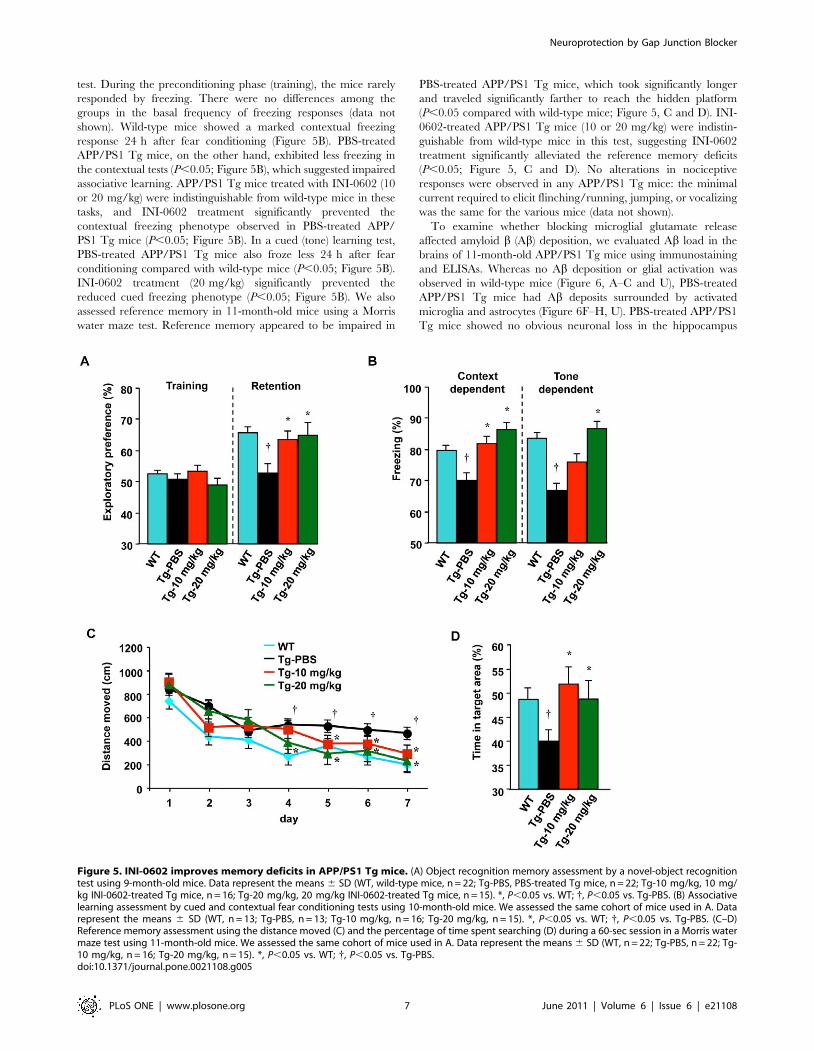
test. During the preconditioning phase (training), the mice rarely
responded by freezing. There were no differences among the
groups in the basal frequency of freezing responses (data not
shown). Wild-type mice showed a marked contextual freezing
response 24 h after fear conditioning (Figure 5B). PBS-treated
APP/PS1 Tg mice, on the other hand, exhibited less freezing in
the contextual tests (P,0.05; Figure 5B), which suggested impaired
associative learning. APP/PS1 Tg mice treated with INI-0602 (10
or 20 mg/kg) were indistinguishable from wild-type mice in these
tasks, and INI-0602 treatment significantly prevented the
contextual freezing phenotype observed in PBS-treated APP/
PS1 Tg mice (P,0.05; Figure 5B). In a cued (tone) learning test,
PBS-treated APP/PS1 Tg mice also froze less 24 h after fear
conditioning compared with wild-type mice (P,0.05; Figure 5B).
INI-0602 treatment (20 mg/kg) significantly prevented the
reduced cued freezing phenotype (P,0.05; Figure 5B). We also
assessed reference memory in 11-month-old mice using a Morris
water maze test. Reference memory appeared to be impaired in
PBS-treated APP/PS1 Tg mice, which took significantly longer
and traveled significantly farther to reach the hidden platform
(P,0.05 compared with wild-type mice; Figure 5, C and D). INI-
0602-treated APP/PS1 Tg mice (10 or 20 mg/kg) were indistin-
guishable from wild-type mice in this test, suggesting INI-0602
treatment significantly alleviated the reference memory deficits
(P,0.05; Figure 5, C and D). No alterations in nociceptive
responses were observed in any APP/PS1 Tg mice: the minimal
current required to elicit flinching/running, jumping, or vocalizing
was the same for the various mice (data not shown).
To examine whether blocking microglial glutamate release
affected amyloid b (Ab) deposition, we evaluated Ab load in the
brains of 11-month-old APP/PS1 Tg mice using immunostaining
and ELISAs. Whereas no Ab deposition or glial activation was
observed in wild-type mice (Figure 6, A–C and U), PBS-treated
APP/PS1 Tg mice had Ab deposits surrounded by activated
microglia and astrocytes (Figure 6F–H, U). PBS-treated APP/PS1
Tg mice showed no obvious neuronal loss in the hippocampus
Figure 5. INI-0602 improves memory deficits in APP/PS1 Tg mice. (A) Object recognition memory assessment by a novel-object recognitiontest using 9-month-old mice. Data represent the means 6 SD (WT, wild-type mice, n = 22; Tg-PBS, PBS-treated Tg mice, n = 22; Tg-10 mg/kg, 10 mg/kg INI-0602-treated Tg mice, n = 16; Tg-20 mg/kg, 20 mg/kg INI-0602-treated Tg mice, n = 15). *, P,0.05 vs. WT; {, P,0.05 vs. Tg-PBS. (B) Associativelearning assessment by cued and contextual fear conditioning tests using 10-month-old mice. We assessed the same cohort of mice used in A. Datarepresent the means 6 SD (WT, n = 13; Tg-PBS, n = 13; Tg-10 mg/kg, n = 16; Tg-20 mg/kg, n = 15). *, P,0.05 vs. WT; {, P,0.05 vs. Tg-PBS. (C–D)Reference memory assessment using the distance moved (C) and the percentage of time spent searching (D) during a 60-sec session in a Morris watermaze test using 11-month-old mice. We assessed the same cohort of mice used in A. Data represent the means 6 SD (WT, n = 22; Tg-PBS, n = 22; Tg-10 mg/kg, n = 16; Tg-20 mg/kg, n = 15). *, P,0.05 vs. WT; {, P,0.05 vs. Tg-PBS.doi:10.1371/journal.pone.0021108.g005
Neuroprotection by Gap Junction Blocker
PLoS ONE | www.plosone.org 7 June 2011 | Volume 6 | Issue 6 | e21108

(Figure 6I), which agreed with previous reports [33,34]. Despite a
significantly reduction in cognitive impairments, INI-0602 treat-
ment did not significantly alter Ab deposition or glial activation
(Figure 6K–M, P–R, U–W). Human Ab specific ELISAs also
revealed no difference in the Ab load among PBS-treated and INI-
0602-treated APP/PS1 Tg mice (Figure 6X). Western blot analysis
Figure 6. INI-0602 does not affect Ab deposition or glial activation. (A–T) Fluorescent microscopic images of hippocampi from 11-month-oldAPP/PS1 Tg mice. Scale bar, 200 mm. (U–W) Percentage of area occupied by Aß (U), microglia (V), and astrocytes (W). Data represent the means 6 SD(n = 6 per group). (X) Human Aß1–40- and Aß1–42-specific ELISAs using homogenized brains from 11-month-old APP/PS1 Tg mice. Data represent themeans 6 SD (n = 6 per group). (Y) Western blot analysis of oligomeric Ab extracted from 11-month-old APP/PS1 Tg mice. *, oligomeric Ab. Datarepresent the means 6 SD (n = 3 per group). WT, wild-type mice; Tg-PBS, PBS-treated Tg mice; Tg-10 mg/kg, 10 mg/kg INI-0602-treated Tg mice; Tg-20 mg/kg, 20 mg/kg INI-0602-treated Tg mice.doi:10.1371/journal.pone.0021108.g006
Neuroprotection by Gap Junction Blocker
PLoS ONE | www.plosone.org 8 June 2011 | Volume 6 | Issue 6 | e21108

also showed no difference in the amount of soluble oligomeric
Ab1–42 (Figure 6Y), which has been reported to exhibit higher
neurotoxicity than fibrillar Ab [35–37]. These findings agreed with
in vitro observations showing that hemichannel blockade neither
enhanced microglial Ab uptake nor altered microglial activation
(data not shown).
Discussion
In the present study, we synthesized a novel gap junction
hemichannel blocker INI-0602 based on CBX using dihydropyr-
idine conjugates as a chemical drug delivery system to enhance
BBB penetration. In general, dihydropyridine conjugates serve the
drug molecule with the sufficient lipophilicity to enter the CNS.
Then, dihydropyridine conjugates undergo chemical conversion to
pyridinium salt by in vivo redox system. This conversion promotes
retention of the drug molecule in the CNS and it is also expected
that this conversion accelerate peripheral elimination of the drug
molecule distribution outside the CNS. Thus, dihydropyridine
conjugates may not only lead to effective drug delivery into the
CNS but also decrease the drug toxicity due to the effect in the
periphery [26]. In fact, we confirmed here that dihydropyridine
conjugates gave INI-0602 the advantage of BBB penetration, CNS
retention and low toxicity over CBX. Unexpectedly, INI-0602 has
a short half life in vivo (9 min in the blood and 25 min in the CNS,
respectively) although INI-0602 showed a strong effect in vivo even
by every other day treatment. We confirmed that only 30-min
treatment with INI-0602, but not CBX, significantly suppressed
LPS-stimulated microglial glutamate release in the following 48 h
in vitro (H. Takeuchi, unpublished data). It suggested that INI-0602
induced long-term inactivation or internalization of gap junction/
hemichannel. However, further studies are needed to elucidate this
issue.
Like other cell types, microglia maintain a resting physiologic
glutamate level via the glutamate dehydrogenase pathway without
significant secretion of glutamate [38–41]. In the activated state,
microglia produce a large amount of glutamate through an
upregulation of glutaminase expression, rather than glutamate
dehydrogenase. The activated microglia then secrete high levels of
glutamate through Cx32 hemichannels [5,21–24]. Interestingly,
inflammatory cytokines, especially tumor necrosis factor-a and
interferon-c, enhance microglial glutaminase expression, gluta-
mate production, and cell-surface expression of gap junction
hemichannels [5,42]. Synergistic and autocrine activities of these
molecules may cause the release of large amounts of glutamate to
induce excitotoxic neuronal death. Activated microglia undergo
dramatic morphologic changes and movements. Accordingly,
hemichannels in activated microglia may be more exposed to the
extracellular space and provide a unique surface for molecular
drug target than those in unactivated microglia and other CNS
cells. Thus, an inhibitor of hemichannel may specifically suppress
glutamate release from microglia, and this specificity may translate
into fewer adverse effects during treatment. Moreover, neuronal
and astrocytic gap junctions are also thought to contribute to the
spread of neuronal damage in neurological disorder models such
as brain ischemia, migraine, and epilepsy [43–48]. Therefore
blockade of neuronal and astrocytic gap junction/hemichannel
may elicit additive neuroprotective effects. Recent reports
demonstrated that activated astrocytes also play an important
role in neuronal death by releasing soluble factors in ALS models
in vitro and in vivo [49–51]. Another recent study reported that Abdramatically enhanced gap junction/hemichannel-mediated Ca2+
wave propagation in astrocytes that may contribute to AD
pathology [52]. Thus blockade of gap junction in astrocytes
besides microglia may contribute to enhance neuroprotective
effect. On the other hand, excessive inhibition of gap junction/
hemichannel may induce dysfunction of neural system because
gap junction/hemichannel also contribute to maintaining homeo-
stasis in neuron-glia interaction [53]. We are planning more
thorough studies to investigate these issues.
In a murine model of ALS, degenerating neurons are thought to
secrete unidentified factor(s) and activate microglia and astrocytes,
thereby exacerbating neurodegeneration [27,51]. Microglial
activation may also be upstream of this vicious feed-forward cycle
of neurodegeneration and glial activation [1], which is likely fueled
by microglia-derived glutamate. In this study, INI-0602 treatment
suppressed both neuronal loss and glial activation in an ALS
model while INI-0602 itself did not alter microglial activation.
Thus we suggest that INI-0602 halted this feed-forward cycle and
reduced glial activation indirectly by inhibiting microglial
glutamate release. In AD model mice, no obvious neuronal loss
was observed [33], suggesting that inhibition of glutamate release
from microglia may improve neuronal dysfunction in the
hippocampus via reduced NMDA-mediated neurotoxicity. Other
NMDA receptor antagonists also have similar effects in AD
patients [12]. It is still possible, however, that the vicious feed-
forward cycle described above may contribute to AD pathology
because AD patients exhibit severe neuronal loss and glial
activation [1]. Additionally, INI-0602 treatment did not signifi-
cantly alter Ab load despite cognitive improvement. In recent
animal models and human clinical trials, cognitive improvements
in response to treatments were minimally related to changes in Abload [54–56]. Our findings also suggest that factors through gap
junction hemichannels (including microglial glutamate) in addition
to Ab neuropathology may be important determinants of cognitive
decline in AD patients.
In conclusion, we have generated a CNS penetratable gap
junction hemichannel blocker and have demonstrated that
pharmacologic blockade of gap junction hemichannels effectively
attenuated glutamate release from neurotoxic activated microglia
and ameliorated symptoms of mouse models of ALS and AD.
Thus, gap junction hemichannel blockers may provide a new
therapeutic approach to prevent microglia-mediated, non-cell
autonomous neuronal death in neurodegenerative diseases includ-
ing ALS and AD.
Methods
ReagentsAll reagents were obtained from Sigma-Aldrich unless specified
otherwise. The gap junction hemichannel blocker INI-0602
(IUPAC name: 3-(((3S,4aR,6aR,6bS,8aS,11S,12aR,14aR,14bS)-
11-carboxy-4,4,6a,6b,8a,11,14b-heptamethyl-14-oxo-
1,2,3,4,4a,5,6,6a,6b,7,8,8a,9,10,11,12,12a,14,14a,14b-icosahydro-
picen-3-yloxy) carbonyl)-1-methylpyridinium iodide) was devel-
oped by H. Takeuchi and A. Suzumura (Patent: WO/2010/
007788). In brief, nicotinoyl chloride hydro chloride salt was
reacted with 18b-glycyrrhetinic acid, thereby introducing nicotin-
ate to the hydroxyl group of the 18b-glycyrrhetinic acid followed
by attaching an alkyl group to the nitrogen atom of the pyridine
ring with methyl iodide. Then, INI-0602 was isolated as a
pyridinium salt. INI-0602 used in this study was chemically
synthesized and purified by the Nard Institute (Osaka, Japan). The
purity of the compound was more than 99%.
AnimalsThis study was carried out in strict accordance with the
guideline for the care and use of laboratory animals of Nagoya
Neuroprotection by Gap Junction Blocker
PLoS ONE | www.plosone.org 9 June 2011 | Volume 6 | Issue 6 | e21108

University. All protocols for animal experiments were approved by
the Animal Experiment Committee of Nagoya University (permit
number: 651 and 652). Transgenic mice expressing mutant
variants of human amyloid precursor protein (APP) with K595N
and M596L mutations and presenilin 1 (PS1) with A264E
mutation [33] were purchased from the Jackson Laboratory
(B6C3-Tg(APP695)3Dbo Tg(PSEN1)5Dbo/J; #003378) and were
backcrossed to C57BL/6J mice for more than 10 generations after
purchase (here designated as APP/PS1 Tg mice). Transgenic mice
carrying a high copy number of a transgene encoding a G93A
mutant of human superoxide dismutase 1 (SOD1) [57] (here
designated as SOD1 G93A Tg mice) were also purchased from the
Jackson Laboratory (B6.Cg-Tg(SOD1-G93A)1Gur/J; #004435).
Transgenic mice carrying a low copy number of a transgene
encoding a G37R mutant of human SOD1 [27] (here designated
as SOD1 G37R Tg mice) were provided by Dr. D. W. Cleveland
(University of California, San Diego, USA).
CellsMouse primary cultures of cortical neurons were prepared from
C57BL/6J mice as described previously [5,6]. Cortices were
dissected and freed of meninges. Cortical fragments were
dissociated into single cells using dissociation solution, and they
were resuspended in Neuron Medium (serum-free conditioned
medium from 48-h rat astrocyte confluent cultures based on
Dulbecco’s modified Eagle’s minimum essential medium
(DMEM)/F12 with N2 supplement, Sumitomo Bakelite, Akita,
Japan). Primary neuronal cells were plated on 12 mm-poly-
ethyleneimine-coated coverslips (Asahi Techno Glass Corporation,
Chiba, Japan). The purity of the cultures was more than 95% as
determined by NeuN-specific immunostaining. Mouse primary
microglia were isolated from primary mixed glial cell cultures from
newborn C57BL/6J mice on the 14th day with the ‘shaking off’
method as described previously [58]. The purity of the cultures
was 97 to 100% as determined by CD11b-specific immunostain-
ing. Cultures were maintained in DMEM supplemented with 10%
fetal bovine serum (JRH Biosciences, Lenexa, KS, USA), 5 mg/ml
bovine insulin, and 0.2% glucose. Mouse primary astrocytes were
also prepared from primary mixed glial cell cultures from newborn
C57BL/6J mice on the 14th day by three to four repetitions of
trypsinization and replating as described previously [21]. The
purity of the cultures was more than 95% as determined by glial
fibrillary acidic protein (GFAP)-specific immunostaining. Astrocyte
cultures were maintained with same medium as microglia cultures.
Cells were plated at a density of 56104 cells per well in 24-well
multidishes. For stimulation, cells were incubated with 1 mg/ml
LPS for 24 h. To assess neuroprotective effects, microglia were
cultured in Neuron Medium containing 1 mg/ml LPS with each
drug as follows: 1–100 mM CBX; 1–100 mM INI-0602; connexin
(Cx) 32 mimetic peptide, 0.25 mg/l 32gap 27 (SRPTEKTVFT,
Thermo Electron GmbH); Cx43 mimetic peptide, 0.25 mg/l43gap 27 (SRPTEKTIFII, Thermo Electron GmbH); pannexin 1
mimetic peptide, 0.25 mg/l 10panx1 (WRQAAFVDSY, Thermo
Electron GmbH). To assess IC50 of drugs, microglia were cultured
in Neuron Medium containing 1 mg/ml LPS with 0.1–500 mM
CBX or INI-0602. After a 24-h incubation, we assessed the
glutamate concentration in microglia-conditioned medium and
applied it to each well containing neurons after 10–13 days in vitro.
Neurotoxicity was evaluated 24 h after application.
ImmunocytochemistryNeuron cultures were live-stained with 0.1 mg/ml Hoechst
33342 and 2 mg/ml propidium iodide (PI) as described previously
[6]. After rinsing with Neuron Medium three times, cells were
fixed in 4% paraformaldehyde for 30 min, permeabilized in
0.05% Triton X-100 for 10 min, and stained using mouse
monoclonal anti-microtubule-associated protein 2 (MAP2) anti-
body (MAB364, Chemicon, Temecula, CA, USA) and Alexa 488–
conjugated secondary antibodies (Molecular Probes, Eugene, OR,
USA). More than 100 neurons in duplicate wells were assessed
blindly in six independent trials with a deconvolution fluorescence
microscope system (Keyence, Osaka, Japan). The ratio of dead
cells was calculated as a percentage of PI-positive cells among
Hoechst-positive cells.
Assessment of microglial hemichannel inhibitionTo assess microglial hemichannel inhibition, we used ethidium
bromide (EtBr) dye uptake method as described previously
[59,60]. Briefly, mouse primary microglia were plated at a density
of 16105 cells per well in 24-well multidishes. They were
pretreated with DMEM containing 100 mM CBX or INI-0602
for 30 min. Then 10 mM EtBr was added. After 1 h incubation,
cells were gently rinsed with phosphate buffered saline (PBS) three
times and fixed with fixed in 4% paraformaldehyde for 30 min.
Cells were counterstained with 0.1 mg/ml Hoechst 33342. EtBr
fluorescence of more than 100 cells was analyzed with a
deconvolution fluorescence microscope system (Keyence, Osaka,
Japan) in six independent trials. Relative dye uptake was
calculated as EtBr fluorescence divided by Hoechst-positive cell
number. The ratio of dye uptake was calculated as a percentage of
relative dye uptake in PBS-treated cells.
Assessment of glutamate releaseTo measure extracellular glutamate concentrations in vitro, we
used a colorimetric Glutamate Assay Kit (Yamasa Corporation,
Tokyo, Japan) as described previously [6].
Assessment of tumor necrosis factor-a (TNF-a)production
To measure microglial TNF-a production, we used a mouse
TNF-a specific ELISA kit (BD Pharmingen, Franklin Lakes, NJ,
USA) as described previously [61].
Assessment of NO productionTo measure microglial NO production, we performed the
Griess reaction on the media as described previously [6].
RNA extraction and reverse transcription (RT)-PCRTo examine microglial mRNA expression of TNF-a, interleukin
(IL)-1b, IL-6, CCL2, CCL5, CXCL10 and glyceraldehyde-3-
phosphate dehydrogenase (GAPDH, as an internal control), we
performed RNA extraction and RT-PCR analysis with each
specific primer as described previously [22].
Assessment of drug concentrationTo assess the drug concentrations from plasma and brain
tissues, mice were intravenously injected with 20 mg/kg CBX or
INI-0602. Plasma and brain tissues were collected at the indicated
time points after injection. Drug concentrations were assessed
using a high-performance liquid chromatography (HPLC)/mass
spectrometry (MS)/MS system (Applied Biosystems Japan, Tokyo,
Japan).
Drug administrationTo examine acute drug toxicity, 12-week-old C57BL/6J mice
were given single intraperitoneal injection with PBS, CBX or INI-
0602 at the indicated doses. To assess chronic drug toxicity, 12-
Neuroprotection by Gap Junction Blocker
PLoS ONE | www.plosone.org 10 June 2011 | Volume 6 | Issue 6 | e21108

week-old C57BL/6J mice were intraperitoneally treated with
20 mg/kg CBX or INI-0602 every other day for five months. To
evaluate drug efficacy, mice were intraperitoneally injected with
the indicated doses of INI-0602 every other day. SOD G93A Tg
mice were treated beginning the week after the onset of hindlimb
motor deficits (approximately 8 weeks old), which is an early
symptom in SOD1 G93A Tg mice [62]. SOD G37R Tg mice
were treated beginning the week after the onset of body weight loss
(approximately 7 months old), which is an early symptom in
SOD1 G37R Tg mice [51]. APP/PS1 Tg mice were treated
beginning the week after the onset of Ab deposition in the brain
(approximately 4 months old).
In vivo microdialysisWe performed real-time monitoring of glutamate or acetylcho-
line concentrations in the hippocampi of mice using in vivo
microdialysis as described previously [63]. To stimulate glutamate
release, 10 mg of LPS in 2 ml of artificial CSF (aCSF: 147 mM
NaCl, 4 mM KCl, and 2.3 mM CaCl2) were injected through the
microinjection tube at a flow rate of 1.0 ml/min. Mice were
administered with 20 mg/kg CBX or INI-0602 intraperitoneally
30 min before stimulation. Levels of glutamate in the dialysates
were measured with a HPLC system (Eicom, Kyoto, Japan). We
used the means of the measured levels for 90 min before LPS
stimulation as baseline level of each group. The results are
expressed as the percentage of the measured levels to baseline
level.
Behavioral testsWe measured cage activity of mice for 24 h using an infrared
ray sensor monitor system (Neuroscience, Yokohama, Japan) as
described previously [64]. To assess memory impairments, 9- to
11-month-old APP/PS1 Tg mice treated with PBS or INI-0602
were examined in a novel-object recognition test, cued and
contextual fear conditioning tests, and a Morris water maze test as
described previously [34,65]. The novel-object recognition test
procedure consisted of three sessions: habituation, training, and
retention. Each mouse was individually habituated to the box (a
black Plexiglas box with abundant wood chips, 30630640 high
cm) with 10 min of exploration in the absence of objects for 3
consecutive days (habituation session, days 1–3). During the
training session, two novel objects were symmetrically fixed to the
floor of the box, 8 cm from the walls, and each animal was allowed
to explore in the box for 10 min (day 5). The objects were
constructed from a golf ball, wooden column, and wooden
triangular pyramid. They were different in shape and color but
similar in size. An animal was considered to be exploring the
object when its head was facing the object or it was touching or
sniffing the object. The time spent exploring each object was
recorded. After training, mice were immediately returned to their
home cages. During the retention sessions (day 6), the animals
were placed back into the same box 24 h after the training session,
but one of the familiar objects used during training had been
replaced with a novel object. The animals were then allowed to
explore freely for 5 min and the time spent exploring each object
was recorded. Throughout the experiments, the objects were used
in a counterbalanced manner. A preference index in the retention
session, a ratio of the amount of time spent exploring the novel
object over the total time spent exploring both objects, was used to
measure cognitive function. In the training session, the preference
index was calculated as a ratio of the time spent exploring the
object that was replaced by the novel object in the retention
session, over the total time exploring. In the cued and contextual
fear conditioning tests, to measure basal levels of freezing response
(preconditioning phase), mice were individually placed in a neutral
cage (a black Plexiglas box with abundant wood chips, 30630640
high cm) for 1 min, then in the conditioning cage (a transparent
Plexiglas box, 30630640 high cm) for 2 min. For training
(conditioning phase), mice were placed in the conditioning cage,
then a 15 s tone (80 dB) was delivered as a conditioned stimulus.
During the last 5 s of the tone stimulus, a foot shock of 0.6 mA was
delivered as an unconditioned stimulus through a shock generator
(Brain Science Idea, Osaka, Japan). This procedure was repeated
four times with 15 s intervals. Cued and contextual tests were
carried out 1 day after fear conditioning. For the contextual test,
mice were placed in the conditioning cage and the freezing
response was measured for 2 min in the absence of the
conditioned stimulus. For the cued test, the freezing response
was measured in the neutral cage for 1 min in the presence of a
continuous-tone stimulus identical to the conditioned stimulus.
The Morris water maze test was conducted in a circular pool
1.2 m in diameter and filled with water at 2261uC. A hidden
platform (7 cm in diameter) was used. The mice were given three
trials (one block) for 7 consecutive days, during which the platform
was left in the same position. The time taken to locate the escape
platform (escape latency) and the distance moved was determined
in each trial by using the SMART system (Panlab, Barcelona,
Spain). One day after the last training trial, the mice were given a
probe test without the platform and allowed 60 s to search the
pool. We separated the pool in four quadrants (target (platform),
left, opposite, and right) and assessed the time spent to search in
each quadrant.
Histological analysisTen-micrometer-thick frozen sections of mouse organs were
prepared using a previously described method [34]. Sections of
organs except brain and spinal cord were stained with hematoxylin
and eosin using standard procedures. Brain and spinal cord
sections were permeabilized with 1% Triton X-100 after blocking
with 10% normal goat serum for 30 min, and they were stained
with specific primary antibodies for neurons (mouse monoclonal
anti-MAP2 antibody, MAB364, Chemicon), microglia (rat mono-
clonal anti-CD11b antibody, clone M1/70.15, AbD Serotec,
Raleigh, NC, USA), astrocytes (rabbit polyclonal anti-glial
fibrillary acidic protein (GFAP) antibody, Dako, Glostrup, Den-
mark), and amyloid b (Ab) (mouse monoclonal antibody, clone
4G8, Chemicon). L5 root sections were stained with 0.5% Sudan
black for myelin stain. Images were analyzed with a deconvolution
fluorescence microscope system (Keyence) as reported previously
[34].
Human Ab ELISATo evaluate the amount of human Ab1–40 and Ab1–42 in
mouse brains, we used a human Ab1–40 and Ab1–42 specific
ELISA kit (Wako Pure Chemical Industries, Osaka, Japan)
according to the manufacturer’s protocol as described previously
[65]. Brains were homogenized with 70% formic acid and
centrifuged at 100,000 g for 1 h. The supernatants of insoluble
70% formic acid extracts were neutralized with 1 M Tris-HCl,
pH 8.0 at a dilution of 1:20 and were measured by each Abspecific ELISA kit. The values obtained were corrected with the
wet weight of each brain sample.
Western blottingOligomeric Ab in APP/PS1 Tg mouse brain was extracted from
the soluble, extracellular-enriched fraction as described previous-
ly[34]. Proteins were separated on a 5–20% Tris-glycine SDS-
polyacrylamide gel and transferred to Hybond-P PVDF mem-
Neuroprotection by Gap Junction Blocker
PLoS ONE | www.plosone.org 11 June 2011 | Volume 6 | Issue 6 | e21108
brane (GE Healthcare UK Ltd. Buckinghamshire, UK). Blots
were incubated in mouse anti-Ab monoclonal antibody (clone
6E10, 1:1000, Chemicon) overnight at 4uC. Bound antibody was
visualized using horseradish peroxidase-conjugated anti-mouse
IgG (1:5000, GE Healthcare) and SuperSignal West Pico
Chemiluminescent Substrate (Thermo Fisher Scientific, Rockford,
IL). The intensity of the bands was calculated by using CS
Analyzer 1.0 (Atto Corporation, Tokyo, Japan).
Statistics and analysisBiochemical and behavioral data were statistically analyzed using
one-way ANOVA followed by post hoc Tukey tests. The survival
rate was assessed using Kaplan-Meier and log-rank tests. All results
were analyzed using GraphPad Prism (GraphPad Software).
Supporting Information
Figure S1 Phase contrast images of microglia. (A)
Activated microglia treated with 1 mg/ml LPS for 24 h. (B)
Activated microglia treated with 1 mg/ml LPS and 100 mM INI-
0602 for 24 h. No morphological difference was observed between
both activated microglia. Scale bar; 20 mm.
(TIF)
Figure S2 Major cytokine/chemokine expression bymicroglia treated with gap junction hemichannel block-ers. Representative RT-PCR data of major cytokine/chemokine
in microglia. Microglia were treated 1 mg/ml LPS for stimulation.
To assess drug effects, cells were simultaneously treated with 1–
100 mM CBX or INI-0602. Assessments were performed 24 h
after treatment.
(TIF)
Figure S3 IC50 and LD50 of INI-0602. (A) Inhibition-
concentration curves of CBX and INI-0602 for microglial
glutamate release. Data represent the means 6 SD (n = 6 per
group). (B) IC50 in vitro (n = 6 per group) and LD50 in vivo (n = 10
per group) of CBX and INI-0602.
(TIF)
Figure S4 Hematoxylin and eosin staining images ofmajor organs from mice treated with PBS or INI-0602.Representative hematoxylin and eosin staining images of the
heart, kidney liver and spleen from 12-week-old C57BL/6J mice
treated with PBS or 20 mg/kg INI-0602 every other day for five
months. No significant differences were observed. Scale bar,
200 mm.
(TIF)
Figure S5 Assessment of 24-h cage activity of micetreated with PBS or INI-0602. Representative 24-h cage
activity data of 12-week-old C57BL/6J mice treated with PBS or
20 mg/kg INI-0602 every other day for five months. No
significant difference was observed. Data represent the means 6
SD (n = 8 per group).
(TIF)
Table S1 Drug concentrations from plasma and braintissues. Mice were intravenously injected with 20 mg/kg CBX
or INI-0602. Drug concentrations were assessed using HPLC/MS.
ND, not detected.
(DOC)
Table S2 Blood analysis of 12-week-old mice treatedwith PBS or INI-0602. Wild-type C57BL6/J mice were treated
with PBS or INI-0602 (5, 10, 20, or 40 mg/kg) every other day for
five months. Blood was collected from the inferior aorta under
deep anesthesia. Whole blood and serum were analyzed for the
items as below with an autoanalyzer (Nagahama Life Science
Laboratory, Nagahama, Japan). Data represent the means 6 SE
(n = 10 per group).
(DOC)
Table S3 Urinalysis of 12-week-old mice treated withPBS or INI-0602. Wild-type C57BL6/J mice were treated with
PBS or INI-0602 (5, 10, 20, or 40 mg/kg) every other day for five
months. Urine was collected from the bladder under deep
anesthesia and was immediately assessed using Uropaper III
(Eiken Chemical, Tokyo, Japan). Data represent the means 6 SE
(n = 10 per group). ND, not detected.
(DOC)
Video S1 A representative movie of a 20-week-old SOD1G93A Tg mouse treated with PBS. The mouse shows severe
muscular atrophy and kyphosis, and is hardly able to walk.
(MOV)
Video S2 A representative movie of a 20-week-old SOD1G93A Tg mouse treated with 10 mg/kg INI-0602. The
mouse shows motor weakness but is still able to walk steadily.
(MOV)
Acknowledgments
The authors thank Drs. T. Cohen, S. Pesiridis, J. Q. Trojanowski and V.
M.-Y. Lee (University of Pennsylvania) for their helpful comments.
Author Contributions
Conceived and designed the experiments: HT. Performed the experiments:
HT HM YD SJ MN JL HL YZ SY EL BP JK YS TM. Analyzed the data:
HT HM YD MN TM AS. Contributed reagents/materials/analysis tools:
RM JS KY GS. Wrote the paper: HT AS.
References
1. Block ML, Zecca L, Hong JS (2007) Microglia-mediated neurotoxicity:
uncovering the molecular mechanisms. Nat Rev Neurosci 8: 57–69.
2. Kempermann G, Neumann H (2003) Neuroscience. Microglia: the enemy
within? Science 302: 1689–1690.
3. Bruijn LI, Miller TM, Cleveland DW (2004) Unraveling the mechanisms involved
in motor neuron degeneration in ALS. Annu Rev Neurosci 27: 723–749.
4. McGeer PL, McGeer EG (2002) Inflammatory processes in amyotrophic lateral
sclerosis. Muscle Nerve 26: 459–470.
5. Takeuchi H, Jin S, Wang J, Zhang G, Kawanokuchi J, et al. (2006) Tumor
necrosis factor-alpha induces neurotoxicity via glutamate release from
hemichannels of activated microglia in an autocrine manner. J Biol Chem
281: 21362–21368.
6. Takeuchi H, Mizuno T, Zhang G, Wang J, Kawanokuchi J, et al. (2005)
Neuritic beading induced by activated microglia is an early feature of neuronal
dysfunction toward neuronal death by inhibition of mitochondrial respiration
and axonal transport. J Biol Chem 280: 10444–10454.
7. Barger SW, Basile AS (2001) Activation of microglia by secreted amyloid
precursor protein evokes release of glutamate by cystine exchange and attenuates
synaptic function. J Neurochem 76: 846–854.
8. Piani D, Spranger M, Frei K, Schaffner A, Fontana A (1992) Macrophage-
induced cytotoxicity of N-methyl-D-aspartate receptor positive neurons involves
excitatory amino acids rather than reactive oxygen intermediates and cytokines.
Eur J Immunol 22: 2429–2436.
9. Lobsiger CS, Cleveland DW (2007) Glial cells as intrinsic components of non-
cell-autonomous neurodegenerative disease. Nat Neurosci 10: 1355–1360.
10. Nelson PT, Soma LA, Lavi E (2002) Microglia in diseases of the central nervous
system. Ann Med 34: 491–500.
11. Suzumura A, Takeuchi H, Zhang G, Kuno R, Mizuno T (2006) Roles of glia-
derived cytokines on neuronal degeneration and regeneration. Ann N Y Acad
Sci 1088: 219–229.
12. Parsons CG, Stoffler A, Danysz W (2007) Memantine: a NMDA receptor
antagonist that improves memory by restoration of homeostasis in the
Neuroprotection by Gap Junction Blocker
PLoS ONE | www.plosone.org 12 June 2011 | Volume 6 | Issue 6 | e21108

glutamatergic system–too little activation is bad, too much is even worse.
Neuropharmacology 53: 699–723.13. Kipnis J, Avidan H, Caspi RR, Schwartz M (2004) Dual effect of CD4+CD25+
regulatory T cells in neurodegeneration: a dialogue with microglia. Proc Natl
Acad Sci U S A 101 Suppl 2: 14663–14669.14. Koenigsknecht J, Landreth G (2004) Microglial phagocytosis of fibrillar beta-
amyloid through a beta1 integrin-dependent mechanism. J Neurosci 24:9838–9846.
15. Schwab JM, Schluesener HJ (2004) Microglia rules: insights into microglial-
neuronal signaling. Cell Death Differ 11: 1245–1246.16. Zietlow R, Dunnett SB, Fawcett JW (1999) The effect of microglia on embryonic
dopaminergic neuronal survival in vitro: diffusible signals from neurons and gliachange microglia from neurotoxic to neuroprotective. Eur J Neurosci 11:
1657–1667.17. Leigh PN, Swash M, Iwasaki Y, Ludolph A, Meininger V, et al. (2004)
Amyotrophic lateral sclerosis: a consensus viewpoint on designing and
implementing a clinical trial. Amyotroph Lateral Scler Other Motor NeuronDisord 5: 84–98.
18. Yamamoto M, Tanaka F, Tatsumi H, Sobue G (2008) A strategy for developingeffective amyotropic lateral sclerosis pharmacotherapy: from clinical trials to
novel pharmacotherapeutic strategies. Expert Opin Pharmacother 9:
1845–1857.19. Orrell RW (2010) Motor neuron disease: systematic reviews of treatment for
ALS and SMA. Br Med Bull 93: 145–159.20. Laird DW (2009) The gap junction proteome and its relationship to disease.
Trends Cell Biol.21. Liang J, Takeuchi H, Doi Y, Kawanokuchi J, Sonobe Y, et al. (2008) Excitatory
amino acid transporter expression by astrocytes is neuroprotective against
microglial excitotoxicity. Brain Res 1210: 11–19.22. Yawata I, Takeuchi H, Doi Y, Liang J, Mizuno T, et al. (2008) Macrophage-
induced neurotoxicity is mediated by glutamate and attenuated by glutaminaseinhibitors and gap junction inhibitors. Life Sci 82: 1111–1116.
23. Takeuchi H, Jin S, Suzuki H, Doi Y, Liang J, et al. (2008) Blockade of microglial
glutamate release protects against ischemic brain injury. Exp Neurol 214:144–146.
24. Jin S, Takeuchi H, Yawata I, Harada Y, Sonobe Y, et al. (2009) Blockade ofglutamate release from microglia attenuates experimental autoimmune enceph-
alomyelitis in mice. Tohoku J Exp Med 217: 87–92.25. Juszczak GR, Swiergiel AH (2009) Properties of gap junction blockers and their
behavioural, cognitive and electrophysiological effects: animal and human
studies. Prog Neuropsychopharmacol Biol Psychiatry 33: 181–198.26. Prokai L, Prokai-Tatrai K, Bodor N (2000) Targeting drugs to the brain by
redox chemical delivery systems. Med Res Rev 20: 367–416.27. Boillee S, Yamanaka K, Lobsiger CS, Copeland NG, Jenkins NA, et al. (2006)
Onset and progression in inherited ALS determined by motor neurons and
microglia. Science 312: 1389–1392.28. Hori N, Carpenter DO (1994) Functional and morphological changes induced
by transient in vivo ischemia. Exp Neurol 129: 279–289.29. Saito Y, Kawashima A, Ruberu NN, Fujiwara H, Koyama S, et al. (2003)
Accumulation of phosphorylated alpha-synuclein in aging human brain.J Neuropathol Exp Neurol 62: 644–654.
30. Delisle MB, Carpenter S (1984) Neurofibrillary axonal swellings and
amyotrophic lateral sclerosis. J Neurol Sci 63: 241–250.31. Takahashi T, Yagishita S, Amano N, Yamaoka K, Kamei T (1997)
Amyotrophic lateral sclerosis with numerous axonal spheroids in thecorticospinal tract and massive degeneration of the cortex. Acta Neuropathol
94: 294–299.
32. Dickson TC, King CE, McCormack GH, Vickers JC (1999) Neurochemicaldiversity of dystrophic neurites in the early and late stages of Alzheimer’s disease.
Exp Neurol 156: 100–110.33. Borchelt DR, Ratovitski T, van Lare J, Lee MK, Gonzales V, et al. (1997)
Accelerated amyloid deposition in the brains of transgenic mice coexpressing
mutant presenilin 1 and amyloid precursor proteins. Neuron 19: 939–945.34. Doi Y, Mizuno T, Maki Y, Jin S, Mizoguchi H, et al. (2009) Microglia activated
with the toll-like receptor 9 ligand CpG attenuate oligomeric amyloid bneurotoxicity in in vitro and in vivo models of Alzheimer’s disease. Am J Pathol
175: 2121–2132.35. De Felice FG, Velasco PT, Lambert MP, Viola K, Fernandez SJ, et al. (2007)
Abeta oligomers induce neuronal oxidative stress through an N-methyl-D-
aspartate receptor-dependent mechanism that is blocked by the Alzheimer drugmemantine. J Biol Chem 282: 11590–11601.
36. Deshpande A, Mina E, Glabe C, Busciglio J (2006) Different conformations ofamyloid beta induce neurotoxicity by distinct mechanisms in human cortical
neurons. J Neurosci 26: 6011–6018.
37. Walsh DM, Klyubin I, Fadeeva JV, Cullen WK, Anwyl R, et al. (2002) Naturallysecreted oligomers of amyloid beta protein potently inhibit hippocampal long-
term potentiation in vivo. Nature 416: 535–539.38. Newsholme P, Newsholme EA (1989) Rates of utilization of glucose, glutamine
and oleate and formation of end-products by mouse peritoneal macrophages inculture. Biochem J 261: 211–218.
39. Newsholme EA, Calder PC (1997) The proposed role of glutamine in some cells
of the immune system and speculative consequences for the whole animal.Nutrition 13: 728–730.
40. Yudkoff M (1997) Brain metabolism of branched-chain amino acids. Glia 21:
92–98.41. Nissim I (1999) Newer aspects of glutamine/glutamate metabolism: the role of
acute pH changes. Am J Physiol 277: F493–497.42. Eugenin EA, Eckardt D, Theis M, Willecke K, Bennett MV, et al. (2001)
Microglia at brain stab wounds express connexin 43 and in vitro form functional
gap junctions after treatment with interferon-gamma and tumor necrosis factor-alpha. Proc Natl Acad Sci U S A 98: 4190–4195.
43. Chanson M, Derouette JP, Roth I, Foglia B, Scerri I, et al. (2005) Gap junctionalcommunication in tissue inflammation and repair. Biochim Biophys Acta 1711:
197–207.44. Frantseva MV, Kokarovtseva L, Perez Velazquez JL (2002) Ischemia-induced
brain damage depends on specific gap-junctional coupling. J Cereb Blood Flow
Metab 22: 453–462.45. Rawanduzy A, Hansen A, Hansen TW, Nedergaard M (1997) Effective
reduction of infarct volume by gap junction blockade in a rodent model ofstroke. J Neurosurg 87: 916–920.
46. Cotrina ML, Kang J, Lin JH, Bueno E, Hansen TW, et al. (1998) Astrocytic gap
junctions remain open during ischemic conditions. J Neurosci 18: 2520–2537.47. Perez Velazquez JL, Carlen PL (2000) Gap junctions, synchrony and seizures.
Trends Neurosci 23: 68–74.48. Fujita K, Nakanishi K, Sobue K, Ueki T, Asai K, et al. (1998) Astrocytic gap
junction blockage and neuronal Ca2+ oscillation in neuron-astrocyte coculturesin vitro. Neurochem Int 33: 41–49.
49. Di Giorgio FP, Carrasco MA, Siao MC, Maniatis T, Eggan K (2007) Non-cell
autonomous effect of glia on motor neurons in an embryonic stem cell-basedALS model. Nat Neurosci 10: 608–614.
50. Nagai M, Re DB, Nagata T, Chalazonitis A, Jessell TM, et al. (2007) Astrocytesexpressing ALS-linked mutated SOD1 release factors selectively toxic to motor
neurons. Nat Neurosci 10: 615–622.
51. Yamanaka K, Chun SJ, Boillee S, Fujimori-Tonou N, Yamashita H, et al. (2008)Astrocytes as determinants of disease progression in inherited amyotrophic
lateral sclerosis. Nat Neurosci 11: 251–253.52. Haughey NJ, Mattson MP (2003) Alzheimer’s amyloid beta-peptide enhances
ATP/gap junction-mediated calcium-wave propagation in astrocytes. Neuro-molecular Med 3: 173–180.
53. Vernadakis A (1996) Glia-neuron intercommunications and synaptic plasticity.
Prog Neurobiol 49: 185–214.54. Gilman S, Koller M, Black RS, Jenkins L, Griffith SG, et al. (2005) Clinical
effects of Abeta immunization (AN1792) in patients with AD in an interruptedtrial. Neurology 64: 1553–1562.
55. Holmes C, Boche D, Wilkinson D, Yadegarfar G, Hopkins V, et al. (2008) Long-
term effects of Abeta42 immunisation in Alzheimer’s disease: follow-up of arandomised, placebo-controlled phase I trial. Lancet 372: 216–223.
56. Pop V, Head E, Hill MA, Gillen D, Berchtold NC, et al. (2010) SynergisticEffects of Long-Term Antioxidant Diet and Behavioral Enrichment on {beta}-
Amyloid Load and Non-Amyloidogenic Processing in Aged Canines. J Neurosci30: 9831–9839.
57. Gurney ME, Pu H, Chiu AY, Dal Canto MC, Polchow CY, et al. (1994) Motor
neuron degeneration in mice that express a human Cu,Zn superoxide dismutasemutation. Science 264: 1772–1775.
58. Suzumura A, Mezitis SG, Gonatas NK, Silberberg DH (1987) MHC antigenexpression on bulk isolated macrophage-microglia from newborn mouse brain:
induction of Ia antigen expression by gamma-interferon. J Neuroimmunol 15:
263–278.59. Contreras JE, Saez JC, Bukauskas FF, Bennett MV (2003) Gating and regulation
of connexin 43 (Cx43) hemichannels. Proc Natl Acad Sci U S A 100:11388–11393.
60. Pelegrin P, Surprenant A (2006) Pannexin-1 mediates large pore formation and
interleukin-1beta release by the ATP-gated P2X7 receptor. EMBO J 25:5071–5082.
61. Jin S, Kawanokuchi J, Mizuno T, Wang J, Sonobe Y, et al. (2007) Interferon-beta is neuroprotective against the toxicity induced by activated microglia. Brain
Res 1179: 140–146.62. Hayworth CR, Gonzalez-Lima F (2009) Pre-symptomatic detection of chronic
motor deficits and genotype prediction in congenic B6.SOD1(G93A) ALS
mouse model. Neuroscience 164: 975–985.63. Mizoguchi H, Yamada K, Niwa M, Mouri A, Mizuno T, et al. (2007) Reduction
of methamphetamine-induced sensitization and reward in matrix metallopro-teinase-2 and -9-deficient mice. J Neurochem 100: 1579–1588.
64. Waza M, Adachi H, Katsuno M, Minamiyama M, Sang C, et al. (2005) 17-
AAG, an Hsp90 inhibitor, ameliorates polyglutamine-mediated motor neurondegeneration. Nat Med 11: 1088–1095.
65. Mouri A, Noda Y, Hara H, Mizoguchi H, Tabira T, et al. (2007) Oralvaccination with a viral vector containing Abeta cDNA attenuates age-related
Abeta accumulation and memory deficits without causing inflammation in amouse Alzheimer model. FASEB J 21: 2135–2148.
Neuroprotection by Gap Junction Blocker
PLoS ONE | www.plosone.org 13 June 2011 | Volume 6 | Issue 6 | e21108