Blockade of A2b adenosine receptor reduces tumor growth and immune suppression mediated by...
12
Blockade of A 2b Adenosine Receptor Reduces Tumor Growth and Immune Suppression Mediated by Myeloid-Derived Suppressor Cells in a Mouse Model of Melanoma 1,2 Raffaella Iannone * , Lucio Miele † , Piera Maiolino ‡ , Aldo Pinto * and Silvana Morello * *Department of Pharmacy, University of Salerno, Salerno, Italy; † Cancer Institute and Departments of Medicine and Pharmacology, University of Mississippi Medical Center, Jackson, MS; ‡ Pharmacy Unit, National Cancer Institute “G Pascale” Foundation, Naples, Italy Abstract The A 2b receptor (A 2b R) belongs to the adenosine receptor family. Emerging evidence suggest that A 2b R is implicated in tumor progression in some murine tumor models, but the therapeutic potential of targeting A 2b R in melanoma has not been examined. This study first shows that melanoma-bearing mice treated with Bay 60-6583, a selective A 2b R agonist, had increased melanoma growth. This effect was associated with higher levels of immune regulatory mediators interleukin-10 (IL-10) and monocyte chemoattractant protein 1 (MCP-1) and accumulation of tumor- associated CD11b positive Gr1 positive cells (CD11b + Gr1 + ) myeloid-derived suppressor cells (MDSCs). Depletion of CD11b + Gr1 + cells completely reversed the protumor activity of Bay 60-6583. Conversely, pharmacological block- ade of A 2b R with PSB1115 reversed immune suppression in the tumor microenvironment, leading to a significant melanoma growth delay. PSB1115 treatment reduced both levels of IL-10 and MCP-1 and CD11b + Gr1 + cell number in melanoma lesions. These effects were associated with higher frequency of tumor-infiltrating CD8 positive (CD8 + ) T cells and natural killer T (NKT) cells and increased levels of T helper 1 (Th1)-like cytokines. Adoptive transfer of CD11b + Gr1 + cells abrogated the antitumor activity of PSB1115. These data suggest that the antitumor activity of PSB1115 relies on its ability to lower accumulation of tumor-infiltrating MDSCs and restore an efficient antitumor T cell response. The antitumor effect of PSB1115 was not observed in melanoma-bearing nude mice. Furthermore, PSB1115 enhanced the antitumor efficacy of dacarbazine. These data indicate that A 2b R antagonists such as PSB1115 should be investigated as adjuvants in the treatment of melanoma. Neoplasia (2013) 15, 1400–1409 Introduction Adenosine has been described as an important regulator of immune response in the tumor microenvironment [1,2]. The immune- suppressive effects of adenosine in tumors are dependent on the A 2a receptor subtype (A 2a R), which inhibits T cell functions, favor- ing tumor development [3]. In contrast, stimulation of A 3 adenosine receptor (A 3 R) subtype can markedly limit tumor growth by promoting an efficient antitumor immune response in mice [4,5]. There is growing evidence that the A 2b receptor subtype (A 2b R) can also influence tumor progression in some murine tumor models. We studied the effects of PSB1115, a selective A 2b R antagonist, in a well-established mouse melanoma model. A 2b R is activated by high levels of adenosine [6], Abbreviations: AR, adenosine receptor Address all correspondence to: Silvana Morello, PhD, Department of Pharmacy, Uni- versity of Salerno, Via Giovanni Paolo II 132, 84084, Fisciano, Salerno, Italy. E-mail: [email protected] 1 Grant support: FARB 2011 by University of Salerno (to S.M.). R.I. was supported by a fellowship from Campania Research in Experimental Medicine (CREME), Fondo Sociale Europeo 2007–2013. The authors have no conflicting financial interests. 2 This article refers to supplementary materials, which are designated by Figures W1 to W5 and are available online at www.neoplasia.com. Received 7 October 2013; Revised 11 November 2013; Accepted 12 November 2013 Copyright © 2013 Neoplasia Press, Inc. All rights reserved 1522-8002/13/$25.00 DOI 10.1593/neo.131748 www.neoplasia.com Volume 15 Number 12 December 2013 pp. 1400–1409 1400
-
Upload
independent -
Category
Documents
-
view
1 -
download
0
Transcript of Blockade of A2b adenosine receptor reduces tumor growth and immune suppression mediated by...

Blockade of A2b AdenosineReceptor Reduces TumorGrowth and ImmuneSuppression Mediated byMyeloid-Derived SuppressorCells in a Mouse Modelof Melanoma1,2
Raffaella Iannone*, Lucio Miele†, Piera Maiolino‡,Aldo Pinto* and Silvana Morello*
*Department of Pharmacy, University of Salerno, Salerno,Italy; †Cancer Institute and Departments of Medicine andPharmacology, University of Mississippi Medical Center,Jackson, MS; ‡Pharmacy Unit, National Cancer Institute“G Pascale” Foundation, Naples, Italy
AbstractThe A2b receptor (A2bR) belongs to the adenosine receptor family. Emerging evidence suggest that A2bR is implicatedin tumor progression in some murine tumor models, but the therapeutic potential of targeting A2bR in melanomahas not been examined. This study first shows that melanoma-bearing mice treated with Bay 60-6583, a selectiveA2bR agonist, had increased melanoma growth. This effect was associated with higher levels of immune regulatorymediators interleukin-10 (IL-10) and monocyte chemoattractant protein 1 (MCP-1) and accumulation of tumor-associated CD11b positive Gr1 positive cells (CD11b+Gr1+) myeloid-derived suppressor cells (MDSCs). Depletionof CD11b+Gr1+ cells completely reversed the protumor activity of Bay 60-6583. Conversely, pharmacological block-ade of A2bR with PSB1115 reversed immune suppression in the tumor microenvironment, leading to a significantmelanoma growth delay. PSB1115 treatment reduced both levels of IL-10 and MCP-1 and CD11b+Gr1+ cell numberin melanoma lesions. These effects were associated with higher frequency of tumor-infiltrating CD8 positive (CD8+)T cells and natural killer T (NKT) cells and increased levels of T helper 1 (Th1)-like cytokines. Adoptive transfer ofCD11b+Gr1+ cells abrogated the antitumor activity of PSB1115. These data suggest that the antitumor activity ofPSB1115 relies on its ability to lower accumulation of tumor-infiltrating MDSCs and restore an efficient antitumorT cell response. The antitumor effect of PSB1115 was not observed in melanoma-bearing nude mice. Furthermore,PSB1115 enhanced the antitumor efficacy of dacarbazine. These data indicate that A2bR antagonists such as PSB1115should be investigated as adjuvants in the treatment of melanoma.
Neoplasia (2013) 15, 1400–1409
IntroductionAdenosine has been described as an important regulator of immuneresponse in the tumor microenvironment [1,2]. The immune-suppressive effects of adenosine in tumors are dependent on theA2a receptor subtype (A2aR), which inhibits T cell functions, favor-ing tumor development [3]. In contrast, stimulation of A3 adenosinereceptor (A3R) subtype can markedly limit tumor growth by promotingan efficient antitumor immune response inmice [4,5]. There is growingevidence that the A2b receptor subtype (A2bR) can also influence tumorprogression in some murine tumor models. We studied the effectsof PSB1115, a selective A2bR antagonist, in a well-established mousemelanoma model. A2bR is activated by high levels of adenosine [6],
Abbreviations: AR, adenosine receptorAddress all correspondence to: Silvana Morello, PhD, Department of Pharmacy, Uni-versity of Salerno, Via Giovanni Paolo II 132, 84084, Fisciano, Salerno, Italy.E-mail: [email protected] support: FARB 2011 by University of Salerno (to S.M.). R.I. was supported bya fellowship from Campania Research in Experimental Medicine (CREME), FondoSociale Europeo 2007–2013. The authors have no conflicting financial interests.2This article refers to supplementary materials, which are designated by Figures W1 toW5 and are available online at www.neoplasia.com.Received 7 October 2013; Revised 11 November 2013; Accepted 12 November 2013
Copyright © 2013 Neoplasia Press, Inc. All rights reserved 1522-8002/13/$25.00DOI 10.1593/neo.131748
www.neoplasia.com
Volume 15 Number 12 December 2013 pp. 1400–1409 1400

achieved in hypoxic tumor microenvironments [1]. Ryzhov and col-leagues [7] provided the first genetic evidence for a pivotal role ofA2bR in tumor development. The growth of Lewis lung carcinomawas reduced in A2bR-deficient mice compared to that in wild-type con-trols. This was due to an effect on adenosine-mediated release of angio-genic factors, such as vascular endothelial growth factor, from hostimmune cells [7]. Together with previous evidence on A2bR-mediatedup-regulation of angiogenic factors in cancer cell lines [8,9], these obser-vations highlight the critical role of A2bR in supporting tumor angio-genesis. More recently, it has been demonstrated that A2bR promotesthe expansion of myeloid-derived suppressor cells (MDSCs) frommousehematopoietic progenitors in vitro [10]. MDSCs contribute to tumorimmune tolerance by releasing adenosine in a CD73-dependent manner[10,11]. Furthermore, A2bR blockade can reduce the growth of bladderand breast cancers in mice, by promoting a T cell–mediated responsein a chemokine C-X-C receptor 3 (CXCR3)-dependent manner [12].These studies suggest that A2bR is implicated in tumor progressionand that blocking A2bR could contribute to improve immune responsein the tumor environment and thus limit tumor growth. Althoughour knowledge of the role of A2bR in promoting cancer developmentis growing, the antitumor activity of A2bR blockade in melanoma hasnot been investigated.Melanoma is the most aggressive skin tumor, with high metastatic
potential. Advanced melanoma is resistant to most chemotherapeutics[13]. Immunotherapy has shown promise in preclinical and clinicalstudies, and currently, melanoma is one of few malignancies for whichthere is a Food and Drug Administration (FDA)-approved immuno-therapeutic agent, ipilimumab [14–17]. However, in most cases ofadvanced melanoma, the prognosis remains dismal, and the current sci-entific challenge is to further improve the efficacy of melanoma therapy.The tumor microenvironment is critical to modulate antitumor immuneresponses. Immune-suppressive cells in tumormicroenvironment, includ-ing MDSCs, promote tumor progression by suppressing antitumorimmune responses and/or modulating angiogenesis [18–21]. MDSCsaccumulate in the blood, lymphoid tissue, and tumor tissue, in humancancers and animal tumor models [18]. MDSCs, identified in mice asCD11b positive Gr1 positive (CD11b+Gr1+) cells [21], are potentsuppressors of T cell–mediated responses, and strategies aimed at reduc-ing MDSC accumulation in tumors or suppressing MDSC functionimprove T cell activity, resulting in tumor growth inhibition [20,21].In this study, we show that Bay 60-6583, a selective A2bR agonist,
enhanced melanoma growth by enriching MDSCs in the tumor lesion.This effect was associated with higher levels of immune-suppressivemediators such as interleukin-10 (IL-10) andmonocyte chemoattractantprotein 1 (MCP-1). These mediators can further stimulate MDSCaccumulation in the tumor [22–24]. Conversely, administration ofPSB1115 inhibited the accumulation of tumor-infiltrating MDSCs.This enhanced T helper 1 (Th1)-like response in the tumor environmentand significantly delayed melanoma growth. PSB1115 also increasedthe efficacy of melanoma chemotherapeutic dacarbazine in our model.These results suggest that selective A2bR antagonists such as PSB1115 canreverse the immune suppression in the tumor environment and poten-tially enhance the efficacy of melanoma chemo- and immunotherapy.
Materials and Methods
Mice and CellsFemale C57Bl6j and Athymic Nude-Foxn1nu (6-8 weeks old) mice
were purchased from Harlan (Harlan Laboratories, Udine, Italy) and
maintained in a pathogen-free animal facility. All the experiments wereconducted according to institutional animal care guidelines, ItalianDL No. 116 of 27 January 1992, and European Communities CouncilDirective of 24 November 1986 (86/609/ECC). B16-F10 murinemelanoma cell line was purchased from American Type Culture Col-lection (LGC Standards Srl, Milan, Italy) and cultured in Dulbecco’smodified Eagle’s medium supplemented with 10% FBS, L-glutamine(2 mM), penicillin (100 U/ml), and streptomycin (0.1 mg/ml; Sigma-Aldrich, Milan, Italy).
In Vivo StudiesFor tumor challenge, 2 × 10⁵ B16-F10 cells were subcutaneously
injected on the right flank of anesthetized mice [25]. The specificadenosine A2bR agonist Bay 60-6583 [26–28] and the selectiveA2bR antagonist PSB1115 [26,29,30] were from Tocris CooksonLtd (London, United Kingdom). Ten days after tumor cell implanta-tion, when palpable tumors had developed, Bay 60-6583 (0.2 mg/kg)or PSB1115 (1 mg/kg) was delivered to the mice for four consecutivedays by the peritumoral (p.t.) route, which is an important route ofadministration to evaluate directly the effect of these drugs on tumorgrowth. Phosphate-buffered saline alone was used as vehicle controlfor PSB1115. Phosphate-buffered saline containing 0.01% DMSOwas used as control for Bay 60-6583. Gemcitabine (Gem, 120 mg/kg;Sigma-Aldrich) [31,32] was intraperitoneally (i.p.) injected once at day10 after tumor cell implantation into mice receiving PSB1115 or Bay60-6583 or vehicle. Dacarbazine (DTIC, 100 mg/kg; Sigma-Aldrich)[33] was i.p. injected once at day 10 after tumor cell implantation intomice receiving PSB1115 or vehicle as described above. Mice were killedthe day after the last injection, and melanoma tissues and spleens wereisolated for further analyses. Tumor growth was daily monitored andcalculated as we previously reported [25]. For long-term experiments,mice were to be killed according to the animal care protocol when thetumor volume reached ∼1000 mm3.
Isolation and Adoptive Transfer of MDSCsSpleens of melanoma-bearing mice were digested and passed through
70-μm cell strainers, and red blood cells were lysed. CD11b+Gr1+ cellswere sorted by FACSAria III (BD Biosciences, Milan, Italy; purity was>92%, as assessed by flow cytometer). An amount of 1.5 × 10⁴ freshlyisolated CD11b+Gr1+ cells was peritumorally injected into melanoma-bearing mice at day 10 after B16.F10 tumor cell implantation, whenPSB1115 treatment started, as described above.
Flow Cytometry AnalysisSingle-cell suspensions from tumors and spleens of treated mice
were prepared for flow cytometry analysis as previously described[25]. Briefly, tissues were digested and passed through 70-μm cellstrainers, and red blood cells were lysed. Cell samples were pre-incubated with anti-mouse CD16/CD32 (eBioscience, San Diego,CA) to block nonspecific Fc-mediated interactions. The followingmouse-specific antibodies were used: CD11c–fluorescein isothio-cyanate, CD11b-phycoerythrin cyanine 5.5 (PeCy5.5), Gr1-phycoerythrin(PE) or Gr1-allophycocyanin, CD3-PeCy5.5, CD8-allophycocyanin orCD8-PE, CD4-allophycocyanin, NK1.1-PE, F4/80-PE, major histo-compatibility complex II (MHC II)–allophycocyanin, CD80-PE,CD25-PE, and forkhead box P3 (FoxP3)-PeCy5.5 (all fromeBioscience). Data were acquired with FACSCalibur flow cytometer(BD Biosciences).
Neoplasia Vol. 15, No. 12, 2013 Therapeutic Potential of PSB1115 in Melanoma Iannone et al. 1401

Intracellular StainingFor intracellular cytokine staining, splenocytes from melanoma-
bearing mice treated with PSB1115 or vehicle (Ctr) were stimulatedwith Dynabeads Mouse T-Activator CD3/CD28 (Invitrogen, Milan,Italy), according to the manufacturer’s instructions, for 20 hours.Cells were stained with CD3-PeCy5.5 and CD8-allophycocyanin orCD4-allophycocyanin. After fixation/permeabilization (eBioscience),cells were then stained with anti-mouse interferon (IFN)-γ–fluoresceinisothiocyanate antibody (Ab). Data were acquired with FACSCaliburflow cytometer (BD Biosciences). Carboxyfluorescein succimidyl ester(CFSE) proliferation assay was performed by labeling splenocytes with1 μMCFSE (eBioscience), cocultured with FACS-sorted CD11b+Gr1+
cells (1:1 ratio), and stimulated with CD3/28 monoclonal antibodies(mAbs). Flow cytometry analysis of CFSE dilution in CD3+ T cellswas performed on day 5 of culture. Bay 60-6583 was tested at 1 μM.
Enzyme-Linked Immunosorbent AssaysIFN-γ, tumor necrosis factor (TNF), granzyme B, IL-10, andMCP-1
were analyzed by ELISA kits (R&DSystems, Abingdon,UnitedKingdom,and eBioscience) in melanoma tissue homogenates. IFN-γ levels were
also measured in the supernatant of splenocytes harvested from bothcontrol and PSB1115-treated mice. IL-10 was also measured in thesupernatant of FACS-sorted CD11b+Gr1+ cells stimulated with Bay(1 μM) for 20 hours.
Statistical AnalysisResults are expressed as means ± SEM. All statistical differences were
evaluated by either two-tailed Student’s t test or one-way analysis ofvariance (ANOVA) as appropriate. P values of <.05 were consideredstatistically significant.
Results
Bay 60-6583 Promotes Melanoma Growth In Vivoby Inducing an Immune-Suppressive Environmentin the Tumor TissueWe first investigated the effect of Bay 60-6583, a selective A2bR
agonist [26–28], on tumor growth in B16 melanoma–bearing mice.Treatment with Bay 60-6583 (0.2 mg/kg, p.t.) significantly enhancedmelanoma growth compared to control (Figure 1A).
Figure 1. Bay 60-6583 promotes tumor accumulation of MDSCs, which favors melanoma growth. (A) Tumor volumes (mm3) in C57Bl6jmice treated with Bay 60-6583 (0.2 mg/kg, p.t.) compared to control (Ctr) are expressed as means ± SEM (n = 12 per group). (B) CD11b-PeCy5.5–positive (+) Gr1-PE+ cells were assessed by flow cytometry in the melanoma lesions from Bay 60-6583–treated and controlmice and expressed as percentage of leukocytes (n = 11 per group). Representative dot plots of tumor-infiltrating CD11b+Gr1+ cells areshown. CD11b-PeCy5.5+ Gr1-PE+ cells in the melanoma lesions (C) and spleen (D) of tumor-bearing mice received a single injection ofGem (120 mg/kg, i.p.) and/or Bay 60-6583. Data as percentage are expressed as means ± SEM (n = 12 per group). (E) Tumor volumes(mm3) in treated and untreated mice (n = 6-10 per group). Data are from three independent experiments and represent means ± SEM.*P < .05, **P < .01, and ***P < .001 (one-way ANOVA analysis and Student’s t test, as appropriate).
1402 Therapeutic Potential of PSB1115 in Melanoma Iannone et al. Neoplasia Vol. 15, No. 12, 2013

Because tumor progression can be promoted by an immune-suppressive environment, we first analyzed immune cell populationsin tumors of Bay 60-6583–treated mice compared to controls. Wefound that the percentage of tumor-infiltrating MDSCs, identifiedas CD11b+Gr1+ cells [21], was increased in Bay 60-6583–treatedanimals compared to control (Figure 1B). There was no changein FoxP3+CD4+CD25+ T cells (regulatory T cells) in melanomalesions of mice treated with Bay 60-6583 compared to controls(data not shown). To better understand the role of CD11b+Gr1+
cells in Bay 60-6583–induced tumor growth in mice, we depletedCD11b+Gr1+ cells by using Gem, which preferentially decreasesCD11b+Gr1+ cell levels in tumor-bearing mice as previously reportedby other groups [31,32], most likely by inducing selective death ofGr1+CD11b+ cells [31]. Gem treatment decreased CD11b+Gr1+ celllevels both in tumor tissues and in spleens of melanoma-bearing mice(Figure 1, C and D, respectively, and Figure W1, A and B). More-over, mice treated with Gem + Bay 60-6583 had decreased numbersof CD11b+Gr1+ cells both in tumor tissues and in spleens, similar toGem alone (Figure 1, C and D, respectively). This effect was associ-ated with decreased tumor growth compared to control (Figure 1E ).Notably, treatment of B16.F10 cells in vitro with Gem caused only∼10% reduction of cell viability (data not shown). Depletion ofCD11b+Gr1+ cells eliminated the increase in tumor volume causedby Bay 60-6358 (Figure 1E ). Tumor growth in mice receivingGem + Bay 60-6583 was not significantly different than those observedin mice treated with Gem alone (Figure 1E). We also tested whetherBay 60-6583 could affectMDSC functionality. Bay 60-6583 treatmentin vitro did not affect either the ability of CD11b+Gr1+ cells to suppressT cell proliferation in a CFSE proliferation assay or the production ofIL-10 (Figure W2, A and B, respectively). These results suggest thatBay 60-6583 promotes CD11b+Gr1+ cell accumulation in tumorbut did not directly influence their function.We next investigated Bay 60-6583–mediated effects on inflammatory
factors in melanoma lesions. The levels of IL-10 andMCP-1 were signifi-cantly increased in the tumor tissue of mice treated with Bay 60-6583compared with control (Figure 2, A and B, respectively). These inflam-matory mediators have been reported to be crucial for MDSC accumu-lation in melanoma tumor lesions [21,24]. Therefore, the increasednumber of MDSCs in Bay 60-6583–treated mice is associated withalteration in inflammatory mediators in the tumor environment.
PSB1115 Arrests Melanoma Growth in MiceThe effect of Bay 60-6583 on melanoma growth was completely
abolished in mice treated with PSB1115 (1 mg/kg, p.t.), a selectiveA2bR antagonist [26,29,30] (Figure 3A). Importantly, melanomagrowth was significantly decreased after PSB1115 administrationcompared to control mice (Figure 3A). In addition, we performedlong-term experiments where tumor growth was monitored up to20 days. PSB1115 significantly delayed melanoma growth comparedwith control (Figure W3).We next investigated the mechanism responsible for the antitumor
activity of PSB1115. Because Bay 60-6583 increased the level oftumor-infiltrating CD11b+Gr1+ cells, we hypothesized that the anti-tumor activity of PSB1115 was due to inhibition of the accumulationof CD11b+Gr1+ cells within melanoma tissue. We found that thenumber of tumor-infiltrating CD11b+Gr1+ cells of PSB1115-treatedmice was significantly reduced compared to control mice (Figure 3B).This effect appeared to be selective for CD11b+Gr1+ cells, because thenumbers of CD11c+Gr1− dendritic cells (DCs) or CD11b+Gr1− cellsin tumor tissue were not significantly altered in mice treated withPSB1115 (Figure W4, A and B, respectively). Administration ofPSB1115 did not affect the maturation of DCs, because the expressionof MHC II and CD80 on DCs was unchanged (Figure W4, C and D,respectively). The levels of MCP-1 and IL-10 in tumors of mice treatedwith PSB1115 were decreased compared to controls (Figure 3, C andD, respectively), suggesting that the antitumor activity of PSB1115 isassociated with a reduced immune-suppressive tumor environment.
T Cells Contribute to the Antitumor Effect of PSB1115Numerous studies reported that MDSCs, which are abundant in
melanoma tissue, are potent suppressors of T cell–mediated immuneresponses in the tumor milieu [23,34]. We tested whether a reducednumber of CD11b+Gr1+ cells within tumor tissue after PSB1115administration was associated with improved antitumor immuneresponse. Mice treated with PSB1115 showed an increased numberof tumor-infiltrating CD8 positive (CD8+) T cells and natural killerT (NKT) cells (identified as CD3+NK1.1+ cells) in melanoma lesionscompared to controls (Figure 4, A and B). This effect was associatedwith increased levels of TNF-α (Figure 4C ), IFN-γ (Figure 4D), andgranzyme B (Figure 4E ) in tumor homogenates compared withcontrol. We also performed ex vivo experiments on splenocytes ofPSB1115-treated mice. IFN-γ production (Figure 4F ) and the numberof IFN-γ–producing CD8+ T cells and CD4+ T cells (FigureW5,A andB, respectively) were significantly increased in splenocytes fromPSB1115-treated mice compared with controls.To elucidate the potential role of nonimmunologic effects in the
antitumor activity of PSB1115, we tested its effects in allografttumors in Athymic Nude-Foxn1nu mice. Nude mice, implanted sub-cutaneously (s.c.) with B16.F10 cells, were treated with PSB1115 asdescribed above for C57Bl6j mice. No antimelanoma effect ofPSB1115 was observed in nude mice (Figure 4G ), indicating thatthe antitumor activity of PSB1115 in our model is entirely due toimmune modulation and underscoring the critical role of T cells inthe antitumor effect of PSB1115.Taken together, our data indicate that PSB1115 administration had
significant antitumor activity in melanoma-bearing mice. This effectwas associated with reduced accumulation of CD11b+Gr1+ cells inthe tumor environment and increased numbers of tumor-infiltratingCD8+ T cells and NKT cells, as well as Th1–like cytokine production.
Figure 2. Effects of Bay 60-6583 on immune regulatory mediatorswithin tumor tissue. IL-10 (A) and MCP-1 (B) levels were analyzed intumor tissue homogenates by ELISA as picogram per milligram pro-tein (n = 7-12 per group). Data are from three independent experi-ments and represent means ± SEM. *P < .05 (Student’s t test).
Neoplasia Vol. 15, No. 12, 2013 Therapeutic Potential of PSB1115 in Melanoma Iannone et al. 1403

PSB1115 Reduces MDSC Accumulation withinMelanoma TissueOur results show that PSB1115 decreases CD11b+Gr1+ cell accu-
mulation within melanoma lesions, leading to reduced tumor growth.To clarify the role of these cells in the antitumor effect of PSB1115 inmelanoma-bearing mice, we administered PSB1115 in CD11b+Gr1+
cell–depleted mice. Gem was administered at day 10 after B16.F10cell implantation as reported above. Depletion of CD11b+Gr1+ cellswith Gem did not alter the antitumor activity of PSB1115 com-pared to mice treated with PSB1115 alone (Figure 5A). The accu-mulation of CD11b+Gr1+ cells in both tumors and spleens aftertreatment with Gem and/or PSB1115 was markedly reduced com-pared with controls (Figure 5, B and C , respectively). Decreasedaccumulation of CD11b+Gr1+ cells was also observed in the spleensof mice treated with PSB1115 alone (Figure 5C ). However, thiswas most likely not due to a systemic effect of PSB1115, which wasperitumorally administered, but a consequence of reduced tumor
volume, consistent with previous studies [35,36]. Indeed, the fre-quency of CD11b+Gr1+ cells in the spleens of mice of both groups(PSB1115 and Ctr) with tumors of equal sizes was similar (datanot shown). These data suggest that inhibition of CD11b+Gr1+ cellrecruitment with PSB1115 or depletion of these cells with Gemhave comparable effects on tumor growth and that these effectsare not additive.Next, to determine whether the antitumor activity of PSB1115
was attributable to impaired accumulation of CD11b+Gr1+ cellswithin tumor tissue, we adoptively transferred CD11b+Gr1+ cellsto melanoma-bearing mice receiving PSB1115 or vehicle. Adoptivetransfer of CD11b+Gr1+ cells from tumor-bearing mice abrogatedthe antitumor activity of PSB1115 (Figure 5D). The percentage oftumor-infiltrating CD8+ T cells and NKT cells in mice adoptivelytransferred with CD11b+Gr1+ cells and treated with PSB1115 returnedto values similar to those observed in control mice groups (Figure 5,E and F , respectively).
Figure 3. Effect of PSB1115 on melanoma growth and immune-suppressive mediators in tumors. (A) Tumor-bearing mice were treatedwith PSB1115 (1 mg/kg, p.t.), Bay 60-6583, or both, Bay 60-6583 and PSB1115. Tumor volumes (mm3) of treated and untreated mice areexpressed as means ± SEM (n = 12-20 per group). (B) CD11b-PeCy5.5+ Gr1-PE+ cells measured in tumor tissue of control mice andPSB1115 treated mice are expressed as means ± SEM (n = 11 per group). Representative dot plots of CD11b+Gr1+ cells are shown.MCP-1 (C) and IL-10 (D) levels as picogram per milligram protein were determined in the tumor tissue homogenate of control miceor mice treated with PSB1115 and expressed as means ± SEM (n = 12 per group). Results are from three independent experiments.*P < .05 and ***P < .001 (one-way ANOVA analysis and Student’s t test, as appropriate).
1404 Therapeutic Potential of PSB1115 in Melanoma Iannone et al. Neoplasia Vol. 15, No. 12, 2013

These results indicate that decreased numbers of tumor-associatedCD11b+Gr1+ cells in PSB1115-treatedmice restored a T cell–dependentantitumor response and accounted for the reduced melanoma growth.
PSB1115 Treatment Enhances the Therapeutic Efficacy ofthe Antimelanoma Chemotherapeutic DacarbazineWe then explored the therapeutic potential of PSB1115 in com-
bination with dacarbazine, a chemotherapeutic currently used fortreating metastatic melanoma [37]. Dacarbazine (100 mg/kg, i.p.)[33] alone significantly reduced tumor growth compared to con-trols (Figure 6A). Interestingly, the combination of dacarbazineand PSB1115 resulted in enhanced antitumor activity in melanoma-bearing mice compared to single agents (Figure 6A). The percentageof CD8+ T cells and NKT cells in the melanoma lesions was signifi-cantly increased compared with control (Figure 6, B and C , respec-tively), as well as the levels of granzyme B in tumor tissue (Figure 6D).
Similar results were also obtained in mice treated with oxaliplatin, achemotherapeutic not currently used for melanoma therapy (datanot shown).
DiscussionThis study shows that A2bR promotes the accumulation of MDSCs inthe melanoma tissue. Hence, the selective A2bR antagonist PSB1115may be an attractive therapeutic drug to reverse MDSC–mediatedimmune suppression and limit melanoma growth.MDSCs are potent immune regulatory cells that accumulate in
blood, in lymphoid organs, as well as in tumor lesions, in mousetumor models, and in patients with cancer [18]. These cells originatefrom bone marrow (BM) myeloid progenitor cells and, under nor-mal conditions, differentiate in peripheral organs into DCs, macro-phages, or granulocytes [21,23]. In the context of cancer, soluble
Figure 4. T-mediated response is restored in PSB1115-treated mice. (A) CD3-PeCy5.5+CD8-allophycocyanin+ cells and (B) CD3-PeCy5.5+NK1.1-PE+ cells in tumor tissue from control or treated mice (n = 8-14 per group). Representative dot plots for each population areshown. TNF-α (C), IFN-γ (D), and granzyme B (E) levels as picogram per milligram protein in the tumor tissue homogenate of treated andcontrol mice (n = 6-10 per group). Data are expressed as means ± SEM and are from three independent experiments. (F) Splenocytesisolated from tumor-bearing mice receiving vehicle or PSB1115 were cultured and stimulated with anti-CD3/28 mAbs (black bars). IFN-γproduction was measured in culture supernatants of nonstimulated and stimulated splenocytes by ELISA and expressed as mean ±SEM (n= 10 per group). (G) Tumor volumes (mm3) were calculated in melanoma-bearing nude mice treated or not treated with PSB1115(n = 8 per group). Data are from two independent experiments and expressed as means ± SEM. *P < .05, **P < .01 and ***P < .001(one-way ANOVA analysis or Student’s t test, as appropriate).
Neoplasia Vol. 15, No. 12, 2013 Therapeutic Potential of PSB1115 in Melanoma Iannone et al. 1405

factors released in the tumor microenvironment can inhibit thismaturation process and drive the accumulation of myeloid cellswith immune-suppressive features (MDSCs) [21–24,34]. In mela-noma mouse models, the production of inflammatory mediatorsduring tumor progression is thought to attract MDSCs into tumorlesions [34,36]. These cells markedly impair the proliferation andfunctions of T cells, hampering their antitumor activity. Severallines of evidence indicate that therapeutic strategies that selectivelyreduce the number of MDSCs inhibit their function or modulatetheir differentiation and can inhibit tumor growth [23,24].Although the role of many inflammatory mediators in inducing
these processes is well known, recent evidence suggests that adenosineis also critical in controlling the accumulation of MDSCs in tumor-bearing mice. It is well established that adenosine accumulates in tissueunder hypoxic conditions typical of tumor microenvironment. Im-portantly, adenosine can critically hamper the adaptive immuneresponse [1,2,38,39]. A recent study by Ryzhov et al. demonstrated
that A2bR-deficient mice showed reduced Lewis lung carcinoma tumordevelopment [10]. Tumor-bearing A2bR−/− mice had lower numberof MDSCs compared with wild-type mice, suggesting a critical roleof this receptor in MDSC expansion [10]. Hence, A2bR stimulationin BM hematopoietic progenitor cells could prevent their differentia-tion into mature myeloid cells and thereby promote MDSC accumu-lation. Moreover, a previous study reported that adenosine, throughA2bR, skews the differentiation of mouse hematopoietic progenitorcells from DCs toward a tolerogenic, angiogenic, and proinflammatoryphenotype [40]. Expansion of immature myeloid cells in BM oftumor-bearing animals results in an accumulation of MDSC in periph-eral lymphoid organs and in the tumor tissue. Our data show thatPSB1115 preferentially decreased the accumulation of tumor-associatedCD11b+Gr1+ cells. We cannot formally exclude the possibility thatPSB1115 might inhibit the differentiation of myeloid progenitor cellsinto MDSCs. However, under our experimental conditions in whichPSB1115 was administered peritumorally, a localized inhibitory effect
Figure 5. The antitumor activity of PSB1115 is associated with impaired CD11b+Gr1+ cell accumulation in tumor. (A) Tumor volumes(mm3) in mice treated with vehicle (Ctr) or PSB1115 and Gem as previously described (n = 7-10 per group). CD11b-PeCy5.5+Gr1-PE+
cells in the tumor tissue (B) or spleen (C) from treated mice are expressed as means ± SEM (n = 6-8 per group). Data are from twoindependent experiments. (D) Tumor volumes (mm3) in melanoma-bearing mice receiving PSB1115 or vehicle, adoptively transferredor not with CD11b+Gr1+ cells, are expressed as means ± SEM (n = 4-5 per group). CD3-PeCy5.5+CD8-allophycocyanin+ cells (E) andCD3-PeCy5.5+NK1.1-PE+ cells (F) were determined in tumor tissues of treated and untreated mice. Numbers indicate the percentageof positive cells gated on lymphocyte cells.
1406 Therapeutic Potential of PSB1115 in Melanoma Iannone et al. Neoplasia Vol. 15, No. 12, 2013

of PSB1115 onMDSC accumulation in the tumormicroenvironment ismore likely. Notably, we did not observe any effect on MDSC accumu-lation in nontumoral peripheral organs, consistent with an effecton tumor recruitment rather than with a putative systemic activity ofPSB1115. We cannot rule out that PSB1115 may promote the dif-ferentiation of MDSCs in macrophages. However, Corzo et al. haveshown that, in tumor microenvironment, MDSCs differentiate prefer-entially to tumor-associated macrophages [41]. Tumor-associated mac-rophage promotes tumor growth rather than inhibiting it [41]. Theantitumor activity we have observed in PSB1115-treated mice is notconsistent with this hypothesis. Furthermore, no clear difference wasfound for DCs between PSB1115-treated and control mice, suggestingthat PSB1115 does not influence the process of MDSC maturation.MDSC accumulation in melanoma is driven by soluble factors re-leased both from tumor and stoma cells [24,34]. Previous reports havedemonstrated that agents able to alter the levels of inflammatorymediators in tumor microenvironment reduce MDSC accumulation[36,42,43]. In this work, we found that PSB1115-treated mice showedreduced concentrations of IL-10 and MCP-1 within tumors. Mela-noma B16.F10 cells stimulated with Bay 60-6583 in vitro producedgreater amounts of IL-10; this effect was completely blocked byPSB1115 (data not shown). In our experimental model, the impairedaccumulation of MDSCs in melanoma lesions of mice treated withPSB1115 might be, at least in part, ascribed to a modulation ofthe cytokine milieu in tumor microenvironment from an immune-suppressive to a nonimmune-suppressive type. To what extent theseinflammatory factors are involved in PSB1115-induced effects in ourmodel remains to be elucidated in future studies. MDSCs are potent
suppressors of T cell–mediated immune responses in the tumor milieu[19,23,24]. PSB1115 treatment enhanced the frequency of tumor-infiltrating CD8+ T cells and NKT cells and the release of IFN-γ com-pared with controls. When nude mice were used to test the antitumoractivity of PSB1115, we did not observe any effect on melanomagrowth, proving that the activity of PSB1115 was dependent on T cells.On the basis of our results, we propose that the impaired recruitmentof MDSCs to tumor lesions in PSB1115-treated mice reduces immuneevasion, thereby restoring a T cell immune response. Although ourdata support a role for A2bR in mediating MDSC accumulation inour melanoma model, other receptors may also contribute to the effectsof adenosine. We have previously shown [4,5] that the A3 AR sub-type has an opposite effect to A2bR. Specifically, activation of A3by a selective agonist suppresses melanoma growth in an NK- andCD8 T cell–dependent fashion. Preliminary experiments with A2areceptor subtype inhibitors indicate that this receptor (which ishighly expressed in T cells) does not modulate MDSC accumulationbut has direct effects on T cells. These results will be reported else-where. Finally, we showed that the combination of PSB1115 anddacarbazine was more effective in suppressing melanoma growth thaneither drug alone. This data is translationally relevant, as it supportsa possible study of chemoimmunotherapy combination regimensincluding standard-of-care chemotherapy with dacarbazine andimmune-stimulatory A2bR antagonists.The therapeutic potential of A2bR blockade has been tested in few
murine tumor models to date. Cekic et al. [12] demonstrated thatadministration of nonselective AR antagonist aminophylline, cur-rently used for treating respiratory diseases [44,45], can reduce the
Figure 6. PSB1115 enhances the efficacy of dacarbazine against melanoma. (A) Tumor volumes (mm3) of mice receiving dacarbazine(100 mg/kg, i.p.) and/or PSB1115 are expressed as means ± SEM (n = 7-10 per group). CD3-PeCy5.5+CD8-allophycocyanin+ cells (B)and CD3-PeCy5.5+NK1.1-PE+ cells (C) were determined in the tumor tissue (n = 6-8 per group). (D) Granzyme B levels as picogram permilligram protein measured in the tumor tissue homogenate of treated mice are expressed as means ± SEM (n = 6-8 per group). Dataare from two independent experiments. *P < .05, **P < .01, and ***P < .001 (one-way ANOVA analysis).
Neoplasia Vol. 15, No. 12, 2013 Therapeutic Potential of PSB1115 in Melanoma Iannone et al. 1407

growth of bladder and breast cancers in mice in an A2bR-dependentmanner. To our knowledge, this is the first study demonstrating thatselective blockade of A2bR by PSB1115 can be effective in delayingmelanoma growth by inhibiting tumor MDSC accumulation andrestoring antitumor immunity. An interesting question for futurestudies is whether A2bR antagonists also increase the efficacy of otherimmunotherapeutic strategies in melanoma, such as ipilimumab ortumor vaccines. As small molecules that presumably have bettertumor penetration than protein-based biologics, A2bR antagonistsmay be very attractive candidates for clinical development.
AcknowledgmentsWe thank Luigi De Lucia, Valentina Iovane, and Maria TeresaLoffredo for their technical assistance.
References[1] Stagg J and Smyth MJ (2010). Extracellular adenosine triphosphate and adenosine
in cancer. Oncogene 29, 5346–5358.[2] Sorrentino R, Pinto A, and Morello S (2013). The adenosinergic system in
cancer: key therapeutic target. Oncoimmunology 2, e22448.[3] Ohta A, Gorelik E, Prasad SJ, Ronchese F, Lukashev D, Wong MK, Huang X,
Caldwell S, Liu K, Smith P, et al. (2006). A2A adenosine receptor protectstumors from antitumor T cells. Proc Natl Acad Sci USA 103, 13132–13137.
[4] Morello S, Sorrentino R, Montinaro A, Luciano A, Maiolino P, Ngkelo A, ArraC, Adcock IM, and Pinto A (2011). NK1.1+ cells and CD8+ T cells mediate theantitumor activity of Cl-IB-MECA in a mouse melanoma model. Neoplasia 13,365–373.
[5] Montinaro A, Forte G, Sorrentino R, Luciano A, Palma G, Arra C, AdcockIM, Pinto A, and Morello S (2012). Adoptive immunotherapy with Cl-IB-MECA-treated CD8+ T cells reduces melanoma growth in mice. PLoS One7, e45401.
[6] Haskó G, Csóka B, Németh ZH, Vizi ES, and Pacher P (2009). A2B adenosinereceptors in immunity and inflammation. Trends Immunol 30, 263–270.
[7] Ryzhov S, Novitskiy SV, Zaynagetdinov R, Goldstein AE, Carbone DP,Biaggioni I, Dikov MM, and Feoktistov I (2008). Host A2B adenosine receptorspromote carcinoma growth. Neoplasia 10, 987–995.
[8] Zeng D, Maa T, Wang U, Feoktistov I, Biaggioni I, and Belardinelli L (2003).Expression and function of A2B adenosine receptors in the U87MG tumor cells.Drug Dev Res 58, 405–411.
[9] Merighi S, Benini A, Mirandola P, Gessi S, Varani K, Simioni C, Leung E,Maclennon S, Baraldi PC, and Borea PA (2007). Caffeine inhibits adenosine-induced accumulation of hypoxia-inducible factor-1alpha, vascular endothelialgrowth factor, and interleukin-8 expression in hypoxic human colon cancercells. Mol Pharmacol 72, 395–406.
[10] Ryzhov S, Novitskiy SV, Goldstein AE, Biktasova A, Blackburn MR,Biaggioni I, Dikov MM, and Feoktistov I (2011). Adenosinergic regulation ofthe expansion and immunosuppressive activity of CD11b+Gr1+ cells. J Immunol187, 6120–6129.
[11] Shevchenko I, Bazhin AV, and Umansky V (2012). Comment on “Adeno-sinergic regulation of the expansion and immunosuppressive activity ofCD11b+Gr1+ cells”. J Immunol 188, 2929–2930.
[12] Cekic C, Sag D, Li Y, Theodorescu D, Strieter RM, and Linden J (2012).Adenosine A2B receptor blockade slows growth of bladder and breast tumors.J Immunol 188, 198–205.
[13] Jemal A, Siegel R, Xu J, and Ward E (2010). Cancer statistics, 2010. CA CancerJ Clin 60, 277–300.
[14] Zikich D, Schachter J, and Besser MJ (2013). Immunotherapy for themanagement of advanced melanoma: the next steps. Am J Clin Dermatol 14,261–272.
[15] Callahan MK, Postow MA, and Wolchok JD (2013). Immunomodulatorytherapy for melanoma: ipilimumab and beyond. Clin Dermatol 31, 191–199.
[16] Blanchard T, Srivastava PK, and Duan F (2013). Vaccines against advancedmelanoma. Clin Dermatol 31, 179–190.
[17] Raaijmakers MI, Rozati S, Goldinger SM, Widmer DS, Dummer R, andLevesque MP (2013). Melanoma immunotherapy: historical precedents, recentsuccesses and future prospects. Immunotherapy 5, 169–182.
[18] Marigo I, Dolcetti L, Serafini P, Zanovello P, and Bronte V (2008). Tumor-induced tolerance and immune suppression by myeloid derived suppressor cells.Immunol Rev 222, 162–179.
[19] Ostrand-Rosenberg S and Sinha P (2009). Myeloid-derived suppressor cells:linking inflammation and cancer. J Immunol 182, 4499–4506.
[20] Montero AJ, Diaz-Montero CM, Kyriakopoulos CE, Bronte V, andMandruzzato S (2012). Myeloid-derived suppressor cells in cancer patients: aclinical perspective. J Immunother 35, 107–115.
[21] Gabrilovich DI, Ostrand-Rosenberg S, and Bronte V (2012). Coordinatedregulation of myeloid cells by tumours. Nat Rev Immunol 12, 253–268.
[22] Serafini P, Borrello I, and Bronte V (2006). Myeloid suppressor cells in cancer:recruitment, phenotype, properties, and mechanisms of immune suppression.Semin Cancer Biol 16, 53–65.
[23] Gabrilovich DI and Nagaraj S (2009). Myeloid-derived suppressor cells asregulators of the immune system. Nat Rev Immunol 9, 162–174.
[24] Umansky V and Sevko A (2012). Melanoma-induced immunosuppression andits neutralization. Semin Cancer Biol 22, 319–326.
[25] Forte G, Sorrentino R, Montinaro A, Luciano A, Adcock IM, Maiolino P,Arra C, Cicala C, Pinto A, and Morello S (2012). Inhibition of CD73 im-proves B cell-mediated anti-tumor immunity in a mouse model of melanoma.J Immunol 189, 2226–2233.
[26] Eckle T, Krahn T, Grenz A, Köhler D, Mittelbronn M, Ledent C, JacobsonMA, Osswald H, Thompson LF, Unertl K, et al. (2007). Cardioprotectionby ecto-5′-nucleotidase (CD73) and A2B adenosine receptors. Circulation115, 1581–1590.
[27] Eckle T, Grenz A, Laucher S, and Eltzschig HK (2008). A2B adenosine receptorsignaling attenuates acute lung injury by enhancing alveolar fluid clearance inmice. J Clin Invest 118, 3301–3315.
[28] Hart ML, Jacobi B, Schittenhelm J, Henn M, and Eltzschig HK (2009).Cutting edge: A2B adenosine receptor signaling provides potent pro-tection during intestinal ischemia/reperfusion injury. J Immunol 182,3965–3968.
[29] Stagg J, Divisekera U, McLaughlin N, Sharkey J, Pommey S, Denoyer D,Dwyer KM, and Smyth MJ (2010). Anti-CD73 antibody therapy in-hibits breast tumor growth and metastasis. Proc Natl Acad Sci USA 107,1547–1552.
[30] Abo-Salem OM, Hayallah AM, Bilkei-Gorzo A, Filipek B, Zimmer A, andMüller CE (2004). Antinociceptive effects of novel A2B adenosine receptorantagonists. J Pharmacol Exp Ther 308, 358–366.
[31] Suzuki E, Kapoor V, Jassar AS, Kaiser LR, and Albelda SM (2005). Gemcita-bine selectively eliminates splenic Gr-1+/CD11b+ myeloid suppressor cells intumor-bearing animals and enhances antitumor immune activity. Clin CancerRes 11, 6713–6721.
[32] Saleem SJ, Martin RK, Morales JK, Sturgill JL, Gibb DR, Graham L, BearHD, Manjili MH, Ryan JJ, and Conrad DH (2012). Cutting edge: mast cellscritically augment myeloid-derived suppressor cell activity. J Immunol 189,511–515.
[33] Hervieu A, Rébé C, Végran F, Chalmin F, Bruchard M, Vabres P, Apetoh L,Ghiringhelli F, and Mignot G (2013). Dacarbazine-mediated upregulation ofNKG2D ligands on tumor cells activates NK and CD8 T cells and restrainsmelanoma growth. J Invest Dermatol 133, 499–508.
[34] Umansky V and Sevko A (2012). Overcoming immunosuppression in themelanoma microenvironment induced by chronic inflammation. Cancer ImmunolImmunother 61, 275–282.
[35] Diaz-Montero CM, Salem ML, Nishimura MI, Garrett-Mayer E, Cole DJ,and Montero AJ (2009). Increased circulating myeloid-derived suppressorcells correlate with clinical cancer stage, metastatic tumor burden, anddoxorubicin–cyclophosphamide chemotherapy. Cancer Immunol Immunother 58,49–59.
[36] Meyer C, Sevko A, Ramacher M, Bazhin AV, Falk CS, Osen W, Borrello I,Kato M, Schadendorf D, Baniyash M, et al. (2011). Chronic inflammationpromotes myeloid-derived suppressor cell activation blocking antitumorimmunity in transgenic mouse melanoma model. Proc Natl Acad Sci USA108, 17111–17116.
[37] Khan KH, Goody RB, Hameed H, Jalil A, Coyle VM, and McAleer JJ (2012).Metastatic melanoma: a regional review and future directions. Tumori 98,575–580.
1408 Therapeutic Potential of PSB1115 in Melanoma Iannone et al. Neoplasia Vol. 15, No. 12, 2013

[38] Junger WG (2011). Immune cell regulation by autocrine purinergic signalling.Nat Rev Immunol 11, 201–212.
[39] Zhang B (2010). CD73: a novel target for cancer immunotherapy. Cancer Res70, 6407–6411.
[40] Novitskiy S, Ryzhov S, Zaynagetdinov R, Goldstein A, Huang Y, TikhomirovOY, Blackburn MR, Biaggioni I, Carbone DP, Feoktistov I, et al. (2008).Adenosine receptors in regulation of dendritic cell differentiation and function.Blood 112, 1822–1831.
[41] Corzo CA, Condamine T, Lu L, Cotter MJ, Youn J, Cheng P, Cho H,Celis E, Quiceno DG, Padhya T, et al. (2010). HIF-1α regulates function anddifferentiation of myeloid-derived suppressor cells in the tumor microenviron-ment. J Exp Med 207, 2439–2453.
[42] Cheng P, Corzo CA, Luetteke N, Yu B, Nagaraj S, Bui MM, Ortiz M, NackenW, Sorg C, Vogl T, et al. (2008). Inhibition of dendritic cell differentiation andaccumulation of myeloid-derived suppressor cells in cancer is regulated byS100A9 protein. J Exp Med 205, 2235–2249.
[43] Sinha P, Okoro C, Foell D, Freeze HH, Ostrand-Rosenberg S, and SrikrishnaG (2008). Proinflammatory S100 proteins regulate the accumulation of myeloid-derived suppressor cells. J Immunol 181, 4666–4675.
[44] Hochwald C, Kennedy K, Chang J, and Moya F (2002). A randomized,controlled, double-blind trial comparing two loading doses of aminophylline.J Perinatol 22, 275–278.
[45] Nuhoglu Y and Nuhoglu C (2008). Aminophylline for treating asthma andchronic obstructive pulmonary disease. Expert Rev Respir Med 2, 305–313.
Neoplasia Vol. 15, No. 12, 2013 Therapeutic Potential of PSB1115 in Melanoma Iannone et al. 1409

Figure W1. Gem treatment reduces MDSCs in melanoma-bearing mice. Representative dot plots of CD11b+Gr1+ cells in the tumor (A)and spleen (B) of mice treated with Gem are compared with control.
Figure W2. (A) Bay 60-6583 did not directly affect the ability of MDSCs to suppress T cell proliferation. (B) IL-10 levels in the super-natant of CD11b+Gr1+ cells stimulated with Bay 60-6583(1 μM). Data are expressed as means ± SEM and are from two independentexperiments. *P < .05 (one-way ANOVA analysis).
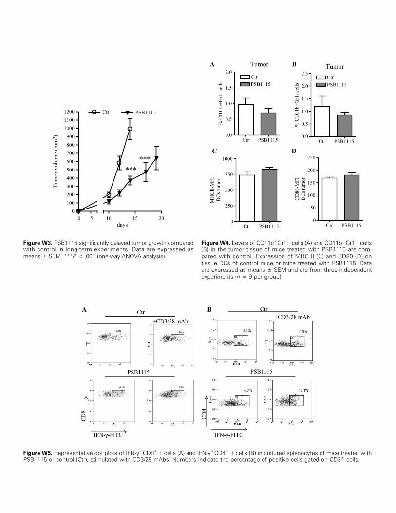
Figure W3. PSB1115 significantly delayed tumor growth comparedwith control in long-term experiments. Data are expressed asmeans ± SEM. ***P < .001 (one-way ANOVA analysis).
Figure W4. Levels of CD11c+Gr1− cells (A) and CD11b+Gr1− cells(B) in the tumor tissue of mice treated with PSB1115 are com-pared with control. Expression of MHC II (C) and CD80 (D) ontissue DCs of control mice or mice treated with PSB1115. Dataare expressed as means ± SEM and are from three independentexperiments (n = 9 per group).
Figure W5. Representative dot plots of IFN-γ+CD8+ T cells (A) and IFN-γ+CD4+ T cells (B) in cultured splenocytes of mice treated withPSB1115 or control (Ctr), stimulated with CD3/28 mAbs. Numbers indicate the percentage of positive cells gated on CD3+ cells.