An Analog VLSI Architecture for High Speed, Low Power Object Tracking
BIOSEG: a bioinspired vlsi analog system for image segmentation
6
BIOSEG: A Bioinspired VLSI Analog System for Image Segmentation Jordi Madrenas, Jordi Cosp, Oscar Lucas, Eduard Alarcón, Eva M. Vidal, Gerard Villar Dept. of Electronics Engineering, Universitat Politècnica de Catalunya, Jordi Girona 1-3, Barcelona, Spain. E-mail: [email protected] Abstract: The architecture of a complete image segmentation system and the development of an embedded VLSI low-power integrated circuit are reported. A neuromorphic engineering approach is adopted, with the purpose of reproducing behaviour of biological neural networks by taking advantage of the microelectronic implementation properties, especially low power consumption and reduced volume and weight. The system is divided in parallel-processing stages. After phototransduction, nonlinear filtering is applied to the image. Then, segmentation is performed and an output stage delivers segmentation and object properties information. Each stage is briefly described and simulations and experimental results are shown. The final goal is to develop a single-chip integrated system that performs all the described operations in focal-plane. 1. Introduction Neuromorphic engineering, was proposed by C. Mead in the late eighties [1]. Its main idea is to build a biological metaphor on silicon by opportunistically employing the rich characteristics of devices instead of using them as digital brute-force switches. The objective is to emulate biology, approaching as much as possible to the neural systems behavior of living beings as more knowledge becomes available. Today, a combination of both classic and neuromorphic approaches seems a good engineering solution [2]. Concerning artificial vision, the first stages have been studied by many research groups, and several artificial retinae were proposed in the past [2-3]. Several other early vision tasks taking place at the retina as edge extraction, velocity/optical flow, etc., have been extensively studied in the literature. On the other hand, implementations of further processing levels taking place in brain have barely been studied from the neuromorphic approach, most probably due to the lack of models. Some of these processes are attention and image segmentation [4-5] which are fundamental even for a simple portable image recognition system. A main characteristic of biological neurons is their oscillatory nature. It has been shown that oscillations may encode information and scientific community is performing a significant effort to understand these mechanisms by means of microelectrode measurements and also simulations [6-8]. Based on previous work from biologists and computer scientists who model living structures, we propose a new approach to this problem, developing algorithms for image processing (segmentation, attention) that include early vision and higher computation stages. By means of low-power ESANN'2004 proceedings - European Symposium on Artificial Neural Networks Bruges (Belgium), 28-30 April 2004, d-side publi., ISBN 2-930307-04-8, pp. 411-416
Transcript of BIOSEG: a bioinspired vlsi analog system for image segmentation

BIOSEG: A Bioinspired VLSI Analog System for Image Segmentation
Jordi Madrenas, Jordi Cosp, Oscar Lucas, Eduard Alarcón, Eva M. Vidal, Gerard Villar
Dept. of Electronics Engineering, Universitat Politècnica de Catalunya, Jordi Girona 1-3, Barcelona, Spain. E-mail: [email protected]
Abstract: The architecture of a complete image segmentation system and the development of an embedded VLSI low-power integrated circuit are reported. A neuromorphic engineering approach is adopted, with the purpose of reproducing behaviour of biological neural networks by taking advantage of the microelectronic implementation properties, especially low power consumption and reduced volume and weight. The system is divided in parallel-processing stages. After phototransduction, nonlinear filtering is applied to the image. Then, segmentation is performed and an output stage delivers segmentation and object properties information. Each stage is briefly described and simulations and experimental results are shown. The final goal is to develop a single-chip integrated system that performs all the described operations in focal-plane.
1. Introduction Neuromorphic engineering, was proposed by C. Mead in the late eighties [1]. Its main idea is to build a biological metaphor on silicon by opportunistically employing the rich characteristics of devices instead of using them as digital brute-force switches. The objective is to emulate biology, approaching as much as possible to the neural systems behavior of living beings as more knowledge becomes available. Today, a combination of both classic and neuromorphic approaches seems a good engineering solution [2]. Concerning artificial vision, the first stages have been studied by many research groups, and several artificial retinae were proposed in the past [2-3]. Several other early vision tasks taking place at the retina as edge extraction, velocity/optical flow, etc., have been extensively studied in the literature. On the other hand, implementations of further processing levels taking place in brain have barely been studied from the neuromorphic approach, most probably due to the lack of models. Some of these processes are attention and image segmentation [4-5] which are fundamental even for a simple portable image recognition system. A main characteristic of biological neurons is their oscillatory nature. It has been shown that oscillations may encode information and scientific community is performing a significant effort to understand these mechanisms by means of microelectrode measurements and also simulations [6-8]. Based on previous work from biologists and computer scientists who model living structures, we propose a new approach to this problem, developing algorithms for image processing (segmentation, attention) that include early vision and higher computation stages. By means of low-power
ESANN'2004 proceedings - European Symposium on Artificial Neural NetworksBruges (Belgium), 28-30 April 2004, d-side publi., ISBN 2-930307-04-8, pp. 411-416

analog circuits we expect a significant reduction of area and power requirements bearing in mind autonomous systems applications. In this paper we introduce a simple but complete neuromorphic vision system that can perform object segmentation and determine some image properties as object count and object centroids. In next section the system architecture is described, in sections 3, 4 and 5 preprocessing, segmentation and output stages are reviewed and, finally, in Section 6 conclusions and future work are reported. 2. System architecture In Figure 1, the general system architecture is shown. A given scene image is projected onto a photodetector array by means of a lens. The transduced light is preprocessed by two stages. In first stage, noise and small luminance differences are filtered and in second stage neighbor pixels with equal or similar luminance are enhanced. The preprocessing stage controls a segmentation stage, composed of an array of coupled oscillators that separates scene objects by encoding information in the oscillator phases. The output stage generates information about the image as different segments, associated centroids and size and number of objects. An external microprocessor reads the network’s outputs and controls each stage parameters for an appropriate segmentation. All these processing stages, except the microprocessor, are focal-plane. Since interconnections are mainly local, their distribution is not a big issue.
Figure 1. VLSI Neuromorphic Image Segmentation System. In Figure 2a, the system is shown from a more detailed viewpoint. For the sake of simplicity, only three pixels in a one-dimension configuration are represented. Extension to more pixels in two dimensions is straightforward. An array of photodiodes ( ) perform a light transducer stage. A logarithmic amplifier stage ( ) extends the system dynamic range. Nonlinear spatial filtering is performed by means of a resistance and resistive fuse mesh ( ). Then the data is applied to peak-shaped functions ( ) that detect similarity of neighbors. An array of coupled oscillators ( ) together with a Global Oscillator (GO) inhibitory cell ( ) perform a modified
Lens
Phot
odet
ecto
rs
Pre-
proc
essi
ng
Segm
enta
tion
Outputstage
µP
Scene
INTEGRATEDCIRCUIT
ESANN'2004 proceedings - European Symposium on Artificial Neural NetworksBruges (Belgium), 28-30 April 2004, d-side publi., ISBN 2-930307-04-8, pp. 411-416
LEGION [9] algorithm that has been adapted for a simple VLSI implementation [4]. Finally, a resistance mesh ( ) computes object centroid.
(a) (b)
Figure 2. a) Three-element functional segmentation system; b) I-V characteristic of the resistive fuse.
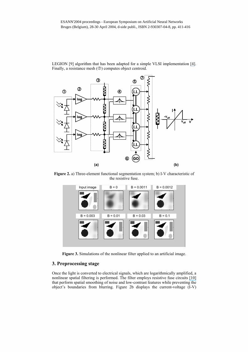
Figure 3. Simulations of the nonlinear filter applied to an artificial image.
3. Preprocessing stage Once the light is converted to electrical signals, which are logarithmically amplified, a nonlinear spatial filtering is performed. The filter employs resistive fuse circuits [10] that perform spatial smoothing of noise and low-contrast features while preventing the object’s boundaries from blurring. Figure 2b displays the current-voltage (I-V)
v
I
voff
-voff
v
I
voff
-voff
log
log
log
GO
log
log
log
GO
Input image B = 0 B = 0.0011 B = 0.0012
B = 0.003 B = 0.01 B = 0.03 B = 0.1
ESANN'2004 proceedings - European Symposium on Artificial Neural NetworksBruges (Belgium), 28-30 April 2004, d-side publi., ISBN 2-930307-04-8, pp. 411-416

characteristic of the fuse. Below a given Voff threshold between adjacent pixels’ luminance, the device operates as a resistor while changing its behavior and remaining open (the current flow becomes null) above Voff. Figure 3 shows simulation results of the nonlinear filter that was applied to an artificial image containing objects with different levels of contrast. By changing the parameter B, which is inversely proportional to Voff, several degrees of filtering can be obtained. Figure 4 shows some simulations results for a simple real image. Different segmentation results are obtained by varying the B parameter.
Figure 4. Nonlinear filter applied to a simple real image. 4. Segmentation stage Figure 5 shows the segmentation process performed by a 2-dimensional array of coupled nonlinear oscillators and a inhibitory cell. Object segmentation [9,4] is achieved by two opposed mechanisms: a phase synchronizing effect due to local excitatory coupling of oscillators controlled by similar pixels and an opposed effect of phase separation of pixels with large contrast due to the global inhibitory oscillator cell. Each input pixel determines the excitatory strength of neighbor oscillator synapses. Therefore, oscillators 2 and 3 become synchronized because there exists an excitatory path among them by means of the local synapses chain. On the other hand, the global inhibitory cell prevents the phase synchronization of separate objects, i.e., oscillator 1 is not in phase with oscillators 2 and 3. This is due to the fact that once an oscillator fires, it excites its neighbors that belong to the same object while all the network oscillators are inhibited simultaneously. Since excitation is set stronger than inhibition, the excited cells are capable of firing while the other ones are not. A testchip containing a 16 × 16 oscillator array segmentation stage in a 0.8 micron CMOS technology was designed and manufactured (figure 6) [4]. The chip is fully functional and demonstrates the segmentation capability of the array for binary
Input image B = 0
B = 0.01B = 0.005
ESANN'2004 proceedings - European Symposium on Artificial Neural NetworksBruges (Belgium), 28-30 April 2004, d-side publi., ISBN 2-930307-04-8, pp. 411-416

images. A sample input image and experimental results are shown in figure 8. A single oscillator and a global inhibitor waveforms are shown. In figure 7, a testchip layout for the preprocessing and segmentation basic blocks in a more advanced 0.35 micron CMOS technology is shown. Measurements were performed and results agree satisfactorily with simulations. Oscillation frequency ranges between tens and hundreds of kHz. The upper limit of the number of segmented objects is about six in this implementation. No special problems with substrate noise or other undesired interferences have been observed.
1
3
I
2
oscillator 1
oscillator 2
oscillator 3
inhibitor
1
3
I
1
3
I
2
oscillator 1
oscillator 2
oscillator 3
inhibitor
Figure 5. LEGION network: Segmentation oscillator array, global inhibitor and drawing of three ideal cells behaviour.
Figure 6. 0.8 micron CMOS 16 x16 oscillators segmentation testchip;
Figure 7. 0.35 micron CMOS preprocessing and segmentation basic block testchip
layout.
(a)
(b) Figure 8. Segmentation experimental results a) Binary input image; b) Waveform of
an oscillator of the array (upper graph) and the global inhibitor (lower graph).
time
V
V
ESANN'2004 proceedings - European Symposium on Artificial Neural NetworksBruges (Belgium), 28-30 April 2004, d-side publi., ISBN 2-930307-04-8, pp. 411-416

5. Output stage Object counting. The inhibitor becomes active (high voltage level) whenever at least a single oscillator is active (high), hence a simple way to obtain the number of segmented objects in a given image is to count the number of pulses of the global inhibitor during an oscillation period. For instance, in the example of figure 8, the number of objects is 4. Segmentation. The global oscillator’s high state also selects the pixels that belong to an object. This way it is very simple to obtain the different objects, when required. Object centroid and size. When a single object is active, information about its size (number of pixels) and position (centroid) is easily obtained with simple postprocessing. Arithmetic addition of currents generated by active oscillators gives an indication of the object size. A resistive diffusion network (stage in Figure 2) indicates the centroid position. 6. Results, conclusions and ongoing work A complete neuromorphic vision system based on oscillators and analog circuits has been reported. Partial experimental results of the segmentation array and preprocessing basic blocks demonstrate the feasibility of the system. Currently, a complete BIOSEG chip is under design. The estimated fill factor is about 25%. This integrated circuit, together with digital control, will enable simple applications of vision in portable, low-power applications. Acknowledgements. This work has been partially funded by project TIC-2001-2183 from the Spanish MCYT. Gerard Villar holds a CICYT Research fellowship. Special thanks to the anonymous reviewers’ suggestions and corrections. References [1] C.A.Mead. Analog VLSI and Neural Systems. Addison-Wesley, 1989. [2] Toward an Artificial eye. Spectrum, IEEE.. Vol. 33, Issue 5, May 1996 [3] C.A.Mead y M.A.Mahowald. A Silicon Model of Early Visual Processing.
Neural Networks, vol., pp.91-97, 1988. [4] J.Cosp, J. Madrenas, Scene Segmentation Using Neuromorphic Oscillatory
Networks, IEEE Trans. Neural Networks, Vol. 14, 5, pp. 1278-1296, Sept. 2003. [5] H.Ando, T.Morie, M.Nagata, A.Iwata: A Nonlinear Oscillator Network for Gray
–level Image Segmentation and PWM/PPM Circuits for its VLSI Implementation. IEICE Trans, Fundamentals, E83-A, 2, 329-336, (2000).
[6] A.K.Engel et al. Interhemispheric Synchronization of Oscillatory Neuronal Responses in Cat Visual Cortex. Science, 252, pp. 1177-1179, 1991.
[7] R.Eckhorn et al. Coherent Oscillations: a Mechanism for Feature Linking in the Visual Cortex?, Biological Cybernetics, 60, 121-130, 1988.
[8] Ch. Von der Malsburg, J.Buhman. Sensory Segmentation with Coupled Neural Networks. Biological Cybernetics, 67, 233-242, 1992.
[9] D.L.Wang, D.Terman: Image Segmentation Based on Oscillatory Correlation. Neural Computation, 9, 805-836, 1997.
[10] A. Lumsdaine, J. Wyatt, and I. Elfadel, "Nonlinear analog networks for image smoothing and segmentation," IEEE ISCAS, pp. 987-991, May 1990.
ESANN'2004 proceedings - European Symposium on Artificial Neural NetworksBruges (Belgium), 28-30 April 2004, d-side publi., ISBN 2-930307-04-8, pp. 411-416