Biology Student Textbook Grade 9 | aacaebc
131
Biology Student Textbook Grade 9 Biology Student Textbook Grade 9 Federal Democratic Republic of Ethiopia Ministry of Education Biology Student Textbook Grade 9 Price: ETB 36.00 FDRE MoE Federal Democratic Republic of Ethiopia Ministry of Education ISBN 978-99944-2-008-7 CVR_BIOL_SB_ETH_0087_CVR.indd 1 10/6/10 4:35:29 pm
-
Upload
khangminh22 -
Category
Documents
-
view
0 -
download
0
Transcript of Biology Student Textbook Grade 9 | aacaebc

BiologyStudent TextbookGrade 9
BiologyStudent TextbookGrade 9
Federal Democratic Republic of EthiopiaMinistry of Education
Biology Student Textbook
Grade 9
Price: ETB 36.00FDREMoE
Federal Democratic Republic of EthiopiaMinistry of Education
ISBN 978-99944-2-008-7
CVR_BIOL_SB_ETH_0087_CVR.indd 1 10/6/10 4:35:29 pm

Take Good Care of This Textbook
This textbook is the property of your school. Take good care not to damage or lose it. Here are 10 ideas to help take care of the book:
1. Cover the book with protecti ve material, such as plasti c, old newspapers or magazines.2. Always keep the book in a clean dry place.3. Be sure your hands are clean when you use the book.4. Do not write on the cover or inside pages.5. Use a piece of paper or cardboard as a bookmark.6. Never tear or cut out any pictures or pages.7. Repair any torn pages with paste or tape.8. Pack the book carefully when you place it in your school bag.9. Handle the book with care when passing it to another person.
10. When using a new book for the fi rst ti me, lay it on its back. Open only a few pages at a ti me. Press lightly along the bound edge as you turn the pages. This will keep the cover in good conditi on.
IFC_BIOL_SB_ETH_0087_IFC.indd 1 10/6/10 4:41:51 pm

Author: Ann Fullick
Adviser: Alemu Asfaw
Evaluators: Solomon Belayneh Getachew Bogale Silas Araya
Federal Democratic Republic of EthiopiaMinistry of Education
BiologyStudent TextbookGrade 9
A01_BIOL_SB_ETHG9_0087_PRE.indd 1 1/6/10 1:34:44 pm


iiiGrade 9
Contents
Unit 1 Biology and technology 11.1 Renowned Ethiopian biologists 1
1.2 Biological research in Ethiopia 7
Unit 2 Cell biology 132.1 The microscope 13
2.2 The cell 22
2.3 The cell and its environment 33
Unit 3 Human biology and health 503.1 Food and nutrition 51
3.2 The digestive system 69
3.3 The respiratory system 82
3.4 Cellular respiration 99
3.5 The circulatory system 104
Unit 4 Micro-organisms and disease 1234.1 Micro-organisms 124
4.2 Diseases 137
4.3 HIV and AIDS 158
Unit 5 Classification 1715.1 Principles of classification 171
5.2 The five kingdoms 178
Unit 6 Environment 2006.1 Ecosystems 200
6.2 Food relationships 204
6.3 Recycling in nature 212
6.4 Adaptations 218
6.5 Tree-growing project 223
Index 226
A01_BIOL_SB_ETHG9_0087_PRE.indd 3 1/6/10 1:34:44 pm

A01_BIOL_SB_ETHG9_0087_PRE.indd 4 1/6/10 1:34:44 pm

1Grade 9
Biology and technology
1.1 Renowned Ethiopian biologists
By the end of this section you should be able to:
• Name at least one renowned Ethiopian biologist.
• Explain the contributions of these Ethopian biologists to international biological knowledge.
Welcome to the study of biology! Biology is the study of life and living organisms. All around you there are many different types of plants, animals and other living organisms. They depend on each other and on the environment where they live. Biologists study both the outer appearance and the internal workings of living things. They study how living organisms interact and where human beings fit into the living world.
All of our biological knowledge comes to us by the work of biologists, scientists who study living organisms. Biology, like all the sciences, moves forward most of the time in small, steady steps. One biologist comes up with an idea. Another carries out more experiments, which either support the new idea or suggest it is wrong. Then more biologists join in until eventually a new idea, or hypothesis, is accepted. Like all scientists, biologists publish their new work (called research) in special magazines called journals. Before an idea is published in a journal, several other well-known biologists have to read it and check that the research has been done to a high standard. This process is called peer review. Sometimes biology takes a great leap forward, when a very gifted biologist comes along with a big new idea!
If you think you would like to be such a biologist, start right now by observing living things around you.
Here in Ethiopia we have some very renowned biologists. Their work is known and important not just here in Ethiopia but around the world. Here are some of their biographies.
Unit 1
Contents Section Learning competencies
1.1 Renowned Ethiopian • Name at least one renowned Ethiopian biologist. biologists (page 1) • Explain the contributions of these Ethiopian biologists to international biological knowledge.
1.2 Biological research • Explain how scientific institutions contribute to scientific research. in Ethiopia • Name some Ethiopian institutions involved in biological research. (page 7) • Explain the activities and contributions of some of these Ethiopian research institutions.
KEY WORDS
hypothesis an idea or statement that explains observed facts and predicts new outcomesjournal a regular publication presenting articles on a particular subjectpeer review evaluation of a person’s work done by others in the same field
M01_BIOL_SB_ETHG9_0087_U01.indd 1 1/6/10 11:31:23 am

2
UNit 1: Biology and technology
Grade 9
Dr Aklilu Lemma and the battle against bilharzia (schistosomiasis)
Schistosomiasis (also known as bilharziasis) is a common parasitic disease. It affects 200–300 million people in Africa (including Ethiopia), South America, Asia and parts of the Caribbean. It is caused by parasitic flatworms which spend part of their lifecycle in freshwater snails and part in humans. Anyone washing, working or playing in shallow fresh water is at risk. Once inside a person, the parasites mature and produce eggs which are passed out in the urine and faeces. They also infest the blood vessels, liver, kidneys, bladder and other organs. The body sets up an immune reaction and an infected person can become weakened and ill for many years.
Some of the most important work in finding a way of controlling this parasite, which is effective but does not cost too much, was carried out by Dr Aklilu Lemma, one of Ethiopia’s most renowned biologists.
Dr Aklilu began his work in 1964, when he was investigating the freshwater snails that carry the schistosomiasis parasite around Adwa in northern Ethiopia. He saw women washing clothes in the water and he noticed that downstream of the washing party there were more dead snails than anywhere else he had collected. The women were using the soapberry, Endod (Phytolacca dodecandra), to make washing suds. Dr Aklilu collected some live snails from above the washing party and asked one of the women to give him some of her Endod suds. Not long after the suds were put in the snail container, the snails all died. This was the start of years of work for Dr Aklilu.
Back in the laboratory he showed that if the Endod berries were dried, crushed and diluted in water they would kill snails at very low concentrations. Other scientists carried out similar investigations and got the same results. If the freshwater snails can be controlled, the spread of schistosomiasis can be greatly reduced. The World Health Organisation recommended a chemical molluscicide (i.e. a compound that kills molluscs including snails) but it was extremely expensive. Endod works well, it is cheap, it is well known by local people who are likely to use it and it is environmentally friendly as it breaks down naturally within about two days.
Dr Aklilu Lemma worked for many years to convince scientists all around the world that his ideas would work. Trials using locally collected Endod showed that using the molluscicide worked. Before the water was treated, 50% of children 1–6 years old were infected. After treatment only 7% were infected by the flatworm. Dr Aklilu’s results were published in journals around the world. He found the best species of the soapberry plant and developed programmes for local communities to treat their own water. Eventually people were convinced and the use of Endod-based molluscicides is spreading throughout Africa and beyond. Hopefully a combination of Endod water treatment to kill the snails, improved hygiene, clean water
Figure 1.1 Highly magnified image of the parasitic flatworms that cause schistosomiasis
KEY WORDS
immune reaction a biological response involving the production of antibodies etc. as a reaction to the presence in the body of bacteria, a poison, or a transplanted organtreated water filtered or disinfected water made safe for consumptiongenetic engineering the deliberate, controlled manipulation of the genes in an organism, with the intent of making that organism better
Figure 1.2 Dr Aklilu Lemma, one of Ethiopia’s most renowned biologists, with the snail-killing soapberries known locally as Endod
M01_BIOL_SB_ETHG9_0087_U01.indd 2 1/6/10 11:31:24 am

3
UNit 1: Biology and technology
Grade 9
wells and medicine for affected people will mean that Ethiopia can be free of this terrible disease. If we succeed it will be largely due to the work of Dr Aklilu Lemma. He has been honoured and recognised in many different ways both in Ethiopia and around the world for his work.
Dr Tewolde Berhan Gebre Egziabher, an ardent lover of nature
Dr Tewolde Berhan Gebre Egziabher was born in 1940. In 2000 he won the Right Livelihood Award (often called the Alternative Nobel Prize) “for his exemplary work to safeguard biodiversity and the traditional rights of farmers and communities to their genetic resources”.
During the 1990s Dr Tewolde Berhan was involved in negotiations at the various biodiversity-related meetings, including the Convention on Biological Diversity (CBD) and the Food and Agriculture Organization. Having built a strong and able team of African negotiators, he managed to help achieve progressive, unified policies for Africa, such as recognition of community rights.
Dr Tewolde Berhan was instrumental in securing recommendations from the Organisation of African Unity (OAU) encouraging African countries to develop and implement community rights, a common position on Trade Related Aspects of Intellectual Property Rights, and a clear stance against patents on living materials. He also helped to draft the OAU model legislation for community rights, which is now used across Africa.
In January 2000 Dr Tewolde Berhan acted as chief negotiator on biosafety for the Like-Minded Group, made up of most of the G77 countries, in Montreal. Here he was central to achieving an outcome protecting biosafety and biodiversity and respecting community rights, against strong US-led representations.
Dr Tewolde Berhan also won the United Nations top environmental prize, Champions of the Earth, in 2006.
Professor Tilahun Yilma and his vaccines Professor Tilahun Yilma is known internationally for the vaccine he developed to help get rid of the terrible cattle disease rinderpest, and for his work on HIV/AIDS vaccines. Rinderpest arrived in Ethiopia in 1888, carried by three infected cattle brought into the country by Italian soldiers. Within a year 90% of the domestic cattle plus many wild animals such as buffalo, giraffe and antelope died. As a result 30–60% of the people starved.
In the 1980s rinderpest became a major problem again. Professor Tilahun worked to develop a vaccine using genetic engineering. He was very successful – his vaccine doesn’t need refrigeration, it is easily scratched onto the animal’s neck or abdomen so cattle don’t need injections from vets and it can be made relatively cheaply in large quantities. By 1997 the vaccine was ready for use across Africa,
DiD yoU kNow?Around 300 million people are affected by schistosomiasis in the tropical and sub-tropical parts of the world – so the work of Aklilu Lemma could make an enormous difference in many other countries as well as Ethiopia. Dr Aklilu recognised this when he said “we found a poor man’s medicine for a poor man’s disease”.
Figure 1.3 Dr Tewolde Berhan Gebre Egziabher
Figure 1.4 Professor Tilahun Yilma
M01_BIOL_SB_ETHG9_0087_U01.indd 3 1/6/10 11:31:25 am

4
UNit 1: Biology and technology
Grade 9
including his own country Ethiopia. Now he is working on using similar methods to develop an effective vaccine against HIV/AIDS, which is affecting people all around the world including millions of Africans. Professor Tilahun has been given many international awards and honours over the years. He is also very active in encouraging young scientists and establishing the highest quality research establishments here in Africa.
Professor yalemtsehay Mekonnen: the first female professor from AAU
Professor Yalemtsehay Mekonnen is a biologist and an academic member of staff at the Department of Biology, Faculty of Science, Addis Ababa University. She has worked in this Department for the last 30 years. She received her PhD, specialising in human physiology, from the University of Heidelberg in Germany.
One of her research areas is the assessment of the impact of chemical pesticide hazard on humans. This research covers almost all government farms including the Upper Awash Agricultural farms and some private horticultural farms in the Rift Valley region. The other area of her research is in the use of plants as medicine against human and animal diseases.
Professor Yalemtsehay Mekonnen served as Head of the Department of Biology from 1993 to 1995 and as the Director of the Aklilu Lemma Institute of Pathobiology from February 2003 to October 2007. In leadership positions she was involved and has initated a number of national and international research networks and collaborations. She is a member of many professional societies, such as the Biological Society of Ethiopia, the Safe Environment Association, the Ethiopian Wildlife and Natural History Society, the New York Academy of Sciences and the Third World Organization for Women in Science. She has served as President of the Biological Society of Ethiopia.
She has been awarded research grants and fellowships nationally from the Ethiopian Science and Technology Commission and the Ethiopian Agricultural Research Organization, and internationally from the British Council, the International Foundation for Science, Third World Academy of Sciences, the German Academic Exchange Service and the Alexander von Humboldt Foundation from Germany.
Dr Melaku woredeDr Melaku Worede was born in 1936 and he has worked for many years to save the genetic diversity of Ethiopia’s domestic plants. He is an internationally acclaimed plant genetics researcher. Dr Melaku set up the Plant Genetic Resources Centre in Addis Ababa. Our country is noted for its great genetic diversity but modern farming methods and problems such as drought can affect this badly. Dr Melaku Worede has preserved many different traditional crop varieties and developed ways of farming that
Figure 1.5 Professor Yalemtsehay Mekonnen working in the Biomedical Laboratory, Addis Ababa University
Figure 1.6 Dr Melaku Worede, an internationally acclaimed plant genetics researcher
M01_BIOL_SB_ETHG9_0087_U01.indd 4 1/6/10 11:31:26 am

5
UNit 1: Biology and technology
Grade 9
produce high yields without commercial fertilisers. Dr Worede’s methods are now widely used both in other areas of Africa and in Asia. He was the first chair of the African Committee for Plant and Genetic Resources and has been Chair of the United Nations Food and Agriculture Organisation’s Commission on Plant Genetic Resources. He has also won the Right Livelihood Award (often called the Alternative Nobel Prize) in 1989 for outstanding vision and work and in 2008 he received the Outstanding International Contribution Award from the National Green Award Foundation.
Dr Gebissa EjetaWhen Dr Gebissa Ejeta was born in a small rural village his mother was determined her son would receive a good education. He walked 20 miles to school every Sunday evening, returning home on Friday after a week of studying. It all paid off as he gained a place at Jimma Agricultural and Technical School and then Alemaya College where he took his first degree. He specialises in plant breeding and genetics. Dr Gebissa Ejeta did his research on sorghum – he got his PhD from Purdue University in the USA where he still holds a professorship. He has helped to develop Africa’s first commercial hybrid strain of sorghum. This not only needs less water and so is resistant to drought, but it also yields more grain than traditional varieties. Dr Gebissa Ejeta developed other strains of sorghum which are also resistant to the parasitic Striga weed, which can destroy a big percentage of a crop. Dr Ejeta’s work has made a very big difference to the food availability in many areas of Ethiopia and other African countries – his varieties yield up to ten times more than the original strains. In 2009 Dr Gebissa Ejeta was awarded the World Food Prize, which is the most important agricultural prize in the world! He has also been awarded the National Hero award of Ethiopia for his work in science and technology.
These are just some of many renowned Ethiopian biologists who have carried out work of great value both in our own country and across the world. You will have the opportunity to find out more about some of the other scientists with your teacher in activity 1.1.
Here are some more examples of Ethiopian biologists.
Professor Beyene Petros• is a biomedical scientist and long serving professor at Addis Ababa University. Professor Beyene Petros, in addition to his distinguished academic career, served as Chairman, Advisory Committee on Health Research and Development, WHO/AFRO, 1997–2000; as vice minster of Education (1991–1993) and many other scientific societies. He has produced more than 43 publications in reputable scientific journals and Published books. Professor Beyene has won Gold Medal Award from Ethiopian Health Association; Fellowships from Fulbright and from Centers for disease control and prevention, Atlanta, USA.Professor Sebsebe Demissew• is a plant taxonomist. He is now the Director of the National Herbarium and the leader of the Ethiopian Flora Project (EFP).
Figure 1.7 Dr Gebissa Ejeta who has been honoured for his work in developing new, high yielding strains of sorghum which grow well in our conditions
M01_BIOL_SB_ETHG9_0087_U01.indd 5 1/6/10 11:31:26 am

6
UNit 1: Biology and technology
Grade 9
Dr Zeresenay Alemseged• discovered a 3.3 million-year-old humanoid child fossil in 2006.Dr Tsehaynesh Meselle• was the Director General of the Ethiopian Health and Nutrition Research Institute (EHNRI) during the writing of this book and leads research in human health, including HIV/AIDS.Dr Berhane Asfaw • is an Ethiopian scientist whose team discovered two 160 000-year-old human skulls, some of the oldest that have ever been found. His discoveries were published in the famous scientific journal Nature. They have had a great impact on the study of human evolution around the world.
Professor Legesse Negash• is a Professor of Plant Physiology in the Department of Biology, Faculty of Science, Addis Ababa University. He is a pioneer in the propagation of Ethiopia’s indigenous trees and is the Founder and Leader of the Center for Indigenous Trees Propagation and Biodiversity Development in Ethiopia. Professor Negash is a winner of several awards, including that from the Stockholm-based International Foundation for Science.
Professor Mogessie Ashenafi • works at the University of Addis Ababa and leads international research into food microbiology.
Professor Ensermu Kelbessa• is one of the leading systematic botanists who has discovered and named many new plants.
Review questionsSelect the correct answer from A to D.
1. Biology is: A the study of matter B the study of life and living organisms C the study of how living organisms interact D the study of the way atoms and molecules react together
2. Bilharzia is caused by: A snails B bacteria C viruses D parasitic flatworms
3. Dr Tewolde Berhan Gebre Egziabher is a biologist who researches into:
A HIV/AIDS B genetic engineering C environmental protection and diversity D human evolution
Activity 1.1: Discovering Ethiopian biologists it is inspiring to know what great work is being done by Ethiopian biologists today. Here you have the opportunity to do some research and find out about some of the people who are active in biology in our country.
• You can choose a biologist from the list on pages 5 and 6, find out more about one of the biologists already mentioned in this chapter or write about a biologist you have discovered for yourself. if you look at the research institutions mentioned in the rest of this chapter you will be able to find lots of Ethiopian biologists to choose from!
• Use any resources you have available. You may find out about Ethiopian biologists in books, magazines, journals, leaflets, in the news or even on the internet if it is available.
• Write a report about your biologist and prepare to give a brief talk on him or her to the rest of the class.
M01_BIOL_SB_ETHG9_0087_U01.indd 6 1/6/10 11:31:26 am

7
UNit 1: Biology and technology
Grade 9
1.2 Biological research in Ethiopia
By the end of this section you should be able to:
• Explain how scientific institutions contribute to scientific research.
• Name some Ethiopian institutions involved in biological research.
• Explain the activities and contributions of some of these Ethiopian research institutions.
Biologists, like other scientists, do not work alone. A biologist needs equipment, laboratories and other biologists to discuss ideas with and develop theories. Biologists work in many different areas, from plants to animals, and from medicine to classification and genetics. Ethiopia has a number of well-known institutions that are involved in biological research. Our country continues to invest in these institutions and to develop more. Our biologists have international reputations in many fields. Biologists from other African nations and from other continents come to our institutions to take part in the research programmes, and our biologists also travel to other countries. Sharing knowledge across the world is an important part of science and Ethiopia plays her part in this.
Here are some of the institutions that play an important part in biological research in Ethiopia.
Addis Ababa University (AAU) Biology DepartmentAddis Ababa University (AAU) is a very large university with an international reputation and the Biology Department is no exception. A top university, AAU is one of the major centres of biological research in the country and it is also home to the Aklilu Lemma Institute of Pathobiology (see the next page).
The university entrance is impressive and the Department of Biology contains much modern and high-level equipment to help biologists in their research.
Addis Ababa University is not the only renowned university in Ethiopia. There are many others, including Haramaya University, Mekelle University, Jimma University, Hawassa University, Gonder University and Bahir Dar University. They all have active biology departments where teaching and research takes place.
Figure 1.8 Addis Ababa University is home to much internationally recognised research.
M01_BIOL_SB_ETHG9_0087_U01.indd 7 1/6/10 11:31:27 am

8
UNit 1: Biology and technology
Grade 9
Armauer Hansen Research institute (AHRi)When Armauer Hansen Research Institute (AHRI) was first set up in 1969 it was sited next to a big hospital dedicated to patients with leprosy and it carried out research only into leprosy. However, in the years since 1969 leprosy has become a disease that we can treat quite effectively. Since 1996 AHRI has widened its research to include tuberculosis (TB, which has become a big threat with the rise in HIV/AIDS), leishmaniasis, malaria and HIV/AIDS, as well as leprosy.
Aklilu Lemma institute of Pathobiology (ALiPB)The department of pathobiology at Addis Ababa University has been renamed the Aklilu Lemma Institute of Pathobiology (ALIPB) in honour of Professor Aklilu Lemma (see page 2). The institute sets out to be a centre of excellence for biomedical research and training. ALIPB carries out research in five major areas. They have a microbiology research programme into the major infectious diseases, they look into vectors of diseases and how to control them, some of the biologists are focused on human parasitic diseases and others work on animal health and disease. Finally, some of the biologists are carrying out research into Endod and other plants, which may be useful as medicines. The institute also plays an important role in training new Ethiopian pathobiologists. Students with a first degree in biomedical sciences (i.e. biology, human medicine, veterinary medicine, laboratory technology) can apply to do a Masters degree in tropical and infectious diseases at the institute.
Ethiopian Health and Nutrition Research institute (EHNRi)
The Ethiopian Health and Nutrition Research Institute (EHNRI) is an organisation that carries out research into health and nutrition issues which affect public health. Its role is both to identify problems and to help everyone in the country become aware of how to overcome the problems and improve their levels of nutrition and health. The laboratory facilities at EHNRI are good and are well equipped for research into immunology and viral diseases. At the moment EHNRI is carrying out a lot of work into HIV/AIDS in Ethiopia. For example, biologists working with EHNRI are following the progression of HIV/AIDS in two populations of factory workers (about 2000 people) over a long period of time – the research began in 1994! They are planning to work with more people in the future, and are also hoping to set up trials of a possible HIV vaccine. EHNRI is also very active in the battle against TB and it houses the National TB Reference Laboratory. It is involved in rapid diagnosis of TB. EHNRI is also involved in many other projects including issues such as the nutritional state of mothers and babies in the country as well as infectious diseases.
Figure 1.9 The AHRI buildings and some of the workers who work there
M01_BIOL_SB_ETHG9_0087_U01.indd 8 1/6/10 11:31:27 am

9
UNit 1: Biology and technology
Grade 9
Ethiopian institute of Agricultural Research (EiAR) also known as institute of Agricultural Research (iAR)
Agriculture – farming crops and livestock – is the life force of our country. We must grow food to eat. The EIAR (IAR) is the institute where biologists with a passion for improving agriculture and supporting everyone who cultivates the land or raises livestock in Ethiopia carry out research.
There are five main areas of research. Biologists working on crop technology are working to help us achieve food security and nutritional quality, so that we always have sufficient food. They are looking at different crops and ways of improving the crops we already grow. Other biologists are focusing on our livestock, looking at ways of managing our animals’ breeding and feeding programmes to make sure that they grow as quickly and as well as possible. They also look at ways of improving the health of our livestock.
Biologists working on crop technology have improved crops like maize, teff and sorghum. Two of these are shown here.
Another important area of research is with regard to the soil and water. Biologists are looking into ways of improving the fertility of the soil, particularly ways of avoiding buying expensive inorganic fertilisers. Other scientists are also looking at ways to water the land more effectively. Forestry is also an area of research. Biologists are very involved in rehabilitating, restoring and conserving some of our forest ecosystems. Finally, the institute also looks at ways of mechanising farming. Biologists research into the species of crop plants that are most suitable for mechanised harvesting.
The Institute of Biodiversity Conservation (IBC)Biodiversity – the range of living organisms in an area – is internationally recognised as one of the biggest issues in biology today. Here in Ethiopia we have a tremendous range of biodiversity, particularly in our plants. There are also many species which are found only in Ethiopia (endemic). So in world terms, when it was decided to set up gene banks to conserve the genetic material of as many plants as possible, Ethiopia was given the highest priority. The Institute of Biodiversity Conservation (IBC) started off conserving the genes of Ethiopian plants. Now the institute is involved in the conservation of plants, animals and micro-organisms in Ethiopia. Research into the management of the ecosystem is also an important part of the work.
Current research in the IBC looks at many areas including forest and aquatic plants, medicinal plants, animal genetic resources, biotechnology and safety, and ecosystem conservation. The institute also holds one of the leading gene banks in the whole of Africa with over 300 plant species represented.
DiD yoU kNow?
Farming is vital to Ethiopia. About 90% of our exports and around 80% of our economy depends on agriculture.
Figure 1.10 Farming is vital to Ethiopia. Biologists with the EIAR work hard to improve our crops, our animals and our soil.
Figure 1.11 An agronomist examining sorghum crop
Figure 1.12 Scientists have improved crop production of Quncho, an improved teff. It is a hybrid crop now yielding more than 30 quintals per hectare.
M01_BIOL_SB_ETHG9_0087_U01.indd 9 1/6/10 11:31:29 am

10
UNit 1: Biology and technology
Grade 9
Activity 1.3: Making a table of research institutionsMake a table to summarise the biological research institutions in Ethiopia that are mentioned in this book. Add any that you or your classmates have discovered. Draw a table as shown below and complete it:
Institution Focus of research
Review questionsSelect the correct answer from A to D.
1. EHNRI carries out research into:
A health and nutrition issues B farming C biodiversity D soil and water
2. Before it widened its research the Armauer Hansen Research Institute studied only:
A HIV/AIDS B tuberculosis C leprosy D cervical cancer
3. ALIPB is world-renowned for research into:
A different diseases and their control B improved agricultural practices C human evolution D environmental conservation
Activity 1.2: Discovering more about research in Ethiopiathere are many great institutions in Ethiopia carrying out biological research. You have learnt a little about some of them. Now you can find out more.
• Investigate the biology department at the university nearest to your school or any other institution with biologists working there. Find out as much as you can about the research they carry out and the biologists who are there.
• Investigate one other biological research institution in Ethiopia. you may choose to find out more about one of the institutions highlighted in this book or you may find another different one.
• Use any resources you have available. You may use books, magazines, journals, leaflets, university prospectuses or reports in the news or even on the internet, if it is available.
• Write a report about your local university biology department and one other Ethiopian research institute and prepare to give a brief talk on them to the rest of the class.
M01_BIOL_SB_ETHG9_0087_U01.indd 10 1/6/10 11:31:29 am

11
UNit 1: Biology and technology
Grade 9
Summary
In this unit you have learnt that:
Biology is the study of life and living organisms. •
Scientific research is based on the ideas of scientists. they •design experiments to test these ideas. Results of these experiments are published in peer-reviewed journals, which are read by scientists around the world.
Ethiopia has some renowned biologists whose work is known •both in Ethiopia and internationally. they include Dr Aklilu Lemma, Professor tilahun yilma, Professor yalemtsehay Mekonnen, Dr Melaku worede, Dr Legesse woldeyes, Dr Gebissa Ejeta, Dr Berhane Asfaw, Professor Legesse Negash, Professor Mogessie Ashenafi, Professor Ensermu kelbessa and many others.
Most biological research is linked to a research institution •that has the facilities which are needed. there are a number of well-known Ethiopian biological research institutions.
End of unit questions1. a) Name two Ethiopian biologists who have made
internationally recognised contributions in their field.
b) Describe the main work of both of the biologists you have chosen and explain why it is so important.
2. What are the main advantages of using Endod in the battle against bilharzia?
3. Why is Professor Yalemtsehay Mekonnen internationally renowned?
4. What is rinderpest?
5. Why is the work of Dr Gebissa Ejeta so important?
6. Why are scientific institutions important to biological research?
7. a) Name three institutions involved in different types of biological research in Ethiopia.
b) Summarise the areas of biological research carried out by each institution.
M01_BIOL_SB_ETHG9_0087_U01.indd 11 1/6/10 11:31:29 am

12
UNit 1: Biology and technology
Grade 9
Copy the crossword puzzle below into your exercise book (or your teacher may give you a photocopy) and solve the numbered clues to complete it. 1
2
3 4
5
6
7
8
9
Across
2 Professor Tilahun Yilma developed a vaccine against this disease (10)
4 The Ethiopian scientist who has helped make food more available with his new breeds of sorghum is Dr Gebissa ***** (5)
7 What type of trees are planted in Ethiopia by Professor Legesse Negash? (10)
8 The Armauer Hansen Research Institute (4)
9 The surname of the Ethiopian scientist who discovered a way to prevent bilharzia (5)
Down
1 A new scientific idea (10)
3 What is studied at the EIAR (IAR)? (11)
5 What is the name of the plant which kills the snails which cause bilharzia? (5)
6 What do we call a special magazine where scientists publish their research? (7)
M01_BIOL_SB_ETHG9_0087_U01.indd 12 1/6/10 11:31:29 am

Cell biology Unit 2
13Grade 9
2.1 The microscope
By the end of this section you should be able to:
• Name different types of microscopes.
• Distinguish between the magnification and resolution of a microscope.
• State the functions of different types of microscopes.
• Compare the different resolutions and dimensions of light and electron microscopes.
• Explain and demonstrate basic techniques using a light microscope.
• Explain the purpose of staining cells.
• Use the microscope to study cells.
• Compare the way materials are prepared for the electron microscope and the light microscope.
Contents Section Learning competencies
2.1Themicroscope • Namedifferenttypesofmicroscopes. (page13) • Distinguishbetweenthemagnificationandresolutionofamicroscope. • Statethefunctionsofdifferenttypesofmicroscopes. • Comparethedifferentresolutionsanddimensionsoflightandelectron
microscopes. • Explainanddemonstratebasictechniquesusingalightmicroscope. • Explainthepurposeofstainingcells. • Usethemicroscopetostudycells. • Comparethewaymaterialsarepreparedfortheelectronmicroscopeand
the light microscope.
2.2Thecell • Statethecelltheory. (page22) • Listthestructuresofcellsanddescribetheirfunction. • Drawandlabeldiagramsandcomparetypicalplantandanimalcells. • Describethetypes,shapesandsizesofavarietyofcellsusingdiagrams.
2.3Thecelland • Describethepermeabilityofthecellmembrane. itsenvironment • Describetheprocessofdiffusionanditsimportanceinlivingorganisms. (page33) • Demonstratediffusionexperimentally. • Explaintheprocessofosmosisanditsimportanceinlivingorganisms. • Demonstrateosmosisexperimentally. • Showthatplantcellsbecomeflaccidwhentheylosewaterandturgid
when they absorb water by osmosis. • Explainplasmolysisandturgorpressure. • Explainpassiveandactivetransportacrosscellmembranes. • Discusstheadvantagesanddisadvantagesofdiffusion,osmosisand activetransportformovingsubstancesintoandoutofcells.
M02_BIOL_SB_ETHG9_0087_U02.indd 13 1/6/10 11:32:07 am

Grade 914
UNIT 2: Cell biology
DID yoU kNow?
The largest single cell in the world is an ostrich egg – they are about 18 cm long and weigh around 1.2 kg. Most cells are much harder to see!
Figure 2.1 Ostrich with eggs
KEY WORDS
microscope an instrument for magnifying specimens light microscope a microscope that uses a beam of light to form the image of an objectelectron microscope a microscope that uses a beam of electrons to form an imagemagnification increase the size of an objectresolution ability to distinguish between two separate pointsresolving power how much detail the microscope is able to show
Biologists use different tools to help them study living organisms. One of the most important is the microscope. Many important organisms are very small and biologists need to be able to see them. The building blocks of life are called cells and scientists need to be able to see cells to understand living organisms. Most cells cannot be seen without some kind of magnification. You will be discovering the secrets of cells revealed with the help of a microscope. In this section you will learn more about microscopes and how they work. In the next section you will be learning more about the structure of cells and how they work.
Seeing cellsThere are some cells that can be seen very easily with the naked eye. Unfertilised birds eggs are single cells, most cells are much smaller than this. Everything we know about the structure of cells has depended on the development of the microscope. For over 300 years we have been able to look at cells, and as microscopes have improved, so has our knowledge and understanding of cell structure. There are two main types of microscopes in use, the light microscope and the electron microscope. The light microscope uses a beam of light to form the image of an object, while the electron microscope uses a beam of electrons to form an image. You are going to learn about both.
MagnificationandresolvingpowerThe reason microscopes are so useful is because they magnify things, making them look bigger. Magnification means increasing the size of an object. The best light microscopes will magnify up to around 2000 times. Light microscopes have given us a lot of information about the structure of cells, but in the last 50 years or so we have also been able to use electron microscopes. An electron microscope can give you a magnification of around 2 000 000 times. Using electron microscopes makes it possible for us to learn a lot more about cells and the ways in which they become specialised for particular functions.
The biggest problem with the light microscope is the limited detail it can show. There is a minimum distance between two objects for them to be seen clearly as separate. If they are closer together than this they are seen as one thing. This distance is known as the limit of resolution. Resolution is the ability to distinguish between two separate points and it is the resolving power of a microscope that affects how much detail it can show. The greater the resolving power of the microscope, the more detail it can show. For the optical light microscope the limit of resolution is approximately 200 nanometres (1 nm = 1 × 10⁻⁹ m). In comparison, the human eye can only resolve down to about 0.1 mm (1 mm = 1 × 10⁻² m) (see figure 2.2). Objects closer than 0.1 mm are seen as one by human eyes.
The magnification we can get with a light microscope is limited by the resolving power possible using the wavelength of light. To see
M02_BIOL_SB_ETHG9_0087_U02.indd 14 1/6/10 11:32:08 am

15Grade 9
UNIT 2: Cell biology
KEY WORD
stains chemicals added to slide tissues to make the cells easier to see
more detail clearly we need an electron microscope where an electron beam is used to make the image. As the wavelength gets smaller, the resolving power is increased. An electron microscope has a resolving power around a thousand times better than a light microscope, about 0.3 nm. Objects that are 0.3 nm apart can be seen as separate by an electron microscope, demonstrating that the resolving power of an electron microscope is greater than that of a light microscope.
Functions of different types of microscopes
We will now look in more detail at the different types of microscope and how they are used.
The light microscopeTo look at a biological specimen using a light microscope you will often use a slide of cells, tissues or individual organisms. These are often very thin slices of biological material that have been specially treated and stained, but you can look at living material directly through a light microscope as well. Often chemicals known as stains are added to the tissue on the slide to make it easier to see particular cells, or parts of a cell. When you are looking at stained cell samples it is important to remember that the cells are dead. The cells have been treated with chemicals or ‘fixed’ so they do not decay. The tissue has also been sliced very thinly. These things can damage or change the cells. Living cells have not been treated in this way, but are less easy to see.
Below is a list of commonly used stains.
Table 2.1 Application of commonly used stains
Type of stain Type of cells Main organelles stained
Haematoxylin Animal and plant cells
Nuclei stained blue/purple or brown
Methylene blue
Animal cells Nuclei stained blue
Acetocarmine Animal and plant cells
Staining the chromosomesindividingnuclei
Iodine Plant cells Any material containing starch
How does a light microscope work?In a light microscope, a specimen is placed on the stage and illuminated (lit) from underneath. The light passes through the specimen and on through the lenses to give an image at the eyepiece lens which is greatly magnified, upside down and right to left.
Figure 2.2 The lines that make up this diagram are actually a mass of dots on the page. The resolving power of your eyes means that you see the dots merged together to make lines because you can’t resolve the dots individually. If you magnify the line, you can see the dots. In the same way, what you can see through the light microscope is limited by the resolving power of the microscope itself.
DID yoU kNow?
If you magnified an average person by the same amount as the best light microscopes (×2000) they would be about 3.5 kilometres tall. Magnified by an electron microscope (×2 000 000), the average person becomes about 3500 kilometres tall!
M02_BIOL_SB_ETHG9_0087_U02.indd 15 1/6/10 11:32:09 am

16 Grade 9
UNIT 2: Cell biology
DID yoU kNow?
Electron beams have a shorter wavelength than light.
DID yoU kNow?
A light microscope with two lenses – the eyepiece lens and the objective lens – is known as a compound microscope. It produces much better magnification than is possible with a single lens.
To calculate the magnification of the specimen, you multiply the magnification of the objective lens by the magnification of the eyepiece lens. So if the magnification of the objective lens is ×10, and the eyepiece lens is also ×10, the overall magnification of the microscope is 10 × 10 = ×100. If you move the objective lenses round and use the ×40 lens, the overall or total magnification will become 40 × 10 = ×400.
eyepiece lens
tube
focusing knobscoarse
fine
stage clips stage
rotating nosepiece
objective lenses
iris diaphragm (under stage)
mirror
Figure 2.3 A compound microscope has two sets of lenses (objective and eyepiece lenses) which are used to magnify the specimen. These microscopes are widely used for looking at cells.
Activity 2.1: Learning to use a microscope
you will need:• a microscope• a lamp• a piece of graph paper• apreparedslideofstainedhumancheek
cells(seefigure2.4),orlookonpage18tofindouthowtomakeaslideforyourself
MethodRemember, microscopes are delicate pieces of equipment so always take care of them and handle them safely.1. Set up your microscope with the lowest
power lens (the smallest lens) in place.2. Clip the prepared slide into place on the
stage using the stage clips. Position the pieceofgraphpaperovertheholein the stage.
3.Ifyourmicroscopehasabuilt-inlamp,switchiton.Ifithasamirror,adjusttheangle of the mirror until the specimen is illuminated.
4.Nowlookthroughtheeyepiecelensandadjusttheirisdiaphragmuntilthelightisbrightbutdoesn’tdazzleyou.Theilluminatedareayoucanseeisknownasthefieldofview.
5.Lookingatyourmicroscopefromtheside(not through the eyepiece lens) and usingthecoarsefocusingknob,move theobjectivelensdownslowlysoitisas close as possible to the paper without touching it.
6.Nowlookthroughtheeyepiecelensagain.Turnthecoarsefocusingknobverygentlyintheoppositedirectiontomovetheobjectivelensawayfromtheslide.Dothiswhileyouarelookingthroughtheeyepiecelens and the lines on the graph paper will gradually appear in focus. once you can seethespecimenclearly,usethefinefocusingknobtogetthefocusassharpasyou can.
7. you may find that if you now shut the iris diaphragmdownfurther,sothattheholeforthelighttopassthroughgetssmaller,you will see the specimen better (the contrast is greater).
M02_BIOL_SB_ETHG9_0087_U02.indd 16 1/6/10 11:32:25 am

17Grade 9
UNIT 2: Cell biology
Advantages and disadvantages of the light microscopeOne of the biggest advantages of using a light microscope is that we can see living plants and animals or parts of them directly. It is very important to observe living cells. It lets us check if what we see on prepared slides of dead tissue is at all like the real living thing.
Any biologist working in a hospital, industrial or research lab will have a light microscope readily available to use at any time. School and university students around the world also rely on light microscopes to enable them to learn about the living world of cells.
Light microscopes can also be used without electricity, which means they can be used anywhere in the world.
Light microscopes are relatively small and not very heavy, so they can be moved around easily. They are quite delicate so they need to be protected, but with care biologists can even take light microscopes out into the field with them to do their research.
The biggest disadvantage of light microscopes is that their resolving power is limited by the wavelength of light. As you saw earlier, this limits their powers of magnification. Also we can’t usually magnify living cells as much as we can dead tissue, which limits what we can learn from living cells.
8.Tousethehighermagnifications,rotatethenosepiecesothatthenextlensclicksintoplace.Donotadjustthefocusingknobsatthispointasthespecimenshouldstillbeinfocusand,withthecoarsefocusingknobinparticular,itisveryeasytobreakaslide.Itisgoodtopractisethisusinggraphpaper,whichwillnotbreak!Ifyoudoneedtoadjustthefocus,usethefinefocusingknobonlywithhighermagnifications.Takegreatcaretoavoidletting the lens touch the slide/paper. Youmaywanttoadjusttheirisdiaphragmas well.
9.Makesimpledrawingstoshowhowmuchof the graph paper you can see at each magnification. This will help you to get an idea of how much the microscope is magnifying what you are seeing. Notice how the appearance of the smooth lines changes as you see them at greater magnification.
10. Return the microscope lenses to their originalpositions.Nowlookataslide ofstainedhumancheekcellsandpractise focusing on what you see.
Figure 2.4 Human cheek cells stained with methylene blue (×100)
Figure 2.5 (a) Typical green plant cells seen under the light microscope
M02_BIOL_SB_ETHG9_0087_U02.indd 17 1/6/10 11:32:26 am

18 Grade 9
UNIT 2: Cell biology
Using the light microscopeIn the next section of this book you will learn how the light microscope can be used to examine many different types of animal and plant cells. It is important to learn how to mount a specimen on a slide to use with a light microscope. Sometimes you may need to add stain to the specimen so that it can be seen more easily. The activity below explains exactly how to carry out this process.
ThepreparedslideyoulookedatinActivity2.1 showed animal cells that were dead and stainedtomakethemeasiertosee.Inthisactivityyouaregoingtolearnhowtomakeaslideoflivingtissueandstainitsothatthecells are easier to see.
you will need:
• amicroscope
• microscopeslides
• coverslips
• forceps
• amountedneedle
• apipette
• alamp
• apieceofonionskin
• iodinesolution
Method
Remember, microscopes are expensive and delicate pieces of equipment so always take care of them and handle them safely.
Onioncells(thesampletaken)donotcontainany chlorophyll so they are not coloured. you canlookatthemastheyare,orstainthemusingiodine,whichreactswiththestarchinthecellsandturnsblue-black.
1.Takeyourpieceofonionandremoveasmallpieceofthethinskin(innerepidermis) on the inside of the fleshy part usingyourforceps.Itisverythinindeedand quite difficult to handle.
2. Place the epidermis onto a microscope slideandaddadropofwater.Makeanother identical slide and add a drop of iodineverygentlyfromapipette.
3. Using the mounted needle (or a sharp pencil),lowerthecoverslipverygentlyoverthefirstspecimen.Takegreatcarenot to trap any air bubbles – these will showupasblackringedcirclesunderthemicroscope.
Figure 2.6 Making a slide
4.Removeanyexcessliquidfromtheslideand place it under the microscope.
5.Repeatthisprocesswiththeotherslide,adding a drop of iodine solution instead of water.
6. Starting with the slide mounted in water andusingthelowestpowerobjectivelens,followtheprocedureforlookingatcellsdescribedinactivity2.1.Usethehigherpowerlensestolookatthecellsinasmuchdetailaspossible.Youcanjudgehowwellyouhavemountedthetissue– itshouldbeasinglelayerthickandthereshouldbeNOairbubbles!
7.Repeatthisprocesslookingatthecellsstained with iodine solution. what differencedoesthestainmake?
8.Makealabelleddrawingofseveralofthecellsyoucansee.Whenyoumakeadrawingofcells,youtryandshow clearly and simply what is seen under the microscope (see figure 2.7).
Activity 2.2: Making a slide of plant cells
lower cover slip slowly toavoid air bubbles becomingtrapped beneath it
mounted needle
cover-slip
specimen mounting medium
glass slide
Figure 2.5 (b) Using the light microscope.
M02_BIOL_SB_ETHG9_0087_U02.indd 18 1/6/10 11:32:27 am

19Grade 9
UNIT 2: Cell biology
You can get even more information from the light microscope by using the light in different ways. Dark-field illumination, which is where the background is dark and the specimen illuminated, can be useful for showing tiny structures inside cells.
There is one big problem to bear in mind when you are working with microscopes. Unless you are looking at living material, or have the use of a scanning electron microscope (see below), all the cells that you see appear flat and two-dimensional. But cells are actually three-dimensional – spheres, cylinders and strange three-dimensional (3-D) shapes. You need to use your imagination when you look at cells and see them as the living things that they really are.
The electron microscope The electron microscope was developed in the 1930s and came into regular use in the 1950s. It has greatly increased our biological knowledge. Instead of relying on light with its limit of resolving power, an electron beam is used to form an image. The electrons behave like light waves, but with a much smaller wavelength. The resolving power is increased as the wavelength gets smaller, and as a result, the electron microscope can resolve detail down to 0.3 nm.
Samples of material have to be specially prepared for the electron microscope. They are fixed, stained and sliced very thinly in a similar way to the preparation of samples for the light microscope but the materials and stains used are very different.
How does an electron microscope work?The image in an electron microscope is formed as electrons, which cannot be seen by the human eye, scattered by the biological material, in much the same way as light is scattered in the light microscope. The electron beams are focused by magnetic lenses. A series of magnifications gives you an image. However, you do not simply look into an electron microscope. Complex electronics
Use a pencil for your drawing and always show the magnification.
(a) mag x100
(b)
Figure 2.7 (a) Onion epidermis cells stained with iodine x100(b) Illustration of some sample onion epidermis cells
nucleus
cell wall
KEY WORD
wavelength the distance between neighbouring wave crests
M02_BIOL_SB_ETHG9_0087_U02.indd 19 1/6/10 11:32:28 am
20 Grade 9
UNIT 2: Cell biology
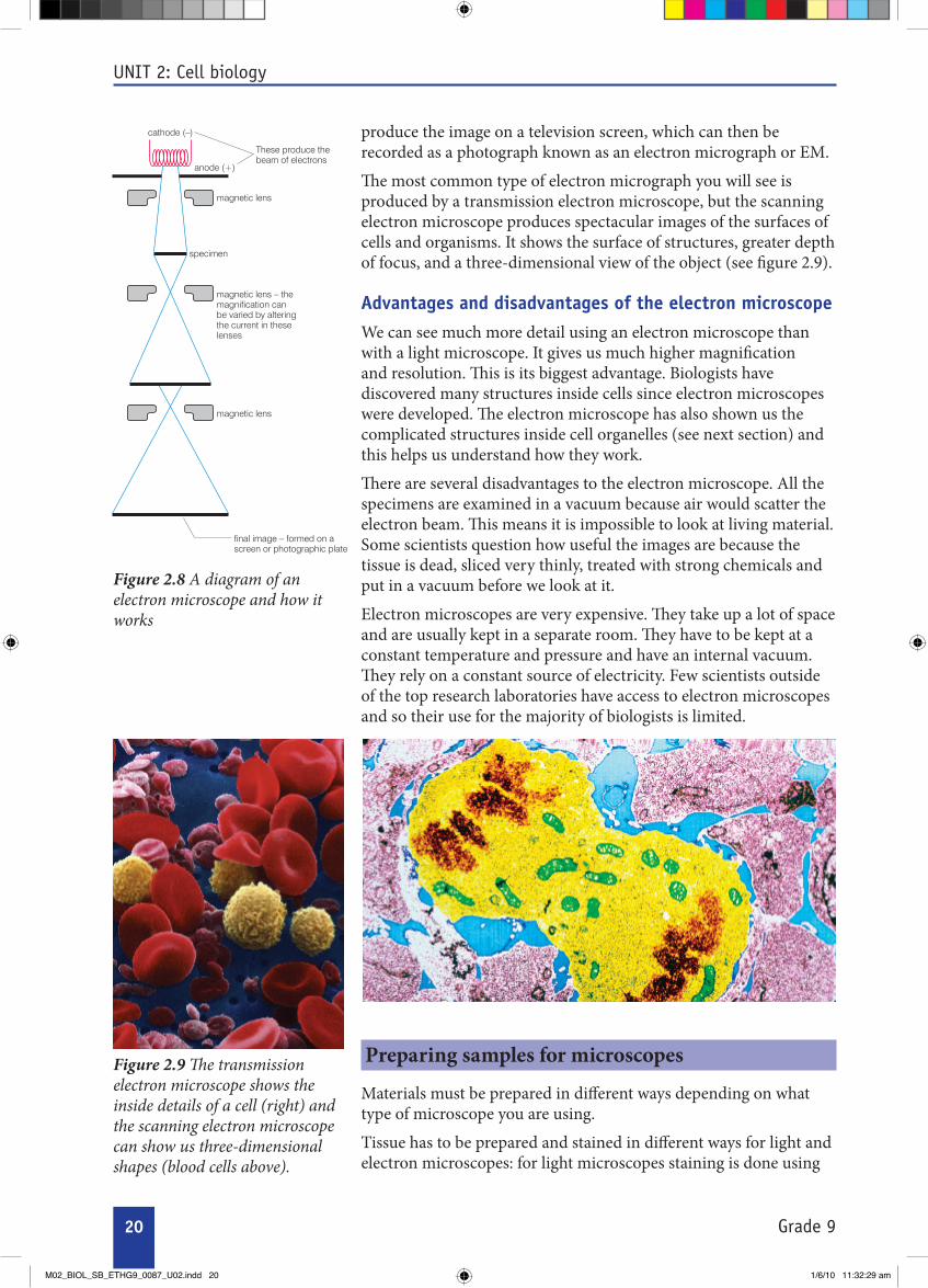
Figure 2.8 A diagram of an electron microscope and how it works
These produce thebeam of electrons
anode (+)
magnetic lens
specimen
magnetic lens – themagnification canbe varied by alteringthe current in these lenses
magnetic lens
final image – formed on ascreen or photographic plate
cathode (–)
Figure 2.9 The transmission electron microscope shows the inside details of a cell (right) and the scanning electron microscope can show us three-dimensional shapes (blood cells above).
produce the image on a television screen, which can then be recorded as a photograph known as an electron micrograph or EM.
The most common type of electron micrograph you will see is produced by a transmission electron microscope, but the scanning electron microscope produces spectacular images of the surfaces of cells and organisms. It shows the surface of structures, greater depth of focus, and a three-dimensional view of the object (see figure 2.9).
Advantages and disadvantages of the electron microscopeWe can see much more detail using an electron microscope than with a light microscope. It gives us much higher magnification and resolution. This is its biggest advantage. Biologists have discovered many structures inside cells since electron microscopes were developed. The electron microscope has also shown us the complicated structures inside cell organelles (see next section) and this helps us understand how they work.
There are several disadvantages to the electron microscope. All the specimens are examined in a vacuum because air would scatter the electron beam. This means it is impossible to look at living material. Some scientists question how useful the images are because the tissue is dead, sliced very thinly, treated with strong chemicals and put in a vacuum before we look at it.
Electron microscopes are very expensive. They take up a lot of space and are usually kept in a separate room. They have to be kept at a constant temperature and pressure and have an internal vacuum. They rely on a constant source of electricity. Few scientists outside of the top research laboratories have access to electron microscopes and so their use for the majority of biologists is limited.
Preparing samples for microscopes
Materials must be prepared in different ways depending on what type of microscope you are using.
Tissue has to be prepared and stained in different ways for light and electron microscopes: for light microscopes staining is done using
M02_BIOL_SB_ETHG9_0087_U02.indd 20 1/6/10 11:32:29 am

21Grade 9
UNIT 2: Cell biology
Inthissectionyouhavelearntthat:
Lightmicroscopesandelectronmicroscopes•are widely used by biologists.
Microscopesmagnifybothlivinganddead•tissuesoyoucanobservethefeaturesofthe cells and tissue.
Magnificationinvolvesincreasingthesize•ofanobject.Toworkoutthemagnificationof a microscope you multiply the magnificationoftheobjectivelensbythemagnification of the eyepiece lens.
Resolution is the ability to distinguish •between two separate points.
Theresolvingpowerofamicroscopeis•dependentonthewavelengthused,sotheresolvingpowerofanelectronmicroscope
is around 1000 times greater than the resolvingpowerofalightmicroscope.
Usingalightmicroscopetakesskilland•practice.
Deadspecimensarefixed,stainedand•sliced before mounting on slides to be observedunderthemicroscope.Livingspecimens are mounted on slides and stains may be added.
Stainsareusedtomakepartsofcells(e.g.•the nucleus) or types of cells show up better under the microscope.
Tissue has to be prepared carefully before •it can be used in the electron microscope. only dead tissue can be used in the electron microscope.
Summary
Review questionsSelect the correct answer from A to D.
coloured dyes to reflect light, whereas for electron microscopes heavy metals such as lead and uranium are used to reflect electrons. For light microscopes only non-living materials need fixation, while living materials are not fixed: specimens are always fixed with electron microscopes.
1. The maximum magnification of a light microscope would make a person:
A 3.5 m tall B 35 m tall C 3.5 km tall D 35 km tall2. The largest single cell is: A an amoeba B a jelly fish C an unfertilised ostrich egg D an unfertilised human egg
3. Which of the following is not an advantage of the light microscope?
A It can be used anywhere without electricity.
B Its resolving power is limited by the wavelength of light.
C It is relatively light so can be carried out into the field for research.
D It is relatively cheap.4. Which of the following is the main advantage
of the electron microscope? A It’s very expensive. B Specimens are examined in a vacuum so
must be dead. C It needs a constant temperature and
pressure. D It gives a greatly increased magnification
and resolution over the light microscope.
M02_BIOL_SB_ETHG9_0087_U02.indd 21 1/6/10 11:32:29 am

22 Grade 9
UNIT 2: Cell biology
2.2 The cell
By the end of this section you should be able to:
• State the cell theory.
• Listthestructuresofcellsanddescribetheirfunction.
• Draw and label diagrams and compare typical plant and animal cells.
• Describethetypes,shapesandsizesofavarietyofcells using diagrams.
The planet we live on is covered with a wide variety of living organisms, including animals, plants and microbes. All living organisms are made up of units called cells. Some organisms, such as amoeba, consist of single cells. Others, such as ourselves, are made up of many millions of cells all working together. Organisms that contain more than one cell are known as multicellular.
Cell theory Cells were first seen over 300 years ago. In 1665, the English scientist Robert Hooke designed and put together one of the first working optical microscopes. He examined many different things including thin sections of cork. Hooke saw that these sections were made up of many tiny, regular compartments, which he called cells.
It took many years of further work for the importance of cells to be recognised. In 1839 Matthias Schleiden and Theodore Schwann introduced an idea known as the cell theory. The cell theory states that cells are the basic units of life and by the 1840s this idea was accepted by most biologists.
All living organisms have certain characteristics, which they carry out regardless of whether they have one cell or millions. When we look at cells we can see how all of these functions are carried out.
The seven life processes that are common to most living organisms are:
Nutrition• – all living organisms need food to provide them with the energy used by their cells. Plants make their own food by photosynthesis, whereas animals eat other organisms.
Respiration • – the process by which living organisms get the energy from their food.
Excretion• – getting rid of the waste products produced by the cells.
Growth• – living organisms get bigger. They increase in both size and mass, using chemicals from their food to build new material.
Irritability• – all living organisms are sensitive to changes in their surroundings.
Figure 2.10 An organism like this Paramecium carries out all the characteristic reactions of life within a single cell.
KEY WORDS
cells the basic structural and functional units in all living organismscell theory states that cells are the basic units of lifenutrition food substances needed by the bodyrespiration process whereby living organisms obtain energy from their foodexcretion removal of poisonous waste products produced by cellsgrowth increase in size and mass of an organism
M02_BIOL_SB_ETHG9_0087_U02.indd 22 1/6/10 11:32:30 am

23Grade 9
UNIT 2: Cell biology
Movement• – all living organisms need to move to get near to things they need or away from problems. Animals move using muscles, plants move more slowly using growth.
Reproduction• – producing offspring is vital to the long-term survival of any type of living organism.
Cell structures and functions
There are some basic similarities between all cells, animal and plant alike. For example, almost all cells have a nucleus, a cell membrane, mitochondria, ribosomes, endoplasmic reticulum and cytoplasm. Other features are often seen in plant cells, particularly from the green parts of the plants, but not in animal cells. This has led scientists to develop a picture of the basic structure of an unspecialised animal cell and an unspecialised green plant cell. Although there are not many cells which are quite this simple, the idea of unspecialised animal and plant cells gives us a very useful base point with which to compare other, more specialised cells.
Structures and functions in unspecialised animal cells
All cells have some features in common and we can see them clearly in typical unspecialised animal cells (like the ones on the inside of your cheek). They contain small units called organelles. Many of these organelles contain enzymes and chemicals to carry out specialised jobs within the cell.
The • nucleus controls all the activities of the cell. It also contains the instructions for making new cells or new organisms in the form of long threads known as chromosomes. This is the genetic material. You will find out more about this in Grade 10.
The • cytoplasm is a liquid gel in which most of the chemical reactions needed for life take place. About 70% of the cytoplasm of a cell is actually water! The cytoplasm contains all the other organelles of the cell where most of the chemical reactions take place.
The • cell membrane forms a barrier like a very thin ‘skin’ around the outside of the cell. The membrane controls the passage of substances such as carbon dioxide, oxygen and water in and out of the cell. Because it lets some substances through but not others it is known as selectively permeable.
The • mitochondria (singular: mitochondrion) are the powerhouse of the cell. They carry out most of the reactions of respiration, whereby energy is released from the food in a form your cells can use. Whenever cells need a lot of energy – such as muscle cells and secreting cells – you will see a lot of mitochondria.
KEY WORDS
irritability sensitivity of an organism to changes in surroundingsmovement the need to get near to or away from thingsreproduction the production of offspring to ensure the survival of a type of organismnucleus controls all cell activity and contains chromosomescell membrane outer layer of living cell that controls the movement of substances in and outmitochondria carry out cellular respirationribosomes organelles involved in protein synthesisendoplasmic reticulum links the nucleus of a cell with the cell membranecytoplasm liquid gel which contains all the organelles of a cellorganelles the small units inside a cellchromosome strand of DNA carrying genetic information
DID yoU kNow?
Human beings contain an enormous number of cells. Estimates range from 10 million million cells (1012) to 100 million million (10012) cells – no one has counted accurately!
M02_BIOL_SB_ETHG9_0087_U02.indd 23 1/6/10 11:32:30 am

24 Grade 9
Figure 2.11 A simple animal cell like this shows the features that are common to almost all living cells. Mitochondria, endoplasmic reticulum and ribosomes cannot be seen easily with a light microscope. They are much clearer using an electron microscope.
Human cheek cells
nucleus cell membrane
mitochondria
cytoplasm
Figure 2.12 Micrograph and a drawing of simple cuboidal epithelial cells
Activity 2.3: Using the microscope to look at animal cells
nucleus
cell membrane
cytoplasm
×400
UNIT 2: Cell biology
The • endoplasmic reticulum is a three-dimensional system of tubules that spreads right through the cytoplasm. It links the nucleus with the cell membrane.
The • ribosomes are found on the endoplasmic reticulum in your cells. They are vital for protein synthesis, the process by which your body makes all the enzymes that control the reactions of your cells.
you will need:
• amicroscope
• alamp
• preparedmicroscopeslidesofhumancheekcells/ epidermal cells
Method
Remember, microscopes are expensive and delicate pieces of equipment so always take care of them and handle them safely.
1.Usetheinstructionsforusingthemicroscope,whichyoulearntintheprevioussection.Youwillbeprovidedwithslidesofhumancheekcellsandsimpleepithelialcells.
2.Humancheekcellsandsimpleepithelialcellsareverysimilar to your diagram of an unspecialised animal cell. Draw some of the cells you see and label them as well as you can. Remember you will NoT see ribosomes and mitochondria under normal light microscopes.
M02_BIOL_SB_ETHG9_0087_U02.indd 24 1/6/10 11:32:31 am

25Grade 9
UNIT 2: Cell biology
Why do cells have organelles?All of the processes of life take place within a single cell. Imagine 100 mixed reactions going on in a laboratory test tube – chemical chaos and probably a few explosions would be the result! But this is the level of chemical activity going on in a cell at any one time. Cell chemistry works because each reaction is controlled by an enzyme, a protein designed to control the rate of a very specifi c reaction and ensure that it takes place without becoming mixed up with any other reaction. What is more, the enzymes involved in diff erent chemical processes are usually found in diff erent parts of the cell. So, for example, most of the enzymes controlling the reactions of respiration are found in the mitochondria. Th e enzymes controlling the reactions of photosynthesis are found in the chloroplasts and the enzymes involved in protein synthesis are found on the surface of the ribosomes. Th ese cell compartments or organelles help to keep your cell chemistry well under control.
Structures and functions in unspecialised plant cellsPlants are very diff erent from animals – they do not move their whole bodies about and they make their own food by photosynthesis. So, whereas plant cells have all the features of a typical animal cell – nucleus, cell membrane, cytoplasm, mitochondria, endoplasmic reticulum and ribosomes – they also have structures that are needed for their very diff erent way of life.
Th e cell wall is made mainly of a carbohydrate called cellulose, which strengthens the cell and gives it support. It is found outside the cell membrane. Th e cell wall structure contains large holes so substances can move freely through it in either direction – it is freely permeable.
Many (but not all) plant cells also have other features.
Chloroplasts are found in all of the green parts of the plant. Th ey •contain the green pigment chlorophyll, which gives the plant its colour. As a result of the chlorophyll they can absorb energy from light to make food by photosynthesis.
A permanent • vacuole is a space in the cytoplasm fi lled with cell sap, a liquid containing sugars, mineral ions and other chemicals dissolved in water. Th e vacuole is important for keeping the cells rigid to support the plant. Th e vacuole pushes the cytoplasm against the cell wall, which keeps the whole structure fi rm. A permanent vacuole is oft en a feature of mature (adult) plant cells.
Figure 2.13 A photosynthetic plant cell has many features in common with an animal cell, but others that are unique to plants.
cell wallcell membrane (inside cell wall) vacuole
cytoplasmmitochondria
chloroplastsnucleus
cell wall (inside cell wall)(inside cell wall)(inside cell wall)
nucleus
mitochondriamitochondriamitochondria
chloroplastschloroplastschloroplastschloroplastschloroplastschloroplasts
cytoplasm
chloroplastschloroplastschloroplasts
KEY WORDS
enzyme protein molecule that acts as a catalyst in cellscell wall outer layer in plant cells and bacteria that is freely permeablecellulose complex carbohydrate that makes up plant cell wallsvacuole a fl uid-fi lled cavity inside a cell
M02_BIOL_SB_ETHG9_0087_U02.indd 25 1/6/10 11:32:31 am

26 Grade 9
UNIT 2: Cell biology
Thepreparedslidesyouhavelookedatshowanimal cells that are dead and stained to makethemeasiertosee.Inthisactivityyouaregoingtolookatoneofanumberof different types of plant cells – either (a) onion(asyouusedintheprevioussection),(b) red pepper or (c) pondweed.
you will need:• amicroscope• microscopeslides• coverslips• forceps• mountedneedles• pipette• alamp• apieceof(a)onion,(b)redpepperor
(c)pondweed,e.g.Elodea or Canadian pondweed
Method
Remember, microscopes are expensive and delicate pieces of equipment so always take care of them and handle them safely.
Activity (a) – onion cells
onion cells do not contain any chlorophyll so theyarenotcoloured.Youcanlookatthemastheyare,orstainthemusingiodine,whichreacts with the starch in the cells and turns blue-black.
1.Takeyourpieceofonionandremoveasmall piece of the epidermis using your forceps. Use the method for preparing a slidegivenintheprevioussection.Youmay use iodine to stain the cells.
2.Removeanyexcessliquidfromtheslide using tissues and place under the microscope.
3.Startingwiththelowpowerlens,followtheprocedureforlookingatcellsdescribedon pages 16–17. Use the higher power lensestolookatthecellsinasmuchdetailaspossible.Thenmakealabelleddrawingofseveralofthecellsyoucansee.
Activity (b) – red pepper
Repeat the instructions for the onion cells exceptthistimeremoveathinepidermallayer of the pepper. Again these cells do not containchlorophyll,buttheyareredsoyoudo not need to use iodine on them.
Activity (c) – pondweed such as Elodea (Canadian pondweed)
These plant cells contain chloroplasts. If you watchverycarefullywhenyouhavethecellsunder a high power of magnification you may wellseethechloroplastsmovingaboutinthelivingcytoplasmofthecell.
1.Takeasingleleaffromapieceofpondweedandcutaverysmallsectionabout 2 mm2.
2. Place the leaf sample onto a microscope slide and add a drop of water.
3.Usingthemountedneedle(orapencil!)lowerthecoverslipverygentlyoverthespecimen,takingcarenottotrapairbubbles.
4.Removeanyexcessliquidfromtheslide using tissues and place under the microscope. Starting with the low power lens,followtheprocedureforlookingatcells described on pages 16–17.
5.Usethehigherpowerlensestolookatthecells in as much detail as possible. Then makealabelleddrawingofseveralofthecells you can see.
a) low power (x250)
b) high power lenses (x1260)
Activity 2.4: Making a slide of plant cells
Figure 2.14 Micrographs of Elodea cells under:
M02_BIOL_SB_ETHG9_0087_U02.indd 26 1/6/10 11:32:32 am

27Grade 9
UNIT 2: Cell biology
Cell specialisation in humans
Looking at the structure of simple unspecialised cells gives us a good basic understanding of how a cell works. But in multicellular organisms like human beings, most cells become specialised – that is, they are adapted to carry out a particular function in your body.
When an egg and a sperm combine to form an embryo, a single cell is formed. This cell divides many times (you will learn more about this in Grade 10) to form a mass of similar undifferentiated cells. Each of these cells (known as embryonic stem cells) carries all of the genetic information of the individual. As the embryo develops, the cells become differentiated – they specialise to carry out a particular function. For example, some cells differentiate to become red blood cells and carry oxygen, some become muscle cells and others become neurones (nerve cells). This differentiation takes place as some of the genetic material (genes) in the nucleus of the cells is switched on and others are switched off. Scientists are still not quite sure what causes these changes to take place, but it seems to be at least partly down to the position of the cells in the embryo itself.
The specialised cells which form as cells differentiate are often grouped together to form a tissue – for example in humans
KEY WORDS
specialised cells adapted to carry out a particular bodily functionundifferentiated cells cells that have not yet assumed their final functional characteristicsembryonic stem cells cells from the early embryo that have the potential to form almost any other type of cell differentiated cells special cells which carry out specific functions red blood cells types of blood cell that carry oxygen around the bodyneurones nerve cellsgenes units of inheritancetissue a group of cells that performs specific functions
DID yoU kNow?
Some scientists are working with human embryonic stem cells in an attempt to grow new adult tissues. The hope is that these can be used to replace diseased tissue in people with serious illnesses. So far progress is slow, partly because scientists are not sure how to persuade human embryonic stem cells to differentiate into the tissues they want, and partly because there are ethical issues about using cells from human embryos.
Figure 2.15 Within an organ like the pancreas at least two very different tissues can be seen. The cells in each type of tissue are specialised to make a very different chemical product, and so they take up stains differently, which allows them to be seen.
a) The cells that are stained pink make hormones that help to control the sugar levels in the blood.
b) The cells that are stained red make enzymes needed to digest the food in the gut.
cell
tissue (lung tissue)
organ (lung)
organ system (respiratory system)
body
Figure 2.16 Large living organisms have many levels of organisation. As a result, each part of the body is perfectly adapted to carry out its functions.
a
b
M02_BIOL_SB_ETHG9_0087_U02.indd 27 1/6/10 11:32:32 am

28 Grade 9
UNIT 2: Cell biology
KEY WORDS
organ a part of the body that carries out special functions epithelial cells cells arranged in one or more layers to form part of a covering or lining of a body surfacealveoli microscopic air sacs in the lungs with a large surface areamicrovilli minute hair-like structures that increase the surface area of a cellmeiosis cell division that reduces the chromosome numbers and forms the sex cellssperm male sex cellacrosome a thin sac at the head of a sperm cell containing enzymes which dissolve the protective layers of an egg cell
squamous epithelium (flattened cells)
cuboidal epithelium ciliated columnar epithelium
Figure 2.17 Epithelial cells are found all over your body, lining the body spaces, organs and tubes inside you as well as forming your skin.
connective tissue joins bits of the body together, while nervous tissue carries information around the body and muscle tissue contracts to move the body about.
In many living organisms, including people, there is another level of organisation. Several different tissues work together to do different jobs and form an organ such as the heart, the kidneys or the lungs. In turn different organs are combined in organ systems to carry out major functions in the body such as transporting the blood or reproduction. Examples include the cardiovascular system (the heart, lungs and blood vessels) and the digestive system.
Specialised cellsWhen cells become specialised to carry out one main function as part of a tissue or organ their structure is often very different to that of a ‘typical’ plant or animal cell. The structure is modified or adapted to suit the very specialised job the cell is doing. For example, cells that use a lot of energy have many mitochondria, whereas cells that are important for diffusion will have a large surface area and cells that produce lots of proteins have many ribosomes as well as mitochondria.
By looking carefully at specialised cells you can see how their structure is adapted to their function. Below are some examples of the specialised cells you will find in the human body.
Epithelial cellsSometimes the specialisation is not to be very specialised! Epithelial cells play many very important roles in the human body. They are usually arranged in thin sheets of epithelial tissue (which are often only one cell thick) and they cover your internal and external surfaces. So your skin is made up of epithelial cells, and your gut, your respiratory system, your reproductive system and many other organ systems of your body are all lined with epithelial cells.
Epithelial cells have many different functions, and their basic structure may be adapted to make them more efficient at their job. Many epithelial cells are there to protect the tissues underneath from damage or infection. They have a very basic simple cell structure, such as in your skin. Epithelial cells often allow the diffusion of materials from one area of your body to another – they line the air sacs (alveoli) of your lungs, and the gut wall as well as many glands which secrete hormones or enzymes. Epithelial cells may be flattened, thin columns or have tiny, hair-like projections known as microvilli on them to increase the surface area of the cell. This is seen in places like the gut, where diffusion is very important. Some epithelial cells found in the respiratory and the reproductive systems have small hair-like cilia, which move and beat and can be used to move substances through a tube. In your airways ciliated epithelial cells move mucus and microbes away from your lungs, whereas in the female reproductive system ciliated epithelium helps to move the ovum towards the uterus.
M02_BIOL_SB_ETHG9_0087_U02.indd 28 1/6/10 11:32:33 am

29Grade 9
UNIT 2: Cell biology
Reproductive cells – the eggs and sperm Th e reproductive cells in your body are specialised to perform very diff erent functions when you reproduce (you will learn more about this in Grade 10). If you are female your body contains ova or egg cells found in your ovaries. Th ese female sex cells have only half the number of chromosomes found in normal body cells (you will learn more on meiosis in Grade 10, a form of cell division that halves the number of chromosomes in your cells). Egg cells have a large nucleus containing genetic information from the woman. Th ey have a protective outer coat to make sure only one sperm gets through to fertilise the egg, and a store of food in the cytoplasm for the developing embryo. In humans, this food store is quite small, but in animals such as birds it is very large – it forms the yolk of the egg.
A small number of relatively large egg cells are released by the ovaries over a woman’s reproductive life.
If you are male, once you have gone through puberty your body will produce millions of male sex cells known as sperm. Like the egg cells, sperm have only half the chromosome number of normal body cells. Sperm cells are usually released a long way from the egg they are going to fertilise. Th ey need to move through the female reproductive system to reach an egg. Th en they need to be able to break into the egg. Th ey have several adaptations to make all this possible. Sperm have long tails containing muscle-like proteins so they can swim towards the egg. Th e middle section of a sperm is full of mitochondria which provide the energy for the tail to work. Th ey have a special sac known as the acrosome, which stores digestive enzymes used for breaking down the outer layers of the egg. Finally, the sperm has a large nucleus containing the genetic information to be passed on to the off spring. Sperm cells are much smaller than egg cells, but they are produced in their millions every day.
Nerve cells (neurones)Nerve cells or neurones are part of the communication and control system of your body. Electrical nerve impulses pass along them at great speed carrying information from one part of your body to another. So, nerve cells have some clearly specialised features that make this possible. Whatever the type of nerve cell, they have a cell
Figure 2.18 a) Egg and b) sperm cells show very clear adaptations to their much specialised functions.
protective outer coat of cells
large nucleus
cytoplasm containing small food store
large nucleus
cytoplasm containing small food storecytoplasm containing small food store
head
mid-piece
tail(fl agellum)
for propulsion
sac of enzymes to penetrate membrane around ovum
nucleus
mitochondria to release energy for movement
sac of enzymes to penetrate to penetrate membrane around ovumaround ovumaround ovum
nucleus
mitochondria to release
nucleus
a)
b)
Figure 2.19 Nerve cells are very diff erent from epithelial cells – but they play a very diff erent role in your body.
a)
nucleusnucleuscell bodycell body
dendritesaxon
myelin sheath
muscle fi bre
myelin sheath
muscle fi bre
b)
M02_BIOL_SB_ETHG9_0087_U02.indd 29 1/6/10 11:32:35 am

30
UNIT 2: Cell biology
Grade 9
KEY WORDS
dendrites short, arm-like protuberances of a nerve cellaxon extension of a neuron that transmits messages
cells
stripes caused by arrangement of actin and myosin
nucleus
cells
nucleus
stripes caused by arrangement of actin and myosin
Figure 2.20 Striated muscle has dark and light stripes under the microscope.
KEY WORDS
muscle cells elongated contractile cells that form musclemuscle fi bres strands of protein that enable muscles to contract
body containing the nucleus, dendrites that communicate with neighbouring nerve cells and nerve fi bres (called the axon) that can carry the nerve impulse long distances. Th e nerve fi bres are oft en covered by a protective myelin sheath that allows the nerve impulses to travel faster. You will fi nd out more about nerve cells in Grade 10.
Muscle cellsYour muscles are responsible for movement in your body. Th ey are made up of very specialised muscle cells or muscle fi bres. Th ese are very long thin (elongated) cells which can contract and relax. When they are relaxed they can be stretched, and when they contract they shorten powerfully. Th e muscle cells contain two proteins, actin and myosin, and it is these which enable the muscle cells to contract. Th e most common muscle in the body is striated or striped muscle, and these proteins are arranged so that the muscle cell looks striped. Muscle cells also contain lots of mitochondria which provide the energy for them to contract. Th e muscle cells are always found in bundles and they all contract together. You will fi nd out more about muscle and its role in your body in Grade 10.
All living cells carry out the characteristic functions of life. As a result, they all have some features in common. But as you have seen there are many ways in which cells become specialised to carry out particular functions in your body. As you study more about the way the human body works in this book, you will discover more examples of specialised cells and their importance in the healthy functioning of your body.
Activity 2.5: Observing different human cellsyou will need:
• amicroscope
• alamp
• preparedmicroscopeslidesofdifferenthumancells–ciliatedepithelia,nervoustissue,musclefibres,spermandovaifpossible
Method
Remember, microscopes are expensive and delicate pieces of equipment so always take care of them and handle them safely.
you will not see mitochondria and ribosomes with a light microscope.
1. Set up your microscope with the lowest power lens (the smallest lens) in place.
2. Clip a prepared slide into place on the stage using the stage clipsandobserveitusingthemicroscopeasdescribedinactivity2.1onpages16–17.
M02_BIOL_SB_ETHG9_0087_U02.indd 30 1/6/10 11:32:36 am

31
UNIT 2: Cell biology
Grade 9
3.Tousethehighermagnifications,rotatethenosepiecesothatthenextlensclicksintoplace.DONOTTOUCHthefocusingknobsasthespecimenshouldstillbeinfocusand–withthecoarsefocusingknobinparticular–itisveryeasytobreaktheslide.Ifyoudoneedtoadjustthefocus,usethefinefocusingknobonlyandthehigherpowersofmagnification.Takegreatcaretoavoidlettingthelenstouchtheslide.Youmaywanttoadjusttheirisdiaphragmas well.
4.Ittakestimetobeabletounderstandandinterpretwhatyou see under the microscope – the cells you see won’t be asclearasthediagramsinyourbooksbecausethosehavebeendrawnbyexpertsusingthebestpossiblespecimens!Draw some of the cells you see and label them as well as you can. Remember you will NoT see ribosomes and mitochondria under normal light microscopes.
Summary
Inthissectionyouhavelearntthat:
Alllivingorganismsarebasedonunitsknownascells.•
Therearesevenlifeprocessesthatarecommontoallliving•organisms:nutrition,respiration,excretion,growth,irritability(responsetostimuli),movementandreproduction.
Unspecialisedanimalcellsallhavethefollowingstructures•andorganelles:acellmembrane,cytoplasm,nucleus,mitochondria,endoplasmicreticulumandribosomes.Eachof these has a characteristic structure and carries out clear functionsintheworkingofthecell.
Unspecialisedplantcellsallhavethesamebasicstructures•andorganellesasananimalcell.Inaddition,theyhaveacellulosecellwall,andmayhaveapermanentvacuole.Inthegreenpartsofaplantallthecellscontainchloroplasts,which in turn contain chlorophyll. Each of these has a characteristic structure and carries out clear functions in the workingofthecell.
Figure 2.21 Micrographs of specialised cells
ciliated epithelium motor neurone spermatozoa
M02_BIOL_SB_ETHG9_0087_U02.indd 31 1/6/10 11:32:36 am

32
UNIT 2: Cell biology
Grade 9
KEY WORDS
diffusion movement of particles from an area of high concentration to an area of low concentration along a concentration gradientosmosis movement of water from an area of high concentration to an area of low concentration along a concentration gradient through a partially permeable membraneactive transport movement of substances against a concentration gradient using energy from respirationconcentration a way of measuring how many particles of a substance are in one place
Inmulticellularorganismslikehumanbeingsthecellsofthe•embryodifferentiatetoformspecialisedcells,whichcarryout particular functions in the body.
Cells specialised to carry out a particular function are •groupedtogethertoformatissue,tissuesgrouptogethertoformanorgan,severaldifferentorgansworkingtogetherformanorgansystemandorgansystemsworkingtogethermakeupthebodyofacomplexmulticellularorganism.
There are many different specialised cells in the human body. •Theyincludeepithelialcells,spermcells,eggcells,nervecellsandmusclecells.Acloselookattheirspecialisationshows how they are adapted to their functions.
Review questionsSelect the correct answer from A to D.
1. Which of the following is not an organelle within a cell?
A nucleus
B chloroplast
C mitochondria
D cytoplasm
2. Which of the following is not one of the seven life processes that characterise living things?
A movement
B language
C reproduction
D respiration
3. One of these is a tissue in the human body. Which one?
A heart
B stomach
C muscle
D uterus
M02_BIOL_SB_ETHG9_0087_U02.indd 32 1/6/10 11:32:36 am

33
UNIT 2: Cell biology
Grade 9
Your cells need to take in substances, such as oxygen and glucose, and to get rid of waste products and chemicals that are needed elsewhere in your body. In human beings, just like all other living organisms, dissolved substances can move into and out of your cells across the cell membrane in three different ways – by diffusion, by osmosis and by active transport. In this section you will look at each of these methods of transport in turn.
Diffusion When you go home from school you will probably know if there is a meal cooking before you get there. How? Because the smell reaches you by diffusion. Diffusion happens when the particles of a gas, or any substance in solution, spread out.
Diffusion is the net (overall) movement of particles from an area of high concentration to an area of lower concentration. Concentration is a way of measuring how much (how many particles) of a substance is in one place.
Diffusion takes place because of the random movements of the particles of a gas or of a substance in solution in water. All the particles are moving and bumping into each other and this moves them all around. Although the molecules are moving in both directions, there are more particles moving in the area of high concentration, and so the net (overall) movement is away from the area of high concentration towards the area of low concentration.
Imagine an empty classroom containing a group of boys and a group of girls. If everyone closes their eyes and moves around briskly but randomly, people will bump into each other and scatter until the room contains a mixture of boys and girls. This gives you a good working model of diffusion.
Figure 2.22 The blood from an injured fish or animal will spread through the water by diffusion. Fish like this piranha will follow the trail of diffusing blood to some easy prey!
2.3 The cell and its environment
By the end of this section you should be able to:
• Describe the permeability of the cell membrane.
• Describetheprocessofdiffusionanditsimportanceinlivingorganisms.
• Demonstrate diffusion experimentally.
• Explaintheprocessofosmosisanditsimportanceinlivingorganisms.
• Demonstrate osmosis experimentally.
• Show that plant cells become flaccid when they lose water and turgid when they absorb water by osmosis.
• Explain plasmolysis and turgor pressure.
• Explainpassiveandactivetransportacrosscellmembranes.
• Discusstheadvantagesanddisadvantagesofdiffusion,osmosisandactivetransportformovingsubstancesintoandoutofcells.
DID yoU kNow?
Some sharks can sense one part blood in 10 or even 100 million parts of sea water – it’s not a good idea to bleed in the sea!
M02_BIOL_SB_ETHG9_0087_U02.indd 33 1/6/10 11:32:37 am

34
UNIT 2: Cell biology
Grade 9
At the moment of adding blue particles to yellow mixture they are not mixed at all.
As the particles move randomly the blue ones begin
to mix with the yellow ones.
As the particles move and spread they bump into each other and keep spreading as a result of all
the random movement.
Eventually the particles are completely mixed and diffusion
is complete.
Figure 2.23 The random movement of particles results in substances spreading out or diffusing from an area of higher concentration to an area of lower concentration.
Activity 2.6: Demonstrating diffusionyou will need:
• stopwatchortimer
Method
Ifyourclassroomorschoolyardissuitable,trytheideadescribed on page 33.
1. All the boys stand in one corner (a high concentration of boys). All the girls stand in another corner (a high concentration of girls).
2.Yourteacherstartsatimerfor30secondsandyoumovearound slowly with your eyes closed until the timer tells you to stop.
3.Openyoureyesandobservewhatishappening,thenstartthetimeragainandmoveslowlywithyoureyesclosedagain.Repeatuntiltheareacontainsanevenmixtureofboys and girls.
Activity 2.7: Detecting diffusionyou will need:
• stopwatch or timer
Method
1. your teacher will open a bottle of a strongly scented chemical such as ammonia or spray some perfume at the front of your class.
2.Starttimingasthesprayisreleased,andthenputyourhandupwhenyoucansmellthescent.You’llseeawaveofhandsmovingfromthefronttothebackandsidesoftheclass as the molecules spread out by diffusion.
3.Timehowlongittakestoreachthelastperson.
Rates of diffusionDiffusion is a relatively slow process. A number of different factors affect the rate at which it takes place.
If there is a big difference in concentration between two areas, diffusion will take place quickly. However, when a substance is moving from a higher concentration to one which is just a bit lower, the movement towards the less concentrated area will appear to be quite small. This is because although some particles move into the area of lower concentration by random movement, at the same time other identical particles are leaving that area by random movement.
M02_BIOL_SB_ETHG9_0087_U02.indd 34 1/6/10 11:32:39 am

35
UNIT 2: Cell biology
Grade 9
the overall or net movement = particles moving in – particles moving out
In general the bigger the difference in concentration the faster the rate of diffusion will be. This difference between two areas of concentration is called the concentration gradient and the bigger the difference the steeper the gradient will be.
Concentration isn’t the only thing that affects the rate of diffusion. An increase in temperature means the particles in a gas or a solution move more quickly. This in turn means diffusion will take place more rapidly as the random movement of the particles speeds up.
Always remember that diffusion is passive – it takes place along a concentration gradient from high to low concentration and uses up no energy.
potassium permanganate particles
potassium permanganate crystal
Con
cent
ratio
n of
pot
assi
um
per
man
gan
ate
Time
Figure 2.24 This diagram shows how the overall movement of particles in a particular direction is more effective if there is a big difference (a steep concentration gradient) between the two areas. This is why so many body systems are adapted to maintain steep concentration gradients.
KEY WORDS
net amount remaining after certain adjustments have been madeconcentration gradient difference between an area of high concentration and an area of low concentrationpassive uses no energy
Potassium manganate(VII) (potassium permanganate) forms purple crystals that dissolveinwater.Thisactivitydemonstratesboth simple diffusion and the impact of temperature on the rate of diffusion.
you will need:
• twoidenticalbeakers(100,200or 250 cm3)
• coldwater
• warm/hotwater
• twocrystalsofpotassiummanganate(VII)
• astopwatch
Method
1.Halffillonebeakerwithcoldwater.
2. Put exactly the same amount of warm orhotwaterinthesecondbeaker. (N.B.ifthewaterishot,becarefulhowyou handle it.)
3. Drop a crystal of potassium manganate(VII)ineachbeakeratthesame time. Simultaneously start the stop- watch.
4.Timehowlongittakesthepurplecolourtoreachdifferentpointsinyourbeaker,and(ifpossible)thetotaltimeittakesfortheliquid to become purple.
5.Writeupyourinvestigationandexplainyour results in terms of diffusion and the effectoftemperatureonthemovementofparticles.
Activity 2.8: The effect of temperature on diffusion
M02_BIOL_SB_ETHG9_0087_U02.indd 35 1/6/10 11:32:40 am

36
UNIT 2: Cell biology
Grade 9
Diffusion in living organismsMany important substances can move across your cell membranes by diff usion. Th e oxygen that you need for respiration passes easily from the air into your lungs into your blood and then into your body cells by diff usion. Similarly the waste carbon dioxide produced by your cells passes out easily by diff usion. Simple sugars like glucose and amino acids from the breakdown of proteins in your gut can also pass through your cell membranes by diff usion.
Because diff usion can be a relatively slow process, individual cells may be adapted to make diff usion easier and more rapid. As movement of substances into and out of cells takes place across the cell membrane, the most common adaptation is to increase the surface area of the cell membrane over which diff usion occurs. Only so many particles of a substance such as oxygen can diff use over a given surface area, so increasing the surface area means there is more room for diff usion to take place. By folding up the membrane of a cell the area over which diff usion can take place is greatly increased and so the amount of substance moved by diff usion is also greatly increased. Tissues and organs show similar adaptations to make sure that diff usion takes place as quickly as possible – the air sacs of the lungs, the villi of the small intestine and the thin, fl at leaves of plants are just three examples of this.
osmosis Diff usion takes place where particles can spread freely from one place to another. However, the solutions inside a cell are separated from those outside the cell by the cell membrane, which does not let all types of particles through. Only the smallest particles can pass through freely. Because the membrane only lets some types of particles through, it is known as partially permeable.
Partially permeable cell membranes allow water to move across them. It is important to remember that a dilute solution of (for example) sugar contains a high concentration of water (the solvent) and a low concentration of sugar (the solute). A concentrated sugar solution contains a relatively low concentration of water and a high concentration of sugar.
Osmosis is a special type of diff usion where only water moves across a partially permeable membrane, from an area of high concentration of water to an area of lower concentration of water.
A cell is basically a solution inside a partially permeable bag (the cell membrane). Th e cell contents contain a fairly concentrated solution of salts and sugars. Water moves from a high concentration of water particles (a dilute solution) to a less concentrated solution of water particles (a concentrated solution) across the membrane of the cell. Th e sugars and salts cannot cross the membrane. In other words, osmosis takes place. Take care when you defi ne osmosis. Make it clear that it is only water that is moving across the membrane, and get your concentrations right!
DID yoU kNow?
Many moths rely on diff usion to fi nd a mate. Th e female moths produce a powerful chemical known as a pheromone to attract males. Pheromone molecules spread through the air by diff usion, sometimes helped by breezes. Male moths can pick up these molecules as far as fi ve miles away from the female – and then fl y up the concentration gradient until they reach their mate!
Cells increase their surface area by infoldings of the membrane like these microvilli.microvilli.
Figure 2.25 An increase in the surface area of a cell membrane means more diff usion can take place.
KEY WORDS
partially permeable allows the passage of some substances but not allsolvent liquid in which a solute is dissolvedsolute chemical that is dissolved in a solvent
M02_BIOL_SB_ETHG9_0087_U02.indd 36 1/6/10 11:32:40 am

37
UNIT 2: Cell biology
Grade 9
The internal concentration of your cells needs to stay the same all the time for the reactions of life to take place. Yet animal and plant cells are bathed in liquid which can be at very different concentrations to the inside of the cells. This can make water move into or out of the cells by osmosis. So osmosis is very important for all living organisms, including human beings.
Cell membranes aren’t the only partially permeable membranes. There are artificial ones too, and these can be used to make a model cell (see the specimen investigation below). By changing the concentration of the solutions inside and outside your model cell, you can see exactly why osmosis is so important in living organisms – and why it is so critical if things go wrong!
There are a number of ways in which you can show how osmosis takes place in living cells. One is described for you here and written out as a specimen investigation. Other ways of investigating osmosis in cells are presented for you to try.
If the concentration of the solutions on both sides of a cell membrane are the same they are isotonic.
If the concentration of the solution on the outside of the cell membrane is higher than the concentration of the solution inside the cell it is hypertonic.
If the concentration of the solution on the outside of the cell membrane is lower than the concentration of the solution inside the cell it is hypotonic.
random movement of water molecules due to their kinetic energy
water
larger sucrose molecules cannot pass through membrane
water molecules attracted to sucrose molecules
sucrose solution
arrows show amount of water movement in each direction
Figure 2.26 This is a model of how osmosis works – with a net movement of water molecules from an area where they are in a high concentration to an area where they are at a lower concentration through a partially permeable membrane.
water molecules
sugar moleculeskey
KEY WORDS
isotonic solutions of equal solute concentrationhypertonic a solution with a greater solute concentration than anotherhypotonic a solution with a lesser solute concentration than another
M02_BIOL_SB_ETHG9_0087_U02.indd 37 1/6/10 11:32:41 am

38
UNIT 2: Cell biology
Grade 9
Apparatus
Figure 2.27 Using bags of partially permeable membrane to make model cells you can clearly see the effect of osmosis as water moves across the membrane from a dilute to a concentrated sugar solution.
Results
InsetA,theliquidlevelhadrisenuptheglasstubingandthemembrane bag was full and tight.
InsetB,theliquidlevelintheglasstubinghaddroppedandthe membrane bag was less full and soft.
Conclusion
Inamodelcell,iftheconcentrationofwaterishigheroutsidetheselectivelypermeablemembranebagthanitison theinside,waterwillmoveintothemodelcellbyosmosis (setA).Thisiswhytheliquidlevelroseandthebagfilled. If the concentration of water is higher inside the membrane bagthanoutside,waterwillleavethemodelcellbyosmosis(setB).Thisiswhytheliquidlevelfellandthebagwasemptier. This mimics what happens in real cells.
Water in partially permeable membrane bag
water
concentrated sugar solution
(hypertonic to membrane bag)
glass tubing
final level
B
glass tubing
water(hypotonic to membrane bag)
concentrated sugar solution
Sugar solution in partially permeable membrane bag
final level
A
Specimen investigation: Demonstrating osmosis in model cells
you will need:
• two sets of the equipment shown in figure 2.27 tomakemodelcellsindifferent situations
• wax crayon (coloured pencil)orsmallstickers
Method
1.InsetA,putconcentrated sucrose (sugar) solution in the Viskingtubingbag,andwater surrounding it in thebeaker.
2.InsetB,putwaterintheViskingtubingbagand concentrated sucrose solution surrounding it in thebeaker.
3.Inbothcases,markthestartingleveloftheliquid on the capillary tubing using the pencil orstickersandobservethestateoftheViskingtubing bag.
4.Leavethemodelcellsfor30 minutes or longer.
5.Observethelevelofwaterin the capillary tubing and the state of the Viskingtubing.
M02_BIOL_SB_ETHG9_0087_U02.indd 38 1/6/10 11:32:41 am

39
UNIT 2: Cell biology
Grade 9
you can use simple potatoes to demonstrate osmosis. A system that shows or measures osmosis is an osmometer.
you will need:
• threerawpotatoesandonecookedpotato(orthreerawhalfpotatoesandonecookedhalf potato)
• fourcontainerse.g.beakers,bowls
• strongsugarsolution/sugar
• water
Method
1.Takeeachpotatoorhalfpotatoandcutoneendtomakeitflat.Peelthelayerdirectlyabovetheflatend(seefigure2.28).
2. Hollow out the other end of the potato to makeacup(seefigure2.28).
3. Set up the experiment as shown in figure 2.28. A is your control.
4. In B place sugar or strong sugar solution inthecup,withwaterinthecontainer.Ifyouusesugarsolutioninthecup,markthelevelbeforeyoustarttheexperiment.
5. In C place water in the potato cup and markthelevel.Placestrongsugarsolutionin the container.
6.InDyouwillbegivenacookedpotato,oryoumustcookityourselfbeforestartingthe experiment. Place sugar or strong sugarsolutioninthecup.Markthelevel.Place water in the container.
7.Leavetheinvestigationsforseveralhoursorovernight.
8. Record your results carefully.
9.Writeuptheinvestigationandmakesomeconclusions. Explain your results in terms of osmosis.
Byvaryingthestrengthofthesugarsolutionsyou use in osmometers B and C you can investigatehowthestrengthofthesolutionaffects the rate and amount of osmosis.
Activity 2.9: Using potato cups as osmometers
(a) hollowed outpotato cup
container
peeledarea
water
(c)
strong sugar solution
water(d)
boiled potato cup
(b) sugar or strongsugar solution
water
water
sugar or strongsugar solution
Figure 2.28 Potato cup osmometers. You can use these to show that osmosis takes place.
Osmosis in animalsOsmosis is an important way of moving water in and out of the cell when needed. If a cell uses up water in its chemical reactions, the cytoplasm becomes more concentrated. The external solution is hypotonic and more water will immediately move in by osmosis. Similarly if the cytoplasm becomes too dilute because water is produced during chemical reactions. The external solution becomes hypertonic and water will leave the cell by osmosis, restoring the balance.
However osmosis can also cause some very serious problems in animal cells. If the solution outside the cell is much more dilute than the cell contents (hypotonic) then water will move into the cell by osmosis, diluting the cytoplasm. The cell will swell and may eventually burst.
On the other hand, if the solution outside the cell is much more concentrated than the cell contents (hypertonic) then water will
KEY WORD
dilute to make less concentrated
M02_BIOL_SB_ETHG9_0087_U02.indd 39 1/6/10 11:32:42 am

40
UNIT 2: Cell biology
Grade 9
move out of the cell by osmosis, the cytoplasm will become too concentrated and the cell will shrivel up. Once you understand the effect osmosis can have on cells, the importance of homeostasis and maintaining constant internal conditions will become clear. You will learn more about this later in your course.
red blood cell in a solution with the
same concentration as the cell’s contents
(isotonic)
cell placed in strong salt solution (hypertonic)
water leaves the cell by osmosis
red blood cell shrinks
water enters the cell by osmosis
(a) (b)
Figure 2.29 Keeping your body fluids at the right concentration is vital. When you realise what can happen to your red blood cells if things go wrong, you can see why!
When the concentration of your body fluids is the same as your red blood cell contents, equal amounts of water enter and leave the cell by random movement and the cell keeps its shape.
If the concentration of the solution around the red blood cells is higher than the concentration of substances inside the cell, water will leave the cell by osmosis. This makes it shrivel and shrink so it can no longer carry oxygen around your body.
If the concentration of your body fluids is lower than your red blood cell contents water enters the cells by osmosis so your red blood cells swell up, lose their shape and eventually burst!
(c)
you can show the effect on animalcellsofwatermovingin or out by osmosis with this simple experiment using eggyolks.
you will need:
• twobeakers
• water
• strongsaltsolutionmadebydissolvingsaltinwater
• twoeggyolks
Activity 2.10: How does osmosis affect animal cells?
Method
1.Fillonebeakerwithwaterandonewithstrongsaltsolution.
2.Verycarefullycrackoneeggopenandseparatetheyolkfromthewhite.Donotbreaktheyolk.Placetheyolkcarefullyinthebeakerofwater.
3.Verycarefullycracktheothereggopenandseparatetheyolkfromthewhite.Donotbreaktheyolk.Placetheyolkcarefullyinthebeakerofsaltsolution.
4.Immediatelyobservetheyolksverycarefully.Drawthemordescribewhattheylooklike.Measurethemifyoucan.
5.Leavetheyolksinthewaterandsaltsolutionforatleastanhour.Thenobserveandrecordanychangesintheappearanceoftheyolks.
6.Writeupyourexperimentandexplainwhatyouhaveobservedintermsofosmosis.
KEY WORD
homeostasis maintenance of a constant internal environment
cell placed in dilute salt solution (hypotonic)
M02_BIOL_SB_ETHG9_0087_U02.indd 40 1/6/10 11:32:45 am

41
UNIT 2: Cell biology
Grade 9
Osmosis in plantsPlants rely on osmosis to support their stems and leaves. Water moves into plant cells by osmosis, making the cytoplasm swell and press against the plant cell walls. Th e pressure builds up until no more water can enter the cell, which is hard and rigid. Th is swollen state is called turgor. It keeps the leaves and stems of the plant rigid and fi rm. So for plants it is important that the fl uid surrounding the cells always has a higher concentration of water (it is a more dilute solution of chemicals or hypotonic) than the cytoplasm of the cells, to keep osmosis working in the right direction.
To understand the diff erence between animal and plant cells when it comes to water moving in by osmosis, imagine blowing up a balloon. As more and more water moves in the balloon gets bigger and bigger and eventually bursts. Th is models an animal cell placed in pure water or a very dilute solution of salts. Now imagine a balloon sealed into a cardboard box. As the balloon infl ates it fi lls the box and then presses out against the box walls. Eventually you simply cannot force any more air into the balloon. Th e box feels very rigid and the balloon does not burst. Th is models a plant cell placed in pure water or a very dilute solution of salts.
If the surrounding fl uid becomes more concentrated than the contents of the plant cells (hypertonic), then water will leave the cells by osmosis. Th e vacuole shrinks and the cell becomes much less rigid – it is fl accid. If water continues to leave the cell by osmosis, eventually the cytoplasm pulls away from the cell walls and the cell goes into a state known as plasmolysis.
As you have seen, in normal conditions water moves into plant cells by osmosis and keeps them rigid. Th is in turn helps to keep the plant upright. But if conditions are very dry, the plant cannot take enough water up through the roots from the soil. Th e cells are no longer rigid and the plant wilts. Many of the chemical reactions slow down and so the plant survives until more water is available. But only for so long – if the osmotic situation is not put right fairly quickly, most plants will die.
Activity 2.11: How does osmosis affect plant cells?you will need:
• onionepidermis–redonionisbestbecausethecytoplasmis red so you can see the effects of osmosis much better
• amicroscope
• twomicroscopeslidesandcoverslips
• mountedneedle
• 1Msucrosesolution
• twosmallbeakers,onelabelled‘water’andtheotherlabelled‘sucrosesolution’
• twodroppingpipettes
• tissue/filterpaper
Figure 2.30 Osmosis plays an important role in maintaining the rigid structure of plants.
cytoplasm
vacuole
cell wall
excessive loss of water by osmosis causes the cell to become
plasmolysed
KEY WORDS
turgor when the cytoplasm of a plant cell is pushed hard against the cell wall by the vacuole which is fi lled with water fl accid fl oppy, limpplasmolysis when the cytoplasm of a plant cell shrinks away from the cell wall due to osmotic movement of water
cell placed in concentrated solution loses water by osmosis and becomes
fl accid
cell placed in dilute solution, or water, absorbs water by osmosis and
becomes turgid
M02_BIOL_SB_ETHG9_0087_U02.indd 41 1/6/10 11:32:46 am

42
UNIT 2: Cell biology
Grade 9
7. Examine both of your slides carefully under the microscope. Lookforanydifferencesbetweenthem.Drawandlabelarepresentativefewcellsfromeachslide.
8.Taketheslidewhichhasthecellsinsucrosesolution.Replacethesucrosesolutionwithwaterandobserveanychangesinthecells.Todothis,placesomedropsofwaterononesideoftheslidebesidethecoverslip.Placesometissueorfilterpapernexttothecoverslipontheotherside (see figure 2.31) and the sucrose solution will be drawnupintotheabsorbentpaper,pullingthewaterunderthecoverslip.Youmayneedtorepeatthisseveraltimestomakesurethecellsarenowinalmostpurewater.
Figure 2.31 Using this simple technique you can change the solution surrounding your plant epidermis cells as many times as you want to.
9.Examinethecellsagaincarefullyandobserveanychangesin the cells after the sucrose solution has been replaced by water.
10.Writeupyourinvestigationfully,includingyourdrawingsandexplainyourobservationsareintermsofosmosis.
To carry out activity 2.11 you need to have a microscope. However, you do not need a microscope to measure the eff ects of osmosis on plant tissue, as you will see if you carry out activity 2.12.
Method
1. Using one of your pipettes,placeadropof water on one of the microscope slides and then place the pipette in thebeakerofwater.
2.Usingtheotherpipette,place one drop of sucrose solution on the other microscope slide and then place the pipette inthebeakerofsucrosesolution.
3. Either collect or prepare a piece of epidermis and place it on the drop of water on your microscope slide.
4. Add another drop of water on top of your piece of epidermisand,usingthemountedneedle,carefullylowerthecoverslipintoplace. Blot any excess water using tissues.
5. Now collect or prepare a piece of epidermis and place it on the drop of sucrose solution on your microscope slide.
6. Add another drop of sucrose solution on top of your piece of epidermis and,usingthemountedneedle,carefullylowerthecoverslipintoplace.Blot any excess liquid using tissues or filter paper.
epidermis mounted in tap water
fi lter paper
water
epidermis mounted in sucrose solution
onion bulb leaf
epidermis mounted in sucrose
sucrose solution
epidermis mounted in
fi lter paper
water
epidermis mounted in sucrose
M02_BIOL_SB_ETHG9_0087_U02.indd 42 1/6/10 11:32:46 am

43
UNIT 2: Cell biology
Grade 9
Table 1:Investigatingtheeffectofosmosisonpotatocylinders:length(mm)
Tube Starting length (mm)
Final length (mm)
Change in length (mm)
% change in length
Condition (Flexible/stiff)
waterSucrose solution
Nothing (air)
Therearetwoalternativeways of carrying out this same experiment. Potato is themostcommonvegetablechosen but you could use others such as sweet potato or yam and compare the results you obtain. The basic equipment is the same for both methods.
you will need:
• apotato
• acorkborerorapplecorerandasharpknifeorscalpel
• atileorchoppingboard
• threetesttubesorbeakers
• tweezers
• abalanceifpossible(sensitiveto0.1g)
• aruler
• filterpaper
• 1Msucrosesolution
• markerpen
Activity 2.12: How does osmosis affect potato tissue?
Method A
1.Ifyouhaveacorkborerorapplecorer,cutthreecylindersoutofyourpotato.Trimtheskinoffthetopandbottomandcutthemalltoapproximatelythesamelength.Ifnot,cutthreelongblocksfromyourpotato(approximately5cmx 1cmx1cm)andtrimoffanyskinfromthetopandbottom.
2. Half fill one boiling tube with tap water and label it. Half fill anotherwith1Msucrosesolutionandlabelit.Leavethethirdtube empty.
3. you are going to be measuring changes in your potato cylinders,somakesurethatyouknowexactlywhichcylinderyou are going to place in which boiling tube before you start measuring!Drawouttableslikethosegivenbelowtorecordyourobservations.
4. Measure the length of each cylinder as accurately as you can and record the measurement.
5.Gentlybloteachpotatocylinderwithfilterpapertoremoveexcessmoisture.Ifyouhaveabalanceavailable,findandrecord each mass carefully.
6.Placeonecylinderinyourtubeofwater,onein1Msucrosesolutionandoneintheair.Leavethemforaminimumof 30 minutes.
7.Usingthetweezers,removeeachcylinderofpotatoandblotit dry if necessary.
Table 2:Investigatingtheeffectofosmosisonpotatocylinders:mass(g)
Tube Starting mass (g)
Final mass (g)
Change in mass (g)
% change in mass
Condition (Flexible/stiff)
waterSucrose solution
Nothing (air)
M02_BIOL_SB_ETHG9_0087_U02.indd 43 1/6/10 11:32:46 am

44
UNIT 2: Cell biology
Grade 9
8. Measure each tube in turn and record the final length in your table.
9.Observetheappearanceof the cylinder compared to a freshly cut one and record it on your table.
10. Calculate the change in length from the start to the finish. This may bepositiveornegative,depending on whether the potato has lost or gained length.
11. Calculate the percentage change in length for each cylinder and enter it on your table:
% change = change in length x 100 starting length
12.Ifyouhavemeasuredthemass,calculatethepercentagechange in mass for each cylinder and enter it on your table:
% change = change in mass x 100 starting mass
13.Writeupyourinvestigation,explainingyourobservationsin terms of osmosis and the concentrations of the liquids surroundingthepotatocylinders.Makesuggestionsforanywaysinwhichyoufeeltheinvestigationmightbeimprovedandtheresultsmademorereliable.Doyouthinkthat measuring the length or finding the mass of the potato is the most reliable method to use?
Figure 2.32 Apparatus for investigating the eff ects of osmosis on plant tissue
Method B (this requires a balance)
Follow method A as far as point 3. In this second method you are only going to investigatechangesinmass,soyouwillonlyneed one table for your results.
3.Whenyouhavecutanddriedyourthreecylindersofpotato,cuteachintoanumber of smaller discs.
4.Weigheachpileofdiscs,andthenplacethem into the different boiling tubes and leavethemforaminimumof30minutes.
5.Usingthetweezers,removeallthediscsfromonetube,blotthemdryifnecessaryand weigh. Record your results in the table.
6. Repeat this for the other two tubes.
7. Calculate the percentage change in mass for each cylinder and enter it on your table:
% change = change in mass x 100 starting mass
8.Writeupyourinvestigationasbefore.
9.Whydoyouthinkthatyoucuteachcylinder into a number of small discs before starting this experiment?
tap water sucrose solution
airsucrose solution
potato cylinders or chips
DID yoU kNow?
When you sprinkle salt on bitter fruit or vegetables before using them in cooking, water moves out of the plant cells by osmosis and dissolves the salt crystals. Th is is why you are left with fl oppy fruit and salty liquor!
M02_BIOL_SB_ETHG9_0087_U02.indd 44 1/6/10 11:32:47 am

45
UNIT 2: Cell biology
Grade 9
ActivetransportTh ere are three main ways in which substances are moved into and out of cells. Diff usion is the passive movement of substances and it depends on a concentration gradient in the right direction to work. Osmosis depends on a concentration gradient of water and a partially permeable membrane. Only water moves in osmosis. However, sometimes the substances needed by your body have to be moved against a concentration gradient, or across a partially permeable membrane, or both. Th e only way you can do this is to use energy produced by respiration. Th e process is known as active transport.
Active transport allows cells to move substances from an area of low concentration to an area of high concentration, completely against the concentration gradient. As a result the cells can absorb ions from very dilute solutions. It also makes it possible for them to move substances like sugars and ions from one place to another through the cell membranes.
It takes energy for the active transport system shown in fi gure 2.33 to carry a molecule across the membrane and then return to its original position. Th at energy comes from cellular respiration. Scientists have shown in a number of diff erent cells that the rate of respiration and the rate of active transport are closely linked. In other words, if a cell is making lots of energy, it can carry out lots of active transport. Cells like root hair cells and your gut lining cells, which are involved in a lot of active transport, usually have lots of mitochondria to provide the energy they need.
The importance of active transportActive transport is widely used in cells. Th ere are some situations where it is particularly important. For example, the mineral ions in the soil are usually found in very dilute solutions – more dilute than the solution within the plant cells. By using active transport plants can absorb these mineral ions needed for making proteins and other important chemicals from the soil, even though it is against a concentration gradient.
Glucose is always moved out of your gut and kidney tubules into your blood, even when it is against a large concentration gradient, so this relies on active transport.
molecule at receptor site
protein forms channel
molecule released on other side of membrane
Figure 2.33 Sometimes it is worth using up energy when a resource is particularly valuable and its transport is really important!
Respiration rate
Act
ive
tran
spor
t
Figure 2.34 Th e rate of active transport depends on the rate of respiration.
Figure 2.35 It takes the use of energy in active transport to move mineral ions against a concentration gradient like this.
mineral ions at low concentration in soil
to move mineral ions into plant is against concentration gradient
mineral ions at higher concentration inside plant
keymineral ions
water
mineral ions mineral ions mineral ions mineral ions mineral ions mineral ions mineral ions mineral ions at higher at higher at higher at higher at higher at higher at higher at higher at higher at higher concentration concentration concentration concentration concentration concentration concentration
water
M02_BIOL_SB_ETHG9_0087_U02.indd 45 1/6/10 11:32:48 am

46
UNIT 2: Cell biology
Grade 9
The cells of all living organisms contain sodium chloride and other chemicals in solution. This means they can always be prone to water moving into them by osmosis. If they are immersed in a solution with a lower concentration of salts than the body cells they will tend to gain water. If in a more concentrated solution, water is lost. Either way can spell disaster. Here are just a few of the different ways in which living organisms attempt – largely successfully – to beat osmosis! Active transport is usually an important part of the solution.
Fish that live in fresh water have a real problem. They need a constant flow of water over their gills to get the oxygen they need for respiration. But water moves into their gill cells and blood by osmosis at the same time. Like all vertebrates, fish have kidneys which play a big part in osmoregulation. So freshwater fish produce huge amounts of very dilute urine, which gets rid of the excess water which gets into their bodies. They also have special salt absorbing glands which use active transport to move salt against the concentration gradient from the water into the fish – rather like the situation in plant root cells.
Some marine birds and reptiles – such as flamingos and green turtles – have the opposite problem. They take in a great deal of salt in the sea water they drink, and their kidneys cannot get rid of it all. The solution is special salt glands which are usually found near the eyes and nostrils. Sodium ions are moved out of the body into the salt glands which then produce a very strong salt solution – up to six times more salty than their urine! The sodium ions have to be moved against a very big concentration gradient, and so active transport is involved in the survival of these marine creatures.
It isn’t just animals that have this problem. Mangrove swamps can only survive in the salty water where they grow because many of the species of mangroves have salt glands in the leaves. They remove the excess salt that gets into their systems by active transport through these glands.
Figure 2.36 These flamingos have salt glands that depend on active transport to move salt out of the body.
Summary
Inthissectionyouhavelearntthat:
Diffusionisthenet(overall)movementof•particles from an area of high concentration to an area of lower concentration.
Diffusiontakesplacebecauseofthe•randommovementsoftheparticlesofagasor of a substance in solution in water.
Diffusion is important in many processes •takingplaceinanimalsandplants.Examples include gaseous exchange in the lungs,theabsorptionofdigestedfoodfromthe gut and the entry of carbon dioxide intotheleavesofplants.
•
osmosis is a special type of diffusion where •onlywatermovesacrossaselectivelypermeablemembrane,fromanareaofhighconcentration of water to an area of lower concentration of water.
Cellmembranesareselectivelypermeable•so osmosis occurs frequently in plant and animal cells.
Osmosiscanbeveryusefultoplantsand•animals,butitcancausemanydifficulties.
Both osmosis and diffusion can be •demonstrated experimentally in the laboratory.
M02_BIOL_SB_ETHG9_0087_U02.indd 46 1/6/10 11:32:49 am

47
UNIT 2: Cell biology
Grade 9
when plant cells lose water by osmosis they •become fl accid. when plant cells absorb water by osmosis they become turgid.
Inactivetransportsubstancesaremoved•against a concentration gradient or across aselectivelypermeablemembrane.
Activetransportusesenergyproducedby•cellular respiration.
Cellswhichcarryoutalotofactive•transportoftenhavemanymitochondriatoprovidetheenergytheyneed.
Activetransportisveryimportantincells•andwholeorganisms,forexampleinthemovementofmineralionsintoplantrootsandinthemovementofexcesssaltoutofthebodyviathesaltglandsinsomemarinecreatures.
Review questions1. a) Why do sharks fi nd an injured fi sh – or person – so easily? b) What is meant by the net movement of particles? c) What factors most aff ect the rate of diff usion?2. a) How does osmosis diff er from diff usion? b) Why is it so important for animals to keep the concentration
of their body fl uids constant? c) Plants don’t have skeletons – instead, osmosis is an
important part of the plant support system. How does osmosis keep plant stems rigid?
3. a) Explain how active transport works in a cell. b) Give some examples of a situation when a substance cannot
be moved into a cell by osmosis or diff usion, and how active transport solves the problem.
c) Th e processes of diff usion and osmosis do not need energy to take place. Why does the organism have to provide energy for active transport and where does it come from?
End of unit questions1. a) Why have microscopes been so important in developing
our understanding of cells? b) Write a set of instructions that could be handed out with a
microscope to make sure that students use it properly.2. What is the cell theory and who were the fi rst scientists to have
the idea?3. What happens in the cytoplasm of a cell?4. What are enzymes and why are they so vital in the cell?5. Why are organelles important in the structure of a cell?6. Th e diagram opposite shows an unspecialised plant cell from a
blade of grass. a) Copy the diagram and use words from the list given below
to help you label it. cell membrane cell wall chloroplast cytoplasm
nucleus vacuole mitochondria
M02_BIOL_SB_ETHG9_0087_U02.indd 47 1/6/10 11:32:49 am

48
UNIT 2: Cell biology
Grade 9
b) Name two parts of this grass cell that you would never see in an animal cell.
7. Make a table to show the similarities and differences in structure between unspecialised animal cells and unspecialised plant cells.
8. How is a sperm cell specialised for its role in reproduction?9. Read the following information about Chlamydomonas and
then answer the questions below. Chlamydomonas is a single-celled organism that lives under
water. It has an eyespot that is sensitive to light and it can move itself about. In fact, it ‘swims’ towards the light using long flagella. It has a large chloroplast and uses the light to photosynthesise, and it stores any excess food as starch. When it is mature and has been in plenty of light it will reproduce by splitting in two.
a) Chlamydomonas is a living organism. What features of Chlamydomonas in this description show you this is true?
b) For many years scientists were not sure whether to classify Chlamydomonas as an animal or a plant. Now it is put in a separate group altogether!
i) What features suggest that Chlamydomonas is an animal cell?
ii) What features suggest that Chlamydomonas is a plant cell?
10. a) Why do cells become specialised in the human body? b) Choose two different types of cells and explain how they
are adapted for the job they do in your body. c) Describe the different levels of organisation in the human
body from cells to the whole body.11. a) Explain using a diagram what would happen if you set up
an experiment with a partially permeable bag containing strong sugar solution in a beaker full of pure water.
b) Explain using a diagram what would happen if you set up an experiment using a partially permeable bag containing pure water in a beaker containing strong sugar solution.
12. Animals which live in fresh water have a constant problem with their water balance. The single-celled organism called an amoeba has a special vacuole in every cell. It fills with water and then moves to the outside of the cell and bursts. A new vacuole starts forming straight away. Explain in terms of osmosis why the amoeba needs one of these vacuoles.
13. Experiments on osmosis are often carried out using potato cylinders. You have been asked to find out if sweet potato or bread fruit would be a good alternative.
Describe in detail how you might find out if either of these would be better than the traditional potato.
M02_BIOL_SB_ETHG9_0087_U02.indd 48 1/6/10 11:32:50 am

49
UNIT 2: Cell biology
Grade 9
Copy this table into your exercise book (or your teacher may give you a photocopy). Draw a pencil line through each of the words in the list below as you find it.
Words go up and down in both directions
M I C R O S C O P E C A
A D H I V P E V E B T F
G I O B A E L S T A I N
N F S O C R L D U N S V
I F M S U M A I L C S A
F U O O G E N E O E U C
Y S S M R O S C O P E U
C I I E L E C T R O N O
E O S N U C L E U S T L
L N E A X E T U L O S E
R E S O L U T I O N E D
Word search: In this table you will find 15 words linked to cell biology.
They are:
microscope sperm magnify tissue ribosome electron solute stain resolution cell gene diffusion nucleus osmosis vacuole
14. You have to produce some revision sheets on diffusion, osmosis and active transport in living organisms. Use the examples given here and in the unit to help you make the sheets as lively and interesting as possible. Use any methods that help YOU to remember things – and save the sheets to help you when exams are approaching!
M02_BIOL_SB_ETHG9_0087_U02.indd 49 1/6/10 11:32:50 am

Grade 950
Human biology and health Unit 3
Contents Section Learning competencies
3.1 Food and nutrition (page 51)
3.2 The digestive system (page 69)
3.3 The respiratory system (page 82)
3.4 Cellular respiration (page 99)
• Listthemajornutrientsneededbythehumanbodyandtheirsources.• Listthemainsourcesofsomeofthevitaminsandmineralsneededbythe
human body. • Carryoutlaboratoryteststoidentifydifferentnutrientgroupsinafood
sample.• Explaintheconceptofabalanceddietandwhatitinvolves.• Definenutritionandmalnutritionanddescribetheeffectsofmalnutrition
on the human body.• Understandtheconceptofheight/weighttablesandhowtheycanbe
used to help maintain a healthy body mass.• Analysealocaldietandifnecessarysuggestwaysinwhichitmightbe improvedtobecomemorebalanced.
• Labeladiagramofthehumandigestivesystem.• Describethefunctionsofthestructuresofthehumandigestivesystem.• Defineenzymesanddescribetheirroleintheprocessofdigestion.• Describethestructureoftheteethandexplaintheirimportance.• Describetheprocessesofdigestioninthemouth,stomachandsmall
intestine.• Demonstratethatstarchdigestionbeginsinthemouthusingsaliva
and bread. • Explainhowtheproductsofdigestionareabsorbedandassimilatedby
the body.• Discussconstipation,carewithcanned,bottledandpackedfoodsand foodhygieneasissuesofdigestivehealth.
• Explaintheimportanceofbreathinginhumans.• Describethestructureandfunctionsofthehumanrespiratorysystem.• Examinethestructureofalungfromananimalsuchasacoworsheep.• Explainthemechanismofbreathingusingalungmodel.• Explaintheprocessofgasexchange.• DemonstratethepresenceofCO2,watervapourandheatinexhaledair.• Comparethecompositionofinhaledandexhaledair.• Listthefactorswhichaffectbreathingandexplainhowtheyaffectit.• Explaintheeffectsofcigarettesmokingandinhalinggaya,suretand
shishaonhealthandontheeconomyofthefamily.• Listthemethodsofmaintainingthehygieneofbreathing.• Describethestepsfollowedduringartificialrespirationandbeableto
demonstrate these steps.
• ExplaincellularrespirationanddescribetheformationofATPandits importancetothebody.
• Defineandcompareaerobicandanaerobicrespiration,andexplaintheir importanceincells.
M03_BIOL_SB_ETHG9_0087_U03.indd 50 1/6/10 1:10:10 pm

51Grade 9
3.5 The circulatory system (page 104)
• Explainhowoxygenandnutrientsaretransportedintheblood.• Indicatethestructuresoftheheartonadiagram/model.• Explainthefunctionsofthestructuresoftheheart.• Examineamammalianheartusingfreshorpreservedspecimens.• Takeyourownpulse,countingtheheartbeatsusingyourfingers.• Listthethreetypesofbloodvessels.• Explainthefunctionsofthebloodvessels.• Namethecomponentsoftheblood.• Tellthefunctionsofthecomponentsoftheblood.• Listthefourbloodgroups.• Indicatethecompatibilityofthefourbloodgroups.• Explainthecausesandpreventionofanaemiaandhypertension.
Figure 3.1 Food comes in all shapes and sizes – but whatever it looks like, the chemicals it contains are surprisingly similar.
KEY WORD
heterotrophs organisms that feed on other organisms
3.1 Food and nutrition
Bytheendofthissectionyoushouldbeableto:
• Listthemajornutrientsneededbythehumanbodyandtheirsources.
• Listthemainsourcesofsomeofthevitaminsandmineralsneeded by the human body.
• Carryoutlaboratoryteststoidentifydifferentnutrientgroupsinafoodsample.
• Explaintheconceptofabalanceddietandwhatitinvolves.
• Definenutritionandmalnutritionanddescribetheeffectsofmalnutritiononthehumanbody.
• Understandtheconceptofheight/weighttablesandhowtheycanbeusedtohelpmaintainahealthybodymass.
• Analysealocaldietandifnecessarysuggestwaysinwhichitmightbeimprovedtobecomemorebalanced.
People, like all living organisms, need a source of energy to survive. In our case this is our food. We are heterotrophs – we cannot make our own energy supply by photosynthesis so we have to eat other living things. Throughout human history almost anything that can be eaten has been eaten, and around the world the variety of food taken in by people is still quite amazing. However, it doesn’t matter what the food is – from tibs to injera be wot, from kifto to kocho – as long as it contains the right balance of chemicals to provide your body with everything it needs to live, grow and reproduce.
UNIT3:Humanbiologyandhealth
M03_BIOL_SB_ETHG9_0087_U03.indd 51 1/6/10 1:10:11 pm

52
UNIT3: Human biology and health
Grade 9
The human diet
What is food? Food is the source of nutrients and energy for the body. It usually comes from animals or plants and is taken into the body where it is broken down to provide the nutrients needed by the body.
Each one of us has to take in all of the chemicals we need from the food that we eat. We use our food in three main ways:
To provide energy for our cells to carry out all the functions •of life.
To provide the raw materials for the new biological material •needed in our bodies to grow and also to repair and replace damaged and worn out cells.
To provide the resources needed to fight disease and maintain a •healthy body.
Some types of food are needed in large amounts – these are known as the macronutrients. There are six main classes of food needed by the body. The main macronutrients are carbohydrates, proteins and fats. Other substances are equally important in your diet, but only in tiny amounts. They are known as the micronutrients and they include minerals and vitamins. You also need water. In this section you are going to look at all of the most important components of a healthy human diet.
Carbohydrates Carbohydrates provide us with energy. Much of the carbohydrate you take into your body is broken down to form glucose, which is used in cellular respiration to produce energy in a form that can be used in all your cells (see section 3.4). Your body stores very little carbohydrate apart from glycogen, which is found in your liver, muscles and brain. Any excess carbohydrate that you eat is converted to fat, which is stored all too easily in your body.
The most commonly known carbohydrates are the sugars and starches. You will already be familiar with a few types of sugar: the sugar that is such an important product of many African countries, including Ethiopia, is known as sucrose; glucose is the sugar made by plants in photosynthesis and it is vital in cells for energy. It is also the energy supplier in sports and health drinks.
Another more complex carbohydrate known as starch is a storage carbohydrate in plants and it is commonly found in teff and potatoes. Carbohydrate-rich foods include anything containing sugar or flour, such as injera, fatira and honey. Potatoes, rice and dabo are also carbohydrate-rich foods.
The basic structure of all carbohydrates is the same. They are made up of carbon, hydrogen and oxygen. They fall into three main types, depending on the complexity of the molecules: simple sugars, double sugars and complex sugars.
KEY WORDS
macronutrients food you need in large amountscarbohydrates major food group made up of carbon, hydrogen and oxygenproteins molecules made up of amino acids that are needed for the body to function properlyminerals inorganic compounds required by the body for good healthvitamins organic substances required by the body in order for it to function properlysucrose a complex carbohydrate found in many plants and used as a sweetening agent glucose sugar made by plants during photosynthesisphotosynthesis the process a plant uses to combine sunlight, water, and carbon dioxide to produce oxygen and sugar (energy)starch a nutrient carbohydrate required by the body for healthy functioning
M03_BIOL_SB_ETHG9_0087_U03.indd 52 1/6/10 1:10:11 pm


54
UNIT3: Human biology and health
Grade 9
polymers or complex sugars have some very important biological properties. They often form very compact molecules that are ideal for storing energy. The sugar units can then be released when they are needed to supply energy. And as complex sugars are physically and chemically very inactive, storing them does not interfere with the other functions of the cell.
Starch is one of the best-known complex sugars. It is particularly important as an energy store in plants. The sugars produced by photosynthesis are rapidly converted to starch. Particularly rich sources are plant storage organs such as potatoes.
Glycogen is sometimes referred to as ‘animal starch’. It is the only carbohydrate energy store found in animals. It is found mainly in muscle and liver tissue, which is very active and needs a readily available energy supply at all times.
Figure 3.4 Starch grains found in potato cells are larger than those found in most plants.
Cellulose is an important structural material in plants. It is the main constituent in plant cell walls. Just like starch and glycogen it consists of long chains of glucose – but in this case the glucose molecules are held together in a slightly different way. This is very important, because human beings, and indeed most other animals, cannot break down these linkages and so they cannot digest cellulose.
So carbohydrates, from the simple sounding combination of carbon, hydrogen and oxygen, form a very varied group of molecules whose functions are vital to most living processes.
There are a number of chemical tests that you can carry out to test for the presence of carbohydrates of different types. Here you are given several tests which you can try first on known samples of carbohydrates. Later you can use these same tests to discover the chemical make-up of different foods that you eat.
KEY WORDS
complex sugars when many single sugar units join to form a long chainglycogen carbohydrate energy store found in animalscellulose the main constituent in plant cell walls
M03_BIOL_SB_ETHG9_0087_U03.indd 54 1/6/10 1:10:12 pm

55
UNIT3:Humanbiologyandhealth
Grade 9
Figure 3.5 The reaction of iodine with starch solution
Youwillneed:
• a1%starchsolutionmadebyboilingamixtureofstarchpowderandcoldwater
• twocleantesttubes
• iodinesolution
Activity 3.1: Starch test
Method
1.Pourabout1cm3ofstarchsolutionintoacleantesttubeandthesamevolumeofwaterintotheothertesttube.
2.Addtwodropsofiodinesolutiontoeachtube.
3.Recordyourobservationsandconclusionintabularform.
Thecolourofiodinesolutionisbrown.Starchreactswithiodinetoformacharacteristicblue-black.Inthistestitisimportantto note that there is no heating involved.Onlyafewdropsofiodinesolutionarenecessary.
Figure 3.6 Results of the Benedict’s test for simple sugars before and after heating
SomesugarsreactreadilywithBenedict’ssolution.Theyreducecopper(II)ionstocopper(I)ionsandforthisreasontheyareknownas reducing sugars. So there isastraightforwardchemicaltestforthereducingsugars.Thereducingsugarsincludeallofthesinglesugarsandsome double sugars.
Youwillneed:
• aBunsenburner
• tripod,gauzeandheat-proofmat
• alargebeakerhalffilledwithwater
• someglucosepowderorfoodtobetested
• boilingtubes
• Benedict’ssolution
• differentfoodsamplestoanalyse(e.g.bread,fruit)
Activity 3.2: Benedict’s test for simple (reducing) sugars
Method
1.Bringthewaterinthebeakertotheboil,usingtheBunsenburner.
2.Inoneboilingtubeaddwatertoadepthofabout2cm–thiswillactasyourcontrol.
3.Inanothertubeaddasampleofglucosepowderandwatertoadepthofabout2cm.
4.Placeanyfoodsamplestobetestedinotherboilingtubesinthesameway.
5.AddafewdropsofBenedict’ssolutiontoeachboilingtube.Addenoughtocolourthemixtureblue.
6.Placethetubesintheboilingwaterandleaveforseveralminutes.TAKECAREwiththeboilingwater.
7.Ifareducingsugarispresenttheclearbluesolutionwillchangeasanorangey-redprecipitateappears.
8.Writeupyourmethodandresults,includingthedifferentfoodsyouhaveanalysed.
M03_BIOL_SB_ETHG9_0087_U03.indd 55 1/6/10 1:10:13 pm

56
UNIT3: Human biology and health
Grade 9
ProteinsProteins are used for body-building. They are broken down in digestion into amino acids that are then rebuilt to form the proteins you need. Protein-rich food includes all meat and fish, dairy products such as cheese and milk as well as pulses, such as white pea beans, chick peas and red kidney beans.
About 17–18% of your body is made up of protein – a high percentage second only to water. Your hair, skin, nails, the enzymes that control all the reactions in your cells and digest your food, many of the hormones that control your organs and their functions, your muscles and much, much more depends on these complex molecules. By understanding the way in which protein molecules are made up and the things that affect their shape and functions, you can begin to develop an insight into the biology not only of your cells but also all living things.
Just like carbohydrates and fats, proteins are made up of the elements carbon, hydrogen and oxygen, but in addition they all contain nitrogen. Some proteins also contain sulphur and various other elements. Proteins are polymers, made up of many small units joined together. These small units are called amino acids. In the same way that monosaccharide units join together to form polysaccharides, so amino acids combine in long chains to produce proteins. There are about 20 different naturally occurring amino acids and they can be joined together in any combination. Amino acids are joined together in a condensation reaction and a molecule of water is lost. The bond formed is known as a peptide link. The long chains of amino acids then coil, twist, spiral and fold in on themselves to make the complex structures we know as proteins. The structure of the proteins is held together by cross-links between the different parts of the molecule, and they can end up with very complex 3-D structures, which are often very important to the way they work in your body (see page 70 on enzymes).
Figure 3.7 Amino acids are the building blocks of proteins and they can be joined in a seemingly endless variety of ways to produce an almost infinite variety of proteins.
Amino acids dissolve in water, but the properties of the proteins that are produced vary greatly. Some proteins are insoluble in water and are very tough, which makes them ideally suited to structural functions within living things. These proteins are found
amino acid 1 amino acid 2
condensation
hydrolysis dipeptide
long chain of amino acids
+ water
+ water
KEY WORDS
nitrogen a colourless, tasteless, odourless, gaseous chemical elementamino acid building block of proteinpeptide link when amino acids are joined together in a condensation reaction and a molecule of water is lost
+
M03_BIOL_SB_ETHG9_0087_U03.indd 56 1/6/10 1:10:13 pm

57
UNIT3:Humanbiologyandhealth
Grade 9
DIDYOUKNOW?
There are some amino acids that you must eat as part of your diet because your body cannot make them. They are known as essential amino acids and if they are lacking in your diet for too long you will die.
KEY WORD
pH measure of the acidity or alkalinity of a solution
in connective tissue, in tendons and the matrix of bones (collagen), in the structure of muscles, in the silk of spiders’ webs and silkworm cocoons and as the keratin that makes up hair, nails, horns and feathers.
Others are soluble in water. These form antibodies, enzymes and some hormones, and are also important for maintaining the structure of the cytoplasm in your cells.
The complicated way in which the structures of proteins are built up means that they can be relatively easily damaged and denatured. The relatively weak forces that hold the different parts of the amino acid chains together can be disrupted very easily. As the functions of most proteins rely very heavily on their structure, this means that the entire biochemistry of cells and whole organisms is very sensitive to changes that might disrupt their proteins. A rise in temperature of a few degrees or a change in pH is enough to destroy the 3-D structure of cellular proteins – and so destroy life itself. This is why your body has so many complex systems that keep the internal conditions as stable as possible and why very high fevers are so dangerous and can lead to death.
Whenwetestforproteinssometimesweaddtwoseparatechemicals(5%potassiumorsodiumhydroxidesolutionand1%coppersulphatesolution)toourtestfood.
Youwillneed:
• testtubes
• 5%potassiumorsodiumhydroxidesolutionand1%coppersulphatesolutionOR
• Biuretreagent
• differentfoodsamples
Method
1.Inonetesttubeaddwatertoadepthofabout2cm–thiswillactasyourcontrol.
2.Inanothertubeaddasampleofproteinpowder,e.g.albumin,andwatertoadepthofabout2cm.Shaketomix.
3.Placeanyotherfoodsamplestobetestedinothertesttubesinthesameway.
4.Addanequalvolumeofdilutepotassiumorsodiumhydroxidesolutioninallthetesttubesandmix.
5.Addafewdropsofdilutecoppersulphatesolution.(IfyouareusingBiuretreagentsteps4and5arecombinedinone.)
6.Apurple(mauve)colourwilldevelopifproteinispresent.
7.Writeupyourmethodandtheresultsforthedifferentfoodsyou have tested.
Figure 3.8 Results of the Biuret test for proteins
Activity 3.3: Biuret test for proteins
M03_BIOL_SB_ETHG9_0087_U03.indd 57 1/6/10 1:10:13 pm

58
UNIT3: Human biology and health
Grade 9
Lack of protein in the diet may well be linked to an overall lack of energy intake, and results in a number of diseases known as protein-energy malnutrition. The best known of these are marasmus and kwashiorkor. In marasmus, both protein and overall energy intake is far below what is needed by the body. An increase of both protein and calories can save a child or adult. But if the body has not got enough protein to make the enzymes it needs it can cause death.
In contrast, kwashiorkor is thought to be caused by a lack of protein in the diet even if the overall energy intake is reasonable. It is particularly common around the time a child is weaned, when a diet high in starchy foods and very low in protein is often substituted for mother’s milk. It is important to introduce protein in limited amounts as the liver is damaged and can’t deal with any excess.
Fats and oilsAnother group of organic chemicals that make up your body cells are the fats and oils, also known as the lipids. Lipids include some of the highest profile chemicals in public health issues at the moment – cholesterol and fat. Lipid-rich foods include anything containing large amounts of fats and oils. So, butter, beef fat, sesame oil, niger seed oil (nug) and olive oil are all lipids. Plant seeds like groundnuts and coconuts are also lipid-rich, providing an energy-rich store for the embryo plant. Meat, oily fish and eggs are high in lipids too. Any food that is cooked in fat or oil is also rich in lipids and the energy that they supply.
Fats and oils are an extremely important group of chemicals with major roles to play in your body. They are an important source of energy in your diet and they are the most effective energy store in your body – they contain more energy per gram than carbohydrates or proteins. This is why your body converts spare food into fat for use at a later date. Combined with other molecules, lipids also play vital roles as hormones, in your cell membranes and in the nervous system.
All lipids are insoluble in water, but dissolve in organic solvents. This is important because when they are present in your cells they do not interfere with the many reactions that go on in the cytoplasm, because the reacting chemicals are all dissolved in water.
The best-known lipids are the fats and oils. They are chemically extremely similar, but fats, e.g. butter, are solids at room temperature and oils, e.g. niger seed oil (nug), are liquids at room temperature. The lipids found in animals are much more likely to be solid at room temperature than plant lipids.
Just like the carbohydrates, the chemical elements that go into all lipids are carbon, hydrogen and oxygen. There is, however, a considerably lower proportion of oxygen in lipid molecules. Fats and oils are made up of combinations of two types of organic chemicals, fatty acids and glycerol.
KEY WORDS
lipids general name for fats and oilscholesterol compound produced by the body; raised blood levels indicate a high risk of heart diseaseoils viscous liquid at room temperatureglycerol a syrupy, sweet liquid obtained from fats and oilssaturated fat with no double bonds in the structureunsaturated fat with one or more double bonds in its structuresingle covalent bond created when two atoms share a single electrondouble bonds created when atoms share two electrons
Figure 3.9 Children suffering from kwashiorkor can have big distended bellies, but the plumpness is swelling due to lack of protein, not health.
M03_BIOL_SB_ETHG9_0087_U03.indd 58 1/6/10 1:10:14 pm


60
UNIT3: Human biology and health
Grade 9
a) Filter paper test
Thefilterpapertestisalsoknownasthegrease spot or translucent mark test.
Youwillneed:
• cookingoilorcookingfat
• acleanfilterpaperorsheetofpaper
• adropper
Method
1.Putadropofcookingfat(orsmearalittlecookingfat)onacleanwhitesheetofpaper.
2.Leavethepaperforafewminutes.
3.Examinethespotwherethecookingoilwasdroppedwhileholdingthepaper against light (not a flame!).Uselightcominginthroughthewindoworfromtheelectricbulbortube.
Apermanenttranslucentmarkisformedbylipidsonpaper.Atranslucentmarkisonethatdoesnotallowallthelighttopassthrough.Ifyousqueezeafoodsamplebetweentwobitsoffilterpaperanywaterthathasbeensqueezedoutwillevaporatefromthepaper.Anylipidswillleaveatranslucentmarkthatdoesnotdryoutanddisappear.However,thistest,althougheffective,isnotveryscientificbecauseitdoesnotdependonachemicalreaction.
b) Emulsion test
Youwillneed:
• clean,drytesttubes–theyMUSTbedry
• ethanol
• cookingoilorcookingfat
Method
1.Placeasampleofethanolinadrytesttubetoadepthofabout2cm.
2.Placeasmallsampleofoil/cookingfatorafoodsampleinadrytesttubeandaddasimilaramountofethanol.
3.Shakethetubetodissolveanylipidintheethanol.
4.Taketwomoretesttubesandabouthalffilleachwithwater.
5.Carefullypourthecontentsofthetubecontainingtheoil,fatorfoodsampleintooneofthetubescontainingwater.
6.Pourabout2cm3 pure ethanol into the other tube containingwaterandcomparethetwo.
7.Iflipidispresent,awhite,cloudy layerformsontopofthelayerofwater.
8.Usethistesttoinvestigateanumberofcommonfoodsandfindoutiftheycontainlipids.
9. Write up your method and the results ofanyfoodsyouhavetested.
Figure 3.12 Results of the ethanol test for fats
Activity 3.4: Test for lipids
M03_BIOL_SB_ETHG9_0087_U03.indd 60 1/6/10 1:10:15 pm

61
UNIT3:Humanbiologyandhealth
Grade 9
MineralsIt isn’t just carbohydrates, proteins and fats that are important in your food. Mineral salts are needed in minute amounts, but lack of them in your diet can lead to a variety of unpleasant conditions. For example, you need calcium (Ca) in your diet to make your bones and teeth hard and strong. Without it, children develop rickets where the bones stay soft and cannot support the weight of the body so the legs become bowed. Milk and other dairy products such as ergoo and ayeeb are a very good source of this calcium. However, calcium alone is not enough to protect you from rickets. You also need vitamin D (see Vitamins).
Iron (Fe) is vital to make the haemoglobin found in your red blood cells that carry oxygen around your body (see section 3.5). If your diet lacks iron you will suffer from anaemia. Iron is found in food such as red meat, liver, red teff and eggs. Without iron you don’t have enough haemoglobin in your red blood cells – in fact you can lack red blood cells – and so you don’t get enough oxygen in the tissues of your body. This makes you look pale (lack of red blood cells) and feel tired and lethargic (lack of oxygen).
Your mineral needs change throughout your life – growing children need plenty of calcium for their bones to grow, whereas girls and women who have menstrual periods need more iron than others to replace the blood lost each month in their periods.
The sodium ions found in your food and in the salt we often add to food are needed to survive. Without it, your nervous system would
Mineral Approximate mass in an adult body (g)
Location or role in body
Examples of foods rich in mineral
Effects of deficiency
Calcium 1000 Makingbonesandteeth
Dairyproducts,fish,bread,vegetables
Rickets
Phosphorus 650 Makingteethandbones;partofmanychemicals,e.g.DNA
Mostfoods Improperformationofteethandbones;failureofmetabolism
Sodium 100 Inbodyfluids,e.g.blood
Commonsalt,mostfoods
Muscularcramps
Chlorine 100 Inbodyfluids,e.g.blood
Commonsalt,mostfoods
Muscularcramps
Magnesium 30 Makingbones;foundinsidecells
Green vegetables Skeletalproblems;cellchemistryaffected,defectsinmetabolism
Iron 3 Partofhaemoglobininredbloodcells;helpscarryoxygen
Redmeat,liver,eggs,greenleafyvegetables,e.g.spinach
Anaemia
Table 3.2 Several of the main minerals needed in the diet and the deficiency diseases associated with them
KEY WORDS
calcium(Ca) important constituent of bones and teeth, needed for many metabolic processesiron(Fe) vital for making haemoglobin in red blood cellshaemoglobin red pigment in the blood cells that carries oxygen around the body sodium vital element needed by the body for survival, by maintaining fluid levelsdeficiency disease any disease caused by a lack of an essential nutrient
M03_BIOL_SB_ETHG9_0087_U03.indd 61 1/6/10 1:10:15 pm

62
UNIT3: Human biology and health
Grade 9
not work and the chemistry of all your cells would be in chaos. But for about a third of the population, too much salt in your diet can lead to high blood pressure. This can damage your heart and kidneys and increase your risk of a stroke. In some countries people can eat too much salt each day without knowing it. That’s because many processed, ready-made foods contain large amounts of salt. But you can control your salt intake by doing your own cooking – or by reading the labels very carefully when you buy ready-made food. In fact there is enough salt in the cells of the animals and plants we eat to supply our needs without adding any extra for flavour. Table 3.2 shows you several of the main minerals needed in the diet and the deficiency diseases associated with them.
VitaminsJust like minerals, vitamins are needed in very small amounts. They are usually complex organic substances that are nevertheless capable of being absorbed directly into your bloodstream from the gut. If any particular vitamin is lacking from your diet in the long term it will result in a deficiency disease. Different foods are rich in different vitamins and it is important to take in a range of all the important vitamin-rich foods in your diet. For example, vitamin A is needed to make the light-sensitive chemicals in the retina of your eye (you will learn more about the eye in Grade 10). If your diet lacks vitamin A – found in fish liver oils, butter and carrots – your eyesight is affected and you find it almost impossible to see in low light levels. This is known as night blindness.
Vitamin B1, found in yeast extract and cereals, is needed for the reactions of cellular respiration to take place. If you don’t eat enough of it you get a condition called beri-beri, when your muscles waste away and you become paralysed. It can be fatal.
Lack of vitamin C causes scurvy, which used to kill many thousands of sailors as they travelled the world in sailing ships. Vitamin C is needed for the formation of the connective tissue which holds your body together! You find vitamin C in fruits, particularly citrus fruits and green vegetables, and once people started to take limes and lemons on sea voyages, scurvy became a thing of the past.
KEY WORDS
night blindness inability to see clearly in dim light due to vitamin A deficiencyberi-beri deficiency disease from lack of vitamin B
DIDYOUKNOW?
If you ate a polar bear’s liver you would die of vitamin A poisoning. Mammals store vitamin A in their livers. The liver of a polar bear is so rich in the vitamin that eating only 500 g (half a pound) would give you a lethal dose of vitamin A.
Figure 3.13 The liver of a polar bear could kill you!
Table 3.3 Several of the main vitamins needed in the diet and the deficiency diseases associated with them
Vitamin Recommended daily amount in diet
Use in the body Some good sources of the vitamin
Effect of deficiency
A 0.8 mg Makingachemicalintheretina;alsoprotectsthesurfaceoftheeye
Fishliveroils,liver,butter,margarine,carrots
Nightblindness; damagedcorneaofeye
B1 1.4 mg Helpswithcellrespiration Yeastextract,cereals Beri-beriC 60 mg Stickstogethercellslining
surfacessuchasthemouthFreshfruitsandvegetables
Scurvy
D 5 g Helps bones absorb calciumandphosphorus
Fish liver oils; also made inskininsunlight
Rickets;poorteeth
M03_BIOL_SB_ETHG9_0087_U03.indd 62 1/6/10 1:10:16 pm

63
UNIT3:Humanbiologyandhealth
Grade 9
Vitamin D is needed for your bones to take up the calcium they need to grow strong. Vitamin D is found in fish liver oils and it is also made in your skin in the sunlight. If children don’t have enough vitamin D in their diet, or don’t get enough sunlight, they will get rickets even if they have plenty of calcium.
Table 3.3 summarises four of the most common vitamins, the best food sources for them and the problems that can arise if they are deficient in your diet. These deficiency diseases can be avoided or remedied using vitamin supplements if it isn’t possible to get them all from the food you eat. The vitamins were given letters to distinguish them in the days before scientists had discovered exactly what each vitamin was. Although we now know all their chemical names, they are still usually referred to as vitamin A (retinol), vitamin D (calciferol), etc. Some vitamins are soluble in water and these include vitamin B1 (thiamine) and vitamin C. Others are fat soluble, including vitamins A and D.
KEY WORDS
scurvy a disease caused by lack of vitamin C in the diet retinol vitamin Acalciferol vitamin Dthiamine vitamin B1
Youwillneed:• freshlysqueezedorange
orlemonjuice• DCPIP(dichlorophenol
indophenol) reagent• threecleantesttubes• testtuberack• pipetteordropper• water
Activity 3.5: Testing for vitamin C
Method1.Pourabout3cm3ofDCPIPintoacleantesttube.2.Usingadropper,addorangeorlemonjuicedropbydropto
DCPIPinthetesttube.3.WhathappenstothecolourofDCPIP?4.Recordyourobservationsinatable.
DiscussionVitaminCispresentincitrusfruitslikeorangesandlemons.OtherfruitsliketomatoesandapplesalsocontainvitaminC.AgradualfadingofthebluecolourofDCPIPintheaboveexperimentshowsthatvitaminCispresentinorangejuice.ThisisbecausevitaminChasareducingactionontheDCPIPreagentwhichmakesitloseitscolour.
InvestigationDesignandcarryoutasuitableexperimenttofindoutwhichfruitscontainthegreatestconcentrationofvitaminC.
Table3.3providesdetailsoffourimportantvitamins.Thereare,however,anumberofothervitaminsthatourbodiesneed.Copythefollowingtableintoyourexercisebookandcompleteitwithasmanymorevitaminsasyoucanfind:conductyourresearchusingreferencebooks,theInternet,yourteacher,andanyothersourcesyoucanthinkof.
Activity 3.5a: Copy and complete
Vitamin Recommended daily amount in diet
Use in the body Some good sources of the vitamin
Effect of deficiency
M03_BIOL_SB_ETHG9_0087_U03.indd 63 1/6/10 1:10:16 pm

64
UNIT3: Human biology and health
Grade 9
TheroleofwaterAnother vital constituent of a balanced diet is water. An average person can survive with little or no food for days if not weeks, but a complete lack of water will bring about death in 2–4 days, depending on other conditions such as temperature. Your body is actually between 60 and 70% water, depending on your age, how much you have drunk recently, etc. So it is not surprising that water is crucial in your body for a number of reasons, including:
All of the chemical reactions which take place in your body take •place in solution in water – it is a vital solvent.
Water is involved in the transport of substances around the body •– food, hormones, waste products such as urea and many other substances are all carried around your body in solution in water as part of your blood.
Water is involved in temperature regulation as you lose heat from •your body through sweating (you will learn more about this in Grade 10).
Water is involved in the removal of waste materials from your •body in the urine and in your sweat (see section 3.2).
Water is a reactant in many important reactions in the body – for •example, as you will discover later in this section, many food molecules are broken down in hydrolysis reactions where water is added.
Water is needed for the osmotic stability of the body (see page •39, on osmosis in animals). The concentration of the chemicals in your cells and in the body fluids surrounding them must be kept constant. If there is not enough water in the blood and tissue fluid, the body cells lose water by osmosis and can no longer function, causing death.
Fibre in the dietA final important part of a healthy diet is something that you can’t even digest or absorb. Roughage or fibre cannot be broken down in the human gut, yet it is an essential part of your diet because it provides bulk for the intestinal muscles to work on. It also absorbs lots of water. In a diet low in roughage the movements of the gut which transport the food through it (peristalsis) are sluggish and the food moves through the gut relatively slowly. This can result in constipation.
Why is a balanced diet important?
Nutrition is obtaining food in order to carry out life processes. Nutrition in plants involves manufacturing their own food in the process of photosynthesis (you will learn more about this in Grade 10). In animals, including ourselves, nutrition involves taking in food based on other living organisms.
KEY WORDS
urea the main component of urinehydrolysis reaction chemical reaction of a compound with waternutrition the obtaining of food in order to survive
M03_BIOL_SB_ETHG9_0087_U03.indd 64 1/6/10 1:10:16 pm

65
UNIT3:Humanbiologyandhealth
Grade 9
Wherever you live and whatever the basis of your diet, it is not enough simply to get food. Th e right balance of food is of enormous importance to your overall health and well-being. A balanced diet includes enough of all the major food groups (carbohydrates, proteins, lipids, minerals, vitamins and water) to supply the energy and nutrients needed to maintain the cells, tissues and organs of your body in a healthy state. A balanced diet supports healthy growth and development of your body when it is needed. If too little food is eaten (undernutrition) or too much food is taken in (overnutrition), or any one element of the diet is lacking then you will suff er from malnutrition. Malnutrition aff ects the health of millions of people all over the world.
One of the most important factors in a balanced diet is that enough food is eaten to supply your energy needs. But how much energy is that? Th e amount of energy you need to live depends on lots of diff erent things. Some of these things you can change and some you can’t. If you are male, you will need to take in more energy than a female of the same age – unless she is pregnant. During pregnancy the energy needs of a woman increase steadily as she has to provide the raw materials for a developing baby and supply the energy it needs to live. If you are a teenager, you will need more energy than if you are in your 70s.
Th e amount of exercise you do aff ects the amount of energy you use up. If you do very little exercise, then you don’t need so much food. Th e more you exercise the more food you need to take in to supply energy to your muscles as they work.
Table 3.4 Daily energy needs
Age/sex/occupation of person
Energy needed per day (kj)
Newbornbaby 2000
Child aged 2 5000
Child aged 6 7500
Girlaged12–14 9000
Boyaged12–14 11 000
Girlaged15–17 9000
Boyaged15–17 12 000
Femaleofficeworker 9500
Maleofficeworker 10 500
Heavymanualworker 15 000
Pregnantwoman 10 000
Breastfeedingwoman 11 300
Woman aged 75+ 7610
Man aged 75+ 8770
is that? Th e amount of energy you need to live depends on lots of diff erent things. Some of these things you can change and some you can’t. If you are male, you will need to take in more energy than a
growth and development of your body when it is needed. If too undernutrition
), or any one element of the diet is lacking then you
growth and development of your body when it is needed. If too undernutrition
), or any one element of the diet is lacking then you
growth and development of your body when it is needed. If too undernutrition
), or any one element of the diet is lacking then you
KEY WORDS
balanced diet taking food from all major food groups in order to maintain a healthy bodyundernutrition too little food is eatenovernutrition too much food is eatenmalnutrition diet is lacking in important elements needed for a healthy body
Figure 3.14 A balanced diet contains a wide variety of foods that give you everything you need.
Figure 3.15 Athletes who spend a lot of time training and taking part in sport will have a great deal of muscle tissue on their bodies – up to 40% of their body mass. So they have to eat a lot of food to supply the energy they need.
M03_BIOL_SB_ETHG9_0087_U03.indd 65 1/6/10 1:10:17 pm

66
UNIT3: Human biology and health
Grade 9
People who exercise regularly are usually much fitter than people who take little exercise. They make bigger muscles – and muscle tissue burns up much more energy than fat. But exercise doesn’t always mean time spent training or ‘working out’ in the gym. Walking to school, running around the house and garden, looking after small children or having a physically active job all count as exercise too.
Malnutrition due to too little food is a major problem in many parts of the world. Yet it is also important that too much food is not consumed. As you have seen the energy requirements of each individual vary depending on their age, sex and levels of activity. If you take in more energy than you need, the excess is stored as fat and obesity may result. In the developed world, overeating and the health issues linked to it are becoming more and more of a problem. Up to a third of the population of America is thought to be seriously overweight, mainly due to eating a diet rich in high-energy fat.
You need some body fat to cushion your internal organs and to act as an energy store for when you don’t feel like eating. But when this is taken to extremes, and you consistently eat more food than you need, you may end up obese, with a BMI of over 30.
Our Ethiopian diet contains a wide range of foods, and by eating a good combination we can easily achieve a balanced diet. For example, a daily menu such as:
Breakfast: bread and groundnuts or chick peas with tea or milk
Lunch: kei wot with injera and orange or banana
Dinner: shiro wot with injera and fresh green pepper
These meals would give you a good balanced diet. If food is short in times of drought or other difficulty, then the diet becomes unbalanced and lacking in calories and various nutrients. On the other hand, if we are tempted by too much processed or fried food then we can become obese and put ourselves at risk. Use activity 3.6 to help you think about balanced diet and good nutrition.
DIDYOUKNOW?
Between 60 and 75% of your daily energy needs are used up in the basic reactions needed to keep you alive. 10% is needed to digest your food – and only the final 15–30% is affected by your physical activity!
DIDYOUKNOW?
Most of us look about the right size but there will always be extremes. Some people are very overweight and others appear unnaturally thin. Scientists and doctors don’t just measure what you weigh. They look at your body/mass index or BMI. This compares your weight to your height in a simple formula:
BMI = weight (height)2
Most people have a BMI in the range 20–30. But, if you have a BMI of below 18.5, or above 35, then you may have some real health problems. Activity 3.6: A diet diary
Inthisactivityyoushouldrecordeverythingyoueatanddrinkeverydayforaweek.Decidehowyouwanttodisplayyourrecord–atableisausefultool–andnotedowneverythingyoueatatmealtimes.Alsonotedownanythingyoueatbetweenmeals.
Analyseyourfoodeachdayanddecideifyouhaveeatensomethingfromallofthemainfoodgroups.Attheendoftheweek,thinkcarefullyaboutyourdiet.Isitbalanced?Ifnot,howcouldyouimproveit?Ifitisbalanced,couldyoumakeitbetterstill?
M03_BIOL_SB_ETHG9_0087_U03.indd 66 1/6/10 1:10:17 pm

67
UNIT3:Humanbiologyandhealth
Grade 9
Summary
Inthissectionyouhavelearntthat:Abalanceddietcontainscarbohydrates,proteins,lipids,•minerals,vitaminsandwaterintherightproportionstokeepyourbodyfunctioningeffectively.Fibreisalsoimportant.Carbohydratesarethemainenergysupplyforthebody.•Carbohydratesarefoundassimplesugars,doublesugarsandcomplexsugars.Iodineisusedtotestforthepresenceofstarchand•Benedict’ssolutionforthepresenceofsimplereducingsugars.Proteinsareusedasthebuildingblocksofthebody.They•aremadeupofsmallunitscalledaminoacids.TheBiurettestusing5%sodiumhydroxidesolutionand1%•coppersulphatesolutionisusedtoshowthepresenceofproteininthefood.Lipidsarefatsandoils.Theyprovideenergyforthebody.•Theyaremadeupoffattyacidsandglycerol.Thetranslucentsmeartestandtheethanoltestidentifies•lipidsinfoods.Ironisneededinthebodyfortheproductionof•haemoglobintocarryoxygenintheredbloodcells.Lackofironinyourdietcausesanaemia.Calciumisneededforhealthybonesandteeth.Lackof•calciuminyourdietcancauserickets.Vitaminsareneededinsmallamountsforyourcellstowork•properly.VitaminsA,B1,CandDareallvitalforhealth.Malnutritioniswhenyourdietisunbalanced.Thiscan•resultfromtoolittlefood,whenyouareatriskofdeficiencydiseases,andalsotoomuchfood,whichcangiverisetoobesity.
Review questionsSelect the correct answer from A to D.
1. Which of the following is NOT part of a balanced diet?
A carbohydrates B proteins C cellulose D lipids
2. Which of the following molecules are the building blocks of proteins?
A monosaccharides B glycerol C fatty acids D amino acids
DIDYOUKNOW?
The heaviest man ever recorded was Jon Brower Minnoch (USA, 1941–83) He was 185 cm (6’1”) tall and was overweight all his life. At his heaviest he weighed 635 kg. The heaviest recorded woman was another American, Rosie Bradford, who weighed 544 kg in 1987.
Figure 3.16 Manuel Uribe who holds the record for being the fattest man in the world at the moment. At his peak he weighed 559 kg.
M03_BIOL_SB_ETHG9_0087_U03.indd 67 1/6/10 1:10:19 pm

68
UNIT3: Human biology and health
Grade 9
3. Which of the following groups are classed as macronutrients?
A proteins, minerals, vitamins B carbohydrates, proteins, fats C fats, fibre, folic acid D carbohydrates, proteins, milk
4. Vitamin A is also known as:
A tocopherol B retinol C ascorbic acid D calciferol
5. In what type of reaction do fatty acids and glycerol join together to form lipids?
A hydrolysis B condensation C reduction D oxidation
6. A student carried out a Benedict’s test on an unknown food sample and the blue liquid turned orange when it was heated. What food substance was present?
A protein B starch C simple sugar D lipid
7. Which of the following will NOT cause obesity, even if you eat very large amounts of it in your diet?
A fat B fibre C carbohydrate D protein
M03_BIOL_SB_ETHG9_0087_U03.indd 68 1/6/10 1:10:19 pm

69
UNIT3:Humanbiologyandhealth
Grade 9
Carbohydrates provide the body with an energy source for respiration. Proteins are needed for building new cells and repairing old ones. Lipids are also an energy source and they provide a way of storing spare energy. However, in the form that they are usually eaten neither carbohydrates, proteins nor fats are useful to the body. The link between what comes in and what the body needs is the digestive system.
The human body needs small, soluble molecules to use in all the reactions of metabolism such as releasing energy and making new larger molecules. The food we eat usually arrives in the system as large chunks bitten off by the teeth, chunks that contain large insoluble molecules such as starch, proteins and fats. These large molecules cannot be absorbed into the bloodstream and used by your body so they need to be broken down into smaller, simpler, soluble molecules. This is the main job of the digestive system – food substances are broken down into small soluble molecules as they pass through the gut. As you have seen earlier in this chapter, the large molecules that make up the carbohydrates, proteins and fats are built up from small molecules that are joined together by condensation reactions, with a molecule of water being lost each time. When these large molecules are broken down during digestion it involves the opposite process – hydrolysis (splitting with water) reactions. As water molecules are added to the large food molecules, the monomer units, whether they are simple sugars, amino acids or fatty acids, are released.
Figure 3.17 The large food molecules are broken down by hydrolysis reactions – the opposite of the condensation reactions by which they are built up in the first place.
3.2 The digestive system
Bytheendofthissectionyoushouldbeableto:
• Labeladiagramofthehumandigestivesystem.
• Describethefunctionsofthestructuresofthehumandigestivesystem.
• Defineenzymesanddescribetheirroleintheprocessofdigestion.
• Describethestructureoftheteethandexplaintheirimportance.
• Describetheprocessesofdigestioninthemouth,stomachandsmallintestine.
• Demonstratethatstarchdigestionbeginsinthemouthusingsalivaandbread.
• Explainhowtheproductsofdigestionareabsorbedandassimilatedbythebody.
• Discussconstipation,carewithcanned,bottledandpackedfoodsandfoodhygiene asissuesofdigestivehealth.
O + H2Ocondensation
hydrolysis
OH + HO
M03_BIOL_SB_ETHG9_0087_U03.indd 69 1/6/10 1:10:19 pm

70
UNIT3: Human biology and health
Grade 9
The working of your digestive system is based on two things:
The physical (or mechanical) breakdown of the food• : The food you eat is physically broken down into smaller pieces in two main ways. Your teeth bite and chew the food up in your mouth. Then your gut, which is a muscular tube, squeezes the food and physically breaks it up, while mixing it with various digestive juices to make it easier to move. By breaking the food up in this way, there is a much larger surface area for the digestive enzymes to work on.
The chemical breakdown of the food• : The large insoluble food molecules must be broken down by hydrolysis reactions into small, soluble molecules so they can be absorbed into your body. This chemical breakdown is controlled by enzymes. Enzymes are proteins that speed up (catalyse) other reactions. They do not actually take part in the reaction or change it in any way except to make it happen faster. Enzymes are biological catalysts that usually work best under very specific conditions of temperature and pH.
MoreaboutenzymesEnzymes play a vital role in digestion – but that is not all they do. For life to carry on successfully it is important that the hundreds of reactions that occur in your body, making new materials and breaking things down, take place in a rapid and controlled way. This control is brought about by biological catalysts known as enzymes. Enzymes are made of protein, and like any catalyst are not affected by the reaction they speed up, so they can be used many times.
Enzymes are very specific – each type of reaction that takes place in your body is controlled by a specific enzyme that does not catalyse any other type of reaction. Some enzymes work inside your cells (intracellular enzymes) and some of them are secreted into organs of your body such as the gut where they catalyse specific reactions (extracellular enzymes). The digestive enzymes are extracellular – they work outside your cells in the lumen of your gut.
Enzyme names usually (but not always) end in –ase, e.g. amylase breaks down starch, lipase breaks down fats, catalase breaks down hydrogen peroxide – but pepsin breaks down proteins!
Figure 3.18 A piece of meat is broken down by hydrochloric acid in a few days – but we can’t wait that long to get energy and useful chemicals from our food. In the human stomach a similar piece of meat is broken down in a few hours, thanks to the action of the enzyme pepsin.
DIDYOUKNOW?
Your gut is actually a hollow tube that runs from your mouth to your anus. Some things (such as a bead or a penny) can be taken in at the mouth and reappear completely unchanged at the other end. Do NOT try this – objects can get stuck and block the tube, making you very ill.
M03_BIOL_SB_ETHG9_0087_U03.indd 70 1/6/10 1:10:20 pm

71
UNIT3:Humanbiologyandhealth
Grade 9
Method
1.Placedropsofiodinesolutioninthedepressionsonbothofthe spotting tiles.
2.Place5cm3ofstarchsuspensionineachoftwoboilingtubes,onelabelledstarchandtheotherlabelledstarch/amylase.
3.Place5cm3ofamylasesolutioninanotherboilingtube,labelled amylase.
4.Placethethreetubesinthewaterbathat30°C.Leaveforfiveminutesforthetemperaturestoequilibrate.
5.Measureout5cm3ofamylasesolutionandaddittothelabelledboilingtubeofstarch.
6.Startthestopwatchandimmediatelytakeasmallsampleofthestarch/amylasemixtureandaddittothefirstdropofiodine on the spotting tile.
7.Takeregularsamplesofthemixture–every30seconds– forabouttenminutesandrecordthecolouroftheiodineeachtime.
8.Attheendofthetenminutes,testasampleofthesimplestarchsuspensioninonewellofthespottingtileandcompareitwiththesamplewhichhasbeenmixedwithamylase.Thiswillconfirmthatanychangeisduetotheenzymeratherthanthetemperatureofthesolutions.
9.Thisinvestigationcanberepeatedwiththestarchsuspensionandthestarch/amylasemixturekeptatarangeofdifferenttemperaturesandtheresultsrecordedinatableliketheoneshownbelow:
Colour of iodine/starch/amylase mixture at different temperatures
Time (min) 20°C 30°C 40°C 50°C 60°C0.51.01.52.0etc.10.0
10.Youcanmakeagraphofyourresults,lookingatthetimetakentobreakdownallthestarchatdifferenttemperaturesorlookingattherateatwhichtheenzymebrokedown1cm3 ofstarchateachtemperature.
11.Writeupyourinvestigation,explainyourresultsandsuggestwaysinwhichyourinvestigationcouldbemademorereliable.
Activity 3.7: Investigating the activity of amylase
Amylaseisanenzymethatismade in the salivary glands in your mouth and in your pancreas;itcatalysesthebreakdownofstarchtothesugarmaltose.Youcanusethereactionofstarchwithiodinesolutiontoindicatehowquicklytheenzymedoesitsjob–andusethistoinvestigatetheeffectoftemperatureonthewaytheenzymeworks.
Youwillneed:
• amylasesolution
• starchsuspension
• iodinesolution
• astopwatchorclockwithclearsecondhand
• twospottingtilesorwhitetiles
• beakerofwaterheatedto30°C
• boilingtubes
• two5cm3 syringes or pipettes
• thermometer
• markerpen
M03_BIOL_SB_ETHG9_0087_U03.indd 71 1/6/10 1:10:20 pm

72
UNIT3: Human biology and health
Grade 9
TheworkingofthegutTh e process by which the food you eat is taken into your body, broken down and used by your cells, with the indigestible material removed, is very complex and it involves the various areas of your digestive system or gut.
As we eat our food, it sets off on a journey of digestion. Th e fi rst stage is ingestion, or taking foodstuff into your body through the mouth. We bite off a chunk of food using our teeth, and then physically chop the food up into smaller pieces by chewing it. Your teeth play a very important role at the beginning of the process of digestion, physically breaking down your food and providing a greater surface area for your digestive enzymes to work on. Th is process is known as mastication.
Teeth have evolved to be very strong – in fact the enamel that covers them is the strongest substance made by the human body. Teeth are needed for a variety of diff erent jobs – gripping food, tearing food and chewing food, for example. Th e shape of diff erent teeth means they are ideally suited to their diff erent functions. Because humans have a very varied diet (we are omnivores so we eat animals and plants) we also have a variety of diff erent types of teeth. Th e incisors and canines are used for biting while the premolars and molars are used for chewing and crushing food.
KEY WORDS
ingestion taking in foodmastication chewingenamel the top surface of teeth
mouthmouthmouth
tongue
mouthmouthmouth
tonguetonguetonguetonguetongue
trachea (windpipe)
liverliver
gall bladdergall bladder
duodenum
ileum
smallintestine
duodenum
ileum
appendixappendix
salivary glandsalivary gland
oesophagus (gullet)oesophagus (gullet)
stomachstomach
pancreaspancreaspancreas
colon
rectum
large intestine
colon
rectum
anus
Teeth have evolved to be very strong – in fact the enamel
anusFigure 3.19 Th e human digestive system
M03_BIOL_SB_ETHG9_0087_U03.indd 72 1/6/10 1:10:22 pm

73
UNIT3:Humanbiologyandhealth
Grade 9
All of your teeth have a similar make-up. Th e top surface is covered by a layer of non-living enamel, and under this is the living dentine. Th is is not as hard as enamel, but it is still very hard, being similar to bone. In the centre of the tooth is the pulp cavity, which contains nerves and blood vessels. Th e dentine contains many fi ne channels fi lled with cytoplasm. Th ese are supplied with oxygen and nutrients by the blood vessels in the pulp cavity. Your teeth are set into your jaw bone, and they are held in place by a layer of fi brous cement. Th is cement keeps your teeth fi rmly in place but at the same time allows a certain amount of fl exibility as you are chewing.
Your adult teeth should last you all through your life. Th is doesn’t always happen, because your teeth can be aff ected by the bacteria that cause dental caries. Th ere are many diff erent bacteria that are found naturally in your mouth. Th ese bacteria, combined with food and saliva, form a thin fi lm known as plaque on your teeth. If these bacteria are given a sugar-rich diet (in other words, if you eat a lot of sweet, sugary food) they produce a lot of acid waste. Th is acid attacks and dissolves the tough enamel coating of your teeth. Once through the enamel, the acid also dissolves away some of the dentine and then the bacteria can get into the inside of your tooth. Th e bacteria will then reproduce and feed, eating away at your tooth until they reach the nerves of the pulp cavity causing toothache. Th e bacteria and the acid they produce can eat away at your teeth to the extent that they break up completely if you don’t get eff ective dental treatment.
What’s more, the bacteria don’t only attack your teeth. Th e same bacteria can aff ect your gums, causing periodontal disease. Th e symptoms include tender gums, bleeding when you clean your teeth and eventually the possible loss of all your teeth, not from tooth decay but from gum disease.
Taking in lots of acidic food and drink, such as fruits and cola, can also weaken the enamel on your teeth. Th is is particularly the case if you clean your teeth straight aft er an acidic drink such as fruit juice or cola, when the soft ening eff ect on the enamel is strongest and brushing your teeth can actually wear the enamel away.
Tooth and gum disease are extremely common all over the world. Th ey cause pain, bad breath, loss of teeth and diffi culty eating. Th e good news is that they can both be avoided, especially if you have good dental care available. Ways to avoid tooth decay include:
Regular brushing of your teeth and gums twice a day. Th is •removes the plaque from the teeth, preventing the build-up of a sticky, acidic fi lm over the enamel.
Avoiding sweet, sugary foods – if the bacteria in your teeth are •deprived of sugar, they cannot make acidic waste and your teeth are safe.
If they are available:
Have regular dental check-ups. A dentist can clean your teeth •more thoroughly than you can, and any early signs of decay can be treated. Your teeth won’t heal themselves, but any tooth decay can be removed and replaced by a fi lling.
crown
root
enameldentine
pulp cavity containing nerves and blood vessels
gum
bone of jaw
cementfi bres attaching tooth to jawbone
enameldentine
pulp cavity containing nerves and blood vessels
nerves
gum
bone of jaw
cementfi bres attaching fi bres attaching tooth to jawbone
Figure 3.20 Th e structure of teeth makes them very well adapted to their various functions.
Figure 3.21 Tooth decay not only causes pain and bad breath – it doesn’t look very nice either!
KEY WORDS
dentine living layer underneath tooth enamelpulp cavity centre of the tooth which contains nerves and blood vesselscement fi brous layer that holds the teeth into the jaw boneperiodontal disease bacterial infection of the gums
M03_BIOL_SB_ETHG9_0087_U03.indd 73 1/6/10 1:10:23 pm

74
UNIT3: Human biology and health
Grade 9
MovingthefoodonTh e breaking down of your food into smaller pieces by the chewing of your teeth isn’t the only part of digestion that takes place in your mouth. Your food is also coated in saliva from the salivary glands. Saliva contains a carbohydrase enzyme called amylase. Carbohydrases break down carbohydrates. Th e amylase in your saliva begins the digestion of the starch in complex carbohydrates such as bread or potatoes, turning it into simpler sugars. Th e saliva- coated chunk of food (known as a bolus) moves to the back of your throat to be swallowed. Swallowing is a refl ex action that takes place when food reaches the back of your throat. As you swallow, your epiglottis closes over the trachea, preventing food going down into your lungs; you can’t swallow and breathe in at the same time. If you try to, you will choke and your body will produce violent coughing and heaving movements to make sure the food doesn’t get down into your lungs, where it can cause serious problems.
When your food is swallowed it travels down the oesophagus or gullet, squeezed along by muscular contractions known as peristalsis. As a result you can eat at any angle you like – even standing on your head – because food does not rely on gravity to arrive in your stomach. Peristalsis is not confi ned to your oesophagus – it is important all the way through your gut to move the food through as it is digested, to mix the food with the digestive enzymes produced in the various parts of the gut and to continue the physical break-up of the food. Th e walls of your gut have a layer of circular muscles forming rings around it and a layer of longitudinal muscles that run the length of the gut. Waves of alternate contraction and relaxation of the diff erent muscles (see fi gure 3.23) move food through from one end of the gut to the other.
DIDYOUKNOW?
Around 40 million people in America have lost all their own teeth and have to wear dentures (false teeth)! Th ey use tiny amounts of fl uoride in their toothpaste and water to help them prevent tooth decay. Here in Ethiopia, some of our water- courses contain so much fl uoride that it damages our teeth. We use defl uorination plants to take fl uoride out of our water to protect our teeth and keep them healthy!
KEY WORDS
carbohydrase an enzyme in salivabolus a saliva-coated chunk of chewed foodrefl ex an automatic reaction in the body that cannot be controlledoesophagus passage that food travels down from mouth to stomachperistalsis wavelike muscle contractions to move food along
food
epiglottis (covers
entrance to trachea)
trachea
oesophagusepiglottis
(covers
tongue pushes food to back of mouth
tongue pushes food to back of mouth
trachea
oesophagus
Figure 3.22 Th e swallowing refl ex means you don’t get food down into your lungs – and you can’t breathe in while you are swallowing food.
M03_BIOL_SB_ETHG9_0087_U03.indd 74 1/6/10 1:10:23 pm

75
UNIT3:Humanbiologyandhealth
Grade 9
StomachchurningactivityAt the lower end of the oesophagus your food passes through a ring of muscle called a sphincter into your stomach. Th is sphincter is usually closed except when you are swallowing food, or being sick. Th e stomach is a muscular bag that produces protease enzymes to digest protein. Th e main protease made in the stomach is pepsin. Th e stomach also produces a relatively concentrated solution of hydrochloric acid. Th is acid kills most of the bacteria that are taken in with our food. Th e acid also helps indirectly in the breakdown of the protein in your food, because pepsin works best in acid conditions. Your stomach also makes a thick layer of mucus, which protects the muscle walls from being digested by the protease enzymes and attacked by the acid. Th e muscles of your stomach squeeze the contents into a thick creamy paste containing partly digested protein along with all the rest of your food.
Aft er a time – usually between one and four hours – a paste of partly digested food is squeezed out of the stomach through another sphincter into the fi rst part of the small intestine known as the duodenum. As soon as it arrives the food is mixed with two more liquids: bile and enzymes.
BileBile is a greenish-yellow alkaline liquid that is produced in the liver (a large reddish-brown organ that carries out lots of important jobs in the body). It is made by the liver cells and then stored in the gall bladder until it is needed. As food comes into the duodenum from the stomach, bile is squirted onto the stomach contents. Th e bile does two important jobs:
It neutralises the acid from the stomach and makes the semi-•digested food alkaline. Th is is ideal for the enzymes in the small intestine, which work most eff ectively in an alkaline environment.
Bile also • emulsifi es the fats in your food – it breaks down large drops of fat into smaller droplets. Th is provides a much bigger surface area of fats for the lipase enzymes to work on to break down the fats completely into fatty acids and glycerol.
EnzymesTh e fi rst part of the small intestine (the duodenum) cannot make its own enzymes, but this doesn’t matter because they are supplied by the pancreas. Part of the pancreas makes the hormone insulin, which helps to control your blood sugar levels (you will learn more about this in Grade 10). Th e rest of the pancreas makes and stores enzymes that digest carbohydrates, proteins and fats. As food enters the small intestine from the stomach these enzymes are released to be mixed with the food paste by muscle action.
longitudinal muscle layer
circular muscles contract
position of food
circular muscles relax
circular muscle layer
direction of peristalsis
position of food
circular muscles relax
direction of peristalsisdirection of peristalsis
Figure 3.23 Muscular action in your gut pushes the food along.
KEY WORDS
sphincter ring of musclestomach muscular bag that produces protease enzymes to digest proteinpepsin enzyme that breaks down proteinshydrochloric acid produced in the stomach to kill bacteriagall bladder muscular sac connected to the liver that stores bilebile produced by the liver to aid in breakdown of food in the duodenum
pH0 2 4 6
Rat
e of
rea
ctio
n of
pep
sin
Figure 3.24 Th e protein-digesting enzymes of the stomach only work at their best in low pH, so the acid made by the stomach wall is very important.
M03_BIOL_SB_ETHG9_0087_U03.indd 75 1/6/10 1:10:23 pm

76
UNIT3: Human biology and health
Grade 9
Figure 3.25 Th e liver and pancreas are important to the successful digestion of food in the small intestine in a number of ways.
Th e rest of the small intestine is a long (6–8 m) coiled tube that produces carbohydrase, protease and lipase enzymes of its own. Th e tube is coiled up to fi t inside the body cavity. Your food, which is rapidly becoming completely digested in the alkaline environment, is moved along by peristalsis.
Th roughout the small intestine enzymes speed up the breakdown of large molecules into smaller molecules. Th e main types of enzymes found in the human digestive system are summarised in the table below:
Once the food molecules have been digested, giving glucose, amino acids, fatty acids and glycerol, they are absorbed by your body (absorption). Th ey leave the small intestine by diff usion and go into the blood supply to be carried around the body to the cells that need them. Th e lining of the small intestine is specially adapted to allow as much diff usion as possible and as rapidly as possible. It has many fi nger-like projections of the lining (called villi) to increase the surface area for diff usion, and each individual villus in turn is covered in even smaller projections called microvilli. Th e villi also have a rich blood supply that carries away the digested food molecules and maintains a steep diff usion gradient. Th e diff usion distances are very small, and the whole process takes place in a water-based solution. All of these factors make the absorption of
liver
gall bladder
bile duct
duodenum
gall bladdergall bladder
bile duct
duodenum
stomach
duct of pancreas
pancreas
stomach
duct of pancreas
pancreaspancreas
KEY WORDS
emulsify breaking of large drops of fat into small dropletslipase enzyme that breaks down fatsabsorption the process whereby the body absorbs food moleculesvilli fi nger-like projections of the lining of the small intestine
Table 3.5 Enzymes of the human digestive system
Type of enzyme Where it is found in the gut
What does it act on?
What are the breakdown products?
Carbohydrase,e.g.amylase,maltase
Salivaryglands,pancreas,small intestine
Starch,maltose Glucose
Protease,e.g.pepsin,trypsin
Stomach,pancreasandsmall intestine
Protein Aminoacids
Lipase Pancreasandsmallintestine Lipids(fatsandoils) Fattyacidsandglycerol
M03_BIOL_SB_ETHG9_0087_U03.indd 76 1/6/10 1:10:24 pm

77
UNIT3:Humanbiologyandhealth
Grade 9
the digested food molecules from the small intestine into the blood supply very effi cient. Th e glucose molecules and amino acids go directly into the blood. Th e fatty acids and glycerol move initially into the lacteals, which are part of the lymph system. Th e lymphatic fl uid with its load of fatty acids and glycerol then eventually drains into the blood as well. Once the digested food molecules have all been taken into the blood they are taken in the hepatic portal vein to the liver, which processes some of the food (see section 3.5). Th e remaining products of digestion are carried around the body to the cells where they are needed. Th ey are built up into the molecules required by the cells. Th is is known as assimilation.
surface cells
lacteal
blood capillarieshighly magnifi ed view
microvilli
surface cellssurface cells
lacteal
microvillimicrovillimicrovilli
blood capillariesblood capillaries
TheendofthestoryAft er the digested food molecules have been absorbed into the blood, a watery mixture of enzymes, undigested food (mainly cellulose), bile pigments, dead cells and mucus is left in the small intestine and is moved along by muscle contractions into the large intestine. In this wide, thin-walled tube water is absorbed back into bloodstream by diff usion. By the end of the large intestine the thick paste that remains is known as the faeces. Th e journey ends as the faeces leave the body through the rectum and the anus as a result of a fi nal set of muscle contractions.
Th is removal of the faeces from your body is called egestion. It is not excretion because excretion involves the removal of the waste products from the cells, and the fi nal contents of your gut have never been inside your cells. Th e number of times people pass faeces varies from person to person and with the diet that is eaten. Once a day is probably average, but some people go several times a day, while others may go only once or twice a week.
So at the beginning of the process of digestion you eat a meal, taking food into your body. Aft er several hours the digestive process, including ingestion, physical and chemical digestion, absorption, assimilation and egestion, will be complete. Th e time it takes to digest a meal completely will depend on a variety of things – in particular, the size of meal you eat and the type of food it contains. If the nutritional balance of the food you eat is right, the chances of your body remaining fi t and healthy throughout your life are greatly increased.
stomach
duct of pancreas
pancreas
KEY WORDS
hepatic portal vein blood vessel that takes digested food molecules to the liverassimilation taking in and use of digested food by the bodyegestion removal of undigested food from the body (faeces)
DIDYOUKNOW?
Th e villi are tiny – each individual villus is only 1–2 mm long. However as there are millions of villi, and each of them in turn is covered in microvilli, it has been calculated that the surface area of your small intestines is actually around 300 m2 – that’s about half the area of a tennis court.
DIDYOUKNOW?
Th e colour of your faeces comes from the breakdown products of your bile. If you have problems with your gall bladder or bile duct, your faeces may turn silvery-white as they are missing the pigments from your bile.
Figure 3.26 Millions of villi make it possible for all the digested food molecules to be transferred from the small intestine into the blood by diff usion.
M03_BIOL_SB_ETHG9_0087_U03.indd 77 1/6/10 1:10:25 pm

78
UNIT3: Human biology and health
Grade 9
Issuesofdigestivehealth
ConstipationIf the faeces remain in your large intestine for too long, too much water is removed from them. They become compacted, hard and difficult to evacuate from your body. This is constipation and the most common causes are a lack of fibre in the diet and not drinking enough water. Straining to pass faeces can cause haemorrhoids (piles) or a tear in the anus. Constipation can usually be treated relatively easily. This may involve eating more fibre (which gives the muscles of the gut more material to work on), drinking plenty (so the faeces remain soft) and sometimes taking laxatives (chemicals which stimulate the gut to contract and force out the faecal material). If the faeces become completely compacted (which happens very rarely) they can block the gut. This is a very serious situation which may have to be relieved by surgery.
DiarrhoeaOn the other hand, if an infection causes the gut to contract more strongly or more rapidly than usual, the faeces that are produced may be very loose and watery. This is known as diarrhoea. Often this condition clears up within 24 hours, but in the very young and the very old – and anyone if it persists – diarrhoea can be fatal as it causes dehydration of the tissues. It can be treated very simply by giving the sufferer frequent drinks of water with rehydration salts (mainly salt and sugar). These replace the fluids that are being lost and keep the body tissues hydrated until the immune system overcomes the infection. Millions of people around the world, particularly children and old people, die from untreated diarrhoea every year.
Food hygiene It is not only the balance of food in your diet that can affect your health. There are a number of food-borne diseases. Bacteria growing on food that you eat can make you very ill and even kill you. For example, raw meat and raw eggs can contain bacteria such as salmonella that cause diarrhoea and sickness (vomiting). In most people food-borne diseases are not too serious, but young children, the elderly and anyone who has other health problems can be very seriously affected.
You need to maintain very strict food hygiene when you are preparing food to avoid these diseases. Store raw meat and eggs separately from salad vegetables and fruit. Wash the knives used to cut meat and the work surfaces on which it is prepared before preparing salads or cutting cheese. Disinfect work surfaces regularly. And most important of all, anyone preparing food must wash their hands between handling different types of food and when they have been to the toilet. Gut bacteria from the faeces can
Activity 3.8: Timing food through your systemThetimeittakesforfoodto travel through your digestivetractisknownasthethroughputtime.Itvariesfromindividualtoindividualandwithdiet.Itis possible to investigate yourownthroughputtimeusingamarkerfood.Forexample,cornisveryhighinfibre,sosomeofitappearsundigestedinyourfaeces.Tomeasuretheactivityofyourowngut,eatacoupleofcorncobsandmeasurehowlongittakesforthemtoputinareappearance!
KEY WORDS
constipation faeces that remain too long in the large intestine have too much water removed and become compacted and difficult to evacuate from the bodydiarrhoea loose, watery faecessalmonella bacteria that causes food poisoning
M03_BIOL_SB_ETHG9_0087_U03.indd 78 1/6/10 1:10:25 pm

79
UNIT3:Humanbiologyandhealth
Grade 9
be transferred from the hands to the food very easily and cause stomach upsets to spread around a family or a community.
Some of the food we eat is preserved so that it will last longer. There are a number of ways of preserving food and they all work by preventing bacteria from growing on the food. When food is canned it is heated to high temperatures and sealed so that the air cannot get in – this kills the bacteria which might cause food poisoning and deprives them of the oxygen they need to grow. Bottling is a similar process which uses glass bottles – people can bottle their own excess crops as well as buying commercially produced bottled food. Often a sugar syrup or brine is used. Again this method kills the bacteria with heat, deprives them of oxygen and also causes osmotic damage using the sugar or salt solution. When food is packed in a vacuum pack the air is sucked out of the packaging which is then sealed. This means there is no oxygen available so bacteria cannot grow in the food. Finally food can be dried – there is no water so bacteria cannot grow and the food stays good.
All of these methods of preserving food should mean that the food lasts a very long time and remains safe and good to eat. However, you need to be careful and employ good food hygiene even when using canned or packaged foods. Here are some of the precautions you should use:
Check that the ‘best before’ date stamped on the can or package •is OK. In many cases, particularly with tinned food, the ‘best before’ date means that the food will not taste at its best rather than that it will be going bad and a health risk. However, it is always best to avoid eating food that is past its ‘best before’ date to avoid the risk of infections.
Make sure that the can, bottle or packet has not been damaged •in any way which would allow air into the container. If air gets into a food container it carries microbes with it which can grow on the food using oxygen from the air to respire. Some of these micro-organisms can cause disease if you then eat the food. Others produce toxins (poisons) as they grow which can cause severe illness and death if they are eaten.
Once a food container has been opened, eat the contents •quickly. If anything is left over, store it in a refrigerator to keep the temperature low if possible to stop bacteria growing. If not, keep the food as cool as possible and cover it to prevent flies from landing on it and transferring microbes from their feet and mouthparts to the food.
Check for any bulging in the shape of a can which might show •you that bacteria have got into the tin and grown, producing gases which build up and make the tin bulge.
Figure 3.27 Canned foods are usually very safe – the food is heated to kill bacteria and sealed in airtight tins. But very occasionally, if there is a tiny hole in the can, the bacterium Clostridium botulinum gets in. These bacteria produce the deadly botulinus toxin and anyone eating the food can become paralysed or even die.
M03_BIOL_SB_ETHG9_0087_U03.indd 79 1/6/10 1:10:25 pm

80
UNIT3: Human biology and health
Grade 9
Inthissectionyouhavelearntthat:
Thebreakdownoflargefoodmoleculesintosmallersoluble•moleculesthroughhydrolysisreactionsiscatalysedbyenzymes.
Enzymesareproteinsthatcatalysespecificreactions.•
Eachenzymehasanactivesitethatfitsthereactantsofthe•reactionitcatalyses.
Enzymesareaffectedbytemperature.Uptotheiroptimum•temperature,raisingthetemperatureincreasestherateofreaction.Oncethetemperaturegoesabovetheoptimum,theproteinstructureoftheenzymeisdenatured,theshapeoftheactivesiteisdestroyedandtherateofthereactiondecreasesrapidly.
EnzymesworkbestatspecificpHlevels–pHaffectsthe•structureoftheactivesite.
DifferentareasoftheguthavedifferentpHstosuitthe•enzymesinvolved.
Theprocessofeatingyourfoodinvolvesingestion,•digestion,absorption,assimilationandegestion.
Thehumandigestivesystemisamusculartuberunning•throughthebodywithspecialisedareasadaptedtocarryoutdifferentpartsofthedigestiveprocess.
Peristalsisisawaveofmuscularcontractionpushingfood•along the gut.
Thelivermakesbile,whichemulsifiesfats,increasingthe•surfaceareaforenzymeaction.
Theileumhasaverylargesurfaceareaduetothepresence•ofvilliandmicrovilli.Thisenablesthedigestedfoodproductstobeabsorbedintothebloodandlymphsystems. Waterisremovedfromtheremainingundigestedfoodinthelargeintestineandtheremainingmaterialisegestedfromthebodyasthefaeces.
Foodmustbehandledandstoredcarefullytoavoidthe•transmissionofdiseases.
Summary
M03_BIOL_SB_ETHG9_0087_U03.indd 80 1/6/10 1:10:25 pm

81
UNIT3:Humanbiologyandhealth
Grade 9
Review questionsSelect the correct answer from A to D.
1. Enzymes are made of:
A carbohydrates B vitamins C proteins D fats
2. Which of the following does NOT affect the activity of an enzyme?
A pH B temperature C the surface area of the reactants D light levels
3. Extracellular enzymes work:
A outside of your cells B inside your cells C inside your mitochondria D only in your mouth
4. Which part of a tooth contains the living nerves?
A enamel B dentine C cement D pulp cavity
5. The finger-like projections in the small intestine are known as:
A bilirubin B microvilli C sphincters D villi
M03_BIOL_SB_ETHG9_0087_U03.indd 81 1/6/10 1:10:25 pm

82
UNIT3: Human biology and health
Grade 9
The first breath a baby takes when it is born signals the start of a new independent life. Why is breathing so important, and how does it work? In single-celled organisms and other small living things, oxygen diffuses into the cells from the air or water, and carbon dioxide diffuses out. But human beings are much too large, and have far too many cells, for simple diffusion from the air to be enough. Breathing brings oxygen into your body and removes the waste carbon dioxide produced by your cells as they work. In this section you will learn how your respiratory system works.
The human respiratory systemYour respiratory system is beautifully adapted for the job it has to do. Your nose contains the nasal passages, which have a large surface area, a good blood supply, lots of hairs and a lining that secretes mucus. The hairs and mucus filter out much of the dust and small particles such as bacteria and pollen that we breathe in, whilst moist surfaces increase the humidity of the air we breathe into our bodies and the rich blood supply warms it. All this means that the air we take in is already warm, clean and moist before it gets into the delicate tissue of our lungs.
As air moves down into the trachea it passes the oesophagus – the entrance to your gut. Whereas air can – and does – make its way down into your gut, this doesn’t matter as you can simply bring it back up in the form of a burp. However, it is very important that food does not get into your lungs. It can block the airways or cause a fatal infection and so the epiglottis closes off your trachea every time you swallow in a reflex action (you will learn more about this in Grade 10). You cannot swallow and breathe at the same time.
Figure 3.28 Once a baby starts breathing on its own, it is well and truly born. We carry on breathing – on average about 14 times a minute – until the day we die.
3.3 The respiratory system
Bytheendofthissectionyoushouldbeableto:
• Explaintheimportanceofbreathinginhumans.
• Describethestructureandfunctionsofthehumanrespiratorysystem.
• Examinethestructureofalungfromananimalsuchasacoworsheep.
• Explainthemechanismofbreathingusingalungmodel.
• Explaintheprocessofgasexchange.
• DemonstratethepresenceofCO2,watervapourandheatinexhaledair.
• Comparethecompositionofinhaledandexhaledair.
• Listthefactorswhichaffectbreathingandexplainhowtheyaffectit.
• Explaintheeffectsofcigarettesmokingandinhalinggaya,suretandshishaonhealthandontheeconomyofthefamily.
• Listthemethodsofmaintainingthehygieneofbreathing.
• Describethestepsfollowedduringartificialrespirationandbeabletodemonstratethesesteps.
KEY WORDS
carbon dioxide colourless, odourless gas formed during respiration and a widely produced greenhouse gastrachea major airway connecting the larynx with the lungs
alveoli
heart
M03_BIOL_SB_ETHG9_0087_U03.indd 82 1/6/10 1:10:26 pm

83
UNIT3:Humanbiologyandhealth
Grade 9
At the top of your trachea sits your larynx or voice box. By directing air leaving the lungs over the vocal cords (fl aps of muscle) in the larynx, you produce the sounds that you use in speech. Th e trachea itself has a series of incomplete rings of cartilage (shaped like the letter C) that support it and hold it open. Th ey are incomplete so that you can swallow your food. Your oesophagus and trachea run next to each other so as a lump (bolus) of food moves down your oesophagus it presses against your trachea. If the trachea had solid cartilage rings this would be very uncomfortable. But the open part of the ring faces the oesophagus so the food passes by with no problems (see fi gure 3.30).
Th e lining of your trachea secretes mucus, which like the surface of the nose collects bacteria and dust particles. Th e cells that line the trachea are also covered in hair-like cilia that beat to move the mucus with any trapped micro-organisms and dirt away from your lungs and towards your mouth. Th is mucus is then either swallowed and digested or coughed up.
KEY WORDS
abdomen contains the digestive system and many body organsdiaphragm muscle separating the chest cavity from the abdomenlarynx voice boxcartilage smooth rubbery material that reduces friction and cushions jointscilia tiny moving hairs attached to cells
oesophagus (gullet)
rings of cartilage
left bronchus
bronchiole
intercostal muscles
ribs
nasal passages
epiglottis
larynx (voice box)
trachea
alveoli
heart
pleural membranes pleural cavity
containing pleural fl uid
muscular diaphragm
epiglottis
larynx (voice box)(voice box)(voice box)
pleural membranesmembranes
pleural membranesmembranes
pleural membranes
pleural
fl uid
muscular diaphragm
ribsribs
intercostal
bronchus
bronchiolebronchiole
rings of cartilage
oesophagus (gullet)
intercostal muscles
pleural cavity containing pleural fl uid
pleural cavity containing pleural fl uid
intercostal muscles
rings of rings of cartilage
Figure 3.29 Th e respiratory system supplies your body with vital oxygen and removes poisonous carbon dioxide. Th e lungs are in the upper part of the body – the thorax. Th e abdomen contains the digestive system and many other body organs. Th e diaphragm is a sheet of muscle separating the thorax and the abdomen, keeping the contents of each part of the body quite separate and making breathing movements.
Activity 3.9: The breathing structures of a mammalThebreathingstructuresofmostmammalsareverysimilartohumanones.Ifyoulookatthetracheaandlungsofananimalsuchasacoworasheepyouwillbeabletoseethedifferenttissues.Yourteacherwillshowyoutheringsofcartilageandthedivisionofthetubeintothebronchiwhichfeedintothelungs.
You may see air breathed into the lungs through a tube (possibly using a bicyclepump)andifsoyouwillseehowthelungtissueinflatesanddeflates.
Cuttingsmallsectionsofthelungswillallowyoutosee inside the tissue and so understand the spongy structurethattheyhave.
M03_BIOL_SB_ETHG9_0087_U03.indd 83 1/6/10 1:10:29 pm

84
UNIT3: Human biology and health
Grade 9
Th e trachea splits into two tubes; the left and right bronchi (singular bronchus), one leading to each lung. Th e bronchi are also supported by rings of cartilage. Inside your lungs, the bronchi divide into smaller tubes known as the bronchioles. Th e bronchioles are much smaller than the bronchi, dividing into ever smaller tubes until they reach the main structures of the lungs – the alveoli (singular alveolus). Th ere are millions of these tiny air sacs, giving a massive surface area for the main exchange of gases in the lungs to take place.
Activity 3.10: Looking at the tissues of therespiratory systemBylookingatsomepreparedmicroscopeslidesyoucanseethedifferenceinthestructureofdifferentareasoftherespiratorysystem.
Youwillneed:
• amicroscope
• alamp
• preparedmicroscopeslidesoftracheaandlungtissueto showbronchiolesandalveoli
Method
Remember, microscopes are expensive and delicate pieces of equipment so always take care of them and handle them safely. Usethetechniqueforusingmicroscopesdescribedinsection2.1.
1.Setupyourmicroscope.
2.Clipthepreparedslideintoplaceonthestageandfocuscarefully.
3.Drawsomeofthestructuresyouseeandlabelthemaswellasyoucan.Lookfortheciliaontheepitheliumofthetracheaandthecartilagerings.Inthelungsthemselveslookcarefullyatthestructureofthealveoliandtrytoworkoutwhytheyaresoeffectiveatgaseousexchange.
How is air brought into your lungs?For your respiratory system to work you need to move air into your lungs and then move it out again. Th is is brought about by movements of the ribcage, which you can see and feel, and by movements of the diaphragm, which you can’t.
Th e breathing movements are brought about by two diff erent sets of muscles that change the pressure in the chest cavity. When we breathe in, our ribs move up and out, and the muscles of the diaphragm contract so that it fl attens from its normal domed shape. Th e intercostal muscles between the ribs contract, pulling them upwards and outwards at the same time as the diaphragm muscles
DIDYOUKNOW?
Using traditional cooking fi res means many people breathe in a lot of smoke particles. Th is means the mucus produced from your nose will oft en be grey or black, as your body fi lters out the particles and gets rid of them.
oesophagusoesophagus
trachea
bulge where food is passing down oesophagus
C-shaped rings of cartilage in wall of trachea
tracheatrachea
where food is passing down oesophagus
rings of cartilage in wall of trachea
Figure 3.30 Th e incomplete cartilage rings that support your trachea are a useful adaptation when it comes to swallowing your food successfully.
KEY WORDS
bronchus one of two main tubes branching off the windpipebronchioles small air tubes in the lungalveoli tiny air sacs in a lung
M03_BIOL_SB_ETHG9_0087_U03.indd 84 1/6/10 1:10:29 pm

85
UNIT3:Humanbiologyandhealth
Grade 9
contract to flatten the diaphragm. These two movements increase the volume of your chest (thorax). Because the same amount of gas is now inside a much bigger space, the pressure inside the chest drops. This in turn means the pressure inside the chest is lower than the pressure of the air outside. As a result air moves into the lungs.
Then, when the intercostal and diaphragm muscles relax, the ribs drop and the diaphragm domes up. The volume of the thorax is decreased, so the pressure inside your chest increases as the air is squeezed and forced out of your lungs. You breathe out.
This movement of air in and out of the body is known as ventilation of the lungs.
We have two sets of intercostal muscles. In normal, quiet breathing we use only our external intercostal muscles, which lift our ribs. When these muscles relax, our ribs fall back to their original position due to gravity. However, if we need to breathe out deliberately, forcing the air out of our lungs, or when we are exercising really hard, we also use our internal intercostal muscles, which pull the ribs down hard and squeeze more air out of your lungs. You can also use the muscles of your abdomen deliberately to increase the amount of air you move in or out of your lungs. Professional singers often make use of this when they perform.
internal intercostal muscles contract, pulling ribs down and in
diaphragm muscles relax – diaphragm becomes dome-shaped
higher pressure than outside body
air pushed out of lungs
b) exhalationa) inhalationair enters
lungs
cavity containing
pleural fluid
lower pressure than outside body
diaphragm muscles contract – diaphragm flattens
trachea
ribs
lung
external intercostal muscles contract, pulling ribs up and out
KEY WORDS
ventilation moving air into and out of the lungsintercostal muscles muscles used in breathing
Figure 3.31 Breathing movements bring about changes in the air pressure in your chest that result in air moving into and out of your lungs. The movements of your diaphragm are hidden but the movements of the ribs can be seen and felt easily.
M03_BIOL_SB_ETHG9_0087_U03.indd 85 1/6/10 1:10:29 pm

86
UNIT3: Human biology and health
Grade 9
glass tubingglass tubing
air out of lungsblood
air in alveolus
air into lungs
oxygen moves into the blood
carbon dioxide moves out of the blood
Figure 3.33 An exchange of gases between the blood and the air takes place in the lungs.
Itisimpossibletoseeexactlywhatishappeninginsideyourchestasyoubreathewithoutusingspecialimagingtechniques.However,therearetwosimpleinvestigationsyoucantrytobuildupausefulmodelofwhatisgoingoninsideyou.
1.Ifyoustandupandplaceyourhandsoneithersideofyourbody,onyourribs,youcanfeelyourbreathingmovements.Experimentwithbreathinggentlyandthenmoredeeply,andfeelthechangesinthesizeandshapeofyourribcage.
2.Youcanalsogetanideaoftheeffectofyourdiaphragmmovingdownandupagainonthepressureinyourthoraxandtheairinyourlungsusingamodelthoraxliketheoneshowninfigure3.32.Pulltherubber‘diaphragm’down,thenforceitupagainandobservetheeffectthishasontheballoon‘lungs’.Thisgivesyouaninsightintotheroleofyourdiaphragminfillingandemptyingyourlungs.
The process of gaseous exchangeBreathing in supplies us with the oxygen we need for cellular respiration, while when we breathe out waste carbon dioxide is removed from the body. But how is this exchange brought about? When the air is breathed into the lungs, oxygen passes into the blood by diff usion along a concentration gradient. At the same time carbon dioxide passes out of the blood into the air of the lungs, also by diff usion along a concentration gradient. Th is exchange of gases takes place in the alveoli, the tiny air sacs with a large surface area that make up much of the structure of the lungs.
Th e movement of oxygen into the blood and carbon dioxide out of the blood takes place at exactly the same time – there is a swap or exchange between the two and so this process is known as gaseous exchange.
Th e tiny air sacs of the alveoli provide an ideal site for the most eff ective possible diff usion of gases into and out of the blood. Th ey have a very large surface area that is kept moist. Th is is important for the most eff ective diff usion of the gases, as they need to be in solution to diff use into the blood.
rubber tubing
‘Y’-piece
balloon ‘lungs’
bell-jar ‘thorax’
rubber sheet ‘diaphragm’
ring to pull rubber sheet
rubber tubing
‘Y’-piece
balloon
‘Y’-piece
balloon ‘lungs’
bell-jar
‘lungs’
bell-jar bell-jar ‘thorax’bell-jar
rubber sheet ‘diaphragm’
ring to pull rubber sheetring to pull rubber sheetFigure 3.32 Bell-jar model of the thorax
Activity 3.11: Investigating breathing movements
M03_BIOL_SB_ETHG9_0087_U03.indd 86 1/6/10 1:10:30 pm

87
UNIT3:Humanbiologyandhealth
Grade 9
Th e alveoli also have a rich blood supply, which is vital if substances are going to move into and out of the blood. Th e blood that the heart pumps to the lungs has come from the active body tissues and is low in oxygen and relatively high in carbon dioxide. Oxygen is constantly moved into the blood, but more deoxygenated blood immediately replaces it. Similarly, carbon dioxide is constantly delivered to the lungs, where it is diluted in the volume of air maintaining a concentration gradient between the blood and the air in the lungs. Th is is made even steeper each time new air moves into the lungs. As a result gas exchange in both directions can take place along the steepest concentration gradients possible, so that it occurs rapidly and eff ectively.
Within the alveoli, the gases in the air and the gases dissolved in the blood are only separated by two cell layers, a distance of only about a thousandth of a millimetre, so the diff usion distances are as short as possible. Th is means that diff usion takes place as quickly as possible.
Th e mechanism of gas exchange in the alveoli depends on a large surface area, moist surfaces, short diff usion distances, and a rich blood supply maintaining steep concentration gradients.
Th e breathing movements tell us that air is moved into and out of the lungs. If we analyse the gases in inhaled and exhaled air we can compare their composition and show the levels of oxygen and carbon dioxide change.
DIDYOUKNOW?
It has been calculated that your lungs contain about 500 million alveoli. If all of these alveoli were spread out fl at, they would have a surface area about the size of a tennis court!
blood from heart
one alveolus
blood back to heart
end of bronchiole
one alveolus
oxygenated bloodair in and out
deoxygenated blood wall of
capillarywall of capillary
carbon dioxide
oxygen
thin layer of fl uid lining alveolus
cells of alveolus wall
red blood cells
thin layer of
cells of alveolus wallcells of alveolus wall
cellscellsred blood cellsred blood cells
Figure 3.34 Th e alveoli are the site of very effi cient gas exchange in the lungs.
M03_BIOL_SB_ETHG9_0087_U03.indd 87 1/6/10 1:10:31 pm

88
UNIT3: Human biology and health
Grade 9
Thereisaslightlymorecomplicatedversionofthissimpleexperimentwhereyoucancomparethecarbondioxidecontentoftheairyoubreatheinwiththeairyoubreatheout.
Youwillneed:
• twoboilingtubes
• bungsanddeliverytubeslinkedasshowninfigure3.35
• limewater
Method
1.Setuptheapparatusasshowninfigure3.35.
2.Observetheappearanceofthelimewaterinbothtubesbeforebeginningyourpracticalinvestigation.
3.Breatheingentlythroughthecentralglasstube.
4.Breatheoutgentlythroughthecentralglasstube.
5.Repeatthissequenceforseveralminutes.Youwilldrawairinsothatitbubblesthroughonetubeoflimewater.Theairyoubreatheoutwillbubblegentlythroughtheothertubeoflimewater.DONOTblowtoohard.DONOTSUCKhard on the tube.
6.Observeanychangesinthelimewaterinbothtubes.
7.Writeupyourinvestigationandrecordyourresults.Explainyourobservationsasfullyasyoucanintermsoftheairthatisinhaledandexhaledfromyourlungs.
Atmospheric gas Air breathed in Air breathed outNitrogen About80% About80%Oxygen 21% 16%Carbondioxide 0.04% 4%
Adetailedanalysisofinhaledandexhaledairisnoteasyintheschoollab,butyoucandoarelativelysimpleexperimenttodemonstratethatthecarbondioxidecontentoftheairyoubreatheoutisdifferentfromtheairbreathedin.Thisexperimentuseslimewater(Ca(OH)2) as an indicatorofthepresenceofcarbondioxide.Theclearliquidturnscloudywhencarbondioxideisbubbledthroughit,andthefasteritturnscloudy,thegreatertheconcentrationofcarbondioxidepresent.
Inthesimplestformofthisexperimentyousimplyneedatubecontainingsomelimewaterandastraw.Breathein normally and gently but breathe out gently through thestraw.Repeatthisuntilthelimewaterturnscloudy,countinghowmanybreathsittakes.Thisshowsthatthereiscarbondioxideinthe air you have breathed out.
Ifyourunonthespotforthree minutes and then repeatthisexperiment,wouldyouexpectthelimewatertoturncloudymoreslowlyormorequickly?Trythisoutandthenexplainyour results.
Activity 3.12: Demonstrating the presence of carbon dioxide (CO2) in exhaled air
inhaled air
exhaled air
breathe in and out
lime water or hydrogen-carbonate indicator solution
carbonate indicator solution
carbonate
A BA air
Figure 3.35 Using relatively simple apparatus it is possible to see diff erences between the air we breathe in and the air we breathe out.
Table 3.6 An analysis of the air taken into and breathed out of the lungs shows how the chemical make-up is changed by the diff usion that takes place in the lungs.
M03_BIOL_SB_ETHG9_0087_U03.indd 88 1/6/10 1:10:31 pm

89
UNIT3:Humanbiologyandhealth
Grade 9
Activity 3.13: Demonstrating the presence of heat in the air you breathe outItisn’teasytodemonstratethatthetemperatureoftheairyoubreatheoutishigherthanthetemperatureoftheairyoubreathein,buttherearewaysofdoingit.Canyouthinkofinvestigationsusingicecubesorthermometersthatwouldletyoudothis?
Whataffectsyourbreathingrate?The average resting breathing rate for an adult human being is around 12–14 breaths per minute. This supplies the oxygen needed for all of the normal activities of your cells, but it does not use up all of the capacity of your lungs. When you are breathing normally at rest, you take about 500 cm3 of air in and out each time you breathe – this is only about 15% of your possible maximum. This is known as your tidal volume of air. The vital capacity of your lungs is the absolute maximum amount of air you can take into or breathe out of your lungs. If you need more oxygen for any reason, you have two ways of getting more air into your body. You may breathe faster and you may breathe more deeply, taking more air into your body with each breath. Usually you do a combination of the two. So what factors affect your breathing rate? Anything that increases the oxygen requirements of your body will tend to increase your breathing rate. The main factors known to have an effect are:
Exercise•
Anxiety •
Drugs•
Environmental factors•
Altitude•
Weight •
Smoking•
ExerciseEven when you are resting, your muscles use up a certain amount of oxygen and glucose. This is because some of your muscle fibres are constantly contracting to keep you in position against the pull of gravity. Muscles are also involved in your life processes such as breathing and circulation of the blood. But when you begin to exercise, your muscles start contracting harder and faster. As a result they need more glucose and oxygen to supply their energy needs. During exercise the muscles also produce increased amounts of carbon dioxide, which needs to be removed for them to keep working effectively.
So during exercise, when muscular activity increases, your breathing rate increases and you breathe more deeply. These
Activity 3.14: Demonstrating the presence of water vapour in exhaled airThe air you breathe in is the airintheclassroomaroundyou.Ifyouhaveapieceofcoldglass,theairinyourclassroomwillnotmakeanychangesappear.
However,ifyoubreatheoutseveraltimesonapieceofcoldglass,tinydropsofacolourlessliquidwillappear.Testthisliquidwithbluecobaltchloridepaperorwhiteanhydrouscopper(II)sulphatetoshowthat the air you breathed outcontainswatervapour–andalotmorewatervapourthantheclassroomairyoubreathed in.
(Bluecobaltchloridepaperturnspinkinthepresenceofwater,whereaswhiteanhydrouscopper(II)sulphate turns blue.)
KEY WORDS
tidal volume the amount of air breathed invital capacity the maximum amount of air that can be taken into the lungs
M03_BIOL_SB_ETHG9_0087_U03.indd 89 1/6/10 1:10:31 pm

90
UNIT3: Human biology and health
Grade 9
changes mean that not only do you breathe more often, but you also bring more air into your lungs each time you breathe. This increases the amount of oxygen brought into your body and carried to the exercising muscles. It also means more carbon dioxide can be removed from the blood in the lungs and breathed out.
Exercising and getting fitter means your lungs get bigger. They can supply more oxygen to your muscles so you build up much less oxygen debt. As a result fit people often have a slower breathing rate than unfit people, because they take more air in and out with each resting breath.
Regular exercise has been shown to have a number of benefits for health and fitness. It keeps your muscles toned, so that the fibres are constantly slightly tensed and ready to contract. This speeds up your reaction time and uses up energy, helping you to maintain a healthy body weight. When you use your muscles regularly they get stronger as more muscle fibres develop. Another benefit is that your muscles are much less likely to feel stiff and sore after exercise when
Hea
rt r
ate
(bea
ts p
er m
inut
e)
Time (min)
heart ratebreathing rate
rest/anticipation recovery
165
145
125
105
85
65
60
45
30
15
+8+7+6+5+4+3+2+111109876543210–1–2–3–4–5–6
exercise
DIDYOUKNOW?
In a 100 m sprint race some athletes do not breathe at all. This means that the muscles use the oxygen taken in at the start of the race and then don’t get any more oxygen until the race is over. Although the race only takes a few seconds, a tremendous amount of energy is used up so a big oxygen debt can develop.
Figure 3.36 During exercise the breathing rate increases to supply the muscles with the oxygen needed and remove the extra carbon dioxide produced.
Bre
athi
ng r
ate
(bre
aths
per
min
ute)
Figure 3.37 Hard exercise means everyone has to pay off their oxygen debt – but if you are fit you can pay it off faster.
exercise starts exercise stops
unfit
fit
Time (minutes)0 10
Bre
athi
ng r
ate
(bre
aths
per
min
ute)
15
30
45
60
75
M03_BIOL_SB_ETHG9_0087_U03.indd 90 1/6/10 1:10:32 pm

91
UNIT3:Humanbiologyandhealth
Grade 9
Agoodwayoftellinghowfityouareistomeasureyour resting breathing rate.Thefitteryouare,thefewerbreathsyouwilltake.Thenseewhathappenswhenyouexercise–theincreaseinyourbreathingrateandhowfastitreturnstonormalisanotherwayoffindingouthowfityouare–oraren’t!Anyonewhoisaffectedbyasthmaorhas any other illness should takecarebeforetakingpartinthispracticalandtakeanymedicationtheywouldnormallyusebeforeaPEsession.AnyonewhodoesnotnormallytakepartinPEshouldactastime-keeperandrecorderinthisinvestigationandnottakepartinthephysicalexercise.
Activity 3.15: Investigating the effect of exercise on breathing rate
Youwillneed:
• stopwatchorclockwithclearsecondhand
Method
1.Findoutyourrestingbreathingrate.Sitquietlywithoutspeakingfortwominutesatleast.Thenstartthestopwatchandrecordhowmanytimesyoubreatheinandoutaminute(breathinginandoutagaincountsasonebreath).Repeatthis three times to get an average resting breathing rate.
2.Nowexercisegentlyfortwominutesbywalkingonthespot.Assoonasyoustopexercisingstarttorecordyourbreathingrate.Recorditeveryminuteuntilitreturnstoyourrestingrate.
3.Nowchangethewayyouexercise.Exerciseharderfortwominutesbygentlejoggingonthespot.Assoonasyoustopexercisingstarttorecordyourbreathingrate.Recorditeveryminute until it returns to your resting rate.
4.Finallyexercisehardfortwominutes–runonthespotashardasyoucan.Assoonasyoustopexercisingstarttorecordyourbreathingrate.Recorditeveryminuteuntilitreturnstoyourrestingrate.Ifyouprefer,youcansimplyextendyourperiodofmoregentleexercise,bywalkingorjogginggentlyforfourminutesinsteadoftwo.
5.Writeupyourinvestigation,includingyourresults.Makeagraphofyourownpersonaldataandexplainwhatyouhaveobserved.Insomecasesyourbreathingratemaydropbelowyournormalrestingrateasyourecover.Canyouexplainwhatishappening?
6.Collectdatafromothermembersoftheclassandcomparethebreathingratesandrecoverytimesofthegroup.Canyoufindanypatternsinthedata?Aretheredifferencesbetweenboysandgirls?Dothemembersofsportsteamsshowdifferentpatternstotherestoftheclass?
you use them regularly. The blood supply to your muscles increases, bringing glucose and oxygen to the tissues more efficiently, removing carbon dioxide and avoiding oxygen debt. Your joints also tend to work more smoothly if you take regular exercise. This is because the blood flow to your joints is increased so your body maintains and repairs them more effectively.
The benefits of regular exercise are not confined to your muscles and skeleton. Your heart and lungs benefit too. Both the heart and the lungs become larger, and they both develop a bigger and very efficient blood supply. This means they function as effectively as possible at all times, whether you are exercising or not.
M03_BIOL_SB_ETHG9_0087_U03.indd 91 1/6/10 1:10:32 pm

92
UNIT3: Human biology and health
Grade 9
Anxiety Anxiety affects your breathing rate because when you are anxious your body reacts as if you are in danger and need extra oxygen. As a result, your breathing rate will increase, ready to supply extra oxygen and get rid of carbon dioxide if you have to run away or fight (you will learn more about this response in Grade 10).
Drugs Drugs can affect your breathing rate in a number of ways. Some of the drugs we take into our bodies are medicines designed to make us better. Others are drugs that we take for pleasure, some of which are legal and some of which are not. But drugs, whether they are medicines, legal or illegal, may affect your breathing rate – sometimes fatally. Khat, amphetamines and cocaine, for example, can cause your breathing rate to increase dramatically, whereas depressants can cause the breathing rate to drop alarmingly and even stop. Any drug that lowers the rate at which you get air into your lungs risks depriving your body and brain tissues of oxygen, which can have devastating results.
Environmental factorsCertain environmental factors can either change your oxygen needs or change the concentrations of the gases that control breathing. If conditions are particularly hot, your body has to work very hard to keep cool and you may find your breathing rate increases. If the levels of carbon dioxide in the air increase, so will your breathing rate – because a build-up of carbon dioxide in the body triggers the breathing response.
Altitude Height above sea level (altitude) can also affect your breathing rate. The higher you go above sea level, the lower both the atmospheric pressure and the oxygen levels in the air. Once you go above 3650 m above sea level, there is a noticeable lack of oxygen and your breathing rate will increase to try and keep your oxygen levels up. Many people feel ill at altitude although you may begin to acclimatise, getting more air into your lungs with every breath as well as producing more red blood cells to carry oxygen. People who are born and live at high altitudes – for example, in the Himalayas and the Andes – don’t suffer in this way. They have an increased lung volume with many more alveoli, as well as more blood capillaries and red blood cells to pick up the oxygen from the air.
WeightExcess weight can also affect your breathing rate. It can be difficult to breathe deeply because of the fat around the abdominal organs, which makes it difficult for the diaphragm to lower properly. Yet your muscles have to work to move the excess weight around. So people who are very overweight or obese (see page 66) are often
KEY WORD
weight measure of the heaviness of a person/object
M03_BIOL_SB_ETHG9_0087_U03.indd 92 1/6/10 1:10:32 pm

93
UNIT3:Humanbiologyandhealth
Grade 9
breathless as they cannot get the oxygen they need very easily; they may do little exercise and as a result they are very unfit. However, if overweight people begin to take more exercise, they will lose weight, their breathing rate will fall and become more efficient and they will quite rapidly see the benefits of improving fitness.
SmokingFinally, one major factor that affects breathing rates is smoking. Smoking is a habit that directly affects your respiratory system as well as other areas of your body, so we will look at it in rather more detail.
Theeffectofsmokingonthelungsandthe restofthebody
People in Ethiopia tend to smoke cigarettes less than those in many other countries. However, we do also smoke shisha, and also people use native tobaccos such as gaya, which may be smoked in a pipe, inhaled like snuff or chewed. In spite of this, scientific evidence suggests that many deaths in Ethiopia are smoking-related. In the year 2000, statistics showed that nine people in every 100 000 of the population died of smoking-related cancers such as lung cancer, and 18.9 in every 100 000 died of cancers linked not just to smoking and inhaling cigarettes but also to pipe smoking and chewing tobacco, such as cancers of the mouth and throat. Every cigarette smoked produces around 4000 chemicals that are inhaled into the lungs. Every time you inhale shisha, pipe smoke or cigarette smoke these chemicals are taken into your mouth, throat and lungs and some go into your blood.
Nicotine is the addictive drug found in tobacco smoke.
Carbon monoxide is a very poisonous gas found in cigarette smoke.
It takes up some of the oxygen carrying capacity of the blood – after smoking a cigarette up to 10% of a smoker’s blood will be carrying carbon monoxide rather than oxygen.
This can lead to a shortage of oxygen for the smoker, and the effect is most marked in pregnant women. If the mother’s blood does not contain enough oxygen as a result of smoking, the foetus is deprived of oxygen and does not grow as well as it should. This can lead to premature births, low birthweight babies and stillbirths where the baby is born dead.
Smoking-related diseasesTar is a sticky black chemical in tobacco smoke that is not absorbed into the bloodstream. It simply accumulates in the lungs, turning them from pink to grey. In a smoker, the cilia which move things away from the lungs are anaesthetised by each cigarette and stop working for a time, allowing dirt and bacteria down into the lungs.
KEY WORDS
nicotine addictive drug found in tobacco smokecarbon monoxide poisonous gas found in smoke
M03_BIOL_SB_ETHG9_0087_U03.indd 93 1/6/10 1:10:32 pm

94
UNIT3: Human biology and health
Grade 9
Tar makes smokers more likely to develop bronchitis – inflammation and infection of the bronchi. The build-up of tar in the delicate lung tissue can also lead to a breakdown in the alveolar structure. In these chronic obstructive pulmonary diseases (COPD) the structure of the alveoli break down and much larger air spaces develop. This means the surface area of the lungs is reduced. As a result the person affected is always short of oxygen and feels breathless. COPD kills and disables millions of people around the world.
Tar is also a major carcinogen (a cancer causing substance). Lung cancer – the most well-known disease linked to smoking – is the result of this accumulation of tar. Up to 90% of lung cancers are directly the result of smoking. Tobacco smoking is also linked to cancers of the throat, mouth and larynx – the whole respiratory tract is affected.
The chemicals in tobacco smoke also affect the heart and blood vessels, making it more likely that blood vessels will become blocked, causing heart attacks, strokes and thrombosis. This link between heart disease and smoking will be considered in more detail in the next section.
Activity 3.16: Demonstration of the tar in cigarette smokeYoucaninvestigatethelevelsoftarindifferenttypesoftobaccoproductsusingtheinternetorsometextbooks.However,youcansetupasimplesmokingmachinetodemonstratethetarthatisproducedfromasinglecigarette–whichwouldgodownintoyourlungsifyouinhaledthecigarettesmoke.
Youwillneed:
• aplasticbottle
• somecottonwool
• alengthoftubingorevenastraw
• stickytape,bluetack,modellingclayoranymaterialthatwillactasaseal
• acigaretteandmatches
Method
1.Makeasmallholeinthebottomoftheplasticbottle.
2.Putthecigaretteintooneendofthetubeandsealthejoinwithtapeorothermaterial–itmustbeairtight.
3.Pushsomecleancottonwoolintotheotherendofthetube.
4.Placethecottonwoolfilledendofthetubeintothetopofthebottleandsealitwithtape,modellingclay,etc.Makesure this seal is airtight.
KEY WORDS
bronchitis inflammation and infection of the bronchichronic obstructive pulmonary diseases (COPD) reduction of surface area of the lungs due to breakdown of the alveolilung cancer disease linked to smoking
Figure 3.38 The difference in the appearance of the lungs of a smoker (a) compared to a non-smoker (b) is obvious even to the untrained eye.
a)
b)
M03_BIOL_SB_ETHG9_0087_U03.indd 94 1/6/10 1:10:35 pm

95
UNIT3:Humanbiologyandhealth
Grade 9
5.Lightthecigaretteandgentlysqueezethebottleandrelease,sothatthesmokeissuckedbackthroughthecottonwoolintothebottle.
6.Repeatuntilthecigarettehascompletelyburnedaway.
7.Removethecottonwoolplugandcompareittotheoriginalcleancottonwool.
8.Toseetheeffectevenmoreclearly,burnmorethanonecigarettebeforelookingatthecottonwool.
Figure 3.39 Equipment needed to set up a plastic bottle smoking machine
Smoking and the familySmoking tobacco – or chewing or inhaling it – is not just a matter for each individual. Smoking has a big effect on the whole family. Smoking costs money – and if part of the family income is spent on smoking, that money cannot be used to buy food and clothes, or help with education or health care. What is more, if a father smokes and becomes ill with one of the diseases linked to smoking, the whole family will suffer from lack of income. They will also have the sadness of seeing a member of the family ill. The same is true if it is the mother who smokes or chews – the loss of a mother to the family is a very great blow. The illnesses of smokers affect the economy of the whole country as well as the family – smoking-related diseases mean people cannot work and they need health care. In Ethiopia at the moment we are very fortunate – our young people are very sensible and levels of smoking are very low compared to many other countries in Africa and around the world. A study published in 2007 by Emmanuel Rudatsikira, Abdurahman Abdo and Adamson S Muula showed that in Addis Ababa only 4.5% of teenage boys and 1% of teenage girls are smokers, and that the great majority of young people thought that smoking was harmful. We are doing well but we must work hard to make sure that young and old alike continue to understand the dangers of inhaling tobacco smoke and avoid it as much as possible.
Figure 3.40 The evidence for the link between smoking and cancer is so strong now that it is universally accepted. Many governments will not ban smoking but anti-smoking messages are given out. Unfortunately many people still ignore the warnings and put their health at risk.
M03_BIOL_SB_ETHG9_0087_U03.indd 95 1/6/10 1:10:37 pm

96
UNIT3: Human biology and health
Grade 9
Breathing hygieneWhen you breathe you take air in and out of your body. This makes the respiratory system a very easy way for microbes that cause disease to get into your body. There are certain basic principles of breathing hygiene which will make you less likely to catch diseases or pass them on to others.
Firstly, sometimes people’s breath smells bad. This is usually due to poor oral hygiene – often food trapped on the teeth or the tongue causes bad breath. You must clean your teeth and tongue – particularly the back of the tongue – regularly to avoid bad breath. However, sometimes bad breath is due to problems with the gut or the kidneys. So if bad breath does not clear up, visit the doctor.
If you have an infection such as a cold or tuberculosis, you will spray out the microbes that cause disease every time you speak or laugh, but most of all when you cough or sneeze. It is very important to cough or sneeze into your hand or the crook of your elbow and then wash it immediately. This avoids passing germs into the air for other people to breathe in.
If a dentist or doctor is going to examine a patient, they may wear a mask over their mouth and nose. This prevents them passing on infections to you, and also helps prevent them getting an infection from you – this is good breathing hygiene.
WhenbreathingfailsSometimes breathing fails. This can be the result of a number of different things, including an accident, drowning or a heart attack. Once breathing stops, death will result in a matter of minutes as the brain in particular is starved of oxygen. However, it is possible to take over breathing for a casualty in this situation, and this may be enough to keep them alive until medical support arrives. The way this is done is by expired air resuscitation, which is also more commonly known as mouth-to-mouth resuscitation. The idea of this technique is that you keep forcing air into the lungs of the person who has stopped breathing, so that gaseous exchange can continue and their tissues continue to receive oxygen. It is very important that mouth-to-mouth resuscitation should ONLY be given when the casualty has stopped breathing, not just when they are unconscious. The procedure for this is as follows:
1. Call for help loudly. Use a phone to get help if you can.
2. Check to see if the casualty is conscious – use their name if you know it, ask their name and ask if they can hear you. NEVER use artificial respiration on a conscious patient. Call for help. If you are sure the patient is unconscious…
3. Open the airway. Remove any obstacles from the mouth which might block the airway, e.g. water weed, vomit. Tilt the head back and lift the chin. This opens the airways and may be enough to start breathing again. Call for help again.
Call for help
Raise the casualty’s chin and press the forehead backwards to open the airway.
Pinch the casualty’s nostrils firmly. Take a deep breath and put your mouth over the casualty’s mouth. Breath four quick, deep breaths into the casualty’s mouth and repeat this every 5 seconds until the casualty starts to breathe independently.
Figure 3.41 Following these instructions can – literally – help you to save someone’s life.
M03_BIOL_SB_ETHG9_0087_U03.indd 96 1/6/10 1:10:38 pm

97
UNIT3:Humanbiologyandhealth
Grade 9
4. Check for breathing. Put your head near the casualty’s nose and mouth.
Look along the chest to check for breathing movement. Listen for the sounds of breathing. Feel for breath on your cheek.
Observe for at least 5 seconds before you decide the person is not breathing. Call for help. NEVER use artificial respiration on a casualty who is breathing.
5. Make sure the airway is open and the head is tilted back. Pinch the casualty’s nostrils closed with one hand. Keep the chin lifted with the other hand.
6. Use a clean piece of cloth over the mouth to avoid the transfer of HIV through contact and other infections. Take a deep breath and then seal your mouth around the person’s mouth. Breathe out firmly into the person’s mouth until you see the chest rise. This will show you that you are getting air into their lungs.
7. Remove your lips and let the chest fall naturally.
8. Repeat these steps at about 12 breaths per minute – a steady rate. The colour should return and the person may begin breathing for themselves. If not, continue until medical help arrives.
Summary
Inthissectionyouhavelearntthat:
Thebreathingsystemtakesairintoandout•ofthebodytosupplyoxygenandremovecarbondioxide.
Therespiratorysystemismadeupofthe•mouthandnose,larynx,trachea,bronchi,bronchiolesandalveoli.Thelungsaresurrounded by the pleural membranes and enclosedinthethorax.
Themovementofairisbroughtaboutby•theintercostalmusclesmovingtheribsandthe diaphragm.
Breathingmovementscausechangesinthe•volumeandpressureofthechestthatbringaboutventilationofthelungs.
Inthelungsoxygenfromtheairdiffuses•into the bloodstream at the same time as carbondioxidefromtheblooddiffusesoutofthebloodstreamintotheair.Thisisknownasgasexchange.
Thealveoliprovideaverylarge,moist•surfacearea,richlysuppliedwithbloodcapillariestoallowthemostefficientpossiblegasexchange.
Therateofbreathingisaffectedbya•numberoffactorsincludingexercise,anxiety,drugs,environmentalfactors,altitude,bodyweightandsmoking.
Nicotineistheaddictivedrugfoundin•tobacco.
Tobaccosmokealsocontainscarbon•monoxide,whichreducestheoxygencarryingcapacityoftheblood.
Inpregnantwomencarbonmonoxide•deprivesthefoetusofoxygenandcanleadtolowbirthweightbabiesandstillbirths.
Tobaccosmokecontainstarandother•chemicals,whichcontributetolungcancer,bronchitis,emphysemaanddiseaseoftheheart and blood vessels.
M03_BIOL_SB_ETHG9_0087_U03.indd 97 1/6/10 1:10:38 pm

98
UNIT3: Human biology and health
Grade 9
Review questionsSelect the correct answer from A to D.
1. The organ of your body where gas exchange takes place is the:
A liver B lungs C trachea D heart
2. The role of the cilia on the epithelium of the trachea is to:
A move dirt and mucus away from the lungs B move dirt and mucus down the trachea into the lungs C to produce mucus D to prevent food getting into the lungs
3. Which of the following is not part of the respiratory response to exercise?
A breathing faster B producing more oxygen C breathing deeper D producing more carbon dioxide
4. Which of the following is NOT a constituent of cigarette smoke?
A oxygen B nicotine C carbon monoxide D tar
M03_BIOL_SB_ETHG9_0087_U03.indd 98 1/6/10 1:10:38 pm

99
UNIT3:Humanbiologyandhealth
Grade 9
3.4 Cellular respiration
Bytheendofthissectionyoushouldbeableto:
• Explaincellularrespirationanddescribetheformationof ATPanditsimportancetothebody.
• Defineandcompareaerobicandanaerobicrespiration,and explaintheirimportanceincells.
AerobicrespirationTh e digestive system, breathing and circulation systems all exist to provide the cells of the human body with what they need for respiration. (You will learn more about the circulatory system in the next section.) During the process of cellular respiration, glucose (a sugar produced as a result of digestion) reacts with oxygen to release energy that can be used by the cell. Carbon dioxide and water are produced as waste products.
Th e reaction can be summed up as follows:
glucose + oxygen → carbon dioxide + water + energy (ATP)
Th is is called aerobic respiration because it uses oxygen from the air. Aerobic respiration takes place in the mitochondria in cells. Th ese are tiny rod-shaped bodies (organelles) that are found in almost all cells. Th ey have a folded inner membrane that provides a large surface area for the enzymes involved in aerobic respiration. Cells that use a lot of energy, such as muscle cells, liver cells and the rods and cones of your eye contain lots of mitochondria because they use a lot of energy.
All of your cells need energy to carry out the reactions of life, and respiration provides this energy.
Respiration releases energy from the food we eat so that the cells of the body can use it. Th e energy that is used by the cells is stored in the form of a molecule known as ATP, which stands for adenosine triphosphate. Th is is an adenosine molecule with three phosphate groups attached to it. When energy is needed for any chemical reaction in the cell, the third phosphate bond is broken in a hydrolysis reaction. Th is results in a new compound, ADP or adenosine diphosphate, a free inorganic phosphate group – and the all-important energy needed in the cell. Th is is a reversible reaction, and so during cellular respiration the energy from the reactions
inner membrane – this is where the
reactions of aerobic respiration take place
outer membrane
reactions of aerobic respiration take place
outer membraneouter membrane
Figure 3.42 Mitochondria are the powerhouses that provide energy for all the functions of a cell.
KEY WORDS
aerobic respiration using oxygenadenosine triphosphate (ATP) main energy storing molecule in a celladenosine diphosphate (ADP) an inorganic phosphate compound
M03_BIOL_SB_ETHG9_0087_U03.indd 99 1/6/10 1:10:40 pm

100
UNIT3: Human biology and health
Grade 9
KEY WORDS
anabolism reactions in the body which build up small chemicals into larger onescatabolism reactions in the body that break down large chemicals into smaller onesmetabolism the sum of anabolism and catabolism in the body
DIDYOUKNOW?
Cyanide is a deadly poison beloved of crime writers. It smells faintly of almonds and once you have taken it, you quickly die. Cyanide kills because it stops the reactions of respiration in your mitochondria. If you give individual cells cyanide, all active transport stops as their energy supply dries up. But if you supply the cells with energy in the form of ATP, even though the mitochondria are still poisoned, active transport starts again.
Figure 3.44 Warm-blooded animals like us use up some of the energy produced by aerobic respiration just keeping a steady body temperature regardless of the weather.
of glucose with oxygen is used to produce large quantities of ATP ready for use in the cells. This is why cellular respiration is so very important – ATP is the single energy providing and energy storing molecule for all the processes in living cells.
energy producedADP + Pi ATP
energy required
TheimportanceofATPtothebodyYour cells need energy from ATP to carry out the basic functions •of life, known as metabolism. One of their main functions is to build up large molecules from smaller ones to make new cell material (anabolism). Much of the energy released in respiration is used for these ‘building’ activities. Cells also break large molecules down into smaller molecules. This is known as catabolism and it also requires energy.
anabolism + catabolism = metabolism
Another important use of the ATP energy from respiration is in •making muscles contract. Muscles are working all the time in our body, even when we are not aware of using them. Even when we are asleep our heart is beating, our rib muscles and diaphragm contract as we breathe, our gut is churning – and all of these muscular activities use energy.
We are ‘warm-blooded’. This means that our bodies are the same •temperature inside almost regardless of the temperature around us. On cold days we use energy to keep our body warm, whilst on hot days we use energy to sweat and keep our body cool.
The ATP produced by aerobic respiration in cells also provides •energy for the active transport of some materials across cell boundaries.
AnaerobicrespirationThe energy released by aerobic respiration in muscle cells allows them to move. However, during vigorous exercise the muscle cells may become short of oxygen – the blood simply cannot supply it fast enough. When this happens the muscle cells can still obtain energy from the glucose but they have to do it by a type of respiration that does not use oxygen (anaerobic respiration).
Figure 3.43 ATP is the energy currency of the cell.
ATP ADP + P + Energy
ADP + P + Energy ATP + H2O
M03_BIOL_SB_ETHG9_0087_U03.indd 100 1/6/10 1:10:42 pm

101
UNIT3:Humanbiologyandhealth
Grade 9
KEY WORDS
anaerobic respiration breaking down food to release energy without oxygenlactic acid product of anaerobic respiration in animal cellsoxygen debt amount of oxygen needed to break down lactic acid
Ifyoucarryoutasinglerepetitiveactionsuchassteppingupanddownorliftingaweightorabookfromthebenchtoyourshouldertimeaftertime,youwillsoonfeeltheeffectofabuild-upoflacticacidinyourmuscles.
Youwillneed:
• book,orotherweightthatcanbeheldeasilyinone hand
• stopwatchorclockwithclearsecondhand
Activity 3.17: Investigating anaerobic respiration in muscles (muscle fatigue)
Method
1.Workinpairs.
2.Onememberofthepairtakestheweightinonehand,withtheirlowerarmflatonthesurfaceofthebenchordesk.Duringtheinvestigationlifttheweightregularlyfromthedesktoyourshoulderandbackdownagain,takingabout onesecondforeachmovement.Waittobetoldwhentostart.
3.Theothermemberofthepairstartsthestopwatchandgivestheinstructiontostartliftingatthesametime.
4.Recordhowlongittakesbeforethefirstachinginthemusclesstart–indicatingthebeginningoffatigueandtheproductionoflacticacidinthemuscles–andhowlongittakesbeforeyoucannolongercontinuelifting.
5.Swaprolesandthenrepeattheinvestigation.
6.Collectdatafromthewholeclassonthetimetakenforthefirstawarenessoffatiguetodevelopandthetotaltimebeforeliftingstopsandproducegraphsorbarchartstohelpyouanalysetheinformation.Whatistherangeoftimesfortheclass?Whataretheaverageandthemeantimesbeforefatiguedevelopsandbeforeexercisestops?Whatfactorsmightbeaffectingthetimeexercisecontinues?
Anaerobic respiration produces far less ATP than aerobic respiration. It also produces a different waste product called lactic acid. The body cannot get rid of lactic acid by breathing it out as it does carbon dioxide, so when the exercise is over, lactic acid has to be broken down. This needs oxygen, and the amount of oxygen needed to break down the lactic acid is known as the oxygen debt. Even though our leg muscles have stopped, our heart rate and breathing rate stay high to supply extra oxygen until we have paid off the oxygen debt. After exercise, the lactic acid is oxidised by oxygen to produce carbon dioxide and water.
Anaerobic respiration: glucose → lactic acid + energy (ATP)
Oxygen debt repayment:lactic acid + oxygen → carbon dioxide and water
When muscle cells have been used for vigorous exercise for a very long time they become fatigued, which means they stop contracting efficiently. They switch to anaerobic respiration, and as the levels of lactic acid build up, your muscles really start to ache. This is known as muscle cramp. Also, anaerobic respiration is not as efficient as aerobic respiration. It does not break down the glucose molecules completely so far less ATP energy is released than during aerobic respiration. So your muscles tire more rapidly and cannot work as well when they are respiring anaerobically, as there is not enough energy for them.
M03_BIOL_SB_ETHG9_0087_U03.indd 101 1/6/10 1:10:42 pm

102
UNIT3: Human biology and health
Grade 9
Yeast cells reproduce asexually by budding. The new yeast cells break off to grow and bud.
cell wall
nucleus
cytoplasm
Figure 3.45 Yeast cells – these microscopic organisms have been useful to us for centuries.
Anaerobic respiration isn’t simply something that aff ects people. It takes place in all living organisms, and in a number of cases we have put anaerobic respiration to very good use, both in our industries and in our homes. For example, one of the micro-organisms that is most useful to people is yeast, a single-celled fungus.
When yeasts have plenty of oxygen they respire aerobically, breaking down sugar to provide energy for the cells and producing water and carbon dioxide as waste products. However, yeast can also respire anaerobically. When yeast cells break down sugar in the absence of oxygen they produce ethanol (commonly referred to as alcohol) and carbon dioxide. Th e anaerobic respiration of yeast is sometimes referred to as fermentation.
Th e yeast cells need aerobic respiration because it provides more energy than anaerobic, so it allows them to grow and reproduce. However, once there are large numbers of yeast cells, they can survive for a long time in low oxygen conditions and will break down all the available sugar to produce ethanol.
glucose → ethanol + CO2 + energy (ATP)
We have used yeast for making bread and alcoholic drinks almost as far back as human records go. We know yeast was used to make bread in Egypt in 4000 BC, and some ancient wine found in Iran dates back to 5400–5000 BC. You will be learning more about using yeast in your studies in Grade 10.
M03_BIOL_SB_ETHG9_0087_U03.indd 102 1/6/10 1:10:44 pm

103
UNIT3:Humanbiologyandhealth
Grade 9
Summary
Inthissectionyouhavelearntthat:
Aerobicrespirationisthebreakdownofglucosewithoxygen•toprovideenergyforthecells.Carbondioxideandwaterarethewasteproducts.
ATPisthemoleculethatsuppliesenergytoallofthe•reactionsinthecell.
Anaerobicrespirationisrespirationwithoutoxygen.In•humans,glucoseisbrokendowntoformlacticacidandasmallamountofenergy.
Ifmusclesworkhardforalongtimetheybecomefatigued•anddon’tcontractproperly.Iftheydon’tgetenoughoxygentheywillrespireanaerobically.
Afterexercise,oxygenisstillneededtobreakdownthe•lacticacidthathasbuiltup.Thisoxygenisknownasanoxygendebt.
Review questionsSelect the correct answer from A to D.
1. glucose + oxygen → carbon dioxide + water + ………?
Which term is needed to complete the word equation for aerobic respiration?
A ADP
B carbon monoxide
C ATP
D gas
2. Which of the following is not a commercial use for anaerobic respiration?
A production of biogas from human waste
B beer making
C yoghurt production
D bread making
M03_BIOL_SB_ETHG9_0087_U03.indd 103 1/6/10 1:10:44 pm

104
UNIT3: Human biology and health
Grade 9
3.5 The circulatory system
Bytheendofthissectionyoushouldbeableto:
• Explainhowoxygenandnutrientsaretransportedintheblood.
• Indicatethestructuresoftheheartonadiagram/model.
• Explainthefunctionsofthestructuresoftheheart.
• Examineamammalianheartusingfreshorpreservedspecimens.
• Takeyourownpulse,countingtheheartbeatsusingyourfingers.
• Listthethreetypesofbloodvessels.
• Explainthefunctionsofthebloodvessels.
• Namethecomponentsoftheblood.
• Tellthefunctionsofthecomponentsoftheblood.
• Listthefourbloodgroups.
• Indicatethecompatibilityofthefourbloodgroups.
• Explainthecausesandpreventionofanaemiaandhypertension.
All organisms need their cells to be supplied with oxygen and food in order to function. Small, single-celled organisms rely on simple diff usion to exchange materials between the outside world and the inside of their cells. Th e diff usion distances are short, so diff usion works really well. However, as animals get larger and are made up of more and more cells, simple diff usion alone is not enough to supply the body needs; there is simply not enough surface area available for the exchanges to take place. Th is is partly because as animals get bigger, the ratio between the surface area and the volume gets smaller. As diff usion takes place through the surface area but has to reach the innermost volume, the bigger the organism, the less eff ective simple diff usion becomes as a means of transport.
Human beings are made up of billions of cells, most of them a very long way from a direct source of food or oxygen, so a more complex transport system is required to supply the needs of the body cells and remove the waste material they produce.
amoeba
human
1 cm 3 cm
Figure 3.46 Th e surface area to volume ratio of the small cube is three times bigger than that of the large cube – imagine the diff erence between an amoeba and you.
DIDYOUKNOW?
William Harvey, a 38-year- old doctor, gave lectures correctly explaining the circulation of the blood for the fi rst time in Europe. In fact he is usually given the credit for having worked out the way the blood circulates in the human body, but in fact the Chinese had understood the process well before the birth of Christ. Th e Arab doctor Ibn al-Nafi is also described the circulation through the lungs in the 13th century AD.
KEY WORDS
surface area to volume ratio amount of surface area per unit of volumeblood circulation system system of blood vessels transporting blood around the body via the heartblood vessels pipes throughout the body carrying bloodheart the pump inside the body that moves blood through the vesselsblood the medium which transports nutrients around the body
M03_BIOL_SB_ETHG9_0087_U03.indd 104 1/6/10 1:10:57 pm

105
UNIT3:Humanbiologyandhealth
Grade 9
body
heart
lungs
Figure 3.47 The two separate circulation systems supply the lungs and the rest of the body.
thick wall with muscle fibres and elastic tissue
lining (endothelium)narrower lumen
(central cavity)
Artery
Figure 3.48 Arteries, veins and capillaries – different blood vessels for different functions in the body.
This is why large animals like humans need very complicated transport systems – our surface area to volume ratio is such that diffusion simply cannot cope. All of the cells need oxygen and glucose for cellular respiration, the waste products of metabolism must be removed and the many chemicals needed everywhere in the body must be transported to and from the different organ systems.
The human transport system is the blood circulation system. It has three elements – the pipes (blood vessels), the pump (the heart) and the medium (the blood). All mammals have a similar system.
AdoublecirculationWe have a double circulation, one carrying blood from the heart to the lungs and back again to exchange oxygen and carbon dioxide with the air, the other carrying blood all around the rest of the body and back again. This gives us a very effective way of getting oxygen into the blood and then supplying it to all the body cells.
In the pulmonary circulation, blood flows from the heart to the lungs and back again. In the systemic circulation blood is pumped from the heart all around the body and back again.
A double circulation like this is very important in warm-blooded, active animals like ourselves because it is very efficient. It lets our blood get fully oxygenated in the lungs before it is sent off to the different parts of the body. In animals like fish that have a single circulation, as soon as the blood has picked up oxygen it starts to lose it again to the tissues, so very few parts of the body receive fully oxygenated blood.
The blood vesselsA very important element of any transport system is the pathways along which the transport takes place. In the human body we have three main types of blood vessels, arteries, veins and capillaries, which are adapted to carry out particular functions within the body, although they are all carrying the same blood.
The arteries carry blood away from the heart so they have to be able to withstand the pumping of the heart forcing the blood out into the circulation. This is usually oxygenated blood so it is bright red. Arteries have thick walls that contain muscle and elastic fibres, so that they can stretch as the blood is forced through them and go
walls a single cell thick
tiny vessel with narrow lumen
Veinthin wall with little muscle and elastic tissue
lining (endothelium)wider lumen
(central cavity)
Capillary
KEY WORDS
double circulation two transport systems within the body carrying blood from the heart to the lungs and back again; and all around the body and back againpulmonary circulation blood flows from the heart to the lungs and back againsystemic circulation blood is pumped from the heart, all around the body and back againarteries carry blood away from the heart
M03_BIOL_SB_ETHG9_0087_U03.indd 105 1/6/10 1:10:58 pm

106
UNIT3: Human biology and health
Grade 9
back into shape afterwards. Arteries have a pulse in them that you can feel at certain places in the body (like the wrist) where they run close to the surface – the pulse is the surge of blood from the heart when it beats. Because the blood in the arteries is under pressure, it is very dangerous if an artery is cut because the blood spurts out rapidly every time the heart beats. This means blood is lost very rapidly and the bleeding is difficult to stop. The only arteries that carry deoxygenated blood are the pulmonary arteries, which carry the blood away from your heart to your lungs, and the umbilical artery, which carries blood away from a foetus into the placenta (you will learn more about this in Grade 10).
The veins carry blood towards your heart – it is usually low in oxygen and so is a deep purple-red colour. They have much thinner walls than arteries and the blood in them is under much lower pressure because it is a long way away from the thrust of the heart. They do not have a pulse, but they often have valves to prevent the back-flow of blood as it moves from the various parts of the body back to the heart. The only veins that carry bright red blood are the pulmonary veins, which carry oxygenated blood back from your lungs to the left-hand side of your heart, and the umbilical vein, which carries oxygenated blood from the placenta back to the developing foetus to supply it with the food and oxygen it needs to grow.
Activity 3.18: Investigating the role of valves in veinsYoucaneasilyinvestigatetheroleofthevalvesinpreventingthebackflowofbloodusingyourownhand.
Swingyourarmaroundafewtimestomoveblooddownintoyourhandandthenkeepyourhandhangingdown.Theveinsinyourhandandlowerarmshouldhavebecomemoreprominentandyoushouldseebulgesinplacesontheveins.Thesearethevalves.Findtwovalveswithsomeveinvisiblebetweenthem.
First press on the valve nearest to your heart and then gently squeezethebloodoutoftheveintowardstheothervalve.Releasethesecondvalve,andyoushouldseebloodflowbackintothatstretchofvein.
Nowrepeattheotherwayround.Pressonthevalvefurthestfromyourheart,gentlyclearthebloodtothenextvalve–andthenreleasethesecondvalve.Thereshouldbenoflowofbloodbackintothatstretchofvein,becausethevalvepreventsthebackflowfromhigherupthearm.Onceyoureleasethefirstvalve,bloodwillflowbackintothevesselfromthe hand.
Between the arteries, that bring blood from the heart, and the veins, that take it back to the heart, are very narrow, thin-walled blood vessels called capillaries. The capillaries link the other two types of blood vessels. These take the blood into all the organs and tissues of
DIDYOUKNOW?
The blood from a severed artery can spurt about 2 metres away from your body!
Figure 3.49 The valves in the veins stop your blood from flowing backwards and so move it on towards your heart.
KEY WORDS
veins carry blood towards the heartcapillaries link veins and arteries and take blood to all the organs and tissues of the body
M03_BIOL_SB_ETHG9_0087_U03.indd 106 1/6/10 1:10:58 pm

107
UNIT3:Humanbiologyandhealth
Grade 9
head and arms
pulmonary artery
vena cava
right atrium
right ventricle
pulmonary artery
hepatic vein
renal vein
lungs
liver
digestive system
sex organs and legs
kidneys
hepatic portal vein
renal artery
hepatic artery
pulmonary vein
left atrium
left ventricle
pulmonary veinaorta
Figure 3.50 This diagram shows you the main components of the human circulatory system.
the body. The capillaries are the site of the exchange of substances within the body. Blood from the arteries passes into the capillaries, which have very thin walls and a massive surface area. Substances such as oxygen and glucose that are needed by the cells of your body can easily pass out of the blood by diffusion along a concentration gradient. In the same way substances produced by the cells such as carbon dioxide pass into the blood through the walls of the capillaries. The blood leaves the capillary network flowing back into veins to be returned to the heart and recirculated around the body.
The human heartThe human heart is a bag of reddish-brown muscle that beats right from the early days of our development in the uterus until the end of our life, sending blood around the body. The heart is made up of two pumps that beat at the same time so that blood can be delivered to the body about 70 times each minute. The heart is made up of a unique type of muscle known as cardiac muscle, which can contract and relax more or less continuously without fatiguing.
The walls of the heart are almost entirely muscle. These muscular walls are supplied with blood by the coronary arteries, so that they have a constant supply of glucose and oxygen and the carbon dioxide produced is not allowed to build up in the tissue. The deoxygenated blood is carried away in the coronary veins, which feed back into the right atrium.
KEY WORDS
cardiac muscle contracting muscle of the heartcoronary arteries supply blood to the cardiac musclesatria top chambers of the heart
M03_BIOL_SB_ETHG9_0087_U03.indd 107 1/6/10 1:10:59 pm

108
UNIT3: Human biology and health
Grade 9
Th e walls of the atria are relatively thin, so they can stretch to contain a lot of blood. Th e walls of the ventricles are much thicker, as they have to pump the blood out through the major blood vessels. Th e muscle walls of the left -hand side of the heart are thicker than on the right (see fi gure 3.51). Th is is because the left -hand side of the heart has to pump blood around the whole body whilst the right-hand side pumps only to the lungs.
The working of the heartTh e two sides of the heart fi ll and empty at the same time to give a strong, co-ordinated beat, but to understand what happens it is easier to follow a single volume of blood around the heart.
Deoxygenated blood, which has supplied oxygen to the cells of •the body and is loaded with carbon dioxide, comes into the right atrium of the heart from the veins of the body.
Th e atrium contracts and forces blood into the right ventricle.•
Th e right ventricle contracts and forces blood out of the heart •and into the lungs where it is oxygenated – it picks up oxygen.
Oxygenated blood returns to the left -hand side of the heart from •the lungs and the left atrium fi lls up.
Th e left atrium contracts forcing blood into the left ventricle.•
Th e left ventricle contracts forcing oxygenated blood out of the •heart and around the body.
Inside the heart there are many diff erent valves. Th eir names describe their appearance – bicuspid (two parts) tricuspid (three parts) and semilunar (half-moon). Each time the muscular walls of the heart contract and force blood out, some of these valves open to allow the blood to fl ow in the right direction, and other valves close to make sure that the blood does not fl ow backwards. Th e noise of the heartbeat we can hear through a stethoscope is actually the sound of these valves transporting the surging blood. First the
vena cava(superior)
semilunar valves
right atrium
tricuspid valve
vena cava(inferior)
right ventricle
left ventricle
bicuspid (mitral) valve
left atrium
aorta
pulmonary artery
pulmonary vein
aorta
(superior)(superior)
semilunar
right atriumright atriumright atrium
tricuspid
vena cava ventricleright
ventricle
left ventricleleft ventricle
(mitral) valve
bicuspid (mitral) (mitral)
pulmonary vein
pulmonary artery
a) External view to show coronary arteries b) Vertical section through the heart
Figure 3.51 Th e structure of the human heart is perfectly adapted to the job it has to do.
Diastole: heart muscle is relaxed and atria and ventricles fi ll with blood.
Figure 3.52 As the heart fi lls and empties the two sides of the heart work in perfect harmony.
Systole: ventricles contract and force blood out through the arteries. Tricuspid and bicuspid valves close to prevent blood passing back into atria.
M03_BIOL_SB_ETHG9_0087_U03.indd 108 1/6/10 1:11:01 pm

109
UNIT3:Humanbiologyandhealth
Grade 9
atria fi ll with blood and then the ventricles fi ll, followed by the contraction of both ventricles, emptying the heart.
Diastole is when the heart muscles relax and it fi lls with blood. Systole is when the heart muscles contract and force the blood out of the heart.
Th e pressure at which the blood travels around your arteries varies as the heart beats. So when doctors measure your blood pressure they usually do it in a way that covers the two extremes of the cardiac cycle. At systole, when the heart is contracting and forcing blood out into your arteries, the blood pressure is at its highest – this is the systolic blood pressure and it is the higher of the two readings taken. At diastole, when the heart is relaxed and fi lling, the pressure is lower – this is the diastolic blood pressure and it is the lower reading. A normal blood pressure is 120 mmHg/80 mmHg – usually quoted as 120 over 80 or 120/80. Your blood pressure will vary through the day and depending on what you are doing. Blood pressure is used as a measure of the health of both the heart and the blood vessels.
Activity 3.19: Examining a mammalian heartIfyouhavetheopportunitytodissecttheheartofananimallikeasheeporapig,youcanseethedifferentfeaturesfromthe diagram and gain an insight into their adaptations and howthewholeheartworks.However,thebloodvesselsandtheatriacanbedamagedbythebutcher,soyoumaynotbeabletoseeeverythingyouwouldliketo.
Youwillneed:
• boardfordissection
• dissectingequipmentincludingascalpelandamountedneedle–takecare,thebladeisverysharp
• heartfromdomesticanimal,e.g.sheep,cow–youneedasmanyofthetubesintactaspossibleandanysurroundingfat
Method
1.Examinetheheartcarefullywhilestillintact.Findthebloodvessels,theatria,theventricles,thecoronaryarteriesandanyfat.Drawandlabelwhatyoucansee.
2.Makecutsthroughthewalloftheheartasshowninfigure3.53.
3.Opentheheartgentlyandtrytoidentifyasmanystructuresasyoucan.Comparethethicknessofthewallsoftheatria(iftheyarepresent),therightventricleandtheleftventricleandremindyourselfofwhytheyaresodifferent.Lookforthevalvesbetweentheatriaandtheventriclesandthevalvesbetweentheventriclesandthegreatvessels–the pulmonary vein and the aorta.
4.Drawandannotateyourdissection.
KEY WORDS
ventricles large, lower chambers of the heart that pump blood out to the lungs and bodyvalve mechanism in the veins that allows blood to fl ow in one direction only diastole the relaxed heart when the blood pressure is lowestsystole the heart as it contracts, when the blood pressure is highest
Cut 2(left atrium)
Cut 1(left ventricle)
Cut 3(right atrium)
(left atrium)
Cut 1(left
Cut 3(right
Figure 3.53 Guide to dissecting a mammalian heart
M03_BIOL_SB_ETHG9_0087_U03.indd 109 1/6/10 1:11:02 pm

110
UNIT3: Human biology and health
Grade 9
The flexible heartWhen we are resting our heart beats steadily at around 70 beats every minute, supplying all the needs of the cells. However, physical exercise means that muscles need more food and oxygen to work, and so the heart needs to supply more blood. It does this in two ways. The heart beats faster – the pulse rate can easily go up from rest to 120 or even 140 beats a minute, increasing the amount of blood flowing around the body. The heart can also increase the amount of blood pumped out at each heartbeat.
If people do lots of physical exercise and are fit, their heart responds by becoming bigger and stronger. Because their heart pumps more blood with each beat, fit people tend to have relatively slow resting heartbeats – some are as low as 50 beats a minute.
Your heart doesn’t beat with a steady rhythm all the time – it responds to all the needs of your body. When you exercise your heart rate increases; if you are worried, stressed or angry your heart rate will go up as well – sitting an exam or having an argument can raise your heart rate as much as if you were running a race!
Figure 3.54 A fit heart responds quickly to exercise and returns rapidly to its resting rate when exercise is finished. People with less fit hearts can feel them racing for several minutes after they stop exercising!
unfit
fitPul
se r
ate
(bea
ts p
er m
inut
e)
400
100
65
before exercise (i.e. at rest)
during exercise (violent exercise)
after exercise (recovering)
M03_BIOL_SB_ETHG9_0087_U03.indd 110 1/6/10 1:11:03 pm

111
UNIT3:Humanbiologyandhealth
Grade 9
Method1.Firstpractiseactuallyfindingandtakingyourpulseeither
inyourwristorinthesideofyourneck!2.Findoutyourrestingpulserate.Sitquietlywithout
speakingfortwominutesatleast.Thenstartthestopwatchandrecordthenumberofpulsebeatsin15seconds.Repeatthis three times to get an average resting pulse.
3.Nowexercisegentlyfortwominutesbywalkingonthespot.4.Assoonasyoustopexercising,findyourpulseandrecord
thenumberofbeatsin15seconds.Repeatthisevery30secondsuntilyourpulsereturnstoyourrestingrate.
5.Nowchangethewayyouexercise.Exerciseharderfortwominutesbygentlejoggingonthespot.Assoonasyoustopexercising,starttorecordyourpulsebeats.Recordfor15secondsevery30secondsuntilitreturnstoyourrestingrate.
6.Finallyexercisehardfortwominutes–runonthespotashardasyoucan.Assoonasyoustopexercisingstarttorecordyourpulse.Recorditasaboveuntilitreturnstoyourrestingrate.Ifyouprefer,youcansimplyextendyourperiodofmoregentleexercise,bywalkingorjogginggentlyforfourminutesinsteadoftwo.
7.Writeupyourinvestigation,includingyourresults.Ifyoumultiplyallofyourresultsbyfouritwillgiveyouyourpulse rate per minute.
Beats in 15 seconds Pulse rate (beats per minute)
Beforeexercise
123mean
Timeafterexercise(s)
0306090120,etc.
Makeagraphofyourownpersonaldataandexplainwhatyouhaveobserved.Insomecasesyourpulseratemaydropbelowyournormalrestingrateasyourecover.Canyouexplainwhatishappening?
8.Collectdatafromothermembersoftheclassandcomparethepulseratesandrecoverytimesofthegroup.Nowlookforpatternsinyourdata.Aretheredifferencesbetweenboysandgirls?Dothemembersofsportsteamsshowdifferentpatternstotherestoftheclass?
Activity 3.20: Measuring your own heart rate and investigating the effect of activity on your heart rate
Agoodwayoftellinghowfityouareistomeasureyour resting heart rate. The simplestwaytoinvestigateyourheartrateistotakeyourpulse–yourpulsesimplyreflectsthesurgeofbloodinthearterialsystemeachtimeyourheartcontracts,soitisagoodwayofrecordingyourheartrate.Thefitteryouare,thefewerbeatsperminuteyouwillhave.Thenseewhathappenswhenyouexercise–theincreaseinyourheartrateandhowfastitreturnstonormalisanotherwayoffindingouthowfityouare–oraren’t!Anyonewhoisaffectedbyasthmaorhas any other illness should takecarebeforetakingpartinthispracticalandtakeanymedicationtheywouldnormallyusebeforeaPEsession.AnyonewhodoesnotnormallytakepartinPEshouldactastimekeeperandrecorderinthisinvestigationandnottakepartinthephysicalexercise.
Youwillneed:
• stopwatchorclockwithclearsecondhand
M03_BIOL_SB_ETHG9_0087_U03.indd 111 1/6/10 1:11:03 pm

112
UNIT3: Human biology and health
Grade 9
The bloodThe heart and the blood vessels are there to carry the transport medium around your body – and the transport medium is your blood. Your blood is a complex mixture of cells and liquid that carries a huge range of substances around the body. It consists of a liquid called the plasma, which carries red blood cells, white blood cells and platelets.
The plasma is a pale yellow liquid that transports all the blood cells but also a number of other things. Carbon dioxide produced in the organs of the body is carried in the plasma back to the lungs. Similarly urea, a waste product from the breakdown of excess proteins formed in the liver, is carried in the plasma to the kidneys where it is removed from the blood to form urine.
All the small, soluble products of digestion pass into the blood from the gut. They are carried in the plasma around the body to the organs and individual cells that need them.
One of the main components of your blood is the red blood cells. There are more red blood cells than any other type of blood cell. They are superbly adapted to their role in carrying oxygen around your body and supplying it to the cells where it is needed.
The red blood cells can do this because they are packed with a special red substance called haemoglobin, which picks up oxygen. Haemoglobin is a very special red pigment, a large protein molecule folded around four iron atoms. In a high concentration of oxygen, such as in the lungs, the haemoglobin reacts with oxygen to form oxyhaemoglobin. This is bright scarlet, which is why most arterial blood is bright red. In areas where the concentration of oxygen is lower, such as the cells and organs of the body, the reaction reverses. The oxyhaemoglobin splits to give purple-red haemoglobin (the colour of venous blood) and oxygen. The oxygen then passes into the cells where it is needed by diffusion. This reversible reaction makes active life as we know it possible by carrying oxygen to all the places where it is really needed.
high oxygen concentrationhaemoglobin + oxygen oxyhaemoglobin low oxygen concentration
The red blood cells are made in your bone marrow and once they are mature they lose their nucleus. This means that there is more room to carry extra haemoglobin – another adaptation to their all-important function. Because they have no nucleus, the red cells only live 100–120 days in your body, so they are constantly being replaced. Because the haemoglobin in your red blood cells is based on iron, it is important to eat enough iron in your diet. Without it, the body cannot make enough red blood cells and you suffer from anaemia. People who are anaemic are pale and lack energy, because they cannot carry enough oxygen around the body for their needs.
The red blood cells have a unique shape – they form biconcave discs. This is another adaptation to their function. The shape gives
DIDYOUKNOW?
Each adult has approximately 5 litres (10.6 pints) of blood containing about 15 billion red blood cells that travel around their body in around 80 000 kilometres (50 000 miles) of blood vessels!
DIDYOUKNOW?
The more red blood cells an athlete has, the more oxygen they can carry and so the better they can perform. Some athletes train at altitude because one way in which your body responds to the low oxygen levels at altitude is to make more red blood cells. Training at altitude is legal, but some other ways of increasing your red blood cell count to avoid oxygen debt are not: sometimes athletes remove some of their own blood, store it and then, just before a competition, transfuse it back again (blood doping); others use hormones to stimulate the growth of more red blood cells. Both of these methods give an athlete extra red blood cells to carry more oxygen to the working muscles so they can run faster or compete better.
KEY WORD
plasma pale yellow liquid component of blood that transports the blood cells
M03_BIOL_SB_ETHG9_0087_U03.indd 112 1/6/10 1:11:03 pm

113
UNIT3:Humanbiologyandhealth
Grade 9
them a large surface area to volume ratio for the diff usion of oxygen into and out of the cell. It also means they are relatively thin, giving short diff usion distances, which again makes the exchange of gases more effi cient.
Red blood cells also have a thin surface membrane for ease of diff usion. Th is allows them to squeeze easily through the very narrow capillaries.
Another important component of your blood is the white blood cells. Th ey are much bigger than the red cells and there are fewer of them. Th ey have a nucleus and form part of the body’s defence system against microbes. Some white blood cells – the lymphocytes – form antibodies against microbes whilst others – the phagocytes – engulf invading bacteria. You will fi nd out more about the role of the white blood cells in your body when you study the immune system later in this book.
Platelets are another component of your blood. Th ey are small fragments of cells and they are very important in helping your blood to clot at the site of a wound. When platelets arrive at a wound site they are involved in the formation of a network of protein threads. Th en as more platelets and red blood cells pour out of the wound they become entangled in the mesh of threads forming a jelly-like clot. Th is soon dries and hardens to form a scab. Th e clotting of the blood is a very important process. It prevents you from bleeding to death from a simple cut. It also protects your body from the entry of bacteria and other pathogens (disease-causing micro-organisms) through an open wound, and protects the new skin from damage as it grows.
Human blood groupsA number of special proteins called antigens are found on the surface of all cells. Th ey allow cells to recognise each other and also to recognise cells from diff erent organisms. If the cells of your immune system recognise a foreign antigen on a cell in your body, they will produce antibodies. Th ese antibodies will join on to the antigen and destroy the foreign cells. Th is is how your immune system recognises and fi ghts the organisms which cause disease.
A number of diff erent antigens are found specifi cally on the surface of the red blood cells. Th is gives us the diff erent human blood groups. Th ere are several diff erent blood grouping systems, but the best known is the ABO system. In this system there are two possible antigens on the red blood cells – antigen A and antigen B. Th ere are also two possible antibodies in the plasma, known as antibody a and antibody b. Unlike most other antibodies, these antibodies are present in your body all the time. Th ey are not made in response to a particular antigen. Table 3.7 shows you the four combinations of antibodies and antigens which give rise to the four ABO blood groups.
phagocyte
platelets
phagocyte
plateletsplateletsplateletsplateletsplateletsplateletsplatelets
red blood cells
lymphocyte phagocyte
red blood cellsred blood
lymphocyte phagocytephagocyte
Figure 3.55 Mixed together with plasma these diff erent types of cells make up our blood.
platelets red blood cell
mesh of protein threadsthreadsthreadsthreads
plateletsplatelets red blood cell
Figure 3.56 Without platelets, clotting would not occur and we could bleed to death from a simple cut.
KEY WORDS
red blood cells carry oxygen around the bodyhaemoglobin red pigment inside red blood cells oxyhaemoglobin bright red product that forms in the blood when oxygen from the lungs combines with haemoglobin anaemia defi ciency disease from lack of ironwhite blood cells form part of the body’s defence against microbes
M03_BIOL_SB_ETHG9_0087_U03.indd 113 1/6/10 1:11:04 pm

114
UNIT3: Human biology and health
Grade 9
RecipientO
(antibodiesa and b)
O(antibodies
a and b)
A(antigen A,antibody b)
A(antigen A,antibody b)
B(antigen B,antibody a)
B(antigen B,antibody a)
AB(antigensA and B)
AB(antigensA and B)
Donor
Table 3.7 Antigens and antibodies of different blood groups
Blood group Antigen on red blood cells
Antibody in the plasma
A A bB B aAB AB noneO none ab
If the blood from different blood groups is mixed together, there may be a reaction between the antigen and the complementary antibody which makes the red blood cells stick together (agglutinate). This means they cannot work properly. They block the capillaries and even larger blood vessels. Most of the time this is not important since everyone keeps their own blood in their own circulatory system. But if someone loses a lot of blood in an accident, an injury, when giving birth or during an operation then they may need a blood transfusion. This is when blood taken from
one person is given to another to save their life. Before a transfusion it is vital to know the blood groups of both the person giving the blood (the donor) and the person receiving the blood (the recipient). This means the right type of blood can be given to prevent agglutination. The blood groups must be compatible. It is not usually the case that only one type of blood can be given, simply that blood containing a particular antigen must not be mixed with blood containing the matching antibody. For example, blood group O has no antigens so it can be given to anyone, but someone who has blood group O has both antibodies so they can only receive group O blood! On the other hand someone with blood group AB which has no antibodies can receive any type of blood! Figure 3.57 summarises the compatibilities of the different blood groups.
There is another important factor which affects the safety of blood donations. HIV/AIDS is a very serious disease which affects the blood and the immune system. If someone receives a blood donation from a person infected with HIV/AIDS they too will become infected. For this reason all blood which is used for blood donations needs to be screened carefully. Only people who are free from HIV infection should donate blood, and blood should be treated to remove any risk of cross-infection. You will be learning a lot more about HIV/AIDS in section 4.3 of this book.
TwocommonproblemsofthecirculatorysystemOne common problem of the circulatory system is a condition called anaemia. If you are anaemic you have too few red blood cells in the body, or the levels of the oxygen-carrying red pigment haemoglobin in your blood are too low. There are a number of causes of anaemia. The most common is a lack of iron in the diet.
Figure 3.57 This diagram shows you the compatibility of the different blood groups.
KEY WORDS
phagocytes white blood cells that engulf invading bacteria platelets small components of blood that help it to clot at the site of a woundantigens proteins found on the outer surface of all cellsantibodies proteins made by white blood cells that inactivate pathogens
M03_BIOL_SB_ETHG9_0087_U03.indd 114 1/6/10 1:11:05 pm

115
UNIT3:Humanbiologyandhealth
Grade 9
As you saw in section 3.1, iron-rich food includes meat and liver as well as apricots, eggs and some green leafy vegetables. If your diet is lacking in these foodstuffs, you may suffer anaemia. The main symptoms are tiredness and lack of energy, because your body cells are constantly deprived of oxygen. This means you cannot study or work as effectively. Girls are more likely to be anaemic than boys because they lose iron each month in their menstrual bleeding. Women are more likely to be anaemic than men because of the demands of pregnancy when they need to take in enough iron for both themselves and their developing baby, and because of the blood loss during and after childbirth. However, both men and women who are malnourished can be affected by anaemia. Anyone who suffers an injury and bleeds a lot, or who has internal bleeding for any cause, is also likely to become anaemic if they do not have a blood transfusion and a diet rich in iron.
Hypertension is another common complaint of the circulatory system. Hypertension is the medical name for high blood pressure. Blood pressure is considered high if the systolic pressure is greater than 140 mmHg or the diastolic pressure is greater than 90 mmHg.
For 90% of the cases of hypertension, the cause is unknown. For the other 10%, hypertension is a symptom of another disease, such as chronic kidney diseases or diseases in the arteries supplying the kidneys, chronic alcohol abuse, hormonal disturbances or tumours.
There are a number of factors that can increase the risk of you developing hypertension. Many of these factors mean that your blood vessels are likely to be getting narrower, or becoming more rigid, both of which increase your blood pressure.
These factors include: increasing age, being overweight, excessive salt intake, excessive consumption of alcohol, sedentary (inactive) lifestyle, smoking, kidney diseases, diabetes and certain medicines, such as steroids.
There is also evidence to suggest that hypertension may be genetic (i.e. run in the family).
Hypertension in EthiopiaThere are record high levels of high blood pressure in Ethiopia. Although people living and working in the countryside have very low levels, recent scientific studies show us that as many as 30% of the adults living in cities such as Addis Ababa have hypertension or are on medication to control high blood pressure. There are similarly high levels of obesity and people who take very little exercise. This growth of hypertension in Ethiopian cities may lead to many problems in the future, because high blood pressure causes damage to many systems in the body. It can cause heart attacks and strokes.
Treatment of hypertensionFor many people hypertension can be managed through lifestyle adjustments. Losing weight, lowering the salt levels in the diet and
KEY WORD
hypertension high blood pressure
Figure 3.58 It takes very little time to take your blood pressure. Yet regular checks to monitor your blood pressure can pick up any problems early before your heart and blood vessels are damaged.
Activity 3.21: Modelling the effects of narrowing blood vessels on blood pressureYoucanmodelthewayinwhichnarrowbloodvesselsincreaseyourbloodpressureusingwaterfromthetap.
Attachapieceofrubbertubing to a tap and let it run.
Observetheforceofthewatercomingout(thewaterpressure).
Nowsqueezetherubbertubinggentlytonarrowit.
Whathappenstothewaterpressurenow–andhowisthisrelevanttowhathappensinyourbodyifyourbloodvesselsnarrowwithageordisease?
M03_BIOL_SB_ETHG9_0087_U03.indd 115 1/6/10 1:11:06 pm

116
UNIT3: Human biology and health
Grade 9
becoming more active will lower the blood pressure back within normal level for some people. However, for some people these changes have little effect on their blood pressure.
Fortunately, if your blood pressure is raised and does not respond to changes in your lifestyle, there are also medications that can be taken. Some common ones include diuretics, which increase the frequency of urination. These remove water from the body, which reduces the blood volume and so lowers the blood pressure. There are other drugs that block the nerves which narrow the arteries. These are known as beta blockers, while there are other drugs which act directly on the brain. Once people start using medication for hypertension, they will usually need it for many years or life. Because of the long timescale for treating hypertension, cost is an important consideration in the choice of drugs.
Figure 3.59 Careful monitoring at clinics, changes in lifestyle and the use of medication will bring high blood pressure under control for most people.
Summary
Inthissectionyouhavelearntthat:
Thebodytransportsystemconsistsofthe•bloodvessels(thepipes),theheart(thepump) and the blood (the medium).
Humanbeingshaveadoublecirculation–•thepulmonarycirculationtothelungsandthesystemiccirculationtothebody.
Thethreemaintypesofbloodvesselsare•thearteries,veinsandcapillariesandtheyareeachadaptedforadifferentfunction.
Theheartismainlymadeofmuscle.•
Itpumpsbloodaroundthebodyin•responsetotheneedsofthetissues.
Bloodenterstheatriaoftheheart,which•contracttoforcebloodintotheventricles.Whentheventriclescontractbloodleavesthehearttogotothelungs(fromtheright)andaroundthebody(fromtheleft).
Valvescontroltheflowofbloodinthe•heart.
Thebloodhasfourmaincomponents: •1–Plasma,whichtransportsdissolvedfoodmolecules,carbondioxideandurea. 2–Redbloodcells,whichtransportoxygen.
3–Whitebloodcells,whichdefendagainstattackbymicrobes. 4–Platelets,whichhelpclottheblood.
Oxygeniscarriedbyhaemoglobin,which•becomesoxyhaemoglobininareversiblereaction.
Tissuefluidisforcedoutofthebloodin•thecapillariesandbathesthecellsofthebody.Exchangeofsubstancesbydiffusionbetweenthebloodandthecellstakesplacethroughthetissuefluid.
Whenthetissuefluidpassesintothe•lymphsystemitbecomeslymph.Lympheventuallyreturnstothebloodenrichedwithantibodies.
Therearefourmainbloodgroups:A,B,AB•andO.Theyarenotallcompatibleandtheymustbematchedcarefullybeforeabloodtransfusion.
Anaemiaandhypertensionaretwodiseases•ofthecirculatorysystemwhichareparticularlycommoninEthiopia.
M03_BIOL_SB_ETHG9_0087_U03.indd 116 1/6/10 1:11:06 pm

117
UNIT3:Humanbiologyandhealth
Grade 9
Review questionsSelect the correct answer from A to D.
1. What are the main parts of the human transport system?
A the heart, the blood vessels and the blood B the heart, the blood and the lymph C the heart, the arteries and the veins D the arteries, the veins and the capillaries
2. The main job of the arteries is:
A to carry deoxygenated blood away from the heart B to carry oxygenated blood away from the heart C to carry deoxygenated blood to the heart D to carry oxygenated blood to the heart
3. Which type of vessels have a pulse?
A capillaries
B lymph vessels C veins D arteries
4. Which chamber of the heart has the thickest walls?
A right atrium B left atrium C right ventricle D left ventricle
5. The main role of the platelets in your blood is in:
A the clotting mechanism B the carriage of oxygen C the carriage of carbon dioxide D the production of antibodies against invading organisms
KEY WORDS
diuretics chemicals which increase the output of urinebeta blockers drugs that are used to lower blood pressure
M03_BIOL_SB_ETHG9_0087_U03.indd 117 1/6/10 1:11:06 pm

118
UNIT3: Human biology and health
Grade 9
End of unit questions1. What are the main similarities and differences between the
three main food groups, carbohydrates, proteins and fats?
2. What is a condensation reaction and why is it so important in the food we eat?
3. How would you test a food sample to see if it contained i) starch and ii) fat?
4. Plan a menu of meals for a day and show how eating this food would give a person a balanced diet.
5. The three main types of food molecules are carbohydrates, proteins and fats.
a) For each of these substances, give three examples of foods and where you would find them.
b) State what each substance is used for in the body.
6. a) What are enzymes?
b) How do enzymes work?
c) List the types of enzymes made in the salivary glands, the stomach, the pancreas and the small intestine. In each case say which food substance the enzymes break down.
7. a) Explain how the gut is adapted to allow digested food to be absorbed readily into the blood.
b) Explain what happens if too much water is reabsorbed into the blood from the material in the large intestine and the problems this can cause.
c) Explain what happens if too little water is reabsorbed into the blood from the material in the large intestine and the problems this can cause.
8. a) Define the terms ingestion, digestion, absorption, assimilation and egestion.
b) There can be a number of problems with egestion. Explain how these problems can affect the health of the individual concerned.
9. a) Explain how canning preserves food.
b) Give two examples of common canned food.
c) Cans should always be handled and stored carefully. Explain why this is.
M03_BIOL_SB_ETHG9_0087_U03.indd 118 1/6/10 1:11:07 pm

119
UNIT3:Humanbiologyandhealth
Grade 9
10. For gas exchange in the lungs to work effectively we need to move air in and out of the lungs regularly. We do this by breathing. Our breathing movements involve the muscles between the ribs and the diaphragm. Explain carefully, using diagrams if you feel they will help, the events that take place:
a) when you breathe in
b) when you breathe out
11. The air you breathe in contains about 20% oxygen and only 0.04% carbon dioxide. The air you breathe out contains around 16% oxygen and 4% carbon dioxide. What happens in your lungs to bring about this change? (Include details of the alveoli of the lungs in your answer.)
12. Make a table summarising the main components of tobacco smoke and their effects on the human body.
13. a) Smokers are more likely to get infections of their breathing system than non-smokers. Why do you think this might be?
b) In bronchitis, the tubes leading down to the lungs produce a lot of mucus. Compare the way the body of a non-smoker would deal with this mucus with the effect it would have on a smoker.
14. a) Define the following terms:
aerobic respiration; anaerobic respiration; oxygen debt
b) Write a word equation for aerobic respiration.
c) How does aerobic respiration differ from anaerobic respiration?
15. a) Aerobic respiration provides energy for the cells of the body. Explain why cells need this energy and what they use it for.
b) If you exercise very hard or for a long time, your muscles begin to ache and do not work so effectively. Explain why.
c) If you exercise very hard, you often puff and pant for some time after you stop. Explain what is happening.
16. Copy and complete this table to show the main components of the blood, their appearance and what they do in your body. Part of the blood Description
M03_BIOL_SB_ETHG9_0087_U03.indd 119 1/6/10 1:11:07 pm

120
UNIT3: Human biology and health
Grade 9
17. The plasma is very important for transporting substances round the body. Three of the main substances transported are carbon dioxide, urea and digested food.
a) For each substance say where in the body it enters the plasma.
b) For each substance say where it is transported to, and what happens to it when it gets there.
18. The red blood cells carry oxygen around the body.
a) Draw and label a typical red blood cell.
b) Explain how red blood cells carry oxygen around your body and release it in the tissues where it is needed.
c) How are red blood cells adapted for their role in your body?
19. a) The diagram opposite shows a vertical section of a human heart. Match the structures listed below to the labels on the heart. Write the letter from the diagram with the correct label.
aorta pulmonary artery
pulmonary vein vena cava left ventricle
b) Describe the flow of blood through the heart, from the time it enters the heart through the vena cava to leaving the heart through the aorta.
20. This diagram shows the double circulation of the human heart. Use it to help you answer the following questions:
a) Copy the diagram and shade it blue in the areas where the blood is deoxygenated and red in the areas where you would expect oxygenated blood.
b) What happens to the blood in the body?
c) What happens to the blood in the lungs?
d) Why is it called a double circulation?
21. a) Name the three main types of blood vessel.
b) Describe the job of each type of blood vessel in the body.
c) Draw an annotated diagram of each type of blood vessel, using the annotations to describe how the blood vessel is adapted to its function.
A
B
C
D
E
F
G
HI
J
left atrium right ventriclebicuspid valvetricuspid valvesemilunar valve
body
heart
lungs
M03_BIOL_SB_ETHG9_0087_U03.indd 120 1/6/10 1:11:07 pm

121
UNIT3:Humanbiologyandhealth
Grade 9
22. Plan an investigation into the heart fitness levels of the teachers in your school. Describe carefully how you would set up the investigation, what precautions you would need to take and how you would display your results.
23. a) Define the term hypertension.
b) Explain the term blood pressure and how it is maintained.
c) Give five major risk factors for hypertension.
d) Levels of hypertension are increasing rapidly in Ethiopian cities, but much less so in rural communities. How would you explain this difference?
M03_BIOL_SB_ETHG9_0087_U03.indd 121 1/6/10 1:11:07 pm

122
UNIT3: Human biology and health
Grade 9
Copy the crossword puzzle below into your exercise book (or your teacher may give you a photocopy) and solve the numbered clues to complete it.
1 2
3
4 5
6 7
8
9
10
11
Across
1 The addictive drug in cigarettes (8)
3 The food group needed for body building and growth (7)
4 Too much food can cause excess fat called ******* (7)
7 The organ in which gas exchange takes place (4)
8 The gas carried around the body in the red blood cells (6)
10 The form in which energy is needed in the cell (3)
11 Food in the digestive system is broken down by ******* (7)
Down
2 The main food group used to supply energy to the body (12)
5 The region of the small intestine where digested food is absorbed into the blood (5)
6 The chemical used to test for starch (6)
9 Greenish liquid formed in the liver and stored in the gall bladder (4)
M03_BIOL_SB_ETHG9_0087_U03.indd 122 1/6/10 1:11:07 pm

226 Grade 9
Index
abdomen 83, 85absorption 76, 80active transport 32, 33,
45–46, 47, 100acute watery diarrhoea
(AWD) see gastroenteritisadenosine diphosphate
(ADP) 99–100adenosine triphosphate
(ATP) 99–102aerobic respiration 99–100,
102agriculture 5, 9altitude 92alveoli 28, 84, 86–87, 92,
97amino acids 56, 76, 77, 214amphibia class 192–193amylase 70–71, 74anaemia 61, 67, 113, 112,
114–115, 116anaerobic respiration
100–102angiospermae 181, 184–185animal kingdom 178,
186–195annelida phylum 186, 188antibiotics 133, 136antibodies 113–114,
126–127, 136 antigens 113–114, 126–127antiretrovirals 162, 165antiseptics 129, 136anus 77, 82arachnida 189, 190arctic animals 218–219arteries 105–106, 107, 116arthropoda phylum 186, 189artificial immunity 133, 136assimilation 77, 80atrium 107–108, 116autoclaving 127aves class 194
B-cells 161bacteria 124–125 as decomposers 205, 217 denitrifying 214 and food hygiene 78–79
kingdom 178, 179 staining to identify
131–133, 136 and teeth 73balanced diet, importance
of 64–66, 67Benedict’s test for reducing
sugars 55betablockers 116bile 75, 77, 80bilharzia (schistosomiasis)
2–3biodiversity 3, 9, 171biomass 208–209, 211birds 194Biuret test for proteins 57,
67blood 104 clotting 113 components of 27, 40,
112–113 double circulation 105 groups 113–14, 116 pressure 109, 115–116 vessels 104, 105–7, 116breathing 84–93, 96, 97 see also smokingbronchi 84bronchioles 84bryophyta division 181–182
calcium 61, 67cancers 93, 94capillaries 106–107, 113,
116, 161carbohydrases 74, 76carbohydrates 52–55, 67, 69carbon 52, 56, 58 cycle 214–217carbon dioxide 36, 46, 82,
86–88, 89, 90, 92, 97, 99, 112, 215–216
carbon monoxide 93, 97carnivores 202, 204, 205,
207, 209catalase 70cell membrane 23, 31, 46cell wall 25, 31
cells 14, 15, 17, 19, 20, 22–32, 45
cellular respiration 45, 47, 86, 99–100
cellulose 25, 54centipedes 189chancroid 152–3, 156chemotrophs 204chest 84–85chilopoda 189chlorophyll 25, 31, 181chloroplasts 25, 31cholera 145–146, 155cholesterol 58, 59chordata phylum 190, 191chromosomes 23, 29chronic obstructive
pulmonary disorder (COPD) 94
cilia 28, 83, 93circulatory system 104–116 blood 105, 112–116 blood vessels 94, 105–7 heart 59, 91, 94, 104,
105, 107–111, 116 problems of 114–116classification 171–177coelenterate phylum 186,
187competition 202–203concentration 32, 33,
34–35, 45, 46condensation reaction 53,
56, 79conifers 181, 183–184constipation 78crustacea 189cyanide 100cytoplasm 23, 24, 31,
39–40, 41
decomposers 205, 212–213, 217
deforestation 216dental caries 73desert animals 219–220diaphragm 84–85, 97diarrhoea 78, 143–147diastole 109, 115
dichotomous key 175dicotyledons class 185diffusion 32, 33–36, 45, 46,
76, 77, 86, 87, 104, 107digestive system 69–80diplopoda 189diseases 126–131, 137–156 fight against 133–136 see also HIV/AIDSdisinfectant 128–129, 136double circulation 105, 116drugs 92 see also smokingduodenum 75
echinodermata phylum 186, 188
ecosystems carbon cycle 214–217 components of 200–203 energy 208–211 food chains 205–209,
210, 211 food relationships
204–205 food webs 207, 211 habitats 201–202 nitrogen cycle 213–214,
217 recycling 212–213egestion 77, 80egg cells 29, 32embryonic stem cells 27endoplasmic reticulum 23,
24, 31energy 58, 65–66, 69,
99–100, 102, 208–10enzymes 25, 58, 70–1, 74,
75–7, 80epiglottis 74epithelial cells 28, 32eukaryotic cells 179, 181excretion 22, 31, 77exercise 89–91, 97,
110–111
faeces 77, 78fats and oils 52, 58–60, 75
Z01_BIOL_SB_ETHG9_0087_IND.indd 226 1/6/10 11:38:28 am

227Grade 9
fatty acids 58–59, 76, 77ferns 181, 182–183
fibre 64fish 191flatworms 2, 187flowering plants 181, 184food and nutrition 51–67food chains 205–209, 210,
211food hygiene 78–79, 80food webs 207, 211fungi 125, 178, 180
gall bladder 75, 77gastroenteritis 143–145,
155genes 27germ theory 126, 136global warming 216–217glucose 45, 52, 53, 54, 76,
77, 89, 91, 99 101, 107glycerol 58–59, 76, 77glycogen 52, 54gonorrhoea 148–149, 156greenhouse effect 216–217gymnospermae 181,
183–184
habitats 201–202 adaptations to 218–221haemoglobin 61, 112heart 59, 91, 94, 104, 105,
107–111, 116herbivores 202, 204, 205,
207, 209heterotrophs 51, 204, 211high blood pressure
see hypertensionHIV/AIDS 3, 4, 8, 114,
158–165 attitudes to 162 Ethiopian perspective on
158–161 immune system, effect
on 127, 161–162 prevention of 163–164 support for sufferers
162–163, 165 transmission of 158 treatment for 162–163hydrogen 52, 56, 58
hydrolysis reaction 64, 69, 70, 80
hypertension 62, 115–116
ileum 80immune system 113,
126–127, 133–136 and HIV/AIDS 127,
161–162insects 189intercostal muscles 85invertebrates 188iodine 15, 67, 71, 132iron 61, 67, 112, 115irritability 22, 31
kingdoms 178–196kwashiorkor 58
lactic acid 101, 102larynx 83, 97lipase 75, 76lipids 58–60, 67, 69liver 75, 77, 80liverworts 181lungs breathing 84–88, 96 breathing rate 89–93 diseases of 94, 97 and exercise 89–91 gaseous exchange 86–89 smoking, effect of 93–95 tuberculosis 140, 155lymph 161lymph glands 161lymphocytes 113, 126, 136,
161
malaria 141–143, 155malnutrition 65, 66mammalian class 195marasmus 58marsupials 195mastication 72measles 135medicines 170–171methane 216micro-organisms control of 127–129 culturing of 129–133,
136 and disease 126–127 drugs for controlling 133 identification 124–125
and vaccine production 133–135
microscopes 14–21millipedes 189
minerals 52, 61–62mitochondria 23, 25, 28,
30, 31, 47, 99, 100mollusca phylum 186, 188monocotyledons class 184mosquitoes 141–143mosses 181–182moulds 125muscles 30, 89–91, 100–
101, 102, 107muscle fatigue 101, 102mutualism 180mycorrhizae 180
nematoda phylum 186, 187nerve cells (neurones)
29–30nicotine 93, 97nitrogen 56 cycle 213–214, 217nose 82, 97notochord 190nucleus 23, 24, 25, 31, 135nutrition see food and
nutrition
obesity 66, 67, 92–3oesophagus 74, 82, 83omnivores 204organelles 23–25organs 28, 32osmosis 32, 33, 36–39, 46 in animals 39–40 in plants 41–44ova see egg cellsoxygen 36, 58, 89, 91, 92,
97 in aerobic respiration
99–100 in the blood 105, 108 in breathing 86–87 in circulatory system
105–107, 112oxygen debt 101, 102oxyhaemoglobin 112ozone 217
pancreas 75parasites 2, 137 see also flatworms; tape-
wormsPasteur, Louis 126pasteurisation 128pepsin 75peptide link 56pH 57, 80phagocytes 113, 114photosynthesis 25, 181,
202, 211, 215, 216phototrophs 204, 211pisces class 191plankton 202plantae kingdom 178,
181–185plants active transport 45 competition among
202–203 in desert climates
220–221 flowering 181, 184 as food 204–205 osmosis 41–44 photosynthesis 25, 181,
202, 211, 215, 216 seed distribution
221–222 surface area:volume ratio
220–1plasma 112, 113, 116platelets 113, 114, 116platyhelminthes phylum
186, 187porifera phylum 186pregnancy issues 65, 93,
97, 135, 161, 162prokaryotic cells 179proteins 30, 52, 55–58, 67,
69, 70protista kingdom 178, 179pteridophyta division
182–183pulmonary circulation 105,
116pulse 106, 110, 111pyramids of energy 209–210
red blood cells 20, 27, 40, 61, 112–113, 116
reproductive cells 27, 28, 29, 32
Z01_BIOL_SB_ETHG9_0087_IND.indd 227 1/6/10 11:38:28 am

228 Grade 9
reptilia class 193–194respiration 22, 31, 69, 86,
99–102respiratory system 82–97resuscitation 96–97ribosomes 23, 28, 31roundworms 186, 187
salivary glands 74salt 46, 47saturated fats 58, 59scurvy 62, 63segmented worms 186, 188sexually transmitted
diseases (STDs) 148–153 see also HIV/AIDSsmoking 93–95, 97sodium 61–62species 172sperm 27, 28
spermatophytes 182–183spiders 189, 190sponges 186starch 52, 54, 71sterilisation 127stomach 75sucrose 52, 53sugars 52–55, 67surface area:volume ratio
104–105, 218, 220–221swallowing reflex 74syphilis 150–151, 156systemic circulation 105,
116systole 109, 115
T-cells 161–162, 165tapeworms 138–139, 155tar 93–95, 97taxonomy 173–175, 177
teeth 72–73temperature 80, 100,
127–8, 201, 210, 212tissue 27–28, 32, 57tissue fluid 116, 161trachea 82, 83–84, 97tree planting 223trophic levels 205tuberculosis (TB) 140, 155typhoid 147–148, 156
ultra high temperature (UHT) 128
unsaturated fats 59
vaccines 4, 133–135, 136vacuole 25, 31valves 106, 108, 109, 116vectors 141–143veins 106, 116
venereal disease (VD) 148ventricles 108, 109, 116vertebrates 191–194villi 76, 80viruses 125, 179 see also HIV/AIDSvitamins 51, 60, 62–3, 64,
69
water 64, 201 see also hydrolysis;
osmosiswhite blood cells 113, 116,
126–127
yeast 102, 125
Z01_BIOL_SB_ETHG9_0087_IND.indd 228 1/6/10 11:38:28 am