Biology of Microtetrameres sturnellae n sp (Nematoda - CORE
151
Retrospective eses and Dissertations Iowa State University Capstones, eses and Dissertations 1967 Biology of Microtetrameres sturnellae n sp (Nematoda: Tetrameridae) Charles Jennings Ellis Iowa State University Follow this and additional works at: hps://lib.dr.iastate.edu/rtd Part of the Zoology Commons is Dissertation is brought to you for free and open access by the Iowa State University Capstones, eses and Dissertations at Iowa State University Digital Repository. It has been accepted for inclusion in Retrospective eses and Dissertations by an authorized administrator of Iowa State University Digital Repository. For more information, please contact [email protected]. Recommended Citation Ellis, Charles Jennings, "Biology of Microtetrameres sturnellae n sp (Nematoda: Tetrameridae) " (1967). Retrospective eses and Dissertations. 3926. hps://lib.dr.iastate.edu/rtd/3926
-
Upload
khangminh22 -
Category
Documents
-
view
1 -
download
0
Transcript of Biology of Microtetrameres sturnellae n sp (Nematoda - CORE

Retrospective Theses and Dissertations Iowa State University Capstones, Theses andDissertations
1967
Biology of Microtetrameres sturnellae n sp(Nematoda: Tetrameridae)Charles Jennings EllisIowa State University
Follow this and additional works at: https://lib.dr.iastate.edu/rtd
Part of the Zoology Commons
This Dissertation is brought to you for free and open access by the Iowa State University Capstones, Theses and Dissertations at Iowa State UniversityDigital Repository. It has been accepted for inclusion in Retrospective Theses and Dissertations by an authorized administrator of Iowa State UniversityDigital Repository. For more information, please contact [email protected].
Recommended CitationEllis, Charles Jennings, "Biology of Microtetrameres sturnellae n sp (Nematoda: Tetrameridae) " (1967). Retrospective Theses andDissertations. 3926.https://lib.dr.iastate.edu/rtd/3926
This dissertation has been
microfilmed exactly as received 67-12,952
ELLIS, Charles Jennings, 1921-BIOLOGY OF MICROTETRAMERES STURNELLAE N. SP. (NEMATODA: TETRAMERIDAE).
Iowa State University of Science and Technology, Ph.D., 1967 Zoology
University Microfilms, Inc., Ann Arbor, Michigan

BIOLOGY OF MICROTEmMEEES STUmELT'AE N. SP.
(KEMA-TODA: TETKAMERIDAE)
by
Charles Jennings Ellis
A Dissertation Submitted to the
Graduate Faculty in Partial Fulfillment of
The Requirements for the Degree of
Major Subject: Zoology (Parasitology)
DOCTOR OF PHILOSOPHY
Approved :
Head of Major Department
De of Graduate College
Iowa State University Of Science and Technology
Ames J Iowa
1967
Signature was redacted for privacy.
Signature was redacted for privacy.
Signature was redacted for privacy.

ii
TABLE OF COMEmiS
Page
BITRODUCTION 1
HISTORIGA.L REVIEW 2
MATERIALS AED METHODS 5
LIFE CYCLE, GENERAL STATEMENT 9
LIFE CYCLE STAGES 10
Adult Female 10
Young Female 13
Male l4
Egg 19
Juveniles 23
Related Species 36
DEFINITIVE HOSTS 38
Natural Definitive Hosts 38
Experimental Definitive Hosts 59
INTERMEDIATE HOSTS 6k
Experimental Intermediate Hosts 6k
Natural Intermediate Hosts 66
SUGGESTED NATURAL LIFE CYCLE 68
PATHOLOGY 71
KEY TO THE SPECIES OF FEMALE MICROIETRAMERES IN THE WESTERN HEMISPHERE 74
TAKONOMIG CONSIDERATIONS 8l
SUMMARY AND CONCLUSIONS 86
LITERATURE CITED 90

ACKMOWIiEDGMENTS
PIATES
iii
Page
102
103
1
INTRODUCTION
Nematodes of the genera Microtetrameres Cram 192? and Tetrameres
Creplin l846 differ markedly from almost all other spiruroids in their
striking sexual dimorphism. These unusual nematodes parasitize the
proventriculi of birds of at least ten orders. Although numerous investi
gators (Boyd, 1956; Mawson; 1956a; Ortlepp, 1964; Easheed, 196O; Schell,
1953) have published recent descriptive accounts of species of Micro
tetrameres , no life-cycle studies have appeared except one by Cram
(193 ) who reported briefly on M. helix and another by Schell (l953)
concerning M. corax. Little information is available concerning details
of laboratory-reared adults and juveniles of this genus.
Finding Microtetrameres sp. in a bronzed grackle (Quiscalus versi
color) and in a meadowlark (Sturnella magna) in north-central Iowa
(Ellis J 1961) gave impetus to this study of Microtetrameres sturnellae.
2
HISTORICAL REVIEW
The history of the genus Microtetrameres is closely related to that
of the genus Tetrameres. Confusion in the literature concerning these
two genera, is due, in part, to varying opinions relating to their taxo-
nomic status. These two genera include nematodes originally grouped
together by Diesing (1835).
In 1835 Diesing erected the genus Iropisurus to include nematodes
recovered from Brazilian birds collected by Johann Batterer. Included in
this genus was a single species, Tropisurus paradoxus. Diesing indicated
in a footnote that this name was constructed from two Greek words,
"tropis" (keel) and "ura" (tail). Wiegmann (1835) stated that Diesing
erred in the construction of this generic name, apparently because the
genitive case of "tropis" is "tropidos". Hence, the proper designation
(masculinized) should be Tropidurus. Stiles and Baker (1930) agreed with
Wiegmann's emendation. Tropidurus, however, as a generic name was pre
occupied by Tropidurus Eeuwied 1824, a genus of reptile, according to
Agassiz (l848), and, hence, was unavailable. In 1846, Creplin re-named
the genus Tetrameres and wide acceptance of this generic designation is
indicated by its frequent usage in textbooks and nematological literature.
Tetrameres is thus considered to be the valid name for this genus
despite the acceptance of Tropisurus by Baylis (1929), Ortlepp (1964),
Sugimoto and Kishiyama (1937) &nd Yamaguti (1961).
To accommodate Tetrameres, Travassos (l9l4) established the family
Tetrameridae and included l4 species which previously had been listed
in the family Filariidae. The following year (1915)5 Travassos subdivided
3
Tetrameres into two subgenera, letrameres and Microtetrameres.
Apparently unaware of Travassos' publication, Skrjabin (1916) also
suggested the name Tetrameridae for this family of nematodes, and
included in it several species, including Tetrameres fissispinus, T.
inermis and T. coccinea as well as four undesignated species. STo refer
ence to any subgenera nor to the genus Microtetrameres appeared in this
paper.
Baylis and Daubney (1926) did not accept Creplin's substitution of
Tetrameres for Tropisurus but retained the genus Iropisurus in the
family Spiruridae. They also did not accept Travassos' subdivision of
Tetrameres into two subgenera.
Yorke and Maplestone (1926), however, in the same year accepted the
genus Tetrameres and its subgenera Tetrameres and Microtetrameres. They
also classified the genus Tetrameres within the family Tetrameridae.
Later, Cram (1927) in her extensive monograph on the nematode
parasites of birds, raised Tetrameres and Microtetrameres to generic
rank within the family Tetrameridae, revised the diagnoses of Tetrameres
and Microtetrameres, presented keys to the species of these genera, and
by synonymy transferred certain species to Microtetrameres and Tetrameres.
Chitwood and Wehr (l93 ) did not accept the familial designation
Tetrameridae, but considered it as a synonym of Spiruridae. However,
they included Tetrameres and Microtetrameres in the subfamily Tetra-
merinae. Baylis (l939) likewise maintained Microtetrameres within the
family Spiruridae and not in the Tetrameridae,
Chabaud (1951) accepted the family Tetrameridae, including in it

4
three sub-families, namely, Tetramerinae Railliet 1915> Geopetitiinae as
a new sub-f9.mily and Crassicaudinae, the latter having originally been
considered by Yorke and Maplestone (1926) as a sub-family of Filariidae.
Chabaud (I951) also discussed the phylogenetic relationship of
Tetrameridae, and considered these three sub-families as representing
evolutionary steps between the families Spiruridae and Filariidae.
Tetramerinae was considered to be more closely associated with spirurid
nematodes Crassicaudinae, with the filarial nematodes. Geopetitiinae,
according to Chabaud, represents an intermediate group having affinities
with both these families.
Much later, Easheed (1960) divided Microtetrameres into two sub
genera, Microtetrameres and Gubernacules, but in her report did not deal
with Tetrameres. However, she considered the genus Microtetrameres as
being included in the Tetrameridae. Oshmarin (1956), Oshmarin and
ParuMiin (1963) and Skrjabin and Sobolev (1963) also accepted Tetra-
meridae instead of Spiruridae as the family including Microtetrameres and
Tetrameres.
Tropisuridae, erected by Yamaguti (1961) as a new name for Tetra-
meridae, is not accepted here because of his reversion to Iropisurus,
an invalid genus as noted previously.
5
MâlERIALS AND METHODS
In the present study, female Microtetrameres sturnellae were
collected from the proventriculi of infected meadowlarks (Sturnella
magna and S. neglecta) taken near Ames, Iowa, and the Iowa Lakeside
Laboratory, Milford, Iowa. The birds were necropsied in the laboratory
usually within 3 to 4 hours post-mortem. After a proventriculus was cut
longitudinally and flattened on a microscope slide, female nematodes
were either expressed from the glands or released by teasing surrounding
host tissue.
In experimental infections, juveniles and young adults within
freshly excised proventriculi were recovered by cutting the organ length
wise into strips. Such cuts ran approximately parallel to rows of pro-
ventricular (Lieberkuhn) glands. Strips were teased apart and mucus and
any nematodes were expressed. Once an entire proventriculus had been
examined in this manner, the tissue was set aside for later re-examination
to recover specimens which may have been overlooked.
Entire M. sturnellae females with embryonated eggs were fed to
grasshopper nymphs (Melanoplus bivittatus, M. mexicanus, M. sanguinipes,
M. femur-rubrum and M. differentialis) and to various other laboratory-
reared insects. Each female was placed on a very small piece of an inner
leaf of fresh lettuce which in turn was placed on the bottom of a small
plastic vial. A cover of aluminum foil was folded over the open end of
the vial and perforated. Grasshoppers were not removed from these vials
until they were sacrificed or until they died.
Gravid M. sturnellae females with non-embryonated eggs also were
6
fed to grasshoppers. !Ehese hosts were dissected subsequently in a
search for juvenile nematodes.
Feeding the entire nematode to these grasshoppers sometimes resulted
in the deaths of hosts, apparently due to the mechanical blockage of the
digestive tract by masses of eggs lodged in the esophageal region. To
avoid this difficulty, more than one (usually four) smaller nymphs
(about 5 mm in length) were introduced into a single vial or a larger
nymph (l - 2 cm in length) was used in the feeding experiments. Those
nymphs having eaten female M. sturnellae were fed more fresh, inner
leaves of lettuce. However, a few nymphs were fed fragments of dog
biscuits in accordance with the suggestion of Frings and Frings (1962)
for maintaining grasshoppers under laboratory conditions.
Nymphs were examined by dissection in physiological saline (0.9%
NaCl), Nematode eggs or egg shells could be detected with the dis
secting microscope at a magnification of 20 diameters. Encysted (third-
stage) juveniles were fed to laboratory-reared definitive hosts (chickens,
canaries, doves, sparrows and pigeons). Two methods of feeding were
used. In one method, the dissected grasshopper containing cysts was put
into a gelatin capsule which was forced into the bird's throat. In the
other method, more frequently used, the infected dissected grasshopper
was placed directly into the bird's pharynx.
Whole mounts of first and second-stage juveniles recovered from
grasshoppers were prepared by placing nematodes directly into CMC-10, a
non-resinous fixative-mountant marketed by the General Biological
Supply House, Inc. Third-stage juveniles were placed into a depression
slide filled with 70% ethanol. Approximately five minutes later, these
7
juveniles were transferred to 70% glycerine-alcohol where they remained
until mounted in CMC-10 or glycerine jelly. A variety of fixatives was
employed including heat, air drying, 30% ethanol, 30% ethanol-saline,
70% ethanol, Bouin's and CMC-10.
Meadowlark, chick and sparrow proventriculi with adult females in
situ were fixed in AFA (alcohol-formalin-acetic acid). They were sec
tioned at 10 - 12 micra. Grasshopper nymphs containing third-stage
juveniles in situ were fixed in alcoholic Bouin's and doubly infiltrated
in celloidin-paraffin. ïhey were sectioned at approximately 20 micra.
All sections were stained in Harris' hematoxylin with eosin or triosin
counterstains.
Water and commercial feed were available at all times to all experi
mental birds. These birds were electrocuted just prior to necropsy.
Living female nematodes were sometimes studied by placing them,
between two number 1 coverslips to permit observation of the entire body
from several aspects. Following preliminary study, the nematode was
fixed by introducing 70% ethanol between the coverslips, followed by 70%
glycerine-alcohol.
An alternative but more cumbersome method for studying females
involved clearing fixed specimens in glycerine and rolling these intact
nematodes under the coverslip until morphologically important structures
were visible.
Schell, of the University of Idaho, suggested in I960 (private
communication) that study of the females could be facilitated by cutting
off anterior and posterior ends after the entire nematode had been
8
cleared in 'JOPjo glycerine-alcohol. This method, in some ways, proved
better than the study of intact animals, whose distended, spherical
body permitted excessive vertical movement of the anterior and posterior
ends between the slide and the coverslip. However, this technique
resulted in the destruction of some morphological features of secondary
diagnostic importance.
All measurements were made with a calibrated ocular micrometer.
With few exceptions, they were made using a 40X phase objective lens.
Lengths of the third-stage juveniles, adult males and the longer spicules
were measured with the ocular micrometer and the lOX phase objective
lens or by measuring microprojected images.
All drawings, except those noted, were made with the aid of a
microproj ector.

LITE CYCLE, GEIŒEAL SIA.TEME1NT
Female Microtetrameres sturnellae invade the proventricular
(Lieberkuhn) glands of meadowlarks. When sexually mature, they produce
embryonated eggs containing first-stage Juveniles. These eggs are
released into the digestive tract of the avian host and are deposited
with its feces. When suitable intermediate hosts such as grasshoppers
ingest these eggs, first-stage juveniles emerge and molt to form
second-stage juveniles. These, in turn, molt to form infective third-
stage juveniles which encyst within the perivisceral sinus of the
hemocoel. If, after approximately 30 days following ingestion of the
eggs, the infected grasshopper is eaten by a suitable bird host, third-
stage juveniles excyst and migrate to the proventricuius. After molting
to form fourth-stage juveniles (presumably sexually differentiated),
females enter the proventricular glands where they ultimately attain
sexual maturity.

10
LIFE CYGIiE SIAGES
Althou differentiation of known species of Microtetrameres from
the western hemisphere is extremely difficult on morphological bases
alone, M. sturnellae females described in detail below, may be dis
tinguished from females of other species principally by two criteria,
namely, egg size and size of buccal capsule.
Adult Female
Microtetrameres sturnellae females can be seen grossly through the
serosa of the definitive host's proventriculus. Here, each individual
nematode resembles a small hemorrhagic area (Figure 20).
The morphology of adult female Microtetrameres spp. has been
described in detail (Boyd, 1956; Cram, 1927; Maws on, 1956a; Rasheed, 196O;
Schell, 1953; Seurat, 19135 191 )» Adult female M. sturnellae, whose
dimensions are reported below and which were recovered in this study,
differ only in dimensional details from those of other species except
M. accipiter with its flanges, M. cruzi and M. pusilia with their
furrows and M. inermis and M. spiralis as described by Seurat (1913) with
their cervical papillae.
The most obvious characteristic of mature, living females of the
genus Microtetrameres is their bright red color (Figure 2l). Female
Tetrameres are red also, but females of the two genera can be differ
entiated by their gross shape. Microtetrameres females, though super
ficially spheroidal, are tightly coiled (Figures 2, 2l); Tetrameres
females, on the other hand, are globose or spindle-shaped and not coiled.

11
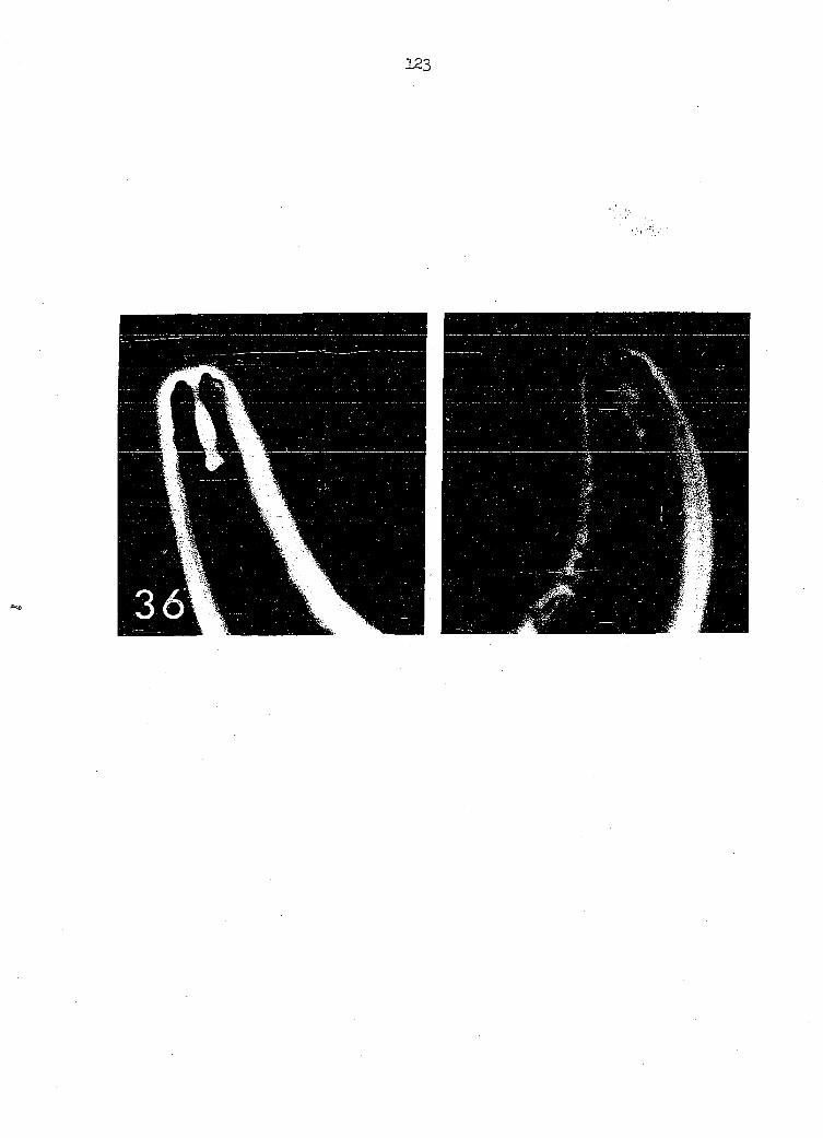
Anterior and posterior ends of female Microtetrameres protrude from
the central portion of the body (Figures 2, 3, 4) or may be wrapped
around the body. The length of the spheroidal portion of the body varies
in accordance with the nematode species, its age, number of eggs present
and other factors. But, in most cases, it is more than one millimeter
long. Obviously, this length will change slightly as the nematode moves.
As it does move, the coiled configuration of the body and the number of
loops appear to remain unchanged. However, when females are removed from
the glands, they may show variations in the direction of coiling
(Figure l). Such variations, when they occur, appear to be associated
with the method employed in expressing the parasite from the gland.
Schell, University of Idaho, indicated in i960 (private communication)
that it is impossible to straighten female Microtetrameres without
breaking their bodies. This was confirmed in the present study. Despite
this, Rasheed (1960), unfortunately used the straightened length of
female M. orioles in her key to the species of Microtetrameres and com
pared it with the coiled length of M. spiculata.
Delicate cuticular transverse striae appear along the length of the
body on female Microtetrameres sturnellae. Cervical papillae are lacking.
Heavily sclerotized rhabdions form the walls of the buccal capsule
(Figure 3)- The latter is defined as the sclerotized anterior portion
of the digestive tract between the mouth and the anterior end of the
pharynx. In the mid-region of the buccal capsule of adult females,
these rhabdions are markedly curved.
Because of its coiled nature, sections of a female Microtetrameres
12
sp. situ show greater histological detail than sections through a
typical filiform nematode. For instance, in a single section various
stages of oogenesis as well as several regions of the digestive tract
(Figure 23) may be seen.
The presence of nucleated red blood cells within the gut of female
Microtetrajueres sturnellae indicates that these parasites are hemato-
phagous. Further, it suggests strongly that the red color of the
female is due to hemoglobin of the host's blood as it is in Tetrameres
confusa as shown by Villela and Ribeiro (l955)* This blood is easily
accessible to female M. sturnellae as they are situated within the
proventricular glands with their anterior ends in close association
with the epithelial lining of the gland (Figures 2k, 25).
Female M. sturnellae probably release their eggs into the host's
proventricular lumen because their posterior ends project therein. No
eggs were seen within the fundus of any gland.
The number of females per host in this study reached as high as I6
with the average being about three. Gravid females usually contained
either all embryonated eggs or all non-embryonated eggs. In natural or
experimental infections, females with both types of eggs were found
rarely. Non-embryonated eggs in living specimens were collapsed except
in the case of one canary infection.
Measurements of female Microtetrameres sturnellae reared in this
study are provided in a later section of this thesis.
13
Young Female
The most obvious difference between this stage and fourth-stage
juveniles is the torsion which becomes apparent only after the young
adult stage has been reached (Figures 53 22). Females which have
completed about one loop of torsion have been recovered from proven- '
triculi of wild and experimental birds. Each of three such females
recovered from experimental chicks in this study was loosely coiled
within a Lieberkuhn gland ; only the most coiled one showed a slight
tinge of red. No such coloring was noted in any young female nematode
recovered from a wild host.
The anterior end of this stage is naked. No well-defined papillae
are. discernible in the cephalic region. Unlike the condition found in
third-stage M. sturnellae juveniles, the posterior end of a young
adult female does not terminate in the characteristic knoblike tail
process ("Ibouton")(Figure 5)- At this stage, transverse cuticular
striations are obvious along the length of the body.
Rhabdions of the buccal capsule (Figure 36) show less curvature
than do those of older adult females. A small intestinal diverticulum
is present at the pharyngointestinal junction.
A striking aspect of young female Microtetrameres sturnellae is the
detritus in its intestine (Figures 22). This material is most
obvious when not obscured by eggs and when the female has described
only one loop.
Reproductive structures, though present in young females, are
sometimes not clearly visible, particularly when CMC-10 is used as a
mountant. As the females mature, however, this system becomes more
obvious and eggs can be seen. Developing eggs in the oviduct are nearly
rectangular-shaped. They assume their characteristic mature shape
(Figures 2, 10) as they enter the uteri.
Young females were recovered from experimental bird hosts 20 - 26
days after the hosts were.fed encysted juveniles.
Dimensions of experimentally-developed, immature, fixed adult
females are: Buccal capsule 0.0156 - 0.0182 mm long by 0.003 - 0.005 mm
wide. Total length of pharynx, approximately 0.600 mm; muscular portion
convoluted, length may be difficult to measure, approximately 0.177 mm
long; width 0.0210 - 0.0260 mm at glandular-muscular pharyngeal junction;
glandular portion over 0.4000 mm long by 0.0 90 - O.O78O mm wide at
pharyngointestinal junction.
Male
Except for Travassos (1919) who reported males and young female
Microtetrameres pusilia in the same gland, little proof of the identity
of a male Microtetrameres has been offered by investigators. Therefore,
previous descriptions of male Microtetrameres must be questioned. In
this study two laboratory-reared males were recovered from one canary
proventriculus and one male from another canary proventriculus. These
two hosts were also parasitized by Microtetrameres sturnellae females.
Precise identification of male Mj.crotetrameres is difficult to
establish solely on the basis of natural infections even though both
sexes may occur together in the same proventriculus. Because previous
workers, except Cram (l93 ) and Schell (1953); have dealt only with
15
naturally-infected definitive hosts, the identity of males remains
questionable. Most authors who have reported the site of males, state
that they are found in the lumen of the proventriculus, while females
occur deep within the proventricular glands. On the other hand,
Travassos (1919), Thomas (nëe Mawson) of the University of Adelaide in
I960 (private communication) and Ulmer of Iowa State University in 1 64
(private communication) saw male nematodes and female Microtetrameres sp.
together within a proventricular gland. Wehr (193 ) and Cvetaeva (1960)
have observed this close association in presumed male and known female
letrameres spp.
Because of difficulties in precise identification, feeding experi
ments were undertaken to establish the morphological characteristics of
male Microtetrameres sturnellae. Data (Table 2) from these experiments
are based upon three laboratory-reared specimens (Figures 6, 7, 26, 27)
recovered from two adult canaries (Serinus canarius) reared under labora
tory conditions. The birds had been fed laboratory-reared grasshoppers
(Melanoplus sp.) containing numerous third-stage juveniles of M.
sturnellae. At necropsy, l84 days later, 7 females (3 of which were
gravid with embryonated eggs) and 2 males were recovered from one
canary's proventriculus; 20 females and 1 male were recovered from the
other canary's proventriculus 72 days post-feeding. Males occurred in
the mucus of the proventricular lumen; females were within the proven
tricular glands.
The only extant description of a laboratory-reared male Micro
tetrameres sp. prior to the present study was that published by Gram
16
(l93 )* Her description of M. helix males is meager and provides few
characteristics differentiating them from males of other genera. She
did not mention, for instance, the presence or absence of cephalic or
cloacal papillae or the dimensions of the buccal capsule.
The males recovered in this investigation were older than those
reared by Cram, and they are the shortest and narrowest yet reported
(Table l). Their cuticle is transversely striated. Four cephalic
papillae lateral to the buccal capsule can be seen on two specimens.
Each buccal capsule is 12 u long, slightly less than 3 u. wide at its
narrowest point. In all three specimens, the oral opening is approxi
mately 5 U- wide and the posterior end of each buccal capsule measures
about 8 u in width.
The pharynx measures 468 u in total length in specimen A (Figure 6),
4 4 u in specimen B (Figure 7) and 420 u in specimen C (figure 8). The
muscular pharynx in all specimens measures about l43 u in length. The
glandular portion of the pharynx in specimen A is 325 u long, that of
specimen B is 351 u- long and it is 280 u long in specimen C.
The digestive tract of specimen A terminates in a cuticularized
rectum. It was difficult to trace this system in either specimen A or
B due to the clearing action of the mountant employed and the masking
effect of the spicules. Specimen C, on the other hand, was mounted in
pure glycerine and most internal structures are visible.
No cervical alae are present in the males recovered (Figures 6,
7, 8).
The following description of the reproductive tract is based upon
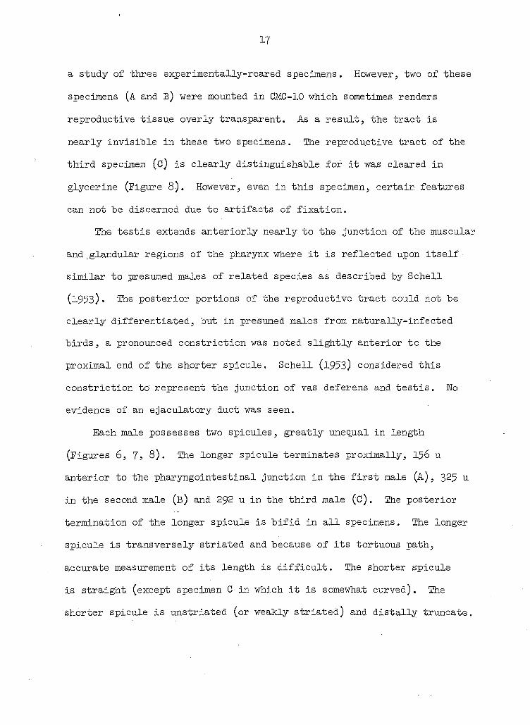
17
a study of three experimentally-reared specimens. However, two of these
specimens (A and B) were mounted in CMC-10 which sometimes renders
reproductive tissue overly transparent. As a result, the tract is
nearly invisible in these two specimens. The reproductive tract of the
third specimen (c) is clearly distinguishable for it was cleared in
glycerine (Figure 8). However, even in this specimen, certain features
can not be discerned due to artifacts of fixation.
The testis extends anteriorly nearly to the junction of the muscular
and glandular regions of the pharynx where it is reflected upon itself
similar to presumed males of related species as described by Schell
(1953)• The posterior portions of the reproductive tract could not be
clearly differentiated, but in presumed males from naturally-infected
birds, a pronounced constriction was noted slightly anterior to the
proximal end of the shorter spicule. Schell (l953) considered this
constriction to' represent the junction of vas deferens and testis. No
evidence of an ejaculatory duct was seen.
Each male possesses two spicules, greatly unequal in length
(Figures 6, 7; 8). The longer spicule terminates proximally, I56 u
anterior to the pharyngointestinal junction in the first male (A), 325 u
in the second male (B) and 292 u in the third male (C). The posterior
termination of the longer spicule is bifid in all specimens. The longer
spicule is transversely striated and because of its tortuous path,
accurate measurement of its length is difficult. The shorter spicule
is straight (except specimen G in which it is somewhat curved). The
shorter spicule is unstriated (or weakly striated) and distally truncate.
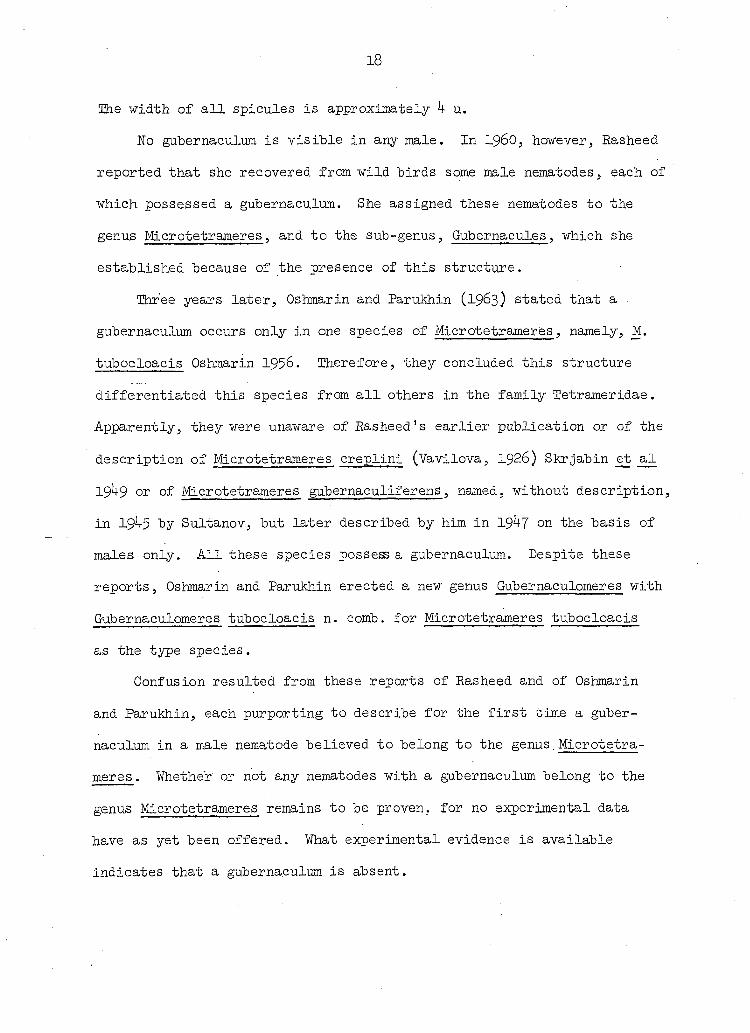
18
The width of all spicules is approximately 4 u.
No gubernaculum is visible in any male. In i960, however, Rasheed
reported that she recovered from wild "birds some male nematodes, each of
which possessed a gubernaculum. She assigned these nematodes to the
genus Microtetrameres, and to the sub-genus, Gubernacules, which she
established because of the presence of this structure.
Three years later, Oshmarin and ParuMiin (1963) stated that a
gubernaculum occurs only in one species of Microtetrameres, namely, M.
tubocloacis Oshmarin 1956. Therefore, they concluded this structure
differentiated this species from all others in the family Tetrameridae.
Apparently, they were unaware of Easheed's earlier publication or of the
description of Microtetrameres creplini (Vavilova, 1926) 8krjabin et al
19 9 or of Microtetrameres gubernaculiferens, named, without description,
in 19 5 by Sultanov, but later described by him in 19 7 on the basis of
males only. All these species possess a gubernaculum. Despite these
reports, Oshmarin and Parukhin erected a new genus Gubernaculomeres with
Gubernaculomeres tubocloacis n. comb. for Microtetrameres tubocloacis
as the type species.
Confusion resulted from these reports of Rasheed and of Oshmarin
and Parukhin, each purporting to describe for the first time a guber
naculum in a male nematode believed to belong to the genus.Microtetra
meres . Whethe'r or not any nematodes with a gubernaculum belong to the
genus Microtetrameres remains to be proven, for no experimental data
have as yet been offered. What experimental evidence is available
indicates that a gubernaculum is absent.
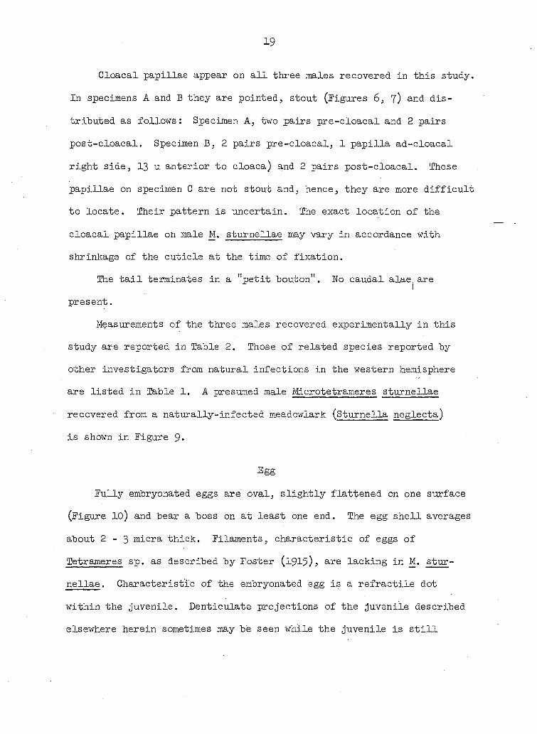
19
Cloacal papillae appear on all three males recovered in this study.
In specimens A and B they are pointed, stout (Figures 6, 7) and dis
tributed as follows: Specimen A, two pairs pre-cloacal and 2 pairs
post-cloacal. Specimen B, 2 pairs pre-cloacal, 1 papilla ad-cloacal
right side, 13 u anterior to cloaca) and 2 pairs post-cloacal. These
papillae on specimen C are not stout and, hence, they are more difficult
to locate. Their pattern is uncertain. The exact location of the
cloacal papillae on male M. sturnellae may vary in accordance with
shrinkage of the cuticle at the time of fixation.
The tail terminates in a "petit bouton". No caudal alae are
present.
Measurements of the three males recovered experimentally in this
study are reported in Table 2. Those of related species reported by
other investigators from natural infections in the western hemisphere
are listed in Table 1. A presumed male Microtetrameres sturnellae
recovered from a naturally-infected meadowlark (Sturnella neglecta)
is shown in Figure 9»
Egg
Fully embryonated eggs are oval, slightly flattened on one surface
(Figure 10) and bear a boss on at least one end. The egg shell averages
about 2-3 micra thick. Filaments, characteristic of eggs of
Tetrameres sp. as described by Foster (1915), are lacking in M. stur
nellae . Characteristic of the embryonated egg is a refractile dot
within the juvenile. Denticulate projections of the juvenile described
elsewhere herein sometimes may bb seen while the juvenile is still
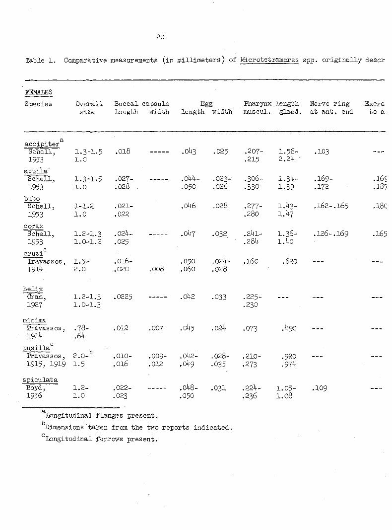
20
Table 1. Comparative measurements (in millimeters) of Microtetrameres spp. originally descr
PEMAIiES
Species Overall Buccal capsule Egg Pharynx length Nerve ring Excre size length width length width muscul. gland, at ant. end to a:
accipiter Schell, 1.3-1.5 .018 .043 .025 .207- 1.56- .103 1953 1.0 .215 2.2ij.
aq.ui'la' Schell, 1.3-1.5 .027- .044- .023- .306- 1.34- .169-1953 1.0 .028 . .050 .026 .330 1.39 .172
bubo Schell, 1-1.2 .021- .046 .028 .277- 1.43- .162-.165 1953 1.0 .022 .280 1.47
corax Schell, 1.2-1.3 .024- .047 .032 .241- I.36- .126-.I69 1953 1.0-1.2 .025 .284 1.40
. c cruzi Travassos, 1.5- .OI6- .050 .024- .160 .620 1914 2.0 .020 .008 .060 .028
helix Cram, 1.2-1.3 .0225 .042 .033 .22 -
1.0-1.3 .230
minima Travassos, .78- .012 .007 .045 .024 .073 .490 1914 .64
pusilla* Travassos, 2.0- .010- .OO9- .042- .028- .210- .920 1915, 1919 1.5 .016 .012 .049 .035 .273 .974
spiculata Boyd J 1.2— .022- ——--- .048- .03I .224— 1.05" 1956 1.0 .023 .050 .236 1.08
Longitudinal flanges present.
Dimensions taken from the two reports indicated.
Longitudinal furrows present.

3rameres spp. originally described from the western hemisphere
Cis *fc9/n c© length Nerve ring Excretory pore Species of host and , , , , ° , . J Cloaca-vulva Cloaca . gland, at ant. end to ant, end geographical location
1.56- .103 .108-.129 .100-.l4o Accipiter gentilis 2.24 Idaho
1.3 +- .169- .169- .082- .115- Aq.uila chrysaetos 1.39 .172 .187 .090 .133 Montana
1.43- .162-.165 .180-.190 .108-.162 .172-.178 Bubo virginianus 1.47 Oregon
1.36- .126-.169 .165-.190 .082-.130 .129-.187 Corvus corax 1.40 Idaho
.620 .200- .074 BUG CO svainsoni .226 .100 Melanerpes flavifrons
Brazil
.075 .l4l Corvus atnericana Washington, D.C.
.490 .068 lachyphonus cristatus bruneus Brazil
.920 .l4o lurdus rufiventris
.974 Flatycichla flavipes Estado do Rio
.109 .090 .096 Cyanocitta cristata .120 .103 Massachusetts
21
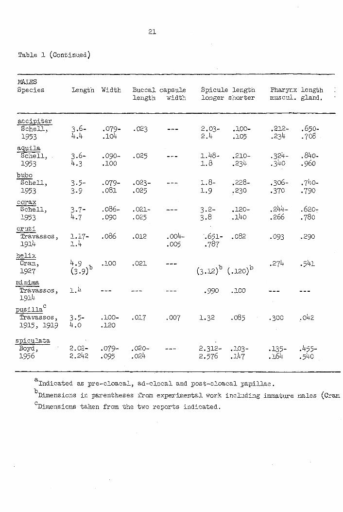
Table 1 (Continued)
mtES Species Length Width Buccal capsule Spicule length Pharynx length
length width longer shorter muscul. gland.
accipiter Schell, 3.6- .079- .023 --- 2.03- .100- .212- .65O-1953 4.4 .104 2.4 .105 .234 .708
aguila Schell; 3.6- .090- .025 —- 1.48- .210- .324- .840-1953 4.3 .100 1.8 .234 .340 .960
bubo Schell, 3.5- .079- .023- --- 1.8- .228- .306- .740-1953 3.9 .081 .025 1.9 .230 .370 .790
corax Schell, 3-7- .086- .021- --- 3.2- .120- .244- .620-1953 4.7 .090 .025 3.8 .l4o .266 .780
cruzi Travassos, 1.17- .086 .012 .004- .651- .082 .093 .290 1914 1.4 .005 .787
helix Cram, 4.9 , .100 .021 — , , .274 .541 1927 (3.9) (3.12) (.120)
minima Travas s OS, 1.4 — — *990 .100 — — 1914
pusilla Travas8OS, 3.5- .100- .017 .007 1.32 .O85 .3OO .042 1915, 1919 4.0 .120
spiculata Boyd, 2.02- .079- .020- —- 2.312- .IO3- .135- .455-1956 2.242 .095 .024 2.57^ .164 .540
Indicated as pre-cloacal, ad-clocal and post-cloacal papillae.
Dimensions in parentheses from experimental work including immature males (Cram
Dimensions taken from the two reports indicated.

length Pharynx length Werve ring Cervical pap. Excretory pore Distance Cloacal horter muscul. gland. to ant. end to ant. end to ant. end Cloaca-tail papillae
.100-
.105 .212-.234
.650-.708
.144-
.151 .162-.183
1 .165- -.178
4 pre-4 post-
.210-.234
.324-
.340 .840-.960
.190-
.205 .298-.300
.240-
.252 .178-.195
4 pre-4 post-
.228-
.230 .306-.370
.740-
.790 .180-.190
.230-
.240 .190-.220
.210- ,
.220 4 pre-6 post-
,120-l4o
.244-.266
.620-
.780 .151-.187
.194-.237
.154-
.194 .160-.207
4 pre-4 post-
082 .093 .290 .132 2 pre-6 post-
120) .274 .541 .191 — " " .183 4 pre-
4 post-
100 — - — - - —
#5 .300 .042 - .170 4 pre-1 4 post-
.03- .135-.164
.455-.540
.086-.100
.162-.165
.100 .127-.147
4 pre-4 post-
. papillae.
lading immature males (Cram, 1927).

22
Table 2. Comparative measurements of experimentally-reared male
Microtetrameres sturnellae
Specimen A Specimen B Specimen C
Body length
Body width at pharyngo-intestinal Junction
Cephalic papillae
Oral opening, diameter
Buccal capsule, length
Buccal capsule, width
Pharynx length, total Muscular portion Glandular portion
Nerve ring to posterior edge of buccal capsule
Shorter spicule, length
Shorter spicule, width
Longer spicule, length
Longer spicule, width
Ratio long spicule length to length short spicule
Cloaca to tail tip
Cloacal papillae
Termination of tail
Alaé, caudal
Alae, cervical
1.073 mm
0.070 mm
k
0.005 1™
0.012 mm
0.003 mm
0.468 mm 0.1 3 0.325 mm
0.088 mm
0.003 mm
1.034 mm
0.004 mm
12
0.133 mm
2 pairs pre-2 pairs post-1 adanal
bouton
absent
absent
1.008 mm
0.070 mm
k
0.005 mm
0.012 mm
0.003 mm
0.4$4 mm 0.14-3 mm 0.351 mm
0.076 mm
0.003 mm
1.036 mm
0.004 mm
Ik
0.135 mm
2 pairs pre-2 pairs post-
bouton
absent
absent
1.4-00 mm
0.075 mm
0.005 mm
0.012 mm
0.005 mm
0.420 mm 0.l40 mm 0.280 mm
0.085 mm
0.109 mm
0.004 mm
1.200 mm
0.004 mm
19
0.130 mm .
spike
absent
absent
23
•within the shell.
Czaplinski (1962), in his survey of nematodes and acanthocephala
parasitic in anseriform birds of Poland discussed morphological
variability among these parasites as related to the various species of
their definitive hosts. Among the anatomical landmarks he discussed
were egg dimensions. He found that egg size showed the smallest range
of variability of all morphological features studied. Although he did
not work with Microtetrameres, he did find in some nematodes that egg
width is more variable than egg length. He was unable to show any
relationship between egg size and age or size of the female nematode
producing them or between egg size and number of eggs within the female.
Dimensions of eggs of M. sturnellae and a comparison of egg sizes
from specimens recovered from naturally-infected meadowlarks and from
an experimentally-infected canary are shown below. The constancy of
egg size in both types of hosts is striking and suggests the value of
using this criterion in differentiating various species of Microte
trameres .
Egg sizes for the type specimen of M. sturnellae average (W=10)
50.7 u long by 35-3 u wide. Egg sizes of M. sturnellae recovered from
naturally-infected meadowlarks average (W=82) 50.5 u long by 35.2 u
wide.
Juveniles
Current Hematological literature includes little information con
cerning morphological characteristics of juvenile Microtetrameres spp.

24
Seurat (1913, I915-I6, I918), however, one of few investigators to
describe Microtetrameres juveniles, included the following morphological
features in his descriptions from naturally-infected hosts: length of
tail, distance from head to nerve ring and to excretory pore, body
length and width, length of the buccal capsule, lengths of the two
pharyngeal portions as well as the ratio of the entire pharynx to the
body length of third-stage and what he termed fourth-stage juveniles of
Tropidocerca (= Microtetrameres) spiralis.
Schell (1953) and Cram (l93 ) have also reported on juvenile stages
of the life cycle of Microtetrameres spp. These reports are discussed
below.
The following account of juveniles of Microtetrameres sturnellae
is based upon specimens experimentally-reared in numerous species of
Melanoplus.
First-stage
Although detailed studies on the process of hatching were not
undertaken in this study, some observations were made on fixed material
and from living specimens.
Juveniles apparently emerge within a few hours after eggs have been
ingested by a grasshopper nymph. Some juveniles recovered from the
intermediate host's foregut were seen partially emerged (Figures 11, 28).
First-stage juveniles escape through one end of the egg; apparently,
they may escape from the egg with either end emerging first. Both
methods were reported from M. inermis (Seurat, I913). In 12 cases in the
present study 9 juveniles had emerged anterior end first (Figures 11, 28)
25
and h tail end first. In another instance, the juvenile had broken the
shell but both of its ends were still within the egg shell. Thirty
minutes later this juvenile had escaped tail end first.
First-stage juveniles possess blunt anterior ends (Figures 11, 12).
Their tail ends are pointed (Figure 12) unlike juveniles of letrameres.
The anterior end of a living first-stage juvenile changes from convex
to concave as the animal moves (Figures 13, l4). In this aspect, Micro-
tetrameres sturnellae first-stage juveniles resemble juveniles of what
appears to be a species of Tetrameres as figured by Lieberklihn (l855)•
A group of three refractile dontoidal projections appears anteriorly,
its position modified somewhat in accordance with the movement of the
anterior region of the body. The rapid movement of living juveniles and
the minute size of these projections make their study difficult.
Juveniles at this stage lash about in an active manner. This movement,
together with the action of the dontoidal projections, may aid
measurably in the migration of juveniles within the tissue of their
intermediate host. These dontoidal projections may be similar to the
"aiguillon c phalique" mentioned by Chabaud (l95 ) who considered this
structure as an aid in permitting first-stage juveniles to escape from
their eggs and in enabling them to penetrate the intermediate host's
gut wall.
The resemblance between a first-stage juvenile of Microtetrameres
sturnellae and a microfilaria is remarkable. About 5P micra from the
anterior end of a living first-stage juvenile of Microtetrameres
sturnellae a clear area similar to the excretory pore of a microfilaria
26
can be seen. A spot somewhat resembling the anal pore of a micro
filaria is seen approximately 50 micra from the tail end. Between
these two areas but closer to the posterior one is a structure faintly
resembling a short length of intestine. Possibly this is connected to
the presumed anal pore, but it does not appear to extend the length of
the juvenile. Within this "gut" usually are three or four refractile
bodies ("inner body" cells) which move slightly when the nematode moves.
Nuclei are seen throughout the living body (Figure 11).
At the anterior end of some living specimens the area destined to
become the pharynx in older juveniles extends posteriorly about 20 micra.
Granules are visible within this region in some living individuals. As
the nematode moves, these move rhythmically, as do the three or four
refractile bodies near the anal pore. Wo connection between the anal
pore and the anterior region was seen under bright-field or phase-
contrast microscopy. However, because of their synchronized movements,
anterior and posterior groups of refractile bodies appear to be related.
Early first-stage juveniles may be distinguished from late first-
stage juveniles on the basis of their size and extent of cuticular
striations. These striations are of 2 types in first-stage juveniles.
Discontinuous striae appearing as rings of minute dots instead of
continuous lines (Figures 11, 12, 13, l4), occur only at the anterior
end of the early first-stage juveniles. These striations are situated
along the anterior l6 to 20 micra of the body. A second type of
striation is continuous and is characteristic of older first-stage
juveniles. Such striae are located posterior to the discontinuous ones.
27
From a point slightly anterior to the posteriormost discontinuous
striation, two slender lines, one on either side of the body, extend
nearly to the posterior end of the juvenile. These were seen on
numerous living and fixed specimens and may represent lateral chords.
At times during this research, female Microtetrameres were not
ingested immediately by grasshoppers and some had dried before they were
eaten by the nymphal grasshopper. In at least two instances, first-stage
juveniles hatched from eggs within a dried female eaten by a nymph.
These dried females had been dead for approximately 48 hours.
Living juveniles displaying characteristics of both first-stage and
second-stage were recovered in this study sixteen days after feeding eggs
of experimentally-reared M. sturnellae to a grasshopper nymph. These
juveniles (Figures 15, l6, 17, l8) were similar to second-stage juveniles
in size and in the characteristic configuration of the tail region, but
resembled first-stage juveniles in possessing anterior denticulate
projections. They were classified, consequently, as first-stage
juveniles undergoing ecdysis to the second stage. The following des
cription is based upon observations made upon living specimens.
The total body length ranged from 470 - 4 0 u; body width, 26 - 39 u.
Transverse cuticular striations were present along the entire length of
most specimens. In others such transverse striations were limited to
the anterior body region. A we11-developed sheath, slightly raised
from the cuticle, and bearing the dontoidal projections of first-stage
juveniles covered the rounded anterior end of each specimen. A clear
area, consisting of two separate translucent spots with definite
28
boundaries and approximately 3 - $ u in diameter, was visible within
13 u of the anterior end. Perhaps these spots represent glands asso
ciated with histolytic activity. The excretory pore on many of these
juveniles was easily located, and was situated slightly more than 80 u
from the anterior end. Wo evidence of a mouth was seen. The first
forty miera of the digestive tract appeared as a fine line, no lumen
being visible. Posteriorly, the gut expanded, reaching its maximum
width at the pharyngointestinal junction. This junction was plainly
visible 13O - 1 3 u posterior to the anterior end. The lining of the
digestive tract could be traced easily under dark-field microscopy.
Body musculature surrounding the tract was seen under phase-contrast
microscopy. The gut appeared to move lengthwise within the juvenile as
the anterior end moved about. In some juveniles detritus was observed
within the gut. The anus was apparent and in some cases measured
approximately 8 u in diameter. It opened internally into a more or
less spherical chamber, details of which were not discernible. Uni
dentified material was seen protruding from the anus in some juveniles.
No genital primordium was visible under dark-field or phase-contrast
microscopy.
A summary of feeding experiments with intermediate hosts is pre
sented in Table 3-

29 ^
Table 3- Summary of feeding experiments with intermediate hosts of
Microtetrameres stuxnellae
Host Days elapsed Stage of recovered post-feeding juvenile
Melanoplus 1 1 Ji 2 1 Ji
3 2 Ji 4 2 JI 5 2 JI 6 3 JI 7 3 JI 8 3 JI 9 4 JI 10 4 JI 11 1+ JI 12 5 JI 13 5 JI l4 6 JI 15 7 JI l6 8 JI 17 8 JI
18 8 J2? 19 15 J2?
20 l6 J2?
21 25 J3 22 27 J3 23 .28 J3 2k 29 J3 25 30 J3 26 32 J3 27 33 J3 28 38 J3 29 38 J3 30 39 J3 31 ho J3 32 ho J3 33 43 J3 34 43 J3 35 45 J3
30
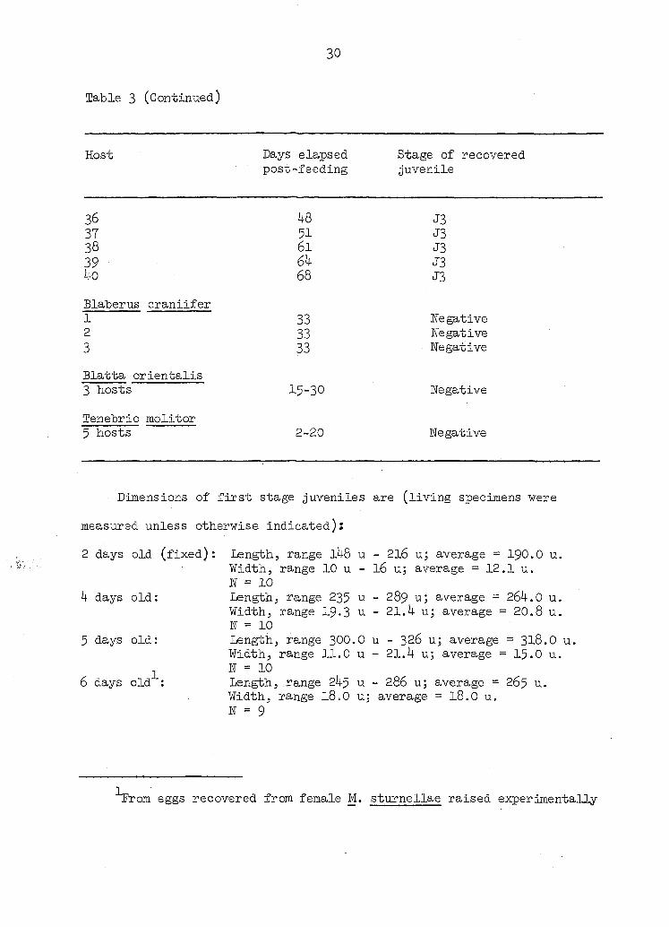
Table 3 (Continued)
Host Days elapsed post-feeding
Stage of recovered juvenile
36 37 38 39 40
Blaberus craniifer 1 2 3
Blatta orientalis 3 hosts
Tenebrio molitor 5 hosts
48 51 61 6k 68
33 33 33
15-30
2-20
J3 J3 J3 J3 J3
Negative Negative Negative
Negative
Negative
Dimensions of first stage juveniles are (living specimens were
measured unless otherwise indicated):
2 days old (fixed): Length, range l48 u - 2l6 u; average = I9O.O u. Width, range 10 u - 16 u; average = 12.1 u. N = 10
4 days old: Length, range 235 u - 289 u; average = 264.0 u. Width, range I9.3 u - 21.4 u; average = 20.8 u. N = 10
5 days old: Length, range 3OO.O u - 326 u; average = 318.O u. Width, range 11.0 u - 21.4 u; average = 15.O u.
N = 10 6 days old : Length, range 245 u - 286 u; average = 265 u.
Width, range 18.O u; average = I8.O u. N = 9
rom eggs recovered from female M. sturnellae raised experimentally
31
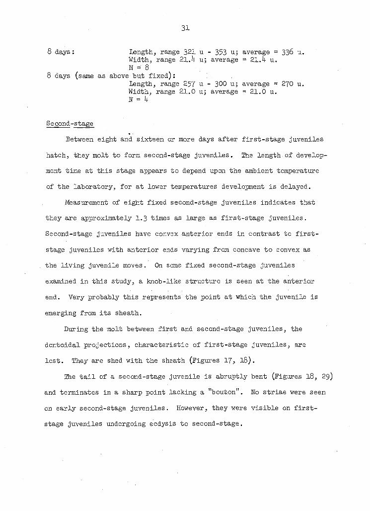
8 days: Length, range 321 u - 353 u; average = 336 u. Width, range 21.4 uj average = 21.k u. n = 8
8 days (same as above but fixed): Length, range 257 u - 300 u; average = 270 u. Width, range 21.0 u; average = 21,0 u. N = 4
Second-stage
Between eight and sixteen or more days after first-stage juveniles
hatch, they molt to form second-stage juveniles. The length of develop
ment time at this stage appears to depend upon the ambient temperature
of the laboratory, for at lower temperatures development is delayed.
Measurement of eight fixed second-stage juveniles indicates that
they are approximately I.3 times as large as first-stage juveniles.
Second-stage juveniles have convex anterior ends in contrast to first-
stage juveniles with anterior ends varying from concave to convex as
the living juvenile moves. On some fixed second-stage juveniles
examined in this study, a knob-like structure is seen at the anterior
end. Very probably this represents the point at which the juvenile is
emerging from its sheath.
During the molt between first and second-stage juveniles, the
dontoidal projections, characteristic of first-stage juveniles, are
lost. They are shed with the sheath (Figures 17, I8).
The tail of a second-stage juvenile is abruptly bent (Figures I8, 29)
and terminates in a sharp point lacking a "bouton". No striae were seen
on early second-stage juveniles. However, they were visible on first-
stage juveniles undergoing ecdysis to second-stage.
32
During this investigation numerous living second-stage juveniles
within sheaths of first-stage juveniles were studied. A detailed
description of them is presented above in the section concerning first-
stage juveniles.
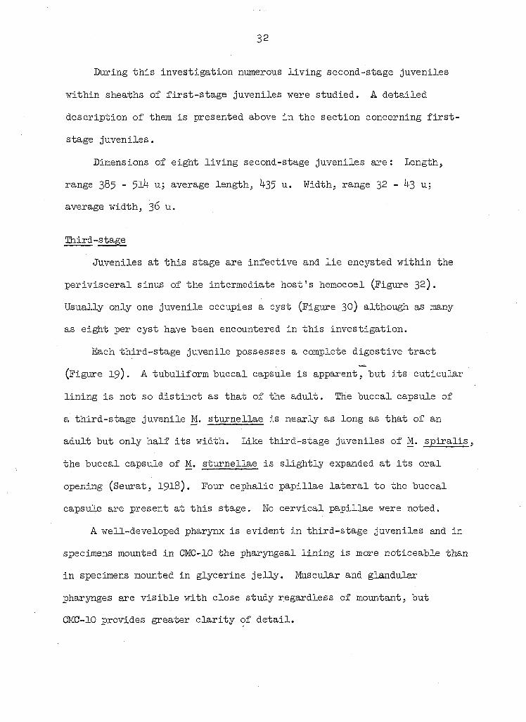
Dimensions of eight living second-stage juveniles are : Length,
range 385 - 51 u; average length, 35 u. Width, range 32 - 43 u;
average width, 36 u.
Third-stage
Juveniles at this stage are infective and lie encysted within the
perivisceral sinus of the intermediate host's hemocoel (Figure 32).
Usually only one juvenile occupies a cyst (Figure 30) although as many
as eight per cyst have been encountered in this investigation.
Each third-stage juvenile possesses a complete digestive tract
(Figure 19). A tubuliform buccal capsule is apparent, but its cuticular
lining is not so distinct as that of the adult. The buccal capsule of
a third-stage juvenile M. sturnellae is nearly as long as that of an
adult but only half its width. Like third-stage juveniles of M. spiralis,
the buccal capsule of M. sturnellae is slightly expanded at its oral
opening (Seurat, I918). Four cephalic papillae lateral to the buccal
capsule are present at this stage. No cervical papillae were noted.
A well-developed pharynx is evident in third-stage juveniles and in
specimens mounted in CMC-10 the pharyngeal lining is more noticeable than
in specimens mounted in glycerine jelly. Muscular and glandular
pharyngés are visible with close study regardless of mountant, but
CMC-10 provides greater clarity of detail.
33
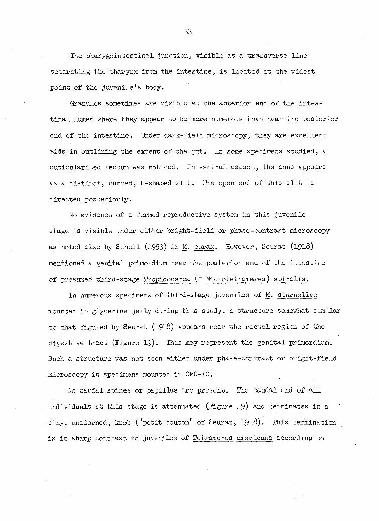
The pharygointestinal Junction, visible as a transverse line
separating the pharynx from the intestine, is located at the widest
point of the juvenile's body.
Granules sometimes are visible at the anterior end of the intes
tinal lumen where they appear to be more numerous than near the posterior
end of the intestine. Under dark-field microscopy, they are excellent
aids in outlining the extent of the gut. In some specimens studied, a
cuticularized rectum was noticed. In ventral aspect, the anus appears
as a distinct, curved, U-shaped slit. The open end of this slit is
directed posteriorly.
No evidence of a formed reproductive system in this juvenile
stage is visible under either bright-field or phase-contrast microscopy
as noted also by Schell (l953) in M. corax. However, Seurat (1918)
mentioned a genital primordium near the posterior end of the intestine
of presumed third-stage iCropidocerca (= Microtetrameres) spiralis.
In numerous specimens of third-stage juveniles of M. sturnellae
mounted in glycerine jelly during this study, a structure somewhat similar
to that figured by Seurat (1918) appears near the rectal region of the
digestive tract (Figure 19). This may represent the genital primordium.
Such a structure was not seen either under phase-contrast or bright-field
microscopy in specimens mounted in CMC-10.
No caudal spines or papillae are present. The caudal end of all
individuals at this stage is attenuated (Figure I9) and terminates in a
tiny, unadorned, knob ("petit bouton" of Seurat, I918). This termination
is in sharp contrast to juveniles of Tetrameres americana according to
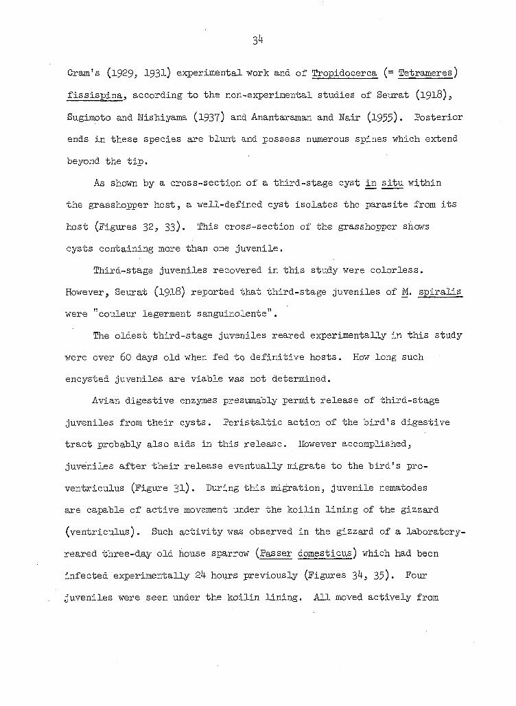
34
Cram's (1929, 193l) experimental work and of Tropidocerca (= Tetrameres)
fissispina, according to the non-experimental studies of Seurat (1918),
Sugimoto and ITishiyama (1937) and Anantaraman and Nair (l955) • Posterior
ends in these species are blunt and possess numerous spines which extend
beyond the tip.
As shown by a cross-section of a third-stage cyst situ within
the grasshopper host, a well-defined cyst isolates the parasite from its
host (Figures 32, 33)• This cross-section of the grasshopper shows
cysts containing more than one juvenile.
Third-stage juveniles recovered in this study were colorless.
However, Seurat (1918) reported that third-stage juveniles of M. spiralis
were "couleur legerment sanguinolente".
The oldest third-stage juveniles reared experimentally in this study
were over 60 days old when fed to definitive hosts. How long such
encysted juveniles are viable was not determined.
Avian digestive enzymes presumably permit release of third-stage
juveniles from their cysts. Peristaltic action of the bird's digestive
tract probably also aids in this release. However accomplished,
juveniles after their release eventually migrate to the bird's pro-
ventriculus (Figure 31). During this miration, juvenile nematodes
are capable of active movement under the koilin lining of the gizzard
(ventriculus). Such activity was observed in the gizzard of a laboratory-
reared three-day old house sparrow (Passer domesticus) which had been
infected experimentally 2k hours previously (Figures 34, 35)- Four
juveniles were seen under the koilin lining. All moved actively from

35
side to side.
The inability of some infective Juveniles to become established in
older experimental hosts may be due to the resistance offered by the
koilin lining of the gizzard. For instance, no adult nematodes were
recovered from 6-week-old chicks and only a very few adult pigeons which
had been fed many infective juveniles. Perhaps juvenile nematodes are
unable to penetrate the koilin lining of older birds.
Eight days post-ingestion by the definitive host it was found that
the width of the two pharyngeal portions had greatly increased in those
third-stage juveniles recovered. Other dimensions of the juveniles
had not changed greatly.
Dimensions of the third-stage juveniles are;
Dimensions of fixed third-stage juveniles (32 - 60 days old) recovered
from 3 intermediate hosts fed eggs from three different avian hosts are :
W = 33. Length, range: 1790 - IO70 u; average = l48l u.
N = 24. Width, range: 6k - 31 u; average = 4$ u.
N = 37. Buccal capsule. Length, range: 15-5 - 12.8 u; average = 13.4 u.
E = 38. Buccal capsule. Width, range: 7.7 u - 5.12 u; average = 5.4 u.
W = 27. Anus-tail. Distance, range: 206 - l4l u; average = I83 u.
Dimensions of fixed third-stage juveniles (30 - 34 days old) recovered
from 3 intermediate hosts fed eggs from one definitive host :
N = 84. Length, range: 2040 - 1122 u; average = 1539 u.
W = 86. Width, range: 100 - 46 u; average = 58 u.
N = 82. Buccal capsule. Length, range: 18.2 - 10.4 u; average = 16.5 u.
W = 85. Eharyngointestinal junction to mouth. Distance, range:
36
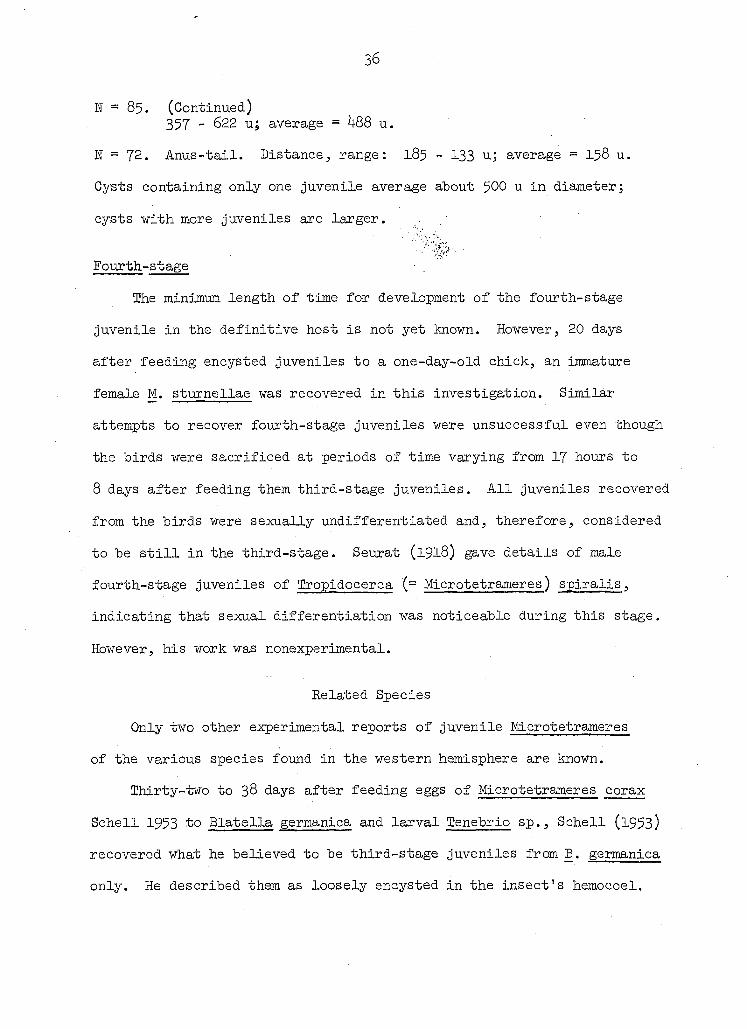
ïï = 85. (Continued) 357 - 622 u; average = 488 u.
N = 72. Anus-tail. Distance, range: 185 - 133 u; average = 158 u.
Cysts containing only one juvenile average about 500 u in diameter;
cysts with more juveniles are larger.
Fourth-stage
The minimum length of time for development of the fourth-stage
juvenile in the definitive host is not yet known. However, 20 days
after feeding encysted juveniles to a one-day-old chick, an immature
female M. sturnellae was recovered in this investigation. Similar
attempts to recover fourth-stage juveniles were unsuccessful even though
the birds were sacrificed at periods of time varying from 17 hours to
8 days after feeding them third-stage juveniles. All juveniles recovered
from the birds were sexually undifferentiated and, therefore, considered
to be still in the third-stage. Seurat (1918) gave details of male
fourth-stage juveniles of Iropidocerca (= Microtetrameres) spiralis,
indicating that sexual differentiation was noticeable during this stage.
However, his work was nonexperimental.
Related Species
Only two other experimental reports of juvenile Microtetrameres
of the various species found in the western hemisphere are known.
Thirty-two to 38 days after feeding eggs of Microtetrameres corax
Schell 1953 to Blatella germanica and larval Tenebrio sp., Schell (l953)
recovered what he believed to be third-stage juveniles from B. germanica
only. He described them as loosely encysted in the insect's hemocoel.
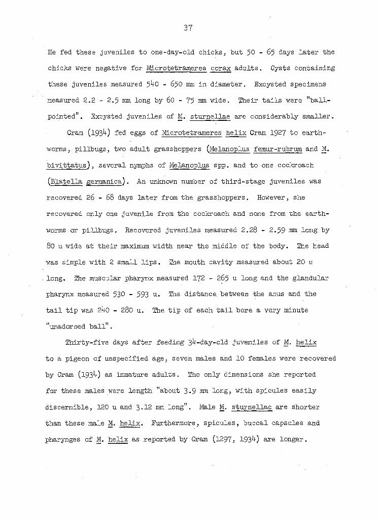
37
He fed these Juveniles to one-day-old chicks, but $0 - 65 days later the
chicks were negative for Microtetrameres corax adults. Cysts containing
these juveniles measured $40 - 65O mm in diameter. Excysted specimens
measured 2.2 - 2.5 mm long by 60 - 75 mm wide. ïheir tails were "ball-
pointed". Excysted juveniles of M. sturnellae are considerably smaller.
Cram (l93 ) fed eggs of Microtetrameres helix Cram I927 to earth
worms, pillbugSj two adult grasshoppers (Melanoplus femur-rubrum and M.
bivittatus), several nymphs of Melanoplus spp. and to one cockroach
(Elatella germanica). An unknown number of third-stage juveniles was
recovered 26 - 68 days later from the grasshoppers. However, she
recovered only one juvenile from the cockroach and none from the earth
worms or pillbugs. Recovered juveniles measured 2.28 - 2.59 long by
80 u wide at their maximum width near the middle of the body. lEhe head
was simple with 2 small lips. The mouth cavity measured about 20 u
long. The muscular pharynx measured I72 - 265 u long and the glandular
pharynx measured 530 - 593 U-- The distance between the anus and the
tail tip was 240 - 280 u. The tip of each tail bore a very minute
"unadorned ball".
Thirty-five days after feeding 3 -day-old juveniles of M. helix
to a pigeon of unspecified age, seven males and 10 females were recovered
by Cram (193 ) as immature adults. The only dimensions she reported
for these males were length "about 3-9 long, with spicules easily
discernible, 120 u and 3*12 mm long". Male M. sturnellae are shorter
than these male M. helix. Furthermore, spicules, buccal capsules and
pharyngés of M. helix as reported by Cram (l297, 193 ) are longer.

38
DEFINITIVE HOSTS
Natural Definitive Hosts
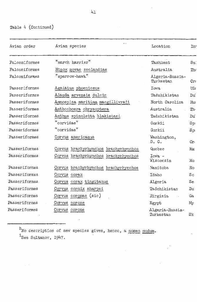
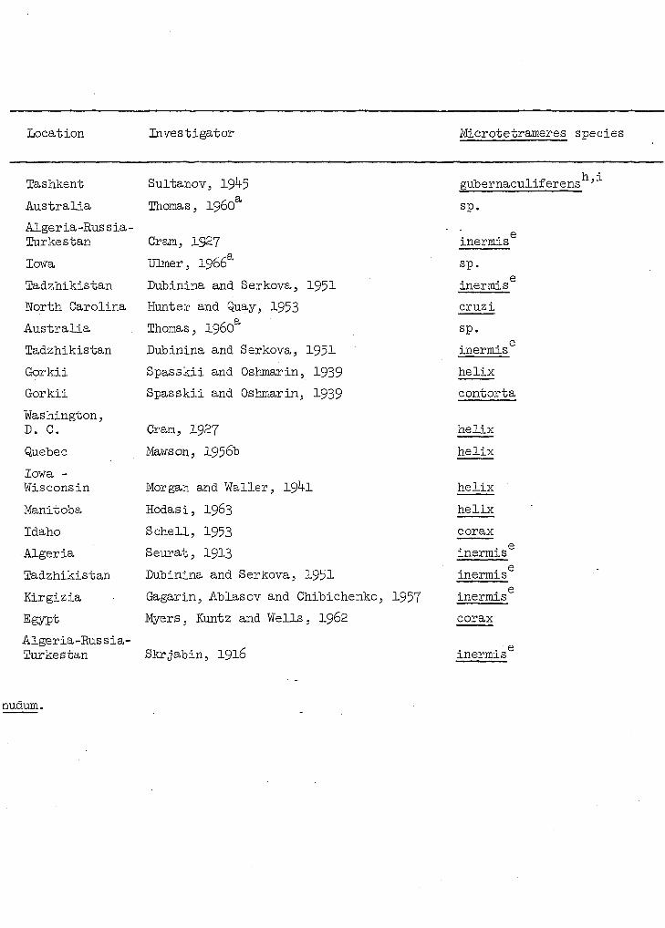
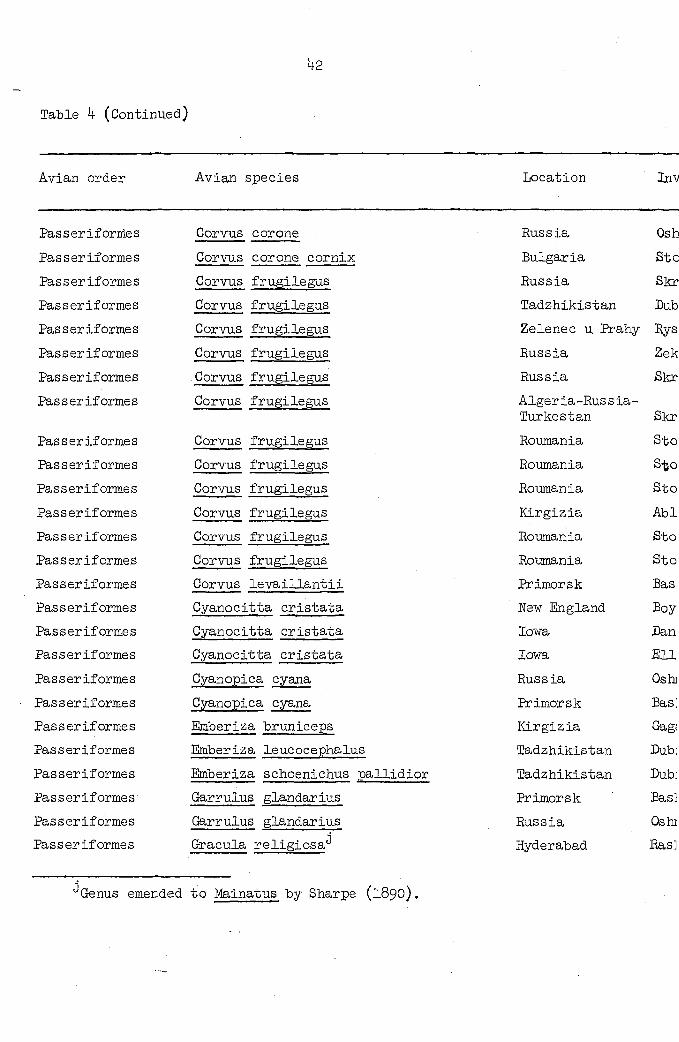
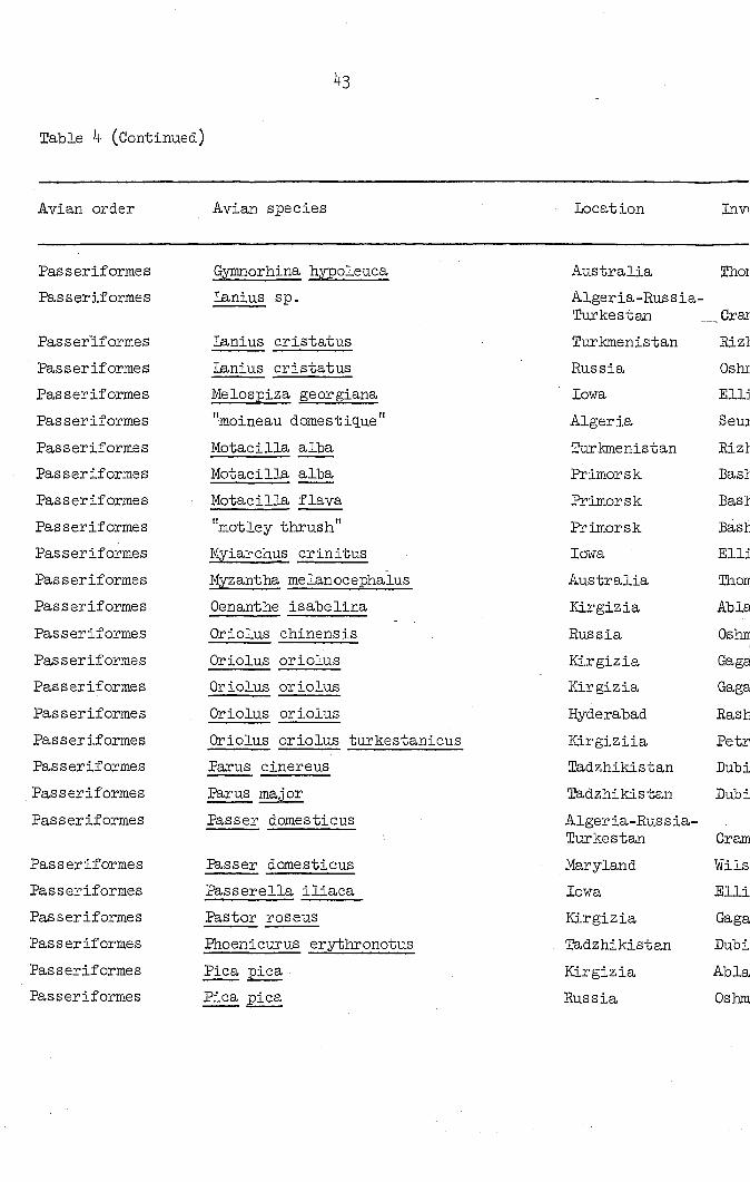
The wide geographical and host ranges of Microtetrameres spp. are
shown in Table 4. At least ten orders, about 84 genera and 115 species
of birds have been reported as hosts for this nematode genus. Despite
this wide distribution, numerous surveys of avian hosts do not include
Microtetrameres among the nematodes recovered as is shown in Table 6.
Microtetrameres spp. are found in non-aquatic birds; Tetrameres is a
genus whose members are parasites primarily in aquatic birds. LaEage
(1961) apparently erred in indicating that Microtetrameres had been found
in the Anatidae. He cited Madsen (l952) as his authority, but a check
of the latter's report indicates that Madsen found no specimens of
either Tetrameres or Microtetrameres in Danish game birds.
Numerous reasons could be offered for the absence of reports of
Microtetrameres from some birds. Johnston (iglO, I912), in a survey of
parasites of Australian birds, cited no record of tetramerid infections.
His studies emphasized cestodes and perhaps with this predilection, he
may have overlooked proventricular nematodes as Thomas (née Mawson) of
the University of Adelaide reported in i960 (private communication) that
she found Microtetrameres to be fairly common in birds of South Australia.
Although other avian hosts have been examined for nematode parasites,
adult Microtetrameres have not been reported from many surveys. This
may be due in a large measure to the peculiar location of adult females
which may have been overlooked in routine necropsies unless particular
39
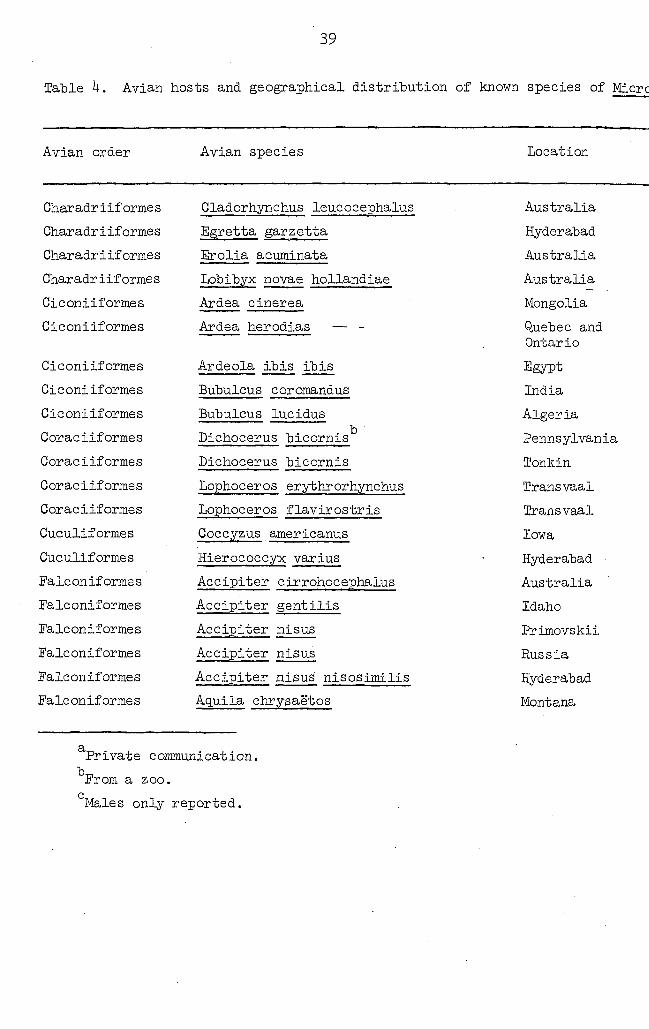
Table 4. Avian hosts and geographical distribution of known species of Micrc
Avian order Avian species Location
Charadriiformes
Charadriiformes
Charadriiformes
Charadriiformes
Ciconiiformes
Ciconiiformes
Ciconiiformes
Ciconiiformes
Ciconiiformes
Coraciiformes
Coraciiformes
Coraciif ormes
Coraciif ormes
Cuculiformes
Cuculiformes
Falcon if orme s
Falconiformes
Falconiformes
Falconiformes
Falconiformes
Falcon if ormes
Gladorhynchus leucocephalus
Egretta garzetta
Erolia acuminata
Lobibyx novae hollandiae
Ardea cinerea
Ardea herodias — -
Ardeola ibis ibis
Bubulcus coromandus
Bubulcus lucidus
Dichocerus bicornis
Dichocerus bicornis
Lophoceros erythrorhynchus
Lophoceros flavirostris
Coccyzus americanus
Hierococcyx varius
Accipiter cirrohocephalus
Accipiter gentilis
Accipiter nisus
Accipiter nisus
Accipiter nisus nisosimilis
Aquila chrysaè'tos
Australia
Hyderabad
Australia
Australia
Mongolia
Quebec and Ontario
Egypt
India
Algeria
Pennsylvania
Tonkin
Transvaal
Transvaal
Iowa
Hyderabad
Australia
Idaho
Primovskii
Russia
Hyderabad
Montana
Private communication.
From a zoo.
' Males only reported.
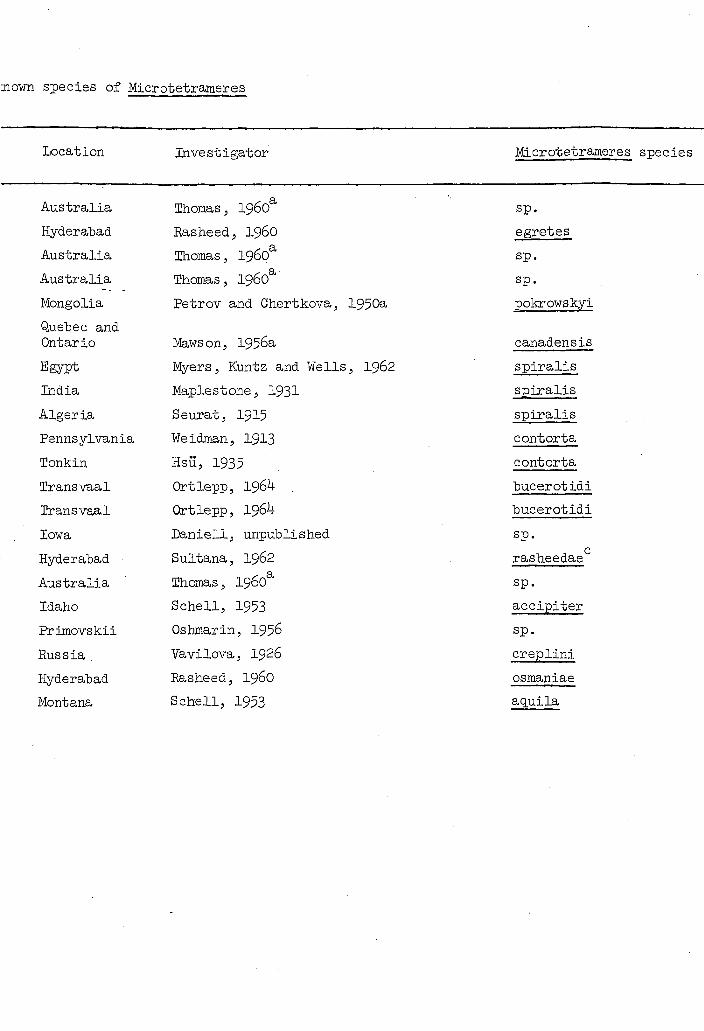
3own species of Microtetrameres
Location Investigator Microtetrameres species
Australia Thomas, 1960 sp.
Hyderabad Rasheed, I960 egretes
Australia Thomas, 1960 sp.
Australia Thomas, 1960 sp.
Mongolia Petrov and Chertkova, 1950a pokrowskyi
Quebec and Ontario Maws on, 1956a canadensis
Egypt Myers, Kuntz and Wells, 1962 spiralis
India Maplestone, 1931 spiralis
Algeria Seurat, I915 spiralis
Pennsylvania Weidman, 1913 contorta
Tonkin HSU, 1935 contorta
Transvaal Ortlepp, 1964 bucerotidi
Transvaal Ortlepp, 1964 bucerotidi
Iowa Daniell, unpublished sp.
Hyderabad Sultana, 1962 rasheedae
Australia Thomas, 1960 sp.
Idaho Schell, 1953 accipiter
Primovskii Oshmarin, 1956 sp.
Russia Vavilova, I926 creplini
Hyderabad Rasheed, I96O osmaniae
Montana Schell, 1953 aquila
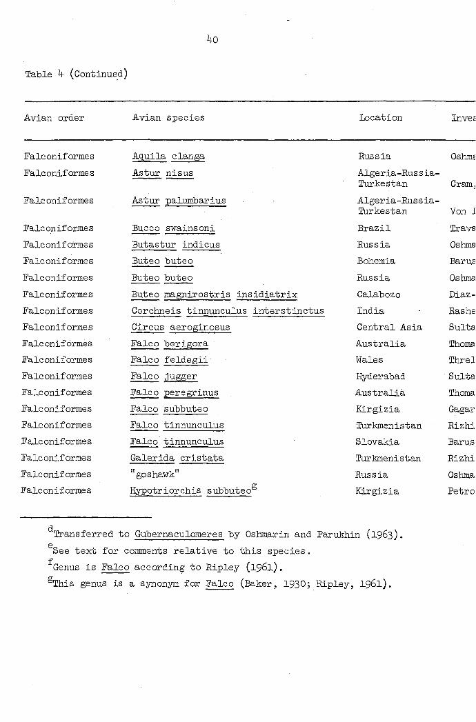
4o
Table 4 (Continued)
Avian order Avian species Location Invee
Falconiformes Aquila clanga Russia Oshms
Falconiformes Astur nisus Algeria-Russia-Turkestan Cram.
Falconiformes Astur palmnbarius Algeria-Russia-Turkestan Von I
Falconiformes Bucco swainsoni Brazil Trava
Falconiformes Butastur indicus Russia Oshtna
Falconiformes Buteo buteo Bohemia Bar us
Falconiformes Buteo buteo Russia Oshma
Falconiformes Buteo magnirostris insidiatrix Calabozo Diaz-
Falconiformes Cerchneis tinnunculus interstinctus India Rashe
Falconiformes Circus aeroginosus Central Asia Sulta
Falconiformes Falco berigora Australia Ihoma
Falconiformes Falco feldegii' Wales Threl
Falconiformes Falco jugger Hyderabad Sulta
Falconiformes Falco peregrinus Australia Thoma
Falconiformes Falco subbuteo Kirgizia Gagar
Falconiformes Falco tinnunculus Turkmenistan Rizhi
Falconiformes Falco tinnunculus Slovakia Barus
Falconiformes Galerida cristata Turkmenistan Rizhi
Falconiformes "goshawk" Russia Oshma
Falconiformes Hypotriorchis subbuteo Kirgizia Petro
' Transferred to Gubernaculomeres by Oshmarin and Parukhin (1963),
See text for comments relative to this species.
Genus is Falco according to Ripley (1961).
®This genus is a synonym for Falco (Baker, 1930; Ripley, I961).

Location Investigator Microtetrameres species
Russia Oshmarin, 1956 c d
tub oc load s '
Algeria-Russia-Turkestan Cram, I927
. e inermis
Algeria-Russia-Turkestan Von Linstow, I879
. e inermis
Brazil Travassos, 19l4 cruzi
Russia Oshmarin; 1956 papillocephala'
Bohemia Barus, 1966 cloacitectus*
Russia Oshmarin, 1956 cloacitectus'
Calabozo Diaz-Ungria, 1965 (3
calabocensis
India Rasheed, I960 mirzai
Central Asia Sultanov, 19 7 gubernaculiferens
Australia Thomas, 1960 sp.
Wales Threlfall, 1965 spiralis
Hyderabad Sultana, I962 singhi*
Australia Thomas, 1960 sp.
Kirgizia Gagarin, Ablasov and Chibichenko, 1957 . e
inermis
Turkmenistan Rizhikov and Kozlov, 1959 sp.
Slovakia Barus, I966 sp.
Turkmenistan Rizhikov and Kozlov, 1959 sp.
Russia Oshmarin and Parukhin 1, 1963 sp.
Kirgizia Petrov and Chertkova, 1950b . e
inermis
i Parukhin (1963).
; Ripley, I961).
4i
Table 4 (Continued)
Avian order Avian species Location In''
Falconiformes "march harrier" Tashkent Su;
Falconiformes Hinox novae zeelandiae Australia Th<
Falconiforme s "sparrow-hawk" Algeria-Euss ia-Turkestan Cri
Passeriforme s Agelaius phoeniceus Iowa Uli
Passeriforme s Alauda arvensis dulcin Tadzhikistan Dul
Passeriformes Ammospiza maritima macgillivraii north Carolina Hui
Passeriforme s Anthochoera chrysoptera Australia Th,
Passeriforme s Anthus spinoletta blakistani Tadzhikistan Dul
Passeriformes "corvidae" Gorkii Spi
Passeriformes "corvidae" Gorkii Sp;
Passeriformes Gorvus americanus Washington, D. C. Cri
Passeriformes Corvus brachyrhynchos brachyrhynchos Quebec Ma
Pâsseriformes Corvus brachyrhynchos brachyrhynchos Iowa -Wisconsin Mo:
Passeriformes Corvus brachyrhynchos brachyrhynchos Manitoba Ho
Passeriformes Corvus corax Idaho 8 c
Passeriformes Corvus corax tingitanus Algeria Be
Passeriformes Corvus cornix sharpei Tadzhikistan Du
Passeriformes Corvus coronae (sic) Kirgizia Ga
Passeriformes Corvus corone Egypt My
Passeriformes Corvus corone Algeria-Eussia-Turkestan 8k
Wo description of new species given, hence, a nomen nudum.
See Sultanov, 1947.
Location Investigator Microtetrameres species
Tashkent Sultanov, 19 5 h i gubernaculiferens '
Australia Thomas, 1960 sp.
Algeria-Euss ia-Turkestan Cram, I927
. e inermis
Iowa Ulmer, I966" sp.
TadzMkistan Dubinina and Serkova, 1951 . e
inermis
North Carolina Hunter and Quay, 1953 cruzi
Australia Thomas, 1960 sp.
Tadzhikistan Dubinina and Serkova, 1951 . e
inermis
Gorkii Spasskii and Oshmarin, 1939 helix
Gorki! Spasskii and Oshmarin, 1939 contorta
Washington, D. C. Cram, 1927 helix
Quebec Mawson, 1956b helix
Iowa -Wisconsin Morgan and Waller, 19 1 helix
Manitoba Hodasi, I963 helix
Idaho Schell, 1953 cor ax
Algeria Seurat, 1913 . e
inermis
Tadzhikistan Dubinina and Serkova, 1951 . e
inermis
Kirgizia Gagarin, Ablasov and Chibichenko, 1957 . e
inermis
Egypt Myers, Euntz and Wells, I962 cor ax
Algeria-Russia-Turkestan Skrjabin, I916 inermis
nudum.
>42
Table 4 (Continued)
Avian order Avian species Location Inv
Passeriformes Gorvus corone Russia Osh
Passeriformes Corvus corone cornix Bulgaria Stc
Passeriformes Gorvus frugilegus Russia Skr
Passeriformes Corvus frugilegus ladzhikistan Dub
Passeriforme s Gorvus frugilegus Zelenec u Prahy Rys
Passeriformes Gorvus frugilegus Russia Zek
Passeriformes Gorvus frugilegus Russia Skr
Passeriforme s Gorvus frugilegus Alger ia-Rus s ia-Turkestan Skr
Passeriforme s Gorvus frugilegus Roumania Sto
Passeriformes Gorvus frugilegus Roumania S-feo
Passeriformes Corvus frugilegus Roumania Sto
Passeriformes Gorvus frugilegus Kirgizia Abl
Passeriformes Corvus frugilegus Roumania Sto
Passeriformes Gorvus frugilegus Roumania Sto
Passeriformes Corvus levaillantii Primorsk Bas;
Passeriformes Cyanocitta cristata New England Boy
Passeriformes Cyanocitta cristata Iowa Dan:
Passeriformes Cyanocitta cristata Iowa Ell:
Passeriformes Cyanopica cyana Russia Oshj
Passeriformes Cyanopica cyana Primorsk Bas:
Passeriformes Emberiza bruniceps Kirgizia Gag;
Passeriformes Emberiza leucocephalus Tadzhikistan Dub:
Passeriformes Emberiza schoenichus pallidior Tadzhikistan Dub:
Passeriformes Garrulus glandarius Primorsk Bas]
Passeriformes Garrulus glandarius Russia Os hi
Passeriformes Gracula religiosa Hyderabad Bas]
Genus emended to Mainatus by Sharps (1890).

Location Investigator Microtetrameres species
Russia
Bulgaria
Russia
Tadzhikistan
Zelenec u Erahy
Russia
Russia
Alger ia-Rus s ia-Turkestan
Roumania
Roumania
Roumania
Kirgizia
Roumania
Roumania
Primorsk
New England
Iowa
Iowa
Russia
Primorsk
Kirgizia
Tadzhikistan
Tadzhikistan
Primorsk
Russia
Hyderabad
Oshmarin, 1956
Stoimenov, I963
Skrjabin, Shikhobalova and Sobolev, 19 9
Dubinina and Serkova, 1951
Rysavy, 1957
ZekhnoVj 19 9
Skrjabin, Shikobalova and Sobolev, 19 +9
Skrjabin, 1916
Stoican, I960
S oican, I960
Stoican, I960
Ablasov and Chibichenko, I962
Stoican, I96O
Stoican, I960
Bashkirova, 196O
BoydJ Diminno and lesslinger, 1956
Daniell
Ellis, unpublished
Oshmarin, 1956
Bashkirova, 196O
Gagarin, Ablasov and Chibichenko, 1957
Dubinina and Serkova, 1951
Dubinina and Serkova, 1951
Bashkirova, 196O
Oshmarin, 1956
Basheed, 196O
Helix asjaticus
helix
contorta
inermis . e
inermis g
inermis
helix
. e inermis
sp.
contorta
inervis (sic)®
M. helix
helix
sp.
hilix (sic)
spiculata
sp.
sp.
helix asiaticus
helix
sp. 0
inermis . e
inermis
helix
helix asiaticus
travassosi
43
Table 4 (Continued)
Avian, order Avian species Locat ion In VI
Passeriformes Gymnorhina hypoleuca Australia Thoi
Passeriformes Lanius sp. Algeria-Eussia-Turkestan _ Crai
Passeriformes Lanius cristatus Turkmenistan Rizl
Passeriformes lanius cristatus Russia Oshi
Passeriformes Melospiza georgiana Iowa Elli
Passeriformes "moineau domestique" Alger ia Seu3
Passeriformes Motacilla alba Turkmenistan Rizl
Passeriforme s Motacilla alba Primorsk Bast
Passeriformes Motacilla flava Primorsk Bast
Passeriformes "motley thrush" Primorsk Bast
Passeriformes Myiarchus crinitus Iowa Elli
Passeriformes Myzantha melanocephaius Australia Thon
Passeriformes Oenanthe isabelina Kirgizia Able
Pas s er if orme s Oriolus chinensis Russia Ostur
Pas seriformes Oriolus oriolus Kirgizia Gaga
Pas seriformes Oriolus oriolus Kirgizia Gaga
Passeriformes Oriolus oriolus Hyderabad Rash
Passeriformes Oriolus oriolus turkestanicus KLrgiziia Petr
Passeriformes Parus cinereus Tadzhikistan Dubi
Passeriformes Parus major Tadzhikistan Dubi
Passeriformes Passer domestieus Alger ia-Rus s ia-Turkestan Cram
Passeriformes lesser domesticus Maryland Mils
Passeriformes Passerella iliaca Iowa Elli
Passeriformes Pastor rOSeus Kirgizia Gaga
Passeriformes Phoenicurus erythronotus Tadzhikistan Dubi
Passeriformes Pica pica Kirgizia Abla
Passeriformes Pica pica Russia Oshm

Location Investigator Microtetrameres species
Australia Thomas, 1960 . sp.
Algeria-Eussia-Turkestan _Cram, 1927
0 inermis
Turkmenistan Rizhikov and KozloV; 1959 sp.
Russia Oshmarin, 1956 asymmetricus
Iowa Ellis, unpublished sp.
Alger ia Seuratj 1913 inermis
Turkmenistan Rizhikov and Kozlov, 1959 sp.
Primorsk BashkirovaJ i960 sp.
Primorsk BashkirovaJ 196O sp.
Primorsk Bashkirova, I96O pusilia
Iowa Ellis J unpublished sp.
Australia Thomas, 1960 sp.
Kirgizia Ablasov and Chibichenko, I962 . e
inermis
Russia Oshmarin, 1956 oriolus orientalis
Kirgizia Gagarin, Ablasov and Chibichenko, 1957 oriolus
Kirgizia Gagarin, Ablasov and Chibichenko, 1957 sp.
Hyderabad Rasheed, I96O sp.
KLrgiziia Petrov and Chertkova. 1950a oriolus
Tadzhikistan Dubinina and Serkova, 1951 . e
inermis
Tadzhikistan Dubinina and Serkova, 1951 . e
inermis
Alger ia-Rus s ia-Turkestan Cram, I927 inermis®
Maryland Wilson, 1956 . e
inermis
Iowa Ellis, unpublished sp.
Kirgizia Gagarin, Ablasov and Chibichenko, 1957 sp.
Tadzhikistan Dubinina and Serkova, 1951 . e
inermis
Kirgizia Ablasov and Chibichenko, I962 M. helix
Russia Oshmarin, 1956 helix asiaticus
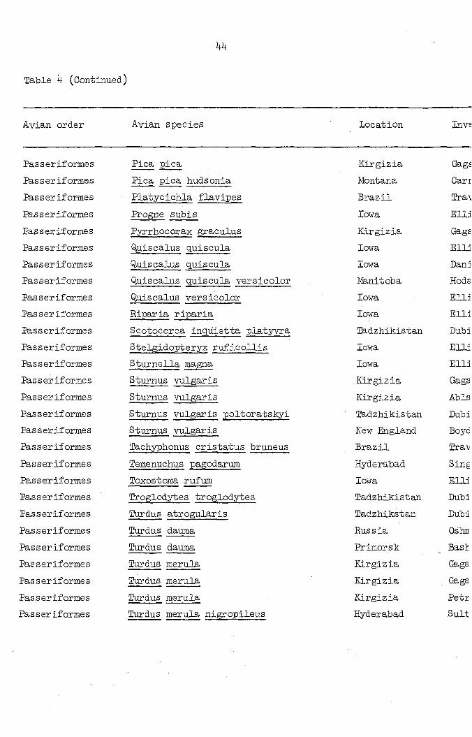
Table k (Continued)
44
Avian order Avian species Location Inve
Passeriformes Pica pica Kirgizia Gage
Passeriformes Pica pica hudsonia Montana Carr
Passeriformes Platycichla flavipes Brazil Tra\
Passeriformes Progne subis Iowa Elli
Passeriforme s Pyrrhocorax graculus Kirgizia Gage
Passeriformes Quiscalus q.uiscula Iowa Elli
Passeriformes Quiscalus quiscula Iowa Dan 3
Passeriforme s Quiscalus quiscula versicolor Manitoba Hods
Passeriformes Quiscalus versicolor Iowa Elli
Passeriformes Riparia riparia Iowa Elli
Passeriformes Scotocerca inq.Uietta platyvra Tadzhikistan Dubi
Passeriformes Stelgidopteryx ruficollis Iowa Elli
Passeriformes Sturnella magna Iowa Elli
Passeriformes Sturnus vulgaris Kirgizia Gaga
Passeriformes Sturnus vulgaris Kirgizia Abla
Passeriformes Sturnus vulgaris poltoratskyi Tadzhikistan Dubi
Passeriformes Sturnus vulgaris New England Boyc
Passeriformes Tachyphonus cristatus bruneus Brazil Tra-v
Passeriformes Temenuchus pagodarum Hyderabad Sing
Passeriformes Toxostoma rufum Iowa Elli
Passeriformes Troglodytes troglodytes Tadzhikistan Dubi
Passeriformes . Turdus atrogularis Tadzhikstan Dubi
Passeriformes Turdus dauma Russia Oshr
Passeriformes Turdus dauma Primorsk Bast
Passeriformes Turdus merula Kirgizia Gaga
Passeriformes Turdus merula Kirgizia Gaga
Passeriformes Turdus merula Kirgizia Petz
Passeriforme s Turdus merula nigropileus Hyderabad Suit

Location Investigator Microtetrameres species
Kirgizia Gagarin, Ablasov and Chibichenko, 1957 sp.
Montana Carney, 196? sp.
Brazil Travassos, 1915 pusilla.
Iowa Elli s, unpubli she d sp.
Kirgizia Gagarin, Ablasov and Chibichenko, 1957 sp.
Iowa Ellis, unpublished sp.
Iowa Daniell sp.
Manitoba Hodasi, I963 sp.
Iowa Ellis, 1961 sp.
Iowa Ellis, unpublished sp.
Tadzhikistan Dubinina and Serkova, , 1951 . e
inermis
Iowa Ellis, unpublished sp.
Iowa Ellis, 1961 sp.
Kirgizia Gagarin, Ablasov and Chibichenko, 1957 . e
inermis
Kirgizia Ablasov and Chibichenko, I962 . e
inermis
Tadzhikistan Dubinina and Serkova, 1951 . e
inermis
New England Boyd, 1951 helix
Brazil •ïravassos, l9lk minima
Hyderabad Singh, I9U9 . e
inermis
Iowa Ellis, unpublished sp.
Tadzhikistan Dubinina and Serkova, 1951 e
inermis
Tadzhikstan Dubinina and Serkova, 1951 . e
inermis
Russia Oshmarin, 1956 pusilla
Primorsk Bashkirova, i960 pusilla
Kirgizia Gagarin, Ablasov and Chibichenko, 1957 sp.
Kirgizia Gagarin, Ablasov and Chibichenko, 1957 oriolus
Kirgizia Petrov and Chertkova, 195Cb sp.
Hyderabad Sultana, I962 cephalatus
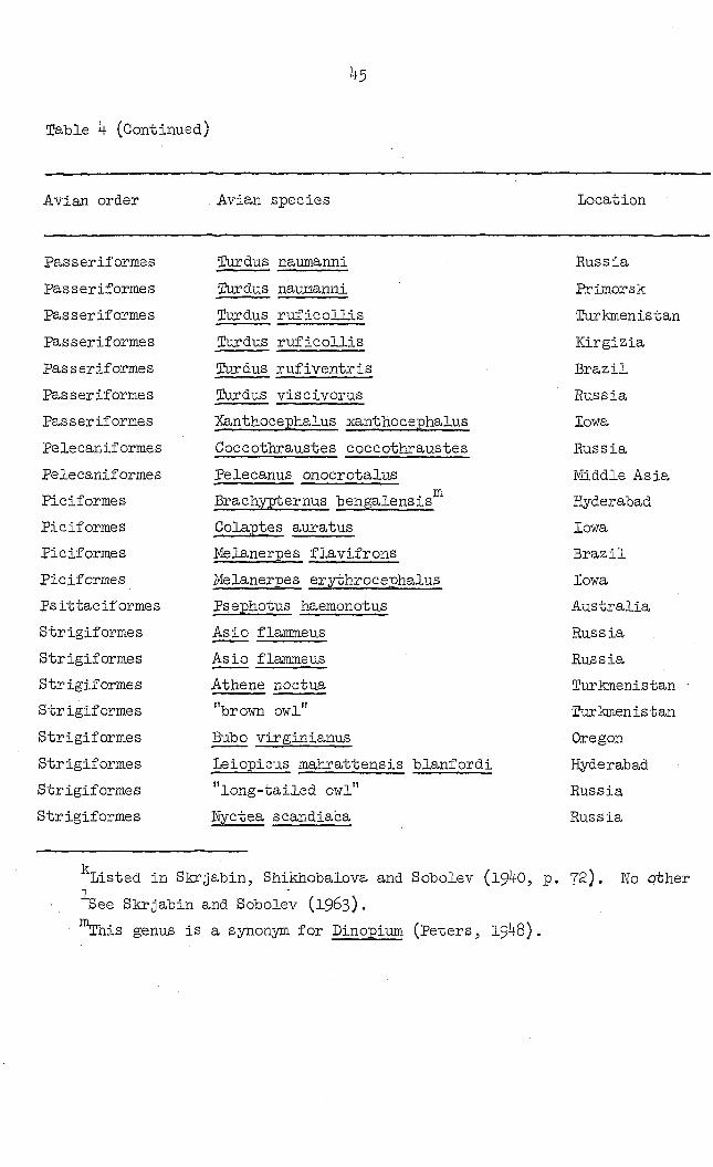
i+5
Table 4 (Continued)
Avian order Avian species Location
Passeriformes lurdus naumanni Russia
passeriformes îurdus naumanni Prlmorsk
Passeriformes lurdus ruficollis Turkmenistan
Passeriformes Turdus ruficollis Kirgizia
Passeriformes Tardus rufiventris Brazil
Pas serif orme s (Durdus viscivorus Russia
Passeriformes Xanthocephalus xanthocepiialus Iowa
Pelecaniformes Coccothraustes coccothraustes Russia
Pelecaniformes Pelecanus onocrotalus Middle Asia
Piciformes Brachypternus bengalensis Hyderabad
Piciformes Colaptes auratus Iowa
Piciformes Melanerpes flavifrons Brazil
Piciformes Melanerpes er.ythrocephalus Iowa
Ps ittac if orme s Psephotus haemonotus Australia
Strigiformes Asio flamme us Russia
Strigiformes Asio flammeus Russia
Strigiformes Athene noctua Turkmenistan
Strigiformes "brown owl" Turkmenistan
Strigiformes Bubo virginianus Oregon
Strigiformes Leiopicus mahrattensis blanfordi Hyderabad
Strigiformes "long-tailed owl" Russia
Strigiformes Wyctea scandiaca Russia
Listed in Skrjabin, Shikliobalova and Sobolev (19 0, p. 72). Ho other 1
See Skrjabin and Sobolev (1963).
™This genus is a synonym for Dinopium (Peters, 1948).

Location Investigator Microtetrameres species
Russia OshmarinJ 1956 pusilla
Primorsk Bas hkir ova, i960 pusilia
Turkmenistan Rizhikov and Kozlov, 1959 sp.
Kirgizia Gagarin, Ablasov and Chibichenko, 1957 or iolus
Brazil Travassos, 1915 pusilla
Russia Skrjabin, Shikhobalova and Sobolev, 19 9 pusilla
Iowa Ellis, unpublished sp.
Russia Oshmarin, 1956 sp.
Middle Asia 1 pelecani
Hyderabad Singh, 1949 . e
inermis
Iowa Ellis, unpublished sp.
Brazil Travassos, 1914 cruzi
Iowa Ellis, unpublished sp.
Australia Thomas, 1960 sp.
Russia Oshmarin, 1956 bubo
Russia 8krjabin and Sobolev, 1963 oshmarini
Turkmenistan - Rizhikov and Kozlov, 1959 sp.
Turkmenistan Rizhikov and Kozlov, 1959 species
Oregon Schell, 1953 bubo
Hyderabad Sultana, I962 . e
inermis
Russia Oshmarin and Parukhin 1963 bubo
Russia Skrjabin and Sobolev, 1963 oshmarini
72). No other information given.
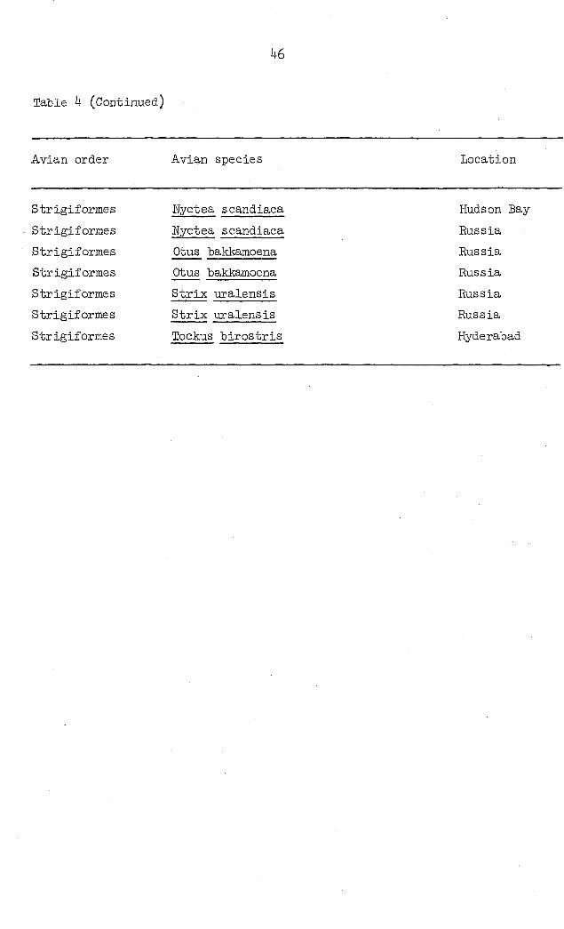
46
Table 4 (Continued)
Avian order Avian species Location
Strigiformes
Strigiformes
Strigiformes
Strigiformes
Strigiformes
Strigiformes
Strigiformes
Uyctea scandiaca
Myctea scandiaca
Otus bakkamoena
Otus bakkamoena
Strix uralensis
Strix uralensis
Tockus birostris
Hudson Bay
Russia
Russia
Russia
Russia
Russia
Hyderabad
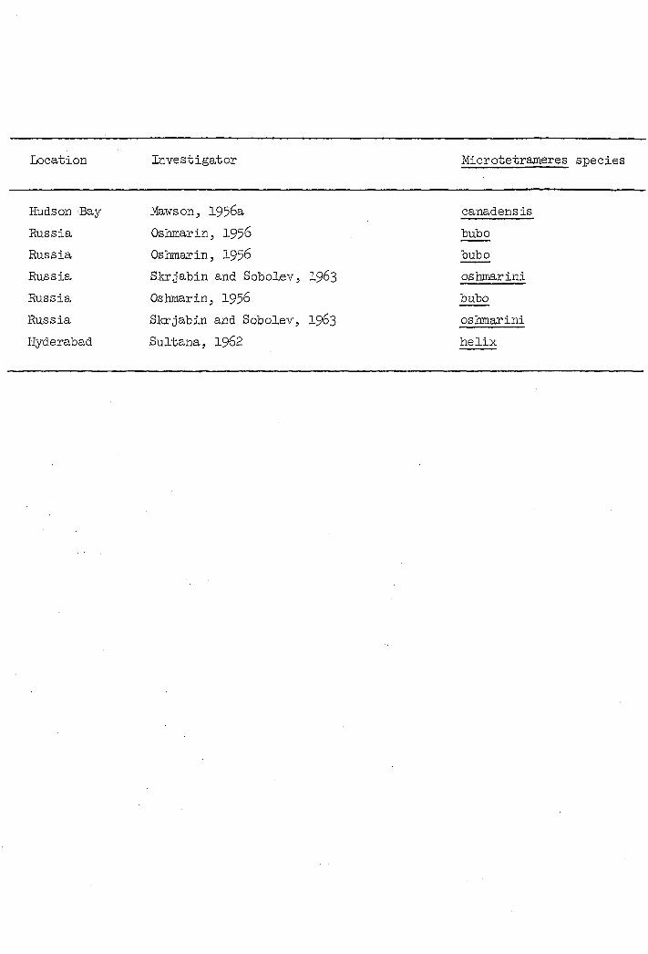
Location Investigator Microtetrameres species
Hudson Bay-
Russia
Russia
Russia
Russia
Russia
Hyderabad
Mawson, 1956a
Oshmarin, 195
Oshmarin, 1956
Skrjabin and Sobolev, I963
Oshmarin, 1956
Skrjabin and Sobolev, 1963
Sultana, I962
canadens is
bubo
bubo
oshmarini
bubo
oshmarini
helix
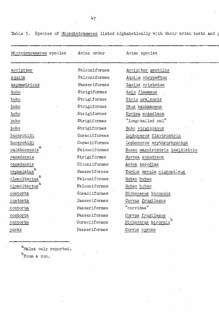
^7
Table 5- Species of Microtetrameres listed alphabetically with their avian hosts and
Microtetrameres species Avian order Avian species
accipiter Falconiformes Accipiter gentilis
aguila Falconiformes Aguila chrysaëtos
asymmetricus Passeriformes Lanius cristatus
bubo Strigiformes Asio flammeus
bubo Strigiformes Strix uralensis
bubo Strigiformes Otus bakkamoena
bubo Strigiformes Wyctea scandiaca
bubo Strigiformes "long-tailed owl"
bubo Strigiformes Bubo Virginianus
bucerotidi Coraciiformes Lophoceros flavirostris
bucerotidi Goraciiformes Lophoceros erythrorhynchus
calabocensis Falconiformes Buteo magnirostris insidiatrix
canadensis Strigiformes Wyctea scandiaca
canadensis Ciconiiformes Ardea herodias
cephalatus Passeriformes Turdus merula nigropileus
cloacitectus Falconiformes Buteo buteo
cloacitectus Falconif ormes Buteo buteo
contorta Goraciiformes Dichocerus bicornis
contorta Passeriformes Corvus frugilegus
contorta Pas serif orme s "corvidae"
contorta Passeriformes Corvus frugilegus
contorta Coraciiformes Dichocerus bicornis
corax Passeriformes Corvus corone
Males only reported.
From a zoo.
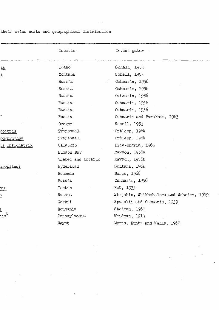
their avian hosts and geographical distribution
Location Investigator
is Idaho Schell, 1953
s Montana Schell, 1953
Russia Oshtnarin, 195
Russia Oshmarin, 1956
Russia Oslamarinj 1956
Russia Oshmarin, 1956
Russia Oshmarin, 1956 II Russia Oshmarin and Parukhin, 1963
Oregon Schell, 1953
rostris Transvaal Ortlepp, 1964
forhynchus Transvaal Ortleppj 1964
Is insidiatrix Calabozo Diaz-Ungria, I965
Hudson Bay MawsonJ 1956a
Quebec and Ontario Mawson, 1956a
gropileus Hyderabad Sultana, 1962
Bohemia Barus, I966
Russia Oshmarin, 1956
lis Tonkin Hsu, 1935
3 Russia Skrjabin, Shikhobalova and Sobolev, 19 9
Gorkii Spasskii and Oshmarin, 1939
3 Roumania Stoican, 196O
Pennsylvania Weidman, I913
Egypt Myers, Kuntz and Wells, 1962
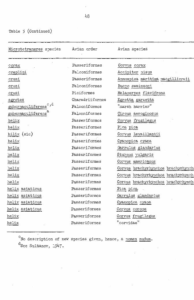
Table 5 (Continued)
48
Microtetrameres species Avian order Avian species
corax Passeriformes Corvus corax
creplini Falconiformes Accipiter nisus
cruzi Passeriforme s Ammospiza maritima macgillivraii
cruzi Falconiformes Bucco swainsoni
cruzi Piciformes Melanerpes flavifrons
egretes Charadriiformes Egretta garzetta c d.
gubernaculiferens ' Falconiformes "marsh harrier"
gub ernaculiferens Falconiformes Circus aeroginosus
helix Passeriformes Corvus frugilegus
helix Passeriformes Pica pica
hilix (sic) Passeriformes Corvus levaillantii
helix Passeriformes Cyanopica cyana
helix Passeriformes Garrulus glandarius
helix Passeriformes Sturnus vulgaris
helix Passeriformes Corvus americanus
helix Passeriformes Corvus brachyrhynchos brachyrhynch
helix Passeriformes Corvus brachyrhynchos brachyrhynch
helix Passeriformes Corvus brachyrhynchos brachyrhynch
helix asiaticus Passeriformes Pica pica-
helix asiaticus Passeriformes Garrulus glandarius
helix asiaticus Pas seriformes Cyanopica cyana
helix asiaticus Passariformes Corvus corone
helix Passeriformes Corvus frugilegus
helix Passeriformes "corvidae"
" Eo description of new species given, hence, a nomen nudum.
See Sultanov, 1947.

s Location Investigator
Idaho SChe11, 1953
sus Russia Vavilova, 1926
ritima macgillivraii North Carolina Hunter and Quay, 1953
oni Brazil Travassos, l^lh
lavifrons Brazil Travassos, 19l4
etta Hyderabad Rasheed, I96O
er" Tashkent Sultanov, 19 +5
inosus Central Asia Sultanov, 19 7
legus Kirgizia Ablasov and Chibichenko, 1962
Kirgizia Ablasov and Chibichenko, 1962
liantii Primorsk BashkirovaJ 196O
a.na Primorsk Bashkirova, 196O
Qdarius Primorsk Bashkir ova .5 I96O
3-ris New England Boyd, 1951
:anu8 Washington, D.C. Cram, I927
yxhynchos brachyrhynclios Manitoba Hodasi, 1963
yrhynchos brachyrhynchos Quebec Mawson, 1956b
/Thynchos brachyrhynchos Iowa - Wisconsin Morgan and Waller, 19 1
Russia Oshmarin, 1956
idarius Russia Oshmarin, 1956
ma Russia Oshmarin, 1956
Russia Oshmarin, 1956
Legus Russia 8krjabin, Shikobalova and Sobolev, 19 9
Gorkii Spasskii and Oshmarin, 1939
lum.

1+9
Table 5 (Continued)
Microtetrameres species Avian order Avian species
helix
helix
helix . e
inermis
inermis
inermis
Passeriforme s
Passeriformes
Strigiformes
Passeriformes
Passeriformes
Falconiformes
Corvus frugilegus
Corvus corone cornix
Tockus birostris
Oenanthe isabelina
Sturnus vulgaris
"sparr ow-hawk"
inermis
inermis
. e inermis
. e inermis
inermis
inermis
inermis
inermis
inermis . e
inermis . e
inermis
inermis g
inermis
inermis
inermis . e
inermis
Passeriformes
.Falconiformes
Passeriformes
Passeriformes
Passeriformes
Passeriformes
Passeriformes
Passeriformes
Passeriformes
Passeriformes
Passeriformes
Passeriformes
Passeriformes
Passeriformes
Passeriformes
Passeriformes
Passer domesticus
Astur nisus
lanius sp.
Alauda arvensis dulcinx
Anthus spinoletta blakistani
Corvus cornix sharpei
Corvus frugilegus
Emberiza leucocephalus
Emberiza schoenichus pallidior
Parus cinereus
Parus major
Phoenicurus erythronotus
Scotocerca inq.uietta platyvra
Turdus atrogularis
Troglodytes troglodytes
Sturnus vulgaris poltoratskyi
®See text for comments relative to this species.

Location Investigator
Routnania
Bulgaria
Hyderabad
Kirgizia
Kirgizia
Algeria-Rus s ia-Turkestan
Algeria-Russia-Turkestan
Alger ia-Rus s ia-Turkestan
Algeria-Russia-Turkestan
Tadzhikistan
Tadzhikistan
Tadzhikistan
Tadzhikistan
Tadzhikistan
Tadzhikistan
Tadzhikistan
Tadzhikistan
Tadzhikistan
Tadzhikistan
Tadzhikstan
Tadzhikstan
Tadzhikistan
Stoican, I960
Stoimenov, I963
Sultana, 1962
Ablasov and Chiblchenko, 1962
Ablasov and Chiblchenko, 1962
Cram, 1927
Cram, 1927
Cram, 192?
Cram, I927
Dubinina and Serkova, 1951
Dubinina and Serkova, 1951
Dubinina and Serkova, 1951
Dubinina and Serkova, 1951
Dubinina and Serkova, 1951
Dubinina and Serkova, 1951
Dubinina and Serkova, 1951
Dubinina and Serkova, 1951
Dubinina and Serkova, 1951
Dubinina and Serkova, 1951
Dubinina and Serkova, 1951
Dubinina and Serkova, 1951
Dubinina and Serkova, 1951
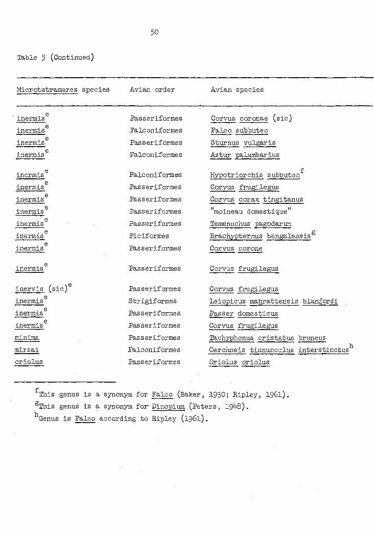
Table 5 (Continued)
50
Microtetrameres species Avian order Avian species
. e inermis Passeriformes Corvus coronae (sic)
. e inermis Falconiformes Falco subbuteo
inermis® Passeriformes Sturnus vulgaris
inermis® Falconiformes Astur palumbarius
. e inermis Falconiformes
f Hypotriorchis subbuteo
inermis Passeriformes Corvus frugilegus
inermis® Passeriformes Corvus corax tingitanus . e
inermis Passeriformes "moineau domestique" . e
inermis Passeriformes Temenuchus pagodarum . e
inermis Piciformes Brachypternus bengalensis®
inermis® Passeriformes Corvus corone
. e inermis Passeriformes Corvus frugilegus
inervis (sic)® Passeriformes Corvus frugilegus . e
inermis Strigiformes Leiopicus mahrattensis blanfordi . e
inermis Passeriformes Passer domesticus . e
inermis Passeriformes Corvus frugilegus
minima Passeriformes 2fe.chyphonus cristatus bruneus
mirzai Falconiforme s Cerchneis tinnunculus interstinct
oriolus Passeriformes Oriolus oriolus
This genus is a synonym for Falco (Baker, 1930; Ripley, 1961).
®This genus is a synonym for-Dinopium (Peters, 19 +8).
Genus is Falco according to Ripley (1961).
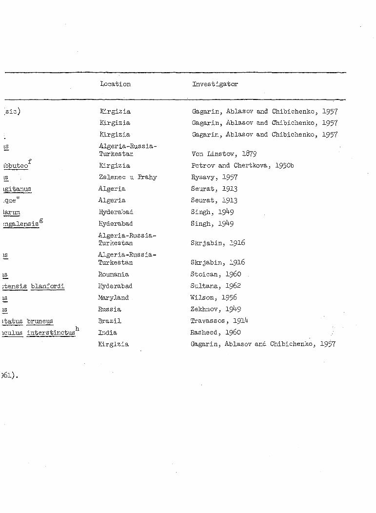
Location Investigator
sic) Kirgizia Gagarin, Ablasov and Chibichenko, 1957 Kirgizia Gagarin, Ablasov and Chibichenko, 1957
: Kirgizia Gagarin, Ablasov and Chibichenko, 1957 IS Algeria-Rus s ia-
Turkestan Von Linstow, 1879 f
Lbbuteo Kirgizia Petrov and Ghertkova, , 1950b
18 Zelenec u Erahy Rysavy, 1957
igitanus Algeria Seurat, 1913
.que" Algeria Seurat, I913
Larum Hyderabad Singh, 19 9 g
:ngalensis° Hyderabad
Algeria-Hussia-lurkestan
Singh, 19 9
Skrjabin, 1916
J£ Alger ia-Rus s ia-Turkestan Skrjabin, I916
Roumania Stoican, I96O
;tensis blanfordi Hyderabad Sultana, I962
iË. Maryland Wilson, 1956
Russia Zekhnov, 19 +9
îtatus brimeus Brazil Travassos, 1914
iculus interstinctus India Rasheed, I96O
Kirgizia Gagarin, Ablasov and Chibichenko, 1957
)6l).

51
Table 5 (Continued)
Microtetrameres species Avian order Avian species
oriolus Passeriformes Oriolus oriolus turkestanicus
oriolus orientalis Passeriformes Oriolus chinensis
oriolus Passeriformes Turdus merula
oriolus Passeriformes Turdus ruficollis
oshmarini Strigiformes Asio flamme us
oshmarini Strigiformes Nyctea scandiaca
oshmarini Strigiformes Otus bakkamoena
oshmarini Strigiformes Strix uralensis
osmaniae Falconiformes Accipiter nisus nisosimilis
papillocephala Falconiformes Butastur indicus
palecani Pelecaniformes Pelecanus onocrotalus
pokrowskyi Ciconiiformes Ardea cinerea
pusilia Passeriformes "motley thrush"
pusilia Passeriformes Turdus dauma
pusilia Passeriformes Turdus dauma
pusilia Passeriformes Turdus naumanni
pusilia Passeriformes Turdus naumanni
pus ilia Passeriformes Turdus rufiventris
pusilia Passeriformes Turdus viscivorus
pus ilia Passeriformes Platycichla flavipes
rasheedae Cueuliformes Hierococcyx varius a
singhi Falconiformes Falco jugger
spiculata Passeriformes Cyanocitta cristata
spiralis Ciconiiformes Ardeola ibis ibis
spiralis Ciconiiformes Bubulcus coromandus
See Skrgabin and Sobolev (1963).
Listed in Skrjabin, Shikhobalova and Sobolev (l$40, p. 72). No other information ,
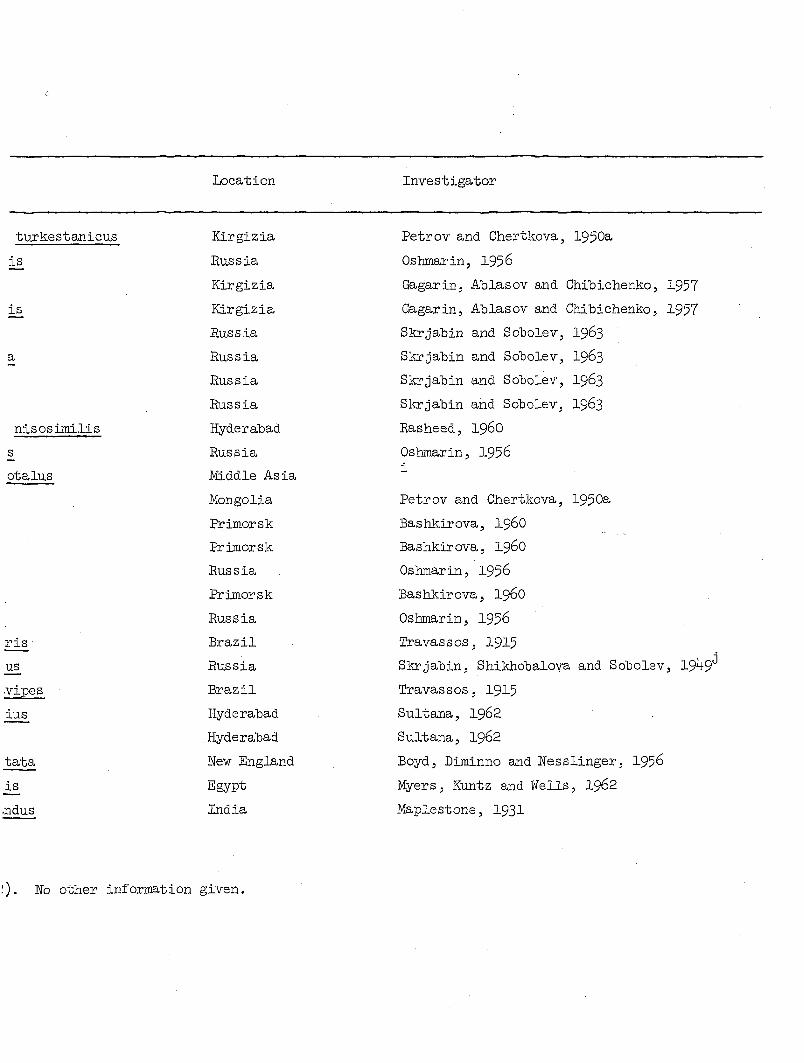
Location Investigator
turkestanicus Kirgizia Petrov and Chertkova, 1950a
i£ Russia Oshmarin, 1956
Kirgizia Gagarin, Ablasov and Chibichenko,
is Kirgizia Gagarin, Ablasov and Chibichenko,
Eussia Skrjabin and Sobolev, I963
a Russia Skrjabin and Sobolev, I963
Russia Skrjabin and Sobolev, I963
Russia Skrjabin and Sobolev, I963
nisosimilis Hyderabad Rasheed, I96O
s Russia Oshmarin, 1956
otalus Middle Asia i
Mongolia Petrov and Chertkova, 1950a
Primorsk Bashkirova, i960
Primorsk Bashkirova, I96O
Russia Oshmarin, 1956
Rrimorsk Bas hkir ova, I96O
Russia Oshmarin, 1956
ris Brazil Travassos, 1915
us Russia Skrjabin, Shikhobalova and Sobolev
vipes Brazil Travassos, 1915
ius Hyderabad Sultana, I962
Hyderabad Sultana, I962
tata New England Boyd, Diminno and Nesslinger, 1956
is Egypt Myers, Kuntz and Wells, I962
.ndus India Maplestone, 1931
No other information given.

52
Table 5 (Continued)
Microtetrameres species Avian order Avian species
spiralis Ciconiiformes Bubulcus lucidus
spiralis Falconiformes Falco feldegii
travassosi Passeriformes Gracula religiosa a,l
tubocloacis Falconiformes AçLuila clanga
sp. Falconiformes Falco tinnunculus
sp. Passeriformes Motacilla alba
sp. Passeriformes Motacilla fiava
sp. Passeriformes Pica pica hudsonia
sp. Cuculiformes Coccyzus atnericanus
sp. Passeriformes Cyanocitta cristata
sp. Passeriformes Quiscalus quiscula
sp. Passeriformes Sturnella magna
sp. Passeriformes Quiscalus versicolor
sp. Piciforme s Colaptes auratus
sp. Passeriformes Cyanocitta cristata
sp. Piciformes Melanerpes erythrocephalus
sp. Passeriformes Melospiza georgiana
sp. Passeriformes Myiarchus crinitus
sp. Passeriformes Passerella iliaca
sp. Passeriformes Progne subis
sp. Passeriformes Quiscalus quiscula
sp. Passeriformes Riparia riparia
sp. Passeriformes Stelgidopteryx ruficollis
Genus emended to Mainatus by 8harpe (1890).
Transferred to Gubernaculomeres by Oshmarin and Parukhin (1963).
Private communication.
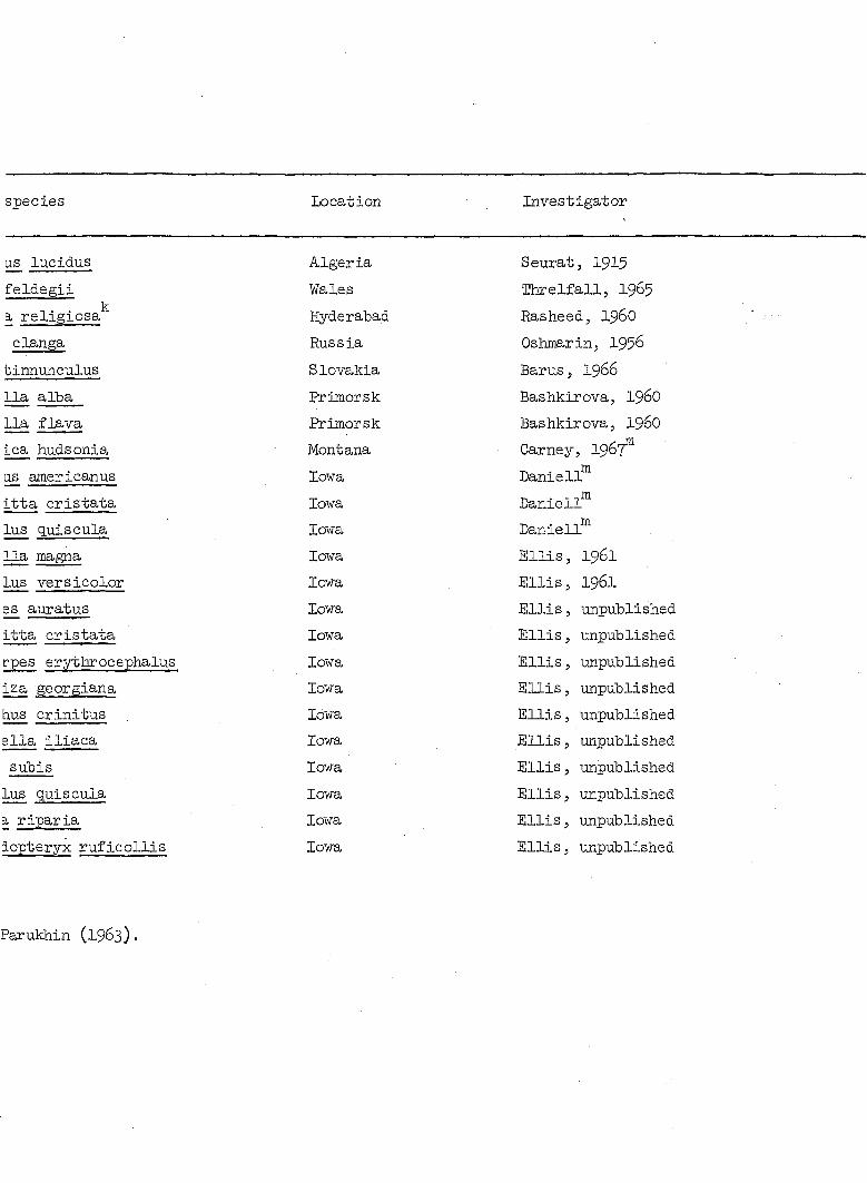
Location Investigator
Algeria
Wales
Hyderabad
Russia
Slovakia
Primorsk
Erimorsk
Montana
Iowa
Iowa
Iowa
Iowa
Iowa
Iowa
Iowa
Iowa
Iowa
Iowa
Iowa
Iowa
Iowa
Iowa
Iowa
Seurat, 1915
Hxrelfall, I965
Rasheedj 196O
Oshjmrin, 1956
Barus, 1966
Bashkirova5 I96O
BashkirovaJ I96O
Carney, I967™
Daniel!
Daniell
Daniell™'
Ellis, 1961
Ellis, 1961
Ellis, unpublished
Ellis, unpublished
Ellis, unpublished
Ellis, unpublished
Ellis, unpublished
Ellis, unpublished
Ellis, unpublished
Ellis, unpublished
Ellis, unpublished
Ellis, unpublished
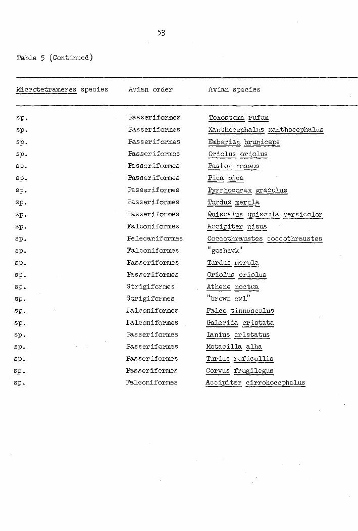
53
Table 5 (Continued)
Microtetrameres species Avian order Avian species
sp. Passeriforme s Toxostoma rufum
sp. Passeriforme s Xanthocephalus xanthocephalus
sp. Passeriformes Emberiza bruniceps
sp. Passeriformes Oriolus oriolus
sp. Passeriformes Pastor roseus
sp. Passeriformes Pica pica
sp. Passeriformes Pyrrhocorax graculus
sp. Passeriformes Turdus merula
sp. Passeriformes Quiscalus quiscula versicolor
sp. Falconiformes Accipiter nisus
sp. Pelecaniformes Coccothxaustes coccothxaustes
sp. Falconiformes "goshawk"
sp. Passeriformes Turdus merula
sp. Passeriforme s Oriolus oriolus
sp. Strigiformes Athene noctua
sp. Strigiformes "brown owl"
sp. Falconiformes Falco tinnuncuius
sp. Falconiformes Galerida cristata
sp. Passeriformes lanius cristatus
sp. Passeriformes Motacilla alba
sp. Passeriformes Turdus ruficollis
sp. Passeriformes Corvus frugilegus
sp. Falconiformes Accipiter cirrohocephalus
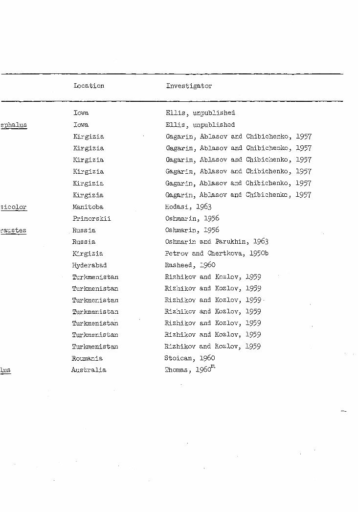
Location Investigator
Iowa
Iowa
Kirgizia
Kirgizia
Kirgizia
Kirgizia
Kirgizia
Kirgizia
Manitoba
Erimorskii
Russia
Russia
Kirgizia
Hyderabad
Turkmenistan
Turkmenistan
Turkmenistan
Turkmenistan
Turkmenistan
Turkmenistan
Turkmenistan
Roumania
Australia
Ellis, unpublished
Ellis, unpublished
Gagarin, Ablasov and Chibichenko,
Gagarin, Ablasov and Chibichenko,
Gagarin, Ablasov and Chibichenko,
Gagarin, Ablasov and Chibichenko,
Gagarin, Ablasov and Chibichenko,
Gagarin, Ablasov and Chibichenko,
Hodas i, I963
Oshmarin, 1956
Oshtnarin, 1956
Oshmarin and Parukhin, I963
Petrov and Chertkova, 1950b
Rasheed, i960
Rizhikov and Kozlov, 1959
Rizhikov and Kozlov, 1959
Rizhikov and Kozlov, 1959•
Rizhikov and Kozlov, 1959
Rizhikov and Kozlov, 1959
Rizhikov and Kozlov, 1959
Rizhikov and Kozlov, 1959
Stoican, 196O
Thomas, 1960
1957
1957
1957
1957
1957
1957
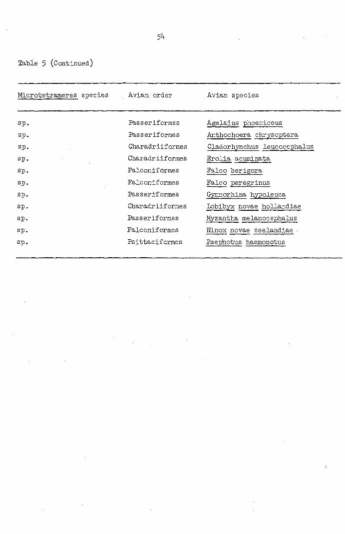
Table 5 (Continued)
54
Microtetrameres species Avian order Avian species
sp. Passeriformes Agelaius phoeniceus
sp. Passeriformes Anthochoera chrysoptera
sp. Charadriiformes Cladorhynchus leucocephalus
sp. Ciiaradr iif ormes Erolia acuminata
sp. Falconiformes Falco berigora
sp. Falconiformes Falco peregrinus
sp. Passeriforme s Gymnorhina hypoleuca
sp. Charadriiformes Lobibyx novae hollandiae
sp. Passeriformes Myzantha melanocephalus
sp. Falconiformes Ninox novae zeelandiae •
sp. Ps ittac if orme s Psephotus haemonotus
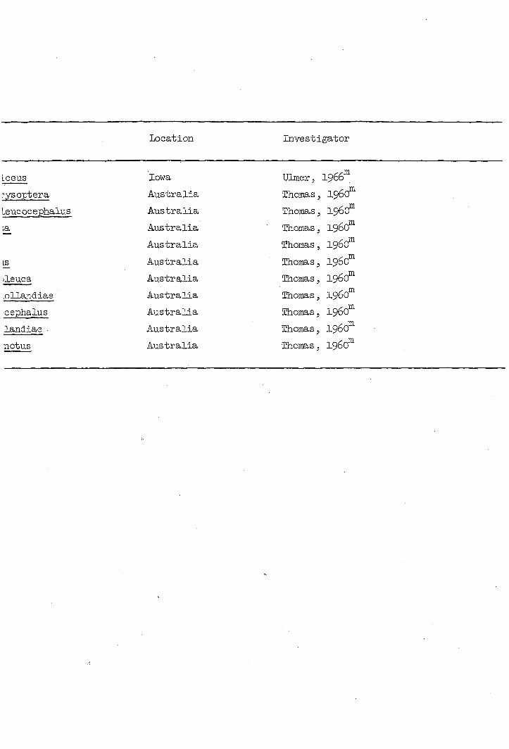
Location Investigator
Lceus lowa Ulraer5 1966*
rysoptera Australia Thomas, 1960
Leucocephalus Australia Thomas, 196(f
|a Australia Thomas, i960*
Australia Thomas, i960*
1£ Australia Thomas, i960*
ileuca Australia Thomas, i960*
.ollandiae Australia Thomas, i960*
cephalus Australia Thomas, 1960F
landiae • Australia Thomas, i960*
notus Australia Thomas, 1960°
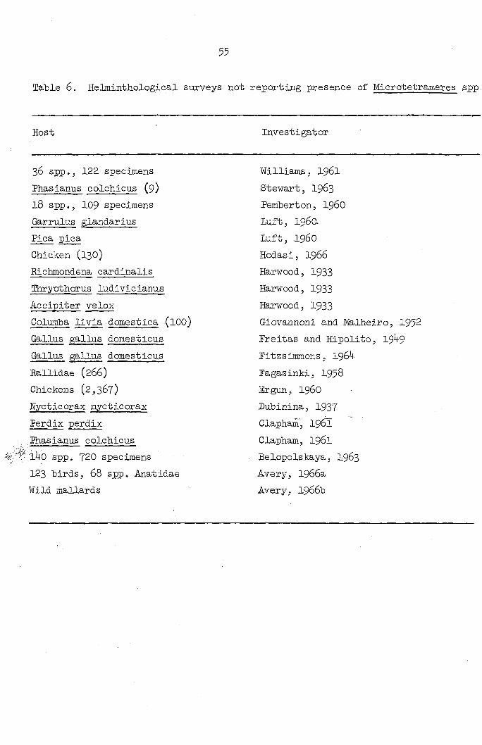
55
Table 6. Helminthological surveys not reporting presence of Microtetrameres spp,
Host
36 spp., 122 specimens
Phasianus colchicus (9)
18 spp., 109 specimens
Garrulus glandarius
Pica pica
Chicken (130)
Richmondena cardinalis
Ihryothorus ludivicianus
Accipiter velox
Columba livia domestica (lOO)
Gallus gallus domesticus
Gallus gallus domesticus
Rallidae (266)
Chickens (2,367)
Wycticorax nycticorax
Perdix perdix
Kiasianus colchicus
'l4o spp. 720 specimens
123 birds, 68 spp. Anatidae
Wild mallards
Investigator
Williams, I96I
Stewart, I963
Pemberton, I96O
Luft, i960.
Luft, i960
Hodasi, 1966
Harwood, 1933
Harwood, I933
Harwood, I933
Giovannoni and iyfe.lheiro, 1952
Freitas and Hipolito, 19 9
Fitzsimmons, 1964
Fagasinki, 1958
Ergun, i960
Dubinina, 1937
Clapham, I961
Clapham, I961
Belopolskaya, I963
Avery, 1966a
Avery, 1966b

not reporting presence of Microtetrameres spp. in avian hosts
Investigator Location
Williams, I96I
Stewart, I963
femberton, 196O
Luf15 i960.
Luftj i960
Hodasi, 1966
Harwood, 1933
Harwood, 1933
Harwood, 1933
Giovannoni and Malheiro, 1952
Freitas and Hipolito, 19 9
Fitzsimmons, 1964
Fagasinki, 1958
Ergun, i960
Dubinina, 1937
Clapham, I961
Clapham, I961
Belopolskaya, I963
Avery, 1966a
Avery, 1966b
Britain
Illinois
Britain
Poland (Lublin Palatinate)
Poland (Lublin Palatinate)
Manitoba
Texas
Texas
Texas
Brazil
Minas Gérais
Nyassaland
Poland
Turkey
Russia
Britain
Britain
Russia (Sudzukjinsk)
Britain
Britain
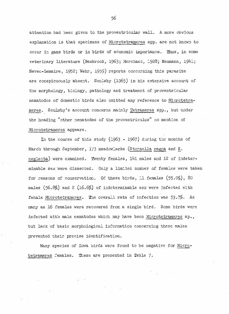
56
attention had been given to the proventricular wall. A more obvious
explanation is that specimens of Microtetrameres spp. are not known to
occur in game birds or in birds of economic importance. Thus, in some
veterinary literature (Benbrook, 1963; Merchant, 1928; Neumann, 19 1;
Neveu-Lemaire, 1952; Wehr, 1959) reports concerning this parasite
are conspicuously absent. Soulsby (1965) in his extensive account of
the morphology, biology, pathology and treatment of proventricular
nematodes of domestic birds also omitted any reference to Microtetra
meres . Soulsby's account concerns mainly Tetrameres spp., but under
the heading "other nematodes of the proventriculus" no mention of
Microtetrameres appears.
In the course of this study (1963 - I967) during the months of
March through September, 173 meadowlarks (Sturnella magna and S_,
neglecta) were examined. Twenty females, l4l males and 12 of indeter
minable sex were dissected. Only a limited number of females were taken
for reasons of conservation. Of these birds, 11 females (55-0 ), 80
males ($6.8%) and 2 (16.6%) of indeterminable sex were infected with
female Microtetrameres. The overall rate of infection was 53•7 - As
many as I6 females were recovered from a single bird. Some birds were
infected with male nematodes which may have been Microtetrameres sp.,
but lack of basic morphological information concerning these males
prevented their precise identification.
Many species of Iowa birds were found to be negative for Micro
tetrameres females. These are presented in Table 7*

57
Table 7- Summary of Iowa birds found uninfected with Microtetrameres spp.
Common name Family ' Scientific name
Pied-billed grebe Colymbidae Podilymbus podic s Pintail Anatidae Anas acuta Blue-winged teal Anat idae Anas discors Ruddy duck Anatidae Oxyura jamaicensis Mallard Anatidae Anas platyrhynchos Red-tailed hawk Accipitridae Buteo jamaicensis Ring-necked pheasant Phasianidae Phasianus colchicus American coot Rallidae Eulica americana Killdeer Charadriidae Charadrius vociferus Upland plover Scolopacidae Bartrajnia longicauda Stilt sandpiper Scolopacidae Micropalama himantopus Least sandpiper Franklin's gull
Scolopacidae Erolia minutilla Least sandpiper Franklin's gull Laridae Larus pipixcan Ring-billed gull Laridae Larus delawarensis Forster's tern laridae Sterna forsteri Black tern Laridae Chlidonias niger Lesser yellowlegs Scolopacidae Totanus flavipes Rock dove Golumbidae Columba livia Mourning dove Columbidae Zenaidura macroura Yellow-billed cuckoo Cuculidae Coccyzus americanus Black-billed cuckoo Cuculidae Coccyzus erythrophthalmus Belted kingfisher Alcedinidae Megaceryle alcyon Yellow-shafted flicker Picidae Colaptes auratus Red-headed woodpecker Picidae MeIanerpes erythrocephalus Hairy woodpecker Picidae Dendrocopos villosus Downy woodpecker Picidae Dendrocopos pubescens Red-bellied woodpecker Picidae Centurus carolinus Eastern kingbird Tyrannidae Tyrannus tyrannus Horned lark Alaudidae Eremophila alpestris Bank swallow Hirundinidae Riparia riparia Purple martin Hirundinidae Progne subis Rough-winged swallow Hirundinidae Stelgidopteryx ruficollis Barn swallow Hirundinidae Hirundo rustica Blue jay Corvidae Cyanocitta cristata Common crow Corvidae Corvus brachyrhynchos
Excluding sturnella spp.

i with Microtetrameres spp.
Scientific name Number of birds examined
Podilymbus podiceps 3 Anas acuta 2 Anas discors 1 Oxyura jamaicensis 2 Anas platyrhynchos 1 Buteo jamaicensis 1 Phasianus colchicus 42 Fulica americana 4 Charadrius vociferus 1 Bartramia longicauda 2 Micropalama himantopus 1 Erolia minutilla 3 Larus pipixcan 4 Larus delawarensis 5 Sterna forsteri 1 Chlidonias niger 1 Totanus flavipes 1 Columba livia 11 Zenaidura macroura 1 Coccyzus americanus 1 Coccyzus erythrophthalmus 1 Megaceryle alcyon 1 Golaptes auratus 1 MeIanerpes erythrocephalus 1 Dendrocopos villosus 1 Dendrocopos pubescens 1 Centurus carolinus 1 Tyrannus tyrannus 2 Eremophila alpestris 2 Eiparia riparia 6 Progne subis 1 Stelgidopteryx ruficollis 1 Hirundo rustica 2 Cyanocitta cristata 13 Corvus brachyrhynchos 1
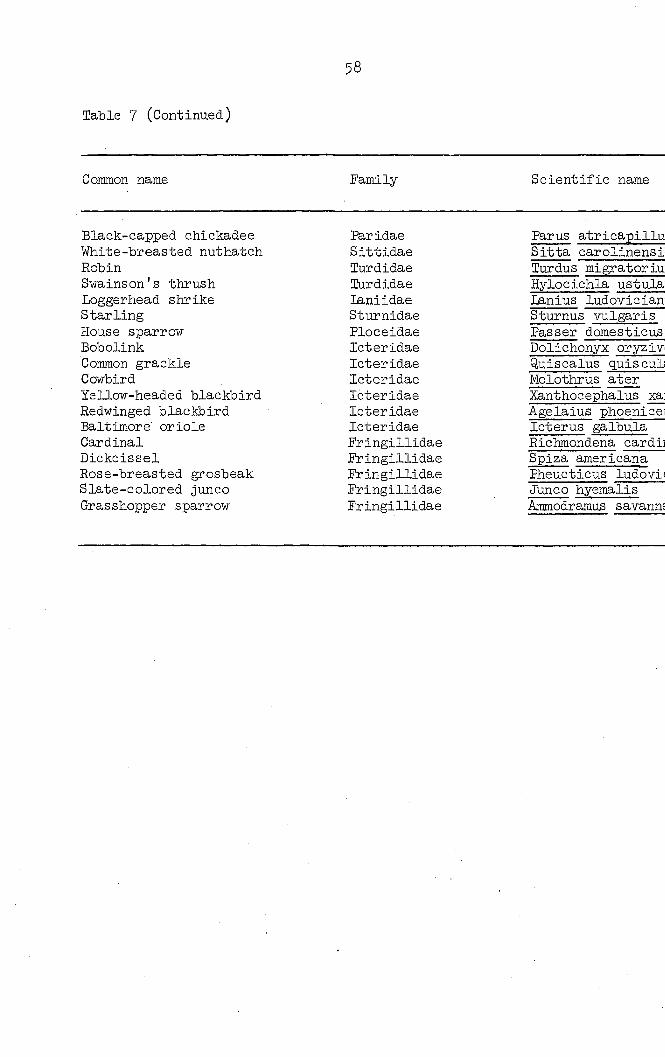
Table 7 (Continued)
58
Common name Family- Scientific name
Black-capped chickadee Par idae Parus atricapillu White-breasted nuthatch Sittidae Sitta carolinensi Robin Turdidae Turdus migratoriu Swains on's thrush [Purdidae Hylocichla ustula Loggerhead shrike laniidae Lanius ludovician Starling Sturnidae Sturnus vulgaris House sparrow Ploceidae Passer domesticus Bobolink leteridae Dolichonyx oryzivi Common grackle Icteridae Quiscalus quiscul Cowbird Icteridae Molothrus ater Yellow-headed blackbird Icteridae Xanthocephalus xai Redwinged blackbird Icteridae Agelaius phoenicei Baltimore oriole Icteridae Icterus galbula Cardinal Fringillidae Richmondena cardii Dickcissel Fringillidae Spiza americana Rose-breasted grosbeak Fringillidae Pheucticus ludovii Slate-colored junco Fringillidae Junco hyemalis Grasshopper sparrow Fringillidae Ammodramus savanns

Scientific name Number of birds examined
.dae Parus atricapillus 3 ;idae Sitta carolinensis 1 .idae Tardus migratorius 12 •idae Hylocichla ustulata 1 idae Lanius ludovicianus 1 nidae Sturnus vulgaris 69 eidae Passer domesticus 10 ridae Dolichonyx oryzivorus 3 ridae Quiscalus q.uis cula 31 ridae Molothrus ater 2 ridae Xanthocephalus xanthocephalus 3 ridae Agelaius phoeniceus 31 ridae Icterus galbula 1 gillidae Kichmondena cardinalis 1 gillidae Spiza americana 1 gillidae Pheucticus ludovicianus 2 gillidae Junco hyemalis 2 gillidae Ammodramus savannarum 2
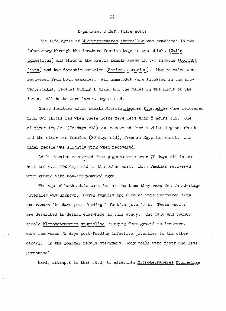
59
Experimental Definitive Hosts
The life cycle of Microtetrameres sturnellae was completed in the
laboratory through the immature female stage in two chicks (Gallus
domesticus) and through the gravid female stage in two pigeons (Colutnba
livia) and two domestic canaries (Serinus canarius). Mature males were
recovered from both canaries. All nematodes were situated in the pro-
ventriculus, females within a gland and the males in the mucus of the
lumen. All hosts were laboratory-reared.
Three immature adult female Microtetrameres sturnellae were recovered
from two chicks fed when these hosts were less than 8 hours old. One
of these females (26 days old) was recovered from a white leghorn chick
and the other two females (20 days old), from an Egyptian chick. The
older female was slightly pink when recovered.
Adult females recovered from pigeons were over 70 days old in one
host and over 102 days old in the other host. Both females recovered
were gravid with non-embryonated eggs.
The age of both adult canaries at the time they were fed third-stage
juveniles was unknown. Seven females and 2 males were recovered from
one canary l84 days post-feeding infective juveniles. These adults
are described in detail elsewhere in this study. One male and twenty
female Microtetrameres sturnellae, ranging from gravid to immature,
were recovered 72 days post-feeding infective juveniles to the other
canary. In the younger female specimens, body coils were fewer and less
pronounced.
Early attempts in this study to establish Microtetrameres sturnellae

60
in chicks J 1-7 weeks old, and adult canaries and pigeons failed.
These negative results, obtained after feeding infective nematode juve
niles to avian hosts, might be explained in at least two ways.
An age'factor may have been operative in the establishment of an
infection of this nematode in the chicks. 'White leghorns more than one
week old could not be infected in the feeding experiments. Yet, two
white leghorns and one Egyptian chick fed a few hours post-hatching
proved to be suitable hosts for a small number of immature female Micro
tetrameres . With only these few iranature females as exceptions, any
chicks older than 10 days failed to continue to harbor any Microtetrameres
juvenile or adult. Further evidence that only very young definitive
hosts may become infected was provided by an experimental infection in a
2-day-old house sparrow (Passer domesticus) to which k encysted third-
stage juvenile Microtetrameres sturnellae had been fed. Twenty-four
hours later, 4 excysted nematodes were recovered between the koilin
lining and the mucosa of the gizzard wall (Figure 35). A two-hour-old
laughing dove (Streptopelia risoria) fed encysted third-stage juveniles
yielded 3 excysted Microtetrameres juveniles when it was necropsied two
days later. On the other hand, an older laughing dove (fledgling)
proved negative for Microtetrameres sturnellae although fed at the same
time as the younger bird.
Another factor which may have prevented M. sturnellae from becoming
established in early experijnents with pigeons and chickens was the
method of feeding employed. In early experiments, grasshoppers were
placed into a gelatin capsule which was forced into the bird's mouth.

6i
No infections resulted when this method was used. When it was suspected
that this feeding method may have prevented M. sturnellae from becoming
established, an adult pigeon was fed one of these capsules containing
physiological saline. Twenty minutes later the pigeon was sacrificed and
examined. The capsule had disintegrated so completely that none of it
was found within the proventriculus or ventriculus. It is possible,
however, that the presence of a capsule in this portion of the digestive
tract may have interfered with the proper sequence of digestive processes
required for normal excystment of the parasite. In subsequent experiments,
when the use of capsules had been discontinued, infections did become
established.
A summary of these feeding experiments is presented in Tables 8
and 9-
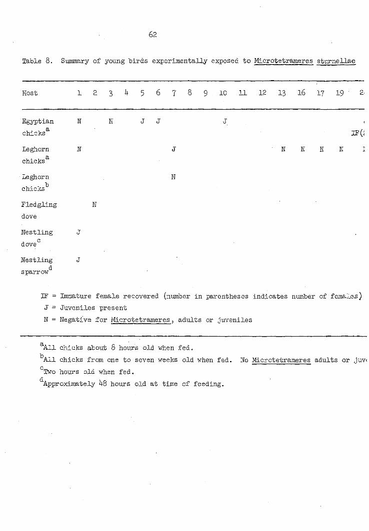
62
Table 8. Summary of young birds experimentally exposed to Microtetrameres sturnellae
Host 1 2 3 I4 5 6 7 8 9 10 11 12 13 16 17 19 2>
Egyptian ST H J J J <
chicks IF(î
Leghorn E J N E N N I
chicks
Leghorn N
chicks
Fledgling N
dove
nestling J
dove
Nestling J d sparrow
IF = Immature female recovered (number in parentheses indicates number of females)
J = Juveniles present
N = Negative for Microtetrameres, adults or juveniles
All chicks about 8 hours old when fed.
All chicks from one to seven weeks old when fed. No Microtetrameres adults or juv(
" Two hours old when fed. d Approximately 48 hours old at time of feeding.
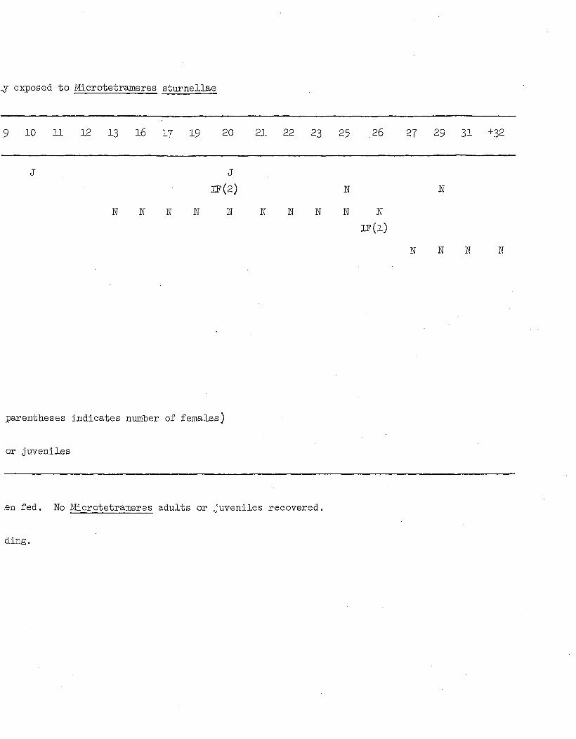
Ly exposed to Microtetrameres sturnellae
9 10 11 12 13 l6 17 19 20 21 22 23 25 26 2? 29 31 +32
J
IF(2) E
N N I N E N N N E
E
N
IF(1)
N 1 N ÏÏ
parentheses indicates number of females)
or Juveniles
en fed. No Microtetrameres adults or juveniles recovered,
ding.
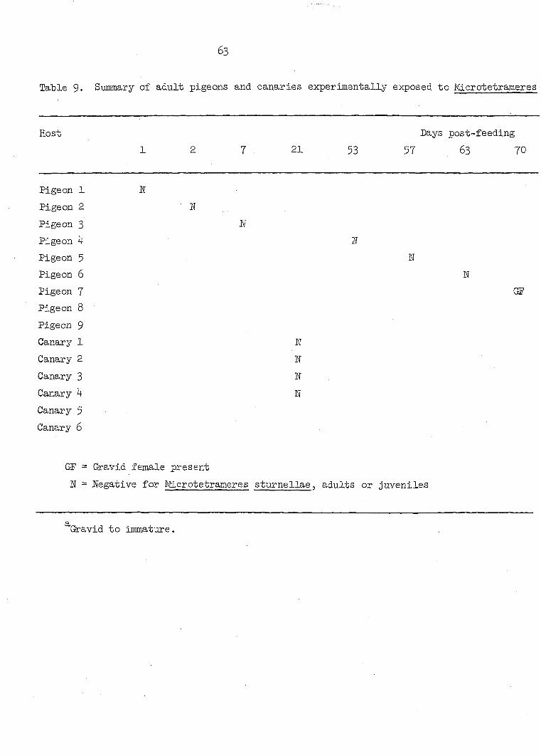
63
Table 9. S-ummary of adult pigeons and canaries experimentally exposed to Microtetrameres
Host Days post-feeding
1 2 7 21 53 57 63 70
Pigeon 1 IÎ
Pigeon 2 N
Pigeon 3 N
Pigeon 4 1
Pigeon 5 n
Pigeon 6 E
Pigeon 7 GF
Pigeon 8
Pigeon 9
Canary 1 W
Canary 2 N
Canary 3 N
Canary 4 N
Canary 5
Canary 6
ŒF = Gravid female present
1 = negative for Microtetrameres sturnellae, adults or juveniles
Gravid to immature.
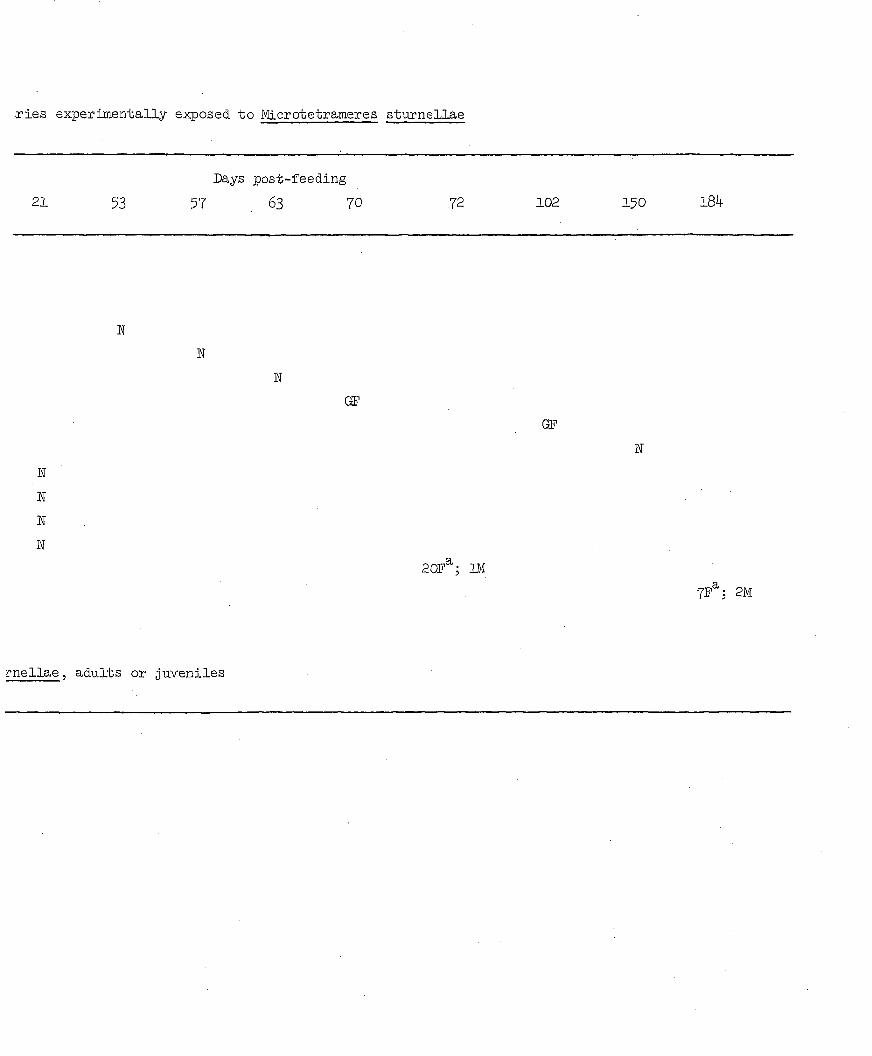
.ries experimentally exposed to Microtetrameres sturnellae
Days post-feeding
21 53 57 63 70 72 102 150 l84
N
N
N
N
ÏÏ
E
GF
OF
20F^; m
7F ; 2M
rnellae, adults or juveniles

64
lEEERMEDIATE HOSTS
Experimental Intermediate Hosts
Microtetrameres sturnellae eggs were fed to laboratory-reared
beetles (Tenebrio molitor, Tribolium confasum), cockroaches (Blaberus
craniifer, Blatta orientalis) as well as to grasshopper nymphs (Melanoplus
femur-rubrum, M. bivittatus, M. sanguinipes, M. mexicanus and M. differ-
entialis). Methods employed in exposing these hosts to these eggs were
discussed in a previous section of this study. Juveniles were recovered
only from species of Melanoplus. No evidence of any Microtetrameres
juveniles was seen from the other experimental hosts. Schell (1953)
was also unsuccessful with Tenebrio, but he did establish an infection of
M. corax in German cockroaches (Blatella germanica).
First-stage juveniles hatch in the esophageal region of the digestive
tract of the grasshopper. Empty egg shells and first-stage juveniles
(some partially within egg shells, some entirely emerged) have been
recovered from this region. These first-stage juveniles, apparently
aided by their anterior dontoidal structures, migrate towards the
insect's hemocoel. During this migration they develop into second-stage
juveniles. Eventually they develop into third-stage juveniles. During
the latter stage they encyst mainly within the dorsal part of the peri
visceral sinus of the host (Figure 32). Fourth-stage juveniles and adults
develop only within the definitive hosts.
During numerous dissections of grasshoppers infected with third-
stage juveniles, cysts containing these juveniles floated free in the
dissecting saline. By scraping the muscles attached to the exoskeleton
65
of the grasshopper after removal of the digestive tract, additional cysts
and occasionally unencysted juveniles were recovered. Juveniles within
these cysts move actively and, if a cyst is punctured, the juvenile
emerges readily. This activity was observed also in M. helix by Cram
(1934).
Because grasshoppers which had been fed Microtetrameres sturnellae
eggs were always confined within their individual plastic vials, they had
little opportunity to move about. They were inactive' probably due to
this close confinement. However, sporadically they would jump and hit
the perforated aluminum cover of the vials. At no time did any grass
hopper display any noticeable external manifestations of infection.
Cram (l93l) mentioned that some of the grasshoppers to which she had fed
eggs of Tetrameres amerlcana became "droopy" after a certain period of
time. This condition may have existed in this research, but it was not
obvious.
Cram reported the presence of juveniles in the femora, and other
sites, of her experimental grasshoppers in her work with T. americana.
In only a few instances during this investigation were any juveniles seen
within a femur. In all instances, third-stage juveniles appeared
within the hemocoel as noted previously.
So far as detectable, no difference in infectibility or refractori
ness was noted among the various grasshopper species used in these
experiments. However, the species employed was dependent upon the
availability of grasshopper eggs (provided by the United States Depart
ment of Agriculture, Entomology Research Division, Montana State
66
University). Hence, no exact study was made concerning any possible
species differences in susceptibility to Microtetrameres sturnellae
infections. With rare exceptions, any grasshopper, regardless of
species, that lived 30 days after having ingested at least a portion of a
female M. sturnellae containing embryonated eggs, became infected with
third-stage juveniles.
Fecal pellets of some infected grasshoppers were examined micro
scopically. These contained shells of M. sturnellae eggs. Although this
examination technique was not carried out for more than a few grass
hoppers, it would be possible to use it to indicate the presence of
Microtetrameres juveniles in any grasshopper so examined. The stage of
the juvenile present would depend upon the time elapsed between the
ingestion of the eggs and the passage of the pellet.
Natural Intermediate Hosts
Approximately $0 wild grasshoppers of various genera, mainly
Melanoplus spp., found in the vicinity of Ames, Iowa, were examined for
Microtetrameres spp. during this study. Eo insects infected with Micro
tetrameres were found. This limited sampling of the grasshopper
population does not permit valid conclusions to be drawn concerning the
presence of Microtetrameres juveniles in nature. Further surveys should
be conducted to determine their presence now that details of their
morphology are known. No other surveys for natural intermediate hosts
of Microtetrameres spp. have been reported. However, grasshoppers are
known to serve as food for the two species of meadowlarks (Sturnella
magna and S. negleeta) found in Iowa as determined by a study of stomach
67
contents of these birds. This fact, together with the recovery of gravid
female Microtetrameres from immature meadowlarks, would indicate the
strong possibility that infections are acquired locally. Infected
meadowlarks collected in Iowa acquire their infections after ingestion
of a suitable intermediate host. The availability of grasshoppers to
meadowlarks and the life cycles of these insects and birds suggest that
grasshoppers serve as the principal natural intermediate hosts for Micro
tetrameres sturnellae and as a source of infection for meadowlarks found
in Iowa during the period of March through September.
68
SUGGESTED HA.TIIRAL LIFE CYCIiE
Although complete experimental confirmation is not available, it is
possible to formulate a hypothetical natural life cycle of Microtetra
meres sturnellae.
Females release embryonated eggs into the bird's proventricular
lumen. Eggs containing first-stage juveniles pass through the digestive
tract and are deposited with the feces. Fecal material, containing these
eggs, is splattered onto vegetation, much of which grows along fences
where grasshoppers frequently oviposit. Thus, viable nematode eggs are
readily available to newly hatched grasshopper nymphs which ingest them,
probably accidentally, while foraging for food. Eggs probably remain
viable on the vegetation or ground for some time.
After ingestion of eggs by a grasshopper, first-stage juveniles
hatch within the esophagus, undergo additional molts to second and third-
stage juveniles which ultimately reach the hemocoel. Approximately 30
days after viable eggs are ingested, third-stage juveniles develop
within the nymph's perivisceral sinus of the hemocoel. If at this stage
the grasshopper is eaten by a suitable bird host, about 30 days elapse
before female M. sturnellae become gravid.
In natural infections, the length of time between ingestion of a
Microtetrameres sturnellae egg by a grasshopper and the maturation of a
female within a Lieberkuhn gland can be only conjectured. However, exper
imental data offered here, together with the recovery of 8 gravid female
Microtetrameres from a single, naturally-infected immature meadowlark.

69
indicate a relatively short period (50 - 60 days).
The above-mentioned time period coincides with Iowa summers as well
as with life cycles of Iowa grasshoppers and meadowlarks. These insects
are available to young birds as early as July. By this date, grass
hoppers could have ingested Microtetrameres eggs from infected bird
feces.
An immature female Microtetrameres completes its maturation only
after entering a proventricular gland.
The known biology of the male Microtetrameres sturnellae still is
somewhat incomplete. Where it copulates with the female and its age
at the time of copulation is still unknown. Travassos (1919)5 Ulmer,
Iowa State University, in 1965 (private communication) and Thomas (née
Mawson), University of Adelaide, in 196 (private communication) found
male nematodes, presumably Microtetrameres sp., completely or partially
inside a Lieberkuhn gland with a female Microtetrameres sp. Possibly
this is the site of copulation. However, in the present investigation,
no males were observed in this location and the only experimentally-
developed males were recovered from the mucus of the proventricular lumen.
The male's life cycle appears to be similar to that of the female
through at least the third-stage juvenile, as no sexual differentiation
is yet noticeable at this stage. This lack of sexual differentiation
was also reported by Schell (1953) in his study of M. corax. However,
such differentiation was reported in what Seurat (1918) called fourth-
stage juveniles of Tropidocerca (= Microtetrameres) spiralis. The life
span of male Microtetrameres sturnellae may be shorter than that of the

70
female because females gravid with embryonated eggs have been found in a
bird host from which no males were recovered, despite intensive examina
tion.
Workers do not agree on the prevalence of males. Cram (l93 )
recovered seven experimentally-developed males of M. helix. However,
Seurat (1918) commenting upon the predominance of males to females in
Iropidocerca (= Microtetrameres) spiralis, reported a ratio of 4o males
to 3 females. On the other hand, in I913 Seurat noted 5 inales to 25
females in what he designated Tropidocerca (= Microtetrameres) inermis.
71
PATHOLOGY
Although pathology due to letrameres spp. has been reported (Cvetae-
va, 196O; Gerasimova, I96O; lange, 1938, LaPage, 1956; Lesbouyries, 19 1;
Pande, Rai and Srivastava, 196O; Popova, 1953j Wehr, 1959j Willomitzer
and Gilka, 1957), that associated with Microtetrameres has not been
described. A study of representative sections of an adult M. sturnellae
female in situ indicates this species to be hematophagous. Nucleated
red blood cells may be seen within the lumen of the gut. The amount of
blood lost by the host presumably is negligible except in very heavy
infections.
LaPage (1956) ascribed a toxic effect to Tetrameres americana, but
stated that greatest damage is caused by juveniles migrating into the
proventricular wall. This migration causes, according to LaPage,
"marked irritation and inflammation". Pathology of juveniles and adults
of Microtetrameres is probably much the same as that resulting from
tetrameriasis despite the report of Cassamagnaghi and Bazerq.ue (l950)
of a species of Tetrameres whose adults were reported to occur within
the pulmonary parenchyma of a pigeon. All other reported instances of
infections of Microtetrameres and Tetrameres have involved only the pro-
ventricular region. During the course of these studies on Microtetra
meres , one example of an unidentified species of Tetrameres was seen
protruding from the serosa of the proventriculus of a meadowlark.
Juveniles of Microtetrameres sturnellae apparently migrate within
tissues of the definitive host. Four were observed as they moved beneath
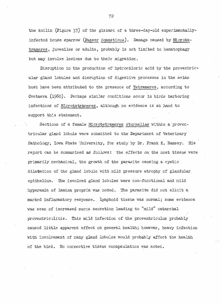
72
the koilin (Figure 35) of the gizzard of a three-day-old experimentally-
infected house sparrow (Passer domesticus). Damage caused by Microte-
trameres, juveniles or adults, probably is not limited to hematophagy
but may involve lesions due to their migration.
Disruption in the production of hydrochloric acid by the proventric-
ular gland lobules and disruption of digestive processes in the avian
host have been attributed to the presence of letrameres, according to
Cvetaeva (i960). Perhaps similar conditions occur in birds harboring
infections of Microtetrameres, although no evidence is at hand to
support this statement.
Sections of a female Microtetrameres sturnellae within a proven-
tricular gland lobule were submitted to the Department of Veterinary
Pathology, Iowa State University, for study by Dr. Frank K. Ramsey. His
report can be summarized as follows: the effects on the host tissue were
primarily mechanical, the growth of the parasite causing a cystic
dilatation of the gland lobule with mild pressure atrophy of glandular
epithelium. The involved gland lobules were non-functional and mild
hyperemia of lamina propria was noted. Ihe parasite did not elicit a
marked inflammatory response. Lymphoid tissue was normal; some evidence
was seen of increased mucus secretion leading to "mild" catarrhal
proventriculitis. This mild infection of the proventriculus probably
caused little apparent effect on general health; however, heavy infection
with involvement of many gland lobules would probably affect the health
of the bird. No connective tissue encapsulation was noted.
73
Contrary to Rasheed (1960), who reported female Microtetrameres
orioles to be encysted, no evidence of a cyst, either of host or parasite
origin, appears in sections of adult female Microtetrameres sturnellae
in situ. Some authors (Singh, 1962; Yorke and Maplestone, I926) have
reported the crop as the site of infection for species of Microtetra
meres . However, these reports seem to have resulted from the incorrect
translation of "ventricule succenturi " as "crop" instead of "proven-
triculus".
74
KEY TO THE SHECIES OF FEMltE
MICROTETRAMERES IN THE WESTERN HEMQSPHEEE
A search of the literature indicates at least 11 species of Micro-
tetrameres to have been originally described from the western hemisphere.
Two of these species, Microtetrameres contorta and M. canadensis, are not
included for reasons indicated below. Table 1 summarizes the comparative
measurements J host range and geographical distribution of the remaining
species.
Microtetrameres contorta Weidman 1913 was recovered from a hornbill
(Pichocerus bicornis) that died in the Philadelphia zoo. Because this
bird belongs to a non-migratory family, Bucerotidae, from Africa
(Van Tyne and Berger, 1959)? Microtetrameres contorta is not considered
a western hemisphere nematode and is not included in the key to the
species presented below.
Microtetrameres canadensis Mawson 1956 was described as being
similar to M. contorta except that the coiling configuration of the female
was quite different. Present laboratory studies, however, have shown this
configuration to be non-characteristic and subject to much variation.
Because the remainder of Mawson's description of M. canadensis is
fragmentary and because the dimensions she reported are similar to those
of M. contorta and M. pusilia, Microtetrameres canadensis is considered
a species inquirenda until it can be further studied.
In addition to the 11 species originally described from the western
hemisphere, Wilson (l95ô) recorded the presence of M. inermis von
Linstow 1879 in house sparrows in Maryland. The status of M. inermis
75
is questionable because morphological descriptions of the species by
Seurat (19I3) and von Linstow (1879) are at variance with one another.
Pinal disposition of the status of this species, however, is not within
the scope of the present study.
Anatomical differences used in separating certain species of Micro-
tetrameres are sometimes minute. In addition, many of the dimensions
reported for several species overlap. Numerous investigators (Cram, 1927?
Travassos, 19l4; Schell, 1953) have reported only a single measurement
rather than a range of measurements for certain structures. Further
more, many workers have not indicated the number of specimens upon which
descriptions were based. Therefore, anyone attempting to determine the
species of a female Microtetrameres experiences difficulty in deciding
which of the reported dimensions match his most closely.
As indicated in this study, certain dimensions of female Microte
trameres change markedly at fixation. Because some authors have not
indicated whether living or fixed nematodes were measured, it is diffi
cult for any investigator to be certain that his measurements are being
made under the same conditions as those reported in the original descrip
tions .
Host-induced intraspecific variations, known to occur among helminths
of other groups such as trematodes (Watertor, I966} very probably
constitute another factor making the identification of species of Micro
tetrameres difficult. Nematode age, host diet and health, and measuring
techniques are additional factors which could cause variations in
dimensions such as those shown in Table 1 (Chitwood, 1957).

76
It may seem unreasonable to differentiate species of Microtetra-
meres on such small variations as those shown in Table 1. Nonetheless,
small variations in certain structures (buccal capsule, spicule and egg)
which are less subject to change at the time of fixation are of greater
significance than are large variations in other dimensions (anus-tail
or vulva-anus distances) which are known to change markedly when speci
mens are fixed. As shown in an earlier section of this thesis, experi
mental evidence indicates that eggs of M. sturnellae are remarkably
constant in size even if the females producing them are harbored by
avian hosts of different families (Ploceidae, Icteridae). Constancy
of size is also reflected in the buccal capsule. The',ranges of 37
buccal capsules measured from females recovered from naturally-infected
meadowlarks were: length 20.8 - 15.6 u, average l8.4 u; width 7.8 -
13.0 u, average 10.5 u. The size of the buccal capsule for the type
specimen of M. sturnellae was 18.2 u long by 10.4 u wide. A thorough
re-examination of living and fixed specimens of other species of Micro-
tetrameres would aid greatly in a better understanding of the signifi
cance of these characteristics.
The validity of certain of the species summarized here must await
further study. In addition to small morphological differences among
species, physiological criteria may be involved. Additional studies
may indicate that the number of species currently assigned to this
relatively large genus may have to be reduced.
Until re-descriptions are available, however, it seems best to
use, where possible, only dimensions of those structures considered to
77
"be the most stable and which change the least under fixation. Hence,
in the key presented below, only measurements of the buccal capsule and
eggs in addition to certain cuticular modifications (flanges and furrows)
are used to differentiate species.
The presence of longitudinal flanges differentiate M. accipiter from
all other known species of Microtetrameres. M. cruzi and M. pus ilia are
both characterized by furrows, but eggs of M. cruzi are larger and
narrower than those of M. pusilia. Thus, these three species may be
readily differentiated from one another.
M. minima has the smallest buccal capsule of any reported western
hemisphere species, and hence is considered distinct.
M. spiculata and M. aquila have eggs as long as $0 mi era. However,
M. aquila can be differentiated from M. spiculata by its larger buccal
capsule.
Egg sizes of M. cor ax and M. bubo are nearly the same. However,
the two species can be differentiated on the basis of the length of the
buccal capsule, that of M. corax being longer.
Although M. helix has a buccal capsule whose dimensions overlap .
those of M. spiculata and M. bubo, its eggs 9.re shorter and wider than
the other two species. The range of differences in length and width of
eggs of the various species of Microtetrameres (with the exception of
M. helix) appears to vary between 15 and 32 micra. In M. helix, how
ever, this range is but 9 micra. The somewhat more spherical eggs of
M. helix may thus be used as a distinguishing characteristic.
The eggs of M. sturnellae are wider than those of any other species
78
from the western hemisphere heretofore reported. Female Microtetrameres
sturnellae recovered in this research lack longitudinal furrows. Except
for the absence of these furrows, dimensions of these females agree very
closely with those reported by Travassos (igl , 1919) for Microtetrameres
pusilia.
Travassos (1919) reported the presence of longitudinal furrows in
female Microtetrameres pusilia 4 years after the publication of his
original report (1915)• In the latter paper, no mention of longitudinal
furrows was made. In the 1919 report, he indicated also the presence of
spines on the presumed males of this species. No reference to the
presence of such spines appeared in his earlier (1915) paper. Certain
measurements of the female were also modified considerably in Travassos'
1919 study so as to increase the range of their dimensions. Only the
measurements presented for the length of the buccal capsule remained
unchanged. For these reasons, the validity of M. pusilla is questioned.
Only one other reference to the presence of longitudinal furrows in
•Microtetrameres appears in the literature, namely, that of Travassos
(1914) in his report on M. cruzl. Dimensions of the buccal capsule and
of the eggs of this species are similar to those of M. pusilla.
Travassos' illustration of a female M. cruzi does not include furrows,
but in many other respects his description of M. pusilla could apply to
M. cruzi• Only with a careful re-description of the type specimens of
M. cruzi and M. pusilla will this confusion be resolved. Such a study
may result in the transfer of M. pusilla to the genus Tetrameres whose
members are characterized by the presence of cuticular spines in males
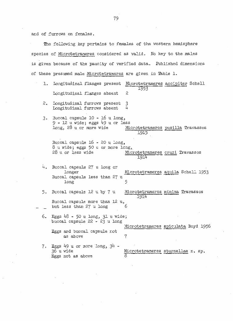
79
and of furrows on females.
The following key pertains to females of the western hemisphere
species of Microtetrameres considered as valid. No key to the males
is given because of the paucity of verified data. Published dimensions
of these presumed male Microtetrameres are given in Table 1.
1. Longitudinal flanges present Microtetrameres accipiter Schell 1953
Longitudinal flanges absent 2
2. Longitudinal furrows present 3 Longitudinal furrows absent 4
3. Buccal capsule 10 - l6 u long, 9 - 12 u wide; eggs 4$ u or less long, 28 u or more wide Microtetrameres pus ilia Travassos
1915
Buccal capsule I6 - 20 u long, 8 u wide; eggs $0 u or more long, 28 u or less wide Microtetrameres cruzi Travassos
Î9ÏÇ
4. Buccal capsule 27 u long or longer Microtetrameres aquila Schell 1953
Buccal capsule less than 27 u long 5
5. Buccal capsule 12 u by 7 u Microtetrameres minima Travassos Ï9Ï5
Buccal capsule more than 12 u, _ _ but less than 27 u long 6
6. Eggs 48 - 50 u long, 31 u wide; buccal capsule 22 - 23 u long
Microtetrameres spiculata Boyd 1956 Eggs and buccal capsule not
as above 7
7. Eggs 49 u or more long, 34 -36 u wide Microtetrameres sturnellae n. sp. Eggs not as above 8

80
•Buccal capsule 24 - 25 u long
Buccal capsule 22.5 u or less long
Eggs 42 u long, 33 u wide Eggs 46 u long, 28 u wide
Microtetrameres corax Schell 1953
9
Microtetrameres helix Cram 1927 Microtetrameres bubo Schell 1953

81
EAXONOMIC CONSIDERATIONS
The status of the family Tetrameridae as a valid taxori has been
questioned by some nematologists. However, this familial designation
has been accepted by many (Bashkirova, 196O; Cram, 192?j Chabaud, 1951j
Diaz-Ungria, I965; Gagarin, Ablasov and Chibichenko, 1957i LaPage, 1956;
Oshmarin, I956; Oshmarin and Parukhin, I963; Petrov and Chertkova, I95O&,
1950b; Rasheed, I96O; Skrjabin, I916; Skrjabin and Sobolev, 19 3j.
Sultana, 1962; Yorke and Maplestone, 1926; Travassos, 1914; Yamaguti,
1935). On the other hand, Tetrameridae was listed as a synonym of
Spiruridae by Chitwood and Wehr (193 +) and Sugimoto and Nishiyama (l937)-
Others (Wehr, 193 ; Lc pez-Neyra, 19 7) have maintained the designation
Spiruridae. Whichever familial designation was used, the two genera
Microtetrameres and Tetrameres have always been included within the same
family.
Despite the fact that Yamaguti listed Tetrameres scolopacis n. sp.
in 1935 in the family Tetrameridae, he recently (1961) reverted to the
use of Tropisurus instead of Tetrameres. He also included a third genus
(Geopetitia) within the family Tropisuridae, which he considered a new
name for Tetrameridae. This new name has been rejected herein for
reasons listed elsewhere.
Lc pez-Neyra (1958) proposed placing the genus Crassicauda in the
family Tetrameridae, but this may be inappropriate as this genus is
found in cetaceans (Joyeux and Baer, 1951; Baylis, I916, 1920, 1922;
Skrjabin and Andreeva, 193 ) while the genera, Tetrameres and Microte
trameres , presently classified within Tetrameridae are found only in

82
birds (except, doubtfully, Tetrameres bispinosa reported by Molin (1860)
from Scincus sp.).
Chabaud (1951) proposed including three sub-families, Crassicaudinae,
Geopetitiinae and Tetramerinae, within the family Tetrameridae, with
Grassicauda and Geopetitia as the type genera, respectively, for the
first two sub-families. Tetrameres was considered the type genus for
Tetramerinae with Microtetrameres included in the last named sub-family.
The three sub-families are related morphologically and show relation
ships both to spirurid and filarial nematodes, according to Chabaud
(1951); and, hence, these sub-families should be grouped together.
Furthermore, Chabaud suggested that evolution links these three genera
within one family, Tetrameridae.
Evidence now at hand substantiates the separation of Microtetra
meres and Tetrameres as two distinct genera. Considerable morphological
differences between adults and Juveniles of the two genera support this
viewpoint. Thus, adult females of Tetrameres are globose or spindle-
shaped; those of Mi or ot etramer e s are coiled. Presumed and known males
of Tetrameres usually possess longitudinal rows of body spines; these
are absent on most presumed and known males of Microtetrameres. First-
stage juveniles of Tetrameres are reported to possess caudal spines or
papillae ; third-stage Tetrameres americana, experimentally-reared, also
exhibit these (Cram, 1931)j however, the present study shows that such
structures are absent from Microtetrameres sturnellae juveniles of the
same stages.
Juveniles of Tetrameres and those of some members of the family

83
Spiruridae share the common morphological characteristic of possessing
spines or papillae on their posterior ends (Chahaud, 195 5 Lieberkuhn,
1855; Seuratj I918). Some of these spines are on a small terminal
"bouton" (Seurat, I915-I6); others adorn the rounded posterior ends
(Chabaud, 195 ),• Larger spines ("aigullons") are found on posterior ends
of Tetrameres fisslspina of presumed fourth-stage juveniles (Seurat,
1918) and papillae have been seen on T. americana third-stage juveniles
experimentally-reared by Cram (1929, 193l)• However, spines and
papillae are absent on posterior ends of laboratory-reared juvenile
stages of Microtetrameres sturnellae (Figures 12, 16, I9). Spines were
not reported on the same stages of M. helix (Cram, 193 ) nor from first-
stage Tropidocerca (= Microtetrameres) inermis as•indicated by Seurat
(1913). Juveniles of M. helix, M. inermis and M. sturnellae terminate
posteriorly in a sharp tail, which in third-stage juveniles extends
to a tiny, glabrous hemisphere.
Because of these considerable morphological differences, one is
tempted to relegate the two genera to separate families Chabaud (1951)
notwithstanding. Such a transfer seems premature, however, because of
insufficient experimental evidence. Until further studies have been
undertaken, consequently, the two genera are considered as members of
the family Tetrameridae. Data from the present study permit the following
diagnosis, based upon laboratory-reared male and female Microtetrameres
sturnellae n. sp.

8k
Microtetrameres sturnellae n. sp.
Living female : Bri t red; overall configuration, globose j body tightly
coiled; overall diameter varies with movement but usually about 1.2 -
1.4 mm under slight coverslip pressure. Anterior end naked. Transverse
cuticular striations present. Longitudinal furrows or flanges absent.
Cuticular folds absent in freshly recovered specimens. Buccal capsule
average (n = 3) width at bulbous midportion 0.0104 mm, no range; buccal
capsule length 0.0182 - 0.0208 mm; average (n = 3) O.OI96. Rhabdions
heavily sclerotized. Distance from posterior end of buccal capsule to
posterior end of nerve ring, 0.193 mm. Muscular portion of pharynx
approximately 0.247 mm; width at junction of glandular and muscular por
tions, 0.034 mm; lumen of muscular portion triradiate in cross section.
Glandular portion of pharynx approximately O.87O mm long. Intestinal
diverticulum present at pharyngointestinal junction. Cervical papillae
absent; excretory pore not observed. Distance from vulva to anus
0.104 - 0.130 mm. Distance of anus to tail-tip, O.I56 - O.I82 mm. No
knoblike appendage on tail. Eggs, O.O338 - 0.0364 mm wide by 0.04 4 -
0.0520 mm wide, average (W = 10) 0.0507 mm long by 0.0353 mm wide. Egg
shell 0.002 - 0.003 mm thick at widest part of egg. _
Host species: Sturnella neglecta (western meadowlark).
Location in host: Eroventricular glands.
Locality: Ames, Iowa.
Type specimens deposited in U.S.N.M, Helminthological collection.
Fixed male ; Filiform, white. I.OO8 - 1.4 mm long; O.07O - 0.075 mm wide
at pharyngointestinal junction. Oral opening O.OO5 mm in diameter.
85
Four cephalic papillae lateral to buccal capsule. Buccal capsule
0.012 mm long by 0.003 - 0.005 wide. No cervical papillae observed.
Length of muscular portion of pharynx 0.l40 - 0.1 3 mm, width 0.010 mm;
length of glandular portion of pharynx, 0.280 - 0.351 mm, width,
0.021 - 0.026 mm. Distance from anterior edge of nerve ring to posterior
end of buccal capsule, approximately O.O85 mm. No cervical alae.
Longer spicule length, I.03 - 1.2 mm; shorter spicule length, O.O67 -
0.109 Longer spicule width, 0.004 mm; shorter spicule width,
0.003 - O.OOU mm. Longer spicule transversely striated; shorter spicule
may be weakly striated, transversely. Ratios of longer spicule length
to shorter spicule length, 12:1 - 19:1. Testis commences about midpoint
of length of body, courses anteriorly and then is reflected upon itself
to posterior part of body; constriction present between testis and vas
deferens. No gubernaculum or telamon present. Cloacal lips protrude
from body wall. Distance from cloaca to tail-tip, O.I3O - 0.135 nmi.
Cloacal papillae: 2 pairs pre-cloacal, 2 pairs post-cloaca!. One ad-
cloacal papilla sometimes present. Tail distally pointed with minute
appendage. Caudal alae absent.
Host species (experimental): Serinus canarius (domestic canary).
Location in host : Proventricular glands.
Locality : Ames, Iowa.
Type specimen deposited in U.S.N.M. Helminthological collection.
86
SUMMARY AED CONCLUSIONS
1. Microtetrameres sturnellae n. sp., a spiruroid nematode of the
family Tetrameridae, parasitizes the proventricular glands of the
meadowlark (Sturnella neglecta) in Iowa.
2. The experimental life cycle of M. sturnellae has been followed from
the adult stage through the third-stage juvenile.
3. Gravid females are bright red when alive, and have a spherical body
configuration characterized by three or four greatly distended body
coils containing masses of eggs.
4. Three adult male Microtetrameres sturnellae were recovered from the
proventricular mucus of two experimentally-infected domestic canaries
(Serinus canarius) and constitute the first laboratory-reared adult male
specimens of this genus.
5. Constancy of egg measurements (and, to a lesser degree, measurements
of the buccal capsule) of M. sturnellae suggests these dimensions as
useful diagnostic tools in differentiating species of the genus.
6. Embryonated eggs recovered from naturally-infected meadowlarks con
tain first-stage juveniles. Viable eggs deposited with the feces
develop further only upon ingestion by suitable intermediate hosts'.
Beetles (Tenebrio molitor, Tribolium confus urn), cockroaches (Blaberus
craniifer, Blatta orientalis), and grasshopper nymphs (Melanoplus femur-
rubrum, M. bivitattus, M. sanguinipes, M. mexicanus, M. differentials)
were used as experimental intermediate hosts. Development occurred only
in species of Melanoplus.

87
7. First-stage juveniles hatch within the foregut of the insect inter
mediate host a few hours post-ingestion. Juveniles at this stage are
characterized by anterior dontoidal projections, discontinuous striae
anteriorly, and aspinose, pointed tails. In many respects, they resemble
microfilariae. Living first-stage juveniles are capable of extensive
lateral movement and are able to extend and withdraw their anterior ends.
8. Second-stage juveniles develop within 8 - 16 days. Older second-
stage juveniles have been recovered from muscles of the body wall and
the femora. In addition to their larger size, they are characterized by
a complete digestive tract and by a bent-tail configuration.
9- Third-stage juveniles are infective within 25 days following inges
tion of eggs and lie encysted within the perivisceral sinus of the
intermediate host's hemocoel. The oldest viable third-stage juvenile
was over 60 days old when fed to an experimental definitive host.
10. Details of fourth-stage juveniles are not known, but presumably
this stage develops within the proventricular glands of the definitive
host.
11. Young females within proventricular glands are characterized by
lack of eggs, by the accumulation of conspicuous detritus within the
digestive tract, and by the presence of only one or two loose body
coils. Rhabdions of their buccal capsules are straighter than those of
gravid females.
12. Laboratory-reared chicks (Gallus domesticus), adult pigeons (Columba
livia), and adult domestic canaries (Serinus canarius) were used as
experimental definitive hosts. Gravid females were recovered only from
88
canaries l84 days and 72 days and from adult pigeons 102 days and 70
days post-feeding third-stage juveniles. immature females were recovered
from chicks 20 days and 26 days post-feeding.
13. Additional experimental definitive hosts included one nestling and
one fledgling dove (Streptopelia risoria)., and a two-day-old house
sparrow (Passer domesticus). Juveniles, "but no adult nematodes, were
recovered from these hosts.
14. Geographical distribution of known species of Microtetrameres and
the range of their definitive hosts are presented.
15. A suggested life cycle of M. sturnellae involving meadowlarks and
grasshoppers is offered, based principally upon experimental data
recorded during this study. The biology of these hosts in Iowa is such
that the cycle may be readily completed in nature.
16. Female Microtetrameres sturnellae are hematophagous as indicated by
the presence of nucleated red blood cells within the gut. Further
pathology involves principally mechanical effects. The presence of
females in the proventriculus is apparently of little consequence to
the avian host unless infections are massive. No evidence of connective
tissue encapsulation was noted.
17. A key to the valid species of female Microtetrameres of the western
hemisphere is presented, with the suggestion that additional studies
may result in the rejection of several of those currently considered
valid.
18. The familial designation Tetrameridae is maintained, but Tropisuridae
Yamaguti I96I is considered invalid.
89
19. Anatomical relationships between juveniles and adults of the
genera Tetrameres and Microtetrameres are discussed with the suggestion
that these genera may well belong to separate families, Spiruridae and
Tetrameridae, respectively.
90
LHEMTUKE CITED
Ablasov, N, A. and 1. T. Chibichenko 1962 Nematode fauna of wild birds in Kirgizia (Translated title).
Iv. Akad.j Nauk kirkiz. SSR. Ser. Biol. Nauk 4:113-130.
AgassiZj L. l848 Nomenclatoris zoologici. Index universalis. Soloduri,
Sumtibus et typis Jent et Gassmann.
Anantaraman, M. and K. P. Chandrasekharan Nair 1955 Tetrameres infection of fowls in India. Zool. Anz.
154:182-193.
Avery, R. A. 1966a Heljninth parasites of wildfowl from Slimbridge, Gloucester
shire. I. Parasites of captive Anatidae. J. Helminth. 40:269-280.
Avery, R. A, 1966b Helminth parasites of wildfowl from Slimbridge, Gloucester
shire. II. Parasites of wild mallard. J. Helminth 40:281-284.
Baru , V, L. 1966 Parasitic nematodes of birds in Czechovlovakia. I. Hosts:
Columbiformes, Piciformes, Falconiformes and Strigiformes. Folia Parasitol. (Praha) 13:7-27-
Bashkirova, E. Ï. i960 Nematodes of birds of the Primorskii krai (Translated title).
Trudy Gel'mint. Lab. 10:46-57.
Baylis, H. A. 1916 On Crassicauda crassicauda (Crepl.) (Nematoda) and its hosts.
Ann. Mag. nat. Hist. 17:l44-l48.
Baylis, H. A. 1920 Observations on the genua Crassicauda. Ann. Mag. nat. Hist.
5:4IO-419.
Baylis, H. A. 1922 Note on the habitat and structure of Crassicauda (Nematoda)
Parasit. l4:9-12.
Baylis, H. A. 1929 A manual of helminthology, medical and veterinary. London,
Bailliere, Tindall and Cox.

91
Baylis, H. A. 1939 The fauna of British India, including Ceylon and Burma,
Nematoda. Volume II: (Filarioidea, Dioctophymoidea and Irichinelloidea). London, Taylor and Francis, Ltd.
Baylis, H. A. and R. Daubney 1926 A synopsis of the families and genera of nematoda, London,
British Museum.
Belopolskaya, M. M. 1963 Survey of the parasite fauna of birds in the Sudzukhinsk
reserve (Primore) (Translated title). Parazit. 8b. 21:221-244.
Benbrook, Edward A. 1963 Outline of parasites reported for domesticated animals in
North America. Ames, Iowa, Iowa State University Press.
Boyd, Elizabeth M. 1951 A survey of the starling Sturnus vulgaris L. in North America.
J. Parasit. 37:56-84.
Boyd, Elizabeth M. 1956 Two new species of stomach worms (Nematoda: Spiruroidea)
from the blue jay, Gyanocitta cristata L. Proc. Helm. Soc. Wash. 23:70-74.
Boyd, Elizabeth M., Rosemarie Diminno and Carlita Nesslinger 1956 Metazoan parasites of the blue jay, Gyanocitta cristata L.
J. Parasit. 42:332-346.
Cassamagnaghi, A. and A. Bianchi Bazerque 1950 Helmintologia aviar. Ornithostrongylosis - localizacion
pulmonar per Tetrameres en columbidos del Uruguay, Boletin mens. Dir. Ganad. Montev. 31: 35- 0.
Ghabaud, A. G. 1951 Description, d'un nematode parasite de Mesange, Geopetitia
pari n.g. n. sp., intermédiare entre Tetrameridae et Grassicaudidae et hypotheses sur 1'interpretation phylogenétique des helminthes de ce groupe. Annals Paras it. hum. comp. 26:190-200.
Ghabaud, A. G. 1954 Sur le cycle évolutif des spirurides et de nematodes ayant
une biologie comparable. Valeur systématique des caractères biologiques (suite et fin). Annals Parasit. hum. comp. 29:358-425.
92
Chitwood, B. G. and Everett E. Wehr 193 The value of cephalic structures as characters in nematode
classification, with special reference to the super-family Spiruroidea. Z. ParasitKde 7"-273-335»
Chitwood, M, B. 1957 Intraspecific variation in parasitic nematodes. Syst.
. Zool. 6:19-23.
Clapham, Phyllis A. 1961 Recent observations on helminthiasis in some British game
birds. J. Helminth. R.T. Leiper Supp. pp. 35- 0.
Cram, Eloise B. 1927 Bird parasites of the nematode suborders Strongylata,
Ascaridata and Spirurata. U.S. Wat. Mus. Bull. l40.
Cram, Eloise B. 1929 The life history of Tetrameres americana (Cram, 1927)
Baylis, I929, a spirurid of the proventricuius of chickens. J. Parasit. 15:292.
Gram, Eloise B. 1931 Developmental stages of some nematodes of the Spiruroidea
parasitic in poultry and game birds. U.S.D.A, Tech. Bull. 227.
Cram, Eloise B. 193 Orthopterans and pigeons as secondary and primary hosts,
respectively, for the crow stomach worm, Microtetrameres helix (Nematoda; Spiruridae). Eroc. Helminth. Soc. Wash. 1:50.
Creplin, E. C. H. l846 Hachtrage zu Gurlt's Verzeichniss der Thiere, bei welchen
Entozoen gefunden worden sind. Arch. Natur. Berlin. 1:129-160.
Cvetaeva, N. P. i960 Pathomorphological changes in the proventriculus of the
ducks by experimental tetrameriasis (Translated title). Heltninthologia 2:143-150.
Czaplinski, B. 1962 Nematodes and acanthocephalans of domestic, and wild
Anseriformes in Poland. Ill, General comment. Acta parasit. pol. 10:395- 10.
93
Diaz-Ungria, Carlos 1965 Nematodes parasites de aves de Calabozo. Boln. Soc. venez.
Cienc. nat. 26:103-128.
Diesingj Karl Moritz 1835 Tropisurus und Thysanosoma, zwei neue Gattungen von
Blnnenwurmern (Entozoen) aus Brasilien. Med. JaJirb. K.K. Oesterr. Staates V.16 n.F. 7:83-115.
Dubinina, M. N. 1937 The parasitic fauna of Nycticorax nycticorax and its change
in connection with the host's migration (Translated title). Zool. Zh. 16:5 7-573.
Dubinina, M. ÏÏ, and 0. P. Serkova 1951 Nematodes of birds hibernating in southern Tadzhikistan
(Translated title). Parazit. Sb. 13:75-95.
Ellis, Charles J. 1961 Microtetrameres in the bronzed grackle and the eastern
meadowlark. J. Parasit. 47:37.
Ergun, H. i960 Ankara ve civar vilayetleri tavuklarinda buldugumuz helmintler
uzerinde arastirmalar. Etlik vet. Bakt. Enstit. Derg. 1:37-44.
Eagasinki, Andrzej. 1958 The helminthofauna of the domestic Rallidae (Translated
title). Wiad. parazyt. 4:683-684.
Eitzsimmons 5 W. M. 1964 A host check list of helminth parasites from domestic
animals in Wyassaland. Br. vet. J. 120:186-190.
Foster, Winthrop D. 1915 A peculiar morphologic development of an egg of the genus
Tropidocerca and its probable significance. J. Paras it. 1:45-47.
Ereitas, M. G-. and 0. Hipolito 1949 Notas de heltaintologia de Gallus gallus domesticus em Minas
Gérais. Archros Esc. sup. Vet. Est. Minas Gérais 2:51-59*
Erings, Hubert and Mable Erings 1962 Simplified methods for laboratory maintenance of saltatory
Orthoptera. J. econ. Entomol. 55:1019-1020.

9k
Gagarin, V. G., N. A. Ablasov and W. T. Chibichenko 1957 Helminth fauna of wild birds of southern Kirgiziia (Translated
title). Trudy Inst. Zool. Parazit. Akad. Nauk Kirgiz. SSR 6:105-120.
Gerasimova, G. U. i960 Study of the pathology and morphology of Tetrameres and
Polymorphus infections in ducks (Translated title}. Tezisi Dokl. Wauchnoi Kbnf. Vses. Obshch. Gelm., Moscow, 15-20 December, pp. 29-31- Original not seen; abstracted in Helminthological Abstracts 35*597.
Giovannoni, M. and D. de M. Malheiro 1952 Incidencia de parasitas em Columba livia domestica. Revta
Pac. Med. vet. Univ. S Paulo 4:595-598.
Harwood, P. D. 1933 Some spiruroid nematodes from Texas birds . Trans. Amer.
Mic. Soc. 52:173-176.
Hodasi, J. K. M. 1963 Helminths from Manitoba birds. Can. J. Zool. 41:1227-1231.
Hodasi, J. K, M. 1966 A note on some helminths of Manitoba chickens. Can. J. comp.
Med. 30:26-27.
Hsu, H. F. 1935 A study of some Strongyloidea and Spiruroidea from French
Indo-China and of Thelazia chungkingensis Hsu, 1933 from China. Z. ParasitKde. 7:579-600.
Hunter, W. S. and T. L. Quay 1953 An ecological study of the helminth fauna of Macgillivray's
seaside sparrow, Ammospiza maritima macgillivraii (Audubon). Amer. Midi. Nat. 50:407-413.
Johnston, Thomas Harvey 1910 On Australian avian entozoa. J. Proc. Roy. Soc. E. South
Wales 44:84-122.
Johnston, Thomas Harvey 1912 Internal parasites recorded from Australian birds . Emu
12:105-112.
Joyeux, C. and J, G. Baer 1951 Les rapports des helminthes et de leurs hStes. Biol. Med.
Paris. 49:230-216.

95
Lange, Harms 1938 Ueber eine durch Iropisurus fissispinus Diesing hervorgerufene
Magenwumseuche beï Enten, mit besonderer Berucksichtigung der Entwicklung des Parasiten auf Grund des pathologis ch-anatomi-schen Befundes. Zeit. InfktKrankh. paras it, Krankh. Hyg. Haustiere 53:1-8.
LaPage, Geoffrey 1956 Monnig's veterinary heltninthology and entomology. 4th ed.
Baltimore, Maryland, The Williams and Wilkins Company.
LaPage, Geoffrey 1961 A list of the parasitic protozoa, helminths and arthropoda
recorded from species of the family Anatidae (ducks, geese and swans). Parasitology $1:1-109.
Lesbouyries, G. 19 1 la pathologie des oiseaux. Paris, Vigot-Freres.
Lieberkiihn, E. 1855 Beitrage zur Anatomie der Nematoden. Arch. Anat. Physiol.
Wissensch. Med. 1855:31 -336.
von Linstow, 0, F, B. 1879 Helminthologische studien. Archiv fur Natur. Berlin
7:165-188.
Lc peZ-Neyra, C. R. 19 7 Helmintos de los vertebrados Ib ricos. Granada, Inst. Nac.
Parasit.
Lopez-Heyra, C. R. 1958 Sobre unos vermes parasitos obtenidos en el Ziphius caviro-
stris Cuvier (Cetaceo: Dentxceto) varado en aimeria, en especial de Crassicauda giliakiana (Spiruroidea : Tetra-meridae). Revta Iber. Parasit. l8:13-l8.
Luft, Krystyna i960 The helminths of jay (Garrulus glandarius L.) and magpie
(pica pica L.) from Lublin Palatinate. Acta parasitol. polonica 8:351-356.
Madsen, H. 1952 A study on the nematodes of Danish gallinaceous game birds.
Dan. Rev. Game Biol. 2:1-126.
96
Maplestone, P. A. 1931 Parasitic nematodes obtained from animals dying in the
Calcutta zoological gardens. Parts 4-8. Eec. Ind. Mus. Calcutta 33:83-88.
Mawson, Patricia M. 1956a Three new species of spirurid nematodes from Canadian birds.
Can. J. Zool. 3 :193-199.
Mawson, Patricia M. 1956b Spirurid nematodes from Canadian birds. Can. J. Zool.
34:206.
Merchant, Ival A. 1928 A survey of nematode parasites of the horse, ox, sheep,
swine and chicken of Iowa. Unpublished M.S. thesis. Ames, Iowa, Library, Iowa State University of Science and Technology.
Mo lin, Raffaele i860 Trenta specie di Nematoidi. Sitzungsb. K. Akad, Wissensch.,
Wien, Math.-Naturw. 01. 40:331-358.
Morgan, B. B. and E. P. Waller 1941 Some parasites of the eastern crow (Corvus brachyxhynchos
brachyrhynchos Brehm). Bird Banding 12:16-22.
Myers, Betty June, R. E. Kuntz and W. H. Wells 1962 Helminth parasites of reptiles, birds and mammals in
Egypt. VII. Check lists of nematodes collected from 1948 - 1955. Can. J. Zool. 40:531-538.
Neumann, Louis Georges 1941 Parasites et maladies parasitaires des oiseaux domestiques.
Paris, Vigot-Freres.
Eeveu-Lemaire, M. 1952 Précis de parasitologie vétérinaire. Paris, Vigot-Freres.
Ortlepp, R. J. 1964 Some helminths recovered from red and yellow-billed
hornbills from the Kruger National Park. Onderstepoort J. vet. Res. 31:39-52.
Oshmarin, P. G. 1956 Tetramerids (Spirurata, Tetrameridae) of domestic and wild
birds of Primorskii krai (Translated title). Trudy dal'nevostoch. Fil. Akad. Nauk SSSR 3:282-3l4.
97
Oshmarin, P. G. and A. M. ï&rukhin 1963 Trematodes and nematodes of birds and mammals of the Sikhote-
Alinsk preserve (Translated title). Trudy sikhote-alin. gos. Zapov. 3:121-181.
Pande, B. P., R. P, Eai and J. S. Srivastava i960 A note on some pathogenic effects observed in certain nematode
infections of wild aquatic birds with remarks on its significance. Poult. Sci. 39*1121-1125.
Pemberton, R. T. • i960 Helminth parasites of some British birds. Ann. Mag. nat.
Hist. 3:455-463.
Peters, James Lee . 1948 Check list of birds of the world: Volume VI. Cambridge,
Mass., Harvard University Press.
Petrov, Aleksandr K. and A. E. Chertkova 1950a On the discovery of three new species of nematodes from birds
of southern Kirgiziia (Translated title). Trudy vses. Inst. Gel'mint. 4:76-81.
Petrov, Aleksandr K. and A. E. Chertkova 1950b Contribution to the study of the nematode fauna of birds of
southern Kirgiziia (Translated title). Trudy gel'mint. Lab. 4:90-99.
Popova, Z. G. 1953 Pathomorphological disorders of proventriculus of ducks in
natural tetramerosis (Translated title). Papers on helminthology presented to Academician K.I. Skrjabin on his 75th birthday, pp. 549-554. Moscow Akademiia Wauk, SSSR.
Rasheed, Suraiya 1960 The nematode parasites of the birds of Hyderabad (India).
Biologia, Lahore 6:l-ll6.
Ripley, S. D. 1961 A synopsis of the birds of India and Pakistan together with
those of Nepal, Sikkim, Bhutan and Ceylon. Madras, Bombay Natural History Society.
Rizhikov, K. and D. Kozlov 1959 On the nematode fauna of free-living birds in Turkmenistan
(Translated title). Helminthologia 1:55-68.
Rysavy, Bohumil 1957 Dalsi poznatky o helmintofaune ptaku v ceskoslovensku.
csikd Paras it. 4:299-329-
98
Schellj Stewart C. 1953 Four new species of Microtetrameres (Kematoda: Spiruroidea)
from North American birds. Trans. Am. microsc. Soc. 77:227-236.
Seurat, L. G. 1913 Observations sur le Tropidocerca inermis Linst. Bull. Soc.
Hist. nat. Afr. N. 4:191-199.
Seurat, L. G. 1914 Sur la morphologie de I'ovejecteur des Tropidocerca. Gompt.
Rend. Soc. Biol. Paris 76:173-176.
Seurat, L. G. 1915 Sur deux Iropidocerca des Ardeidae. Gompt. Rend. Soc, Biol.
Paris 78:279-282.
Seurat, L. G. 1915- Contributions a l'étude des formes larvaires des nematodes, 1916 parasites hétëroxenes. Bull. Scient. France et Belgique
49:297-377.
Seurat, L. G. . 1918 Contributions nouvelles a l'é tude des formes larvaires des
nematodes parasites héteroxènes. Bull. Biol. France et Belgique 52:344-378.
Sharpe, R. Bowdler 1890 Catalogue of the birds in the British Museum: Vol. XIII.
London, British Museum.
Singh, Kunwar Suresh 1962 Helminths of India. Part III. Nematoda, Wildlife Disease,
December, Microcard No. 29.
Singh, Satya N. 1949 Studies on the helminth parasites of birds in Hyderabad
State. Nematoda. III. J. Helminth. 23:25-38.
Skrjabin, K. I. 1916 Nematodes dès oiseaux du Turkestan russe. Ann. Mus. Zool.
I'Acad. Imper. Soi. Petrograd. 20:457-557.
Skrjabin, K. I. and N. K. Andreeva 1934 Un nouveau nematode: Crassicauda giliakiana n. sp. trouvé
dans les reins de Delphinoptera leucos. Annals Paras it. hum. comp. 12:15-29.
99
Skrjabin, K. I., N. Shikhobalova and A. A, Sobolev 19 9 Key to parasitic nematodes. Vol. I. (Translated title)
Moscow and Leningrad. Akad. laiak SSSE, gel'mint. Lab.
Skrjabin, K. I, and A. A, Sobolev 1963 Osnovy nematodologii. Volume XI. Spirurata (Translated
title). Moscow, Gelmin. Lab. Akad. Nauk SSSR.
Soulsby, E, J. L. 1965 Textbook of veterinary clinical parasitology. Vol. I.
Helminths. Philadelphia, Pennsylvania, F.D. Davis Co.
Spasskii, A. A. and P. G. Oshmarin 1939 Parasitic worms of Corvidae. On the helminthfauna of birds
of the Gorkii region (Translated title). Trudy gor'kovsk. gos. Pedagog. Inst.
Stewart, T. Bonner 1963 Thominx phasianina (Wematoda: Capillariidae) and other
nematodes from the ring-neck pheasant (Phasianus colchicus) in Illinois. J. Parasit. 49:701-702.
Stiles, C. W. and C. E. Baker 1930 On the confusion between Tetrajneres, Tropidurus and
Tropisurus. J. Parasit. 16:163.
Stoican, E. i960 Contributions to the study of the helminthic fauna of
Corvus frugilegus in the Popular Republic of Rumania (Translated title). Probleme Parazit. vet. Inst. Patol. Ig. Anim. 8:67-7 .
Stoimenov, K. 1963 Helminth fauna of Corvus corone cornix in north-eastern
Bulgaria (Translated title). îzv. tsent. khelmint. lab. Sof. 8:175-180. Original not seen; abstracted in Helmin-thological Abstracts 35:1038.
Sugimoto, Matsaatsu and Sadao Eishiyama 1937 On the nematode, Tropisurus fissispinus (Diesing, I861),
and its transmission to chickens in Formosa. J. Jap. Soc. vet. Sci. 16:305-313.
Sultana, Ameer 1962 On some known and new species of the family Tetrameridae
Travassos, 19l4, from Indian birds. J. Helminth. 36:327-338.
100
Sultanov, M. A. 19 5 Nematode fauna of birds of prey of the Tashkent oblast
(Translated title). Biul. Sredne-Aziatsk. Gosudarstv. Univ. 23:152-154.
Sultanov, M. A. 19 7 New nematodes of predaceous birds of Central Asia (Translated
title)» Akad. Nauk Kirgizskoi SSR. Frunze. Biol. inst. Trudy. 1:137-14$.
Threlfall, W. 1965 Helminth parasites and possible causes of death of some birds.
Ibis 107:5 45-5 8.
Travassos, Lauro 1914 Contribuipoes para o conheclmento da fauna helmintolojica
brazileira III. Sobre as especies brazileiras do genero Tetrameres Creplin, l846. Mems Inst. Oswaldo Cruz 6:150-162.
Travassos, lauro 1915 Sobre as especies brazileiras de genero Tetrameres Cliplin
(sic), 1846 (Nota previa). Brazil - Medico 29:297-298.
Travassos, Lauro 1919 Contribuicoes para o conheclmento da fauna helmintolojica
brazileira. VIII. Sobre as especies brazileiras do genero Tetrameres Creplin, l846. Mems Inst. Oswaldo Cruz 11:71-79.
Van Tyne, Josselyn and Andrew J. Berger 1959 Fundamentals of Ornithology. New York, New York, John Wiley
and Sons, Inc.
Vavilova, N. M. 1926 Vogelnematoden des Moskauer Gouvernements (Translated title).
Trudy gos. Inst. eksp. Vet., Moscow 3:111-131.
Villela, Gilberto G. and Luiz P. Ribeiro 1955 Hemoglobins of the worm Tetrameres confusa. Revta bras.
Biol. 15:383-390.
Watartor, Jean L. 1966 Intraspecific variation among trematodes of the genus
Telorchis. Diss. Abstr. 26:6256-6257.
Wehr, E. E. 193 Coexistence of adult male and female Tetrameres (Nematoda:
Spiruridae) in proventriculus of the Florida grackle. Proc. Helminth. Soc. Wash. 1:50.

101
Wehr, E. E. 1959 Nematodes and acanthocephalids of poultry. In Blester, H. E.
and L. H. Schwarte, eds. Diseases of Poultry, pp. 7 1-781. Ames J Iowa, Iowa State University Press.
Weidman, Frederick 1913 A study of metazoan parasites found in the Philadelphia
zoological gardens. Proc. Acad. Nat. Sci. Phil. 65:126-151.
Wiegmann, A. P. A. 1835 Bericht uber die Portschritte der Zoologie im Jahre 183 .
Arch. Naturg, 1:301-361.
Williams, I. C. • -1961 A list of parasitic worms, including 25 new records, from
British birds. Ann. Mag. nat. Hist. 4:467- 80.
Willomitzer, J. and P. Gilka 1957 Prispevek k vyskytu hlxstice Tetrameres fissispina u kachen.
8b. csl. Akad. zemed. Ved. Vet. Med. 30:825-828.
Wilson, Grant I. 1956 Some parasites of the English sparrow in Maryland. J.
Parasit. 42 (Sec. 2):40.
Yamaguti, S. 1935 Studies on the helminth fauna of Japan. Part 12. Avian
nematodes. Jap. J. Zool. 6:403-431.
Yamaguti, S. 1961 Systema helminthum. The nematodes of vertebrates. Vol.
III. New York, N.Y., Interscience Publishers, Inc.
Yorke, Warrington and P. A, Maplestone 1926 The nematode parasites of vertebrates. London, England,
J. A. Churchill.
Zekhnov, M. I. 1949 Seasonal and annual changes in the helminthfauna of the
rook (Corvus frugllegus L.). Zool. Zh. 28:231-242.
102
ACKNOWLEDGMENTS
I am. indebted to many for help in the preparation of this thesis.
My gratitude to Dr. Martin J. Ulmer for his counsel and advice can not
be described or acknowledged adequately. Nonetheless, I thank him
deeply therefor.
Blanks are due to; the American Bird Company for supplying
canaries for experimental studies; Dr. Willard Hollander for providing
pigeons and doves; Dr. R. E. Buchanan for aid in translation of Greek
and Latin; Dr. Jean Laffoon for help in taxonomy; Dr. Milton Weller for
his advice on various aspects of ornithology; Dr. Frank Ramsey for his
report on the pathology of Microtetrameres; members of my doctoral
committee for their patience and advice during this study.
To my oldest son, Charles, goes my particular thanks for his help
fulness in collecting meadowlarks. If the customs of zoological nomen
clature had been different, the new species described herein would have
been named in his honor.
Partial support for this study was provided by National Science
Foundation grants G-23597, GB-2384 and GB-5 65X.

103
PLATES
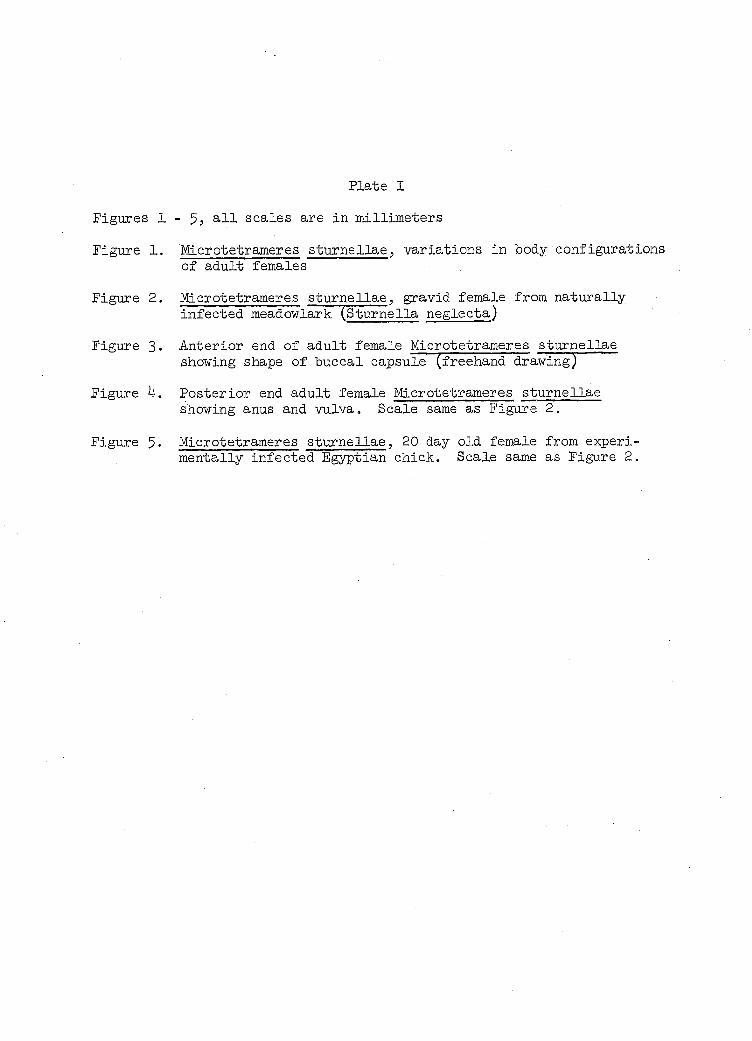
Plate I
Figures 1 - all scales are in millimeters
Figure 1. Microtetrameres sturnellae, variations in body configurations of adult females
Figure 2. Microtetrameres sturnellae, gravid female from naturally infected meadowlark (Sturnella neglecta)
Figure 3. Anterior end of adult female Microtetrameres sturnellae showing shape of buccal capsule (freehand drawing)
Figure 4. Posterior end adult female Microtetrameres sturnellae showing anus and vulva. Scale same as Figure 2.
Figure 5. Microtetrameres sturnellae, 20 day old female from experimentally infected Egyptian chick. Scale same as Figure 2.
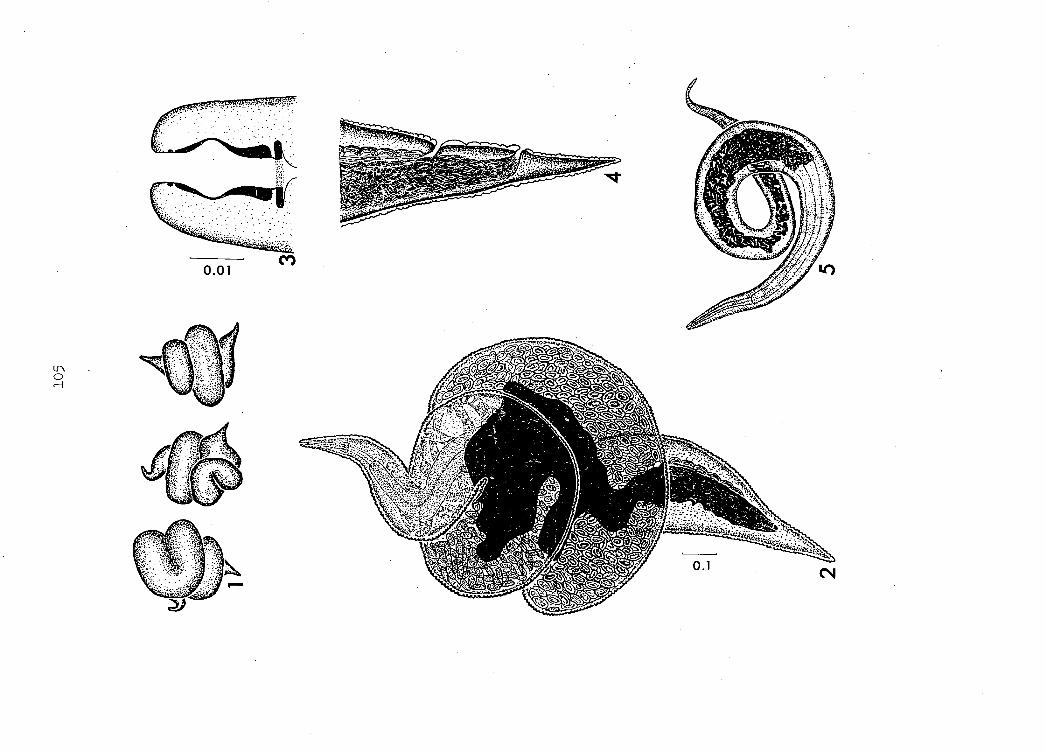
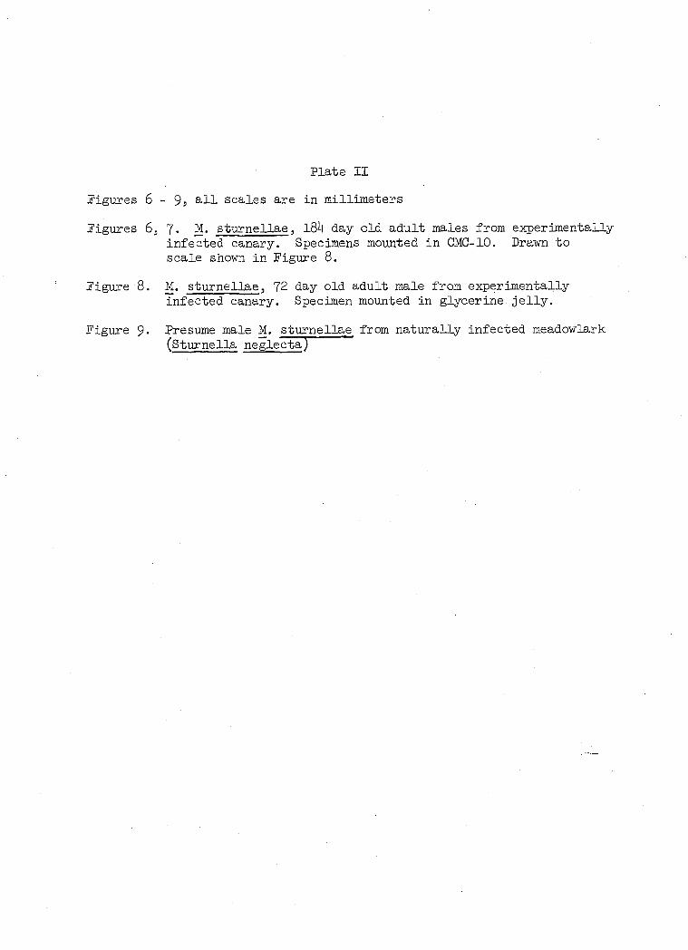
Plate II
Figures 6 - 9, all scales are in millimeters
Figures 6, 7. M. sturnellae, 184 day old adult males from experimentally infected canary. Specimens mounted in GMC-10. Drawn to scale shown in Figure 8.
Figure 8. M. sturnellae, 72 day old adult male from experimentally infected canary. Specimen mounted in glycerine jelly.
Figure 9- Presume male M. sturnellae from naturally infected meadowlark (Sturnella negleeta)
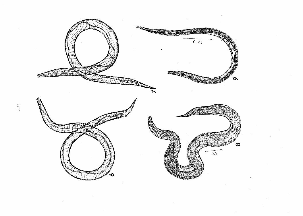
0.1

Plate III
Figures 10 - 19, all scales are in millimeters
Figure 10. Egg of M. sturnellae containing first-stage juvenile
Figure 11. First-stage juvenile emerging from egg
Figure 12. Living four-day old first-stage juvenile from experimentally infected grasshopper nymph
Figure 13. First-stage juvenile, anterior end extended showing denticulate projections
Figure l4. First-stage juvenile, anterior end withdrawn. Same scale as Figure 13.
Figures 15, l6. Living first-stage juvenile recovered from grasshopper nymph l6 days post-feeding
Figure 15. Anterior end. Ecdysis commencing.
Figure l6. Posterior end, same specimen. Same scale as Figure 15.
Figure 17. Fixed first-stage juvenile undergoing ecdysis to second-stage juvenile approximately l6 days after feeding grasshopper nymph. Same scale as Figure 15.
Figure l8. Anterior end, first-stage juvenile undergoing ecdysis. Specimen recovered from grasshopper nymph approximately l6 days post-feeding. Note presence of denticulate projections on sheath.
Figure 19. Third-stage juvenile, excysted. Specimen mounted in glycerine jelly and recovered from grasshopper nymph 38 days post-feeding.
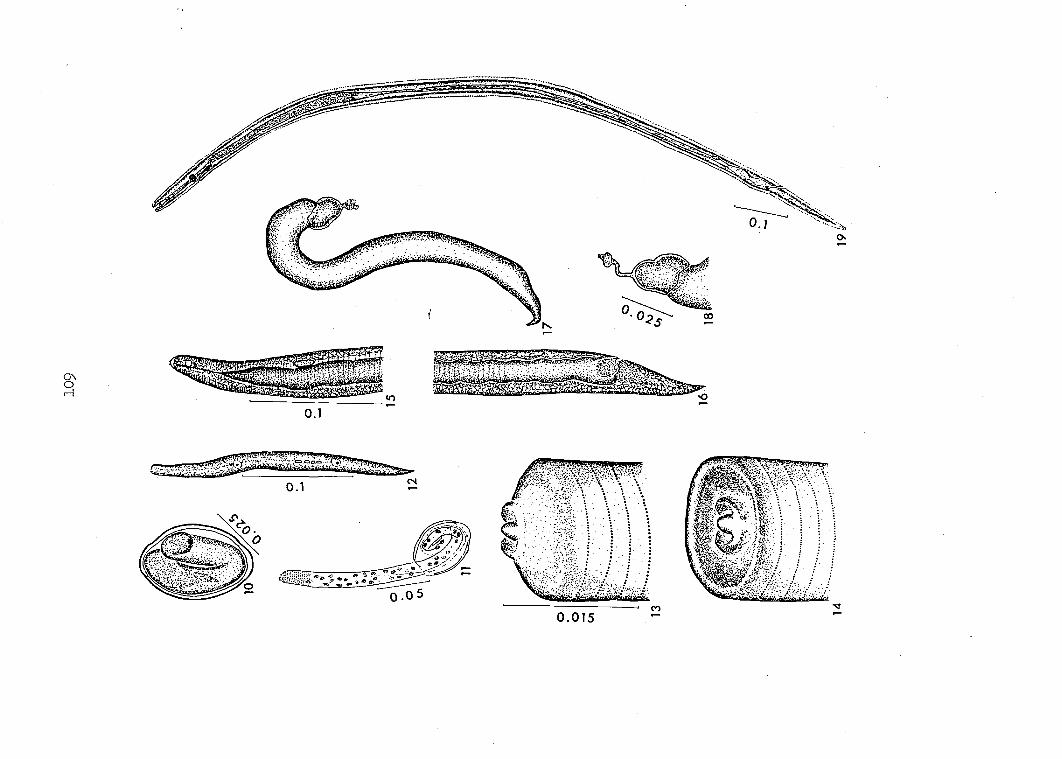
00
0.1
cs
CO
0.015

Plate IV
Figure 20. Serosal surface of meadowlark proventriculus showing M. sturnellae females situ
Figure 21. Living gravid M. sturnellae females removed from meadowlark proventriculus. Note body configurations and uterine coils.
Figure 22. Experimentally reared adult female recovered 20 days post-feeding from laboratory-reared canary. Note detritus and cuticular striations. Darkfield microscopy.

Ill
Plate V
Figure 23. Section of female M. sturnellae in situ in meadowlark pro-ventricular gland
Figure 2k. Female M. sturnellae in situ showing cephalic end within proventricular gland
Figure 25. Enlargement of anterior end shown in Figure 2k
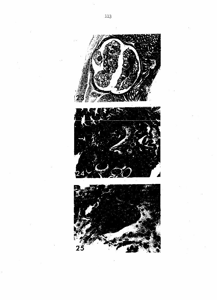
113
Plate VI
Figures 26, 27. Microtetrameres sturnellae, maies, l84 days old, experimentally-reared in canary. Phase-contrast microscopy.
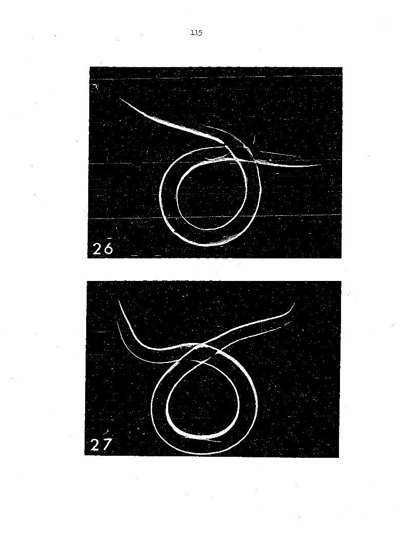
115
Plate VII
Figure 28. First-stage juvenile emerging from egg. Baase-contrast microscopy.
Figure 29. Second-stage juvenile about 10 days post-feeding grasshopper numph. Note tail configuration.
Figure 30. Encysted third-stage juvenile from experimentally infected grasshopper nymph. Phase contrast microscopy.
Figure 3I. Section of proventriculus showing third-stage juveniles in situ within proventricular gland, 7 days post-feeding. Note arrow indicating juvenile within interlobular tissue of glands.
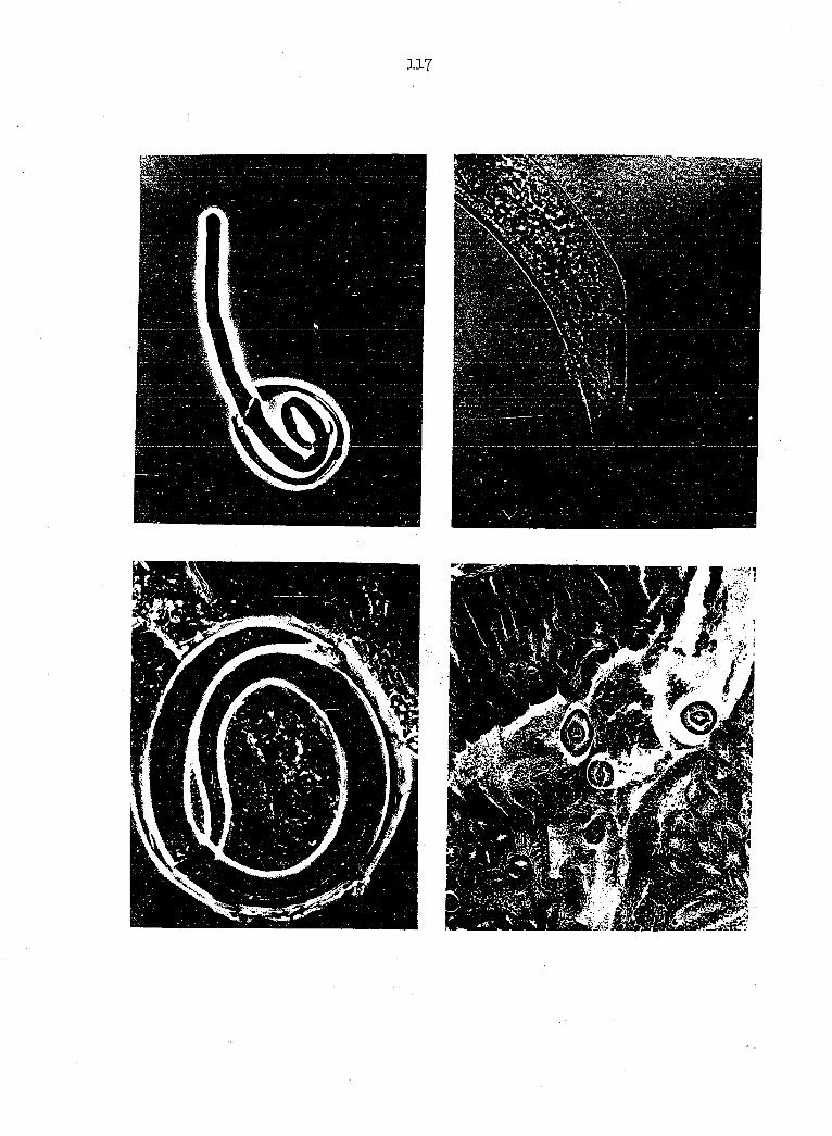
117
Plate VIII
Figure 32. Cross section of grasshopper nymph showing cysts containing •third-stage juveniles of M. sturnellae in perivisceral sinus of hemocoel.
Figure 33- Enlargement of one cyst shown in Figure 32.
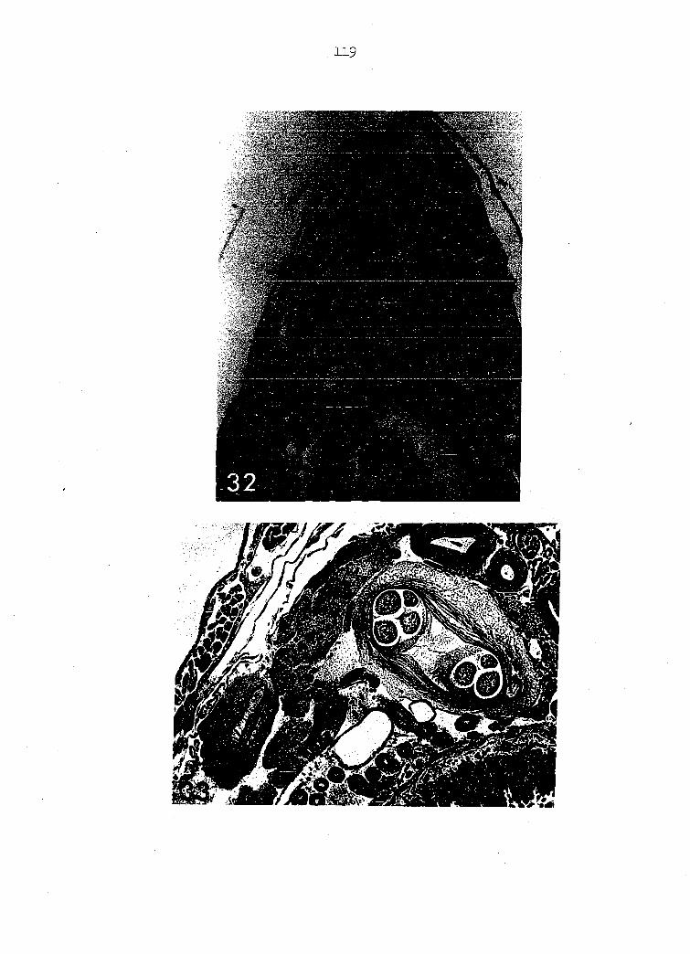
119
%
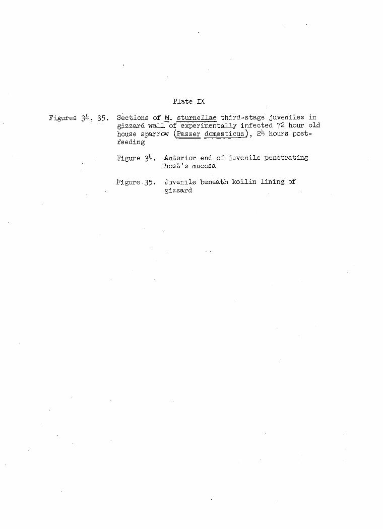
Plate DC
Figures 34, 35. Sections of M. sturnellae third-stage juveniles in gizzard wall of experimentally infected 72 hour old house sparrow (Passer domesticus), 2h hours post-feeding
Figure 3 4. Anterior end of juvenile penetrating host's mucosa
Figure.35. Juvenile beneath koilin lining of gizzard

121
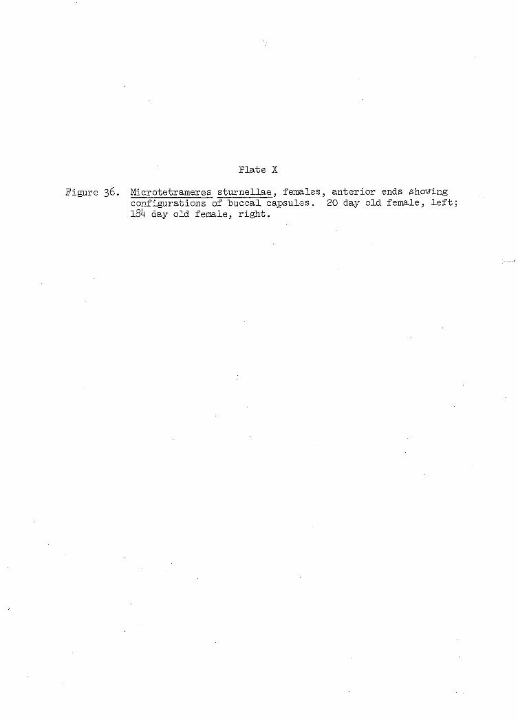
Plate X
Figure 36. Microtetrameres sturnellae, females, anterior ends showing configurations of buccal capsules. 20 day old female, left; l84 day old female, right.