New records of species of Philometra (Nematoda: Philometridae) from marine fishes off New Caledonia,...
6
ORIGINAL PAPER New records of species of Philometra (Nematoda: Philometridae) from marine fishes off New Caledonia, including P. cephalopholidis sp. n. from Cephalopholis sonnerati (Serranidae) František Moravec 1 & Jean-Lou Justine 2 Received: 28 April 2015 /Accepted: 6 May 2015 /Published online: 19 May 2015 # Springer-Verlag Berlin Heidelberg 2015 Abstract Two different species of Philometra Costa, 1845 were collected from marine perciform fishes, the tomato hind Cephalopholis sonnerati (Valenciennes) (Serranidae) and the painted sweetlips Diagramma pictum (Thunberg) (Haemulidae), from off New Caledonia, South Pacific. Nematodes (only males) from the gonad of C. sonnerati represent a new taxon, P. cephalopholidis sp. n., which is mainly characterized by almost equally long spicules (length 186–228 μm), the shape and structure of the gubernaculum with a dorsally lamellate distal tip, and the structure of the caudal end. The nematodes (only gravid females) from abdominal tissues of D. pictum may repre- sent an undescribed species, but, because of the absence of conspecific males, they could not be specifically identified. Philometra cephalopholidis is the sixth nominal species of this genus recorded from fishes off New Caledonia and the thirteenth species of these parasites described from fishes of the family Serranidae. Keywords Dracunculoidea . Marine fish . Nematode parasite . South Pacific Introduction As recently mentioned by Moravec and Justine (2014), the knowledge of the fauna of philometrid nematodes (Philometridae) parasitizing marine fishes in New Caledonian waters remains little known, despite the fact that 13 species of these important, tissue-dwelling parasites have been reported in this region (Moravec and Justine 2005, 2008, 2009, 2011; Moravec and Justine 2014). A taxo- nomic evaluation of additional samples of these nematodes collected from two species of perciform fishes, the tomato hind Cephalopholis sonnerati (Valenciennes) (Serranidae) and the painted sweetlips Diagramma pictum (Thunberg) (Haemulidae), revealed the presence of two species of Philometra Costa, 1845. Results of this study are presented herein. Both host species are tropical, reef-associated marine fishes which are widespread in the Indo-Pacific and are targeted by commercial fisheries (Froese and Pauly 2015). Materials and methods Fish were caught off Nouméa, New Caledonia, or purchased from the fishmarket in Nouméa. The nematodes for morpho- logical studies were fixed in hot 4 % formalin. For light mi- croscopical examination, they were cleared with glycerine. Drawings were made with the aid of a Zeiss microscope draw- ing attachment. Specimens used for scanning electron micro- scopical examination were postfixed in 1 % osmium tetroxide (in phosphate buffer), dehydrated through a graded acetone series, critical-point-dried and sputter-coated with gold; they were examined using a JEOL JSM-7401F scanning electron microscope at an accelerating voltage of 4 kV (GB low mode). All measurements are in micrometers unless otherwise * František Moravec [email protected] 1 Institute of Parasitology, Biology Centre, Czech Academy of Sciences, Branišovská 31, 370 05 České Budějovice, Czech Republic 2 Institut Systématique, Évolution, Biodiversité, ISYEB, UMR7205 CNRS, EPHE, MNHN, UPMC, Muséum National d’Histoire Naturelle, Sorbonne Universités, CP51, 57 rue Cuvier, 75231 Paris, Cedex 05, France Parasitol Res (2015) 114:3223–3228 DOI 10.1007/s00436-015-4538-4
Transcript of New records of species of Philometra (Nematoda: Philometridae) from marine fishes off New Caledonia,...

ORIGINAL PAPER
New records of species of Philometra (Nematoda:Philometridae) from marine fishes off New Caledonia,including P. cephalopholidis sp. n. from Cephalopholissonnerati (Serranidae)
František Moravec1 & Jean-Lou Justine2
Received: 28 April 2015 /Accepted: 6 May 2015 /Published online: 19 May 2015# Springer-Verlag Berlin Heidelberg 2015
Abstract Two different species of Philometra Costa, 1845were collected from marine perciform fishes, the tomatohind Cephalopholis sonnerati (Valenciennes) (Serranidae)and the painted sweetlips Diagramma pictum (Thunberg)(Haemulidae), from off New Caledonia, South Pacific.Nematodes (only males) from the gonad of C. sonneratirepresent a new taxon, P. cephalopholidis sp. n., which ismainly characterized by almost equally long spicules(length 186–228 μm), the shape and structure of thegubernaculum with a dorsally lamellate distal tip, and thestructure of the caudal end. The nematodes (only gravidfemales) from abdominal tissues of D. pictum may repre-sent an undescribed species, but, because of the absenceof conspecific males, they could not be specifically identified.Philometra cephalopholidis is the sixth nominal species ofthis genus recorded from fishes off New Caledonia and thethirteenth species of these parasites described from fishes ofthe family Serranidae.
Keywords Dracunculoidea .Marine fish . Nematodeparasite . South Pacific
Introduction
As recently mentioned by Moravec and Justine (2014),the knowledge of the fauna of philometrid nematodes(Philometridae) parasitizing marine fishes in NewCaledonian waters remains little known, despite the factthat 13 species of these important, tissue-dwelling parasiteshave been reported in this region (Moravec and Justine2005, 2008, 2009, 2011; Moravec and Justine 2014). A taxo-nomic evaluation of additional samples of these nematodescollected from two species of perciform fishes, the tomatohind Cephalopholis sonnerati (Valenciennes) (Serranidae)and the painted sweetlips Diagramma pictum (Thunberg)(Haemulidae), revealed the presence of two species ofPhilometra Costa, 1845. Results of this study are presentedherein. Both host species are tropical, reef-associated marinefishes which are widespread in the Indo-Pacific and aretargeted by commercial fisheries (Froese and Pauly 2015).
Materials and methods
Fish were caught off Nouméa, New Caledonia, or purchasedfrom the fishmarket in Nouméa. The nematodes for morpho-logical studies were fixed in hot 4 % formalin. For light mi-croscopical examination, they were cleared with glycerine.Drawings were made with the aid of a Zeiss microscope draw-ing attachment. Specimens used for scanning electron micro-scopical examination were postfixed in 1 % osmium tetroxide(in phosphate buffer), dehydrated through a graded acetoneseries, critical-point-dried and sputter-coated with gold; theywere examined using a JEOL JSM-7401F scanning electronmicroscope at an accelerating voltage of 4 kV (GB lowmode). All measurements are in micrometers unless otherwise
* František [email protected]
1 Institute of Parasitology, Biology Centre, Czech Academy ofSciences, Branišovská 31, 370 05 ČeskéBudějovice, Czech Republic
2 Institut Systématique, Évolution, Biodiversité, ISYEB, UMR7205CNRS, EPHE, MNHN, UPMC, Muséum National d’HistoireNaturelle, Sorbonne Universités, CP51, 57 rue Cuvier,75231 Paris, Cedex 05, France
Parasitol Res (2015) 114:3223–3228DOI 10.1007/s00436-015-4538-4
indicated. The fish nomenclature adopted follows FishBase(Froese and Pauly 2015).
Results
Family Philometridae Baylis et Daubney, 1926Philometra cephalopholidis sp. n.Description of male (ten specimens; measurements of ho-
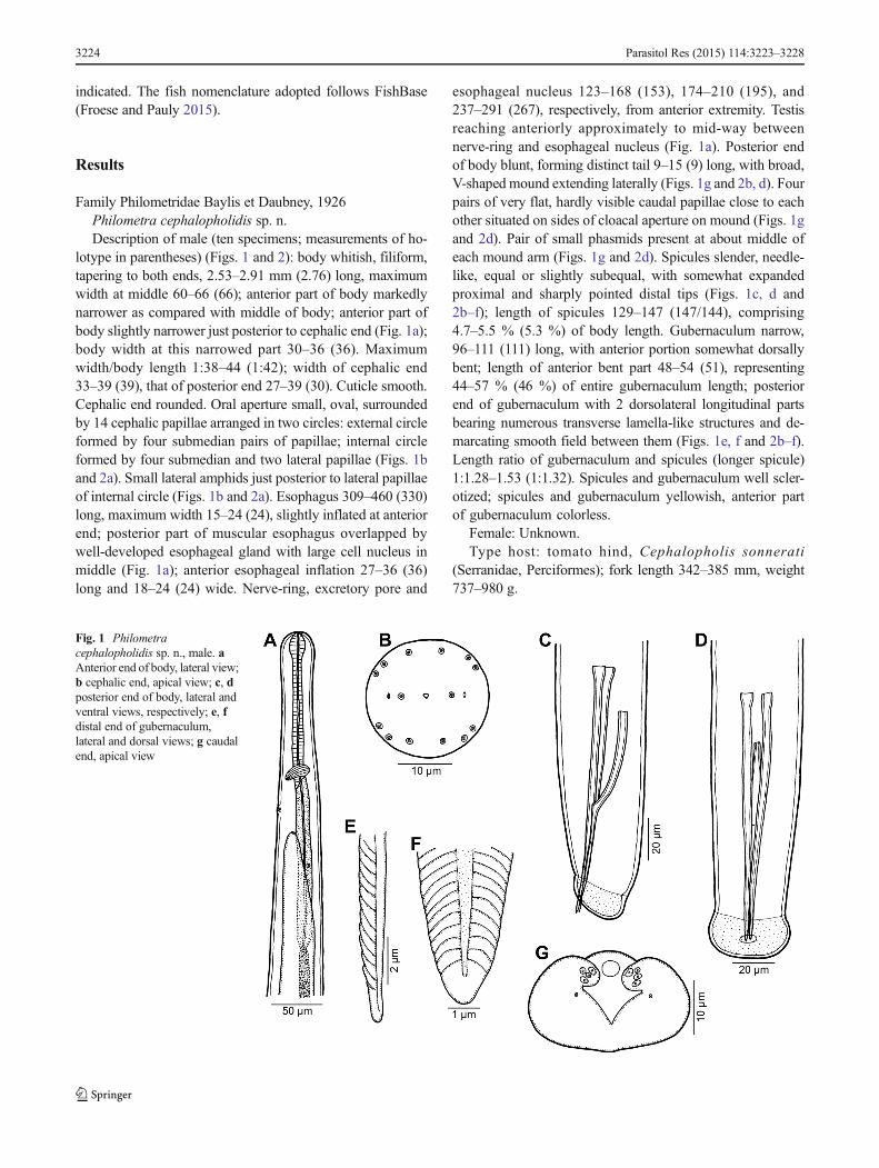
lotype in parentheses) (Figs. 1 and 2): body whitish, filiform,tapering to both ends, 2.53–2.91 mm (2.76) long, maximumwidth at middle 60–66 (66); anterior part of body markedlynarrower as compared with middle of body; anterior part ofbody slightly narrower just posterior to cephalic end (Fig. 1a);body width at this narrowed part 30–36 (36). Maximumwidth/body length 1:38–44 (1:42); width of cephalic end33–39 (39), that of posterior end 27–39 (30). Cuticle smooth.Cephalic end rounded. Oral aperture small, oval, surroundedby 14 cephalic papillae arranged in two circles: external circleformed by four submedian pairs of papillae; internal circleformed by four submedian and two lateral papillae (Figs. 1band 2a). Small lateral amphids just posterior to lateral papillaeof internal circle (Figs. 1b and 2a). Esophagus 309–460 (330)long, maximum width 15–24 (24), slightly inflated at anteriorend; posterior part of muscular esophagus overlapped bywell-developed esophageal gland with large cell nucleus inmiddle (Fig. 1a); anterior esophageal inflation 27–36 (36)long and 18–24 (24) wide. Nerve-ring, excretory pore and
esophageal nucleus 123–168 (153), 174–210 (195), and237–291 (267), respectively, from anterior extremity. Testisreaching anteriorly approximately to mid-way betweennerve-ring and esophageal nucleus (Fig. 1a). Posterior endof body blunt, forming distinct tail 9–15 (9) long, with broad,V-shaped mound extending laterally (Figs. 1g and 2b, d). Fourpairs of very flat, hardly visible caudal papillae close to eachother situated on sides of cloacal aperture on mound (Figs. 1gand 2d). Pair of small phasmids present at about middle ofeach mound arm (Figs. 1g and 2d). Spicules slender, needle-like, equal or slightly subequal, with somewhat expandedproximal and sharply pointed distal tips (Figs. 1c, d and2b–f); length of spicules 129–147 (147/144), comprising4.7–5.5 % (5.3 %) of body length. Gubernaculum narrow,96–111 (111) long, with anterior portion somewhat dorsallybent; length of anterior bent part 48–54 (51), representing44–57 % (46 %) of entire gubernaculum length; posteriorend of gubernaculum with 2 dorsolateral longitudinal partsbearing numerous transverse lamella-like structures and de-marcating smooth field between them (Figs. 1e, f and 2b–f).Length ratio of gubernaculum and spicules (longer spicule)1:1.28–1.53 (1:1.32). Spicules and gubernaculum well scler-otized; spicules and gubernaculum yellowish, anterior partof gubernaculum colorless.
Female: Unknown.Type host: tomato hind, Cephalopholis sonnerati
(Serranidae, Perciformes); fork length 342–385 mm, weight737–980 g.
Fig. 1 Philometracephalopholidis sp. n., male. aAnterior end of body, lateral view;b cephalic end, apical view; c, dposterior end of body, lateral andventral views, respectively; e, fdistal end of gubernaculum,lateral and dorsal views; g caudalend, apical view
3224 Parasitol Res (2015) 114:3223–3228

Site in host: ovary.Type locality: Off Nouméa, New Caledonia, North of Récif
Toombo (22° 31 S, 166° 28 E) (collected 12 May 2009).Prevalence and intensity: two fish infected/four fish
examined; up to 50 nematodes in one fish.Deposition of type specimens: Muséum National d’Histoire
Naturelle, Paris (holotype and 44 paratypes—cat. nos. MNHNJNC2935 andMNHN JNC2936) and Institute of Parasitology,Biology Centre of the Czech Academy of Sciences, ČeskéBudějovice (four paratypes—cat. no. N–1088).
Etymology: The specific name of this nematode relates tothe genitive form of the generic name of the host.
Remarks: At present, 61 gonad-infecting species ofPhilometra are known to parasitize marine fishes (Moravecand de Buron 2013; Quiazon and Yoshinaga 2013; Moravecand Diggles 2014; Moravec and Justine 2014; Moravec andManoharan 2014a, b; Moravec et al. 2014; Moravec andBarton 2015). However, as indicated by some recent studiesof these nematodes from marine fishes, they exhibit a highdegree of host specificity, when different morphologicallywell-distinguishable species of Philometra are recorded from
different species of congeneric hosts in the same locality(Moravec and Manoharan 2014a , b; Moravec et al. 2014).Therefore, it is possible to deal with these parasites accordingto the families of their fish hosts.
To date, 12 nominal gonad-infecting species of Philometraare known to parasitize fishes of the perciform familySerranidae: P. charlestonensis Moravec, de Buron, Baker etGonzález-Solís, 2008, P. cyanopodiMoravec et Justine, 2008,P. fasciatiMoravec et Justine, 2008, P. hyporthodiMoravec etBakenhaster, 2013, P. indica Moravec et Manoharan, 2014,P. jordanoi (López-Neyra, 1951), P. managatuwo Yamaguti,1941, P. margolisiMoravec, Vidal-Martínez et Aguirre-Macedo,1995, P. mexicana Moravec et Salgado-Maldonado, 2007,P. piscaria Moravec et Justine, 2014, P. serranellicabrillaeJaniszewska, 1949, and P. tropica Moravec et Manoharan,2014 (see Moravec and Justine 2014; Moravec and Manoharan2014a).
The new species can be easily distinguished by thelength of spicules from P. cyanopodi (spicules 186–228 μm vs 129–147 μm), P. indica (192–195 μm),P. jordanoi (about 260 μm), P. margolisi (432–468 μm),
Fig. 2 Philometracephalopholidis sp. n., scanningelectron micrographs of male. aCephalic end, subapical view(arrow indicates amphid); bcaudal end, lateral view; c regionof cloaca, apical view; d caudalend, apical view (arrows indicatephasmids); e, f region of cloaca,lateral, and dorsal views,respectively. a Submedian pair ofcephalic papillae of externalcircle, b submedian singlecephalic papilla of internal circle,c lateral cephalic papilla ofinternal circle, d group of fourcaudal papillae, e caudal mound,g gubernaculum, s spicule
Parasitol Res (2015) 114:3223–3228 3225

P. mexicana (90–120 μm), P. piscaria (171–180 μm), andP. tropica (168–186 μm). On the other hand, by thelength of spicules, P. cephalopholidis sp. n. resemblesP. charlestonensis (132–141 μm) parasitic in Mycteropercaphenax (Jordan et Swain) in the region of the West Atlantic(Moravec et al. 2008; Moravec and Bakenhaster 2012),P. fasciati (147–156 μm) from Epinephelus fasciatus(Forsskål) off New Caledonia (Moravec and Justine 2005,2008), and P. hyporthodi (135–138 μm) from Hyporthodusflavolimbatus (Poey) in the Gulf of Mexico (Moravec andBakenhaster 2013). However, in contrast to the new spe-cies, the male caudal mound is widely interrupted dorsally(vs dorsally uninterrupted), and a pair of large postcloacalpapillae is present (vs these papillae are absent) in each ofboth P. charlestonensis and P. hyporthodi. The male ofP. fasciati has not yet been studied by SEM, but in additionto mostly longer spicules, this species can be separated fromP. cephalopholidis sp. n. by the extent of testis, whichreaches anteriorly only to the end of esophagus (vs testisreaching to about mid-way between esophageal nucleusand nerve ring).
Two gonad-infecting species parasitizing serranid fishes,P. managatuwo and P. serranellicabrillae, were describedsolely from females, whereas conspecific males remain un-known. Therefore, these two species can be distinguishedfrom P. cephalopholidis sp. n. mainly by the genera of hosts(Serranus Cuvier and Epinephelus Bloch vs CephalopholisBloch et Schneider) and, in the case of P. serranellicabrillae,
Fig. 3 Philometra sp., gravidfemale from Diagramma pictum.a Anterior end of body, lateralview; b, c cephalic end, lateral,and apical views, respectively;d posterior end of body, lateralview; e larva from uterus, lateralview
Fig. 4 Philometra sp., scanning electron micrographs of gravid femalefrom Diagramma pictum. a Region of oral aperture, subapical view; bposterior end of body, lateral view. a Pair of submedian cephalic papillaeof external circle; b submedian cephalic papilla of internal circle; o oralaperture
3226 Parasitol Res (2015) 114:3223–3228

also by the geographical distribution (Mediterranean Sea vsSouth Pacific).
The only previous record of a philometrid fromC. sonnerati is that of Schmidt and Kuntz (1969), whoreported Philometra lateolabracis (Yamaguti, 1935) fromthe body cavity of this host off the Philippines (Palawan).However, this is an evident species misidentification, becauseP. lateolabracis is a specific gonad-infecting parasite ofLateolabrax japonicus (Cuvier) (Lateolabracidae) off Japan(see Quiazon et al. 2008).
Philometra sp.Description of gravid female (many body fragments of two
larvigerous specimens) (Figs. 3, 4, and 5): body of fixedspecimens gray-brown, with rounded ends. Posterior partof body narrower than anterior part; maximum width inregion posterior to esophagus. Total body length not de-termined, but may be estimated from available fragmentsto be c. 450–470 mm; length of anterior body fragments2.4–40 mm, maximum width 1.43–1.77 mm; length ofposterior body fragment 6.5 mm. Anterior end of bodymarkedly bottle-shaped; width of cephalic end 286–299.Cephalic papillae very small. Oral aperture large, oval,surrounded by four submedian pairs of cephalic papillaeof external circle and four submedian single papillae (lateralpapillae indistinct) of internal circle (Figs. 3c and 4a).Amphids indistinct. Bottom of mouth formed by lobes ofthree esophageal sectors (Figs. 3c and 4a). Esophagus
including anterior bulbous inflation 1.55–1.66 mm long; bulbwell developed, 177–190 long and 177 wide; maximumwidthof esophagus including gland 163–190. Esophageal glandwell developed, opening into esophagus just posterior to nervering, with distinct cell nucleus in middle. Nerve ring andesophageal nucleus 354–367 and 930–952, respectively, fromanterior extremity. Ventriculus 41 long, 109 wide (Fig. 3a).Intestine wide at anterior end; its posterior end narrow, at-tached by short ligament ventrally to body wall near caudalend; ligament 435–748 long. Ovaries reflected, situated nearbody ends. Uterus occupying most space of body, anteriorlyextended to base of esophagus, filled with numerous larvae(n=5) 399–486 long and 15–18 wide; length of esophagus126–150, of tail 102–120, representing 31–32 and 25–28 %,respectively, of entire body length of larva (Fig. 3e). Posteriorend of female rounded, 272–367 wide, without any caudalprojections (Fig. 3d).
Male: Unknown.Host: painted sweetlips, Diagramma pictum (Haemulidae,
Perciformes); fork length 340 mm, weight 502 g.Site in host: tissue around anus.Locality: Fishmarket of Nouméa, NewCaledonia (collected
1 December 2010).Prevalence and intensity: one fish infected/three fish exam-
ined; two nematode specimens.Deposition of voucher specimens: Muséum National
d’Histoire Naturelle, Paris (cat. no. MNHN JNC3292).
Fig. 5 Gravid females ofPhilometra sp. a Nematodesremoved from host’s tissues(scale, squares=5 mm); b part ofnematode body hanging out ofhost’s tissue near anus; c infectedDiagramma pictum withPhilometra sp. specimen partlyprotruding out of tissues aroundthe anus
Parasitol Res (2015) 114:3223–3228 3227

Remarks: An infected fish specimen (painted sweetlips), inperfect state of freshness, was noticed at the fishmarket with afemale philometrid hanging out from behind the anus (Fig. 5).The fish was brought back to the laboratory. Two femalephilometrids, alive, were found entangled in the tissues (mus-cles, skin) around the anus; it was impossible to separate thenematodes from the tissues without breaking them. The tissuearound the anus was dilacerated in search of males, but nonewas found. The fish had no visible male or female gonad.
To date, the only species ofPhilometra described from fishesof the family Haemulidae is P. isaki Quiazon, Yoshinaga etOgawa, 2008 from the gonads of Parapristipoma trilineatum(Thunberg) in the East China Sea off Japan (Quiazon et al.2008), whereas the record of another gonad-infecting speciesP. lateolabracis (Yamaguti, 1935) from Haemulon plumieri(Lacépède) in the Atlantic Ocean off Brazil by Crisp andKlein (1973) represents an evident misidentification (seeMoravec 2008; Quiazon et al. 2008). The present NewCaledonian gravid females may be approximately as long asthose ofP. isaki, but in contrast to the latter species, their anteriorend of body is bottle-shaped and their esophagus is somewhatlonger (1.55–1.66 mm vs 0.79–1.10 mm) with a well-developed bulbous inflation (vs anterior esophageal inflationpoorly developed). Considering a different location in the host,the fact that the hosts of these two forms belong to different fishgenera and these parasites were collected in distant geographicalregions, it is probable that the present specimens belong to anundescribed species. However, since conspecific males are notavailable, we refrain from describing them as a new species.
Acknowledgments Thanks are due to the staff of the Laboratory ofElectron Microscopy, Institute of Parasitology, BC CAS, in ČeskéBudějovice for their technical assistance, and to Blanka Škoríková of thesame Institute for helpwith the illustrations. This studywas partly supportedby the Institute of Parasitology (with institutional support RVO 60077344)and the Czech Science Foundation (Project No. P505/12/G112).
References
Froese R, Pauly D (eds) (2015) FishBase. World Wide Web electronicpublication, version 04/2015. http://www.fishbase.org
Moravec F (2008) Systematic status of Philometra jordanoi (López-Neyra, 1951) and some other congeneric species previously identi-fied as Philometra lateolabracis (Yamaguti, 1935) (Nematoda:Philometridae). Folia Parasitol 55:159–160
Moravec F, Bakenhaster M (2012) New observations on philometrid nema-todes (Philometridae) inmarine fishes from the northernGulf ofMexicoand the Indian River Lagoon of Florida (USA), with first description ofthe male of Caranginema americanum. J Parasitol 98:398–403
Moravec F, Bakenhaster M (2013) Two new gonad-infecting philometrids(Nematoda: Philometridae) from the yellowedge grouperHyporthodus flavolimbatus (Serranidae) and the great northern tile-fish Lopholatilus chamaeleonticeps (Malacanthidae) in the northernGulf of Mexico. Syst Parasitol 86:113–123
Moravec F, Barton DP (2015) Two gonad-infecting species ofPhilometra(Nematoda: Philometridae) from marine fishes off the northernAustralia. Parasite 22:4
Moravec F, de Buron I (2013) A synthesis of our current knowledge ofphilometrid nematodes, a group of increasingly important fish par-asites. Folia Parasitol 60:81–101
Moravec F, Diggles BK (2014) Two new gonad-infecting species ofPhilometra Costa, 1845 (Nematoda: Philometridae) from marinefishes off the northern coast of Australia. Syst Parasitol 89:33–44
Moravec F, Justine J-L (2005) Two species of Philometra (Nematoda,Philometridae) from serranid fishes off New Caledonia. ActaParasitol 50:323–331
Moravec F, Justine J-L (2008) Some philometrid nematodes (Philometridae),including four new species of Philometra, from marine fishes off NewCaledonia. Acta Parasitol 53:369–381
Moravec F, Justine J-L (2009) New data on dracunculoid nematodes fromfishes off New Caledonia, including four new species of Philometra(Philometridae) and Ichthyofilaria (Guyanemidae). Folia Parasitol56:129–142
Moravec F, Justine J-L (2011) Two new gonad-infecting Philometraspecies (Nematoda: Philometridae) from the marine fish Lutjanusvitta (Perciformes: Lutjanidae) off New Caledonia. Folia Parasitol58:302–310
Moravec F, Justine J-L (2014) Philometrids (Nematoda: Philometridae) incarangid and serranid fishes off New Caledonia, including three newspecies. Parasite 21:21
Moravec F, Manoharan J (2014a) Gonad-infecting species of Philometra(Nematoda: Philometridae) from groupers Epinephelus spp.(Osteichthyes: Serranidae) in the Bay of Bengal, India. ActaParasitol 59:596–605
Moravec F, Manoharan J (2014b) Two new gonad-infecting species ofPhilometra (Nematoda: Philometridae) parasitic in Lutjanus spp.(Osteichthyes: Lutjanidae) in the Bay of Bengal, India. ParasitolRes 113:3299–3307
Moravec F, de Buron I, Baker TG, González-Solís D (2008) Some gonad-infecting species of Philometra (Nematoda: Philometridae) fromoffshore fishes of South Carolina and Georgia, USA, includingPhilometra charlestonensis sp. nov. from scamp Mycteropercaphenax. Acta Parasitol 53:382–391
Moravec F, Bakenhaster M, Fajer-Ávila EJ (2014) Three new gonad-infecting species of Philometra (Nematoda: Philometridae) parasiticin Lutjanus spp. (Lutjanidae) in the northern Gulf of Mexico offFlorida, USA. Folia Parasitol 61:355–369
Quiazon KMA, Yoshinaga T (2013) Gonad-infecting philometridPhilometra philippinensis sp. nov. (Nematoda, Philometridae) fromthe bigeye barracuda Sphyraena forsteri Cuvier (Sphyraenidae) offMariveles, Bataan Province, Philippine Archipelago. Acta Parasitol58:504–514
Quiazon KMA, Yoshinaga T, Ogawa K (2008) Taxonomical study intotwo new species of Philometra (Nematoda: Philometridae) previ-ously identified asPhilometra lateolabracis (Yamaguti, 1935). FoliaParasitol 55:29–41
3228 Parasitol Res (2015) 114:3223–3228