Description of Bathynema nodinauti gen. n., sp. n. and four new Trophomera species (Nematoda:...
24
Accepted by K.H. George: 11 Mar. 2009; published: 11 May 2009 173 ZOOTAXA ISSN 1175-5326 (print edition) ISSN 1175-5334 (online edition) Copyright © 2009 · Magnolia Press Zootaxa 2096: 173–196 (2009) www.mapress.com/ zootaxa/ Article Description of Bathynema nodinauti gen. n., sp. n. and four new Trophomera species (Nematoda: Benthimermithidae) from the Clarion-Clipperton Fracture Zone (Eastern Tropic Pacific), supplemented with the keys to genera and species* DMITRY M. MILJUTIN # & MARIA A. MILJUTINA Abt. DZMB, Forschungsinstitut Senckenberg, Südstrand 44, D-26382 Wilhelmshaven, Germany # Corresponding author: [email protected] * In: Brökeland, W. & George, K.H. (eds) (2009) Deep-sea taxonomy — a contribution to our knowledge of biodiversity. Zootaxa, 2096, 1–488. Abstract Seven specimens of the family Benthimermithidae from the Clarion-Clipperton Fracture Zone (Eastern Tropic Pacific), from the depths 4,800–5,040 m, were examined. Bathynema nodinauti gen. et sp. n. is described. The new genus differs from other genera of Benthimermithidae by the presence of an inner pharyngeal lumen. Four new species of Trophomera are also described. Body length of female of T. elegantis sp. n. approximately 1.5 mm; body cylindrical, anterior and posterior ends in shape of rounded cone; cephalic setae 2.0–2.5 μm long; trophosome consisting of 1 row of cells; female reproductive system didelphic, amphidelphic, occupying approximately 1/6 of total body length; ovaries reflected; diameter of mature eggs 17 μm; males not found. Body length of female of T. minutissima sp. n. 0.9 mm; body cylindrical, with thickest body part at anterior half of body; anterior and posterior ends rounded; cephalic setae 1.5 μm long; trophosome consisting of 1 row of cells; female reproductive system didelphic, amphidelphic, occupying approximately 1/3 of total body length; ovaries reflected; size of mature eggs 24x23 μm; males not found. Body length of female of T. pacifica sp. n. 5.4 mm; body cylindrical, anterior end rounded; posterior end conical, with thick conical terminal spine 81 μm long, showing granular core; cephalic setae 2.5 μm long inserted in tiny pits; trophosome consisting of 1 row of cells; female reproductive system didelphic, amphidelphic, occupying approximately 2/3 of total body length; ovaries outstretched; size of mature eggs 34x20 μm; males not found. Body length of female of T. senckenbergi sp. n. 1.6 mm; body fusiform; anterior and posterior ends in shape of a cone with rounded tip; cephalic setae 2 μm long; trophosome consisting of 1 row of cells; female reproductive system didelphic, amphidelphic, occupying approximately 1/6 of total body length; ovaries non-reflected; males not found. The new finding of T. marionensis is recorded. One rest specimen (in bad condition) of a new species is described as a morphotype. A dichotomous key to the genera of the Benthimermithidae and tabular keys to Trophomera species are presented. Key words: Benthimermis, biodiversity, deep sea, taxonomy, manganese nodule, oozy sediments, taxonomy Introduction At present, there are a number of nematode species, which are known as internal parasites with marine invertebrates as the definitive hosts. Most of these nematodes belong to the family Benthimermithidae. Larval (morphologically unidentifiable) stages of benthimermithids infest body cavities or internal organs of various marine nematodes, polychaetes, priapulids, and crustaceans (Hope 1977; Rubtzov 1980; Petter 1980; Petter & Gourbault 1985; Chesunov 1988a, 1997; Miljutin 2004). Late parasitic stages can occupy the whole body of their hosts and, evidently, can result in the death of the host. Before terminal moulting, benthimermithids exit their host to the external environment, where they do not feed but do reproduce (at least as it has been inferred from their vestigial mouth and pharynx, and their intestine lacking an internal lumen).
-
Upload
senckenberg -
Category
Documents
-
view
0 -
download
0
Transcript of Description of Bathynema nodinauti gen. n., sp. n. and four new Trophomera species (Nematoda:...
ZOOTAXAISSN 1175-5326 (print edition)
ISSN 1175-5334 (online edition)Copyright © 2009 · Magnolia Press
Zootaxa 2096: 173–196 (2009) www.mapress.com/zootaxa/ Article
Description of Bathynema nodinauti gen. n., sp. n. and four new Trophomera species (Nematoda: Benthimermithidae) from the Clarion-Clipperton Fracture Zone (Eastern Tropic Pacific), supplemented with the keys to genera and species*
DMITRY M. MILJUTIN# & MARIA A. MILJUTINAAbt. DZMB, Forschungsinstitut Senckenberg, Südstrand 44, D-26382 Wilhelmshaven, Germany# Corresponding author: [email protected]
* In: Brökeland, W. & George, K.H. (eds) (2009) Deep-sea taxonomy — a contribution to our knowledge of biodiversity. Zootaxa, 2096, 1–488.
Abstract
Seven specimens of the family Benthimermithidae from the Clarion-Clipperton Fracture Zone (Eastern Tropic Pacific), from the depths 4,800–5,040 m, were examined. Bathynema nodinauti gen. et sp. n. is described. The new genus differs from other genera of Benthimermithidae by the presence of an inner pharyngeal lumen. Four new species of Trophomeraare also described. Body length of female of T. elegantis sp. n. approximately 1.5 mm; body cylindrical, anterior and posterior ends in shape of rounded cone; cephalic setae 2.0–2.5 μm long; trophosome consisting of 1 row of cells; female reproductive system didelphic, amphidelphic, occupying approximately 1/6 of total body length; ovaries reflected; diameter of mature eggs 17 μm; males not found. Body length of female of T. minutissima sp. n. 0.9 mm; body cylindrical, with thickest body part at anterior half of body; anterior and posterior ends rounded; cephalic setae 1.5 μm long; trophosome consisting of 1 row of cells; female reproductive system didelphic, amphidelphic, occupying approximately 1/3 of total body length; ovaries reflected; size of mature eggs 24x23 μm; males not found. Body length of female of T. pacifica sp. n. 5.4 mm; body cylindrical, anterior end rounded; posterior end conical, with thick conical terminal spine 81 μm long, showing granular core; cephalic setae 2.5 μm long inserted in tiny pits; trophosome consisting of 1 row of cells; female reproductive system didelphic, amphidelphic, occupying approximately 2/3 of total body length; ovaries outstretched; size of mature eggs 34x20 μm; males not found. Body length of female of T.senckenbergi sp. n. 1.6 mm; body fusiform; anterior and posterior ends in shape of a cone with rounded tip; cephalic setae 2 μm long; trophosome consisting of 1 row of cells; female reproductive system didelphic, amphidelphic, occupying approximately 1/6 of total body length; ovaries non-reflected; males not found. The new finding of T. marionensis is recorded. One rest specimen (in bad condition) of a new species is described as a morphotype. A dichotomous key to the genera of the Benthimermithidae and tabular keys to Trophomera species are presented.
Key words: Benthimermis, biodiversity, deep sea, taxonomy, manganese nodule, oozy sediments, taxonomy
Introduction
At present, there are a number of nematode species, which are known as internal parasites with marine invertebrates as the definitive hosts. Most of these nematodes belong to the family Benthimermithidae. Larval (morphologically unidentifiable) stages of benthimermithids infest body cavities or internal organs of various marine nematodes, polychaetes, priapulids, and crustaceans (Hope 1977; Rubtzov 1980; Petter 1980; Petter & Gourbault 1985; Chesunov 1988a, 1997; Miljutin 2004). Late parasitic stages can occupy the whole body of their hosts and, evidently, can result in the death of the host. Before terminal moulting, benthimermithids exit their host to the external environment, where they do not feed but do reproduce (at least as it has been inferred from their vestigial mouth and pharynx, and their intestine lacking an internal lumen).
Accepted by K.H. George: 11 Mar. 2009; published: 11 May 2009 173

Benthimermithids are deep-sea nematodes, which to date have been very rarely found. The hosts of the majority of the described benthimermithids are still unknown. Most species descriptions are based on very few non-feeding, free-living, adult specimens, either males or females. Both sexes are known in only two nominal species T. australis (Petter, 1983) (Petter 1983b) and T. petterae (Miljutin, 2004). Some species descriptions have been based on a single specimen.
Thirty three species of benthimermithids have been described to date. They are widespread from the Arctic to Antarctic and throughout the Atlantic and the Indian Oceans, but so far only six of them have been found in the Pacific Ocean (Rubtzov & Platonova 1974; Bussau 1993; Miljutin 2006). However, new benthimermithids were found when examining deep-sea meiobenthic samples from the cruise NODINAUT (Clarion-Clipperton Fracture Zone, Eastern Tropic Pacific).
The seabed in the study area was characterized by abyssal hills from 100 to 300 m in height, which were spaced 5 to 10 km apart. Most of the area (about 90%) between such hills (and on the hill slopes) was covered by ferromanganese nodules, the size of which varied from 2 cm to 15+ cm in diameter. Sediments between nodules were very fine-grained (<2μm) silicate oozes (radiolarian and diatomaceous) (Khripounoff et al.2006).
Of the 5,700 nematodes examined from the samples of this cruise, seven specimens of benthimermithids were found (0.12% of examined nematodes). Five new species and one new genus, as well as one specimen of a known species, are described in the current paper. One specimen, clearly representing a new species, is described as Trophomera sp. because it was in too poor a condition for description.
Material and methods
The specimens of benthimermithids examined in this study were in the marine nematode collection of the German Centre for Marine Biodiversity Research (Abt. DZMB, Senckenberg am Meer Wilhelmshaven, Germany). Samples were obtained in the Clarion-Clipperton Fracture Zone (Eastern Tropic Pacific) with either the 8-tube-multicorer (tube diameter 100 mm), the USNEL box corer (0.5m x 0.5m), or using the ”Nautile” submersible: RV "L’Atalante”, voyage NODINAUT (May–June, 2004), organized by IFREMER (Brest, France).
Once collected, the samples were initially fixed with 4% formaldehyde, buffered in seawater. Later, under
laboratory conditions, meiobenthic organisms were separated from sediments using Levasil®-kaolin medium, following three consecutive centrifugations (McIntyre & Warwick 1984) at 4000 rpm for 6 min and rinsed in 70% alcohol. Then nematodes were sorted out, processed into glycerin using Seinhorst’s method of slow evaporation (Seinhorst 1959), and permanently mounted on glycerin-paraffin slides. All nematodes were measured and studied using a LEICA DMR interference microscope with the x1.5 and x2 magnifier equipped with a camera lucida.
All type specimens were deposited in the Musée National d’Histoire Naturelle, Paris (MNHN) collection.Abbreviations:a: ratio of ‘body length / maximum body width’;amph.dist.: distance from anterior end to amphid;c: ratio of ‘body length / tail length’;c’: ratio of ‘tail length / anal body diameter’;c.b.d: corresponding body diameter;L: total body length;V: distance from anterior end to vulva / total body length’, %;
MILJUTIN & MILJUTINA174 · Zootaxa 2096 © 2009 Magnolia Press
Taxonomy
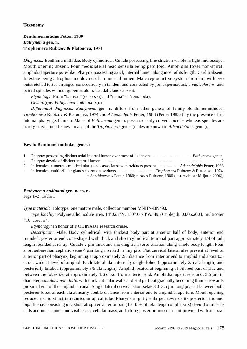
Benthimermitidae Petter, 1980Bathynema gen. n.Trophomera Rubtzov & Platonova, 1974
Diagnosis: Benthimermithidae. Body cylindrical. Cuticle possessing fine striation visible in light microscope. Mouth opening absent. Four mediolateral head sensilla being papilloid. Amphidial fovea non-spiral, amphidial aperture pore-like. Pharynx possessing axial, internal lumen along most of its length. Cardia absent. Intestine being a trophosome devoid of an internal lumen. Male reproductive system diorchic, with two outstretched testes arranged consecutively in tandem and connected by joint spermaduct, a vas deferens, and paired spicules without gubernaculum. Caudal glands absent.
Etymology: From “bathyal” (deep sea) and “nema” (=Nematoda).Generotype: Bathynema nodinauti sp. n.Differential diagnosis: Bathynema gen. n. differs from other genera of family Benthimermithidae,
Trophomera Rubtzov & Platonova, 1974 and Adenodelphis Petter, 1983 (Petter 1983a) by the presence of an internal pharyngeal lumen. Males of Bathynema gen. n. possess clearly curved spicules whereas spicules are hardly curved in all known males of the Trophomera genus (males unknown in Adenodelphis genus).
Key to Benthimermithidae genera
1 Pharynx possessing distinct axial internal lumen over most of its length ........................................ Bathynema gen. n.- Pharynx devoid of distinct internal lumen.................................................................................................................... 22 In females, numerous multicellular glands associated with oviducts present ...................... Adenodelphis Petter, 1983- In females, multicellular glands absent on oviducts ......................................Trophomera Rubtzov & Platonova, 1974
[= Benthimermis Petter, 1980; = Abos Rubtzov, 1980 (last revision: Miljutin 2006)]
Bathynema nodinauti gen. n. sp. n.Figs 1–2; Table 1
Type material: Holotype: one mature male, collection number MNHN-BN493.Type locality: Polymetallic nodule area, 14°02.7’N, 130°07.73’W, 4950 m depth, 03.06.2004, multicorer
#16, corer #4.Etymology: In honor of NODINAUT research cruise.Description: Male. Body cylindrical, with thickest body part at anterior half of body; anterior end
rounded, posterior end cone-shaped with thick and short cylindrical terminal part approximately 1/4 of tail, length rounded at its tip. Cuticle 2 µm thick and showing transverse striation along whole body length. Four short submedian cephalic setae 4 μm long inserted in tiny pits. Flat cervical lateral alae present at level of anterior part of pharynx, beginning at approximately 2/5 distance from anterior end to amphid and about 0.5 c.b.d. wide at level of amphid. Each lateral ala anteriorly single-lobed (approximately 2/5 ala length) and posteriorly bilobed (approximately 3/5 ala length). Amphid located at beginning of bilobed part of alae and between the lobes i.e. at approximately 1.6 c.b.d. from anterior end. Amphidial aperture round, 3.5 μm in diameter; canalis amphidialis with thick cuticular walls at distal part but gradually becoming thinner towards proximal end of the amphidial canal. Single lateral cervical short setae 3.0–3.5 μm long present between both posterior lobes of each ala at nearly double distance from anterior end to amphidial aperture. Mouth opening reduced to indistinct intracuticular apical tube. Pharynx slightly enlarged towards its posterior end and bipartite i.e. consisting of a short atrophied anterior part (10–15% of total length of pharynx) devoid of muscle cells and inner lumen and visible as a cellular mass, and a long posterior muscular part provided with an axial
Zootaxa 2096 © 2009 Magnolia Press · 175BENTHIMERMITHIDAE FROM THE NE PACIFIC

inner lumen and surrounded by longitudinal muscles; this inner lumen also irregular with wide and narrow parts, as well as meandering. Several large hyaline cells situated around posterior part of pharynx. Nerve ring located at level of middle of pharynx. Two large hyaline cells approximately 0.5 c.b.d. in diameter located at anterior part of midgut; each cell with a large single nucleus and a single nucleolus. Cardia absent. Midgut an oligocellular trophosome without visible internal lumen and obviously consisting of 1 row of large cells, except most anterior part consisting of several small trophosomal cells. All trophosomal cells containing large hyaline granules; borders between cells distinct. Male reproductive system diorchic, occupying approximately 2/3 of total body length and consisting of two outstretched testes (each about 450 μm long) arranged consecutively in tandem and connected by joint spermaduct (130 μm long), vas deferens (650 μm long), and two spicules. Spicules moderately bent, consisting of long, relatively optically dense lamina and a short, funnel-shaped, relatively hyaline, proximal part (capitulum). Capitulum ventrally bent and evidently representing a flexible structure (this supposition is based on the location of capitulum at different angles in different spicules). Four midventral short supplementary setae present anterior to cloacal opening. Caudal glands absent.
Host unknown.Female, juvenile stages: unknown.
TABLE 1. Main measurements and characteristics of Bathynema nodinauti gen. n. sp. n., (male, holotype).
Remarks: Two large hyaline cells at level of anterior part of midgut may be considered as cellular bodies of secretory-excretory system (renetta). However, neither channel nor outlet of renetta has been found. However, these hyaline cells may be also interpreted as a vestigial dorsal gland. In earlier larvae of benthimermithids, this gland together with a stylet in the anterior part of pharynx forms a complex whereby larvae penetrate into host’s internal issues (Chesunov 1997). This complex usually disappears at later larval stages, however, some mass of hyaline cells located between the pharynx and trophosome was also noted in male of T. australis (Petter 1983b).
Degree of maturity gravid
L, μm 2670
a 65.0
c 26.0
c' 3.2
Ratio of ‘length of genital system / total body length’, % 70
Amph.dist. 51
Cephalic sensilla length, mkm 4.0
Spicule length (μm):
in chord 43
in arc 51
Amphidial aperture diameter, μm 3.5
Maximal body diameter, μm 45
Body diameter (μm) at level of:
cephalic sensilla 16
amphid 36
1/4 body length 45
midbody 44
3/4 body length 42
anus 32
MILJUTIN & MILJUTINA176 · Zootaxa 2096 © 2009 Magnolia Press

FIGURE 1. Bathynema nodinauti gen. n. sp. n., male, holotype, total view.
Zootaxa 2096 © 2009 Magnolia Press · 177BENTHIMERMITHIDAE FROM THE NE PACIFIC

FIGURE 2. Bathynema nodinauti gen. n. sp. n., male, holotype. A, anterior end, lateral view; B, anterior end laterally, surface view; C, anterior end, frontal view; D, posterior end; E, spicula.
MILJUTIN & MILJUTINA178 · Zootaxa 2096 © 2009 Magnolia Press

Trophomera Rubtzov & Platonova, 1974= Benthimermis Petter, 1980 n. syn., = Abos Rubtzov, 1980 (op. Chesunov 1997)
Trophomera elegantis sp. n.Fig. 3; Table 2
Type material: Holotype: one gravid female, collection number MNHN-BN494.Type locality: 13°55.63’N, 130°12.20’W, 4900 m depth, 2–5 cm sediment layer, 28.05.2004, submersible
“Nautile”, station 1597-5 CL 06. Etymology: From Latin elegans/elegantis as adj. (slender, well-proportioned).Description: Female. Body cylindrical, with fine cuticular transversal striation visible using light
microscope; anterior and posterior ends in shape of rounded cone. Cuticle thickness approximately 2 μm along whole body. Amphidial apertures pore-like, 2 μm in diameter, located at 1.3 c.b.d. from anterior end. Amphidial fovea 3.5–4.0 μm in diameter; visible under cuticle. There are four submedian, thick papillae-like, cephalic setae 2.0–2.5 μm long at half the c.b.d. of their insertion from anterior end. Two cervical papilloid sensilla 1 μm long visible laterally before and behind amphid. Mouth opening reduced to a thin apical channel in cuticle. Pharynx a non-muscular, cellular mass devoid of an internal lumen; thin, axial, tube-like structure approximately 10 μm long visible in anterior part of pharynx only. Cardia absent. Midgut, an oligocellular trophosome without internal lumen and consisting of 1 row of cells at most part of body length; borders between cells indistinct. Trophosomal cells filled with large hyaline granules; density of these granules decreasing from anterior and posterior ends towards mid-body region. Rectum vestigial, in shape of hardly distinguishable duct. Anus also vestigial, visible as a thin transverse line/bar in cuticle. Female reproductive system didelphic, amphidelphic, occupying approximately 1/6 of total body length. Ovaries reflected, both about 120 μm long; anterior one lying at right side of body, and posterior one at left side. Oviducts short, with thin walls. Uterus with several mature eggs 24x23 μm in size. Neither morphologically differentiated spermatheca nor spermatozoa observed. Hyaline ring representing a circular vaginal sphincter visible around vagina. Caudal glands absent.
Host unknown.Male, juveniles: unknown.Differential diagnosis: T. elegantis sp. n. resembles T. iturupiensis Rubtzov & Platonova, 1974, T. hureaui
(Petter, 1983) (Petter 1983b), and T. conicauda (Petter, 1987) by the shape and tail characteristics (tail in shape of rounded cone; ratio c’ is 1.5; ratio “maximum c.b.d. / anal c.b.d.” is between 1 and 2).
The new species differs from T. iturupiensis by its shorter body length (1.5 mm vs. 26–38 mm), by the size of the cephalic sensilla (2.0–2.5 μm vs. 30–32 μm), by the structure of the trophosome (consisting of one row of cells vs. several cells on the sagittal optical section).
T. elegantis sp. n. differs from T. hureaui by its shorter body length (1.5 mm vs. 2.5–3.8 mm); by the smaller length of the mature, female genital system (occupying 1/6 body length vs. 1/4–1/3 body length); by the relatively bigger size of the eggs (ratio “c.b.d. at the uterus level / maximum egg diameter” = 1.8 vs. 4.0–5.0); by the caudal tip shape (rounded vs. pointed).
T. elegantis sp. n. differs from T. conicauda by its shorter body length (1.5 mm vs. 5.2–6.8 mm); by a thicker body (a = 37.3 vs. 49–58); by the relatively bigger size of the eggs (ratio “c.b.d. at the uterus level / maximum egg diameter” = 1.8 vs. 3.0). Each ovary of T. elegantis contains only 10–20 oocytes, whereas, in T. conicauda, number of oocytes in each ovary reaches hundred or more.
T. elegantis sp. n. resembles T. minutissima sp. n. by parameters of its female genital system (short reflected ovaries, short oviducts, few oocytes, and comparatively large mature eggs (ratio “c.b.d. at the uterus level / maximum egg diameter” is approximately 2 in both species). T. elegantis sp. n. differs from T. minutissima by its tail shape (in shape of rounded cone vs. rounded); and by its longer and thinner tail (c = 36.3 vs. 81.8 and the ratio “tail length / anal body diameter” = 1.5 vs. 0.55).
Zootaxa 2096 © 2009 Magnolia Press · 179BENTHIMERMITHIDAE FROM THE NE PACIFIC
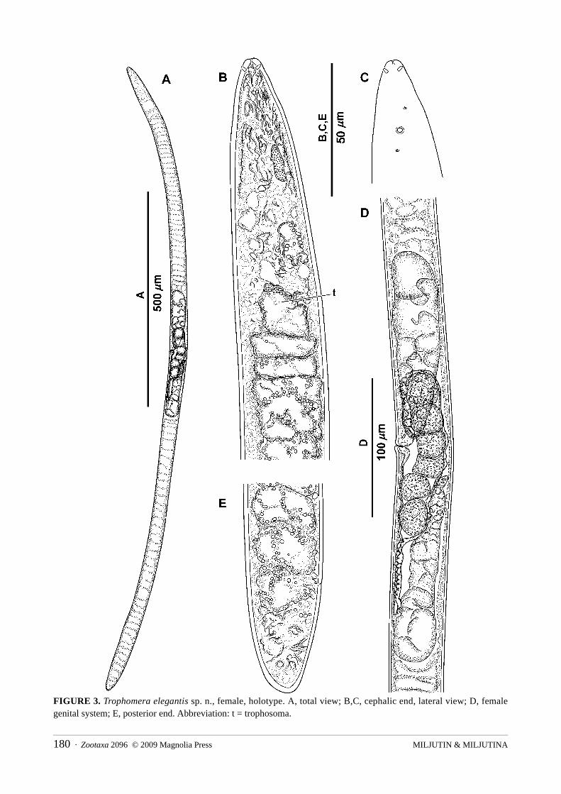
FIGURE 3. Trophomera elegantis sp. n., female, holotype. A, total view; B,C, cephalic end, lateral view; D, female genital system; E, posterior end. Abbreviation: t = trophosoma.
MILJUTIN & MILJUTINA180 · Zootaxa 2096 © 2009 Magnolia Press
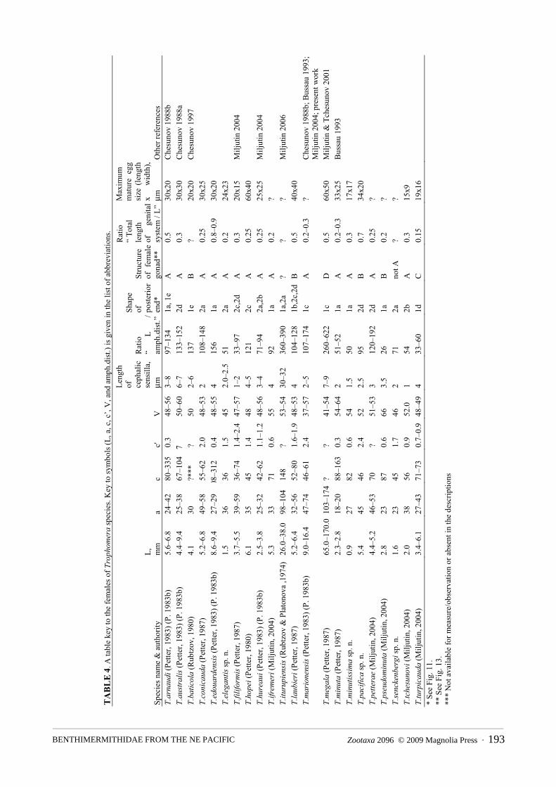
TABLE 2. Main measurements and characteristics of Trophomera species found in Clarion-Clipperton area. Key to
symbols (L, a, c, c’, V, and amph.dist.) is given in the list of abbreviations.
Trophomera marionensis (Petter, 1983)Fig. 4; Table 2
Additional data and illustrations as Benthimermis marionensis in Petter (1983b): 9–10, Fig. 5; in Chesunov (1988b): 15–17, Figs 3–4; in Bussau (1993): 570–571, Fig. 247; and in Miljutin (2004): 32–34, Fig. 7.
Material: One virgin female. Locality: 13°55.63’N, 130°12.20’W, 4800 m depth, 2–5 cm sediment layer, 26.05.2004, submersible
“Nautile”, station 1595-3 CL 05. Description: Female. Body thread-like, cylindrical. Anterior end in shape of a rounded cone. Posterior
end conical, with a terminal spine 110 μm long, possessing a cytoplasmic core with granular inclusions. Mouth opening vestigial, looking like an obscure channel at apical part of head end. Four submedian papilloid cephalic setae approximately 3.5 μm long inserted in tiny pits. Amphids not observed. Pharynx a non-muscular, parenchymal string without an internal lumen. Nerve ring visible between pharynx and trophosome. Midgut, a multicellular trophosome without visible internal lumen, consisting of several rows of cells in optical section. Anterior trophosomal cells located ahead of posterior part of pharynx. Borders between trophosomal cells distinct. Rectum and anus present. Female reproductive system amphidelphic.
Remarks: B. marionensis was described upon eight gravid females from the southern part of the Indian Ocean, 46°52′S, 37°51′E, 31–110 m depth (Petter 1983b). Other specimens were described later by Chesunov
Measurements and characteristicsT.elegantis
sp. n.
T.marione-nsis (Petter
1983b)
T.minu-tissimasp. n.
T.pacificasp. n.
T.sencken-bergisp. n.
T.sp.� *
��* Specimen in bad condition
Sex female female female female female maleDegree of maturity gravid virgin gravid gravid virgin senile
L, μm 1490 10215 900 5430 1560 6615a 36.3 60.1 27.3 44.5 22.6 92.0c 36.3 60.6 81.8 45.6 44.6 101.8c' 1.5 2.4 0.55 2.4 1.7 1.3V (%) 45.0 57.3 54.4 51.6 46.2 ?� **
��** It was impossible to measure this parameter.
Ratio of ‘length of genital system / total body length’, %
17 –� ***
��*** Parameter is absent in this specimen.
33 70 – ?
Amph.dist. 29 ? 18 57 22 98
Cephalic sensilla length, μm 2.0–2.5 3.5 1.5 2.5 2.0 4.0
Spicule length (μm): in chord – – – – – 49in arc – – – – – 49.5
Mature egg size (length x width), μm 24x23 – 17x17 34x20 – –
Amphidial aperture diameter, μm 2.0 ? 1.0 1.0 1.5 6.0
Maximal body diameter, μm 40 170 33 122 69 72
Body diameter (μm) at level of:cephalic sensilla 8 40 11 28 12 28amphid 22 ? 21 66 25 ?1/4 body length 33 167 33 122 69 ?midbody 41 170 30 115 75 723/4 body length 33 167 30 107 58 69anus 28 70 20 50 19 51
Zootaxa 2096 © 2009 Magnolia Press · 181BENTHIMERMITHIDAE FROM THE NE PACIFIC

(1988b) from the Southern Atlantic (57°09′S, 26°09′W, depth 1729 m), by Bussau (1993) from the Eastern Pacific (07°04.04′S, 88°27,41′W, depth 4154 m), and by Miljutin (2004) from the Western Atlantic (8°07.88′N, 49°03.71′ W, 4440 m depth). The new finding is the second record for the Pacific Ocean but it is far away from the previous region i.e. at a distance of about 5200 km. The new specimen agrees with the original description of B. marionensis in tail shape with terminal spine possessing a cytoplasmic core and structure of trophosome; body length and “V” value (10.2 mm and 57.3% vs. 9.3–14.8 mm and 46.5–74.0% respectively). However, the terminal spine of the new specimen is half as long as in the original description (110 μm vs. 200–320 μm) but closely resembles the specimen described in Miljutin (2004) (130 μm). Given the distance of the new specimen from the sites of the previous records, the slightly different morphological features may be attributed to differences between populations.
FIGURE 4. Trophomera marionensis (Petter 1983b), female. A, cephalic end; B, posterior end. Abbreviations: n.r = nerve ring; t = trophosome.
MILJUTIN & MILJUTINA182 · Zootaxa 2096 © 2009 Magnolia Press
Trophomera minutissima sp. n.Fig. 5; Table 2
Type material: Holotype: one gravid female, collection number MNHN-BN495.Type locality: 14°02.86’N, 130°05.35’W, 5040 m depth, 30.05.2004, multicorer #6, corer #2.Etymology: From Latin minutissima (smallest).Description: Female. Body cylindrical, with thickest body part at anterior half of body; anterior and
posterior ends rounded. Transverse cuticular striation along whole body length hardly discernable using light microscope. Cuticle thickness 2.5 μm at anterior tip, 1.5 μm at mid-body region, and 3 μm at posterior tip. Amphidial apertures pore-like, 1 μm in diameter, located in 0.81 c.b.d. from anterior and. Amphidial fovea 3 μm in diameter visible under cuticle. Four submedian, setiform papillae 1.5 μm long. Mouth opening absent. Pharynx a non-muscular string devoid of an internal lumen. Cardia absent. Midgut, an oligocellular trophosome without visible internal lumen and obviously consisting of 1 row of cells. Numerous big, hyaline granules within trophosomal cells. Borders between trophosomal cells distinct. Anus and rectum present. Female reproductive system didelphic, amphidelphic, occupying approximately 1/3 body length. Ovaries reflected, 100–110 μm long, containing several tens of oocytes. Neither spermatheca, nor spermatozoa, nor vulvar glands observed. Hyaline ring representing a circular vaginal sphincter visible around vagina. Mature eggs in oviducts and uterus approximately 17 μm in diameter. Caudal glands absent.
Host unknown.Male, juvenile stages unknown.Differential diagnosis: The female of Trophomera minutissima sp. n. shows resemblance to females of T.
bathycola (Rubtzov, 1980), T. edouardensis (Petter, 1983) (Petter 1983b), and T. minuta (Petter, 1987) by its reflected ovary and rounded tail without any process but differs from these species by its shorter body (0.9 mm vs. 2.3, 4.1, and 9.4 mm respectively). It is the smallest species in the genus Trophomera.
Trophomera pacifica sp. n. Figs 6–7; Table 2
Type material: Holotype: one gravid female, collection number MNHN-BN496.Type locality: Polymetallic nodule area, 14°02.52’N, 130°07.98’W, 4950 m depth, 07.06.2004, multicorer
#18, corer #2.Etymology: “Pacifica” means “from the Pacific Ocean”.Description: Female. Body cylindrical, with thickest body part at anterior half of body. Anterior end
rounded, posterior end conical, with thick conical terminal spine 81 μm long, showing granular core. Cuticle with very thin and hardly visible transverse striations along whole body length; striations being more pronounced at caudal region. Cuticle thickness 5.5 μm at anterior tip, 4 μm at level of amphids, and 3 μm in the rest of the body parts. Amphids located at 0.86 c.b.d. from anterior end; amphidial apertures pore-like, 1 μm in diameter. Four submedian cephalic setae 2.5 μm long inserted in tiny pits. Somatic sensilla in shape of small papillae 1–1.5 μm long arranged in 4 longitudinal lateromedian rows from postamphidial region to anal region. Mouth opening reduced to a thin channel in cuticle. Pharynx a non-muscular string devoid of an internal lumen. Several large hyaline cells situated at posterior pharynx. Cardia absent. Midgut an oligocellular trophosome without visible internal lumen and obviously consisting of 1 row of cells. Borders between trophosomal cells distinct. Rectum reduced to an indistinctly visible tube. Female reproductive system didelphic, amphidelphic, occupying approximately 2/3 body length in the gravid specimen. Ovaries outstretched, 530 µm long in anterior branch, 715 µm in posterior branch. Oviducts very short. Uterus very long, occupying approximately 3/5 body length. Mature eggs 34x20 μm in diameter situated in one row in uterus except vulva region. Neither morphologically differentiated spermatheca nor spermatozoa observed. Vulvar glands present. Caudal glands absent.
Zootaxa 2096 © 2009 Magnolia Press · 183BENTHIMERMITHIDAE FROM THE NE PACIFIC
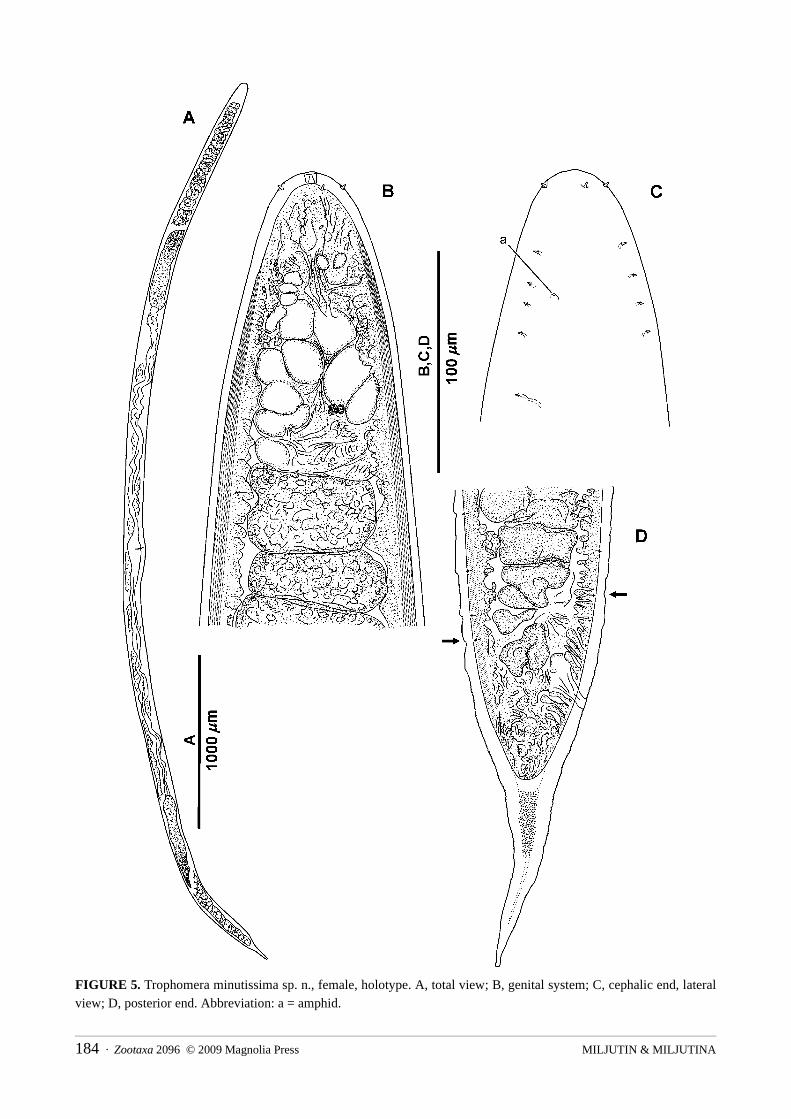
FIGURE 5. Trophomera minutissima sp. n., female, holotype. A, total view; B, genital system; C, cephalic end, lateral view; D, posterior end. Abbreviation: a = amphid.
MILJUTIN & MILJUTINA184 · Zootaxa 2096 © 2009 Magnolia Press

FIGURE 6. Trophomera pacifica sp. n., female, holotype. A, total view; B, cephalic end, lateral view; C, anterior part of cephalic end laterally, surface view; D, posterior end (some small, hardly visible lateromedian, somatic sensilla pointed to by arrows). Abbreviation: a = amphidial aperture.
Zootaxa 2096 © 2009 Magnolia Press · 185BENTHIMERMITHIDAE FROM THE NE PACIFIC

FIGURE 7. Trophomera pacifica sp. n., female, holotype. A, anterior ovary; B, vulva region; C, posterior ovary. Some small, hardly visible lateromedian, somatic sensilla are pointed to by arrows.
MILJUTIN & MILJUTINA186 · Zootaxa 2096 © 2009 Magnolia Press

Host unknown.Male, juvenile stages unknown.Differential diagnosis: The female of T. pacifica sp. n. possesses outstretched ovaries. Only three
Trophomera species have outstretched ovaries: T. laubieri (Petter 1987), T. pseudominuta (Miljutin 2004), and, possibly, T. senckenbergi sp. n.
T. pacifica sp. n. differs from T. pseudominuta by its larger body length (5.4 mm vs. 2.8 mm), and its tail shape (long conical spine vs. rounded).
T. pacifica sp. n. differs from T. senckenbergi sp. n. by its larger body length (5.4 mm vs. 1.5 mm), and its tail shape (long conical spine vs. cone-shaped with rounded tip).
In its general appearance, T. pacifica sp. n. strongly resembles T. laubieri. It differs from T. laubieri by: presence of a granular core inside the caudal terminal spine; a thickened cuticle at the cephalic apex; a difference in the ratio “distance from anterior end to amphid / c.b.d.” (0.86 vs. 0.72); trophosome structure (one row of cells at the longitudinal optical section vs. several rows); a longer female reproductive system (occupying approximately 0.7 body length vs. 0.45–0.50); egg size (34x20 μm vs. from 38x38 to 40x40 μm); and a different egg layout inside oviduct (only one row of eggs vs. several ones).
Trophomera senckenbergi sp. n.Fig. 8; Table 2
Type material: Holotype: one virgin female, collection number MNHN-BN497.Type locality: 14°02.55’N, 130°05.35’W, 5035 m depth, 31.05.2004, multicorer #8, corer #2.Etymology: In honour of Senckenberg Research Institute (Germany).Description: Female known only. Body slightly fusiform, with thickest body part at first half of body.
Anterior and posterior ends in shape of a cone with rounded tip. Cuticular, very thin, transverse, striation along whole body length being hardly visible except at caudal region. Cuticle thickness 1.5 μm at anterior tip and approximately 1 μm in rest body parts. Amphids located at 0.88 c.b.d. from anterior end; amphidial apertures pore-like, 1.5 μm in diameter. Four submedian, papilloid, cephalic setae 2 μm long inserted in tiny pits. Mouth opening reduced to a narrow, apical channel in cuticle. Pharynx looking like a non-muscular string devoid of an internal lumen. Cardia absent. Midgut an oligocellular trophosome without visible internal lumen and consisting of one row of big cells in longitudinal optical section. Borders between trophosomal cells distinct. Anus and rectum present. Female reproductive system didelphic, amphidelphic. Ovaries outstretched or hologonic (non-reflected) in single virgin specimen. No spermathecae nor sperm observed. Hyaline ring representing a circular vaginal sphincter visible around vagina. Caudal glands absent.
Host unknown.Males, juvenile stages unknown.Differential diagnosis: The female of Trophomera senckenbergi sp. n. is characterized by a non-reflected
ovary (it is impossible to clarify the certain construction because of immaturity of the type specimen). There are only five described Trophomera species, which have non-reflected ovaries: T. megala (Petter, 1987) and T. turpicauda Miljutin, 2004 possessing hologonic ovaries, as well as T. laubieri (Petter, 1987), T. pacifica sp. n. and T. pseudominuta Miljutin, 2004 possessing telogonic outstretched ovaries.
T. senckenbergi sp. n. differs from T. megala by its shorter body length (1.5 mm vs. 65–170 mm), body shape (fusiform vs. cylindrical), design of trophosome (oligocellular vs. multicellular), and its tail shape (conical with rounded tip vs. rounded with long terminal spine).
T. senckenbergi sp. n. differs from T. turpicauda by its shorter body length (1.5 mm vs. 3.4–6.1 mm), the body shape (fusiform vs. cylindrical), and its tail shape (conical with rounded tip vs. rounded with terminal, cuticular process of irregular shape).
T. senckenbergi sp. n. differs from T. laubieri and T. pacifica by its shorter body length (1.5 mm vs. 5.2–6.4 mm), the body shape (fusiform vs. cylindrical), and its tail shape (conical with rounded tip vs. conical with an edged elongated tip).
Zootaxa 2096 © 2009 Magnolia Press · 187BENTHIMERMITHIDAE FROM THE NE PACIFIC
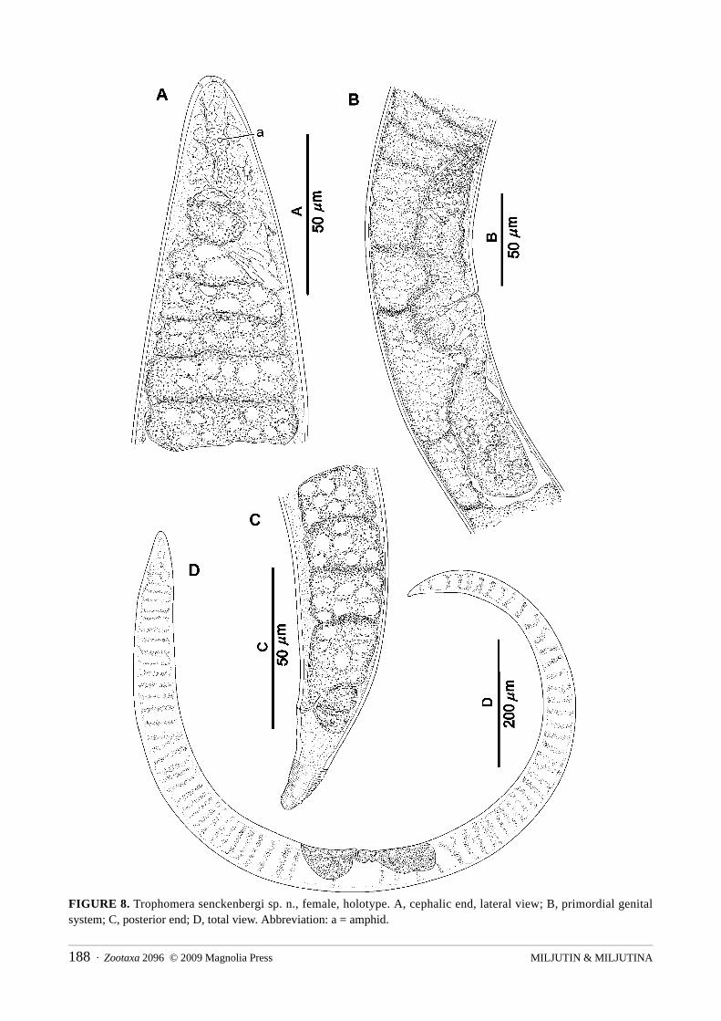
FIGURE 8. Trophomera senckenbergi sp. n., female, holotype. A, cephalic end, lateral view; B, primordial genital system; C, posterior end; D, total view. Abbreviation: a = amphid.
MILJUTIN & MILJUTINA188 · Zootaxa 2096 © 2009 Magnolia Press

T. senckenbergi sp. n. differs from T. pseudominuta by its shorter body length (1.5 mm vs. 2.8 mm), its longer tail (c = 44.6 vs. 86.9), and its tail shape (conical with rounded tip vs. rounded).
Remarks: The ovary of T. senckenbergi sp.n. is probably non-reflected. The reflected structure of ovaries is usually clearly visible even in virgin specimens (e.g. in T. tchesunovi (Miljutin, 2004) or in T. petterae(Miljutin, 2004)), whereas the structure of ovaries of virgin female of T. turpicauda (possessing a hologonic ovaries) was clearly non-reflected.
Trophomera sp.Fig. 9; Table 2
Material: One male in a bad condition though with most diagnostic features discernable.Locality: 13°55.63’N, 130°12.20’W, 4900 m depth, 2–5 cm sediment layer, 28.05.2004, submersible
“Nautile”, station 1597-5 CL 06.
FIGURE 9. Trophomera sp., male. A, B, cephalic end; C, posterior end; D, spicula. Abbreviations: l.a = lateral ala; a = amphid; v.d = vas deferens.
Description: Body thread-like, cylindrical. Anterior end a slightly narrowing cone with rounded apex, posterior end a pointed cone shape. Transverse cuticular striation visible along whole body length. Cuticle thickness 3 μm along whole body length except terminal part of tail, where its thickness increasing to 6–10 μm, and 20 μm at caudal tip. Four submedian, short cephalic setae with rounded tips 4 μm long inserted in tiny pits. Two flat, cervical, lateral alae with a preamphidial part beginning at 1/3 distance from cephalic end to amphid and a postamphidial part (1/9 ala length). Amphidial aperture round, 6.0 μm in diameter. Short, somatic papillae approximately 1 μm long. Mouth opening reduced to a narrow apical tube. Pharynx a non-
Zootaxa 2096 © 2009 Magnolia Press · 189BENTHIMERMITHIDAE FROM THE NE PACIFIC

muscular, cellular mass devoid of an internal lumen. Midgut an oligocellular trophosome without internal lumen and consisting of 1 row of cells. Borders between trophosomal cells distinct. Trophosomal cells consisting of large hyaline granules; density of these granules decreasing from anterior and posterior ends towards mid-body region. Spicules paired, almost straight. 15–16 mid-ventral, small, setiform supplementary organs present, from anterior to anus. Caudal glands absent.
Host unknown.Females, juvenile stages unknown.Remarks: The single specimen is in bad condition. Its body is strongly crumpled at the postamphidial part
of the pharynx and beginning of the trophosome. The male reproductive system is wrinkled and collapsed, and it is impossible to ascertain its structure (number and arrangement of testes).
However, the described specimen evidently belongs to a new species of Trophomera because of its peculiar tail, characterized by a very thick cuticle. It only resembles T. gracilis (Petter 1982a) in the shape and structure of cervical lateral alae (long pre-amphidial part and short, not bilobed post-amphidial part). But, unlike the male specimen described, T. gracilis possesses a rounded tail. Unfortunately, the poor condition of the specimen does not facilitate good description and makes the specimen unsuitable for the status of holotype.
Table keys to Trophomera species
The tabular keys to all known Trophomera species are presented in Tables 3 and 4. Because most of species were described on the basis of either males or female only, the keys for males and females are given separately. The keys are also supplemented with pictorial, schematic material on the main types of structure of male cervical lateral alae (Fig. 10), shape of tails (Fig. 11), and structure of male (Fig. 12) and female (Fig. 13) genital systems occurring in Trophomera species. These schemes should not be considered as morphological/phylogenetic series.
FIGURE 10. Main types of structure of cervical lateral alae occuring in males of Trophomera species. A, alae preamphidial, extending the whole distance from the cephalic apex to amphid; B, alae preaphaidial, extending only a part of the distance from the cephalic apex to amphid; C, alae possessing preamphidial and postamphidial parts.
MILJUTIN & MILJUTINA190 · Zootaxa 2096 © 2009 Magnolia Press

FIGURE 11. Main types of shapes of posterior ends occuring in Trophomera species. 1a, rounded tail; 1b, rounded tail with terminal spine; 1c, rounded tail with terminal spine possesing additional cuticular layers and granular inclusions; 1d, rounded tail with abnormal terminal spine; 1e, rounded tail with small terminal knob; 2a, tail in shape of rounded cone; 2b, cone-shaped tail; 2c, cone-shaped tail with attenuatous tip; 2d, cone-shaped tail with terminal spine.
Discussion
The new findings of the pacific benthimermithids are from sites located far from previously known records in the Pacific Ocean. Bussau (1993) described 2 new species and 2 known species from the Atlantic and from the deep-sea area (DISCOL area, 07°04′S, 88°27′W, the Eastern Pacific) located at a distance of about 5200 km from the area of the new findings. Two species were described from the tidal zone on the Kuril Archipelago (North-western Pacific, about 7800 km from the area of the new findings) by Rubtzov and Platonova (1974) and Miljutin (2006).
So, at present, among 38 known species of benthimermithids, 11 species were described from the Pacific (in 4 publications), 9 species from the Indian Ocean (in 3 publications), and 32 species from the Atlantic (in 11 publications), both from northern and southern hemispheres. Several species were found in two oceans, and one species was described from three oceans.
It is obvious that benthimermithids are wide-spread in the World Oceans. It is highly likely the known, natural habitat of benthimermithids will be extended as new deep-sea areas of the world ocean will be studied.
Acknowledgements
The authors thank Dr. Prof. Pedro Martinez Arbizu (Senckenberg Research Institute, the German Centre for Marine Biodiversity Research, Wilhelmshaven, Germany), who kindly provided the collected meiobentic samples for this investigation, and Mr. Radith Mahatma (the same institution) for his participation in sample treatment. Investigation was also supported by CeDAMar and CoML. The authors also express their gratitude to two anonymous reviewers and the guest editor Dr. Kai Horst George (Senckenberg am Meer Wilhelmshaven, Abt. DZMB, Germany) for valuable remarks.
Zootaxa 2096 © 2009 Magnolia Press · 191BENTHIMERMITHIDAE FROM THE NE PACIFIC

TAB
LE 3
. A ta
ble
key
to th
e m
ales
of T
roph
omer
a sp
ecie
s. K
ey to
sym
bols
(L, a
, c, c
’, an
d am
ph.d
ist.)
is g
iven
in th
e lis
t of a
bbre
viat
ions
.
Spec
ies n
ame
& a
utho
rity
Stru
ctur
e of
ce
rvic
al
alae
*
Rat
io
“Len
gth
of
prea
mph
idia
l pa
rt of
ala
e /
amph
.dis
t.”
Rat
io
“Len
gth
of
post
amph
idia
l pa
rt of
al
ae /
amph
.dis
t.”
Rat
io
“ L
/ am
ph.d
ist
.”
Leng
th
of
ceph
alic
se
nsill
a,
μmL,
m
ma
c с'
Shap
e
of
post
erio
r en
d**
Stru
ctur
e of
m
ale
gona
d***
Spic
ule
leng
th
in a
rc,
μmN
umbe
r of
supp
lem
ents
T.
abys
salis
(Bus
sau,
199
3)
B
0.7
–***
*67
–72
5–6
5.9–
7.2
66–7
2 54
–70
1.5–
1.9
2c,2
d C
70
16
T.
acut
icau
da (P
ette
r, 19
81) (
P. 1
981a
) B
1.
0 –
53–9
2 5
4.8–
8.3
52
53–1
041.
3 2c
C
45
–70
12–1
9 T.
apte
ra (P
ette
r, 19
82) (
P. 1
982b
) B
0.
1 –
49
3–5
3.7
74
74
1.1
2c,2
d C
30
.00
19
T.au
stra
lis (P
ette
r, 19
83) (
P. 1
983b
) A
1.
0 –
87–1
23
7 3.
3–4.
3 41
–54
65–8
6 1.
0 2a
C
52
–55
18–2
4 T.
brev
ipte
ra (P
ette
r, 19
82) (
P. 1
982b
) B
0.
2 –
27–3
3 3–
4 3.
5–5.
1 10
2 58
–73
1.3–
1.4
2c
A
30–4
0 12
T.
cros
eten
sis (
Pette
r, 19
83) (
P. 1
983b
) B
0.
7 –
68–8
2 5
4.0–
4.5
40–4
5 31
–32
2.0–
2.1
2b,2
c C
50
–60
14–2
2 T.
dipl
opte
ra (P
ette
r, 19
81) (
P. 1
981b
) C
0.
3 0.
7 53
–115
3
6.6–
15.0
130
83–3
001.
3 2b
,2c
E 70
–120
18–6
8T.
grac
ilis (
Pette
r, 19
82) (
P. 1
982a
) C
0.
7 0.
3 53
–76
4 4.
4–6.
1 72
–98
285–
543
0.5–
0.6
1a
D
75–1
0016
–26
T.im
prov
isa
(Milj
utin
, 200
4)
B
0.3
– 73
2.
5–3
4.8
88
92
1.5
2a
F 28
.00
9 T.
lept
osom
a (P
ette
r, 19
81) (
P. 1
981a
) A
1.
0 –
42–1
47
4 4.
2–6.
6 86
53
–120
1.3
2b,2
c C
38
–50
14–3
8 T.
litor
alis
Milj
utin
, 200
6 C
0.
7 0.
7 12
3–14
517
5.
8–9.
3 67
–107
61–9
7 1.
1–1.
42a
C
50
–76
>100
T.
pette
rae
(Milj
utin
, 200
4)
B
0.7
– 68
5
5.8
78
70
1.6
2b,2
c B
29
.00
10
T.pl
atyp
tera
(Milj
utin
, 200
4)A
1.
0 –
69
1.5
3.0
84
102
0.8
1a
C
19.0
0 2
T.re
galis
(Bus
sau,
199
3)
C
0.3
0.7
83
10
14.8
12
4 13
6 1.
5 2b
C
13
0 49
T.
rosa
liae
(Che
suno
v, 1
988)
(Ch.
198
8b) B
0.
7 –
75
4–5
4.7
50
31
2.3
2c
C
55–5
8 11
–15
T.ro
tund
icau
da (P
ette
r, 19
81) (
P. 1
981a
)A
1.
0 –
83–9
8 6–
7 10
.0–1
3.8
94
321–
630
0.3
1a
C
140–
160
<=12
* Se
e Fi
g. 1
0.
** S
ee F
ig. 1
1.
***
See
Fig.
12.
**
** P
osta
mph
idia
l par
t of a
lae
is a
bsen
t.
MILJUTIN & MILJUTINA192 · Zootaxa 2096 © 2009 Magnolia Press
TAB
LE
4. A
tabl
e ke
y to
the
fem
ales
of T
roph
omer
a sp
ecie
s. K
ey to
sym
bols
(L, a
, c, c
’, V
, and
am
ph.d
ist.)
is g
iven
in th
e lis
t of a
bbre
viat
ions
.
Spec
ies n
ame
& a
utho
rity
L,
mm
a
c c'
V
Leng
th
of
ceph
alic
se
nsill
a,
μm
Rat
io
“ L
/ am
ph.d
ist.”
Shap
e
of
post
erio
r en
d*
Stru
ctur
e of
fem
ale
gona
d**
Rat
io
“ To
tal
leng
th
of
geni
tal
syst
em /
L”
Max
imum
m
atur
e eg
g si
ze (
leng
th
x w
idth
), μm
Oth
er re
fere
nces
T.
arna
udi (
Pette
r, 19
83) (
P. 1
983b
) 5.
6–6.
8 24
–42
80–3
350.
3 48
–56
3–8
97–1
34
1a, 1
e A
0.
5 30
x20
Che
suno
v 19
88b
T.au
stra
lis (P
ette
r, 19
83) (
P. 1
983b
) 4.
4–9.
4 25
–38
67–1
047
50–6
06–
7 13
3–15
2 2d
A
0.
3 30
x30
Che
suno
v 19
88a
T.ba
ticol
a (R
ubtz
ov, 1
980)
4.
1 30
?*
**
? 50
2–
6 13
7 1e
B
?
20x2
0 C
hesu
nov
1997
T.
coni
caud
a (P
ette
r, 19
87)
5.2–
6.8
49–5
8 55
–62
2.0
48–5
32
108–
148
2a
A
0.25
30
x25
T.
edou
arde
nsis
(Pet
ter,
1983
) (P.
198
3b)
8.6–
9.4
27–2
9 08
–312
0.
4 48
–55
4 15
6 1a
A
0.
8–0.
9 30
x20
T.
eleg
antis
sp. n
. 1.
5 36
36
1.
5 45
2.
0–2.
5 51
2a
A
0.
2 24
x23
T.
filifo
rmis
(Pet
ter,
1987
) 3.
7–5.
5 39
–59
36–7
4 1.
4–2.
447
–57
1–2
33–9
7 2c
,2d
A
0.3
20x1
5 M
iljut
in 2
004
T.ho
pei (
Pette
r, 19
80)
6.1
35
45
1.4
48
4–5
121
2c
A
0.25
60
x40
T.
hure
aui (
Pette
r, 19
83) (
P. 1
983b
) 2.
5–3.
8 25
–32
42–6
2 1.
1–1.
248
–56
3–4
71–9
4 2a
,2b
A
0.25
25
x25
Milj
utin
200
4 T.
ifrem
eri (
Milj
utin
, 200
4)
5.3
33
71
0.6
55
4 92
1a
A
0.
2 ?
T.
ituru
pien
sis (
Rub
tzov
& P
lato
nova
,197
4)26
.0–3
8.0
98–1
0414
8 ?
53–5
430
–32
360–
390
1a,2
a ?
? ?
Milj
utin
200
6 T.
laub
ieri
(Pet
ter,
1987
) 5.
2–6.
4 32
–56
52–8
0 1.
6–1.
948
–53
4 10
4–12
8 1b
,2c,
2dB
0.
5 40
x40
T.
mar
ione
nsis
(Pet
ter,
1983
) (P.
198
3b)
9.0–
16.4
47
–74
46–6
1 2.
4 37
–57
2–5
107–
174
1c
A
0.2–
0.3
? C
hesu
nov
1988
b; B
ussa
u 19
93;
Milj
utin
200
4; p
rese
nt w
ork
T.m
egal
a (P
ette
r, 19
87)
65.0
–170
.010
3–17
4?
? 41
–54
7–9
260–
622
1c
D
0.5
60x5
0 M
iljut
in &
Tch
esun
ov 2
001
T.m
inut
a (P
ette
r, 19
87)
2.3–
2.8
18–2
0 88
–163
0.3
54–6
42
51–5
2 1a
A
0.
2–0.
3 33
x25
Bus
sau
1993
T.
min
utis
sim
a sp
. n.
0.9
27
82
0.6
54
1.5
50
1a
A
0.3
17x1
7
T.pa
cific
a sp
. n.
5.4
45
46
2.4
52
2.5
95
2d
B
0.7
34x2
0
T.pe
ttera
e (M
iljut
in, 2
004)
4.
4–5.
2 46
–53
70
? 51
–53
3 12
0–19
2 2d
A
0.
25
?
T.ps
eudo
min
uta
(Milj
utin
, 200
4)
2.8
23
87
0.6
66
3.5
26
1a
B
0.2
?
T.se
ncke
nber
gi sp
. n.
1.6
23
45
1.7
46
2 71
2a
no
t A
? ?
T.
tche
suno
vi (M
iljut
in, 2
004)
2.
0 38
56
0.
9 52
.0
1 54
2b
A
0.
3 15
x9
T.
turp
icau
da (M
iljut
in, 2
004)
3.
4–6.
1 27
–43
71–7
3 0.
7–0.
948
–49
4 33
–60
1d
C
0.15
19
x16
*
See
Fig.
11.
**
See
Fig
. 13.
**
* N
ot a
vaila
ble
for m
easu
re/o
bser
vatio
n or
abs
ent i
n th
e de
scrip
tions
Zootaxa 2096 © 2009 Magnolia Press · 193BENTHIMERMITHIDAE FROM THE NE PACIFIC

FIGURE 12. Main types of Trophomera female reproductive systems. A, two testes directed anterior; B, testes arranged consecutively in tandem and connected by joint spermaduct, posterior testis having a small caecum directed anterior; C, testes arranged consecutively in tandem and connected by joint spermaduct; D, testes arranged consecutively in tandem and connected by joint shortened spermaduct; E, testes almost merged; F, testes merged in one long testis. From Miljutin (2004).
MILJUTIN & MILJUTINA194 · Zootaxa 2096 © 2009 Magnolia Press

FIGURE 13. Main types of Trophomera female reproductive systems (germinal zones are indicated by the hatching). A, telogonic reflected ovaries (most species), B, telogonic outstretched ovaries (two known species); C, short hologonic
ovaries; D, long hologonic ovaries. Abbreviations: o, oocytes or eggs; od, oviduct; v, vulva. From Miljutin (2004).
References
Bussau, C. (1993) Taxonomische und ökologische Untersuchungen an Nematoden des Peru-Beckens. Dissertation zur Erlangung des Doktorgrades der Mathematisch-Naturwissenschaftlichen Fakultät der Christian-Albrechts-Universität zu Kiel. 621 pp.
Chesunov, A. V. (1988a) A case of nematode parasitism in nematodes. A new find and re-description of a rare species Benthimermis australis Petter, 1983 (Nematoda: Marimermithida: Benthimermithidae) in South Atlantic. Helminthologia, 25, 115–128.
Chesunov, A.V. (1988b) [New finds of deep-sea nematodes of the family Benthimermithidae in South Atlantic with description of a new species.] Vestnik Zoologii, 5, 12–22. In Russian.
Chesunov, A.V. (1997) Marimermithids (Nematoda): anatomy, taxonomy, and phylogeny. Russian Journal of Zoology, 1, 440–455.
Hope W. D. (1977) Gutless nematodes of the deep-sea. Mikrofauna des Meersbodens, 61, 307–308.Khripounoff, A., Caprais, J.-C., Crassous, P. (2006) Geochemical and biological recovery of the disturbed seafloor in
polymetallic nodule fields of the Clipperton-Clarion Fracture Zone (CCFZ) at 5,000-m depth. Limnology and Oceanography, 51(5) 203, 32–41.
McIntyre, A.D. & Warwick, R.M. (1984) Meiofauna techniques. In: Holme, N.A. & McIntyre, A.D. (Eds.), Methods for the Study of Marine Benthos. Blackwell Scientific Publishers, Oxford, pp. 217–244.
Miljutin, D. M. (2004) New findings of deep-sea nematodes of the family Benthimermithidae Petter, 1980 with descriptions of seven new species. Zoosystema, 26(1), 21–48.
Miljutin, D. M. (2006) The genus Trophomera Rubtzov & Platonova, 1974 with description of T. litoralis sp. n. (Nematoda: Benthimermithidae) from the tidal zone of the Kuril Archipelago and proposal of Benthimermis Petter, 1980 as a junior synonym. Nematology, 8(3), 411–423.
Petter, A.-J. (1980) Une nouvelle famille de Nématodes parasites d'Invertébrés marins, les Benthimermithidae. Annales de Parasitologie Humaine et Comparée, 55(2), 209–224.
Zootaxa 2096 © 2009 Magnolia Press · 195BENTHIMERMITHIDAE FROM THE NE PACIFIC
Petter, A.-J. (1981a) Description des mâles de trois nouvelles espèces de nématodes de la famille des Benthimermithidae. Bulletin du Muséum National d’Histoire Naturelle, Paris, 4e série, 3, section A 2, 455–465.
Petter, A.-J. (1981b) Description des mâles d’une nouvelles espèce de nématode marin de la famille des Benthimermithidae. Annales de Parasitologie Humaine et Comparée 56, 285–295.
Petter, A.-J. (1982a) Benthimermis gracilis n. sp., nouveau mâle de la famille des Benthimermithidae (Nematoda). Bulletin du Muséum national d’Histoire naturelle, Paris, 4e série, 9, section A, 1–2, 71–74.
Petter, A.-J. (1982b) Description de deux nouveaux mâles de la famille des Benthimermithidae (Nematoda) de l’Atlantique sud-oriental. Bulletin du Muséum National d’Histoire Naturelle, Paris, 4e série, 9, section A 3–4, 397–403.
Petter, A.-J. (1983a) Description d’un nouveau genre de Benthimermithidae (Nematoda) présentant des utérus munis de glandes annexes. Annales de Parasitologie Humaine et Comparée, 58, 177–184.
Petter, A.-J. (1983b) Quelques nouvelles espèces du genre Benthimermis Petter, 1980 (Benthimermithidae: Nematoda) du Sud de l'Océan Indien. Systematic Parasitology, 5, 1–15.
Petter, A.-J. (1987) Quelques nouvelles espèces de femelles du genre Benthimermis Petter, 1980 (Benthimermithidae: Nematoda) des grands fonds de la mer de Norvège. Bulletin du Muséum national d’Histoire naturelle, Paris, 4e série, 9, section A 3, 565–578.
Petter, A. J. & Gourbault, N. (1985) Nématodes abyssaux (campagne Walda du N/O “Jean Charcot”) IV. Des nématodes parasites de Nématodes. Bulletin du Musèum national d’Histoire naturelle 4-e série, 7, section A, no. 1, 125–130.
Rubtzov, I. A. (1980) [The new marine parasitic nematode, Abos bathycola, from priapulids and a taxonomic position of the family Marimermithidae in the class Nematoda]. Parazitologiya, 14, 177–181. In Russian with English summary.
Rubtzov, I.A. & Platonova, T.A. (1974) [A new family of marine parasitic nematodes.] Zoologicheskij Zhurnal, 53, 1445–1458. In Russian with English summary.
Seinhorst, J. W. (1959) A rapid method for the transfer of nematodes from fixative to anhydrous glycerin. Nematologica, 4, 67–69.
MILJUTIN & MILJUTINA196 · Zootaxa 2096 © 2009 Magnolia Press